Note: Descriptions are shown in the official language in which they were submitted.
Application No. 3055962 Our
Ref 31110-136
CA National Phase of PCT/US2018/021527
(60836CA01)
METHOD AND SYSTEM FOR DELIVERING A SELF-EXPANDING STENT TO
THE VENOUS SINUSES
CROSS-REFERENCE TO RELATED APPLICATIONS
[01] This application claims priority to and benefit from United States Patent
Application No. 15/456,352, filed on March 10, 2017.
F __________________________________ LD
[02] Certain embodiments relate to stents and systems and methods for
delivering a
stent. More specifically, certain embodiments relate to a method and system
for treating
a stenosis or collapse in the venous sinuses by delivering a self-expanding
stent. In
various embodiments, the self-expanding stent comprises a proximal end having
a first
radial outward expansion strength (RES) that is greater than a second RES at a
distal end
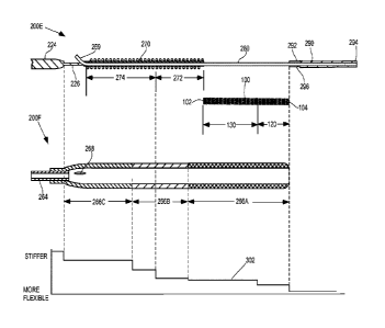
of the stent. In a representative embodiment, the proximal end of the stent
comprises a
diameter that is greater than the diameter at the distal end of the stent. In
certain
embodiments, the flexibility of the stent delivery system and/or the stent
increases from
the proximal end toward the distal end of the system and/or stent.
BACKGROUND
[03] When blood exiting the brain is slowed by a restriction in the venous
sinuses, it
causes an increase to the distal blood pressure, which may translate to an
increase in the
brain fluid pressure. Patients experiencing Increased Intracranial Pressure
(ICP), where
the Cerebral Spinal Fluid (CSF) pressure in the cranium has increased, may
suffer from
headaches, loss of vision, and/or tinnitus, among other things. The preferred
method for
treating a collapse of and/or a stenosis in the sigmoid and/or transverse
sinus has been
1
Date recue/Date received 2023-04-20
CA 03055962 2019-09-09
WO 2018/165415
PCT/US2018/021527
drugs and/or using a shunt to relieve the CSF fluid pressure. The use of drugs
or a shunt
is not ideal, however, because both are temporary solutions that each carry
associated
risks.
[04] More, recently, a new procedure has been carried out that involves
placing a stent
in the venous sinus system of patients to ameliorate a collapse of and/or a
stenosis in the
sigmoid and/or transverse sinus and to restore improved blood flow out of the
brain. The
stent used in the new procedure typically is the same stent used for
procedures in other
parts of the body, such as the carotid artery. The venous sinus structure,
however, does
not resemble any vein or arteries of other parts of the body. Instead, the
venous sinus is a
void created where the dura joins and forms a cavity (i.e., sinus) primarily
along the
inside of the skull. The dura has no smooth muscle cell lining and is
inelastic when
compared to veins and arteries.
[05] FIG. 1 illustrates an exemplary venous sinus system having an identified
stent
zone. The venous sinus system comprises venous channels found between the
periosteal
and meningeal layers of dura mater in the brain. The venous sinus system
receives blood
from internal and external veins of the brain, receives CSF from the
subarachnoid space
via arachnoid granulations, and mainly empties into the internal jugular vein.
As
illustrated in FIG. 1, the venous sinus system includes the transverse sinus,
sigmoid sinus,
and the sigmoid junction. The sigmoid sinus integrates into the jugular vein
at the
sigmoid junction. FIG. 1 also identifies an exemplary stent zone for placing a
stent to
treat a collapse of and/or a stenosis in the sigmoid and/or transverse sinus.
[06] Existing stent delivery systems and stents have several inadequacies for
delivering
a stent to the venous sinuses. For example, existing stents and systems may be
incapable
of or difficult to navigate through the tortuous sigmoid junction for
placement of the stent
in the stent zone.
[07] As another example, the properties of existing stents may be undesirable
for
placement in the venous sinuses. The length of a typical carotid artery stent
may be 4-6
cm long. However, after placement of a carotid artery stent in the venous
sinuses, a
portion of the transverse sinus could collapse, particularly a portion that is
distal to the
distal end of the stent. The collapse of a portion of the transverse sinus may
occur if the
2
CA 03055962 2019-09-09
WO 2018/165415 PCT/US2018/021527
stent is placed in the sigmoid to transverse junction and is not long enough
to scaffold
most or the entirity of transverse sinus. Additionally, multiple carotid
artery stents may
be required if there are collapses and/or stenosis at multiple locations in
the sigmoid
and/or transverse sinuses. Also, a stent having an inappropriate length could
be
incorrectly positioned at the curves in the sigmoid sinus to block off future
access to the
sinus (e.g., a stent jail). For example, a stent that terminates within a
curve, instead of
being positioned through the curve, may block a portion or the entire sinus
lumen at the
curve.
[08] Furthermore, exiting stents typically come in one set diameter. However,
the
middle and distal region of the sigmoid sinus has on average a larger diameter
(e.g., ¨10-
12 mm) than the distal section of the transverse sinus (e.g., ¨6-9 mm).
Accordingly,
existing stent diameters positioned in both the sigmoid and transverse sinuses
may be
inadequate for at least one of the sinuses. For example, if the stent is too
small for a
vessel, a portion of the stent can be left dangling or free floating in the
vessel, which may
prevent proper endothelium tissue growth over the stent struts. As another
example, if
the stent is too large for a vessel, various problems may occur because the
radial outward
expansion strength (RES) of typical stents may be too forceful for use in the
venous
sinuses. Specifically, stents intended for placement in large vessels such as
the carotid
artery, femoral artery or veins, and the like, may have a high RES required
for treatment
of occlusions, atherosclerosis plaque, and lesion calcification, and/or that
can withstand
an outside force capable of pushing in on the stent. This high RES, coupled
with a stent
size that is too large for a vessel, can create a problem of tissue in contact
with the stent
struts dying due to the strong outward pressure exerted on the tissue. Another
problem
arising with a high RES when stents are too large for a vessel is that the
stent may push
through the vessel wall and show on the outside of the vessel.
[09] Existing stent designs may also have an abundance of struts members.
However,
the venous sinus structure includes numerous small veins leading from the
brain.
Accordingly, the quantity of strut members of a typical stent increases the
chance that
one strut might block, or partially inhibit the venous inflow from the brain
via the veins.
3
CA 03055962 2019-09-09
WO 2018/165415
PCT/US2018/021527
[10] Further limitations and disadvantages of conventional and traditional
approaches
will become apparent to one of skill in the art, through comparison of such
systems with
some aspects of the present invention as set forth in the remainder of the
present
application with reference to the drawings.
4
CA 03055962 2019-09-09
WO 2018/165415
PCT/US2018/021527
BRIPF SUMMARY
[11] Enhanced navigation of a stent delivery system for placement of a stent
is
provided by increasing the flexibility of the stent delivery system and/or the
stent from
the proximal end toward the distal end of the system and/or stent,
substantially as shown
in and/or described in connection with at least one of the figures, as set
forth more
completely in the claims.
[12] These and other advantages, aspects and novel features of the present
invention,
as well as details of an illustrated embodiment thereof, will be more fully
understood
from the following description and drawings.
CA 03055962 2019-09-09
WO 2018/165415
PCT/US2018/021527
BRIEF DESCRIPTION OF SEVERAL VIEWS OF THE DRAWINGS
[13] FIG. 1 illustrates an exemplary venous sinus system having an identified
stent
zone, in accordance with various embodiments.
[14] FIG. 2 illustrates an exemplary stent comprising a distal end and a
proximal end,
the distal end having a greater flexibility than the proximal end, in
accordance with
various embodiments.
[15] FIG. 3 illustrates exemplary strut members of the exemplary stent of FIG.
2, in
accordance with various embodiments.
[16] FIG. 4 illustrates an exemplary profile of the exemplary stent 100 of
FIG. 2
having a distal end with a smaller diameter than the diameter of the proximal
end, in
accordance with various embodiments.
[17] FIG. 5 illustrates an exemplary stent delivery system, in accordance with
various
embodiments.
[18] FIG. 6 illustrates a detail view of portions of the exemplary stent
delivery system
of FIG. 5, in accordance with various embodiments.
[19] FIG. 7 illustrates a detail view of an inner portion of the stent
delivery system of
FIG. 5, in accordance with various embodiments.
[20] FIG. 8 illustrates a detail view of an outer portion of the stent
delivery system of
FIG. 5, in accordance with various embodiments.
[21] FIG. 9 illustrates an exploded, cross-sectional view of the inner
portion of the
stent delivery system, the stent, and the outer portion of the stent delivery
system, where
the increasing flexibility of the stent delivery system with the stent from
the proximal end
toward the distal end of the system and stent is illustrated by a mapping to
an exemplary
flexibility chart, in accordance with various embodiments.
[22] FIG. 10 is a flow chart illustrating exemplary steps that may be utilized
for
providing enhanced navigation of a stent delivery system for placement of a
stent, in
accordance with various embodiments.
6
CA 03055962 2019-09-09
WO 2018/165415
PCT/US2018/021527
DETAILED DESCRIPTION
[23] Certain embodiments may provide enhanced navigation of a stent delivery
system
for placement of a stent by increasing the flexibility of the stent delivery
system and/or
the stent from the proximal end toward the distal end of the system and/or
stent. Various
embodiments provide a self-expanding stent that comprises a proximal end
having a first
radial outward expansion strength (RES) that is greater than a second RES at a
distal end
of the stent. In a representative embodiment, the proximal end of the stent
comprises a
diameter that is greater than the diameter at the distal end of the stent. In
certain
embodiments, the stent delivery system may be configured to treat a stenosis
or collapse
in the venous sinuses by delivering the self-expanding stent.
[24] The foregoing summary, as well as the following detailed description of
certain
embodiments will be better understood when read in conjunction with the
appended
drawings. It should be understood that the various embodiments are not limited
to the
arrangements and instrumentality shown in the drawings. It should also be
understood
that the embodiments may be combined, or that other embodiments may be
utilized and
that structural changes may be made without departing from the scope of the
various
embodiments. The following detailed description is, therefore, not to be taken
in a
limiting sense, and the scope of the present invention is defined by the
appended claims
and their equivalents.
[25] As used herein, an element or step recited in the singular and proceeded
with the
word "a" or "an" should be understood as not excluding plural of said elements
or steps,
unless such exclusion is explicitly stated. Furthermore, references to "one
embodiment"
are not intended to be interpreted as excluding the existence of additional
embodiments
that also incorporate the recited features. Moreover, unless explicitly stated
to the
contrary, embodiments "comprising" or "having" an element or a plurality of
elements
having a particular property may include additional elements not having that
property.
As referred to herein, the terms "proximal" and "distal" are in relation to
the delivery
handle 210 of the stent delivery system 200 (also referred to as a catheter).
For example,
the distal end 104, 204 of the stent 100 and the catheter 200 is the end that
is inserted first
7
CA 03055962 2019-09-09
WO 2018/165415
PCT/US2018/021527
into a body lumen of a patient and the proximal end 102, 204 is opposite the
distal end
104, 204.
[26] FIG. 2 illustrates an exemplary stent 100 comprising a distal end 104 and
a
proximal end 102, the distal end 104 having a greater flexibility than the
proximal end
102, in accordance with various embodiments. FIG. 3 illustrates exemplary
strut
members 112 of the exemplary stent 100 of FIG. 2, in accordance with various
embodiments. FIG. 4 illustrates an exemplary profile of the exemplary stent
100 of FIG.
2 having a distal end with a smaller diameter than the diameter of the
proximal end, in
accordance with various embodiments. Although FIGS. 2 and 3 may illustrate the
stent
100 in a flat view, the top ends of the stent 100 would be joined with the
bottom ends to
form the stent 100 in a cylindrical form. Referring to FIGS. 2 _________ 1,
the self-expanding
cylindrical stent 100 comprises a distal end 104, a proximal end 102, and a
plurality of
circumferential strut segments 110. The strut segments 110 may comprise strut
members
112 and longitudinal connecting members 118. The strut members 112 may be
arranged
in a pattern, such as a zig-zag pattern having peaks 114 and valleys 116, or
any suitable
pattern. The strut segments 110 may each be coupled to at least one other
strut segment
110 by the longitudinal connecting members 118.
[27] Stents are typically implemented as either an open cell stent or a
closed cell stent.
A closed cell stent has each peak and valley of each strut segment connected
to a peak or
valley of an adjacent strut segment, with the exception of the strut segments
on the
proximal and distal ends. Open cell stents, on the other hand, have some peaks
and/or
valleys that are not connected to peaks and/or valleys of adjacent strut
segments. In a
preferred embodiment, the stent 100 may be an open cell design, for example,
to
minimize the reduction in length of the stent 100 when expanding the stent 100
from a
pre-deployed state to a deployed state. Moreover, an open cell stent structure
has an
enhanced ability to expand and conform to a non-circular cavity wall, such as
the sinuses,
than a closed cell structure. For example, the individual segments of an open
cell stent
have less dependence on neighbor segments than in a closed cell design.
Accordingly,
the open cell segments are better suited for conforming to irregularities of a
non-circular
cavity. Referring to FIG. 3, the longitudinal connecting members 118 may be
arranged in
a periodic peak-to-valley connection scheme, such as every third peak
connected to every
8
CA 03055962 2019-09-09
WO 2018/165415
PCT/US2018/021527
third valley by a longitudinal connecting member 118. Although a peak-to-
valley
connection scheme with a period of three is illustrated in FIG. 3, other
connection
schemes and periods are contemplated. For example, the connections scheme may
be a
peak-to-peak connection scheme, midstrut-to-midstrut connection scheme, a
hybrid
connection scheme, or any suitable connection scheme. As another example, the
period
may be two, four, variable periods, or the like. Furthermore, the longitudinal
connecting
member 118 may be flex connections, non-flex connections, a hybrid of flex and
non-flex
connections, or any suitable connections.
[28] The stent 100 may be sized to cover the sigmoid sinus and substantially
the entire
transverse sinus. For example, depending on a size and height of a patient,
the length of
the stent may be 6-9 cm long with a mean of approximately 7 cm. The
appropriately
sized stent maintains patency of both sinus structures while substantially
eliminating the
chance of a re-collapse and substantially eliminating the possibility of a
stent jail.
[29] The stent 100 may be made of nickel titanium, also known as nitinol, or
any
suitable material. In the case of a nitinol stent 100, the collapsed stent 100
can be
inserted into a body lumen, where body temperature warms the stent 100 and the
stent
100 returns to its original expanded shape following removal of a constraining
sheath as
described below with reference to FIGS. 5-10.
[30] In various embodiments, the stent 100 may comprise segments 110 of strut
members 112 having different flexibility. Specifically, one or more segments
110 at the
distal end 104 of the stent 100 may have a greater flexibility than one or
more segments
110 at the proximal end 102 of the stent 100. For example, as illustrated in
FIG. 2, the
stent 100 may have a first group of flexible segments 120 and a second group
of stiff
segments 130. The first group of flexible segments 120 may include eight or
any suitable
number of segments 110 and the second group of stiff segments 130 may include
fourteen or any suitable number of segments 110. The stent 100 may transition
from the
group of flexible segments 120 to the group of stiff segments 130 at a
transition point 142
between the two groups 120, 130. FIG. 3 illustrates the detail of the
transition 142
between the flexible segments 120 and stiff segments 130. Additionally and/or
alternatively, the segments 110 of the stent 100 may progressively increase in
stiffness
9
CA 03055962 2019-09-09
WO 2018/165415
PCT/US2018/021527
from the distal end 104 to the proximal end 102 of the stent 100. For example,
each
segment 110 may have the same or more flexibility than the adjacent segment
110 in the
proximal end 102 direction.
[31] In a representative embodiment, the flexibility of a segment 110 may
correspond
with the radial outward expansion strength (RES) of that segment 110. For
example, the
group of flexible segments 120 may have a lower RES than the group of stiff
segments
130. Accordingly, if placing the stent in the venous sinuses, the group of
flexible
segments 120 having the low RES at the distal end 104 of the stent 100 may
scaffold and
hold open the transverse sinus region while not exerting too much pressure to
the dura
inner lining. The group of stiff segments 130 at the proximal end of the stent
100 and
having an RES greater than the flexible segments 120 are positioned in the
sigmoid
region that can contain excessive arachnoid granulation ingrowth and/or
stenosis that
may require more force to open and restore better blood outflow. The low RES
distal
end 104 of the stent 100 transitioning to a higher RES proximal end 102 may
translate to
a more flexible and integral transition within the stent delivery system 200.
Specifically,
the integration of the stent 100 in the stent delivery system 200 provides a
faster and
easier delivery of the stent 100 by improving the ability to navigate the
sigmoid junction,
as described below with reference to FIG. 9, for example.
[32] In various embodiments, the amount of RES and flexibility of portions of
the stent
100 may be constructed based on the distance between stent segments 110 and/or
the
length of the longitudinal connecting members 118, the number of longitudinal
connecting members 118, the amount of strut members 112, and/or the width of
the strut
members 112 and/or the longitudinal connecting members 118. For example, a
greater
distance between stent segments 110 and/or longer longitudinal connecting
member 118
may correspond with a lower RES and greater flexibility. As another example, a
larger
number of longitudinal connecting members 118 may correspond with a higher RES
and
greater stiffness. Furthermore, a greater amount of strut members 112 may
correspond
with a higher RES and larger stiffness. Additionally, a narrower width of the
strut
members 112 and/or the longitudinal connecting members 118 may correspond with
a
lower RES and greater flexibility. For example, referring to FIG. 3, the
widths of the
strut members 112, strut member peaks 114, and longitudinal members 118 in the
group
CA 03055962 2019-09-09
WO 2018/165415
PCT/US2018/021527
of stiff segments 130 are referred to as W 1. The widths of the strut members
112, strut
member peaks 114, and longitudinal members 118 in the group of flexible
segments 120
are referred to as W2. The widths W1 in the group of stiff segments 130 may be
greater
than the widths W2 in the group of flexible segments 120. As an example, the
width W1
of the strut members 112 and longitudinal members 118 in the group of stiff
segments
130 may be approximately 0.0050 inches and the width W1 of the strut member
peaks
114 may be approximately 0.0065 inches. In the group of flexible segments 120,
the
width W2 of the strut members 112 and the longitudinal connecting members 118
may be
approximately 0.0045 inches and the width W2 of the strut member peaks 114 may
be
approximately 0.0060 inches. In certain embodiments, the approximately 10
percent
reduction in width W2 may correspond with a reduction in stiffness by
approximately 33
percent of the group of flexible segments 120 compared to the group of stiff
segments
130.
[331 Referring to FIG. 4, the stent 100 may be conically shaped or stepped
such that
the lumen diameter D 1/D2 of the stent 100 is greater at the proximal end 102
than at the
distal end 104. FIG. 4, for example, illustrates a profile of a cylindrical
stent 100 that is
conically shaped and includes a greater lumen diameter D1 of the stent 100 at
the
proximal end 102 than the stent lumen diameter D2 at the distal end 104.
Additionally
and/or alternatively, the stent 100 may have a mix of straight and conical
portions. For
example, the stent 100 may have straight portions at the distal 104 and
proximal 102 ends
with a conical portion therebetween. As another example, the stent 100 may
have a
straight portion at the distal end 104 followed by a conical portion between
the straight
portion and the proximal end 102, or vice versa. The inclusion of a conical
portion
ensures different lumen diameters Dl/D2 at the proximal 102 and distal 104
ends of the
stent 100. In a representative embodiment, the proximal end 102 of the stent
100 has a
greater diameter D1 than the diameter D2 at the distal end 104. For example,
the
diameter D1 at the proximal end 102 may be approximately 0.3937 inches and the
diameter D2 at the distal end 104 may be approximately 0.2756 inches.
Accordingly, if
placing the stent 100 in the venous sinuses, the smaller diameter D2 at the
distal end 104
of the stent may be appropriately sized for the transverse sinus region and
the transition
to the larger diameter D1 at the proximal end 102 of the stent 100 may be
appropriately
11
CA 03055962 2019-09-09
WO 2018/165415
PCT/US2018/021527
sized for the sigmoid sinus region. In that way, the contact between the strut
members
112 and the dura wall of both the transverse sinus region and the sigmoid
sinus region
may be maximized so that portions of the stent 100 are not left free in the
open blood
flow of the lumen of the venous sinuses.
[34] FIG. 5 illustrates an exemplary stent delivery system 200, in accordance
with
various embodiments. FIG. 6 illustrates a detail view of portions 200A, 200B,
200C,
200D of the exemplary stent delivery system 200 of FIG. 5, in accordance with
various
embodiments. FIG. 7 illustrates a detail view of an inner portion 200E of the
stent
delivery system 200 of FIG. 5, in accordance with various embodiments. FIG. 8
illustrates a detail view of an outer portion 200F of the stent delivery
system 200 of FIG.
5, in accordance with various embodiments. FIG. 9 illustrates an exploded,
cross-
sectional view of the inner portion 200E of the stent delivery system 200, the
stent 100,
and the outer portion 200F of the stent delivery system 200, where the
increasing
flexibility of the stent delivery system 200 with the stent 100 from the
proximal end 102,
202 toward the distal end 104, 204 of the system 200 and stent 100 is
illustrated by a
mapping to an exemplary flexibility chart 300, in accordance with various
embodiments.
[35] Referring to FIGS. 5-9, a stent delivery system 200 may comprise an outer
portion 200F and an inner portion 200E extending between a proximal end 202
and a
distal end 204 of the system 200.
[36] The inner portion 200E of the stent delivery system 200 may comprise a
delivery
handle 210 at the proximal end 202, a delivery tip 290 at the distal end 204,
and a shaft
220 extending from the delivery handle into the delivery tip 290. The shaft
220 may
comprise a proximal portion of the shaft 222 that connects to the delivery
handle 210, a
central portion of the shaft 224, and a distal portion of the shaft 226 that
includes and/or
extends through a push coil 270 and a stent bed 280. In various embodiments,
the shaft
portions 222, 224, 226, 270, 280 may be tubular structures that are made of
different
materials and/or may have different outer diameters, for example, to increase
flexibility
from the proximal end 202 along a longitudinal axis to the distal end 204. For
example,
the proximal portion of the shaft 222 attached to the delivery handle and the
central
portion of the shaft 224 may be a hypotube or any suitable tube having a first
diameter.
12
CA 03055962 2019-09-09
WO 2018/165415
PCT/US2018/021527
The distal portion of the shaft 224 may have a second diameter that is less
than the first
diameter of the proximal 222 and central 224 portions and/or may include
sections made
of different materials such as a coiled section 270.
[37] The stent bed 280 may be the portion of the distal shaft 226 between the
push coil
270 and the delivery tip 290. The stent bed 280 may be a thin wall polyimide
tube
having a constant stiffness. The stent bed 280 may extend through a lumen in a
pre-
deployed stent 100 such that the pre-deployed stent 100 is positioned and
carried on the
stent bed 280 until deployment. The pre-deployed stent 100 positioned on the
stent bed
280 may be held in a pre-deployed state by sheathing 260 that is slidable over
the stent
100 as described below. In various embodiments, the proximal and/or distal
ends of the
stent bed 280 may include one or more markers, such as radio-opaque markers,
to
enhance visualization of the location of the pre-deployed stent 100 within the
stent
delivery system 200. For example, an operator of the stent delivery system 200
may
monitor the navigation of the system 200 via medical image data, such as
fluoroscopic
images, ultrasound images, or images of any suitable medical imaging modality.
The
marker(s) may be readily identifiable in the image data to assist the operator
in accurately
positioning the stent delivery system 200 in the stent zone.
[38] The push coil 270 may be a portion of the distal shaft 226 at a proximal
end of the
stent bed 280. Additionally and/or alternatively, the push coil 270 may be
arranged
concentrically between the distal shaft 226 and the sheathing 260. The push
coil 270 may
act as a stop for a stent 100 positioned on the stent bed 280 by preventing
the pre-
deployed stent 100 from sliding from the stent bed 280 toward the proximal end
202. In
various embodiments, the push coil 270 may have a greater flexibility at a
distal end of
the coil 270 than at the proximal end of the coil 270. For example, the push
coil 270 may
have a plurality of sections, where each of the sections has an increased
flexibility from
the proximal end of the coil 270 along a longitudinal axis to the distal end
of the coil 270.
[39] The delivery tip 290 may comprise a distal end 294 and a proximal end
292. The
delivery tip 290 may comprise a lumen configured to allow a guidewire 269 to
pass
through the delivery tip 290 such that the stent delivery system may glide
over the
guidewire 290 during navigation of the system to the stent zone in the venous
sinuses or
13
CA 03055962 2019-09-09
WO 2018/165415
PCT/US2018/021527
other body lumen. The delivery tip 290 may comprise a tip transition 296 at
the proximal
end 292 of the delivery tip 290. The tip transition 296 may have a larger
outer diameter
configured to prevent the sheathing 260 of the outer portion 200F of the stent
delivery
system 200 from sliding distally over the delivery tip 290. In a
representative
embodiment, the delivery tip 290 may be made of a medical grade polymer, e.g.,
polyether block amide, such as PEBAX, and may have a durometer of
approximately 35.
[40] In various embodiments, the stent delivery system 200 may include a rapid
exchange junction 268 through the sheathing 260 and into the distal shaft
portion 226.
The guidewire 269 runs within a guidewire lumen in the stent delivery system
200 from
the lumen in the delivery tip 290 at the distal end 204 of the system 200 to a
point where
the guidewire lumen terminates on the outside of the system 200 at the rapid
exchange
junction 268 at the distal shaft portion 226 and distal sheathing portion 266
that is
proximal the push coil 270. The rapid exchange junction 268 may facilitate the
rapid
placement of the stent delivery system 200 over the guidewire 269 and allow
for the use
of shorter guidewires than used in over-the-wire catheter systems.
[41] The outer portion 200F of the stent delivery system 200 may comprise a
hub 230,
240, 250 and sheathing 260. The hub may comprise a lock 230, a Tuohy Borst
valve
240, and a Luer wing 250. The lock 230 may be, for example, a standard Luer
lock or
any suitable lock for connecting the Tuohy Borst valve 240 to the proximal
portion 222
of the shaft 220. The lock 230 may be loosened to allow the hub 230, 240, 250
and
sheathing 260 to slide over the shaft 220 and may be tightened to prevent such
movement. The Tuohy Borst valve (also known as a hemostasis valve) 240 may be
attached to the lock 230 at a proximal end and may be coupled to a Luer wing
250 at a
distal end. The Tuohy Borst valve 240 may receive the internally inserted
shaft 220 that
can move within the valve 240 in a direction parallel to its longitudinal
axis. The Tuohy
Borst valve 240 may include a Luer port 242 for securing the valve 240 to
other medical
instruments and devices that may be used during a procedure to deliver a stent
100 to the
stent zone within a patient. The Luer wing 250 may securely attach to the
sheathing 260.
The shaft 220 is configured to extend through the lock 230, Tuohy Borst valve
240, Luer
wing 250, and sheathing 260.
14
CA 03055962 2019-09-09
WO 2018/165415
PCT/US2018/021527
[42] The sheathing 260 may include a proximal portion 262 terminating at the
Luer
wing 250, a distal portion 266 terminating at the tip transition 296 at the
proximal end
292 of the delivery tip 290, and a central portion 264 between the proximal
262 and distal
266 portions. In various embodiments, the sheathing portions 262, 264, 266 may
be
tubular structures that are made of different materials and/or may have
different outer
diameters, for example, to increase flexibility from the proximal end 202
along a
longitudinal axis to the distal end 204. The sheathing 260 is configured to
slide
longitudinally over the shaft 220 and stent 100 between a pre-deployed
position and a
deployed position. For example, in a pre-deployed position, the sheathing 260
extends
over the pre-deployed stent 100 to the tip transition 296 of the delivery tip
290. After the
stent delivery system 200 is navigated to the stent zone, the sheathing 260
may be pulled
back over the stent 100 by releasing lock 230 and pulling the hub 230, 240,
250 toward
the delivery handle 210 at the proximal end 202 of the system 200. The stent
100
deploys by expanding as the sheathing 260 passes over and releases the stent
100 from its
pre-deployed compressed state. In various embodiments, the sheathing 260 may
comprise one or more markers, such as radio-opaque markers, to enhance
visualization in
medical image data of the location of the pre-deployed stent 100 within the
stent delivery
system 200. In a representative embodiment, the distal portion 266 of the
sheathing 260
may be made of a medical grade polymer, e.g., polyether block amide, such as
PEBAX.
In an exemplary embodiment, the distal portion 266 of the sheathing 260 may
include a
most distal section 266a having a flexible durometer of approximately 35, a
central
section 266b having a semi-flexible durometer of approximately 55, and a
proximal
section 266c having a stiff durometer of approximately 72. In this way, the
stiffness of
the distal portion 266 of the sheathing 260 may increase from the most distal
section 266a
to the proximal section 266c.
[43] Referring to FIG. 9, a chart 300 is shown mapping the stiffness or
flexibility 302
of the combined inner portion 200E of the stent delivery system 200, stent
100, and outer
portion 200F of the stent delivery system 200. As shown in FIG. 9, the
stiffness 302
gradually increases and/or steps up from the distal end 204 of the stent
delivery system
200 having the loaded stent 100 toward the proximal end 202 of the system 200.
For
example, the delivery tip may have a durometer of approximately 35. As shown
in FIG.
CA 03055962 2019-09-09
WO 2018/165415
PCT/US2018/021527
9, the delivery tip 290 portion of the stent delivery system 200 may be the
most flexible
302. The next section in the proximal direction from the delivery tip 290 is
the stent bed
280 loaded with the stent 100 and a section of the distal portion of the
sheathing 266.
The distal portion 266 of the sheathing 260 may have a most distal section
266a having a
flexible durometer of approximately 35. Accordingly, the combination of the
distal
portion 266 of the sheathing with the stent bed 280 and the flexible group of
segments
120 of the stent 100 may have a greater stiffness 302 than the delivery tip
290.
Continuing in the proximal direction, the stiffness 302 of the combination of
the most
distal section 266a of the distal portion of the sheathing 266, the stiff
group of segments
130 of the stent 100, and the stent bed 280 increases due to the stiffer group
of segments
130 of the stent 100.
[44] The central section 266b of the distal portion 266 of the sheathing may
have a
semi-flexible durometer of approximately 55 and the proximal section 266c may
have a
stiff durometer of approximately 72. The push coil 270 may have a flexible
section 272
with loose windings and a stiff section 274 having tight windings.
Consequently, the
stiffness 302 continues to increase for the combination of the flexible
section 272 of the
coil 270 and the central section 266b of the distal portion of the sheathing
266. In the
same way, the stiffness 302 steps up for the combination of the stiff section
274 of the
coil and the central section 266b of the distal portion of the sheathing 266.
[45] The distal shaft portion 226 in the proximal direction from the coil 270
may have
a greater stiffness than the coil. Accordingly, the stiffness 302 of the stent
delivery
system 200 having the loaded stent 100 may step up again for the combined
system
components including the distal shaft portion 226 in the proximal direction
from the coil
270 and the proximal section 266c of the distal portion 266 of the sheathing
260.
[46] In summary, not only does the different materials and the different
durometer of
the individual components effect the flexibility of the stent delivery system
200 having
the loaded stent 100, but the combination of components along the longitudinal
axis of
the system 200 loaded with the stent 100 provides a gradual increase in of
stiffness 302
from the distal end 204 toward the proximal end 202 of the system 200 in a new
way that
improves control and navigation of the system 200 for delivering the stent
100.
16
CA 03055962 2019-09-09
WO 2018/165415
PCT/US2018/021527
[47] FIG. 10 is a flow chart 400 illustrating exemplary steps 402-410 that may
be
utilized for providing enhanced navigation of a stent delivery system 200 for
placement
of a stent 100, in accordance with various embodiments. Referring to FIG. 10,
there is
shown a flow chart 400 comprising exemplary steps 402 through 410. Certain
embodiments may omit one or more of the steps, and/or perform the steps in a
different
order than the order listed, and/or combine certain of the steps discussed
below. For
example, some steps may not be performed in certain embodiments. As a further
example, certain steps may be perfolined in a different temporal order,
including
simultaneously, than listed below.
[48] At step 402, a stent delivery system 200 may be inserted into the venous
sinuses
or other body lumen. For example, the stent delivery system 200 may access the
venous
sinuses at the sigmoid junction via the jugular vein. The stent delivery
system 200 may
include a collapsed, pre-deployed stent 100 carried between a shaft 220 and/or
stent bed
280 and sheathing 260 near the distal end 203 of the system 200. In various
embodiments, the stent 100 may be made of nitinol. The insertion of the stent
delivery
system 200 into the venous sinuses or other body lumen provides body
temperature that
warms the nitinol stent 100, which allows the stent 100 to return to its
original expanded
shape after a sheath 260 of the system is removed at step 408.
[49] At step 404, the stent delivery system 200 is navigated to position
the stent 100 at
a target site in the venous sinuses or other body lumen. For example, the
stent delivery
system 200 may access the venous sinuses via the jugular vein, through the
sigmoid
junction and sigmoid sinus, and into transverse sinus. The target site, or
stent zone, for
placement of the stent 100 may span from substantially the distal end of the
transverse
sinus into the sigmoid sinus. The navigation of the stent delivery system 200
having the
stent 100 includes traversing the tortuous sigmoid junction. Accordingly, in
various
embodiments, both the stent 100 and the stent delivery system 200 may have a
flexibility
that increases from the proximal end 102, 202 of the stent 100 and catheter
200 to the
distal end 104, 204 of the stent 100 and catheter 200. This progressive change
in
flexibility provides increased maneuverability at the distal end 104, 204
while providing
the stiffness to control the system 200 toward the proximal end 102 of the
system 200.
17
CA 03055962 2019-09-09
WO 2018/165415 PCT/US2018/021527
[50] At step 406, the lock 230 of the stent delivery system 200 is released to
allow
movement of the sheathing 260 over the shaft 220 of the system 200. For
example, lock
230 may be unscrewed or otherwise loosened from the shaft 220.
[51] At step 408, the catheter hub 230, 240, 250 may be pulled toward the
delivery
handle 210 to slide the sheathing 260 back over the stent 100 to deploy the
stent 100. For
example, the sheathing may be attached to the catheter hub 230, 240, 250 at
the Luer
wing 250 such that when the hub 230, 240, 250 is pulled over the shaft 220,
the sheathing
260 moves with the hub 230, 240, 250.
[52] At step 410, the delivery tip 290 may be pulled through the lumen in the
deployed
stent 100 and the stent delivery system 200 may be removed from the venous
sinuses or
other body lumen. For example, the removal of the sheathing 260 at step 408
may deploy
the collapsed stent 100 to an expanded state that opens the stent lumen.
Accordingly, the
delivery tip 290 of the stent delivery system 200 may pass through the opened
stent
lumen as the stent delivery system 200 is pulled back through and out of the
venous
sinuses or other body lumen to remove the stent delivery system 200 from the
patient.
[53] Aspects of the present invention provide a stent delivery system 200. In
accordance with various embodiments, the stent delivery system 200 comprises a
delivery handle 210 at a proximal end 202 of the stent delivery system 200, a
catheter
hub 230, 240, 250, a delivery tip 290 at a distal end 204 of the stent
delivery system 200,
a shaft 220, a stent 100, and sheathing 260. The delivery tip 290 comprises a
tip distal
end 294 and a tip proximal end 292. The delivery tip 290 has a first
flexibility. The shaft
220 extends from the delivery handle 210 through the catheter hub 230, 240,
250 and into
the delivery tip 290. The shaft 220 comprises a coil 270 and a stent bed 280.
The coil
270 comprises a coil distal end and a coil proximal end. The stent bed 280 is
between the
coil distal end and the tip proximal end 292. The stent 100 is loaded on to
the stent bed
280 and comprises a stent distal end 104, a stent proximal end 102, and a
cylindrical body
between the stent distal end 104 and the stent proximal end 102. A first
portion 120 of
the cylindrical body at the stent distal end 104 has a greater flexibility
than a second
portion 130 of the cylindrical body at the stent proximal end 130. The
sheathing 260 is
coupled to the catheter hub 230, 240, 250 and moveable over the stent bed 280
between
18
CA 03055962 2019-09-09
WO 2018/165415
PCT/US2018/021527
pre-deployed and deployed positions. The sheathing 260 extends over the stent
bed 280
if in the pre-deployed position. The sheathing 260 is pulled back from the
stent bed 280
if in the deployed position. The stent 100 is compressed by the sheathing 260
on the stent
bed 280 if in the pre-deployed position. The stent 100 expands if the
sheathing 260 is
pulled back from the stent bed 280 in the deployed position. The sheathing 260
comprises a sheathing distal end and a sheathing proximal end. The sheathing
260
comprises a flexible section 266a at the sheathing distal end, a semi-flexible
section 266b
adjacent the flexible section 266a, and a stiff section 266c adjacent the semi-
flexible
section 266b. The combination of the stent bed 280, the first portion 120 of
the
cylindrical body of the stent 100, and the flexible section 266a of the
sheathing 260 has a
second flexibility that is less than the first flexibility. The combination of
the stent bed
280, the second portion 130 of the cylindrical body of the stent 100, and the
flexible
section 266a of the sheathing 260 has a third flexibility that is less than
the second
flexibility.
[54] In various embodiments, the coil 270 comprises a loose wound region 272
at the
coil distal end having a greater flexibility than a tight wound region 274 of
the coil 270 at
the coil proximal end. In certain embodiments, the combination of the loose
wound
region 272 of the coil 270 and the semi-flexible section 266a of the sheathing
260 has a
fourth flexibility that is less than the third flexibility. In a
representative embodiment, the
combination of the tight wound region 274 of the coil 270 and the semi-
flexible section
266b of the sheathing 260 has a fifth flexibility that is less than the fourth
flexibility. In
various embodiments, the combination of the tight wound region 274 of the coil
270 and
the stiff section 266c of the sheathing 260 has a sixth flexibility that is
less than the fifth
flexibility. In certain embodiments, the shaft 220 adjacent the coil 270 at
the coil
proximal end in combination with the stiff section 266c of the sheathing 260
has a
seventh flexibility that is less than the sixth flexibility.
[55] In a representative embodiment, one or more of the delivery tip 290 and
the
sheathing 260 is made of a medical grade polymer, e.g., polyether block amide.
In
various embodiments, the stent bed 280 is a thin wall tube having a constant
stiffness. In
certain embodiments, the delivery tip 290 has a durometer of approximately 35.
In a
representative embodiment, one or more of the flexible section 266a of the
sheathing 260
19
CA 03055962 2019-09-09
WO 2018/165415
PCT/US2018/021527
has a durometer of approximately 35, the semi-flexible section 266b of the
sheathing 260
has a durometer of approximately 55, and the stiff section 266c of the
sheathing 260 has a
durometer of approximately 72.
[56] Various embodiments provide a stent 100 comprising a distal end 104
having a
first diameter D2, a proximal end 102 having a second diameter D1 that is
greater than
the first diameter D2, and a cylindrical body between the distal end 104 and
the proximal
end 102. The cylindrical body comprises circumferential strut segments 110 and
longitudinal connecting members 118. Each of the circumferential strut
segments 110
comprises strut members 112 arranged in a pattern. Each of the circumferential
strut
segments 110 is connected to at least one other of the circumferential strut
segments 110
by a portion of the longitudinal connecting members 118. A first plurality of
the
circumferential strut segments 120 at the distal end 104 of the stent 100 has
a greater
flexibility than a second plurality of the circumferential strut segments 130
at the
proximal end 102 of the stent 100.
[57] In certain embodiments, the first plurality of the circumferential strut
segments
120 at the distal end 104 of the stent 100 has a lower radial outward
expansion strength
than the second plurality of the circumferential strut segments 130 at the
proximal end
102 of the stent 100. In a representative embodiment, at least a portion of
the cylindrical
body is conically-shaped. In various embodiments, the longitudinal connecting
members
118 are arranged as an open cell design. In certain embodiments, the
cylindrical body is
made of nickel titanium. In a representative embodiment, the cylindrical body
is 6 to 9
centimeters long.
[58] In various embodiments, the pattern of the strut members 112 is a zig zag
pattern
having peaks 114 and valleys 116. In certain embodiments, the longitudinal
connecting
members 118 are arranged in a periodic peak-to-valley connection scheme. In a
representative embodiment, a first width W2 of one or both of the strut
members 112 and
the longitudinal connecting members 118 of the first plurality of the
circumferential strut
segments 120 at the distal end 104 of the stent 100 is less than a second
width W1 of one
or both of the strut members 112 and the longitudinal connecting members 118
of the
second plurality of the circumferential strut segments 130 at the proximal end
102 of the
Application No. 3055962 Our
Ref 31110-136
CA National Phase of PCT/U S2018/021527
(60836CA01)
stent 100. In various embodiments, the first plurality of the circumferential
strut
segments 120 at the distal end 104 of the stent 100 is 8 circumferential strut
segments 110
and the second plurality of the circumferential strut segments 130 at the
proximal end 102
of the stent 100 is 14 circumferential strut segments 110.
[59] As utilized herein, "and/or" means any one or more of the items in the
list joined
by "and/or". As an example, "x and/or y" means any element of the three-
element set
{(x), (y), (x, y)} . As another example, "x, y, and/or z" means any element of
the seven-
element set {(x), (y), (z), (x, y), (x, z), (y, z), (x, y, z)}. As utilized
herein, the term
"exemplary" means serving as a non-limiting example, instance, or
illustration. As
utilized herein, the terms "e.g.," and "for example" set off lists of one or
more non-
limiting examples, instances, or illustrations. As utilized herein, a
structure that is
"configured" to or "operable" to perform a function requires that the
structure is more
than just capable of performing the function, but is actually made to perform
the function,
regardless of whether the function is actually performed.
[60] While the present invention has been described with reference to certain
embodiments, it will be understood by those skilled in the art that various
changes may
be made and equivalents may be substituted without departing from the scope of
the
present invention. In addition, many modifications may be made to adapt a
particular
situation or material to the teachings of the present invention without
departing from its
scope. Therefore, it is intended that the present invention not be limited to
the particular
embodiment disclosed.
21
Date recue/Date received 2023-04-20