Note: Descriptions are shown in the official language in which they were submitted.
CA 03056087 2019-09-10
WO 2018/170176 PCT/US2018/022506
PAIN-REDUCING INJECTION APPARATUS
CROSS-REFERENCE
[0001] This application is related to U.S. Provisional Patent Application No.
62/471,168, filed
March 14, 2017, which is entirely incorporated herein by reference.
BACKGROUND
[0002] Millions of injections are given each day for an array of reasons
including cosmetic
procedures, life-saving injections of antibiotics for the acutely ill, and
repeated injections for those
suffering from chronic diseases such as diabetes. In fact, approximately 1.25
million people in the
United States have Type 1 diabetes, which often requires numerous injections
each day for both
drawing blood and injecting insulin. Of these 1.25 million people,
approximately 200,000 are
pediatric patients.
[0003] Many patients who receive injections have an inherent fear of
injections, often attributed to
the pain associated with such injections. Such a fear can lead to a patient
refusing necessary
treatment, which results in further complications. As a result, the medical
industry has been
working to reduce pain associated with injections as a way of minimizing such
a fear.
[0004] Pain reduction methods for injections include topical anesthetic
medications, skin
refrigerant spray, and topical skin-cooling devices. However, all of these
methods have issues
which deter from more widespread use. First, the use of topical anesthetic
medications and skin
refrigerant spray has serious side effects; including, without limitation,
allergic reactions, skin
irritation, permanently freezing skin cells, seizures, arrhythmias, and even
death. In addition to the
potentially severe side effects, topical anesthetic medications work to
chemically block the
transmission of impulses through nerves, generally taking a period of time,
anywhere from 10 to 30
minutes, to start working. Moreover, topical skin-cooling devices must be
stored in a freezer for a
period of time before use in order to sufficiently lower the temperature of
the device. While topical
skin-cooling devices are generally used with fewer side effects, they still
present the opportunity for
the transmission of blood borne pathogens as the frozen surface of the device
is reused among
patients.
[0005] The most-advanced devices used for pre-injection pain reduction utilize
topical cooling and
vibration in tandem. However, similar to other topical skin-cooling methods
and the potential for
the transmission of blood borne pathogens, these devices are substantially
limited in the ability to
only cool skin within the proximity of needle insertion without the ability to
cool the exact location
where the needle will be inserted. Such proximity cooling is not as effective
as cooling the exact
site of needle insertion, which results in little to no reduction in pain.
-1-
CA 03056087 2019-09-10
WO 2018/170176 PCT/US2018/022506
[0006] Accordingly, a need exists for a pain-reducing measure which minimizes
the amount of
time to cool the skin at the specific needle-insertion site. Additionally,
such a pain-reducing
measure would ideally minimize or negate the risk for reaction, infection, or
transmission of blood
borne pathogens by utilizing acceptable sanitation measures within the medical
industry. To date,
no such medical apparatus exists.
SUMMARY
[0007] Disclosed herein, in certain embodiments, are pain-reducing injection
apparatuses,
comprising: a pen-type injector sleeve comprising: a housing configured to
operatively receive a
drug delivery device, the housing comprising a distal end comprising a distal
wall between an inner
surface and an active-cooling surface positioned to contact an injection
region of an individual
when in use; and a thermoelectric cooling system comprising: a thermoelectric
cooler comprising a
cooling plate, the thermoelectric cooler mounted against the inner surface of
the distal end and
configured to cool the active-cooling surface by conduction; and a controller
operatively coupled to
the thermoelectric cooler. In some embodiments, the drug delivery device is an
injector pen. In
some embodiments, the drug delivery device is a syringe. In some embodiments,
the drug delivery
device is a jet injector. In some embodiments, the housing is configured to
reversibly receive the
drug delivery device. In some embodiments, the housing is configured to
permanently receive the
drug delivery device. In some embodiments, the cooling plate is composed of a
thermally insulated
material. In some embodiments, the cooling plate is a ceramic plate. In some
embodiments, the
controller controls a temperature of the cooling plate. In some embodiments,
the thermoelectric
cooling system comprises a power source operatively coupled to the
thermoelectric cooler and to
the controller. In some embodiments, the thermoelectric cooling system
comprises a temperature
sensor operatively connected to the controller, the thermoelectric cooler, and
the active-cooling
surface. In some embodiments, the temperature sensor is configured to detect a
temperature of the
cooling plate and of the active-cooling surface. In some embodiments, the
thermoelectric cooling
system comprises a heating plate facing away from the inner surface of the
distal end. In some
embodiments, the heating plate is in thermal connection with a heat sink. In
some embodiments, the
heat sink absorbs heat emitted by the heating plate. In some embodiments, the
thermoelectric
cooling system comprises a fan configured to dissipate heat emitted by the
heating plate. In some
embodiments, the pain reduction apparatus is configured to reversibly receive
a needle assembly. In
some embodiments, the needle assembly comprises a needle and a needle hub. In
some
embodiments, the pen-type injector sleeve comprises a needle cap configured to
receive a needle
assembly. In some embodiments, the pain-reducing injection apparatus comprises
a fingerprint
authentication locking mechanism comprising a fingerprint sensor and a needle
cap lock. In some
-2-
CA 03056087 2019-09-10
WO 2018/170176 PCT/US2018/022506
embodiments, the needle cap lock is unlocked upon recognition of a fingerprint
of a user by the
fingerprint sensor. In some embodiments, the fingerprint sensor is located on
the needle cap. In
some embodiments, the power source is a battery. In some embodiments, the
battery is
rechargeable.
[0008] Disclosed herein, in certain embodiments, are pain-reducing injection
apparatuses,
comprising: a pen-type injector sleeve comprising: a housing configured to
operatively receive a
drug delivery device, the housing comprising a distal surface located at a
distal region of the
housing, the distal surface positioned to contact an injection region of an
individual when in use;
and a vibrator mounted in the housing, the vibrator configured to cause a
distal region of the
housing to vibrate. In some embodiments, the drug delivery device is an
injector pen. In some
embodiments, the drug delivery device is a syringe. In some embodiments, the
drug delivery device
is a jet injector. In some embodiments, the housing is configured to
reversibly receive the drug
delivery device. In some embodiments, the housing is configured to permanently
receive the drug
delivery device. In some embodiments, the vibrator is configured to cause the
distal surface, the
drug delivery device, or a needle to vibrate. In some embodiments, the
vibrator comprises a motor.
In some embodiments, the motor is an eccentric rotating mass vibration motor
or a linear resonant
actuator. In some embodiments, the vibrator is operatively coupled to a power
source. In some
embodiments, the power source is a battery. In some embodiments, the battery
is rechargeable. In
some embodiments, the pen-type injector sleeve is configured to reversibly
receive a needle
assembly. In some embodiments, the needle assembly comprises a needle and a
needle hub. In
some embodiments, the pain-reducing injection apparatus comprises a needle cap
configured to
receive a needle assembly. In some embodiments, the pain-reducing injection
apparatus comprises
a fingerprint authentication locking mechanism comprising a fingerprint sensor
and a needle cap
lock. In some embodiments, the needle cap lock is unlocked upon recognition of
a fingerprint of a
user by the fingerprint sensor. In some embodiments, the fingerprint sensor is
located on the needle
cap.
[0009] Disclosed herein, in certain embodiments, are pain-reducing injection
apparatuses,
comprising: a pen-type injector sleeve comprising: a housing configured to
operatively receive a
drug delivery device, the housing comprising a distal end comprising a distal
wall between an inner
surface and an active-cooling surface positioned to contact an injection
region of an individual
when in use; a thermoelectric cooler comprising a cooling plate mounted
against the inner surface
of the distal end and configured to cool the active-cooling surface by
conduction; and a vibrator
mounted in the housing, the vibrator configured to cause the distal end of the
housing to vibrate.
-3-
CA 03056087 2019-09-10
WO 2018/170176 PCT/US2018/022506
[0010] In some embodiments, the drug delivery device is an injector pen. In
some embodiments,
the drug delivery device is a syringe. In some embodiments, the drug delivery
device is a jet
injector. In some embodiments, the housing is configured to reversibly receive
the drug delivery
device. In some embodiments, the housing is configured to permanently receive
the drug delivery
device. In some embodiments, the thermoelectric cooler comprises a cooling
plate, a heating plate,
a controller, a power source, and a temperature sensor. In some embodiments,
the cooling plate and
the heating plate are composed of a thermally insulating material. In some
embodiments, the
cooling plate is a ceramic plate. In some embodiments, the controller controls
a temperature of the
cooling plate. In some embodiments, the temperature sensor is operatively
coupled to the cooling
plate, to the heating plate, and to the active-cooling surface. In some
embodiments, the temperature
sensor detects a temperature of the cooling plate. In some embodiments, the
temperature sensor
detects a temperature of the active-cooling surface. In some embodiments, the
power source is
operatively connected to the thermoelectric cooler, to the controller, and to
the temperature sensor.
In some embodiments, the power source is a battery. In some embodiments, the
battery is
rechargeable. In some embodiments, the active-cooling surface is a thermally
conductive surface.
In some embodiments, the vibrator is configured to cause the active-cooling
surface, the drug
delivery device, and/or a needle to vibrate. In some embodiments, the vibrator
comprises a motor.
In some embodiments, the motor is an eccentric rotating mass vibration motor
or a linear resonant
actuator. In some embodiments, the pain reduction apparatus is configured to
reversibly receive a
needle assembly. In some embodiments, the needle assembly comprises a needle
and a needle hub.
In some embodiments, the pain-reducing injection apparatus comprises a needle
cap. In some
embodiments, the pain reduction apparatus comprises a fingerprint
authentication locking
mechanism comprising a fingerprint sensor and a needle cap lock. In some
embodiments, the
needle cap lock is unlocked upon recognition of a fingerprint of a user by the
fingerprint sensor. In
some embodiments, the fingerprint sensor is located on the needle cap.
[0011] Disclosed herein, in certain embodiments, are pain-reducing injection
apparatuses,
comprising: a pen-type injector sleeve comprising: a housing configured to
operatively receive a
drug delivery device, the housing comprising a distal surface located at a
distal region of the
housing, the distal surface configured to contact an injection region of an
individual when in use;
and a needle assembly comprising: a needle, an outer sleeve having a first
inner surface comprising
a ramped track, and a first outer surface, an inner sleeve positioned within
the outer sleeve, the
inner sleeve comprising: a second inner surface, a second outer surface facing
the first inner
surface, an aperture comprising a perimeter and a perimeter wall extending
between the second
inner surface and the second outer surface along the perimeter, and a distal
needle insertion shield
-4-
CA 03056087 2019-09-10
WO 2018/170176 PCT/US2018/022506
coaxially aligned with the inner sleeve and with the outer sleeve, the distal
needle insertion shield
comprising a distal shield insertion arm configured to travel along the
perimeter of the aperture in
contact with the perimeter wall of the aperture as the distal needle insertion
shield rotates about its
axis, and wherein the distal shield insertion arm extends through the aperture
and is configured to
travel within and along the ramped track as the distal needle insertion shield
is axially rotated
about the axis.
[0012] In some embodiments, the drug delivery device is an injector pen. In
some embodiments,
the drug delivery device is a syringe. In some embodiments, the drug delivery
device is a jet
injector. In some embodiments, the housing is configured to reversibly receive
the drug delivery
device. In some embodiments, the drug delivery device is reversibly screwed
into the housing. In
some embodiments, the drug delivery device is reversibly snapped onto the
housing. In some
embodiments, the housing is configured to permanently receive the drug
delivery device. In some
embodiments, the pain-reducing injection apparatus comprises a vibrator. In
some embodiments,
the vibrator comprises a motor. In some embodiments, the motor is an eccentric
rotating mass
vibration motor or a linear resonant actuator. In some embodiments, the
vibrator is mounted in the
housing. In some embodiments, the vibrator is configured to cause the distal
region of the housing
and/or the needle assembly to vibrate. In some embodiments, the vibrator is
configured to cause the
distal surface, the drug delivery device, or the needle to vibrate. In some
embodiments, the pain-
reducing injection apparatus comprises a thermoelectric cooler. In some
embodiments, the
thermoelectric cooler comprises a cooling plate, a heating plate, a
controller, a power source, and a
temperature sensor. In some embodiments, the cooling plate and the heating
plate are composed of
a thermally insulating material. In some embodiments, the cooling plate is a
ceramic plate. In some
embodiments, the controller controls a temperature of the cooling plate. In
some embodiments, the
temperature sensor is operatively coupled to the cooling plate, to the heating
plate, and to the distal
surface. In some embodiments, the temperature sensor detects a temperature of
the cooling plate. In
some embodiments, the temperature sensor detects a temperature of the distal
surface. In some
embodiments, the power source is operatively connected to the thermoelectric
cooler, to the
controller, and to the temperature sensor. In some embodiments, the power
source is a battery. In
some embodiments, the battery is rechargeable. In some embodiments, the distal
surface is a
thermally conductive surface. In some embodiments, In some embodiments, the
pain-reducing
injection apparatus comprises a thermoelectric cooler and a vibrator. In some
embodiments, the
pain reduction apparatus comprises a needle cap configured to receive the
needle assembly. In
some embodiments, the pain reduction apparatus comprises a fingerprint
authentication locking
mechanism comprising a fingerprint sensor and a needle cap lock. In some
embodiments, the
-5-
CA 03056087 2019-09-10
WO 2018/170176 PCT/US2018/022506
needle cap lock is unlocked upon recognition of a fingerprint of a user by the
fingerprint sensor. In
some embodiments, the fingerprint sensor is located on the needle cap. In some
embodiments, the
ramped track has a start and a finish and a bump positioned medially
therebetween. In some
embodiments, the distal shield insertion arm travels from the start of the
ramped track to the finish
of the ramped track as the distal needle insertion shield is axially rotated
about the axis. In some
embodiments, the bump prevents the distal shield insertion arm to travel from
the finish to the start
of the ramped track once the distal shield insertion arm overcomes the bump.
In some
embodiments, deployment of the needle from the needle assembly causes the
distal shield insertion
arm to overcome the bump and subsequently rest within the track. In some
embodiments, the
ramped track has a track locking notch positioned at the finish of the ramped
track. In some
embodiments, the track locking notch is configured to lock the distal shield
insertion arm in place
after the needle is deployed and retracted. In some embodiments, the ramped
track is angled. In
some embodiments, the ramped track is angled at an angle of about 45 degrees
with respect to the
distal surface of the housing. In some embodiments, the aperture has a first
end of the aperture and
a second end of the aperture. In some embodiments, the first end of the
aperture aligns with the
start of the ramped track and the second end of the aperture aligns with the
finish of the ramped
track prior to deployment of the needle. In some embodiments, the perimeter of
the aperture has an
aperture locking notch positioned at the second end of the aperture. In some
embodiments, the
perimeter of the aperture has a sloped region originating from the first end
of the aperture and
ending at a vertical region of the aperture. In some embodiments, the vertical
region of the aperture
originates from a peak of the sloped region and ends at the aperture locking
notch. In some
embodiments, the deployment of the needle causes the distal shield insertion
arm to rest at the peak
of the sloped region, on the perimeter of the aperture. In some embodiments,
the aperture locking
notch is configured to lock the distal shield insertion arm in place after the
needle is deployed and
retracted. In some embodiments, the distal shield insertion arm travels from
the first end of the
aperture to the second end of the aperture as the distal needle insertion
shield is axially rotated
about the axis. In some embodiments, the distal shield insertion arm is
resting on the perimeter of
the aperture at the first end of the aperture and within the ramped track at
start of the ramped track
prior to deployment of the needle. In some embodiments, the outer sleeve is
cylindrical. In some
embodiments, the inner sleeve is cylindrical. In some embodiments, the outer
sleeve is coaxially
aligned with the inner sleeve. In some embodiments, the aperture locking notch
is aligned with the
track locking notch. In some embodiments, the needle is contained within the
inner sleeve prior to
deployment. In some embodiments, the ramped track is unidirectional. In some
embodiments, the
-6-
CA 03056087 2019-09-10
WO 2018/170176 PCT/US2018/022506
distal shield insertion arm is moved distally as the as the distal needle
insertion shield is axially
rotated about the axis.
[0013] Disclosed herein, in certain embodiments, are needle assemblies,
comprising: a needle; an
outer sleeve having a first inner surface comprising a ramped track, and a
first outer surface; an
inner sleeve positioned within the outer sleeve, the inner sleeve comprising:
a second inner surface,
a second outer surface facing the first inner surface, and an aperture
comprising a perimeter and a
perimeter wall extending between the second inner surface and the second outer
surface along the
perimeter; and a distal needle insertion shield coaxially aligned with the
inner sleeve and with the
outer sleeve, the distal needle insertion shield comprising a distal shield
insertion arm configured to
travel along the perimeter of the aperture in contact with the perimeter wall
of the aperture as the
distal needle insertion shield rotates about its axis, and wherein the distal
shield insertion arm
extends through the aperture and is configured to travel within and along the
ramped track as the
distal needle insertion shield is axially rotated about the axis. In some
embodiments, the needle
assembly reversibly attaches to a pen injector. In some embodiments, the
needle assembly
permanently attaches to a pen injector. In some embodiments, the needle
assembly is screwed into a
pen injector. In some embodiments, the needle assembly is snapped onto a pen
injector. In some
embodiments, the needle assembly comprises a needle cap configured to receive
the needle
assembly. In some embodiments, the needle assembly comprises a fingerprint
authentication
locking mechanism comprising a fingerprint sensor and a needle cap lock. In
some embodiments,
the needle cap lock is unlocked upon recognition of a fingerprint of a user by
the fingerprint sensor.
In some embodiments, the fingerprint sensor is located on the needle cap. In
some embodiments,
the ramped track has a start and a finish and a bump positioned medially
therebetween. In some
embodiments, the distal shield insertion arm travels from the start of the
ramped track to the finish
of the ramped track as the distal needle insertion shield is axially rotated
about the axis. In some
embodiments, the bump prevents the distal shield insertion arm to travel from
the finish to the start
of the ramped track once the distal shield insertion arm overcomes the bump.
In some
embodiments, deployment of the needle from the needle assembly causes the
distal shield insertion
arm to overcome the bump and subsequently rest within the track. In some
embodiments, the
ramped track has a track locking notch positioned at the finish of the ramped
track. In some
embodiments, the track locking notch is configured to lock the distal shield
insertion arm in place
after the needle is deployed and retracted. In some embodiments, the ramped
track is angled. In
some embodiments, the ramped track is angled at an angle of about 45 degrees
with respect to the
distal surface of the housing. In some embodiments, the aperture has a first
end of the aperture and
a second end of the aperture. In some embodiments, the first end of the
aperture aligns with the
-7-
CA 03056087 2019-09-10
WO 2018/170176 PCT/US2018/022506
start of the ramped track and the second end of the aperture aligns with the
finish of the ramped
track prior to deployment of the needle. In some embodiments, the perimeter of
the aperture has an
aperture locking notch positioned at the second end of the aperture. In some
embodiments, the
perimeter of the aperture has a sloped region originating from the first end
of the aperture and
ending at a vertical region of the aperture. In some embodiments, the vertical
region of the aperture
originates from a peak of the sloped region and ends at the aperture locking
notch. In some
embodiments, the deployment of the needle causes the distal shield insertion
arm to rest at the peak
of the sloped region, on the perimeter of the aperture. In some embodiments,
the aperture locking
notch is configured to lock the distal shield insertion arm in place after the
needle is deployed and
retracted. In some embodiments, the distal shield insertion arm travels from
the first end of the
aperture to the second end of the aperture as the distal needle insertion
shield is axially rotated
about the axis. In some embodiments, the distal shield insertion arm is
resting on the perimeter of
the aperture at the first end of the aperture and within the ramped track at
start of the ramped track
prior to deployment of the needle. In some embodiments, the outer sleeve is
cylindrical. In some
embodiments, the inner sleeve is cylindrical. In some embodiments, the outer
sleeve is coaxially
aligned with the inner sleeve. In some embodiments, the aperture locking notch
is aligned with the
track locking notch. In some embodiments, the needle is contained within the
inner sleeve prior to
deployment. In some embodiments, the ramped track is unidirectional. In some
embodiments, the
distal shield insertion arm is moved distally as the as the distal needle
insertion shield is axially
rotated about the axis.
[0014] Disclosed herein, in certain embodiments, are needle caps, comprising:
a housing, the
housing configured to receive a needle assembly; and a fingerprint
authentication locking
mechanism for selectively engaging and disengaging the needle cap from the
needle assembly;
wherein the fingerprint authentication locking mechanism comprises a
fingerprint sensor and a
needle cap lock. The In some embodiments, the fingerprint sensor is located on
the needle cap. In
some embodiments, the needle cap lock is unlocked upon recognition of a
fingerprint of a user by
the fingerprint sensor. In some embodiments, the needle cap comprises a power
source operatively
coupled to the fingerprint sensor and needle cap lock. In some embodiments,
the power source is a
battery. In some embodiments, the battery is rechargeable. In some
embodiments, the needle cap is
configured to operatively receive a distal end of a pen-type injector.
[0015] Disclosed herein, in certain embodiments, are methods of using the pain-
reducing
apparatuses provided herein, comprising: obtaining the pain-reducing injection
apparatus with a
drug delivery device loaded therein, cooling the active-cooling surface using
the thermoelectric
cooling system, contacting the injection region with the active-cooling
surface of the pain-reducing
-8-
CA 03056087 2019-09-10
WO 2018/170176 PCT/US2018/022506
injection apparatus, inserting a needle of the drug delivery device into the
injection region, and
delivering a medicament into the injection region of the individual. In some
embodiments, the
active-cooling surface is cooled to a temperature ranging between about -10
degrees C to about 10
degrees C. In some embodiments, the injection region is contacted with the
active-cooling surface
for about 1 minute to about 5 minutes prior to insertion of the needle.
[0016] Disclosed herein, in certain embodiments, are methods of using the pain-
reducing
apparatuses provided herein, comprising: obtaining the pain-reducing injection
apparatus with a
drug delivery device loaded therein, activating a vibration using the
vibrator, contacting the
injection region with the distal surface of the pain-reducing injection
apparatus, inserting a needle
of the drug delivery device into the injection region, and delivering a
medicament into the injection
region of the individual. In some embodiments, the distal surface is vibrating
when the needle is
inserted into the injection region of the individual. In some embodiments, the
needle is vibrating
when the needle is inserted into the injection region of the individual. In
some embodiments, the
vibration has a vibration frequency ranging from about 100 Hz to about 300 Hz.
In some
embodiments, the vibration has an amplitude ranging from about .3 G to about
125 G.
[0017] Disclosed herein, in certain embodiments, are methods of using the pain-
reducing
apparatuses provided herein, comprising: obtaining the pain-reducing injection
apparatus with a
drug delivery device loaded therein, cooling the active-cooling surface using
the thermoelectric
cooler, activating a vibration using the vibrator, contacting the injection
region with the active-
cooling surface of the pain-reducing injection apparatus, inserting a needle
of the drug delivery
device into the injection region, and delivering a medicament into the
injection region of the
individual. In some embodiments, the active-cooling surface is cooled to a
temperature ranging
between about -10 degrees C to about 10 degrees C. In some embodiments, the
injection region is
contacted with the active-cooling surface for about 1 minutes to about 5
minutes prior to insertion
of the needle. In some embodiments, the active-cooling surface is vibrating
when the needle is
inserted into the injection region of the individual. In some embodiments, the
needle is vibrating
when the needle is inserted into the injection region of the individual. In
some embodiments, the
vibration has a vibration frequency ranging from about 100 Hz to about 300 Hz.
In some
embodiments, the vibration has an amplitude ranging from about .3 G to about
125 G.
[0018] Disclosed herein, in certain embodiments, are methods of using the pain-
reducing
apparatuses, comprising: obtaining the pain-reducing injection apparatus with
a drug delivery
device loaded therein, applying a force distally on the drug delivery device,
the force translating
distally onto the outer sleeve and the inner sleeve causing the needle to be
deployed, inserting the
needle into the injection region, and delivering a medicament into the
injection region of the
-9-
CA 03056087 2019-09-10
WO 2018/170176 PCT/US2018/022506
individual. In some embodiments, applying the force distally on the drug
delivery device causes an
axial rotation of the distal needle insertion shield about the axis. In some
embodiments, applying
the force distally on the drug delivery device causes the distal shield
insertion arm to travel through
the aperture and along the track. In some embodiments, applying the force
distally on the drug
delivery device causes the distal shield insertion arm to travel from the
first end of the aperture and
the start of the ramped track to the second end of the aperture and finish of
the ramped track. In
some embodiments, the needle is retracted into the inner sleeve when a user
stops applying the
force distally on the drug delivery.
[0019] Disclosed herein, in certain embodiments, are methods comprising
delivering or providing a
device disclosed herein.
[0020] Disclosed herein, in certain embodiments, are methods of activating
cooling or activating
vibration in a pain-reducing injection apparatuses provided herein, comprising
cooling a surface of
the device using a thermoelectric cooler, activating vibration in the device
using a vibrator, loading
a drug delivery device into the pain-reducing injection apparatus, and loading
a needle assembly
into the pain-reducing injection apparatus.
[0021] Disclosed herein, in certain embodiments, are methods comprising
delivering or providing a
needle assembly described herein.
[0022] Disclosed herein, in certain embodiments, are methods of activating
cooling or activating
vibration in a needle assembly provided herein, comprising cooling a surface
of the device using a
thermoelectric cooler, activating vibration in the device using a vibrator,
and loading the needle
assembly into a pain-reducing injection apparatus or a pen injector.
[0023] In accordance with the present disclosure, the safety and effectiveness
of topical pain-
reducing measures is enhanced through the utilization of a handheld,
electrothermal apparatus
(hereinafter, "pain-reducing injection apparatus") capable of both
administering localized pain-
reducing measures and injecting a hypodermic needle. Additionally, in some
embodiments,
interchangeable, disposable needles and a skin surface barrier are utilized to
prevent the
transmission of dangerous pathogens. Such an apparatus provides a safer method
to minimize pain
by applying the pain-reducing measures directly to the site of needle
insertion and decreasing or
eliminating the time between the administration of the pain-reducing measure
and the insertion of a
hypodermic needle.
[0024] In some embodiments, the pain-reducing injection apparatus features
pain-reducing
measures; including, without limitation, a thermoelectric cooling unit, a
vibrating unit, and other
non-invasive components. In some embodiments, such components are to be used
in accord or as
-10-
CA 03056087 2019-09-10
WO 2018/170176 PCT/US2018/022506
alternatives of each other. In some embodiments, such pain-reducing measures
are manually
activated by a control on the exterior of the apparatus.
[0025] In some embodiments, the tip of the pain-reducing injection apparatus
features a replaceable
cap and/or a needle assembly comprised of a retractable hypodermic needle.
Such a configuration
allows for a safe, disease-free injection at the point of maximum pain
reduction due to the sterilized
nature of each replaceable needle assembly at the tip of the pain-reducing
injection pin.
[0026] In some embodiments, the pain-reducing injection apparatus also
features options for the
administration of medication through the replaceable hypodermic needle. In
certain embodiments,
the pain-reducing injection apparatus features an additional attachment to
hold a vial of medication.
In some embodiments, the medication attachment is replaceable and adjustable
to allow for the
administration of a variety of medication and dosage amounts. In addition to
the medication
attachment, in certain embodiments, the pain-reducing injection apparatus
attaches directly to a
syringe for the administration of medication.
[0027] Aside from the common materials used to create hypodermic needles and
electrothermal,
vibrating, or other pain-reducing components, the pain-reducing injection
apparatus is made of any
desired material. However, certain embodiments of the pain-reducing injection
apparatus
advantageously utilize materials which offer the highest strength-to-weight
ratio while also
providing for optimal thermodynamic transfer between the injection apparatus
and a patient's skin.
[0028] The above as well as additional features and advantages of the present
invention will
become apparent in the following written detailed description.
BRIEF DESCRIPTION OF THE DRAWINGS
[0001] The novel features of the subject matter disclosed herein are set forth
with particularity in
the appended claims. A better understanding of the features and advantages of
the subject matter
disclosed herein will be obtained by reference to the following detailed
description that sets forth
illustrative embodiments, in which the principles of the subject matter
disclosed herein are utilized,
and the accompanying drawings of which:
[0029] FIG. 1 depicts a front view of a pain-reducing injection apparatus
comprising a sleeve, an
injector, and a cap.
[0030] FIGS. 2A and 2B depict an embodiment of a pain-reducing injection. FIG.
2A illustrates
an example of the pain-reducing injection apparatus comprising a sleeve, an
injector, and a needle
assembly. FIG. 2B illustrates an example of the pain-reducing injection
apparatus comprising a
sleeve, an injector, and a needle assembly; the needle assembly is shown with
an exposed needle.
-11-
CA 03056087 2019-09-10
WO 2018/170176 PCT/US2018/022506
[0031] FIGS. 3A, 3B, 3C, and 3D depict an embodiment of internal component
view of a
replaceable needle assembly for a pain-reducing injection apparatus. FIG. 3A
illustrates the needle
assembly at its initial position. FIG. 3B illustrates the needle assembly
after initiating deployment
of the needle. FIG. 3C illustrates the needle assembly once the needle has
been exposed. FIG. 3D
illustrates the needle assembly at its final, retracted position.
[0032] FIG. 4 depicts an embodiment of an exploded component view of a pain-
reducing injection
apparatus.
[0033] FIG. 5 depicts an embodiment of the main, inner components of the
needle assembly.
[0034] FIG. 6 depicts an embodiment of a pain-reducing injection apparatus
comprising a sleeve,
without a needle assembly.
[0035] FIG. 7 depicts an embodiment of a pain-reducing injection apparatus.
DETAILED DESCRIPTION
[0002] While preferred embodiments of the subject matter disclosed herein have
been shown and
described herein, it will be obvious to those skilled in the art that such
embodiments are provided
by way of example only. Numerous variations, changes, and substitutions will
now occur to those
skilled in the art without departing from the subject matter disclosed herein.
It should be understood
that various alternatives to the embodiments of the subject matter disclosed
herein may be
employed in practicing the subject matter disclosed herein. It is intended
that the following claims
define the scope of the subject matter disclosed herein and that methods and
structures within the
scope of these claims and their equivalents be covered thereby.
Definitions
[0003] Throughout this application, various embodiments of this invention may
be presented in a
range format. It should be understood that the description in range format is
merely for convenience
and brevity and should not be construed as an inflexible limitation on the
scope of the invention.
Accordingly, the description of a range should be considered to have
specifically disclosed all the
possible subranges as well as individual numerical values within that range.
For example,
description of a range such as from 1 to 6 should be considered to have
specifically disclosed
subranges such as from 1 to 3, from 1 to 4, from 1 to 5, from 2 to 4, from 2
to 6, from 3 to 6 etc., as
well as individual numbers within that range, for example, 1, 2, 3, 4, 5, and
6. This applies
regardless of the breadth of the range.
[0036] The term "about" or "approximately" refers to an amount that is near
the stated amount by
about 10%, 5%, or 1%, including increments therein. For example, "about" or
"approximately" can
mean a range including the particular value and ranging from 10% below that
particular value and
spanning to 10% above that particular value.
-12-
CA 03056087 2019-09-10
WO 2018/170176 PCT/US2018/022506
[0004] The terms "subject," "individual," "user," and "patient" are used
interchangeably herein to
refer to a vertebrate, for example, a mammal. Mammals include, but are not
limited to, murine,
simians, humans, farm animals, sport animals, and pets. In some instances, the
terms "user" and
"patient" are used interchangeably; for example, when the "patient" utilizes
the pain-reducing
injection apparatuses described herein. Designation as a "subject,"
"individual," "user," or
"patient" does not necessarily entail supervision of a medical professional.
[0005] The terminology used herein is for the purpose of describing particular
cases only and is not
intended to be limiting. As used herein, the singular forms "a", "an" and
"the" are intended to
include the plural forms as well, unless the context clearly indicates
otherwise. Furthermore, to the
extent that the terms "including", "includes", "having", "has", "with", or
variants thereof are used
in either the detailed description and/or the claims, such terms are intended
to be inclusive in a
manner similar to the term "comprising."
[0006] As used herein, the term "injection region" is defined as the area of
skin or tissue of an
individual that is adjacent to or proximal to an injection site where a needle
or a jet of an injection
liquid enters or penetrates the skin or tissue of the individual.
Pain-Reducing Injection Apparatus
[0037] In some embodiments provided herein, are pain-reducing injection
apparatuses that feature
a thermoelectric cooling function, a vibration function, or a combination of
both a thermoelectric
cooling and a vibration function. In some embodiments, mechanical vibration
and cooling of a skin
surface near or at an injection site (i.e., the point where a needle
penetrates the skin of a patient)
decreases pain and anxiety significantly compared to the normal injection
procedures that do not
have the capability to use cooling and/or vibration features.
[0007] In addition, in some embodiments, provided herein, are replaceable
needle caps that
comprise a needle cap lock mechanism further comprising a fingerprint sensor.
The Center for
Disease Control (CDC) and the Federal Drug Administration (FDA) have raised
concern regarding
the transmission of viruses, bloodborne pathogens, etc. from shared used of
injector pens. For
example, bloodborne pathogen transmission due to multi-patient sharing of
insulin injectors is a
common issue for hospitals. In some embodiments, needle caps that can be
securely engaged and
disengaged via recognition of a fingerprint of a patient, such as those
provided herein, prevent the
use of accidental and/or unwanted sharing of needles, syringes, and/or
injectors.
[0008] Disclosed herein, in certain embodiments, are pain-reducing injection
apparatuses,
comprising: a pen-type injector sleeve comprising: a housing configured to
operatively receive a
drug delivery device, the housing comprising a distal end comprising a distal
wall between an inner
surface and an active-cooling surface positioned to contact an injection
region of an individual
-13-
CA 03056087 2019-09-10
WO 2018/170176 PCT/US2018/022506
when in use; and a thermoelectric cooling system comprising: a thermoelectric
cooler comprising a
cooling plate, the thermoelectric cooler mounted against the inner surface of
the distal end and
configured to cool the active-cooling surface by conduction; and a controller
operatively coupled to
the thermoelectric cooler.
[0009] Further disclosed herein, in certain embodiments, are pain-reducing
injection apparatuses,
comprising: a pen-type injector sleeve comprising: a housing configured to
operatively receive a
drug delivery device, the housing comprising a distal surface located at a
distal region of the
housing, the distal surface positioned to contact an injection region of an
individual when in use;
and a vibrator mounted in the housing, the vibrator configured to cause a
distal region of the
housing to vibrate.
[0010] Additionally disclosed herein, in certain embodiments, are pain-
reducing injection
apparatuses, comprising: a pen-type injector sleeve comprising: a housing
configured to operatively
receive a drug delivery device, the housing comprising a distal surface
located at a distal region of
the housing, the distal surface configured to contact an injection region of
an individual when in
use; and a needle assembly comprising: a needle, an outer sleeve having a
first inner surface
comprising a ramped track, and a first outer surface, an inner sleeve
positioned within the outer
sleeve, the inner sleeve comprising: a second inner surface, a second outer
surface facing the first
inner surface, an aperture comprising a perimeter and a perimeter wall
extending between the
second inner surface and the second outer surface along the perimeter, and a
distal needle insertion
shield coaxially aligned with the inner sleeve and with the outer sleeve, the
distal needle insertion
shield comprising a distal shield insertion arm configured to travel along the
perimeter of the
aperture in contact with the perimeter wall of the aperture as the distal
needle insertion shield
rotates about its axis, and wherein the distal shield insertion arm extends
through the aperture and is
configured to travel within and along the ramped track as the distal needle
insertion shield is axially
rotated about the axis.
[0011] Further disclosed herein, in certain embodiments, are needle
assemblies, comprising: a
needle; an outer sleeve having a first inner surface comprising a ramped
track, and a first outer
surface; an inner sleeve positioned within the outer sleeve, the inner sleeve
comprising: a second
inner surface, a second outer surface facing the first inner surface, and an
aperture comprising a
perimeter and a perimeter wall extending between the second inner surface and
the second outer
surface along the perimeter; and a distal needle insertion shield coaxially
aligned with the inner
sleeve and with the outer sleeve, the distal needle insertion shield
comprising a distal shield
insertion arm configured to travel along the perimeter of the aperture in
contact with the perimeter
wall of the aperture as the distal needle insertion shield rotates about its
axis, and wherein the distal
-14-
CA 03056087 2019-09-10
WO 2018/170176 PCT/US2018/022506
shield insertion arm extends through the aperture and is configured to travel
within and along the
ramped track as the distal needle insertion shield is axially rotated about
the axis.
[0012] Additionally disclosed herein, in certain embodiments, are needle caps,
comprising: a
housing, the housing configured to receive a needle assembly; and a
fingerprint authentication
locking mechanism for selectively engaging and disengaging the needle cap from
the needle
assembly; wherein the fingerprint authentication locking mechanism comprises a
fingerprint sensor
and a needle cap lock.
[0013] Further disclosed herein, in certain embodiments, are methods of using
a pain-reducing
apparatus, comprising: obtaining the pain-reducing injection apparatus with a
drug delivery device
loaded therein, cooling the active-cooling surface using the thermoelectric
cooling system,
contacting the injection region with the active-cooling surface of the pain-
reducing injection
apparatus, inserting a needle of the drug delivery device into the injection
region, and delivering a
medicament into the injection region of the individual.
[0014] Disclosed herein, in certain embodiments, are methods of using a pain-
reducing apparatus,
comprising: obtaining the pain-reducing injection apparatus with a drug
delivery device loaded
therein, activating a vibration using the vibrator, contacting the injection
region with the distal
surface of the pain-reducing injection apparatus, inserting a needle of the
drug delivery device into
the injection region, and delivering a medicament into the injection region of
the individual.
[0015] Additionally disclosed herein, in certain embodiments, are methods of
using a pain-reducing
apparatus, comprising: obtaining the pain-reducing injection apparatus with a
drug delivery device
loaded therein, cooling the active-cooling surface using the thermoelectric
cooler, activating a
vibration using the vibrator, contacting the injection region with the active-
cooling surface of the
pain-reducing injection apparatus, inserting a needle of the drug delivery
device into the injection
region, and delivering a medicament into the injection region of the
individual.
[0016] Further disclosed herein, in certain embodiments, are methods of using
a pain-reducing
apparatus, comprising: obtaining the pain-reducing injection apparatus with a
drug delivery device
loaded therein, applying a force distally on the drug delivery device, the
force translating distally
onto the outer sleeve and the inner sleeve causing the needle to be deployed,
inserting the needle
into the injection region, and delivering a medicament into the injection
region of the individual.
[0017] Disclosed herein, in certain embodiments, are methods comprising
delivering or providing a
pain-reducing injection apparatus. Additionally disclosed herein, in certain
embodiments, are
methods of activating cooling or activating vibration in a pain-reducing
injection apparatus,
comprising cooling a surface of the device using a thermoelectric cooler,
activating vibration in the
-15-
CA 03056087 2019-09-10
WO 2018/170176 PCT/US2018/022506
device using a vibrator, loading a drug delivery device into the pain-reducing
injection apparatus,
and loading a needle assembly into the pain-reducing injection apparatus.
[0018] Disclosed herein, in certain embodiments, are methods comprising
delivering or providing a
needle assembly. Further disclosed herein, in certain embodiments, are methods
of activating
cooling or activating vibration in a needle assembly, comprising cooling a
surface of the device
using a thermoelectric cooler, activating vibration in the device using a
vibrator, and loading the
needle assembly into a pain-reducing injection apparatus or a pen injector.
[0019] As noted above, current measures utilized to reduce pain prior to the
insertion of a needle
into a patient's skin are lacking in disease prevention, speed or ease of use,
location-accurate pain
reduction, convenience, or any combination of the above. Currently, no known
apparatus exists to
remedy the problems associated with known pain-reducing measures.
[0038] Provided herein is a pain-reducing injection apparatus which provides
fast, accurate, and
sterilized pain reduction capable of also inserting a needle into the
patient's skin at the spot of pain
reduction. In an embodiment provided herein, the pain-reducing injection
apparatus is generally
comprised of the body of the pain-reducing injection apparatus and a
replaceable cap or cartridge
that is secured and removed from the end of the pain-reducing injection
apparatus. In some
embodiments, the body of the injection apparatus houses a vial-containing
medication or other
fluid. In some embodiments, the body of the injection apparatus is configured
to attach directly to
a standard syringe. In some embodiments, the needle assembly is generally
comprised of a
hypodermic needle enclosed within a cylindrical, center shaft at the distal
tip of the injection
apparatus.
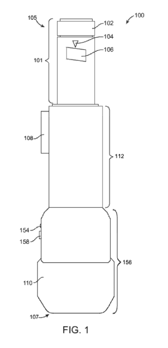
[0039] FIG. 1 shows an injection apparatus with a cap 110 secured. In some
embodiments, the
pain-reducing measures of the apparatus are located at the very tip of a tube
protruding from the
distal end of the injection apparatus. In some embodiments, the replaceable
needle assembly with
the cylindrical shaft is uniquely designed to interconnect with the distal end
of the injection
apparatus, as the cylindrical shaft (and enclosed needle) fits within the
interior of the of the tube
protruding from the distal end of the injection apparatus while also
encapsulating the exterior of the
distal end of the injection apparatus. The unique design allows for the
injection site and pain-
reducing measures to be located on the same plane and centered on the same
axis.
[0040] In some embodiments provided herein, when the needle assembly is
secured to the body of
the pain-reducing injection apparatus, the cylindrical shaft, with the
hypodermic needle enclosed, is
secured to a vial or syringe containing medication or other fluid. In some
embodiments, a user then
places the end of the needle assembly attached to the pain-reducing injection
apparatus in contact
with the patient, such that the body of the device is perpendicular to the
surface of the patient's
-16-
CA 03056087 2019-09-10
WO 2018/170176 PCT/US2018/022506
skin: only the distal, external surface of the needle assembly contacts the
patient's skin. In some
embodiments, a user then activates the pain-reducing component of the
apparatus via a manual
control located on the exterior of the body of the apparatus.
[0041] In an embodiment utilizing a cooling pain-reducing component, an
activated thermoelectric
cooler located at the distal end of the apparatus allows for heat to transfer
from the patient's skin,
through the surface of the needle assembly, and into the thermoelectric
cooler, thereby locally
reducing the temperature and pain sensation in the skin while not overcooling
the skin within a
matter of seconds. In some embodiments, once the skin has cooled to a desired
temperature, the
apparatus notifies the user through a light, sound, or other notifying means.
Although a
thermoelectric cooler is described herein, in some embodiments, other pain-
reduction means
incorporate vibration or any other non-invasive pain-reduction method either
as an alternative or in
combination.
[0042] In some embodiments, after the pain-reduction method has been utilized
to a satisfactory
degree, a user then depresses a manual control on the exterior of the body of
the apparatus to
activate the insertion of the needle and/or delivery of the injection. In some
embodiments, once the
injection is completed, the device automatically retracts the needle and shuts
down the pain-
reducing component of the apparatus. In some embodiments, the removable cap is
then removed
and discarded by the user.
[0043] In some embodiments provided herein, the manual control for injection
is located at the
proximal end of the apparatus. In some embodiments, such a manual control for
injection also
contains a dial or other controller for selecting a desired dosage amount for
injection.
[0044] In yet other certain embodiments of the present, the apparatus is
designed to attach directly
to a standard syringe. In such an embodiment, a user first secures a needle
assembly to the distal
end of the apparatus as described above. In some embodiments, the user then
inserts a standard
syringe into the apparatus, whereby the needle within the needle assembly
penetrates the syringe to
prepare for the administration of medication. In some embodiments, as
described above, a manual
control on the exterior of the body of the apparatus initiates the pain-
reducing component, which
then is followed by the advancement of the needle and injection of medication.
In some
embodiments, once finished, the cap is then discarded. In some embodiments,
the injector or
syringe has no needle when placed in the pain-reducing apparatus; in such
case, either the needle
assembly described herein is attached thereto, or a standard pen needle is
attached thereto. In some
embodiments, an injector having a syringe and needle pre-attached thereto is
used in the pain-
reducing apparatus.
-17-
CA 03056087 2019-09-10
WO 2018/170176 PCT/US2018/022506
[0045] Referring to FIG. 1, the pain-reducing injection apparatus attaches to
a standard syringe or
an injector. In some embodiments, FIG. 1 illustrates a front view of a pain-
reducing injection
apparatus 100 operatively receiving an injector 101. In some embodiments, the
injector 101 is a
standard repeating injector. In some embodiments, the injector 101 is a pen-
style injector or an
injector pen. In some embodiments, the injector 101 is a reusable injector
pen, a disposable injector
pen, a wearable autoinjector, or a handheld autoinjector. In some embodiments,
the injector or
syringe has no needle when placed in the pain-reducing apparatus; in such
case, either the needle
assembly described herein is attached thereto, or a standard pen needle is
attached thereto. In some
embodiments, an injector having a syringe and needle pre-attached thereto is
used in the pain-
reducing apparatus.
[0046] In some embodiments, the injector 101 delivers a drug. Non-limiting
examples of the drug
the injector 101 delivers are: an antibody, a hormone, a small molecule drug,
a cytokine, a protein,
an antibiotic, an anti-inflammatory, an analgesic, a psychoactive drug, or an
anticoagulant. In some
embodiments, the antibody is adalimumab. In some embodiments, the hormone is
insulin or
epinephrine. In some embodiments, the small molecule drug is a drug for the
treatment of diabetes.
In some embodiments, the small molecule drug is a glucagon-like peptide-1
receptor agonist (GLP-
1). In some embodiments, the small molecule drug is a drug that is used for
first aid against a
chemical warfare agent. In some embodiments, the small molecule drug is a
nerve agent antidote.
In some embodiments, the psychoactive drug is a benzodiazepine compound, such
as but not
limited to diazepam, chlordiazepoxide, temazepam, midazolam, and clonazepam.
In some
embodiments, the cytokine is interferon-f31a or erythropoietin. In some
embodiments, the protein is
a fusion protein such as but not limited to etanercept. In some embodiments,
the anticoagulant is
enoxaparin.
[0047] In some embodiments, the injector 101 comprises a cartridge containing
a drug (not shown
in FIG. 1). In some embodiments, the injector 101 comprises a dial 102 that
adjusts a unit dose of
a drug. In some embodiments, the user rotates the dial 102 clockwise to
increase the unit dose of a
drug. In some embodiments, the user rotates the dial 102 counterclockwise to
increase the unit
dose of a drug. In some embodiments, the injector 101 comprises a unit dose
indicator 104. In
some embodiments, the injector 101 comprises a unit dose display 106. In some
embodiments, the
unit dose display 106 displays a unit dose number or a unit dose marker
selected by a user. In some
embodiments, the unit dose indicator 104 assists the user in aligning a unit
dose number or a unit
dose marker with the unit dose indicator 104. In some embodiments, the unit
dose indicator 104 is
a line, a triangle, a light such as a light emitting diode (LED), or a notch.
In some embodiments,
-18-
CA 03056087 2019-09-10
WO 2018/170176 PCT/US2018/022506
the injector 101 makes a clicking sound to indicate an increment in a dose
unit of a drug as the user
rotates the dial 102 clockwise or counterclockwise.
[0048] In some embodiments, the pain-reducing injection apparatus 100
comprises an upper body
112 and a lower body 156. In some embodiments, the pain-reducing injection
apparatus 100 is
configured to hold an injector 101. In some embodiments, the pain-reducing
injection apparatus
100 is configured to hold a syringe (not shown in FIG. 1). In some
embodiments, the upper body
112 and the lower body 156 are shaped in the form of a "U" which allows the
pain-reducing
injection apparatus 100 to reversibly hold the cylindrical body of an injector
101 or a syringe for its
retention and or release. In some embodiments, the "U"-shaped upper body 112
and the "U"-
shaped lower body 156 have a first lateral wall and a second lateral wall
opposite from one another
and with a curved profile that defines the "U" shape, in a way that
accommodates the cylindrical
body of the injector 101 or the cylindrical body of a syringe (not shown in
FIG. 1). In some
embodiments, an injector 101 or a syringe is reversibly attached to the pain-
reducing injection
apparatus 100 via a snap-on mechanism. In some embodiments, the upper body 112
and/or the
lower body 156 comprises at least one projection or groove that reversibly
secure the injector 101
into the upper body 112 and/or into the lower body 156. In some embodiments,
the projection is a
grip, a recess, a lip, or a ring.
[0049] In some embodiments, the user inserts the injector 101 into the upper
body 112 and/or lower
body 156 by pushing the injector 101 into at least one projection, with a
light force so that the body
of the injector 101 overcomes the projections. In some embodiments, the user
releases the injector
101, by pulling on its proximal end 105, with a light force so that the body
of the injector 101
passes the projections. In some embodiments, alternatively, the user releases
the injector 101, by
sliding the injector 101 along the upper body 112 and/or lower body 156,
towards the proximal end
105. In some embodiments, the upper body 112 and/or lower body 156 are
composed of a soft,
flexible material in order to enable separation of the first lateral wall of
the "U" and second lateral
wall of the "U" when the injector 101 is either inserted or released. In some
embodiments, the
upper body 112 and/or lower body 156 are composed of a rigid material. In some
embodiments,
the projection is composed of a soft, flexible material in order to enable its
separation when the
injector 101 is either inserted or released.
[0050] In some embodiments, a needle cap 110 is attached to the distal end 107
of the lower body
of the device 156 in its lateral aspects and is attached to the distal end of
the injector 101 in its
medial aspects, as shown in FIG. 1. In some embodiments, the upper body 112
has an insertion
activation button 108 which is depressed by a user to allow the insertion of a
needle into the tissue
of a patient. FIG. 1 further shows the lower body 156 comprising a light
indicator 154 and a
-19-
CA 03056087 2019-09-10
WO 2018/170176 PCT/US2018/022506
thermoelectric cooling and vibration activator button 158. In some
embodiments, the light indicator
154 turns on when a user activates the thermoelectric cooling function of the
device and/or the
vibration function of the device. In some embodiments, the light indicator 154
is a light-emitting
diode (LED). In some embodiments, the light indicator 154 will flash until the
thermoelectric
cooling function has reached a desired temperature. In some embodiments, the
light indicator 154
comprises a first LED of a first color and a second LED of a second color. In
some embodiments,
the first LED turns on when the thermoelectric function is activated and the
second LED turns on
when the vibration function is activated.
[0051] FIGS. 2A and 2B shows an injection apparatus with a cap removed and a
cross-section
view of a needle assembly 211. FIG. 2A and FIG. 2B show the pain-reducing
injection apparatus
200 receiving an injector 201 and a needle assembly 211. FIG. 2A shows the
pain-reducing
injection apparatus 200 comprising a needle assembly 211 with a needle 214
that is retracted (i.e.,
not yet deployed). FIG. 2B shows the pain-reducing injection apparatus 200
comprising a needle
assembly 211 with a needle 214 that is deployed. In some embodiments, the pain-
reducing injection
apparatus 200 comprises a pen-type injector sleeve. In some embodiments, the
pen-type injector
sleeve comprises a lower body 256 and an upper body 212. In some embodiments,
the pain-
reducing injection apparatus 200 comprises a pain-reducing injection apparatus
housing 215. In
some embodiments, the injector 201 comprises a reservoir 213 containing an
injection fluid (e.g., a
medicament). In some embodiments, the lower body 256 contains a battery 260, a
vibrator 230, and
a thermoelectric cooler 232. In some embodiments, the lower body operatively
and reversibly
receives a needle assembly 211. In some embodiments, a needle cap (not shown
in FIG. 2)
reversibly attaches to the needle assembly 211. In some embodiments, the
needle cap protects the
user from an unwanted or accidental needle puncture. In some embodiments, the
needle cap
protects the needle from foreign contaminants such as but not limited to
bacteria, fungi, and/or
viruses.
[0052] Referring to FIG. 2A, in some embodiments, the proximal end 205 of the
device is defined
as the end of the device that is closer to the hand of a user; i.e., the
proximal end 205 is towards the
upper body 212 of the device. In some embodiments, the distal end 207 of the
device is defined as
the end of the device that is closer to the injection site of a user or of a
patient; i.e., the distal end
207 is towards the lower body 256 of the device. In some embodiments, the
upper body 212 of the
device and lower body 256 of the device only translocate in the longitudinal
axis of the device in a
proximal and distal direction, but not in a polar axis of the device; i.e.,
the upper body 212 of the
device and lower body 256 of the device do not rotate with respect to one
another.
-20-
CA 03056087 2019-09-10
WO 2018/170176 PCT/US2018/022506
[0053] In some embodiments, the needle assembly 211 comprises an inner sleeve
266 and an outer
containing cylinder 268. In some embodiments, the inner sleeve 266 and an
outer containing
cylinder 268 translocate only in the longitudinal directions as well, relative
to each other and
relative to the upper body 212 and lower body 256 of the device. In some
embodiments, the needle
assembly 211 comprises a proximal needle shield 220 and a distal needle shield
264. In some
embodiments, the distal needle shield 264 translocates in a longitudinal
direction as well as in a
polar direction. In some embodiments, the distal needle shield 264 is able to
rotate with respect to
the inner sleeve 266 and to the outer containing cylinder 268.
[0054] In some embodiments, a cylindrical aperture located at the proximal end
205 of the upper
body 212 of the device is configured to receive the injector 201. In some
embodiments, the
cylindrical aperture is slightly larger in diameter than that of the injector
201. In some
embodiments, the upper body 212 of the device is configured to receive the
injector 201. In some
embodiments, the upper body 212 comprises at least one projection or groove
(not shown in FIGS.
2A and 2B), as described supra, that reversibly secure the injector 201 into
the upper body 212. In
some embodiments, the injector 201 fits snuggly within the upper body 212 when
loaded onto or
into the pen-injector type sleeve. In some embodiments, once secured, the
injector aligns with a
gasket 226 at the distal end of the upper body 212. In some embodiments, the
gasket 226 is a rubber
gasket. in some embodiments, the gasket is a re-sealable gasket. In some
embodiments, the gasket
is penetrated by the needle 214 when the needle assembly 211 is attached to
the injector 201.
[0055] In some embodiments, the attachments alternatively take different
forms. For example, in
certain embodiments, the upper body 212 of the device comprises a chuck style
clamp or at least
one fastener to reversibly secure the injector 201. Furthermore, in yet other
embodiments, the
upper body 212 of the device comprises a screwing mechanism to screw directly
to a distal end of
the injector 201.
[0056] FIG. 2A shows the upper body 212 of the device having an insertion
activation button 208
that is depressed by the user to allow for the insertion of the needle 214
into the skin and/or tissue
of a user or patient. In some embodiments, the insertion activation button 208
is depressed to
access and/or clean the distal tip of the injector 201 when the needle
assembly 211 is not attached
to the device (e.g., before and after using the device) without the need to
remove the injector 201
from the upper body 212 of the device. In some embodiments, the upper body 212
of the device is
attached to a lower body 256 of the device at its distal end by a flexible
collar (not shown in FIG.
2). In some embodiments, the upper body 212 and the lower body 256 translate
slidably in a
cylinder formed by an aperture in the lower body. In some embodiments, the
insertion activation
button 208 comprises a first insertion activator reset spring 228a and a
second insertion activator
-21-
CA 03056087 2019-09-10
WO 2018/170176 PCT/US2018/022506
resent spring 228b. In some embodiments, the first insertion activator reset
spring 228a and the
second insertion activator resent spring 228b are positioned in between the
insertion activation
button 208 and the upper body insertion blocking piece 262. In some
embodiments, the first
insertion activator reset spring 228a and the second insertion activator
resent spring 228b are
positioned in a first groove or first cavity of the insertion activation
button 208 and in a second
groove or second cavity insertion activation button 208, respectively, that is
configured to receive
the insertion activator resent springs. In some embodiments, the first
insertion activator reset
spring 228a and a second insertion activator resent spring 228b activates the
insertion and/or
deployment of a needle. In some embodiments, a proximal cylinder (not shown in
FIG. 2) of the
lower body 256 has walls which extend proximally within walls of the upper
body 212. In some
embodiments, the walls of the lower body 256 contain two recesses referred to
as a first lower body
insertion wing recess 278a and a second lower body insertion wing recess 278b.
The first lower
body insertion wing recess 278a and the second lower body insertion wing
recess 278b are
configured to accept a first upper body insertion wing 276a and a second upper
body insertion wing
276b. In some embodiments, the first upper body insertion wing 276a and a
second upper body
insertion wing 276b are configured to be slidably moved into the first lower
body insertion wing
recess 278a and the second lower body insertion wing recess 278b. In some
embodiments, the first
upper body insertion wing 276a and a second upper body insertion wing 276b are
used for
stabilization of the device and to limit the maximum length of insertion of
the upper body 212 of
the device into the lower body 256 of the device when the needle assembly 211
is not present. In
some embodiments, the proximal walls of the lower body 256 comprise a lower
body insertion
blocking piece 263 which abuts an upper body insertion blocking piece 262. In
some embodiments,
the upper body insertion blocking piece 262 is composed of a semi-flexible
material. In some
embodiments, the distal end of the upper body insertion blocking piece 262 is
bent slightly laterally
so as to prevent any distal translocation of the upper body 212. In some
embodiments, these
components keep the upper body 212 in the farthest proximal location possible
relative to the lower
body 256 of the device. In some embodiments, the lower body insertion blocking
piece 263 and
the upper body insertion blocking piece 262 are manipulated by the user by
depressing the insertion
activation button 208 on the upper body 212. In some embodiments, depressing
the insertion
activation button 208 pushes the distal end of the upper body insertion
blocking piece 262 medially
and out of contact with the lower body insertion blocking piece 263, thereby
allowing the upper
body 212 to translocate in the distal direction relative to the lower body
256.
[0057] In some embodiments, the user activates the insertion activation button
208 by depressing
the button in order to extend the needle 214 to an exposed position and allow
it to penetrate the skin
-22-
CA 03056087 2019-09-10
WO 2018/170176 PCT/US2018/022506
and/or tissue of the patient. In some embodiments, the user additionally
applies moderate pressure
on the upper body 212 of the device in the distal direction in order to extend
the needle 214 to an
exposed position and allow it to penetrate the skin and/or tissue of the
patient. In some
embodiments, the upper body 212 translocates distally and slides until the
inner sleeve 266 and the
needle 214 reach their maximum insertion depth. In some embodiments, the
needle 214 is affixed
to the inner sleeve 266. In some embodiments, the maximum insertion depth of
the needle 214
(i.e., inner sleeve 266 as well) corresponds to the depth of lower body
insertion stopping piece 203
relative to the initial location of the upper body insertion blocking piece
262, thereby also
controlling the depth of insertion while the needle assembly 211 is present.
In some embodiments,
if the needle assembly 211 is removed from the lower body 256 of the device
and manual pressure
is applied to the upper body 212 in a distal direction relative to the lower
body 256 while
depressing the insertion activation button 208, the upper body 212
translocates in the distal
direction relative to the lower body 256 and the upper body insertion blocking
piece 262 stops at
the lower body insertion stopping piece 203. In some embodiments, if the
insertion activation
button 208 is depressed a second time while in this position, the upper body
insertion blocking
piece 262 is then released in a medial direction from the lower body insertion
stopping piece 203
and allow it to slidably advance to its most distal allowable position.
[0058] In some embodiments, this distal translocation of the upper body 212 is
then halted when a
first upper body wing 276a and a second upper body wing 276b slidably stop at
a distal end of a
first lower body insertion wing recess 278a and a second lower body insertion
wing recess 278b,
respectively. In some embodiments, in this most collapsed configuration, the
distal tip of the repeat
injector is easily accessed and cleaned with sanitizing wipes. The ability to
clean and sanitize the
distal tip of the repeat injector provides various advantages which include,
but are not limited to
decreasing the likelihood of infection for a patient before use and preventing
a transmittable disease
from infecting any subsequent patients. In some embodiments, the distal tip of
the injector position
is unable to be reached while the needle cap is present because the inner
sleeve 266 is stopped from
distally advancing further. In some embodiments, when the needle assembly 211
is attached to the
pain-reducing injection apparatus 200, the inner sleeve 266 does not distally
advance sufficiently
enough to expose the distal tip of the injector because a ledge (not shown in
FIG. 2A and FIG. 2B)
on the internal aspect of the outer sleeve 268, blocks it from doing so. In
some embodiments, the
ledge (not shown in FIG. 2A and FIG. 2B) on the internal aspect of the outer
sleeve 268 creates a
maximum insertion depth for the needle 214.
[0059] FIG. 2A shows the lower body 212 of the device containing a
thermoelectric cooling and
vibration activator 258 and a light indicator 254. In some embodiments, the
light indicator 254
-23-
CA 03056087 2019-09-10
WO 2018/170176 PCT/US2018/022506
turns on when a user activates the thermoelectric cooling function of the
device and/or activates the
vibration function of the device. In some embodiments, the light indicator 254
is a light-emitting
diode (LED). In some embodiments, the light indicator 254 will flash until the
thermoelectric
cooling function has reached a desired temperature. In some embodiments, the
light indicator 254
comprises a first LED of a first color and a second LED of a second color. In
some embodiments,
the first LED turns on when the thermoelectric function is activated and the
second LED turns on
when the vibration function is activated.
[0060] In some embodiments, the thermoelectric cooling and vibration activator
258 is a button. In
some embodiments, the thermoelectric cooling and vibration activator 258 is a
switch. In some
embodiments, when the thermoelectric cooling and vibration activator 258 is
activated by a user,
the thermoelectric cooling and vibration activator 258 activates an electric
current through
embedded wiring from a battery 260 to the thermoelectric cooler 232, thereby
providing electric
power to and turning the thermoelectric cooler 232 on. In some embodiments,
the thermoelectric
cooling and vibration activator 258 is located on the proximal end of the
lower body 212 of the
pain-reducing injection apparatus 200. In some embodiments, the light
indicator 254 is operatively
connected to the thermoelectric cooling and vibration activator 258, to the
thermoelectric cooler
232 and to the vibrator 230. In some embodiments, the activation of the
thermoelectric cooling
and vibration activator 258 sends a signal to the light indicator 254 to turn
on and display a colored
light that notifies the user that the thermoelectric cooler 232 and/or
vibrator 230 is activated. In
some embodiments, the thermoelectric cooler and vibration activation button
spring 271 is
operatively connected to the thermoelectric cooler and vibration activation
button 258. In some
embodiments, the thermoelectric cooler and vibration activation button spring
271 is positioned
directly below the thermoelectric cooler and vibration activation button 258.
In some
embodiments, the thermoelectric cooler and vibration activation button spring
271 is positioned
directly adjacent to the thermoelectric cooler and vibration activation button
258. In some
embodiments, the thermoelectric cooler and vibration activation button spring
271 is activates the
thermoelectric cooler and vibration activation button 258.
Thermoelectric Cooling System
[0061] Disclosed herein, in certain embodiments, are pain-reducing injection
apparatuses,
comprising: a pen-type injector sleeve comprising: a housing configured to
operatively receive a
drug delivery device, the housing comprising a distal end comprising a distal
wall between an inner
surface and an active-cooling surface positioned to contact an injection
region of an individual
when in use; and a thermoelectric cooling system comprising: a thermoelectric
cooler comprising a
cooling plate, the thermoelectric cooler mounted against the inner surface of
the distal end and
-24-
CA 03056087 2019-09-10
WO 2018/170176 PCT/US2018/022506
configured to cool the active-cooling surface by conduction; and a controller
operatively coupled to
the thermoelectric cooler.
[0062] In some embodiments, the drug delivery device is an injector pen. In
some embodiments,
the drug delivery device is a syringe. In some embodiments, the drug delivery
device is a jet
injector. In some embodiments, the housing 215 is configured to reversibly
receive the drug
delivery device. In some embodiments, the housing 215 is configured to
permanently receive the
drug delivery device.
[0063] The thermoelectric cooler functions based on the Peltier effect;
namely, it creates a heat flux
between the junction of two different types of materials. In some embodiments,
the thermoelectric
cooler has a first side and a second side which upon application of a direct
current (DC) electric
current, heat is transferred from the first side to the second side. In some
embodiments, the
thermoelectric cooler decreases the temperature of a distal side of the
thermoelectric cooler and
increases the temperature of a proximal side of the thermoelectric cooler.
[0064] In some embodiments, the pain-reducing injection apparatus comprises a
thermoelectric
cooling system. In some embodiments, the thermoelectric cooling system
comprises a
thermoelectric cooler further comprising a cooling plate (not shown in FIGS.
2A and 2B). In some
embodiments, the thermoelectric cooling system comprises a thermoelectric
cooler further
comprising a heating plate (not shown in FIGS. 2A and 2B). In some
embodiments, the
thermoelectric cooler is mounted against the inner surface 221 of the distal
end of the housing 215,
as shown in FIG. 2B. In some embodiments, the inner surface 221 is an inner
surface of a distal
wall 219, as shown in FIG. 2B. In some embodiments, the thermoelectric cooler
232 sits adjacent
and makes thermal contact with the inner surface 221. In some embodiments,
thermoelectric cooler
232 is mounted against the inner surface 221 of the distal end of the housing
215. In some
embodiments, the thermoelectric cooler transfers heat from an injection
region, to an active cooling
surface 235, through a distal wall 219, to the inner surface 221 of the distal
end of the housing, to
the cooling plate of the thermoelectric cooler, and finally to the heating
plate and heat sink of the
thermoelectric cooler; thereby reducing the surface temperature of the
injection region of a patient
and/or user. In some embodiments, the heat transferred to the heating plate
and/or the heat sink of
the thermoelectric cooler is dissipated. In some embodiments, the
thermoelectric cooler is
configured to cool the active-cooling surface 235 by conduction. In some
embodiments, the active-
cooling surface 235 is a flat, circular surface surrounding the opening where
the needle exits the
needle assembly 211. In some embodiments, the active-cooling surface 235 is a
distal surface at a
distal end of the pain-reducing injection apparatus. In some embodiments, the
active-cooling
surface 235 is a surface of a housing of the pain-reducing injection
apparatus. In some
-25-
CA 03056087 2019-09-10
WO 2018/170176 PCT/US2018/022506
embodiments, the active-cooling surface 235 is composed of a thermally
conductive material. In
some embodiments, the active-cooling surface 235 is composed of a metal or a
metal alloy. In
some embodiments, thermoelectric cooling system comprises a controller (not
shown in FIGS. 2A
and 2B). In some embodiments, the controller is operatively coupled to the
thermoelectric cooler.
In some embodiments, the cooling plate is mounted against the inner surface of
the distal end of the
pain-reducing injection apparatus housing. In some embodiments, the cooling
plate is configured
to cool the active-cooling surface by conduction. In some embodiments, the
cooling plate is
composed of a thermally insulating material. In some embodiments, the cooling
plate is a ceramic
plate. In some embodiments, the controller controls a temperature of the
cooling plate. In some
embodiments, the thermoelectric cooling system comprises a power source
operatively coupled to
the thermoelectric cooler and to the controller. In some embodiments, the
power source is the
battery 260. In some embodiments, the battery 260 is a nickel cadmium (NiCd)
battery, nickel-metal
hydride (NiM1-1) battery, a nickel zinc (NiZn) battery, a lead acid battery, a
lithium ion battery (Li-ion),
or a lithium ion polymer (Li-ion polymer) battery.
[0065] In some embodiments, the thermoelectric cooling system comprises a
temperature sensor
(not shown in FIGS. 2A and 2B). In some embodiments, the temperature sensor is
operatively
connected to the controller. In some embodiments, the temperature sensor is
operatively connected
the thermoelectric cooler. In some embodiments, the temperature sensor is
operatively connected
the active-cooling surface. In some embodiments, the temperature sensor is
configured to detect a
temperature of the cooling plate. In some embodiments, the temperature sensor
is configured to
detect a temperature of the active-cooling surface 235. In some embodiments,
the thermoelectric
cooling system comprises a heating plate facing away from the inner surface of
the distal end of the
pain-reducing injection apparatus housing 215. In some embodiments, the
heating plate is in
thermal connection with a heat sink. In some embodiments, the heat sink
absorbs heat emitted by
the heating plate. In some embodiments, the thermoelectric cooling system
comprises a fan. In
some embodiments, the fan is configured to dissipate heat emitted by the
heating plate.
[0066] In some embodiments, the cooling plate and the heating plate of the
thermoelectric cooler
are separated by a material that does not conduct heat. In some embodiments,
the controller
controls and/or limits the length of time when the thermoelectric cooler is
activated. In some
embodiments, the controller controls and/or limits the activation of the
thermoelectric cooler. In
some embodiments, the temperature sensor provides a feedback signal to the
controller. In some
embodiments, the feedback signal is a temperature of the cooling plate, a
temperature of the heating
plate, and/or the temperature of the active-cooling surface 235. In some
embodiments, the
temperature sensor assists the controller in controlling and/or limiting the
length of time when the
-26-
CA 03056087 2019-09-10
WO 2018/170176 PCT/US2018/022506
thermoelectric cooler is activated. In some embodiments, the temperature
sensor assists the
controller in controlling and/or limiting the activation of the thermoelectric
cooler. In some
embodiments, the thermoelectric cooling system further comprises a limits the
length of time when
the thermoelectric cooler is activated. In some embodiments, the
thermoelectric cooler absorbs heat
from a skin surface of a patient. In some embodiments, the thermoelectric
cooler absorbs heat from
a skin surface of a patient by direct contact between the cooled active-
cooling surface 235 and the
skin surface. In some embodiments, the absorption of heat from the skin
surface of the patient
creates an anesthetic effect in underlying tissue.
[0067] In some embodiments, the active-cooling surface 235 is cooled to a
temperature ranging
between about at least -10 to about 10 degrees Celsius or more. In some
embodiments, the active-
cooling surface 235 is cooled to a temperature ranging between about at least -
10 to about 5 degrees
Celsius or more. In some embodiments, the active-cooling surface 235 is cooled
to a temperature
ranging between at least about -10 to about 0 degrees Celsius or more. In some
embodiments, the
active-cooling surface 235 is cooled to a temperature ranging between at least
about -10 to about -5
degrees Celsius or more. In some embodiments, the active-cooling surface 235
is cooled to a
temperature ranging between at least about -15 to about 15 degrees Celsius or
more. In some
embodiments, the active-cooling surface 235 is cooled to a temperature ranging
between at least
about -15 to about 10 degrees Celsius or more. In some embodiments, the active-
cooling surface
235 is cooled to a temperature ranging between at least about -15 to about 5
degrees Celsius or
more. In some embodiments, the active-cooling surface 235 is cooled to a
temperature ranging
between at least about -15 to about 0 degrees Celsius or more. In some
embodiments, the active-
cooling surface 235 is cooled to a temperature ranging between at least about -
15 to about -5
degrees Celsius or more. In some embodiments, the active-cooling surface 235
is cooled to a
temperature ranging between at least about -15 to about -10 degrees Celsius or
more.
[0068] In some embodiments, the active-cooling surface 235 is cooled to a
temperature of about -
15 degrees Celsius. In some embodiments, the active-cooling surface 235 is
cooled to a temperature
of about -14 degrees Celsius. In some embodiments, the active-cooling surface
235 is cooled to a
temperature of about -13 degrees Celsius. In some embodiments, the active-
cooling surface 235 is
cooled to a temperature of about -12 degrees Celsius. In some embodiments, the
active-cooling
surface 235 is cooled to a temperature of about -11 degrees Celsius. In some
embodiments, the
active-cooling surface 235 is cooled to a temperature of about -10 degrees
Celsius. In some
embodiments, the active-cooling surface 235 is cooled to a temperature of
about -9 degrees Celsius.
In some embodiments, the active-cooling surface 235 is cooled to a temperature
of about -8 degrees
Celsius. In some embodiments, the active-cooling surface 235 is cooled to a
temperature of about -
-27-
CA 03056087 2019-09-10
WO 2018/170176 PCT/US2018/022506
7 degrees Celsius. In some embodiments, the active-cooling surface 235 is
cooled to a temperature
of about -6 degrees Celsius. In some embodiments, the active-cooling surface
235 is cooled to a
temperature of about -5 degrees Celsius. In some embodiments, the active-
cooling surface 235 is
cooled to a temperature of about -4 degrees Celsius. In some embodiments, the
active-cooling
surface 235 is cooled to a temperature of about -3 degrees Celsius. In some
embodiments, the
active-cooling surface 235 is cooled to a temperature of about -2 degrees
Celsius. In some
embodiments, the active-cooling surface 235 is cooled to a temperature of
about -1 degree Celsius.
In some embodiments, the active-cooling surface 235 is cooled to a temperature
of about 0 degrees
Celsius. In some embodiments, the active-cooling surface 235 is cooled to a
temperature of about 1
degree Celsius. In some embodiments, the active-cooling surface 235 is cooled
to a temperature of
about 2 degrees Celsius. In some embodiments, the active-cooling surface 235
is cooled to a
temperature of about 3 degrees Celsius. In some embodiments, the active-
cooling surface 235 is
cooled to a temperature of about 4 degrees Celsius. In some embodiments, the
active-cooling
surface 235 is cooled to a temperature of about 5 degrees Celsius. In some
embodiments, the
active-cooling surface 235 is cooled to a temperature of about 6 degrees
Celsius. In some
embodiments, the active-cooling surface 235 is cooled to a temperature of
about 7 degrees Celsius.
In some embodiments, the active-cooling surface 235 is cooled to a temperature
of about 8 degrees
Celsius. In some embodiments, the active-cooling surface 235 is cooled to a
temperature of about 9
degrees Celsius. In some embodiments, the active-cooling surface 235 is cooled
to a temperature of
about 10 degrees Celsius. In some embodiments, the active-cooling surface 235
is cooled to a
temperature of about 11 degrees Celsius. In some embodiments, the active-
cooling surface 235 is
cooled to a temperature of about 12 degrees Celsius. In some embodiments, the
active-cooling
surface 235 is cooled to a temperature of about 13 degrees Celsius. In some
embodiments, the
active-cooling surface 235 is cooled to a temperature of about 14 degrees
Celsius. In some
embodiments, the active-cooling surface 235 is cooled to a temperature of
about 15 degrees
Celsius.
[0069] In some embodiments, the injection region of a patient is cooled by
direct contact with the
active-cooling surface 235 of the pain-reducing injection apparatus. In some
embodiments, the
injection region is cooled to a temperature ranging between about at least -10
to about 10 degrees
Celsius or more. In some embodiments, the injection region is cooled to a
temperature ranging
between about at least -10 to about 5 degrees Celsius or more. In some
embodiments, the injection
region is cooled to a temperature ranging between at least about -10 to about
0 degrees Celsius or
more. In some embodiments, the injection region is cooled to a temperature
ranging between at
least about -10 to about -5 degrees Celsius or more. In some embodiments, the
injection region is
-28-
CA 03056087 2019-09-10
WO 2018/170176 PCT/US2018/022506
cooled to a temperature ranging between at least about -15 to about 15 degrees
Celsius or more. In
some embodiments, the injection region is cooled to a temperature ranging
between at least about -
15 to about 10 degrees Celsius or more. In some embodiments, the injection
region is cooled to a
temperature ranging between at least about -15 to about 5 degrees Celsius or
more. In some
embodiments, the injection region is cooled to a temperature ranging between
at least about -15 to
about 0 degrees Celsius or more. In some embodiments, the injection region is
cooled to a
temperature ranging between at least about -15 to about -5 degrees Celsius or
more. In some
embodiments, the injection region is cooled to a temperature ranging between
at least about -15 to
about -10 degrees Celsius or more.
[0070] In some embodiments, the injection region is cooled to a temperature of
about -15 degrees
Celsius. In some embodiments, the injection region is cooled to a temperature
of about -14 degrees
Celsius. In some embodiments, the injection region is cooled to a temperature
of about -13 degrees
Celsius. In some embodiments, the injection region is cooled to a temperature
of about -12 degrees
Celsius. In some embodiments, the injection region is cooled to a temperature
of about -11 degrees
Celsius. In some embodiments, the injection region is cooled to a temperature
of about -10 degrees
Celsius. In some embodiments, the injection region is cooled to a temperature
of about -9 degrees
Celsius. In some embodiments, the injection region is cooled to a temperature
of about -8 degrees
Celsius. In some embodiments, the injection region is cooled to a temperature
of about -7 degrees
Celsius. In some embodiments, the injection region is cooled to a temperature
of about -6 degrees
Celsius. In some embodiments, the injection region is cooled to a temperature
of about -5 degrees
Celsius. In some embodiments, the injection region is cooled to a temperature
of about -4 degrees
Celsius. In some embodiments, the injection region is cooled to a temperature
of about -3 degrees
Celsius. In some embodiments, the injection region is cooled to a temperature
of about -2 degrees
Celsius. In some embodiments, the injection region is cooled to a temperature
of about -1 degree
Celsius. In some embodiments, the injection region is cooled to a temperature
of about 0 degrees
Celsius. In some embodiments, the injection region is cooled to a temperature
of about 1 degree
Celsius. In some embodiments, the injection region is cooled to a temperature
of about 2 degrees
Celsius. In some embodiments, the injection region is cooled to a temperature
of about 3 degrees
Celsius. In some embodiments, the injection region is cooled to a temperature
of about 4 degrees
Celsius. In some embodiments, the injection region is cooled to a temperature
of about 5 degrees
Celsius. In some embodiments, the injection region is cooled to a temperature
of about 6 degrees
Celsius. In some embodiments, the injection region is cooled to a temperature
of about 7 degrees
Celsius. In some embodiments, the injection region is cooled to a temperature
of about 8 degrees
Celsius. In some embodiments, the injection region is cooled to a temperature
of about 9 degrees
-29-
CA 03056087 2019-09-10
WO 2018/170176 PCT/US2018/022506
Celsius. In some embodiments, the injection region is cooled to a temperature
of about 10 degrees
Celsius. In some embodiments, the injection region is cooled to a temperature
of about 11 degrees
Celsius. In some embodiments, the injection region is cooled to a temperature
of about 12 degrees
Celsius. In some embodiments, the injection region is cooled to a temperature
of about 13 degrees
Celsius. In some embodiments, the injection region is cooled to a temperature
of about 14 degrees
Celsius. In some embodiments, the injection region is cooled to a temperature
of about 15 degrees
Celsius.
[0071] In some embodiments, the injection region of the patient is cooled to a
temperature of about
degrees Celsius by direct contact between the injection region and the active-
cooling surface
235. In some embodiments, when the injection region reaches a desired
temperature (e.g., 10
degrees Celsius), the temperature sensor sends a feedback signal to inactivate
the thermoelectric
cooler. In some embodiments, the feedback signal triggers a change in the
color of the indicator
light 254. In some embodiments, the change in color of the indicator light 254
notifies the user that
the injection region is at the correct temperature and appropriately
anesthetized for needle insertion.
[0072] In some embodiments, the injection region is contacted with the active-
cooling surface 235
for at least about 1 minute to about 5 minutes or more prior to insertion of
the needle. In some
embodiments, the injection region is contacted with the active-cooling surface
235 for at least about
seconds to about 10 minutes or more prior to insertion of the needle. In some
embodiments, the
injection region is contacted with the active-cooling surface 235 for at least
about 30 seconds to
about 10 minutes or more prior to insertion of the needle. In some
embodiments, the injection
region is contacted with the active-cooling surface 235 for at least about 45
seconds to about 10
minutes or more prior to insertion of the needle. In some embodiments, the
injection region is
contacted with the active-cooling surface 235 for at least about 1 minute to
about 10 minutes or
more prior to insertion of the needle. In some embodiments, the injection
region is contacted with
the active-cooling surface 235 for at least about 2 minutes to about 5 minutes
or more prior to
insertion of the needle. In some embodiments, the injection region is
contacted with the active-
cooling surface 235 for at least about 1 minute to about 5 minutes or more
prior to insertion of the
needle. In some embodiments, the injection region is contacted with the active-
cooling surface 235
for at least about 3 minutes to about 5 minutes or more prior to insertion of
the needle.
[0073] In some embodiments, the injection region is contacted with the active-
cooling surface 235
for about 5 seconds prior to insertion of the needle. In some embodiments, the
injection region is
contacted with the active-cooling surface 235 for about 10 seconds prior to
insertion of the needle.
In some embodiments, the injection region is contacted with the active-cooling
surface 235 for
about 15 seconds prior to insertion of the needle. In some embodiments, the
injection region is
-30-
CA 03056087 2019-09-10
WO 2018/170176 PCT/US2018/022506
contacted with the active-cooling surface 235 for about 30 seconds prior to
insertion of the needle.
In some embodiments, the injection region is contacted with the active-cooling
surface 235 for
about 45 seconds prior to insertion of the needle. In some embodiments, the
injection region is
contacted with the active-cooling surface 235 for about 1 minute prior to
insertion of the needle. In
some embodiments, the injection region is contacted with the active-cooling
surface 235 for about 2
minutes prior to insertion of the needle. In some embodiments, the injection
region is contacted
with the active-cooling surface 235 for about 3 minutes prior to insertion of
the needle. In some
embodiments, the injection region is contacted with the active-cooling surface
235 for about 4
minutes prior to insertion of the needle. In some embodiments, the injection
region is contacted
with the active-cooling surface 235 for about 5 minutes prior to insertion of
the needle. In some
embodiments, the injection region is contacted with the active-cooling surface
235 for about 6
minutes prior to insertion of the needle. In some embodiments, the injection
region is contacted
with the active-cooling surface 235 for about 7 minutes prior to insertion of
the needle. In some
embodiments, the injection region is contacted with the active-cooling surface
235 for about 8
minutes prior to insertion of the needle. In some embodiments, the injection
region is contacted
with the active-cooling surface 235 for about 9 minutes prior to insertion of
the needle. In some
embodiments, the injection region is contacted with the active-cooling surface
235 for about 10
minutes prior to insertion of the needle. In some embodiments, the injection
region is contacted
with the active-cooling surface 235 for about 15 minutes prior to insertion of
the needle.
Vibrator
[0074] Disclosed herein, in certain embodiments, are pain-reducing injection
apparatuses,
comprising: a pen-type injector sleeve comprising: a housing configured to
operatively receive a
drug delivery device, the housing comprising a distal surface located at a
distal region of the
housing, the distal surface positioned to contact an injection region of an
individual when in use;
and a vibrator mounted in the housing, the vibrator configured to cause a
distal region of the
housing to vibrate.
[0075] In some embodiments, the vibrator 230 is activated by a user depressing
the cold and
vibration activation button 208. Alternatively, in other embodiments, the
vibrator 230 is activated
remotely or by activation of an additionally button and/or switch. In some
embodiments, the
vibrator 230 creates vibrations that are transmitted through the distal end of
the needle assembly
surface. In some embodiments, the vibrator 230 generates vibrations that
further increase the
anesthetic effect perceived by the patient. In some embodiments, activation of
the vibrator 230 is
controlled by the controller. In some embodiments, activation of the vibrator
230 is stopped based
on the activation time of the thermoelectric cooler.
-31-
CA 03056087 2019-09-10
WO 2018/170176 PCT/US2018/022506
[0076] In some embodiments, the distal surface of the pain-reducing injection
apparatus housing
215 is vibrating when the needle is inserted into the injection region of the
individual. In some
embodiments, the needle is vibrating when the needle is inserted into the
injection region of the
individual. In some embodiments, the vibration has a vibration frequency
ranging from about 100
Hertz (Hz) to about 300 Hz. In some embodiments, the vibration has an
amplitude ranging from
about .3 G (wherein G is gravitational acceleration, i.e., 9.8 meters per
second squared) to about
125 G. In some embodiments, the vibrator is configured to cause the active-
cooling surface, the
drug delivery device, and/or a needle to vibrate. In some embodiments, the
vibrator comprises a
motor. In some embodiments, the motor is an eccentric rotating mass vibration
motor or a linear
resonant actuator.
[0077] Further disclosed herein, in certain embodiments, are pain-reducing
injection apparatuses,
comprising: a pen-type injector sleeve comprising: a housing configured to
operatively receive a
drug delivery device, the housing comprising a distal end comprising a distal
wall between an inner
surface and an active-cooling surface positioned to contact an injection
region of an individual
when in use; a thermoelectric cooler comprising a cooling plate mounted
against the inner surface
of the distal end and configured to cool the active-cooling surface by
conduction; and a vibrator
mounted in the housing, the vibrator configured to cause the distal end of the
housing to vibrate.
[0078] In some embodiments, the control of the activation and inactivation of
the vibration and
thermoelectric cooler is automatically triggered by attaching the needle
assembly 211 to the pain-
reducing injection apparatus housing 215. In some embodiments the needle
assembly comprises a
needle and a needle hub. In some embodiments, the needle assembly is a needle
assembly other
than the one described herein in FIGS. 2A-B and 3A-D. In some embodiments, the
control of the
activation and inactivation of the vibration and thermoelectric cooler is
automatically triggered by a
mechanism that detects pressure on the distal end of the lower body 256 and a
needle assembly
surface. The device could also contain other types of pain reducing units and
modalities that could
be transmitted through the needle cap to anesthetize the patient's skin such
as direct electrical
pulses among others, while the cap still creates a barrier protecting the
cleanliness, sterility, and
safety of the device and injector or syringe.
Needle Assembly
[0079] Additionally disclosed herein, in certain embodiments, are pain-
reducing injection
apparatuses, comprising: a pen-type injector sleeve comprising: a housing
configured to operatively
receive a drug delivery device, the housing comprising a distal surface
located at a distal region of
the housing, the distal surface configured to contact an injection region of
an individual when in
use; and a needle assembly comprising: a needle, an outer sleeve having a
first inner surface
-32-
CA 03056087 2019-09-10
WO 2018/170176 PCT/US2018/022506
comprising a ramped track, and a first outer surface, an inner sleeve
positioned within the outer
sleeve, the inner sleeve comprising: a second inner surface, a second outer
surface facing the first
inner surface, an aperture comprising a perimeter and a perimeter wall
extending between the
second inner surface and the second outer surface along the perimeter, and a
distal needle insertion
shield coaxially aligned with the inner sleeve and with the outer sleeve, the
distal needle insertion
shield comprising a distal shield insertion arm configured to travel along the
perimeter of the
aperture in contact with the perimeter wall of the aperture as the distal
needle insertion shield
rotates about its axis, and wherein the distal shield insertion arm extends
through the aperture and is
configured to travel within and along the ramped track as the distal needle
insertion shield is axially
rotated about the axis.
[0080] Also disclosed herein, in certain embodiments, are needle assemblies,
comprising:
a needle; an outer sleeve having a first inner surface comprising a ramped
track, and a first outer
surface; an inner sleeve positioned within the outer sleeve, the inner sleeve
comprising: a second
inner surface, a second outer surface facing the first inner surface, and an
aperture comprising a
perimeter and a perimeter wall extending between the second inner surface and
the second outer
surface along the perimeter; and a distal needle insertion shield coaxially
aligned with the inner
sleeve and with the outer sleeve, the distal needle insertion shield
comprising a distal shield
insertion arm configured to travel along the perimeter of the aperture in
contact with the perimeter
wall of the aperture as the distal needle insertion shield rotates about its
axis, and wherein the distal
shield insertion arm extends through the aperture and is configured to travel
within and along the
ramped track as the distal needle insertion shield is axially rotated about
the axis.
[0081] In some embodiments, the needle assembly reversibly attaches to a pen
injector. In some
embodiments, the needle assembly permanently attaches to a pen injector. In
some embodiments,
the needle assembly is screwed into a pen injector. In some embodiments, the
needle assembly is
snapped onto a pen injector. In some embodiments, the needle assembly
reversibly attaches to a
pain-reducing injection apparatus. In some embodiments, the needle assembly
permanently attaches
to a pain-reducing injection apparatus. In some embodiments, the needle
assembly is screwed into a
pain-reducing injection apparatus. In some embodiments, the needle assembly is
snapped onto a
pain-reducing injection apparatus. In some embodiments, the needle assembly
reversibly attaches to
a pen-style injector sleeve. In some embodiments, the needle assembly
permanently attaches to a
pen-style injector sleeve. In some embodiments, the needle assembly is screwed
into a pen-style
injector sleeve. In some embodiments, the needle assembly is snapped onto a
pen-style injector
sleeve.
-33-
CA 03056087 2019-09-10
WO 2018/170176 PCT/US2018/022506
[0082] In some embodiments, the needle assembly comprises a needle cap
configured to receive
the needle assembly. In some embodiments, the needle cap is reusable. In some
embodiments, the
needle cap protects a needle from external contaminants. In some embodiments,
the needle cap
protects a user and/or patient from accidental punctures.
[0083] In some embodiments, the needle assembly comprises a fingerprint
authentication locking
mechanism comprising a fingerprint sensor and a needle cap lock. In some
embodiments, the
needle cap lock is unlocked upon recognition of a fingerprint of a user by the
fingerprint sensor. In
some embodiments, the fingerprint sensor is located on the needle cap.
[0084] FIGS. 3A, 3B, 3C, and 3D show the needle assembly safety mechanism and
its components
at various stages of a needle deployment and retraction process. In some
embodiments, the needle
assembly safety mechanism is designed to retract and lock a needle after
deployment and use (e.g.,
after insertion into an injection region) in order to prevent unwanted disease
transmission,
punctures, and/or incorrect delivery of a medicament. FIG. 3A shows the needle
assembly at its
initial (i.e., resting) position and the needle being shielded within the
walls of an inner sleeve 266
and an outer sleeve 268. In some embodiments, at this stage, a distal needle
shield spring 218 is
compressed, a proximal needle shield spring 270 is compressed, and an inner
sleeve spring 216 is
fully extended. In some embodiments, the components of the needle assembly are
encased in or
protected by a needle housing 259, as shown in FIGS. 3A, 3B, 3C, and 3D.
[0085] In some embodiments, the outer shield 268 comprises a ramped track 236.
In some
embodiments, the ramped track 236 has a start and a finish and a bump 237
positioned medially
therebetween. FIG. 3A shows a second distal needle shield insertion arm 272b
at the start of the
ramped track 236. In some embodiments, the ramped track 236 is angled. In some
embodiments,
the ramped track is angled at an angle of about 45 degrees with respect to the
distal surface of the
needle assembly housing. In some embodiments, the distal shield insertion arm
travels from the
first end of the aperture to the second end of the aperture as the distal
needle insertion shield is
axially rotated about the axis. In some embodiments, the distal shield
insertion arm is resting on the
perimeter of the aperture at the first end of the aperture and within the
ramped track at start of the
ramped track prior to deployment of the needle.
[0086] In some embodiments, the outer sleeve 268 comprises an aperture 222. In
some
embodiments, the aperture 222 has a first end of the aperture and a second end
of the aperture. In
some embodiments, the first end of the aperture aligns with the start of the
ramped track and the
second end of the aperture aligns with the finish of the ramped track prior to
deployment of the
needle. FIG. 3A shows a second distal needle shield insertion arm 272b
inserted at the first end of
the aperture.
-34-
CA 03056087 2019-09-10
WO 2018/170176 PCT/US2018/022506
[0087] FIG. 3B shows the needle assembly after the needle has started to be
deployed (i.e., the
needle has been advanced distally away from the proximal region of the needle
assembly). FIG.
3B shows the needle starting to exit a needle opening located in the center of
a distal wall of the
needle assembly. In some embodiments, deployment of the needle from the needle
assembly
causes the distal shield insertion arm to overcome the bump 237 and
subsequently rest within the
track 236. In some embodiments, the distal shield insertion arm travels from
the start of the ramped
track 236 to the finish of the ramped track as the distal needle insertion
shield 264 is axially rotated
about the axis. In some embodiments, at this stage, the distal needle shield
spring 218 is extended,
the proximal needle shield spring 270 is compressed, and the inner sleeve
spring 216 is
compressed. In some embodiments, the distal shield insertion arm travels from
the first end of the
aperture to the second end of the aperture as the distal needle insertion
shield 264 is axially rotated
about the axis. In some embodiments, the distal shield insertion arm is
resting on the perimeter of
the aperture 297 at the first end of the aperture and within the ramped track
236 at start of the
ramped track prior to deployment of the needle.
[0088] FIG. 3C shows the needle assembly 211 after deployment of the needle
214 has been
completed. In FIG. 3C, the needle is shown to be exposed and outside a needle
opening. In some
embodiments, at this stage, the distal needle shield spring 218 is extended,
the proximal needle
shield spring 270 is compressed, and the inner sleeve spring 216 is
compressed. In some
embodiments, the bump 237 prevents the distal shield insertion arm to travel
from the finish to the
start of the ramped track once the distal shield insertion arm overcomes the
bump 237. In some
embodiments, the perimeter of the aperture 297 has an aperture locking notch
295 positioned at the
second end of the aperture. In some embodiments, the perimeter of the aperture
has a sloped region
originating from the first end of the aperture 222 and ending at a vertical
region of the aperture. In
some embodiments, the vertical region of the aperture originates from a peak
of the sloped region
and ends at the aperture locking notch 295. In some embodiments, the
deployment of the needle
causes the distal shield insertion arm to rest at the peak of the sloped
region, on the perimeter of the
aperture.
[0089] FIG. 3D shows the needle assembly at its final stage, after deployment
and use of the
needle. FIG. 3D shows the needle retracted and in a locked position; thereby
unable to be used
and/or deployed again. In some embodiments, the ramped track has a track
locking notch 294
positioned at the finish of the ramped track. In some embodiments, the track
locking notch 294 is
configured to lock the distal shield insertion arm in place after the needle
214 is deployed and
retracted. In some embodiments, the aperture locking notch is configured to
lock the distal shield
insertion arm in place after the needle is deployed and retracted.
-35-
CA 03056087 2019-09-10
WO 2018/170176 PCT/US2018/022506
[0090] FIG. 4 shows an exploded view of the pain-reducing injection apparatus
200. In some
embodiments, the distal needle insertion shield 264 is positioned within the
inner sleeve 266. In
some embodiments, the inner sleeve 266 containing the distal needle insertion
shield 264 is
positioned within the outer sleeve 268. As shown in FIG. 4, the inner sleeve
266 comprises a first
aperture 222a and a second aperture 222b. In some embodiments, the inner
sleeve 266 is coaxially
aligned with the outer sleeve 268. In some embodiments, the first aperture
222a and the second
aperture 222b of the inner sleeve 266 are coaxially aligned with the first
ramped track and the
second ramped track of outer sleeve 268 (not shown in FIG. 4). In some
embodiments, the
proximal region of the pain-reducing injection apparatus 200 comprises an
insertion activation
button 208, a barrel 234, and upper body insertion wings, as shown in FIG. 4.
[0091] In some embodiments, the outer sleeve 268 comprises at least one slit
(not shown in FIG. 4)
configured to receive a distal needle shield insertion arm. In some
embodiments, a distal needle
insertion arm is inserted into at least one slit of the outer sleeve 268 when
inserting the inner sleeve
266 into the outer sleeve 268. In some embodiments, the inner sleeve 266
comprises at least one slit
(not shown in FIG. 4) configured to receive a distal needle shield insertion
arm. In some
embodiments, a distal needle insertion arm is inserted into at least one slit
of the inner sleeve 266
when inserting the distal needle shield 264 into the inner sleeve 266. In some
embodiments, the
outer sleeve 268 comprises at least one outer sleeve ramp (not shown in FIG.
4) connecting to the
ramped track, the outer sleeve ramp configured to receive a distal needle
shield insertion arm and
guide it towards the ramped track 236 when inserting the inner sleeve 266 into
the outer sleeve 268.
In some embodiments, the inner sleeve 266 comprises at least one inner sleeve
ramp (not shown in
FIG. 4) connecting to the aperture 222, the inner sleeve ramp configured to
receive a distal needle
shield insertion arm and guide it towards the aperture 222 when inserting the
distal needle shield
264 into the inner sleeve 266.
[0092] In some embodiments, the distal needle shield 264 comprises a distal
needle shield opening
265, as shown in FIG. 4. In some embodiments, the distal needle shield opening
265 is an opening
located at a distal end of the distal needle shield 264, configured to allow
the needle 214 to exit
therethrough. In some embodiments, the outer sleeve 268 and the needle
assembly housing 259
comprises a needle opening 269, as shown in FIG. 4. In some embodiments, the
needle opening
269 is an opening located at the distal end of the needle assembly, configured
to allow the needle
214 to exit therethrough.
[0093] In some embodiments, the outer sleeve 268 comprises an outer sleeve
ridge 257, as shown
in FIG. 3B and FIG. 4. In some embodiments, the outer sleeve 268 comprises an
outer sleeve
ridge 257 is configured to align the outer sleeve 268 with the inner sleeve
266. In some
-36-
CA 03056087 2019-09-10
WO 2018/170176 PCT/US2018/022506
embodiments, the outer sleeve ridge 257 is configured to align the outer
sleeve 268 with the inner
sleeve 266. In some embodiments, the inner sleeve 266 comprises a first inner
sleeve ridge 261a
and a second inner sleeve ridge 261b, as shown in FIG. 3B and FIG. 4. In some
embodiments, the
first inner sleeve ridge 261a and the second inner sleeve ridge 261b are
configured to align the
inner sleeve 266 with the outer sleeve 268.
[0094] In some embodiments, the outer sleeve is cylindrical. In some
embodiments, the inner
sleeve is cylindrical. In some embodiments, the outer sleeve is coaxially
aligned with the inner
sleeve. In some embodiments, the aperture locking notch is aligned with the
track locking notch.
In some embodiments, the needle is contained within the inner sleeve prior to
deployment. In some
embodiments, the ramped track is unidirectional. In some embodiments, the
distal shield insertion
arm is moved distally as the as the distal needle insertion shield is axially
rotated about the axis.
[0095] In some embodiments, the distal end 207 of the lower body 212 of the
pain-reducing
injection apparatus 200 contains a cylindrical aperture (not shown in FIGS.
2A, 2B, 3A, 3B, 3C, or
3D) that is slightly wider than the diameter of the outer sleeve 268 of the
needle assembly 211. In
some embodiments, the cylindrical aperture extends proximally through and
beyond the
thermoelectric cooler. In some embodiments, a proximal wall of the cylindrical
aperture is a distal
wall of the flexible collar 280. In some embodiments, the flexible collar 280
(shown in FIG. 4)
connects the lower body 256 of the pain-reducing injection apparatus 200 to
the upper body 212 of
the device pain-reducing injection apparatus 200. In some embodiments, the
proximal wall of the
cylindrical aperture is a distal end of the upper body 212.
[0096] In some embodiments, the lateral walls of the needle assembly 211
attach to the lateral
walls of the distal end 207 of the adaptor 209 (shown in FIG. 2B). In some
embodiments, the inner
sleeve 266 of the needle assembly 211 attach to the distal end of the injector
201. In some
embodiments, the diameter of the outer sleeve 268 of the needle assembly 211
is larger than that of
the inner sleeve 266. In some embodiments, the inner sleeve has a cylindrical
aperture that is wider
than a threaded cylinder of the distal end of the injector 201 or a syringe.
In some embodiments, the
user slides the center column 217 (i.e., the outer sleeve 268 and the inner
sleeve 266 encased by the
needle assembly housing 259) of the needle assembly 211 into the cylindrical
aperture of the
adaptor 209 in order to attach the needle assembly 211 to a distal end of the
pain-reducing injection
apparatus 200. In some embodiments, the user slides the center column 217 of
the needle
assembly 211 into a cylindrical aperture of the adaptor 209 in order to attach
the needle assembly
211 to the distal end of the standard repeating injector 100. In some
embodiments, the distal region
of the walls of the center column of the needle assembly 211 comprises the
exterior walls of the
outer sleeve 268. In some embodiments, the proximal region of the walls of the
center column of
-37-
CA 03056087 2019-09-10
WO 2018/170176 PCT/US2018/022506
the needle assembly 211 comprises the exterior walls of the inner sleeve 266.
In some
embodiments, the needle assembly 211 is reversibly attached to the pain-
reducing injection
apparatus 200 by the user. In some embodiments, the needle assembly 211 is
reversibly screwed
into or snapped onto the pain-reducing injection apparatus 200. In some
embodiments, the inner
walls of the proximal region of the inner sleeve 266 attach to the distal end
of the injector 200 while
the external walls of the needle assembly 211 are attached to the lateral ends
of the pain-reducing
injection apparatus housing 215. In some embodiments, the needle assembly
serves as a barrier to
prevent the skin, blood, and/or other various body substances and fluids from
contaminating the
pain-reducing injection apparatus 200. In some embodiments, the exterior walls
of the needle
assembly 211 have increased length in a proximal direction relative to the
center column 217 of the
needle assembly 211. In some embodiments, exterior walls of the needle
assembly 211 with an
increased length relative to the center column 217 covers provide for a
barrier of increased utility
for using the pain-reducing injection apparatus 200 to inject into bodily
orifices and/or more
contaminated areas of a tissue. For example, in some embodiments, the longer
needle assembly
exterior walls are necessary for procedures such as oral injections.
[0097] In some embodiments, the needle assembly 211 has a large cylindrical
recess on its
proximal side surrounding the inner sleeve 266. In some embodiments, the
recess is the same shape
of the distal end of the adaptor 209 and fits snuggly to that surface when
screwed into place. In
some embodiments, the distal end of the recess is composed of a material that
is able to conduct the
heat from the injection region to the thermoelectric cooler when activated. In
some embodiments,
the distal end of the recess is composed of a material that is able to conduct
vibration to the
injection region. In some embodiments, the distal end of the recess is
composed of a plastic, a
metal, and/or a metal alloy. In some embodiments, the distal end of the needle
assembly 211 is
able to conduct other anesthetic functions that are present in the lower body
256; for example,
electrical pulses, among others. In some embodiments, the needle assembly 211
comprises an
active-cooling surface 235. In some embodiments, the active-cooling surface
235 is able to
conduct heat away from the injection region. In some embodiments, the active-
cooling surface 235
is able to deliver a vibration to an injection region. In some embodiments,
conducting heat away
from the injection region and/or delivering a vibration to the injection
region allows for an
anesthetic effect to occur near the needle penetration point, within the
injection region.
[0098] In some embodiments, the injection region is at least about 0.1
centimeters (cm) to about 5
cm or more away from the needle penetration point. In some embodiments, the
injection region is
at least about 0.5 centimeters (cm) to about 5 cm or more away from the needle
penetration point.
In some embodiments, the injection region is at least about 1 centimeters (cm)
to about 5 cm or
-38-
CA 03056087 2019-09-10
WO 2018/170176 PCT/US2018/022506
more away from the needle penetration point. In some embodiments, the
injection region is at least
about 2 centimeters (cm) to about 5 cm or more away from the needle
penetration point. In some
embodiments, the injection region is at least about 3 centimeters (cm) to
about 5 cm or more away
from the needle penetration point. In some embodiments, the injection region
is at least about 4
centimeters (cm) to about 5 cm or more away from the needle penetration point.
In some
embodiments, the injection region is at least about 0.1 centimeters (cm) to
about 10 cm or more
away from the needle penetration point. In some embodiments, the injection
region is at least about
0.1 centimeters (cm) to about 15 cm or more away from the needle penetration
point.
[0099] In some embodiments, the injection region is at least about 0.1 cm away
from the needle
penetration point. In some embodiments, the injection region is at least about
0.5 cm away from the
needle penetration point. In some embodiments, the injection region is at
least about 1 cm away
from the needle penetration point. In some embodiments, the injection region
is at least about 2 cm
away from the needle penetration point. In some embodiments, the injection
region is at least about
3 cm away from the needle penetration point. In some embodiments, the
injection region is at least
about 4 cm away from the needle penetration point. In some embodiments, the
injection region is at
least about 5 cm away from the needle penetration point. In some embodiments,
the injection
region is at least about 6 cm away from the needle penetration point. In some
embodiments, the
injection region is at least about 7 cm away from the needle penetration
point. In some
embodiments, the injection region is at least about 8 cm away from the needle
penetration point. In
some embodiments, the injection region is at least about 9 cm away from the
needle penetration
point. In some embodiments, the injection region is at least about 10 cm away
from the needle
penetration point. In some embodiments, the injection region is at least about
15 cm away from the
needle penetration point.
[0100] In some embodiments, the diameter of the injection region is minimized
yet still provides an
adequate blood/device barrier. In some embodiments, the diameter of the
injection region is
minimized by providing a center column 217 of the needle assembly 211
comprising a smaller
diameter. In some embodiments, the smaller center attaches on its proximal end
to a proximal
region of the adaptor 209 instead of attaching to the injector 201. In some
embodiments, the inner
diameter of the center column 217 of the needle assembly 211 does not have to
be larger the
exterior diameter of the distal end of the injector or syringe.
[0101] In some embodiments, the center column 217 of the needle assembly 211
is able to collapse
upon the translation of the upper body 212 moving distally with respect to the
lower body 256,
thereby extending and exposing the needle 214 to deliver an injection. In some
embodiments, upon
completion of the injection, the resilient components (i.e. the distal shield
spring 218, the proximal
-39-
CA 03056087 2019-09-10
WO 2018/170176 PCT/US2018/022506
needle shield spring 270, and the inner sleeve spring 216) of the needle
assembly 211 reset the
position of the center column components. In some embodiments, upon completion
of the
injection, the resilient components (i.e. the distal shield spring 218, the
proximal needle shield
spring 270, and the inner sleeve spring 216) of the needle assembly 211reset
the position of the
upper body 212 with respect to the lower body 256. In some embodiments, upon
completion of the
injection, the resilient components (i.e. the distal shield spring 218, the
proximal needle shield
spring 270, and the inner sleeve spring 216) of the needle assembly 211
activate the proximal
needle shield 220 and the distal needle shield 264 within the needle assembly
211 so as to cover the
proximal and distal aspects of the needle 214. In some embodiments, upon
completion of the
injection, the resilient components (i.e. the distal shield spring 218, the
proximal needle shield
spring 270, and the inner sleeve spring 216) of the needle assembly 211 lock
the needle assembly
211 such as to prevent a user from reusing the needle assembly and/or
accidentally puncturing
themselves or others with the needle 214. In some embodiments, upon completion
of the injection,
the resilient components of the needle assembly 211, lock the needle assembly
and create a barrier
between the inner walls of the distal cylindrical aperture of the adaptor 209
and the patient's skin,
blood, and/or other bodily fluids.
[0102] In some embodiments, the components of the center column 217 which
create this resetting
and/or locking mechanism include the inner sleeve 266, an aperture 222, the
inner sleeve spring
216, the outer cylinder 268, the ramped track 236, the proximal needle shield
220, the proximal
needle shield spring 270, the distal needle shield 264, a first distal needle
shield wing 272a, a
second distal needle shield wing 272b, and the distal needle shield spring
216.
[0103] FIG. 5 shows the movement and components of the inner sleeve 266, the
outer sleeve 268,
and the distal needle insertion shield 264. As shown in FIG. 5, the inner
sleeve 266, the outer
sleeve 268 only move in a longitudinal axis (i.e., they only moved distally
and proximally),
whereas the distal needle shield 264 is able to rotate about its axis, labeled
as the Y-axis in FIG. 5.
In some embodiments, the action of needle deployment, insertion, and
retraction as described supra
is initiated by the distal translocation of the upper body 212 with respect to
the lower body 256. In
some embodiments, this distal translocation the upper body 212 with respect to
the lower body 256
exerts a distal force (i.e., distally presses) on the inner sleeve 266. In
some embodiments, the distal
force on the inner sleeve 266 causes the distal slidable translocation of the
inner 266 relative to the
outer sleeve 268. In some embodiments, there are two points of resistance
which oppose that distal
translocation: the first distal shield insertion arm 272a and the second
distal shield insertion arm
272b; and the resilient element of the inner sleeve spring 216.
-40-
CA 03056087 2019-09-10
WO 2018/170176 PCT/US2018/022506
[0104] In some embodiments, the first distal shield insertion arm 272a and the
second distal shield
insertion arm 272b extend through the aperture 222. In some embodiments, the
aperture 222 fully
extends through the wall of the inner sleeve 266. In some embodiments, the
lateral ends of the first
distal shield insertion arm 272a and the second distal shield insertion arm
272b ride along a ramped
track 236. In some embodiments, the ramped track 236 is created by a groove on
the interior wall
of the outer sleeve 268. In some embodiments, the ramped track 236 does not
fully penetrate the
wall of the outer sleeve 268 at any point along the ramped track. In some
embodiments, the
ramped track 236 has a bump 237 and a track locking notch 294. In some
embodiments, the
ramped track runs in a 45 degree angle relative to the plane of the active-
cooling surface 235. In
some embodiments, the bump 237 extends medially in the ramped track 236. In
some
embodiments, the bump 237 has a round proximal aspect and a vertical distal
aspect. In some
embodiments, from their initial position, the first distal shield insertion
arm 272a and the second
distal shield insertion arm 272b are advanced down the ramped track 236 due to
the exerted distal
force. In some embodiments, once the first distal shield insertion arm 272a
and the second distal
shield insertion arm 272b overcome the bumps, they are unable to be reset and
the distal shield
insertion arms continue down the ramped track onto the track locking notch
where they are locked
in position. In some cases, the distal shield insertion arms are blocked by an
edge of the aperture
through the wall of the inner sleeve 266. In some embodiments, the track
locking notch 294 is
circular. In some embodiments, the track locking notch 294 is laterally deeper
in the than the rest
of the ramped track. In some embodiments, the track locking notch 294 has a
relative vertical wall
and is set at about 90 degrees lengthwise relative to that of the outer sleeve
268. In some
embodiments, once the first distal shield insertion arm 272a and the second
distal shield insertion
arm 272b enter the track locking notch 294 and are locked in that position,
they cannot be pushed
out with any amount of proximal or distal pressure on the distal shield
insertion arms, on the distal
needle shield 264, on the inner sleeve 266, or on the outer sleeve 268.
[0105] In some embodiments, the aperture 222 is shaped so as to push the first
distal shield
insertion arm 272a and the second distal shield insertion arm 272b distally
upon an initial distal
translocation of the inner sleeve 266. In some embodiments, the aperture 222
does not put any
more pressure on the first distal shield insertion arm 272a and the second
distal shield insertion arm
272b in a proximal or distal direction once the resistance of the bump 237 in
the track are
overcome. In some embodiments, the aperture 222 blocks the first distal shield
insertion arm 272a
and the second distal shield insertion arm 272b from entering the track
locking notch 294 until the
inner sleeve 266 returns to its resting (i.e., initial) position. In some
embodiments, the aperture 222
blocks the first distal shield insertion arm 272a and the second distal shield
insertion arm 272b
-41-
CA 03056087 2019-09-10
WO 2018/170176 PCT/US2018/022506
from entering the track locking notch 294 if distal pressure is placed on the
inner sleeve 266
relative to the outer sleeve 268 while the first distal shield insertion arm
272a and the second distal
shield insertion arm 272b sit in their initial position. In some embodiments,
a portion of the edge of
the aperture 222 is immediately proximal to the first distal shield insertion
arm 272a and the second
distal shield insertion arm 272b while they sit in their initial position. In
some embodiments, this
portion of the edge applies a distal force on the distal shield insertion arms
relative to the outer
sleeve 268. In some embodiments, this distal force (i.e., pressure) pushes the
first distal shield
insertion arm 272a and the second distal shield insertion arm 272b down the
ramped track 236 in a
distal direction and causes polar axial turning of the distal needle shield
264. In some embodiments,
the first distal shield insertion arm 272a and the second distal shield
insertion arm 272b are
therefore able to overcome the resistant force of the bump 237, and the distal
shield insertion arms
are urged the rest of the way, distally down the ramped track 236 by the
distal needle shield spring
218.
[0106] In some embodiments, the shape the aperture 222, is designed such that
the inner sleeve 266
is able to slide to its most distal possible position within the outer sleeve
268 without putting any
more proximal or distal pressure on the first distal shield insertion arm 272a
and the second distal
shield insertion arm 272b. In some embodiments, the perimeter of the aperture
297 has an edge
with a vertical portion and an aperture locking notch 295. In some
embodiments, the vertical
portion of the edge lies medial to the proximal edge of the track locking
notch 294. In some
embodiments, the aperture locking notch 295 extends to the other side of the
track locking notch
294 in the axial direction and the height of the track locking notch 294 is
greater than its diameter.
In some embodiments, only when the distal needle shield insertion arms have
overcome the bumps
in the ramped track 236 and the inner sleeve 266 returns to its resting
position, the track locking
notch 294 is exposed to the interior of the inner sleeve 266 and the distal
needle shield insertion
arms are able to enter and stop in the track locking notches. In some
embodiments, the aperture in
the inner sleeve 266 initiates the movement of the distal needle shield 264,
blocks it from entering
the lock position until the distal pressure on the inner sleeve 266 is
removed, and the resilient forces
of the inner sleeve spring 216 return the inner sleeve 266 to its proximal
resting position. In some
embodiments, when the distal needle shield arms reach the lock position, they
are resting in the
aperture locking notch 294 of the inner sleeve 266. In some embodiments, this
prevents the inner
sleeve 266 from being compressed again; thereby preventing the reuse of the
needle 214 and/or
accidental needle punctures from a deployed needle. In some embodiments, as
the distal needle
shield arms ride along the ramped track 236, the proximal medial aspects of
the distal needle shield
264 interact with a first proximal needle shield wing 290a and a second
proximal needle shield
-42-
CA 03056087 2019-09-10
WO 2018/170176 PCT/US2018/022506
wing 290b, causing the distal needle shield 264 to turn, and releasing the
proximal needle shield
220. In some embodiments, the proximal needle shield 220 is subsequently held
in place distally
by the first proximal needle shield wing 290a and a second proximal needle
shield wing 290b and
proximally by a first proximal securing tab 292a and a second proximal
securing tab 292b (shown
in FIG. 3A).
[0107] FIG. 4 shows an exploded view of the pain-reducing injection apparatus
200. In some
embodiments, the distal needle insertion shield 264 is positioned within the
inner sleeve 266. In
some embodiments, the inner sleeve 266 containing the distal needle insertion
shield 264 is
positioned within the outer sleeve 268. As shown in FIG. 4, the inner sleeve
266 comprises a first
aperture 222a and a second aperture 222b. In some embodiments, the inner
sleeve 266 is coaxially
aligned with the outer sleeve 268. In some embodiments, the first aperture
222a and the second
aperture 222b of the inner sleeve 266 are coaxially aligned with the first
ramped track and the
second ramped track of outer sleeve 268 (not shown in FIG. 4). In some
embodiments, the
proximal region of the pain-reducing injection apparatus 200 comprises an
insertion activation
button 208, a barrel 234, and upper body insertion wings, as shown in FIG. 4.
[0108] In some embodiments, the outer sleeve 268 comprises at least one slit
(not shown in FIG. 4)
configured to receive a distal needle shield insertion arm. In some
embodiments, a distal needle
insertion arm is inserted into at least one slit of the outer sleeve 268 when
inserting the inner sleeve
266 into the outer sleeve 268. In some embodiments, the inner sleeve 266
comprises at least one slit
(not shown in FIG. 4) configured to receive a distal needle shield insertion
arm. In some
embodiments, a distal needle insertion arm is inserted into at least one slit
of the inner sleeve 266
when inserting the distal needle shield 264 into the inner sleeve 266. In some
embodiments, the
outer sleeve 268 comprises at least one outer sleeve ramp (not shown in FIG.
4) connecting to the
ramped track, the outer sleeve ramp configured to receive a distal needle
shield insertion arm and
guide it towards the ramped track 236 when inserting the inner sleeve 266 into
the outer sleeve 268.
In some embodiments, the inner sleeve 266 comprises at least one inner sleeve
ramp (not shown in
FIG. 4) connecting to the aperture 222, the inner sleeve ramp configured to
receive a distal needle
shield insertion arm and guide it towards the aperture 222 when inserting the
distal needle shield
264 into the inner sleeve 266.
[0109] In some embodiments, the distal needle shield 264 comprises a distal
needle shield opening
265, as shown in FIG. 4. In some embodiments, the distal needle shield opening
265 is an opening
located at a distal end of the distal needle shield 264, configured to allow
the needle 214 to exit
therethrough. In some embodiments, the outer sleeve 268 and the needle
assembly housing 259
comprises a needle opening 269, as shown in FIG. 4. In some embodiments, the
needle opening
-43-
CA 03056087 2019-09-10
WO 2018/170176 PCT/US2018/022506
269 is an opening located at the distal end of the needle assembly, configured
to allow the needle
214 to exit therethrough.
[0110] In some embodiments, the outer sleeve 268 comprises an outer sleeve
ridge 257, as shown
in FIG. 3B and FIG. 4. In some embodiments, the outer sleeve 268 comprises an
outer sleeve
ridge 257 is configured to align the outer sleeve 268 with the inner sleeve
266. In some
embodiments, the outer sleeve ridge 257 is configured to align the outer
sleeve 268 with the inner
sleeve 266. In some embodiments, the inner sleeve 266 comprises a first inner
sleeve ridge 261a
and a second inner sleeve ridge 261b, as shown in FIG. 3B and FIG. 4. In some
embodiments, the
first inner sleeve ridge 261a and the second inner sleeve ridge 261b are
configured to align the
inner sleeve 266 with the outer sleeve 268.
Needle Cap
[0111] Disclosed herein, in certain embodiments, are needle caps, comprising:
a housing, the
housing configured to receive a needle assembly; and a fingerprint
authentication locking
mechanism for selectively engaging and disengaging the needle cap from the
needle assembly;
wherein the fingerprint authentication locking mechanism comprises a
fingerprint sensor and a
needle cap lock.
[0112] In some embodiments, a needle cap comprises a sensor to identify a
patient and unlock
needle cap lock. In some embodiments, the sensor is a biometric sensor. In
some embodiments, the
pen-type injector sleeve comprises a needle cap with a patient authentication
locking mechanism.
In some embodiments, the needle cap is reversibly attached to a distal end of
the pen-type injector
sleeve. In some embodiments, the needle cap comprising a needle cap lock and a
patient
authentication mechanism locks and/or unlocks a needle assembly that is
attached to a distal end of
a pen-type injector sleeve. In some embodiments, the patient authentication
mechanism is a
biometric sensor authentication mechanism. Non-limiting examples of the
biometric sensor
includes a fingerprint sensor, a face recognition device, and a retinal
scanner.
[0113] In some embodiments, the fingerprint sensor is located on the needle
cap. In some
embodiments, the fingerprint sensor is located on the pain-reducing injection
apparatus housing. In
some embodiments, the needle cap lock is unlocked upon recognition of a
fingerprint of a user by
the fingerprint sensor. In some embodiments, the needle cap lock is unlocked
upon recognition of a
bar code or electronic identification tag. In some embodiments, the needle cap
comprises a power
source operatively coupled to the fingerprint sensor and needle cap lock. In
some embodiments, the
needle cap comprises an iris scanner. In some embodiments, the needle cap lock
is unlocked upon
recognition of a patient's iris by the iris scanner. In some embodiments, the
needle cap comprises
-44-
CA 03056087 2019-09-10
WO 2018/170176 PCT/US2018/022506
an facial recognition device. In some embodiments, the needle cap lock is
unlocked upon
recognition of a patient's face by the facial recognition device.
[0114] In some embodiments, the needle cap comprises a barcode reader that
identifies and/or
tracks the needle cap, a pain-reducing injection apparatus housing, a pain-
reducing injection
apparatus, and/or a syringe. In some embodiments, the needle cap, a pain-
reducing injection
apparatus housing, a pain-reducing injection apparatus, and/or a syringe
comprise a label or a
barcode. In some embodiments, the label or barcode is scanned prior to
delivering a medicament to
a patient, prior to loading a drug delivery device, and/or prior to removing
the needle cap.
Alternatively, in some embodiments, the needle cap includes a radiofrequency
identification
(RFID) unit, memory, or chip for identifying and/or tracking the needle cap,
pain-reducing
injection apparatus housing, a pain-reducing injection apparatus, and/or a
syringe. In some
embodiments, the RFID, memory, or chip includes a communications interface for
permitting
communication to or from a reader.
[0115] In some embodiments, the needle cap comprises a retinal scanner that
uses ocular-based
biometric technology to identify a patient's iris. In some embodiments, the
retinal scanner scans a
patient's iris and unlocks a needle cap lock upon successful recognition of a
patient's iris.
[0116] FIG. 6 shows yet another example of an embodiment designed to protect a
needle, a needle
assembly, or a distal end of an injector. FIG. 6 shows a cap cover 286 that is
configured to be
opened or closed by the cap cover control tab 284. In some embodiments, the
cap cover control tab
284 is configured to insert into a cap cover recess 282 when controlling the
position of the cap
cover 286. In some embodiments, the cap cover control tab 284 is manually or
automatically
controlled.
[0117] FIG. 7 shows a front view of a pain-reducing injection apparatus with
an enclosed, non-
removable injector 700. In some embodiments, the pain-reducing injection
apparatus 700
comprises an activation button 708, a light indicator 754, a thread 752
configure to receive a needle
assembly, a tip 740 comprising an active-cooling surface, a unit dose display
706, and a dial 702.
Methods
[0020] Further disclosed herein, in certain embodiments, are methods of using
a pain-reducing
apparatus, comprising: obtaining the pain-reducing injection apparatus with a
drug delivery device
loaded therein, cooling the active-cooling surface using the thermoelectric
cooling system,
contacting the injection region with the active-cooling surface of the pain-
reducing injection
apparatus, inserting a needle of the drug delivery device into the injection
region, and delivering a
medicament into the injection region of the individual.
-45-
CA 03056087 2019-09-10
WO 2018/170176 PCT/US2018/022506
[0021] Disclosed herein, in certain embodiments, are methods of using a pain-
reducing apparatus,
comprising: obtaining the pain-reducing injection apparatus with a drug
delivery device loaded
therein, activating a vibration using the vibrator, contacting the injection
region with the distal
surface of the pain-reducing injection apparatus, inserting a needle of the
drug delivery device into
the injection region, and delivering a medicament into the injection region of
the individual.
[0022] Additionally disclosed herein, in certain embodiments, are methods of
using a pain-reducing
apparatus, comprising: obtaining the pain-reducing injection apparatus with a
drug delivery device
loaded therein, cooling the active-cooling surface using the thermoelectric
cooler, activating a
vibration using the vibrator, contacting the injection region with the active-
cooling surface of the
pain-reducing injection apparatus, inserting a needle of the drug delivery
device into the injection
region, and delivering a medicament into the injection region of the
individual.
[0023] Further disclosed herein, in certain embodiments, are methods of using
a pain-reducing
apparatus, comprising: obtaining the pain-reducing injection apparatus with a
drug delivery device
loaded therein, applying a force distally on the drug delivery device, the
force translating distally
onto the outer sleeve and the inner sleeve causing the needle to be deployed,
inserting the needle
into the injection region, and delivering a medicament into the injection
region of the individual.
[0024] Disclosed herein, in certain embodiments, are methods comprising
delivering or providing a
pain-reducing injection apparatus. Additionally disclosed herein, in certain
embodiments, are
methods of activating cooling or activating vibration in a pain-reducing
injection apparatus,
comprising cooling a surface of the device using a thermoelectric cooler,
activating vibration in the
device using a vibrator, loading a drug delivery device into the pain-reducing
injection apparatus,
and loading a needle assembly into the pain-reducing injection apparatus.
[0025] Disclosed herein, in certain embodiments, are methods comprising
delivering or providing a
needle assembly. Further disclosed herein, in certain embodiments, are methods
of activating
cooling or activating vibration in a needle assembly, comprising cooling a
surface of the device
using a thermoelectric cooler, activating vibration in the device using a
vibrator, and loading the
needle assembly into a pain-reducing injection apparatus or a pen injector.
[0118] The foregoing merely illustrates the principles of the present
invention. Therefore, it will be
appreciated that those skilled in the art will be able to devise numerous
alternative arrangements
that, while not shown or described herein, embody the principles of the
present invention and are
thus within the spirit and scope of the invention.
EXAMPLES
[0119] Example 1 ¨ Administration of Insulin with a Pain-Reducing Injection
Apparatus
-46-
CA 03056087 2019-09-10
WO 2018/170176 PCT/US2018/022506
[0120] A patient with diabetes type I loads a pen-type injector into the pen-
type injector sleeve of
the pain-reducing injection apparatus. The patient then screws in the
disposable needle assembly
comprising a safety mechanism into the distal end of the pain-reducing
injection apparatus. The
patient turns on a thermoelectric cooler of the pain-reducing injection
apparatus and waits for about
1 minute until the pain-reducing injection apparatus reaches a desired
temperature of 10 degrees
Celsius. The patient knows the desired temperature is reached because the pain-
reducing injection
apparatus alerts her by turning on an indicator light and emitting a beeping
sound. The patient then
turns on the vibrator of the pain-reducing injection apparatus and places the
active-cooling surface
of the pain-reducing injection apparatus in thermal contact with the injection
region located on her
abdomen. The patient maintains the pain-reducing injection apparatus in
contact with the injection
region for about 1 minute then depresses the insertion activation button to
deploy the needle. The
insulin contained in the pain-reducing injection apparatus is delivered into
the injection region. The
patient experiences a needle-insertion pain of about a 2 on a pain scale of 1
to10, 1 being least
painful and 10 being the most painful, when compared to a delivery of insulin
using a pen-type
injector without cooling and vibration features. The pain-reducing injection
apparatus reduces pain
insertion needle.
[0121] While preferred embodiments of the present invention have been shown
and described
herein, it will be obvious to those skilled in the art that such embodiments
are provided by way of
example only. Numerous variations, changes, and substitutions will now occur
to those skilled in
the art without departing from the invention. It should be understood that
various alternatives to the
embodiments of the invention described herein may be employed in practicing
the invention. It is
intended that the following claims define the scope of the invention and that
methods and structures
within the scope of these claims and their equivalents be covered thereby.
-47-