Note: Descriptions are shown in the official language in which they were submitted.
1
OPTICALLY-CONTROLLED CNS DYSFUNCTION
CROSS REFERENCE TO RELATED APPLICATION
This application claims the priority benefit of U.S. provisional application
serial nos.
61/410,748 filed on November 5, 2010, and 61/464,806 filed on March 8, 2011.
BACKGROUND OF THE INVENTION
Anxiety is a sustained state of heightened apprehension in the absence of
immediate
threat, which in disease states becomes severely debilitating. Anxiety
disorders represent the
most common of the psychiatric diseases (with 28% lifetime prevalence), and
have been
linked to the etiology of major depression and substance abuse. While the
amygdala, a brain
region important for emotional processing, has long been hypothesized to play
a role in
anxiety, the neural mechanisms which control and mediate anxiety have yet to
be identified.
Despite the high prevalence and severity of anxiety disorders, the
corresponding neural
circuit substrates are poorly understood, impeding the development of safe and
effective
treatments. Available treatments tend to be inconsistently effective or, in
the case of
benzodiazepines, addictive and linked to significant side effects including
sedation and
respiratory suppression that can cause cognitive impairment and death. A
deeper
understanding of anxiety control mechanisms in the mammalian brain is
necessary to
develop more efficient treatments that have fewer side-effects. Of particular
interest and
novelty would be the possibility of recruiting native pathways for anxiolysis.
SUMMARY OF THE INVENTION
Provided herein is an animal comprising a light-responsive opsin expressed in
glutamatergic pyramidal neurons of the basolateral amygdala (BLA), wherein the
selective
illumination of the opsin in the BLA-CeL induces anxiety or alleviates anxiety
of the
animal.
Provided herein is an animal comprising a light-responsive opsin expressed in
glutamatergic pyramidal neurons of the BLA, wherein the opsin is an opsin
which induces
hyperpolarization by light, and wherein the selective illumination of the
opsin in the BLA-
CeL induces anxiety of the animal. In some embodiments, the opsin is NpHR, BR,
AR, or
GtR3. In some embodiments, the NpHR comprises the amino acid sequence of SEQ
ID
NO:1, 2, or 3. In some embodiments, the animal further comprises a second
light-responsive
opsin expressed in glutamatergic pyramidal neurons of the BLA, wherein the
second opsin
CA 3056186 2019-09-19
=
WO 2012/061690 PCT/US2011/059298
2
is an opsin that induces depolarization by light, and wherein the selective
illumination of the
second opsin in the BLA-CeL reduces anxiety of the animal. In some
embodiments, the
second opsin is ChR2, VChRl, or DChR. In some embodiments, the second opsin is
a
C1V1 chimeric protein comprising the amino acid sequence of SEQ ID NO:8, 9,
10, or 11.
In some embodiments, the second opsin comprises the amino acid sequence of SEQ
ID
NO:6 or 7.
Provided herein is an animal comprising a light-responsive opsin expressed in
the
glutamatergic pyramidal neurons of the BLA, wherein the opsin is an opsin that
induces
depolarization by light, and wherein the selective illumination of the opsin
in the BLA-CeL
reduces anxiety of the animal. In some embodiments, the opsin is ChR2, VChRl,
or DChR.
In some embodiments, the opsin is a C1V1 chimeric protein comprising the amino
acid
sequence of SEQ ID NO:8, 9, 10, or 11. In some embodiments, the opsin
comprises the
amino acid sequence of SEQ ID NO:6 or 7.
Also provided herein is a vector for delivering a nucleic acid to
glutamatergic
pyramidal neurons of the BLA in an individual, wherein the vector comprises
the nucleic
acid encoding a light-responsive opsin and the nucleic acid is operably linked
to a promoter
that controls the specific expression of the opsin in the glutamatergic
pyramidal neurons. In
some embodiments, the promoter is a CaMlUla promoter. In some embodiments, the
vector
is an AAV vector. In some embodiments, the opsin is an opsin that induces
depolarization by
light, and wherein selective illumination of the opsin in the BLA-CeL
alleviates anxiety. In
some embodiments, the opsin that induces depolarization by light is ChR2,
VChRl, or
DChR. In some embodiments, the opsin is a C1V1 chimeric protein comprising the
amino
acid sequence of SEQ ID NO:8, 9, 10, or 11. In some embodiments, the opsin
comprises
the amino acid sequence of SEQ ID NO:6 or 7. In some embodiments, the opsin is
an opsin
that induces hypeipolarization by light, and wherein selective illumination of
the opsin in
the BLA-CeL and induces anxiety. In some embodiments, the opsin that induces
hyperpolarization by light is NpHR, BR, AR, or GtR3. In some embodiments, the
NpHR
comprises the amino acid sequence of SEQ ID NO:1, 2, or 3. In some
embodiments, the
individual is a mouse or a rat. In some embodiments, the individual is a
human.
Also provided here is a method of delivering a nucleic acid to glutamatergic
pyramidal neurons of the BLA in an individual, comprising administering to the
individual
an effective amount of a vector comprising a nucleic acid encoding a light-
responsive opsin
and the nucleic acid is operably linked to a promoter that controls the
specific expression of
the opsin in the glutamatergic pyramidal neurons. In some embodiments, the
promoter is a
CA 3056186 2019-09-19
WO 2012/061690 PCT/US2011/059298
3
CaMKIla promoter. In some embodiments, the vector is an AAV vector. In some
embodiments, the opsin is an opsin that induces depolarization by light, and
wherein
selective illumination of the opsin in the BLA-CeL alleviates anxiety. In some
embodiments,
the opsin that induces depolarization by light is ChR2, VCIal, or DChR. In
some
embodiments, the opsin is a C1V1 chimeric protein comprising the amino acid
sequence of
SEQ ID NO:8, 9, 10, or 11. In some embodiments, the opsin comprises the amino
acid
sequence of SEQ ID NO:6 or 7. In some embodiments, the opsin is an opsin that
induces
hyperpolarization by light, and wherein selective illumination of the opsin in
the BLA-CeL
and induces anxiety. In some embodiments, the opsin that induces
hyperpolarization by light
is NpHR, BR, AR, or GtR3. In some embodiments, the NpHR comprises the amino
acid
sequence of SEQ ID NO:1, 2, or 3. In some embodiments, the individual is a
mouse or a rat.
In some embodiments, the individual is a human.
Also provided herein is a coronal brain tissue slice comprising BLA, CeL, and
CeM,
wherein a light-responsive opsin is expressed in the glutamatergic pyramidal
neurons of the
BLA. In some embodiments, the opsin is an opsin that induces depolarization by
light. In
some embodiments, the opsin that induces depolarization by light is ChR2,
VChRl, or
DChR. In some embodiments, the opsin is a Cl VI chimeric protein comprising
the amino
acid sequence of SEQ ID NO:8, 9, 10, or 11. In some embodiments, the opsin
comprises
the amino acid sequence of SEQ ID NO:6 or 7.1n some embodiments, the opsin is
an opsin
that induces hyperpolarization by light. In some embodiments, the opsin that
induces
hyperpolarization by light is NpHR, BR, AR, or GtR3. In some embodiments, the
NpHR
comprises the amino acid sequence of SEQ ID NO:1, 2, or 3. In some
embodiments, the
tissue is a mouse or a rat tissue.
Also provided herein is a method for screening for a compound that alleviates
anxiety,
comprising (a) administering a compound to an animal having anxiety induced by
selectively illumination of an opsin expressed in the glutamatergic pyramidal
neurons of the
BLA, wherein the animal comprises a light-responsive opsin expressed in the
glutamatergic
pyramidal neurons of the BLA, wherein the opsin is an opsin that induces
hyperpolarization
by light; and (b) determining the anxiety level of the animal, wherein a
reduction of the
anxiety level indicates that the compound may be effective in treating
anxiety. In some
embodiments, the opsin is NpHR, BR, AR, or GtR3. In some embodiments, the NpHR
comprises the amino acid sequence of SEQ ID NO:1, 2, or 3.
Also provided herein is a method for alleviating anxiety in an individual,
comprising: (a) administering to the individual an effective amount of a
vector comprising a
CA 3056186 2019-09-19
4
nucleic acid encoding a light-responsive opsin and the nucleic acid is
operably linked to a
promoter that controls the specific expression of the opsin in the
glutamatergic pyramidal
neurons of the BLA, wherein the opsin is expressed in the glutamatergic
pyramidal neurons of
the BLA, wherein the opsin is an opsin that induces depolarization by light;
and (b) selectively
illuminating the opsin in the glutamatergic pyramidal neurons in the BLA-CeL
to alleviate
anxiety. In some embodiments, the promoter is a CaMKIIa promoter. In some
embodiments,
the vector is an AAV vector. In some embodiments, the opsin is ChR2, VChRl, or
DChR. In
some embodiments, the opsin is a C1V1 chimeric protein comprising the amino
acid sequence
of SEQ ID NO:8, 9, 10, or 11. In some embodiments, the opsin comprises the
amino acid
sequence of SEQ ID NO:6 or 7.
Also provided herein is a method for inducing anxiety in an individual,
comprising: (a)
administering to the individual an effective amount of a vector comprising a
nucleic acid
encoding an opsin and the nucleic acid is operably linked to a promoter that
controls the
specific expression of the opsin in the glutamatergic pyramidal neurons of the
BLA, wherein
the opsin is expressed in the glutamatergic pyramidal neurons, wherein the
opsin is an opsin
that induces hyperpolarization by light; and (b) selectively illuminating the
opsin in the
glutamatergic pyramidal neurons in the BLA-CeL to induce anxiety. In some
embodiments,
the promoter is a CaMKIIa promoter. In some embodiments, the vector is an AAV
vector. In
some embodiments, the opsin is NpHR, BR, AR, or GtR3. In some embodiments, the
NpHR
comprises the amino acid sequence of SEQ ID NO:1, 2, or 3.
It is to be understood that one, some, or all of the properties of the various
embodiments
described herein may be combined to form other embodiments of the present
invention. These
and other aspects of the invention will become apparent to one of skill in the
art.
Various embodiments of the claimed invention relate to use of a vector
comprising a
.. nucleic acid encoding a light-responsive opsin for alleviating anxiety in
an individual, wherein
the nucleic acid is operably linked to a promoter that controls the specific
expression of the
opsin in the glutamatergic pyramidal neurons of the basolateral amygdala
(BLA), and wherein
the opsin is an opsin that induces depolarization by light.
Various embodiments of the claimed invention relate to use of a vector
comprising a
nucleic acid encoding a light-responsive opsin in preparation of a medicament
for alleviating
anxiety in an individual, wherein the nucleic acid is operably linked to a
promoter that controls
CA 3056186 2019-09-19
,
,
4a
the specific expression of the opsin in the glutamatergic pyramidal neurons of
the basolateral
amygdala (BLA), and wherein the opsin is an opsin that induces depolarization
by light.
Various embodiments of the claimed invention relate to use of a light-
responsive opsin
for alleviating anxiety in an individual, wherein the opsin is for specific
expression in the
glutamatergic pyramidal neurons of the basolateral amygdala (BLA), and wherein
the opsin is
an opsin that induces depolarization by light.
Various embodiments of the claimed invention relate to a vector comprising a
nucleic
acid encoding a light-responsive opsin for alleviating anxiety in an
individual, wherein the
nucleic acid is operably linked to a promoter that controls the specific
expression of the opsin
in the glutamatergic pyramidal neurons of the basolateral amygdala (BLA), and
wherein the
opsin is an opsin that induces depolarization by light.
Various embodiments of the claimed invention relate to a light-responsive
opsin for
alleviating anxiety in an individual, wherein the opsin is for specific
expression in the
glutamatergic pyramidal neurons of the basolateral amygdala (BLA), and wherein
the opsin is
an opsin that induces depolarization by light.
Various embodiments of the claimed invention relate to use of a vector
comprising a
nucleic acid encoding a light-responsive opsin for inducing anxiety in an
individual, wherein
the nucleic acid is operably linked to a promoter that controls the specific
expression of the
opsin in the glutamatergic pyramidal neurons of the basolateral amygdala
(BLA), and wherein
the opsin is an opsin that induces hyperpolarization by light.
Various embodiments of the claimed invention relate to use of a light-
responsive opsin
for inducing anxiety in an individual, wherein the opsin is for specific
expression in the
glutamatergic pyramidal neurons of the basolateral amygdala (BLA), and wherein
the opsin is
an opsin that induces hyperpolarization by light.
Various embodiments of the claimed invention relate to a vector comprising a
nucleic
acid encoding a light-responsive opsin for inducing anxiety in an individual,
wherein the
nucleic acid is operably linked to a promoter that controls the specific
expression of the opsin
in the glutamatergic pyramidal neurons of the basolateral amygdala (BLA), and
wherein the
opsin is an opsin that induces hyperpolarization by light.
Various embodiments of the claimed invention relate to a light-responsive
opsin for
inducing anxiety in an individual, wherein the opsin is for specific
expression in the
CA 3056186 2019-09-19
4b
glutamatergic pyramidal neurons of the basolateral amygdala (BLA), and wherein
the opsin is
an opsin that induces hyperpolarization by light.
BRIEF DESCRIPTION OF THE DRAWINGS
Various example embodiments may be more completely understood in consideration
of
the following description and the accompanying drawings, in which:
FIG. 1 shows a system for providing optogenetic targeting of specific
projections of the
brain, consistent with an embodiment of the present disclosure; and
FIG. 2 shows a flow diagram for use of an anxiety-based circuit model,
consistent with
an embodiment of the present disclosure.
FIG. 3 shows that projection-specific excitation of BLA terminals in the CeA
induced acute
reversible anxiolysis. a) All mice were singly-housed in a high-stress
environment for at least 1
week prior to behavioral manipulations and receive 5-ms light pulses at 20 Hz
for
CA 3056186 2019-09-19
WO 2012/061690 PCT/US2011/059298
all light on conditions. Mice in the ChR2:BLA-CeA group received viral
transduction
of ChR2 in BLA neurons under the CaMKII promoter and were implanted with a
beveled
cannula shielding light away from BLA somata to allow selective illumination
of BLA
terminals in the CeA, while control groups either received a virus including
fluorophore
5 only (EYFP:BLA-CeA group) or a light fiber directed to illuminate BLA
somata
(ChR2:BLA Somata group). (b-c) Mice in the ChR2:BLA-CeA group (n=8) received
selective illumination of BLA terminals in the CeA during the light on epoch
during the
elevated plus maze, as seen in this ChR2:BLA-CeA representative path (b),
which
induced a 5-fold increase in open arm time during the light on epoch relative
to the light
off epochs and EYFP:BLA-CeA (n=9) and ChR2:BLA Somata (n=7) controls (c), as
well as a significant increase in the probability of entering the open arm
(see inset). (d-f)
Mice in the ChR2:BLA-CeA group also showed an increase in the time spent in
the center
of the open field chamber, as seen in this representative trace (d), during
light on epochs
relative to light off epochs and EYFP:BLA-CeA and ChR2:BLA Somata controls
(e), but
did not show a significant change in locomotor activity during light on epochs
(f). g)
Confocal image of a coronal slice showing the CeA and BLA regions from a
mouse in the ChR2:BLA-CeA group wherein 125 mx125p.m squares indicate
regions used for quantification. b) Expanded regions are arranged in rows by
group and in
columns by brain region. (i-k) Percent of EYFP-positive and c-fos-positive
neurons of all
DAPI-identified cells for all groups, by region. Numbers of counted per group
and region
are indicated in legends. None of the regions examined showed detectable
differences in
the proportion of EYFP-positive cells among groups. i) Proportion of BLA
neurons that
were EYFP-positive or c-fos-positive. The ChR2:BLA Somata group had a
significantly
higher proportion of c-fos-positive BLA neurons (F2,9=10.12, p<0.01) relative
to
ChR2:BLA-CeA (p<0.01) or EYFP:BLA-CeA (p<0.05) groups. j) The ChR2:BLA-CeA
group had a significantly higher proportion of c-fos-positive cells in the CeL
relative to the
EYFP:BLA-CeA group (p<0.05), but not the ChR2:BLA Somata group. k) Summary
data
for CeM neurons show no detectable differences among groups.
FIG. 4 shows projection-specific excitation of BLA terminals in the CeA
activates CeL
neurons and elicits feed-forward inhibition of CeM neurons. a) Live two-photon
images of
representative light-responsive BLA, CeL and CeM cells all imaged from the
same
slice, overlaid on a brightfield image. (b-f) Schematics of the recording and
illumination
sites for the associated representative current-clamp traces (V.=-70 mV). b)
Representative trace from a BLA pyramidal neuron expressing ChR2, all BLA
neurons
CA 3056186 2019-09-19
WO 2012/061690 PCT/US2011/059298
6
expressing ChR2 in the BLA spiked for every 5ms pulse (n=4). c) Representative
trace
from a CeL neuron in the terminal field of BLA projection neurons, showing
both sub-
threshold and supra-threshold excitatory responses to light-stimulation
(n=16). Inset left,
population summary of mean probability of spiking for each pulse in a 40-pulse
train
at 20Hz, dotted lines indicate SEM. Inset right, frequency histogram showing
individual cell spiking fidelity for 5ms light pulses delivered at 20Hz, y-
axis is the number
of cells per each 5% bin. d) Six sweeps from a CeM neuron spiking in response
to a
current step (-60 pA; indicated in black) and inhibition of spiking upon 20Hz
illumination of BLA terminals in the CeL. Inset, spike frequency was
significantly
reduced during light stimulation of CeL neurons (n=4). (e-t) Upon broad
illumination
of the CeM, voltage-clamp summaries show that the latency of EPSCs is
significantly
shorter than the latency of IPSCs, while there was a non-significant
difference in the
amplitude of EPSCs and IPSCs (n=11; *p=0.04, see insets). The same CeM neurons
(n=7) showed either net excitation when receiving illumination of the CeM (e)
or net
inhibition upon selective illumination of the CeL (f).
FIG. 5 shows light-induced anxiolytic effects were attributable to activation
of BLA-CeL
synapses alone. (a-b) 2-photon z-stack images of 18 dye-filled BLA neurons
were
reconstructed, and their projections to the CeL and CeM are summarized in (a),
with
their images shown in (b) wherein red indicates projections to CeL, blue
indicates
projections to CeM and purple indicates projections to both CeL and CeM. c)
Schematic
of the recording site and the light spot positions, as whole-cell recordings
were performed
at each location of the light spot, which was moved in 100um-steps away from
the cell
soma both over a visualized axon and in a direction that was not over an axon.
d)
Normalized current-clamp summary of spike fidelity to a 20 Hz train delivered
at various
distances from the soma, showing that at ¨300um away from the cell soma,
illumination of
an axon terminal results in low (<5%) spike fidelity. e) Normalized voltage-
clamp
summary of depolarizing current seen at the cell soma upon illumination per
distance
from cell soma. (f-i) Representative current-clamp traces upon illumination
with a
¨150um-diameter light spot over various locations within each slice
preparation
(n=7). Illumination of the cell soma elicits high-fidelity spiking (1).
Illumination of
BLA terminals in CeL elicits strong sub- and supra-threshold excitatory
responses in
the postsynaptic CeL neuron (g), but does not elicit reliable antidromic
spiking in the
BLA neuron itself (h), and light delivered off axon is shown for comparison as
a control
for light scattering (i). (k-j) A separate group of ChR2:BLA-CeL mice (n=8)
were each run
CA 30561 8 6 20 1 9-0 9-1 9
WO 2012/061690 PCT/US2011/059298
7
twice on the elevated plus maze and the open field test, one session preceded
with intra-
CeA infusions of saline (red) and the other session with the glutamate
receptor antagonists
NBQX and AP5 (purple), counterbalanced for order. k) Glutamate receptor
blockade in
the CeA attenuated light-induced increases in both time spent in open arms as
well as the
probability of open arm entry (inset) on the elevated plus maze without
impairing
performance during light off epochs. j) Local glutamate receptor antagonism
significantly
attenuated light-induced increases in center time on the open field test,
inset shows pooled
summary.
FIG. 6 shows that selective inhibition of BLA terminals in the CeA induced an
acute
and reversible increase in anxiety. a) All mice were group-housed in a low-
stress environment and
received bilateral constant 591 tun light during light on epochs. Mice in the
eNpHR3.0:BLA-CeA group (n=9) received bilateral viral transduction of eNpHR3.0
in BLA
neurons under the CaMKII promoter and were implanted with a beveled cannula
shielding
light away from BLA somata to allow selective illumination of BLA terminals in
the CeA,
while control groups either received bilateral virus transduction of a
fluorophore only
(EYFP:BLA-CeA bil group; n=8) or a light fiber directed to illuminate BLA
somata
(eNpHR3.0:BLA Somata group; n=6). b) Confocal image of the BLA and CeA of a
mouse
treated with eNpHR3Ø (c-c) In the same animals used in anxiety assays below,
a
significantly higher proportion of neurons in the CeM (e) from the eNpHR3.0
group
expressed c-fos relative to the EYFP group (*p<0.05). 1) Representative path
of a mouse in
the eNpHR3.0:BLA-CeA group, showing a decrease in open arm exploration on the
elevated plus maze during epochs of selective illumination of BLA terminals in
the CeA.
g) eNpHR3.0 mice showed a reduction in the time spent in open arms and
probability of
open arm entry (inset) during light stimulation, relative to controls. h)
Representative path of
a mouse from the eNpHR3.0:BLA-CeA group during pooled light off and on epochs
in
the open field test. i) Significant reduction in center time in the open field
chamber for the
eNpIIR3.0:BLA-CeA group during light on, but not light off, epochs as compared
to
controls, inset shows pooled data summary. (j-1) Selective illumination of
eNpHR3.0-
expressing BLA terminals is sufficient to reduce spontaneous vesicle release
in the
presence of carbachol. Representative trace of a CeL neuron (j) from an acute
slice
preparation in which BLA neurons expressed eNpHR 3.0, shows that when BLA
terminals
¨300 gm away from the BLA soma are illuminated, there is a reduction in the
amplitude
(k) and frequency (1) of sEPSCs seen at the postsynaptic CeL neuron.
Cumulative
distribution frequency of the amplitude (k) and frequency (1) of sEPSCs
recorded at CeL
CA 3056186 2019-09-19
WO 2012/061690 PCT/US2011/059298
8
neurons (n=5) upon various lengths of illumination 5-60s, insets show
respective
mean+SEM in the epochs Of matched duration before, during and after
illumination
(**p<0.01; ***p<0.001). (m-p) Selective illumination of BLA terminals
expressing eNpHR
3.0 suppresses vesicle release evoked by electrical stimulation in the BLA. m)
Schematic
indicating the locations of the stimulating electrode, the recording electrode
and the ¨150 Jim
diameter light spot. n) Representative traces of EPSCs in a CeL neuron before
(Offi),
during (On) and after (Off2) selective illumination of BLA terminals
expressing
eNpHR3 Ø Normalized EPSC amplitude summary data (o) and individual cell data
(p)
from slice preparations containing BLA neurons expressing eNpHiR 3.0 (n=7) and
non-
transduced controls (n=5) show that selectively illuminating BLA terminals in
the CeL
significantly (*p=0.006) reduces the amplitude of electrically-evoked EPSCs in
postsynaptic CeL neurons.
FIG. 7 is a diagram showing the histologically verified placements of mice
treated
with 473 nm light. Unilateral placements of the virus injection needle
(circle) and the tip of
beveled cannula (x) are indicated, counter-balanced for hemisphere. Colors
indicate
treatment group, see legend. Coronal sections containing the BLA and the CeA
are shown
here, numbers indicate the anteroposterior coordinates from bregma (Aravanis
et al., J
Neural Eng, 4:S143-156, 2007).
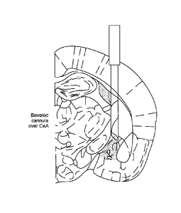
FIG. 8 shows the beveled cannula and illumination profile design. a) Light
cone
from bare fiber emitting 473 nm light over cuvette filled with fluorescein in
water. The
angle of the light cone is approximately 12 degrees. b) Light cone from the
same fiber and
light ensheathed in a beveled cannula. The beveled cannula blocks light
delivery to one
side, without detectably altering perpendicular light penetrance. c) Diagram
of light
delivery via the optical fiber with the beveled cannula over CeA. d) Chart
indicating
estimated light power density seen at various distances from the fiber tip in
mouse brain
tissue when the light power density seen at the fiber tip was 7 mW (-99
mW/mm2). Inset,
cartoon indicating the configuration. Optical fiber is perpendicular and aimed
at the center
of the power meter, through a block of mouse brain tissue. e) Table showing
light power
(mW) as measured by a standard power meter and the estimated light power
density
(mW/mm2) seen at the tip, at the CeL (-0.5-0.7 mm depth in brain tissue) and
at the CeM
(-1.1 mm depth in brain tissue).
FIG. 9 demonstrates that the beveled cannula prevented light delivery to BLA
and
BLA spiking at light powers used for behavioral assays. a) Schematic
indicating the
configuration of light delivery by optical fiber to the CeA and recording
electrode (red) in
CA 3056186 2019-09-19
=
WO 2012/061690 PCT/US2011/059298
9
the BLA. b) Scatterplot summary of recordings in the BLA with various light
powers
delivered to the CeA with and without the beveled eannula (n=4 sites). For
each site,
repeated alternations of recordings were made with and without the beveled
cannula. The
x-axis shows both the light power density at the fiber tip (black) and the
estimated light
power density at the CeL (grey). The blue vertical or shaded region indicates
the range of
light power densities used for behavioral assays (-7 mW; ¨99 mW/mm2 at the tip
of the
fiber). Reliable responses from BLA neurons were not observed in this light
power density
range. c) Representative traces of BLA recordings with 20 Hz 5ms pulse light
stimulation
at 7mW (-99 mW/ mm2 at fiber tip; ¨5.9 mW/ mm2 at CeL) at the same recording
site in
the CeA. d) Population spike waveforms in response to single pulses of light
reveal
substantial light restriction even at high 12 mV power (-170 mW/ mm2 at the
tip of the
fiber; ¨10.1 mW/ mm2 at CeL).
FIG. 10 demonstrates that viral transduction excluded intercalated cell
clusters. a)
Schematic of the intercalated cells displayed in subsequent confocal images.
(b-d)
Representative images of intercalated cells from mice that received EYFP b),
eNpHR 3.0 e)
and ChR2 d) injections into the BLA that were used for behavioral
manipulations. Viral
expression was not observed in intercalated cell clusters. (e-f) There were
very low (<2%)
levels of YFP expression in intercalated cell clusters for all 6 groups used
in behavioral
assays. There were no statistically significant differences among groups in e-
fos
expression.
FIG. 11 shows that unilateral intra-CeA administration of glutamate
antagonists did
not alter locomotor activity. Administration of NBQX and AP5 prior to the open
field test
did not impair locomotor activity (as measured by mean velocity) relative to
saline infusion
(F1,77= 2.34, p = 0.1239).
FIG. 12 demonstrates that bath application of glutamate antagonists blocked
optically-evoked synaptic transmission. 4-6 weeks following intra-BLA
infusions of AAV5-
CamKII-ChR2-EYFP into the BLA of wild-type mice, we examined the ability of
the
glutamate receptor antagonists NBQX and AP5 to block glutamatergic
transmission. a)
Representative current-clamp (top) and voltage-clamp (bottom) traces of a
representative
CeL neuron upon a 20 Hz train of 473 nm light illumination of BLA terminals
expressing
ChR2. b) The same cell's responses following bath application of NBQX and AP5
show
abolished spiking and EPSCs. c) Population summary (n=5) of the depolarizing
current
seen before and after bath application of NBQX and AP5, normalized to the pre-
drug
response.
CA 3056186 2019-09-19
WO 2012/061690 PCT/US2011/059298
FIG. 13 is a diagram depicting the histologically verified placements of mice
treated
with 594 run light. Bilateral placements of virus injection needle (circle)
and tip of beveled
cannula (x) are indicated. Colors indicate treatment group, see legend.
Coronal sections
containing BLA and CeA are shown; numbers indicate AP coordinates from bregma
5 (Aravanis et el., J Neural Eng, 4:S143-156, 2007).
FIG. 14 shows that light stimulation parameters used in the eNpHR 3.0 terminal
inhibition experiments does not block spiking at the cell soma. (a-c)
Schematics of the light
spot location and recording sites alongside corresponding representative
traces upon a
current step lasting the duration of the spike train, paired with yellow light
illumination at
10 each location during the middle epoch (indicated by yellow horizontal
bar). a)
Representative current-clamp trace from a BLA neuron expressing eNpHR 3.0 upon
direct
illumination shows potent inhibition of spiking during illumination of cell
soma. b)
Representative current-clamp trace from a BLA neuron expressing eNpHR 3.0 when
a ¨125
pm diameter light spot is presented ¨300 pm away from the cell soma without
illuminating
an axon. e) Representative current-clamp trace from a BLA neuron expressing
eNpHR 3.0
when a ¨125 gm diameter light spot is presented ¨300 gm away from the cell
soma when
illuminating an axon. d) While direct illumination of the cell soma induced
complete
inhibition of spiking that was significant from all other conditions (F3,9=
81.50, p < 0.0001;
n = 3 or more per condition), there was no significant difference among the
distal
illumination ¨300 gm away from the soma of BLA neurons expressing eNpHR 3.0
conditions and the no light condition (F2,7= 0.79, p = 0.49), indicating that
distal
illumination did not significantly inhibit spiking at the cell soma. e)
Schematic indicating
light spot locations relative to recording site, regarding the population
summary shown to
the right. Population summary shows the normalized hyperpolarizing current
recorded from
the cell soma per distance of light spot from cell soma, both on and off axon
collaterals (n =
5).
FIG. 15 demonstrates that selective illumination of BLA terminals induced
vesicle
release onto CeL neurons without reliably eliciting antidromic action
potentials. Schematics
and descriptions refer to the traces below, and trace color indicates cell
type. Light
illumination patterns are identical for both series of traces. Left column,
CeL traces for three
overlaid sweeps of a 40-pulse light train per cell (n = 8). Here, both time-
locked EPSCs
indicate vesicle release from the presynaptic ChR2-expressing BLA terminal,
and for all
postsynaptic CeL cells, there were excitatory responses to 100% of light
pulses. Right
column, BLA traces for three overlaid 40-pulse sweeps per cell (n=9), with the
mean
CA 3056186 2019-09-19
WO 2012/061690 PCT/US2011/059298
11
number of light pulses delivered at the axon terminal resulting in a supra-
threshold
antidromic action potential (5.4% 2%, mean SEM).
FIG.16 is a graph demonstrating that light stimulation did not alter locomotor
activity in eNpHR 3.0 and control groups. There were no detectable differences
in
locomotor activity among groups nor light epochs (F1,20= 0.023, p = 0.3892;
F1,100= 3.08,
p = 0.086).
DETAILED DESCRIPTION
The present disclosure relates to control over nervous system disorders, such
as
disorders associated with anxiety and anxiety symptoms, as described herein.
While the
present disclosure is not necessarily limited in these contexts, various
aspects of the
invention may be appreciated through a discussion of examples using these and
other
contexts.
Various embodiments of the present disclosure relate to an optogenetic system
or
method that correlates temporal control over a neural circuit with measurable
metrics. For
instance, various metrics or symptoms might be associated with a neurological
disorder
exhibiting various symptoms of anxiety. The optogenetic system targets a
neural circuit
within a patient for selective control thereof. The optogenetic system
involves monitoring
the patient for the metrics or symptoms associated with the neurological
disorder. In this
manner, the optogenetic system can provide detailed information about the
neural circuit, its
function and/or the neurological disorder.
Consistent with the embodiments discussed herein, particular embodiments
relate to
studying and probing disorders. Other embodiments relate to the identification
and/or study
of phenotypes and endophenotypes. Still other embodiments relate to the
identification
of treatment targets.
Aspects of the present disclosure are directed to using an artificially-
induced
anxiety state for the study of anxiety in otherwise healthy animals. This can
be particularly
useful for monitoring symptoms and aspects that are poorly understood and
otherwise
difficult to accurately model in living animals. For instance, it can be
difficult to test and/or
study anxiety states due to the lack of available animals exhibiting the
anxiety state.
Moreover, certain embodiments allow for reversible anxiety states, which can
be
particularly useful in establishing baseline/control points for testing and/or
for testing the
effects of a treatment on the same animal when exhibiting the anxiety state
and when not
exhibiting the anxiety state. The reversible anxiety states of certain
embodiments can also
CA 3056186 2019-09-19
WO 2012/061690 PCT/US2011/059298
12
allow for a reset to baseline between testing the effects of different
treatments on the same
animal.
Certain aspects of the present disclosure are directed to a method related to
control
over anxiety and/or anxiety symptoms in a living animal. In certain more
specific
embodiments, the monitoring of the symptoms also includes assessing the
efficacy of the
stimulus in mitigating the symptoms of anxiety. Various other methods and
applications
exist, some of which are discussed in more detail herein.
Light-responsive opsins that may be used in the present invention includes
opsins
that induce hyperpolarization in neurons by light and opsins that induce
depolarization in
neurons by light Examples of opsins are shown in Tables 1 and 2 below.
Table 1 shows identified opsins for inhibition of cellular activity across the
visible
spectrum:
Table 1: Fast optogenetics: inhibition across the visible spectrum
Wavelength
Opsin Type Biological Origin Defined action
Sensitivity
Natronomonas Inhibition
NpHR 589nm max
pharaonis (hyperpolarization)
Halobacterium Inhibition
BR 570nm max
helobium (hyperpolarization)
Acetabulaira Inhibition
AR 518nm max
acetabulum (hyperpolarization)
Inhibition
GtR3 Guillardia theta 472nm max
(hyperpolarization)
Leptosphaeria Inhibition
Mac 470-500nm max
maculans (hyperpolarization)
Natronomonas 680nm utility Inhibition
NpHr3.0
pharaonis 589nm max (hyperpolarization)
Natronomonas 680nm utility Inhibition
NpHR3.1 =
pharaonis 589nm max (hyperpolarization)
Table 2 shows identified opsins for excitation and modulation across the
visible spectrum:
Table 2: Fast optogenetics: excitation and modulation across the visible
spectrum
Wavelength
Opsin Type Biological Origin Defined action
Sensitivity
589nm utility Excitation
VChR1 Volvox carteri
535nm max (depolarization)
Excitation
DChR Dunaliella sauna 500nrn max
(depolarization)
Chlamydomonas 470nm max Excitation
ChR2
reinhardtii 380-405nm utility (depolarization)
CA 3056186 2019-09-19
WO 2012/061690 PCT/US2011/059298
13
Table 2 (continued):
Table 2: Fast optogenetics: excitation and modulation across the visible
spectrum
Wavelength
Opsin Type Biological Origin Defined action
Sensitivity
ChETA Chlamydomonas 470nm max Excitation
reinhardtii 380-405mn utility
(depolarization)
470nm max Excitation
Chlamydomonas
SFO (depolarization)
reinhardtii
530nm max Inactivation
445nm max Step-like activation
Chlamydomonas
SSFO (depolarization)
reinhardtii
590nm; 390-400nm Inactivation
Volvox carteri and Excitation
C/ V1 Chlamydomonas 542nm max (depolarization)
reinhardtii
Volvox carieri and Excitation
C/ V1 E122 Chlamydomonas 546nm max (depolarization)
reinhardtii
Volvox carteri and Excitation
C/ V1 E162 Chlamydomonas 542nm max (depolarization)
reinhardtii
Volvox carteri and Excitation
C/ V1 E122/E162 Chlamydomonas 546nm max (depolarization)
reinhardtii
As used herein, a light-responsive opsin (such as NpHRõ BR, AR, GtR3, Mac,
ChR2, VChRl, DChR, and ChETA) includes naturally occurring protein and
functional
variants, fragments, fusion proteins comprising the fragments or the full
length protein.
For example, the signal peptide may be deleted. A variant may have an amino
acid
sequence at least about any of 90%, 91%, 92%, 93%, 94%, 95%, 96%, 97%, 98%,
99%, or
100% identical to the naturally occurring protein sequence. A functional
variant may have
the same or similar hyperpolarization function or depolarization function as
the naturally
occurring protein.
In some embodiments, the NpHR is eNp11R3.0 or eNpHR3.1 (See
www.stanfordedu/group/dlab/optogenetics/sequence_info.htrn1). In some
embodiments,
the light-responsive opsin is a C1V1 chimeric protein or a C1V1-E162 (SEQ ID
NO:10),
C IV1-E122 (SEQ ID NO:9), or C1V1-E122/E162 (SEQ NO:11) mutant chimeric
protein (See, Yizhar et al, Nature, 2011, 477(7363):171-78 and
www.stanford.edu/group/dlab/optogeneties/sequence_infoltml). In some
embodiments,
the light-responsive opsin is a SFO (SEQ ID NO:6) or SSFO (SEQ ID NO:7) (See,
Yizhar
CA 3056186 2019-09-19
=
WO 2012/061690 PCT/US2011/059298
14
et al, Nature, 2011, 477(7363):171-78; Berndt et al., Nat. Neurosci.,
12(2):229-34 and
www.stanford.edu/group/dlab/optogeneties/sequence_info.html).
In some embodiments, the light-activated protein is a NpHR opsin comprising an
amino acid sequence at least 95%, at least 96%, at least 97%, at least 98%, at
least 99% or
100% identical to the sequence shown in SEQ ID NO:1. In some embodiments, the
NpHR
opsin further comprises an endoplasrnic reticulum (ER) export signal and/or a
membrane
trafficking signal. For example, the NpHR opsin comprises an amino acid
sequence at least
95% identical to the sequence shown in SEQ ID NO:1 and an endoplasmic
retieulum (ER)
export signal. In some embodiments, the amino acid sequence at least 95%
identical to the
sequence shown in SEQ ID NO:1 is linked to the ER export signal through a
linker. In
some embodiments, the ER export signal comprises the amino acid sequence
FXYENE,
where X can be any amino acid. In another embodiment, the ER export signal
comprises
the amino acid sequence WOCSL, where X can be any amino acid. In some
embodiments,
the ER export signal comprises the amino acid sequence FCYENEV. In some
embodiments, the NpHR opsin comprises an amino acid sequence at least 95%
identical to
the sequence shown in SEQ ID NO:1, an ER export signal, and a membrane
trafficking
signal. In other embodiments, the NpHR opsin comprises, from the N-terminus to
the C-
terminus, the amino acid sequence at least 95% identical to the sequence shown
in SEQ ID
NO:1, the ER export signal, and the membrane trafficking signal. In other
embodiments,
the NpHR opsin comprises, from the N-terminus to the C-terminus, the amino
acid
sequence at least 95% identical to the sequence shown in SEQ ID NO:1, the
membrane
trafficking signal, and the ER export signal. In some embodiments, the
membrane
trafficking signal is derived from the amino acid sequence of the human inward
rectifier
potassium channel Kk2.1. In some embodiments, the membrane trafficking signal
comprises the amino acid sequence KSRITSEGEYIPLDQIDIN V. In some
embodiments, the membrane trafficking signal is linked to the amino acid
sequence at least
95% identical to the sequence shown in SEQ ID NO:1 by a linker. In some
embodiments,
the membrane trafficking signal is linked to the ER export signal through a
linker. The
linker may comprise any of 5, 10,20, 30, 40, 50, 75, 100, 125, 150, 175, 200,
225, 250, 275,
300, 400, or 500 amino acids in length. The linker may further comprise a
fluorescent
protein, for example, but not limited to, a yellow fluorescent protein, a red
fluorescent
protein, a green fluorescent protein, or a cyan fluorescent protein. In some
embodiments,
the light-activated opsin further comprises an N-terminal signal peptide. In
some
embodiments, the light-activated opsin comprises the amino acid sequence of
SEQ ID
CA 3056186 2019-09-19
WO 2012/061690 PCT/US2011/059298
NO:2. In some embodiments, the light-activated protein comprises the amino
acid sequence
of SEQ ID NO:3.
In some embodiments, the light-activated opsin is a chimeric protein derived
from
VChR1 from Volvox carteri and ChR1 from Chlamydomonas reinhardti. In some
5 embodiments, the chimeric protein comprises the amino acid sequence of
VCIal having at
least the first and second transmembrane helices replaced by the corresponding
first and
second transmembrane helices of ChRl. In other embodiments, the chimeric
protein
comprises the amino acid sequence of VChR1 having the first and second
transmembrane
helices replaced by the corresponding first and second transmembrane helices
of ChR1 and
10 further comprises at least a portion of the intracellular loop domain
located between the
second and third transmembrane helices replaced by the corresponding portion
from ChRl.
In some embodiments, the entire intracellular loop domain between the second
and third
transmembrane helices of the chimeric light-activated protein can be replaced
with the
corresponding intracellular loop domain from ChRl. In some embodiments, the
light-
15 activated chimeric protein comprises an amino acid sequence at least
90%, 91%, 92%, 93%,
94%, 95%, 96%, 97%, 98%, 99%, or 100% identical to the sequence shown in SEQ
ID
NO:8 without the signal peptide sequence. In some embodiments, the light-
activated
chimeric protein comprises an amino acid sequence at least 90%, 91%, 92%, 93%,
94%,
95%, 96%, 97%, 98%, 99%, or 100% identical to the sequence shown in SEQ ID
NO:8.
C1V1 chimeric light-activated opsins that may have specific amino acid
substitutions at key
positions throughout the retinal binding pocket of the VChR1 portion of the
chimeric
polypeptide. In some embodiments, the C1V1 protein has a mutation at amino
acid residue
E122 of SEQ ID NO:8. In some embodiments, the C1V1 protein has a mutation at
amino
acid residue E162 of SEQ ID NO:8. In other embodiments, the C1V1 protein has a
mutation at both amino acid residues E162 and E122 of SEQ ID NO:8. In some
embodiments, each of the disclosed mutant C1V1 chimeric proteins can have
specific
properties and characteristics for use in depolarizing the membrane of an
animal cell in
response to light.
As used herein, a vector comprises a nucleic acid encoding a light-responsive
opsin
described herein and the nucleic acid is operably linked to a promoter that
controls the
specific expression of the opsin in the glutamatergic pyramidal neurons. Any
vectors that
are useful for delivering a nucleic acid to glutamatergic pyramidal neurons
may be used.
Vectors include viral vectors, such as AAV vectors, retroviral vectors,
adenoviral vectors,
HSV vectors, and lentiviral vectors. Examples of AAV vectors are AAV1, AAV2,
AAV3,
CA 3056186 2019-09-19
WO 2012/061690 PCT/US2011/059298
16
AAV4, AAV5, AAV6, AAV7, AAV8, AAV9, AAV10, AAV11, AAV12, AAV13,
AAV14, AAV15, and AAV16. A CaMICIla promoter and any other promoters that can
control the expression of the opsin in the glutamatergic pyramidal neurons may
be used.
An "individual" is a mammal, such as a human. Mammals also include, but are
not
limited to, farm animals, sport animals, pets (such as cats, dogs, horses),
primates, mice and
rats. An "animal" is a non-human mammal.
As used herein, "treatment" or "treating" or "alleviation" is an approach for
obtaining beneficial or desired results including and preferably clinical
results. For
purposes of this invention, beneficial or desired clinical results include,
but are not limited
to, one or more of the following: showing observable and/or measurable
reduction in one or
more signs of the disease (such as anxiety), decreasing symptoms resulting
from the disease,
increasing the quality of life of those suffering from the disease, decreasing
the dose of
other medications required to treat the disease, and/or delaying the
progression of the
disease.
As used herein, an "effective dosage" or "effective amount" of a drug,
compound, or
pharmaceutical composition is an amount sufficient to effect beneficial or
desired results.
For therapeutic use, beneficial or desired results include clinical results
such as decreasing
one or more symptoms resulting from the disease, increasing the quality of
life of those
suffering from the disease, decreasing the dose of other medications required
to treat the
disease, enhancing effect of another medication such as via targeting, and/or
delaying the
progression of the disease. As is understood in the clinical context, an
effective dosage of a
drug, compound, or pharmaceutical composition may or may not be achieved in
conjunction
with another drug, compound, pharmaceutical composition, or another treatment.
Thus, an
"effective dosage" may be considered in the context of administering one or
more
therapeutic agents or treatments, and a single agent may be considered to be
given in an
effective amount if, in conjunction with one or more other agents or
treatments, a desirable
result may be or is achieved.
The above overview is not intended to describe each illustrated embodiment or
every implementation of the present disclosure.
DETAILED DESCRIPTION AND EXAMPLE EXPERIMENTAL EMBODIMENTS
The present disclosure is believed to be useful for controlling anxiety states
and/or
symptoms of anxiety. Specific applications of the present invention relate to
optogenetic
systems or methods that correlate temporal, spatio and/or cell-type control
over a neural circuit
associated with anxiety states and/or symptoms thereof. As many aspects of the
example
CA 3056186 2019-09-19
17
embodiments disclosed herein relate to and significantly build on previous
developments
in this field, the following discussion summarizes such previous developments
to provide a
solid understanding of the foundation and underlying teachings from which
implementation details and modifications might be drawn, including those found
in the
Examples.
While the present invention is
not necessarily limited to such applications, various aspects of the invention
may be
appreciated through a discussion of various examples using this context_
Anxiety refers to a sustained state of heightened apprehension in the absence
of an
immediate threat, which in disease states becomes severely debilitating.
Embodiments of
the present disclosure are directed toward the use of one or more of cell type-
specific
optogenetic tools with two-photon microscopy, electrophysiology, and anxiety
assays to study
and develop treatments relating to neural circuits underlying anxiety-related
behaviors.
Aspects of the present disclosure are related to the optogenetic tArgeting of
specific
projections of the brain, rather than cell types, in the study of neural
circuit fimction
relevant to psychiatric disease.
Consistent with particular embodiments of the present disclosure, temporally-
precise optogenetic stimulation of basolateral amygdala (BLA) terminals in the
central
nucleus of the amygdala (CeA) are used to produce a reversible anxiolytic
effect. The
optogenetic stimulation can be implemented by viral transduction of BLA with a
light-
responsive opsin, such as ChR2, followed by restricted illumination in
downstream CeA.
Consistent with other embodiments of the present disclosure, optogenetic
inhibition
of the basolateral amygdala (BLA) terminals in the central nucleus of the
amygdala (CeA)
are used to increase anxiety-related behaviors. The optogenetic stimulation
can be
implemented by viral transduction of BLA with a light-responsive opsin, such
as
eNpHR3.0, followed by restricted illumination in downstream CeA.
Embodiments of the present disclosure are directed towards the specific
targeting of
neural cell populations, as anxiety-based effects were not observed with
direct optogenetic
control of BLA somata. For instance, targeting of specific BLA-CeA projections
as circuit
elements have been experimentally shown to be sufficient for endogenous
anxiety control in
the mammalian brain.
Consistent with embodiments of the present disclosure, the targeting of the
specific
BLA-CeA projections as circuit elements is based upon a number of factors
discussed in more
detail hereafter. The amygdala is composed of functionally and morphologically
CA 3056186 2019-09-19
WO 2012/061690 PCT/US2011/059298
18
heterogeneous subnuclei with complex interconnectivity. A primary subdivision
of the
amygdala is the basolateral amygdala complex (BLA), which encompasses the
lateral
(LA), basolateral (BL) and basomedial (BM) amygdala nuclei (-90% of BLA
neurons are
glutamatergic). In contrast, the central nucleus of the amygdala (CeA), which
is composed
of the centrolateral (CeL) and centromedial (CeM) nuclei, is predominantly (-
95%) comprised
of GABAergic medium spiny neurons. The BLA is ensheathed in dense clusters of
GABAergic intercalated cells (ITCs), which are functionally distinct from both
local
intemeurons and the medium spiny neurons of the CeA. The primary output
nucleus of the
amygdala is the CeM, which, when chemically or electrically excited, is
believed to
mediate autonomic and behavioral responses that are associated with fear and
anxiety via
projections to the brainstem. While the CeM is not directly controlled by the
primary
amygdala site of converging environmental and cognitive information (LA), LA
and BLA
neurons excite GABAergic CeL neurons, which can provide feed-forward
inhibition onto
CeM "output" neurons and reduce amygdala output. The BLA-CeL-CeM is a less-
characterized pathway suggested to be involved not in fear extinction but in
conditioned
inhibition. The suppression of fear expression, possibly due to explicit
unpairing of the
tone and shock, suggested to be related to the potentiation of BLA-CeL
synapses.
BLA cells have promiscuous projections throughout the brain, including to the
bed
nucleus of the stria terminalis (BNST), nucleus accumbens, hippocampus and
cortex.
Aspects of the present disclosure relate to methods for selective control of
BLA terminals in
the CeL, without little or no direct affect/control of other BLA projections.
Preferential
targeting of BLA-CeL synapses can be facilitated by restricting opsin gene
expression to
BLA glutamatergic projection neurons and by restricting light delivery to the
CeA.
For instance, control of BLA glutamatergic projection neurons can be achieved
with an
adeno-associated virus (AAV5) vector carrying light-activated optogenetic
control genes
under the control of a CaMKIla promoter. Within the BLA, CaMKIla is only
expressed
in glutamatergic pyramidal neurons, not in local intemetwons or intercalated
cells.
FIG. 1 shows a system for providing optogenetic targeting of specific
projections of
thebrain, consistent with an embodiment of the present disclosure. For
instance, a beveled
guide cannula can be used to direct light, e.g., prevent light delivery to the
BLA and allow
selective illumination of the CeA. This preferential delivery of light to the
CeA projection
can be accomplished using stereotaxic guidance along with implantation over
the CeL.
Geometric and functional properties of the resulting light distribution can be
quantified
both in vitro and in vivo, e.g., using in vivo electrophysiological recordings
to determine light
CA 3056186 2019-09-19
WO 2012/061690 PCT/US2011/059298
19
power parameters for selective control of BLA terminals but not BLA cell
bodies.
Experimental results, such as those described in the Examples, support that
such selective
excitation or inhibition result in significant, immediate and reversible
anxiety-based effects.
Embodiments of the present disclosure are directed toward the above
realization
being applied to various ones of the anatomical, functional, structural, and
circuit targets
identified herein. For instance, the circuit targets can be studied to develop
treatments for the
psychiatric disease of anxiety. These treatments can include, as non-limiting
examples,
pharmacological, electrical, magnetic, surgical and optogenetic, or other
treatment means.
FIG. 2 shows a flow diagram for use of an anxiety-based circuit model,
consistent
with an embodiment of the present disclosure. An optogenetic delivery device,
such as a.
viral delivery device, is generated 202. This delivery device can be
configured to introduce
optically responsive opsins to the target cells and may include targeted
promoters for
specific cell types. The delivery device can then be stereotaxically (or
otherwise) injected
204 into the BLA. A light delivery device can then be surgical implanted 206.
This light
delivery device can be configured to provide targeted illumination (e.g.,
using a directional
optical element). The target area is then illuminated 208. The target area can
be, for
example, the BLA-CeA. The effects thereof can then be monitored and/or
assessed 210.
This can also be used in connection with treatments or drug screening.
Various embodiments of the present disclosure relate to the use of the
identified
model for screening new treatments for anxiety. For instance, anxiety can be
artificially
induced or repressed using the methods discussed herein, while
pharmacological,
electrical, magnetic, surgical, or optogenetic treatments are then applied and
assessed. In
other embodiments of the present disclosure, the model can be used to develop
an in vitro
approximation or simulation of the identified circuit, which can then be used
in the
screening of devices, reagents, tools, technologies, methods and approaches
and for
studying and probing anxiety and related disorders. This study can be directed
towards, but
not necessarily limited to, identifying phenotypes, endophenotypes, and
treatment targets.
Embodiments of the present disclosure are directed toward modeling the BLA-CeL
pathway as an endogenous neural substrate for bidireetionally modulating the
unconditioned
expression of anxiety. Certain embodiments are directed toward other
downstream circuits,
such as CeA projections to the BNST, for their role in the expression of
anxiety or anxiety-
related behaviors. For instance, it is believed that corticotropin releasing
hormone (CRH)
networks in the BNST may be critically involved in modulating anxiety-related
behaviors,
as the CeL is a primary source of CRH for the BNST. Other neurotransmitters
and
CA 3056186 2019-09-19
20
neuromodulators may modulate or gate effects on distributed neural circuits,
including
serotonin, dopamine, acetylcholine, glycine, GABA and CRH. Still other
embodiments are
directed toward control of the neural circuitry converging to and diverging
from this
pathway, as parallel or downstream circuits of the BLA-CeL synapse are
believed to
contribute to the modulation or expression of anxiety phenotypes. Moreover,
upstream of the
amygdala, this microcircuit is well-positioned to be recruited by top-down
cortical control
from regions important for processing fear and anxiety, including the
prelimbic, infralirnbic
and insular cortices that provide robust innervation to the BLA and CeL.
Experimental results based upon the BLA anatomy suggest that the populations
of
BLA neurons projecting to CeL and CeM neurons are largely non-overlapping. In
natural
states, the CeL-projecting BLA neurons may excite CeM-projecting BLA neurons
in a
microcircuit homeostatic mechanism, which can then be used to study underlying
anxiety
disorders when there are synaptic changes that skew the balance of the circuit
to allow
uninhibited CeM activation.
The embodiments and specific applications discussed herein (including the
Examples) may be implemented in connection with one or more of the above-
described
aspects, embodiments and implementations, as well as with those shown in the
figures
and described below.
, For further details on light- responsive molecules and/or
opsins, including methodology, devices and substances, reference may also be
made to the
following background publications: U.S. Patent Publication No. 2010/0190229,
entitled
"System for Optical Stimulation of Target Cells" to Zhang etal.; U.S. Patent
Publication
No. 2010/0145418, also entitled "System for Optical Stimulation of Target
Cells" to Zhang
et al.; U.S. Patent Publication No. 2007/0261127, entitled "System for Optical
Stimulation
of Target Cells" to Boyden et al.; and PCT WO 2011/116238, Entitled "Light
Sensitive Ion
Passing Molecules".
Consistent with these publications, numerous opsins
can be used in mammalian cells in vivo and in vitro to provide optical
stimulation and
control of target cells. For example, when ChR2 is introduced into an
electrically-excitable
cell, such as a neuron, light activation of the ChR2 channelrhodopsin can
result in excitation
and/or firing of the cell. In instances when NpHR is introduced into an
electrically-excitable
cell, such as a neuron, light activation of the NpHR opsin can result in
inhibition of firing
of the cell. These and other aspects of the disclosures of the above-
referenced patent
applications may be useful in implementing various aspects of the present
disclosure.
CA 3056186 2019-09-19
WO 2012/061690 PCT/US2011/059298
21
While the present disclosure is amenable to various modifications and
alternative forms,
specifics thereof have been shown by way of example in the drawings and will
be described
in further detail. It should be understood that the intention is not to limit
the disclosure to
the particular embodiments and/or applications described. On the contrary, the
intention is
to cover all modifications, equivalents, and alternatives falling within the
spirit and scope of the
present disclosure.
EXAMPLES
Introduction
Anxiety is a sustained state of heightened apprehension in the absence of
immediate
threat, which in disease states becomes severely debilitating'. Anxiety
disorders represent
the most common of the psychiatric diseases (with 28% lifetime prevalence)2,
and have
been linked to the etiology of major depression and substance abuse3-5. While
the amygdala,
a brain region important for emotional pr0cessing9-17, has long been
hypothesized to play a
role in anxiety111-23, the neural mechanisms which control and mediate anxiety
have yet to be
identified. Here, we combine cell type-specific optogenetic tools with two-
photon
microscopy, electrophysiology, and anxiety assays in freely-moving mice to
identify neural
circuits underlying anxiety-related behaviors. Capitalizing on the unique
capability of
0ptogeneties24-26 to control not only cell types, but also specific
connections between cells,
we observed that temporally-precise optogenetic stimulation of basolateral
amygdala (BLA)
terminals in the central nucleus of the amygdala (CcA), resolved by viral
transduction of
BLA with ChR2 followed by restricted illumination in downstream CeA, exerted a
profound, immediate, and reversible anxiolytic effect. Conversely, selective
optogenetic
inhibition of the same defmed projection with eNpHR3.025 potently, swiftly,
and reversibly
increased anxiety-related behaviors. Importantly, these effects were not
observed with direct
optogenetic control of BLA somata themselves. Together, these results
implicate specific
BLA-CeA projections as circuit elements both necessary and sufficient for
endogenous
anxiety control in the mammalian brain, and demonstrate the importance of
optogenetically
targeting specific projections, rather than cell types, in the study of neural
circuit function
relevant to psychiatric disease.
Despite the high prevalence and severity' of anxiety disorders, the
corresponding neural
circuit substrates are poorly understood, impeding the development of safe and
effective treatments.
Available treatments tend to be inconsistently effective or, in the case of
benzodiazepines,
addictive and linked to significant side effects including sedation and
iespiratory suppression that
can cause cognitive impairment and death27' 28. A deeper understanding of
anxiety control
CA 3056186 2019-09-19
WO 2012/061690 PCT/US2011/059298
22
mechanisms in the mammalian brain29' 3 is necessary to develop more efficient
treatments that
have fewer side-effects. Of particular interest and novelty would be the
possibility of recmiting native
pathways for anxiolysis.
The amygdala is critically involved in processing associations between neutral
stimuli and
positive or negative outcomes, and has also been implicated in processing
unconditioned
emotional states. While the amygdala microcircuit has been functionally
dissected in the context of
fear conditioning, amygdalar involvement has been implicated in a multitude of
other
functions and emotional states, including unconditioned anxiety. The amygdala
is composed of
fractionally and motphologically heterogeneous subnuclei with complex
interconnectivity. A primary
subdivision of the amygdala is the basolateral amygdala complex (BLA), which
encompasses the
lateral (LA), basolateral (BL) and basomedial (BM) amygdala nuclei (-90% of
BLA neurons are
glutamatergic)33' 34. In contrast, the central nucleus of the amygdala (CeA),
which is composed of
the centrolateral (CeL) and centromedial (CeM) nuclei, is predominantly (-95%)
comprised of
GABAergic medium spiny neurons35. The BLA is ensheathed in dense clusters of
GABAergic
intercalated cells (ITCs), which are functionally distinct from both local
interneurons and the
medium spiny neurons of the CeA36' 37. The primary output nucleus ofthe
amygdala is the CeM,32'
35' 3e'4 which when chemically or electrically excited mediates autonomic and
behavioral responses
associated with fear and anxiety via projections to the brainstem& 12' 32' 35.
While the C,eM is not
directly controlled by the primary amygdala site of converging environmental
and cognitive
information (LA)12' 38, 41,
LA and BLA neurons excite GABAergic C,eL neurons42 which can
provide feed-forward inhibition onto CeM40' 46 "output" neurons and reduce
amygdala output.
The BIA-CeL-CeM is a less-characterized pathway suggested to be involved not
in fear extinction
but in conditioned inhibition, the suppression of fear expression due to
explicit unpairing of the
tone and shock, due to the potentiation of BLA-CeL synapses47. Although fear
is characterized to
be a phasic state triggered by an external cue, while anxiety is a sustained
state that may occur in
the absence of an external trigger, we wondered if circuits modulating
conditioned
inhibition of fear might also be involved in modulating unconditioned
inhibition of anxiety.
Materials and Methods
Subjects: Male C57BL/6 mice, aged 4-6 weeks at the start of experimental
procedures, were maintained with a reverse 12-hr light/dark cycle and given
food and water
ad libitum. Animals shown in Figures 3, 4 and 5 (mice in the ChR2 Terminals,
EYFP
Terminals and ChR2 Cell Bodies groups) were all single-housed in a typical
high-traffic
mouse facility to increase baseline anxiety levels. Each mouse belonged to a
single
treatment group. Animals shown in Figure 6 (Bilateral EYFP and eNpl-IR 3.0
groups) were
CA 3056186 2019-09-19
WO 2012/061690 23 PCT/US2011/059298
group-housed in a special low-traffic facility to decrease baseline anxiety
levels. Animal
husbandry and all aspects of experimental manipulation of our animals were in
accordance
with the guidelines from the National Institute of Health and have been
approved by
members of the Stanford Institutional Animal Care and Use Committee.
Optical Intensity Measurements: Light transmission measurements were conducted
with blocks of brain tissue from acutely sacrificed mice. The tissue was then
placed over
the photodetector of a power meter (ThorLabs, Newton, NJ) to measure the light
power of
the laser penetrated the tissue. The tip of a 300 um diameter optical fiber
was coupled to a
473 nm blue laser (OEM I aser Systems, East Lansing, MI). To characterize the
light
transmission to the opposite side of the bevel, the photodetector of the power
meter was
placed parallel to the beveled cannula. For visualization of the light cone,
we used
Fluorescein isothiocyanate-dextran (FD150s; Sigma, Saint Louis, MO) at
approximately
5mg/m1 placed in a cuvette with the optical fibers either with or without
beveled cannula
shielding aimed perpendicularly over the fluorescein solution. Power density
at specific
depths were calculated considering both fractional decrease in intensity due
to the conical
output of light from the optical fiber and the loss of light due to scattering
in tissue
(Aravanis et al., J Neural Eng, 4:S143-156, 2007) (Gradinaru et al., J
Neurosci, 27:14231-
14238, 2007). The half-angle of divergence Odiv for a multimode optical fiber,
which
determines the angular spread of the output light, is
si
.n_i (NAfit)
urnu ¨
where ntis is the index of refraction of gray matter (1.36, Vo-Dinh T 2003,
Biomedical
Photonics Handbook (Boca Raton, FL: CRC Press)) and NAfit,(0.37) is the
numerical
aperture of the optical fiber. The fractional change in intensity due to the
conical spread of
the light with distance (z) from the fiber end was calculated using
trigonometry
i(z) P2 n 12 ,
where p r ¨+ --1
i(z. 0) (z + p)2 NA )
and r is the radius of the optical fiber (100 um).
The fractional transmission of light after loss due to scattering was modeled
as a
hyperbolic function using empirical measurements and the Kubelka-Munk model
1'2, and
the combined product of the power density at the tip of the fiber and the
fractional changes
CA 3056186 2019-09-19
24
due to the conical spread and light Reoffering, produces the value of the
power density at a
specific depth below the fiber.
Virus construction and packaging: The recombinant AAV vectors were serotyped
with AAV5 coat proteins and packaged by the viral vector core at the
University of North
Carolina. Viral titers were 2 x 10e12 particles / mL, 3 x 10e12 particles /
mL, 4 x 10e12
particles / mL respectively for AAV-CaMK.fla-hChR2(H134R)- EYFP, AAV-CaMKIIa-
EYFP, and AAV-CaMKIla-eNplIR 3.0-EYFP. The pAAV-CaMKIla-eNpHR3.0-EYFP
plasmid was constructed by cloning CaMICIIa-eNpliR3.0-EYFP into an AAV
backbone
using Mlul and EcoRI restriction sites. Similarly, The pAAV-CaMKIla-EYFP
plasmid was
constructed by cloning CaMK.11a-EYFP into an AAV backbone using Mlul and EcoRI
restriction sites. The maps are available online at www.optogeneties.org.
Stereota.ctic injection and optical fiber placement: All surgeries were
performed
under aseptic conditions under stereotaxic guidance. Mice were anaesthetized
using 1.5-
3.0% isoflourane. All coordinates are relative to bregma in mm3. In all
experiments, both
in vivo and in vitro, virus was delivered to the BLA only, and any viral
expression in the
CeA rendered exclusion from all experiments. Carmula guides were beveled to
form a 45-
55 degree angle for the restriction of the illumination to the CeA. The short
side of the
beveled cannula guide was placed antero-medially, the long side of the beveled
c,annula
shielded the posterior-lateral portion of the light cone, facing the opposite
direction of the
viral injection needle. To preferentially target BLA-CeL synapses, we
restricted opsin gene
expression to BLA glutamatergic projection neurons and restricted light
delivery to the
CeA. Control of BLA glutamatergic projection neurons was achieved using an
adeno-
associated virus (AAV5) vector carrying light-activated optogenetic control
genes under the
control of a CaMICIla promoter. Within the BLA, CaMKIIa is only expressed in
glutamatergic pyramidal neurons, not in local intemeurons4. Mice in the ChR2
Terminals
and EYFP Terminals groups received unilateral implantations of beveled
cannulae for the
optical fiber (counter-balanced for hemisphere), while mice in the eNpHR 3.0
or respective
EYFP group received bilateral implantations of the beveled cannulae over the
CeA (-1.06
mm anteroposterior (AP); +2.25 mm mediolateral (ML); and -4.4 mm dorsoventral
(DV);
PlasticsOne, Roanoke, VA)3. Mice in the ChR2 Cell Bodies groups received
unilateral
implantation of a Doric patchcord chronically implantable fiber (NA=0.22;
Doric lenses,
Quebec, Canada) over the BLA at (-1.6 mm AP; 3.1 mm ML; -4.5 mm DV)3. For all
mice, 0.5 ul of purified AAV was injected unilaterally or bilaterally in the
BLA (13.1 mm
CA 3056186 2019-09-19
WO 2012/061690 PCT/US2011/059298
AP, 1.6 mm ML, -4.9 mm DV)3 using beveled 33 or 35 gauge metal needle facing
postero-
lateral side to restrict the viral infusion to the BLA. 10 Al Hamilton
microsyringe (nanofil;
WPI, Sarasota, FL) were used to deliver concentrated AAV solution using a
microsyringe
pump (UMP3; WPI, Sarasota, FL) and its controller (Micro4; WPI, Sarasota, FL).
Then,
5 0.5 111 of virus solution was injected at each site at a rate of 0.1 I
per min. After injection
completion, the needle was lifted 0.1 mm and stayed for 10 additional minutes
and then
slowly withdrawn. One layer of adhesive cement (C&B metabond; Parkell,
Edgewood,
NY) followed by cranioplastic cement (Dental cement; Stoelting, Wood Dale, IL)
was used
to secure the fiber guide system to the skull. After 20 min, the incision was
closed using
10 tissue adhesive (Vetbond; Fisher, Pittsburgh, PA). The animal was kept
on a heating pad
until it recovered from anesthesia. A dummy cap (rat: C312G, mouse: C313G) was
inserted to keep the cannula guide patent. Behavioral and electrophysiological
experiments
were conducted 4-6 weeks later to allow for viral expression.
In vivo recordings: Simultaneous optical stimulation of central amygdala (CeA)
and
15 electrical recording of basolateral amygdala (BLA) of adult male mice
previously (4-6
weeks prior) transduced in BLA with AAV-CaMICIIa-ChR2-eYFP viral construct was
carried out as described previously (Gradinaru et al., J Neurosci, 27:14231-
14238, 2007).
Animals were deeply anesthetized with isoflurane prior to craniotomy and had
negative toe
pinch. After aligning mouse stereotaxically and surgically removing
approximately 3mm2
20 skull dorsal to amygdala. Coordinates were adjusted to allow for
developmental growth of
the skull and brain, as mice received surgery when they were 4-6 weeks old and
experiments were performed when the mice were 8-10 weeks old (centered at -
1.5mm AP,
2.75mm ML)3, a 1Mohm 0.005-in extracellular tungsten electrode (A-M systems)
was
stereotactically inserted into the craniotomized brain region above the BLA
(in mm: -1.65
25 AP, 3.35 ML, -4.9 DV)3. Separately, a 0.2 N.A. 200 tun core diameter
fiber optic cable
(Thor Labs) was stereotactically inserted into the brain dorsal to CeA (-1.1
AP, 2.25 ML, -
4.2 DV)3. After acquiring a light evoked response, voltage ramps were used to
vary light
intensity during stimulation epochs (20 Hz, 5 ms pulse width) 2s in length.
After acquiring
optically evoked signal, the exact position of the fiber was recorded, the
fiber removed from
the brain, inserted into a custom beveled cannula, reinserted to the same
position, and the
same protocol was repeated. In most trials, the fiber/cannula was then
extracted from the
brain, the cannula removed, and the bare fiber reinserted to ensure the
fidelity of the
population of neurons emitting the evoked signal. Recorded signals were
bandpass filtered
between 300 Hz and 20 kHz, AC amplified either 1000x or 10000x (A-M Systems
1800),
CA 3056186 2019-09-19
WO 2012/061690 PCT/US2011/059298
26
and digitized (Molecular Devices Digidata 1322A) before being recorded using
Clampex
software (Molecular Devices). Clampex software was used for both recording
field signals
and controlling a 473nm (OEM I aser Systems) solidstate laser diode source
coupled to the
optrode. Light power was titrated between < 1 mW (-14 mW/mm2) and 28 mW (-396
mW/mm2) from the fiber tip and measured using a standard light power meter
(ThorLabs).
Electrophysiological recordings were initiated approximately lmm dorsal to BLA
after
lowering isoflurane anesthesia to a constant level of 1%. Optrode was lowered
ventrally in
¨0.1mm steps until localization of optically evoked signal.
Behavioral assays: All animals used for behavior received viral transduction
of BLA
neurons and the implantation enabling unilateral (for ChR2 groups and
controls) or bilateral
(for eNpHR3.0 groups and controls) light delivery. For behavior, multimode
optical fibers
(NA 0.37; 300 gm core, BFL37-300; ThorLabs, Newton, NJ) were precisely cut to
the
optimal length for restricting the light to the CeA, which was shorter than
the long edge of
the beveled cannula, but longer than the shortest edge of the beveled eannula.
For optical
stimulation, the fiber was connected to a 473 rim or 594 nm laser diode (OEM
Laser
Systems, East Lansing, MI) through an FC/PC adapter. Lascr output was
controlled using a
Master-8 pulse stimulator (A.M.P.I., Jerusalem, Israel) to deliver light
trains at 20 Hz, 5 ms
pulse-width for 473 nm light, and constant light for 594 nm light experiments.
All included
animals had the center of the viral injection located in the BLA, though there
was
sometimes leak to neighboring regions or along the needle tract. Any case in
which there
was any detectable viral expression in the CeA, the animals were excluded. All
statistically
significant effects of light were discussed, and undiscussed comparisons did
not show
detectable differences.
The elevated plus maze was made of plastic and consisted of two light gray
open
arms (30 x 5 cm), two black enclosed arms (30 x 5 x 30 cm) extending from a
central
platform (5 x 5 x 5 cm) at 90 degrees in the form of a plus. The maze was
placed 30 cm
above the floor. Mice were individually placed in the center. 1-5 minutes were
allowed for
recovery from handling before the session was initiated. Video tracking
software
(BiObserve, Fort Lee, NJ) was used to track mouse location, velocity and
movement of
head, body and tail. All measurements displayed were relative to the mouse
body. Light
stimulation protocols are specified by group. ChR2:BLA-CeA mice and
corresponding
controls groups (EYFP:BLA-CeA and ChR2:BLA Somata) were singly-housed in a
high-
stress environment for at least 1 week prior to anxiety assays: unilateral
illumination of
BLA terminals in the CeA at 7-8 mW (-106 mW/mm2 at the tip of the fiber, ¨6.3
IIIW/MM2
CA 3056186 2019-09-19
WO 2012/061690 PCT/US2011/059298
27
at CeL and ¨2.4 mW/mm2 at the CeM) of 473 nm light pulse trains (5 ms pulses
at 20 Hz).
For the ChR2 Cell Bodies group BLA neurons were directly illuminated with a
lower light
power because illumination with 7-8 mW induced seizure activity, so we
unilaterally
illuminated BLA neurons at 3-5 mW (-57 mW/mm2) of 473 nm light pulse trains (5
ms
pulses at 20 Hz). For the eNpHR 3.0 and corresponding EYFP group, all mice
were group-
housed and received bilateral viral injections and bilateral illumination of
BLA terminals in
the CeA at 4-6 mW (-71 mW/mm2 at the tip of the fiber, ¨4.7 mW/mm2 at the CeL
and
¨1.9 mW/mm2 at the CeM) of 594 nm light with constant illumination throughout
the 5-min
light on epoch. The 15-min session was divided into 3 5-min epochs, the first
epoch there
was no light stimulation (off), the second epoch light was delivered as
specified above (on),
and the third epoch there was no light stimulation (off).
The open-field chamber (50 x 50 cm) and the open field was divided into a
central
field (center, 23 x 23 cm) and an outer field (periphery). Individual mice
were placed in the
periphery of the field and the paths of the animals were recorded by a video
camera. The
total distance traveled was analyzed by using the same video-tracking
software, Viewer2
(BiObserve, Fort Lee, NJ). The open field assessment was made immediately
after the
elevated-plus maze test. The open field test consisted of an 18-min session in
which there
were six 3-min epochs. The epochs alternated between no light and light
stimulation
periods, beginning with a light off epoch. For all analyses and charts where
only "off' and
"on" conditions are displayed, the 3 "off' epochs were pooled and the 3 "on"
epochs were
pooled.
For the glutamate receptor antagonist manipulation, a glutamate antagonist
solution
consisting of 22.0 mM of NBQX and 38.0 mM of D-APV (Tocris, Ellisville, MO)
dissolved
in saline (0.9% NaCl). 5-15 min before the anxiety assays, 0.3 I of the
glutamate
antagonist solution was infused into the CeA via an internal infusion needle,
inserted into
the same guide carmulae used for light delivery via optical fiber, that was
connected to a 10-
Hamilton syringe (nanofil; WPI, Sarasota, FL). The flow rate (0.1 piper min)
was
regulated by a syringe pump (Harvard Apparatus, MA). Placements of the viral
injection,
guide eannula and chronically-implanted fiber were histologically verified as
indicated in
Figures 7 and 10.
Two-photon optogenetic circuit mapping and ex vivo electrophysiological
recording:
Mice were injected with AAV5-CaMKIla-ChR2-EYFP at 4 weeks of age, and were
sacrificed for acute slice preparation 4-6 weeks to allow for viral
expression. Coronal slices
containing the BLA and CeA were prepared to examine the functional
connectivity between
CA 3056186 2019-09-19
WO 2012/061690 PCT/US2011/059298
28
the BLA and the CeA. Two-photon images and electrophysiological recordings
were made
under the constant perfusion of aCSF, which contained (in mM): 126 NaC1, 26
NaHCO3,
2.5 KC1, 1.25 Na1-I2PO4, 1 MgCl, 2 CaCl2, and 10 glucose. All recordings were
at 32 C.
Patch electrodes (4-6 MOlims) were filled (in rnM): 10 HEPES, 4 Mg-ATP, 0.5
MgCl2, 0.4
Na3-GTP, 10 NaC1, 140 potassium gluconate, and 80 Alexa-Fluor 594 hydrazide
(Molecular
Probes, Eugene OR). Whole-cell patch-clamp recordings were performed in BLA,
CeL and
CeM neurons, and cells were allowed to fill for approximately 30 minutes
before imaging
on a modified two-photon microscope (Prairie Microscopes, Madison WI) where
two-
photon imaging, whole-cell recording and optogenetic stimulation could be done
simultaneously. Series resistance of the pipettes was usually 10-20 MOhms.
Blue light
pulses were elicited using a 473nm LED at ¨7mW/mm2 (Thorlabs, Newton NJ)
unless
otherwise noted. A Coherent Ti-Saphire laser was used to image both ChR2-YFP
(940 nm)
and Alexa-Fluor 594 (800 nm). A FF560 dichroic with filters 630/69 and 542/27
(Semrock,
Rochester NY) was also used to separate both molecules' emission. All images
were taken
using a 40X/.8 NA LUMPlanFL/ER Objective (Olympus, Center Valley PA). In order
to
isolate fibers projecting to CeL from the BLA and examine responses in the
CeM, slices
were prepared as described above with the BLA excluded from illumination.
Whole-cell
recordings were performed in the CeM with illumination from the objective
aimed over the
CeL. To further ensure activation of terminals from the BLA to CeL was
selective,
illumination was restricted to a ¨125 gm diameter around the center of the
CeL. Here, blue
light pulses were elicited using an XCite halogen light source (EXPO,
Mississauga,
Ontario) with a 470/3 filter at 6.5 mW/mm2 coupled to a shutter (Uniblitz,
Rochester NY).
For functional mapping, we first recorded from a BLA neuron expressing ChR2
and
simultaneously collected electrophysiological recordings and filled the cell
with Alexa-
Fluor 594 hydrazide dye to allow for two-photon imaging. Two-photon z-stacks
were
collected at multiple locations along the axon of the filled BLA neuron. We
then followed
the axon of the BLA neuron projecting to the CeL nucleus and recorded from a
CeL neuron
in the BLA terminal field. We then simultaneously recorded from a CeL neuron,
filled the
cell with dye and performed two-photon live imaging before following the CeL
neuronal
axons to the CeM. We then repeated this procedure in a CeM neuron, but moved
the light
back to the terminal field in the CeL to mimic the preferential illumination
of BLA-CeL
synapses with the same stimulation parameters as performed in vivo. Voltage-
clamp
recordings were made at both -70 mV, to isolate EPSCs, and at 0 mV, to isolate
PSCs.
EPSCs were confirmed to be EPSCs via bath application of the glutamate
receptor
CA 3056186 2019-09-19
WO 2012/061690 PCT/1JS2011/059298
29
antagonists (n = 5), NBQX (22 gM) and AP5 (38 gM), IPSCs were confirmed to be
IPSCs
via bath application of bicuculline (10 gM; n = 2), which abolished them,
respectively. We
also performed current-clamp recordings when the cell was resting at
approximately -70
mV.
For the characterization of optogenetically-driven antidromic stimulation in
BLA
axon terminals, animals were injected with AAV5-CalVIKIIa-ChR2-EYFP at 4 weeks
of
age, and were sacrificed for acute slice preparation 4-6 weeks to allow for
viral expression.
Slice preparation was the same as above. To the aCSF we added 0.1 mM
picrotoxin, 10 gM
CNQX and 25 gM AP5 (Sigma, St. Louis, MO). Whole-cell patch-clamp recordings
were
performed in BLA neurons and were allowed to fill for approximately 30 minutes
before
two-photon imaging. Series resistance of the pipettes was usually 10-20 MOhms.
All
images were taken using a 40X/.8 NA LUMPlanFL/IR Objective (Olympus, Center
Valley
PA). Blue light pulses were elicited using an XCite halogen light source
(EXPO,
Mississauga, Ontario) with a 470/30 filter at 6.5 mW/mm2 coupled to a shutter
(Uniblitz,
Rochester NY). Two-photon z- stacks were collected at multiple locations along
the axon of
the filled BLA neuron. Only neurons whose axons could be visualized for over
¨300 gm
diameter towards the CeL nucleus were included for the experiment, and neurons
that had
processes going in all directions were also excluded. Stimulation on/off axon
was
accomplished by moving the slice relative to a ¨125 gm diameter blue light
spot. In order
to calibrate the slice for correct expression, whole-cell patch-clamp was
performed on a
CeL cell and a ¨125 gm diameter spot blue pulse was used to ensure that
synaptic release
from the BLA terminals on to the CeL neuron was reliable.
For the dissection of direct and indirect projections to CeM, animals were
injected
with AAV5-CaMKIla-ChR2-EYFP at 4 weeks of age, and were sacrificed for acute
slice
preparation 4-6 weeks to allow for viral expression. Slice preparation was the
same as
above. Light was delivered through a 40X/.8 NA LUMPlanFL/IR Objective
(Olympus,
Center Valley PA). Prior to whole cell patch clamping in the CeM nucleus, the
location of
the CeL nucleus was noted in order to revisit it with the light spot
restricted to this region.
Whole-cell patch-clamp recordings were performed in CeM neurons. Series
resistance of
the pipettes was usually 10-20 MOhms. Blue light pulses were elicited using a
XCite
halogen light source (EXPO, Mississauga, Ontario) with a 470/30 filter at 6.5
mW/mm2
coupled to a shutter (Uniblitz, Rochester NY). During CeM recordings, broad
illumination
(-425-450 gm in diameter) of BLA terminals in the CeA and 20 Hz, 5 ms light
train for 2s
was applied. Voltage-clamp recordings were made at 70mV and OmV to isolate
EPSCs and
CA 3056186 2019-09-19
WO 2012/061690 PCT/US2011/059298
IPSCs respectively. Current-clamp recordings were also made. Then,
illumination was
moved to the CeL using a restricted light spot ¨125 p.m in diameter. We again
performed
voltage clamp recordings at -70mV and OmV and used 20 Hz, 5 ms light train for
2s. For
the CeM neuron spiking inhibition experiments, in current-clamp, we applied
the minimal
5 current step required to induce spiking (-60pA) and simultaneously
applied preferential
illumination of ChR2-expressing BLA terminals in the CeL with a 20 Hz, 5 ms
light train
for 2s (mean over 6 sweeps per cell). For the experiments comparing the broad
illumination
of the BLA terminal field centered in the CeM to selective illumination of BLA-
CeL
terminals, these conditions were performed in repeated alternation in the same
CeM cells
10 (n=7).
To verify that terminal inhibition did not alter somatic spiking, animals were
injected with AAV5-CaMKIIa-eNpHR3.0-EYFP at 4 weeks of age, and were
sacrificed for
acute slice preparation 4-6 weeks to allow for viral expression. Slice
preparation was the
same as above. Whole-cell patch-clamp recordings were performed in BLA neurons
and
15 were allowed to fill for approximately 30 minutes. Light was delivered
through a 40X/.8
NA LUMPlanFL/IR Objective (Olympus, Center Valley PA). Whole-cell patch-clamp
recordings were performed on BLA neurons. Series resistance of the pipettes
was usually
10-20 MOhms. Yellow light pulses were elicited using a XCite halogen light
source
(EXPO, Mississauga, Ontario) with a 589/24 filter at 6.5 mW/mm2 coupled to a
shutter
20 (Uniblitz, Rochester NY). After patching, an unrestricted light spot (-
425-450 microns in
diameter) was placed over the BLA soma and a is pulse was applied. Cells were
excluded
if the current recorded was under 600 pA of hyperpolarizing current and the
axon did not
travel over ¨300 pm towards the CeL nucleus. The light spot was then
restricted to ¨125
p.m in diameter. On and off axon voltage clamp recordings were taken with a is
pulse of
25 light. For the current clamp recordings, action potentials were
generated by applying 250
pA of current to the cell soma through the patch pipette.
To demonstrate that selective illumination of eNpHR3.0-expressing BLA
terminals
reduced the probability of spontaneous vesicle .release, animals were injected
with AAV5-
CaMKIIa-eNpHR3.0-EYFP at 4 weeks of age, and were sacrificed for acute slice
30 preparation 4-6 weeks to allow for viral expression. Slice preparation
was the same as
above. Whole-cell patch-clamp recordings were performed in central lateral
neurons. Light
was delivered through a 40X./.8 NA LUMPlanFL/IR Objective (Olympus, Center
Valley
PA). Series resistance of the pipettes was usually 10-20 MOluns. Yellow light
pulses were
elicited using a XCite halogen light source (EXPO, Mississauga, Ontario) with
a 589/24
CA 3056186 2019-09-19
WO 2012/061690 PCT/US2011/059298
31
filter at 6.5 mW/mm2 coupled to a shutter (Uniblitz, Rochester NY). The light
spot was
restricted to ¨125 jim in diameter. Carbachol was added to the bath at a
concentration of 20
M. After sEPSC activity increased in the CeL neuron, light pulses were applied
ranging in
times from 5s to 30s.
To demonstrate that selective illumination of eNpHR3.0-expressing BLA
terminals
could reduce the probability of vesicle release evoked by electrical
stimulation, animals
were injected with AAV5-CaMKIIa-eNpHR3.0-EYFP at 4 weeks of age, and were
sacrificed for acute slice preparation 4-6 weeks to allow for viral
expression. Slice
preparation was the same as above. A bipolar concentric stimulation probe
(FHC, Bowdoin
ME) was placed in the BLA. Whole-cell patch-clamp recordings were performed in
CeL
neurons. Light was delivered through a 40X/.8 NA LUMPlanFUIR Objective
(Olympus,
Center Valley PA). Series resistance of the pipettes was usually 10-20 MOhms.
Amber
light pulses over the central lateral cell were elicited using a XCite halogen
light source
(EXPO, Mississauga, Ontario) with a 589/24 filter at 6.5 mW/min2 coupled to a
shutter
(Uniblitz, Rochester NY). The light spot was restricted to ¨125 m in
diameter. Electrical
pulses were delivered for 40 seconds and light was delivered starting at 10
seconds and shut
off at 30 seconds in the middle.
For the anatomical tracing experiments, neurons were excluded when the traced
axons were observed to be severed and all BLA neurons included in the
anatomical assay
(Fig. 5 a-i) showed spiking patterns typical of BLA pyramidal neurons18 upon a
current
step.
Slice immunohistochemistry: Anesthetized mice were transeardially perfused
with
ice-cold 4% paraformaldehyde (PFA) in PBS (pH 7.4) 100-110 min after
termination of in
vivo light stimulation. Brains were fixed overnight in 4% PFA and then
equilibrated in 30%
sucrose in PBS. 40 m-thick coronal sections were cut on a freezing microtome
and stored
in cryoprotectant at 4 C until processed for immunohistochemistry. Free-
floating sections
were washed in PBS and then incubated for 30 min in 0.3% Tx100 and 3% normal
donkey
serum (NDS). Primary antibody incubations were performed overnight at 4 C in
3%
NDS/PBS (rabbit anti-c-fos 1:500, Calbiochem, La Jolla, CA; mouse anti-CaMKII
1:500,
Abeam, Cambridge, MA). Sections were then washed and incubated with secondary
antibodies (1:1000) conjugated to Cy3 or Cy5 (Jackson Laboratories, West
Grove, PA) for 3
hrs at room temperature. Following a 20 min incubation with DAPI (1:50,000)
sections
were washed and mounted on microscope slides with PVD-DABCO.
CA 3056186 2019-09-19
WO 2012/061690 PCT/US2011/059298
32
Confocal microscopy and analysis: Confocal fluorescence images were acquired
on
a Leica TCS SP5 scanning laser microscope using a 20X/0.70NA or a 40X/1.25NA
oil
immersion objective. Serial stack images covering a depth of 10 gm through
multiple
sections were acquired using equivalent settings. The Volocity image analysis
software
(Improvision / PerkinElmer, Waltham, MA) calculated the number of c-fos
positive cells
per field by thresholding c-fos immunoreactivity above background levels and
using the
DAPI staining to delineate nuclei. All imaging and analysis was performed
blind to the
experimental conditions.
Statistics: For behavioral experiments and the ex vivo electrophysiology data,
binary
comparisons were tested using nonparametric bootstrapped t-tests (paired or
unpaired where
appropriate)5, while hypotheses involving more than two group means were
tested using
linear contrasts (using the "boot" and "1me4" packages in R6, respectively);
the latter were
formulated as contrasts between coefficients of a linear mixed-effects model
(a "two-way
repeated-measures ANOVA") with the fixed effects being the genetic or
pharmacological
manipulation and the light treatment (on or off). All hypothesis tests were
specified a
priori. Subjects were modeled as a random effects. For c-fos quantification
comparisons,
we used a one-way ANOVA followed by Tukey's multiple comparisons test.
Plots of the data clearly show a relationship between observation mean and
observation variance (that is, they are heteroskedastic; see for example,
Figure 3e and
Figure 5j). We found that a standard square-root transformation corrected this
well.
Additionally, eNPITR3.0 elevated plus maze (EPM) data required detrending by a
linear fit
over time to account for a decrease in exploration behavior over time. As is
standard for a
two-way linear mixed effects model (also known as a two-way repeated-measures
ANOVA), we model (the square-root corrected value of) the kth observation in
the ijth cell
(yijk) as
it + cõ, tj : + b + etik (1)
where
4 is the grand mean across all cells (where the ijth "cell" in the collection
of observations
corresponding to the ith condition and jth treatment)
ci is a fixed effect due to the ith animal condition across treatments (for
example, a genetic
manipulation)
tj is a fixed effect due to the jth treatment across conditions (for example,
light on or light
off)
CA 3056186 2019-09-19
WO 2012/061690 PCT/US2011/059298
33
(c: t)i is a fixed effect due to the interaction of the ith condition and jth
treatment in the ijth
cell
k is a random effect corresponding to animals being used across treatments,
and
eyk is an independent and identically distributed (i.i.d.) random normal
disturbance in the
ijkth observation with mean 0 and variance G2, and independent of b, for all j
Collecting the fixed effects into a 2-way analysis of variance (ANOVA) design
matrix X C Rw, dummy coding the random effects in a sparse matrix Z C R'q, and
letting
= Nwe can express the model in matrix form as
fi = X,3 + Zb + e
(2)
where 5 c Rn, b c Rg, and c:a 3:"' are observations of random variables 3), t,
and c respectively
and our model assumes
c _kr(0,02/), J..8
(S)IB = b) AI' (X + Z b cr2 I)
where N (pa) denotes the multivariate Gaussian distribution with mean vector p
and
variance-covariance matrix E, and I indicates that two variables are
independent. To
estimate the coefficient vectors 13 R", b C IV, and the variance parameter a
and sparse
(block-diagonal) relative variance-covariance matrix E C IV, we use the 1me4
package in
R written by Douglas Bates and Martin Maechler, which first finds a linear
change of
coordinates that "spheres" the random effects and then finds the maximum
likelihood
estimates for fl, a, and E using penalized iteratively reweighted least-
squares, exploiting the
sparsity of the random effects matrix to speed computation. For more details
see the
documentation accompanying the package in the 1me4 repository at http://www.r-
proj ect. org/.
To solve for the maximum likelihood estimates, the design matrix X in equation
2
must be of full column rank. It is well known that this is not the case for a
full factorial
design matrix with an intercept (as in equation 1), and thus linear
combinations
("contrasts") must be used to define the columns of X in order for the fixed-
effect
coefficients to be estimable. As our designs are balanced (or nearly
balanced), we used
orthogonal (or nearly orthogonal) Helmert contrasts between the coefficients
associated
with light on as compared to light off conditions, terminal stimulation as
compared to
control conditions, and so on, as reported in the main text. Such contrasts
allowed us to
compare pooled data (e.g., from several sequential light on vs. light off
conditions) against
CA 30561 8 6 2 01 9-0 9-1 9
WO 2012/061690 PCT/US2011/059298
34
each other within a repeated-measures design _____________________________
yielding improved parameter estimation and
test power while accounting for within-animal correlations.
Results
BLA cells have promiscuous projections throughout the brain, including to the
bed nucleus of the stria tenninalis (BNST), nucleus accumbens, hippocampus and
cortex38'
43. To test whether BLA-CeL synapses could be causally involved in anxiety, it
was
therefore necessary to develop a method to selectively control BLA terminals
in the
CeL, without directly affecting other BLA projections. To preferentially
target BLA-CeL
synapses, we restricted opsin gene expression to BLA glutamatergic projection
neurons and
restricted light delivery to the CeA. Control of BLA glutamatergic projection
neurons was
achieved with an adeno-associated virus (AAV5) vector carrying light-activated
optogenetic control genes under the control of a CaMKIIa promoter; within the
BLA,
CaMIC_IIa is only expressed in glutamatergic pyramidal neurons, not in local
interneurons
or intercalated cells". To preferentially deliver light to the CeA projection,
virus was
delivered unilaterally into the BLA under stereotaxic guidance (Fig. 7 and 8)
along with
implantation of a beveled guide cannula over the CeL to prevent light delivery
to the BLA
and allow selective illumination of the CeA. Geometric and functional
properties of the
resulting light distribution were quantified both in vitro and in vivo, with
in vivo
electrophysiological recordings to detennine light power parameters for
selective control of
BLA terminals but not BLA cell bodies (Figs. 9).
To test the hypothesis that the BLA-CeA pathway could implement an
endogenous mechanism for anxiolysis, we probed freely-moving mice under
projection-
specific optogenetic control in two distinct and well-validated anxiety
assays: the elevated
plus maze and the open field test (Fig. 3a-f). Mice display anxiety-related
behaviors when
exposed to open or exposed spaces, therefore increased time spent in the
exposed arms
of the elevated plus maze or in the center of the open field chamber indicates
reduced
anxiety49' 50. To test for both induction and reversal of relevant behaviors,
we first exposed
mice to the elevated plus maze for three 5-min epochs, in which light was
delivered during
the second epoch only.
To determine whether the anxiolytic effect we observed would be specific to
activation of BLA terminals in the CeA, and not BLA cells in general, we
compared mice
receiving projection-specific control (in the ChR2:BLA-CeA group; Fig. 3a) to
both a
negative control group receiving transduction with a control virus given the
same
pattern of illumination (EYFP:BLA-CeA) and a positive control group transduced
with
CA 3056186 2019-09-19
WO 2012/061690 PCT/US2011/059298
the AAV-CaMKIla-ChR2-EYFP virus in the BLA with a fiber implanted directly
over
the BLA (ChR2:BLA Somata). For this group (ChR2:BLA Somata), light stimulation
did
not elicit the anxiolysis observed in the ChR2:BLA-CeA group (Fig.3b and c);
indeed, the ChR2:BLA-CeA group spent significantly more time in open arms
5 (t(42)=8.312; p<0.00001; Fig.3b,c) during light-induced activation of BLA
terminals in
the CeA , in comparison to controls (EYFP:BLA-CeA and ChR2:BLA Somata
groups). The ChR2:BLA-CeA mice also showed an increase in the probability of
entering an open arm rather than a closed arm, from the choice point of the
center of the
maze (Fig.3c inset), indicating an increased probability of selecting the
normally
10 anxiogenic environment.
We also probed mice on the open field arena for six 3-minute epochs, again
testing for
reversibility by alternating between no light (off) and light stimulation (on)
conditions.
Experimental (ChR2:BLA-CeA) mice displayed an immediate, robust, and
reversible
light-induced anxiolytic response as measured by the time in center of the
open field
15 chamber (Fig. 3d and e), while mice in the EYFP:BLA-CeA and ChR2:BLA
Somata
groups did not (Fig.3e). Light stimulation did not significantly alter
locomotor activity
(Fig.30. While there was no detectable difference among groups in the off
conditions,
there was a significant increase in center time of the open field spent by
mice in the
ChR2:BLA-CeA group relative to the EYFP:BLA-CeA or ChR2:BLA Somata groups
20 during the on conditions (t(105)=4.96178; p<0.0001 for each contrast).
We concluded that
selective stimulation of BLA projections to the CeA, but not BLA somata,
produces an
acute, rapidly reversible anxiolytic effect, supporting the hypothesis that
the BLA-CeL-
CeM pathway could represent a native microcircuit for anxiety control.
We next investigated the physiological basis of this light-induced anxiolytic
25 effect. Glutamatergic neurons in the BLA send robust excitatory
projections to CeL
neurons as well as to CeM neurons3g; however, not only are the CeM synapses
distant
from the light source (Fig. 8), but also any residual direct excitation of
these CeM neurons
would be expected to result in an arudogenic, rather than an anxiolytic,
effect12. However,
CeL neurons exert strong inhibition onto these brainstem-projecting CeM output
30 neurons32' 35' 40, and we therefore hypothesized that illumination of
BLA terminals in the
CeA could activate BLA-CeL neurons and thereby elicit feed-forward inhibition
onto
CeM neurons and implement the observed anxiolytic phenomenon.
CA 3056186 2019-09-19
WO 2012/061690 PCT/US2011/059298
36
To confirm the operation of this optogenetically-defined projection, we
undertook in
vivo experiments, with light delivery protocols matched to those delivered in
the behavioral
experiments, and activity-dependent immediate early gene (c-fos) expression
analysis as
the readout to verify the pattern of neuronal activation (Fig.3g-k). Under
blinded
conditions, we quantified the proportion of neurons in the BLA, CeL and CeM
(Fig. 3i-k)
for ChR2:BLA-CeA, EYFP:BLA-CeA and ChR2:BLA Somata groups that expressed
EYFP or showed c-fos immunoreactivity. Virus expression under the CaMKIIa
promoter
in the BLA targeted glutamatergic neurons42, and we did not observe EYFP
expression in
local interneurons nor intercalated cells (Figure 10). No significant
differences among
groups were detected in the proportion of EYFP-positive cells within each
region (Fig.3g-
k), but we found a significantly higher proportion of c-fos positive BLA cells
in the
ChR2:BLA Somata group, relative to ChR2:BLA-CeA or EYFP:BLA-CeA groups
(Fig.3i;p<0.01 and p<0.05, respectively). There was no detectable difference
in c-fos
between the ChR2:BLA-CeA and EYFP:BLA-CeA groups, indicating that the beveled
cannula shielding effectively prevented direct illumination to BLA cell
bodies. A
significantly higher proportion of CeL neurons expressed c-fos in the ChR2:BLA-
CeA
group relative to the EYFP:BLA-CeA group (p<0.05), but not the ChR2:BLA Somata
group (Fig.3j). Thus, selective illumination of BLA terminals expressing ChR2
in
the CeA led to preferential activation of CeL neurons, without activating BLA
somata.
In the CeM, we found twice as many c-fos positive neurons (relative to total
neurons) in
the ChR2:BLA Somata group than in the ChR2:BLA-CeA (Fig.3k), consistent with
anatomical projections, as LA neurons selectively innervate CeL neurons, while
neurons in
the BL and BM nuclei of the amygdala have mono synaptic projections to both
the CeL
and the CeM38' 43' 31. Together, these data reveal that the in vivo
illumination that triggers
an acute anxiolytic behavioral phenotype implements selective illumination of
BLA-CeL
synapses without activating BLA cell bodies.
To test the hypothesis that selective illumination of BLA terminals in the CeL
induces feed-forward inhibition of CeM output neurons, we combined whole-cell
patch-
clamp recording with live two-photon imaging to visualize the microcircuit
while
simultaneously probing the functional relationships among these cells during
projection-specific optogenetic control (Fig.4a-f). While the light-
stimulation parameters
used in vivo were delivered via a fiber optic and the parameters used in our
ex vivo
experiments were delivered onto acute slices, we matched the light power
density at our target
location ¨6 mW/mm2. A two-photon image of the BLA-CeL-CeM circuit is shown in
CA 3056186 2019-09-19
WO 2012/061690 PCT/US2011/059298
37
Figure 4a, with all three cells imaged from the same slice (Fig.4a). The BLA
neuron
expressing ChR2- EYFP showed robust, high-fidelity spiking to direct
illumination with
20 Hz, 5 ms pulses of 473 nm light (Fig.4b). A representative trace from a CeL
neuron,
recorded during illumination of the terminal field of BLA neurons expressing
ChR2-
EYFP, demonstrates the typical excitatory responses seen in CeL (Fig.4c), with
population summaries revealing that spiking fidelity was steady throughout the
40-pulse
light train and that responding cells include both weakly and strongly-excited
CeL cells
(n=16; Fig.4c). To test whether illumination of BLA-CeL synapses would be
functionally
significant at the level of blocking spiking in CeM cells due to the robust
feed-forward
inhibition from CeL neurons, we recorded from CeM neurons while selectively
illuminating BLA-CeL synapses (Fig.4d). Indeed, we observed potent spiking
inhibition (F2,11=15.35, p=0.0044) in the CeM due to light stimulation of BLA
terminals
in the CeL (Fig.4d; spikes per second before (49+9.0), during (1.5+0.87), and
after
(33+8.4) illumination; mean + s.e.m ). Next, Fig.4e shows CeM responses
recorded
during illumination of the terminal field of BLA neurons in the CeM expressing
ChR2-
EYFP, and the combined excitatory and inhibitory input. Population summaries
from
voltage-clamp recordings indicated that latencies of EPSCs were shorter than
those of the
disynaptic IPSCs, as expected, and that the mean IPSC amplitude was greater
than mean
EPSC amplitude (recorded at 0 and -70 mV, respectively; Fig.4e). Importantly,
the very
same CeM neurons (n=7) yielded net excitation with broad illumination of BLA
inputs to
the CeM (Fig. 4e), but displayed net inhibition with selective illumination of
BLA inputs
to the CeL (Fig. 4f) in a repeatable fashion with alternation between sites.
This
demonstrates that the balance of direct and indirect inputs from the BLA to
the CeM can
modulate CeM output. Together, these data reveal a structurally- and
functionally-identified physiological microcircuit, whereby selective
illumination of
BLA terminals in the CeA activates BLA-CeL synapses, thus increasing feed-
forward
inhibition from CeL neurons onto the brainstem-projecting CeM neurons.
To further elucidate the amygdalar microcircuits underlying this anxiolytic
effect,
we carefully dissected the anatomical and functional properties governing this
phenomenon. While some efforts to map the projections of BLA collaterals in
the CeA
have been made in the rat, we empirically tested whether overlapping or
distinct
populations of BLA neurons projected to the CeL and CeM (Fig.5a,b). A
noteworthy
caveat is that we visualized these neurons in ¨350 urn thick coronal sections
and while
every attempt was made to exclude neurons in which the axons were severed, we
CA 3056186 2019-09-19
WO 2012/061690 PCT/US2011/059298
38
cannot exclude the possibility that this occurred nor can we deny that this
induced some
sampling bias for BLA neurons closer to the CeA. Fig.5a summarizes the
anatomical
projections of the BLA neurons sampled (n=18) and shows that the 44% of
neurons
projected to the CeL alone and 17% projected to the CeM alone. However, a
minority
of BLA cells (n=1;6%), projected to both the CeL and the CeM, one of which
sent
separate collaterals to the CeL and CeM and one of which sent a collateral
that sent
branches to the CeL and CeM. Fig. 5b shows the 2-photon image of each cell
sampled, all
of which showed spiking patterns typical of BLA pyramidal neurons upon a
current step.
Next, as our c-fos assays suggested that illumination of BLA terminals in the
CeL
were sufficient to excite CeL neurons, but not BLA neurons themselves, we
sought to
confirm this hypothesis with whole-cell recordings. With electrical
stimulation,
depolarization of axon terminals leads to antidromic spiking at the cell soma.
However,
there has been evidence that optogenetically-induced depolarization functions
via a
distinct mechanism. To evaluate the properties of optogenetically-induced
terminal
stimulation in this amygdalar microcircuit, we recorded from BLA pyramidal
neurons
expressing ChR2 and moved a light spot (-120 gm in diameter) in 100 gm steps
from
the cell soma, both in a direction over a visually- identified axon collateral
and in a
direction where there was no axon (Fig.5c). The spike fidelity of the BLA
neuron given a 20
Hz train of light at each distance from the soma is summarized in Fig.5d,
while the
depolarizing current is summarized in Fig.5e. In all preparations, we
confirmed that the
light stimulation parameters used were sufficient to elicit high-fidelity
spiking at the BLA
cell soma (Fig.5f) and reliable vesicle release at BLA terminals as shown by
recordings
from a postsynaptic CeL neuron (Fig.5g; Fig. 15). In contrast, when recording
from the
same BLA neurons with the light spot 300 um away from the cell soma we did not
observe
reliable action potential induction, regardless of whether we were over an
axon (Fig.5h) or
not (Fig.5i). This absence of antidromic spiking was observed even upon bath
application of GABA and AMPA receptor antagonists (n=7), thus excluding the
possible
contribution of local inhibitory constraints. While we demonstrate that
optogenetically-
induced vesicle release can occur in the absence of antidromic stimulation in
BLA
pyramidal neurons, it is possible that at antidromic stimulation could be
achieved with
greater light power density than we used here (-6 mW/mm2). Thusfar, we have
demonstrated that the populations of BLA neurons projecting to the CeL and the
CeM
are largely distinct and that illumination of BLA-CeL synapses induces vesicle
release and
CeL excitation without strong activation of BLA somata themselves.
CA 3056186 2019-09-19
WO 2012/061690 PCT/US2011/059298
39
Finally, we further explored the mechanism with in vivo pharmacological
analysis in
the setting of projection-specific optogenetic control. To determine whether
the anxiolytic
effect we observed could be due to the selective activation of BLA-CeL
synapses alone,
and not BLA fibers passing through the CeA, nor back-propagation of action
potentials
to BLA cell bodies which then would innervate all BLA projection target
regions, we
tested whether local glutamate receptor antagonism would attenuate light-
induced
anxiolytic effects. This question is of substantial interest since lesions in
the CeA that
alter anxiety are confounded by the likelihood of ablation of BLA projections
to the
BNST which pass through CeA6. We unilaterally transduced BLA neurons with AAV-
CaMKIIa-ChR2-EYFP and implanted beveled cannulae to implement selective
illumination of BLA terminals in the CeA as before (n=8; Figure 8), and tested
mice on the
elevated plus maze and open field test. In this case, however, we infused
either the
glutamate antagonists NBQX and AP5 using the optical fiber guide cannula, or
saline
control on different trials in the same animals, with trials counter-balanced
for order.
Confirming a local synaptic mechanism rather than control of fibers of
passage, for the
same mice and light stimulation parameters, local glutamate receptor
antagonism
in the CeA abolished light-induced reductions in anxiety on both the elevated
plus
maze (Fig.5k) and the open field test (Fig.5j). Importantly, in control
experiments, drug
treatment did not impair locomotor activity (Figure 11), and in acute slices
time-locked
light-evoked excitatory responses were abolished upon bath application of NBQX
and
AP5 (Figure 12). Together these data indicate that the light-induced
anxiolytic effects
we observed were caused by the activation of BLA-CeL synapses, and not
attributable to
BLA projections to distal targets passing through the CeA.
In a fmal series of experiments, to determine if endogenous anxiety-reducing
processes could be blocked by selectively inhibiting this pathway, we tested
whether the
selective inhibition of these optogenetically defined synapses could
reversibly increase
anxiety. We performed bilateral viral transductions of either eNpHR3.0, a
light-activated
chloride pump which hyperpolarizes neuronal membranes upon illumination with
amber
light25, or EYFP alone, both under the CaMKIIa promoter in the BLA, and
implanted
bilateral beveled guide cannulae to allow selective illumination of BLA
terminals in the
CeA (Fig.6a; Fig. 13). eNpHR3.0 expression was restricted to glutamatergic
CaMKIIa-
positive neurons in the BLA (Fig.6b). The eNpHR3.0:BLA-CeA group only showed
significantly elevated levels of c-fos expression, relative to the EYFP:BLA-
CeA bil and
eNpHR 3.0:Soma groups, in the CeM (p<0.05; Fig.6c-e), consistent with the
hypothesis
CA 3056186 2019-09-19
WO 2012/061690
PCT/US2011/059298
that selective inhibition of BLA terminals in the CeA suppresses feed-forward
inhibition from
CeL neurons to CeM neurons, thus increasing CeM excitability and the
downstream
processes leading to increased anxiety phenotypes. Importantly, inhibition of
BLA somata
did not induce an anxiogenic response, likely due to the simultaneous decrease
in direct
5 BLA-CeM excitatory input. We also found that the eNpHR3.0:BLA-CeA group
showed a
significant reduction in open arm time and probability of open arm entry on
the elevated
plus maze during light-on epochs, but not light-off epochs, relative to the
EYFP and Soma
groups (Fig.6f,g), without altering locomotor activity (Figure 16). The
eNpHR3.0:BLA-
CeA group also showed a significant reduction in center time upon illumination
with 594
10 nm light, relative to the EYFP and Soma groups (statistics, p---0.002;
Fig.611,i). Finally, we
also demonstrate that selective illumination of eNpHR3 .0-expressing axon
terminals can
reduce the probability of both spontaneously occurring (Fig.6j -1) and evoked
(Fig.6m-p)
vesicle release, without preventing spiking at the cell soma (Figure 14).
These data
demonstrate that selective inhibition of BLA terminals in the CeA induces an
acute
15 increase in anxiety-like behaviors.
Conclusions: In these experiments, we have identified the BLA-CeL pathway as
an endogenous neural substrate for bidirectionally modulating the
unconditioned
expression of anxiety. While we identify the BLA-CeL pathway as the critical
substrate
rather than BLA fibers passing through the CeL, it is likely that other
downstream
20 circuits, such as CeA projections to the BNST play an important role in
the expression
of anxiety or anxiety-related behaviors4' 6' 13. Indeed, our findings may
support the
notion that corticotrophin releasing hormone (CRT{) networks in the BNST can
be critically
involved in modulating anxiety-related behaviors6' 52' as the CeL is a primary
source of
CRH for the BNST53.
25 Other
neurotransmitters and neuromodulators may modulate or gate effects on
distributed neural circuits, including serotonin54' 55, dopamine56,
acetylcholine57, glycine58,
GABA13 and CR1459. Thc neural circuitry converging to and diverging from this
pathway will
provide many opportunities for modulatory control, as parallel or downstream
circuits of the
BLA-CeL synapse likely contribute to modulate the expression of anxiety
phenotypes6' 56.
30 Moreover, upstream of the amygdala, this microcircuit is well-positioned
to be recruited by top-
down cortical control from regions important for processing fear and anxiety,
including the prelimbic,
infralimbic and insular cortices that provide robust innervation to the BLA
and CeL.4, 13, 23, 60.
CA 3056186 2019-09-19
41
Our examination of the BLA anatomy suggests that the populations of BLA
neurons
projecting to CeL and CeM neurons are largely non-overlapping. In natural
states, the CeL-
projecting BLA neurons may excite CeM-projecting BLAneurons in a microcircuit
homeostatic
mechanism. This may also represent a potential mechanism underlying anxiety
disorders, when there
are synaptic changes that skew the balance of the circuit to allow uninhibited
CeM activation.
Together, the data presented here support identification of the BLA-CeL
synapse as a
critical circuit element both necessary and sufficient for the expression of
endogenous
anxiolysis in the mammalian brain, providing a novel source of insight into
anxiety as well as anew
kind of treatment target, and demonstrate the importance of resolving specific
projections inthe study of
neural circuit function relevant to psychiatric disease
Although the foregoing invention has been described in some detail by way of
illustration and example for purposes of clarity of understanding, the
descriptions and
examples should not be construed as limiting the scope of the invention.
CA 3056186 2019-09-19
WO 2012/061690 PCT/US2011/059298
42
REFERENCES
1. Lieb, R. Anxiety disorders: clinical presentation and epidemiology.
Handb Exp
Pharmacol, 405-432 (2005).
2. Kessler, R.C., et aL Lifetime prevalence and age-of-onset distributions
of DSM-IV
disorders in the National Comorbiclity Survey Replication. Arch Gen Psychiatry
62,
593-602 (2005).
3. Koob, G.F. Brain stress systems in the amygdala and addiction. Brain Res
1293, 61-
75 (2009).
4. Ressler, K.J. & Mayberg, H.S. Targeting abnormal neural circuits in mood
and anxiety
disorders: from the laboratory to the clinic. Nat Neurosci 10, 1116-1124
(2007).
5. Vanderschuren, L.J. & Everitt, B.J. Behavioral and neural mechanisms of
compulsive drug seeking. Eur J Pharmacol 526,77-88 (2005).
6. Davis, M., Walker, D.L., Miles, L. & Grillon, C. Phasic vs sustained
fear in rats and
humans: role of the extended amygdala in fear vs anxiety.
Neuropsychopharmacology
35, 105-135.
7. Ehrlich, I., et al. Amygdala inhibitory circuits and the control of fear
memory. Neuron
62, 757-771 (2009).
8. Han, J.H., et al. Selective erasure of a fear memory. Science 323, 1492-
1496
(2009).
9. Herry, C., et al. Switching on and off fear by distinct neuronal
circuits. Nature 454,
600-606 (2008).
10. LeDoux, J. The emotional brain, fear, and the amygdala. Cell Mol
Neurobiol 23, 727-
738 (2003).
11. Maren, S. & Quirk, G.J. Neuronal signaling of fear memory. Nat Rev
Neurosci 5,844-
852 (2004).
12. Pare, D., Quirk, G.J. & Ledoux, J.E. New vistas on amygdala networks in
conditioned fear. JNeurophysiol 92, 1-9(2004).
13. Shin, L.M. & Liberzon, I. The neurocircuitry of fear, stress, and
anxiety
disorders. Neuropsychopharmaeology 35, 169-191.
CA 3056186 2019-09-19
WO 2012/061690 PCT/US2011/059298
43
14. Davis, M. The role of the amygdala in conditioned and unconditioned
fear and
anxiety. in The Amygdala (ed. A. JP) p. 213-288 (Oxford University Press,
Oxford,
UK, 2000).
15. Killcross, S., Robbins, T.W. & Everitt, B.J. Different types of fear-
conditioned
behaviour mediated by separate nuclei within amygdala. Nature 388, 377-380
(1997).
16. Tye, K.M. & Janak, P.H. Amygdala neurons differentially encode
motivation and
reinforcement JNeurosci 27, 3937-3945 (2007).
17. Tye, K.M., Stuber, G.D., de Ridder, B., Bond, A. & Janak, P.H. Rapid
strengthening
of thalamo-amygdala synapses mediates cue-reward learning. Nature 453, 1253-
1257
(2008).
18. Bahi, A., Mineur, Y.S. & Picciotto, M.R. Blockade of protein
phosphatase 2B
activity in the amygdala increases anxiety- and depression-like behaviors in
mice.
Biol Psychiatry 66, 1139-1146 (2009).
19. Davis, M. Are different parts of the extended amygdala involved in fear
versus
anxiety? Biol Psychiatry 44, 1239-1247 (1998).
20. Eticin, A., et at. Individual differences in trait anxiety predict the
response of the
basolateral amygdala to unconsciously processed fearful faces. Neuron 44, 1043-
1055
(2004).
21. Kahn, N.H., Shelton, S.E. & Davidson, R.J. The role of the central
nucleus of the
amygdala in mediating fear and anxiety in the primate. J Neurosci 24,5506-5515
(2004).
22. Roozendaal, B., McEwen, B.S. & Chattarji, S. Stress, memory and the
amygdala.
Nat Rev Neurosci (2009).
23. Stein, M.B., Simmons, A.N., Feinstein, J.S. & Paulus, M.P. Increased
amygdala
and insula activation during emotion processing in anxiety-prone subjects. Am
J
Psychiatry 164, 318-327 (2007).
24. Boyden, E.S., Zhang, F., Bamberg, E, Nagel, G. & Deisseroth, K.
Millisecond-
timescale, genetically targeted optical control of neural activity. Nat
Neurosci 8, 1263-
1268 (2005).
25. Gradinaru, V., at aL Molecular and cellular approaches for diversifying
and
extending optogenetics. Cell 141, 154-165.
CA 3056186 2019-09-19
WO 2012/061690 PCT/US2011/059298
44
26. Nagel, G., at al. Channelrhodopsin-2, a directly light-gated cation-
selective membrane
channel. Proc Nat' Acad Sci U S A 100, 13940-13945 (2003).
27. Fraser, A.D. Use and abuse of the benzodiazepines. Ther Drug Monit 20,
481-489
(1998).
28. Woods, J.H., Katz, J.L. & Winger, G. Benzodiazepines: use, abuse, and
consequences.
Phannacol Rev 44, 151-347 (1992).
29. Hovatta, I. & Barlow, C. Molecular genetics of anxiety in mice and men.
Anti Med
40, 92-109 (2008).
30. Hovatta, I., et al. Glyoxalase 1 and glutathione reductase 1 regulate
anxiety in mice.
Nature 438, 662-666(2005).
31. Blanchard, R.J., Yudko, E.B., Rodgers, R.J. & Blanchard, D.C. Defense
system psychopharmacology: an ethological approach to the pharmacology of fear
and anxiety. Behav Brain Res 58, 155-165 (1993).
32. LeDoux, J.E., Iwata, J., Cicchetti, P. & Reis, D.J. Different
projections of the central
amygdaloid nucleus mediate autonomic and behavioral correlates of conditioned
fear. J Neurosci 8, 2517-2529 (1988).
33. Carlson, J. lmmunocytochemical localization of glutamate decarboxylase
in the rat
basolateral amygdaloid nucleus, with special reference to GABAergic
innervation of
amygdalostriatal projection neurons. J Comp Neurol 273, 513-526 (1988).
34. Smith, Y. & Pare, D. Intra-amygdaloid projections of the lateral
nucleus in the
cat: PHA-L anterograde labeling combined with postembedding GABA and glutamate
immunocytochemistry. J Comp Neuro1342, 232-248 (1994).
35. McDonald, A.J. Cytoarchitecture of the central amygdaloid nucleus of
the rat. J Comp
Neurol 208, 401-418 (1982).
36. Bissiere, S., Humeau, Y. & Luthi, A. Dopamine gates LTP induction in
lateral
amygdala by suppressing feedforward inhibition. Nat Neurosci 6, 587-592
(2003).
37. Marowsky, A., Yanagawa, Y., Obata, K. & Vogt, K.E. A specialized
subclass of
intemeurons mediates dopaminergic facilitation of amygdala function. Neuron
48,
1025-1037 (2005).
CA 3056186 2019-09-19
WO 2012/061690 PCT/US2011/059298
38. Pitkanen, A. Connectivity of the rat amygdaloid complex. in The
Amygdala (ed. A.
JP) p. 31-99 (Oxford University Press, Oxford, UK, 2000).
39. Krettek, J.E. & Price, J.L. Amygdaloid projections to subcortical
structures within
the basal forebrain and brainstem in the rat and cat. J Comp Neurol 178,225-
254
5 (1978).
40. Petrovich, G.D. & Swanson, L.W. Projections from the lateral part of
the central
amygdalar nucleus to the postulated fear conditioning circuit. Brain Res 763,
247-254
(1997).
41. LeDoux, J.E., Cicchetti, P., Xagoraris, A. & Romanski, L.M. The lateral
10 amygdaloid nucleus: sensory interface of the amygdala in fear
conditioning. J
Neurosci 10, 1062-1069 (1990).
42. Krettek, J.E. & Price, J.L. A description of the amygdaloid complex in
the rat
and cat with observations on intra-amygdaloid axonal connections. J Comp
Neurol
178, 255-280 (1978).
15 43. Petrovich, G.D., Risold, P.Y. & Swanson, .L.W. Organization of
projections from the
basomedial nucleus of the amygdala: a PHAL study in the rat J Comp Neural 374,
387-420 (1996).
44. Pare, D. & Smith, Y. The intercalated cell masses project to the
central and medial
nuclei of the amygdala in cats. Neuroscience 57, 1077-1090 (1993).
20 45. Lilchtik, E., Popa, D., Apergis-Schoute, J. Fidacaro, G.A. &
Pare, D. Amygdala
intercalated neurons are required for expression of fear extinction. Nature
454, 642-
645 (2008).
46. Jolkkonen, E. & Pitkanen, A. Intrinsic connections of the rat
amygdaloid complex:
projections originating in the central nucleus. J Comp Neurol 395, 53-72
(1998).
25 47. Amano, T., Unal, C.T. & Pare, D. Synaptic correlates of fear
extinction in the
amygdala. Nat Neurosci 13, 489-494.
48. McDonald, A.J., Muller, J.F. & Mascagni, F. GABAergic innervation of
alpha type II calcium/calmodulin-dependent protein kinase inununoreactive
pyramidal neurons in the rat basolateral amygdala. J Comp Neurol 446, 199-
218(2002).
CA 3056186 2019-09-19
WO 2012/061690 PCT/US2011/059298
46
49. Choleris, E., Thomas, A.W., Kavaliers, M. & Prato, F.S. A detailed
ethological
analysis of the mouse open field test: effects of diazepam, chlordiazepoxide
and an
extremely low frequency pulsed magnetic field. Neurosci Biobehav Rev 25,235-
260
(2001).
50. Pellow, S., Chopin, P., File, S.E. & Briley, M. Validation of
open:closed arm entries
in an elevated plus-maze as a measure of anxiety in the rat. JNeurosci Methods
14,
149-167 (1985).
51. Sah, P. & Lopez De Armentia, M. Excitatory synaptic transmission in the
lateral
and central amygdala. Ann N YAcad Sci 985, 67-77 (2003).
52. Davis, M. & Shi, C. The extended amygdala: are the central nucleus of
the amygdala
and the bed nucleus of the stria terminalis differentially involved in fear
versus
anxiety? Ann N YAcad Sci 877, 281291 (1999).
53. Sakanaka, M., Shibasald, T. & Lederis, K. Distribution and efferent
projections of
corticotropin-releasing factor-like immunoreactivity in the rat amygdaloid
complex.
Brain Res 382, 213-238 (1986).
54. Holmes, A., Yang, R.J., Leach, K.P., Crawley, J.N. & Murphy, D.L. Mice
lacking the
serotonin transporter exhibit 5-HT(1A) receptor-mediated abnormalities in
tests
for anxiety-like behavior. Neuropsychopharmacology 28, 2077-2088 (2003).
55. Lesch, K.P., et al. Association of anxiety-related traits with a
polymorphism in the
scrotonin transporter gene regulatory region. Science 274, 1527-1531 (1996).
56. Graybiel, A.M. & Rauch, S.L. Toward a neurobiology of obsessive-
compulsive
disorder. Neuron 28, 343-347 (2000).
57. Picciotto, M.R., Brunzell, D.H. & Caldarone, B.J. Effect of nicotine
and nicotinic
receptors on anxiety and depression. Neuroreport 13, 1097-1106 (2002).
58. Snyder, S.H. & Enna, S.J. The role of central glycine receptors in the
phannacologic
actions of benzodiazepines. Adv Biochem P,sychopharmacol, 81-91(1975).
59. Lesscher, H.M., etal. Amygdala protein lcinase C epsilon regulates
corticotropin-
releasing factor and anxiety-like behavior. Genes Brain Behav 7, 323-333
(2008).
60. Milad, M.R., Rauch, S.L., Pitman, R.K. & Quirk, G.J. Fear extinction in
rats:
implications for human brain imaging and anxiety disorders. Biol Psycho! 73,
61-71
(2006).
CA 3056186 2019-09-19
47
This description contains a sequence listing in electronic form in ASCII text
format. A
copy of the sequence listing in electronic form is available from the Canadian
Intellectual
Property Office. Sequences 1-11 in the sequence listing in electronic form are
reproduced in the
following table.
SEQUENCE TABLE
SEQ ID NO:1 (NpHR amino acid sequence without the signal peptide):
VTQRELFEFVLNDPLLASSLYINIALAGLS I LL FVFMTRGLDDPRAKL IAVST ILVPVVS
IASYTGLASGLTISV
LEMPAGHFAEGSSVMLGGEEVDGVVTMWGRYLTWALSTPMILLALGLLAGSNATKLETAITEDIAMCVTGLAAAL
TTSSHLMRWFWYAI SCACELVVLYILLVEWAQDAKAAGTADMENTLKLLTVVMWLGYPIVWALGVEGIAVLPVGV
TSWGYS FLDI VAKY I FAFLLLNYLTSNESVVSG S I LDVPSASGT PADD
SEQ ID NO:2 (eYFP-NpHR3.0 amino acid sequence):
MTETLPPVTESAVALQAEVTQREL FE FVLNDPLLASSLYINIALAGLS ILLFVFMTRGLDDPRAKLIAVST I
LVP
VVS IASYTGLASGLT I SVLEMPAGHFAEGS SVMLGGEEVDGVVTMWGRYLTWALST PMI
LLALGLLAGSNATKLF
TM TEDIAMCVTGLAAALTTSSHLMRWFWYAISCACELVVLYILLVEWAQDAKAAGTADMENTLKLLTVVMWLGY
PIVWALGVEGIAVLPVGVTSWGYS FL DIVAKY I FAFLLLNYLTSNESVVSGS ILDVPSASGT PADDAAAKS
RI TS
EGEYI PLDQ I DI NVVSKGEELFTGVVPILVELDGDVNGHKESVSGEGEGDATYGKLTLKFI
CTTGKLPVPWPTLV
TTFGYGLQCFARY PDHMKQHDFFKSAMPEGYVQERTIFFKDDGNYKTRAEVKFEGDTLVNRIELKGI DFKE DGN
I
LGHKLEYNYNSHNVYIMADKQKNG IKVNFKIRHNI EDGSVQLADHYQQNTPIGDGPVLLPDNHYLSYQSALSKDP
NEKRDHMVLLEFVTAAG I TLGMDELYKFCYENEV
SEQ ID NO:3 (eYFP-NpHR3.1 amino acid sequence):
MVTQRELFE FVLNDPLLASSLY IN IALAGLS ILLFVFMTRGLDDPP.AKLIAVSTILVPVVS
IASYTGLASGLT IS
VLEMPAGHFAEGSSVMLGGEEVDGVVTMWGRYLTWALSTPMILLALGLLAGSNATKLETAI T FD IAMCVTGLAAA
LTT SSHLMRWFWYAI SCACFLVVL YI LLVEWAQDAKAAGTADMENTLKLLTVVMWLGYPIVWALGVEG
IAvLPVG
VTSWGYS FLDIVAKY I FAFLLLNYLTSNESVVSGS ILDVPSASGTPADDAAAKSRI TSEGEY I PLDO ID
INVVSK
GEELFTGVVPILVELDGDVNGHKESVSGEGEGDATYGKLTLKFICTTGKLPVPWPTLVTTEGYGLQCFARYPDHM
KQH DFFKSAMPEGYVQERTI FFKDDGNYKTRAEVKFEGDTLVNRI ELKGI DFKEDGN I LGHKLEYNYNS
HNVY IM
ADKQKNG IKVNFKI RHN I EDGS VQLADHYQQNT P I GDGPVLLPDNHYLSYQSALSKDPNEKRDHMVLLE
EVTAAG
I TLGMDELYKFCYENEV
CA 3056186 2019-09-19
48
SEQ ID NO:4 (GtR3 amino acid sequence):
ASSEGKALLEFVFIVFACITLLLGINAAKSKAASRVLEPATFVTGIASIAYFSMASGGGWVIAPDCRQLEVARYL
DWLITTPLLLIDLGLVAGVSRWDIMALCLSDVLMIATGAFGSLTVGNVKWVWWFFGMCWELHIIFALGKSWAEAA
KAKGGDSASVYSKIAGITVITWFCYPVVWVFAEGFGNFSVTFEVLIYGVLDVISKAVFGLILMSGAATGYESI
SEQ ID NO:5 (ChR2 amino acid sequence):
MDYGGALSAVGRELLFVTNPVVVNGSVLVPEDQCYCAGWIESRGINGAQTASNVLQWLAAGFSILLLMFYAYQTWK
STCGWEEIYVCAIEMVKVILEFFFEFKNPSMLYLATGHRVQWLRYAEWLLTCPVILIHLSNLTGLSNDYSRRTMGL
LVSDIGTIVWGATSAMATGYVKVIFFCLGLCYGANIFFHAAKAYIEGYHTVPKGRCRQVVIGMAWLFFVSWGMFPI
LFILGPEGFGVLSVYGSTVGHTIIDLMSKNCWGLLGHYLRVLIHEHILIHGDIRKTTKLNIGGTEIEVETLVEDEA
EAGAVP
SEQ ID NO:6 (SFO amino acid sequence):
MDYGGALSAVGRELLEVTNPVVVNGSVLVPEDQCYCAGWIESRGTNGAQTASNVLQWLAAGESILLLMFYAYQTWK
SICGWEEIYVCAIEMVKVILEFFFEEKNPSMLYLAIGHRVQWLRYAEWLLTSPVILIHLSNLTGLSNDYSRRTMGL
LVSDIGTIVWGATSAMATGYVKVIFFCLGLCYGANTFFHAAKAYIEGYHTVPKGRCRQVVTGMAWLFFVSWGMFPI
LFILGPEGFGVLSVYGSTVGHTIIDLMSKNCWGLLGHYLRVLIHEHILIHGDIRKTTKLNIGGTEIEVETLVEDEA
EAGAVP
SEQ ID NO:7 (SSFO amino acid sequence):
MDYGGALSAVGRELLEVTNPVVVNGSVLVPEDQCYCAGWIESRGTNGAQTASNVLQWLAAGFSILLLMFYAYQTWK
STCGWEEIYVCAIEMVKVILEFFFEFKNPSMLYLAIGHRVQWLRYAEWLLTSPVILIHLSNLTGLSNDYSRRTMGL
LVSAIGTIVWGATSAMATGYVKVIFFCLGLCYGANIFFHAAKAYIEGYHTVPKGRCRQVVTGMAWLFFVSWGMFPI
1,1LGPEGFGVLSVYGSTVGHTIIDLmSKNCWGLLGHYLRVLIHERILIHGDIRKTTKLNIGGTEIEVEILVEDEA
EAGAVP
SEQ ID NO:8 (C 1V1 amino acid sequence):
msRRPwLLALALAvALAAGsAGAsTGsDATvevAT4DGPDYvFHRAHERmLFQTsYTLENNGsvicIPNNGQcFc
LAWLKSNGTNAEKLAANILQWITFALSALCLMFYGYQTWKSTCGWEEIYVATIEMIKFIIEYFHEFDEPAVIYSS
NGNKTVWLRYAEWLLTCPVLLIHLSNLTGLKDDYSKRTMGLLVSDVGCIVWGATSAMCTGWTKILFFLISLSYGM
YTYFHAAKVYIEAFHTVPKGICRELVRVMAWTFETAWGMFPVLFLLGTEGFGHISPYGSAIGHSILDLIAKNMWG
VLGNYLRVKIHEHILLYGDIRKKQKITIAGQEMEVETLVAEEED
CA 3056186 2019-09-19
49
SEQ ID NO:9 (C1 V1-E122T amino acid sequence):
MS RRPWLLALALAVALAAGSAGAS TGSDATVPVATQDGP DYVFHRAHERML FQT S Y T LENNGSV I C I
PNNGQCFCL
AWLKSNGTNAEKLAANT LQWI T FALSALCLMFYGYQTWKSTCGWET I YVAT I EMI KFI I EYFHE EDE
PAVI YS SNG
NKTVWLRYAEWLLTC PVLL I HLSNLTGLKDDYSKRTMGLLVSDVGC IVWGAT SAMC TGWTKI LF FL I
SLS YGMYTY
FHAAKVY I EAFHTVPKGI CRELVRVMAWT FFVAWGMFPVL FLLGT EGFGHI S PYGSAI GHS I LDL
IAKNMWGVLGN
YLRVKI HEH ILLYGD I RKKQKI T I AGQEMEVETLVAEEED
SEQ ID NO:10 (C1 V I-El 62T amino acid sequence):
MSRRPWLLALALAVALAAGSAGAS TGS DATVPVATQDG PDYVFHRAHERML FQTSYTLENNGSVI CI
PNNGQCFC
LAWLKSNGTNAEKLAANI LQWI T FAL SALCLMFYGYQTWKSTCGWEE IYVATIEMI KFI IEYFHE
FDEPAVIYSS
NGNKTVWLRYATWLLTCPVLLIHL SNLTGLKDDYSKRTMGLLVSDVGC IVWGAT SAMCT GWTK IL FFLI
SLSYGM
YTYFHAAKVYI EAFHTVPKGICRELVRVMAWTF FVAWGMF PVL FLLGTEGFGH IS PYGSAIGHS I
LDLIAKNMWG
VLGNYLRVKIHEHILLYGDIRKKQKITIAGQEMEVETLVAEEED
SEQ ID NO:11 (C1V1-E122T/E162T amino acid sequence):
MSF RPWLLALALAVALAAGSAGAS TGS DATVPVATQDG P DYVEHRAHERMLFQTS YTLENNGSVI CI
PNNGQCFC
LAWLKSNGTNAEKLAANILQWITFALSALCLMFYGYQTWKSTCGWET IYVATIEMIKFI I EYFHE
FDEPAVIYSS
NGNKTVWLRYATWLLTC PVLL I HLSNLTGLKDDYSKRTMGLLVSDVGCIVWGATSAMCTGWTKILFFLI
SLSYGM
YTYFHAAKVYIEAFHTVPKGICRELVRVMAWT FFVAWGMF PVL FLLGTEGFGH IS PYGSAI GHS I
LDLIAKNMWG
VLGNYLRVKIHEHILLYGDIRKKQKITIAGQEMEVETLVAEEED
CA 3056186 2019-09-19