Note: Descriptions are shown in the official language in which they were submitted.
1
Title: Multi-Sensor Magneto-Monitoring-Imaging System
Field of Invention: The present invention relates to bio imaging and bio-
monitoring, in
particular, radiofrequency beam forming and targeting for treatments.
Prior Art of Invention and limitations
A number of magnetoencapholography (MEG) manufacturers have produced varying
types
of MEG systems. Prior art MEG manufacturers have presented systems capable of
enabling
their MEG helmet and associated Dewar vessels for containing liquid nitrogen
to be adjusted
for different subject positions during the MEG brain scanning process In this
way, the prior
art MEG systems can be adjusted to accommodate various subject positions
during MEG
scanning, including seated, supine (such as lying in a bed), as well as other
subject positions
and different heights. Fot example, Elekta (a Swedish MEG manufacturer)
presented an
MEG scanner capable of enabling the MEG Dewar and helmet to be tilted in a
manner
whereby the subject can undergo MEG brain scanning whilst positioned on a bed
in supine
position or whilst seated in an upright position. In another example, the
Korean Research
Institute of Standards and Science (KRISS) presented an MEG scanner capable of
enabling
the MEG Dewar and helmet which could be lowered and lifted in order to be
adjusted to
safely and effectively accommodate the different heights of seated subject.
Some of the shortcomings or the prior art include that current Multi-Sensor
Magneto-
Monitoring-Imaging System (MMS, including MEG, MCG and subject or object
magneto
monitoring and imaging systems) have limited capability to accurately image
and/or monitor
MMS/MEG signals because Dewars known in the art are spatially limited in
movement and
positioning capabilities, thus restricting the morphological characteristics
(i.e. ability to
reconstruct a realistic or accurate spatial sources, connectivity and
connectivity of brain
signals) of subjects monitored from brain activity.
Summary of the Invention
The present invention comprises of a Magnetic Monitoring System (MMS refers to
imaging
and/or monitoring and/or scanning and/or mapping system, such as
Magnetoencephalography
(MEG) and/or Magnetocardiography (MCG)) incorporating a multi-sensor-system
(plurality
of helmets and/or other Dewar subject imaging or monitoring interfaces
enabling optimal sensor
location or minimal distance between sensors and different shaped or sized
object or subject
signals of interest. For the MEG example embodiment a i) multi-helmet Dewar
system (i.e.
plurality of Dewar sensor system interfaces (i.e. helmets) enabling (for
example) in the case
of MEG imaging or monitoring of adults or children based on selecting the
appropriate sized
sensor system helmet (i.e. paediatric or adult) by means of horizontal Dewar
rotation.
The invention provides a magnetic monitoring system or MMS for imaging,
monitoring,
scanning or mapping for brain or heart activity of subjects including children
and adults, the
system comprising of a magnetoencephalographic or magnetocardiographic system
incorporating SQUID sensors for brain activity and heart activity; a plurality
of Dewar
Date Recue/Date Received 2021-02-16
2
helmets of variable sizes and shapes; anda plurality of monitoring interfaces;
wherein the
sensor system helmet is moveable by horizontal Dewar rotation. The MN/IS may
further
comprising of meaning for multifunctional MMS selectable or reconfigurable
sensor (i.e.
helmet) system. The MMS Dewar helmets most advantageously may be of different
sizes and
shapes. The MMS may include a three-dimensional sensor system positioning
adjustment
capability for optimal spatial resolution, signal sensitivity, and/or enhanced
multi-modal
image co-registration precision. The MMS may have the three-dimensional sensor
system
positioned automatically or manually. The MMS may include a shared or common
coolant
system including single reliquification coolant system. The MMS may include
means for
simultaneous electroencephalographic monitoring capabilities. The MMS may
include
precision co-registration fiduci al options. The MMS may include a Double
Relaxation
Oscillator SQUID (DROS) system with close coupled SQUID chip and coil,
enabling high
flux to voltage sensitivity. The MMS ,
wherein the close coupled SQUID chip and
coil is configured for maximal flux to voltage sensitivity, thereby minimizing
the stray pickup
noise and impedance (resistance; inductance; capacitance) by way of virtual
direct coupling
between SQUID amplification chips and corresponding pick-up coils. The MMS may
include
capabilities localization (i.e. selection and positioning of Dewars fitted
with radio frequency
therapy and/or Dewars and/or RF beams to be positioned optimally) in terms of
neural
sources to be avoided in radio frequency targeting or beam-shaping or regions
to be targeted
for removal or mitigation of cancer cell risk
Most advantageously, the system may incorporate a vacuum cooled sensory
system, with
very low-vibration enabling continuous coolant operation, even during coolant
recovery or
recycling phases, due to sealed and vacuum chamber separation of sensitive
SQUID and
other sensor components. The system may also include an automatic level
adjusting Dewar
coolant system, including 3-D repositioning super-conducting coolant
operability (regardless
of gravity, tilt and other Dewar repositioning). The system may also include 3
D subject
scanning and/or pre-scanning means to determine optimal 3-D sensor position co-
ordinates
and associated optimal positioning control. The system may include Dewar
positioning safety
sensor and/or sensor systems located on and around the Dewar and other moving
components
so that collision with subjects or others in vicinity of the MMS is avoided at
all times and
under all conditions, including adjustment or repositioning of the Dewar.
Brief Descriptions of the Figures
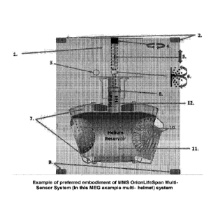
Figure 1 shows an example of preferred embodiment of MMS OrionLifeSpan Multi-
Sensor
System (in this MEG example multi-helmet) system
Figure 2 shows an embodiment of exploded diagrammatic views of present
invention
showing dual-helmet dewer configuration with horizontal rotation ((1) and
vertical angular
tilt adjustment capabilities
Figure 3 shows exploded diagrammatic views of an example of the dual-Dewar
function,
incorporating vertical height adjustment capability, enabling accommodation or
different bed,
seat, standing or other subject variable imaging and/or monitoring
requirements.
Date Recue/Date Received 2021-02-16
CA 03056330 2019-09-12
WO 2018/207061 PCT/IB2018/053101
3
Figure 4 shows an example of a magnetic shielded room and reliquifier coolant
system.
Figure 5 shows an example of schematic plan of dual-helmet selectable Dewar
system (5) to
accommodate child or adult child inside shielded room with external and
adjacent computer
processing room.
Figure 6 shows an exploded diagrammatic view of a dual-helmet Dewar system
adjusted for
45 degree tilted angle position to accommodate child helmet (2) and associated
child subject
in seated position
Figure 7 shows an example of a dual-helmet Dewar system adjusted for
horizontal or non-
tilted position (1) to accommodate adult shaped and sized helmet with
associated sensor
system for imaging and monitoring adult subject lying in supine position.
Figure 8 shows an example of a dual-helmet Dewar system adjusted for
horizontal or non-
tilted position (1) to accommodate child shaped and sized helmet with
associated sensor
system for imaging and monitoring child subject lying in supine position.
Figure 9 shows an embodiment of a MNIS with dual-sensor/helmet Dewar system.
Figure 10 shows an example of image and monitoring neural source localisation
and image
reconstruction conducted in adjoining computer processing room.
Detailed Description of the Invention
The invention provides the following features with reference to the Figures.
It will be
understood that the invention is limited only by the claims appended hereto.
Figure 1 shows an embodiment showing MMS multi-helmet Dewar system with x-axis
(4;
horizontal rotation) and/or y-axis (5; vertical height) and/or z-axis (6;
angular tilt) manual or
automatic positioning system (3D positioning system or X/Y/Z auto-positioning
system) as
well as a unique vacuum radiation cooled senor system (10) designed for very
low-vibration
during super-conductive cooling conditions of the sensory system, and/or;
With respect to the Blocks in Figure 1, the following is described. Block 1.
Dewar adjustment
mechanism cabinet.
Blocks 2. & 9. Multiple light-beam and/or RF and/or ultrasonic transceiver
systems located
within the magnetic shielded room (or external area simulating the subject
positioning of the
magnetic shielded room), enabling subject to be scanned, whilst in ideal
monitoring position,
typically prior to MMS investigation to allow exact region of subject of
interest (i.e. head
scan) to be captured in an accurate 3-dimential (3-D) co-ordinate format, for
MMS system
operator validation and adjustment is required, followed by option of storing
subject's body,
limb and head outline details, so that the MMS system can then compute the
optimal 3-
Dimensional position of the Dewar in order to provide the best image and
monitoring
capabilities, whilst ensuring a safe space is established between the subject
and the MMS.
In this manner the present invention's MMS automatic horizontal rotation
selection (i.e. as
part of the present invention's multi-sensor Dewar system can be selected in
accordance to
CA 03056330 2019-09-12
WO 2018/207061 PCT/IB2018/053101
4
Dewar that best matches shape and size of subject's monitored region (i.e.
adult or paediatric
head, in this example embodiment), as well as ideal vertical height position
(to accommodate
standing, lying or seating position or size of bed or seat etc.), as well as
ideal tilt angle (i.e.
accommodate seating or lying or other positions).
The scanned subject outline co-ordinates can then be referenced in order to
determine the
corresponding optimal Dewar 3-Dimentional Dewar positioning.
Additionally, precise electrode positions can be accessed via available data
or derived via
scanning (RE or light beam) subject with electrode and/or associated fiducial
markers.
Various combinations of MMS Dewar 3-D positioning and subject pre-scanned 3-D
data can
be stored and recalled as part of a library of test montages. This data can be
transferred
directly for Dewar manual or automatic x and y and/or z axis configuration.
This Dewar
adjustment can occur when subject is in magnetic shielded room or otherwise at
a separate
time, as required in terms of regulatory, safety and other important
conditions.
Block 3. Dewar adjustment cabinet interface electric, electronic, fibre-optic
and/or hydraulic
interface for remote location of Dewar x, y and/or z axis positioning system
Block 4. Dewar horizontal rotate system.
Block 5. Dewar vertical height adjust telescopic rod and sleeve using
adjustment cog and slot
Block 6. Dewar vertical pivot/tilt joint.
Block 7. Insulation Space
Block 8. Vertical Dewar rotation telescopic rod and sleeve with tilt rotate
joint using clamp
bracket mechanism, can be fitted to the outside of the "helium fill" chamber
adjustment cog
and slot.
Block 10. Coils, Bobbins, SQUIDS in Vacuum Space
Block 11. Simple, stream-lined, minimalistic design with no infrastructure
support columns
required within helium reservoir which can move with vibration in liquid
helium.
Figure 2.
Left exploded diagrammatic view showing an example of dual-Dewar function
incorporating
180 degree horizontal rotational capability in left drawing, allowing
selecting between one or
more Dewar and helmet head shapes or sized arrangements (such as child or
adult
configurations).
Block 1. MMS (MEG version) example embodiment of 180 degree rotation for
paediatric and
adult Dewar usage capability.
Block 2. 1VIMS (MEG version) of tilt angular positioning adjustment enabling
subject to be
seated, lying, standing or positioned in any other intermediate positions
during MMS
investigations.
Right exploded diagrammatic view showing an example of dual-Dewar function
incorporating vertical tilt capability for adjustment with seating, supine or
any range of
sloped or other subject positions, enabling imaging and/or monitoring to be
conducted, for
example, during any number of physical positions such as rest, sleep, wake,
unconsciousness
and other psychological and physical states.
CA 03056330 2019-09-12
WO 2018/207061 PCT/IB2018/053101
The present invention example embodiment, as shown here provides a means of
flipping or
rotating the dual-Dewar system around to select the most appropriate Dewar for
the subject
under MEG investigation.
Typically the dual-Dewar format can be configured to accommodate 2 adults, 2
paediatrics or
one adult and one paediatric.
To minimise space or overall MEG facility footprint (area required for MEG
operation) and
cost requirements, whilst maximise the system usage capabilities, a dual-Dewar
systems can
be deployed, whereby the system is configured in a single small magnetic
shielded room,
thereby only accommodating room for one MEG investigational subject at any one
time.
Alternatively, in other configurations a larger magnetic shielded room can be
deployed with a
more comprehensive system configuration (allowing both Dewars to be operated
simultaneously, whereby two complete sets of electronics are deployed to
process all SQUID
signals from both Dewars simultaneously. In such a configuration the magnetic
shielded
room would be of adequate size to allow subjects to be investigated from each
of the two
Dewars at one time or separate times, as required A visual barrier and if
required additional
magnetic shielded barrier could be configured to separate the two Dewar and
MEG subject
imaging and monitoring areas.
A key advantage of the dual-Dewar system is to enable the provision for two
separate
optimally sized MEG helmets. This is particularly important as the sensitivity
and
subsequent signal to noise of the MMS/MEG/MCG system is greatly enhanced by
minimising the space between the head (or other region) of the subject being
imaged and
monitored and the SQUID sensor coils. Therefore having a larger head sized
helmet and
SQUID system contained as part of one Dewar (i.e. adult sized) and a smaller
(i.e. paediatric
sized) monitoring configuration as part of the second Dewar provides improved
data quality,
with optimal sensitivity, based on the subject/object demographics, when
compared to
utilising one general sized Dewar for the larger and smaller head sizes. In
prior art systems it
was often necessaryõ during MEG investigations, to image and monitor one
hemisphere of a
child's brain (i.e. by leaning the child towards one side of the Dewar helmet)
and then
separately image and monitor the other hemisphere of a child's brain (i.e. by
leaning the child
towards the other side of the Dewar helmet). The two separate MEG scans would
then need
to be combined, which of course added further errors and complexity to the MEG
process.
Figure 3.
Block I. Example of vertical MMS (MEG Dewar in this example) height adjustment
capability.
Figure 4.
Block 1: MMS/MEG/MCG/Subject typical magnetic shielded room (1) isolating
external
noise and interference so that magnetic signals from a subjects head can be
imaged and
CA 03056330 2019-09-12
WO 2018/207061 PCT/IB2018/053101
6
monitored, even when such signals are about 1 billionth the size of the earths
normal
magnetic field. Coolant reliquifier system (2).
Block 2: Integrated zero-loss helium recycling reliquifier system, enabling
virtual vibration-
free continuous operation helium reliquification system integrated into the
present invention
MMS (MEG format in this example embodiment), thereby also greatly reducing
operational
and maintenance costs.
Figure 5.
Schematic of Dual-Dewar system (5) demonstrating the ability of the dual-Dewar
MMS/MEG/MCG/Subject system incorporate a smaller magnetic shielded room (/),
using
the horizontal selection (Dewar flip/rotate) function, as shown here.
The subject in this schematic representation shows dual usage of system with
paediatrics (3)
or adults (4), using dual 180 degree rotation Dewar selection function. The
subject is shown
lying in supine position.
The left square room is the magnetic shielded room, whilst the right of
drawing section shows
the computer, cognition test control and image viewing room (2).
Figure 6.
Exploded diagrammatic view showing example of dual-Dewar with selection of
child
imaging and/or monitoring configuration during seated subject position. The
picture shows
the Dewar tilted at an angle of about 45 degrees off horizontal axis (1) to
accommodate
seating position, whilst the Sensing system optimised for shape and size for
child (2)
investigational purposes.
Figure 7.
Example of dual-Dewar with selection of adult supine positioned imaging and/or
monitoring
configuration. The picture shows the Dewar at the horizontal position without
tilt (1) to
accommodate supine lying position of adult subject (2), in this example. This
example
embodiment of the present invention shows the adult Dewar sensor system (3)
being
deployed (versus the paediatric sensor system on the other side of dual-
DewarError!
Reference source not found.; Block 1), whereby this adult MMS (MEG) Dewar
selection
and usage deploys the optimal sensory system and helmet shape and size, thus
minimising the
space between the sensitive sensors and the subject's brain signals of
interest.
Figure 8.
Example of dual-Dewar (1) with selection of child imaging and/or monitoring
configuration
for minimal space between child's brain signals and sensory system (2) to
achieve maximum
signal to noise MMS quality) during supine subject position (3) A plastic
helmet-shaped
insert (2) safely separates and insulates (electrically and temperature-wise)
the MMS (MEG
CA 03056330 2019-09-12
WO 2018/207061 PCT/IB2018/053101
7
in this example embodiment) from the super-cooled sensor system from the
child. This
example embodiment of the present invention shows the paediatric/child Dewar
sensor
system (2) being deployed (versus the adult sensor system on the other side of
the dual-
Dewar; Block 1), whereby this paediatric/child MMS (MEG) Dewar selection and
usage
deploys the optimal sensory system and helmet shape and size, thus minimising
the space
between the sensitive sensors and the subject's brain signals of interest.
Figure 9.
Unique dual-helmet (dual-Dewar) rotating (/) adult/paediatric Dewar system
with shred
coolant entry and feed system (2) and unique sensor system (3) (shown via
exploded view in
this diagram) surrounded by a vacuum seal compartment able to the cooled for
super-
conductive SQUID requirements using radiation principles, thereby isolating
the very
sensitive SQUID chips, bobbins and associated pick-up coils from excessive
vibration noise
and other problems associated with the coolant flowing or interacting directly
with these said
sensitive sensor system components,
Figure 10.
Integrated reference fiducial markers with multi-modality imaging and/or
monitoring
capability enables precision convergence capability, calibration and
verification, thereby
improving determination of combined or discreet combinations of surgical
output
visualisations and co-ordinates for surgical or cancer treatment guidance
during, pre and post-
operative, with options of actual or simulated surgical or cancer treatment
synchronised
views. The upper right hand computer monitor view is a 2D representation of a
3D (3D
viewing capabilities are possible) head with the ability for the EEG
electrodes to be
visualised (1) and also for the MEG scanner to automatically scan via any
combination of
video radiofrequency or light beams the actual and precise position of each
EEG electrode
during imaging as well as the precise locational measurement centres of the
MEG reference
coils (by way of touch or RF or light beam or ultrasound touch sensors
providing positional
information of the subjects head and/or EEG sensors and/or additional; subject
-located
fiducial convergence alignment sensors or locators). Other modalities such as
CAT, CT,
PET, MRI (3), near field infrared imaging can be overlayed using cross-
compatible physical
or image computed image and/or monitoring convergence fiducial alignment
sensors or
markers or locators)
The top left of computer display presents a time segment of EEG data capable
of being time-
synchronised and spatially aligned with right hand computer screen image (2).
The lower right computer screen image represents the scanned image (2) used as
part of the
real boundary element modelling determination in order to enhance the accuracy
of neural
source localisation, which can be overlayed and/or time synchronised and/or or
superimposed
in the lower left and lower right computer screen views.
The present invention includes improved Dewar components, functions and
methods for
MMS including MEG systems, and thereby enable greater range and flexibility to
CA 03056330 2019-09-12
WO 2018/207061 PCT/IB2018/053101
8
accommodate positioning closer proximity imaging and monitoring to the bodies
of a broad
range of subjects, the present invention further comprising any of:
ii) Multifunctional MMS selectable or reconfigurable sensor (i.e. helmet)
system, and/or;
The present invention includes the provision for any number of Dewars with any
number of
sensor interfaces (including helmets in the case of MEG imaging) or other
subject/object
measurement interfaces.
The present invention includes the provision for a multiple-rotor selectable
MIMS Dewar,
whereby any number of subject/object interfaces can be deployed to accommodate
a range of
different subject/object sizes and positions for imaging or monitoring.
iii) 3-dimensional (3-D) sensor system positioning adjustment capability
for optimal
spatial resolution, signal sensitivity, and/or enhanced multi-modal image co-
registration
precision along with associated descriptions.
iv) 3-D manual or automatic Dewar and/or sensor system positioning or
reconfiguration
system, and/or;
v) shared or common coolant system including single reliquification coolant
system,
block 11) enabling thermal radiation (i.e. from coolant reservoir block 11) to
adjacent but
separately vacuum sealed and radiation super-conductive operation cooled
sensor
chamber(s); block 11) )to a plurality of sensory systems (block 10 and
associated
descriptions), and/or;
vi) shared or common coolant feed and/or return system, including 3-D
movement
positioning flexibility (; block 4, 5 and 6, along with associated
descriptions), and/or;
vii) vacuum cooled sensory system, with very low-vibration enabling
continuous coolant
operation per block 10, showing coolant reservoir and separation of vacuum
sealed sensor
chambers per block 10, along with associated descriptions (see block 1
reliquifier coolant
system and associated descriptions), and/or;
viii) automatic level adjusting Dewar coolant system, including 3-D
repositioning
super-conducting coolant operability (regardless of gravity, tilt and other
Dewar block 4, 6
and 6, along with associated descriptions, and/or;
ix) 3 D subject scanning and/or pre-scanning system to determine optimal 3-
D sensor
position co-ordinates and associated optimal positioning control (block 2 and
2, along with
associated descriptions), and/or;
x) simultaneous EEG or other imaging or monitoring modality capabilities
including
precision co-registration fi duci al options, and/or;
xi) Double Relaxation Oscillator SQUID (DROS) system with close coupled
SQUID
chip and coil, enabling high flux to voltage sensitivity, and/or;
xii) close coupled SQUID chip and coil for maximal flux to voltage
sensitivity, and/or;
CA 03056330 2019-09-12
WO 2018/207061 PCT/IB2018/053101
9
xiii) whereby MMS therapy capabilities include localization (i.e. selection
and positioning
of Dewars fitted with radio frequency (RF) therapy and/or Dewars and/or RF
beams to be
positioned optimally) in terms of neural sources to be avoided (i.e. healthy
cells) in radio
frequency targeting or beam-shaping or regions to be targeted for removal or
mitigation of
cancer cell risk; and/or
ix) Dewar positioning safety sensor and/or control system.
The present invention includes a Magnetic Sensing (MMS refers to imaging
and/or
monitoring and/or scanning and/or mapping system, including any of:
Magnetoencephalography (MEG) and/or Magnetocardiography (MCG)) system
incorporating
multiple MMS Dewar-interface (Dewar- subject/object-interface, such as helmet
or other
head, body or limb Dewar-interface) shapes or sizes, in order to accommodate
optimal MMS
sensing (including SQUIDS) by way of minimising locational proximities for
different
groups of subjects or objects (I.e. head, body, limb) sizes or shapes,
applicable (I.e. but not
limited) to different age groups such as paediatrics (Infants ¨ a child of up
to 12 months of
age; Child ¨ 1 to 12 years; Adolescent ¨ 13 to 16 years) and/or adults, the
present invention
further comprising of any of (i) to xiii) :
i) two or more sensor systems and associated helmets are arranged into a
single Dewar,
whereby the desired sensor system (i.e. preferred shape or size) to image or
monitor subject
of interest, can be selected via horizontal rotation of Dewar system until
selected sensory
system is located at required subject investigation position, and/or;
Plurality of helmet sensor system interfaces whereby the said MMS system
incorporate a
plurality of MMS Dewar-interfaces (such as but not limited to MCG body-
interfaces and/or
MEG helmets , which presents an example embodiment of multiple helmets and
associated
Dewars, along with corresponding magnetic sensor configurations (i.e. SQUID
arrays per
Block 10), and/or;
-Whereby, and as reference to "helmet" suggests, this relates to Dewar-
interface for MEG
head sensing but word helmet can be substituted for Dewar-interface which
covers other
biological or objects under investigation, and/or;
ii) Multifunctional MMS selectable or reconfigurable sensor (i.e. helmet)
system, including
capability to rotate Dewar in accordance to selected sensor system, whereby
dual Dewar
system allows (for example, only) 180 degree horizontal rotation between
selection of adult
or paediatric sensor system. In the case of a three sensor system Dewar the
Dewar can be
rotate at 120 degree intervals for the required selection of any of 3 sensor
shapes and size
types. Similarly, the formula applied to determine the rotational activation
for selecting each
successive sensor system can be represented by the following: number of
sensory systems
divided by 360 degrees
- The said MMS incorporates a multifunctional selectable multi-sensor system,
whereby
MEG helmets or other magnetic sensing founats can be reconfigured to suit
subject under
diagnostic or therapeutic investigation (i.e. cancer or radiofrequency beam
shaping and/or
targeting capabilities), and/or;
CA 03056330 2019-09-12
WO 2018/207061 PCT/1B2018/053101
- the said MMS system incorporating a plurality of IVIEG helmets or other
types of MMS
Dewar-interfaces corresponding to one or more MMS Dewars,
- whereby in some configurations multiple helmets or other Dewar interface
with any
combination of one or more Dewars can be enabled. Similarly, the formula
applied to
determine the rotational activation for selecting each successive sensor
system can be
represented by the following: number of sensory systems divided by 360
degrees, and/or;
- The said MMS incorporating a plurality of sensor interfaces, such as MEG
helmets or
other magnetic sensing interfaces (including within one of more Dewars),
thereby enabling
the Dewar sensor-interfaces (i.e. helmet) to accommodate imaging and/or
monitoring a range
of different head shapes and sizes of subject/object with minimal distance
between sensor
system and brain signals of subject/object under investigation, and/or;
- the said MSS system incorporating a plurality of MEG Helmets or other
types of MSS
Dewar interfaces (including but not limited to MEG or Magnetocardiography
(MCG) Dewar
interfaces, thereby enabling selection or re-configuration of optimal MSS
Imaging/monitoring proximity localisation corresponding to investigation
and/or therapy,
applicable to a range of object or subject sizes and/or shapes, in accordance
to different
magnetic sensor configurations (i.e. types or formats or sizes or techniques
or number of
monitored or imaged channels etc.) for subject(s) or object(s) of interest,
and/or;
- whereby MMS therapy capabilities include localization of neural sources
to be
avoided (i.e. healthy cells) in radio frequency targeting or beam-shaping or
to be targeted for
removal or mitigation of cancer cell risk; and/or
- whereby MMS configuration can be deployed in a manner enabling one or
more
objects or subjects to be imaged or monitored at any time, and/or
- whereby MMS configuration can be deployed in a manner where a single
object or
subject can be imaged or monitored at any one time, thus reducing on magnetic
shielded
room size or other requirements, and/or;
iii) 3-DIMENTIONAL SENSOR POSITIONING SYSTEM: In one embodiment the present
invention incorporates any combination of tilt, horizontal rotate (i.e.
enabling multi-sensor
Dewar capability) and/or vertical height adjustment; and/or;
- the said MMS incorporating multiple sensor-systems (such as multiple
helmets in case of
MEG deployment example of MMS), enabling a plurality of different head sizes
and shapes
to be accommodated, whilst minimizing the distance between brain signal
activity of interest
and corresponding sensor system;
- the capability for said MMS system (including option of MEG sensor
helmets) to enable
horizontal rotation (i.e. 180 degrees in case where 2 MEG sensor helmets (or
other MIVIS
Dewar-interfaces) are used or 120 degrees in case where 3 MEG sensor helmets
(or other
MMS Dewar-interfaces) are used or 90 degrees in case where 4 MEG sensor
helmets (or
other MIVIS Dewar-interfaces) are used and so on - i.e. 360 degrees divided by
number of
different Dewar-interfaces deployed equals rotational increment associated
with selection of
each different Dewar-interface, and/or;
CA 03056330 2019-09-12
WO 2018/207061 PCT/IB2018/053101
11
- the MMS system further incorporating the capability for vertical angular
tilt adjustment of
helmet (or other sensor interface) to accommodate a range of subject positions
such as seated
or supine subject imaging and/or monitoring positions, and/or;
-The present invention further enables adjustable vertical tit of Dewar and/or
Dewar
interface, to accommodate a range of subject/object shapes and sizes or
positions for imaging
or monitoring, and/or
- capability for said MEG sensor helmet able to be vertically repositioned
(I.e. Pivoted
through 90 degree positional adjustment arc ) in order to accommodate a range
of subject
positions including supine or seated upright or any intermediate positions,
and/or;
- whereby MMS configuration can be deployed in a manner where noise
reduction techniques
can be incorporated in order to minimise or avoid magnetic shielded room
requirements,
and/or;
- whereby selection or rotation of Dewars and/or Dewar-interfaces can be
deployed in a
manual or automatic manner, and/or;
- The MMS incorporates 3-D Dewar spatial parametric adjustment positioning
capability
enabling any combination of tilt, horizontal rotate and vertical height
adjustment, and/or;
iv) the present invention enables any combination of automatic or manual multi-
sensor
Dewar positioning options (for MMS system, including MEG or MCG or other
subject or
object imaging and monitoring fointats) in order to configure MMS Dewar system
for the
required vertical height (x-axis adjustment), the selected helmet sensor
system (horizontal
rotation of Dewar (Block 1) or the angular tilt of the Dewar helmet (block 2)
in accordance to
the respective height and posture (i.e. age, sitting, standing, lying, along
with any other
positions), according to selecting the most appropriate helmet shape and size
(i.e. head shape
and size and whether subject is an adult or paediatric), and in accordance to
the optimal
angular tilt, based on whether subject under investigation, and/or;
As it relates to manual or automatic horizontal rotation adjustment
capability, in one
embodiment example a "vertical Dewar rotation telescopic rod and sleeve",
using clamp
bracket mechanism, can be fitted to the outside of the "helium fill" chamber
(see figure 3) in
a manner where using an arrangement of cogs and connection rods or chains
interacting with
the "vertical Dewar rotation telescopic rod and sleeve" in a manner where a
manual wheel
adjustment or automatic computer controlled motor servo adjustment can
horizontally rotate
the Dewar to and desired position across a 360 degree range of rotational
positions. I.e. A cog
positioned around the outer circumference of the "vertical Dewar rotation rod
and sleeve
mechanism" can interact with another cog, which is fixed to the "Dewar ceiling
mount
bracket" support system in a manner where a manually driven or automatic
service driven
cog, attached to the fixed platform of the stationary part of the "Dewar
ceiling mount bracket"
support system (versus "vertical Dewar rotation telescopic rod and sleeve
mechanism" which
rotates in accordance to desired Dewar rotational selection) can rotate the
"vertical Dewar
rotation telescopic rod and sleeve mechanism" as selected by system operator,
and/or;
As it relates to manual or automatic vertical rotation or tilting adjustment
capability, in one
embodiment the said "vertical Dewar rotation telescopic rod and sleeve" can
incorporate a
flexible joint, whereby the said "rod and sleeve" arrangement resides in the
upper arm of the
CA 03056330 2019-09-12
WO 2018/207061 PCT/1B2018/053101
12
said join, so that the lower join arm can be rotated in respect to the upper
joint arm across a
range from 0 degrees (Dewar in horizontal untitled position) to plus or minus
about 90
degrees, via a series of manual or automatically driven cogs and/or drive
chains or belts to
rotational position required. i.e. a screw thread adjustment arm arrangement
whereby the
upper arm's (above swivel joint block 3) inner core or adjustable outer sleeve
can
interconnected via a screw threaded bar to the lower arm (below swivel joint
block 3) via a
matching screw, so that the upper threaded screw can be screwed clock-wise or
anti-
clockwise to lift or lower the lower arm with respect to the upper arm, in
order to change the
angular tile of the Dewar system., and/or;
As it relates to manual or automatic horizontal Dewar height adjustment,
capability, in one
embodiment example the said "vertical Dewar rotation telescopic rod" (upper
arm of vertical
tilt joint) can incorporate a sleeved rod arrangement telescopic adjustment
arrangement,
whereby the inner rod has precision ratchet impressions located in a the upper
vertical tilt arm
and the outer sleeve contains a manually or automatically driven cog
arrangement that, via a
slot window in the outer sleeve, allows the inner rod and outer sleeve to be
extended and
contracted in accordance to the system users desired Dewar height adjustment,
and/or;
The Dewar horizontal rotation, vertical height and pivot or tilt adjustments
can be contained
within "Dewar adjustment mechanism" (shown here in the upper section of the
"helium fill"
chamber. Mechanical couple rods and coupling mechanism can be attached via
bracket and
manual adjustment kit to the "Dewar adjustment mechanism" cabinet, enabling
manual
adjustments to be accessed conveniently by system operator or technician. The
Dewar
movement and adjustment mechanisms can be covered by external system covers to
enhance
system industrial design aesthetics and system operation eloquence, aesthetics
and also
improve system operational safety. In this manner the system can be configured
or
automatically (I e. via a range of preprogrammed or screen display options) or
manually
adjusted, as required, and/or;
Additionally, the present invention can deployed using a combination of
mechanical screw
adjustment systems and/or hydraulic positioning systems, as commonly used in
aeronautics
design for the movement of tail-rudders, wing flaps and other mechanical
adjustment systems
Similar to fundamental aeronautics design principles, the present invention
can be controlled
by wire (i.e. as most modern planes are flown by wire), whereby computer servo
positioning
and remote wire and/or wireless interconnection provide the control and/or
drive signals
required to undertake mechanical Dewar horizontal rotation, vertical height
adjustment or tilt
angular adjustments in a 3-dimentional (x and/or y and/or z-axis of movement).
Orion LifeSpan Multi-Dewar (example shows dual-helmet MEG squid sensor
system),
and/or;
v) A single shared or common Helium Reliquification system (see Block 1) is
enabled by
way of sharing a common helium coolant reservoir by a plurality of Dewars
(dual-helmet
sensor system in this example embodiment, Block 11). The single coolant
reservoir includes
associated filling and return feed coolant gas isolated for vibration via
flexible piping to the
cooling head mount, located outside the MSR (i.e. via access with top central
round pipe
(Block 12) feeding into a central central (between 2 sensory systems located
in separate
vacuum spaces) helium reservoir section (Block 11), and/or;
CA 03056330 2019-09-12
WO 2018/207061 PCT/1B2018/053101
13
- the MMS incorporates a shared or combined multi-helmet Dewar reliquifier
coolant
reservoir capable of being deployed for super-conduction sensor system
operation across a
plurality of MMS sensor systems, and/or;
vi) common or shared (between a plurality of sensor systems) coolant liquid
(I.e. Helium)
feed and coolant gas return hoses (or pipes or ducts), interconnected between
the top of the
"helium fill" chamber (as shown in figure 3) and the reliquifier coolant unit
(located outside
the magnetic shielded room as shown in can be arranged in a manner whereby
additional free
hose flexibility allows the Dewar unit (dual in this example but any multi-
Dewar founat can
be enabled) to be: -lifted or lowered (to accommodate different vertical
height positions of
subject under investigation) and/or;
-vertically pivoted or tilted to enable different Dewar horizontal angles,
thereby
accommodating MMS/MEG/MCG sensory system to be optimally adjusted for ideal
close
proximity and ideally localised sensor MMS/MEG/MCG/SUBJECT system usage (i.e.
minimising distance between sensing system and the signals of interest of the
subject under
investigation) during various positions of subject under investigation. i.e.
Dewar adjusted for
horizontal position with subject in supine position or angled downwards (from
0 degree
horizontal axis) by 45 degrees (per seated subject example, and/or; -
horizontally rotated to
select appropriate Dewar for subject of investigational interest. i.e. 180
degree rotation
capability to select between adult or paediatric Dewar and sensor system for
the example in
this Figure 3, and/or;
vii) One or more vacuum cooled sensory systems partitioned adjacent to helium
reservoir to
enable cool temperature via radiation, (versus noisier and more vibration-
prone direct thermal
conduction or thermal convection cooling reliance), with physically separated
thermal
radiation cooled sensor system (i.e. avoid direct contact coolant and sensor
system in order to
minimise vibration and other noise created with coolant flowing over sensitive
sensor system
components), and/or;
- the MMS incorporates a vacuum space around the sensor systems), thereby
enabling
radiation cooling, with coolant reservoir containing coolant fluid or gas,
Block 11) to be
separated from sensor system(s) (Block 10) in order to minimise noise and
vibration and
other interference, by way of deploying coolant radiation versus need to apply
direct contact
coolant and/or coolant conductance method and/or or coolant conventional
method, and/or;
-whereby the coolant system's gas or liquid does not need to flow directly
around the
sensitive sensor system components. In this example the MEG sensor system
semiconductor
SQUID chips, SQUID pick-up coils, pick-up coil bobbins per Block 10. This
approach is
also applicable to MCG, as well as imaging or monitoring other object, body,
limb, subject
parts. Additionally, this vacuum separation (between coolant and sensor
systems) and
radiation cooling technique can be applied to sensitive sensor (vibration and
other noise
sensitive) components of MRI; PET; CT CAT, X-ray, ultrasound imaging or
monitoring
systems, and/or;
-whereby coolant gas/liquid is isolated from the low-noise but super cooled
vacuum sensor
system region of the MMS - the sensor system is cooled via radiation versus
direct coupling
temperature convection or conduction approaches, thereby minimising noise and
vibration
otherwise induce via coolant flowing directly over sensitive sensor
components; , and/or;
CA 03056330 2019-09-12
WO 2018/207061 PCT/1B2018/053101
14
-whereby vacuum radiation cooled sensor system can be deployed. The unique
combination
of the vacuum cooling system, and the multi-sensor Dewar (i.e. in this
example: multi-helmet
Dewar system), coupled with the single shared helium (or other coolant
composition)
reliquifier coolant system (Error! Reference source not found.Block 1) and
coolant
reservoir (Block 11) delivering around 4 Kelvin temperature or minus 269
degrees Celsius
required to maintain the sensor system super-conducting conditions
(temperature required to
activate low impedance electrical circuit superconducting state for SQUID
amplifiers and
coils) enables a very low-noise and low-vibration configuration so that helium
recycling can
be continuous, allowing 24/7 (continuous or uninterrupted) operation of the
said MMS (I.e.
no need to turn off coolant system during MMS Imaging or monitoring). In
contrast,
traditional earlier art systems require the coolant solution/gas to be
switched off during
critical scanning, due to excessive vibrational and other (i.e. impedance
variations across
(Block 12) sensitive circuits and pick-up coils) noise generated when the
coolant flows
directly over the most sensitive imaging and monitoring parts (including
Coils, Bobbins,
SQUIDs which can be located in the Vacuum Space, formed around these sensitive
sensor
system parts, Block 10). Additionally, the shared coolant system and separate
vacuum cooled
sensor system compartments (separation between Blocks 10 and 11 ) enables
greater range of
measurement capability (via two or more separate shaped and sized sensor
systems), whilst
also reducing maintenance costs associated due to the simpler system (compared
to multiple
cooling systems), or avoidance of corrosion or wear or related impedance
variations,
associated with coolant making direct contact with or flowing around sensitive
sensor system
parts (SQUIDs, coils, etc.). The use of the vacuum coolant system in the
present invention
reduces vibration and noise, which is otherwise more apparent in traditional
MEG coolant
system where direct coupling to SQUID coils and/or SQUID amplifiers create
additional
noise, such as vibration noise. Importantly, the vacuum coolant system
requires no down time
for Helium Return (i.e. helium return cycle does not discernibly increase
imaging or
monitoring system signal noise), and/or;
viii) multi-sensor Dewar system has sensors and control systems controlling
Helium level
and/or avoidance of helium flow or slushing effect so that the helium or other
coolant is
always appropriately covering (regardless of tile and gravity impact on flow
levels) the
sensitive sensor compartment wall (where compartment wall represents barrier
between
vacuum cooled sensory system and coolant) or sensor systems (where coolant
directly flows
over sensor system components). In this way the angular tilting or other Dewar
movement or
positioning never prevent the coolant from cooling sensor system in a manner
that enables
super-conducting operational conditions (i.e. SQUID coils and chips need to
operate at ¨ 269
degrees Celsius to achieve super-conduction capabilities). An automatic
coolant level
adjustment is enabled by way of sensing systems within Dewar and/or the sensor
systems to
ensure coolant fluid levels, regardless of adjustment or tilt of Dewar,
enables correct coolant
operation for super-conducting sensor system operational requirements at all
times;
ix) A subject scanning and/or pre-scanning system, enabling precision co-
registration
between MMS sensor systems and/or subject investigational region/spatial area
of interest
and/or multi-modality co-registration precision, and/or;
- Capability to pre-scan (light and/or radio frequency with or without
associated marker
points/measurement fiducials) patient inside magnetic shielded room or
external to magnetic
shielded room (i.e. calibrated co-registration of seating or patient gantry
can enable precision
scanning outside magnetic shielded room but 3-dimensional measures and
associated output
co-ordinates of subject's observational target (i.e. head; body; limb; object)
shape and size
CA 03056330 2019-09-12
WO 2018/207061 PCT/1B2018/053101
characteristics to be computed via scanning system. In this way the resulting
co-ordinates can
be deployed by the present invention to allow automatic or manual positioning
of the Dewar
and associated sensory system in context of the most optimal x-axis (optimal
sized Dewar
rotational selection), y-axis (height) and z-axis (tilt) configuration for
subject under
investigation, and/or;
- the said MMS incorporating a subject scanning capability, thereby
enabling the
determination of the optimal head shape and size Dewar interface selection
(i.e. MEG
helmet) as well as the optimal x-y-z positioning of the Dewar with sensor
interface, to
achieve the most precise and sensitive (i.e. minima distance between brain
signals of interest
and MEG sensors) imaging conditions, and/or;
- the scanning of subject outline for purposes of determining optimal
helmet shape and size
selection as well as Dewar helmet positioning can be undertaken prior, before
or during the
MIMS imaging and monitoring activation, and/or;
x) Similarly, in terms of scanning subject for simultaneous or separately
recorded
(electroencephalography) EEG signals the subject EEG electrode placements can
be
manually entered and/or , scanned and transferred to MMS system data in order
to ensure
precise, calibrated and specified co-registration of EEG and MMS image or
monitoring data
is achieved. The scanning of other modalities such as positron emission
tomography (PET),
computer aided tomography (CAT), X-rays, ultrasonic, RF treatment modalities,
and/or
proton treatment modalities can also be co-registered in this manner, and/or;
- Whereby optimal sensory system adjustment or positioning refers to x-
axis, y-axis and z-
axis 3-dimensional spatial positing and Dewar selection (and/or sensor system
and/or
associated or subsequent data or signal processing or control systems) to
enable multiple-
Dewar or single Dewar system to be optimised for maximal subject safety and/or
comfort
and/or sensitivity imaging or monitoring of the sensory system. For one
example MEG
embodiment of the present MMS invention, the MEG head helmet is selected by
way of
scanning subject's head with light-beam, radio frequency (RF) and/or
ultrasound or other
subject/object scanning technique, in order to determine the exact shape, size
and position of
the subject/object under investigation (including head in case of MEG example)
so that the
multi-Dewar horizontal selection function determines which Dewar is most
appropriate for
the particular subject under investigation (this is determined by the Dewar
which is larger
than the subjects head but the closest fitting to minimise the space between
the subjects brain
signals of main interest and the Dewar "helmet".
- Whereby the "helmet" refers to the concave helmet-shaped, or in other
words a plastic head-
shaped plastic receptacle, that sits closely around subject's head on the
concave side, whilst
the convex side of the helmet separates the very cold sensor system (QUID
coils, bobbins and
chips) from coming into contact with the subject's head, and/or;
- On the convex side of the said helmet the SQUID sensory system and very
cold components
are located, in a manner where the helmet prevents subject under investigation
from being
exposed to super cool temperatures. It is also important that this said helmet
is large enough
to avoid pressure being applied to subject under =investigation's head but
small enough and
shaped closely to the subject's head to minimise the space between the sensory
system and
the monitored and measured brain signals, and/or;
CA 03056330 2019-09-12
WO 2018/207061 PCT/1B2018/053101
16
xi) Double Relaxation Oscillator SQUID (DROS) system with close coupled SQUID
chip
and coil, enabling high flux to voltage sensitivity, and/or,
xii) Dewar positioning safety sensor and/or control system, and/or;
- the said MMS further incorporating a Dewar and/or Dewar-sensor interface
(i.e. helmet)
safety system to avoid Dewar movement during repositioning and other
adjustment or
movement circumstances from being able to harm subjects or system users,
and/or
- the incorporation of a safety sensing system which detects any slight
touching of a person
(i.e. detects slight pressure or has sensors (i.e. light or pressure touch)
capable of alarming or
deactivating MMS movement to avoid any halm to a person, and/or;
- the incorporation of a safety sensing system, enabling prevention and/or
alert and/ alarm
during circumstances where people are in safe-isolation area suitable to
prevent any risk of
harm to any individuals during selection or reconfiguration associated with
appropriate
Dewar and/or Dewar-subject ¨interface, and/or;
xiii) whereby MMS therapy capabilities include localization (i.e. selection
and positioning of
Dewars fitted with radio frequency (RF) therapy and/or Dewars and/or RF beams
to be
positioned optimally) in terms of neural sources to be avoided (i.e. healthy
cells) in radio
frequency targeting or beam-shaping or regions to be targeted for removal or
mitigation of
cancer cell risk; and/or
whereby MMS therapy capabilities include localization (i.e. selection and
positioning of
Dewars fitted with radio frequency (RF) therapy
xiv) Close coupled SQUID chip and coil for maximal flux to voltage
sensitivity;
The unique axial radial, double-relaxation axial gradiometer sensing system,
block 10, block
2; block 3) comprising of double relaxation bonding squid (DROS),
incorporating reduced
SQUIDs stray coil and associated wiring noise for enhanced high flux-to-
voltage transfer
capabilities as well as large modulation amplitude and high stability against
offset drift
conditions.