Note: Descriptions are shown in the official language in which they were submitted.
CA 03056642 2019-09-13
WO 2018/170433 PCT/US2018/022929
DELIVERY AIDS FOR GLAUCOMA SHUNTS
BACKGROUND
[0001] Aqueous humor is a fluid that fills the anterior chambers of the
eye and
contributes to the intraocular pressure or fluid pressure inside the eye.
Glaucoma is a
progressive disease of the eye characterized by an increase of the eye's
intraocular
pressure. This increase in intraocular pressure is commonly caused by an
insufficient
amount of aqueous humor being reabsorbed by the body. In some cases, the
aqueous
humor is not absorbed fast enough or even at all, while in other cases, the
aqueous
humor is additionally or alternatively being produced too quickly. An increase
in
intraocular pressure is associated with a gradual and sometimes permanent loss
of
vision in the afflicted eye.
[0002] A number of attempts have been made to treat glaucoma. However,
some of the conventional devices lack the flexibility, conformity, and
device/tissue
attachment that is required to avoid relative movement between the device and
the
surrounding tissue. Such movement can lead to persistent irritation of the
surrounding
tissue. Irritation, in turn, can lead to an augmented chronic inflammatory
tissue
response, excessive scar formation at the device site, and a heightened risk
of device
erosion through the conjunctiva and endophthalmitis. In instances where
erosion does
not occur, the scar tissue effectively prevents reabsorption of the aqueous
humor.
These complications can serve to prevent proper functioning of the device. The
resulting effect is a gradual increase in intraocular pressure and progression
of
glaucoma.
SUMMARY
[0003] According to one example, ("Example 1"), a biological fluid
drainage
system includes a body; a compliant fluid conduit fluidly coupled to the body
and
including a first end, a second end, and a lumen, the first end being
positionable within
a fluid-filled body cavity of a biological tissue, and the second end being
positionable
outside of the fluid-filled body cavity such that a fluid from the fluid-
filled body cavity is
transferrable through the lumen of the fluid conduit to the body; and a
stiffening member
removably coupled with the fluid conduit, the stiffening member being
positioned within
the lumen and extending a length of the fluid conduit.
[0004] According to another example, ("Example 2") further to Example 1,
the
stiffening member and the fluid conduit, in combination, form an assembly, and
wherein
1
CA 03056642 2019-09-13
WO 2018/170433 PCT/US2018/022929
one of a column strength, a lateral stiffness, and a hoop strength of the
assembly
exceeds a column strength, a lateral stiffness, and a hoop strength of the
fluid conduit,
respectively.
[0005] According to another example, ("Example 3") further to any of
Examples
1 and 2, an end of the stiffening member extends from one of the first and
second ends
of the fluid conduit such that the end of the stiffening member is accessible
during an
implantation procedure.
[0006] According to another example, ("Example 4") further to any of the
preceding Examples, the stiffening member forms a coil within the lumen of the
fluid
conduit.
[0007] According to another example, ("Example 5") further to Example 4,
the
stiffening member is configured to unravel upon an application of tension to
one of the
first and second ends of the stiffening member.
[0008] According to another example, ("Example 6") further to any of the
preceding Examples the stiffening member is a first stiffening member, the
system
further comprising a second stiffening member removably coupled with the fluid
conduit,
wherein the second stiffening member extends through a sidewall of the fluid
conduit
such that a first portion of the second stiffening member extends within the
lumen of the
tube and such that a second portion of the second stiffening member extends
exterior to
the tube along the sidewall of the tube, the second portion of the second
stiffening
member being accessible during an implantation procedure.
[0009] According to another example, ("Example 7") further to Example 6,
a
second end of the second stiffening member extends from one of the first and
second
ends of the fluid conduit such that the second end of the stiffening member is
accessible
during an implantation procedure.
[00010] According to another example, ("Example 8") further to any of the
preceding Examples, the fluid conduit comprises expanded
polytetrafluoroethylene.
[00011] According to another example, ("Example 9") further to any of the
preceding Examples, the fluid-filled body cavity is an anterior chamber of an
eye and
the fluid is aqueous humor, and wherein the biological fluid drainage system
is
configured to regulate an intraocular pressure of a patient's eye when
implanted.
[00012] According to another example, ("Example 10") further to any of the
preceding Examples, an axial length of the stiffening member is configured to
increase
upon an application of tension to the stiffening member independent of the
fluid conduit.
2
CA 03056642 2019-09-13
WO 2018/170433 PCT/US2018/022929
[00013] According to another example, ("Example 11") a biological fluid
drainage
system includes a compliant fluid conduit having a first end and a second end
and
defining a lumen, the first end being positionable within a fluid-filled body
cavity of a
biological tissue, and the second end being positionable outside the reservoir
of the
biological tissue such that a fluid from the fluid-filled body cavity is
transferrable through
the lumen of the fluid conduit to a region outside of the fluid-filled body
cavity; and a
stiffening member coupled to the fluid conduit, the stiffening member being
positioned
within the lumen of the fluid conduit and extending a length of the fluid
conduit such that
the stiffening member and the fluid conduit, in combination, form an assembly,
and
wherein a column strength of the assembly exceeds a column strength of the
fluid
conduit.
[00014] According to another example, ("Example 12") further to Example 11,
the
system further includes a microporous body fluidly coupled with the fluid
conduit,
wherein the second end of the fluid conduit is positioned within the
microporous body.
[00015] According to another example, ("Example 13") further to any of
Examples
11 to 12, the stiffening member is removably coupled to the fluid conduit.
[00016] According to another example, ("Example 14") further to any of
Examples
11 to 13, the stiffening member is a first stiffening member, the system
further
comprising a second stiffening member removably coupled with the fluid
conduit,
wherein the second stiffening member extends through a sidewall of the fluid
conduit
such that a first portion of the second stiffening member extends within the
lumen of the
tube and such that a second portion of the second stiffening member extends
exterior to
the tube along the sidewall of the tube, the second portion of the second
stiffening
member being accessible during an implantation procedure.
[00017] According to another example, ("Example 15") further to Example 14, a
second end of the second stiffening member extends from one of the first and
second
ends of the fluid conduit such that the second end of the stiffening member is
accessible
during an implantation procedure.
[00018] According to another example, ("Example 16") further to any of
Examples
11 to 15, the fluid conduit comprises expanded polytetrafluoroethylene.
[00019] According to another example, ("Example 17") further to any of
Examples
11 to 16, the fluid-filled body cavity is an anterior chamber of an eye and
the fluid is
aqueous humor, and wherein the biological fluid drainage system is configured
to
regulate an intraocular pressure of a patient's eye when implanted.
3
CA 03056642 2019-09-13
WO 2018/170433 PCT/US2018/022929
[00020] According to another example, ("Example 18") a method includes
providing a tube having a lumen extending therethrough; coupling the tube to a
body
such that the lumen of the tube is fluidly coupled to the body; and arranging
a stiffening
member within the lumen of the tube such that the stiffening member is
removable from
the lumen of the tube and such that the stiffening member and the tube, in
combination,
form an assembly, and wherein a column strength of the assembly exceeds a
column
strength of the tube.
[00021] According to another example, ("Example 19") further to Example 18, a
lateral stiffness of the assembly exceeds a lateral stiffness of the tube, and
a hoop
strength of the assembly exceeds a hoop strength of the tube
[00022] According to another example, ("Example 20") further to any of
Examples
18 to 19, arranging a stiffening member within the lumen of the tube includes
winding an
elongate element about a mandrel to form a coil about the mandrel; forming a
tube
about the coiled elongate element such that the coiled elongate element is
disposed
within a lumen of the tube and such that the coiled elongate element is
removable from
the lumen of the tube; and removing the mandrel such that the elongate element
remains coiled within the lumen of the tube.
[00023] According to another example, ("Example 21") further to Example 20,
forming the tube about the coiled elongate element includes wrapping a film
about the
coiled elongate element.
[00024] According to another example, ("Example 22") further to Example 21,
the
film is a tape.
[00025] According to another example, ("Example 23") further to any of
Examples
20 to 22, the elongate element is a fiber, and wherein one of the film and the
fiber is a
fluoropolymer.
[00026] According to another example, ("Example 24") further to Example 23,
the
fluoropolymer is expanded polytetrafluoroethylene.
[00027] According to another example, ("Example 25") further to any of
Examples
18 to 24, the stiffening member is a first stiffening member, and the method
further
includes arranging a second stiffening member within the lumen of the tube
such that
the second stiffening member extends through a sidewall of the tube, such that
a first
portion of the second stiffening member extends within the lumen of the tube
and such
that a second portion of the second stiffening member extends exterior to the
tube along
the sidewall of the tube, the second portion of the second stiffening member
being
accessible during an implantation procedure.
4
CA 03056642 2019-09-13
WO 2018/170433 PCT/US2018/022929
[00028] According to another example, ("Example 26") further to Example 25,
the
first and second stiffening members are independently removable from the lumen
of the
tube.
[00029] According to another example, ("Example 27") a method includes
winding an elongate element about a mandrel to form a coil about the mandrel;
forming
a tube about the coiled elongate element such that the coiled elongate element
is
disposed within a lumen of the tube and such that the coiled elongate element
is
removable from the lumen of the tube; and removing the mandrel without
removing the
elongate element from the lumen of the tube such that the elongate element
defines a
stiffening member.
[00030] According to another example, ("Example 28") further to Example 27,
the
elongate element is a fiber.
[00031] According to another example, ("Example 29") further to any of
Examples
27 to 28, forming the tube about the coiled elongate element includes wrapping
a film
about the coiled elongate element.
[00032] According to another example, ("Example 30") further to Example 29,
the
film is a tape.
[00033] According to another example, ("Example 31") further to Example 30,
the
film is a membrane.
[00034] According to another example, ("Example 32") further to any of
Examples
29 to 31, one of the film and the fiber is a fluoropolymer.
[00035] According to another example, ("Example 33") further to Example 32,
the
fluoropolymer is expanded polytetrafluoroethylene.
[00036] According to another example, ("Example 34") further to any of
Examples
27 to 33, the method further includes coupling the tube to a microporous body
such that
the lumen of the tube is fluidly coupled to the microporous body, wherein the
stiffening
member extends within an interior of the microporous body.
[00037] According to another example, ("Example 35") further to any of
Examples
27 to 34, the stiffening member is a first stiffening member, the method
further
comprising arranging a second stiffening member within the lumen of the tube
such that
a first end of the second stiffening member extends through a sidewall of the
tube and
such that the second stiffening member is removable from the lumen of the
tube.
[00038] According to another example, ("Example 36") further to Example 35,
arranging a second stiffening member within the lumen of the tube includes
inserting the
CA 03056642 2019-09-13
WO 2018/170433 PCT/US2018/022929
second stiffening member into the lumen of the tube after the tube is formed
such that
the second stiffening member pierces the sidewall of the tube.
[00039] According to another example, ("Example 37") further to any of
Examples
35 to 36, the first and second stiffening members are independently removable
from the
lumen of the tube.
[00040] According to another example, ("Example 38") a method includes
providing a tube having a first end, a second end, and a lumen extending from
the first
end to the second end, wherein a first stiffening member extends within the
lumen of the
tube such that the stiffening member and the tube, in combination, form a
tubular
assembly, and wherein at least one of a column strength of the tubular
assembly
exceeds a column strength of the tube, a lateral stiffness of the tubular
assembly
exceeds a lateral stiffness of the tube, and a hoop strength of the tubular
assembly
exceeds a hoop strength of the tube; securing a position of the first end of
the tube;
[00041] advancing the second end of the tube to a position within a fluid
reservoir
of a biological tissue; and removing the first stiffening member from the tube
such that
the tube operates as a fluid conduit for the egress of fluid within the fluid
reservoir of the
biological tissue.
[00042] According to another example, ("Example 39") further to Example 38,
the
tube further comprises a second stiffening member extending within the lumen
of the
tube, the second stiffening member extending through a sidewall of the tube,
the
method further comprising puncturing the biological tissue with an end of the
second
stiffening member and advancing the second stiffening member and the second
end of
the tube until the second end of the tube is advanced to the position within
the fluid
reservoir.
[00043] According to another example, ("Example 40") further to any of
Examples
38 to 39, securing the position of the first end of the tube includes positing
the first end
of the tube between tissue layers of a patient's eye, and wherein the fluid is
aqueous
humor within an anterior chamber of the patient's eye.
BRIEF DESCRIPTION OF THE DRAWINGS
[00044] The accompanying drawings are included to provide a further
understanding of embodiments of the disclosure and are incorporated in and
constitute
a part of this specification, illustrate examples, and together with the
description serve to
explain the principles of the disclosure.
6
CA 03056642 2019-09-13
WO 2018/170433 PCT/US2018/022929
[00045] FIG. 1 is an illustration of a glaucoma drainage system consistent
with
various aspects of the present disclosure.
[00046] FIG. 2A is an illustration of a glaucoma drainage system in a deflated
state consistent with various aspects of the present disclosure.
[00047] FIG. 2B is an illustration of a glaucoma drainage system in an
inflated
state consistent with various aspects of the present disclosure
[00048] FIG. 3 is an exploded view of the glaucoma drainage system illustrated
in
FIG. 2.
[00049] FIGS. 4A-4D are illustrations of constriction diffusion membrane
interface
surfaces consistent with various aspects of the present disclosure.
[00050] FIG. 5 is an illustration of a glaucoma drainage system consistent
with
various aspects of the present disclosure.
[00051] FIG. 6 is an illustration of a glaucoma drainage system consistent
with
various aspects of the present disclosure.
[00052] FIG. 7A is an illustration of a glaucoma drainage system in a deflated
state consistent with various aspects of the present disclosure.
[00053] FIG. 7B is an illustration of a glaucoma drainage system in an
inflated
state consistent with various aspects of the present disclosure.
[00054] FIG. 8 is an illustration of a fluid conduit consistent with
various aspects
of the present disclosure.
[00055] FIG. 9A is an illustration of a glaucoma drainage system consistent
with
various aspects of the present disclosure.
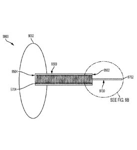
[00056] FIG. 9B is a detailed view of a region 9B of the glaucoma drainage
system of FIG. 9A but that is not cross sectioned.
[00057] FIG. 9C is an illustration of a glaucoma drainage system consistent
with
various aspects of the present disclosure.
[00058] FIG. 9D is an illustration of a glaucoma drainage system consistent
with
various aspects of the present disclosure.
[00059] FIG. 10A is an illustration of a glaucoma drainage device consistent
with
various aspects of the present disclosure.
[00060] FIG. 10B is cross sectional view of the glaucoma drainage system of
FIG. 9A taken along line 10B-10B.
[00061] FIG. 10C is cross sectional view of the glaucoma drainage system of
FIG. 9A taken along line 10C-10C.
7
CA 03056642 2019-09-13
WO 2018/170433 PCT/US2018/022929
[00062] FIG. 11 is an exploded view of a glaucoma drainage system consistent
with various aspects of the present disclosure.
[00063] FIG. 12 is an illustration of a glaucoma drainage system implanted
within
an eye tissue consistent with various aspects of the present disclosure.
[00064] FIG. 13 is an illustration of a glaucoma drainage system implanted
within
an eye tissue consistent with various aspects of the present disclosure.
[00065] While multiple embodiments are disclosed, still other embodiments will
become apparent to those skilled in the art from the following detailed
description, which
shows and describes illustrative examples. Accordingly, the drawings and
detailed
description are to be regarded as illustrative in nature and not restrictive.
DETAILED DESCRIPTION
[00066] Persons skilled in the art will readily appreciate that the various
embodiments of the inventive concepts provided in the present disclosure can
be
realized by any number of methods and apparatuses configured to perform the
intended
functions. It should also be noted that the accompanying drawing figures
referred to
herein are not necessarily drawn to scale, but may be exaggerated to
illustrate various
aspects of the present disclosure, and in that regard, the drawing figures
should not be
construed as limiting. As used herein, the term "diffusion membranes" is meant
to
encompass one or more proliferation diffusion membrane and/or one or more
constriction diffusion membrane.
[00067] Various aspects of the present disclosure are directed toward glaucoma
drainage devices, drainage systems, and drainage methods. More specifically,
the
present disclosure relates to devices, systems, and methods for draining
aqueous
humor from the anterior chamber of a patient's eye such that it may be
reabsorbed by
the body. Providing a mechanism for reabsorption of the aqueous humor that has
been
evacuated from the anterior chamber of the eye operates to lower or otherwise
stabilize
the intraocular pressure.
[00068] A glaucoma drainage system 1000 according to some embodiments is
illustrated in FIG. 1. The glaucoma drainage system 1000 is an implantable
medical
system that operates to facilitate the drainage of a fluid, such as aqueous
humor, from a
fluid filled body cavity, such as the anterior chamber of the eye. The
glaucoma drainage
system 1000 includes a fluid conduit 1500 and a body, such as an aqueous humor
diffusion member 1002. While the following disclosure refers to a glaucoma
drainage
system 1000 for use in draining aqueous humor from the anterior chamber of the
eye, it
8
CA 03056642 2019-09-13
WO 2018/170433 PCT/US2018/022929
is to be understood and appreciated by one of skill in the art that the
glaucoma drainage
system 1000 depicted can be configured and utilized to evacuate other fluids
from other
fluid filled body chambers. In some examples, as explained in greater detail
below, the
glaucoma drainage system 1000 additionally helps facilitate reabsorption of
the
evacuated fluid by the body. For instance, in some embodiments, the glaucoma
drainage system 1000 provides an interface between the evacuated aqueous humor
and tissues, vessels and/or cells that have the ability to absorb aqueous
humor and are
sufficiently proximate the glaucoma drainage system 1000 to interact with the
evacuated aqueous humor. Thus, in some examples, aqueous humor evacuated from
the anterior chamber of the eye travels through the glaucoma drainage system
1000
before being reabsorbed by the body.
[00069] In some embodiments, when the glaucoma drainage system 1000 is
implanted, aqueous humor is evacuated from the anterior chamber through the
fluid
conduit 1500. The evacuated aqueous humor then enters a reservoir of the
aqueous
humor diffusion member 1002 and percolates through one or more porous
membranes
of the aqueous humor diffusion member 1002, where the aqueous humor can then
be
reabsorbed by the body. In various embodiments, in addition to aqueous humor
permeability, tissue ingrowth is permitted or promoted along one or more
regions of the
glaucoma drainage system 1000. For instance, the exterior of the aqueous humor
diffusion member 1002 may include or be defined by one or more membranes that
are
porous or otherwise permeable to the fluid of the fluid filled body cavity
(referred to
hereinafter as diffusion membranes), and that are configured to permit or
promote
tissue ingrowth. Permitting tissue ingrowth along surfaces or within regions
of the
glaucoma drainage system 1000 helps facilitate biointegration of the glaucoma
drainage
system 1000 into the surrounding tissue (e.g., eye tissue), and helps
facilitate
reabsorption of the evacuated aqueous humor by the surrounding tissue.
Moreover,
biointegration including tissue ingrowth and attachment helps minimize
relative
movement between the glaucoma drainage system 1000 and the tissue surrounding
the
glaucoma drainage system 1000, which helps avoid irritation of the eye tissue
that can
lead to foreign body tissue response, scar formation, and/or erosion and site
infection of
the glaucoma drainage system 1000.
[00070] In some examples, as discussed in greater detail below, the fluid
conduit
of the glaucoma drainage system 1000 is a soft and compliant biocompatible
tubular
structure. Accordingly, in some examples, the glaucoma drainage system 1000
further
includes a stiffening member that is removably integrated with the fluid
conduit 1500,
9
CA 03056642 2019-09-13
WO 2018/170433 PCT/US2018/022929
which helps aid in the delivery/implantation of the glaucoma drainage system
1000.
That is, in some examples, the glaucoma drainage system 1000 includes a
removable
component (e.g., a stiffening member) to provide temporary stiffness to the
fluid conduit,
which helps physicians manipulate the fluid conduit and/or the body of the
glaucoma
drainage system. Such a configuration provides for a glaucoma drainage system
1000
that is complaint and operable to conform to the tissue (e.g., eye tissue) and
profile of
the anatomy in which the glaucoma drainage system 1000 is being implanted,
while
maintains a minimum profile to avoid irritation and/or interference with
normal body
functions (e.g., blinking of the eye) and while being easily implantable, as
such soft and
compliant structures would be otherwise difficult to manipulate and properly
orient within
the anatomy.
[00071] In various embodiments, the aqueous humor diffusion member 1002
includes an interior region that defines a reservoir for the aqueous humor
that is
evacuated from the anterior chamber through the fluid conduit 1500. The
interior region
of the aqueous humor diffusion member 1002 may include one or more membranes
that
are porous or otherwise permeable to the fluid of the fluid filled body cavity
(referred to
hereinafter as diffusion membranes). For example, as discussed in greater
detail
below, one or more of the diffusion membranes may be formed of a porous media,
such
as a polymeric material, that has a microstructure that is suitable for
transporting fluid
through a pore space of the porous media. Thus, in some embodiments, the
reservoir
may be defined by the pore space of one or more of the diffusion membranes
that form
the aqueous humor diffusion member 1002. In some embodiments, the aqueous
humor
diffusion member 1002 may be configured such that the reservoir is
additionally or
alternatively defined between two or more of the diffusion membranes that form
the
aqueous humor diffusion member 1002. For instance, in some embodiments, at
least a
portion of the surface areas between adjacently situated diffusion membranes
forming
the aqueous humor diffusion member 1002 remains unbonded or unadhered such
that
the adjacently situated diffusion membranes are operable to separate from one
another
along at least a portion of their surface areas to form and define the
reservoir. In some
embodiments, as discussed further below, the reservoir defined between
adjacently
situated diffusion membranes is operable to inflate or dilate in a controlled
manner (e.g.,
to a predetermined profile when inflated) so that the glaucoma drainage system
1000
does not interfere with normal eye function (e.g., regular eye movement,
including
pivoting and blinking).
[00072] In various embodiments, the aqueous humor diffusion member 1002 is
CA 03056642 2019-09-13
WO 2018/170433 PCT/US2018/022929
sized and shaped such that it is implantable within the patient's anatomy. For
instance,
in some embodiments, the aqueous humor diffusion member 1002 is sized and
shaped
such that it is implantable within a dissected subconjunctival space (e.g.,
between a
sclera and a conjunctiva of the patient's eye). In some embodiments, the
aqueous
humor diffusion member 1002 is a thin, circular-shaped member. In some
embodiments, the aqueous humor diffusion member 1002 has a thickness (e.g., a
distance measured between the first exterior surface 1004 and the second
exterior
surface 1006) of less than or equal to half of a millimeter (0.5mm), such as
between
one-tenth of a millimeter (0.1mm) and half of a millimeter (0.5mm). However,
given
differing anatomies of the human body, an aqueous humor diffusion member 1002
may
exceed of half of a millimeter (0.5mm) provided that the thickness does not
substantially
interfere with normal eye functioning (e.g., pivoting and blinking) or
substantially reduce
the flexibility of the aqueous humor diffusion member 1002 to the extent that
undesirable relative movement occurs between the glaucoma drainage system 1000
and the surrounding tissue when implanted, resulting with a likely consequence
of
tissue irritation, foreign body tissue response, and/or excessive scar
formation.
[00073] In some embodiments, the aqueous humor diffusion member 1002 may
have a diameter in the range of five (5) millimeters to fifteen (15)
millimeters, such as
ten (10) millimeters for example. In some embodiments, the aqueous humor
diffusion
member 1002 may be ovular and include a major dimension (e.g., along a major
axis of
the ellipse) of up to about thirty (30) millimeters and corresponding minor
dimension
(e.g., along a major axis of the ellipse) of up to about ten (10) millimeters.
As discussed
above, given differing anatomies of the human body, an aqueous humor diffusion
member 1002 may exceed such dimensions (e.g., fifteen (15), and ten (10) and
thirty
(30) millimeters) provided that the size does not substantially interfere with
normal eye
functioning (e.g., pivoting and blinking) or substantially reduce the
flexibility of the
aqueous humor diffusion member undesirable relative movement occurs between
the
glaucoma drainage system 1000 and the surrounding tissue when implanted,
resulting
with a likely consequence of tissue irritation, foreign body tissue response,
and/or
excessive scar formation. Likewise, the aqueous humor diffusion member 1002
may
have a diameter of less than five (5) millimeters, three (3) millimeters, or
even less than
three (3) millimeters provided that the aqueous humor diffusion member 1002 is
operable to accommodate a sufficient degree of evacuated aqueous humor and is
operable to facilitate the reabsorption of aqueous humor to constitute an
effective
treatment for the patient.
11
CA 03056642 2019-09-13
WO 2018/170433 PCT/US2018/022929
[00074] In various embodiments, the fluid conduit 1500 operates to fluidly
couple
the reservoir with the fluid filled body cavity (e.g., the anterior chamber of
the eye) when
implanted in the body such that a differential pressure is achievable between
the
reservoir and the environment exterior to the glaucoma drainage system 1000
(e.g.,
atmosphere). Thus, when implanted, it is to be appreciated that a pressure
within the
reservoir is based, at least in part, on the pressure within the fluid filled
body cavity
(e.g., the Intraocular Pressure of the Anterior Chamber of the eye). In some
embodiments, such a differential pressure causes the reservoir to inflate or
dilate.
Moreover, in some embodiments, such a differential pressure causes the aqueous
humor to percolate through the diffusion membranes of the aqueous humor
diffusion
member 1002. That is, in some embodiments, the evacuated aqueous humor enters
the reservoir and percolates through the diffusion membranes of the aqueous
humor
diffusion member 1002, where the aqueous humor can then be reabsorbed by the
body.
[00075] Turning now to FIGS. 2A and 2B, a glaucoma drainage system 1000
including an aqueous humor diffusion member 1002 comprised of a plurality of
diffusion
membranes is shown. The aqueous humor diffusion member 1002 includes a first
exterior surface 1004, a second, exterior surface 1006 opposing the first
exterior
surface 1004, and a periphery 1008. FIG. 2A shows the glaucoma drainage system
1000 in a deflated state. FIG. 2B shows the glaucoma drainage system 1000 in
an
inflated state, where aqueous humor is present within an inflatable or
dilatable reservoir
1010. While the glaucoma drainage system 1000 is shown in FIG. 2B in an
inflated
state where the glaucoma drainage system 1000 is not uniformly inflated (e.g.,
the first
proliferation and constriction diffusion membranes 1100 and 1200 are shown
adopting a
generally nonlinear configuration while the second proliferation and
constriction diffusion
membranes 1300 and 1400 are shown in a generally linear configuration), it is
to be
appreciated that the glaucoma drainage system 1000 may deform uniformly (e.g.,
the
second proliferation and constriction diffusion membranes 1300 and 1400 may
deform
in a manner that mirrors the deformation of the first proliferation and
constriction
diffusion membranes 1100 and 1200). The aqueous humor diffusion member 1002
includes a body defined by a plurality of diffusion membranes including first
and second
proliferation diffusion membranes 1100 and 1400 and first and second
constriction
diffusion membranes 1200 and 1300. In some examples, the first and second
proliferation diffusion membranes 1100 and 1400 and the first and second
constriction
diffusion membranes 1200 and 1300 are stacked upon one another as shown to
form
the aqueous humor diffusion member 1002. As discussed further below, the first
and
12
CA 03056642 2019-09-13
WO 2018/170433 PCT/US2018/022929
second proliferation diffusion membranes 1100 and 1400 are configured to
permit tissue
ingrowth and attachment, while the first and second constriction diffusion
membranes
1200 and 1300 are configured to minimize, resist, or prevent tissue ingrowth
and
attachment.
[00076] In some embodiments, the first and second proliferation diffusion
membranes 1100 and 1400 form or otherwise define an exterior of the aqueous
humor
diffusion member 1002, while the first and second constriction diffusion
membranes
1200 and 1300 are situated between the first and second proliferation
diffusion
membranes 1100 and 1400 and define an interior region of the aqueous humor
diffusion
member 1002. In various embodiments, the first and second proliferation
diffusion
membranes 1100 and 1400 and the first and second constriction diffusion
membranes
1200 and 1300 are each permeable to aqueous humor in that each is configured
to
allow evacuated aqueous humor (e.g., aqueous humor disposed within the sealed
reservoir) to percolate therethrough and/or diffuse thereacross. However, the
first and
second proliferation diffusion membranes 1100 and 1400 are configured to
permit tissue
ingrowth and attachment, while the first and second constriction diffusion
membranes
1200 and 1300 are configured to minimize, resist, or prevent tissue ingrowth
and
attachment. A configuration of constriction diffusion membranes sandwiched or
otherwise situated between proliferation diffusion membranes as shown in FIGS.
2A
and 2B helps to minimize, for instance, an ingress of bacteria in excess of
the size of
perforations or small holes present in the constriction diffusion membranes
and/or
migration thereof to the anterior chamber of the eye.
[00077] In various examples, the first and second proliferation diffusion
membranes 1100 and 1400 of the aqueous humor diffusion member 1002 are
microporous, permeable to aqueous humor, and are configured to permit the
ingrowth
and/or attachment of vessels and tissue. In various embodiments, the first and
second
constriction diffusion membranes 1200 and 1300 are also microporous and
permeable
to aqueous humor, but are configured to resist or otherwise minimize the
ingrowth and
attachment of vessels and tissue structures. Thus, in various embodiments, the
aqueous humor diffusion member 1002 is formed of a plurality of distinct
diffusion
membranes including at least a first proliferation diffusion membrane 1100 and
at least
a first constriction diffusion membrane 1200.
[00078] While the glaucoma drainage system 1000 shown in FIGS. 2A and 2B
includes separate and distinct first and second proliferation diffusion
membranes 1100
and 1400, it is to be appreciated that the aqueous humor diffusion member 1002
may
13
CA 03056642 2019-09-13
WO 2018/170433 PCT/US2018/022929
include the first proliferation diffusion membrane 1100 without also requiring
a separate
and distinct second proliferation diffusion membrane 1400. For instance, the
first
proliferation diffusion membrane 1100 may be folded such that the first
proliferation
diffusion membrane 1100 surrounds the constriction diffusion membrane portion
(e.g.,
the first and/or second constriction diffusion membranes 1200 and 1300) of the
aqueous
humor diffusion member 1002. In some such embodiments, one or more portions of
the
folded portion of the proliferation diffusion membrane 1100 is bonded or
welded to
adjacent portions of the non-folded portion of the proliferation diffusion
membrane 1200
and/or one or more portions of the constriction diffusion membrane portion of
the
aqueous humor diffusion member 1002. Additionally or alternatively, while the
glaucoma drainage system 1000 shown in FIGS. 2A and 2B includes separate and
distinct first and second constriction diffusion membranes 1200 and 1300, it
is to be
appreciated that the aqueous humor diffusion member 1002 may include the first
constriction diffusion membrane 1200 without also requiring a separate and
distinct
second constriction diffusion membrane 1300. For instance, the first
constriction
diffusion membrane 1200 may be folded over upon itself to form a multilayered
constriction diffusion membrane, wherein one or more portions of the folded
portion of
the constriction diffusion membrane 1200 is bonded or welded to adjacent
portions of
the non-folded portion of the constriction diffusion membrane 1200. Moreover,
a
proliferation diffusion membrane 1100 may additionally be folded about the
folded
constriction diffusion membrane 1200, where the constriction diffusion
membrane 1200
is folded over upon itself with a fluid conduit 1500 situated between the
folded and
unfolded portions of the constriction diffusion membrane 1200. In some such
embodiments, a reservoir may be defined between at least the folded and
unfolded
portions of the constriction diffusion membrane 1200.
[00079] FIG. 3 is an exploded view of the glaucoma drainage system 1000 shown
in FIGS. 2A and 2B. As shown in FIG. 3, the aqueous humor diffusion member
1002
includes a body defined by a first proliferation diffusion membrane 1100, a
first
constriction diffusion membrane 1200, a second constriction diffusion membrane
1300,
and a second proliferation diffusion membrane 1400. As shown, the various
proliferation and constriction diffusion membranes each include interface
surfaces and a
periphery. For example, the first proliferation diffusion membrane 1100
includes a first
interface surface 1102, a second interface surface 1104, and a periphery 1106.
In
some examples, the first interface surface 1102 of the first proliferation
diffusion
membrane 1100 corresponds with or otherwise defines the first exterior surface
1004 of
14
CA 03056642 2019-09-13
WO 2018/170433 PCT/US2018/022929
the glaucoma drainage system 1000. Additionally, as shown in FIG. 3, first
constriction
diffusion membrane 1200 includes a first interface surface 1202, a second
interface
surface 1204, and a periphery 1206. Likewise, as shown in FIG. 3, second
constriction
diffusion membrane 1300 includes a first interface surface 1302, a second
interface
surface 1304, and a periphery 1306. As shown, the second proliferation
diffusion
membrane 1400 includes a first interface surface 1402, a second interface
surface
1404, and a periphery 1406. In some examples, the second interface surface
1404 of
the second proliferation diffusion membrane 1400 corresponds with or otherwise
defines the second exterior surface 1006 of the glaucoma drainage system 1000.
[00080] In various embodiments, the diffusion membranes (i.e., the
proliferation
diffusion membranes and the constriction diffusion membranes) forming the
aqueous
humor diffusion member 1002 are situated adjacent to one another in a stacked
configuration. For example, as illustrated in FIGS. 2A, 2B, and 3, the first
and second
proliferation diffusion membranes 1100 and 1400 and first and second
constriction
diffusion membranes 1200 and 1300 are situated adjacent to one another in a
stacked
configuration, with the first and second proliferation diffusion membranes
1100 and
1400 forming or otherwise defining an exterior of the aqueous humor diffusion
member
1002, and with the first and second constriction diffusion membranes 1200 and
1300
sandwiched or otherwise situated between the first and second proliferation
diffusion
membranes 1100 and 1400. Thus, the proliferation diffusion membranes forming
the
exterior region of the aqueous humor diffusion member 1002 are configured to
support
or permit tissue ingrowth and attachment, while the constriction diffusion
membranes
forming the interior region of the aqueous humor diffusion member 1002 are
configured
to minimize, resist, or prevent tissue ingrowth and attachment beyond or
interior to a
boundary or interface between the proliferation and constriction diffusion
membranes.
[00081] By minimizing, resisting, or preventing tissue ingrowth and attachment
beyond or interior to the constriction diffusion membranes, the glaucoma
drainage
system 1000 minimizes, resists, or prevents tissue ingrowth into the reservoir
1010,
which helps maintain performance of the glaucoma drainage system 1000 during
and
after biointegration thereof. For example, it is to be appreciated that
minimizing,
resisting, or preventing tissue ingrowth into the constriction diffusion
membranes, and
thus the reservoir 1010 operates to maintain a flexibility of the glaucoma
drainage
system 1000, which as discussed herein helps minimize relative movement
between the
glaucoma drainage system 1000 and the surrounding tissue and thus helps
minimize
irritation of the surrounding tissue. In particular, minimizing, resisting, or
preventing
CA 03056642 2019-09-13
WO 2018/170433 PCT/US2018/022929
tissue ingrowth into the constriction diffusion membranes helps avoid tissue
from
proliferating across the interface between adjacent constriction diffusion
membranes an
thus helps avoid such tissue ingrowth from interlocking the constriction
diffusion
membranes together. Avoiding the interlocking the constriction diffusion
membranes
helps maintain the ability of the constriction diffusion membranes to slide
and move
relative to one another, which helps maintain flexibility of the glaucoma
drainage system
1000.
[00082] In some examples, as discussed further below, the aqueous humor
diffusion membrane 1002 is configured such that the interface surfaces of
adjacently
situated diffusion membranes face one another. In some examples, the first and
second proliferation diffusion membranes 1100 and 1400 and the first and
second
constriction diffusion membranes 1200 and 1300 are oriented such that their
peripheries
align with and/or are coaxial with one another. In some embodiments, one or
more of
the peripheries of the diffusion members forming the body of the aqueous humor
diffusion member 1002 form the periphery 1008 of the aqueous humor diffusion
member 1002. For example, as shown in FIGS. 2A and 2B, the peripheries 1106,
1206,
1306, and 1406, collectively, form or define the periphery 1008 of the aqueous
humor
diffusion member 1002. It is to be appreciated, however, that the periphery of
the
aqueous humor diffusion member 1002 may be formed from less than all of the
peripheries of the diffusion membranes forming the body of the aqueous humor
diffusion member 1002. For instance, in some examples, the periphery 1008 of
the
aqueous humor diffusion member 1002 may be formed or defined by the
peripheries
1106 and 1406 of the first and second proliferation diffusion membranes 1100
and
1400.
[00083] As mentioned above, in various embodiments, adjacently situated
diffusion membranes are generally oriented such that one or more of their
interface
surfaces is situated adjacent to or otherwise faces an interface surface of an
adjacently
situated diffusion membrane. That is, in various embodiments, the interface
surfaces of
adjacently situated diffusion membranes face each other. In the embodiment
depicted
in FIGS. 2A, 2B, and 3, the first proliferation diffusion membrane 1100 and
the first
constriction diffusion membrane 1200 are adjacently situated such that the
second
interface surface 1104 of first proliferation diffusion membrane 1100 faces
the first
interface surface 1202 of first constriction diffusion membrane 1200.
Similarly, as
shown in FIGS. 2A, 2B, and 3, first constriction diffusion membrane 1200 and
second
constriction diffusion membrane 1300 are adjacently situated such that the
second
16
CA 03056642 2019-09-13
WO 2018/170433 PCT/US2018/022929
interface surface 1204 of first constriction diffusion membrane 1200 faces the
first
interface surface 1302 of second constriction diffusion membrane 1300.
Similarly, as
shown in FIGS. 2A, 2B, and 3, second constriction diffusion membrane 1300 and
second proliferation diffusion membrane 1400 are adjacently situated such that
the
second interface surface 1304 of second constriction diffusion membrane 1300
faces
the first interface surface 1402 of second proliferation diffusion membrane
1400.
[00084] Thus, in some embodiments, stacked configurations like those described
above provide for a first diffusion membrane having first and second interface
surfaces
and a second diffusion membrane having first and second interface surfaces
where the
first and second diffusion membranes are adjacently situated such that the
second
interface surface of the first diffusion membrane faces the first interface
surface of the
second diffusion membrane.
[00085] In various embodiments, the first and second proliferation diffusion
membranes 1100 and 1400 and the first and second constriction diffusion
membranes
1200 and 1300 may include or be formed of one or more layers or sheets of
expanded
polytetrafluoroethylene (ePTFE), or other polymers, such as, but not limited,
to
polyurethane, polysulfone, polyvinylidene fluoride or polyvinylidene
difluoride (PVDF),
polyhexafluoropropylene (PHFP), perfluoroalkoxy polymer (PFA), polyolefin,
fluorinated
ethylene propylene (FEP), acrylic copolymers and other suitable fluoro-
copolymers.
These polymers can be in sheet, knitted or woven (including individual or
multi-fiber
strands), or non-woven porous forms. In some examples, one or more of the
first and
second proliferation diffusion membranes 1100 and 1400 and/or the first and
second
constriction diffusion membranes 1200 and 1300 may be formed from a plurality
of
layers or sheets of polymer material. In some such examples, the layers or
sheets of
polymer material may be laminated or otherwise mechanically coupled together,
such
as by way of heat treatment and/or high pressure compression and/or adhesives
and/or
other lamination methods known by those of skill in the art. In some
embodiments, as
explained in greater detail below, the layers of polymer material may be
coupled
together at discrete locations to form stabilizing structures that extend
through the
resulting proliferation and/or constriction diffusion membranes. Similarly, in
some
embodiments, as explained in greater detail below, proliferation and/or
constriction
diffusion membranes may be coupled together at discrete locations to form
stabilizing
structures that extend through the resulting aqueous humor diffusion member
1002. It
is to be appreciated that such stabilizing structures are operable to
constrain a shape or
profile of the aqueous humor diffusion member 1002 upon inflation or dilation
of the
17
CA 03056642 2019-09-13
WO 2018/170433 PCT/US2018/022929
reservoir 1010, as mentioned above.
[00086] In some embodiments, the layers or sheets of polymer material forming
the first and/or second proliferation diffusion membranes 1100 and 1400 and/or
the first
and/or second constriction diffusion membranes 1200 and 1300 may be subjected
to
one or more processes prior to or after their formation to modify their
microstructure
(and thus their material properties) to increase or decrease a natural
permeability (e.g.,
a permeability to aqueous humor) of the polymeric material(s). In some
examples, such
processes include, but are not limited to, material coating processes, surface
preconditioning processes, and/or perforation processes. Material coating
processes
may be utilized to at least partially fill the porous space of the polymeric
material(s), to
thereby reduce permeability, as those of skill will appreciate. Additionally
or
alternatively, material coating processes may be utilized to apply one or more
drug or
antimicrobial coatings to the surface of the polymer material (such as
metallic salts,
including silver carbonate), and organic compounds (e.g. chlorhexidine
diacetate), to
the polymer material.
[00087] In some embodiments, one or both of the first and second proliferation
diffusion membranes 1100 and 1400 and/or one or both of the first and second
constriction diffusion membranes 1200 and 1300 may be hydrophilic. In some
embodiments, one or both of the first and second proliferation diffusion
membranes
1100 and 1400 and/or one or both of the first and second constriction
diffusion
membranes 1200 and 1300 may be hydrophobic. Thus, in some examples, the
aqueous humor diffusion member 1002 may include one or more hydrophilic
membranes, and one or more hydrophobic membranes.
[00088] Accordingly, hydrophilic coatings to enable wet out of the polymer
matrix
may also be applied as if the polymer surfaces are hydrophobic in nature.
Surface
coatings comprising antioxidant components can be applied to mitigate the
body's
inflammatory response that naturally occurs during wound healing after
surgery.
Surfaces can be modified with anti-proliferative compounds (e.g. Mitomycin C,
5-
fluoracil), to moderate the surrounding tissue response in the eye. In some
examples,
one or more surface preconditioning processes may additionally or
alternatively be
utilized to form layers exhibiting a preferred microstructure (e.g., wrinkles,
folds, or other
geometric out-of-plane structures), as explained in U.S. Patent Number
9,849,629 to
Zagl, et al. Such surface preconditioning could facilitate a bolder early
inflammatory
phase after surgery, providing an early stable interface between porous device
and
tissue. In some examples, a heparin coating (e.g., thromboresistant) may
additionally
18
CA 03056642 2019-09-13
WO 2018/170433 PCT/US2018/022929
or alternatively be applied to help minimize or reduce cell formation
including fibrinogen
buildup following a surgical implantation procedure.
[00089] In some embodiments, one or more perforation processes may
additionally or alternatively be utilized to form a plurality of perforations
or small holes in
the polymeric material(s) in addition to any perforations or small holes
naturally
occurring in the polymeric material(s), which operates to increase a natural
permeability
(e.g., a permeability to aqueous humor) of the polymeric material(s). Such
perforation
processes may increase a number of perforations or small holes present in the
polymeric material(s) and/or may increase an average size of the perforations
or small
holes present in the polymeric material(s), and may be performed before and/or
after
the formation of the proliferation and/or constriction diffusion membranes. In
some
embodiments, the permeability of the first and/or second proliferation
diffusion
membranes 1100 and 1400 and/or the first and second constriction diffusion
membranes 1200 and 1300 may be altered to tune or otherwise modify flux and/or
flow
resistance of aqueous humor to a desired amount.
[00090] In various embodiments, the first and/or second proliferation
diffusion
membranes 1100 and 1400 may include perforations or small holes that range in
size
(or average size) from between twenty (20) microns and one-hundred (100)
microns. In
other examples, the size (or average size) of the perforations or small holes
in the first
and/or second proliferation diffusion membranes 1100 and 1400 may exceed one-
hundred-fifty (150) microns. In various embodiments, the first and/or second
proliferation diffusion membranes 1100 and 1400 may include perforations or
small
holes less than twenty (20) microns, but larger than one (1) or two (2)
microns, as
perforations or small holes less than one (1) or two (2) microns generally
inhibit, resist,
or otherwise prevent ingrowth of vessels and other tissues.
[00091] Accordingly, in various embodiments, the first and second constriction
diffusion membranes 1200 and 1300 are configured or selected such that the
perforations or small holes therein are generally sized at less than (or have
an average
size of less than) one (1) micron or two (2) microns to minimize, resist, or
prevent the
ingrowth and attachment of tissue, while maintaining aqueous humor
permeability.
[00092] It is to be appreciated that the first and second proliferation
diffusion
membranes 1100 and 1400 may be configured to have the same or different
permeabilities. Similarly, it is to be appreciated that the first and second
constriction
diffusion membranes 1200 and 1300 may be configured to have the same or
different
permeabilities. In some examples, the various proliferation and constriction
diffusion
19
CA 03056642 2019-09-13
WO 2018/170433 PCT/US2018/022929
membranes discussed herein may possess the same inherent permeabilities, but
undergo one or more of the material modification processes discussed herein to
achieve different relative permeabilities. In some embodiments, one or more of
the
material modification processes discussed herein operates to change or
otherwise
modify the naturally occurring permeability of the polymeric material(s).
Thus, in some
embodiments, the permeabilities of the proliferation and/or constriction
diffusion
membranes may be based on the naturally occurring microstructure of the
polymeric
material(s) and/or one or more of the material modification processes
discussed herein.
Those of skill in the art will appreciate that a permeability is generally
related to the
resistance of a fluid transporting through the pore space of porous media, and
that
materials associated with low permeabilities exhibit greater resistance to
flow than do
those materials with higher permeability.
[00093] In some embodiments, the perforations or small holes in the
proliferation
and constriction diffusion membranes may be formed through one or more salt
inclusion
processes, or through the use of one or more drilling, die-punching, needle-
puncturing,
or laser cutting processes, which may be performed before and/or after the
formation of
the proliferation and/or constriction diffusion membranes.
[00094] Generally, the processes described above may be utilized to form
proliferation diffusion membranes having a microstructure that permits the
ingrowth of
surrounding vessels and other tissues and that is permeable to aqueous humor.
Similarly, the processes described above may be utilized to form constriction
diffusion
membranes having a microstructure that minimizes, resists, or otherwise
prevents the
ingrowth of surrounding vessels and other tissues, but that is permeable to
aqueous
humor. The aqueous humor that percolates and/or diffuses across the
constriction and
proliferation diffusion membranes may be absorbed by the vessels that have
grown into
the proliferation diffusion membranes and/or the vessels exterior to the
aqueous humor
diffusion member 1002, and/or may percolate through the surrounding tissues
and into
the tear film.
[00095] As mentioned above, in some embodiments, the differential pressure
observed between the reservoir 1010 of the glaucoma drainage system 1000 and
the
environment exterior to the glaucoma drainage system 1000 (e.g., atmospheric
pressure) is a mechanism that facilitates the flow of aqueous humor through
the
aqueous humor diffusion member 1002 of the glaucoma drainage system 1000. In
some embodiments, the mechanism of reabsorption and the carrying away of the
evacuated aqueous humor by the vessels grown into and surrounding the glaucoma
CA 03056642 2019-09-13
WO 2018/170433 PCT/US2018/022929
drainage system 1000 helps facilitate the evacuation of aqueous humor from the
anterior chamber.
[00096] However, it is to be appreciated that in addition to facilitating the
reabsorption and carrying away of evacuated aqueous humor, the ingrowth of
tissues,
vessels, and cells into the proliferation diffusion membrane(s) of the aqueous
humor
diffusion member 1002 also helps prevent, reduce, minimize, or limit the onset
of
foreign body tissue responses. Specifically, as mentioned above tissue
ingrowth and
attachment helps minimize relative movement between the glaucoma drainage
system
1000 and the tissue of the eye. By helping minimize such relative movement,
the
glaucoma drainage system 1000 helps avoid irritation of the eye tissue that
can occur
and that can lead to foreign body tissue response, which can lead to excessive
scar
formation and/or erosion and site infection of the glaucoma drainage system
1000.
[00097] In some embodiments, one or more of the adjacently situated diffusion
membranes forming the body of the aqueous humor diffusion member 1002 are
connected or otherwise coupled to together. In some embodiments, adjacently
situated
diffusion membranes are coupled at one or more discrete portions or regions
along their
adjacently facing interface surfaces. In some embodiments, adjacently situated
diffusion membranes may be coupled along at least a portion of an adjoining
edge (or
edges). In other embodiments, adjacently situated diffusion membranes may be
additionally or alternatively coupled at one or more discrete location along
the adjoining
surfaces interior to the edge (or edges). In yet other embodiments, adjacently
situated
diffusion membranes may be coupled along an entirety of their adjacently
facing
interface surfaces (e.g., applying an adhesive across an entirety of a surface
area of
adjacently facing interface surfaces). Thus, in some embodiments, one or more
of the
adjacently situated diffusion membranes may be coupled at less than all of
their
adjacently facing interface surfaces (e.g., at discrete locations or a portion
thereof) or
they may be coupled along an entirety of the facing interface surfaces.
[00098] In those embodiments where adjacently situated diffusion membranes are
coupled along a portion of less than all of their adjacently facing interface
surfaces, one
or more discrete locations along adjacently facing interface surfaces are
connected or
otherwise coupled together while one or more other discrete locations along
adjacently
facing interface surfaces are not coupled together. That is, in some
embodiments, at
least one region or area of adjacently facing interface surfaces remains
intentionally
unadhered, unbonded, or otherwise uncoupled.
[00099] In some such embodiments, these uncoupled regions or areas may
21
CA 03056642 2019-09-13
WO 2018/170433 PCT/US2018/022929
include regions or areas central to a peripheral edge. Generally, these
uncoupled
regions or areas are free to move or slide relative to one another, and may
separate
from one another to serve as a reservoir for the accumulation of evacuated
aqueous
humor. In various examples, providing such a degree of freedom (e.g., in
shear)
provides for considerable flexibility because diffusion membranes can move
relative to
one another to conform to changes in curvature as the aqueous humor diffusion
member 1002 is bent and moves, such as with natural movement of the eye. Thus,
the
discontinuity of coupling of the diffusion membranes provides for a glaucoma
drainage
system 1000 exhibiting better eye conformity and that is better suited to
dynamically
respond to changes in curvature of the eye 2000 as the patient blinks,
focuses, and
moves the eye within the eye socket. Unlike the more rigid conventional
designs, the
increased flexibility also minimizes movement of the glaucoma drainage system
1000
relative to the surrounding tissue.
[000100] Turning now to FIGS. 4A to 4D, examples of interface surfaces
including
coupled and uncoupled (e.g., bonded and unbonded) regions are illustrated.
FIG. 4A is
a cross sectional view of second interface surface 1204 taken along the
boundary (4-
4, FIG. 2) situated between adjacently facing first and second interface
surfaces 1204
and 1302, and with fluid conduit 1500 removed for clarity. As mentioned above,
in
some embodiments, adjacently facing interface surfaces may be coupled together
at a
plurality of discrete locations such that adjacently facing interface surfaces
include
coupled regions and uncoupled regions. FIG. 4A shows second interface surface
1204
of first constriction diffusion membrane 1200, which includes coupled regions
1210
(illustrated as cross-hatched regions) where the second interface surface 1204
is
coupled to adjacently facing first interface surface 1302 of second
constriction diffusion
membrane 1300 in addition to a coupling along the peripheral edge 1206. As
shown in
FIG. 4A, second interface surface 1204 of first constriction diffusion
membrane 1200
also includes uncoupled regions 1208 (illustrated as regions between and
around the
cross-hatched regions) where the second interface surface 1204 is situated
adjacent to
but otherwise uncoupled from adjacently facing first interface surface 1302 of
second
constriction diffusion membrane 1300. In this illustrated example of FIG. 4A,
adjacently
facing first and second interface surfaces 1204 and 1302 are free to slide and
move
relative to one another along uncoupled regions 1208. Moreover, these
uncoupled
regions 1208 are free to separate from one another to form the reservoir 1010
for the
accumulation of aqueous humor.
[000101] It will be appreciated that while the uncoupled regions 1208 between
the
22
CA 03056642 2019-09-13
WO 2018/170433 PCT/US2018/022929
first and second constriction diffusion membranes 1200 and 1300 shown in FIGS.
4A to
4D are free to separate from one another to form the reservoir 1010, the
coupled
regions 1210 are configured to remain coupled. In various examples, these
coupled
regions 1210 operate to control the profile of the glaucoma drainages system
1000 as
the reservoir 1010 inflates or dilates.
[000102] FIG. 4B is a cross sectional view of second interface surface 1204
taken
along the boundary (4-4, FIG. 2) situated between adjacently facing first and
second
interface surfaces 1204 and 1302. FIG. 4B illustrates another configuration
where
second interface surface 1204 includes a centrally positioned coupled region
1210
(illustrated as cross-hatched regions) and where second interface surface 1204
is
coupled to adjacently facing first interface surface 1302 of second
constriction diffusion
membrane 1300 in addition to being coupled along the peripheral edge 1206.
Though
not illustrated, it is to be appreciated that the coupling configurations of
FIGS. 4B and
4A may be combinable in-whole or in-part.
[000103] FIG. 4C illustrates another configuration where second interface
surface
1204 includes a peripherally positioned coupled region 1210 (illustrated as a
cross-
hatched region) while second interface surface 1204 is coupled to adjacently
facing first
interface surface 1302 of second constriction diffusion membrane 1300. Though
not
illustrated, it should be appreciated that the coupling configurations of
FIGS. 4C, 4B,
and/or 4A may be combinable in-whole or in-part.
[000104] FIG. 4D illustrates another alternative configuration where second
interface surface 1204 includes a peripherally positioned coupled region 1210
and a
concentric annular inner coupled region 1210 (both illustrated as cross-
hatched regions)
and where second interface surface 1204 is coupled to adjacently facing first
interface
surface 1302 of second constriction diffusion membrane 1300. The configuration
shown in FIG. 4D is one that includes a possibility of two distinct reservoirs
for the
accumulation of aqueous humor. The first reservoir corresponds to the
uncoupled
portion 1208 radially inwardly of the concentric annular inner coupled region
1210
radially inwardly of the peripherally positioned coupled region 1210 about the
periphery
1206. The second reservoir corresponds to the uncoupled portion 1208 situated
between the concentric annular inner coupled region 1210 and the peripherally
positioned coupled region 1210. It is to be appreciated that a first fluid
conduit may be
fluidly coupled with the first reservoir while a second fluid conduit is
coupled with the
second reservoir of the configuration shown in FIG. 4D. Alternatively, a
single fluid
conduit may be fluidly coupled with both of the first and second reservoirs
shown in FIG.
23
CA 03056642 2019-09-13
WO 2018/170433 PCT/US2018/022929
4D, such as by way of corresponding apertures in the fluid conduit. In another
alternative example, a portion of less than all of the concentric annular
inner coupled
region 1210 may alternatively be uncoupled such that the first and second
reservoir are
fluidly coupled. While not illustrated, it should be appreciated that the
coupling
configurations of FIGS. 4D, 4C, 4B, and/or 4A may be combinable in-whole or in-
part.
[000105] It should also be appreciated that while FIGS. 4A-4D illustrate
exemplary
coupled and uncoupled (e.g., bonded and unbonded) regions of second interface
surface 1204, adjacently facing first interface surface 1302 includes coupled
and
uncoupled regions corresponding to those coupled and uncoupled regions,
respectively,
of second interface surface 1204. Additionally, it should be appreciated that
the
illustrated embodiments of FIGS. 4A-4D should not be interpreted as limiting
the
disclosure to the illustrated embodiments. Instead, those of skill in the art
will
appreciate that virtually any pattern of coupled and uncoupled regions may be
utilized
without departing from the spirit or scope of the disclosure.
[000106] Though the boundary between first proliferation diffusion membrane
1100
and first constriction diffusion membrane 1200 is not illustrated, it should
be appreciated
that adjacently facing first and second interface surfaces 1202 and 1104 may
be
uniformly coupled across the entire boundary or alternatively coupled
according to the
above-discussed embodiments. Likewise, though the boundary between second
proliferation diffusion membrane 1400 and second constriction diffusion
membrane
1300 is not illustrated, it should be appreciated that adjacently facing first
and second
interface surfaces 1402 and 1304 may be uniformly coupled across the entire
boundary
or alternatively coupled according to the above-discussed embodiments.
[000107] As previously discussed, adjacent diffusion membranes may be
connected or coupled to one another by way of one or more heat treatment
processes
and/or one or more bonding agents such as one or more adhesives. In some
embodiments, adjacently situated diffusion membranes and/or the layers of
material
forming a diffusion membrane, are partially or completely bonded via thermal
methods
when each of the materials are brought to or above their melting temperatures.
In some
embodiments, such thermal processes facilitate adhesive or cohesive bond
formation
between the polymer materials or layers of polymeric material. In some
embodiments,
adjacently situated diffusion membranes forming a diffusion membrane, are
partially or
completely bonded via thermal methods when at least one of the materials is
brought to
or above its melting temperature. In some embodiments, such thermal processes
facilitate adhesive or cohesive bond formation between the materials or layers
of
24
CA 03056642 2019-09-13
WO 2018/170433 PCT/US2018/022929
material. In some embodiments, one or more suitable adhesives are utilized and
provide a sufficiently bonded interface, which can be continuous or
discontinuous.
[000108] As discussed above, in various embodiments, the glaucoma drainage
system 1000 is operable or otherwise configured to evacuate aqueous humor from
the
anterior chamber (AC) of the eye. In some embodiments, the glaucoma drainage
system 1000 includes a fluid conduit 1500, as shown in at least FIG. 1. In
various
embodiments, fluid conduit 1500 is a compliant tubular structure (e.g., a
catheter) that
extends into an interior of the aqueous humor diffusion member 1002 and
fluidly
couples the aqueous humor diffusion member 1002 and the anterior chamber of
the
eye. The fluid conduit 1500 provides fluid egress from the anterior chamber.
As shown
in FIG. 3, the fluid conduit 1500 includes a first end 1502 and a second end
1504, and
lumen extending from the first end 1502 to the second end 1504. Generally, the
fluid
conduit 1500 may be formed from silicone, ePTFE, polycarbonate, polyethylene,
polyurethane, polysulfone, PVDF, PHFP, PFA, polyolefin, FEP, acrylic
copolymers and
other suitable fluoro-copolymers, alone or in combination or any other
biocompatible
polymer suitable for forming a compliant fluid conduit 1500.
[000109] In some embodiments, the fluid conduit 1500 is formed via a tubular
melt
extrusion process. In some embodiments, an extruded fluid conduit 1500 may be
drawn down to a final target dimension. In some embodiments, the fluid conduit
1500 is
formed via a tube paste-extrusion and expansion process commensurate with
producing a desired wall thickness, porosity, stiffness, and/or dimension. In
some
embodiments, the fluid conduit 1500 is formed via one or more tape wrapping
processes where a tape is wrapped around a mandrel of a designated dimension
and
cross-section. In some embodiments, the wound tape may further be bonded to
itself
via one or more thermal or adhesive methods before or after removal from the
mandrel.
In various embodiments, a wrapped tape configuration (e.g., ePTFE or other
suitable
materials as discussed herein) provides for a fluid conduit 1500 construction
having
different layers with differing porosities. For example, an inner wound layer
may be
more porous than an outer wound layer. In some embodiments, the fluid conduit
1500
is formed via successive dip-coating of a material onto a properly-sized
mandrel
followed by solvent removal and mandrel extraction from the formed fluid
conduit 1500.
[000110] In some embodiments, a diameter of lumen of the fluid conduit 1500 is
one that is sufficient to allow flow of aqueous humor through the fluid
conduit 1500 from
the anterior chamber to the aqueous humor diffusion member 1002, but that does
not
result in a fluid conduit 1500 having an exterior diameter that significantly
interferes with
CA 03056642 2019-09-13
WO 2018/170433 PCT/US2018/022929
or impairs normal eye functions (e.g., does not interfere with blinking or
regular eye
movement).
[000111] As mentioned above, the fluid conduit 1500 fluidly couples the
aqueous
humor diffusion member 1002 to the anterior chamber of the eye such that
aqueous
humor can be evacuated from the anterior chamber and delivered to the aqueous
humor diffusion member 1002, and in particular to the reservoir defined within
the
interior region of the aqueous humor diffusion member 1002. Accordingly, the
fluid
conduit 1500 is configured to extend between the anterior chamber of the eye
and the
position on the eye at which the aqueous humor diffusion member 1002 is
mounted or
otherwise integrated. In some embodiments, a length of the fluid conduit 1500
may be
between one (1) millimeter and thirty (30) millimeters, though generally the
fluid conduit
1500 length is oversized (or otherwise longer than necessary) such that a
physician
may trim its length to a specific length required for the unique anatomy of
the patient.
However, in various embodiments, the length and diameter of the lumen of the
fluid
conduit 1500 are preselected to control pressure drop across the length to
minimize the
risk of hypotony (e.g. dangerously low eye pressure), as the pressure drop
across the
fluid conduit 1500 is a function of the length of the fluid conduit 1500. In
some
embodiments, the fluid conduit 1500 may be premarked with cutoff length
identifiers that
correspond to theoretically expected pressure drops when implanted. Such a
configuration provides the physician with an option for specifically tailoring
the pressure
drop to the patient's particular needs. In such embodiments, after trimming
the fluid
conduit 1500 to the length corresponding to the desired pressure drop, the
physician
may optionally advance the first end 1502 of the fluid conduit 1500 further
into the
anterior chamber or alternatively position the aqueous humor diffusion member
1002
further from the point of penetration of the fluid conduit 1500 into the
anterior chamber
(e.g., further around the eye) to accommodate a desired length.
[000112] In various embodiments, the fluid conduit 1500 may be porous or non-
porous, or may include a combination of porous portions and non-porous
portions. For
instance, in some embodiments, the fluid conduit 1500 may have a length
defined by a
first portion (or region) and a second portion (or region). In some
embodiments, the first
portion may be a non-porous portion while the second portion is a porous
portion. In
some embodiments, the non-porous portion is impermeable to aqueous humor while
the
porous portion is permeable to aqueous humor. Thus, in some embodiments,
aqueous
humor evacuated from the anterior chamber by the fluid conduit 1500 may
percolate
through the porous portion of the fluid conduit 1500. For example, the portion
of the
26
CA 03056642 2019-09-13
WO 2018/170433 PCT/US2018/022929
fluid conduit 1500 in the anterior chamber may have an outer surface that is
impermeable to aqueous humor or cellular penetration, while a portion of the
fluid
conduit 1500 outside the anterior chamber may permit or otherwise allow
cellular
infiltration and tissue ingrowth and biointegration. In some embodiments, an
inner
surface of the fluid conduit 1500 may be impermeable to aqueous humor and is
configured to minimize the ingress of bacteria and the ingrowth of vessels and
tissue
structures.
[000113] In some embodiments, the porous portion of the fluid conduit 1500 may
be
formed by subjecting one region (e.g., a portion of the length of the fluid
conduit 1500)
to one or more of the perforation processes discussed above to form a
plurality of
perforations in the subjected region. However, the fluid conduit 1500 need not
include a
portion that is permeable to aqueous humor.
[000114] Generally, the flow of aqueous humor through the glaucoma drainage
system 1000 is governed by a pressure difference between the intraocular
pressure and
the pressure within the aqueous humor diffusion member 1002 (e.g., which is a
function
of the forces acting on the aqueous humor diffusion member 1002, such as
atmospheric
pressure). A pressure difference between these pressure regions will cause
aqueous
humor to flow from the anterior chamber to the glaucoma drainage system 1000.
In
some embodiments, the rate at which the aqueous humor flows through the
glaucoma
drainage system 1000 is governed by this pressure difference and a resistance
to flow.
In some embodiments, the resistance to flow is a function of fluid conduit
flux resistance
(e.g., based on tube geometry, diameter, and length, generally based on the
Hagen-
Poiseuille Equation) and a flux resistance of the aqueous humor through the
aqueous
humor diffusion member 1002, as those of skill will appreciate. In some
embodiments,
as mentioned above, a flux resistance of the aqueous humor through the aqueous
humor diffusion member 1002 can be controlled through a permeability of the
underlying materials forming the aqueous humor diffusion member 1002.
[000115] As mentioned above, the fluid conduit 1500 is a soft and compliant
biocompatible tubular structure. In some embodiments, the fluid conduit 1500
is
compliant in that it exhibits low column strength and is generally incapable
of supporting
its own weight. That is, in some embodiments, the fluid conduit 1500 lacks a
sufficient
amount of structural integrity (e.g. compressive hoop strength) necessary to
avoid
collapsing (e.g., a collapse of the inner lumen extending through the fluid
conduit 1500)
under its own weight.
[000116] In some embodiments, the intraocular pressure of the anterior chamber
27
CA 03056642 2019-09-13
WO 2018/170433 PCT/US2018/022929
inflates or otherwise operates to maintain the generally tubular geometry
(e.g., avoid
collapse of the inner lumen 1506A) of the fluid conduit 1500. That is, in some
embodiments, the aqueous humor flowing through the lumen of the fluid conduit
1500
operates to inflate the lumen. Such a configuration provides for a soft and
compliant
fluid conduit 1500 that conforms to the curvature of the eye and avoids
interfering with
normal eye function (e.g., pivoting and blinking). It is to be appreciated
that, in some
embodiments, the fluid conduit 1500 may alternatively be constructed such that
it
exhibits a sufficient amount of structural integrity to maintain its generally
tubular
geometry and/or avoid a collapse of the inner lumen.
[000117] Referring again to FIG. 3, in some embodiments, the fluid conduit
1500
includes a first end 1502 and an opposing second end 1504. In some embodiments
(not illustrated in FIG. 3), the fluid conduit 1500 includes a lumen extending
from the
first end 1502 to the second end 1504. In some embodiments, the first end 1502
is
insertable into the anterior chamber and the second end 1504 inserted into or
otherwise
attached to the aqueous humor diffusion member 1002. In some embodiments, the
first
end 1502 is positionable within the anterior chamber such that the first end
1502
extends into an interior region of the anterior chamber.
[000118] In some embodiments, after placing the first end 1502 of the fluid
conduit
1500 into the anterior chamber, the fluid conduit 1500 may be secured to avoid
dislodgement of the fluid conduit 1500 from within the anterior chamber. In
some
embodiments, one or more stitches are utilized to couple the fluid conduit
1500 and/or
the aqueous humor diffusion member 1002 to the eye tissue. In some
embodiments, a
biocompatible tissue adhesive is used to bond the fluid conduit 1500 and/or
the
aqueous humor diffusion member 1002 to surrounding or adjacent tissue. In some
embodiments, a needle track that is created through tissue prior to placement
of the
fluid conduit 1500 can be sized so as to provide a sufficient interface fit
with the fluid
conduit 1500 over the length of the needle-tract. In some embodiments, the
first end
1502 of the fluid conduit 1500 can additionally or alternatively be flared to
a greater
diameter than other portions (e.g., a central portion) of the fluid conduit
1500 (or a
lumen in the tissue through which the fluid conduit 1500 extends) to create an
interference attachment that helps to maintain placement of the first end 1502
within the
anterior chamber of the eye. In some examples, the flared first end 1502 of
the fluid
conduit 1500 helps avoid dislodgment of the fluid conduit 1500 from it
position within the
anterior chamber.
[000119] In some embodiments, the second end 1504 of the fluid conduit 1500 is
28
CA 03056642 2019-09-13
WO 2018/170433 PCT/US2018/022929
coupled with the aqueous humor diffusion member 1002 such that the reservoir
defined
within the aqueous humor diffusion member 1002 is fluidly coupled with the
fluid conduit
1500, and thus the fluid filled body cavity (e.g., the anterior chamber of the
eye) when
the glaucoma drainage system 1000 is implanted within the body. In some
embodiments, the second end 1504 of the fluid conduit 1500 extends into or
otherwise
terminates within the interior of the aqueous humor diffusion member 1002,
such as
between the first and second constriction diffusion membranes 1200 and 1300
defining
the reservoir. For example, as shown in FIG. 5, the fluid conduit 1500 is
coupled to the
aqueous humor diffusion member 1002 such that the fluid conduit 1500
terminates
within an interior of the aqueous humor diffusion member 1002. That is, in
some
embodiments, the second end 1504 is coupled to the aqueous humor diffusion
member
1002 such that evacuated aqueous humor exiting the fluid conduit 1500 at the
second
end 1504 diffuses or is otherwise injected into the aqueous humor diffusion
member
1002 beginning at some position interior to its periphery 1008. Though not
shown
separated from one another in FIG. 5, it is to be appreciated that the first
and second
constriction diffusion membranes 1200 and 1300 are operable to separate from
one
another, as discussed above, such that the reservoir is inflatable or
dilatable.
[000120] As shown in FIG. 5, aqueous humor traveling through the fluid conduit
1500 along arrow 1602 exits the second end 1504 of the fluid conduit 1500 and
diffuses
or is otherwise injected into the reservoir 1010. As mentioned above, the
reservoir 1010
may include the pore space of the first and second constriction diffusion
membranes
1200 and 1300 and/or a region defined between the first and second
constriction
diffusion membranes 1200 and 1300. As shown in FIG. 5 the aqueous humor is
shown
exiting the fluid conduit 1500 into the reservoir 1010, which includes at
least the region
defined between the first and second constriction diffusion membranes 1200 and
1300.
[000121] As the evacuated aqueous humor percolates through the constriction
and
diffusion membranes of the aqueous humor diffusion member 1002, the aqueous
humor
generally percolates toward an exterior of the aqueous humor diffusion member
1002,
as shown by arrows 1604A-1604E. It should be appreciated that arrows 1604A-
1604E
are not intended to represent actual paths of aqueous humor, but are instead
intended
to represent that aqueous humor is intended to percolate away from an interior
region,
such as the reservoir 1010, of the aqueous humor diffusion member 1002 or at
least
away from the second end 1504 of the fluid conduit 1500.
[000122] In some other embodiments, the second end 1504 of the fluid conduit
1500 is coupled to the periphery 1008 of the aqueous humor diffusion member
1002.
29
CA 03056642 2019-09-13
WO 2018/170433 PCT/US2018/022929
For example, as shown in FIG. 6, the second end 1504 of the fluid conduit 1500
is
coupled to the aqueous humor diffusion member 1002 at its periphery 1008. That
is, in
some embodiments, the second end 1504 is coupled to the aqueous humor
diffusion
member 1002 such that evacuated aqueous humor exiting the fluid conduit 1500
at the
second end 1504 diffuses or is otherwise injected into the first and second
constriction
diffusion membranes 1200 and 1300 beginning at or proximate to a periphery
1008 of
the aqueous humor diffusion member 1002.
[000123] In some such embodiments, as the evacuated aqueous humor percolates
through the aqueous humor diffusion member 1002, the aqueous humor may
percolate
toward an interior of the aqueous humor diffusion member 1002 and/or may
percolate
toward an exterior of the aqueous humor diffusion member 1002. In some
embodiments, as aqueous humor traveling through fluid conduit 1500 exits the
second
end 1504 of the fluid conduit 1500 between the first and second constriction
diffusion
membranes 1200 and 1300, as mentioned above. As similarly discussed above, the
aqueous humor enters the reservoir 1010 of the aqueous humor diffusion member
1002, which may be defined between the first and second constriction diffusion
membranes 1200 and 1300, or which may additionally or alternatively correspond
with
the pore space of the first and second constriction diffusion membranes 1200
and 1300.
As mentioned above, the glaucoma drainage system 1000 is configured to allow
the
evacuated aqueous humor to percolate from the interior of the aqueous humor
diffusion
member 1002 toward an exterior of the aqueous humor diffusion member 1002.
[000124] Arrows 1604A-1604C of FIG. 6 are representative of aqueous humor
generally percolating through the aqueous humor diffusion member 1002. As
shown,
arrow 1604A represents aqueous humor percolating through the aqueous humor
diffusion member 1002 generally toward an interior region of the aqueous humor
diffusion member 1002, while arrows 1604B and 1604C represent aqueous humor
percolating through the aqueous humor diffusion member 1002 generally toward
an
exterior of the aqueous humor diffusion member 1002. As mentioned above, it
should
be appreciated that arrows 1604A-1604C are not intended to represent actual
paths of
aqueous humor, but are instead intended to represent that aqueous humor is
intended
to percolate at least away from the second end 1504 of the fluid conduit 1500.
Moreover, though not shown separated from one another in FIG. 6, it will be
appreciated that the first and second constriction diffusion membranes 1200
and 1300
are operable to separate from one another to define the reservoir 1010
therebetween.
[000125] In various embodiments, the second end 1504 of the fluid conduit 1500
CA 03056642 2019-09-13
WO 2018/170433 PCT/US2018/022929
may be coupled to the periphery 1008 of the aqueous humor diffusion member
1002 by
way of an adhesive, a weld, stitching, or one or more mechanical fastening
mechanisms. In some embodiments, the second end 1504 of the fluid conduit 1500
may be coupled to the periphery 1008 via one or more of the above-discussed
thermal
bonding methods to create an adhesive or cohesive bond between the material or
the
layers of material.
[000126] In various embodiments, the fluid conduit 1500 is coupled to the
aqueous
humor diffusion member 1002 such that evacuated aqueous humor exiting the
fluid
conduit 1500 at the second end 1504 diffuses into a constriction diffusion
membrane
prior to diffusing into a proliferation diffusion membrane. For example, as
illustrated in
FIGS. 5 and 6, the second end 1504 of the fluid conduit 1500 is coupled to the
aqueous
humor diffusion member 1002 such that evacuated aqueous humor exiting the
fluid
conduit 1500 at the second end 1504 diffuses into one or more of first and
second
constriction diffusion membranes 1200 and 1300 prior to diffusing into first
and second
proliferation diffusion membranes 1100 and 1400.
[000127] Unlike conventional designs, the glaucoma drainage system 1000 is
soft
and compliant, and does not require the preservation of a hollow aqueous humor
reservoir internal to its aqueous humor diffusion member 1002. Conventional
permeable hollow aqueous humor reservoirs must therefore be sufficiently rigid
to
preserve their volumes. Accordingly, in comparison to the glaucoma drainage
system
1000, conventional designs are relatively rigid and susceptible to causing
relative
movement between the tissue and the device and thus tissue irritation which
may lead
to excessive scar formation and erosion of conventional devices.
[000128] As discussed above, in various embodiments, the aqueous humor
diffusion member 1002 includes one or more adjacently situated diffusion
membranes
having adjacently facing interface surfaces that can slide or otherwise move
relative to
one another. In some embodiments, aqueous humor evacuated from the anterior
chamber and introduced to the aqueous humor diffusion member 1002 operates as
a
lubricant that reduces friction between such interface surfaces and further
facilitates
sliding or relative movement between the uncoupled portions or regions.
Specifically,
as aqueous humor enters the aqueous humor diffusion member 1002, the aqueous
humor percolates and diffuses across the various diffusion membranes. As the
aqueous humor percolates and diffuses across the diffusion membranes, some
aqueous humor diffuses across the boundaries separating adjacently situated
diffusion
membranes. In some embodiments, as the aqueous humor diffuses across the
31
CA 03056642 2019-09-13
WO 2018/170433 PCT/US2018/022929
boundaries, it operates as a lubricant that reduces friction between the
interface
surfaces of the boundary which further adds to flexibility of the aqueous
humor diffusion
member 1002.
[000129] As discussed above, in some embodiments, the fluid conduit 1500 is
soft
and compliant and generally lacks a sufficient amount of structural integrity
(e.g., hoop
strength) to avoid collapsing under its own weight. In some embodiments, this
lack of
structural integrity results in a deformation of the fluid conduit 1500 to the
extent that the
lumen extending therethrough loses a significant portion of its cross-
sectional area. In
some embodiments, this lack of structural integrity results in a deformation
of the fluid
conduit 1500 to the extent that the aqueous humor in the anterior chamber is
significantly restricted from even entering the lumen of the fluid conduit
1500. In some
embodiments, to avoid these potential risks, the fluid conduit 1500 may be
configured
such one or more of its ends are sufficiently structurally sound in that they
can be
operable to maintain lumen integrity and avoid collapse or otherwise
significant
deformation of the lumen. In such embodiments, an intermediate portion of the
fluid
conduit 1500 situated between the first and/or second ends 1502 and 1504 is
generally
not structurally sound in that it cannot support its own weight. For example,
the end (or
an end portion) of the fluid conduit 1500 that is positioned within the
anterior chamber is
configured such that it is operable to maintain lumen integrity and avoid
collapse or
otherwise significant deformation of the lumen. In this example, the above
discussed
risks associated with relative movement and tissue irritation due to rigidity
are generally
avoided because the structurally sound end of the fluid conduit 1500 is
suspended
within the aqueous humor of the anterior chamber and thus does not interact
with tissue
in a manner that could lead to tissue irritation.
[000130] In various embodiments, the fluid conduit 1500 material may be
subjected
to one or more material conditioning processes to achieve structurally sound
first and/or
second ends. In some embodiments, one or more structural members, such as one
or
more stents or struts or reinforcing rings may be incorporated, integrated, or
otherwise
coupled to the first and/or second ends 1502 and 1504 to achieve the above-
discussed
structural integrity. These stents, struts, and/or reinforcing rings may be
formed of any
suitable biocompatible metallic or polymeric material discussed herein (e.g.,
FEP). In
some embodiments, a localized densification to the first and/or second ends
1502 and
1504 of the fluid conduit 1500 can increase a structural integrity thereof to
an extent
sufficient to resist closure forces exerted on the ends by the body tissue.
[000131] While the aqueous humor diffusion member 1002 illustrated and
described
32
CA 03056642 2019-09-13
WO 2018/170433 PCT/US2018/022929
herein includes a body defined by four diffusion membranes, the body of the
aqueous
humor diffusion member 1002 may alternatively be defined by as little as three
diffusion
membranes or in excess of four diffusion membranes without departing from the
spirit or
scope of the present disclosure. For example, while the above-discussed
embodiments
include an aqueous humor diffusion member 1002 including a plurality of
constriction
diffusion membranes and a plurality of proliferation diffusion membranes, in
some
embodiments, the aqueous humor diffusion member 1002 includes a constriction
diffusion membrane that is sandwiched between a plurality of proliferation
diffusion
membranes. For example, turning now to FIGS. 7A and 7B, a glaucoma drainage
system 7000 is shown and includes an aqueous humor diffusion member 7002
defined
by a first proliferation diffusion membrane 7100, a first constriction
diffusion membrane
7200 and a second proliferation diffusion membrane 7300. As shown, the first
constriction diffusion membrane 7200 is situated between the first and second
proliferation diffusion membranes 7100 and 7300. The first constriction
diffusion
membrane 7200 is configured to minimize, resist, or prevent tissue ingrowth
and
attachment, while the first and second proliferation diffusion membranes 7100
and 7300
are configured to permit tissue ingrowth and attachment. FIG. 7A shows the
glaucoma
drainage system 7000 in a deflated state. FIG. 7B shows the glaucoma drainage
system 7000 in an inflated state, where aqueous humor is present within an
inflatable or
dilatable reservoir 7010 defined between the first proliferation diffusion
membrane 7100
and the first constriction diffusion membrane 7200. While the glaucoma
drainage
system 7000 is shown in FIG. 7B in an inflated state where the glaucoma
drainage
system 7000 is not uniformly inflated (e.g., the first proliferation diffusion
membrane
7100 is shown adopting a generally nonlinear configuration while the second
proliferation diffusion membrane 7300 and the constriction diffusion membrane
7200
are shown in a generally linear configuration), it is to be appreciated that
the glaucoma
drainage system 7000 may deform uniformly (e.g., the second proliferation
diffusion
membrane 7300 and the constriction diffusion membrane 7200 may deform in a
manner
that mirrors the deformation of the first proliferation diffusion membrane
7100). The
fluid conduit 7500 may be situated between the first constriction diffusion
membrane
7200 and one of the first and second proliferation diffusion membranes 7100
and 7300.
As shown, the fluid conduit 7500 is situated between the first constriction
diffusion
membrane 7200 and the first proliferation diffusion membrane 7100. The
constriction
and proliferation diffusion membranes may be coupled together along an
entirety of
their adjoining surface areas, or may include one or more unbonded or
uncoupled areas
33
CA 03056642 2019-09-13
WO 2018/170433 PCT/US2018/022929
or regions, consistent with the discussion above.
[000132] As shown in FIG. 7B the first constriction diffusion membrane 7200
and
the first proliferation diffusion membrane 7100 are coupled along their
peripheral edges,
but include an unbonded or uncoupled region interior thereto, which defines
the
reservoir 7010. Thus, the unbonded or uncoupled regions between the first
constriction
diffusion membrane 7200 and the first proliferation diffusion membrane 7100
can
separate from one another as the reservoir 7010 inflates or dilates as aqueous
humor
enters the reservoir 7010.
[000133] It is to be appreciated that the configuration of the glaucoma
drainage
system 7000 shown in FIGS. 7A and 7B includes a reservoir 7010 that is defined
between a constriction diffusion membrane and a proliferation diffusion
membrane.
Such a configuration provides that tissue ingrowth is permitted along one side
of the
reservoir while tissue ingrowth is minimized, resisted, or prevented along
another side
of the reservoir. Moreover, as the constriction diffusion membrane and the
proliferation
diffusion membrane are associated with different permeabilities, the evacuated
aqueous
humor will percolate through the constriction diffusion membrane and the
proliferation
diffusion membrane at different rates.
[000134] In some embodiments, these differential rates at which aqueous humor
diffuses into or percolates through different membranes can be utilized to
influence,
direct, or otherwise "steer" the aqueous humor through the aqueous humor
diffusion
member. In some embodiments, the aqueous humor diffusion member may be
configured such that a higher percentage (or higher volume) of aqueous humor
is
directed toward a first exterior surface of the aqueous humor diffusion member
than
toward a second exterior surface of the aqueous humor diffusion member.
Likewise, in
some embodiments, the aqueous humor diffusion member may be configured such
that
a percentage of the aqueous humor is directed toward a periphery of the
aqueous
humor diffusion member. Such configurations provide that the evacuated aqueous
humor can be steered toward a designated region of the surrounding tissue,
such as a
region of the surrounding tissue that is more adapted to absorb the evacuated
aqueous
humor and that is more adapted to facilitate absorption into the tear film.
[000135] For example, with continued reference to FIGS. 7A and 7B, in some
embodiments, the first proliferation diffusion membrane 7100 a higher flux
than the flux
of the first constriction diffusion membrane 7200, and thus a higher
percentage (or
higher volume) of aqueous humor is steered toward an exterior surface
extending along
the first proliferation diffusion membrane 7100 relative to a percentage (or
volume) of
34
CA 03056642 2019-09-13
WO 2018/170433 PCT/US2018/022929
aqueous humor that is steered toward an exterior surface extending along the
second
proliferation diffusion membrane 7400. It is to be appreciated that, in some
embodiments, such a configuration may be additionally or alternatively
achieved by
forming a first constriction diffusion membrane that has a higher flux than
the flux of a
second constriction diffusion membrane. In some embodiments, such a
configuration is
additionally or alternatively achieved by forming first proliferation
diffusion membrane
such that it has a higher flux than the flux of second proliferation diffusion
membrane.
In some embodiments, such a configuration may additionally or alternatively be
achieved by forming the boundaries between adjacently situated diffusion
membranes
such that different boundaries are associated with different flux. Differing
boundaries
associated with different flux may be achieved through the manner in which
adjacently
situated diffusion membranes are adhered or bonded to one another.
[000136] While the glaucoma drainage system 7000 shown in FIGS. 7A and 7B
includes a fluid conduit 7500 that is situated between the first proliferation
diffusion
membrane 7100 and the first constriction diffusion membrane 7200, and a
reservoir
7010 that is defined between the first proliferation diffusion membrane 7100
and the first
constriction diffusion membrane 7200, it should be appreciated that the first
constriction
diffusion membrane may be formed of a plurality of laminated layers of polymer
material
(as discussed above) and the fluid conduit 7500 may be situated between
adjacent
layers of the polymer material. Additionally or alternatively, in some
examples, one or
more of the adjacently facing layers of polymer material forming the
constriction
membrane may include one or more unbonded, uncoupled, or unlaminated areas or
regions, consistent with the discussion above, such that the unbonded,
uncoupled, or
unlaminated areas or regions of the adjacently facing layers of polymer
material remain
free to separate from, or slide or move relative to one another and may
define, at least
in part, the reservoir 7010.
[000137] It should be appreciated that while the aqueous humor diffusion
members
illustrated and described herein are generally thin, flat, and circular (or
ovular), the
aqueous humor diffusion member may be of any suitable shape without departing
from
the spirit or scope of the disclosure. For instance, the aqueous humor
diffusion member
may be square, rectangular, trapezoidal, or some other polygonal shape, and
may
include chamfered or rounded edges between sides, and the sides may be linear
or
generally curved in nature. Alternatively, the aqueous humor diffusion member
may
have a generally continuous curved edge in that it is circular or ovular, or
of another
suitable shape (e.g., bean-shaped). Accordingly, the embodiments, and
illustrations
CA 03056642 2019-09-13
WO 2018/170433 PCT/US2018/022929
included herein should not be interpreted as limiting and those of skill in
the art will
appreciate that the aqueous humor diffusion member may be of any desired shape
provided that the aqueous humor diffusion member is operable to accommodate a
sufficient degree of evacuated aqueous humor and to help facilitate the
reabsorption of
aqueous humor to constitute an effective treatment for the patient.
[000138] In some alternative embodiments, an aqueous humor diffusion member
may have a tubular or cylindrical profile including a plurality of
concentrically situated
diffusion membranes. For example, an aqueous humor diffusion member may
include a
tubular constriction diffusion membrane and a tubular proliferation diffusion
membrane,
where the tubular constriction diffusion membrane corresponds to an interior
diffusion
membrane that is concentric with the proliferation diffusion membrane, which
defines an
exterior of the aqueous humor diffusion member. Turing now to FIG. 8, a
glaucoma
drainage system 8000 is shown and includes an aqueous humor diffusion member
8002
that is defined by an outer tubular proliferation diffusion membrane 8100 that
is
concentric with an inner tubular constriction diffusion membrane 8200. A
portion of the
aqueous humor diffusion member 8002 is shown cut away to expose the interior
region
of the aqueous humor diffusion member 800. As shown, a reservoir 8010 is
defined
within a central lumen of the inner tubular constriction diffusion membrane
8200, and a
fluid conduit 8500 is fluidly coupled with the reservoir 8010 at a second end
8006 of the
aqueous humor diffusion member 8002. In some embodiments, the concentric
diffusion
membranes of the aqueous humor diffusion member 8002 shown in FIG. 8 may be
uncoupled or partially uncoupled with one another, as discussed herein. In
some
embodiments, at least one end (e.g., the first end 8004 which is opposite the
fluid
conduit 8500) of the aqueous humor diffusion member 8002 is sealed to cause
evacuated aqueous humor to percolate through the concentric diffusion
membranes of
the aqueous humor diffusion member 8002.
[000139] As discussed above, in various embodiments, the fluid conduit is a
soft
and compliant tubular member insertable into the anterior chamber of the eye.
Generally, regardless of the specific surgical approach adopted by the
physician, one or
more of the fluid conduit and the aqueous humor diffusion member will be
advanced or
pushed during the implantation procedure. Soft, thin, and compliant components
are
generally difficult to advance through tissue during implantation procedures.
Accordingly, in various embodiments, the glaucoma drainage systems discussed
herein
may further include a stiffening member that is removably integrated with the
glaucoma
drainage systems. The removable stiffening member operates with the fluid
conduit to
36
CA 03056642 2019-09-13
WO 2018/170433 PCT/US2018/022929
temporarily form an installation assembly having column strength in excess of
the
column strength of the fluid conduit.
[000140] Additionally, while the glaucoma drainage systems discussed herein
include aqueous humor diffusion members and are described as including one or
more
diffusion membranes that are permeable to biological fluids (e.g., aqueous
humor) and
configured to permit tissue ingrowth, as well as one or more diffusion
membranes that
are permeable to biological fluids (e.g., aqueous humor) and configured to
resist tissue
ingrowth, it is to be appreciated that the stiffening members discussed herein
may be
utilized with any soft and compliant fluid conduit to form an installation
assembly having
column strength in excess of the column strength of the fluid conduit. That
is, while the
stiffening members disclosed herein may be configured for use with any of the
various
glaucoma drainage systems disclosed herein, it is to be appreciated that the
stiffening
members disclosed herein are not limited to systems having aqueous humor
diffusion
members that include one or more diffusion membranes that are permeable to
biological fluids (e.g., aqueous humor) and configured to permit tissue
ingrowth and one
or more diffusion membranes that are permeable to biological fluids (e.g.,
aqueous
humor) and configured to resist tissue ingrowth.
[000141] Turning now to FIGS. 9A to 9C, various glaucoma drainage systems 9000
are shown that include one or more stiffening members, such as stiffening
member
9700, to aid in the delivery of the glaucoma drainage system 9000. The
glaucoma
drainage system 9000 includes a fluid conduit 9500 and a body 9002. The body
9002 is
configured to receive a biological fluid, such as aqueous humor, that has been
evacuated through the fluid conduit 9500. Thus, while the body 9002 may
correspond
in construction, form, and makeup to any the various aqueous humor diffusion
members
(e.g., such as aqueous humor diffusion member 1002), it is to be appreciated
that the
body 9002 may alternatively correspond to any suitable device configured to
receive a
biological fluid that has been evacuated through the fluid conduit 9500. That
is, the
stiffening members do not require that the body 9002 includes one or more
diffusion
membranes that are permeable to biological fluids and configured to permit
tissue
ingrowth, as well as one or more diffusion membranes that are permeable to
biological
fluids and configured to resist tissue ingrowth.
[000142] Looking specifically at FIG. 9A, the glaucoma drainage system 9000
may
include a helically coiled stiffening member 9700 that aids in the delivery of
the
glaucoma drainage system 9000 to the eye. The stiffening member 9700 includes
a
removable elongate element that extends into the fluid conduit 9500. As shown,
the
37
CA 03056642 2019-09-13
WO 2018/170433 PCT/US2018/022929
stiffening member 9700 is wound into a helical coil, the construction of which
operates
to provide increased axial, lateral, and radial stiffness, while maintaining
some lateral
flexibility such that the fluid conduit 9500 can be bent or otherwise
manipulated into
position within a fluid-filled body cavity, such as an anterior chamber of a
patient's eye.
In particular, such a helical configuration of the stiffening member 9700
provides that
the stiffening member 9700 can be axially compressed along a longitudinal axis
of the
helical coil, while providing some column strength. Axial compression is
accomplished
by adjacent loops or windings of the helical coil engaging one another and
reacting off
of one another as the helical coil is compressed. Thus, in examples where the
stiffening
member 9700 is situated within the fluid conduit 9500, these adjacent loops or
windings
of the helical coil are configured to engage one another and react off of one
another as
the fluid conduit 9500 is compressed. The helical configuration of the
stiffening member
9700 also permits the stiffening member 9700 to have some lateral stiffness,
without
being too rigid. Lateral flexibility is accomplished by adjacent loops or
winding of the
helical coil being able to translate and/or pitch slightly relative to one
another as lateral
force is applied to the stiffening member 9700. Thus, in examples where the
stiffening
member 9700 is situated within the fluid conduit 9500, the adjacent loops or
windings of
the helical coil of the stiffening member 9700 are configured to translate
and/or pitch
slightly relative to one another as lateral force is applied to the fluid
conduit 9500, to
allow for some bending of the fluid conduit 9500. Radial stiffness is
accomplished by a
hoop strength of the helical windings of the helical coil. Thus, in examples
where the
stiffening member 9700 is situated within the fluid conduit 9500, the hoop
strength of the
helical windings may help provide the fluid conduit 9500 a temporarily
increased hoop
strength.
[000143] In various embodiments, the stiffening member 9700 includes a first
end
9702 and a second end 9704 as shown in FIG. 9A. When arranged within the fluid
conduit 9500, the first end 9702 of the stiffening member 9700 extends from
the first
end 9502 of the fluid conduit 9500. The second end 9704 of the stiffening
member
9700 terminates within the glaucoma drainage system 9000. As shown in FIG. 9A,
in
some embodiments, the portion of the stiffening member 9700 extending within
the fluid
conduit 9500 is helically coiled. In some embodiments, the portion of the
stiffening
member 9700 extending from the first end of the fluid conduit 9500 is
uncoiled, as
shown.
[000144] In some embodiments, the second end 9704 of the stiffening member
9700 extends to a position within the fluid conduit 9500, such as proximate to
the
38
CA 03056642 2019-09-13
WO 2018/170433 PCT/US2018/022929
second end 9504 of the fluid conduit. In some embodiments, the second end 9704
of
the stiffening member 9700 extends from the second end 9504 of the fluid
conduit 9500
to a position within the body 9002 of the glaucoma drainage system 9000. For
example, in some embodiments, the second end 9704 of the stiffening member
9700
extends from the second end 9504 of the fluid conduit 9500 to a position
between
adjacent diffusion membranes.
[000145] The stiffening member 9700 may include one or more fibers (such as
structures having minimal or relatively minimal column strength), one or more
wires
(such as structures exhibiting some column strength), or a combination of
fibers and
wires. In some embodiments, the stiffening member may include silicone, ePTFE,
polycarbonate, polyethylene, polyurethane, polysulfone, PVDF, PHFP, PFA,
polyolefin,
FEP, acrylic copolymers and other suitable fluoro-copolymers, or any other
suitable
polymer, or metallic components such as stainless steel or nitinol (straight
or braided).
It will be appreciated that the material properties of the stiffening material
and/or gauge
can be varied to produce stiffening members of a desired axial, lateral,
and/or radial
stiffness. In other embodiments, the stiffening member may additionally or
alternatively
be formed of an ablateable or alternatively an absorbable material.
[000146] The incorporation of the stiffening member 9700 into the otherwise
soft,
thin, and compliant structure forming the fluid conduit 9500 provides that the
fluid
conduit 9500, in combination with the stiffening member 9700, can be advanced
through or advanced between one or more tissues. That is, in addition to or as
an
alternative to being drawn through or drawn between one or more tissues, the
fluid
conduit 9500, in combination with the stiffening member 9700, can be advanced
through or advanced between the one or more tissues. For example, such a
configuration provides that the fluid conduit 9500 of the glaucoma drainage
system
9000 is advanceable between scleral and conjunctival tissue, as well as
advanceable
through a perforation, incision, or hole in the sclera and into an anterior
chamber (AC) of
a patient's eye. In some examples, the fluid conduit 9500, in combination with
the
stiffening member 9700, can be grasped, such with a grasping device, by a
physician
implanting the glaucoma drainage system 9000 and advanced to a position where
the
first end 9502 of the fluid conduit 9500 is situated within an anterior
chamber (AC) of a
patient's eye.
[000147] In some embodiments, after the fluid conduit is advanced into the
anterior
chamber, the stiffening member 9700 is accessed and removed from the fluid
conduit
through a front clear-corneal approach. For example, after device placement
and
39
CA 03056642 2019-09-13
WO 2018/170433 PCT/US2018/022929
insertion into the anterior chamber, a small incision is made near the limbus
of the clear-
cornea. The physician can enter the anterior chamber with one or more small
grasping
devices to snare the exposed end of the removable stiffening member 9700 to
facilitate
removal of the stiffening member 9700 from the fluid conduit 9500. Such small
corneal
incisions typically do not required suture closure. In embodiments involving a
coiled
stiffening member, such as stiffening member 9700, the stiffening member may
uncoil,
partially uncoil, or remain coiled during removal.
[000148] In various embodiments, the stiffening member 9700 is removable from
the fluid conduit 9500. The stiffening member 9700 may be removed from the
fluid
conduit 9500 after the physician has installed the glaucoma drainage system
9000 or at
least after an end (such as the first end 9502) of the fluid conduit 9500 has
been
advanced into an anterior chamber or other fluid-filled body cavity. In some
embodiments, the stiffening member 9700 is removed from the evacuation chamber
by
pulling on one end of the stiffening member 9700, such as an end of the
stiffening
member 9700 proximate the end of the fluid conduit projecting into or being
disposed
within the anterior chamber when the glaucoma drainage system 9000 is
implanted. In
various embodiments, an application of tension to the first end 9702 of the
stiffening
member 9700 causes the successive helical winding of the stiffening member
9700 to
unravel. In various embodiments, as the stiffening member 9700 is
progressively
unraveled, it is removed or withdrawn from the fluid conduit 9500 as
illustrated in FIG.
9B. In some embodiments, unraveling the stiffening member 9700 causes an axial
length of the stiffening member to increase. For example, when in a coiled
configuration the stiffening member 9700 has a first axial length, and when
unraveled to
an uncoiled configuration the stiffening member 9700 has a second axial length
that
exceeds the first axial length. In some embodiments, uncoiling or unraveling
the
stiffening member 9700 causes a reduction in an effective diameter of the
stiffening
member 9700. For example, when in a coiled configuration the stiffening member
9700
has a first effective diameter based on a diameter of the windings, and when
unraveled
to an uncoiled configuration the stiffening member 9700 has a second effective
diameter based on a diameter of the element (e.g., fiber) from which the
stiffening
member 9700 is formed, where the second effective diameter is less than the
first
effective diameter. It is thus to be appreciated that stiffening member 9700
is easier to
remove from the fluid conduit 9500 when unraveled or uncoiled than is the
stiffening
member 9700 when coiled because of the reduction in effective diameter from
the first
effective diameter to the second effective diameter.
CA 03056642 2019-09-13
WO 2018/170433 PCT/US2018/022929
[000149] In some embodiments, in lieu of a stiffening member being situated
within
the lumen of the fluid conduit, a stiffening member may be disposed about an
exterior of
one or more portions of the glaucoma drainage system 9000, such as, for
example,
about an exterior of the fluid conduit 9500. For example, as shown in FIG. 9C,
the
stiffening member 9700 is shown disposed about and extending along an exterior
of the
fluid conduit 9500.
[000150] In some such embodiments, the glaucoma drainage system 9000 includes
or is otherwise associated with a delivery system that includes a needle-like
injector/insertion tool having sufficient column and/or other mechanical
stiffness to
facilitate delivery of the fluid conduit and/or other components of the
glaucoma drainage
system 9000 to the eye. In some embodiments, the needle-like
injector/insertion tool is
disposed about at least the fluid conduit, which facilitates placement of the
fluid conduit
9500 into the anterior chamber (AC).
[000151] Referring now to FIG. 9D, in some embodiments, the glaucoma drainage
system 9000 includes a plurality of stiffening members, such as a first
stiffening member
2100 and a second stiffening member 2200. As shown, the first stiffening
member 2100
extends within the fluid conduit 9500 and includes a first end 2102 and a
second end
2104. The first end 2102 extends from the first end 9502 of the fluid conduit
9500, and
may terminate at some position within the fluid conduit 9500 provided that the
first
stiffening member 2100 can be later accessed and removed. As shown in FIG. 9D,
the
second end 2104 of the first stiffening member 2100 extends to a position
interior to or
within the glaucoma drainage system 9000. Although not depicted, in some
embodiments, the first stiffening member 2100 may extend to a position
proximate the
second end 9504 of the fluid conduit. In other embodiments, the second end
2104 of
the first stiffening member 2100 may extend from the second end 9504 of the
fluid
conduit 9500 to a position between adjacent layers, membranes, or stratums of
the
glaucoma drainage system 9000. As shown in FIG. 9D, the second end 2104 of the
first stiffening member 2100 extends to a position within the body 9002. In
some
embodiments, the second end 2104 of the first stiffening member 2100 may
extend to a
position within the body 9002between adjacent diffusion membranes.
[000152] Similar to the stiffening member 9700 discussed above, the first
stiffening
member 2100 is operable as a mechanism for advancing or pushing the otherwise
soft,
thin, and compliant structure forming the fluid conduit 9500 between one or
more
tissues. Thus, in some embodiments, the stiffening member 2100 is operable to
help
advance the fluid conduit 9500 of the glaucoma drainage system 9000 to a
target
41
CA 03056642 2019-09-13
WO 2018/170433 PCT/US2018/022929
delivery position, such as be between scleral and conjunctival tissue. In some
embodiments, after the fluid conduit 9500 and the body 9002 are positioned
within the
subconjunctival pocket such that the first end 9502 of the fluid conduit 9500
is
positioned for advancement into the fluid-filled body cavity, the first
stiffening member
2100 may be removed from the glaucoma drainage system 9000 and discarded. The
removal of the first stiffening member 2100 from the glaucoma drainage system
9000
may alternatively occur after the fluid conduit 9500 of the glaucoma drainage
system
9000 is properly situated within the fluid-filled body cavity.
[000153] In addition to the first stiffening member 2100, the glaucoma
drainage
system 9000 shown in FIG. 9D includes a second stiffening member 2200. As
shown,
the second stiffening member 2200 extends within a portion of the fluid
conduit 9500
and includes a first end 2202 and a second end 2204. As shown in FIG. 9D, the
first
end 2202 extends from the first end 9502 of the fluid conduit 9500. However,
it is to be
appreciated that the first end 2202 may terminate at some position within the
fluid
conduit 9500.
[000154] In some embodiments, the stiffening member 2200 extends through a
wall
of the fluid conduit 9500. For example, in some embodiments, the fluid conduit
9500
includes an aperture 9510, and the stiffening member 2200 extends through the
aperture 9510 of the fluid conduit 9500. As shown in FIG. 9D, the second end
2104 of
the first stiffening member 2100 extends exterior to the fluid conduit 9500
such that the
stiffening member 2200 extends through the aperture 9510 in the fluid conduit
9500. In
various embodiments, the aperture 9510 is situated between the first and
second ends
9502 and 9504 of the fluid conduit 9500. In some embodiments, the aperture
9510 is
situated more proximate the first end 9502 than the second end 9504. That is,
in some
embodiments, the aperture 9510 is situated more proximate the end of the fluid
conduit
that is configured to be positioned within a fluid-filled body cavity than an
end of the fluid
conduit 9500 coupled with the body 9002. In some embodiments, by positioning
the
aperture 9510 more proximate the first end 9502 of the fluid conduit 9500 than
the
second end 9504, the aperture 9510 may be situated such that the aperture 9510
is
positioned within the fluid-filled cavity (e.g., the anterior chamber of the
eye) when the
glaucoma drainage system 9000 is implanted. Such a configuration provides that
the
aperture 9510 does not afford an avenue for fluid traveling through the fluid
conduit
9500 to leak therefrom. It is to be appreciated, however, that the aperture
9510 may
alternatively be positioned more proximate the second end 9504 than the first
end 9504,
or may be positioned equidistant between the first end 9502 and the body 9002.
42
CA 03056642 2019-09-13
WO 2018/170433 PCT/US2018/022929
[000155] As mentioned above, the second stiffening member 2200 includes a
second end 2204 that extends from the aperture 9510 in the fluid conduit 9500.
During
implantation of the glaucoma drainage system 9000, the second stiffening
member
2200 can be utilized to advance the first end 9502 of the fluid conduit 9500
to a position
within the fluid-filled body cavity (e.g., the anterior chamber of the eye).
For example,
the second stiffening member 2200 can be manipulated by a physician and
utilized to
guide the first end 9502 of the fluid conduit 9500 into a preformed
penetration tract
through a tissue of a fluid-filled body cavity.
[000156] In some embodiments, in addition to facilitating the advancement of
the
fluid conduit 9500 through a preformed incision in a tissue of a fluid-filled
body cavity,
the second stiffening member 2200 can be utilized to form the puncture in the
tissue
(e.g., scleral tissue) to gain access to the fluid-filled body cavity (e.g.,
the anterior
chamber of the eye). That is, in lieu of incising or otherwise perforating the
tissue with a
separate instrument, the stiffening member 2200 may be configured such that it
can be
used to penetrate the tissue and gain access to the fluid-filled body cavity
within which
the fluid conduit 9500 is to be placed. For instance, in some embodiments, the
first end
2202 of the stiffening member 2200 includes a pointed or sharp tip that is
configured to
puncture tissue, such as scleral tissue.
[000157] Suitable devices for attaching the fluid conduit 9500 to first and/or
the
second stiffening member 2100 and 2200 include, but are not limited to
tethers, sutures,
clasps, and/or biocompatible adhesives that are soluble in biological fluids
such as
aqueous humor. In some embodiments, a diameter of the first and/or second
stiffening
members 2100 and 2200 may taper such that the diameter exceeds a diameter of
the
fluid conduit 9500, which minimizes or even prevents the first and/or second
stiffening
members from being advanced into the fluid conduit 9500 beyond a designated
amount,
due to an interference between the first and/or second stiffening member 2100
and
2200 and the fluid conduit 9500. It is to be appreciated, however, that in
such
embodiments, the first and/or second stiffening members 2100 and 2200 are
removable
or retractable from the fluid conduit 9500 without also causing a withdrawal
of the fluid
conduit 9500. In some embodiments, the diameter of the first and/or second
stiffening
members 2100 and 2200 may taper in a continuous or discontinuous manner. For
example, in some embodiments, the first and/or second stiffening member 2100
and
2200 may include one or more discrete regions, including a first region having
a first
diameter and a second region having a second diameter. In some embodiments,
the
transition between the first and second regions is configured such that the
first and
43
CA 03056642 2019-09-13
WO 2018/170433 PCT/US2018/022929
second regions are discrete regions. For instance, the transition between the
first and
second regions may be in the form of a step that extends radially
perpendicularly to a
longitudinal axis of the stiffening member. In some other embodiments, the
transition
may alternatively be tapered or angled relative to the longitudinal axis of
the stiffening
member. Such tapering configurations provide that the first and/or second
stiffening
member 2100 and 2200 is releasably coupled to the fluid conduit 9500 and can
be
removed from the fluid conduit 9500 after being advanced through the tissue,
such as
after the fluid conduit 9500 has been advanced through the sclera and into the
anterior
chamber of the eye.
[000158] In various embodiments, after positioning the first end 9502 of the
fluid
conduit 9500 within the fluid-filled body cavity, the second stiffening member
2200 can
be removed from the glaucoma drainage system 9000 and discarded. In some
embodiments, a physician may delay removal of the first stiffening member 2100
until
after the first end 9502 of the fluid conduit 9500 has been properly
positioned within the
fluid-filled body cavity.
[000159] As discussed above, one of both of the first and second stiffening
members 2100 and 2200 may include or be formed of nylon, Polyether ether
ketone
(PEEK), polyimide, polycarbonate, polyethylene, polyurethane, PVDF,
polyolefin, acrylic
copolymers, or any other suitable polymer, or metallic components such as
stainless
steel, nitinol, or other biocompatible alloy (straight or braided). The
material properties
of the stiffening material and/or gauge can be varied to produce stiffening
members of
desirable axial, lateral, and/or radial stiffness, as those of skill will
appreciate. In some
embodiments, an exterior surface of at least one of the first and second
stiffening
members 2100 and 2200 may be textured to provide for better traction of the
fluid
conduit 9500 with the respective stiffening member.
[000160] The novel concepts of this application has been described above both
generically and with regard to specific embodiments. It will be apparent to
those skilled
in the art that various modifications and variations can be made in the
embodiments
without departing from the scope of the disclosure. Thus, it is intended that
the
embodiments cover such modifications and variations provided they come within
the
scope of the appended claims and their equivalents.
[000161] In various embodiments, the fluid conduits of the various glaucoma
drainage systems discussed here may be configured such that they include
multiple
lumens. Thus, while the embodiments discussed above relating to stiffening
members
are illustrated and described in association with single lumen fluid conduits,
it is to be
44
CA 03056642 2019-09-13
WO 2018/170433 PCT/US2018/022929
appreciated that the glaucoma drainage systems discussed herein may include a
multi-
lumen fluid conduit and may include one or more stiffening members removably
integrated with one or more of the lumens of the multi-lumen fluid conduit, to
temporarily
form an installation assembly having column strength in excess of the column
strength
of the multi-lumen fluid conduit.
[000162] In some embodiments, the glaucoma drainage systems discussed herein
may be implanted ab-internally (e.g., from inside the eye), such as through a
clear-
corneal incision, and placed through the sclera and into a dissected
subconjunctival
space, as those of skill in the art will appreciate. In some other
embodiments, the
glaucoma drainage systems are implantable ab-externally (e.g., from outside of
the
eye), such as through a conjunctival incision, as those of skill in the art
should
appreciate. In some embodiments, a conjunctival radial incision is performed
typically
near the limbal junction, and blunt dissection of the conjunctiva is performed
to expose
the sclera and provide a site for placement of aqueous humor diffusion member.
In
some embodiments, this may require suturing of the aqueous humor diffusion
member
to the sclera. In some embodiments, a small needle, typically a 22 or 23 gauge
needle,
is also inserted near the scleral spur to provide a track for subsequent
insertion and
placement of the fluid conduit into the anterior chamber.
[000163] As discussed above, in various embodiments, the aqueous humor
diffusion members discussed herein are formed of a plurality of diffusion
membranes
including a proliferation diffusion membrane and a constriction diffusion
membrane,
where the porosity or permeability to aqueous humor of the proliferation
diffusion
membrane exceeds the porosity of the constriction diffusion membrane. Thus,
the
disclosed aqueous humor diffusion members comprise a plurality of different
membranes having different degrees of porosity (e.g., different quantity of
pores and/or
pores having different sizes). Generally, different diffusion membranes having
different
degrees of porosity will be associated with different rates at which aqueous
humor
diffuses into the associated membrane (also described as flux). For example,
the
aqueous humor diffusion member may be configured such that an amount of
aqueous
humor diffuses into the constriction diffusion membranes at a different rate
(e.g., a lower
flux) than the amount of aqueous humor diffuses into the proliferation
diffusion
membranes (e.g., higher flux). Thus, the aqueous humor diffusion member may be
configured such that aqueous humor diffuses into a first region of the aqueous
humor
diffusion member at a different rate than the aqueous humor diffuses into a
second
region of the aqueous humor diffusion member.
CA 03056642 2019-09-13
WO 2018/170433 PCT/US2018/022929
[000164] As discussed above, in various embodiments, the layers of polymeric
material(s) forming the diffusion membranes may be coupled together at one or
more
discrete locations to form stabilizing structures that extend through the
diffusion
membrane. In some embodiments, during a lamination process, the various layers
forming the diffusion membrane may be laminated together such that one or more
discrete pillar or column-like structures extend through the diffusion
membrane from a
first interface surface of the diffusion membrane to a second interface
surface of the
diffusion membrane. In various embodiments, these pillar or column-like
structures can
be formed of adhesives. In some embodiments, one or more of these pillars can
effectively hold open or otherwise maintain an effective strainable,
shearable, slideable
interface such that the glaucoma drainage device is flexible and is operable
to
accommodate the evacuated aqueous humor. Additionally, in some embodiments,
unintended expansion (e.g., ballooning) of the aqueous humor diffusion member
beyond a designated amount or beyond a designated profile can be minimized
and/or
avoided by discretely bonding adjacently facing interface surfaces of
adjacently situated
diffusion membranes, as mentioned above.
[000165] As mentioned above, in various embodiments, the fluid conduit and/or
the
body of the aqueous humor diffusion member can be formed from soft and
compliant
materials to create a construct that conforms to the curvature of the eye,
which helps
minimizes relative movement between the glaucoma drainage systems and the
surrounding tissue that can lead to tissue irritations, foreign body tissue
response,
excessive scar formation, and/or erosion. Another potential problem
experienced with
conventional designs includes erosion of the fluid conduit through the
conjunctiva,
generally proximate the region where the fluid conduit passes through the
sclera and
extends into the anterior chamber of the eye. Conjunctival erosion in this
manner can
lead to direct exposure of the anterior chamber, providing a pathway for
bacteria to
enter the eye, a risk of endophthalmitis, and potential loss of vision in the
eye.
[000166] Though a number of approaches have been attempted to minimize the
potential of such erosion through the conjunctiva, none of the known solutions
include a
singular device or system that combines aqueous humor drainage while
protecting
against erosion of the fluid conduit.
[000167] Turning now to FIGS. 10A to 11, in various embodiments, a glaucoma
drainage system 10000 includes an aqueous humor diffusion member 10002 and a
fluid
conduit 10500. The fluid conduit 10500 may be consistent in construction,
form,
makeup, and function to the various fluid conduits (e.g., fluid conduit 1500)
discussed
46
CA 03056642 2019-09-13
WO 2018/170433 PCT/US2018/022929
above. Similarly, the aqueous humor diffusion member 10002 may be consistent
in
construction, form, makeup, and function to the various aqueous humor
diffusion
members (e.g., aqueous humor diffusion member 1002) discussed above, but with
the
exception that the aqueous humor diffusion member 10002 additionally includes
one or
more erosion elements 10600.
[000168] In various embodiments, an erosion element 10600 is an element,
feature,
component, or portion of the glaucoma drainage system 10000 that overlays a
portion
of the fluid conduit 10500 to help minimize erosion of the fluid conduit 10500
through
one or more tissues of the eye when the glaucoma drainage system 10000 is
implanted.
As discussed above, in various embodiments, the glaucoma drainage system 10000
is
implantable within a pocket formed between the conjunctiva and the sclera of
the eye,
as those of skill will appreciate.
[000169] In some instances, for example, the erosion element 10600 extends
from
the body of the glaucoma drainage system 10000 to overlay the fluid conduit
10500.
The erosion element 10600 operates as a protective barrier between the fluid
conduit
10500 and one or more surrounding tissues of the eye. For example, the
glaucoma
drainage system 10000 may be configured such that an erosion element 10600
extends
along the fluid conduit 10500 between the fluid conduit 10500 and a
conjunctiva of the
eye when implanted. In some such embodiments, the erosion element 10600 helps
minimize or even prevent erosion of the fluid conduit 10500 through the
conjunctiva by
forming a barrier between the fluid conduit 10500 and the conjunctiva when the
glaucoma drainage device 10000 is implanted in the eye, as discussed further
below.
[000170] In some embodiments, the erosion element 10600 forms an integral, non-
separable element, feature, component, or portion of the glaucoma drainage
system
10000. In some other embodiments, the erosion element 10600 is formed as a
distinct
element or component that is coupled with one or more portions of the glaucoma
drainage system 10000. In some such embodiments, the erosion element 10600 may
be coupled with the one or more portions of the glaucoma drainage system 10000
and
thereby become integral to the glaucoma drainage system 10000. Alternatively,
in
some embodiments, the erosion element 10600 may be coupled with the one or
more
portions of the glaucoma drainage system 10000 such that the erosion element
10600
can be subsequently separated and removed from the glaucoma drainage system
10000.
[000171] As indicated above, the glaucoma drainage system 10000 may include
multiple (or a plurality of) erosion elements 10600. In some such embodiments,
the
47
CA 03056642 2019-09-13
WO 2018/170433 PCT/US2018/022929
fluid conduit 10500 of the glaucoma drainage system 10000 may be isolated from
interfacing with the surrounding tissue of the eye (e.g., a sclera or a
conjunctiva) by the
incorporation of multiple erosion elements 10600. That is, in some
embodiments, the
glaucoma drainage system 10000 may include one or more erosion elements 10600
that isolate the fluid conduit 10500 of the glaucoma drainage system 10000
from the
tissue of the eye. For instance, the glaucoma drainage system 10000 may be
configured such that erosion elements 10600 flank the fluid conduit 10500 on
either side
of a plane bisecting the fluid conduit 10500 along a longitudinal axis
thereof. In such a
configuration, for example, a first one of the erosion elements 10600 may
extend along
the fluid conduit 10500 between the fluid conduit 10500 and a sclera of the
eye.
Similarly, a second one of the erosion elements 10600 may extend along the
fluid
conduit 10500 between the fluid conduit 10500 and a conjunctiva of the eye.
Such a
configuration provides erosion protection for both a conjunctiva and a sclera
of an eye
when the glaucoma drainage device 10000 is implanted in the eye (e.g., when
implanted within a pocket formed between the conjunctiva and the sclera), as
the fluid
conduit 10500 is prevented from directly interfacing with the conjunctiva and
the sclera
of the eye.
[000172] As mentioned above, with the exception of the erosion element 10600,
the
glaucoma drainage system 10000 is similar in construction, form, and makeup to
the
other glaucoma drainage systems discussed herein (e.g., glaucoma drainage
system
1000). Thus, in various embodiments, the glaucoma drainage system 10000
comprises
a multilayered construction and is configured to help drain aqueous humor from
the
anterior chamber of the eye by facilitating not only the evacuation of aqueous
humor
from within the anterior chamber of the eye, but also reabsorption of the
evacuated
aqueous humor by the body, for example. Like the glaucoma drainage system
1000, in
various embodiments, the glaucoma drainage system 10000 similarly includes one
or
more constriction diffusion membranes and one or more proliferation diffusion
membranes organized to optimize aqueous humor drainage and reabsorption (see
discussion above).
[000173] In various embodiments, the erosion element 10600 includes a thin,
flexible, porous membrane consistent in construction, form, and makeup with
the
various other thin, flexible, porous membranes discussed herein (e.g., the
diffusion
membranes discussed above). For example, the erosion element 10600 may include
a
microstructure that is configured to resist tissue ingrowth (e.g., a
constriction diffusion
membrane), or may alternatively include a microstructure that is configured to
promote
48
CA 03056642 2019-09-13
WO 2018/170433 PCT/US2018/022929
or permit tissue ingrowth (e.g., proliferation diffusion membrane).
Alternatively, in some
embodiments, the erosion element 10600 may comprise a multilayered construct
including a first membrane configured to promote or permit tissue ingrowth
(e.g.,
proliferation diffusion membrane) and a second membrane configured to resist
tissue or
cellular ingrowth (e.g., a constriction diffusion membrane). The
permittive/resistive
membranes in such embodiments are oriented to optimize their effect when the
glaucoma drainage device 10000 is implanted in the eye. For instance, as
discussed in
greater detail below, in various embodiments, the erosion element 10600 is
configured
to promote or permit tissue ingrowth along an interface between the erosion
element
10600 and a tissue of the eye (e.g., such as the sclera or the conjunctiva).
It will thus
be appreciated that the material of the erosion element 10600 may include any
material
and may be constructed according to any method discussed herein as being
suitable for
the diffusion membranes discussed above.
[000174] Accordingly, in various embodiments, the erosion element 10600 may be
coupled with (or alternatively may be an extension of or integral with) any of
the various
proliferation diffusion membranes or constriction diffusion membranes
discussed herein.
Thus, in some embodiments, the erosion element 10600 may itself be a
constriction
diffusion membrane (e.g., configured to minimize, resist, or prevent tissue
ingrowth) or a
proliferation diffusion membrane (e.g., configured to permit tissue ingrowth).
In some
such embodiments, the erosion element 10600 is a constriction diffusion
membrane
coupled to or integral with a constriction diffusion membrane of the aqueous
humor
diffusion member. Additionally or alternatively, in some embodiments, the
erosion
element 10600 is a constriction diffusion membrane coupled to a proliferation
diffusion
membrane of the aqueous humor diffusion member. In some embodiments, the
erosion
element 10600 is a proliferation diffusion membrane coupled to a constriction
diffusion
membrane of the aqueous humor diffusion member. In some embodiments, the
erosion
element 10600 is a proliferation diffusion membrane coupled to or integral
with a
proliferation diffusion membrane of the aqueous humor diffusion member.
[000175] Referring to FIGS. 10A to 10C and 11, a glaucoma drainage system
10000 is shown. FIG.10A is a top view of the glaucoma drainage system. FIG.10B
is a
cross-sectional view of the glaucoma drainage system 10000 taken along line
10B-
10B in FIG.10A. FIG.10C is a cross-sectional view of the glaucoma drainage
system
10000 taken along line 10C-10C in FIG.10A. FIG. 11 is an exploded view of the
glaucoma drainage system 10000.
[000176] As shown, the glaucoma drainage system 10000 includes an aqueous
49
CA 03056642 2019-09-13
WO 2018/170433 PCT/US2018/022929
humor diffusion member 10002, a fluid conduit 10500 (e.g., a shunt), and an
erosion
element 10600. The aqueous humor diffusion member 10002 includes a plurality
of
layers including a first stratum 10010 and a second stratum 10020. The first
and
second stratum 10010 and 10020 each include one or more diffusion membranes
configured to promote or permit tissue ingrowth(e.g., proliferation diffusion
membrane)
and/or one or more diffusion membranes configured to resist tissue ingrowth
(e.g., a
constriction diffusion membrane). Thus, it will be appreciated that the first
stratum
10010 may be comprised of one or more diffusion membranes configured to
promote or
permit tissue ingrowth and one or more diffusion membranes configured to
minimize,
resist, or prevent tissue ingrowth. Similarly, it will be appreciated that the
section
stratum 10020 may additionally or alternatively be formed of one or more
diffusion
membranes configured to promote or permit tissue ingrowth and one or more
diffusion
membranes configured to minimize, resist, or prevent tissue ingrowth. Thus, it
will be
appreciated that the aqueous humor diffusion member 10002 may be similar in
construction, form, and function to the various other aqueous humor diffusion
members
discussed herein.
[000177] As shown in FIGS. 10A-12, the glaucoma drainage system 10000 includes
an erosion element 10600. The erosion element 10600 extends away from the
aqueous humor diffusion member 10002 of the glaucoma drainage system 10000 as
shown. In some embodiments, the erosion element 10600 extends away from the
aqueous humor diffusion member 10002 along the fluid conduit 10500 between the
fluid
conduit 10500 and the aqueous humor diffusion member 10002. In some
embodiments, the erosion element 10600 extends between the aqueous humor
diffusion member 10002 and an end of the fluid conduit 10500 (e.g., a first
end or a
second end of the fluid conduit 10500) that is configured to access a
biological fluid-
filled body cavity, such as an anterior chamber of an eye, among other
embodiments as
will be appreciated by those of skill in the art.
[000178] Though illustrated in FIGS. 10A to 10C and 11 as including a
rectangular
shape, it will be appreciated that the erosion element 10600 may be of any
suitable
shape without departing from the spirit or scope of the disclosure. For
instance, the
erosion element 10600 may be square, rectangular, trapezoidal, or some other
polygonal shape, and may include chamfered or rounded edges between sides, and
the
sides may be linear or generally curved in nature. The erosion element 10600
may
have a generally continuous curved edge in that it is circular or ovular, or
of another
suitable shape (e.g., bean-shaped). It is to be appreciated that one of skill
in the art will
CA 03056642 2019-09-13
WO 2018/170433 PCT/US2018/022929
appreciate that the erosion element 10600 may be of any desired shape provided
that
the erosion element 10600 helps protect against erosion of the fluid conduit
through
tissue surrounding the fluid conduit and provided the erosion element 10600
can be
placed within a subconjunctival space (such as a pocket formed between the
conjunctiva and the sclera) as described herein.
[000179] In some embodiments, the erosion element 10600 extends along a length
of the fluid conduit, but includes a length that is shorter than a length of
the portion of
the fluid conduit extending from the aqueous humor diffusion member 10002. In
other
embodiments, the erosion element 10600 extends along a length of the fluid
conduit,
and includes a length that is equal to or greater than a length of the portion
of the fluid
conduit extending from the aqueous humor diffusion member 10002. In some
embodiments, the erosion element 10600 has a width that is greater than or
equal to a
diameter of the fluid conduit 10500. However, in some embodiments, the width
of the
erosion element 10600 may be less than the diameter of the fluid conduit,
provided that
the erosion element 10600 is not rendered ineffective against helping protect
against
erosion of the fluid conduit through surrounding tissue. Consistent with the
versatility in
suitable sizes and shapes of the erosion element 10600 discussed above, it
will be
appreciated that the width of the erosion element 10600 may remain constant
along the
length of the erosion element 10600, or alternatively, the width of the
erosion element
10600 may vary along the length of the erosion element 10600. For example, the
width
may taper (linearly or nonlinearly) along the longitudinal length of the
erosion element.
[000180] In some embodiments, the erosion element 10600 may be configured
such that it is more abrasion resistant in high wear or high abrasion areas
(e.g., areas
where the fluid conduit 10500 has a potential to move relative to the erosion
plate
10600). Resistance to abrasion in such areas may be accomplished according to
any
known methods, including material compositions and/or material thickness. A
thickness
of the erosion element 10600 may thus vary along the length of the erosion
element
10600, and/or may vary laterally across its width. For example, the thickness
may taper
(linearly or nonlinearly) along the length of the erosion element 10600 and/or
transversely thereacross. For instance, a thickness of the erosion element
10600 along
a longitudinally extending centerline may be in excess of a thickness of the
erosion
element 10600 along one or more of its longitudinally extending edges.
Alternatively, it
will be appreciated that a thickness of the erosion element 10600 along a
longitudinally
extending centerline may be less than a thickness of the erosion element 10600
along
one or more of its longitudinally extending edges. Additionally or
alternatively, a
51
CA 03056642 2019-09-13
WO 2018/170433 PCT/US2018/022929
thickness of the erosion element 10600 along a section of its longitudinal
length may be
in excess of a thickness of the erosion element 10600 along a second section
of its
longitudinal length. For example, if a region where the fluid conduit 10500
accesses the
fluid-filled body cavity corresponds to a high abrasion region, a section of
the erosion
element 10600 that is more proximate the end of the fluid conduit 10500 that
is
configured to access the fluid-filled body cavity may be thicker than is a
section of the
erosion element 10600 that is more proximate the aqueous humor diffusion
member
10002. It is to be appreciated that a thickness of the erosion plate 10600 can
be
optimized in high wear or high abrasion areas to reduce a risk of premature
failure of
the glaucoma drainage system 10000, due to abrasion of the erosion plate 10600
by the
fluid conduit 10500. These variances in thickness may be achieved through
selective
layering of materials that collectively form the erosion element 10600 or
other known
methods.
[000181] In some embodiments, the erosion element 10600 may be longitudinally
spaced apart from the aqueous humor diffusion member 10002, or may include a
region
of reduced width (e.g., as illustrated in FIG. 10) and/or thickness (not
illustrated)
extending between the erosion element 10600 and the aqueous humor diffusion
member 10002 along those regions of the fluid conduit 10500 that are
associated with a
low risk of erosion through the surrounding tissue. For example, if the
portion of the
fluid conduit 10500 adjacent the aqueous humor diffusion member 10002 is
associated
with a low risk of erosion through the surrounding tissue, a region of reduced
width
and/or thickness of the erosion element 10600 may be situated adjacent this
region of
the fluid conduit 10500. Alternatively, the erosion element 10600 may be
configured
such that the fluid conduit 10500 is exposed to the surrounding tissue in this
region of
low risk for erosion. Thus, in some examples, the erosion element 10600 may
not
extend from the aqueous humor diffusion member 10002.
[000182] In some embodiments, the erosion element 10600 is coupled to the
fluid
conduit 10500. The erosion element 10600 may be coupled to the fluid conduit
10500
continuously along a length of the fluid conduit 10500, or alternatively along
the fluid
conduit 10500 at one or more discrete locations. The erosion element 10600 may
be
coupled to the fluid conduit 10500 according to any known methods including,
but not
limited to suturing or stitching of the erosion element along the length of
the conduit. In
some embodiments, suturing can be a series of interrupted sutures or a
continuous
running stitch. Additionally or alternatively, the fluid conduit 10500 can be
mechanically
adhered to the erosion element 10600 by partially melting the fluid conduit
10500 into
52
CA 03056642 2019-09-13
WO 2018/170433 PCT/US2018/022929
the microporous structure of the erosion element 10600. In some embodiments,
the
erosion element 10600 may be coated with an adhesive that is tacky such that
the fluid
conduit 10500 can releasably stick to the erosion element 10600. In some
embodiments, one or more bands of material (e.g., microporous material) can
have their
ends adhered to the erosion element 10600 such that an eyelet is formed
between the
band of material and the erosion element 10600 and the fluid conduit 10500 can
be
threaded through the gap.
[000183] As discussed above, when used to treat conditions such as glaucoma,
the
glaucoma drainage system 10000 may be situated within a subconjunctival space
(e.g.,
a pocket formed between the conjunctiva and the sclera of the eye). The
glaucoma
drainage system 10000 is situated such that it adopts a relatively flat and
minimal radial
profile within the subconjunctival space, and such that the anterior chamber
of the eye
can be accessed by the fluid conduit 10500. With reference now to FIGS. 11 and
12,
glaucoma drainage systems are illustrated in implanted configurations. FIG.12
includes
a glaucoma drainage system 10000 having an aqueous humor diffusion member
10002,
a fluid conduit 10500, and an erosion element 10600. FIG.13 includes a
glaucoma
drainage system 10000 having an aqueous humor diffusion member 10002, a fluid
conduit 10500, and a plurality of first and second erosion elements 10600A and
10600B.
[000184] With reference to FIG. 12, for example, the glaucoma drainage system
10000 is shown disposed in a subconjunctival space 2006 between the
conjunctiva
2002 and the sclera 2004 of the eye 2000. The glaucoma drainage system 10000
is
shown oriented such that the first stratum 10010 extends along the sclera 2004
and
such that the second stratum 10020 extends along the conjunctiva 2002. It will
be
appreciated that the portion of the second stratum 10020 that interfaces with
the
conjunctiva 2002 may be configured to promote or permit tissue ingrowth, as
discussed
above. It will also be appreciated that the portion of the first stratum 10010
that
interfaces with the sclera may additionally or alternatively be configured to
promote or
permit tissue ingrowth, as discussed above. Such configurations help minimize
relative
movement between the aqueous humor diffusion member 10002 and the surrounding
tissue.
[000185] Moreover, the fluid conduit 10500 is shown in FIG.12 as extending
from
the aqueous humor diffusion member 10002, and extending through a scleral
access,
perforation, or hole 2008 (e.g., made by a physician during the implantation
procedure
according to known methods) such that a first end 10502 accesses the anterior
53
CA 03056642 2019-09-13
WO 2018/170433 PCT/US2018/022929
chamber (AC). Additionally, as shown the erosion element 10600 extends between
the
fluid conduit 10500 and the conjunctiva 2002 of the eye 2000. In particular,
the erosion
element 10600 extends between the fluid conduit 10500 and the conjunctiva 2002
such
that a portion of the erosion element 10600 is positioned adjacent or
proximate the
scleral access 2008 and/or adjacent or proximate the portion 10506 of the
fluid conduit
extending through the scleral access 2008. Such a configuration provides that
the
conjunctiva 2002 is not directly exposed to the fluid conduit 10500. Instead,
as shown,
the erosion element 10600 extends along the conjunctiva 2002. This
configuration
helps protect against erosion of the fluid conduit 10500 thorough the
conjunctiva 2002,
as the erosion element 10600 operates as a protective barrier between the
conjunctiva
2002 and the fluid conduit 10500. For example, the erosion element 10600
operates as
a protective barrier between the fluid conduit and a portion 2010 of the
conjunctiva
positioned adjacent or proximate the scleral access 2008, as shown.
[000186] It will be appreciated that, the portion of the erosion element 10600
that
interfaces with the conjunctiva 2002 may be configured to promote or permit
tissue
ingrowth, as discussed above. Such a configuration helps minimize relative
movement
between the erosion element 10600 and the conjunctiva, even where relative
movement
may exist between the fluid conduit 10500 and the erosion element 10600.
[000187] Though the erosion element 10600 is shown in FIG.12 as including a
portion that extends beyond the scleral access 2008 (and thus the portion of
the fluid
conduit 10500 extending through the scleral access), in some embodiments, the
erosion
element 10600 may extend up to or even short of the scleral access 2008
provided the
erosion element 10600 is not rendered ineffective against helping protect
against
erosion.
[000188] In some embodiments, when implanted, aqueous humor enters the first
end 10502 of the fluid conduit 10500 and travels to a second end 10504 of the
fluid
conduit in fluid communication with the aqueous humor diffusion member 10002.
In
some embodiments the second end 10504 is positioned within the aqueous humor
diffusion member 10002 in the same manner discussed above with regard to the
second end 1504 of the fluid conduit 1500 and the aqueous humor diffusion
member
1002. Accordingly as discussed above, the evacuated aqueous humor enters a
reservoir defined within the aqueous humor diffusion member 10002 and
percolates
through the various diffusion membranes of the aqueous humor diffusion member
10002, where the aqueous humor is then absorbable by the surrounding and/or
ingrown
tissue.
54
CA 03056642 2019-09-13
WO 2018/170433 PCT/US2018/022929
[000189] Turning now to FIG.13, a glaucoma drainage system 10000 is shown
disposed in a subconjunctival space 2006 between the conjunctiva 2002 and the
sclera
2004 of the eye 2000. The configuration of the glaucoma drainage system 10000
shown in FIG.13 is similar to the configuration of the glaucoma drainage
system 10000
shown in FIG.12 with the exception that the glaucoma drainage system 10000
shown in
FIG.13 includes two erosion elements (e.g., a first erosion element 10600A and
a
second erosion element 10600B). The first erosion element 10600A, corresponds
in
construction, form, and function to the erosion element 10600 discussed above
with
regard to FIG.12. It will be appreciated that while the glaucoma drainage
system 10000
shown in FIG.13 includes second erosion element 10600B in combination with
first
erosion element 10600A, a glaucoma drainage system may include second erosion
element 10600B without also requiring first erosion element 10600A. That is,
in some
embodiments, the glaucoma drainage system 10000 may be configured to include
an
erosion element that extends between the fluid conduit 10500 and the sclera
2004
without also requiring an erosion element that extends between the fluid
conduit 10500
and the conjunctiva 2002.
[000190] Additionally, as shown in FIG.13, the fluid conduit 10500 extends
through
an aperture 10602B of the second erosion element 10600B before extending
through
the scleral access, perforation, or hole 2008 (e.g., made by a physician
during the
implantation procedure according to known methods) and into the anterior
chamber
(AC). Thus, it will be appreciated that in various embodiments, an erosion
element
(such as second erosion element 10600B) may include one or more incisions,
perforations, or apertures that are configured to accommodate the fluid
conduit 10500.
In some embodiments, the second erosion element 10600B is constructed or
manufactured with such a preformed aperture. In some other embodiments, an
incision, perforation, or aperture may be formed in the erosion element during
the
implantation procedure or just prior thereto. In some embodiments, the
incision,
perforation, or aperture is formed by the physician or a physician's
assistant.
[000191] As shown, the second erosion element 10600B extends between the fluid
conduit 10500 and the sclera 2004, while the first erosion element 10600A
extends
between the fluid conduit 10500 and the conjunctiva 2002. Though the second
erosion
element 10600B shown in FIG.13 includes aperture 10602B and thus a portion
thereof
that extends beyond the scleral access 2008, it will be appreciated that the
second
erosion element 10600B may not extend up to or beyond the scleral access 2008,
and
thus may not require an aperture 10602B. In such configurations, the second
erosion
CA 03056642 2019-09-13
WO 2018/170433 PCT/US2018/022929
element 10600B may extend between the sclera 2004 and the fluid conduit 10500
to a
position short of the scleral access 2008 (not shown).
[000192] Configurations including an erosion element that is positionable
between
the fluid conduit 10500 and the sclera 2004 provide that the sclera 2004 is
not directly
exposed to the fluid conduit 10500. Such configurations help protect against
erosion of
the fluid conduit 10500 thorough the sclera 2004, as such erosion elements
operate as
a protective barrier between the sclera 2004 and the fluid conduit 10500.
[000193] In some embodiments, the portion of the second erosion element 10600B
that interfaces with the sclera 2004 may be configured to promote or permit
tissue
ingrowth, as discussed above. Such a configuration helps minimize relative
movement
between the second erosion element 10600B and the sclera 2004, even where
relative
movement may exist between the fluid conduit 10500 and the second erosion
element
10600B.
[000194] In various embodiments, one or more portions of the glaucoma drainage
systems discussed herein may include or be coated by one or more therapeutic
agents,
such as one or more glaucoma medications, as those of skill will appreciate.
Additionally or alternatively, in various embodiments, one or more portions of
the
glaucoma drainage systems discussed herein may include one or more markers for
visually or electronically (e.g., radiopaque markers) determining proper
placement of the
glaucoma drainage system within the anatomy.
[000195] It should be appreciated that in various embodiments, the diffusion
membrane materials may additionally or alternatively be subjected to one or
more
processes to remove air trapped within the various voids within the material
(e.g.,
denucleation). These processes may be combined with one or more of the
hydrophilic
coating processes discussed above. Entrapped air can sometimes interfere with
wetting or saturation of the material with aqueous humor which could impair
the
efficiency of the aqueous humor diffusing into the aqueous humor diffusion
member and
being reabsorbed by the body. In some embodiments, entrapped air can be
removed
by soaking the material in a series of baths. In some embodiments, these baths
may
progress from one or more alcohol baths to one or more sterile water baths.
Example 1
[000196] A medical device was constructed according to the following method. A
bottom sacrificial compression layer of thick distended PTFE tape was prepared
by
laser cutting a small coupon of PTFE distended tape. In particular, the shape
of the
glaucoma drainage device laser cut from the sacrificial PTFE layer
corresponded to the
56
CA 03056642 2019-09-13
WO 2018/170433 PCT/US2018/022929
shape of the first stratum 9010 illustrated in FIG. 10. All chads were removed
and the
sacrificial layer was aligned and placed on a jig plate configured to
accommodate the
small coupon. A first coupon of microporous diffusion material (e.g.,
multilayered
ePTFE) was then placed over the small coupon of sacrificial PTFE material. The
shape
of the glaucoma drainage device was not laser cut into the first coupon of
microporous
diffusion material. The first coupon of microporous diffusion material was
oriented such
that the tissue ingrowth proliferation side of the first coupon of microporous
diffusion
material was facing downwardly toward the sacrificial PTFE coupon.
[000197] A layer of adhesive film (e.g., FEP) was then prepared by laser
cutting the
shape of the glaucoma drainage device into the adhesive film identical in size
and
location to that done in the sacrificial PTFE coupon. All chads were then
removed, and
the adhesive film layer was aligned and placed over the microporous diffusion,
ensuring
that the adhesive film lies flat with no wrinkles or foldovers. A second
coupon of
microporous diffusion material (e.g., multilayered ePTFE) was then placed over
the
adhesive film. The shape of the glaucoma drainage device was not laser cut
into the
second coupon of microporous diffusion material. The second coupon of
microporous
diffusion material was oriented such that the tissue ingrowth proliferation
side of the
second coupon of microporous diffusion material was facing upwardly away from
the
adhesive film. A top sacrificial compression layer of thick distended PTFE
tape was
then placed over the second coupon of microporous diffusion material. The
shape of
the glaucoma drainage device was not laser cut into the top sacrificial
compression
layer of thick distended PTFE tape. With this lamination stack setup, the jig
was
compressed such that the first and second coupons of microporous diffusion
material
were uniformly compressed with the exception of laser cut areas corresponding
to the
size and shape of the glaucoma drainage device. That is, with the cut out of
the
glaucoma drainage device shape performed in the first bottom sacrificial later
layer, only
minimal force insufficient to create a bond between the first and second
coupons of
microporous diffusion material is applied to the area corresponding in size
and shape to
the glaucoma drainage device. Similarly, because a chad corresponding to the
size and
shape of the glaucoma drainage device was removed from the adhesive layer
during
the layup process, no adhesive film is applied to the corresponding areas of
the first and
second coupons of microporous diffusion material.
[000198] The jig and layup was then placed onto a heated press platen, such as
that of a desktop hot press, preheated to about 280C, and sufficiently
compressed for
designated period of at least 5 minutes for a bond to occur between the first
and second
57
CA 03056642 2019-09-13
WO 2018/170433 PCT/US2018/022929
coupons of microporous diffusion material and adhesive film, while avoiding
any
significant bonding of the laminate to the sacrificial layers. The laminate
was then
removed from the press and allowed to cool to room temperature.
[000199] The resulting laminate was then lasercut to final size. In
particular, the cut
line followed the trace of the glaucoma drainage device shape formed in the
sacrificial
first layer of PTFE, offset a short distance (-1mm) outward so that the
perimeter of the
device shape included the portion of first and second coupons of microporous
diffusion
material that were bonded together.
[000200] A fluid conduit formed of a silicone tube was inserted between the
uncompressed layers leading into the interior of glaucoma drainage device by
separating uncompressed layers slightly and inserting the tube up to an
interior
perimeter defined by where the first and second coupons of microporous
diffusion
material were bonded together. To tube was then secured to the glaucoma
drainage
device according to known methods.
Example 2
[000201] A medical device was constructed according to the following method. A
bottom sacrificial compression layer of thick distended PTFE tape was prepared
by
laser cutting a small coupon of PTFE distended tape. The shape of a glaucoma
drainage device consistent with the above was laser cut from the small coupon,
and
included approximately an 8mm circular dimension. In particular, the shape of
the
glaucoma drainage device laser cut from the small coupon corresponded to the
shape
of the second stratum 9020 illustrated in FIG. 10. That is, the shape of the
glaucoma
drainage device laser cut from the small coupon included an ovular aqueous
humor
diffusion region and a rectangular erosion element consistent with the
disclosure above.
All chads were removed and the sacrificial layer was aligned and placed on a
jig plate
configured to accommodate the small coupon. A first coupon of microporous
diffusion
material (e.g., multilayered ePTFE) was then placed over the small coupon of
sacrificial
PTFE material. The shape of the glaucoma drainage device was not laser cut
into the
first coupon of microporous diffusion material. The first coupon of
microporous diffusion
material was oriented such that the tissue ingrowth proliferation side of the
first coupon
of microporous diffusion material was facing downwardly toward the sacrificial
PTFE
coupon.
[000202] A layer of adhesive file (e.g., FEP) was then prepared by laser
cutting the
shape of the glaucoma drainage device, less the rectangular erosion element
feature,
into the adhesive film identical in size and location (but with the exception
of the
58
CA 03056642 2019-09-13
WO 2018/170433 PCT/US2018/022929
rectangular erosion element feature) to that done in the sacrificial PTFE
coupon. In
particular, the shape of the glaucoma drainage device laser cut from the
adhesive film
corresponded to the shape of the first stratum 9010 illustrated in FIG. 10.
All chads
were then removed, and the adhesive film layer was aligned and placed over the
microporous diffusion, ensuring that the adhesive film lies flat with no
wrinkles or
foldovers. A second coupon of microporous diffusion material (e.g.,
multilayered
ePTFE) was then placed over the adhesive film. The shape of the rectangular
erosion
element was laser cut into the second coupon of microporous diffusion
material,
identical in size and location to that done in the sacrificial PTFE coupon.
All chads were
then removed, and the second coupon of microporous diffusion material was
oriented
such that the tissue ingrowth proliferation side of the second coupon of
microporous
diffusion material was facing upwardly away from the adhesive film.
[000203] A top sacrificial compression layer of thick distended PTFE tape was
then
placed over the second coupon of microporous diffusion material. The shape of
the
glaucoma drainage device was not laser cut into the top sacrificial
compression layer of
thick distended PTFE tape. With this lamination stack setup, the jig was
compressed
such that the first and second coupons of microporous diffusion material were
uniformly
compressed with the exception of laser cut areas corresponding to the size and
shape
of the glaucoma drainage device cut into the first sacrificial layer.
[000204] The jig and layup was then placed onto a heated press platen, such as
that of a desktop hot press, preheated to about 280C, and sufficiently
compressed for
designated period of at least 5 minutes for a bond to occur between the first
and second
coupons of microporous diffusion material and adhesive film, while avoiding
any
significant bonding of the laminate to the sacrificial layers. The laminate
was then
removed from the press and allowed to cool to room temperature.
[000205] The resulting laminate was then laser cut to final size consistent
with the
laser cutting process of Example 1, with the exception that no offset was cut
around the
rectangular portion defining the erosion element. The resulting laminate
included a
bottom microporous diffusion material layer consistent in size and shape with
the shape
of the second stratum 9020 illustrated in FIG. 10, and a top microporous
diffusion
material layer consistent in size and shape with the shape of the first
stratum 9010
illustrated in FIG. 10.
[000206] A fluid conduit formed of a silicone tube was inserted between the
uncompressed layers leading into the interior of glaucoma drainage device by
separating uncompressed layers slightly and inserting the tube up to an
interior
59
CA 03056642 2019-09-13
WO 2018/170433 PCT/US2018/022929
perimeter defined by where the first and second coupons of microporous
diffusion
material were bonded together. To tube was then secured to the glaucoma
drainage
device according to known methods.
Example 3
[000207] The hydrophobic ePTFE device assembly from Example 1 or 2 was
hydrophilically coated in the following manner. The ePTFE was wet out by
directly
delivering about 1m1 of 100% isopropyl alcohol through device's fluid conduit
(e.g.,
silicone tubing) and flushed through the ePTFE reservoir. The excess alcohol
was then
flushed out of device with about 1m1 deionized water (nominal resistance -10"6
ohm)
directed through the fluid conduit and ePTFE reservoir. Approximately lml of
0.2wt%
polyvinylalcohol aqueous solution was then directly flushed through the fluid
conduit and
ePTFE reservoir, and allowed to equilibrate for approximately 10 minutes.
Approximately lml of distilled water was them flushed through the fluid
conduit and
ePTFE reservoir. Approximately lml of crosslinking aqueous solution (2 vol%
glutaraldehyde in approximately 0.3 Molar hydrochloric acid was raised in
temperature
to about 40C and directly flushed through the device, and allowed to
equilibrate for
approximately 15 minutes. Approximately 2.5m1 of deionized water was flushed
directly
through the fluid conduit and ePTFE reservoir. The material was then
equilibrated in a
beaker of approximately 40m1 of fresh deionized water.
[000208] The resulting assembly was then dried in an air oven at 115C for
approximately 10 minutes.
Example 4
[000209] A device from Example 3 was implanted in the superotemporal quadrant
in the subconjunctival plane of a New Zealand White Rabbit and evaluated for
an in-life
period of 14 days. During implantation, a tunnel was made at the limbus using
a 25
gauge needle, in which the fluid conduit was passed into the anterior chamber.
To
visualize the reservoir of aqueous fluid, an aqueous solution of 0.01% sodium
fluorescein was used. Infused fluorescein is excited by ultraviolet light and
strongly
fluoresces, easily visible in a darkened environment. Prior to sacrifice, a
0.01% sodium
fluorescein aqueous solution was injected into the anterior chamber of the
implanted
eye through a 30 gauge needle at a nominal flowrate of approximately 10 1/m
in for a
period of about 10 minutes. At the 14 day timepoint, a strongly fluorescent
reservoir
was observed as well as fluorescent vessels emanating from the implant
reservoir area.
[000210] The inventive scope of this application has been described above both
generically and with regard to specific examples. It will be apparent to those
skilled in
CA 03056642 2019-09-13
WO 2018/170433 PCT/US2018/022929
the art that various modifications and variations can be made in the examples
without
departing from the scope of the disclosure. Likewise, the various components
discussed in the examples discussed herein are combinable. Thus, it is
intended that
the examples cover the modifications and variations of the inventive scope.
Example 5
[000211] A thin coil was created by tightly helically winding an ePTFE suture
around
on a 0.045" stainless steel wire. The suture coil was then helically
overwrapped with
approximately 2 layers of ePTFE (with a FEP coating on the ePTFE facing out).
The
ePTFE was then overwrapped with another layer of ePTFE. The entire assembly
was
heated to 320C for approximately 5 minutes. Once cooled, the 0.045" stainless
steel
wire center wire was removed, leaving an ePTFE fluid conduit with a suture
coil
stiffening member in the lumen of the fluid conduit.
[000212] To remove suture coil stiffening member from the lumen of the fluid
conduit, the physician grasps the end of the suture at the end of the fluid
conduit, and
pulls, which causes the suture coil to unravel and emerge from the fluid
conduit.
Example 6
[000213] A -2.3cm length of untreated 6 mil Nitinol wire ("First Mandrel") was
inserted through the end of the fluid conduit of Example 1, and advanced up
into center
of the reservoir of the device of Example 1. The First Mandrel (e.g., first
stiffening
member) is configured to add temporary stiffness to the device and ease
placement of
the device during the implantation procedure. The First Mandrel is removable
once the
device is implanted, but prior to the fluid conduit being inserted through the
scleral
tissue and into the anterior chamber.
[000214] A -0.9cm length of straight 7 mil Nitinol wire ("Stub Mandrel") was
inserted
through the end of the fluid conduit of Example, advanced approximately 0.2cm
from
the end of the fluid conduit, and then advanced through the wall of the fluid
conduit.
The Stub Mandrel (e.g., second stiffening member) is configured to remain
coupled with
the fluid conduit to aid the physician in advancing the end of the fluid
conduit through
the scleral tissue and into the anterior chamber. The Stub Mandrel is
removable once
the end of the fluid conduit is advanced through the scleral tissue into the
anterior
chamber. The aperture formed in the fluid conduit for the Stub Mandrel is
close enough
to the end of the fluid conduit that it can fully reside in the anterior
chamber to avoid an
aqueous humor leakage risk.
61