Note: Descriptions are shown in the official language in which they were submitted.
CA 03056694 2019-09-16
WO 2018/210422 PCT/EP2017/061985
1
DETERMINING A CLINICAL TARGET VOLUME
The present invention relates to a medical image data processing method for
determining a clinical target volume for a medical treatment, a corresponding
.. computer program, a non-transitory program storage medium storing such a
program
and a computer for executing the program, as well as a system for determining
a
clinical target volume for a medical treatment.
Technical Background
For planning and performing medical treatments, in particular radiotherapy
treatments, different types of volumes to be treated may be defined.
For example, a treatment volume may be defined by the position and extent of a
gross tumor, i.e. what can be seen, palpated or imaged by diagnostic imaging
methods like computed tomography (CT) or magnetic resonance imaging (MRI).
Such a volume is also referred to as the gross tumor volume (GTV).
However, for a successful outcome after radiation treatment, every single
tumor cell
should be eradicated, including those which have invaded beyond the visible
disease. Such a volume comprises the gross tumor volume plus a margin for sub-
clinical disease spread which cannot be fully visualized by standard imaging
methods. Such a volume is also referred to as the clinical target volume
(CTV).
Today several guidelines exist to define a CTV margin, e.g. a geometrical
guideline
like a spherical 5 mm extension of the GTV.
However, the tumor cells may actually spread outside a predefined margin,
sometimes for a surprisingly large distance. Accordingly, the actual extent of
a tumor
CA 03056694 2019-09-16
WO 2018/210422 PCT/EP2017/061985
2
assessed by current diagnostic cancer imaging and the final definition of a
target
volume, for example for radiotherapy planning or surgical resection, may
differ
significantly. The same applies for treating other diseased cells, for example
infected
cells.
In particular, applying generic safety margins to the gross tumor volume (GTV)
for
defining the clinical target volume (CTV) may lead to irradiation of tissue
that
potentially does not need to be treated. Furthermore, a potential under dosage
of
areas that do have a high risk of tumor cell spread may occur.
Determining a clinical target volume which represents the actual spreading of
diseased cells more accurately is of fundamental importance for the success of
a
medical treatment. By preventing spreading of diseased cells, for example
tumor
cells, the survival rates may be improved. Furthermore, by determining an
optimized
clinical target volume the risk of treating healthy cells, for example by a
radiotherapy
treatment, may be reduced.
The present invention allows for determining an optimized clinical target
volume. In
particular, the determined clinical target volume considers spreading of
diseased
cells more accurately.
Aspects of the present invention, examples and exemplary steps and their
embodiments are disclosed in the following. Different exemplary features of
the
invention can be combined in accordance with the invention wherever
technically
expedient and feasible.
Exemplary Short Description of the Present Invention
In the following, a short description of the specific features of the present
invention is
given which shall not be understood to limit the invention only to the
features or a
combination of the features described in this section.
3
The disclosed method encompasses acquiring first image data describing at
least one
image of an anatomical structure or body region of a patient. Moreover, second
image
data describing an indicator for a preferred spreading direction or
probability distribution
of a diseased cell is acquired. The second image data represents information
about
metabolic, molecular, physical or biological parameters correlating with
spreading of
diseased cells. The first image data and the second image data is co-
registered by
performing a co-registration. A target region is defined in the at least one
image of the
anatomical structure by means of segmentation. A safety margin is defined
around the
target region to be treated, for example a tumor. Based on the information
about
metabolic, molecular, physical or biological parameters correlating with
spreading of
diseased cells underlying the co-registered data an optimized clinical target
volume is
determined.
General Description of the Present Invention
In this section, a description of the general features of the present
invention is given for
example by referring to possible embodiments of the invention.
Different advantageous features can be combined in accordance with the
invention
wherever technically expedient and feasible. Specifically, a feature of one
embodiment
which has the same or a similar function to another feature of another
embodiment can
be exchanged with said other feature, and a feature of one embodiment which
adds an
additional function to another embodiment can in particular be added to said
other
embodiment.
In one embodiment of the present invention there is provided a medical image
data
processing method for determining a clinical target volume for a medical
treatment,
wherein the method comprises executing, on at least one processor (3) of at
least one
computer (2), steps of: a) acquiring (S1) first image data describing at least
one image of
CA 3056694 2021-03-02
3a
an anatomical structure of a patient; b) acquiring (S2) second image data
describing an
indicator for a preferred spreading direction or probability distribution of
at least one target
cell; c)determining (S3) registration data describing a registration of the
first image data
to the second image data by performing a co-registration between the first
image data and
the second image data using a registration algorithm; d) determining (S4)
gross target
region data describing a target region in the at least one image of the
anatomical structure
based on the first image data; e) determining (S5) margin region data
describing a
margin around the target region based on the gross target region data; f)
determining
(S6) clinical target volume data describing a volume in the anatomical
structure for the
medical treatment based on the registration data, the gross target region data
and the
margin region data, wherein by considering the second image data an optimized
target
value is determined, characterized in that the method further comprises
executing, on the
at least one processor (3) of the at least one computer (2), a step of:
acquiring threshold
data describing at least one threshold for a value described by the second
image data,
wherein determining the optimized target volume is further based on the
threshold data,
wherein the optimized clinical target volume is generated automatically.
The disclosed method provides, in a first aspect, a medical image data
processing method
for determining a clinical target volume (CTV) for a medical treatment. In one
example,
the medical treatment is a radiation treatment (for example, a radiotherapy
treatment).
CA 3056694 2021-03-02
CA 03056694 2019-09-16
WO 2018/210422 PCT/EP2017/061985
4
The method comprises executing, on at least one processor of at least one
computer, the following exemplary steps which are executed by the at least one
processor.
In an (for example first) exemplary step, first image data describing at least
one
image of an anatomical structure or body region of a patient is acquired. The
first
image data may be acquired by means of diagnostic imaging modalities, for
example
magnetic resonance imaging (MRI) or computed tomography (CT).
The first image data may describe structural information of an anatomical
structure of
a patient. In one example, the first image data describes at least part of a
patient's
brain.
In one embodiment, the first image data comprises color values, which define
the
appearance and/or the information content of the image. In one example, the
color
values are multicolor color values (which are defined for example in the RGB
color
space). In another example, the color values are greyscale color values.
In one example, the first image data allows for a differentiation between
different
parts (for example, different types of tissue) of the anatomical structure.
Different
types of tissue may be characterized by associated different color values.
In an (for example second) exemplary step, second image data is acquired. The
second image data describes an indicator for a preferred spreading direction
or
probability distribution (of a position) of at least one target cell. The
target cell may be
a diseased cell, for example an infected cell. In one example, the target cell
is a
tumor cell.
The preferred spreading direction may be a direction in which a target cell is
preferably spreading (for example, moving) within an anatomical structure. The
preferred spreading direction may be associated, for example, with the
direction of
fibers (for example, of a brain).
CA 03056694 2019-09-16
WO 2018/210422 PCT/EP2017/061985
The probability distribution may describe a probability for the presence of a
target cell
versus the position within an anatomical structure. In one example the
probability
distribution is a probability distribution of a position of a target cell
within an
anatomical structure.
5
In one embodiment, the second image data is acquired by means of diffusion
tensor
imaging (DTI), diffusion kurtosis imaging (DKI), diffusion weighted imaging
(DWI),
diffusion spectrum imaging (DS!) or perfusion weighted imaging (PWI). In one
example, the second image data is acquired by means of nuclear imaging
methods,
for example positron emission tomography (PET) or single-photon emission
computed tomography (SPECT).
The first image data and the second image data may be 20 image data or 3D
image
data. The first image data and/or the second may have been generated before
the
disclosed method is executed. Alternatively, generation of the first image
data and/or
the second image data may be implemented as a step of the disclosed method.
In an (for example third) exemplary step, registration data describing a
registration of
the first image data to the second image data by performing a co-registration
between the first image data and the second image data using a registration
algorithm is determined. The registration (co-registration) may be a rigid
registration
or an elastic registration. In the following the term "registration" (co-
registration) is
used synonymously to the term "fusion".
In an (for example fourth) exemplary step, gross target region data describing
a
target region in the at least one image of the anatomical structure is
determined
based on the first image data. The target region may be a region of interest
(ROI), for
example, at least part of a tumor. In one embodiment the target region
comprises the
position of at least one tumor cell or at least one infected cell.
In one example, determining the gross target region data comprises
segmentation of
the first image data, in particular manual, semi-automatic or automatic
segmentation.
Manual, semi-automatic or automatic contouring techniques may be applied to
define
a target region in the at least one image of the anatomical structure. By
determining a
CA 03056694 2019-09-16
WO 2018/210422 PCT/EP2017/061985
6
gross target region (described by the target region data) in at least one
image of the
anatomical structure a gross target volume (GTV) may be determined. The at
least
one image may comprise an axial, sagittal or coronal reconstruction plane
through
the anatomical structure. In one example, a gross target volume (GTV) may be
determined based on the gross target region (described by the target region
data) for
at least two planes (for example, orthogonal planes) through the anatomical
structure.
In an (for example fifth) exemplary step, margin region data describing a
margin
around the target region is determined based on the gross target region data.
In one
embodiment, determining margin region data comprises computing safety margin
distance data describing a distance from a specified point of the target
region by
means of a distance function. The specified point may be a point comprised in
the
outer contour of the target region. For example, the specified point may be
located on
the outer contour of the target region.
In one embodiment, the distance function may describe a distance from a point
on
the outer contour of the target region. The distance may range from 1 mm to 50
mm,
in particular from 4 mm to 10 mm. By means of the distance function a safety
margin
may be determined around the target region. Accordingly, a safety margin
volume
may be determined around a target volume. In the following, the target region
plus
the safety margin is also referred to as the safety margin region of interest
(safety
margin ROI).
In an (for example sixth) exemplary step, clinical target volume data
describing a
volume in the anatomical structure for the medical treatment is determined
based on
the registration data, the gross target region data and the margin region
data. By
considering the second image data describing an indicator for a preferred
spreading
direction or probability distribution (of a position) of at least one target
cell an
optimized target volume may be determined.
In one embodiment, the method comprises executing, on the at least one
processor
of the at least one computer, a step of acquiring threshold data describing at
least
one threshold for a value described by the second image data. Determining the
CA 03056694 2019-09-16
WO 2018/210422 PCT/EP2017/061985
7
clinical target volume data may be further based on the threshold data. In one
example, the at least one threshold may be pre-defined (for example, set
manually).
Accordingly, the threshold value may be defined (for example, pre-defined) by
a user.
In one example, the second image data provides a visual representation (for
example, a black/white, greyscale or color pixel image) of (abstract)
correlated tissue
values associated with a metabolic, physical or biological parameter of tissue
within
the anatomical structure (for example, a fiber of a brain). In other words,
the second
image data may describe correlated tissue values. For example, each pixel or
voxel
described by the second image data may be associated with a correlated tissue
value.
The correlated tissue values may provide an indicator for a preferred
spreading
direction or probability distribution of at least one target cell. For
example, tumor cells
(or other diseased cells) spread anisotropically rather than isotropically
through an
anatomical structure. For example, tumor cells (or other diseased cells)
travel along
brain fiber tracts using the diffusion characteristics of these tissue cells
or they
connect to certain metabolic processes within neighboring cells. These cell
spreading
characteristics may be quantified, for example, by means of the correlated
tissue
values described above. Accordingly, these cell spreading characteristics may
be
considered when determining the (optimized) clinical target volume according
to the
disclosed method. The obtained (optimized) clinical target volume therefore
considers the most likelihood of tumor cell presence or spreading (or the
presence or
spreading of other diseased cells).
In one embodiment, the correlated tissue values are obtained by means of
diffusion
tensor imaging (DTI), diffusion kurtosis imaging (DKI), diffusion weighted
imaging
(DWI), diffusion spectrum imaging (DSO or perfusion weighted imaging (PWI).
The
correlated tissue value may be a fractional anisotropy value (FA), an apparent
diffusion coefficient (ADC), a diffusion coefficient (DWI) or a permeability
coefficient.
The correlated tissue values may, for example, be stored in a diffusion tensor
matrix
which can be read out for each pixel (for example, 2D pixel) or voxel (for
example,
3D voxel) position. The threshold data may be acquired based on the diffusion
tensor
CA 03056694 2019-09-16
WO 2018/210422 PCT/EP2017/061985
8
matrix. For example, a threshold of larger than 0.25 FA may be predefined or,
for
example, may be set manually.
Thresholds defined by the user for values described by the second image data
associated with a metabolic or biological process or a physical parameter, for
example a fractional anisotropy value (FA), an apparent diffusion coefficient
(ADC), a
diffusion coefficient (DWI) or a permeability coefficient, all together or
each for itself
representing information of potentially preferred directions for cell
spreading may
serve as a seed to automatically include pixels or voxels within the target
region plus
the margin. All pixels or voxels within the defined seed threshold range
located within
the target region plus margin may be used for determining the (optimized)
clinical
target volume. For example, the user may define a diffusion tensor threshold
of larger
than 0.25 FA that should be used to select pixels or voxels within an, for
example,
Euclidean distance of 20 millimeters added to the GTV. A respective
(optimized)
clinical target volume may be generated automatically.
By underlying physical, chemical, metabolic or biological characteristics of
the tissue,
that, for example, enable the tumor cells to spread outside the visible tumor
boarders
into neighboring tissue or into a preferred anatomical direction, by means of
the
second image data, an optimized (for example, patient specific) clinical
target volume
may be determined.
In one embodiment, the threshold data may be provided by or stored in a
template. In
one example the margin region data may be stored in a template. All settings
for
determining the safety margin ROI and/or the (optimized) clinical target
volume may
be provided by or saved in at least one template. The user may select a
specific
template regarding the threshold data (for example, specific to a certain kind
of tumor
or specific to a certain kind of tissue) and specify the target region, for
example by
means of segmentation of the first image data. In one example, the target
region may
be selected manually, for example by marking or surrounding a specific part of
the
image described by the first image data. The (optimized) clinical target
volume may
then be determined automatically.
CA 03056694 2019-09-16
WO 2018/210422 PCT/EP2017/061985
9
In one embodiment, the method comprises executing, on the at least one
processor
of the at least one computer, a step of acquiring atlas data describing a
(image-
based) model of the anatomical structure. In one example, the atlas data
comprises
the second image data and/or threshold data describing at least one threshold
for a
value described by the second image data. For example, the atlas data
comprises
information about the identity (i.e. anatomical classification) of certain
parts of the
image-based model. By matching the atlas data to the image data (first image
data
and/or second image data) the identity of anatomical structures described by
the
image data corresponding to those described by the atlas data can be
determined.
In a second aspect, the invention is directed to a computer program which,
when
running on at least one processor (for example, a processor) of at least one
computer (for example, a computer) or when loaded into at least one memory
(for
example, a memory) of at least one computer (for example, a computer), causes
the
at least one computer to perform the above-described method according to the
first
aspect and/or to a (physical, for example electrical, for example technically
generated) signal wave, for example a digital signal wave, carrying
information which
represents the program, for example the aforementioned program, which for
example
comprises code means which are adapted to perform any or all of the method
steps
described herein.
In a third aspect, the invention is directed to a non-transitory computer-
readable
program storage medium on which the program according to the second aspect is
stored.
In a fourth aspect, the invention is directed to at least one computer (for
example, a
computer), comprising at least one processor (for example, a processor) and at
least
one memory (for example, a memory), wherein the program according to the
second
aspect is running on the at least one processor or is loaded into the at least
one
memory, or wherein the at least one computer comprises the program storage
medium according to the third aspect.
In a fifth aspect, the invention is directed to system for determining a
clinical target
volume for a medical treatment, the system comprising:
CA 03056694 2019-09-16
WO 2018/210422 PCT/EP2017/061985
a) at least one medical imaging device for acquiring image data; and
b) the at least one computer according to the according to the fourth
aspect,
wherein the at least one computer is operably coupled to the at least one
medical
imaging device for acquiring, from the at least one medical imaging device,
the first
5 image data and/or the second image data.
It is within the scope of the present invention to combine one or more
features of one
or more embodiments or aspects of the invention in order to form a new
embodiment
wherever this is technically expedient and/or feasible. Specifically, a
feature of one
10 embodiment which has the same or a similar function to another feature
of another
embodiment can be exchanged with said other feature, and a feature of one
embodiment which adds an additional function to another embodiment can for
example be added to said other embodiment.
Definitions
In this section, definitions for specific terminology used in this disclosure
are offered
which also form part of the present disclosure.
The method in accordance with the invention is for example a computer
implemented
method. For example, all the steps or merely some of the steps (i.e. less than
the
total number of steps) of the method in accordance with the invention can be
executed by a computer (for example, at least one computer). An embodiment of
the
computer implemented method is a use of the computer for performing a data
processing method. An embodiment of the computer implemented method is a
method concerning the operation of the computer such that the computer is
operated
to perform one, more or all steps of the method.
The computer for example comprises at least one processor and for example at
least
one memory in order to (technically) process the data, for example
electronically
and/or optically. The processor being for example made of a substance or
composition which is a semiconductor, for example at least partly n- and/or p-
doped
semiconductor, for example at least one of II-, Ill-, IV-, V-, VI-
semiconductor material,
CA 03056694 2019-09-16
WO 2018/210422 PCT/EP2017/061985
11
for example (doped) silicon and/or gallium arsenide. The calculating steps
described
are for example performed by a computer. Determining steps or calculating
steps are
for example steps of determining data within the framework of the technical
method,
for example within the framework of a program. A computer is for example any
kind
of data processing device, for example electronic data processing device. A
computer can be a device which is generally thought of as such, for example
desktop
PCs, notebooks, netbooks, etc., but can also be any programmable apparatus,
such
as for example a mobile phone or an embedded processor. A computer can for
example comprise a system (network) of "sub-computers", wherein each sub-
computer represents a computer in its own right. The term "computer" includes
a
cloud computer, for example a cloud server. The term "cloud computer" includes
a
cloud computer system which for example comprises a system of at least one
cloud
computer and for example a plurality of operatively interconnected cloud
computers
such as a server farm. Such a cloud computer is preferably connected to a wide
area
network such as the world wide web (WWW) and located in a so-called cloud of
computers which are all connected to the world wide web. Such an
infrastructure is
used for "cloud computing", which describes computation, software, data access
and
storage services which do not require the end user to know the physical
location
and/or configuration of the computer delivering a specific service. For
example, the
term "cloud" is used in this respect as a metaphor for the Internet (world
wide web).
For example, the cloud provides computing infrastructure as a service (laaS).
The
cloud computer can function as a virtual host for an operating system and/or
data
processing application which is used to execute the method of the invention.
The
cloud computer is for example an elastic compute cloud (EC2) as provided by
Amazon Web ServicesTm. A computer for example comprises interfaces in order to
receive or output data and/or perform an analogue-to-digital conversion. The
data are
for example data which represent physical properties and/or which are
generated
from technical signals. The technical signals are for example generated by
means of
(technical) detection devices (such as for example devices for detecting
marker
devices) and/or (technical) analytical devices (such as for example devices
for
performing (medical) imaging methods), wherein the technical signals are for
example electrical or optical signals. The technical signals for example
represent the
data received or outputted by the computer. The computer is preferably
operatively
coupled to a display device which allows information outputted by the computer
to be
CA 03056694 2019-09-16
WO 2018/210422 PCT/EP2017/061985
12
displayed, for example to a user. One example of a display device is an
augmented
reality device (also referred to as augmented reality glasses) which can be
used as
"goggles" for navigating. A specific example of such augmented reality glasses
is
Google Glass (a trademark of Google, Inc.). An augmented reality device can be
used both to input information into the computer by user interaction and to
display
information outputted by the computer. Another example of a display device
would be
a standard computer monitor comprising for example a liquid crystal display
operatively coupled to the computer for receiving display control data from
the
computer for generating signals used to display image information content on
the
display device. A specific embodiment of such a computer monitor is a digital
lightbox. The monitor may also be the monitor of a portable, for example
handheld,
device such as a smart phone or personal digital assistant or digital media
player.
The expression "acquiring data" for example encompasses (within the framework
of a
computer implemented method) the scenario in which the data are determined by
the
computer implemented method or program. Determining data for example
encompasses measuring physical quantities and transforming the measured values
into data, for example digital data, and/or computing the data by means of a
computer and for example within the framework of the method in accordance with
the
invention. The meaning of "acquiring data" also for example encompasses the
scenario in which the data are received or retrieved by the computer
implemented
method or program, for example from another program, a previous method step or
a
data storage medium, for example for further processing by the computer
implemented method or program. Generation of the data to be acquired may but
need not be part of the method in accordance with the invention. The
expression
"acquiring data" can therefore also for example mean waiting to receive data
and/or
receiving the data. The received data can for example be inputted via an
interface.
The expression "acquiring data" can also mean that the computer implemented
method or program performs steps in order to (actively) receive or retrieve
the data
from a data source, for instance a data storage medium (such as for example a
ROM, RAM, database, hard drive, etc.), or via the interface (for instance,
from
another computer or a network). The data acquired by the disclosed method or
device, respectively, may be acquired from a database located in a data
storage
device which is operably to a computer for data transfer between the database
and
CA 03056694 2019-09-16
WO 2018/210422 PCT/EP2017/061985
13
the computer, for example from the database to the computer. The computer
acquires the data for use as an input for steps of determining data. The
determined
data can be output again to the same or another database to be stored for
later use.
The database or database used for implementing the disclosed method can be
located on network data storage device or a network server (for example, a
cloud
data storage device or a cloud server) or a local data storage device (such as
a mass
storage device operably connected to at least one computer executing the
disclosed
method). The data can be made "ready for use" by performing an additional step
before the acquiring step. In accordance with this additional step, the data
are
generated in order to be acquired. The data are for example detected or
captured (for
example by an analytical device). Alternatively or additionally, the data are
inputted in
accordance with the additional step, for instance via interfaces. The data
generated
can for example be inputted (for instance into the computer). In accordance
with the
additional step (which precedes the acquiring step), the data can also be
provided by
performing the additional step of storing the data in a data storage medium
(such as
for example a ROM, RAM, CD and/or hard drive), such that they are ready for
use
within the framework of the method or program in accordance with the
invention. The
step of "acquiring data" can therefore also involve commanding a device to
obtain
and/or provide the data to be acquired. In particular, the acquiring step does
not
involve an invasive step which would represent a substantial physical
interference
with the body, requiring professional medical expertise to be carried out and
entailing
a substantial health risk even when carried out with the required professional
care
and expertise. In particular, the step of acquiring data, for example
determining data,
does not involve a surgical step and in particular does not involve a step of
treating a
human or animal body using surgery or therapy. In order to distinguish the
different
data used by the present method, the data are denoted (i.e. referred to) as
"XY data"
and the like and are defined in terms of the information which they describe,
which is
then preferably referred to as "XY information" and the like.
The n-dimensional image of a body is registered when the spatial location of
each
point of an actual object within a space, for example a body part in an
operating
theatre, is assigned an image data point of an image (CT, MR, etc.) stored in
a
system.
CA 03056694 2019-09-16
WO 2018/210422 PCT/EP2017/061985
14
Image registration is the process of transforming different sets of data into
one co-
ordinate system. The data can be multiple photographs and/or data from
different
sensors, different times or different viewpoints. It is used in computer
vision, medical
imaging and in compiling and analysing images and data from satellites.
Registration
is necessary in order to be able to compare or integrate the data obtained
from these
different measurements.
The invention also relates to a program which, when running on a computer,
causes
the computer to perform one or more or all of the method steps described
herein
and/or to a program storage medium on which the program is stored (in
particular in
a non-transitory form) and/or to a computer comprising said program storage
medium
and/or to a (physical, for example electrical, for example technically
generated) signal
wave, for example a digital signal wave, carrying information which represents
the
program, for example the aforementioned program, which for example comprises
code means which are adapted to perform any or all of the method steps
described
herein.
Within the framework of the invention, computer program elements can be
embodied
by hardware and/or software (this includes firmware, resident software, micro-
code,
etc.). Within the framework of the invention, computer program elements can
take the
form of a computer program product which can be embodied by a computer-usable,
for example computer-readable data storage medium comprising computer-usable,
for example computer-readable program instructions, "code" or a "computer
program" embodied in said data storage medium for use on or in connection with
the
instruction-executing system. Such a system can be a computer; a computer can
be
a data processing device comprising means for executing the computer program
elements and/or the program in accordance with the invention, for example a
data
processing device comprising a digital processor (central processing unit or
CPU)
which executes the computer program elements, and optionally a volatile memory
(for example a random access memory or RAM) for storing data used for and/or
produced by executing the computer program elements. Within the framework of
the
present invention, a computer-usable, for example computer-readable data
storage
medium can be any data storage medium which can include, store, communicate,
propagate or transport the program for use on or in connection with the
instruction-
CA 03056694 2019-09-16
WO 2018/210422 PCT/EP2017/061985
executing system, apparatus or device. The computer-usable, for example
computer-
readable data storage medium can for example be, but is not limited to, an
electronic,
magnetic, optical, electromagnetic, infrared or semiconductor system,
apparatus or
device or a medium of propagation such as for example the Internet. The
computer-
5 usable or computer-readable data storage medium could even for example be
paper
or another suitable medium onto which the program is printed, since the
program
could be electronically captured, for example by optically scanning the paper
or other
suitable medium, and then compiled, interpreted or otherwise processed in a
suitable
manner. The data storage medium is preferably a non-volatile data storage
medium.
10 The computer program product and any software and/or hardware described
here
form the various means for performing the functions of the invention in the
example
embodiments. The computer and/or data processing device can for example
include
a guidance information device which includes means for outputting guidance
information. The guidance information can be outputted, for example to a user,
15 visually by a visual indicating means (for example, a monitor and/or a
lamp) and/or
acoustically by an acoustic indicating means (for example, a loudspeaker
and/or a
digital speech output device) and/or tactilely by a tactile indicating means
(for
example, a vibrating element or a vibration element incorporated into an
instrument).
For the purpose of this document, a computer is a technical computer which for
example comprises technical, for example tangible components, for example
mechanical and/or electronic components. Any device mentioned as such in this
document is a technical and for example tangible device.
The information on the imaging geometry preferably comprises information which
allows the analysis image (x-ray image) to be calculated, given a known
relative
position between the imaging geometry analysis apparatus and the analysis
object
(anatomical body part) to be analysed by x-ray radiation, if the analysis
object which
is to be analysed is known, wherein "known" means that the spatial geometry
(size
and shape) of the analysis object is known. This means for example that three-
dimensional, "spatially resolved" information concerning the interaction
between the
analysis object (anatomical body part) and the analysis radiation (x-ray
radiation) is
known, wherein "interaction" means for example that the analysis radiation is
blocked
or partially or completely allowed to pass by the analysis object. The
location and in
particular orientation of the imaging geometry is for example defined by the
position
CA 03056694 2019-09-16
WO 2018/210422 PCT/EP2017/061985
16
of the x-ray device, for example by the position of the x-ray source and the x-
ray
detector and/or for example by the position of the multiplicity (manifold) of
x-ray
beams which pass through the analysis object and are detected by the x-ray
detector. The imaging geometry for example describes the position (i.e. the
location
and in particular the orientation) and the shape (for example, a conical shape
exhibiting a specific angle of inclination) of said multiplicity (manifold).
The position
can for example be represented by the position of an x-ray beam which passes
through the centre of said multiplicity or by the position of a geometric
object (such as
a truncated cone) which represents the multiplicity (manifold) of x-ray beams.
Information concerning the above-mentioned interaction is preferably known in
three
dimensions, for example from a three-dimensional CT, and describes the
interaction
in a spatially resolved way for points and/or regions of the analysis object,
for
example for all of the points and/or regions of the analysis object. Knowledge
of the
imaging geometry for example allows the location of a source of the radiation
(for
example, an x-ray source) to be calculated relative to an image plane (for
example,
the plane of an x-ray detector). With respect to the connection between three-
dimensional analysis objects and two-dimensional analysis images as defined by
the
imaging geometry, reference is made for example to the following publications:
1. "An Efficient and Accurate Camera Calibration Technique for 3D Machine
Vision", Roger Y. Tsai, Proceedings of the IEEE Conference on Computer Vision
and
Pattern Recognition. Miami Beach, Florida, 1986, pages 364-374
2. "A Versatile Camera Calibration Technique for High-Accuracy 3D Machine
Vision Metrology Using Off-the-Shelf TV Cameras and Lenses", Roger Y. Tsai,
IEEE
Journal of Robotics and Automation, Volume RA-3, No. 4, August 1987, pages 323-
344.
3. "Fluoroscopic X-ray Image Processing and Registration for Computer-Aided
Orthopedic Surgery", Ziv Yaniv
4. EP 08 156 293.6
5. US 61/054,187
CA 03056694 2019-09-16
WO 2018/210422 PCT/EP2017/061985
17
Shape representatives represent a characteristic aspect of the shape of an
anatomical structure. Examples of shape representatives include straight
lines,
planes and geometric figures. Geometric figures can be one-dimensional such as
for
example axes or circular arcs, two-dimensional such as for example polygons
and
circles, or three-dimensional such as for example cuboids, cylinders and
spheres.
The relative position between the shape representatives can be described in
reference systems, for example by co-ordinates or vectors, or can be described
by
geometric variables such as for example length, angle, area, volume and
proportions.
The characteristic aspects which are represented by the shape representatives
are
for example symmetry properties which are represented for example by a plane
of
symmetry. Another example of a characteristic aspect is the direction of
extension of
the anatomical structure, which is for example represented by a longitudinal
axis.
Another example of a characteristic aspect is the cross-sectional shape of an
anatomical structure, which is for example represented by an ellipse. Another
example of a characteristic aspect is the surface shape of a part of the
anatomical
structure, which is for example represented by a plane or a hemisphere. For
example, the characteristic aspect constitutes an abstraction of the actual
shape or
an abstraction of a property of the actual shape (such as for example its
symmetry
properties or longitudinal extension). The shape representative for example
represents this abstraction.
Preferably, atlas data is acquired which describes (for example defines, more
particularly represents and/or is) a general three-dimensional shape of the
anatomical body part. The atlas data therefore represents an atlas of the
anatomical
body part. An atlas typically consists of a plurality of generic models of
objects,
wherein the generic models of the objects together form a complex structure.
For
example, the atlas constitutes a statistical model of a patient's body (for
example, a
part of the body) which has been generated from anatomic information gathered
from
a plurality of human bodies, for example from medical image data containing
images
of such human bodies. In principle, the atlas data therefore represents the
result of a
statistical analysis of such medical image data for a plurality of human
bodies. This
result can be output as an image ¨ the atlas data therefore contains or is
comparable
to medical image data. Such a comparison can be carried out for example by
CA 03056694 2019-09-16
WO 2018/210422 PCT/EP2017/061985
18
applying an image fusion algorithm which conducts an image fusion between the
atlas data and the medical image data. The result of the comparison can be a
measure of similarity between the atlas data and the medical image data.
The atlas data comprises positional information which can be matched (for
example
by applying an elastic or rigid image fusion (registration) algorithm) for
example to
positional information contained in medical image data so as to for example
compare
the atlas data to the medical image data in order to determine the position of
anatomical structures in the medical image data which correspond to anatomical
structures defined by the atlas data.
The human bodies, the anatomy of which serves as an input for generating the
atlas
data, advantageously share a common feature such as at least one of gender,
age,
ethnicity, body measurements (e.g. size and/or mass) and pathologic state. The
anatomic information describes for example the anatomy of the human bodies and
is
extracted for example from medical image information about the human bodies.
The
atlas of a femur, for example, can comprise the head, the neck, the body, the
greater
trochanter, the lesser trochanter and the lower extremity as objects which
together
make up the complete structure. The atlas of a brain, for example, can
comprise the
telencephalon, the cerebellum, the diencephalon, the pons, the nnesencephalon
and
the medulla as the objects which together make up the complex structure. One
application of such an atlas is in the segmentation of medical images, in
which the
atlas is matched to medical image data, and the image data are compared with
the
matched atlas in order to assign a point (a pixel or voxel) of the image data
to an
object of the matched atlas, thereby segmenting the image data into objects.
The movements of the treatment body parts are for example due to movements
which are referred to in the following as "vital movements". Reference is also
made in
this respect to EP 2 189 943 Al and EP 2 189 940 Al, also published as US
2010/01 251 95 Al and US 2010/0160836 Al, respectively, which discuss these
vital
movements in detail. In order to determine the position of the treatment body
parts,
analytical devices such as x-ray devices, CT devices or MRT devices are used
to
generate analytical images (such as x-ray images or MRT images) of the body.
For
example, analytical devices are constituted to perform medical imaging
methods.
CA 03056694 2019-09-16
WO 2018/210422 PCT/EP2017/061985
19
Analytical devices for example use medical imaging methods and are for example
devices for analysing a patient's body, for instance by using waves and/or
radiation
and/or energy beams, for example electromagnetic waves and/or radiation,
ultrasound waves and/or particles beams. Analytical devices are for example
devices
which generate images (for example, two-dimensional or three-dimensional
images)
of the patient's body (and for example of internal structures and/or
anatomical parts
of the patient's body) by analysing the body. Analytical devices are for
example used
in medical diagnosis, for example in radiology. However, it can be difficult
to identify
the treatment body part within the analytical image. It can for example be
easier to
identify an indicator body part which correlates with changes in the position
of the
treatment body part and for example the movement of the treatment body part.
Tracking an indicator body part thus allows a movement of the treatment body
part to
be tracked on the basis of a known correlation between the changes in the
position
(for example the movements) of the indicator body part and the changes in the
position (for example the movements) of the treatment body part. As an
alternative to
or in addition to tracking indicator body parts, marker devices (which can be
used as
an indicator and thus referred to as "marker indicators") can be tracked using
marker
detection devices. The position of the marker indicators has a known
(predetermined)
correlation with (for example, a fixed relative position relative to) the
position of
indicator structures (such as the thoracic wall, for example true ribs or
false ribs, or
the diaphragm or intestinal walls, etc.) which for example change their
position due to
vital movements.
The present application also relates to the field of controlling a treatment
beam. The
treatment beam treats body parts which are to be treated and which are
referred to in
the following as "treatment body parts". These body parts are for example
parts of a
patient's body, i.e. anatomical body parts.
The present application relates to the field of medicine and for example to
the use of
beams, such as radiation beams, to treat parts of a patient's body, which are
therefore also referred to as treatment beams. A treatment beam treats body
parts
which are to be treated and which are referred to in the following as
"treatment body
parts". These body parts are for example parts of a patient's body, i.e.
anatomical
body parts. Ionising radiation is for example used for the purpose of
treatment. For
CA 03056694 2019-09-16
WO 2018/210422 PCT/EP2017/061985
example, the treatment beam comprises or consists of ionising radiation. The
ionising
radiation comprises or consists of particles (for example, sub-atomic
particles or ions)
or electromagnetic waves which are energetic enough to detach electrons from
atoms or molecules and so ionise them. Examples of such ionising radiation
include
5 x-rays, high-energy particles (high-energy particle beams) and/or
ionising radiation
emitted from a radioactive element. The treatment radiation, for example the
treatment beam, is for example used in radiation therapy or radiotherapy, such
as in
the field of oncology. For treating cancer in particular, parts of the body
comprising a
pathological structure or tissue such as a tumour are treated using ionising
radiation.
10 The tumour is then an example of a treatment body part.
The treatment beam is preferably controlled such that it passes through the
treatment
body part. However, the treatment beam can have a negative effect on body
parts
outside the treatment body part. These body parts are referred to here as
"outside
15 body parts". Generally, a treatment beam has to pass through outside
body parts in
order to reach and so pass through the treatment body part.
Reference is also made in this respect to the following web pages:
http://www.elekta.com/healthcare_us_elekta_valat.php and
20
http://www.varian.com/us/oncology/treatments/treatment_techniques/rapidarc.
A treatment body part can be treated by one or more treatment beams issued
from
one or more directions at one or more times. The treatment by means of the at
least
one treatment beam thus follows a particular spatial and temporal pattern. The
term
"beam arrangement" is then used to cover the spatial and temporal features of
the
treatment by means of the at least one treatment beam. The beam arrangement is
an
arrangement of at least one treatment beam.
The "beam positions" describe the positions of the treatment beams of the beam
arrangement. The arrangement of beam positions is referred to as the
positional
arrangement. A beam position is preferably defined by the beam direction and
additional information which allows a specific location, for example in three-
dimensional space, to be assigned to the treatment beam, for example
information
about its co-ordinates in a defined co-ordinate system. The specific location
is a
CA 03056694 2019-09-16
WO 2018/210422 PCT/EP2017/061985
21
point, preferably a point on a straight line. This line is then referred to as
a "beam
line" and extends in the beam direction, for example along the central axis of
the
treatment beam. The defined co-ordinate system is preferably defined relative
to the
treatment device or relative to at least a part of the patient's body. The
positional
arrangement comprises and for example consists of at least one beam position,
for
example a discrete set of beam positions (for example, two or more different
beam
positions), or a continuous multiplicity (manifold) of beam positions.
For example, one or more treatment beams adopt(s) the treatment beam
position(s)
defined by the positional arrangement simultaneously or sequentially during
treatment (for example sequentially if there is only one beam source to emit a
treatment beam). If there are several beam sources, it is also possible for at
least a
subset of the beam positions to be adopted simultaneously by treatment beams
during the treatment. For example, one or more subsets of the treatment beams
can
adopt the beam positions of the positional arrangement in accordance with a
predefined sequence. A subset of treatment beams comprises one or more
treatment
beams. The complete set of treatment beams which comprises one or more
treatment beams which adopt(s) all the beam positions defined by the
positional
arrangement is then the beam arrangement.
In the field of medicine, imaging methods (also called imaging modalities
and/or
medical imaging modalities) are used to generate image data (for example, two-
dimensional or three-dimensional image data) of anatomical structures (such as
soft
tissues, bones, organs, etc.) of the human body. The term "medical imaging
methods" is understood to mean (advantageously apparatus-based) imaging
methods (for example so-called medical imaging modalities and/or radiological
imaging methods) such as for instance computed tomography (CT) and cone beam
computed tomography (CBCT, such as volumetric CBCT), x-ray tomography,
magnetic resonance tomography (MRT or MRI), conventional x-ray, sonography
and/or ultrasound examinations, and positron emission tomography. For example,
the medical imaging methods are performed by the analytical devices. Examples
for
medical imaging modalities applied by medical imaging methods are: X-ray
radiography, magnetic resonance imaging, medical ultrasonography or
ultrasound,
endoscopy,elastography, tactile imaging, thermography, medical photography and
CA 03056694 2019-09-16
WO 2018/210422 PCT/EP2017/061985
22
nuclear medicine functional imaging techniques as positron emission tomography
(PET) and Single-photon emission computed tomography (SPECT), as mentioned by
Wikipedia.
The image data thus generated is also termed "medical imaging data".
Analytical
devices for example are used to generate the image data in apparatus-based
imaging methods. The imaging methods are for example used for medical
diagnostics, to analyse the anatomical body in order to generate images which
are
described by the image data. The imaging methods are also for example used to
detect pathological changes in the human body. However, some of the changes in
the anatomical structure, such as the pathological changes in the structures
(tissue),
may not be detectable and for example may not be visible in the images
generated
by the imaging methods. A tumour represents an example of a change in an
anatomical structure. If the tumour grows, it may then be said to represent an
expanded anatomical structure. This expanded anatomical structure may not be
detectable; for example, only a part of the expanded anatomical structure may
be
detectable. Primary/high-grade brain tumours are for example usually visible
on MRI
scans when contrast agents are used to infiltrate the tumour. MRI scans
represent an
example of an imaging method. In the case of MRI scans of such brain tumours,
the
signal enhancement in the MRI images (due to the contrast agents infiltrating
the
tumour) is considered to represent the solid tumour mass. Thus, the tumour is
detectable and for example discernible in the image generated by the imaging
method. In addition to these tumours, referred to as "enhancing" tumours, it
is
thought that approximately 10% of brain tumours are not discernible on a scan
and
are for example not visible to a user looking at the images generated by the
imaging
method.
Image fusion can be elastic image fusion or rigid image fusion. In the case of
rigid
image fusion, the relative position between the pixels of a 2D image and/or
voxels of
a 3D image is fixed, while in the case of elastic image fusion, the relative
positions
are allowed to change.
In this application, the term "image morphing" is also used as an alternative
to the
term "elastic image fusion", but with the same meaning.
CA 03056694 2019-09-16
WO 2018/210422 PCT/EP2017/061985
23
Elastic fusion transformations (for example, elastic image fusion
transformations) are
for example designed to enable a seamless transition from one dataset (for
example
a first dataset such as for example a first image) to another dataset (for
example a
second dataset such as for example a second image). The transformation is for
example designed such that one of the first and second datasets (images) is
deformed, for example in such a way that corresponding structures (for
example,
corresponding image elements) are arranged at the same position as in the
other of
the first and second images. The deformed (transformed) image which is
transformed
from one of the first and second images is for example as similar as possible
to the
other of the first and second images. Preferably, (numerical) optimisation
algorithms
are applied in order to find the transformation which results in an optimum
degree of
similarity. The degree of similarity is preferably measured by way of a
measure of
similarity (also referred to in the following as a "similarity measure"). The
parameters
of the optimisation algorithm are for example vectors of a deformation field.
These
vectors are determined by the optimisation algorithm in such a way as to
result in an
optimum degree of similarity. Thus, the optimum degree of similarity
represents a
condition, for example a constraint, for the optimisation algorithm. The bases
of the
vectors lie for example at voxel positions of one of the first and second
images which
is to be transformed, and the tips of the vectors lie at the corresponding
voxel
positions in the transformed image. A plurality of these vectors is preferably
provided,
for instance more than twenty or a hundred or a thousand or ten thousand, etc.
Preferably, there are (other) constraints on the transformation (deformation),
for
example in order to avoid pathological deformations (for instance, all the
voxels being
shifted to the same position by the transformation). These constraints include
for
example the constraint that the transformation is regular, which for example
means
that a Jacobian determinant calculated from a matrix of the deformation field
(for
example, the vector field) is larger than zero, and also the constraint that
the
transformed (deformed) image is not self-intersecting and for example that the
transformed (deformed) image does not comprise faults and/or ruptures. The
constraints include for example the constraint that if a regular grid is
transformed
simultaneously with the image and in a corresponding manner, the grid is not
allowed
to interfold at any of its locations. The optimising problem is for example
solved
iteratively, for example by means of an optimisation algorithm which is for
example a
first-order optimisation algorithm, such as a gradient descent algorithm.
Other
CA 03056694 2019-09-16
WO 2018/210422 PCT/EP2017/061985
24
examples of optimisation algorithms include optimisation algorithms which do
not use
derivations, such as the downhill simplex algorithm, or algorithms which use
higher-
order derivatives such as Newton-like algorithms. The optimisation algorithm
preferably performs a local optimisation. If there is a plurality of local
optima, global
algorithms such as simulated annealing or generic algorithms can be used. In
the
case of linear optimisation problems, the simplex method can for instance be
used.
In the steps of the optimisation algorithms, the voxels are for example
shifted by a
magnitude in a direction such that the degree of similarity is increased. This
magnitude is preferably less than a predefined limit, for instance less than
one tenth
or one hundredth or one thousandth of the diameter of the image, and for
example
about equal to or less than the distance between neighbouring voxels. Large
deformations can be implemented, for example due to a high number of
(iteration)
steps.
The determined elastic fusion transformation can for example be used to
determine a
degree of similarity (or similarity measure, see above) between the first and
second
datasets (first and second images). To this end, the deviation between the
elastic
fusion transformation and an identity transformation is determined. The degree
of
deviation can for instance be calculated by determining the difference between
the
determinant of the elastic fusion transformation and the identity
transformation. The
higher the deviation, the lower the similarity, hence the degree of deviation
can be
used to determine a measure of similarity.
A measure of similarity can for example be determined on the basis of a
determined
correlation between the first and second datasets.
In particular, the invention does not involve or in particular comprise or
encompass
an invasive step which would represent a substantial physical interference
with the
body requiring professional medical expertise to be carried out and entailing
a
substantial health risk even when carried out with the required professional
care and
expertise. For example, the invention does not comprise a step of positioning
a
medical implant in order to fasten it to an anatomical structure or a step of
fastening
the medical implant to the anatomical structure or a step of preparing the
anatomical
CA 03056694 2019-09-16
WO 2018/210422 PCT/EP2017/061985
structure for having the medical implant fastened to it. More particularly,
the invention
does not involve or in particular comprise or encompass any surgical or
therapeutic
activity. The invention is instead directed as applicable to positioning a
tool relative to
the medical implant, which may be outside the patient's body. For this reason
alone,
5 no surgical or therapeutic activity and in particular no surgical or
therapeutic step is
necessitated or implied by carrying out the invention.
Description of the Figures
In the following, the invention is described with reference to the appended
figures
which represent a specific embodiment of the invention. The scope of the
invention is
however not limited to the specific features disclosed in the context of the
figures,
wherein
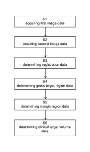
Fig. 1 is a flow diagram showing the basic steps of the disclosed
method;
Fig. 2 is a schematic view of a system performing the disclosed
method;
Fig. 3a is an MR image along a sagittal plane of an anatomical
structure
indicating a gross tumor volume and a safety margin;
Fig. 3b is an MR image along a coronal plane of an anatomical structure
indicating a gross tumor volume and a safety margin;
Fig. 4a is an MR image along a sagittal plane of an anatomical
structure
indicating a gross tumor volume, a safety margin and a clinical target
volume;
Fig. 4b is an MR image along a coronal plane of an anatomical structure
indicating a gross tumor volume, a safety margin and a clinical target
volume;
Fig. 5a is fractional anisotropy mapping along a sag ittal plane of an
anatomical
structure indicating a gross tumor volume, a safety margin and a clinical
target volume;
Fig. 5b is fractional anisotropy mapping along a coronal plane of an
anatomical
structure indicating a gross tumor volume, a safety margin and a clinical
target volume.
CA 03056694 2019-09-16
WO 2018/210422 PCT/EP2017/061985
26
Fig. 1 is a flow diagram illustrating the basic steps of the disclosed method
which in
the illustrative example of Fig. 1 starts with a step S1 of acquiring first
image data
describing at least one image of an anatomical structure of a patient. Then,
step S2
is executed, which encompasses acquiring second image data describing an
indicator for a preferred spreading direction or probability distribution of
at least one
target cell. In subsequent step S3 registration data describing a registration
of the
first image data to the second image data is determined. In step S4 gross
target
region data is determined. Subsequently, margin region data is determined in
step
S5. The last step shown in Fig. 1 is step S6, which is directed to determining
clinical
target volume data based on the registration data, the gross target region
data and
the margin region data.
Figure 2 shows an exemplary system for performing the disclosed method. The
system comprises a computer 2 as well as a medical imaging device 8 operably
coupled to the computer 2. The computer 2 comprises a processor 3, a memory 4
and an interface 5. The computer 2 is connected to an input unit 6, such as a
mouse,
a keyboard or a touch-sensitive surface, and an output unit 7 such as a
display, a
speaker or a tactile sensation generation unit. A program causing the computer
2 to
perform the disclosed method may be loaded into the memory 4 of the computer.
In
one embodiment the program may be stored on a program storage medium
comprised in or connected to the computer 2. Furthermore, the computer 2 may
be
operably coupled to at least one electronic data storage device for storing
atlas data.
Figs. 3a and 3b depict MR images along the sagittal and the coronal plane of
an
anatomical structure, respectively. A gross tumor volume (GTV) is surrounded
by a 6
mm safety margin (line hatch). Figs. 4a and 4b additionally indicate an
optimized
clinical target volume (cross hatch) determined according to the disclosed
method is
depicted in Figs. 4a and 4b.
Figs. 5a and 5b depict fractional anisotropy (FA) mappings along a saggital
plane
and the coronal plane, respectively. The gross tumor volume (GTV), the safety
margin (line hatch) and the clinical target volume (cross hatch) are overlaid
on the
respective fractional anisotropy (FA) mappings.