Note: Descriptions are shown in the official language in which they were submitted.
CA 03056700 2019-09-13
WO 2018/170329
PCT/US2018/022747
LIQUID BIOPSY FOR cfRNA
[0001] This application claims priority to our copending US provisional
applications having
the serial numbers 62/473,273, filed March 17, 2017, 62/522,509, filed June
20, 2017, and
62/593,534, filed December 1, 2017.
Field of the Invention
[0002] The field of the invention is systems and methods of detection and
quantification of
circulating free RNA (cfRNA), especially as it relates to cfRNA from tumor
cells.
Background of the Invention
[0003] The background description includes information that may be useful in
understanding
the present invention. It is not an admission that any of the information
provided herein is
prior art or relevant to the presently claimed invention, or that any
publication specifically or
implicitly referenced is prior art.
[0004] All publications and patent applications herein are incorporated by
reference to the
same extent as if each individual publication or patent application were
specifically and
individually indicated to be incorporated by reference. Where a definition or
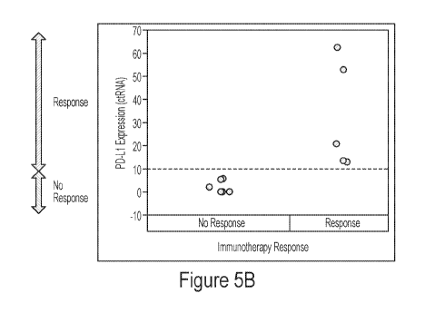
use of a term in
an incorporated reference is inconsistent or contrary to the definition of
that term provided
herein, the definition of that term provided herein applies and the definition
of that term in
the reference does not apply.
[0005] Over the last decade, cancer therapy has changed from a general
chemotherapy based
therapy in combination with surgery and radiation to a more personalized
treatment that takes
into account the genetic variability of tumors across patients. Therefore,
treatment plans often
now require identification of molecular markers that allow a more targeted
therapy. In many
cases, such information is obtained by analysis of various nucleic acid
molecules from cancer
tissue biopsies. However, tissue biopsies are often limited to initial
diagnosis or surgery, and
later biopsies tend to incur significant risk and discomfort to the patient.
Moreover, tumor
tissue biopsies tend to be problematic in terms of sampling bias and limited
ability to monitor
nucleic acid molecules as tumor markers in patients during the course of
therapy.
[0006] While it was known that nucleic acid molecules from tumor and non-tumor
cells can
be obtained from blood (see e.g., Clin Canc. Res. (1999) Vol 5, 1961-1965;
Cane Res. (1977)
1
CA 03056700 2019-09-13
WO 2018/170329
PCT/US2018/022747
37:646-650), it was not clear whether or not these nucleic acids were
associated or bound
with any carrier or other structure. Indeed, more recently it was discovered
that RNA can
originate from various sources, including circulating tumor cells (see e.g.,
WO 2017/180499),
exosomes (see e.g., WO 2015/082372), and carrier proteins (see e.g., WO
2010/079118, or
Proc. Natl. Acad. Sci. (1985) 82, 3455).
[0007] Unfortunately, and possibly due to the different locations/associations
of RNA with
various carriers or other structures, accurate quantification of circulating
nucleic acids has
often been problematic. For example, disease status detection in neuroblastoma
using cell
free RNA was shown not to be a reliable alternative to whole cell RNA analysis
(see e.g.,
Pediatr Blood Cancer. 2010 Jul 1;54(7):897-903). Similarly, while being able
to detect
relatively small quantities of cfRNA from mutated or improperly fused genes in
the blood
regardless of their particular association as described in WO 2016/077709, the
detected
quantities of such RNAs varied significantly. Moreover, it remained unknown
whether any of
the detected quantities was a reflection of physiological reality within a
cell or a function of
stability of the particular RNA in question. For example, data in the '709
publication indicate
that the quantities of cfRNA encoding PD-1/PD-L1 is often highly variable and
may depend
on the sample, patient condition, and other factors. Consequently, there has
to date been no
report of using PD-Li cfRNA expression levels as a prognostic agent and/or
indicator to
determine eligibility of cancer patients for anti-PD-1/PD-L1 therapy.
[0008] Therefore, even though numerous methods of nucleic acid analysis from
biological
fluids are known in the art, all or almost all of them suffer from various
disadvantages. Thus,
there remains a need for improved systems and methods for cfRNA analysis.
Summary of The Invention
[0009] The inventive subject matter is directed to various compositions and
methods of using
cfRNA levels of one or more cfRNA to predict treatment response, to track
treatment, and/or
to diagnose a cancer. In especially preferred aspects, the inventors
discovered that expression
threshold levels for certain cfRNA, and especially PD-Li and HER2, can be
determined that
are predictive for treatment response for certain cancers.
[0010] In one aspect of the inventive subject matter, method of predicting
treatment response
of an individual with cancer to treatment with a checkpoint inhibitor that
includes a step of
obtaining blood from the individual and isolating cfRNA from the blood,
wherein the cfRNA
2
CA 03056700 2019-09-13
WO 2018/170329
PCT/US2018/022747
encodes a checkpoint inhibition gene and a further step of quantifying the
cfRNA using
quantitative PCR method. A positive treatment response is then predicted when
the quantity
of the cfRNA is above a threshold level.
[0011] In preferred embodiments, the checkpoint inhibitor is an antibody
against PD1 or PD-
Li and the cfRNA is PD-Li cfRNA. Moreover, it is generally preferred that the
step of
isolating the cfRNA uses at least one of RNA stabilization and cell
preservation. Most
typically, the quantitative PCR method includes real time PCR, preferably with
13-actin as an
internal standard. Where PD-Li is quantified, the threshold level may be
AACT>10 for PD-
Li relative to 13-actin. Additionally, where desired, at least one second
cfRNA may be
quantified using the quantitative PCR method. While not limiting to the
inventive subject
matter, contemplated second cfRNAs may encode TIM3 or LAG3, a gene having a
tumor and
patient specific mutation, a tumor associated gene, or a cancer specific gene.
[0012] In another aspect of the inventive subject matter, the inventors also
contemplate a
method of monitoring treatment of an individual with cancer that includes a
step of obtaining
blood from the individual and isolating cfRNA from the blood, wherein the
cfRNA encodes a
checkpoint inhibition gene, or wherein the cfRNA encodes a tumor associated or
cancer
specific gene, or wherein the cfRNA encodes a gene having a tumor and patient
specific
mutation; a step of quantifying the cfRNA using quantitative PCR method; and a
step of
updating a patient record using the quantity of the cfRNA.
[0013] For example, suitable checkpoint inhibition gene include PD-L1, TIM3,
or LAG3,
tumor associated or cancer specific gene include CEA, MUC1, brachyury, HER2,
PCA3, or
AR-V7, and suitable genes having a tumor and patient specific mutation
preferably encode a
neoepitope. As noted above, it is generally preferred that the step of
isolating the cfRNA uses
RNA stabilization and cell preservation, and that the quantitative PCR method
includes real
time PCR (e.g., using 13-actin as an internal standard). The patient record
may be updated
when the quantity of the cfRNA is AACT>5 for HER2 relative to 13-actin or
AACT>10 for
PCA3 relative to 13-actin.
[0014] In still another aspect of the inventive subject matter, the inventors
contemplate a
method of detecting prostate cancer that includes a step of obtaining blood
from the
individual and isolating cfRNA from the blood, wherein the cfRNA encodes PCA3
or a
splice variant 7 of an androgen receptor; a further step of quantifying the
cfRNA using
3
CA 03056700 2019-09-13
WO 2018/170329
PCT/US2018/022747
quantitative PCR method; and a still further step of diagnosing the individual
as having
cancer when the cfRNA quantity is above a threshold level. Most typically, the
individual is
diagnosed as having cancer when the quantity of PCA3 cfRNA is AACT>10 relative
to (3-
actin.
[0015] Where desired, at least a second cfRNA may be quantified that encodes a
gene having
a tumor and patient specific mutation, a tumor associated gene, a cancer
specific gene, or a
checkpoint inhibition gene. Therefore, such second genes include PD-L1, LAG3,
TIM3, AR-
V7, PSA, and PSMA.
[0016] In yet another aspect of the inventive subject matter, the inventors
also contemplate a
method of treating a cancer that includes the steps of administering a drug to
an individual
diagnosed with a PD-Li negative cancer; monitoring treatment of the individual
by isolating
cfRNA from the blood, wherein the cfRNA encodes PD-Li; quantifying the cfRNA
using
quantitative PCR method; and including a checkpoint inhibitor to the treatment
upon
detection of the cfRNA.
[0017] In such methods, it is typically contemplated that the PD-Li negative
cancer is a solid
cancer (e.g., breast cancer), and /or that the drug is afinitor. Most
typically, the step of
quantifying cfRNA uses real-time PCR, and the checkpoint inhibitor is included
when the
cfRNA is detected and increases over time. In further preferred aspects of
such methods, the
checkpoint inhibitor is included when the cfRNA is detected and the cfRNA
level is
AACT>10 relative to 13-actin.
[0018] Moreover, the inventors also contemplate a method of determining an
immune
signature in a patient that includes a step of determining quantities of
distinct cfRNA
molecules in blood of an individual, wherein the cfRNA molecules encode
distinct
checkpoint inhibition genes (e.g., PD-L1, TIM3, LAG3). Typically, the step of
determining
is performed prior to or during treatment with at least one of a checkpoint
inhibitor, a
chemotherapeutic drug, an immune therapeutic drug, and radiation treatment.
[0019] Various objects, features, aspects and advantages of the inventive
subject matter will
become more apparent from the following detailed description of preferred
embodiments,
along with the accompanying drawing figures in which like numerals represent
like
components.
4
CA 03056700 2019-09-13
WO 2018/170329
PCT/US2018/022747
Brief Description of the Drawing
[0020] Figure 1 depicts graphs comparing plasma concentrations for cfDNA and
cfRNA for
healthy subjects and subjects diagnosed with cancer.
[0021] Figure 2A depicts a graph comparing plasma concentrations for PD-Li
cfRNA for
across various cancer types.
[0022] Figure 2B depicts a graph showing plasma concentrations for PD-Li cfRNA
for
healthy subjects.
[0023] Figure 2C depicts a graph showing the linear range for plasma
concentrations for PD-
Li cfRNA.
[0024] Figure 3A depicts a graph showing the relative expression of PD-Li
cfRNA for lung
cancer patients in a clinical trial.
[0025] Figure 3B depicts data showing PD-Li expression as measured by IHC for
the lung
cancer patients in the clinical trial.
[0026] Figure 4 depicts a graph showing PD-Li cfRNA levels for a non-responder
and a
responder to nivolumab and corresponding IHC staining of lung tumor samples,
along with
PD-Li cfRNA levels during treatment.
[0027] Figure 5A depicts a graph correlating PD-Li cfRNA levels with the PD-Li
status as
determined by PD-Li IHC.
[0028] Figure 5B depicts a graph correlating PD-Li cfRNA levels with nivolumab
response
status demonstrating a clinically relevant expression threshold for PD-Li
cfRNA levels.
[0029] Figures 6A-6D depicts graphs comparing plasma concentrations for PD-Li
cfRNA
levels of subjects diagnosed with cancer and undergoing treatment.
[0030] Figure 7 depicts a graph illustrating PD-Li cfRNA levels as a function
of treatment
with afinitor suggesting treatment with anti-PD 1/PD-L1 compositions.
[0031] Figure 8 depicts a graph correlating cancer treatment response status
with overall
cfRNA/beta-actin cfRNA.
CA 03056700 2019-09-13
WO 2018/170329
PCT/US2018/022747
[0032] Figure 9A depicts a graph showing relative co-expression of PD-Li and
HER2 as
measured by cfRNA levels.
[0033] Figure 9B depicts a graph correlating HER2 cfRNA levels with the HER2
status as
determined by HER2 IHC/FISH demonstrating a clinically relevant expression
threshold for
HER2 cfRNA levels.
[0034] Figure 10 depicts a graph showing relative co-expression of PD-Li and
HER2 in
gastric cancer as measured by cfRNA levels.
[0035] Figure 11 depicts a graph correlating pertuzumab/trustuzumab treatment
response
with HER2 cfRNA levels.
[0036] Figure 12 depicts cfRNA signatures for selected checkpoint relevant
genes.
[0037] Figure 13 depicts exemplary results for AR-V7 cfRNA levels and AR cfRNA
levels
in prostate cancer patients indicating that AR-V7 cfRNA is a suitable marker.
[0038] Figure 14 depicts exemplary results for PCA3 cfRNA levels in non-
prostate cancer
and prostate cancer patients indicating that PCA3 cfRNA is a suitable marker.
Detailed Description
[0039] The inventors have discovered that cfRNA can be employed as a
sensitive, selective,
and quantitative marker for diagnosis, monitoring of treatment, and even as
discovery tool
that allows repeated and non-invasive sampling of a patient. In most preferred
aspects, the
cfRNA is isolated from whole blood that is processed under conditions that
preserve cellular
integrity and stabilize cfRNA and/or ctDNA. Notably, the ratio of cfRNA to RNA
released
from non-tumor cells damaged during whole blood processing under such cell
preserving
conditions is sufficiently high to perform quantitative analysis that can
provide clinically
meaningful results. Once separated from the non-nucleic acid components, the
circulating
nucleic acids are then quantified, preferably using real time quantitative
PCR. Therefore, the
inventors also contemplated kits, reagents, and instructions for isolation,
monitoring, and
quantification of cfRNA in blood, and especially oligonucleotides for primers
suitable to
quantitatively determine presence of cfRNA for specific genes as is further
discussed in more
detail below.
6
CA 03056700 2019-09-13
WO 2018/170329
PCT/US2018/022747
[0040] Of course, and as is discussed in more detail below, it should be
appreciated that one
or more desired nucleic acids may be selected for a particular disease,
disease stage, specific
mutation, or even on the basis of personal mutational profiles or presence of
expressed
neoepitopes. Alternatively, where discovery or scanning for new mutations or
changes in
expression of a particular gene is desired, real time quantitative PCR may be
replaced or
supplemented by RNAseq to so cover at least part of a patient cfRNA
transcriptome.
Moreover, it should be appreciated that analysis can be performed static, or
over a time
course with repeated sampling to obtain a dynamic picture without the need for
biopsy of the
tumor or a metastasis.
[0041] Viewed form a different perspective, the inventors have generally
discovered various
methods and compositions for blood-based RNA expression testing of circulating
tumor
RNA (cfRNA) that identifies and quantitates expression, and that allows for
non-invasive
monitoring of changes in indicators and/or drivers of disease that have
heretofore only been
available by protein-based analysis of biopsied tissue. For example,
contemplated systems
and methods allow monitoring changes in indicators and/or drivers of a
disease, and/or
identification of changes in drug targets that may be associated with emerging
resistance to
chemotherapies. Advantageously, contemplated systems and methods integrate
with other
omics analysis platforms, and especially GPS Cancer (that provides whole
genome or exome
sequencing, RNA sequence and expression analysis, and quantitative protein
analysis) to
establish a powerful primary analysis/monitoring combination tool in which
alterations
identified by an omics platform are non-invasively, molecularly monitored by
systems and
methods presented herein.
[0042] In some embodiments, the inventors contemplate method of determining
status of a
(e.g., solid) cancer in a patient that includes a step of selecting a cancer
related gene on the
basis of at least one of a known association of a gene with the cancer and/or
a prior omics
analysis of cancer tissue in the patient. In another step, cfRNA of the cancer
related gene is
quantified in a bodily fluid (e.g., whole blood, serum, or plasma) of the
patient, and in a
further step the quantity of the cfRNA is associated with the cancer status.
Alternatively, or in
addition to the cancer related gene, other cfRNA may also be monitored. For
example, the
cancer status may be susceptibility of the cancer to treatment with a drug, or
presence or
absence of the cancer in the patient. Most typically, the cancer related gene
is a cancer
associated gene, a cancer specific gene, or a gene encoding a patient and
tumor specific
7
CA 03056700 2019-09-13
WO 2018/170329
PCT/US2018/022747
neoepitope (which may be determined using GPS cancer omics analysis). In
further
contemplated aspects, as described in more detail below, the step of
quantifying will include
isolation of the cfRNA under RNA stabilization and cell preservation, and/or
the step of
quantifying includes real time quantitative PCR of a cDNA prepared from cfRNA.
[0043] In other embodiments, the inventors also contemplate methods of
selecting a patient
for treatment with a checkpoint inhibitor that may include a step of obtaining
a bodily fluid
from the patient and quantifying a cfRNA in the bodily fluid for at least one
checkpoint
inhibition related gene. Among other suitable cfRNAs, especially contemplated
cfRNA
include those encoding PD-Li and HER2. Of course, it should be recognized that
the cfRNA
need not encode the full gene, but may be a fragment of the gene under
investigation. The
quantity of the cfRNA is then compared against a threshold value that
associates the quantity
with a likely treatment outcome. Consequently, and among other options,
treatment outcomes
may be related to treatments with one or more checkpoint inhibitors (e.g.,
antibody or
antibody fragment against PD-1, PD-L1, TIM3, and/or LAG3) and/or treatment
with
antibodies targeting various receptors (e.g., EGFR, ERCC1, IGF1, HER2, etc.)
[0044] Therefore, the inventors also contemplate various methods of treating a
cancer that
includes a step of determining cfRNA quantities of a first and a second marker
in a blood
sample of a patient, wherein the first marker is a checkpoint inhibition
related gene, and
wherein the second marker is one of a cancer associated gene, a cancer
specific gene, or a
gene encoding a patient and tumor specific neoepitope. It is further
contemplated that the
quantities of the first and second markers in such methods are (e.g.,
positively) associated.
The quantity of the second marker may then be used to determine treatment with
a
checkpoint inhibitor. For example, the first marker is PD-1 or PD-Li (or other
checkpoint
inhibition related marker), and the second marker is HER2. Likewise, the first
marker is PD-1
or PD-Li (or other checkpoint inhibition related marker), and the second
marker is a cfRNA
encoding a neoepitope.
[0045] In still other embodiments, the inventors also contemplate a method of
determining an
immune signature in a patient that includes a step of determining cfRNA
quantities of a
plurality of markers in a blood sample of the patient, wherein the plurality
of markers
comprise checkpoint inhibition related genes. Most typically, the step of
determining is
performed prior to or during treatment with at least one of a checkpoint
inhibitor, a
chemotherapeutic drug, an immune therapeutic drug, and radiation treatment.
Moreover,
8
CA 03056700 2019-09-13
WO 2018/170329
PCT/US2018/022747
contemplated methods may further comprise a step of determining a cfRNA
quantity of at
least one costimulatory marker, and/or a step of generating or updating a
treatment plan based
on the determined quantities.
[0046] In general, it is contemplated that cfRNA analysis is performed using
any bodily fluid
that contains cfRNA. Therefore, suitable bodily fluids include whole blood,
plasma, serum,
lymphatic fluid saliva, ascites fluid, spinal fluid, urine, etc., each of
which may be fresh or
preserved/frozen. However, it is especially preferred that the cfRNA analysis
uses whole
blood as a biological sample. Whole blood is readily obtained without
significant patient
discomfort and can be processed in a simple and effective manner. As is
further described in
more detail below, the inventors discovered that the protocols used for
removal of cells from
whole blood had a significant impact on stability and yield of the RNA.
Notably, the
inventors discovered that quantitative cfRNA analyses were significantly
improved where the
cells were removed from the whole blood under conditions that maintained
integrity of the
cells. While not wishing to be bound by any theory or hypothesis, the
inventors contemplate
that cell lysis of non-tumor cells in blood is a substantial contributing
factor in release of non-
cfRNA. Moreover, certain RNA stabilizing agents may also adversely affect
white and red
blood cells, and as such contribute to release of non-cfRNA into the plasma.
[0047] For example, for the analyses presented herein, specimens were accepted
as 10 ml of
whole blood drawn into cell-free RNA BCT tubes or cell-free DNA BCT tubes
(which
are both commercially available from Streck Inc.,7002 S. 109th St., La Vista
NE 68128)
containing RNA or DNA stabilizers, respectively. Advantageously, cfRNA is
stable in whole
blood in the cell-free RNA BCT tubes for seven days while ctDNA is stable in
whole blood
in the cell-free DNA BCT Tubes for fourteen days, allowing time for shipping
of patient
samples from various locations without the degradation of cfRNA or ctDNA.
However, it
should be noted that numerous alternative collection tubes and compositions
are also deemed
suitable so long as the RNA stabilization agents will not lead to substantial
cell lysis (e.g.,
equal or less than 3%, equal or less than 1%, or equal or less than 0.1%, or
equal or less than
0.01%, or equal or less than 0.001%) lyse white and/or red blood cells. Viewed
from a
different perspective, suitable RNA stabilization reagents will not lead to a
substantial
increase (e.g., increase in total RNA no more than 10%, or no more than 5%, or
no more than
2%, or no more than 1%) in RNA quantities in serum or plasma after the
reagents are
combined with blood. Of course, it should be recognized that numerous other or
additional
9
CA 03056700 2019-09-13
WO 2018/170329
PCT/US2018/022747
collection modalities are also deemed appropriate, and that the cfRNA and/or
ctDNA can be
at least partially purified or temporarily adsorbed to a solid phase to so
increase stability prior
to further processing.
[0048] As will be readily appreciated, fractionation of plasma and extraction
of ctDNA and
cfRNA can be done in numerous manners. In one exemplary preferred aspect,
whole blood
in 10 mL tubes is centrifuged to fractionate plasma at 1600 rcf for 20
minutes. Appropriate
centrifugation speeds can be calculated for various rotors following known
conversions (e.g.,
RCF=1.1118 x 10⁻⁵x rpm2, with r being rotor radius in cm). The so obtained
plasma is then
further centrifuged at 16,000 rcf for 10 minutes to remove cell debris. Of
course, various
alternative centrifugal protocols are also deemed suitable so long as the
centrifugation will
not lead to substantial cell lysis/maintains integrity of the blood cells
(e.g., lysis of no more
than 3%, or no more than 1%, or no more than 0.1%, or no more than 0.01%, or
no more than
0.001% of all cells). cfDNA and cfRNA can then be extracted from a desirable
volume (e.g.,
2mL) of plasma using Qiagen or other commercially available reagents. All
isolated ctDNA
and/or cfRNA are then kept in preferably bar-coded matrix storage tubes (e.g.,
with DNA
stored at -4 C, RNA stored at -80 C, or reverse-transcribed to cDNA that is
then stored at -
4 C).
[0049] Quantification of cfRNA can be performed in numerous manners, and
contemplated
methods include quantification by digital PCR methods, absolute quantification
methods
using external standards, and most typically relative quantification methods
using internal
standards (e.g., expressed as 2 Act). For example, real-time qPCR
amplification can be
performed using an assay in a 10 uL reaction mix containing 2 uL cDNA,
primers, and
probe. 13-actin can be used as an internal standard for the input level of ct-
cDNA. A standard
curve of samples with known concentrations of each analyte can be included in
each PCR
plate as well as positive and negative controls for each gene. Test samples
are then identified
by scanning the 2D barcode on the matrix tubes containing the nucleic acids.
Delta Ct (dCT)
were calculated from the Ct value derived from quantitative PCR (qPCR)
amplification for
each analyte subtracted by the Ct value of 13-actin for each individual
patient's blood sample.
Relative expression of patient specimens is calculated using a standard curve
of delta Cts of
serial dilutions of Universal Human Reference RNA set at a gene expression
value of 10
(when the delta CTs were plotted against the log concentration of each
analyte). ctDNA can
be analyzed in a similar fashion.
CA 03056700 2019-09-13
WO 2018/170329
PCT/US2018/022747
[0050] With regard to ctDNA, it should be noted that the accuracy of ctDNA in
diagnostic
tests has been in question since its adoption as a diagnostic tool for cancer.
Issues with
unusually high false positive rates must be addressed when relying on ctDNA in
monitoring
disease progression, but especially when considering the use of ctDNA in
prediction of
disease existence. As shown in Figure 1, healthy individuals produce similar
amounts of total
ctDNA as cancer patients, however, levels of total cfRNA (e.g., as determined
by quantitation
using beta actin) are significantly low in healthy individuals. Moreover, and
when cfRNA
isolation protocols were performed under conditions that did not lead to
substantial cell lysis,
the levels of total cfRNA were significantly different between cancer patients
and healthy
individuals. Indeed, there was no overlap between the groups of healthy
individuals thereby
allowing the cancer patients to be distinguished by their total cfRNA levels.
Conversely,
there was overlap between the levels of ctDNA in cancer patients and healthy
individuals.
Therefore ctDNA could not distinguish between these two groups. In further
contemplated
methods, it should be appreciated that where total cfRNA is isolated, cfDNA
may be
removed and/or degraded using appropriate DNAses (e.g., using on-column
digestion of
DNA). Likewise, where ctDNA is isolated, cfRNA may be removed and/or degraded
using
appropriate RNAses. Moreover, the linear detection range for cfRNA (here: PD-
L1) was
significant when isolation protocols were performed under conditions that did
not lead to
substantial cell lysis as is shown in more detail below.
[0051] It should be noted that the term cfRNA includes full length RNA as well
as fragments
of full length RNA (which may have a length of 50-150 bases, 15-500 bases, or
500-1,000
bases, or more). Thus, cfRNA may represent a portion of an RNA, which may be
between
100-80% of the full length RNA (typically mRNA), or between 80-60%, or between
60-40%,
or between 40-20%, or even less. Moreover, it should be appreciated that the
term cfRNA
typically refers to a tumor-derived RNA (as opposed to an RNA from a non-tumor
cell) and
that the cfRNA may therefore be from a tumor cell of a solid tumor, a blood
borne cancer,
circulating tumor cells, and exosomes. Most typically, however, the cfRNA will
be not be
enclosed by a membrane (and as such be from a circulating tumor cell or
exosome).
Moreover, it should be appreciated that the cfRNA may be uniquely expressed in
a tumor
(e.g., as a function of drug resistance or in response to a treatment regimen,
as a splice
variant, etc.) or as a mutated form of a gene (e.g., as a fusion transcript,
as a transcript of a
gene having a single or multi-base mutation, etc.). Therefore, and viewed from
a different
perspective, contemplated cfRNA especially include transcripts that are unique
to a tumor
11
CA 03056700 2019-09-13
WO 2018/170329 PCT/US2018/022747
cell relative to a corresponding non-tumor cell, or significantly over-
expressed (e.g., at least
3-fold, or at least 5-fold, or at least 10-fold) in a tumor cell relative to a
corresponding non-
tumor cell, or have a mutation (e.g., missense or nonsense mutation leading to
a neoepitope)
relative to a corresponding non-tumor cell.
[0052] Therefore, with respect to suitable target nucleic acids, it should be
appreciated that
appropriate targets particularly include genes that are relevant to a disease
and/or treatment of
a disease. For example, disease targets include one or more cancer associated
genes, cancer
specific genes, genes with patient and tumor-specific mutations (and
especially those leading
to the formation of neoepitopes), cancer driver genes, and genes known to be
overexpressed
in cancer. Consequently, suitable targets include those that encode
'functional' proteins (e.g.,
enzymes, receptors, transcription factors, etc.) and those that encode `non-
functional' proteins
(e.g., structural proteins, tubulin, etc.). Viewed from a different
perspective, suitable targets
may also include targets that are specific to a diseased cell or organ (e.g.,
PCA3, PSA, for
prostate, etc.), or targets that are more commonly found in different cancers,
such as various
mutations in KRAS (e.g., G12V, G12D, G12C, etc) or BRAF (e.g., V600E), etc.
Exemplary
targets validated by the inventors include AKT1, BRAF, CDK6, CYP3A4, ERBB3,
FGFR1,
JAK1, MAP2K1, AR-V7, ALK, BRCA1, CDKN2A, DDR2, ERBB4, FGFR2, JAK2, MET,
AR, ARAF, BRCA2, CTNNB1, OPYD, FGF19, FGFR3, KOR, MTOR, PD-U, ATM,
CCND1, CYP2C19, EGFR, FGF3, FLT3, KIT, NRAS, PD-1, BIM, CDK4, CYP2D6,
HER2, FGF4, HRAS, KRAS, NRG1, TIM3, NTRK1, PTCH1, SMO, NTRK2, PTEN,
STK11, NTRK3, RAF1, LAG3, TP53, PDGFRA, RET, TSC1, PIK3CA, RO-S1, TSC2, and
UGT1A1.
[0053] Consequently, it should be appreciated that suitable treatment targets
include one or
more markers that are indicative of susceptibility of a diseased cell to
treatment with a
specific drug that targets a specific molecular entity. For example systems
and methods
presented herein may be useful to identify the presence and expression level
of a specific
kinase that is targeted by a kinase inhibitor, or the presence and expression
level of a specific
signaling receptor targeted by synthetic ligand, or the presence and
expression level of a
specific checkpoint receptor targeted by synthetic antagonist or antibody,
etc., and suitable
targets may also be grouped by indication as shown in Table 1 below.
EGFR ROS1 KRAS ALK PD-L1 NRAS BRAF AR-V7
Lung V V V V V
Colon V V V V
Prostate V V
Melanoma V V
12
CA 03056700 2019-09-13
WO 2018/170329
PCT/US2018/022747
Table 1
[0054] In addition to known markers such as tumor associated antigens and
tumor specific
antigens, it should also be appreciated that prior omics analysis of a
patient's tumor may
reveal the presence of one or more neoepitopes. For example, prior analysis
can be done by
tumor versus matched normal comparison of the whole genome or exome,
preferably using
incremental synchronous alignment as described in US 9721062, and/or using
RNAseq. In
addition, proteomics analysis can be performed, most preferably using
quantitative mass
spectroscopic methods. Therefore, it should be appreciated that cfRNA may also
be used to
detect in a patient and tumor specific manner tumor RNA where the cfRNA
contains such
patient and tumor specific mutation (e.g., neoepitope). For example, such
detection may be
useful in monitoring treatment effect, particularly where the treatment is an
immune therapy
that targets the patient and tumor specific mutation (e.g., neoepitope). In
another example,
detection of a patient and tumor specific mutation may also reveal a (newly
arisen) treatment
target that may be treated with immune or chemotherapy.
[0055] Therefore, it should be appreciated that contemplated compositions and
methods can
be used in the discovery of disease associated markers, and more typically in
quantification
of suitable targets to so obtain information about presence of a mechanistic
target for
treatment and/or to obtain a quantitative proxy baseline for a cancer cell
population to follow
treatment or predict response development. For example, contemplated
compositions and
methods are especially suitable for immune therapy where the target is a
neoepitope as
expression and quantity of the neoepitope can be used to validate the
neoepitope as a
therapeutic target and to use the expression and quantity of the neoepitope as
a proxy marker
for treatment progress. Thus, it should be noted that cfRNA can be used to
ascertain presence
of expressed neoepitope before, during, and after treatment and as such allows
to predict
and/or quantitate treatment efficacy on an individual basis.
[0056] Alternatively, and among other preferred uses, cfRNA may be quantified
to identify
patients suitable for treatment with checkpoint inhibitors (e.g., targeting PD-
1 and PD-L1).
Such is especially useful as there is currently no convenient and non-invasive
way to
ascertain levels of PD-1 and PD-L1, which will inform a clinician if a patient
will benefit
from treatment with checkpoint inhibitors (e.g., nivolumab, pembrolizumab,
atezolizumab,
etc.). Indeed, immune checkpoints, such as programmed death ligand 1 (PD-L1)
or its
receptor, programmed death 1 (PD-1), appear to be Achilles' heels for multiple
tumor types.
13
CA 03056700 2019-09-13
WO 2018/170329
PCT/US2018/022747
PD-Li not only provides immune escape for tumor cells but also turns on the
apoptosis
switch on activated T cells. Therapies that block this interaction have
demonstrated
promising clinical activity in several tumor types. Tumoral PD-Li expression
status has been
shown to be prognostic in multiple tumor types, including melanoma (MEL),
renal cell
carcinoma (RCC), and non¨small-cell lung cancer (NSCLC). In addition, tumoral
PD-Li
expression appears to correlate closely with response to anti¨PD-1 antibodies.
However, no
test is uniformly accepted as the standard for quantitating PD-Li expression.
Moreover, a few
anti-PD-Li antibodies are in clinical trial stages and two were already
approved by FDA for
treating NSCLC. Thus it is important to measure PD-Li expression before giving
the patient
anti-PD-Li immunotherapy. The inventors have now discovered that that PD-Li
expression
and other immune therapy relevant cancer markers can be quantitated using
cfRNA by
analyzing the frequency and level of PD-Li (and other marker) expression in
cfRNA isolated
from various cancer types as is shown in more detail below.
Examples
[0057] Isolation of crRNA from whole blood: Whole blood was obtained by
venipuncture
and 10 ml were collected into cell-free RNA BCT tubes or cell-free DNA BCT
tubes
(Streck Inc.,7002 S. 109th St., La Vista NE 68128) containing RNA or DNA
stabilizers,
respectively. The sample tubes were then centrifuged at 1,600 rcf for 20
minutes, plasma was
withdrawn and further centrifuged at 16,000 rcf for 10 minutes to remove cell
debris. Plasma
was used to isolate cfRNA using commercially available RNA isolation kits
following the
manufacturer's protocol with slight modification. Specifically, DNA was
removed from the
sample in an on-column DNAse digest.
[0058] In an alternative approach, cfRNA was also obtained in an automated
manner using a
robotic extraction method on QiaSymphony instrumentation (Qiagen, 19300
Germantown
Road; Germantown, MD 20874), slightly modified to accommodate for DNA removal
where
desired. The robotic extraction maintained approximately 12% DNA contamination
in the
cfRNA sample. We measured the relative expression of Excision Repair Cross-
Complementing enzyme (ERCC1) vs beta actin in the same twenty-one NSCLC
samples to
determine whether there was a significant difference between the two
extraction procedures.
Notably, there was no statistical difference in the relative expression
generated by the
automated process and the manual process as shown in the table below. p=
0.4111 (paired t-
test; a statistically difference would have been p<0.05 for this test).
14
CA 03056700 2019-09-13
WO 2018/170329 PCT/US2018/022747
\\, "
1 30.09 22.31 0.00 2.75 29.76 22.27 0.01 3.37
* 31.22 ;iv: 2346 000 179 ataa 23.35 000 2.47
3 1. 31.50 I. 23.65 0.00 1 2.64 1. 30.64 1 23.48
0.01 1 4.26
1! 421
:21:41620 1! 45:3132 0: 0411 ::3778 2234:1;10 n 0:41011 ,
4 2 040 3030 232T543 cL.010 ""t! 1 5T4 3:1:479
'it' 2 C3 331
n .11 7 30 94 2 3 o 0 0 3196
t0 54
!t! 22162: Otio 112.6:4 110:161 221:
0µ:11011 438
4.26
aa !.t! 3 294
2::77: !..:t 2242:6243- (11:71 !.! 113
3187 5 !.1! 2242:121 n (11:t0tC11 !.1!1.92
:1.4448 2243:;33 COL.Vt.! 32;32 ".!
33131:82: t 221.25: " (01.43010 !.t! 62:D9713 4 "i
:30:64; t 2214110 !! 80':110411 ::76 23::13t ''.1" 22134:
001 328
15 0.01 6.59
:35: 22::;13 2242.!1 001 6 " 54:317 3
as t :::3077 2392 11 4;:4536 3322:314;
2234:71 n 000 23:21:
19 gsk 31.90 23.52 amo 3W 3166 23.24 u amo 2.31
21 i 30.42 i 23.85 1 0.01 5.48 ,t 30.50 i 23.29 0.01
Li 5.33
[0059] Custom kit from Qiagen (QiaSymphony Circulating NA kit #1074536)
included two
virus extraction kits in one custom kit (the virus kits are called QiaSymphony
DSP
Virus/Pathogen Midi Kit Version 1 #937055). Analyses were run within single,
proprietary
program on Qiagen instrument (custom program protocol CF 20005_CR21040_ID993;
from
Qiagen).
[0060] Quantification of cfRNA: Unless otherwise noted, quantification was
performed using
relative quantification via rtPCT and gene specific primer pairs along with
primer pairs for
beta-actin as internal control. For example, amplifications were performed
using an assay in a
[it reaction mix containing 2 [it cDNA, primers, and probe. 13-actin can be
used as an
internal standard for the input level of ct-cDNA. A standard curve of samples
with known
concentrations of each analyte wad included in each PCR plate as well as
positive and
negative controls for each gene. Test samples were identified by scanning the
2D barcode on
the matrix tubes containing the nucleic acids. Delta Ct (dCT) were calculated
from the Ct
value derived from quantitative PCR (qPCR) amplification for each analyte
subtracted by the
Ct value of 13-actin for each individual patient's blood sample. Relative
expression of patient
specimens was calculated using a standard curve of delta Cts of serial
dilutions of Universal
Human Reference RNA set at a gene expression value of 10 (when the delta CTs
were plotted
against the log concentration of each analyte). ctDNA was analyzed in a
similar fashion.
[0061] Delta Cts vs. log ioRelative Gene Expression (standard curves) for each
gene test were
captured over hundreds of PCR plates of reactions (historical reactions). A
linear regression
CA 03056700 2019-09-13
WO 2018/170329
PCT/US2018/022747
analysis was performed for each assay and used to calculate gene expression
from a single
point from the original standard curve going forward.
[0062] Notably, as is shown in Figure 1, where ctDNA was quantified from
healthy donors
and cancer patients, non-small cancer (NSCLC), 10 cancer and 9 healthy
individuals. No
statistically significant difference could be overserved with total ctDNA
between the two
populations. In contrast, total cfRNA quantities (as measured by (3-actin)
were significant
different between the two populations, indicating that measurement of total
cfRNA may be a
valid indicator for the presence of cancer.
[0063] The inventors then investigated whether the above results could be
confirmed across
various other cancer types and selected genes (e.g., PD-L1) and analyzed blood
samples from
selected patients diagnosed with breast cancer, colon cancer, gastric cancer,
lung cancer, and
prostate cancer. In this series of tests, relative expression of PD-L1cfRNA
was quantitated,
and the results are depicted in Figure 2A. Interestingly, not all cancers
expressed PD-Li as
shown in Figure 2A, and the frequencies of positivity in the various cancers
was concordant
with the published expression of PD-Li using IHC in solid tissue. PD-L1cfRNA
was not
detectable in healthy patients as can be seen from Figure 2B.
[0064] Assay Validation ¨ Accuracy: Accuracy of an exemplary PD-Li Expression
Assay
was determined by comparing the results generated by the present PD-Li assay
("LiquidGeneDx") from 61 clinical samples against a digital PCR PD-Li assay
(lab
developed reference method, an alternative PD-Li detection assay). The results
were used to
determine the clinical sensitivity and clinical specificity of the assay. The
accuracy results
from the present PD-Li assay and the digital PCR PD-Li assay are summarized in
Table 2.
Positive Agreement Negative Agreement
(LiquidGeneDx vs Digital PCR) (LiquidGeneDx vs Digital PCR)
PD- 91% 94%
Li
Table 2
[0065] Assay Validation ¨ Limit of Detection (LOD): Analytical sensitivity of
the present
PD-Li assay ("LiquidGeneDx") was determined by 20 replicates at a 95%
detection rate.
cfRNA was extracted from patients' plasma, reverse-transcribed using random
hexamers to
cDNA and pre-amplified using Thermo Fisher's pre-amplification product Taqman
Preamp
Master Mix with PD-Li and beta-actin primers for 10 cycles per the
manufacturer's
16
CA 03056700 2019-09-13
WO 2018/170329
PCT/US2018/022747
instructions. The resulting pre-amplified cDNA was diluted in 2-fold
increments with cDNA
from patients' plasma negative for PD-Li. All dilution samples were examined
by
LiquidGeneDx for the minimum amount of PD-Li cDNA required for amplification
and
successful PCR. Then 20 replicates at the presumptive LOD level were used to
confirm the
final LOD. The limit of detection (LOD) acceptance criteria in this study was
determined as
the lowest concentration at which all 20 replicates generated a 95% above the
detection rate.
If 20 replicates could not generate a 95% above detection rate, the next
higher concentration
of dilution samples were used as presumptive LOD to repeat with 20 replicates.
A summary
of LOD study results is shown in Table 3 in which the * denotes the final LOD.
Valid Positive Results/Total Tested
PD-L1 Dilution 1.884ng 0.941ng 0.471ng 0.236ng 11';.;
0.059ng
Sample
PD-L1 4/4 4/4 4/4 4/4 15/20
Expression
Table 3
[0066] Assay Validation ¨ Limit of Detection (LOD): The precision panel
included a low
positive PD-Li sample, a medium positive PD-Li sample, a high negative PD-Li
sample,
positive control, and no-template control. All positive samples were made from
a PD-Li
positive cancer cell line. Each precision panel was examined in quadruplicate
per run, 2 runs
per instrument for 2 instruments per day for total of 3 days (consecutive or
non-consecutive)
by three different operators (Op). Each sample of the precision panel
generated total 48 data
points across 3 days. The study design is illustrated in Table 4.
Instrument 1 Instrument 2
Day 1 Op 1 0p2 0p3 Op 1
Day 2 0p2 0p3 Op 1 0p2
Day 3 Op 3 Op 1 Op 2 Op 3
Table 4
[0067] The intra-assay precision was done using two instruments, two
operators, one day,
and four replicates per samples. Result concordance for all replicates are 96%
or above.
Table 5 is an exemplary summary of the intra-assay precision.
Sample Expression Operator 1 Operator 2
Run 1 Run 2 Run 3 Run 1 Run 2 Run
3
1 Positive PD-L1 100% 96% 100% 100% 100% 100%
2 Negative Water 100% 100% 100% 100% 100% 100%
Table 5
17
CA 03056700 2019-09-13
WO 2018/170329 PCT/US2018/022747
[0068] Two instruments, two operators, three runs were done over three days,
and
quadruplicate runs were tested for inter-assay precision. Result concordance
reached 96% or
above for all replicates across independent runs. Result summary is listed in
Table 6.
Comparison Standard Result
Operator #1 vs. Operator #2 Result Agreement 99%
Operator #1, between runs Result Agreement 96%
Operator #2, between runs Result Agreement 100%
Table 6
[0069] Assay Validation ¨ Linear Range: Quantitative linear range of the
present PD-Li
assay ("LiquidGeneDx") was determined by diluting PD-Li-positive patients'
cDNA from
cfRNA into a pooled negative matrix (PD-Li-negative cDNA from cfRNA). ct RNA
was
extracted from patients' plasma, reverse-transcribed using random hexamers to
cDNA and
pre-amplified using Thermo Fisher's pre-amplification product Taqman Preamp
Master
Mix with PD-Li and beta-actin primers for 10 cycles per the manufacturer's
instructions.
The resulting pre-amplified cDNA was diluted in 2-fold increments with cDNA
from
patients' plasma negative for PD-Li. All dilution samples were examined by
LiquidGeneDx
PD-Li to determine its quantitative linear range. Figure 2C shows the final
linear range. The
linear portion of the line extends to a Ct of approximately 32.5. Beta-actin
and PD-Li slopes
are also concordant.
[0070] Assay Validation ¨ Specificity: Test samples were prepared by serial
dilution of
human PD-Li cell line cDNA in TE buffer matrix. Concentration of target
analyte for
medium positive samples was 4 times the LOD concentration. Medium-positive
samples
with each interferent (one analyte with each interferent) as well as baseline
samples were
examined in triplicate by the present PD-Li assay ("LiquidGeneDx"). Table 7 is
the list of
interferents and their testing concentration. All samples with testing
concentration of different
interferents were still determined as positive by the LiquidGeneDx PD-Li
assay.
Interferents Interference Concentration
Buffer ACL 0.1% in total volume
Buffer ACB 0.2% in total volume
Buffer ACW1 1% in total volume
Buffer ACW2 1% in total volume
Buffer AVE 1% in total volume
Albumin 2 mg/mL
Casein 2 mg/mL
Hemoglobin 0.4 mg/mL
Actin DNA/RNA mix 1 ng in total
18
CA 03056700 2019-09-13
WO 2018/170329
PCT/US2018/022747
Table 7
[0071] Notably, all samples with testing concentration of different
interferents were still
determined as positive by the LiquidGeneDx PD-Li assay.
[0072] The present PD-Li assay ("LiquidGeneDx") was designed as a real-time
PCR assay
to detect expression of the PD-Li gene and other genes in blood of cancers
patients. Among
other benefits, such measurements can inform a clinician about the likely
treatment success
with a specific drug (e.g., anti-PD-1 antibody) before and during drug
therapy.
[0073] Based on the above findings that cfRNA can be accurately quantified,
the inventors
sought to determine whether the quantified cfRNA levels would also correlate
with known
analyte levels measured by conventional methods such as FISH, mass
spectroscopy, etc.
More specifically, the frequency and strength of PD-Li expression was measured
by cfRNA
from the plasma of 320 consecutive NSCLC patients using LiquidGenomicsDx and
compared to the frequency of positive patients in the Keynote Trial, a
registration trial of
pembrolizumab (Keytruda), using a tissue IHC test. Notably, 66% of NSCLC
patients
(1,475/2,222) in the Keynote trial had any expression of PD-Li by IHC (>i% of
cells
positive), while 64% of NSCLC (204/320) patients with blood-based cfRNA
testing of PD-
Li were positive as can be seen from Figures 3A and 3B. Remarkably, there was
no
significant difference in PD-Li status between the two analytical methods, but
the cfRNA
testing afforded quantitative data.
[0074] Notably, the difference in PD-Li status (i.e., PD-Li positive or PD-Li
negative) of
two selected patients (Pt#1 and Pt#2) also correlated well with IHC analysis
and treatment
response with nivolumab as can be seen from Figure 4. Here, two squamous cell
lung cancer
patients were treated with the anti-PD-1 antibody nivolumab. Patient 1 had no
expression of
PD-Li in the tissue or in the blood using cfRNA measurement. Patient 1 did not
respond to
nivolumab. Tumor growth was documented by CT scan and the patient expired
rapidly. In
contrast, Patient 2 had high levels of PD-Li in the tissue and in the blood at
baseline using
cfRNA measurement. Patient 2 responded to nivolumab with a durable response
over several
cycles of the drug. The response was documented by CT scan with dramatic tumor
shrinkage. Interestingly, the high levels of gene expression in the blood of
this patient
(measured by cfRNA) disappeared after three and a half weeks while the patient
continued to
respond.
19
CA 03056700 2019-09-13
WO 2018/170329
PCT/US2018/022747
[0075] Based on the above observed correlation, the inventors set out to
investigate whether
or not expression levels of PD-Li cfRNA could provide threshold levels
suitable for response
prediction to treatment with nivolumab or other therapeutics interfering with
PD 1/PD-L1
signaling. To that end, PD-Li expression was measured in NSCLC patient plasma
using
cfRNA and compared with IHC status. Figure 5A shows the correlation between
treatment
response status with an anti-PD-Li therapeutic and PD-Li status as determined
by IHC and
PD-Li expression above response threshold by cfRNA. Patients determined to be
treatment
responders were also determined by IHC as PD-Li positive, while all patients
determined to
be non-responders to treatment were determined by IHC as PD-Li negative.
Remarkably, the
same separation between responders and non-responders could be achieved using
PD-Li
cfRNA levels when a response threshold was applied to then data. In this
example, a relative
expression threshold of 10 accurately separated responders from non-
responders. Figure 5B
shows that a cfRNA response threshold of AACT>10 for PD-Li relative to 13-
actin predicts
positive response to a PD 1/PD-L1 checkpoint inhibitor (here: nivolumab). All
responders to
nivolumab expressed PD-Li above the threshold level prior to treatment.
[0076] The inventors further investigated if PD-Li cfRNA expression levels
could be used in
other cancer treatments as an indicator for progressive disease (PD), stable
disease (SD),
and/or partial response (PR). To that end, dynamic changes in PD-Li measured
by cfRNA
were found during the course of therapy under various treatment regimens as is
exemplarily
shown in Figures 6A-6D. Panel A shows the relative expression levels for PD-Li
over the
course of treatment of breast cancer with abraxane in a patient with
progressive disease. The
lack of response to treatment is reflected in the rise of PD-Li cfRNA, and
abraxane treatment
was discontinued in favor of treatment with CDX-011(glembatumumab vedotin). As
can be
seen from Figure 6A, treatment with CDX-011 lead to disease stabilization,
which is also
reflected in a decrease of PD-Li cfRNA. Similarly, as can be taken from Figure
6B, a lung
cancer patient was treated at stable disease with a carboplatin/alimta
combination therapy,
and initially high levels of PD-Li cfRNA dramatically decreased as the patient
showed
partial response. In the case of colon cancer, a patient with progressive
disease was treated
with capecitabine and bevacizumab. During treatment, relative PD-Li cfRNA
expression
significantly increased. Upon treatment of the cancer with 5-FU and
bevacizumab, the patient
had a partial response with concomitant significant drop in PD-Li cfRNA levels
as can be
taken from Figure 6C. Therefore, the inventors contemplate that quantitative
levels of PD-Li
cfRNA can also accurately serve to monitor treatment response.
CA 03056700 2019-09-13
WO 2018/170329
PCT/US2018/022747
[0077] In yet another example, the inventors observed a rapid increase in PD-
Li cfRNA in a
patient with stable disease breast cancer upon treatment with
exemestane/afinitor as is shown
in Figure 6D. Notably, the patient did not have measureable quantities of PD-
Li cfRNA
before treatment. Based on this observation, the inventors tested further
breast cancer patient
samples that underwent afinitor treatment and exemplary results are depicted
in Figure 7. As
is readily apparent, relative PD-Li cfRNA significantly increased post
treatment at the
second blood draw to levels suitable for treatment with PD 1/PD-L1 checkpoint
inhibitors.
Therefore, the inventors also contemplate that cancer treatments (especially
those using drugs
other than PD 1/PD-L1 checkpoint inhibitors) can be followed by at least
monitoring PD-Li
cfRNA to identify emergence of PD-Li cfRNA expression, which can then serve as
an
indicator of treatment with a PD 1/PD-L1 checkpoint inhibitors. Viewed from a
different
perspective, detection and quantitation of previously not detectable PD-Li
cfRNA expression
during a cancer treatment may be used as an indicator to (additionally) treat
a patient with a
PD1/PD-L1 checkpoint inhibitor.
[0078] Interestingly, disease status of cancer also paralleled to at least
some extent 13-actin
cfRNA as can be seen from Figure 8. Blood was drawn from patients under
various therapies
every 6-8 weeks, at the same time that the CT scans were done. cfRNA was
extracted from
plasma of 45 patients with metastatic breast cancer, and 30 patients completed
the first two
cycles of therapy: 6/6 patients with PR showed either no change (NC) or a
decrease (DEC) in
levels of 13-actin cfRNA, 13/16 patients with SD showed NC or DEC in cfRNA
levels, and
6/8 patients with PD underwent increases (INC) in levels of cfRNA. CfRNA was
reverse
transcribed with random hexamers to cDNA. Levels of cfRNA were quantitated by
RT-qPCR
and correlated with patient response (PR/SD/PD), as determined by CT scans.
Levels of gene
expression in cfRNA (including PD-Li and HER2) were monitored in patients
across blood
draws. Notably, 13-actin cfRNA levels of breast cancer patients with
progressive disease was
higher than 13-actin cfRNA levels of patients with stable disease and/or
partial response. Thus,
it should be appreciated that an increase in 13-actin cfRNA levels can serve
as a leading
indicator of disease status, and especially of progressive disease in patients
already diagnosed
with cancer.
[0079] Upon further investigation of breast cancer samples, the inventors also
discovered that
HER2 cfRNA in tumors appeared to be co-expressed or co-regulated with PD-Li as
is shown
in Figure 9A. On this basis, the inventors then used HER2 status
classification by immune
21
CA 03056700 2019-09-13
WO 2018/170329
PCT/US2018/022747
histochemical analysis using antiHER2 antibodies (IHC) to correlate IHC-HER2
status with
quantitative relative expression of HER2 as measured by cfRNA levels.
Remarkably, there
was a significant correlation (82% concordance) between HER2 cfRNA expression
levels
and IHC HER2 status where a AACT>5 for HER2 relative to 13-actin was applied
as is
exemplarily shown in Figure 9B. Therefore, it is contemplated that HER2 status
may also be
determined using detection and quantification of HER2 cfRNA using an
expression threshold
as provided above.
[0080] In further experiments, the inventors also discovered that that HER2
cfRNA in at least
some gastric tumors also appeared to be co-expressed or co-regulated with PD-
Li as is
shown in Figure 10. Such finding is particularly notable as it is known that
about 15% of all
gastric cancers do express HER2. Consequently, the inventors contemplate
methods of
detecting or quantifying HER2 cfRNA in patients with gastric cancer.
Furthermore, the
inventors also contemplate that one or more immune checkpoint genes (e.g., PD-
L1, TIM3,
LAG3) as measured by cfRNA may be used as proxy markers for other cancer
specific
markers or tumor associated markers (e.g., CEA, PSA, MUC1, brachyury, etc.).
[0081] As will be readily appreciated, the quantification of HER2 cfRNA levels
may also be
employed to follow treatment, and particularly to assess whether or not
treatment with an
anti-HER2 drug has therapeutic effect. For example, partial treatment response
to two anti-
HER2 drugs (pertuzumab and trustuzumab) in two exemplary patients (patients 25
and 12,
respectively) of a cohort of metastatic breast cancer patients showed that
positive response
directly correlated with a reduction of cfRNA as is depicted in Figure 11.
Indeed, past three
months of treatment no detectable quantities of HER2 cfRNA were present.
[0082] Based on the observed co-expression or co-regulation, the inventors
then investigated
whether or not other cfRNA levels for immune checkpoint related genes would
correlate with
PD-Li cfRNA levels and exemplary results are depicted in Figure 12. Here,
cfRNA levels
for PD-L1, TIM3, and LAG3 were measured from blood samples of prostate cancer
patients.
Notably, in all but one sample more than one checkpoint related gene was
strongly expressed.
Interestingly and importantly, levels of TIM3 and LAG3, the former of which
has been
shown to serve as an escape mechanism or resistance factor for PD-1 or PD-Li
inhibition,
often mirrored PD-Li expression, underscoring a need to address all checkpoint
proteins
besides PD-1 and PD-Li. Therefore, it should be appreciated that cfRNA levels
for immune
checkpoint relevant genes may be analyzed for cancer patients to so obtain an
immune
22
CA 03056700 2019-09-13
WO 2018/170329
PCT/US2018/022747
signature or the patient, and the appropriate treatment with more than one
checkpoint
inhibition drug may be then be advised. As will be appreciated, suitable
threshold values for
the genes can be established following the methods described for PD-Li and
HER2 above.
[0083] In still further aspects of the inventive subject matter, various
alternate cfRNA species
were demonstrated to quantitatively distinguish healthy individuals from those
afflicted with
cancer and/or to predict treatment response. For example, the detection of the
splice variant 7
of the androgen receptor (AR-V7) has been an important consideration for the
treatment of
prostate cancer with hormone therapy. The inventors therefore investigated
whether or not
hormone therapy resistance is associated with prostate cancer tumor growth and
detection of
AR-V7 via detection and quantification of AR-V7 cfRNA. Figure 13 depicts
exemplary
results for AR and AR-V7 gene expression via cfRNA methods using plasma from
prostate
cancer patients. AR-V7 was also measured using IHC technology from CTCs from
the same
patients. Notably, the results from CTCs and cfRNA for AR-V7 were concordant
(data not
shown).
[0084] Furthermore, PCA3 was identified as a marker for prostate cancer in a
test in which
PCA3 cfRNA was detected and quantified in plasma from prostate cancer patients
and in
which non-prostate cancer patient samples had relatively low to non-detectable
levels. Non-
prostate cancer patients were NSCLC and CRC patients. As can be taken from
Figure 14,
PCA3 was shown to be differentially expressed between the two groups (non-
overlapping
medians between prostate and non-prostate cancer patients) by cfRNA,
indicating that the
non-invasive blood based cfRNA test may be used to detect prostate cancer.
Once more,
based on a priori knowledge of the tested population, a threshold value (here:
AACT>10 for
PCA3 relative to (3-actin) for expression could be established as is
exemplarily depicted in
Figure 14.
[0085] In yet a further study, the inventors used analysis of total cell-free
circulating tumor
RNA (cfRNA) extracted from plasma of cancer patients (pts) as a tool to
measure dynamic
changes in gene expression as well as in total levels of nucleic acids
including cfRNA. These
analyses provided yet again insight into disease status and allowed predicting
outcome to
anti-tumoral therapy.
[0086] More specifically, blood was drawn from pts under various treatments
(tx) every 6-8
weeks, at the same time that CT scans were done. CfRNA was extracted from the
resulting
23
CA 03056700 2019-09-13
WO 2018/170329
PCT/US2018/022747
plasma and reverse transcribed with random hexamers to cDNA as described
above. Levels
of total cfRNA were quantitated by RT-qPCR and correlated with pt response
(PR/SD/PD),
as determined by CT scans. In this study, a total of 30 lung cancer pts were
enrolled in a 2-
year clinical study. Ethnicities included: 73% (22/30) Caucasian, 20% (6/30)
Hispanic, and
7% (2/30) other. Non-SQCC were 87% (26/30) of the total. 23 pts completed the
first two
cycles of tx. Of these, 6/8 pts with progressive disease (PD) showed increased
(INC) levels of
total cfRNA, 8/12 pts with stable disease (SD) showed either no change (NC) or
decreased
(DEC) total cfRNA, and 3/3 pts with partial response (PR) had DEC total cfRNA,
corresponding to 74% concordance between total cfRNA and pt response. PD-Li
expression
measured in plasma cfRNA matched that of tissue in 7/10 pts. In the one pt
where PD-Li
was negative in blood and positive in tissue, the pt progressed on
pembrolizumab. Among 7
pts treated with immunotherapy (nivolumab, pembrolizumab, atezolizumab), 3/3
pts with PD
showed INC PD-Li cfRNA expression, 3/3 pts with SD had NC in PD-Li cfRNA, and
1 pt
with PR showed DEC PD-Li cfRNA, corresponding to 100% correlation between PD-
Li
expression levels and pt response. Upon treatment, a significant concordance
was observed
between clinical response and changes in plasma cfRNA levels in NSCLC pts
(74%).
Detection of PD-Li expression in pt plasma also correlated with results
obtained from tissue
of same pts (70%). While on targeted therapy, levels of PD-Li expression
correlated with
response in 7/7 pts. It can therefore be concluded that cfRNA levels can
indicate tx response,
and PD-Li in plasma could be used to monitor response to immunotherapy.
[0087] It should be apparent to those skilled in the art that many more
modifications besides
those already described are possible without departing from the inventive
concepts herein.
The inventive subject matter, therefore, is not to be restricted except in the
scope of the
appended claims. Moreover, in interpreting both the specification and the
claims, all terms
should be interpreted in the broadest possible manner consistent with the
context. In
particular, the terms "comprises" and "comprising" should be interpreted as
referring to
elements, components, or steps in a non-exclusive manner, indicating that the
referenced
elements, components, or steps may be present, or utilized, or combined with
other elements,
components, or steps that are not expressly referenced. Where the
specification claims refers
to at least one of something selected from the group consisting of A, B, C
.... and N, the text
should be interpreted as requiring only one element from the group, not A plus
N, or B plus
N, etc.
24