Note: Descriptions are shown in the official language in which they were submitted.
CA 03056837 2019-09-17
WO 2018/177966 PCT/EP2018/057566
Improved antigen binding receptors
FIELD OF THE INVENTION
The present invention generally relates to antigen binding receptors capable
of specific
binding to mutated Fc domains with reduced Fc receptor binding and T cells
expressing these
antigen binding receptors. More precisely, the present invention relates to T
cells,
transfected/transduced with an antigen binding receptor which is recruited by
specifically
binding to/interacting with the mutated Fc domain of therapeutic antibodies.
Furthermore, the
invention relates to a kit comprising the T cells of the invention and/or
nucleic acid
molecules, vectors expressing antigen binding receptors of the present
invention and (a) tumor
targeting antibody/antibodies comprising a mutated Fc domain. The invention
also provides
the production and use of T cells in a method for the treatment of particular
diseases in
conjunction with tumor-specific antibodies as well as pharmaceutical
compositions/medicaments comprising T cells and/or therapeutic antibodies,
wherein T cells
are to be administered in combination with therapeutic-tumor targeting
antibody/antibodies
comprising a mutated Fc domain with reduced Fc receptor binding.
BACKGROUND
Adoptive T cell therapy (ACT) is a powerful treatment approach using cancer-
specific T cells
(Rosenberg and Restifo, Science 348(6230) (2015), 62-68). ACT may use
naturally occurring
tumor-specific cells or T cells rendered specific by genetic engineering using
T cell or
chimeric antigen receptors (Rosenberg and Restifo, Science 348(6230) (2015),
62-68). ACT
can successfully treat and induce remission in patients suffering even from
advanced and
otherwise treatment refractory diseases such as acute lymphatic leukemia, non-
hodgkins
lymphoma or melanoma (Dudley et al., J Clin Oncol 26(32) (2008), 5233-5239;
Grupp et al.,
N Engl J Med 368 (16) (2013), 1509-1518; Kochenderfer et al., J Clin Oncol.
(2015)
33(6):540-549, doi: 10.1200/JC0.2014.56.2025. Epub 2014 Aug 25).
However, despite impressive clinical efficacy, ACT is limited by treatment-
related toxicities.
The specificity, and resulting on-target and off-target effects, of engineered
T cells used in
ACT is mainly driven by the tumor targeting antigen binding moiety implemented
in the
chimeric antigen receptor (CAR). Non-exclusive expression of the tumor antigen
or temporal
1
CA 03056837 2019-09-17
WO 2018/177966 PCT/EP2018/057566
difference in the expression level can result with serious side effects or
even abortion of ACT
due to non-tolerable toxicity of the treatment.
Additionally, the availability of tumor-specific T cells for efficient tumor
cells lysis is
dependent on the long-term survival and proliferation capacity of engineered T
cells in vivo.
On the other hand, in vivo survival and proliferation of T cells may result
with unwanted long-
term effects due to the persistence of an uncontrolled CAR-T response (Grupp
et al. 2013 N
Engl J Med 368(16):1509-18, Maude et al. 2014 2014 N Engl J Med 371(16):1507-
17).
One approach for limiting serious treatment-related toxicities and to improve
safety of ACT is
to restrict the activation and proliferation of CAR-T cells by introducing
adaptor molecules in
the immunological synapse. Such adaptor molecules comprise small molecular
bimodular
switches as e.g. recently described folate-FITC switch (Kim et al. J Am Chem
Soc 2015;
137:2832-2835). A further approach included artificially modified antibodies
comprising a tag
to guide and direct the specificity of CAR-T cells to target tumor cells (Ma
et al. PNAS 2016;
113(4):E450-458, Cao et al. Angew Chem 2016; 128:1-6, Rogers et al. PNAS 2016;
113(4):E459-468, Tamada et al. Clin Cancer Res 2012; 18(23):6436-6445).
However, existing approaches have several limitations. Immunological synapses
relying on
molecular switches require introduction of additional elements which might
elicit an immune
response or result with non-specific off-target effects. Furthermore, the
complexity of such
multicomponent systems may limit treatment efficacy and tolerability. On the
other hand, the
introduction of tag structure in existing therapeutic monoclonal antibodies
may affect the
efficacy and safety profile of these constructs.
Accordingly, the targeted tumor therapy, particularly the adoptive T cell
therapy needs to be
improved in order to suffice the needs of the cancer patients. Thus, there is
still a need to
provide improved means having the potential to improve safety and efficacy of
ACT and
overcome the above disadvantages.
SUMMARY OF THE INVENTION
The present invention generally relates to antigen binding receptors capable
of specific
binding to mutated Fc domains with reduced Fc receptor binding and T cells
expressing these
antigen binding receptors.
In one aspect the invention relates to an antigen binding receptor comprising
an anchoring
transmembrane domain and an extracellular domain comprising an antigen binding
moiety,
wherein the antigen binding moiety is capable of specific binding to a mutated
fragment
crystallizable (Fc) domain but not capable of specific binding to the non-
mutated parent Fc
2
CA 03056837 2019-09-17
WO 2018/177966 PCT/EP2018/057566
domain, wherein the mutated Fc domain comprises at least one amino acid
substitution
compared to the non-mutated parent Fc domain.
In one embodiment, Fc receptor binding of the mutated Fc domain is reduced
compared to Fc
receptor binding of the non-mutated parent Fc domain, particularly wherein the
Fc receptor is
a Fcy receptor or neonatal Fc receptor (FcRn). In one embodiment, Fc receptor
binding is
measured by Surface Plasmon Resonance (SPR) at 25 C.
In one embodiment, the antigen binding moiety is a scFv, a Fab, a crossFab, or
a scFab. In a
preferred embodiment, the antigen binding moiety is a scFv. In another
preferred
embodiment, the antigen binding moiety is a Fab or a crossFab.
In one embodiment, the anchoring transmembrane domain is a transmembrane
domain
selected from the group consisting of the CD8, the CD3z, the FCGR3A, the
NKG2D, the
CD27, the CD28, the CD137, the 0X40, the ICOS, the DAP10 or the DAP12
transmembrane
domain or a fragment thereof.
In one embodiment, the anchoring transmembrane domain is the CD28
transmembrane
domain, in particular wherein the anchoring transmembrane domain comprises the
amino acid
sequence of SEQ ID NO:11.
In one embodiment, the antigen binding receptor further comprises at least one
stimulatory
signaling domain and/or at least one co-stimulatory signaling domain. In one
embodiment, the
at least one stimulatory signaling domain is individually selected from the
group consisting of
the intracellular domain of CD3z, of FCGR3A and of NKG2D, or fragments
thereof. In one
embodiment, the at least one stimulatory signaling domain is a fragment of the
intracellular
domain of CD3z, in particular wherein the at least one stimulatory signaling
domain
comprises the amino acid sequence of SEQ ID NO:13. In one embodiment, the at
least one
co-stimulatory signaling domain is individually selected from the group
consisting of the
intracellular domain of CD27, of CD28, of CD137, of 0X40, of ICOS, of DAP10
and of
DAP12, or fragments thereof. In one embodiment, the at least one co-
stimulatory signaling
domain is a fragment of the CD28 intracellular domain. In one embodiment, the
antigen
binding receptor comprises one stimulatory signaling domain comprising the
intracellular
domain of CD3z, or a fragment thereof, and wherein the antigen binding
receptor comprises
one co-stimulatory signaling domain comprising the intracellular domain of
CD28, or a
fragment thereof. In one embodiment, the stimulatory signaling domain
comprises the amino
acid sequence of SEQ ID NO:13 and the co-stimulatory signaling domain
comprises the
amino acid sequence of SEQ ID NO:12.
3
CA 03056837 2019-09-17
WO 2018/177966 PCT/EP2018/057566
In one embodiment, the extracellular domain is connected to the anchoring
transmembrane
domain, optionally through a peptide linker. In one embodiment, the peptide
linker comprises
the amino acid sequence GGGGS (SEQ ID NO:17). In one embodiment, the anchoring
transmembrane domain is connected to a co-signaling domain or to a signaling
domain,
optionally through a peptide linker. In one embodiment, the signaling and/or
co-signaling
domains are connected, optionally through at least one peptide linker.
In one embodiment, the antigen binding moiety is a scFv fragment, wherein the
scFv
fragment is connected at the C-terminus to the N-terminus of the anchoring
transmembrane
domain, optionally through a peptide linker.
In one embodiment, the antigen binding moiety is a Fab fragment or a crossFab
fragment,
wherein the Fab or crossFab fragment is connected at the C-terminus of the
heavy chain to the
N-terminus of the anchoring transmembrane domain, optionally through a peptide
linker.
In one embodiment, the antigen binding receptor comprises one co-signaling
domain, wherein
the co-signaling domain is connected at the N-terminus to the C-terminus of
the anchoring
transmembrane domain. In one embodiment, the antigen binding receptor
comprises one
stimulatory signaling domain, wherein the stimulatory signaling domain is
connected at the
N-terminus to the C-terminus of the co-stimulatory signaling domain.
In one embodiment, the non-mutated parent Fc domain is an IgG1 or an IgG4 Fc
domain,
particularly a human IgG1 Fc domain. In one embodiment, the mutated Fc domain
comprises
at least one amino acid mutation at a position selected from the group
consisting of L234,
L235, 1253, H310, P331, P329 and H435 according to EU numbering, in particular
wherein
the amino acid mutation is L234A, L235A, I253A, N297A, H310A, P329G and/or
H435A.
In one embodiment, the mutated Fc domain comprises at least one amino acid
mutation at a
position selected from the group consisting of L234, L235 and P329 according
to EU
numbering, in particular the amino acid mutations L234A, L235A and P329G
("PGLALA").
In one embodiment, the mutated Fc domain comprises the amino acid mutation
P329G
according to EU numbering, wherein Fcy receptor binding of the mutated Fc
domain is
reduced compared to Fcy receptor binding of the non-mutated parent Fc domain,
in particular
wherein the Fcy receptor is human FcyRIIIa and/or FcyRIIa.
In one embodiment, the mutated Fc domain comprises at least one amino acid
mutation at a
position selected from the group consisting of 1253, H310 and H435 according
to EU
numbering, in particular the amino acid mutations I253A, H310A and H435A
("AAA"),
wherein FcRn binding of the mutated Fc domain is reduced compared to FcRn
binding of the
non-mutated parent Fc domain.
4
CA 03056837 2019-09-17
WO 2018/177966 PCT/EP2018/057566
In one embodiment, the at least one antigen binding moiety is capable of
specific binding to a
mutated Fc domain comprising the P329G mutation but not capable of specific
binding to the
non-mutated parent Fc domain, wherein the antigen binding moiety comprises:
(i) a heavy chain variable region (VH) comprising
(a) the heavy chain complementarity-determining region (CDR H) 1 amino acid
sequence RYWMN (SEQ ID NO:1);
(b) the CDR H2 amino acid sequence EITPDSSTINYTPSLKD (SEQ ID NO:2); and
(c) the CDR H3 amino acid sequence PYDYGAWFAS (SEQ ID NO:3); and
(ii) a light chain variable region (VL) comprising
(d) the light chain complementary-determining region (CDR L) 1 amino acid
sequence
RSSTGAVTTSNYAN (SEQ ID NO:4);
(e) the CDR L2 amino acid sequence GTNKRAP (SEQ ID NO:5); and
(f) the CDR L3 amino acid sequence ALWYSNHWV (SEQ ID NO:6).
In one embodiment, the at least one antigen binding moiety is capable of
specific binding to a
mutated Fc domain comprising the P329G mutation but not capable of specific
binding to the
non-mutated parent Fc domain, wherein the antigen binding moiety comprises a
heavy chain
variable region (VH) comprising an amino acid sequence that is at least about
95%, 96%,
97%, 98%, 99% or 100% identical to an amino acid sequence selected from the
group
consisting of SEQ ID NO:8 and SEQ ID NO:32, and a light chain variable region
(VL)
comprising an amino acid sequence that is at least about 95%, 96%, 97%, 98%,
99% or 100%
identical to an amino acid sequence selected from the group consisting of SEQ
ID NO:9 and
SEQ ID NO:33.
In one embodiment, the at least one antigen binding moiety comprises the heavy
chain
variable region (VH) of SEQ ID NO:8 and the light chain variable region (VL)
of SEQ ID
NO:9.
In one embodiment, the at least one antigen binding moiety is a scFv capable
of specific
binding to a mutated Fc domain comprising the P329G mutation but not capable
of specific
binding to the non-mutated parent Fc domain, wherein the antigen binding
receptor comprises
an amino acid sequence that is at least about 95%, 96%, 97%, 98%, 99% or 100%
identical to
an amino acid sequence selected from the group consisting of SEQ ID NO:7 and
SEQ ID
NO:31. In one embodiment, the antigen binding receptor comprises the amino
acid sequence
of SEQ ID NO:7.
In one embodiment, the at least one antigen binding moiety is a Fab fragment
capable of
specific binding to a mutated Fc domain comprising the P329G mutation but not
capable of
CA 03056837 2019-09-17
WO 2018/177966 PCT/EP2018/057566
specific binding to the non-mutated parent Fc domain, wherein the antigen
binding receptor
comprises
a) a heavy chain fusion polypeptide that is at least about 95%, 96%, 97%, 98%,
99%
or 100% identical to an amino acid sequence selected from the group consisting
of
SEQ ID NO:39 and SEQ ID NO:48; and
b) a light chain polypeptide that is at least about 95%, 96%, 97%, 98%, 99% or
100%
identical to an amino acid sequence selected from the group consisting of SEQ
ID
NO:41 and SEQ ID NO:50.
In one embodiment, the antigen binding receptor comprises
a) the heavy chain fusion polypeptide of SEQ ID NO:39; and
b) the light chain polypeptide of SEQ ID NO:41.
In one embodiment, the at least one antigen binding moiety is capable of
specific binding to a
mutated Fc domain comprising the I253A, H310A and H435A ("AAA") mutations but
not
capable of specific binding to the non-mutated parent Fc domain, wherein the
antigen binding
moiety comprises:
(i) a heavy chain variable region (VH) comprising
(a) the heavy chain complementarity-determining region (CDR H) 1 amino acid
sequence SYGMS (SEQ ID NO:53);
(b) the CDR H2 amino acid sequence SSGGSY (SEQ ID NO:54); and
(c) the CDR H3 amino acid sequence LGMITTGYAMDY (SEQ ID NO:55); and
(ii) a light chain variable region (VL) comprising
(d) the light chain complementary-determining region (CDR L) 1 amino acid
sequence
RSSQTIVHSTGHTYLE (SEQ ID NO:56);
(e) the CDR L2 amino acid sequence KVSNRFS (SEQ ID NO:57); and
(f) the CDR L3 amino acid sequence FQGSHVPYT (SEQ ID NO:58).
In one embodiment, the at least one antigen binding moiety is capable of
specific binding to a
mutated Fc domain comprising the I253A, H310A and H435A ("AAA") mutations but
not
capable of specific binding to the non-mutated parent Fc domain, wherein the
antigen binding
moiety comprises a heavy chain variable region (VH) comprising an amino acid
sequence that
is at least about 95%, 96%, 97%, 98%, 99% or 100% identical to the amino acid
sequence of
SEQ ID NO:61 and a light chain variable region (VL) comprising an amino acid
sequence
that is at least about 95%, 96%, 97%, 98%, 99% or 100% identical to the amino
acid sequence
of SEQ ID NO:62.
In one embodiment, the at least one antigen binding moiety comprises
6
CA 03056837 2019-09-17
WO 2018/177966 PCT/EP2018/057566
a) the heavy chain variable region (VH) of SEQ ID NO:61; and
b) the light chain variable region (VL) of SEQ ID NO:62.
In one embodiment, the at least one antigen binding moiety is a scFv capable
of specific
binding to a mutated Fc domain comprising the I253A, H310A and H435A ("AAA")
mutations but not capable of specific binding to the non-mutated parent Fc
domain, wherein
the antigen binding receptor comprises an amino acid sequence that is at least
about 95%,
96%, 97%, 98%, 99% or 100% identical to the amino acid sequence of SEQ ID
NO:59. In one
embodiment, the antigen binding receptor comprises the amino acid sequence of
SEQ ID
NO:59.
In one embodiment, the at least one antigen binding moiety is a Fab fragment
capable of
specific binding to a mutated Fc domain comprising the P329G mutation but not
capable of
specific binding to the non-mutated parent Fc domain, wherein the antigen
binding receptor
comprises
a) a heavy chain fusion polypeptide that is at least about 95%, 96%, 97%, 98%,
99%
or 100% identical to the amino acid sequence of SEQ ID NO:39; and
b) a light chain polypeptide that is at least about 95%, 96%, 97%, 98%, 99% or
100%
identical to the amino acid sequence of SEQ ID NO:41.
In one embodiment, the antigen binding receptor comprises
a) the heavy chain fusion polypeptide of SEQ ID NO:39; and
b) the light chain polypeptide of SEQ ID NO:41.
In one embodiment, provided is an isolated polynucleotide encoding the antigen
binding
receptor as described herein. In one embodiment, provided is an isolated
polynucleotide
encoding a heavy chain fusion polypeptide or a light chain polypeptide of the
antigen binding
receptor as described herein. In one embodiment, provided is a composition
encoding the
antigen binding receptor as described herein, comprising a first isolated
polynucleotide
encoding a heavy chain fusion polypeptide, and a second isolated
polynucleotide encoding a
light chain polypeptide.
In one embodiment, provided is a polypeptide encoded by the polynucleotide as
described
herein or by the composition as described herein.
In one embodiment, provided is a vector, particularly an expression vector,
comprising the
polynucleotide(s) as described herein.
In one embodiment, provided is a transduced T cell comprising the
polynucleotide(s) as
described herein or the vector as described herein. In one embodiment,
provided is a
transduced T cell capable of expressing the antigen binding receptor as
described herein. In
7
CA 03056837 2019-09-17
WO 2018/177966 PCT/EP2018/057566
one embodiment, provided is the transduced T cell as described herein, wherein
the
transduced T cell is co-transduced with a T cell receptor (TCR) capable of
specific binding of
a target antigen.
In one embodiment, provided is a kit comprising
(A) a transduced T cell capable of expressing the antigen binding receptor
as
described herein; and
(B) an antibody comprising a mutated Fc domain;
wherein the antigen binding receptor is capable of specific binding to the
mutated Fc
domain but not capable of specific binding to the non-mutated parent Fc
domain.
In one embodiment, provided is a kit comprising
(A) an isolated polynucleotide encoding the antigen binding receptor as
described
herein; and
(B) an antibody comprising a mutated Fc domain;
wherein the antigen binding receptor is capable of specific binding to the
mutated Fc
domain but not capable of specific binding to the non-mutated parent Fc
domain.
In one embodiment, provided is a kit comprising
(A) the composition or the vector as described herein encoding the antigen
binding
receptor as described herein; and
(B) an antibody comprising a mutated Fc domain;
wherein the antigen binding receptor is capable of specific binding to the
mutated Fc
domain but not capable of specific binding to the non-mutated parent Fc
domain.
In one embodiment, the non-mutated parent Fc domain is an IgG1 or an IgG4 Fc
domain,
particularly a human IgG1 Fc domain. In one embodiment, provided is a mutated
Fc domain
comprising at least one amino acid mutation at a position selected from the
group consisting
of L234, L235, 1253, H310, P331, P329 and H435 according to EU numbering, in
particular
wherein the amino acid mutation is L234A, L235A, I253A, N297A, H310A, P329G
and/or
H435A. In one embodiment, the mutated Fc domain comprises at least one amino
acid
mutation at a position selected from the group consisting of L234, L235 and
P329 according
to EU numbering, in particular the amino acid mutations L234A, L235A and P329G
("PGLALA"). In one embodiment, the mutated Fc domain comprises the amino acid
mutation
P329G according to EU numbering. In one embodiment, the mutated Fc domain
comprises at
least one amino acid mutation at a position selected from the group consisting
of 1253, H310
and H435 according to EU numbering, in particular the amino acid mutations
I253A, H310A
and H435A ("AAA").
8
CA 03056837 2019-09-17
WO 2018/177966 PCT/EP2018/057566
In one embodiment, the antibody comprising the mutated Fc domain is capable of
specific
binding to an antigen on the surface of a tumor cell, in particular wherein
the antigen is
selected from the group consisting of FAP, CEA, p95, BCMA, EpCAM, MSLN, MCSP,
HER-1, HER-2, HER-3, CD19, CD20, CD22, CD33, CD38, CD52F1t3, FOLR1, Trop-2, CA-
12-5, HLA-DR, MUC-1 (mucin), A33-antigen, PSMA, PSCA, transferrin-receptor,
TNC
(tenascin) and CA-IX, and/or to a peptide bound to a molecule of the human
major
histocompatibility complex (MHC). In one embodiment, the antibody comprising
the mutated
Fc domain is capable of specific binding to an antigen selected from the group
consisting of
fibroblast activation protein (FAP), carcinoembryonic antigen (CEA),
mesothelin (MSLN),
CD20, folate receptor 1 (FOLR1) and tenascin (TNC).
In one embodiment, provided is the kit as described herein for use as a
medicament.
In one embodiment, provided is the antigen binding receptor or the transduced
T cell as
described herein for use as a medicament, wherein the transduced T cell
expressing the
antigen binding receptor is administered before, simultaneously with or after
administration of
an antibody comprising a mutated Fc domain wherein the antigen binding
receptor is capable
of specific binding to the mutated Fc domain but not capable of specific
binding to the non-
mutated parent Fc domain.
In one embodiment, provided is the kit as described herein for use in the
treatment of a
malignant disease. In one embodiment, provided is the antigen binding receptor
or the
transduced T cell as described herein for use in the treatment of a malignant
disease, wherein
the treatment comprises administration of a transduced T cell expressing the
antigen binding
receptor before, simultaneously with or after administration of an antibody
comprising a
mutated Fc domain wherein the antigen binding receptor is capable of specific
binding to the
mutated Fc domain but not capable of specific binding to the non-mutated
parent Fc domain.
In one embodiment, said malignant disease is selected from cancer of
epithelial, endothelial or
mesothelial origin and cancer of the blood.
In one embodiment, the transduced T cell is derived from a cell isolated from
the subject to be
treated. In one embodiment, the transduced T cell is not derived from a cell
isolated from the
subject to be treated.
In one embodiment, provided is a method of treating a disease in a subject,
comprising
administering to the subject a transduced T cell capable of expressing the
antigen binding
receptor as described herein and administering before, simultaneously with or
after
administration of the transduced T cell a therapeutically effective amount of
an antibody
comprising a mutated Fc domain, wherein the antigen binding receptor is
capable of specific
9
CA 03056837 2019-09-17
WO 2018/177966 PCT/EP2018/057566
binding to the mutated Fc domain but not capable of specific binding to the
non-mutated
parent Fc domain. In one embodiment, the T cell is additionally isolated from
the subject and
the transduced T cell is generated by transducing the isolated T cell with the
polynucleotide,
the composition or the vector as described herein. In one embodiment, the T
cell is transduced
with a retroviral or lentiviral vector construct or with a non-viral vector
construct. In one
embodiment, the non-viral vector construct is a sleeping beauty minicircle
vector.
In one embodiment, the transduced T cell is administered to the subject by
intravenous
infusion. In one embodiment, the transduced T cell is contacted with anti-CD3
and/or anti-
CD28 antibodies prior to administration to the subject. In one embodiment, the
transduced T
cell is contacted with at least one cytokine prior to administration to the
subject, preferably
with interleukin-2 (IL-2), interleukin-7 (IL-7), interleukin-15 (IL-15),
and/or interleukin-21,
or variants thereof.
In one embodiment, the disease is a malignant disease. In one embodiment, the
malignant
disease is selected from cancer of epithelial, endothelial or mesothelial
origin and cancer of
the blood.
In one embodiment, provided is a method for inducing lysis of a target cell,
comprising
contacting the target cell with a transduced T cell capable of expressing the
antigen binding
receptor as described herein in the presence of an antibody comprising a
mutated Fc domain
wherein the antigen binding receptor is capable of specific binding to the
mutated Fc domain
but not capable of specific binding to the non-mutated parent Fc domain.
In one embodiment, the target cell is a cancer cell. In one embodiment, the
target cell
expresses an antigen selected from the group consisting of FAP, CEA, p95,
BCMA, EpCAM,
MSLN, MCSP, HER-1, HER-2, HER-3, CD19, CD20, CD22, CD33, CD38, CD52F1t3,
FOLR1, Trop-2, CA-12-5, HLA-DR, MUC-1 (mucin), A33-antigen, PSMA, PSCA,
transferrin-receptor, TNC (tenascin) and CA-IX. In one embodiment, the target
cell expresses
an antigen selected from the group consisting of carcinoembryonic antigen
(CEA), mesothelin
(MSLN), CD20, folate receptor 1 (FOLR1), and tenascin (TNC).
In one embodiment, the polynucleotides or the transduced T cell as described
herein is used
for the manufacture of a medicament. In one embodiment, the medicament is for
treatment of
a malignant disease.
CA 03056837 2019-09-17
WO 2018/177966 PCT/EP2018/057566
SHORT DESCRIPTION OF THE FIGURES
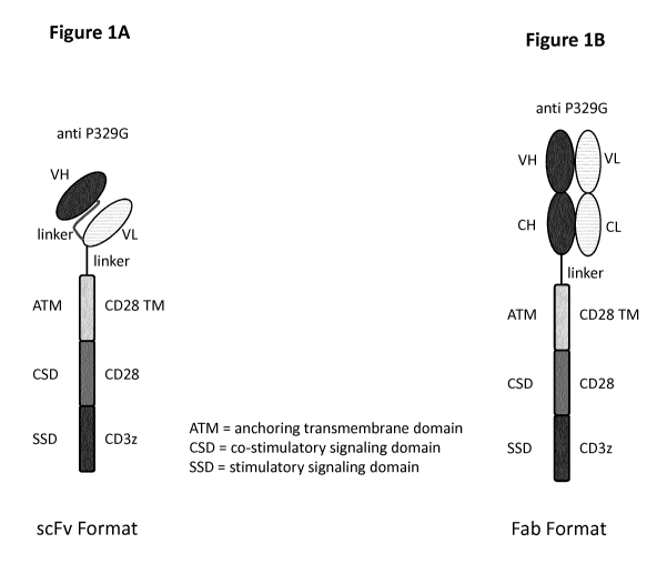
Figure 1 depicts the architecture of exemplary antigen binding receptors
according to the
invention. Figure lA shows the architecture of the anti-P329G-scFv-CD28ATD-
CD28CSD-
CD3zSSD format and anti-P329G-ds-scFv-CD28ATD-CD28CSD-CD3zSSD format.
Depicted is the extracellular domain comprising an antigen binding moiety
capable of specific
binding to a mutated Fc domain comprising the P329G mutation. The antigen
binding moiety
consists of a variable heavy and a variable light chain. Both are connected by
a (Gly4Ser)4
linker. Attached to the variable light chain, a Gly4Ser linker connects the
antigen recognition
domain with the CD28 transmembrane domain (TM) which is fused to the
intracellular co-
stimulatory signaling domain (CSD) of CD28 which in turn is fused to the
stimulatory
signaling domain (SSD) of CD3z. Figure 1B shows the architecture of the anti-
P329G-Fab-
CD28ATD-CD28CSD-CD3zSSD and anti-P329G-ds-Fab-CD28ATD-CD28CSD-CD3zSSD
format. Depicted is the extracellular domain comprising an antigen binding
moiety capable of
specific binding to a mutated Fc domain comprising the P329G mutation. The
antigen binding
moiety consists of an Ig heavy chain and an Ig light chain. Attached to the
heavy chain, a
Gly4Ser linker connects the antigen recognition domain with the CD28
transmembrane
domain which is fused to the intracellular co-stimulatory signaling domain of
CD28 which in
turn is fused to the stimulatory signaling domain of CD3z.
Figure 2 depicts a schematic representation illustrating the modular
composition of exemplary
expression constructs encoding antigen binding receptors of the invention.
Figure 2A depicts
a P392G-targeted scFv format. Figure 2B depicts a P392G-targeted Fab format.
Figure 3 depicts an exemplary IgG1 molecule harboring the P329G mutation in
the Fc domain
which is recognized by an anti-P329G antigen binding receptor of the
invention.
Figure 4 depicts a schematic representation of a tumor associated antigen
(TAA) bound IgG
harboring the P329G mutation. This antibody can in turn be recognized by an
anti-P329G
antigen binding receptor expressing T cell, whereby the T cell gets activated.
Figure 5 shows a schematic representation of a Jurkat NFAT T cell reporter
assay. TAA
bound IgG harboring the P329G mutation can be recognized by the anti-P329G
antigen
binding receptor expressing Jurkat NFAT T cell. This recognition leads to the
activation of
the cell which can be detected by measuring luminescence (cps).
11
CA 03056837 2019-09-17
WO 2018/177966 PCT/EP2018/057566
Figure 6 depicts the Jurkat NFAT T cell reporter assay using CD20 expressing
SUDHDL4
tumor cells as target cells. An anti-CD20 IgG antibody (GA101) harboring the
P329G
mutation was used, which on one hand recognizes the tumor associated antigen
and on the
other hand is recognized by Jurkat NFAT T cells expressing antigen binding
receptors
according to the invention. In Figure 6A a sorted pool of anti-P329G-ds-Fab-
CD28ATD-
CD28CSD-CD3zSSD expressing Jurkat NFAT T cells was used as as effector cells.
In Figure
6B a sorted pool of anti-P329G-ds-scFv-CD28ATD-CD28CSD-CD3zSSD expressing
Jurkat
NFAT T cells was used as effector cells.
Figure 7 depicts the Jurkat NFAT T cell reporter assay using CD20 tumor cells
as target cells.
An anti-CD20 IgG antibody (GA101) harboring the P329G mutation was used which
recognizes the tumor associated antigen and is recognized by the Jurkat NFAT T
cells
expressing antigen binding receptors according to the invention. In Figure 7A
the single clone
of anti-P329G-ds-Fab-CD28ATD-CD28CSD-CD3zSSD expressing Jurkat NFAT T cells
were used as effector cells and WSUDLCL2 cells as tumor cells. In Figure 7B
the single
clone 2 of anti-P329G-ds-Fab-CD28ATD-CD28CSD-CD3zSSD expressing Jurkat NFAT T
cells were used as effector cells and WSUDLCL2 cells as tumor cells. In Figure
7C the single
clone 5 of anti-P329G-ds-Fab-CD28ATD-CD28CSD-CD3zSSD expressing Jurkat NFAT T
cells were used as effector cells and SUDHL4 cells as tumor cells. In Figure
7D the single
clone 2 of anti-P329G-ds-Fab-CD28ATD-CD28CSD-CD3zSSD expressing Jurkat NFAT T
cells were used as effector cells and SUDHL4 as tumor cells.
Figure 8 depicts the Jurkat NFAT T cell reporter assay performed using
adherent FAP
expressing NIH/3T3-huFAP cl 19 tumor cells as target cells. The anti-FAP IgG
antibody
clone 4B9 harboring the P329G mutation was used which the tumor associated
antigen and is
recognized by the Jurkat NFAT T cells expressing antigen binding receptors
according to the
invention. IgG DP47/vk3 harboring P329G mutation was included as isotype
control. In
Figure 8A a sorted pool of anti-P329G-ds-Fab-CD28ATD-CD28CSD-CD3zSSD
expressing
Jurkat NFAT T cells was used as effector cells. In Figure 8B a sorted pool of
anti-P329G-ds-
scFv-CD28ATD-CD28CSD-CD3zSSD expressing Jurkat NFAT T cells was used as
effector
cells. In Figure 8C a sorted pool of anti-P329G-ds-Fab-CD28ATD-CD28CSD-CD3zSSD
expressing Jurkat NFAT T cells was used as effector cells. In Figure 8D a
sorted pool of anti-
12
CA 03056837 2019-09-17
WO 2018/177966 PCT/EP2018/057566
P329G-ds-scFv-CD28ATD-CD28CSD-CD3zSSD expressing Jurkat NFAT T cells was used
as effector cells
Figure 9 depicts the Jurkat NFAT T cell reporter assay using adherent CEA
expressing
MKN45 tumor cells as target cells. Either the anti-CEA IgG clone A5B7 or the
anti-CEA IgG
clone T84 LCHA both harboring the P329G mutation were used which recognize the
tumor
associated antigen and are recognized by the Jurkat NFAT T cells expressing
antigen binding
receptors according to the invention. Further IgG DP47/vk3 harboring the P329G
mutation
was included as isotype control. In Figure 9A and in Figure 9B a sorted pool
of anti-P329G-
ds-Fab-CD28ATD-CD28CSD-CD3zSSD expressing NFAT T cells was used as effector
cells.
In Figure 9C and in Figure 9D a sorted pool of anti-P329G-ds-scFv-CD28ATD-
CD28CSD-
CD3zSSD expressing NFAT T cells was used as effector cells.
Figure 10 depicts the Jurkat NFAT T cell reporter assay using adherent CEA
expressing
MKN45 tumor cells as target cells. Either the anti-CEA clone CH1A1A 98 99 or
the anti-
CEA IgG clone hMN14 IgG both harboring the P329G mutation were used which
recognize
the tumor associated antigen and are recognized by the Jurkat NFAT T cells
expressing
antigen binding receptors according to the invention. Further IgG DP47/vk3
harboring P329G
mutation was included as isotype control. In Figure 10A and in Figure 10B a
sorted pool of
anti-P329G-ds-scFv-CD28ATD-CD28CSD-CD3zSSD expressing NFAT T cells was used as
effector cells. In Figure 10C and in Figure 10D a sorted pool of anti-P329G-ds-
Fab-
CD28ATD-CD28CSD-CD3zSSD expressing NFAT T cells was used as effector cells.
Figure 11 depicts the Jurkat NFAT T cell reporter assay using adherent TNC
expressing
CT26TNC cl 19 tumor cells as target cells. The anti-TNC IgG clone A2B10
harboring the
P329G mutation was used as IgG antibody which recognizes the tumor associated
antigen and
is recognized by the Jurkat NFAT T cells expressing antigen binding receptors
according to
the invention. Further IgG DP47/vk3 harboring P329G mutation was included as
isotype
control. In Figure 11A and in Figure 11B a sorted pool of anti-P329G-ds-scFv-
CD28ATD-
CD28CSD-CD3zSSD expressing NFAT T cells was used as effector cells. In Figure
11C and
in Figure 11D a sorted pool of anti-P329G-ds-Fab-CD28ATD-CD28CSD-CD3zSSD
expressing NFAT T cells was used as effector cells
13
CA 03056837 2019-09-17
WO 2018/177966 PCT/EP2018/057566
Figure 12A and Figure 12B depict the Jurkat NFAT T cell reporter assay using
adherent TNC
expressing CT26TNC cl 19 tumor cells as target cells. The anti-TNC IgG clone
A2B10
harboring the P329G mutation was used which recognizes the tumor associated
antigen and is
recognized by the Jurkat NFAT T cells expressing antigen binding receptors
according to the
invention. Further IgG DP47/vk3 harboring P329G mutation was included as
isotype control.
A sorted pool of anti-P329G-Fab-CD28ATD-CD28CSD-CD3zSSD expressing Jurkat NFAT
T cells was used as effector cells.
Figure 13 depicts depicts the Jurkat NFAT T cell reporter assay using CD20
tumor cells as
target cells. Either an anti-CD20 IgG antibody (GA101) harboring the P329G and
the LALA
mutation mutation, a P329G and D265A mutation, the LALA mutation alone or no
mutation
at all were used in order to detect the tumor associated antigen and is
recognized by the Jurkat
NFAT T cells expressing antigen binding receptors according to the invention.
In Figure 13A
the pool of cells of anti-P329G-ds-scFv-CD28ATD-CD28CSD-CD3zSSD expressing
Jurkat
NFAT T cells were used as effector cells and SUDHL4 cells as tumor cells. In
Figure 13B the
pool of cells of anti-P329G-ds-Fab-CD28ATD-CD28CSD-CD3zSSD expressing Jurkat
NFAT T cells were used as effector cells and SUDHL4 cells as tumor cells.
Figure 14 depicts the Jurkat NFAT T cell reporter assay using CD20 tumor cells
as target
cells. Either an anti-CD20 IgG antibody (GA101) harboring the P329G and the
LALA
mutation mutation, a P329G mutation alone, the LALA mutation alone or no
mutation at all
were used in order to detect the tumor associated antigen and is recognized by
the Jurkat
NFAT T cells expressing antigen binding receptors according to the invention.
In Figure 14A
the pool of cells of anti-P329G-ds-scFv-CD28ATD-CD28CSD-CD3zSSD expressing
Jurkat
NFAT T cells were used as effector cells and SUDHL4 cells as tumor cells. In
Figure 14B the
pool of cells of anti-P329G-ds-Fab-CD28ATD-CD28CSD-CD3zSSD expressing Jurkat
NFAT T cells were used as effector cells and SUDHL4 cells as tumor cells.
14
CA 03056837 2019-09-17
WO 2018/177966 PCT/EP2018/057566
DETAILED DESCRIPTION
Definitions
Terms are used herein as generally used in the art, unless otherwise defined
in the following.
An "activating Fc receptor" is an Fc receptor that following engagement by an
Fc domain of
an antibody elicits signaling events that stimulate the receptor-bearing cell
to perform effector
functions. Human activating Fc receptors include FcyRIIIa (CD16a), FcyRI
(CD64), FcyRIIa
(CD32), and FcaRI (CD89).
Antibody-dependent cell-mediated cytotoxicity ("ADCC") is an immune mechanism
leading
to the lysis of antibody-coated target cells by immune effector cells. The
target cells are cells
to which antibodies or derivatives thereof comprising an Fc region
specifically bind, generally
via the protein part that is N-terminal to the Fc region. As used herein, the
term "reduced
ADCC" is defined as either a reduction in the number of target cells that are
lysed in a given
time, at a given concentration of antibody in the medium surrounding the
target cells, by the
mechanism of ADCC defined above, and/or an increase in the concentration of
antibody in
the medium surrounding the target cells, required to achieve the lysis of a
given number of
target cells in a given time, by the mechanism of ADCC. The reduction in ADCC
is relative to
the ADCC mediated by the same antibody produced by the same type of host
cells, using the
same standard production, purification, formulation and storage methods (which
are known to
those skilled in the art), but that has not been mutated. For example the
reduction in ADCC
mediated by an antibody comprising in its Fc domain an amino acid mutation
that reduces
ADCC, is relative to the ADCC mediated by the same antibody without this amino
acid
mutation in the Fc domain. Suitable assays to measure ADCC are well known in
the art (see
e.g., PCT publication no. WO 2006/082515 or PCT publication no. WO
2012/130831).
An "effective amount" of an agent (e.g., an antibody) refers to the amount
that is necessary to
result in a physiological change in the cell or tissue to which it is
administered.
"Affinity" refers to the strength of the sum total of non-covalent
interactions between a single
binding site of a molecule (e.g., a receptor) and its binding partner (e.g., a
ligand). Unless
indicated otherwise, as used herein, "binding affinity" refers to intrinsic
binding affinity
which reflects a 1:1 interaction between members of a binding pair (e.g., an
antigen binding
moiety and an antigen and/or a receptor and its hg and). The affinity of a
molecule X for its
partner Y can generally be represented by the dissociation constant (KD),
which is the ratio of
dissociation and association rate constants (koff and icon, respectively).
Thus, equivalent
affinities may comprise different rate constants, as long as the ratio of the
rate constants
remains the same. Affinity can be measured by well-established methods known
in the art,
CA 03056837 2019-09-17
WO 2018/177966 PCT/EP2018/057566
including those described herein. A preferred method for measuring affinity is
Surface
Plasmon Resonance (SPR) and a preferred temperature for the measurement is 25
C.
The term "amino acid" refers to naturally occurring and synthetic amino acids,
as well as
amino acid analogs and amino acid mimetics that function in a manner similar
to the naturally
occurring amino acids. Naturally occurring amino acids are those encoded by
the genetic
code, as well as those amino acids that are later modified, e.g.
hydroxyproline, y-
carboxyglutamate, and 0-phosphoserine. Amino acid analogs refer to compounds
that have
the same basic chemical structure as a naturally occurring amino acid, i.e.,
an a carbon that is
bound to a hydrogen, a carboxyl group, an amino group, and an R group, e.g.,
homoserine,
norleucine, methionine sulfoxide, methionine methyl sulfonium. Such analogs
have modified
R groups (e.g., norleucine) or modified peptide backbones, but retain the same
basic chemical
structure as a naturally occurring amino acid. Amino acid mimetics refers to
chemical
compounds that have a structure that is different from the general chemical
structure of an
amino acid, but that function in a manner similar to a naturally occurring
amino acid. Amino
acids may be referred to herein by either their commonly known three letter
symbols or by the
one-letter symbols recommended by the IUPAC-IUB Biochemical Nomenclature
Commission.
The term "amino acid mutation" as used herein is meant to encompass amino acid
substitutions, deletions, insertions, and modifications. Any combination of
substitution,
deletion, insertion, and modification can be made to arrive at the final
construct, provided that
the final construct possesses the desired characteristics, e.g., reduced
binding to an Fc
receptor. Amino acid sequence deletions and insertions include amino- and/or
carboxy-
terminal deletions and insertions of amino acids. Particular amino acid
mutations are amino
acid substitutions. For the purpose of altering e.g., the binding
characteristics of an Fc region,
non-conservative amino acid substitutions, i.e. replacing one amino acid with
another amino
acid having different structural and/or chemical properties, are particularly
preferred. Amino
acid substitutions include replacement by non-naturally occurring amino acids
or by naturally
occurring amino acid derivatives of the twenty standard amino acids (e.g., 4-
hydroxyproline,
3-methylhistidine, ornithine, homoserine, 5-hydroxylysine). Amino acid
mutations can be
generated using genetic or chemical methods well known in the art. Genetic
methods may
include site-directed mutagenesis, PCR, gene synthesis and the like. It is
contemplated that
methods of altering the side chain group of an amino acid by methods other
than genetic
engineering, such as chemical modification, may also be useful. Various
designations may be
used herein to indicate the same amino acid mutation. For example, a
substitution from
16
CA 03056837 2019-09-17
WO 2018/177966 PCT/EP2018/057566
proline at position 329 of the Fc domain to glycine can be indicated as 329G,
G329, G329/
P329G, or Pro329Gly.
The term "antibody" herein is used in the broadest sense and encompasses
various antibody
structures, including but not limited to monoclonal antibodies, polyclonal
antibodies, and
antibody fragments so long as they exhibit the desired antigen-binding
activity. Accordingly,
in context of the present invention, the term antibody relates to full
immunoglobulin
molecules as well as to parts of such immunoglobulin molecules. Furthermore,
the term
relates, as discussed herein, to modified and/or altered antibody molecules,
in particular to
mutated antibody molecules. The term also relates to recombinantly or
synthetically
generated/synthesized antibodies. In the context of the present invention the
term antibody is
used interchangeably with the term immunoglobulin.
An "antibody fragment" refers to a molecule other than an intact antibody that
comprises a
portion of an intact antibody that binds the antigen to which the intact
antibody binds.
Examples of antibody fragments include but are not limited to Fv, Fab, Fab',
Fab'-SH, F(aN)2,
diabodies, linear antibodies, single-chain antibody molecules (e.g., scFv),
and single-domain
antibodies. For a review of certain antibody fragments, see Hudson et al., Nat
Med 9, 129-134
(2003). For a review of scFy fragments, see e.g., Pliickthun, in The
Pharmacology of
Monoclonal Antibodies, vol. 113, Rosenburg and Moore eds., Springer-Verlag,
New York,
pp. 269-315 (1994); see also WO 93/16185; and U.S. Patent Nos. 5,571,894 and
5,587,458.
Diabodies are antibody fragments with two antigen-binding sites that may be
bivalent or
bispecific. See, for example, EP 404,097; WO 1993/01161; Hudson et al., Nat
Med 9, 129-
134 (2003); and Hollinger et al., Proc Natl Acad Sci USA 90, 6444-6448 (1993).
Triabodies
and tetrabodies are also described in Hudson et al., Nat Med 9, 129-134
(2003). Single-
domain antibodies are antibody fragments comprising all or a portion of the
heavy chain
variable domain or all or a portion of the light chain variable domain of an
antibody
(Domantis, Inc., Waltham, MA; see e.g., U.S. Patent No. 6,248,516 B1).
Antibody fragments
can be made by various techniques, including but not limited to proteolytic
digestion of an
intact antibody as well as production by recombinant host cells (e.g., E. coli
or phage), as
described herein.
As used herein, the term "antigen binding molecule" refers in its broadest
sense to a molecule
that specifically binds an antigenic determinant. Examples of antigen binding
molecules are
immunoglobulins and derivatives, e.g., fragments, thereof as well as antigen
binding receptors
and derivatives thereof.
17
CA 03056837 2019-09-17
WO 2018/177966 PCT/EP2018/057566
As used herein, the term "antigen binding moiety" refers to a polypeptide
molecule that
specifically binds to an antigenic determinant. In one embodiment, an antigen
binding moiety
is able to direct the entity to which it is attached (e.g., an immunoglobulin
or an antigen
binding receptor) to a target site, for example to a specific type of tumor
cell or tumor stroma
bearing the antigenic determinant or to an immunoglobulin binding to the
antigenic
determinant on a tumor cell. In another embodiment an antigen binding moiety
is able to
activate signaling through its target antigen, for example signaling is
activated upon binding
of an antigenic determinant to an antigen binding receptor on a T cell. In the
context of the
present invention, antigen binding moieties may be included in antibodies and
fragments
thereof as well as in antigen binding receptors and fragments thereof as
further defined herein.
Antigen binding moieties include an antigen binding domain, comprising an
immunoglobulin
heavy chain variable region and an immunoglobulin light chain variable region.
In certain
embodiments, the antigen binding moieties may comprise immunoglobulin constant
regions
as further defined herein and known in the art. Useful heavy chain constant
regions include
any of the five isotypes: a, 6, 8, y, or IA. Useful light chain constant
regions include any of the
two isotypes: lc and X.
In the context of the present invention the term "antigen binding receptor"
relates to an
antigen binding molecule comprising an anchoring transmembrane domain and an
extracellular domain comprising at least one antigen binding moiety. An
antigen binding
receptor can be made of polypeptide parts from different sources. Accordingly,
it may be also
understood as a "fusion protein" and/or a "chimeric protein". Usually, fusion
proteins are
proteins created through the joining of two or more genes (or preferably
cDNAs) that
originally coded for separate proteins. Translation of this fusion gene (or
fusion cDNA)
results in a single polypeptide, preferably with functional properties derived
from each of the
original proteins. Recombinant fusion proteins are created artificially by
recombinant DNA
technology for use in biological research or therapeutics. Further details to
the antigen binding
receptors of the present invention are described herein below. In the context
of the present
invention a CAR (chimeric antigen receptor) is understood to be an antigen
binding receptor
comprising an extracellular portion comprising an antigen binding moiety fused
by a spacer
sequence to an anchoring transmembrane domain which is itself fused to the
intracellular
signaling domains of CD3z and CD28.
An "antigen binding site" refers to the site, i.e. one or more amino acid
residues, of an antigen
binding molecule which provides interaction with the antigen. For example, the
antigen
binding site of an antibody or an antigen binding receptor comprises amino
acid residues from
18
CA 03056837 2019-09-17
WO 2018/177966 PCT/EP2018/057566
the complementarity determining regions (CDRs). A native immunoglobulin
molecule
typically has two antigen binding sites, a Fab or a scFv molecule typically
has a single antigen
binding site.
The term "antigen binding domain" refers to the part of an antibody or an
antigen binding
receptor that comprises the area which specifically binds to and is
complementary to part or
all of an antigen. An antigen binding domain may be provided by, for example,
one or more
immunoglobuling variable domains (also called variable regions). Particularly,
an antigen
binding domain comprises an immunoglobulin light chain variable region (VL)
and an
immunoglobulin heavy chain variable region (VH).
The term "variable region" or "variable domain" refers to the domain of an
immunoglobulin
heavy or light chain that is involved in binding the antigen. The variable
domains of the heavy
chain and light chain (VH and VL, respectively) of a native antibody generally
have similar
structures, with each domain comprising four conserved framework regions (FRs)
and three
hypervariable regions (HVRs). See, e.g., Kindt et al., Kuby Immunology, 6th
ed., W.H.
Freeman and Co, page 91 (2007). A single VH or VL domain is usually sufficient
to confer
antigen-binding specificity.
The term "ATD" as used herein refers to "anchoring transmembrane domain" which
defines a
polypeptide stretch capable of integrating in (the) cellular membrane(s) of a
cell. The ATM
can be fused to further extracellular and/or intracellular polypeptide domains
wherein these
extracellular and/or intracellular polypeptide domains will be confined to the
cell membrane
as well. In the context of the antigen binding receptors of the present
invention the ATM
confers membrane attachment and confinement of the antigen binding receptor of
the present
invention. The antigen binding receptors of the present invention comprise at
least one ATM
and an extracellular domain comprising an antigen binding moiety.
Additionally, the ATM
may be fused to further intracellular signaling domains.
The term "binding to" as used in the context of the antigen binding receptors
of the present
invention defines a binding (interaction) of an "antigen-interaction-site" and
an antigen with
each other. The term "antigen-interaction-site" defines, in accordance with
antigen binding
receptors of the present invention, a motif of a polypeptide which shows the
capacity of
specific interaction with a specific antigen or a specific group of antigens
(i.e. mutated Fc
domains). Said binding/interaction is also understood to define a "specific
recognition". The
term "specifically recognizing" means in accordance with this invention that
the antigen
binding receptor is capable of specifically interacting with and/or binding to
a modified
molecule as defined herein whereas the non-modified molecule is not
recognized. The antigen
19
CA 03056837 2019-09-17
WO 2018/177966 PCT/EP2018/057566
binding moiety of an antigen binding receptor can recognize, interact and/or
bind to different
epitopes on the same molecule. This term relates to the specificity of the
antigen binding
receptor, i.e., to its ability to discriminate between the specific regions of
a modified
molecule, i.e. a mutated Fc domain, as defined herein. The specific
interaction of the antigen-
interaction-site with its specific antigen may result in an initiation of a
signal, e.g. due to the
induction of a change of the conformation of the polypeptide comprising the
antigen, an
oligomerization of the polypeptide comprising the antigen, an oligomerization
of the antigen
binding receptor, etc. Thus, a specific motif in the amino acid sequence of
the antigen-
interaction-site and the antigen bind to each other as a result of their
primary, secondary or
tertiary structure as well as the result of secondary modifications of said
structure.
Accordingly, the term binding to does not only relate to a linear epitope but
may also relate to
a conformational epitope, a structural epitope or a discontinuous epitope
consisting of two
regions of the target molecules or parts thereof. In the context of this
invention, a
conformational epitope is defined by two or more discrete amino acid sequences
separated in
the primary sequence which comes together on the surface of the molecule when
the
polypeptide folds to the native protein (Sela, Science 166 (1969), 1365 and
Laver, Cell 61
(1990), 553-536). Moreover, the term "binding to" is interchangeably used in
the context of
the present invention with the term "interacting with". The ability of the
antigen binding
moiety (e.g. a Fab or scFv domain) of an antigen binding receptor or an
antibody to bind to a
specific target antigenic determinant can be measured either through an enzyme-
linked
immunosorbent assay (ELISA) or other techniques familiar to one of skill in
the art, e.g.,
surface plasmon resonance (SPR) technique (analyzed on a BIAcore instrument)
(Liljeblad et
al., Glyco J 17, 323-329 (2000)), and traditional binding assays (Heeley,
Endocr Res 28, 217-
229 (2002)). In one embodiment, the extent of binding of a antigen binding
moiety to an
unrelated protein is less than about 10% of the binding of the antigen binding
moiety to the
target antigen as measured, in particular by SPR. In certain embodiments, an
antigen binding
moiety that binds to the target antigen, has a dissociation constant (KD) of <
li.tM, < 100 nM,
<10 nM, < 1 nM, < 0.1 nM, < 0.01 nM, or < 0.001 nM (e.g., 10-8M or less, e.g.,
from 10-8M
to 10-13 M, e.g., from 10-9 M to 10-13 M). The term "specific binding" as used
in accordance
with the present invention means that the molecules of the invention do not or
do not
essentially cross-react with (poly-) peptides of similar structures, i.e. with
a non-mutated
parent Fc domain wherein an antigen binding receptor of the invention is
capable of specific
binding to a mutated Fc domain. Accordingly, the antigen binding receptor of
the invention
specifically binds to/interacts with a mutated Fc domain. Cross-reactivity of
a panel of
CA 03056837 2019-09-17
WO 2018/177966 PCT/EP2018/057566
constructs under investigation may be tested, for example, by assessing
binding of a panel of
antigen binding moieties under conventional conditions (see, e.g., Harlow and
Lane,
Antibodies: A Laboratory Manual, Cold Spring Harbor Laboratory Press, (1988)
and Using
Antibodies: A Laboratory Manual, Cold Spring Harbor Laboratory Press, (1999))
to the
mutated Fc domain of interest as well as to parent non-mutated Fc domain. Only
those
constructs (i.e. Fab fragments, scFvs and the like) that bind to the mutated
Fc domain of
interest but do not or do not essentially bind to a non-mutated parent Fc
domain are
considered specific for the mutated Fc domain of interest and selected for
further studies in
accordance with the method provided herein. These methods may comprise, inter
alia, binding
studies, blocking and competition studies with structurally and/or
functionally closely related
Fc domains. The binding studies also comprise FACS analysis, surface plasmon
resonance
(SPR, e.g. with BIAcore ), analytical ultracentrifugation, isothermal
titration calorimetry,
fluorescence anisotropy, fluorescence spectroscopy or by radiolabeled ligand
binding assays.
The term "CDR" as employed herein relates to "complementary determining
region", which
is well known in the art. The CDRs are parts of immunoglobulins or antigen
binding receptors
that determine the specificity of said molecules and make contact with a
specific ligand. The
CDRs are the most variable part of the molecule and contribute to the antigen
binding
diversity of these molecules. There are three CDR regions CDR1, CDR2 and CDR3
in each V
domain. CDR-H depicts a CDR region of a variable heavy chain and CDR-L relates
to a CDR
region of a variable light chain. VH means the variable heavy chain and VL
means the
variable light chain. The CDR regions of an Ig-derived region may be
determined as
described in "Kabat" (Sequences of Proteins of Immunological Interest", 5th
edit. NM
Publication no. 91-3242 U.S. Department of Health and Human Services (1991);
Chothia J.
Mol. Biol. 196 (1987), 901-917) or "Chothia" (Nature 342 (1989), 877-883).
The term " CD3z" refers to T-cell surface glycoprotein CD3 zeta chain, also
known as "T-cell
receptor T3 zeta chain" and "CD247".
The term "chimeric antigen receptor" or "chimeric receptor" or "CAR" refers to
an antigen
binding receptor constituted of an extracellular portion of an antigen binding
moiety (e.g. a
single chain antibody domain) fused by a spacer sequence to the intracellular
signaling
domains of CD3z and CD28. The invention additionally provides antigen binding
receptors
wherein the antigen binding moiety is a Fab or a crossFab fragment. The term
"CAR" is
understood in its broadest form to comprise antigen binding receptors
constituted of an
extracellular portion comprising an antigen binding moiety fused to CD3z and
fragment
thereof and to CD28 and fragments thereof, optionally through one or several
peptide linkers.
21
CA 03056837 2019-09-17
WO 2018/177966 PCT/EP2018/057566
The "class" of an antibody or immunoglobulin refers to the type of constant
domain or
constant region possessed by its heavy chain. There are five major classes of
antibodies: IgA,
IgD, IgE, IgG, and IgM, and several of these may be further divided into
subclasses
(isotypes), e.g., IgGi, IgG2, IgG3, IgG4, IgAi, and IgA2. The heavy chain
constant domains
that correspond to the different classes of immunoglobulins are called a, 6,
8, y, and IA,
respectively.
By a "crossover Fab molecule" (also termed "crossFab" or "crossover Fab
fragment") is
meant a Fab molecule wherein either the variable regions or the constant
regions of the Fab
heavy and light chain are exchanged, i.e. the crossFab fragment comprises a
peptide chain
composed of the light chain variable region and the heavy chain constant
region, and a
peptide chain composed of the heavy chain variable region and the light chain
constant
region. For clarity, in a crossFab fragment wherein the variable regions of
the Fab light chain
and the Fab heavy chain are exchanged, the peptide chain comprising the heavy
chain
constant region is referred to herein as the heavy chain of the crossover Fab
molecule.
Conversely, in a crossFab fragment wherein the constant regions of the Fab
light chain and
the Fab heavy chain are exchanged, the peptide chain comprising the heavy
chain variable
region is referred to herein as the heavy chain of the crossFab fragment.
Accordingly, a
crossFab fragment comprises a heavy or light chain composed of the heavy chain
variable and
the light chain constant regions (VH-CL), and a heavy or light chain composed
of the light
chain variable and the heavy chain constant regions (VL-CH1). In contrast
thereto, by a
"conventional Fab" molecule is meant a Fab molecule in its natural format,
i.e. comprising a
heavy chain composed of the heavy chain variable and constant regions (VH-
CH1), and a
light chain composed of the light chain variable and constant regions (VL-CL).
The term "CSD" as used herein refers to co-stimulatory signaling domain.
The term "effector functions" refers to those biological activities
attributable to the Fc region
of an antibody, which vary with the antibody isotype. Examples of antibody
effector functions
include: Clq binding and complement dependent cytotoxicity (CDC), Fc receptor
binding,
antibody-dependent cell-mediated cytotoxicity (ADCC), antibody-dependent
cellular
phagocytosis (ADCP), cytokine secretion, immune complex-mediated antigen
uptake by
antigen presenting cells, down regulation of cell surface receptors (e.g., B
cell receptor), and
B cell activation.
As used herein, the terms "engineer", "engineered", "engineering", are
considered to include
any manipulation of the peptide backbone or the post-translational
modifications of a
naturally occurring or recombinant polypeptide or fragment thereof.
Engineering includes
22
CA 03056837 2019-09-17
WO 2018/177966 PCT/EP2018/057566
modifications of the amino acid sequence, of the glycosylation pattern, or of
the side chain
group of individual amino acids, as well as combinations of these approaches.
The term "expression cassette" refers to a polynucleotide generated
recombinantly or
synthetically, with a series of specified nucleic acid elements that permit
transcription of a
particular nucleic acid in a target cell. The recombinant expression cassette
can be
incorporated into a plasmid, chromosome, mitochondrial DNA, plastid DNA,
virus, or nucleic
acid fragment. Typically, the recombinant expression cassette portion of an
expression vector
includes, among other sequences, a nucleic acid sequence to be transcribed and
a promoter. In
certain embodiments, the expression cassette of the invention comprises
polynucleotide
sequences that encode antigen binding molecules of the invention or fragments
thereof.
A "Fab molecule" refers to a protein consisting of the VH and CH1 domain of
the heavy
chain (the "Fab heavy chain") and the VL and CL domain of the light chain (the
"Fab light
chain") of an antigen binding molecule.
The term "Fe domain" or "Fe region" herein is used to define a C-terminal
region of an
immunoglobulin heavy chain that contains at least a portion of the constant
region. The term
includes native sequence Fc regions and variant Fc regions. Although the
boundaries of the Fc
region of an IgG heavy chain might vary slightly, the human IgG heavy chain Fc
region is
usually defined to extend from Cys226, or from Pro230, to the carboxyl-
terminus of the heavy
chain. However, the C-terminal lysine (Lys447) of the Fc region may or may not
be present.
Unless otherwise specified herein, numbering of amino acid residues in the Fc
region or
constant region is according to the "EU numbering" system, also called the EU
index, as
described in Kabat et al., Sequences of Proteins of Immunological Interest,
5th Ed. Public
Health Service, National Institutes of Health, Bethesda, MD, 1991. A subunit
of an Fc domain
as used herein refers to one of the two polypeptides forming the dimeric Fc
domain, i.e. a
polypeptide comprising C-terminal constant regions of an immunoglobulin heavy
chain,
capable of stable self-association. For example, a subunit of an IgG Fc domain
comprises an
IgG CH2 and an IgG CH3 constant domain.
"Framework" or "FR" refers to variable domain residues other than
hypervariable region
(HVR) residues. The FR of a variable domain generally consists of four FR
domains: FR1,
FR2, FR3, and FR4. Accordingly, the HVR and FR sequences generally appear in
the
following sequence in VH (or VL): FR1-H1 (L1 )-FR2-H2 (L2)-FR3-H3 (L3 )-FR4.
The term "full length antibody" denotes an antibody consisting of two "full
length antibody
heavy chains" and two "full length antibody light chains". A "full length
antibody heavy
chain" is a polypeptide consisting in N-terminal to C-terminal direction of an
antibody heavy
23
CA 03056837 2019-09-17
WO 2018/177966 PCT/EP2018/057566
chain variable domain (VH), an antibody constant heavy chain domain 1 (CH1),
an antibody
hinge region (HR), an antibody heavy chain constant domain 2 (CH2), and an
antibody heavy
chain constant domain 3 (CH3), abbreviated as VH-CH1-HR-CH2-CH3; and
optionally an
antibody heavy chain constant domain 4 (CH4) in case of an antibody of the
subclass IgE.
Preferably the "full length antibody heavy chain" is a polypeptide consisting
in N-terminal to
C-terminal direction of VH, CH1, HR, CH2 and CH3. A "full length antibody
light chain" is a
polypeptide consisting in N-terminal to C-terminal direction of an antibody
light chain
variable domain (VL), and an antibody light chain constant domain (CL),
abbreviated as VL-
CL. The antibody light chain constant domain (CL) can be lc (kappa) or k
(lambda). The two
full length antibody chains are linked together via inter-polypeptide
disulfide bonds between
the CL domain and the CH1 domain and between the hinge regions of the full
length antibody
heavy chains. Examples of typical full length antibodies are natural
antibodies like IgG (e.g.
IgG 1 and IgG2), IgM, IgA, IgD, and IgE.) The full length antibodies used
according to the
invention can be from a single species e.g. human, or they can be chimerized
or humanized
antibodies. In some embodiments, the full length antibodies used according to
the invention,
i.e. a therapeutic antibody comprising a mutated Fc domain, comprise two
antigen binding
sites each formed by a pair of VH and VL, which both specifically bind to the
same antigen.
In further embodiments, the full length antibodies used according to the
invention comprise
two antigen binding sites each formed by a pair of VH and VL, wherein the two
antigen
binding sites bind to different antigens, e.g. wherein the antibodies are
bispecific. The C-
terminus of the heavy or light chain of said full length antibody denotes the
last amino acid at
the C-terminus of said heavy or light chain.
By "fused" is meant that the components (e.g., a Fab and a transmembrane
domain) are linked
by peptide bonds, either directly or via one or more peptide linkers.
The terms "host cell", "host cell line" and "host cell culture" are used
interchangeably and
refer to cells into which exogenous nucleic acid has been introduced,
including the progeny of
such cells. Host cells include "transformants" and "transformed cells" which
include the
primary transformed cell and progeny derived therefrom without regard to the
number of
passages. Progeny may not be completely identical in nucleic acid content to a
parent cell, but
may contain mutations. Mutant progeny that have the same function or
biological activity as
screened or selected for in the originally transformed cell are included
herein. A host cell is
any type of cellular system that can be used to generate an antibody used
according to the
present invention. Host cells include cultured cells, e.g., mammalian cultured
cells, such as
CHO cells, BHK cells, NSO cells, SP2/0 cells, YO myeloma cells, P3X63 mouse
myeloma
24
CA 03056837 2019-09-17
WO 2018/177966 PCT/EP2018/057566
cells, PER cells, PER.C6 cells or hybridoma cells, yeast cells, insect cells,
and plant cells, to
name only a few, but also cells comprised within a transgenic animal,
transgenic plant or
cultured plant or animal tissue.
The term "hypervariable region" or "HVR", as used herein, refers to each of
the regions of an
antibody variable domain which are hypervariable in sequence and/or form
structurally
defined loops ("hypervariable loops"). Generally, native four-chain antibodies
comprise six
HVRs; three in the VH (H1, H2, H3), and three in the VL (L1, L2, L3). HVRs
generally
comprise amino acid residues from the hypervariable loops and/or from the
complementarity
determining regions (CDRs), the latter being of highest sequence variability
and/or involved
in antigen recognition. With the exception of CDR1 in VH, CDRs generally
comprise the
amino acid residues that form the hypervariable loops. Hypervariable regions
(HVRs) are also
referred to as complementarity determining regions (CDRs), and these terms are
used herein
interchangeably in reference to portions of the variable region that form the
antigen binding
regions. This particular region has been described by Kabat et al., U.S. Dept.
of Health and
Human Services, Sequences of Proteins of Immunological Interest (1983) and by
Chothia et
al., J Mol Biol 196:901-917 (1987), where the definitions include overlapping
or subsets of
amino acid residues when compared against each other. Nevertheless,
application of either
definition to refer to a CDR of an antibody and/or an antigen binding receptor
or variants
thereof is intended to be within the scope of the term as defined and used
herein. The
appropriate amino acid residues which encompass the CDRs as defined by each of
the above
cited references are set forth below in Table 1 as a comparison. The exact
residue numbers
which encompass a particular CDR will vary depending on the sequence and size
of the CDR.
Those skilled in the art can routinely determine which residues comprise a
particular CDR
given the variable region amino acid sequence of the antibody.
TABLE 1. CDR Definitionsl
CDR Kabat Chothia AbM2
VH CDR1 31-35 26-32 26-35
VH CDR2 50-65 52-58 50-58
VH CDR3 95-102 95-102 95-102
VL CDR1 24-34 26-32 24-34
VL CDR2 50-56 50-52 50-56
VL CDR3 89-97 91-96 89-97
'Numbering of all CDR definitions in Table 1 is according to the numbering
conventions set forth by Kabat et al. (see below).
2 "AbM" with a lowercase "b" as used in Table 1 refers to the CDRs as
defined by Oxford Molecular's "AbM" antibody modeling software.
CA 03056837 2019-09-17
WO 2018/177966 PCT/EP2018/057566
Kabat et al. also defined a numbering system for variable region sequences
that is applicable
to any antibody. One of ordinary skill in the art can unambiguously assign
this system of
Kabat numbering to any variable region sequence, without reliance on any
experimental data
beyond the sequence itself. As used herein, "Kabat numbering" refers to the
numbering
system set forth by Kabat et al., U.S. Dept. of Health and Human Services,
"Sequence of
Proteins of Immunological Interest" (1983). Unless otherwise specified,
references to the
numbering of specific amino acid residue positions in an antigen binding
moiety variable
region are according to the Kabat numbering system. The polypeptide sequences
of the
sequence listing are not numbered according to the Kabat numbering system.
However, it is
well within the ordinary skill of one in the art to convert the numbering of
the sequences of
the Sequence Listing to Kabat numbering.
An "individual" or "subject" is a mammal. Mammals include, but are not limited
to,
domesticated animals (e.g., cows, sheep, cats, dogs, and horses), primates
(e.g., humans and
non-human primates such as monkeys), rabbits, and rodents (e.g., mice and
rats). Particularly,
the individual or subject is a human.
By "isolated nucleic acid" molecule or polynucleotide is intended a nucleic
acid molecule,
DNA or RNA, which has been removed from its native environment. For example, a
recombinant polynucleotide encoding a polypeptide contained in a vector is
considered
isolated for the purposes of the present invention. Further examples of an
isolated
polynucleotide include recombinant polynucleotides maintained in heterologous
host cells or
purified (partially or substantially) polynucleotides in solution. An isolated
polynucleotide
includes a polynucleotide molecule contained in cells that ordinarily contain
the
polynucleotide molecule, but the polynucleotide molecule is present
extrachromosomally or at
a chromosomal location that is different from its natural chromosomal
location. Isolated RNA
molecules include in vivo or in vitro RNA transcripts of the present
invention, as well as
positive and negative strand forms, and double-stranded forms. Isolated
polynucleotides or
nucleic acids according to the present invention further include such
molecules produced
synthetically. In addition, a polynucleotide or a nucleic acid may be or may
include a
regulatory element such as a promoter, ribosome binding site, or a
transcription terminator.
By a nucleic acid or polynucleotide having a nucleotide sequence at least, for
example, 95%
"identical" to a reference nucleotide sequence of the present invention, it is
intended that the
nucleotide sequence of the polynucleotide is identical to the reference
sequence except that
the polynucleotide sequence may include up to five point mutations per each
100 nucleotides
of the reference nucleotide sequence. In other words, to obtain a
polynucleotide having a
26
CA 03056837 2019-09-17
WO 2018/177966 PCT/EP2018/057566
nucleotide sequence at least 95% identical to a reference nucleotide sequence,
up to 5% of the
nucleotides in the reference sequence may be deleted or substituted with
another nucleotide,
or a number of nucleotides up to 5% of the total nucleotides in the reference
sequence may be
inserted into the reference sequence. These alterations of the reference
sequence may occur at
the 5' or 3' terminal positions of the reference nucleotide sequence or
anywhere between
those terminal positions, interspersed either individually among residues in
the reference
sequence or in one or more contiguous groups within the reference sequence. As
a practical
matter, whether any particular polynucleotide sequence is at least 80%, 85%,
90%, 95%, 96%,
97%, 98% or 99% identical to a nucleotide sequence of the present invention
can be
determined conventionally using known computer programs, such as the ones
discussed
below for polypeptides (e.g., ALIGN-2).
By an "isolated polypeptide" or a variant, or derivative thereof is intended a
polypeptide that
is not in its natural milieu. No particular level of purification is required.
For example, an
isolated polypeptide can be removed from its native or natural environment.
Recombinantly
produced polypeptides and proteins expressed in host cells are considered
isolated for the
purpose of the invention, as are native or recombinant polypeptides which have
been
separated, fractionated, or partially or substantially purified by any
suitable technique.
"Percent (%) amino acid sequence identity" with respect to a reference
polypeptide sequence
is defined as the percentage of amino acid residues in a candidate sequence
that are identical
with the amino acid residues in the reference polypeptide sequence, after
aligning the
sequences and introducing gaps, if necessary, to achieve the maximum percent
sequence
identity, and not considering any conservative substitutions as part of the
sequence identity.
Alignment for purposes of determining percent amino acid sequence identity can
be achieved
in various ways that are within the skill in the art, for instance, using
publicly available
computer software such as BLAST, BLAST-2, ALIGN or Megalign (DNASTAR)
software.
Those skilled in the art can determine appropriate parameters for aligning
sequences,
including any algorithms needed to achieve maximal alignment over the full
length of the
sequences being compared. For purposes herein, however, % amino acid sequence
identity
values are generated using the sequence comparison computer program ALIGN-2.
The
ALIGN-2 sequence comparison computer program was authored by Genentech, Inc.,
and the
source code has been filed with user documentation in the U.S. Copyright
Office, Washington
D.C., 20559, where it is registered under U.S. Copyright Registration No.
TXU510087. The
ALIGN-2 program is publicly available from Genentech, Inc., South San
Francisco,
California, or may be compiled from the source code. The ALIGN-2 program
should be
27
CA 03056837 2019-09-17
WO 2018/177966 PCT/EP2018/057566
compiled for use on a UNIX operating system, including digital UNIX V4.0D. All
sequence
comparison parameters are set by the ALIGN-2 program and do not vary. In
situations where
ALIGN-2 is employed for amino acid sequence comparisons, the % amino acid
sequence
identity of a given amino acid sequence A to, with, or against a given amino
acid sequence B
(which can alternatively be phrased as a given amino acid sequence A that has
or comprises a
certain % amino acid sequence identity to, with, or against a given amino acid
sequence B) is
calculated as follows:
100 times the fraction X/Y
where X is the number of amino acid residues scored as identical matches by
the sequence
alignment program ALIGN-2 in that program's alignment of A and B, and where Y
is the
total number of amino acid residues in B. It will be appreciated that where
the length of amino
acid sequence A is not equal to the length of amino acid sequence B, the %
amino acid
sequence identity of A to B will not equal the % amino acid sequence identity
of B to A.
Unless specifically stated otherwise, all % amino acid sequence identity
values used herein
are obtained as described in the immediately preceding paragraph using the
ALIGN-2
computer program.
The term "nucleic acid molecule" relates to the sequence of bases comprising
purine- and
pyrimidine bases which are comprised by polynucleotides, whereby said bases
represent the
primary structure of a nucleic acid molecule. Herein, the term nucleic acid
molecule includes
DNA, cDNA, genomic DNA, RNA, synthetic forms of DNA and mixed polymers
comprising
two or more of these molecules. In addition, the term nucleic acid molecule
includes both,
sense and antisense strands. Moreover, the herein described nucleic acid
molecule may
contain non-natural or derivatized nucleotide bases, as will be readily
appreciated by those
skilled in the art.
The term "package insert" is used to refer to instructions customarily
included in commercial
packages of therapeutic products, that contain information about the
indications, usage,
dosage, administration, combination therapy, contraindications and/or warnings
concerning
the use of such therapeutic products.
The term "pharmaceutical composition" refers to a preparation which is in such
form as to
permit the biological activity of an active ingredient contained therein to be
effective, and
which contains no additional components which are unacceptably toxic to a
subject to which
the formulation would be administered. A pharmaceutical composition usually
comprises one
or more pharmaceutically acceptable carrier(s).
28
CA 03056837 2019-09-17
WO 2018/177966 PCT/EP2018/057566
A "pharmaceutically acceptable carrier" refers to an ingredient in a
pharmaceutical
composition, other than an active ingredient, which is nontoxic to a subject.
A
pharmaceutically acceptable carrier includes, but is not limited to, a buffer,
excipient,
stabilizer, or preservative.
As used herein, term "polypeptide" refers to a molecule composed of monomers
(amino
acids) linearly linked by amide bonds (also known as peptide bonds). The term
polypeptide
refers to any chain of two or more amino acids, and does not refer to a
specific length of the
product. Thus, peptides, dipeptides, tripeptides, oligopeptides, protein,
amino acid chain, or
any other term used to refer to a chain of two or more amino acids, are
included within the
definition of polypeptide, and the term polypeptide may be used instead of, or
interchangeably
with any of these terms. The term polypeptide is also intended to refer to the
products of post-
expression modifications of the polypeptide, including without limitation
glycosylation,
acetylation, phosphorylation, amidation, derivatization by known
protecting/blocking groups,
proteolytic cleavage, or modification by non-naturally occurring amino acids.
A polypeptide
may be derived from a natural biological source or produced by recombinant
technology, but
is not necessarily translated from a designated nucleic acid sequence. It may
be generated in
any manner, including by chemical synthesis. A polypeptide of the invention
may be of a size
of about 3 or more, 5 or more, 10 or more, 20 or more, 25 or more, 50 or more,
75 or more,
100 or more, 200 or more, 500 or more, 1,000 or more, or 2,000 or more amino
acids.
Polypeptides may have a defined three-dimensional structure, although they do
not
necessarily have such structure. Polypeptides with a defined three-dimensional
structure are
referred to as folded, and polypeptides which do not possess a defined three-
dimensional
structure, but rather can adopt a large number of different conformations, and
are referred to
as unfolded.
The term "polynucleotide" refers to an isolated nucleic acid molecule or
construct, e.g.,
messenger RNA (mRNA), virally-derived RNA, or plasmid DNA (pDNA). A
polynucleotide
may comprise a conventional phosphodiester bond or a non-conventional bond
(e.g., an amide
bond, such as found in peptide nucleic acids (PNA). The term nucleic acid
molecule refers to
any one or more nucleic acid segments, e.g., DNA or RNA fragments, present in
a
polynucleotide.
"Reduced binding", for example reduced binding to an Fc receptor, refers to a
decrease in
affinity for the respective interaction, as measured for example by SPR. For
clarity the term
includes also reduction of the affinity to zero (or below the detection limit
of the analytic
29
CA 03056837 2019-09-17
WO 2018/177966 PCT/EP2018/057566
method), i.e. complete abolishment of the interaction. Conversely, "increased
binding" refers
to an increase in binding affinity for the respective interaction.
The term "regulatory sequence" refers to DNA sequences, which are necessary to
effect the
expression of coding sequences to which they are ligated. The nature of such
control
sequences differs depending upon the host organism. In prokaryotes, control
sequences
generally include promoter, ribosomal binding site, and terminators. In
eukaryotes generally
control sequences include promoters, terminators and, in some instances,
enhancers,
transactivators or transcription factors. The term "control sequence" is
intended to include, at
a minimum, all components the presence of which are necessary for expression,
and may also
include additional advantageous components.
As used herein, the term "single-chain" refers to a molecule comprising amino
acid
monomers linearly linked by peptide bonds. In certain embodiemtns, one of the
antigen
binding moieties is a scFv fragment, i.e. a VH domain and a VL domain
connected by a
peptide linker. In certain embodiments, one of the antigen binding moieties is
a single-chain
Fab molecule, i.e. a Fab molecule wherein the Fab light chain and the Fab
heavy chain are
connected by a peptide linker to form a single peptide chain. In a particular
such embodiment,
the C-terminus of the Fab light chain is connected to the N-terminus of the
Fab heavy chain in
the single-chain Fab molecule.
The term "SSD" as used herein refers to stimulatory signaling domain.
As used herein, "treatment" (and grammatical variations thereof such as
"treat" or "treating")
refers to clinical intervention in an attempt to alter the natural course of a
disease in the
individual being treated, and can be performed either for prophylaxis or
during the course of
clinical pathology. Desirable effects of treatment include, but are not
limited to, preventing
occurrence or recurrence of disease, alleviation of symptoms, diminishment of
any direct or
indirect pathological consequences of the disease, preventing metastasis,
decreasing the rate
of disease progression, amelioration or palliation of the disease state, and
remission or
improved prognosis. In some embodiments, cell expressing antigen binding
receptors of the
invention are used together with therapeutic antibodies comprising a mutated
Fc domain to
delay development of a disease or to slow the progression of a disease.
As used herein, the term "target antigenic determinant" is synonymous with
"target antigen",
"target epitope" and "target cell antigen" and refers to a site (e.g., a
contiguous stretch of
amino acids or a conformational configuration made up of different regions of
non-contiguous
amino acids) on a polypeptide macromolecule to which an antibody binds,
forming an antigen
binding moiety-antigen complex. Useful antigenic determinants can be found,
for example, on
CA 03056837 2019-09-17
WO 2018/177966 PCT/EP2018/057566
the surfaces of tumor cells, on the surfaces of virus-infected cells, on the
surfaces of other
diseased cells, on the surface of immune cells, free in blood serum, and/or in
the extracellular
matrix (ECM). The proteins referred to as antigens herein (e.g., CD20, CEA,
FAP, TNC) can
be any native form of the proteins from any vertebrate source, including
mammals such as
primates (e.g., humans) and rodents (e.g., mice and rats), unless otherwise
indicated. In a
particular embodiment the target antigen is a human protein. Where reference
is made to a
specific target protein herein, the term encompasses the "full-length",
unprocessed target
protein as well as any form of the target protein that results from processing
in the target cell.
The term also encompasses naturally occurring variants of the target protein,
e.g., splice
variants or allelic variants. Exemplary human target proteins useful as
antigens include, but
are not limited to: CD20, CEA, FAP, TNC, MSLN, Fo1R1, HER1 and HER2. The
ability of
an antibody to bind to a specific target antigenic determinant can be measured
either through
an enzyme-linked immunosorbent assay (ELISA) or other techniques familiar to
one of skill
in the art, e.g., surface plasmon resonance (SPR) technique (analyzed on a
BIAcore
instrument) (Liljeblad et al., Glyco J 17, 323-329 (2000)), and traditional
binding assays
(Heeley, Endocr Res 28, 217-229 (2002)). In one embodiment, the extent of
binding of the
antibody to an unrelated protein is less than about 10% of the binding of the
antibody to the
target antigen as measured, e.g., by SPR. In certain embodiments, the antibody
binds to the
target antigen with an affinity dissociation constant (KD) of < 1 1AM, < 100
nM, < 10 nM, < 1
nM, < 0.1 nM, < 0.01 nM, or < 0.001 nM (e.g., 10-8 M or less, e.g., from 10-8
M to 10-13 M,
e.g., from 10-9M to 10-13 M).
"Antibodies comprising a mutated Fc domain" according to the present
invention, i.e.
therapeutic antibodies may have one, two, three or more binding domains and
may be
monospecific, bispecific or multispecific. The antibodies can be full length
from a single
species, or be chimerized or humanized. For an antibody with more than two
antigen binding
domains, some binding domains may be identical and/or have the same
specificity.
"T cell activation" as used herein refers to one or more cellular response of
a T lymphocyte,
particularly a cytotoxic T lymphocyte, selected from: proliferation,
differentiation, cytokine
secretion, cytotoxic effector molecule release, cytotoxic activity, and
expression of activation
markers. The antigen binding receptors of the invention are capable of
inducing T cell
activation. Suitable assays to measure T cell activation are known in the art
described herein.
In accordance with this invention, the term "T cell receptor" or "TCR" is
commonly known in
the art. In particular, herein the term "T cell receptor" refers to any T cell
receptor, provided
that the following three criteria are fulfilled: (i) tumor specificity, (ii)
recognition of (most)
31
CA 03056837 2019-09-17
WO 2018/177966 PCT/EP2018/057566
tumor cells, which means that an antigen or target should be expressed in
(most) tumor cells
and (iii) that the TCR matches to the HLA-type of the subjected to be treated.
In this context,
suitable T cell receptors which fulfill the above mentioned three criteria are
known in the art
such as receptors recognizing NY-ESO-1 (for sequence information(s) see, e.g.,
PCT/GB2005/001924) and/or HER2neu (for sequence information(s) see WO-Al
2011/0280894).
A "therapeutically effective amount" of an agent, e.g., a pharmaceutical
composition, refers to
an amount effective, at dosages and for periods of time necessary, to achieve
the desired
therapeutic or prophylactic result. A therapeutically effective amount of an
agent for example
eliminates, decreases, delays, minimizes or prevents adverse effects of a
disease.
The term "vector" or "expression vector" is synonymous with "expression
construct" and
refers to a DNA molecule that is used to introduce and direct the expression
of a specific gene
to which it is operably associated in a target cell. The term includes the
vector as a self-
replicating nucleic acid structure as well as the vector incorporated into the
genome of a host
cell into which it has been introduced. The expression vector of the present
invention
comprises an expression cassette. Expression vectors allow transcription of
large amounts of
stable mRNA. Once the expression vector is inside the target cell, the
ribonucleic acid
molecule or protein that is encoded by the gene is produced by the cellular
transcription
and/or translation machinery. In one embodiment, the expression vector of the
invention
comprises an expression cassette that comprises polynucleotide sequences that
encode antigen
binding receptors of the invention or fragments thereof.
Antigen binding receptors capable of specific binding to (a) mutated Fc
domain(s)
The present invention relates to antigen binding receptors capable of specific
binding to the
mutated Fc domain of an antibody, i.e. a therapeutic antibody targeting a
cancer cell. In
particular, the present invention relates to antigen binding receptors
comprising an
extracellular domain comprising at least one antigen binding moiety capable of
specific
binding to a mutated Fc domain but not capable of specific binding to the
parent non-mutated
Fc domain. In preferred embodiments, the mutated Fc domain comprises at least
one amino
acid substitution compared to the non-mutated parent Fc domain, wherein Fc
receptor binding
by the mutated Fc domain is reduced compared to Fc receptor binding by the non-
mutated Fc
domain. In particular embodiments, the present invention relates to antigen
binding receptors
comprising an extracellular domain comprising at least one antigen binding
moiety capable of
specific binding to a mutated Fc domain, wherein the at least one antigen
binding moiety is
32
CA 03056837 2019-09-17
WO 2018/177966 PCT/EP2018/057566
not capable of specific binding to the parent non-mutated Fc domain, wherein
the mutated Fc
domain comprises at least one amino acid substitution selected from the group
consisting of
L234, L235, 1253, H310, P331, P329 and H435, in particular wherein the amino
acid
mutation is L234A, L235A, I253A, N297A, H310A, P329G and/or H435A, compared to
the
non-mutated parent Fc domain, wherein Fc receptor binding by the mutated Fc
domain is
reduced compared to Fc receptor binding by the non-mutated Fc domain. In one
preferred
embodiment, the amino acid mutation is P329G wherein binding to Fcy receptor
is reduced as
measured by SPR at 25 C. In a further preferred embodiment, the amino acid
mutations are
I253A, H310A and H435A wherein binding to the neonatal Fc receptor (FcRn) is
reduced as
measured by SPR at 25 C.
The present invention further relates to the transduction of T cells, such as
CD8+ T cells,
CD4+ T cells, CD3+ T cells, y6 T cells or natural killer (NK) T cells,
preferably CD8+ T
cells, with an antigen binding receptor as described herein and their targeted
recruitment, e.g.,
to a tumor, by an antibody molecule, e.g. a therapeutic antibody, comprising a
mutated Fc
domain. In one embodiment, the antibody is capable of specific binding to a
tumor-specific
antigen that is naturally occurring on the surface of a tumor cell.
As shown in the appended Examples, as a proof of the inventive concept, the
antigen binding
receptor comprising an anchoring transmembrane domain and an extracellular
domain
according to the invention pETR17096 (SEQ ID NO:7 as encoded by the DNA
sequence
shown in SEQ ID NO:19) was constructed which is capable of specific binding to
a
therapeutic antibody (represented by the anti-CD20 antibody comprising a heavy
chain of
SEQ ID NO ID: 112 and a light chain of SEQ ID NO:113) comprising the P329G
mutation.
Transduced T cells (Jurkat NFAT T cells) expressing the Anti-P329G-scFv-
CD28ATD-
CD28CSD-CD3zSSD protein (SEQ ID NO:7 as encoded by the DNA sequence shown in
SEQ ID NO:19) could be strongly activated by co-incubation with the anti-CD20
antibody
comprising the P329G mutation in the Fc domain together with CD20 positive
tumor cells.
The inventors further provided multiple formats of the antigen binding
receptor capable of
specific binding to a mutated Fc domain but not capable of specific binding to
the non-
mutated parent Fc domain to support the proof of the inventive concept.
The treatment of tumor cells by the combination of an antibody directed
against a tumor
antigen wherein the antibody comprises the P329G mutation together with
transduced T cells
expressing the Anti-P329G-Fab-ds-CD28ATD-CD28CSD-CD3zSSD protein (SEQ ID NOs:
44 (DNA) and 39, 41 (protein)) surprisingly leads to stronger activation of
the transduced T
cell compared to the transduced T cells expressing the Anti-P329G-scFv-CD28ATD-
33
CA 03056837 2019-09-17
WO 2018/177966 PCT/EP2018/057566
CD28CSD-CD3zSSD (SEQ ID NOs: 19 (DNA) and 7 (protein)) fusion protein.(see
e.g. Figs.
6 and 8 to 11).
Accordingly, it was surprisingly and unexpectedly found that T cells,
preferably CD8+ T
cells, that were transduced with an antigen binding receptor of the present
invention can be
specifically stimulated by the use of a tumor-specific antibody comprising a
mutated Fc
domain and recruited by the tumor-specific antibody as linking element to the
tumor cell.
Thus, it was surprisingly and unexpectedly shown in the present invention that
pairing a
tumor-specific antibody, i.e. a therapeutic antibody, comprising a mutated Fc
domain with T
cells transduced with an antigen binding receptor which comprise/consist of an
extracellular
domain comprising an antigen binding moiety capable of specific binding to the
mutated Fc
domain would result in a specific activation of the T cells and subsequent
lysis of the tumor
cell. This approach bears significant safety advantages over conventional T
cell based
approaches, as the T cell would be inert in the absence of the antibody
comprising the mutated
Fc domain and their availability may be controlled by the antibody molecule
format chosen
(i.e. smaller molecules for shorter half-life and vice-versa). Accordingly,
the invention
provides a versatile therapeutic platform wherein IgG type antibodies may be
used to mark or
label tumor cells as a guidance for T cell and wherein transduced T cells are
specifically
targeted toward the tumor cells by providing specificity for a mutated Fc
domain of the IgG
type antibody. After binding to the mutated Fc domain of the antibody on the
surface of a
tumor cell, the transduced T cell as described herein becomes activated and
the tumor cell will
subsequently be lysed. The platform is flexible and specific by allowing the
use of diverse
(existing or newly developed) target antibodies or co-application of multiple
antibodies with
different antigen specificity but comprising the same mutation in the Fc
domain. The degree
of T cell activation can further be adjusted by adjusting the dosage of the co-
applied
therapeutic antibody or by switching to different antibody specificities or
formats. Transduced
T cell according to the invention are inert without co-application of a
targeting antibody
comprising a mutated Fc domain because mutations to the Fc domain as described
herein do
not occur in natural or non-mutated immunoglobulins. Accordingly, in one
embodiment, the
mutated Fc domain does not naturally occur in wild type immunoglobulins.
Accordingly, the present invention relates to an antigen binding receptor
comprising an
extracellular domain comprising at least one antigen binding moiety capable of
specific
binding to a mutated Fc domain, wherein the at least one antigen binding
moiety is not
capable of specific binding to the parent non-mutated Fc domain, wherein the
mutated Fc
domain comprises at least one amino acid mutation compared to the non-mutated
parent Fc
34
CA 03056837 2019-09-17
WO 2018/177966 PCT/EP2018/057566
domain, wherein Fc receptor binding by the mutated Fc domain and/or effector
function
induced by the mutated Fc domain is reduced compared to Fc receptor binding
and/or effector
function induced by the non-mutated Fc domain. It may be particularly
desirable to use
therapeutic antibodies with reduced effector function in cancer therapy since
effector function
may lead to severe side effects of antibody-based tumor therapies as further
described herein.
In the context of the present invention, the antigen binding receptor
comprises an extracellular
domain that does not naturally occur in or on T cells. Thus, the antigen
binding receptor is
capable of providing tailored binding specificity to cells expressing the
antigen binding
receptor according to the invention. Cells, e.g. T cells, transduced with (an)
antigen binding
receptor(s) of the invention become capable of specific binding to a mutated
Fc domain but
not to the non-mutated parent Fc domain. Specificity is provided by the
antigen binding
moiety of the extracellular domain of the antigen binding receptor, such
antigen binding
moieties are considered to be specific for the mutated Fc domain as defined
herein. In the
context of the present invention and as explained herein, the antigen binding
moiety capable
of specific binding to a mutated Fc domain bind to/interact with the mutated
Fc domain but
not to/with the non-mutated parent Fc domain.
Antigen binding moieties
In an illustrative embodiment of the present invention, as a proof of concept,
antigen binding
receptors are provided comprising an anchoring transmembrane domain and an
extracellular
domain comprising at least one antigen binding moiety, wherein the at least
one antigen
binding moiety is capable of specific binding to a mutated Fc domain but not
capable of
specific binding to the non-mutated parent Fc domain, wherein the mutated Fc
domain
comprises at least one amino acid substitution compared to the non-mutated
parent Fc.
In certain embodiment, at least one of the antigen binding moieties is a
conventional Fab
fragment, i.e. a Fab molecule consisting of a Fab light chain and a Fab heavy
chain. In certain
embodiment, at least one of the antigen binding moieties is a crossFab
fragment, i.e. a Fab
molecule consisting of a Fab light chain and a Fab heavy chain, wherein either
the variable
regions or the constant regions of the Fab heavy and light chain are
exchanged. In certain
embodiments, at least one of the antigen binding moieties is a scFv fragment.
In a particular
such embodiment, the C-terminus of the variable heavy chain (VH) is connected
to the N-
terminus of the variable light chain (VL) in the scFv molecule, optionally
through a peptide
linker. In certain embodiments, at least one of the antigen binding moieties
is a single-chain
Fab molecule, i.e. a Fab molecule wherein the Fab light chain and the Fab
heavy chain are
CA 03056837 2019-09-17
WO 2018/177966 PCT/EP2018/057566
connected by a peptide linker to form a single peptide chain. In a particular
such embodiment,
the C-terminus of the Fab light chain is connected to the N-terminus of the
Fab heavy chain in
the single-chain Fab molecule, optionally through a peptide linker.
Accordingly, in the context of the present invention, the antigen binding
moiety is capable of
specific binding to a mutated Fc domain but not capable of specific binding to
the non-
mutated parent Fc domain, wherein the mutated Fc domain comprises at least one
amino acid
substitution compared to the non-mutated parent Fc domain.
Antigen binding moieties capable of specific binding to a mutated Fc domain
may be
generated by immunization of e.g. a mammalian immune system. Such methods are
known in
the art and e.g. are described in Burns in Methods in Molecular Biology 295:1-
12 (2005).
Alternatively, antigen binding moieties of the invention may be isolated by
screening
combinatorial libraries for antibodies with the desired activity or
activities. Methods for
screening combinatorial libraries are reviewed, e.g., in Lerner et al. in
Nature Reviews
16:498-508 (2016). For example, a variety of methods are known in the art for
generating
phage display libraries and screening such libraries for antigen binding
moieties possessing
the desired binding characteristics. Such methods are reviewed, e.g., in
Frenzel et al. in mAbs
8:1177-1194 (2016); Bazan et al. in Human Vaccines and Immunotherapeutics
8:1817-1828
(2012) and Zhao et al. in Critical Reviews in Biotechnology 36:276-289 (2016)
as well as in
Hoogenboom et al. in Methods in Molecular Biology 178:1-37 (O'Brien et al.,
ed., Human
Press, Totowa, NJ, 2001) and further described, e.g., in the McCafferty et
al., Nature 348:552-
554; Clackson et al., Nature 352: 624-628 (1991); Marks et al., J. Mol. Biol.
222: 581-597
(1992) and in Marks and Bradbury in Methods in Molecular Biology 248:161-175
(Lo, ed.,
Human Press, Totowa, NJ, 2003). ;Sidhu et al., J. Mol. Biol. 338(2): 299-310
(2004); Lee et
al., J. Mol. Biol. 340(5): 1073-1093 (2004); Fellouse, Proc. Natl. Acad. Sci.
USA 101(34):
12467-12472 (2004); and Lee et al., J. Immunol. Methods 284(1-2): 119-
132(2004). In certain
phage display methods, repertoires of VH and VL genes are separately cloned by
polymerase
chain reaction (PCR) and recombined randomly in phage libraries, which can
then be
screened for antigen-binding phage as described in Winter et al. in Annual
Review of
Immunology 12: 433-455 (1994). Phage typically display antibody fragments,
either as
single-chain Fv (scFv) fragments or as Fab fragments. Libraries from immunized
sources
provide high-affinity antigen binding moieties to the immunogen without the
requirement of
constructing hybridomas. Alternatively, the naive repertoire can be cloned
(e.g., from human)
to provide a single source of antigen binding moieties to a wide range of non-
self and also
self antigens without any immunization as described by Griffiths et al. in
EMBO Journal 12:
36
CA 03056837 2019-09-17
WO 2018/177966 PCT/EP2018/057566
725-734 (1993). Finally, naive libraries can also be made synthetically by
cloning
unrearranged V-gene segments from stem cells, and using PCR primers containing
random
sequence to encode the highly variable CDR3 regions and to accomplish
rearrangement in
vitro, as described by Hoogenboom and Winter in Journal of Molecular Biology
227: 381-388
(1992). Patent publications describing human antibody phage libraries include,
for example:
US Patent Nos. 5,750,373; 7,985,840; 7,785,903 and 8,679,490 as well as US
Patent
Publication Nos. 2005/0079574, 2007/0117126, 2007/0237764 and 2007/0292936.
and
2009/0002360. Further examples of methods known in the art for screening
combinatorial
libraries for antibodies with a desired activity or activities include
ribosome and mRNA
display, as well as methods for antibody display and selection on bacteria,
mammalian cells,
insect cells or yeast cells. Methods for yeast surface display are reviewed,
e.g., in Scholler et
al. in Methods in Molecular Biology 503:135-56 (2012) and in Cherf et al. in
Methods in
Molecular biology 1319:155-175 (2015) as well as in the Zhao et al. in Methods
in Molecular
Biology 889:73-84 (2012). Methods for ribosome display are described, e.g., in
He et al. in
Nucleic Acids Research 25:5132-5134 (1997) and in Hanes et al. in PNAS 94:4937-
4942
(1997).
In the context of the present invention, provided herein are antigen binding
receptors
comprising at least one antigen binding moiety capable of specific binding to
a mutated Fc
domain. Accordingly, transduced cells, i.e. T cells, expressing an antigen
binding receptor
according to the invention are capable of specific binding to the mutated Fc
domain of an
antibody, i.e. of a therapeutic antibody. The Fc domain confers to antibodies,
i.e. therapeutic
antibodies, favorable pharmacokinetic properties, including a long serum half-
life which
contributes to good accumulation in the target tissue and a favorable tissue-
blood distribution
ratio. At the same time it may, however, lead to undesirable targeting of
therapeutic
antibodies to cells expressing Fc receptors rather than to the preferred
antigen-bearing cells.
Moreover, the co-activation of Fc receptor signaling pathways may lead to
cytokine release
which, results in excessive activation of cytokine receptors and severe side
effects upon
systemic administration of therapeutic antibodies. Activation of (Fc receptor-
bearing) immune
cells other than T cells may even reduce efficacy of therapeutic antibodies
due to the potential
destruction of immune cells. Accordingly, therapeutic antibodies known in the
art may be
engineered or mutated to exhibit reduced binding affinity to an Fc receptor
and/or reduced
effector function, as compared to, e.g., a native IgGi Fc domain. The antigen
binding
receptors according to the invention may be used to target effector cells,
e.g. T cells,
expressing the antigen binding receptors according to the invention in vitro
and/or in vivo to
37
CA 03056837 2019-09-17
WO 2018/177966 PCT/EP2018/057566
target cells, i.e. tumor cells, which are labeled with an antibody capable of
specific binding to
the target cells, wherein the antibody comprise an engineered and/or mutated
Fc domain as
described herein.
In an illustrative embodiment of the present invention, as a proof of concept,
provided are
antigen binding receptors capable of specific binding to a mutated Fc domain
comprising the
amino acid mutation P329G and effector cells expressing said antigen binding
receptors. The
P329G mutation reduces binding to Fcy receptors and associated effector
function.
Accordingly, the mutated Fc domain comprising the P329G mutation binds to Fcy
receptors
with reduced or abolished affinity compared to the non-mutated Fc domain. In
an alternative
illustrative embodiment of the present invention, as a proof of concept
provided are antigen
binding receptors capable of specific binding to a mutated Fc domain
comprising the amino
acid mutations I253A, H310A and H435A ("AAA"). The AAA mutations essentially
abolishes binding to the FcRn.
However, antibodies with reduced with improved or diminished binding to Fc
receptors
(FcRs) and/or effector function comprising a mutated Fc domain are widely used
in the art.
Accordingly, herein provided are antigen binding receptors capable of specific
binding to
antibodies comprising a mutated Fc domain, such antibodies are herein also
referred to as
target antibodies. Accordingly, in one embodiment the antigen binding receptor
of the present
invention is capable of specific binding to a target antibody comprising a
mutated Fc domain
with reduced binding affinity to an Fc receptor and/or reduced effector
function. Target
antibodies with reduced effector function include those with mutation of one
or more of Fc
region residues 238, 265, 269, 270, 297, 327 and 329 (U.S. Patent No.
6,737,056). Such Fc
mutants include Fc mutants with mutations at two or more of amino acid
positions 265, 269,
270, 297 and 327, including the so-called "DANA" Fc mutant with mutation of
residues 265
and 297 to alanine (US Patent No. 7,332,581). Certain antibody variants with
improved or
diminished binding to FcRs are described. (See, e.g., U.S. Patent No.
6,737,056; WO
2004/056312, and Shields et al., J. Biol. Chem. 9(2): 6591-6604 (2001).) In
certain
embodiments, an antigen binding receptor is provided capable of specific
binding to an
antibody variant comprises an Fc region with one or more amino acid mutations
which
improve ADCC, e.g., mutations at positions 298, 333, and/or 334 of the Fc
region (EU
numbering of residues). In certain embodiments, a target antibody variant
comprises an Fc
region with one or more amino acid mutations, which reduce or diminish FcRn
binding, e.g.,
mutations at positions 253, and/or 310, and/or 435 of the Fc region (EU
numbering of
residues). In certain embodiments, the target antibody variant comprises an Fc
region with the
38
CA 03056837 2019-09-17
WO 2018/177966 PCT/EP2018/057566
amino acid mutations at positions 253, 310 and 435. In one embodiment the
mutations are
I253A, H310A and H435A in an Fc region derived from a human IgG1 Fc region.
See e.g.,
Grevys, A., et al., J. Immunol. 194 (2015) 5497-5508.
In certain embodiments, an antigen binding receptor is provided capable of
specific binding to
an antibody variant comprising an Fc region with one or more amino acid
mutations, which
reduced or diminished FcRn binding, e.g., mutations at one of the positions
310 and/or, 433
and/or 436 of the Fc region (EU numbering of residues). In certain
embodiments, the target
antibody variant comprises an Fc region with the amino acid mutations at
positions 310, 433
and 436. In one embodiment the mutations are H310A, H433A and Y436A in an Fc
region
derived from a human IgG1 Fc region. In certain embodiments, a target antibody
variant
comprises an Fc region with one or more amino acid mutations, which increased
FcRn
binding, e.g., mutations at positions 252 and/or, 254 and/or 256 of the Fc
region (EU
numbering of residues). In certain embodiments, the target antibody variant
comprises an Fc
region with the amino acid mutations at positions 252, 254, and 256. In one
embodiment the
mutations are M252Y, 5254T and T256E in an Fc region derived from a human IgG1
Fc
region. In certain embodiments, an antigen binding receptor is provided
capable of specific
binding to an antibody variant comprising an Fc region with amino acid
mutations, which
diminish FcyR binding, e.g., mutations at positions 234, 235 and 329 of the Fc
region (EU
numbering of residues). In one embodiment the mutations are L234A and L235A
(LALA). In
certain embodiments, the target antibody variant further comprises D265A
and/or P329G in
an Fc region derived from a human IgG1 Fc region. In one embodiment the
mutation is
P329G ("PG") in an Fc region derived from a human IgG1 Fc region. In another
embodiment,
the mutations are I253A, H310A and H435A ("AAA") in an Fc region derived from
a human
IgG1 Fc region.
In one embodiment the antigen binding moiety is capable of specific binding to
a mutated Fc
domain composed of a first and a second subunit capable of stable association.
In one
embodiment the Fc domain is an IgG, specifically an IgGi or IgG4, Fc domain.
In one
embodiment the Fc domain is a human Fc domain. In one embodiment the mutated
Fc domain
exhibits reduced binding affinity to an Fc receptor and/or reduced effector
function, as
compared to a native IgGi Fc domain. In one embodiment the Fc domain comprises
one or
more amino acid mutations that reduce binding to an Fc receptor and/or
effector function.
In one preferred embodiment the one or more amino acid mutation is at one or
more position
selected from the group of L234, L235, and P329 (Kabat numbering). In one
particular
embodiment each subunit of the Fc domain comprises three amino acid mutations
that reduce
39
CA 03056837 2019-09-17
WO 2018/177966 PCT/EP2018/057566
binding to an activating Fc receptor and/or effector function wherein said
amino acid
mutations are L234A, L235A and P329G. In one particular embodiment the Fc
receptor is an
Fcy receptor. In one embodiment the effector function is antibody-dependent
cell-mediated
cytotoxicity (ADCC).
In a particular embodiment, the mutated Fc domain comprises the P329G
mutation.
Accordingly, the mutated Fc domain comprising the P329G mutation binds to Fcy
receptors
with reduced or abolished affinity compared to the non-mutated Fc domain. In
one
embodiment, the extracellular domain of the antigen binding receptor comprises
an antigen
binding moiety capable of specific binding to an Fc domain comprising the
P329G mutation,
wherein the antigen binding moiety comprises a heavy chain variable region
comprising at
least one of:
(a) a heavy chain complementarity determining region (CDR H) 1 amino acid
sequence of RYWMN (SEQ ID NO:1);
(b) a CDR H2 amino acid sequence of EITPDSSTINYTPSLKD (SEQ ID NO:2);
and
(c) a CDR H3 amino acid sequence of PYDYGAWFAS (SEQ ID NO:3).
In one embodiment the extracellular domain of the antigen binding receptor
comprises an
antigen binding moiety capable of specific binding to an Fc domain comprising
the P329G
mutation, wherein the antigen binding moiety comprises a light chain variable
region
comprising at least one of:
(d) a light chain (CDR L)1 amino acid sequence of RSSTGAVTTSNYAN (SEQ
ID NO:4);
(e) a CDR L2 amino acid sequence of GTNKRAP (SEQ ID NO:5); and
(f) a CDR L3 amino acid sequence of ALWYSNHWV (SEQ ID NO:6).
In one embodiment the extracellular domain of the antigen binding receptor
comprises an
antigen binding moiety capable of specific binding to an Fc domain comprising
the P329G
mutation, wherein the antigen binding moiety comprises at least one heavy
chain
complementarity determining region (CDR) comprising an amino acid sequence
that is at
least about 95%, 96%, 97%, 98%, 99% or 100% identical to an amino acid
sequence selected
from the group consisting of SEQ ID NO:1, SEQ ID NO:2 and SEQ ID NO:3 and at
least one
light chain CDR selected from the group of SEQ ID NO:4, SEQ ID NO:5 and SEQ ID
NO:6.
In one embodiment the extracellular domain of the antigen binding receptor
comprises an
antigen binding moiety capable of specific binding to an Fc domain comprising
the P329G
mutation, wherein the antigen binding moiety comprises the heavy chain
complementarity
CA 03056837 2019-09-17
WO 2018/177966 PCT/EP2018/057566
determining region (CDRs) of SEQ ID NO:1, SEQ ID NO:2 and SEQ ID NO:3 and the
light
chain CDRs of SEQ ID NO:4, SEQ ID NO:5 and SEQ ID NO:6.
In one preferred embodiment the extracellular domain of the antigen binding
receptor
comprises an antigen binding moiety capable of specific binding to an Fc
domain comprising
the P329G mutation, wherein the antigen binding moiety comprises a heavy chain
variable
region comprising:
(a) a heavy chain complementarity determining region (CDR H) 1 amino acid
sequence of RYWMN (SEQ ID NO:1);
(b) a CDR H2 amino acid sequence of EITPDSSTINYTPSLKD (SEQ ID NO:2);
(c) a CDR H3 amino acid sequence of PYDYGAWFAS (SEQ ID NO:3);
and a light chain variable region comprising:
(d) a light chain (CDR L)1 amino acid sequence of RSSTGAVTTSNYAN (SEQ
ID NO:4);
(e) a CDR L2 amino acid sequence of GTNKRAP (SEQ ID NO:5); and
(f) a CDR L3 amino acid sequence of ALWYSNHWV (SEQ ID NO:6).
In one embodiment the extracellular domain of the antigen binding receptor
comprises an
antigen binding moiety capable of specific binding to an Fc domain comprising
the P329G
mutation, wherein the antigen binding moiety comprises a heavy chain variable
region (VH)
comprising an amino acid sequence that is at least about 95%, 96%, 97%, 98%,
99% or 100%
identical to an amino acid sequence selected from SEQ ID NO:8 and SEQ ID NO:32
and a
light chain variable region (VL) comprising an amino acid sequence that is at
least about
95%, 96%, 97%, 98%, 99% or 100% identical to an amino acid sequence selected
from SEQ
ID NO:9 and SEQ ID NO:33.
In one embodiment the extracellular domain of the antigen binding receptor
comprises an
antigen binding moiety capable of specific binding to an Fc domain comprising
the P329G
mutation, wherein the antigen binding moiety comprises a heavy chain variable
region (VH)
comprising an amino acid sequence selected from SEQ ID NO:8 and SEQ ID NO:32,
and a
light chain variable region (VL) comprising an amino acid sequence selected
from SEQ ID
NO:9 and SEQ ID NO:33.
In one embodiment the extracellular domain of the antigen binding receptor
comprises an
antigen binding moiety capable of specific binding to an Fc domain comprising
the P329G
mutation, wherein the antigen binding moiety comprises a heavy chain variable
region (VH)
comprising the amino acid sequence of SEQ ID NO:32 and a light chain variable
region (VL)
comprising the amino acid sequence of SEQ ID NO:33.
41
CA 03056837 2019-09-17
WO 2018/177966 PCT/EP2018/057566
In one preferred embodiment the extracellular domain of the antigen binding
receptor
comprises an antigen binding moiety capable of specific binding to an Fc
domain comprising
the P329G mutation, wherein the antigen binding moiety comprises a heavy chain
variable
region (VH) comprising the amino acid sequence of SEQ ID NO:8 and a light
chain variable
region (VL) comprising the amino acid sequence of SEQ ID NO:9.
In one embodiment, the at least one antigen binding moiety is a scFv, a Fab, a
crossFab or a
scFab fragment. In one embodiment the extracellular domain of the antigen
binding receptor
comprises an antigen binding moiety capable of specific binding to an Fc
domain comprising
the P329G mutation, wherein the antigen binding moiety is a Fab fragment.
In a preferred embodiment the extracellular domain of the antigen binding
receptor comprises
an antigen binding moiety capable of specific binding to an Fc domain
comprising the P329G
mutation, wherein the Fab fragment comprising a heavy chain of SEQ ID NO:40
and a light
chain of SEQ ID NO:41.
In one embodiment the extracellular domain of the antigen binding receptor
comprises an
antigen binding moiety capable of specific binding to an Fc domain comprising
the P329G
mutation, wherein the at least one antigen binding moiety is a scFv fragment
which is a
polypeptide consisting of an heavy chain variable domain (VH), an light chain
variable
domain (VL) and a linker, wherein said variable domains and said linker have
one of the
following configurations in N-terminal to C-terminal direction: a) VH-linker-
VL or b) VL-
linker-VH. In a preferred embodiment, the scFv fragment has the configuration
VH-linker-
VL.
In a preferred embodiment the extracellular domain of the antigen binding
receptor comprises
an antigen binding moiety capable of specific binding to an Fc domain
comprising the P329G
mutation, wherein the scFv fragment comprises the amino acid sequence of SEQ
ID NO:10.
In an alternative particular embodiment, the mutated Fc domain comprises the
I253A, H310A
and H435A ("AAA") mutations. The AAA mutations reduce binding to the neonatal
Fc
receptor (FcRn). Accordingly, the mutated Fc domain comprising the AAA
mutations binds
to FcRn with reduced or abolished affinity compared to the non-mutated Fc
domain.
Accordingly, in one embodiment, the extracellular domain of the antigen
binding receptor
comprises an antigen binding moiety capable of specific binding to an Fc
domain comprising
the I253A, H310A and H435A mutations, wherein the antigen binding moiety
comprises a
heavy chain variable region comprising at least one of:
(a) a heavy chain complementarity determining region (CDR H) 1 amino acid
sequence of SYGMS (SEQ ID NO:53);
42
CA 03056837 2019-09-17
WO 2018/177966 PCT/EP2018/057566
(b) a CDR H2 amino acid sequence of SSGGSY (SEQ ID NO:54); and
(c) a CDR H3 amino acid sequence of LGMITTGYAMDY (SEQ ID NO:55).
In one embodiment the extracellular domain of the antigen binding receptor
comprises an
antigen binding moiety capable of specific binding to an Fc domain comprising
the I253A,
H310A and H435A mutations, wherein the antigen binding moiety comprises a
light chain
variable region comprising at least one of:
(d) a light chain (CDR L)1 amino acid sequence of RSSQTIVHSTGHTYLE (SEQ
ID NO:56);
(e) a CDR L2 amino acid sequence of KVSNRFS (SEQ ID NO:57); and
(f) a CDR L3 amino acid sequence of FQGSHVPYT (SEQ ID NO:58).
In one embodiment the extracellular domain of the antigen binding receptor
comprises an
antigen binding moiety capable of specific binding to an Fc domain comprising
the I253A,
H310A and H435A mutations, wherein the antigen binding moiety comprises at
least one
heavy chain complementarity determining region (CDR) comprising an amino acid
sequence
that is at least about 95%, 96%, 97%, 98%, 99% or 100% identical to an amino
acid sequence
selected from the group consisting of SEQ ID NO:53, SEQ ID NO:54 and SEQ ID
NO:55 and
at least one light chain CDR selected from the group of SEQ ID NO:56, SEQ ID
NO:57 and
SEQ ID NO:58.
In one embodiment the extracellular domain of the antigen binding receptor
comprises an
antigen binding moiety capable of specific binding to an Fc domain comprising
the P329G
mutation, wherein the antigen binding moiety comprises the heavy chain
complementarity
determining region (CDRs) of SEQ ID NO:53, SEQ ID NO:54 and SEQ ID NO:55 and
the
light chain CDRs of SEQ ID NO:56, SEQ ID NO:57 and SEQ ID NO:58.
In a preferred embodiment the extracellular domain of the antigen binding
receptor comprises
an antigen binding moiety capable of specific binding to an Fc domain
comprising the I253A,
H310A and H435A mutations, wherein the antigen binding moiety comprises a
heavy chain
variable region comprising:
(a) a heavy chain complementarity determining region (CDR H) 1 amino acid
sequence of SYGMS (SEQ ID NO:53);
(b) a CDR H2 amino acid sequence of SSGGSY (SEQ ID NO:54);
(c) a CDR H3 amino acid sequence of LGMITTGYAMDY (SEQ ID NO:55); and
a light chain variable region comprising:
(d) a light chain (CDR L)1 amino acid sequence of RSSQTIVHSTGHTYLE (SEQ
ID NO:56);
43
CA 03056837 2019-09-17
WO 2018/177966 PCT/EP2018/057566
(e) a CDR L2 amino acid sequence of KVSNRFS (SEQ ID NO:57); and
(f) a CDR L3 amino acid sequence of FQGSHVPYT (SEQ ID NO:58).
In one embodiment the extracellular domain of the antigen binding receptor
comprises an
antigen binding moiety capable of specific binding to an Fc domain comprising
the I253A,
H310A and H435A mutations, wherein the antigen binding moiety comprises a
heavy chain
variable region (VH) comprising an amino acid sequence that is at least about
95%, 96%,
97%, 98%, 99% or 100% identical to the amino acid sequence of SEQ ID NO:61 and
a light
chain variable region (VL) comprising an amino acid sequence that is at least
about 95%,
96%, 97%, 98%, 99% or 100% identical to the amino acid sequence selected of
SEQ ID
NO:62.
In one embodiment the extracellular domain of the antigen binding receptor
comprises an
antigen binding moiety capable of specific binding to an Fc domain comprising
the I253A,
H310A and H435A mutations, wherein the antigen binding moiety comprises a
heavy chain
variable region (VH) comprising the amino acid sequence of SEQ ID NO:61, and a
light
chain variable region (VL) comprising the amino acid sequence of SEQ ID NO:62.
In one embodiment, the at least one antigen binding moiety is a scFv, a Fab, a
crossFab or a
scFab fragment. In one embodiment the extracellular domain of the antigen
binding receptor
comprises an antigen binding moiety capable of specific binding to an Fc
domain comprising
the I253A, H310A and H435A mutations, wherein the at least the antigen binding
moiety is a
Fab fragment. In a particular embodiment the extracellular domain of the
antigen binding
receptor comprises an antigen binding moiety capable of specific binding to an
Fc domain
comprising the I253A, H310A and H435A mutations, wherein the Fab fragment
comprising a
heavy chain of SEQ ID NO:64 and a light chain of SEQ ID NO:65.
In one embodiment the extracellular domain of the antigen binding receptor
comprises an
antigen binding moiety capable of specific binding to an Fc domain comprising
the I253A,
H310A and H435A mutations, wherein the at least one antigen binding moiety is
a scFv
fragment. In a particular embodiment the extracellular domain of the antigen
binding receptor
comprises an antigen binding moiety capable of specific binding to an Fc
domain comprising
the I253A, H310A and H435A mutations, wherein the scFv fragment comprises the
amino
acid sequence of SEQ ID NO:60.
In further embodiments according to the invention the antigen binding moiety
comprised in
the extracellular domain is a single chain Fab fragment or scFab.
Fab and scFab fragments are stabilized via the natural disulfide bond between
the CL domain
and the CH1 domain. Antigen binding moieties comprising a heavy chain variable
domain
44
CA 03056837 2019-09-17
WO 2018/177966 PCT/EP2018/057566
(VH) and a light chain variable domain (VL), such as the Fab, crossFab, scFv
and scFab
fragments as described herein might be further stabilized by introducing
interchain disulfide
bridges between the VH and the VL domain. Accordingly, in one embodiment, the
Fab
fragment(s), the crossFab fragment(s), the scFv fragment(s) and/or the scFab
fragment(s)
comprised in the antigen binding receptors according to the invention might be
further
stabilized by generation of interchain disulfide bonds via insertion of
cysteine residues (e.g.,
position 44 in the variable heavy chain and position 100 in the variable light
chain according
to Kabat numbering). Such stabilized antigen binding moieties are referred to
by the term "ds"
within the appended examples and Figures.
Anchoring transmembrane domain
In the context of the present invention, the anchoring transmembrane domain of
the antigen
binding receptors of the present invention may be characterized by not having
a cleavage site
for mammalian proteases. In the context of the present invention, proteases
refer to proteolytic
enzymes that are able to hydrolyze the amino acid sequence of a transmembrane
domain
comprising a cleavage site for the protease. The term proteases include both
endopeptidases
and exopeptidases. In the context of the present invention any anchoring
transmembrane
domain of a transmembrane protein as laid down among others by the CD-
nomenclature may
be used to generate the antigen binding receptors of the invention, which
activate T cells,
preferably CD8+ T cells, upon binding to a mutated Fc domain as defined
herein.
Accordingly, in the context of the present invention, the anchoring
transmembrane domain
may comprise part of a murine/mouse or preferably of a human transmembrane
domain. An
example for such an anchoring transmembrane domain is a transmembrane domain
of CD28,
for example, having the amino acid sequence as shown herein in SEQ ID NO:11
(as encoded
by the DNA sequence shown in SEQ ID NO:24). In the context of the present
invention, the
transmembrane domain of the antigen binding receptor of the present invention
may
comprise/consist of an amino acid sequence as shown in SEQ ID NO:11 (as
encoded by the
DNA sequence shown in SEQ ID NO:24).
In an illustrative embodiment of the present invention, as a proof of concept,
an antigen
binding receptor is provided which comprises an antigen binding moiety
comprising an amino
acid sequence of SEQ ID NO:10 (as encoded by the DNA sequence shown in SEQ ID
NO:22), and a fragment/polypeptide part of CD28 (the Uniprot Entry number of
the human
CD28 is P10747 (with the version number 173 and version 1 of the sequence)) as
shown
herein as SEQ ID NO:71 (as encoded by the DNA sequence shown in SEQ ID NO:70).
CA 03056837 2019-09-17
WO 2018/177966 PCT/EP2018/057566
Alternatively, any protein having a transmembrane domain, as provided among
others by the
CD nomenclature, may be used as an anchoring transmembrane domain of the
antigen
binding receptor protein of the invention. As described above, the herein
provided antigen
binding receptor may comprise the anchoring transmembrane domain of CD28 which
is
located at amino acids 153 to 179, 154 to 179, 155 to 179, 156 to 179, 157 to
179, 158 to 179,
159 to 179, 160 to 179, 161 to 179, 162 to 179, 163 to 179, 164 to 179, 165 to
179, 166 to
179, 167 to 179, 168 to 179, 169 to 179, 170 to 179, 171 to 179, 172 to 179,
173 to 179, 174
to 179, 175 to 179, 176 to 179, 177 to 179 or 178 to 179 of the human full
length CD28
protein as shown in SEQ ID NO:71 (as encoded by the cDNA shown in SEQ ID
NO:70).
Accordingly, in context of the present invention the anchoring transmembrane
domain may
comprise or consist of an amino acid sequence as shown in SEQ ID NO:11 (as
encoded by the
DNA sequence shown in SEQ ID NO:24).
In one embodiment provided is an antigen binding receptor comprising an
anchoring
transmembrane domain and an extracellular domain comprising a Fab fragment
capable of
specific binding to an Fc domain comprising the I253A, H310A and H435A
mutations,
wherein antigen binding receptor comprises a
(a) a heavy chain comprising the amino acid sequence of SEQ ID NO:64 fused at
the C-terminus to the N-terminus of the anchoring transmembrane domain of
SEQ ID NO:11, optionally through the peptide linker of SEQ ID NO:17; and
(b) a light chain comprising the amino acid sequence of SEQ ID NO:65.
In one embodiment provided is an antigen binding receptor comprising an
anchoring
transmembrane domain and an extracellular domain comprising a Fab fragment
capable of
specific binding to an Fc domain comprising the P329G mutation, wherein the
antigen
binding receptor comprises a
(a) a heavy chain comprising an amino acid sequence selected from SEQ ID
NO:40 and SEQ ID NO:49 fused at the C-terminus to the N-terminus of the
anchoring transmembrane domain of SEQ ID NO:11, optionally through the
peptide linker of SEQ ID NO:17; and
(b) a light chain comprising an amino acid sequence selected from SEQ ID NO:41
and SEQ ID NO:50.
In one embodiment provided is an antigen binding receptor comprising an
anchoring
transmembrane domain and an extracellular domain comprising a Fab fragment
capable of
specific binding to an Fc domain comprising the P329G mutation, wherein the
antigen
binding receptor comprises a
46
CA 03056837 2019-09-17
WO 2018/177966 PCT/EP2018/057566
(a) a heavy chain comprising the amino acid sequence of SEQ ID NO:40 fused at
the C-terminus to the N-terminus of the anchoring transmembrane domain of
SEQ ID NO:11, optionally through the peptide linker of SEQ ID NO:17; and
(b) a light chain comprising the amino acid sequence of SEQ ID NO:41.
In one preferred embodiment provided is an antigen binding receptor comprising
an
anchoring transmembrane domain and an extracellular domain comprising a Fab
fragment
capable of specific binding to an Fc domain comprising the P329G mutation,
wherein the
antigen binding receptor comprises a
(a) a heavy chain comprising the amino acid sequence of SEQ ID NO:49 fused at
the C-terminus to the N-terminus of the anchoring transmembrane domain of
SEQ ID NO:11, optionally through the peptide linker of SEQ ID NO:17; and
(b) a light chain comprising the amino acid sequence of SEQ ID NO:50.
In one embodiment provided is an antigen binding receptor comprising an
anchoring
transmembrane domain and an extracellular domain comprising a scFv fragment
capable of
specific binding to an Fc domain comprising the I253A, H310A and H435A
mutations but not
capable of specific binding to the non-mutated parent Fc domain, wherein the
antigen binding
receptor comprises the amino acid of SEQ ID NO:60 fused at the C-terminus to
the N-
terminus of the anchoring transmembrane domain of SEQ ID NO:11, optionally
through the
peptide linker of SEQ ID NO:17.
In one embodiment provided is an antigen binding receptor comprising an
anchoring
transmembrane domain and an extracellular domain comprising a scFv fragment
capable of
specific binding to an Fc domain comprising the P329G mutation but not capable
of specific
binding to the non-mutated parent Fc domain, wherein the antigen binding
receptor comprises
an amino acid sequence selected from SEQ ID NO:10 and SEQ ID NO:34 fused at
the C-
terminus to the N-terminus of the anchoring transmembrane domain of SEQ ID
NO:11,
optionally through the peptide linker of SEQ ID NO:17.
In one preferred embodiment provided is an antigen binding receptor comprising
an
anchoring transmembrane domain and an extracellular domain comprising a scFv
fragment
capable of specific binding to an Fc domain comprising the P329G mutation but
not capable
of specific binding to the non-mutated parent Fc domain, wherein the antigen
binding receptor
comprises the amino acid sequence of SEQ ID NO:10 fused at the C-terminus to
the N-
terminus of the anchoring transmembrane domain of SEQ ID NO:11, optionally
through a
peptide linker of SEQ ID NO:17.
47
CA 03056837 2019-09-17
WO 2018/177966 PCT/EP2018/057566
In one embodiment provided is an antigen binding receptor comprising an
anchoring
transmembrane domain and an extracellular domain comprising a scFab fragment
capable of
specific binding to an Fc domain comprising the P329G mutation but not capable
of specific
binding to the non-mutated parent Fc domain, wherein the scFv fragment
comprises the
amino acid sequence of SEQ ID NO:34 fused at the C-terminus to the N-terminus
of the
anchoring transmembrane domain of SEQ ID NO:11, optionally through a peptide
linker of
SEQ ID NO:17.
Stimulatory signaling domain (SSD) and co-stimulatory signaling domain (CSD)
Preferably, the antigen binding receptor of the present invention comprises at
least one
stimulatory signaling domain and/or at least one co-stimulatory signaling
domain.
Accordingly, the herein provided antigen binding receptor preferably comprises
a stimulatory
signaling domain, which provides T cell activation. The herein provided
antigen binding
receptor may comprise a stimulatory signaling domain which is a
fragment/polypeptide part
of murine/mouse or human CD3z (the UniProt Entry of the human CD3z is P20963
(version
number 177 with sequence number 2; the UniProt Entry of the murine/mouse CD3z
is P24161
(primary citable accession number) or Q9D3G3 (secondary citable accession
number) with
the version number 143 and the sequence number 1)), FCGR3A (the UniProt Entry
of the
human FCGR3A is P08637 (version number 178 with sequence number 2)), or NKG2D
(the
UniProt Entry of the human NKG2D is P26718 (version number 151 with sequence
number
1); the UniProt Entry of the murine/mouse NKG2D is 054709 (version number 132
with
sequence number 2)).
Thus, the stimulatory signaling domain which is comprised in the herein
provided antigen
binding receptor may be a fragment/polypeptide part of the full length of
CD3z, FCGR3A or
NKG2D. The amino acid sequences of the murine/mouse full length of CD3z, or
NKG2D are
shown herein as SEQ ID NOs: 96 (CD3z), 100 (FCGR3A) or 104 (NKG2D)
(murine/mouse
as encoded by the DNA sequences shown in SEQ ID NOs:97 (CD3z), 101 (FCGR3A) or
105
(NKG2D). The amino acid sequences of the human full length CD3z, FCGR3A or
NKG2D
are shown herein as SEQ ID NOs:94 (CD3z), 98 (FCGR3A) or 102 (NKG2D) (human as
encoded by the DNA sequences shown in SEQ ID NOs:95 (CD3z), 99 (FCGR3A) or 103
(NKG2D)). The antigen binding receptor of the present invention may comprise
fragments of
CD3z, FCGR3A or NKG2D as stimulatory domain, provided that at least one
signaling
domain is comprised. In particular, any part/fragment of CD3z, FCGR3A, or
NKG2D is
suitable as stimulatory domain as long as at least one signaling motive is
comprised.
48
CA 03056837 2019-09-17
WO 2018/177966 PCT/EP2018/057566
However, more preferably, the antigen binding receptor of the present
invention comprises
polypeptides which are derived from human origin. Thus, more preferably, the
herein
provided antigen binding receptor comprises the amino acid sequences as shown
herein as
SEQ ID NOs:94 (CD3z), 98 (FCGR3A) or 102 (NKG2D) (human as encoded by the DNA
sequences shown in SEQ ID NOs:95 (CD3z), 99 (FCGR3A) or 103 (NKG2D)). For
example,
the fragment/polypeptide part of the human CD3z which may be comprised in the
antigen
binding receptor of the present invention may comprise or consist of the amino
acid sequence
shown in SEQ ID NO:13 (as encoded by the DNA sequence shown in SEQ ID NO:26).
Accordingly, in one embodiment the antigen binding receptor comprises the
sequence as
shown in SEQ ID NO:13 or a sequence which has up to 1, 2, 3, 4, 5, 6, 7, 8, 9,
10, 11, 12, 13,
14, 15, 16, 17, 18, 19, 20, 21, 22, 23, 23, 24, 25, 26, 27, 28, 29 or 30
substitutions, deletions
or insertions in comparison to SEQ ID NO:13 and which is characterized by
having a
stimulatory signaling activity. Specific configurations of antigen binding
receptors comprising
a stimulatory signaling domain (SSD) are provided herein below and in the
Examples and
Figures. The stimulatory signaling activity can be determined; e.g., by
enhanced cytokine
release, as measured by ELISA (IL-2, IFN7, TNFcc), enhanced proliferative
activity (as
measured by enhanced cell numbers), or enhanced lytic activity as measured by
LDH release
assays.
Furthermore, the herein provided antigen binding receptor preferably comprises
at least one
co-stimulatory signaling domain which provides additional activity to the T
cell. The herein
provided antigen binding receptor may comprise a co-stimulatory signaling
domain which is a
fragment/polypeptide part of murine/mouse or human CD28 (the UniProt Entry of
the human
CD28 is P10747 (version number 173 with sequence number 1); the UniProt Entry
of the
murine/mouse CD28 is P31041 (version number 134 with sequence number 2)),
CD137 (the
UniProt Entry of the human CD137 is Q07011 (version number 145 with sequence
number
1); the UniProt Entry of murine/mouse CD137 is P20334 (version number 139 with
sequence
number 1)), 0X40 (the UniProt Entry of the human 0X40 is P23510 (version
number 138
with sequence number 1); the UniProt Entry of murine/mouse 0X40 is P43488
(version
number 119 with sequence number 1)), ICOS (the UniProt Entry of the human ICOS
is
Q9Y6W8 (version number 126 with sequence number 1)); the UniProt Entry of the
murine/mouse ICOS is Q9WV40 (primary citable accession number) or Q9JL17
(secondary
citable accession number) with the version number 102 and sequence version
2)), CD27 (the
UniProt Entry of the human CD27 is P26842 (version number 160 with sequence
number 2);
the Uniprot Entry of the murine/mouse CD27 is P41272 (version number 137 with
sequence
49
CA 03056837 2019-09-17
WO 2018/177966 PCT/EP2018/057566
version 1)), 4-1-BB (the UniProt Entry of the murine/mouse 4-1-BB is P20334
(version
number 140 with sequence version 1); the UniProt Entry of the human 4-1-BB is
Q07011
(version number 146 with sequence version)), DAP10 (the UniProt Entry of the
human
DAP10 is Q9UBJ5 (version number 25 with sequence number 1); the UniProt entry
of the
murine/mouse DAP10 is Q9QUJO (primary citable accession number) or Q9R1E7
(secondary
citable accession number) with the version number 101 and the sequence number
1)) or
DAP12 (the UniProt Entry of the human DAP12 is 043914 (version number 146 and
the
sequence number 1); the UniProt entry of the murine/mouse DAP12 is 0054885
(primary
citable accession number) or Q9R1E7 (secondary citable accession number) with
the version
number 123 and the sequence number 1). In certain embodiments of the present
invention the
antigen binding receptor of the present invention may comprise one or more,
i.e. 1, 2, 3, 4, 5,
6 or 7 of the herein defined co-stimulatory signaling domains. Accordingly, in
the context of
the present invention, the antigen binding receptor of the present invention
may comprise a
fragment/polypeptide part of a murine/mouse or preferably of a human CD28 as
first co-
stimulatory signaling domain and the second co-stimulatory signaling domain is
selected from
the group consisting of the murine/mouse or preferably of the human CD27,
CD28, CD137,
0X40, ICOS, DAP10 and DAP12, or fragments thereof. Preferably, the antigen
binding
receptor of the present invention comprises a co-stimulatory signaling domain
which is
derived from a human origin. Thus, more preferably, the co-stimulatory
signaling domain(s)
which is (are) comprised in the antigen binding receptor of the present
invention may
comprise or consist of the amino acid sequence as shown in SEQ ID NO:12 (as
encoded by
the DNA sequence shown in SEQ ID NO:25).
Thus, the co-stimulatory signaling domain which may be optionally comprised in
the herein
provided antigen binding receptor is a fragment/polypeptide part of the full
length CD27,
CD28, CD137, 0X40, ICOS, DAP10 and DAP12. The amino acid sequences of the
murine/mouse full length CD27, CD28, CD137, 0X40, ICOS, CD27, DAP10 or DAP12
are
shown herein as SEQ ID NOs:69 (CD27), 73 (CD28), 77 (CD137), 81 (0X40), 85
(ICOS), 89
(DAP10) or 93 (DAP12) (murine/mouse as encoded by the DNA sequences shown in
SEQ ID
NOs:68 (CD27), 72 (CD28), 76 (CD137), 80 (0X40), 84 (ICOS), 88 (DAP10) or 92
(DAP12)). However, because human sequences are most preferred in the context
of the
present invention, the co-stimulatory signaling domain which may be optionally
comprised in
the herein provided antigen binding receptor protein is a fragment/polypeptide
part of the
human full length CD27, CD28, CD137, 0X40, ICOS, DAP10 or DAP12. The amino
acid
sequences of the human full length CD27, CD28, CD137, 0X40, ICOS, DAP10 or
DAP12
CA 03056837 2019-09-17
WO 2018/177966 PCT/EP2018/057566
are shown herein as SEQ ID NOs: 67(CD27), 71 (CD28), 75 (CD137), 79 (0X40), 83
(ICOS), 87 (DAP10) or 91 (DAP12) (human as encoded by the DNA sequences shown
in
SEQ ID NOs: 66 (CD27), 70 (CD28), 74 (CD137), 78 (0X40), 82 (ICOS), 86 (DAP10)
or 90
(DAP12)).
In one preferred embodiment, the antigen binding receptor comprises CD28 or a
fragment
thereof as co-stimulatory signaling domain. The herein provided antigen
binding receptor may
comprise a fragment of CD28 as co-stimulatory signaling domain, provided that
at least one
signaling domain of CD28 is comprised. In particular, any part/fragment of
CD28 is suitable
for the antigen binding receptor of the invention as long as at least one of
the signaling
motives of CD28 is comprised. For example, the CD28 polypeptide which is
comprised in the
antigen binding receptor protein of the present invention may comprise or
consist of the
amino acid sequence shown in SEQ ID NO:12 (as encoded by the DNA sequence
shown in
SEQ ID NO:25). In the present invention the intracellular domain of CD28,
which functions
as a co-stimulatory signaling domain, may comprise a sequence derived from the
intracellular
domain of the CD28 polypeptide having the sequence(s) YMNM (SEQ ID NO:106)
and/or
PYAP (SEQ ID NO:107). Preferably, the antigen binding receptor of the present
invention
comprises polypeptides which are derived from human origin. For example, the
fragment/polypeptide part of the human CD28 which may be comprised in the
antigen
binding receptor of the present invention may comprise or consist of the amino
acid sequence
shown in SEQ ID NO:12 (as encoded by the DNA sequence shown in SEQ ID NO:25).
Accordingly, in the context of the present invention the antigen binding
receptor comprises
the sequence as shown in SEQ ID NO:12 or a sequence which has up to 1, 2, 3,
4, 5, 6, 7, 8, 9
or 10 substitutions, deletions or insertions in comparison to SEQ ID NO:12 and
which is
characterized by having a co-stimulatory signaling activity. Specific
configurations of antigen
binding receptors comprising a co-stimulatory signaling domain (CSD) are
provided herein
below and in the Examples and Figures. The co-stimulatory signaling activity
can be
determined; e.g., by enhanced cytokine release, as measured by ELISA (IL-2,
IFN7, TNFcc),
enhanced proliferative activity (as measured by enhanced cell numbers), or
enhanced lytic
activity as measured by LDH release assays.
As mentioned above, in an embodiment of the present invention, the co-
stimulatory signaling
domain of the antigen binding receptor may be derived from the human CD28 gene
(Uni Prot
Entry No: P10747 (accession number with the entry version: 173 and version 1
of the
sequence)) and provides CD28 activity, defined as cytokine production,
proliferation and lytic
activity of the transduced cell described herein, like a transduced T cell.
CD28 activity can be
51
CA 03056837 2019-09-17
WO 2018/177966 PCT/EP2018/057566
measured by release of cytokines by ELISA or flow cytometry of cytokines such
as
interferon-gamma (IFN-y) or interleukin 2 (IL-2), proliferation of T cells
measured e.g. by
ki67-measurement, cell quantification by flow cytometry, or lytic activity as
assessed by real
time impedence measurement of the target cell (by using e.g. an ICELLligence
instrument as
described e.g. in Thakur et al., Biosens Bioelectron. 35(1) (2012), 503-506;
Krutzik et al.,
Methods Mol Biol. 699 (2011), 179-202; Ekkens et al., Infect Immun. 75(5)
(2007), 2291-
2296; Ge et al., Proc Natl Acad Sci U S A. 99(5) (2002), 2983-2988; Diiwell et
al., Cell Death
Differ. 21(12) (2014), 1825-1837, Erratum in: Cell Death Differ. 21(12)
(2014), 161). The co-
stimulatory signaling domains PYAP (AA 208 to 211 of SEQ ID NO:107 and YMNM
(AA
191 to 194 of SEQ ID NO:106) are beneficial for the function of the CD28
polypeptide and
the functional effects enumerated above. The amino acid sequence of the YMNM
domain is
shown in SEQ ID NO:106; the amino acid sequence of the PYAP domain is shown in
SEQ ID
NO:107. Accordingly, in the antigen binding receptor of the present invention,
the CD28
polypeptide preferably comprises a sequence derived from intracellular domain
of a CD28
polypeptide having the sequences YMNM (SEQ ID NO:106) and/or PYAP (SEQ ID
NO:107). In the context of the present invention an intracellular domain of a
CD28
polypeptide having the sequences YMNM (SEQ ID NO:106) and/or PYAP (SEQ ID
NO:107)
characterized by a CD28 activity, defined as cytokine production,
proliferation and lytic
activity of a transduced cell described herein, like e.g. a transduced T cell.
Accordingly, in the
context of the present invention the co-stimulatory signaling domain of the
antigen binding
receptors of the present invention has the amino acid sequence of SEQ ID NO:12
(human) (as
encoded by the DNA sequence shown in SEQ ID NO:25). However, in the antigen
binding
receptor of the present invention, one or both of these domains may be mutated
to FMNM
(SEQ ID NO:108) and/or AYAA (SEQ ID NO:109), respectively. Either of these
mutations
reduces the ability of a transduced cell comprising the antigen binding
receptor to release
cytokines without affecting its ability to proliferate and can advantageously
be used to
prolong the viability and thus the therapeutic potential of the transduced
cells. Or, in other
words, such a non-functional mutation preferably enhances the persistence of
the cells which
are transduced with the herein provided antigen binding receptor in vivo.
These signaling
motives may, however, be present at any site within the intracellular domain
of the herein
provided antigen binding receptor.
52
CA 03056837 2019-09-17
WO 2018/177966 PCT/EP2018/057566
Linker and signal peptides
Moreover, the herein provided antigen binding receptor may comprise at least
one linker (or
"spacer"). A linker is usually a peptide having a length of up to 20 amino
acids. Accordingly,
in the context of the present invention the linker may have a length of 1, 2,
3, 4, 5, 6, 7, 8, 9,
10, 11, 12, 13, 14, 15, 16, 17, 18, 19 or 20 amino acids. For example, the
herein provided
antigen binding receptor may comprise a linker between the extracellular
domain comprising
at least one antigen binding moiety capable of specific binding to a mutated
Fc domain, the
anchoring transmembrane domain, the co-stimulatory signaling domain and/or the
stimulatory
signaling domain. Such linkers have the advantage that they increase the
probability that the
different polypeptides of the antigen binding receptor (i.e. the extracellular
domain
comprising at least one antigen binding moiety capable of specific binding to
a mutated Fc
domain, the anchoring transmembrane domain, the co-stimulatory signaling
domain and/or
the stimulatory signaling domain) fold independently and behave as expected.
Thus, in the
context of the present invention, the extracellular domain comprising at least
one antigen
binding moiety capable of specific binding to a mutated Fc domain, the
anchoring
transmembrane domain that does not have a cleavage site for mammalian
proteases, the co-
stimulatory signaling domain and the stimulatory signaling domain may be
comprised in a
single-chain multi-functional polypeptide. A single-chain fusion construct
e.g. may consist of
(a) polypeptide(s) comprising (an) extracellular domain(s) comprising at least
one antigen
binding moiety capable of specific binding to a mutated Fc domain, (an)
anchoring
transmembrane domain(s), (a) co-stimulatory signaling domain(s) and/or (a)
stimulatory
signaling domain(s). In alternative embodiments, the antigen binding receptor
comprises a
antigen binding moiety which is not a single chain fusion construct, i.e. the
antigen binding
moiety is a Fab or a crossFab fragment. In such embodiments the antigen
binding receptor is
not a single chain fusion construct comprising only one polypeptide chain.
Preferably such
constructs will comprise a single chain heavy chain fusion polypeptide
combined with an
immunoglobulin light chain as described herein, e.g., heavy chain fusion
polypeptide
comprises (an) immunoglobulin heavy chain(s), (an) anchoring transmembrane
domain(s), (a)
co-stimulatory signaling domain(s) and/or (a) stimulatory signaling domain(s)
and is
combined with (an) immunoglobulin light chain(s). Accordingly, the antigen
binding moiety,
the anchoring transmembrane domain, the co-stimulatory signaling domain and
the
stimulatory signaling domain may be connected by one or more identical or
different peptide
linker as described herein. For example, in the herein provided antigen
binding receptor the
linker between the extracellular domain comprising at least one antigen
binding moiety
53
CA 03056837 2019-09-17
WO 2018/177966 PCT/EP2018/057566
capable of specific binding to a mutated Fc domain and the anchoring
transmembrane domain
may comprise or consist of the amino and amino acid sequence as shown in SEQ
ID NO:17.
Accordingly, the anchoring transmembrane domain, the co-stimulatory signaling
domain
and/or the stimulatory domain may be connected to each other by peptide
linkers or
alternatively, by direct fusion of the domains.
In some embodiments according to the invention the antigen binding moiety
comprised in the
extracellular domain is a single-chain variable fragment (scFv) which is a
fusion protein of
the variable regions of the heavy (VH) and light chains (VL) of an antibody,
connected with a
short linker peptide of ten to about 25 amino acids. The linker is usually
rich in glycine for
flexibility, as well as serine or threonine for solubility, and can either
connect the N-terminus
of the VH with the C-terminus of the VL, or vice versa. For example, in the
herein provided
antigen binding receptor the linker may have the amino and amino acid sequence
as shown in
SEQ ID NO:16. The scFv antigen binding moiety as described herein retains the
specificity of
the original antibody, despite removal of the constant regions and the
introduction of the
linker. scFv antibodies are, e.g. described in Houston, J.S., Methods in
Enzymol. 203 (1991)
46-96).
In some embodiments according to the invention the antigen binding moiety
comprised in the
extracellular domain is a single chain Fab fragment or scFab which is a
polypeptide consisting
of an heavy chain variable domain (VH), an antibody constant domain 1 (CH1),
an antibody
light chain variable domain (VL), an antibody light chain constant domain (CL)
and a linker,
wherein said antibody domains and said linker have one of the following orders
in N-terminal
to C-terminal direction: a) VH-CH1-linker-VL-CL, b) VL-CL-linker-VH-CH1, c) VH-
CL-
linker-VL-CH1 or d) VL-CH1-linker-VH-CL; and wherein said linker is a
polypeptide of at
least 30 amino acids, preferably between 32 and 50 amino acids. Said single
chain Fab
fragments are stabilized via the natural disulfide bond between the CL domain
and the CH1
domain.
In some embodiments according to the invention the antigen binding moiety
comprised in the
extracellular domain is a crossover single chain Fab fragment which is a
polypeptide
consisting of an antibody heavy chain variable domain (VH), an antibody
constant domain 1
(CH1), an antibody light chain variable domain (VL), an antibody light chain
constant domain
(CL) and a linker, wherein said antibody domains and said linker have one of
the following
orders in N-terminal to C-terminal direction: a) VH-CL-linker-VL-CH1 and b) VL-
CH1-
linker-VH-CL; wherein VH and VL form together an antigen-binding site which
binds
specifically to an antigen and wherein said linker is a polypeptide of at
least 30 amino acids.
54
CA 03056837 2019-09-17
WO 2018/177966 PCT/EP2018/057566
The herein provided antigen binding receptor or parts thereof may comprise a
signal peptide.
Such a signal peptide will bring the protein to the surface of the T cell
membrane. For
example, in the herein provided antigen binding receptor the signal peptide
may have the
amino and amino acid sequence as shown in SEQ ID NO:110 (as encoded by the DNA
sequence shown in SEQ ID NO:111).
T cell activating antigen binding receptors capable of specific binding to
mutated Fc domains
The components of the antigen binding receptors as described herein can be
fused to each
other in a variety of configurations to generate T cell activating antigen
binding receptors.
In some embodiments, the antigen binding receptor comprises an extracellular
domain
composed of a heavy chain variable domain (VH) and a light chain variable
domain (VL)
connected to an anchoring transmembrane domain. In some embodiments, the VH
domain is
fused at the C-terminus to the N-terminus of the VL domain, optionally through
a peptide
linker. In other embodiments, the antigen binding receptor further comprises a
stimulatory
signaling domain and/or a co-stimulatory signaling domain. In a specific such
embodiment,
the antigen binding receptor essentially consists of a VH domain and a VL
domain, an
anchoring transmembrane domain, and optionally a stimulatory signaling domain
connected
by one or more peptide linkers, wherein the VH domain is fused at the C-
terminus to the N-
terminus of the VL domain, and the VL domain is fused at the C-terminus to the
N-terminus
of the anchoring transmembrane domain, wherein the anchoring transmembrane
domain is
fused at the C-terminus to the N-terminus of the stimulatory signaling domain.
Optionally, the
antigen binding receptor further comprises a co-stimulatory signaling domain.
In one such
specific embodiment, the antigen binding receptor essentially consists of a VH
domain and a
VL domain, an anchoring transmembrane domain, a stimulatory signaling domain
and a co-
stimulatory signaling domain connected by one or more peptide linkers, wherein
the VH
domain is fused at the C-terminus to the N-terminus of the VL domain, and the
VL domain is
fused at the C-terminus to the N-terminus of the anchoring transmembrane
domain, wherein
the anchoring transmembrane domain is fused at the C-terminus to the N-
terminus of the
stimulatory signaling domain, wherein the stimulatory signaling domain is
fused at the C-
terminus to the N-terminus of the co-stimulatory signaling domain. In an
alternative
embodiment, the co-stimulatory signaling domain is connected to the anchoring
transmembrane domain instead of the stimulatory signaling domain. In a
preferred
embodiment, the antigen binding receptor essentially consists of a VH domain
and a VL
domain, an anchoring transmembrane domain, a co-stimulatory signaling domain
and a
CA 03056837 2019-09-17
WO 2018/177966 PCT/EP2018/057566
stimulatory signaling domain connected by one or more peptide linkers, wherein
the VH
domain is fused at the C-terminus to the N-terminus of the VL domain, and the
VL domain is
fused at the C-terminus to the N-terminus of the anchoring transmembrane
domain, wherein
the anchoring transmembrane domain is fused at the C-terminus to the N-
terminus of the co-
stimulatory signaling domain, wherein the co-stimulatory signaling domain is
fused at the C-
terminus to the N-terminus of the stimulatory signaling domain.
In preferred embodiments, one of the binding moieties is a Fab fragment or a
crossFab
fragment. In one preferred embodiment, the antigen binding moiety is fused at
the C-terminus
of the Fab or crossFab heavy chain to the N-terminus of the anchoring
transmembrane
domain, optionally through a peptide linker. In an alternative embodiment, the
antigen
binding moiety is fused at the C-terminus of the Fab or crossFab light chain
to the N-terminus
of the anchoring transmembrane domain, optionally through a peptide linker. In
other
embodiments, the antigen binding receptor further comprises a stimulatory
signaling domain
and/or a co-stimulatory signaling domain. In a specific such embodiment, the
antigen binding
receptor essentially consists of a Fab or crossFab fragment, an anchoring
transmembrane
domain, and optionally a stimulatory signaling domain connected by one or more
peptide
linkers, wherein the Fab or crossFab fragment is fused at the C-terminus of
the heavy or light
chain to the N-terminus of the anchoring transmembrane domain, wherein the
anchoring
transmembrane domain is fused at the C-terminus to the N-terminus of the
stimulatory
signaling domain. Preferably, the antigen binding receptor further comprises a
co-stimulatory
signaling domain. In one such embodiment, the antigen binding receptor
essentially consists
of a Fab or crossFab fragment, an anchoring transmembrane domain, a
stimulatory signaling
domain and a co-stimulatory signaling domain connected by one or more peptide
linkers,
wherein the Fab or crossFab fragment is fused at the C-terminus of the heavy
or light chain to
the N-terminus of the anchoring transmembrane domain, wherein the stimulatory
signaling
domain is fused at the C-terminus to the N-terminus of the co-stimulatory
signaling domain.
In a preferred embodiment, the co-stimulatory signaling domain is connected to
the anchoring
transmembrane domain instead of the stimulatory signaling domain. In a most
preferred
embodiment, the antigen binding receptor essentially consists of a Fab or
crossFab fragment,
an anchoring transmembrane domain, a co-stimulatory signaling domain and a
stimulatory
signaling domain, wherein the Fab or crossFab fragment is fused at the C-
terminus of the
heavy chain to the N-terminus of the anchoring transmembrane domain through a
peptide
linker, wherein the anchoring transmembrane domain is fused at the C-terminus
to the N-
56
CA 03056837 2019-09-17
WO 2018/177966 PCT/EP2018/057566
terminus of the co-stimulatory signaling domain, wherein the co-stimulatory
signaling domain
is fused at the C-terminus to N-terminus of the stimulatory signaling domain.
The antigen binding moiety, the anchoring transmembrane domain and the
stimulatory
signaling and/or co-stimulatory signaling domains may be fused to each other
directly or
through one or more peptide linker, comprising one or more amino acids,
typically about 2-20
amino acids. Peptide linkers are known in the art and are described herein.
Suitable, non-
immunogenic peptide linkers include, for example, (G45)., (5G4)., (G45)11 or
a4(5G4). peptide
linkers, wherein "n" is generally a number between 1 and 10, typically between
2 and 4. A
preferred peptide linker for connecting the antigen binding moiety and the
anchoring
transmembrane moiety is GGGGS (G45) according to SEQ ID NO 17. An exemplary
peptide
linker suitable for connecting variable heavy chain (VH) and the variable
light chain (VL) is
GGGSGGGSGGGSGGGS (G45)4 according to SEQ ID NO 16.
Additionally, linkers may comprise (a portion of) an immunoglobulin hinge
region.
Particularly where an antigen binding moiety is fused to the N-terminus of an
anchoring
transmembrane domain, it may be fused via an immunoglobulin hinge region or a
portion
thereof, with or without an additional peptide linker.
As described herein, the antigen binding receptors of the present invention
comprise an
extracellular domain comprising at least one antigen binding moiety. An
antigen binding
receptor with a single antigen binding moiety capable of specific binding to a
target cell
antigen is useful and preferred, particularly in cases where high expression
of the antigen
binding receptor is needed. In such cases, the presence of more than one
antigen binding
moiety specific for the target cell antigen may limit the expression
efficiency of the antigen
binding receptor. In other cases, however, it will be advantageous to have an
antigen binding
receptor comprising two or more antigen binding moieties specific for a target
cell antigen,
for example to optimize targeting to the target site or to allow crosslinking
of target cell
antigens.
In one particular embodiment, the antigen binding receptor comprises one
antigen binding
moiety capable of specific binding to a mutated Fc domain, in particular an
IgG1 Fc domain,
comprising the P329G mutation. In one embodiment, the antigen binding moiety
capable of
specific binding to a mutated Fc domain but not capable of specific binding to
the non-
mutated parent Fc domain is a scFv, a Fab or a crossFab.
In one embodiment, the antigen binding moiety is fused at the C-terminus of
the scFv
fragment or at the C-terminus of the Fab or crossFab heavy chain to the N-
terminus of an
57
CA 03056837 2019-09-17
WO 2018/177966 PCT/EP2018/057566
anchoring transmembrane domain, optionally through a peptide linker. In one
embodiment the
peptide linker comprises the amino acid sequence GGGGS (SEQ ID NO:16). In one
embodiment, the anchoring transmembrane domain is a transmembrane domain
selected from
the group consisting of the CD8, the CD3z, the FCGR3A, the NKG2D, the CD27,
the CD28,
the CD137, the 0X40, the ICOS, the DAP10 or the DAP12 transmembrane domain or
a
fragment thereof. In a preferred embodiment, the anchoring transmembrane
domain is the
CD28 transmembrane domain or a fragment thereof. In a particular embodiment,
the
anchoring transmembrane domain comprises or consist of the amino acid sequence
of
FWVLVVVGGVLACYSLLVTVAFIIFWV (SEQ ID NO:11). In one embodiment, the
antigen binding receptor further comprises a co-stimulatory signaling domain
(CSD). In one
embodiment, the anchoring transmembrane domain of the antigen binding receptor
is fused at
the C-terminus to the N-terminus of a co-stimulatory signaling domain. In one
embodiment,
the co-stimulatory signaling domain is individually selected from the group
consisting of the
intracellular domain of CD27, of CD28, of CD137, of 0X40, of ICOS, of DAP10
and of
DAP12, or fragments thereof as described herein before. In a preferred
embodiment, the co-
stimulatory signaling domain is the intracellular domain of CD28 or a fragment
thereof. In a
particular embodiment the co-stimulatory signaling domain comprises or
consists of the
sequence RSKRSRLLHSDYMNMTPRRPGPTRKHYQPYAPPRDFAAYRS (SEQ ID
NO:12). In one embodiment, the antigen binding receptor further comprises a
stimulatory
signaling domain. In one embodiment, the co-stimulatory signaling domain of
the antigen
binding receptor is fused at the C-terminus to the N-terminus of the
stimulatory signaling
domain. In one embodiment, the at least one stimulatory signaling domain is
individually
selected from the group consisting of the intracellular domain of CD3z, FCGR3A
and
NKG2D, or fragments thereof. In a preferred embodiment, the co-stimulatory
signaling
domain is the intracellular domain of CD3z or a fragment thereof. In a
particular embodiment
the co-stimulatory signaling domain comprises or consists of the sequence:
RVKFSRSADAPAYQQGQNQLYNELNLGRREEYDVLDKRRGRDPEMGGKPRRKNPQ
EGLYNELQKDKMAEAYSEIGMKGERRRGKGHDGLYQGLSTATKDTYDALHMQALP
PR (SEQ ID NO:13).
In one embodiment, the antigen binding receptor is fused to a reporter
protein, particularly to
GFP or enhanced analogs thereof. In one embodiment, the antigen binding
receptor is fused at
the C-terminus to the N-terminus of eGFP (enhanced green fluorescent protein),
optionally
through a peptide linker as described herein. In a preferred embodiment, the
peptide linker is
GEGRGSLLTCGDVEENPGP (T2A) according to SEQ ID NO:18.
58
CA 03056837 2019-09-17
WO 2018/177966 PCT/EP2018/057566
In a particular embodiment, the antigen binding receptor comprises an
anchoring
transmembrane domain and an extracellular domain comprising at least one
antigen binding
moiety, wherein the at least one antigen binding moiety is a scFv fragment
capable of specific
binding to a mutated Fc domain but not capable of specific binding to the non-
mutated parent
Fc domain, wherein the mutated Fc domain comprises the P329G mutation. The
P329G
mutation reduces Fcy receptor binding. In one embodiment, the antigen binding
receptor of
the invention comprises an anchoring transmembrane domain (ATD), a co-
stimulatory
signaling domain (CSD) and a stimulatory signaling domain (SSD). In one such
embodiment,
the antigen binding receptor has the configuration scFv-ATD-CSD-SSD. In a
preferred
embodiment, the antigen binding receptor has the configuration scFv-G4S-ATD-
CSD-SSD,
wherein G4S is a linker comprising the sequence GGGGS of SEQ ID NO:17.
Optionally, a
reporter protein can be added to the C-terminus of the antigen binding
receptor, optionally
through a peptide linker.
In a particular embodiment, the antigen binding moiety is a scFv fragment
capable of specific
binding to a mutated Fc domain comprising the P329G mutation, wherein the
antigen binding
moiety comprises at least one heavy chain complementarity determining region
(CDR)
selected from the group consisting of SEQ ID NO:1, SEQ ID NO:2 and SEQ ID NO:3
and at
least one light chain CDR selected from the group of SEQ ID NO:4, SEQ ID NO:5,
SEQ ID
NO:6.
In a preferred embodiment, the antigen binding moiety is a scFv capable of
specific binding to
a mutated Fc domain comprising the P329G mutation, wherein the antigen binding
moiety
comprises the complementarity determining region (CDR H) 1 amino acid sequence
RYWMN (SEQ ID NO:1), the CDR H2 amino acid sequence EITPDSSTINYTPSLKD (SEQ
ID NO:2), the CDR H3 amino acid sequence PYDYGAWFAS (SEQ ID NO:3), the light
chain complementary-determining region (CDR L) 1 amino acid sequence
RSSTGAVTTSNYAN (SEQ ID NO:4), the CDR L2 amino acid sequence GTNKRAP (SEQ
ID NO:5) and the CDR L3 amino acid sequence ALWYSNHWV (SEQ ID NO:6).
In one embodiment the present invention provides an antigen binding receptor
comprising in
order from the N-terminus to the C-terminus:
(i) an antigen binding moiety which is a scFv fragment capable of specific
binding to a
mutated Fc domain comprising the P329G mutation, wherein the scFv fragment
comprises a
heavy chain variable region (VH) comprising the heavy chain complementarity
determining
region (CDR) 1 of SEQ ID NO:1, the heavy chain CDR 2 of SEQ ID NO:2, the heavy
chain
CDR 3 of SEQ ID NO:3, and a light chain variable region (VH) comprising the
light chain
59
CA 03056837 2019-09-17
WO 2018/177966 PCT/EP2018/057566
CDR 1 of SEQ ID NO:4, the light chain CDR 2 of SEQ ID NO:5 and the light chain
CDR 3
of SEQ ID NO:6;
(ii) a peptide linker, in particular the peptide linker of SEQ ID NO:17;
(iii) an anchoring transmembrane domain, in particular the anchoring
transmembrane domain
of SEQ ID NO:11;
(iii) a co-stimulatory signaling domain, in particular the co-stimulatory
signaling domain of
SEQ ID NO:12; and
(iv) a stimulatory signaling domain, in particular the stimulatory signaling
domain of SEQ ID
NO:13.
In one embodiment, the present invention provides an antigen binding receptor
comprising in
order from the N-terminus to the C-terminus:
(i) an antigen binding moiety which is a scFv molecule capable of specific
binding to a
mutated Fc domain comprising the P329G mutation, wherein the scFv comprises a
heavy
chain variable domain (VH) selected from SEQ ID NO:8 and SEQ ID NO:32 and the
light
chain variable domain (VL) selected from SEQ ID NO:9 and SEQ ID NO:33;
(ii) a peptide linker, in particular the peptide linker of SEQ ID NO:17;
(iii) an anchoring transmembrane domain, in particular the anchoring
transmembrane domain
of SEQ ID NO:11;
(iii) a co-stimulatory signaling domain, in particular the co-stimulatory
signaling domain of
SEQ ID NO:12; and
(iv) a stimulatory signaling domain, in particular the stimulatory signaling
domain of SEQ ID
NO:13.
In a preferred embodiment, the present invention provides an antigen binding
receptor
comprising in order from the N-terminus to the C-terminus
(i) an antigen binding moiety which is a scFv molecule capable of specific
binding to a
mutated Fc domain comprising the P329G mutation, wherein the scFv comprises
the heavy
chain variable domain (VH) SEQ ID NO:8 and the light chain variable domain
(VL) SEQ ID
NO:9;
(ii) a peptide linker, in particular the peptide linker of SEQ ID NO:17;
(iii) an anchoring transmembrane domain, in particular the anchoring
transmembrane domain
of SEQ ID NO:11;
(iii) a co-stimulatory signaling domain, in particular the co-stimulatory
signaling domain of
SEQ ID NO:12; and
CA 03056837 2019-09-17
WO 2018/177966 PCT/EP2018/057566
(iv) a stimulatory signaling domain, in particular the stimulatory signaling
domain of SEQ ID
NO:13.
In a preferred embodiment, the present invention provides an antigen binding
receptor
comprising in order from the N-terminus to the C-terminus
(i) an antigen binding moiety which is a scFv molecule capable of specific
binding to a
mutated Fc domain comprising the P329G mutation, wherein the scFv comprises an
amino
acid sequence of SEQ ID NO:10 or SEQ ID NO:34;
(ii) a peptide linker, in particular the peptide linker of SEQ ID NO:17;
(iii) an anchoring transmembrane domain, in particular the anchoring
transmembrane domain
of SEQ ID NO:11;
(iii) a co-stimulatory signaling domain, in particular the co-stimulatory
signaling domain of
SEQ ID NO:12; and
(iv) a stimulatory signaling domain, in particular the stimulatory signaling
domain of SEQ ID
NO:13.
In a particular embodiment, the antigen binding moiety is capable of specific
binding to a
mutated Fc domain comprising the P329G mutation, wherein the antigen binding
receptor
comprises an amino acid sequence that is at least about 95%, 96%, 97%, 98%,
99% or 100%
identical to the amino acid sequence of: SEQ ID NO:31.
In a preferred embodiment, the antigen binding moiety is capable of specific
binding to a
mutated Fc domain comprising the P329G mutation, wherein the antigen binding
receptor
comprises the amino acid sequence of: SEQ ID NO:31
In a preferred embodiment, the antigen binding moiety is a Fab fragment. In
one embodiment,
the antigen binding moiety is fused at the C-terminus of the Fab heavy chain
to the N-
terminus of an anchoring transmembrane domain. In one embodiment, the
anchoring
transmembrane domain is a transmembrane domain selected from the group
consisting of the
CD8, the CD3z, the FCGR3A, the NKG2D, the CD27, the CD28, the CD137, the 0X40,
the
ICOS, the DAP10 or the DAP12 transmembrane domain or a fragment thereof. In a
preferred
embodiment, the anchoring transmembrane domain is the CD28 transmembrane
domain or a
fragment thereof. In a particular embodiment, the anchoring transmembrane
domain is
FWVLVVVGGVLACYSLLVTVAFIIFWV (SEQ ID NO:11). In one embodiment, the
antigen binding receptor further comprises a co-stimulatory signaling domain
(CSD). In one
embodiment, the anchoring transmembrane domain of the antigen binding receptor
is fused at
the C-terminus to the N-terminus of a co-stimulatory signaling domain. In one
embodiment,
the co-stimulatory signaling domain is individually selected from the group
consisting of the
61
CA 03056837 2019-09-17
WO 2018/177966 PCT/EP2018/057566
intracellular domain of CD27, CD28, CD137, 0X40, ICOS, DAP10 and DAP12, or
fragments thereof as described herein before. In a preferred embodiment, the
co-stimulatory
signaling domain is the intracellular domain of CD28 or a fragment thereof. In
a particular
embodiment the co-stimulatory signaling domain comprises or consists of the
sequence:
RSKRSRLLHSDYMNMTPRRPGPTRKHYQPYAPPRDFAAYRS (SEQ ID NO:12). In one
embodiment, the antigen binding receptor further comprises a stimulatory
signaling domain.
In one embodiment, the co-stimulatory signaling domain of the antigen binding
receptor is
fused at the C-terminus to the N-terminus of the stimulatory signaling domain.
In one
embodiment, the at least one stimulatory signaling domain is individually
selected from the
group consisting of the intracellular domain of CD3z, FCGR3A and NKG2D, or
fragments
thereof. In a preferred embodiment, the co-stimulatory signaling domain is the
intracellular
domain of CD3z or a fragment thereof. In a particular embodiment the co-
stimulatory
signaling domain comprises or consists of the
sequence:
RVKFSRSADAPAYQQGQNQLYNELNLGRREEYDVLDKRRGRDPEMGGKPRRKNPQ
EGLYNELQKDKMAEAYSEIGMKGERRRGKGHDGLYQGLSTATKDTYDALHMQALP
PR (SEQ ID NO:13).
In one embodiment, the antigen binding receptor is fused to a reporter
protein, particularly to
GFP or enhanced analogs thereof. In one embodiment, the antigen binding
receptor is fused at
the C-terminus to the N-terminus of eGFP (enhanced green fluorescent protein),
optionally
through a peptide linker as described herein. In a preferred embodiment, the
peptide linker is
GEGRGSLLTCGDVEENPGP (T2A) of SEQ ID NO:18.
In a particular embodiment, the antigen binding receptor comprises an
anchoring
transmembrane domain and an extracellular domain comprising at least one
antigen binding
moiety, wherein the at least one antigen binding moiety is a Fab fragment
capable of specific
binding to a mutated Fc domain but not capable of specific binding to the non-
mutated parent
Fc domain, wherein the mutated Fc domain comprises the P329G mutation, wherein
the
P329G mutation reduces Fcy receptor binding. In one embodiment, the antigen
binding
receptor of the invention comprises an anchoring transmembrane domain (ATD), a
co-
stimulatory signaling domain (CSD) and a stimulatory signaling domain (SSD).
In one such
embodiment, the antigen binding receptor has the configuration Fab-ATD-CSD-
SSD. In a
preferred embodiment, the antigen binding receptor has the configuration Fab-
G45-ATD-
CSD-SSD, wherein GLIS is a linker comprising the sequence GGGGS of SEQ ID
NO:17.
Optionally, a reporter protein can be added to the C-terminus of the antigen
binding receptor,
optionally through a peptide linker.
62
CA 03056837 2019-09-17
WO 2018/177966 PCT/EP2018/057566
In a particular embodiment, the antigen binding moiety is capable of specific
binding to a
mutated Fc domain comprising the P329G mutation, wherein the antigen binding
moiety is a
Fab fragment comprising at least one heavy chain complementarity determining
region (CDR)
selected from the group consisting of SEQ ID NO:1, SEQ ID NO:2 and SEQ ID NO:3
and at
least one light chain CDR selected from the group of SEQ ID NO:4, SEQ ID NO:5,
SEQ ID
NO:6.
In a preferred embodiment, the antigen binding moiety is a Fab fragment
capable of specific
binding to a mutated Fc domain comprising the P329G mutation, wherein the
antigen binding
moiety comprises the complementarity determining region (CDR H) 1 amino acid
sequence
RYWMN (SEQ ID NO:1), the CDR H2 amino acid sequence EITPDSSTINYTPSLKD (SEQ
ID NO:2), the CDR H3 amino acid sequence PYDYGAWFAS (SEQ ID NO:3), the light
chain complementary-determining region (CDR L) 1 amino acid sequence
RSSTGAVTTSNYAN (SEQ ID NO:4), the CDR L2 amino acid sequence GTNKRAP (SEQ
ID NO:5) and the CDR L3 amino acid sequence ALWYSNHWV (SEQ ID NO:6).
In one embodiment the present invention provides an antigen binding receptor
comprising in
order from the N-terminus to the C-terminus
(i) an antigen binding moiety which is a Fab molecule capable of specific
binding to a
mutated Fc domain comprising the P329G mutation, comprising the heavy chain
complementarity determining region (CDR) 1 of SEQ ID NO:1, the heavy chain CDR
2 of
SEQ ID NO:2, the heavy chain CDR 3 of SEQ ID NO:3, the light chain CDR 1 of
SEQ ID
NO:4, the light chain CDR 2 of SEQ ID NO:5 and the light chain CDR 3 of SEQ ID
NO:6;
(ii) a peptide linker, in particular the peptide linker of SEQ ID NO:17;
(iii) an anchoring transmembrane domain, in particular the anchoring
transmembrane domain
of SEQ ID NO:11;
(iii) a co-stimulatory signaling domain, in particular the co-stimulatory
signaling domain of
SEQ ID NO:12; and
(iv) a stimulatory signaling domain, in particular the stimulatory signaling
domain of SEQ ID
NO:13.
In one embodiment the present invention provides an antigen binding receptor
comprising:
a) a heavy chain fusion polypeptide comprising in order from the N-terminus to
the C-
terminus;
(i) a heavy chain comprising the heavy chain complementarity determining
region (CDR) 1 of SEQ ID NO:1, the heavy chain CDR 2 of SEQ ID NO:2, the
heavy chain CDR 3 of SEQ ID NO:3;
63
CA 03056837 2019-09-17
WO 2018/177966 PCT/EP2018/057566
(ii) a peptide linker, in particular the peptide linker of SEQ ID NO:17;
(iii) an anchoring transmembrane domain, in particular the anchoring
transmembrane domain of SEQ ID NO:11;
(iii) a co-stimulatory signaling domain, in particular the co-stimulatory
signaling domain of SEQ ID NO:12; and
(iv) a stimulatory signaling domain, in particular the stimulatory signaling
domain of SEQ ID NO:13 and
b) a light chain comprising the light chain CDR 1 of SEQ ID NO:4, the light
chain
CDR 2 of SEQ ID NO:5 and the light chain CDR 3 of SEQ ID NO:6.
In one embodiment the present invention provides an antigen binding receptor
comprising:
a) a heavy chain fusion polypeptide comprising in order from the N-terminus to
the C-
terminus;
(i) the heavy chain variable domain (VH) SEQ ID NO:8;
(ii) a peptide linker, in particular the peptide linker of SEQ ID NO:17;
(iii) an anchoring transmembrane domain, in particular the anchoring
transmembrane domain of SEQ ID NO:11;
(iii) a co-stimulatory signaling domain, in particular the co-stimulatory
signaling domain of SEQ ID NO:12; and
(iv) a stimulatory signaling domain, in particular the stimulatory signaling
domain of SEQ ID NO:13 and
b) the light chain variable domain (VL) SEQ ID NO:9.
In one embodiment the antigen binding moiety is a Fab fragment comprising a
heavy chain
comprising or consisting of an amino acid sequence of SEQ ID NO:40 or SEQ ID
NO:49, and
a light chain comprising or consisting of the amino acid sequence of SEQ ID
NO:41 or SEQ
ID NO:50. In a preferred embodiment the antigen binding moiety is a Fab
fragment
comprising a heavy chain comprising or consisting of an amino acid sequence of
SEQ ID
NO:40 and a light chain comprising or consisting of the amino acid sequence of
SEQ ID
NO:41.
In a particular embodiment, the antigen binding moiety is a Fab fragment
capable of specific
binding to a mutated Fc domain comprising the P329G mutation, wherein the
antigen binding
receptor comprises a heavy chain fusion polypeptide comprising an amino acid
sequence that
is at least about 95%, 96%, 97%, 98%, 99% or 100% identical to an amino acid
sequence
selected from the group of SEQ ID NO:39 and SEQ ID NO:48 and a light chain
polypeptide
comprising an amino acid sequence that is at least about 95%, 96%, 97%, 98%,
99% or 100%
64
CA 03056837 2019-09-17
WO 2018/177966 PCT/EP2018/057566
identical to an amino acid sequence selected from the group of SEQ ID NO:41
and SEQ ID
NO:50.
In a preferred embodiment, the antigen binding moiety is a Fab fragment
capable of specific
binding to a mutated Fc domain comprising the P329G mutation, wherein the
antigen binding
receptor comprises a heavy chain fusion polypeptide comprising the amino acid
sequence of
SEQ ID NO:39 and a light chain polypeptide comprising the amino acid sequence
of SEQ ID
NO:41.
In an alternative embodiment, the antigen binding receptor comprises one
antigen binding
moiety capable of specific binding to a mutated Fc domain, in particular an
IgG1 Fc domain,
comprising the mutations I253A, H310A and H435A ("AAA"), In one embodiment,
antigen
binding moiety capable of specific binding to a mutated Fc domain but not
capable of specific
binding to the non-mutated parent Fc domain is a scFv, a Fab or a crossFab.
In one embodiment, the antigen binding moiety is fused at the C-terminus of
the scFv
fragment or at the C-terminus of the Fab or crossFab heavy chain to the N-
terminus of an
anchoring transmembrane domain, optionally through a peptide linker. In one
embodiment the
peptide linker comprises the amino acid sequence GGGGS (SEQ ID NO:16). In one
embodiment, the anchoring transmembrane domain is a transmembrane domain
selected from
the group consisting of the CD8, the CD3z, the FCGR3A, the NKG2D, the CD27,
the CD28,
the CD137, the 0X40, the ICOS, the DAP10 or the DAP12 transmembrane domain or
a
fragment thereof. In a preferred embodiment, the anchoring transmembrane
domain is the
CD28 transmembrane domain or a fragment thereof. In a particular embodiment,
the
anchoring transmembrane domain comprises or consist of the amino acid sequence
of
FWVLVVVGGVLACYSLLVTVAFIIFWV (SEQ ID NO:11). In one embodiment, the
antigen binding receptor further comprises a co-stimulatory signaling domain
(CSD). In one
embodiment, the anchoring transmembrane domain of the antigen binding receptor
is fused at
the C-terminus to the N-terminus of a co-stimulatory signaling domain. In one
embodiment,
the co-stimulatory signaling domain is individually selected from the group
consisting of the
intracellular domain of CD27, of CD28, of CD137, of 0X40, of ICOS, of DAP10
and of
DAP12, or fragments thereof as described herein before. In a preferred
embodiment, the co-
stimulatory signaling domain is the intracellular domain of CD28 or a fragment
thereof. In a
particular embodiment the co-stimulatory signaling domain comprises or
consists of the
sequence: RSKRSRLLHSDYMNMTPRRPGPTRKHYQPYAPPRDFAAYRS (SEQ ID
NO:12). In one embodiment, the antigen binding receptor further comprises a
stimulatory
CA 03056837 2019-09-17
WO 2018/177966 PCT/EP2018/057566
signaling domain. In one embodiment, the co-stimulatory signaling domain of
the antigen
binding receptor is fused at the C-terminus to the N-terminus of the
stimulatory signaling
domain. In one embodiment, the at least one stimulatory signaling domain is
individually
selected from the group consisting of the intracellular domain of CD3z, FCGR3A
and
NKG2D, or fragments thereof. In a preferred embodiment, the co-stimulatory
signaling
domain is the intracellular domain of CD3z or a fragment thereof. In a
particular embodiment
the co-stimulatory signaling domain comprises or consists of the sequence:
RVKFSRSADAPAYQQGQNQLYNELNLGRREEYDVLDKRRGRDPEMGGKPRRKNPQ
EGLYNELQKDKMAEAYSEIGMKGERRRGKGHDGLYQGLSTATKDTYDALHMQALP
PR (SEQ ID NO:13).
In one embodiment, the antigen binding receptor is fused to a reporter
protein, particularly to
GFP or enhanced analogs thereof. In one embodiment, the antigen binding
receptor is fused at
the C-terminus to the N-terminus of eGFP (enhanced green fluorescent protein),
optionally
through a peptide linker as described herein. In a preferred embodiment, the
peptide linker is
GEGRGSLLTCGDVEENPGP (T2A) according to SEQ ID NO:18.
In a particular embodiment, the antigen binding receptor comprises an
anchoring
transmembrane domain and an extracellular domain comprising at least one
antigen binding
moiety, wherein the at least one antigen binding moiety is a scFv fragment
capable of specific
binding to a mutated Fc domain but not capable of specific binding to the non-
mutated parent
Fc domain, wherein the mutated Fc domain comprises the I253A, H310A and H435A
mutations. The I253A, H310A and H435A mutations reduce FcRn receptor binding.
In one
embodiment, the antigen binding receptor of the invention comprises an
anchoring
transmembrane domain (ATD), a co-stimulatory signaling domain (CSD) and a
stimulatory
signaling domain (SSD). In one such embodiment, the antigen binding receptor
has the
configuration scFv-ATD-CSD-SSD. In a preferred embodiment, the antigen binding
receptor
has the configuration scFv-G45-ATD-CSD-SSD, wherein GLIS is a linker
comprising the
sequence GGGGS of SEQ ID NO:17. Optionally, a reporter protein can be added to
the C-
terminus of the antigen binding receptor, optionally through a peptide linker.
In a particular embodiment, the antigen binding moiety is a scFv fragment
capable of specific
binding to a mutated Fc domain comprising the I253A, H310A and H435A
mutations,
wherein the antigen binding moiety comprises at least one heavy chain
complementarity
determining region (CDR) selected from the group consisting of SEQ ID NO:53,
SEQ ID
NO:54 and SEQ ID NO:55 and at least one light chain CDR selected from the
group of SEQ
ID NO:56, SEQ ID NO:57, SEQ ID NO:58.
66
CA 03056837 2019-09-17
WO 2018/177966 PCT/EP2018/057566
In a preferred embodiment, the antigen binding moiety is a scFv capable of
specific binding to
a mutated Fc domain comprising the I253A, H310A and H435A mutations, wherein
the
antigen binding moiety comprises the complementarity determining region (CDR
H) 1 amino
acid sequence SYGMS (SEQ ID NO:53), the CDR H2 amino acid sequence SSGGSY (SEQ
ID NO:54), the CDR H3 amino acid sequence LGMITTGYAMDY (SEQ ID NO:55), the
light chain complementary-determining region (CDR L) 1 amino acid sequence
RSSQTIVHSTGHTYLE (SEQ ID NO:56), the CDR L2 amino acid sequence KVSNRFS
(SEQ ID NO:57) and the CDR L3 amino acid sequence FQGSHVPYT (SEQ ID NO:58).
In one embodiment the present invention provides an antigen binding receptor
comprising in
order from the N-terminus to the C-terminus:
(i) an antigen binding moiety which is a scFv fragment capable of specific
binding to a
mutated Fc domain comprising the I253A, H310A and H435A mutations, wherein the
scFv
fragment comprises a heavy chain variable region (VH) comprising the heavy
chain
complementarity determining region (CDR) 1 of SEQ ID NO:53, the heavy chain
CDR 2 of
SEQ ID NO:54, the heavy chain CDR 3 of SEQ ID NO:55, and a light chain
variable region
(VH) comprising the light chain CDR 1 of SEQ ID NO:56, the light chain CDR 2
of SEQ ID
NO:57 and the light chain CDR 3 of SEQ ID NO:58;
(ii) a peptide linker, in particular the peptide linker of SEQ ID NO:17;
(iii) an anchoring transmembrane domain, in particular the anchoring
transmembrane domain
of SEQ ID NO:11;
(iii) a co-stimulatory signaling domain, in particular the co-stimulatory
signaling domain of
SEQ ID NO:12; and
(iv) a stimulatory signaling domain, in particular the stimulatory signaling
domain of SEQ ID
NO:13.
In one embodiment, the present invention provides an antigen binding receptor
comprising in
order from the N-terminus to the C-terminus:
(i) an antigen binding moiety which is a scFv molecule capable of specific
binding to a
mutated Fc domain comprising the I253A, H310A and H435A mutations, wherein the
scFv
comprises the heavy chain variable domain (VH) of SEQ ID NO:61 and the light
chain
variable domain (VL) of SEQ ID NO:62;
(ii) a peptide linker, in particular the peptide linker of SEQ ID NO:17;
(iii) an anchoring transmembrane domain, in particular the anchoring
transmembrane domain
of SEQ ID NO:11;
67
CA 03056837 2019-09-17
WO 2018/177966 PCT/EP2018/057566
(iii) a co-stimulatory signaling domain, in particular the co-stimulatory
signaling domain of
SEQ ID NO:12; and
(iv) a stimulatory signaling domain, in particular the stimulatory signaling
domain of SEQ ID
NO:13.
In one embodiment, the present invention provides an antigen binding receptor
comprising in
order from the N-terminus to the C-terminus:
(i) an antigen binding moiety which is a scFv molecule capable of specific
binding to a
mutated Fc domain comprising the I253A, H310A and H435A mutations, wherein the
scFv
comprises the amino acid sequence of SEQ ID NO:60;
(ii) a peptide linker, in particular the peptide linker of SEQ ID NO:17;
(iii) an anchoring transmembrane domain, in particular the anchoring
transmembrane domain
of SEQ ID NO:11;
(iii) a co-stimulatory signaling domain, in particular the co-stimulatory
signaling domain of
SEQ ID NO:12; and
(iv) a stimulatory signaling domain, in particular the stimulatory signaling
domain of SEQ ID
NO:13.
In a particular embodiment, the antigen binding moiety is capable of specific
binding to a
mutated Fc domain comprising the I253A, H310A and H435A mutations, wherein the
antigen
binding receptor comprises an amino acid sequence that is at least about 95%,
96%, 97%,
98%, 99% or 100% identical to the amino acid sequence of: SEQ ID NO:59.
In a preferred embodiment, the antigen binding moiety is capable of specific
binding to a
mutated Fc domain comprising the I253A, H310A and H435A mutations, wherein the
antigen
binding receptor comprises the amino acid sequence of: SEQ ID NO:595
In a preferred embodiment, the antigen binding moiety is a Fab fragment. In
one embodiment,
the antigen binding moiety is fused at the C-terminus of the Fab heavy chain
to the N-
terminus of an anchoring transmembrane domain. In one embodiment, the
anchoring
transmembrane domain is a transmembrane domain selected from the group
consisting of the
CD8, the CD3z, the FCGR3A, the NKG2D, the CD27, the CD28, the CD137, the 0X40,
the
ICOS, the DAP10 or the DAP12 transmembrane domain or a fragment thereof. In a
preferred
embodiment, the anchoring transmembrane domain is the CD28 transmembrane
domain or a
fragment thereof. In a particular embodiment, the anchoring transmembrane
domain is
FWVLVVVGGVLACYSLLVTVAFIIFWV (SEQ ID NO:11). In one embodiment, the
antigen binding receptor further comprises a co-stimulatory signaling domain
(CSD). In one
embodiment, the anchoring transmembrane domain of the antigen binding receptor
is fused at
68
CA 03056837 2019-09-17
WO 2018/177966 PCT/EP2018/057566
the C-terminus to the N-terminus of a co-stimulatory signaling domain. In one
embodiment,
the co-stimulatory signaling domain is individually selected from the group
consisting of the
intracellular domain of CD27, CD28, CD137, 0X40, ICOS, DAP10 and DAP12, or
fragments thereof as described herein before. In a preferred embodiment, the
co-stimulatory
signaling domain is the intracellular domain of CD28 or a fragment thereof. In
a particular
embodiment the co-stimulatory signaling domain comprises or consists of the
sequence
RSKRSRLLHSDYMNMTPRRPGPTRKHYQPYAPPRDFAAYRS (SEQ ID NO:12). In one
embodiment, the antigen binding receptor further comprises a stimulatory
signaling domain.
In one embodiment, the co-stimulatory signaling domain of the antigen binding
receptor is
fused at the C-terminus to the N-terminus of the stimulatory signaling domain.
In one
embodiment, the at least one stimulatory signaling domain is individually
selected from the
group consisting of the intracellular domain of CD3z, FCGR3A and NKG2D, or
fragments
thereof. In a preferred embodiment, the co-stimulatory signaling domain is the
intracellular
domain of CD3z or a fragment thereof. In a particular embodiment the co-
stimulatory
signaling domain comprises or consists of the
sequence:
RVKFSRSADAPAYQQGQNQLYNELNLGRREEYDVLDKRRGRDPEMGGKPRRKNPQ
EGLYNELQKDKMAEAYSEIGMKGERRRGKGHDGLYQGLSTATKDTYDALHMQALP
PR (SEQ ID NO:13).
In one embodiment, the antigen binding receptor is fused to a reporter
protein, particularly to
GFP or enhanced analogs thereof. In one embodiment, the antigen binding
receptor is fused at
the C-terminus to the N-terminus of eGFP (enhanced green fluorescent protein),
optionally
through a peptide linker as described herein. In a preferred embodiment, the
peptide linker is
GEGRGSLLTCGDVEENPGP (T2A) of SEQ ID NO:18.
In a particular embodiment, the antigen binding receptor comprises an
anchoring
transmembrane domain and an extracellular domain comprising at least one
antigen binding
moiety, wherein the at least one antigen binding moiety is a Fab fragment
capable of specific
binding to a mutated Fc domain but not capable of specific binding to the non-
mutated parent
Fc domain, wherein the mutated Fc domain comprises the I253A, H310A and H435A
mutations, wherein the I253A, H310A and H435A mutations reduce FcRn receptor
binding.
In one embodiment, the antigen binding receptor of the invention comprises an
anchoring
transmembrane domain (ATD), a co-stimulatory signaling domain (CSD) and a
stimulatory
signaling domain (SSD). In one such embodiment, the antigen binding receptor
has the
configuration Fab-ATD-CSD-SSD. In a preferred embodiment, the antigen binding
receptor
has the configuration Fab- G45-ATD-CSD-SSD, wherein GLIS is a linker
comprising the
69
CA 03056837 2019-09-17
WO 2018/177966 PCT/EP2018/057566
sequence GGGGS of SEQ ID NO:17. Optionally, a reporter protein can be added to
the C-
terminus of the antigen binding receptor, optionally through a peptide linker.
In a particular embodiment, the antigen binding moiety is capable of specific
binding to a
mutated Fc domain comprising the I253A, H310A and H435A mutations, wherein the
antigen
binding moiety is a Fab fragment comprising at least one heavy chain
complementarity
determining region (CDR) selected from the group consisting of SEQ ID NO:53,
SEQ ID
NO:54 and SEQ ID NO:55 and at least one light chain CDR selected from the
group of SEQ
ID NO:56, SEQ ID NO:57, SEQ ID NO:58.
In a preferred embodiment, the antigen binding moiety is a Fab fragment
capable of specific
binding to a mutated Fc domain comprising the I253A, H310A and H435A
mutations,
wherein the antigen binding moiety comprises the complementarity determining
region (CDR
H) 1 amino acid sequence SYGMS (SEQ ID NO:53), the CDR H2 amino acid sequence
SSGGSY (SEQ ID NO:54), the CDR H3 amino acid sequence LGMITTGYAMDY (SEQ ID
NO:55), the light chain complementary-determining region (CDR L) 1 amino acid
sequence
RSSQTIVHSTGHTYLE (SEQ ID NO:56), the CDR L2 amino acid sequence KVSNRFS
(SEQ ID NO:57) and the CDR L3 amino acid sequence FQGSHVPYT (SEQ ID NO:58).
In one embodiment the present invention provides an antigen binding receptor
comprising in
order from the N-terminus to the C-terminus
(i) an antigen binding moiety which is a Fab molecule capable of specific
binding to a
mutated Fc domain comprising the I253A, H310A and H435A mutations, comprising
the
heavy chain complementarity determining region (CDR) 1 of SEQ ID NO:53, the
heavy chain
CDR 2 of SEQ ID NO:54, the heavy chain CDR 3 of SEQ ID NO:55, the light chain
CDR 1
of SEQ ID NO:56, the light chain CDR 2 of SEQ ID NO:57 and the light chain CDR
3 of
SEQ ID NO:58;
(ii) a peptide linker, in particular the peptide linker of SEQ ID NO:17;
(iii) an anchoring transmembrane domain, in particular the anchoring
transmembrane domain
of SEQ ID NO:11;
(iii) a co-stimulatory signaling domain, in particular the co-stimulatory
signaling domain of
SEQ ID NO:12; and
(iv) a stimulatory signaling domain, in particular the stimulatory signaling
domain of SEQ ID
NO:13.
In one embodiment the present invention provides an antigen binding receptor
comprising:
a) a heavy chain fusion polypeptide comprising in order from the N-terminus to
the C-
terminus;
CA 03056837 2019-09-17
WO 2018/177966 PCT/EP2018/057566
(i) a heavy chain comprising the heavy chain complementarity determining
region (CDR) 1 of SEQ ID NO:53, the heavy chain CDR 2 of SEQ ID NO:54,
the heavy chain CDR 3 of SEQ ID NO:55;
(ii) a peptide linker, in particular the peptide linker of SEQ ID NO:17;
(iii) an anchoring transmembrane domain, in particular the anchoring
transmembrane domain of SEQ ID NO:11;
(iii) a co-stimulatory signaling domain, in particular the co-stimulatory
signaling domain of SEQ ID NO:12; and
(iv) a stimulatory signaling domain, in particular the stimulatory signaling
domain of SEQ ID NO:13 and
b) a light chain comprising the light chain CDR 1 of SEQ ID NO:56, the light
chain
CDR 2 of SEQ ID NO:57 and the light chain CDR 3 of SEQ ID NO:58.
In one embodiment the present invention provides an antigen binding receptor
comprising:
a) a heavy chain fusion polypeptide comprising in order from the N-terminus to
the C-
terminus;
(i) the heavy chain variable domain (VH) SEQ ID NO:61;
(ii) a peptide linker, in particular the peptide linker of SEQ ID NO:17;
(iii) an anchoring transmembrane domain, in particular the anchoring
transmembrane domain of SEQ ID NO:11;
(iii) a co-stimulatory signaling domain, in particular the co-stimulatory
signaling domain of SEQ ID NO:12; and
(iv) a stimulatory signaling domain, in particular the stimulatory signaling
domain of SEQ ID NO:13 and
b) the light chain variable domain (VL) SEQ ID NO:62.
In one particular embodiment the antigen binding moiety is a Fab fragment
comprising a
heavy chain comprising or consisting of the amino acid sequence of SEQ ID
NO:64 and a
light chain comprising or consisting of the amino acid sequence of SEQ ID
NO:65.
In a particular embodiment, the antigen binding moiety is a Fab fragment
capable of specific
binding to a mutated Fc domain comprising the I253A, H310A and H435A
mutations,
wherein the antigen binding receptor comprises a heavy chain fusion
polypeptide comprising
an amino acid sequence that is at least about 95%, 96%, 97%, 98%, 99% or 100%
identical to
the amino acid sequence of SEQ ID NO:63 and a light chain polypeptide
comprising an
amino acid sequence that is at least about 95%, 96%, 97%, 98%, 99% or 100%
identical to the
amino acid sequence of SEQ ID NO:65.
71
CA 03056837 2019-09-17
WO 2018/177966 PCT/EP2018/057566
In a preferred embodiment, the antigen binding moiety is a Fab fragment
capable of specific
binding to a mutated Fc domain comprising the I253A, H310A and H435A
mutations,
wherein the antigen binding receptor comprises a heavy chain fusion
polypeptide comprising
the amino acid sequence of SEQ ID NO:63 and a light chain polypeptide
comprising the
amino acid sequence of SEQ ID NO:65.
In certain alternative embodiments, the antigen binding receptor of the
invention, the Fab light
chain polypeptide and the Fab heavy chain fusion polypeptide are fused to each
other,
optionally via a linker peptide. Fusion of the Fab heavy and light chains can
improve pairing
of Fab heavy and light chains, and also reduces the number of plasmids needed
for expression
of some of the antigen binding receptor of the invention. An alternative
strategy to reduce the
number of plasmids needed for expression of the antigen binding receptor is
the use of an
internal ribosomal entry side to enable expression of both heavy and light
chain constructs
from the same plasmid as illustrated e.g. in Figure 2.
In certain embodiments the antigen binding receptor comprises a polypeptide
wherein the Fab
light chain variable region of the antigen binding moiety shares a carboxy-
terminal peptide
bond with the Fab heavy chain constant region of the antigen binding moiety
(i.e. a the
antigen binding moiety comprises a crossFab heavy chain, wherein the heavy
chain variable
region is replaced by a light chain variable region), which in turn shares a
carboxy-terminal
peptide bond with the anchoring transmembrane domain (VL(l)-CH1(l)-ATD). In
some
embodiments the antigen binding receptor further comprises a polypeptide
wherein the Fab
heavy chain variable region of the first antigen binding moiety shares a
carboxy-terminal
peptide bond with the Fab light chain constant region of the first antigen
binding moiety
(VH(l)-CL(l)). In certain embodiments the polypeptides are covalently linked,
e.g., by a
disulfide bond. In alternative embodiments the antigen binding receptor
comprises a
polypeptide wherein the Fab heavy chain variable region of the antigen binding
moiety shares
a carboxy-terminal peptide bond with the Fab light chain constant region of
the antigen
binding moiety (i.e. the antigen binding moiety comprises a crossFab heavy
chain, wherein
the heavy chain constant region is replaced by a light chain constant region),
which in turn
shares a carboxy-terminal peptide bond with an anchoring transmembrane domain
(VH(l)-
CL(l)-ATD). In some embodiments the antigen binding receptor further comprises
a
polypeptide wherein the Fab light chain variable region of the antigen binding
moiety shares a
carboxy-terminal peptide bond with the Fab heavy chain constant region of the
antigen
72
CA 03056837 2019-09-17
WO 2018/177966 PCT/EP2018/057566
binding moiety (VL(l)-CH1(l)) In certain embodiments the polypeptides are
covalently linked,
e.g., by a disulfide bond.
According to any of the above embodiments, components of the antigen binding
receptor
(e.g., VH and VL, antigen binding moiety, anchoring transmembrane domain, co-
stimulatory
signaling domain, stimulatory signaling domain) may be fused directly or
through various
linkers, particularly peptide linkers comprising one or more amino acids,
typically about 2-20
amino acids, that are described herein or are known in the art. Suitable, non-
immunogenic
peptide linkers include, for example, (G45)., (Sat)n, (G45)11 or at(Sat)n
peptide linkers,
wherein n is generally a number between 1 and 10, preferably between 1 and 4.
Exemplary T cell activating antigen binding receptors
As illustratively shown in the appended Examples and in Figure 1A, as a proof
of concept of
the present invention, the antigen binding receptor "Anti-P329G-ds-scFv-
CD28ATD-
CD28CSD-CD3zSSD pETR17096" (SEQ ID NO:7) was constructed which comprises one
stabilized scFv antigen binding moiety binding to/directed against/interacting
with or on an
antibody comprising the P329G mutation in the Fc domain. The construct further
comprises
the CD28 transmembrane domain, a fragment of CD28 as co-stimulatory signaling
domain
and a fragment of CD3z as stimulatory signaling domain. The sequences (amino
acid and
cDNA) of the antibody binding molecule "Anti-P329G-ds-scFv-CD28ATD-CD28CSD-
CD3zSSD pETR17096" are shown in Tables 2 and 3.
Furthermore, as illustrated in Fig. 1B, as a further proof of concept of the
present invention,
the antigen binding receptor "Anti-P329G-ds-Fab-CD28ATD-CD28CSD-CD3zSSD
pETR17100" (SEQ ID NOs: 39, 41) was constructed which comprises one stabilized
Fab
antigen binding moiety binding to/directed against/interacting with or on an
antibody
comprising the P329G mutations in the Fc domain. The construct further
comprises the CD28
transmembrane domain, a fragment of CD28 as co-stimulatory signaling domain
and a
fragment of CD3z as stimulatory signaling domain. The sequences (amino acid
and DNA) of
the antigen binding receptor "Anti-P329G-ds-Fab-CD28ATD-CD28CSD-CD3zSSD
pETR17100" are shown in Tables 4 and 5.
As a further proof of concept of the present invention, the antigen binding
receptor "Anti-
P329G-Fab-CD28ATD-CD28CSD-CD3zSSD pETR17594" (SEQ ID NOs: 48, 50) was
constructed which comprises one Fab antigen binding moiety binding to/directed
against/interacting with or on an antibody comprising the P329G mutations in
the Fc domain.
The construct further comprises the CD28 transmembrane domain, a fragment of
CD28 as co-
73
CA 03056837 2019-09-17
WO 2018/177966 PCT/EP2018/057566
stimulatory signaling domain and a fragment of CD3z as stimulatory signaling
domain. The
sequences (amino acid and DNA) of the antigen binding receptor "Anti-P329G-Fab-
CD28ATD-CD28CSD-CD3zSSD pETR17594" are shown in Tables 6 and 7.
As a further proof of concept of the present invention, the antigen binding
receptor "Anti-
AAA scFv" (SEQ ID NO:59) was constructed which comprises one scFv antigen
binding
moiety binding to/directed against/interacting with or on an antibody
comprising the I253A,
H310A and H435A mutations in the Fc domain. The construct further comprises
the CD28
transmembrane domain, a fragment of CD28 as co-stimulatory signaling domain
and a
fragment of CD3z as stimulatory signaling domain. The sequences (amino acid
and cDNA) of
the antibody binding molecule "Anti-AAA scFv" are shown below in Tables 8 and
9.
As a further proof of concept of the present invention, the antigen binding
receptor "Anti-
AAA Fab" (SEQ ID NOs: 63, 65) was constructed which comprises one Fab antigen
binding
moiety binding to/directed against/interacting with or on an antibody
comprising the I253A,
H310A and H435A mutations in the Fc domain. The construct further comprises
the CD28
transmembrane domain, a fragment of CD28 as co-stimulatory signaling domain
and a
fragment of CD3z as stimulatory signaling domain. The sequences (amino acid
and cDNA) of
the antibody binding molecule "Anti-AAA scFv" are shown below in Tables 10 and
11.
The invention also provides (a) nucleic acid molecule(s) encoding antigen
binding receptors
of the invention as described herein. Also encompassed by the present
invention are (a)
nucleic acid molecule(s) encoding the antigen binding receptors of the present
invention and
kits comprising nucleic acid molecule(s) according to the invention as further
described
herein.
Kits
A further aspect of the present invention are kits comprising or consisting of
a nucleic acid
encoding an antigen binding receptor of the invention and/or cells, preferably
T cells
transduced with antigen binding receptors of the invention and, optionally,
(an)
antibody/antibodies comprising a mutated Fc domain, wherein the antigen
binding receptor is
capable of specific binding to the mutated Fc domain.
Accordingly, provided is a kit comprising
(A) a transduced T cell capable of expressing an antigen binding receptor of
the
invention; and
(B) an antibody comprising a mutated Fc domain;
74
CA 03056837 2019-09-17
WO 2018/177966 PCT/EP2018/057566
wherein the antigen binding receptor is capable of specific binding to the
mutated Fc domain
but not capable of specific binding to the non-mutated parent Fc domain.
Further provided is a kit comprising
(A) an isolated polynucleotide and/or a vector encoding an antigen binding
receptor
of the invention; and
(B) an antibody comprising a mutated Fc domain;
wherein the antigen binding receptor is capable of specific binding to the
mutated Fc domain
but not capable of specific binding to the non-mutated parent Fc domain.
In the context of the present invention, the kits of the present invention may
comprise
transduced T cells, isolated polynucleotides and/or vectors and one or more
antibodies
comprising a mutated Fc domain. In particular embodiments, the antibody is a
therapeutic
antibody, e.g. a tumor specific antibody. Tumor specific antigens are known in
the art and
described herein. In the context of the present invention, the antibody is
administered before,
simultaneously with or after administration of transduced T cell expressing an
antigen binding
receptor of the invention. The kits according to the present invention
comprise transduced T
cells or polynucleotides/vectors to generate transduced T cells. In this
context, the transduced
T cells are universal T cells since they are not specific for a given tumor
but can be targeted to
any tumor depending on the therapeutic antibody comprising the mutated Fc
domain. Herein
provided are examples of antibodies comprising a mutated Fc domain, however,
any antibody
comprising a mutated Fc domain as described herein may be included in the
herein provided
kits. In particular embodiments the mutated Fc domain of the antibodies
exhibits reduced
binding affinity to an Fc receptor and/or reduced effector function, as
compared to a native
IgGi Fc domain. In one such embodiment the mutated Fc domain (or the antibody
comprising
said Fc mutated domain) exhibits less than 50%, preferably less than 20%, more
preferably
less than 10% and most preferably less than 5% of the binding affinity to an
Fc receptor, as
compared to a native IgGi Fc domain (or an antibody comprising a native IgGi
Fc domain),
and/or less than 50%, preferably less than 20%, more preferably less than 10%
and most
preferably less than 5% of the effector function, as compared to a native IgGi
Fc domain (or
an antibody comprising a native IgGi Fc domain). In one embodiment, the
mutated Fc domain
(or the antibody comprising said mutated Fc domain) does not substantially
bind to an Fc
receptor and/or induce effector function. In a particular embodiment the Fc
receptor is an Fcy
receptor. In one embodiment the Fc receptor is a human Fc receptor. In one
embodiment the
Fc receptor is an activating Fc receptor. In a specific embodiment the Fc
receptor is an
activating human Fcy receptor, more specifically human FcyRIIIa, FcyRI or
FcyRIIa, most
CA 03056837 2019-09-17
WO 2018/177966 PCT/EP2018/057566
specifically human FcyRIIIa. In one embodiment the effector function is one or
more selected
from the group of CDC, ADCC, ADCP, and cytokine secretion. In a particular
embodiment
the effector function is ADCC. In one embodiment the mutated Fc domain
exhibits
substantially altered binding affinity to neonatal Fc receptor (FcRn), as
compared to a native
IgGi Fc domain. In one embodiment the antibody comprising mutated Fc domain
exhibits less
than 20%, particularly less than 10%, more particularly less than 5% of the
binding affinity to
an Fc receptor as compared to a antibody comprising a non-engineered Fc
domain. In a
particular embodiment the Fc receptor is an Fcy receptor. In some embodiments
the Fc
receptor is a human Fc receptor. In some embodiments the Fc receptor is an
activating Fc
receptor. In a specific embodiment the Fc receptor is an activating human Fcy
receptor, more
specifically human FcyRIIIa, FcyRI or FcyRIIa, most specifically human
FcyRIIIa.
Preferably, binding to each of these receptors is reduced. In some embodiments
binding
affinity to a complement component, specifically binding affinity to Clq, is
also reduced.
In certain embodiments the Fc domain of the antibody is mutated to have
reduced effector
function, as compared to a non-mutated Fc domain. The reduced effector
function can
include, but is not limited to, one or more of the following: reduced
complement dependent
cytotoxicity (CDC), reduced antibody-dependent cell-mediated cytotoxicity
(ADCC), reduced
antibody-dependent cellular phagocytosis (ADCP), reduced cytokine secretion,
reduced
immune complex-mediated antigen uptake by antigen-presenting cells, reduced
binding to NK
cells, reduced binding to macrophages, reduced binding to monocytes, reduced
binding to
polymorphonuclear cells, reduced direct signaling inducing apoptosis, reduced
crosslinking of
target-bound antibodies, reduced dendritic cell maturation, or reduced T cell
priming. In one
embodiment the reduced effector function is one or more selected from the
group of reduced
CDC, reduced ADCC, reduced ADCP, and reduced cytokine secretion. In a
particular
embodiment the reduced effector function is reduced ADCC. In one embodiment
the reduced
ADCC is less than 20% of the ADCC induced by a non-engineered Fc domain (or an
antibody
comprising a non-engineered Fc domain).
In one embodiment the amino acid mutation that reduces the binding affinity of
the Fc domain
to an Fc receptor and/or effector function is an amino acid substitution. In
one embodiment
the Fc domain comprises an amino acid substitution at a position selected from
the group of
E233, L234, L235, N297, P331 and P329. In a more specific embodiment the Fc
domain
comprises an amino acid substitution at a position selected from the group of
L234, L235 and
P329. In some embodiments the Fc domain comprises the amino acid substitutions
L234A
and L235A. In one such embodiment, the Fc domain is an IgGi Fc domain,
particularly a
76
CA 03056837 2019-09-17
WO 2018/177966 PCT/EP2018/057566
human IgGi Fc domain. In one embodiment the Fc domain comprises an amino acid
substitution at position P329. In a more specific embodiment the amino acid
substitution is
P329A or P329G, particularly P329G. In one embodiment the Fc domain comprises
an amino
acid substitution at position P329 and a further amino acid substitution at a
position selected
from E233, L234, L235, N297 and P331. In a more specific embodiment the
further amino
acid substitution is E233P, L234A, L235A, L235E, N297A, N297D or P33 1S. In
particular
embodiments the Fc domain comprises amino acid substitutions at positions
P329, L234 and
L235. In one embodiment the Fc domain comprises the amino acid mutations
L234A, L235A
and P329G ("P329G LALA"). In one such embodiment, the Fc domain is an IgGi Fc
domain,
particularly a human IgGi Fc domain. The "P329G LALA" combination of amino
acid
substitutions almost completely abolishes Fcy receptor (as well as complement)
binding of a
human IgGi Fc domain, as described in PCT publication no. WO 2012/130831,
incorporated
herein by reference in its entirety. WO 2012/130831 also describes methods of
preparing such
mutant Fc domains and methods for determining its properties such as Fc
receptor binding or
effector functions.
In a particular embodiment the Fc domain exhibiting reduced binding affinity
to an Fc
receptor and/or reduced effector function, as compared to a native IgGi Fc
domain, is a
human IgGi Fc domain comprising the amino acid mutations L234A, L235A and
optionally
P329G, or a human IgG4 Fc domain comprising the amino acid mutations S228P,
L235E and
optionally P329G.
In certain embodiments N-glycosylation of the Fc domain has been eliminated.
In one such
embodiment the Fc domain comprises an amino acid mutation at position N297,
particularly
an amino acid mutation replacing asparagine by alanine (N297A) or aspartic
acid (N297D).
In addition to the Fc domains described hereinabove and in PCT publication no.
WO
2012/130831, Fc domains with reduced Fc receptor binding and/or effector
function also
include those with mutation of one or more of Fc domain residues 238, 265,
269, 270, 297,
327 and 329 (U.S. Patent No. 6,737,056). Such Fc mutants include Fc mutants
with mutations
at two or more of amino acid positions 265, 269, 270, 297 and 327, including
the so-called
"DANA" Fc mutant with mutation of residues 265 and 297 to alanine (US Patent
No.
7,332,581).
Mutant Fc domains can be prepared by amino acid deletion, substitution,
insertion or
modification using genetic or chemical methods well known in the art. Genetic
methods may
include site-specific mutagenesis of the encoding DNA sequence, PCR, gene
synthesis, and
the like. The correct nucleotide changes can be verified for example by
sequencing.
77
CA 03056837 2019-09-17
WO 2018/177966 PCT/EP2018/057566
Binding to Fc receptors can be easily determined e.g., by ELISA, or by Surface
Plasmon
Resonance (SPR) using standard instrumentation such as a BIAcore instrument
(GE
Healthcare), and Fc receptors such as may be obtained by recombinant
expression.
Alternatively, binding affinity of Fc domains or cell activating bispecific
antigen binding
molecules comprising an Fc domain for Fc receptors may be evaluated using cell
lines known
to express particular Fc receptors, such as human NK cells expressing FcyllIa
receptor.
Effector function of an Fc domain, or an antibody comprising an Fc domain, can
be measured
by methods known in the art. Other examples of in vitro assays to assess ADCC
activity of a
molecule of interest are described in U.S. Patent No. 5,500,362; Hellstrom et
al. Proc Natl
Acad Sci USA 83, 7059-7063 (1986) and Hellstrom et al., Proc Natl Acad Sci USA
82, 1499-
1502 (1985); U.S. Patent No. 5,821,337; Bruggemann et al., J Exp Med 166, 1351-
1361
(1987). Alternatively, non-radioactive assays methods may be employed (see,
for example,
ACTITm non-radioactive cytotoxicity assay for flow cytometry (CellTechnology,
Inc.
Mountain View, CA); and CytoTox 96 non-radioactive cytotoxicity assay
(Promega,
Madison, WI)). Useful effector cells for such assays include peripheral blood
mononuclear
cells (PBMC) and Natural Killer (NK) cells. Alternatively, or additionally,
ADCC activity of
the molecule of interest may be assessed in vivo, e.g., in an animal model
such as that
disclosed in Clynes et al., Proc Natl Acad Sci USA 95, 652-656 (1998).
In some embodiments, binding of the Fc domain to a complement component,
specifically to
Clq, is reduced. Accordingly, in some embodiments wherein the Fc domain is
engineered to
have reduced effector function, said reduced effector function includes
reduced CDC. Clq
binding assays may be carried out to determine whether the antibody is able to
bind Clq and
hence has CDC activity. See e.g., Clq and C3c binding ELISA in WO 2006/029879
and WO
2005/100402. To assess complement activation, a CDC assay may be performed
(see, for
example, Gazzano-Santoro et al., J Immunol Methods 202, 163 (1996); Cragg et
al., Blood
101, 1045-1052 (2003); and Cragg and Glennie, Blood 103, 2738-2743 (2004)).
In one embodiment binding affinity to neonatal Fc receptor (FcRn) is reduced.
In particular
embodiments a mutated Fc domain according to the invention exhibits reduced
binding
affinity to FcRn receptor, as compared to a native IgGi Fc domain. In one such
embodiment
the Fc domain (or the antibody comprising said Fc domain) exhibits less than
50%, preferably
less than 20%, more preferably less than 10% and most preferably less than 5%
of the binding
affinity to neonatal Fc receptor, as compared to a native IgGi Fc domain (or
an antibody
comprising a native IgGi Fc domain), and/or less than 50%, preferably less
than 20%, more
preferably less than 10% and most preferably less than 5% of the effector
function, as
78
CA 03056837 2019-09-17
WO 2018/177966 PCT/EP2018/057566
compared to a native IgGi Fc domain (or an antibody comprising a native IgGi
Fc domain). In
one embodiment, the mutated Fc domain (or the antibody comprising said mutated
Fc
domain) does not substantially bind to neonatal Fc receptor. In a particular
embodiment the Fc
receptor is an FcRn receptor. In one embodiment the Fc receptor is a human
FcRn receptor. In
particular embodiments the Fc domain comprises amino acid substitutions at
positions 1253,
H310 and H435. In more particular embodiments the Fc domain comprises the
amino acid
mutations I253A, H310A and H435A ("AAA"). In one such embodiment, the Fc
domain is an
IgGi Fc domain, particularly a human IgGi Fc domain. The "AAA" combination of
amino
acid substitutions almost completely abolishes FcRn receptor binding of a
human IgGi Fc
domain.
In a specific embodiment, the antibody comprising the mutated Fc region is
capable of
specific binding to CD20 and comprises the heavy chain sequence of SEQ ID
NO:112, and
the light chain sequence of SEQ ID NO:113. In one embodiment, the antibody
comprising the
mutated Fc region is capable of specific binding to FAP and comprises the
heavy chain
sequence of SEQ ID NO:114, and the light chain sequence of SEQ ID NO:115. In
one
embodiment, the antibody comprising the mutated Fc region is capable of
specific binding to
CEA and comprises the heavy chain sequence of SEQ ID NO:116 and the light
chain
sequence of SEQ ID NO:117, the heavy chain sequence of SEQ ID NO:118 and the
light
chain sequence of SEQ ID NO:119, the heavy chain sequence of SEQ ID NO:120 and
the
light chain sequence of SEQ ID NO:121, or the heavy chain sequence of SEQ ID
NO:122 and
the light chain sequence of SEQ ID NO:123. In further embodiments, the
antibody comprising
the mutated Fc region is capable of specific binding to tenascin (TNC) and
comprises the
heavy chain sequence of SEQ ID NO:124, and the light chain sequence of SEQ ID
NO:125.
In a further embodiment, the antibody comprising the mutated Fc region is a
bispecific
antibody, e.g. a T-cell activating bispecific antibody. In one such embodiment
the bispecific
antibody comprises a first binding moiety capable of specific binding to a T-
cell activating
target, in particular CD3, and a second binding moiety capable of specific
binding to a tumor
antigen as described herein.
In one embodiment, the antibody comprising the mutated Fc region is bispecific
and capable
of specific binding to Her2, wherein the bispecific antibody comprises a first
heavy chain
sequence of SEQ ID NO:126, a first light chain sequence of SEQ ID NO:127, a
second heavy
chain sequence of SEQ ID NO:128 and a second light chain sequence of SEQ ID
NO:129.
In and illustrative embodiment of the present invention, as a proof of
concept, a kit is
provided comprising an amino acid sequence as shown in SEQ ID NO:7 ("Anti-
P329G-ds-
79
CA 03056837 2019-09-17
WO 2018/177966 PCT/EP2018/057566
scFv-CD28ATD-CD28CSD-CD3zSSD" (as encoded by the DNA sequence shown in SEQ ID
NO:19)) combined with the antibody comprising a heavy chain of SEQ ID NO:112
and a light
chain of SEQ ID NO:113. Alternatively, the kit may comprise an amino acid
sequence as
shown in SEQ ID NO:31 ("Anti-P329G-scFv-CD28ATD-CD28CSD-CD3zSSD" (as encoded
by the DNA sequence shown in SEQ ID NO:35)) combined with the antibody
comprising a
heavy chain of SEQ ID NO:112 and a light chain of SEQ ID NO:113. Moreover, in
the
context of the present invention the kit may comprise an amino acid sequence
as shown in
SEQ ID NO:39 ("Anti-P329G-ds-Fab-CD28ATD-CD28CSD-CD3zSSD" (as encoded by the
DNA sequence shown in SEQ ID NO:44)) combined with the antibody comprising a
heavy
chain of SEQ ID NO:112 and a light chain of SEQ ID NO:113. Alternatively, the
kit may
comprise an amino acid sequence as shown in SEQ ID NO:48 ("Anti-P329G-Fab-
CD28ATD-
CD28CSD-CD3zSSD" (as encoded by the DNA sequence shown in SEQ ID NO:51))
combined with an antibody comprising a heavy chain of SEQ ID NO:112 and a
light chain of
SEQ ID NO:113. Alternatively, the kit may comprise an amino acid sequence as
shown in
SEQ ID NO:59 ("Anti-AAA-scFv-CD28ATD-CD28CSD-CD3zSSD") combined with an
antibody comprising a heavy chain of SEQ ID NO:112 and a light chain of SEQ ID
NO:113.
Moreover, in the context of the present invention the kit may comprise an
amino acid
sequence as shown in SEQ ID NO:63 ("Anti-AAA-Fab-CD28ATD-CD28CSD-CD3zSSD")
combined with an antibody comprising a heavy chain of SEQ ID NO:112 and a
light chain of
SEQ ID NO:113. Moreover, in the context of the present invention the kit may
comprise at
least one antibody molecule comprising a heavy chain and a light chain
selected from the
group consisting of SEQ ID NO:112 and SEQ ID NO:113, SEQ ID NO:114 and SEQ ID
NO:115, SEQ ID NO:116 and SEQ ID NO:117, SEQ ID NO:118 and SEQ ID NO:119, SEQ
ID NO:120 and SEQ ID NO:121, SEQ ID NO:122 and SEQ ID NO:123, and SEQ ID
NO:124
and SEQ ID NO:125. Moreover, in the context of the present invention the kit
may comprise
a bispecific antibody molecule, in particular a bispecific antibody comprising
a first heavy
chain of SEQ ID NO:128, a first light chain of SEQ ID NO:129, a second heavy
chain of SEQ
ID NO:130 and a second light chain of SEQ ID NO:131.
Furthermore, parts of the kit of the invention can be packaged individually in
vials or bottles
or in combination in containers or multicontainer units. Additionally, the kit
of the present
invention may comprise a (closed) bag cell incubation system where patient
cells, preferably
T cells as described herein, can be transduced with (an) antigen binding
receptor(s) of the
invention and incubated under GMP (good manufacturing practice, as described
in the
guidelines for good manufacturating practice published by the European
Commission under
CA 03056837 2019-09-17
WO 2018/177966 PCT/EP2018/057566
http://ec.europa.eu/health/documents/eudralex/index_en.htm) conditions.
Furthermore, the kit
of the present invention comprises a (closed) bag cell incubation system where
isolated/obtained patients T cells can be transduced with (an) antigen binding
receptor(s) of
the invention and incubated under GMP. Furthermore, in the context of the
present invention,
the kit may also comprise a vector encoding (the) antigen binding receptor(s)
as described
herein. The kit of the present invention may be advantageously used, inter
alia, for carrying
out the method of the invention and could be employed in a variety of
applications referred
herein, e.g., as research tools or medical tools. The manufacture of the kits
preferably follows
standard procedures which are known to the person skilled in the art.
In this context, patient derived cells, preferably T cells, can be transduced
with an antigen
binding receptor of the invention capable of specific binding to a mutated Fc
domain as
described herein using the kit as described above. The extracellular domain
comprising an
antigen binding moiety capable of specific binding to a mutated Fc domain does
not naturally
occur in or on T cells. Accordingly, the patient derived cells transduced with
the kits of the
invention will acquire the capability of specific binding to a mutated Fc
domain of an
antibody, e.g. a therapeutic antibody and will become capable of inducing
elimination/lysis of
target cells via interaction with a therapeutic antibody comprising the
mutated Fc domain,
wherein the therapeutic antibody is able to bind to a tumor-specific antigen
naturally
occurring (that is endogenously expressed) on the surface of a tumor cell.
Binding of the
extracellular domain of the antigen binding receptor as described herein
activates that T cell
and brings it into physical contact with the tumor cell through the
therapeutic antibody
comprising the mutated Fc domain. Non-transduced or endogenous T cells (e.g.
CD8+ T
cells) are unable to bind to the mutated Fc domain of the therapeutic antibody
comprising the
mutated Fc domain. The transduced T cells expressing the antigen binding
receptor
comprising the extracellular domain capable of specific binding to a mutated
Fc domain
remain unaffected by a therapeutic antibody not comprising the mutations in
the Fc domain as
described herein. Accordingly, T cells expressing the inventive antigen
binding receptor
molecule have the ability to lyse target cells in the presence of an antibody
comprising the
mutations in the Fc domain as described herein in vivo and/or in vitro.
Corresponding target
cells comprise cells expressing a surface molecule, i.e. a tumor-specific
antigen naturally
occurring on the surface of a tumor cell, which is recognized by at least one,
preferably two,
binding domains of the therapeutic antibody as described herein. Such surface
molecules are
characterized herein below.
81
CA 03056837 2019-09-17
WO 2018/177966 PCT/EP2018/057566
Lysis of the target cell can be detected by methods known in the art.
Accordingly, such
methods comprise, inter alia, physiological in vitro assays. Such
physiological assays may
monitor cell death, for example by loss of cell membrane integrity (e.g. FACS
based
propidium Iodide assay, trypan blue influx assay, photometric enzyme release
assays (LDH),
radiometric 51Cr release assay, fluorometric Europium release and CalceinAM
release
assays). Further assays comprise monitoring of cell viability, for example by
photometric
MTT, XTT, WST-1 and alamarBlue assays, radiometric 3H-Thd incorporation assay,
clonogenic assay measuring cell division activity, and fluorometric
Rhodamine123 assay
measuring mitochondrial transmembrane gradient. In addition, apoptosis may be
monitored
for example by FACS-based phosphatidylserin exposure assay, ELISA-based TUNEL
test,
caspase activity assay (photometric, fluorometric or ELISA-based) or analyzing
changed cell
morphology (shrinking, membrane blebbing).
Transduced T cells capable of expressing antigen binding receptors of the
invention
A further aspect of the present invention are transduced T cells capable of
expressing an
antigen binding receptor of the present invention. The antigen binding
receptors as described
herein relate to molecules which are naturally not comprised in and/or on the
surface of T
cells and which are not (endogenously) expressed in or on normal (non-
transduced) T cells.
Thus, the antigen binding receptor of the invention in and/or on T cells is
artificially
introduced into T cells. In the context of the present invention said T cells,
preferably CD8+ T
cells, may be isolated/obtained from a subject to be treated as defined
herein. Accordingly, the
antigen binding receptors as described herein which are artificially
introduced and
subsequently presented in and/or on the surface of said T cells comprise
domains comprising
one or more antigen binding moiety accessible (in vitro or in vivo) to (Ig-
derived)
immunoglobulins, preferably antibodies, in particular to the Fc domain of the
antibodies. In
the context of the present invention, these artificially introduced molecules
are presented in
and/or on the surface of said T cells after (retroviral or lentiviral)
transduction as described
herein below. Accordingly, after transduction, T cells according to the
invention can be
activated by immunoglobulins, preferably (therapeutic) antibodies comprising
specific
mutations in the Fc domain as described herein.
The invention also relates to transduced T cells expressing an antigen binding
receptor
encoded by (a) nucleic acid molecule(s) encoding the antigen binding receptor
of the present
invention. Accordingly, in the context of the present invention, the
transduced cell may
comprise a nucleic acid molecule encoding the antigen binding receptor of the
present
82
CA 03056837 2019-09-17
WO 2018/177966 PCT/EP2018/057566
invention or a vector of the present invention which expresses an antigen
binding receptor of
the present invention.
In the context of the present invention, the term "transduced T cell" relates
to a genetically
modified T cell (i.e. a T cell wherein a nucleic acid molecule has been
introduced
deliberately). The herein provided transduced T cell may comprise the vector
of the present
invention. Preferably, the herein provided transduced T cell comprises the
nucleic acid
molecule encoding the antigen binding receptor of the present invention and/or
the vector of
the present invention. The transduced T cell of the invention may be a T cell
which transiently
or stably expresses the foreign DNA (i.e. the nucleic acid molecule which has
been introduced
into the T cell). In particular, the nucleic acid molecule encoding the
antigen binding receptor
of the present invention can be stably integrated into the genome of the T
cell by using a
retroviral or lentiviral transduction. By using mRNA transfection, the nucleic
acid molecule
encoding the antigen binding receptor of the present invention may be
expressed transiently.
Preferably, the herein provided transduced T cell has been genetically
modified by
introducing a nucleic acid molecule in the T cell via a viral vector (e.g. a
retroviral vector or a
lentiviral vector). Accordingly, the expression of the antigen binding
receptors may be
constitutive and the extracellular domain of the antigen binding receptor may
be detectable on
the cell surface. This extracellular domain of the antigen binding receptor
may comprise the
complete extracellular domain of an antigen binding receptor as defined herein
but also parts
thereof. The minimal size required being the antigen binding site of the
antigen binding
moiety in the antigen binding receptor.
The expression may also be conditional or inducible in the case that the
antigen binding
receptor is introduced into T cells under the control of an inducible or
repressible promoter.
Examples for such inducible or repressible promoters can be a transcriptional
system
containing the alcohol dehydrogenase I (alcA) gene promoter and the
transactivator protein
AlcR. Different agricultural alcohol-based formulations are used to control
the expression of a
gene of interest linked to the alcA promoter. Furthermore, tetracycline-
responsive promoter
systems can function either to activate or repress gene expression system in
the presence of
tetracycline. Some of the elements of the systems include a tetracycline
repressor protein
(TetR), a tetracycline operator sequence (tet0) and a tetracycline
transactivator fusion protein
(tTA), which is the fusion of TetR and a herpes simplex virus protein 16
(VP16) activation
sequence. Further, steroid-responsive promoters, metal-regulated or
pathogenesis-related (PR)
protein related promoters can be used.
83
CA 03056837 2019-09-17
WO 2018/177966 PCT/EP2018/057566
The expression can be constitutive or constitutional, depending on the system
used. The
antigen binding receptors of the present invention can be expressed on the
surface of the
herein provided transduced T cell. The extracellular portion of the antigen
binding receptor
(i.e. the extracellular domain of the antigen binding receptor can be detected
on the cell
surface, while the intracellular portion (i.e. the co-stimulatory signaling
domain(s) and the
stimulatory signaling domain) are not detectable on the cell surface. The
detection of the
extracellular domain of the antigen binding receptor can be carried out by
using an antibody
which specifically binds to this extracellular domain or by the mutated Fc
domain which the
extracellular domain is capable to bind. The extracellular domain can be
detected using these
antibodies or Fc domains by flow cytometry or microscopy.
The transduced cells of the present invention may be any immune cell. These
include but are
not limited to B-cells, T cells, Natural Killer (NK) cells, Natural Killer
(NK) T cells, y6 T
cells, innate lymphoid cells, macrophages, monocytes, dendritic cells, or
neutrophils.
Preferentially, said immune cell would be a lymphocyte, preferentially a NK or
T cells. The
said T cells include CD4 T cells and CD8 T cells. Triggering of the antigen
binding receptor
of the present invention on the surface of the leukocyte will render the cell
cytotoxic against a
target cell in conjunction with a therapeutic antibody comprising a mutated Fc
domain
irrespective of the lineage the cell originated from. Cytotoxicity will happen
irrespective of
the stimulatory signaling domain or co-stimulatory signaling domain chosen for
the antigen
binding receptor and is not dependent on the exogenous supply of additional
cytokines.
Accordingly, the transduced cell of the present invention may be, e.g., a CD4+
T cell, a
CD8+-T cell, a y6 T cell, a Natural Killer (NK) T cell, a Natural Killer (NK)
cell, a tumor-
infiltrating lymphocyte (TIL) cell, a myeloid cell, or a mesenchymal stem
cell. Preferably, the
herein provided transduced cell is a T cell (e.g. an autologous T cell), more
preferably, the
transduced cell is a CD8+ T cell. Accordingly, in the context of the present
invention, the
transduced cell is a CD8+ T cell. Further, in the context of the present
invention, the
transduced cell is an autologous T cell. Accordingly, in the context of the
present invention,
the transduced cell is preferably an autologous CD8+ T cell. In addition to
the use of
autologous cells (e.g. T cells) isolated from the subject, the present
invention also
comprehends the use of allogeneic cells. Accordingly, in the context of the
present invention
the transduced cell may also be an allogeneic cell, such as an allogeneic CD8+
T cell. The use
of allogeneic cells is based on the fact that cells, preferably T cells can
recognize a specific
antigen epitope presented by foreign antigen-presenting cells (APC), provided
that the APC
express the MHC molecule, class I or class II, to which the specific
responding cell
84
CA 03056837 2019-09-17
WO 2018/177966 PCT/EP2018/057566
population, i.e. T cell population is restricted, along with the antigen
epitope recognized by
the T cells. Thus, the term allogeneic refers to cells from an unrelated
coming from an
unrelated donor individual/subject which is human leukocyte antigen (HLA)
compatible to the
individual/subject which will be treated by e.g. the herein described antigen
binding receptor
expressing transduced cell. Autologous cells refer to cells which are
isolated/obtained as
described herein above from the subject to be treated with the transduced cell
described
herein.
The transduced cell of the invention may be co-transduced with further nucleic
acid
molecules, e.g. with a nucleic acid molecule encoding a T cell receptor.
The present invention also relates to a method for the production of a
transduced T cell
expressing an antigen binding receptor of the invention, comprising the steps
of transducing a
T cell with a vector of the present invention, culturing the transduced T cell
under conditions
allowing the expressing of the antigen binding receptor in or on said
transduced cell and
recovering said transduced T cell.
In the context of the present invention, the transduced cell of the present
invention is
preferably produced by the following process: cells (e.g., T cells, preferably
CD8+ T cells)
are isolated/obtained from a subject (preferably a human patient). Methods for
isolating/obtaining cells (e.g. T cells, preferably CD8+ T cells) from
patients or from donors
are well known in the art and in the context of the present the cells (e.g. T
cells, preferably
CD8+ T cells) from patients or from donors may be isolated by blood draw or
removal of
bone marrow. After isolating/obtaining cells as a sample of the patient, the
cells (e.g. T cells)
are separated from the other ingredients of the sample. Several methods for
separating cells
(e.g. T cells) from the sample are known and include, without being limiting,
e.g.
leukapheresis for obtaining cells from the peripheral blood sample from a
patient or from a
donor, isolating/obtaining cells by using a FACSort apparatus, picking living
of dead cells
from fresh biopsy specimens harboring living cells by hand or by using a
micromanipulator
(see, e.g., Dudley, Immunother. 26 (2003), 332-342; Robbins, Clin. Oncol. 29
(201 1), 917-
924 or Leisegang, J. Mol. Med. 86 (2008), 573-58). The isolated/obtained cells
T cells,
preferably CD8+ T cells, are subsequently cultivated and expanded, e.g., by
using an anti-
CD3 antibody, by using anti-CD3 and anti-CD28 monoclonal antibodies and/or by
using an
anti-CD3 antibody, an anti-CD28 antibody and interleukin-2 (IL-2) (see, e.g.,
Dudley,
Immunother. 26 (2003), 332-342 or Dudley, Clin. Oncol. 26 (2008), 5233-5239).
In a subsequent step the cells (e.g. T cells) are artificially/genetically
modified/transduced by
methods known in the art (see, e.g., Lemoine, J Gene Med 6 (2004), 374-386).
Methods for
CA 03056837 2019-09-17
WO 2018/177966 PCT/EP2018/057566
transducing cells (e.g. T cells) are known in the art and include, without
being limited, in a
case where nucleic acid or a recombinant nucleic acid is transduced, for
example, an
electroporation method, calcium phosphate method, cationic lipid method or
liposome
method. The nucleic acid to be transduced can be conventionally and highly
efficiently
transduced by using a commercially available transfection reagent, for
example,
Lipofectamine (manufactured by Invitrogen, catalogue no.: 11668027). In a case
where a
vector is used, the vector can be transduced in the same manner as the above-
mentioned
nucleic acid as long as the vector is a plasmid vector (i.e. a vector which is
not a viral vector
In the context of the present invention, the methods for transducing cells
(e.g. T cells) include
retroviral or lentiviral T cell transduction, non-viral vectors (e.g.,
sleeping beauty minicircle
vector) as well as mRNA transfection. "mRNA transfection" refers to a method
well known to
those skilled in the art to transiently express a protein of interest, like in
the present case the
antigen binding receptor of the present invention, in a cell to be transduced.
In brief cells may
be electroporated with the mRNA coding for the antigen binding receptor of the
present by
using an electroporation system (such as e.g. Gene Pulser, Bio-Rad) and
thereafter cultured by
standard cell (e.g. T cell) culture protocol as described above (see Zhao et
al., Mol Ther. 13(1)
(2006), 151-159.) The transduced cell of the invention is a T cell, most
preferably a CD8+ T
cell, and is generated by lentiviral, or most preferably retroviral T cell
transduction.
In this context, suitable retroviral vectors for transducing T cells are known
in the art such as
SAMEN CMV/SRa (Clay et al., J. Immunol. 163 (1999), 507-513), LZRS-id3-IHRES
(Heemskerk et al., J. Exp. Med. 186 (1997), 1597-1602), FeLV (Neil et al.,
Nature 308
(1984), 814-820), SAX (Kantoff et al., Proc. Natl. Acad. Sci. USA 83 (1986),
6563-6567),
pDOL (Desiderio, J. Exp. Med. 167 (1988), 372-388), N2 (Kasid et al., Proc.
Natl. Acad. Sci.
USA 87 (1990), 473-477), LNL6 (Tiberghien et al., Blood 84 (1994), 1333-1341),
pZipNE0
(Chen et al., J. Immunol. 153 (1994), 3630-3638), LASN (Mullen et al., Hum.
Gene Ther. 7
(1996), 1123-1129), pG1XsNa (Taylor et al., J. Exp. Med. 184 (1996), 2031-
2036), LCNX
(Sun et al., Hum. Gene Ther. 8 (1997), 1041-1048), SFG (Gallardo et al., Blood
90 (1997),
and LXSN (Sun et al., Hum. Gene Ther. 8 (1997), 1041-1048), SFG (Gallardo et
al., Blood 90
(1997), 952-957), HMB-Hb-Hu (Vieillard et al., Proc. Natl. Acad. Sci. USA 94
(1997),
11595-11600), pMV7 (Cochlovius et al., Cancer Immunol. Immunother. 46 (1998),
61-66),
pSTITCH (Weitjens et al., Gene Ther 5 (1998), 1195-1203), pLZR (Yang et al.,
Hum. Gene
Ther. 10 (1999), 123-132), pBAG (Wu et al., Hum. Gene Ther. 10 (1999), 977-
982),
rKat.43.267bn (Gilham et al., J. Immunother. 25 (2002), 139-151), pLGSN
(Engels et al.,
Hum. Gene Ther. 14 (2003), 1155-1168), pMP71 (Engels et al., Hum. Gene Ther.
14 (2003),
86
CA 03056837 2019-09-17
WO 2018/177966 PCT/EP2018/057566
1155-1168), pGCSAM (Morgan et al., J. Immunol. 171 (2003), 3287-3295), pMSGV
(Zhao
et al., J. Immunol. 174 (2005), 4415-4423), or pMX (de Witte et al., J.
Immunol. 181 (2008),
5128-5136). In the context of the present invention, suitable lentiviral
vector for transducing
cells (e.g. T cells) are, e.g. PL-SIN lentiviral vector (Hotta et al., Nat
Methods. 6(5) (2009),
370-376), p156RRL-sinPPT-CMV-GFP-PRE/NheI (Campeau et al., PLoS One 4(8)
(2009),
e6529), pCMVR8.74 (Addgene Catalogoue No. :22036), FUGW (Lois et al., Science
295(5556) (2002), 868-872, pLVX-EF1 (Addgene Catalogue No.: 64368), pLVE
(Brunger et
al., Proc Natl Acad Sci U S A 111(9) (2014), E798-806), pCDH1-MCS1-EF1 (Hu et
al., Mol
Cancer Res. 7(11) (2009), 1756-1770), pSLIK (Wang et al., Nat Cell Biol. 16(4)
(2014), 345-
356), pLJM1 (Solomon et al., Nat Genet. 45(12) (2013), 1428-30), pLX302 (Kang
et al., Sci
Signal. 6(287) (2013), rs13), pHR-IG (Xie et al., J Cereb Blood Flow Metab.
33(12) (2013),
1875-85), pRRLSIN (Addgene Catalogoue No.: 62053), pLS (Miyoshi et al., J
Virol. 72(10)
(1998), 8150-8157), pLL3.7 (Lazebnik et al., J Biol Chem. 283(7) (2008), 11078-
82), FRIG
(Raissi et al., Mol Cell Neurosci. 57 (2013), 23-32), pWPT (Ritz-Laser et al.,
Diabetologia.
46(6) (2003), 810-821), pBOB (Marr et al., J Mol Neurosci. 22(1-2) (2004), 5-
11), or pLEX
(Addgene Catalogue No.: 27976).
The transduced T cell/T cells of the present invention is/are preferably grown
under controlled
conditions, outside of their natural environment. In particular, the term
"culturing" means that
cells (e.g. the transduced cell(s) of the invention) which are derived from
multi-cellular
eukaryotes (preferably from a human patient) are grown in vitro. Culturing
cells is a
laboratory technique of keeping cells alive which are separated from their
original tissue
source. Herein, the transduced cell of the present invention is cultured under
conditions
allowing the expression of the antigen binding receptor of the present
invention in or on said
transduced cells. Conditions which allow the expression or a transgene (i.e.
of the antigen
binding receptor of the present invention) are commonly known in the art and
include, e.g.,
agonistic anti-CD3- and anti-CD28 antibodies and the addition of cytokines
such as
interleukin 2 (IL-2), interleukin 7 (IL-7), interleukin 12 (IL-12) and/or
interleukin 15 (IL-15).
After expression of the antigen binding receptor of the present invention in
the cultured
transduced cell (e.g., a CD8+ T), the transduced cell is recovered (i.e. re-
extracted) from the
culture (i.e. from the culture medium).
Accordingly, also encompassed by the invention is a transduced cell,
preferably a T cell, in
particular a CD8+ T expressing an antigen binding receptor encoded by a
nucleic acid
molecule of the invention obtainable by the method of the present invention.
87
CA 03056837 2019-09-17
WO 2018/177966 PCT/EP2018/057566
Nucleic acid molecules
A further aspect of the present invention are nucleic acids and vectors
encoding one or several
antigen binding receptors of the present invention. Exemplary nucleic acid
molecules
encoding the antigen binding receptors of the present invention are shown in
SEQ ID NOs:19,
30, 35, 38, 44, 47, 51 and 52. The nucleic acid molecules of the invention may
be under the
control of regulatory sequences. For example, promoters, transcriptional
enhancers and/or
sequences which allow for induced expression of the antigen binding receptor
of the invention
may be employed. In the context of the present invention, the nucleic acid
molecules are
expressed under the control of constitutive or inducible promoter. Suitable
promoters are e.g.
the CMV promoter (Qin et al., PLoS One 5(5) (2010), e10611), the UBC promoter
(Qin et al.,
PLoS One 5(5) (2010), e10611), PGK (Qin et al., PLoS One 5(5) (2010), e10611),
the EF1A
promoter (Qin et al., PLoS One 5(5) (2010), e10611), the CAGG promoter (Qin et
al., PLoS
One 5(5) (2010), e10611), the 5V40 promoter (Qin et al., PLoS One 5(5) (2010),
e10611), the
COPIA promoter (Qin et al., PLoS One 5(5) (2010), e10611), the ACT5C promoter
(Qin et
al., PLoS One 5(5) (2010), e10611), the TRE promoter (Qin et al., PLoS One.
5(5) (2010),
e10611), the 0ct3/4 promoter (Chang et al., Molecular Therapy 9 (2004),
S367¨S367 (doi:
10.1016/j.ymthe.2004.06.904)), or the Nanog promoter (Wu et al., Cell Res.
15(5) (2005),
317-24). The present invention therefore also relates to (a) vector(s)
comprising the nucleic
acid molecule(s) described in the present invention. Herein the term vector
relates to a circular
or linear nucleic acid molecule which can autonomously replicate in a host
cell (i.e. in a
transduced cell) into which it has been introduced. Many suitable vectors are
known to those
skilled in molecular biology, the choice of which would depend on the function
desired and
include plasmids, cosmids, viruses, bacteriophages and other vectors used
conventionally in
genetic engineering. Methods which are well known to those skilled in the art
can be used to
construct various plasmids and vectors; see, for example, the techniques
described in
Sambrook et al. (loc cit.) and Ausubel, Current Protocols in Molecular
Biology, Green
Publishing Associates and Wiley Interscience, N.Y. (1989), (1994).
Alternatively, the
polynucleotides and vectors of the invention can be reconstituted into
liposomes for delivery
to target cells. As discussed in further details below, a cloning vector was
used to isolate
individual sequences of DNA. Relevant sequences can be transferred into
expression vectors
where expression of a particular polypeptide is required. Typical cloning
vectors include
pBluescript SK, pGEM, pUC9, pBR322, pGA18 and pGBT9. Typical expression
vectors
include pTRE, pCAL-n-EK, pESP-1, p0P13CAT.
88
CA 03056837 2019-09-17
WO 2018/177966 PCT/EP2018/057566
The invention also relates to (a) vector(s) comprising (a) nucleic acid
molecule(s) which is
(are) a regulatory sequence operably linked to said nucleic acid molecule(s)
encoding an
antigen binding receptor as defined herein. In the context of the present
invention the vector
can be polycistronic. Such regulatory sequences (control elements) are known
to the skilled
person and may include a promoter, a splice cassette, translation initiation
codon, translation
and insertion site for introducing an insert into the vector(s). In the
context of the present
invention, said nucleic acid molecule(s) is (are) operatively linked to said
expression control
sequences allowing expression in eukaryotic or prokaryotic cells. It is
envisaged that said
vector(s) is (are) an expression vector(s) comprising the nucleic acid
molecule(s) encoding the
antigen binding receptor as defined herein. Operably linked refers to a
juxtaposition wherein
the components so described are in a relationship permitting them to function
in their intended
manner. A control sequence operably linked to a coding sequence is ligated in
such a way that
expression of the coding sequence is achieved under conditions compatible with
the control
sequences. In case the control sequence is a promoter, it is obvious for a
skilled person that
double-stranded nucleic acid is preferably used.
In the context of the present invention the recited vector(s) is (are) an
expression vector(s). An
expression vector is a construct that can be used to transform a selected cell
and provides for
expression of a coding sequence in the selected cell. An expression vector(s)
can for instance
be cloning (a) vector(s), (a) binary vector(s) or (a) integrating vector(s).
Expression comprises
transcription of the nucleic acid molecule preferably into a translatable
mRNA. Regulatory
elements ensuring expression in prokaryotes and/or eukaryotic cells are well
known to those
skilled in the art. In the case of eukaryotic cells they comprise normally
promoters ensuring
initiation of transcription and optionally poly-A signals ensuring termination
of transcription
and stabilization of the transcript. Possible regulatory elements permitting
expression in
prokaryotic host cells comprise, e.g., the PL, lac, trp or tac promoter in E.
coli, and examples
of regulatory elements permitting expression in eukaryotic host cells are the
A0X1 or GAL1
promoter in yeast or the CMV-, 5V40 , RSV-promoter (Rous sarcoma virus), CMV-
enhancer,
5V40-enhancer or a globin intron in mammalian and other animal cells.
Beside elements which are responsible for the initiation of transcription such
regulatory
elements may also comprise transcription termination signals, such as the 5V40-
poly-A site or
the tk-poly-A site, downstream of the polynucleotide. Furthermore, depending
on the
expression system used leader sequences encoding signal peptides capable of
directing the
polypeptide to a cellular compartment or secreting it into the medium may be
added to the
89
CA 03056837 2019-09-17
WO 2018/177966 PCT/EP2018/057566
coding sequence of the recited nucleic acid sequence and are well known in the
art; see also,
e.g., appended Examples.
The leader sequence(s) is (are) assembled in appropriate phase with
translation, initiation and
termination sequences, and preferably, a leader sequence capable of directing
secretion of
translated protein, or a portion thereof, into the periplasmic space or
extracellular medium.
Optionally, the heterologous sequence can encode an antigen binding receptor
including an N-
terminal identification peptide imparting desired characteristics, e.g.,
stabilization or
simplified purification of expressed recombinant product; see supra. In this
context, suitable
expression vectors are known in the art such as Okayama-Berg cDNA expression
vector
pcDV1 (Pharmacia), pCDM8, pRc/CMV, pcDNA1, pcDNA3 (In-vitrogene), pEF-DHFR,
pEF-ADA or pEF-neo (Raum et al. Cancer Immunol Immunother 50 (2001), 141-150)
or
pSPORT1 (GIBCO BRL).
In the context of the present invention, the expression control sequences will
be eukaryotic
promoter systems in vectors capable of transforming or transfecting eukaryotic
cells, but
control sequences for prokaryotic cells may also be used. Once the vector has
been
incorporated into the appropriate cell, the cell is maintained under
conditions suitable for high
level expression of the nucleotide sequences, and as desired. Additional
regulatory elements
may include transcriptional as well as translational enhancers.
Advantageously, the above-
described vectors of the invention comprise a selectable and/or scorable
marker. Selectable
marker genes useful for the selection of transformed cells and, e.g., plant
tissue and plants are
well known to those skilled in the art and comprise, for example,
antimetabolite resistance as
the basis of selection for dhfr, which confers resistance to methotrexate
(Reiss, Plant Physiol.
(Life Sci. Adv.) 13 (1994), 143-149), npt, which confers resistance to the
aminoglycosides
neomycin, kanamycin and paromycin (Herrera-Estrella, EMBO J. 2 (1983), 987-
995) and
hygro, which confers resistance to hygromycin (Marsh, Gene 32 (1984), 481-
485). Additional
selectable genes have been described, namely trpB, which allows cells to
utilize indole in
place of tryptophan; hisD, which allows cells to utilize histinol in place of
histidine (Hartman,
Proc. Natl. Acad. Sci. USA 85 (1988), 8047); mannose-6-phosphate isomerase
which allows
cells to utilize mannose (WO 94/20627) and ODC (ornithine decarboxylase) which
confers
resistance to the ornithine decarboxylase inhibitor, 2-(difluoromethyl)-DL-
ornithine, DFMO
(McConlogue, 1987, In: Current Communications in Molecular Biology, Cold
Spring Harbor
Laboratory ed.) or deaminase from Aspergillus terreus which confers resistance
to Blasticidin
S (Tamura, Biosci. Biotechnol. Biochem. 59 (1995), 2336-2338).
CA 03056837 2019-09-17
WO 2018/177966 PCT/EP2018/057566
Useful scorable markers are also known to those skilled in the art and are
commercially
available. Advantageously, said marker is a gene encoding luciferase
(Giacomin, Pl. Sci. 116
(1996), 59-72; Scikantha, J. Bact. 178 (1996), 121), green fluorescent protein
(Gerdes, FEBS
Lett. 389 (1996), 44-47) or B-glucuronidase (Jefferson, EMBO J. 6 (1987), 3901-
3907). This
embodiment is particularly useful for simple and rapid screening of cells,
tissues and
organisms containing a recited vector.
As described above, the recited nucleic acid molecule(s) can be used alone or
as part of (a)
vector(s) to express the antigen binding receptors of the invention in cells,
for, e.g., adoptive
T cell therapy but also for gene therapy purposes. The nucleic acid molecules
or vector(s)
containing the DNA sequence(s) encoding any one of the herein described
antigen binding
receptors is introduced into the cells which in turn produce the polypeptide
of interest. Gene
therapy, which is based on introducing therapeutic genes into cells by ex-vivo
or in-vivo
techniques is one of the most important applications of gene transfer.
Suitable vectors,
methods or gene-delivery systems for in methods or gene-delivery systems for
in-vitro or in-
vivo gene therapy are described in the literature and are known to the person
skilled in the art;
see, e.g., Giordano, Nature Medicine 2 (1996), 534-539; Schaper, Circ. Res. 79
(1996), 911-
919; Anderson, Science 256 (1992), 808-813; Verma, Nature 389 (1994), 239;
Isner, Lancet
348 (1996), 370-374; Muhlhauser, Circ. Res. 77 (1995), 1077-1086; Onodera,
Blood 91
(1998), 30-36; Verma, Gene Ther. 5 (1998), 692-699; Nabel, Ann. N.Y. Acad.
Sci. 811
(1997), 289-292; Verzeletti, Hum. Gene Ther. 9 (1998), 2243-51; Wang, Nature
Medicine 2
(1996), 714-716; WO 94/29469; WO 97/00957; US 5,580,859; US 5,589,466; or
Schaper,
Current Opinion in Biotechnology 7 (1996), 635-640. The recited nucleic acid
molecule(s)
and vector(s) may be designed for direct introduction or for introduction via
liposomes, or
viral vectors (e.g., adenoviral, retroviral) into the cell. In the context of
the present invention,
said cell is a T cells, such as CD8+ T cells, CD4+ T cells, CD3+ T cells, y6 T
cells or natural
killer (NK) T cells, preferably CD8+ T cells.
In accordance with the above, the present invention relates to methods to
derive vectors,
particularly plasmids, cosmids and bacteriophages used conventionally in
genetic engineering
that comprise a nucleic acid molecule encoding the polypeptide sequence of an
antigen
binding receptor defined herein. In the context of the present invention, said
vector is an
expression vector and/or a gene transfer or targeting vector. Expression
vectors derived from
viruses such as retroviruses, vaccinia virus, adeno-associated virus, herpes
virus, or bovine
papilloma virus, may be used for delivery of the recited polynucleotides or
vector into
targeted cell populations.
91
CA 03056837 2019-09-17
WO 2018/177966 PCT/EP2018/057566
Methods which are well known to those skilled in the art can be used to
construct (a)
recombinant vector(s); see, for example, the techniques described in Sambrook
et al. (loc cit.),
Ausubel (1989, loc cit.) or other standard text books. Alternatively, the
recited nucleic acid
molecules and vectors can be reconstituted into liposomes for delivery to
target cells. The
vectors containing the nucleic acid molecules of the invention can be
transferred into the host
cell by well-known methods, which vary depending on the type of cellular host.
For example,
calcium chloride transfection is commonly utilized for prokaryotic cells,
whereas calcium
phosphate treatment or electroporation may be used for other cellular hosts;
see Sambrook,
supra. The recited vector may, inter alia, be the pEF-DHFR, pEF-ADA or pEF-
neo. The
vectors pEF-DHFR, pEF-ADA and pEF-neo have been described in the art, e.g. in
Mack et al.
Proc. Natl. Acad. Sci. USA 92 (1995), 7021-7025 and Raum et al. Cancer Immunol
Immunother 50 (2001) , 141-150.
The invention also provides for a T cell transformed or transfected with a
vector as described
herein. Said T cell may be produced by introducing at least one of the above
described vector
or at least one of the above described nucleic acid molecules into the T cell
or its precursor
cell. The presence of said at least one vector or at least one nucleic acid
molecule in the T cell
may mediate the expression of a gene encoding the above described antigen
binding receptor
comprising an extracellular domain comprising an antigen binding moiety
capable of specific
binding to a mutated Fc domain. The vector of the present invention can be
polycistronic.
The described nucleic acid molecule(s) or vector(s) which is (are) introduced
in the T cell or
its precursor cell may either integrate into the genome of the cell or it may
be maintained
extrachromosomally.
Tumor specific antigens
As mentioned above, the (Ig-derived) domain(s) of the herein-described
antibody comprising
a mutated Fc domain may comprise an antigen-interaction-site with specificity
for a cell
surface molecule, i.e. a tumor-specific antigen that naturally occurs on the
surface of a tumor
cell. In the context of the present invention, such antibodies will bring
transduced T cells as
described herein comprising the antigen binding receptor of the invention in
physical contact
with a tumor cell, wherein the transduced T cell becomes activated. Activation
of transduced
T cells of the present invention can result with lysis of the tumor cell as
described herein.
Examples of tumor markers that naturally occur on the surface of tumor cells
are given herein
below and comprise, but are not limited to FAP (fibroblast activation
protein), CEA
(carcinoembryonic antigen), p95 (p95HER2), BCMA (B-cell maturation antigen),
EpCAM
92
CA 03056837 2019-09-17
WO 2018/177966 PCT/EP2018/057566
(epithelial cell adhesion molecule), MSLN (mesothelin), MCSP (melanoma
chondroitin
sulfate proteoglycan), HER-1 (human epidermal growth factor 1), HER-2 (human
epidermal
growth factor 2), HER-3 (human epidermal growth factor 3), CD19, CD20, CD22,
CD33,
CD38, CD52F1t3, folate receptor 1 (FOLR1), human trophoblast cell-surface
antigen 2 (Trop-
2) cancer antigen 12-5 (CA-12-5), human leukocyte antigen - antigen D related
(HLA-DR),
MUC-1 (Mucin-1), A33-antigen, PSMA (prostate-specific membrane antigen), FMS-
like
tyrosine kinase 3 (FLT-3), PSMA (prostate specific membrane antigen), PSCA
(prostate stem
cell antigen), transferrin-receptor, TNC (tenascin), carbon anhydrase IX (CA-
IX), and/or
peptides bound to a molecule of the human major histocompatibility complex
(MHC).
Accordingly, in the context of the present invention, the antigen binding
receptor as described
herein binds to the mutated Fc domain of an antibody, i.e. a therapeutic
antibody capable of
specific binding to an antigen/marker that naturally occurs on the surface of
tumor cells
selected from the group consisting of FAP (fibroblast activation protein), CEA
(carcinoembryonic antigen), p95 (p95HER2), BCMA (B-cell maturation antigen),
EpCAM
(epithelial cell adhesion molecule), MSLN (mesothelin), MCSP (melanoma
chondroitin
sulfate proteoglycan), HER-1 (human epidermal growth factor 1), HER-2 (human
epidermal
growth factor 2), HER-3 (human epidermal growth factor 3), CD19, CD20, CD22,
CD33,
CD38, CD52F1t3, folate receptor 1 (FOLR1), human trophoblast cell-surface
antigen 2 (Trop-
2) cancer antigen 12-5 (CA-12-5), human leukocyte antigen - antigen D related
(HLA-DR),
MUC-1 (Mucin-1), A33-antigen, PSMA (prostate-specific membrane antigen), FMS-
like
tyrosine kinase 3 (FLT-3), PSMA (prostate specific membrane antigen), PSCA
(prostate stem
cell antigen), transferrin-receptor, TNC (tenascin), carbon anhydrase IX (CA-
IX), and/or
peptides bound to a molecule of the human major histocompatibility complex
(MHC).
The sequence(s) of the (human) members of the A33-antigen, BCMA (B-cell
maturation
antigen), cancer antigen 12-5 (CA-12-5), carbon anhydrase IX (CA-IX), CD19,
CD20, CD22,
CD33, CD38, CEA (carcinoembryonic antigen), EpCAM (epithelial cell adhesion
molecule),
FAP (fibroblast activation protein), FMS-like tyrosine kinase 3 (FLT-3),
folate receptor 1
(FOLR1), HER-1 (human epidermal growth factor 1), HER-2 (human epidermal
growth
factor 2), HER-3 (human epidermal growth factor 3), human leukocyte antigen -
antigen D
related (HLA-DR), MSLN (mesothelin), MCSP (melanoma chondroitin sulfate
proteoglycan),
MUC-1 (Mucin-1), PSMA (prostate specific membrane antigen), PSMA (prostate-
specific
membrane antigen), PSCA (prostate stem cell antigen), p95 (p95HER2),
transferrin-receptor,
TNC (tenascin), human trophoblast cell-surface antigen 2 (Trop-2) are
available in the
UniProtKB/Swis s-Prot database and can be retrieved
from
93
CA 03056837 2019-09-17
WO 2018/177966 PCT/EP2018/057566
http://www.uniprot. org/uniprot/?query=reviewed%3Ayes. These (protein)
sequences also
relate to annotated modified sequences. The present invention also provides
techniques and
methods wherein homologous sequences, and also genetic allelic variants and
the like of the
concise sequences provided herein are used. Preferably such variants and the
like of the
concise sequences herein are used. Preferably, such variants are genetic
variants. The skilled
person may easily deduce the relevant coding region of these (protein)
sequences in these
databank entries, which may also comprise the entry of genomic DNA as well as
mRNA/cDNA. The sequence(s) of the (human) FAP (fibroblast activation protein)
can be
obtained from the Swiss-Prot database entry Q12884 (entry version 168,
sequence version 5);
The sequence(s) of the (human) CEA (carcinoembryonic antigen) can be obtained
from the
Swiss-Prot database entry P06731 (entry version 171, sequence version 3); the
sequence(s) of
the (human) EpCAM (Epithelial cell adhesion molecule) can be obtained from the
Swiss-Prot
database entry P16422 (entry version 117, sequence version 2); the sequence(s)
of the
(human) MSLN (mesothelin) can be obtained from the UniProt Entry number Q13421
(version number 132; sequence version 2); the sequence(s) of the (human) FMS-
like tyrosine
kinase 3 (FLT-3) can be obtained from the Swiss-Prot database entry P36888
(primary citable
accession number) or Q13414 (secondary accession number) with the version
number 165
and the sequence version 2; the sequences of (human) MCSP (melanoma
chondroitin sulfate
proteoglycan) can be obtained from the UniProt Entry number Q6UVK1 (version
number
118; sequence version 2); the sequence(s) of the (human) folate receptor 1
(F0LR1) can be
obtained from the UniProt Entry number P15328 (primary citable accession
number) or
Q53EW2 (secondary accession number) with the version number 153 and the
sequence
version 3; the sequence(s) of the (human) trophoblast cell-surface antigen 2
(Trop-2) can be
obtained from the UniProt Entry number P09758 (primary citable accession
number) or
Q15658 (secondary accession number) with the version number 172 and the
sequence version
3; the sequence(s) of the (human) PSCA (prostate stem cell antigen) can be
obtained from the
UniProt Entry number 043653 (primary citable accession number) or Q6UW92
(secondary
accession number) with the version number 134 and the sequence version 1; the
sequence(s)
of the (human) HER-1 (Epidermal growth factor receptor) can be obtained from
the Swiss-
Prot database entry P00533 (entry version 177, sequence version 2); the
sequence(s) of the
(human) HER-2 (Receptor tyrosine-protein kinase erbB-2) can be obtained from
the Swiss-
Prot database entry P04626 (entry version 161, sequence version 1); the
sequence(s) of the
(human) HER-3 (Receptor tyrosine-protein kinase erbB-3) can be otained from
the Swiss-Prot
database entry P21860 (entry version 140, sequence version 1); the sequence(s)
of the
94
CA 03056837 2019-09-17
WO 2018/177966 PCT/EP2018/057566
(human) CD20 (B-lymphocyte antigen CD20) can be obtained from the Swiss-Prot
database
entry P11836 (entry version 117, sequence version 1); the sequence(s) of the
(human) CD22
(B-lymphocyte antigen CD22) can be obtained from the Swiss-Prot database entry
P20273
(entry version 135, sequence version 2); the sequence(s) of the (human) CD33
(B-lymphocyte
antigen CD33) can be obtained from the Swiss-Prot database entry P20138 (entry
version
129, sequence version 2); the sequence(s) of the (human) CA-12-5 (Mucin 16)
can be
obtained from the Swiss-Prot database entry Q8WXI7 (entry version 66, sequence
version 2);
the sequence(s) of the (human) HLA-DR can be obtained from the Swiss-Prot
database entry
Q29900 (entry version 59, sequence version 1); the sequence(s) of the (human)
MUC-1
(Mucin-1) can be obtained from the Swiss-Prot database entry P15941 (entry
version 135,
sequence version 3); the sequence(s) of the (human) A33 (cell surface A33
antigen) can be
obtained from the Swiss-Prot database entry Q99795 (entry version 104,
sequence version 1);
the sequence(s) of the (human) PSMA (Glutamate carboxypeptidase 2) can be
obtained from
the Swiss-Prot database entry Q04609 (entry version 133, sequence version 1),
the
sequence(s) of the (human) transferrin receptor can be obtained from the Swiss-
Prot database
entries Q9UP52 (entry version 99, sequence version 1) and P02786 (entry
version 152,
sequence version 2); the sequence of the (human) TNC (tenascin) can be
obtained from the
Swiss-Prot database entry P24821 (entry version 141, sequence version 3); or
the sequence(s)
of the (human) CA-IX (carbonic anhydrase IX) can be obtained from the Swiss-
Prot database
entry Q16790 (entry version 115, sequence version 2).
Therapeutic use and methods of treatment
The molecules or constructs (i.e., antigen binding receptors, transduced T
cells and kits)
provided herein are particularly useful in medical settings, in particular for
treatment of a
malignant disease. For examples a tumor may be treated with a transduced T
cell expressing
an antigen binding receptor of the present invention in conjunction with a
therapeutic
antibody specific to the tumor cell and comprising a mutated Fc domain.
Accordingly, in
certain embodiments, the antigen binding receptor, the transduced T cell or
the kit are used in
the treatment of a malignant disease, in particular wherein the malignant
disease is selected
from cancer of epithelial, endothelial or mesothelial origin and cancer of the
blood.
The tumor specificity of the treatment is provided by the therapeutic antibody
comprising a
mutated Fc domain, wherein the antibody is administered before, simultaneously
with or after
administration of transduced T cell expressing an antigen binding receptor of
the invention. In
this context, the transduced T cells are universal T cells since they are not
specific for a given
CA 03056837 2019-09-17
WO 2018/177966 PCT/EP2018/057566
tumor but can be targeted to any tumor depending on the therapeutic antibody
comprising the
mutated Fc domain used according to the invention.
In this context the malignant disease may be a cancer/carcinoma of epithelial,
endothelial or
mesothelial origin or a cancer of the blood. In the context of the present
invention the
cancer/carcinoma is selected from the group consisting of gastrointestinal
cancer, pancreatic
cancer, cholangiocellular cancer, lung cancer, breast cancer, ovarian cancer,
skin cancer, oral
cancer, gastric cancer, cervical cancer, B and T cell lymphoma, myeloid
leukemia, ovarial
cancer, leukemia, lymphatic leukemia, nasopharyngeal carcinoma, colon cancer,
prostate
cancer, renal cell cancer, head and neck cancer, skin cancer (melanoma),
cancers of the
genitourinary tract, e.g., testis cancer, ovarial cancer, endothelial cancer,
cervix cancer and
kidney cancer, cancer of the bile duct, esophagus cancer, cancer of the
salivatory glands and
cancer of the thyroid gland or other tumorous diseases like haematological
tumors, gliomas,
sarcomas or osteosarcomas.
For example, tumorous diseases and/or lymphomas may be treated with a specific
construct
directed against these medical indication(s). The indication for a transduced
T cell of the
present invention combined with a therapeutic antibody comprising a mutated Fc
domain is
given by specificity of the therapeutic antibody to a tumor antigen. For
example,
gastrointestinal cancer, pancreatic cancer, cholangiocellular cancer, lung
cancer, breast
cancer, ovarian cancer, skin cancer and/or oral cancer may be treated with an
antibody
comprising a mutated Fc domain wherein the antibody is directed against
(human) EpCAM
(as the tumor-specific antigen naturally occurring on the surface of a tumor
cell).
Gastrointestinal cancer, pancreatic cancer, cholangiocellular cancer, lung
cancer, breast
cancer, ovarian cancer, skin cancer and/or oral cancer may be treated with a
transduced T cell
of the present invention administered before, simultaneously with or after
administration of a
therapeutic antibody comprising a mutated Fc domain wherein the antibody is
directed against
HER1, preferably human HER1. Furthermore, gastrointestinal cancer, pancreatic
cancer,
cholangiocellular cancer, lung cancer, breast cancer, ovarian cancer, skin
cancer, glioblastoma
and/or oral cancer may be treated with a transduced T cell of the present
invention
administered before, simultaneously with or after administration of a
therapeutic antibody
comprising a mutated Fc domain wherein the antibody is directed against MCSP,
preferably
human MCSP. Gastrointestinal cancer, pancreatic cancer, cholangiocellular
cancer, lung
cancer, breast cancer, ovarian cancer, skin cancer, glioblastoma and/or oral
cancer may be
treated with a transduced T cell of the present invention administered before,
simultaneously
with or after administration of a therapeutic antibody comprising a mutated Fc
domain
96
CA 03056837 2019-09-17
WO 2018/177966 PCT/EP2018/057566
wherein the antibody is directed against FOLR1, preferably human FOLR1.
Gastrointestinal
cancer, pancreatic cancer, cholangiocellular cancer, lung cancer, breast
cancer, ovarian
cancer, skin cancer, glioblastoma and/or oral cancer may be treated with a
transduced T cell
of the present invention administered before, simultaneously with or after
administration of a
therapeutic antibody comprising a mutated Fc domain wherein the antibody is
directed against
Trop-2, preferably human Trop-2. Gastrointestinal cancer, pancreatic cancer,
cholangiocellular cancer, lung cancer, breast cancer, ovarian cancer, skin
cancer, glioblastoma
and/or oral cancer may be treated with a transduced T cell of the present
invention
administered before, simultaneously with or after administration of a
therapeutic antibody
comprising a mutated Fc domain wherein the antibody is directed against PSCA,
preferably
human PSCA. Gastrointestinal cancer, pancreatic cancer, cholangiocellular
cancer, lung
cancer, breast cancer, ovarian cancer, skin cancer, glioblastoma and/or oral
cancer may be
treated with a transduced T cell of the present invention administered before,
simultaneously
with or after administration of a therapeutic antibody comprising a mutated Fc
domain
wherein the antibody is directed against EGFRvIII, preferably human EGFRvIII.
Gastrointestinal cancer, pancreatic cancer, cholangiocellular cancer, lung
cancer, breast
cancer, ovarian cancer, skin cancer, glioblastoma and/or oral cancer may be
treated with a
transduced T cell of the present invention administered before, simultaneously
with or after
administration of a therapeutic antibody comprising a mutated Fc domain
wherein the
antibody is directed against MSLN, preferably human MSLN. Gastric cancer,
breast cancer
and/or cervical cancer may be treated with a transduced T cell of the present
invention
administered before, simultaneously with or after administration of a
therapeutic antibody
comprising a mutated Fc domain wherein the antibody is directed against HER2,
preferably
human HER2. Gastric cancer and/or lung cancer may be treated with a transduced
T cell of
the present invention administered before, simultaneously with or after
administration of a
therapeutic antibody comprising a mutated Fc domain wherein the antibody is
directed against
HER3, preferably human HER3. B-cell lymphoma and/or T cell lymphoma may be
treated
with a transduced T cell of the present invention administered before,
simultaneously with or
after administration of a therapeutic antibody comprising a mutated Fc domain
wherein the
antibody is directed against CD20, preferably human CD20. B-cell lymphoma
and/or T cell
lymphoma may be treated with a transduced T cell of the present invention
administered
before, simultaneously with or after administration of a therapeutic antibody
comprising a
mutated Fc domain wherein the antibody is directed against CD22, preferably
human CD22.
Myeloid leukemia may be treated with a transduced T cell of the present
invention
97
CA 03056837 2019-09-17
WO 2018/177966 PCT/EP2018/057566
administered before, simultaneously with or after administration of a
therapeutic antibody
comprising a mutated Fc domain wherein the antibody is directed against CD33,
preferably
human CD33. Ovarian cancer, lung cancer, breast cancer and/or gastrointestinal
cancer may
be treated with a transduced T cell of the present invention administered
before,
simultaneously with or after administration of a therapeutic antibody
comprising a mutated Fc
domain wherein the antibody is directed against CA12-5, preferably human CA12-
5.
Gastrointestinal cancer, leukemia and/or nasopharyngeal carcinoma may be
treated with a
transduced T cell of the present invention administered before, simultaneously
with or after
administration of a therapeutic antibody comprising a mutated Fc domain
wherein the
antibody is directed against HLA-DR, preferably human HLA-DR. Colon cancer,
breast
cancer, ovarian cancer, lung cancer and/or pancreatic cancer may be with a
transduced T cell
of the present invention administered before, simultaneously with or after
administration of a
therapeutic antibody comprising a mutated Fc domain wherein the antibody is
directed against
MUC-1, preferably human MUC-1. Colon cancer may be treated with a transduced T
cell of
the present invention administered before, simultaneously with or after
administration of a
therapeutic antibody comprising a mutated Fc domain wherein the antibody is
directed against
A33, preferably human A33. Prostate cancer may be treated with a transduced T
cell of the
present invention administered before, simultaneously with or after
administration of a
therapeutic antibody comprising a mutated Fc domain wherein the antibody is
directed against
PSMA, preferably human PSMA. Gastrointestinal cancer, pancreatic cancer,
cholangiocellular cancer, lung cancer, breast cancer, ovarian cancer, skin
cancer and/or oral
cancer may be treated with a transduced T cell of the present invention
administered before,
simultaneously with or after administration of a therapeutic antibody
comprising a mutated Fc
domain wherein the antibody is directed against the transferrin receptor,
preferably the human
transferring receptor. Pancreatic cancer, lunger cancer and/or breast cancer
may be treated
with a transduced T cell of the present invention administered before,
simultaneously with or
after administration of a therapeutic antibody comprising a mutated Fc domain
wherein the
antibody is directed against the transferrin receptor, preferably the human
transferring
receptor. Renal cancer may be with a transduced T cell of the present
invention administered
before, simultaneously with or after administration of a therapeutic antibody
comprising a
mutated Fc domain wherein the antibody is directed against CA-IX, preferably
human CA-IX.
Accordingly, the invention also relates to a method for the treatment of a
disease, a malignant
disease such as cancer of epithelial, endothelial or mesothelial origin and/or
cancer of blood.
In the context of the present invention, said subject is a human.
98
CA 03056837 2019-09-17
WO 2018/177966 PCT/EP2018/057566
In the context of the present invention a particular method for the treatment
of a disease
comprises the steps of
(a) isolating T cells, preferably CD8+ T cells, from a subject;
(b) transducing said isolated T cells, preferably CD8+ T cells, with an
antigen binding
receptor as described herein; and
(c) administering the transduced T cells, preferably CD8+ T cells, to said
subject.
In the context of the present invention, said transduced T cells, preferably
CD8+ T cells,
and/or therapeutic antibody/antibodies are co-administered to said subject by
intravenous
infusion.
Moreover, in the context of the present invention the present invention,
provides a method for
the treatment of a disease comprising the steps of
(a) isolating T cells, preferably CD8+ T cells, from a subject;
(b) transducing said isolated T cells, preferably CD8+ T cells, with an
antigen binding
receptor as described herein;
(c) optionally co-transducing said isolated T cells, preferably CD8+ T
cells, with a T cell
receptor;
(d) expanding the T cells, preferably CD8+ T cells, by anti-CD3 and anti-
CD28
antibodies; and
(e) administering the transduced T cells, preferably CD8+ T cells, to said
subject.
The above mentioned step (d) (referring to the expanding step of the T cells
such as TIL by
anti-CD3 and/or anti-CD28 antibodies) may also be performed in the presence of
(stimulating) cytokines such as interleukin-2 and/or interleukin-15 (IL-15).
In the context of
the present invention, the above mentioned step (d) (referring to the
expanding step of the T
cells such as TIL by anti-CD3 and/or anti-CD28 antibodies) may also be
performed in the
presence of interleukin-12 (IL-12), interleukin-7 (IL-7) and/or interleukin-21
(IL-21).
The method for the treatment, in addition, comprise the administration of the
antibody used
according to the present invention. Said antibody may be administered before,
simultaneously
with or after the transduced T cells are to be administered. In the context of
the present
invention the administration of the transduced T cells will be performed by
intravenous
infusion. In the context of the present invention that transduced T cells are
isolated/obtained
from the subject to be treated.
99
CA 03056837 2019-09-17
WO 2018/177966 PCT/EP2018/057566
Compositions
Furthermore, the invention provides compositions (medicaments) comprising (an)
antibody
molecule(s) with (a) mutated Fc domain(s), (a) transduced T cell(s) comprising
an antigen
binding receptor of the invention, (a) nucleic acid molecule(s) and (a)
vector(s) encoding the
antigen binding receptors according to the invention, and/or and kits
comprising one or more
of said compositions. In the context of the present invention, the composition
is a
pharmaceutical composition further comprising, optionally, suitable
formulations of carrier,
stabilizers and/or excipients. Accordingly, in the context of the present
invention a
pharmaceutical composition (medicament) is provided that comprises an antibody
molecule
comprising a mutated Fc domain as defined herein which is to be administered
in combination
with a transduced T cell comprising an antigen binding receptor as described
herein and/or a
composition comprising said transduced T cell, wherein said antibody molecule
is to be
administered before, simultaneously with or after administration of transduced
T cells
comprising an antigen binding receptor of the invention.
In accordance with this invention, the term "pharmaceutical composition"
relates to a
composition for administration to a patient, preferably a human patient.
Furthermore, in the
context of the present invention that patient suffers from a disease, wherein
said disease is a
malignant disease, especially cancers/carcinomas of ephithelial, endothelial
or mesothelial
origin or a cancer of the blood. In the context of the present invention the
cancers/carcinomas
is selected from the group consisting of gastrointestinal cancer, pancreatic
cancer,
cholangiocellular cancer, lung cancer, breast cancer, ovarian cancer, skin
cancer, oral cancer,
gastric cancer, cervical cancer, B and T cell lymphoma, myeloid leukemia,
ovarial cancer,
leukemia, lymphatic leukemia, nasopharyngeal carcinoma, colon cancer, prostate
cancer,
renal cell cancer, head and neck cancer, skin cancer (melanoma), cancers of
the genitor-
urinary tract, e.g., testis cancer, endothelial cancer, cervix cancer and
kidney cancer, cancer of
the bile duct, esophagus cancer, cancer of the salivatory glands and cancer of
the thyroid
gland or other tumorous diseases like haematological tumors, gliomas, sarcomas
or
osteosarcomas.
In a preferred embodiment, the pharmaceutical composition/medicament comprises
an
antibody and/or a transduced T cell as defined herein for parenteral,
transdermal, intraluminal,
intraarterial, intravenous, intrathecal administration or by direct injection
into the tissue or
tumor. In the context of the present invention the composition/medicament
comprises an
antibody comprising a mutated Fc domain as defined herein that is to be
administered before,
simultaneously with or after administration of transduced T cells comprising
an antigen
100
CA 03056837 2019-09-17
WO 2018/177966 PCT/EP2018/057566
binding receptor as defined herein. In the context of the present invention
the pharmaceutical
composition/medicament comprising an antibody as defined herein is to be
administered in
combination with a composition/medicament comprising a transduced T cell
comprising an
antigen binding receptor as defined herein, wherein said T cell was obtained
from a subject to
be treated.
The use of the term "in combination" does not restrict the order in which the
components of
the treatment regimen are to be administered to the subject. Accordingly, the
pharmaceutical
composition/medicament described herein encompass the administration of an
antibody as
defined herein before, simultaneously with or after administration of
transduced T cells
comprising an antigen binding receptor of the present invention. "In
combination" as used
herein also does not restrict the timing between the administration of an
antibody as defined
herein before and the transduced T cells comprising an antigen binding
receptor as defined
herein. Thus, when the two components are not administered simultaneously
with/concurrently, the administrations may be separated by 1 minute, 5
minutes, 15 minutes,
30 minutes, 45 minutes, 1 hour, 2 hours, 4 hours, 6 hours, 12 hours, 24 hours,
48 hours or 72
hours or by any suitable time differential readily determined by one of skill
in art and/or
described herein.
In the context of the present invention the term "in combination" also
encompasses the
situation where the antibody as defined herein and the transduced T cells
comprising an
antigen binding receptor according to the invention are pre-incubated together
before
administration to the subject. Thus, the two components may be pre-incubated
before
administration, for example, for 1 minute, 5 minutes, 10 minutes, 15 minutes,
30 minutes, 45
minutes or 1 hour or for any suitable time readily determined by one skilled
in the art. The
invention, in another preferred embodiment, relates to a treatment regimen, in
which the
antibody as defined herein and the transduced T cells comprising an antigen
binding receptor
as defined herein, are to be administered simultaneously with/concurrently. In
the context of
the present invention, the antibody as defined herein may be administered
after the transduced
T cells comprising an antigen binding receptor has been administered.
Further, "in combination" as used herein does not restrict the disclosed
treatment regimens to
the administration of an antibody as defined herein and transduced T cells,
preferably CD8+ T
cells, comprising an antigen binding receptor of the invention in immediate
sequence (i.e., the
administration of one of the two components, followed (after a certain time
interval) by the
administration of the other without the administration and/or practice of any
other treatment
protocol in between. Therefore, the present treatment regimens also encompass
the separate
101
CA 03056837 2019-09-17
WO 2018/177966 PCT/EP2018/057566
administration of an antibody molecule as defined herein and transduced T
cells, preferably
CD8+ T cells, comprising an antigen binding receptor according to the
invention, wherein the
administrations are separated by one or more treatment protocols necessary
and/or suitable for
the treatment or prevention of the disease, or a symptom thereof. Examples of
such
intervening treatment protocols include but are not limited to, administration
of pain
medications; administration of chemotherapeutics, surgical handling of the
disease or a
symptom thereof. Accordingly, the treatment regimens as disclosed herein
encompass the
administration of an antibody as defined herein and transduced T cells,
preferably CD8+ T
cells, comprising an antigen binding receptor as defined herein together with
none, one, or
more than one treatment protocol suitable for the treatment or prevention of a
disease, or a
symptom thereof, as described herein or as known in the art.
It is particular envisaged, that said pharmaceutical
composition(s)/medicament(s) is (are) to
be administered to a patient via infusion or injection. In the context of the
present invention
the transduced T cells comprising an antigen binding receptor as described
herein is to be
administered to a patient via infusion or injection. Administration of the
suitable
compositions/medicaments may be effected by different ways, intravenous,
intraperitoneal,
subcutaneous, intramuscular, topical or intradermal administration.
The pharmaceutical composition/medicament of the present invention may further
comprise a
pharmaceutically acceptable carrier. Examples of suitable pharmaceutical
carriers are well
known in the art and include phosphate buffered saline solutions, water,
emulsions, such as
oil/water emulsions, various types of wetting agents, sterile solutions, etc.
Compositions
comprising such carriers can be formulated by well-known conventional methods.
These
pharmaceutical compositions can be administered to the subject at a suitable
dose. The dosage
regimen will be determined by the attending physician and clinical factors. As
is well known
in the medical arts, dosages for any one patient depend upon many factors,
including the
patient's size, body surface area, age, the particular compound to be
administered, sex, time
and route of administration, general health, and other drugs being
administered concurrently.
Generally, the regimen as a regular administration of the pharmaceutical
composition should
be in the range of 1 jig to 5 g units per day. However, a more preferred
dosage for continuous
infusion might be in the range of 0.01 jig to 2 mg, preferably 0.01 jig to 1
mg, more
preferably 0.01 jig to 100 jig, even more preferably 0.01 jig to 50 jig and
most preferably
0.01 jig to 10 jig units per kilogram of body weight per hour. Particularly
preferred dosages
are recited herein below. Progress can be monitored by periodic assessment.
Dosages will
102
CA 03056837 2019-09-17
WO 2018/177966 PCT/EP2018/057566
vary but a preferred dosage for intravenous administration of DNA is from
approximately 106
to 1012 copies of the DNA molecule.
The compositions of the invention may be administered locally or
systematically.
Administration will generally be parenterally, e.g., intravenously; transduced
T cells may also
be administered directed to the target site, e.g., by catheter to a site in an
artery. Preparations
for parenteral administration include sterile aqueous or non-aqueous
solutions, suspensions,
and emulsions. Examples of non-aqueous solvents are propylene glycol,
polyethylene glycol,
vegetable oils such as olive oil, and injectable organic esters such as ethyl
oleate. Aqueous
carriers include water, alcoholic/aqueous solutions, emulsions or suspensions,
including saline
and buffered media. Parenteral vehicles include sodium chloride solution,
Ringer's dextrose,
dextrose and sodium chloride, lactated Ringer's, or fixed oils. Intravenous
vehicles include
fluid and nutrient replenishes, electrolyte replenishers (such as those based
on Ringer's
dextrose), and the like. Preservatives and other additives may also be present
such as, for
example, antimicrobials, anti-oxidants, chelating agents, and inert gases and
the like. In
addition, the pharmaceutical composition of the present invention might
comprise
proteinaceous carriers, like, e.g., serum albumine or immunoglobuline,
preferably of human
origin. It is envisaged that the pharmaceutical composition of the invention
might comprise, in
addition to the proteinaceous antibody constructs or nucleic acid molecules or
vectors
encoding the same (as described in this invention), and/or cells, further
biologically active
agents, depending on the intended use of the pharmaceutical composition. Such
agents might
be drugs acting on the gastro-intestinal system, drugs acting as cytostatica,
drugs preventing
hyperurikemia, drugs inhibiting immunereactions (e.g. corticosteroids), drugs
acting on the
circulatory system and/or agents such as T cell co-stimulatory molecules or
cytokines known
in the art.
Possible indication for administration of the composition(s)/medicament(s) of
the invention
are malignant diseases such as cancer of epithelial, endothelial or
mesothelial origin and
cancer of the blood, especially epithelial cancers/carcinomas such as breast
cancer, colon
cancer, prostate cancer, head and neck cancer, skin cancer (melanoma), cancers
of the genitor-
urinary tract, e.g., ovarial cancer, testis cancer, endothelial cancer, cervix
cancer and kidney
cancer, lung cancer, gastric cancer, cancer of the bile duct, esophagus
cancer, cancer of the
salivatory glands and cancer of the thyroid gland or other tumorous diseases
like
haematological tumors, gliomas, sarcomas or osteosarcomas.
The invention further envisages the co-administration protocols with other
compounds, e.g.,
molecules capable of providing an activation signal for immune effector cells,
for cell
103
CA 03056837 2019-09-17
WO 2018/177966 PCT/EP2018/057566
proliferation or for cell stimulation. Said molecule may be, e.g., a further
primary activation
signal for T cells (e.g. a further costimulatory molecule: molecules of B7
family, Ox40L, 4.1
BBL, CD4OL, anti-CTLA-4, anti-PD-1), or a further cytokine interleukin (e.g.,
IL-2).
The composition of the invention as described above may also be a diagnostic
composition
further comprising, optionally, means and methods for detection.
Accordingly, in preferred embodiments, provided are the kit, the antigen
binding receptors or
the transduced T cell as described herein for use as a medicament. In the
context of the
present invention, the antigen binding receptor according to the invention for
use as a
medicament is provided, wherein one or more antibodies comprising a mutated Fc
domain as
described herein is/are to be administered before, simultaneously with or
after administration
of transduced T cells, preferably CD8+ T cells, comprising and/or expressing
an antigen
binding receptor as defined herein and wherein said T cells, preferably CD8+ T
cells, were
obtained from a subject to be treated. Said medicament may be employed in a
method of
treatment of malignant diseases especially cancers/carcinomas of epithelial,
endothelial or
mesothelial origin or of the blood. In the context of the present invention
the
cancer/carcinoma is selected from the group consisting of gastrointestinal
cancer, pancreatic
cancer, cholangiocellular cancer, lung cancer, breast cancer, ovarian cancer,
skin cancer, oral
cancer, gastric cancer, cervical cancer, B and T cell lymphoma, myeloid
leukemia, ovarial
cancer, leukemia, lymphatic leukemia, nasopharyngeal carcinoma, colon cancer,
prostate
cancer, renal cell cancer, head and neck cancer, skin cancer (melanoma),
cancers of the
genitor-urinary tract, e.g., testis cancer, ovarial cancer, endothelial
cancer, cervix cancer and
kidney cancer, cancer of the bile duct, esophagus cancer, cancer of the
salivatory glands and
cancer of the thyroid gland or other tumorous diseases like haematological
tumors, gliomas,
sarcomas or osteosarcomas.
Furthermore, in the context of the present invention the antibody as described
herein
comprising a mutated Fc domain binds to a tumor-specific antigen naturally
occurring on the
surface of a tumor cell, wherein said antibody molecule is to be administered
before,
simultaneously with or after administration of transduced T cells, preferably
CD8+ T cells,
from said subject comprising an antigen binding receptor as defined herein. In
the context of
the present invention the cancer/carcinoma is selected from the group
consisting of
gastrointestinal cancer, pancreatic cancer, cholangiocellular cancer, lung
cancer, breast
cancer, ovarian cancer, skin cancer, oral cancer, gastric cancer, cervical
cancer, B and T cell
lymphoma, myeloid leukemia, ovarial cancer, leukemia, lymphatic leukemia,
nasopharyngeal
carcinoma, colon cancer, prostate cancer, renal cell cancer, head and neck
cancer, skin cancer
104
CA 03056837 2019-09-17
WO 2018/177966 PCT/EP2018/057566
(melanoma), cancers of the genitor-urinary tract, e.g., testis cancer, ovarial
cancer, endothelial
cancer, cervix cancer and kidney cancer, cancer of the bile duct, esophagus
cancer, cancer of
the salivatory glands and cancer of the thyroid gland or other tumorous
diseases like
haematological tumors, gliomas, sarcomas or osteosarcomas.
Furthermore, in accordance to the invention, a molecule or construct (i.e., an
antibody
molecule described herein) comprising one or two binding domains directed
to/binding
to/interacting with a tumor antigen, preferably a human tumor antigen, (as the
tumor-specific
antigen naturally occurring on the surface of a tumor cell) and comprising a
mutated Fc
domain, wherein the herein defined extracellular domains of the antigen
binding receptor of
the present invention is directed to/binding to/interacting with the mutated
Fc domain, is
provided for in the treatment of gastrointestinal cancer, pancreatic cancer,
cholangiocellular
cancer, lung cancer, breast cancer, ovarian cancer, skin cancer and/or oral
cancer. Thus, in the
context of the present invention an antibody molecule comprising two binding
domains
directed to/binding to/interacting with a tumor antigen, preferably a human
tumor antigen, and
comprising a mutated Fc domain, wherein the herein defined extracellular
domains of the
antigen binding receptor is directed to/binding to/interacting with the
mutated Fc domain, for
use in the treatment of epithelial, endothelial or mesothelial origin and
cancer of the blood is
provided.
In one embodiment, provided is (i) an antibody, comprising two binding domains
directed
to/binding to/interacting with a tumor antigen, preferably a human tumor
antigen, and a
mutated Fc domain; and (ii) the antigen binding receptor according to the
invention directed
to/binding to/interacting with the mutated Fc domain, for use in the treatment
of
gastrointestinal cancer, pancreatic cancer, cholangiocellular cancer, lung
cancer, breast
cancer, ovarian cancer, skin cancer and/or oral cancer.
In one embodiment, provided is (i) an antibody, comprising one or two binding
domain(s)
against HER1, preferably human HER1, and a mutated Fc domain, and (ii) the
antigen
binding receptor according to the invention directed to/binding to/interacting
with the mutated
Fc domain, for use in the treatment of gastrointestinal cancer, pancreatic
cancer,
cholangiocellular cancer, lung cancer, breast cancer, ovarian cancer, skin
cancer and/or oral
cancer.
In one embodiment, provided is (i) an antibody, comprising one or two binding
domain(s)
against HER2, preferably human HER2, and a mutated Fc domain, and (ii) the
antigen
binding receptor according to the invention directed to/binding to/interacting
with the mutated
Fc domain, for use in the treatment of gastric cancer, breast cancer and/or
cervical cancer.
105
CA 03056837 2019-09-17
WO 2018/177966 PCT/EP2018/057566
In one embodiment, provided is (i) an antibody, comprising one or two binding
domain(s)
against HER3, preferably human HER3, and a mutated Fc domain, and (ii) the
antigen
binding receptor according to the invention directed to/binding to/interacting
with the mutated
Fc domain, for use in the treatment of gastric cancer and/or lung cancer.
In one embodiment, provided is (i) an antibody, comprising one or two binding
domain(s)
against CEA, preferably human CEA, and a mutated Fc domain, and (ii) the
antigen binding
receptor according to the invention directed to/binding to/interacting with
the mutated Fc
domain, for use in the treatment of cancer of epithelial, endothelial or
mesothelial origin and
cancer of the blood.
In one embodiment, provided is (i) an antibody, comprising one or two binding
domain(s)
against p95, preferably human p95, and a mutated Fc domain, and (ii) the
antigen binding
receptor according to the invention directed to/binding to/interacting with
the mutated Fc
domain, for use in the treatment of cancer of epithelial, endothelial or
mesothelial origin and
cancer of the blood.
In one embodiment, provided is (i) an antibody, comprising one or two binding
domain(s)
against BCMA, preferably human BCMA, and a mutated Fc domain, and (ii) the
antigen
binding receptor according to the invention directed to/binding to/interacting
with the mutated
Fc domain, for use in the treatment of cancer of epithelial, endothelial or
mesothelial origin
and cancer of the blood.
In one embodiment, provided is (i) an antibody, comprising one or two binding
domain(s)
against MSLN, preferably human MSLN, and a mutated Fc domain, and (ii) the
antigen
binding receptor according to the invention directed to/binding to/interacting
with the mutated
Fc domain, for use in the treatment of cancer of epithelial, endothelial or
mesothelial origin
and cancer of the blood.
In one embodiment, provided is (i) an antibody, comprising one or two binding
domain(s)
against MCSP, preferably human MCSP, and a mutated Fc domain, and (ii) the
antigen
binding receptor according to the invention directed to/binding to/interacting
with the mutated
Fc domain, for use in the treatment of cancer of epithelial, endothelial or
mesothelial origin
and cancer of the blood.
In one embodiment, provided is (i) an antibody, comprising one or two binding
domain(s)
against CD19, preferably human CD19, and a mutated Fc domain, and (ii) the
antigen binding
receptor according to the invention directed to/binding to/interacting with
the mutated Fc
domain, for use in the treatment of cancer of epithelial, endothelial or
mesothelial origin and
cancer of the blood.
106
CA 03056837 2019-09-17
WO 2018/177966 PCT/EP2018/057566
In one embodiment, provided is (i) an antibody, comprising one or two binding
domain(s)
against CD20, preferably human CD20, and a mutated Fc domain, and (ii) the
antigen binding
receptor according to the invention directed to/binding to/interacting with
the mutated Fc
domain, for use in the treatment of B-cell lymphoma and/or T cell lymphoma.
In one embodiment, provided is (i) an antibody, comprising one or two binding
domain(s)
against CD22, preferably human CD22, and a mutated Fc domain, and (ii) the
antigen binding
receptor according to the invention directed to/binding to/interacting with
the mutated Fc
domain, for use in the treatment of B-cell lymphoma and/or T cell lymphoma.
In one embodiment, provided is (i) an antibody, comprising one or two binding
domain(s)
against CD38, preferably human CD38, and a mutated Fc domain, and (ii) the
antigen binding
receptor according to the invention directed to/binding to/interacting with
the mutated Fc
domain, for use in the treatment of cancer of epithelial, endothelial or
mesothelial origin and
cancer of the blood.
In one embodiment, provided is (i) an antibody, comprising one or two binding
domain(s)
against CD52F1t3, preferably human CD52F1t3, and a mutated Fc domain; and (ii)
the antigen
binding receptor according to the invention directed to/binding to/interacting
with the mutated
Fc domain, for use in the treatment of cancer of epithelial, endothelial or
mesothelial origin
and cancer of the blood.
In one embodiment, provided is (i) an antibody, comprising one or two binding
domain(s)
against Fo1R1, preferably human Fo1R1, and a mutated Fc domain; and (ii) the
antigen
binding receptor according to the invention directed to/binding to/interacting
with the mutated
Fc domain, for use in the treatment of cancer of epithelial, endothelial or
mesothelial origin
and cancer of the blood.
In one embodiment, provided is (i) an antibody, comprising one or two binding
domain(s)
against Trop-2, preferably human Trop-2, and a mutated Fc domain; and (ii) the
antigen
binding receptor according to the invention directed to/binding to/interacting
with the mutated
Fc domain, for use in the treatment of gastrointestinal cancer, pancreatic
cancer,
cholangiocellular cancer, lung cancer, breast cancer, ovarian cancer, skin
cancer, glioblastoma
and/or oral cancer.
In one embodiment, provided is (i) an antibody, comprising one or two binding
domain(s)
against CA-12-5, preferably human CA-12-5, and a mutated Fc domain; and (ii)
the antigen
binding receptor according to the invention directed to/binding to/interacting
with the mutated
Fc domain, for use in the treatment of ovarian cancer, lung cancer, breast
cancer and/or
gastrointestinal cancer.
107
CA 03056837 2019-09-17
WO 2018/177966 PCT/EP2018/057566
In one embodiment, provided is (i) an antibody, comprising one or two binding
domain(s)
against HLA-DR, preferably human HLA-DR, and a mutated Fc domain; and (ii) the
antigen
binding receptor according to the invention directed to/binding to/interacting
with the mutated
Fc domain, for use in the treatment of gastrointestinal cancer, leukemia
and/or
nasopharyngeal carcinoma.
In one embodiment, provided (i) is an antibody, comprising one or two binding
domain(s)
against MUC-1, preferably human MUC-1, and a mutated Fc domain; and (ii) the
antigen
binding receptor according to the invention directed to/binding to/interacting
with the mutated
Fc domain, for use in the treatment cancer of colon cancer, breast cancer,
ovarian cancer, lung
cancer and/or pancreatic cancer.
In one embodiment, provided is (i) an antibody molecule, comprising one or two
binding
domain(s) against A33, preferably human A33, and a mutated Fc domain; and (ii)
the antigen
binding receptor according to the invention directed to/binding to/interacting
with the mutated
Fc domain, for use in the treatment of colon cancer.
In one embodiment, provided is (i) an antibody, comprising one or two binding
domain(s)
against PSMA, preferably human PSMA, and a mutated Fc domain; and (ii) the
antigen
binding receptor according to the invention directed to/binding to/interacting
with the mutated
Fc domain, for use in the treatment of prostate cancer.
In one embodiment, provided is (i) an antibody molecule, comprising one or two
binding
domain(s) against PSCA, preferably human PSCA, and a mutated Fc domain; and
(ii) the
antigen binding receptor according to the invention directed to/binding
to/interacting with the
mutated Fc domain, for use in the treatment cancer of epithelial, endothelial
or mesothelial
origin and cancer of the blood.
In one embodiment, provided is (i) an antibody molecule, comprising one or two
binding
domain(s) against transferrin-receptor, preferably human transferring-
receptor, and a mutated
Fc domain; and (ii) the antigen binding receptor according to the invention
directed to/binding
to/interacting with the mutated Fc domain, for use in the treatment of cancer
of epithelial,
endothelial or mesothelial origin and cancer of the blood.
In one embodiment, provided is (i) an antibody, comprising one or two binding
domain(s)
against tenascin, preferably human tenascin, and a mutated Fc domain; and (ii)
the antigen
binding receptor according to the invention directed to/binding to/interacting
with the mutated
Fc domain, for use in the treatment of cancer of epithelial, endothelial or
mesothelial origin
and cancer of the blood.
108
CA 03056837 2019-09-17
WO 2018/177966 PCT/EP2018/057566
In one embodiment, provided is (i) an antibody molecule, comprising one or two
binding
domain(s) against CA-IX, preferably human XA-IX, and a mutated Fc domain; and
(ii) the
antigen binding receptor according to the invention directed to/binding
to/interacting with the
mutated Fc domain, for use in the treatment of renal cancer.
Exemplary embodiments
1. An antigen binding receptor comprising an anchoring transmembrane domain
and an
extracellular domain comprising an antigen binding moiety, wherein the antigen
binding
moiety is capable of specific binding to a mutated fragment crystallizable
(Fc) domain but not
capable of specific binding to the non-mutated parent Fc domain, wherein the
mutated Fc
domain comprises at least one amino acid substitution compared to the non-
mutated parent Fc
domain.
2. The antigen binding receptor of embodiment 1, wherein Fc receptor binding
of the mutated
Fc domain is reduced compared to Fc receptor binding of the non-mutated parent
Fc domain,
particularly wherein the Fc receptor is a Fcy receptor or neonatal Fc receptor
(FcRn).
3. The antigen binding receptor of any one of embodiments 1 or 2, wherein Fc
receptor
binding is measured by Surface Plasmon Resonance (SPR) at 25 C.
4. The antigen binding receptor of any one of embodiments 1 to 3, wherein the
antigen
binding moiety is a scFv, a Fab, crossFab or a scFab.
5. The antigen binding receptor of any one of embodiments 1 to 4, wherein the
anchoring
transmembrane domain is a transmembrane domain selected from the group
consisting of the
CD8, the CD3z, the FCGR3A, the NKG2D, the CD27, the CD28, the CD137, the 0X40,
the
ICOS, the DAP10 or the DAP12 transmembrane domain or a fragment thereof.
6. The antigen binding receptor of any one of embodiments 1 to 5, wherein the
anchoring
transmembrane domain is the CD28 transmembrane domain, in particular wherein
the
anchoring transmembrane domain comprises the amino acid sequence of SEQ ID
NO:11.
7. The antigen binding receptor of any one of embodiments 1 to 6 further
comprising at least
one stimulatory signaling domain and/or at least one co-stimulatory signaling
domain.
8. The antigen binding receptor of any one of embodiments 1 to 7, wherein the
at least one
stimulatory signaling domain is individually selected from the group
consisting of the
intracellular domain of CD3z, of FCGR3A and of NKG2D, or fragments thereof.
9. The antigen binding receptor of any one of embodiments 1 to 8, wherein the
at least one
stimulatory signaling domain is the intracellular domain of CD3z or a fragment
thereof, in
109
CA 03056837 2019-09-17
WO 2018/177966 PCT/EP2018/057566
particular wherein the at least one stimulatory signaling domain comprises the
amino acid
sequence of SEQ ID NO:13.
10. The antigen binding receptor of any one of embodiments 1 to 9, wherein the
at least one
co-stimulatory signaling domain is individually selected from the group
consisting of the
intracellular domain of CD27, of CD28, of CD137, of 0X40, of ICOS, of DAP10
and of
DAP12, or fragments thereof.
11. The antigen binding receptor of any one of embodiments 1 to 10, wherein
the at least one
co-stimulatory signaling domain is the CD28 intracellular domain or a fragment
thereof, in
particular, wherein the at least one co-stimulatory signaling domain comprises
the amino acid
sequence of SEQ ID NO:12.
12. The antigen binding receptor of any one of embodiments 1 to 11, wherein
the antigen
binding receptor comprises one stimulatory signaling domain comprising the
intracellular
domain of CD3z, or a fragment thereof, and wherein the antigen binding
receptor comprises
one co-stimulatory signaling domain comprising the intracellular domain of
CD28, or a
fragment thereof.
13. The antigen binding receptor of embodiment 12, wherein the stimulatory
signaling
domain comprises the amino acid sequence of SEQ ID NO:13 and the co-
stimulatory
signaling domain comprises the amino acid sequence of SEQ ID NO:12.
14. The antigen binding receptor of any one of embodiments 1 to 13, wherein
the extracellular
domain is connected to the anchoring transmembrane domain, optionally through
a peptide
linker.
15. The antigen binding receptor of embodiment 14, wherein the peptide linker
comprises the
amino acid sequence GGGGS (SEQ ID NO:17).
16. The antigen binding receptor of any one of embodiments 1 to 15, wherein
the anchoring
transmembrane domain is connected to a co-signaling domain or to a signaling
domain,
optionally through a peptide linker.
17. The antigen binding receptor of any one of embodiments 1 to 16, wherein
the signaling
and/or co-signaling domains are connected, optionally through at least one
peptide linker.
18. The antigen binding receptor of any one of embodiments 1 to 17, wherein
the antigen
binding moiety is a scFv fragment, wherein the scFv fragment is connected at
the C-terminus
to the N-terminus of the anchoring transmembrane domain, optionally through a
peptide
linker.
19. The antigen binding receptor of any one of embodiments 1 to 17, wherein
the antigen
binding moiety is a Fab fragment or a crossFab fragment, wherein the Fab or
crossFab
110
CA 03056837 2019-09-17
WO 2018/177966 PCT/EP2018/057566
fragment is connected at the C-terminus of the heavy chain to the N-terminus
of the anchoring
transmembrane domain, optionally through a peptide linker.
20. The antigen binding receptor of any one of embodiments 7 to 19, wherein
the antigen
binding receptor comprises one co-signaling domain, wherein the co-signaling
domain is
connected at the N-terminus to the C-terminus of the anchoring transmembrane
domain.
21. The antigen binding receptor of embodiment 20, wherein the antigen binding
receptor
additionally comprises one stimulatory signaling domain, wherein the
stimulatory signaling
domain is connected at the N-terminus to the C-terminus of the co-stimulatory
signaling
domain.
22. The antigen binding receptor of any one of embodiments 1 to 21, wherein
the non-mutated
parent Fc domain is an IgG1 or an IgG4 Fc domain, particularly a human IgG1 Fc
domain.
23. The antigen binding receptor of any one of embodiments 1 to 22, wherein
the mutated Fc
domain comprises at least one amino acid mutation at a position selected from
the group
consisting of L234, L235, 1253, H310, P331, P329 and H435 according to EU
numbering, in
particular wherein the amino acid mutation is L234A, L235A, I253A, N297A,
H310A, P329G
and/or H435A.
24 The antigen binding receptor of any one of embodiments 1 to 23, wherein the
mutant Fc
domain comprises an amino acid substitution at a position selected from the
group consisting
of residue 117, 118, 136, 180, 193, 212, 214, and 318 of human IgG1 Fc (SEQ ID
NO: 130),
in particular wherein the amino acid mutation is L117A, L118A, I136A, N180A,
H193A,
P212G, P214G and/or H318A.
25. The antigen binding receptor of any one of embodiments 1 to 24, wherein
the mutated Fc
domain comprises at least one amino acid mutation at a position selected from
the group
consisting of L234, L235 and P329 according to EU numbering, in particular the
amino acid
mutations L234A, L235A and P329G ("PGLALA").
26. The antigen binding receptor of any one of embodiments 1 to 25, wherein
the mutated Fc
domain comprises the amino acid mutation P329G according to EU numbering,
wherein Fcy
receptor binding of the mutated Fc domain is reduced compared to Fcy receptor
binding of the
non-mutated parent Fc domain, in particular wherein the Fcy receptor is human
FcyRIIIa
and/or FcyRIIa.
27 The antigen binding receptor of any one of embodiments 1 to 26, wherein the
mutant Fc
domain comprises an amino acid substitution at position 212 of human IgG1 Fc
(SEQ ID NO:
130), in particular wherein the amino acid mutation is P212G.
111
CA 03056837 2019-09-17
WO 2018/177966 PCT/EP2018/057566
28. The antigen binding receptor of any one of embodiments 1 to 24, wherein
the mutated Fc
domain comprises at least one amino acid mutation at a position selected from
the group
consisting of 1253, H310 and H435 according to EU numbering, in particular the
amino acid
mutations I253A, H310A and H435A ("AAA"), wherein FcRn binding of the mutated
Fc
domain is reduced compared to FcRn binding of the non-mutated parent Fc
domain.
29 The antigen binding receptor of any one of embodiments 1 to 24 or 28,
wherein the mutant
Fc domain comprises an amino acid substitution at positions 136, 193, and 318
of human
IgG1 Fc (SEQ ID NO: 130), in particular wherein the amino acid mutation is
I136A, H193A,
and H318A ("AAA").
30. The antigen binding receptor of any one of embodiments 1 to 27, wherein
the at least one
antigen binding moiety is capable of specific binding to a mutated Fc domain
comprising the
P329G mutation but not capable of specific binding to the non-mutated parent
Fc domain,
wherein the antigen binding moiety comprises:
(i) a heavy chain variable region (VH) comprising
(a) the heavy chain complementarity-determining region (CDR H) 1 amino acid
sequence RYWMN (SEQ ID NO:1);
(b) the CDR H2 amino acid sequence EITPDSSTINYTPSLKD (SEQ ID NO:2); and
(c) the CDR H3 amino acid sequence PYDYGAWFAS (SEQ ID NO:3); and
(ii) a light chain variable region (VL) comprising
(d) the light chain complementary-determining region (CDR L) 1 amino acid
sequence
RSSTGAVTTSNYAN (SEQ ID NO:4);
(e) the CDR L2 amino acid sequence GTNKRAP (SEQ ID NO:5); and
(f) the CDR L3 amino acid sequence ALWYSNHWV (SEQ ID NO:6).
31. The antigen binding receptor of any one of embodiments 1 to 27 or 30,
wherein the at
least one antigen binding moiety is capable of specific binding to a mutated
Fc domain
comprising the P329G mutation but not capable of specific binding to the non-
mutated parent
Fc domain, wherein the antigen binding moiety comprises a heavy chain variable
region (VH)
comprising an amino acid sequence that is at least about 95%, 96%, 97%, 98%,
99% or 100%
identical to an amino acid sequence selected from the group consisting of SEQ
ID NO:8 and
SEQ ID NO:32, and a light chain variable region (VL) comprising an amino acid
sequence
that is at least about 95%, 96%, 97%, 98%, 99% or 100% identical to an amino
acid sequence
selected from the group consisting of SEQ ID NO:9 and SEQ ID NO:33.
112
CA 03056837 2019-09-17
WO 2018/177966 PCT/EP2018/057566
32. The antigen binding receptor of embodiment 1 to 27, 30 or 31, wherein the
at least one
antigen binding moiety comprises the heavy chain variable region (VH) of SEQ
ID NO:8 and
the light chain variable region (VL) of SEQ ID NO:9.
33. The antigen binding receptor of any one of embodiments 1 to 27 or 30 to
32, wherein the
at least one antigen binding moiety is a scFv capable of specific binding to a
mutated Fc
domain comprising the P329G mutation but not capable of specific binding to
the non-
mutated parent Fc domain, wherein the antigen binding receptor comprises an
amino acid
sequence that is at least about 95%, 96%, 97%, 98%, 99% or 100% identical to
an amino acid
sequence selected from the group consisting of SEQ ID NO:7 and SEQ ID NO:31.
34. The antigen binding receptor of embodiment 33, comprising the amino acid
sequence of
SEQ ID NO:7.
35. The antigen binding receptor of any one of embodiments 1 to 27 or 30 to
32, wherein the
at least one antigen binding moiety is a Fab fragment capable of specific
binding to a mutated
Fc domain comprising the P329G mutation but not capable of specific binding to
the non-
mutated parent Fc domain, wherein the antigen binding receptor comprises
a) a heavy chain fusion polypeptide that is at least about 95%, 96%, 97%, 98%,
99% or 100%
identical to an amino acid sequence selected from the group consisting of SEQ
ID NO:39 and
SEQ ID NO:48; and
b) a light chain polypeptide that is at least about 95%, 96%, 97%, 98%, 99% or
100%
identical to an amino acid sequence selected from the group consisting of SEQ
ID NO:41 and
SEQ ID NO:50.
36. The antigen binding receptor of embodiment 35, comprising
a) the heavy chain fusion polypeptide of SEQ ID NO:39; and
b) the light chain polypeptide of SEQ ID NO:41.
37. The antigen binding receptor of any one of embodiments 1 to 24 or 28 to
29, wherein the
at least one antigen binding moiety is capable of specific binding to a
mutated Fc domain
comprising the I253A, H310A and H435A ("AAA") mutations but not capable of
specific
binding to the non-mutated parent Fc domain, wherein the antigen binding
moiety comprises:
(i) a heavy chain variable region (VH) comprising
(a) the heavy chain complementarity-determining region (CDR H) 1 amino acid
sequence SYGMS (SEQ ID NO:53);
(b) the CDR H2 amino acid sequence SSGGSY (SEQ ID NO:54); and
(c) the CDR H3 amino acid sequence LGMITTGYAMDY (SEQ ID NO:55); and
(ii) a light chain variable region (VL) comprising
113
CA 03056837 2019-09-17
WO 2018/177966 PCT/EP2018/057566
(d) the light chain complementary-determining region (CDR L) 1 amino acid
sequence
RSSQTIVHSTGHTYLE (SEQ ID NO:56);
(e) the CDR L2 amino acid sequence KVSNRFS (SEQ ID NO:57); and
(f) the CDR L3 amino acid sequence FQGSHVPYT (SEQ ID NO:58).
38. The antigen binding receptor of any one of embodiments 1 to 24, 28, 29 or
37, wherein
the at least one antigen binding moiety is capable of specific binding to a
mutated Fc domain
comprising the I253A, H310A and H435A ("AAA") mutations but not capable of
specific
binding to the non-mutated parent Fc domain, wherein the antigen binding
moiety comprises
a heavy chain variable region (VH) comprising an amino acid sequence that is
at least about
95%, 96%, 97%, 98%, 99% or 100% identical to the amino acid sequence of SEQ ID
NO:61
and a light chain variable region (VL) comprising an amino acid sequence that
is at least
about 95%, 96%, 97%, 98%, 99% or 100% identical to the amino acid sequence of
SEQ ID
NO:62.
39. The antigen binding receptor of embodiment 1 to 24, 28, 29 or 37 to 38,
wherein the at
least one antigen binding moiety comprises
a) the heavy chain variable region (VH) of SEQ ID NO:61; and
b) the light chain variable region (VL) of SEQ ID NO:62.
40. The antigen binding receptor of any one of embodiments 1 to 24, 28, 29 or
37 to 39,
wherein the at least one antigen binding moiety is a scFv capable of specific
binding to a
mutated Fc domain comprising the I253A, H310A and H435A ("AAA") mutations but
not
capable of specific binding to the non-mutated parent Fc domain, wherein the
antigen binding
receptor comprises an amino acid sequence that is at least about 95%, 96%,
97%, 98%, 99%
or 100% identical to the amino acid sequence of SEQ ID NO:59.
41. The antigen binding receptor of embodiment 40, comprising the amino acid
sequence of
SEQ ID NO:59.
42. The antigen binding receptor of any one of embodiments 1 to 27 or 30 to
32, wherein the
at least one antigen binding moiety is a Fab fragment capable of specific
binding to a mutated
Fc domain comprising the P329G mutation but not capable of specific binding to
the non-
mutated parent Fc domain, wherein the antigen binding receptor comprises
a) a heavy chain fusion polypeptide that is at least about 95%, 96%, 97%, 98%,
99% or 100%
identical to the amino acid sequence of SEQ ID NO:39; and
b) a light chain polypeptide that is at least about 95%, 96%, 97%, 98%, 99% or
100%
identical to the amino acid sequence of SEQ ID NO:41.
43. The antigen binding receptor of embodiment 42, comprising
114
CA 03056837 2019-09-17
WO 2018/177966 PCT/EP2018/057566
a) the heavy chain fusion polypeptide of SEQ ID NO:39; and
b) the light chain polypeptide of SEQ ID NO:41.
44. An isolated polynucleotide encoding the antigen binding receptor of any
one of
embodiments 1 to 43.
45. An isolated polynucleotide encoding a heavy chain fusion polypeptide or a
light chain
polypeptide of the antigen binding receptor of any one of embodiments 1 to 32,
35 to 39 and
42 to 43.
46. A composition encoding the antigen binding receptor of any one of
embodiments 1 to 32,
35 to 39 and 42 to 43, comprising a first isolated polynucleotide encoding a
heavy chain
fusion polypeptide, and a second isolated polynucleotide encoding a light
chain polypeptide.
47. A polypeptide encoded by the polynucleotide of any one of embodiments 44
or 45 or by
the composition of embodiment 46.
48. A vector, particularly an expression vector, comprising the polynucleotide
of embodiment
44 or the polynucleotides of embodiment 45.
49. A transduced T cell comprising the polynucleotide of embodiment 44, the
composition of
embodiment 46 or the vector of embodiment 48.
50. A transduced T cell capable of expressing the antigen binding receptor of
any one of
embodiments 1 to 43.
51. The transduced T cell of any one of embodiments 49 or 50, wherein the
transduced T cell
is co-transduced with a T cell receptor (TCR) capable of specific binding of a
target antigen.
52. A kit comprising
(A) a transduced T cell capable of expressing the antigen binding receptor
of any one of
embodiments 1 to 43; and
(B) an antibody comprising a mutated Fc domain;
wherein the antigen binding receptor is capable of specific binding to the
mutated Fc domain
but not capable of specific binding to the non-mutated parent Fc domain.
53. A kit comprising
(A) an isolated polynucleotide encoding the antigen binding receptor of any
one of
embodiments 1 to 43; and
(B) an antibody comprising a mutated Fc domain;
wherein the antigen binding receptor is capable of specific binding to the
mutated Fc domain
but not capable of specific binding to the non-mutated parent Fc domain.
54. A kit comprising
115
CA 03056837 2019-09-17
WO 2018/177966 PCT/EP2018/057566
(A) the composition of embodiment 46 or the vector of embodiment 48 encoding
the
antigen binding receptor of any one of embodiments 1 to 43; and
(B) an antibody comprising a mutated Fc domain;
wherein the antigen binding receptor is capable of specific binding to the
mutated Fc domain
but not capable of specific binding to the non-mutated parent Fc domain.
55. The kit of any one of embodiments 52 to 54, wherein the non-mutated parent
Fc domain is
an IgG1 or an IgG4 Fc domain, particularly a human IgG1 Fc domain.
56. The kit of any one of embodiments 52 to 55, wherein Fc receptor binding of
the mutated
Fc domain is reduced compared to Fc receptor binding of the non-mutated parent
Fc domain,
particularly wherein the Fc receptor is a Fcy receptor or neonatal Fc receptor
(FcRn).
57. The kit of embodiment 56, wherein Fc receptor binding is measured by
Surface Plasmon
Resonance (SPR) at 25 C.
58. The kit of any one of embodiments 52 to 57, wherein the mutated Fc domain
comprises at
least one amino acid mutation at a position selected from the group consisting
of L234, L235,
1253, H310, P331, P329 and H435 according to EU numbering, in particular
wherein the
amino acid mutation is L234A, L235A, I253A, N297A, H310A, P329G and/or H435A.
59. The kit of any one of embodiments 52 to 58, wherein the mutated Fc domain
comprises at
least one amino acid mutation at a position selected from the group consisting
of L234, L235
and P329 according to EU numbering, in particular the amino acid mutations
L234A, L235A
and P329G ("PGLALA").
60. The kit of any one of embodiments 52 to 59, wherein the mutated Fc domain
comprises
the amino acid mutation P329G according to EU numbering.
61. The kit of any one of embodiments 52 to 60, wherein the mutated Fc domain
comprises at
least one amino acid mutation at a position selected from the group consisting
of 1253, H310
and H435 according to EU numbering, in particular the amino acid mutations
I253A, H310A
and H435A ("AAA").
62. The kit of any one of embodiments 52 to 61, wherein the antibody
comprising the mutated
Fc domain is capable of specific binding to an antigen on the surface of a
tumor cell, in
particular wherein the antigen is selected from the group consisting of FAP,
CEA, p95,
BCMA, EpCAM, MSLN, MCSP, HER-1, HER-2, HER-3, CD19, CD20, CD22, CD33,
CD38, CD52F1t3, FOLR1, Trop-2, CA-12-5, HLA-DR, MUC-1 (mucin), A33-antigen,
PSMA, PSCA, transferrin-receptor, TNC (tenascin) and CA-IX, and/or to a
peptide bound to
a molecule of the human major histocompatibility complex (MHC).
116
CA 03056837 2019-09-17
WO 2018/177966 PCT/EP2018/057566
63. The kit of any one of embodiments 52 to 62, wherein the antibody
comprising the mutated
Fc domain is capable of specific binding to an antigen selected from the group
consisting of
fibroblast activation protein (FAP), carcinoembryonic antigen (CEA),
mesothelin (MSLN),
CD20, folate receptor 1 (FOLR1) and tenascin (TNC).
64. The kit of any one of embodiments 52 to 63 for use as a medicament.
65. The antigen binding receptor of any one of embodiments 1 to 43 or the
transduced T cell
of any one of embodiments 49 to 51 for use as a medicament, wherein a
transduced T cell
expressing the antigen binding receptor is administered before, simultaneously
with or after
administration of an antibody comprising a mutated Fc domain wherein the
antigen binding
receptor is capable of specific binding to the mutated Fc domain but not
capable of specific
binding to the non-mutated parent Fc domain.
66. The kit of any one of embodiments 52 to 63 for use in the treatment of a
disease, in
particular for use in the treatment of a malignant disease.
67. The antigen binding receptor of any one of embodiments 1 to 43 or the
transduced T cell
of any one of embodiments 49 to 51 for use in the treatment of a malignant
disease, wherein
the treatment comprises administration of a transduced T cell expressing the
antigen binding
receptor before, simultaneously with or after administration of an antibody
comprising a
mutated Fc domain wherein the antigen binding receptor is capable of specific
binding to the
mutated Fc domain but not capable of specific binding to the non-mutated
parent Fc domain.
68. The antigen binding receptor, the transduced T cell or the kit for use
according to
embodiment 66 or 67, wherein said malignant disease is selected from cancer of
epithelial,
endothelial or mesothelial origin and cancer of the blood.
69. The antigen binding receptor, the transduced T cell or the kit for use
according to
embodiments 66 to 68, wherein the antibody comprising the mutated Fc domain is
capable of
specific binding to an antigen on the surface of tumor cells, in particular
wherein the antigen
is selected from the group consisting of FAP, CEA, p95, BCMA, EpCAM, MSLN,
MCSP,
HER-1, HER-2, HER-3, CD19, CD20, CD22, CD33, CD38, CD52F1t3, FOLR1, Trop-2, CA-
12-5, HLA-DR, MUC-1 (mucin), A33-antigen, PSMA, PSCA, transferrin-receptor,
TNC
(tenascin) and CA-IX, and/or to a peptide bound to a molecule of the human
major
histocompatibility complex (MHC).
70. The antigen binding receptor, the transduced T cell or the kit for use
according to
embodiments 66 to 69, wherein the antibody comprising the mutated Fc domain is
capable of
specific binding to an antigen selected from the group consisting of
fibroblast activation
117
CA 03056837 2019-09-17
WO 2018/177966 PCT/EP2018/057566
protein (FAP), carcinoembryonic antigen (CEA), mesothelin (MSLN), CD20, folate
receptor
1 (FOLR1) and tenascin (TNC).
71. The antigen binding receptor, the transduced T cell or the kit for use
according to any one
of embodiments 66 to 70, wherein the transduced T cell is derived from a cell
isolated from
the subject to be treated.
72. The antigen binding receptor, the transduced T cell or the kit for use
according to any one
of embodiments 66 to 70, wherein the transduced T cell is not derived from a
cell isolated
from the subject to be treated.
73. A method of treating a disease in a subject, comprising administering to
the subject a
transduced T cell capable of expressing the antigen binding receptor of any
one of
embodiments 1 to 43 and administering before, simultaneously with or after
administration of
the transduced T cell a therapeutically effective amount of an antibody
comprising a mutated
Fc domain, wherein the antigen binding receptor is capable of specific binding
to the mutated
Fc domain but not capable of specific binding to the non-mutated parent Fc
domain.
74. The method of embodiment 73, additionally comprising isolating a T cell
from the subject
and generating the transduced T cell by transducing the isolated T cell with
the polynucleotide
of embodiment 44, the composition of embodiment or the vector of embodiment
48.
75. The method of embodiment 74, wherein the T cell is transduced with a
retroviral or
lentiviral vector construct or with a non-viral vector construct.
76. The method of embodiment 75, wherein the non-viral vector construct is a
sleeping beauty
minicircle vector.
77. The method of any one of embodiments 73 to 76, wherein the transduced T
cell is
administered to the subject by intravenous infusion.
78. The method of any one of embodiments 73 to 77, wherein the transduced T
cell is
contacted with anti-CD3 and/or anti-CD28 antibodies prior to administration to
the subject.
79. The method of any one of embodiments 73 to 78, wherein the transduced T
cell is
contacted with at least one cytokine prior to administration to the subject,
preferably with
interleukin-2 (IL-2), interleukin-7 (IL-7), interleukin-15 (IL-15), and/or
interleukin-21, or
variants thereof.
80. The method of any one of embodiments 73 to 79, wherein the disease is a
malignant
disease.
81. The method of any one of embodiments 73 to 79, wherein the disease is
selected from
cancer of epithelial, endothelial or mesothelial origin and cancer of the
blood.
118
CA 03056837 2019-09-17
WO 2018/177966 PCT/EP2018/057566
82. A method for inducing lysis of a target cell, comprising contacting the
target cell with a
transduced T cell capable of expressing the antigen binding receptor of any
one of
embodiments 1 to 43 in the presence of an antibody comprising a mutated Fc
domain wherein
the antigen binding receptor is capable of specific binding to the mutated Fc
domain but not
capable of specific binding to the non-mutated parent Fc domain.
83. The method of embodiment 82, wherein the target cell is a cancer cell.
84. The method of any one of embodiments 82 or 83, wherein the target cell
expresses an
antigen selected from the group consisting of FAP, CEA, p95, BCMA, EpCAM,
MSLN,
MCSP, HER-1, HER-2, HER-3, CD19, CD20, CD22, CD33, CD38, CD52F1t3, FOLR1,
Trop-2, CA-12-5, HLA-DR, MUC-1 (mucin), A33-antigen, PSMA, PSCA, transferrin-
receptor, TNC (tenascin) and CA-IX.
85. The method of any one of embodiments 82 to 84, wherein the target cell
expresses an
antigen selected from the group consisting of fibroblast activation protein
(FAP),
carcinoembryonic antigen (CEA), mesothelin (MSLN), CD20, folate receptor 1
(FOLR1), and
tenascin (TNC).
86. Use of the antigen binding receptor of any one of embodiments 1 to 43, the
polynucleotides of any one of embodiments 44 and 45 or the transduced T cell
of any one of
embodiments 49 to 51 for the manufacture of a medicament.
87. The use of embodiment 86, wherein the medicament is for treatment of a
malignant
disease.
88. The use of embodiment 86, wherein the medicament is for treatment of a
disease.
89. The use of embodiment 87, characterized in that said malignant disease is
selected from
cancer of epithelial, endothelial or mesothelial origin and cancer of the
blood.
90. The use of embodiment 88, characterized in that said disease is selected
from cancer of
epithelial, endothelial or mesothelial origin and cancer of the blood.
These and other embodiments are disclosed and encompassed by the description
and
Examples of the present invention. Further literature concerning any one of
the antibodies,
methods, uses and compounds to be employed in accordance with the present
invention may
be retrieved from public libraries and databases, using for example electronic
devices. For
example, the public database "Medline", available on the Internet, may be
utilized, for
example under http://www.ncbi.nlm.nih.gov/PubMed/medline.html. Further
databases and
addresses, such as http://www.ncbi.nlm.nih.gov/,
http ://www. infobio gen. fr/,
119
CA 03056837 2019-09-17
WO 2018/177966 PCT/EP2018/057566
http://www.fmi.ch/biology/research_tools.html, http://www.tigr.org/, are known
to the person
skilled in the art and can also be obtained using, e.g., http://www.lycos.com.
Examples
The following are examples of methods and compositions of the invention. It is
understood
that various other embodiments may be practiced, given the general description
provided
above.
Recombinant DNA techniques
Standard methods were used to manipulate DNA as described in Sambrook et al.,
Molecular
cloning: A laboratory manual; Cold Spring Harbor Laboratory Press, Cold Spring
Harbor,
New York, 1989. The molecular biological reagents were used according to the
manufacturer's instructions. General information regarding the nucleotide
sequences of human
immunoglobulin light and heavy chains is given in: Kabat, E.A. et al., (1991)
Sequences of
Proteins of Immunological Interest, Fifth Ed., NIH Publication No 91-3242.
DNA sequencing
DNA sequences were determined by double strand sequencing.
Gene synthesis
Desired gene segments were either generated by PCR using appropriate templates
or were
synthesized by Geneart AG (Regensburg, Germany) from synthetic
oligonucleotides and PCR
products by automated gene synthesis. The gene segments flanked by singular
restriction
endonuclease cleavage sites were cloned into standard cloning / sequencing
vectors. The
plasmid DNA was purified from transformed bacteria and concentration
determined by UV
spectroscopy. The DNA sequence of the subcloned gene fragments was confirmed
by DNA
sequencing. Gene segments were designed with suitable restriction sites to
allow sub-cloning
into the respective expression vectors. All constructs were designed with a 5'
-end DNA
sequence coding for a leader peptide which targets proteins for secretion in
eukaryotic cells.
Protein purification
Proteins were purified from filtered cell culture supernatants referring to
standard protocols.
In brief, antibodies were applied to a Protein A Sepharose column (GE
healthcare) and
washed with PBS. Elution of antibodies was achieved at pH 2.8 followed by
immediate
neutralization of the sample. Aggregated protein was separated from monomeric
antibodies
120
CA 03056837 2019-09-17
WO 2018/177966 PCT/EP2018/057566
by size exclusion chromatography (Superdex 200, GE Healthcare) in PBS or in 20
mM
Histidine, 150 mM NaCl pH 6Ø Monomeric antibody fractions were pooled,
concentrated (if
required) using e.g., a MILLIPORE Amicon Ultra (30 MWCO) centrifugal
concentrator,
frozen and stored at -20 C or -80 C. Part of the samples were provided for
subsequent protein
analytics and analytical characterization e.g. by SDS-PAGE and size exclusion
chromatography (SEC).
SDS-PA GE
The NuPAGE Pre-Cast gel system (Invitrogen) was used according to the
manufacturer's
instruction. In particular, 10% or 4-12% NuPAGE Novex Bis-TRIS Pre-Cast gels
(pH
6.4) and a NuPAGE MES (reduced gels, with NuPAGE Antioxidant running buffer
additive) or MOPS (non-reduced gels) running buffer was used.
Analytical size exclusion chromatography
Size exclusion chromatography (SEC) for the determination of the aggregation
and
oligomeric state of antibodies was performed by HPLC chromatography. Briefly,
Protein A
purified antibodies were applied to a Tosoh TSKgel G3000SW column in 300 mM
NaCl, 50
mM KH2PO4/K2HPO4, pH 7.5 on an Agilent HPLC 1100 system or to a Superdex 200
column (GE Healthcare) in 2 x PBS on a Dionex HPLC-System. The eluted protein
was
quantified by UV absorbance and integration of peak areas. BioRad Gel
Filtration Standard
151-1901 served as a standard.
Antibody production
The Pro329Gly, Leu234Ala and Leu235Ala mutations were introduced in the
constant region
to abrogate binding to Fc gamma receptors according to the method described in
International
Patent Appl. Publ. No. W02012/130831A1. Accordingly, the I253A, H310A and
H435A
("AAA") mutations were introduced in the constant region to abrogate binding
to FcRn. The
respective antibodies were produced by co-transfecting HEK293-EBNA cells with
the
mammalian expression vectors using polyethylenimine. The cells were
transfected with the
corresponding expression vectors for heavy and light chains in a 1:1 ratio
Lentiviral transduction of Jurkat NFAT T cells
To produce lentiviral vectors, respective DNA sequences for the correct
assembly of the
antigen binding receptor were cloned in frame in a lentiviral polynucleotide
vector under a
constitutively active human cytomegalovirus immediate early promoter (CMV).
The
121
CA 03056837 2019-09-17
WO 2018/177966 PCT/EP2018/057566
retroviral vector contained a woodchuck hepatitis virus posttranscriptional
regulatory element
(WPRE), a central polypurine tract (cPPT) element, a pUC origin of replication
and a gene
encoding for antibiotic resistance facilitating the propagation and selection
in bacteria.
To produce functional virus particles, Lipofectamine LTXTm based transfection
was
performed using 60-70% confluent Hek293T cells (ATCC CRL3216) and CAR
containing
vectors as well as pCMV-VSV-G:pRSV-REV:pCgpV transfer vectors at 3:1:1:1
ratio. After
48h supernatant was collected, centrifuge for 5 minutes at 250 g to remove
cell debris and
filtrated through 0.45 or 0.22 gm polyethersulfon filter. Concentrated virus
particles (Lenti-x-
Concentrator, Takara) were used to transduce Jurkat NFAT cells (Signosis).
Positive
transduced cells were sorted as pool or single clones using FACSARIA sorter
(BD
Bioscience). After cell expansion to appropriate density Jurkat NFAT T cells
were used for
experiments.
Example 1
Described herein is a Jurkat NFAT T cell reporter assay using CD20 expressing
SUDHDL4
tumor cells as target cells and a sorted pool of Anti-P329G-ds-Fab-CD28ATD-
CD28CSD-
CD3zSSD expressing Jurkat NFAT T cells (Figure 6A) or a pool of Anti-P329G-ds-
scFv-
CD28ATD-CD28CSD-CD3zSSD expressing Jurkat NFAT T cells (Figure 6B) as target
cells.
GA101 IgG with P329G LALA mutation was used as IgG, which on one hand
recognizes the
tumor antigen and on the other hand is recognized by the transduced Jurkat
NFAT T cells. As
positive control a 96 well plate (Cellstar Greiner-bio-one, CAT-No. 655185)
was coated with
jug/m1CD3 antibody (from Biolegend@) in phosphate buffered saline (PBS) either
for 4 C
over night or for at least lh at 37 C. The CD3 coated wells were washed twice
with PBS,
after the final washing step PBS was fully removed. Effector cells or Jurkat
NFAT wild type
cells were counted and checked for their viability using Cedex HiRes. The cell
number was
adjusted to 1x10⁶ viable cells/ml. Therefore an appropriate aliquot of the
cell suspension was
pelleted at 210g for 5 min at room temperature (RT) and resuspended in fresh
RPMI-
160+10% FCS+1% Glutamax (growth medium). Target cells expressing the antigen
of
interest, were counted and checked for their viability as well. The cell
number was adjusted,
analog as described for the effector cells, to 1x10⁶ viable cells/ml in growth
medium. Target
cells and effector cells were plated in either 5:1 or 1:1 E:T ratio (110.000
cells per well in
total) in triplicates in a 96- well suspension culture plate (Greiner-bio
one). As a next step a
serial dilution of GA101 with P329G LALA mutation, targeting the antigen of
interest, was
prepared in growth medium using a 2 ml deep well plate (Axygen@). To obtain
final
122
CA 03056837 2019-09-17
WO 2018/177966 PCT/EP2018/057566
concentrations ranging from 1 jug/m1 to 0.0001 jug/m1 in a final volume of 200
ul per well, a
50 jul aliquot of the different dilutions was pipetted to the respective
wells. The 96 well plate
was centrifuged for 2 min at 190g and RT. Sealed with Parafilm , the plate was
incubated at
37 C and 5% CO2 in a humidity atmosphere. After 20h incubation the content of
each well
was mixed by pipetting up and down 10 times using a multichannel pipette. 100
jul cell
suspension was transferred to a new white flat clear bottom 96 well plate
(Greiner-bio-one)
and 100 ul ONE-GbTM Luciferase Assay (Promega) was added. After 15 min
incubation in
the dark on a rotary shaker at 300 rpm and RT luminescence was measured using
Tecan
SparklOM plate reader, 1 sec/well as detection time.
Upon co-cultivation of target and effector cells in a ratio 5:1 (dots) or 1:1
(squares) for 20 h
the graphs show a dose-dependent activation of Anti-P329G-ds-Fab-CD28ATD-
CD28CSD-
CD3zSSD expressing Jurkat NFAT T cells as well as Anti-P329G-ds-scFv-CD28ATD-
CD28CSD-CD3zSSD expressing Jurkat NFAT T cells when GA101 IgG with P329G LALA
mutation was used as antibody (Figures 6 A and B, depicted in black). If the
GA101 IgG
without P329G LALA mutation (Figures 6 A and B, depicted in grey) was used, no
activation
of the transduced Jurkat NFAT T cells was detectable. Each point represents
the mean value
of biological duplicates, each performed as technical duplicate. All values
are depicted as
baseline corrected. Standard deviation is indicated by error bars.
Example 2
Described herein is a Jurkat NFAT T cell reporter assay using CD20 expressing
SUDHDL4
(Figure 7C and 7D) or WSUDLCL2 (Figure 7A and 7B) tumor cells as target cells
and single
clone Jurkat NFAT cells expressing Anti-P329G-ds-Fab-CD28ATD-CD28CSD-CD3zSSD
as
target cells. GA101 IgG with P329G LALA mutation was used as IgG which on one
hand
recognizes the tumor antigen and on the other hand is recognized by the Jurkat
NFAT T cells.
Effector cells or Jurkat NFAT wild type cells were counted and checked for
their viability
using Cedex HiRes. The cell number was adjusted to lx106 viable cells/ml.
Therefore an
appropriate aliquot of the cell suspension was pelleted at 210g for 5 min at
room temperature
(RT) and resuspended in fresh RPMI-160+10% FCS+1% Glutamax (growth medium).
Target
cells expressing the antigen of interest, were counted and checked for their
viability as well.
The cell number was adjusted, analog as described for the effector cells, to
1x10⁶ viable
cells/ml in growth medium. Target cells and effector cells were plated in
either 10:1, 5:1 or
1:1 E:T ratio (110.000 cells per well in total) in triplicates in a 96- well
suspension culture
plate (Greiner-bio one ). As a next step a serial dilution of GA101 with P329G
LALA
123
CA 03056837 2019-09-17
WO 2018/177966 PCT/EP2018/057566
mutation, targeting the antigen of interest, was prepared in growth medium
using a 2 ml deep
well plate (Axygeni0). To obtain final concentrations ranging from 1 jug/m1 to
0.0001 jug/m1
in a final volume of 200 ul per well, a 50 jul aliquot of the different
dilutions was pipetted to
the respective wells. The 96 well plate was centrifuged for 2 min at 190g and
RT. Sealed with
Parafilm , the plate was incubated at 37 C and 5% CO2 in a humidity
atmosphere. After 20h
incubation the content of each well was mixed by pipetting up and down 10
times using a
multichannel pipette. 100 jul cell suspension was transferred to a new white
flat clear bottom
96 well plate (Greiner-bio-one) and 100 ul ONE-GbTM Luciferase Assay (Promega)
was
added. After 15 min incubation in the dark on a rotary shaker at 300 rpm and
RT
luminescence was measured using Tecan SparklOM plate reader, 1 sec/well as
detection
time.
Upon co-cultivation of target and effector cells in a ratio 10:1 (dots), 5:1
(squares) or 1:1
(triangles) for 20 h the graphs show a GA101 IgG with P329G LALA dose-
dependent
activation of Anti-P329G-ds-Fab-CD28ATD-CD28CSD-CD3zSSD expressing Jurkat NFAT
T cells (Figure 7A-D , depicted in black). If the GA101 IgG without P329G LALA
mutation
(Figure 7A-D, depicted in grey) was used, then only little activation of the
transduced Jurkat
NFAT T cells was detectable at the highest antibody concentration of lug/ml.
Each point
represents the mean value of technical duplicate. All values are depicted as
baseline corrected.
Standard deviation is indicated by error bars.
Example 3
Described herein is a Jurkat NFAT T cell reporter assay performed using
adherent FAP
expressing NIH/3T3-huFAP cl 19 tumor cells as target cells. As effector cells
a sorted pool of
Anti-P329G-ds-Fab-CD28ATD-CD28CSD-CD3zSSD expressing Jurkat NFAT T cells
(Figure 8A) or Anti-P329G-ds-scFv-CD28ATD-CD28CSD-CD3zSSD expressing Jurkat
NFAT T cells (Figure 8C) were used. FAP 4B9 IgG with P329G LALA mutation was
used as
IgG which on one hand recognizes the tumor antigen and on the other hand is
recognized by
the Jurkat NFAT T cells. IgG DP47/vk3 harboring P329G LALA mutation was
included as
isotype control. As positive control wells of a 96 well plate (Greiner-bio-
one, CAT-No.
655185) were coated with 10 jug/m1 CD3 antibody (from Biolegend(D) in
phosphate buffered
saline (PBS) for at least lh at 37 C. The CD3 coated wells were washed twice
with PBS, after
the final washing step PBS was fully removed. Adherent NIH/3T3-huFAP cl 19
target cells
were washed once with PBS and detached using Trypsin. Detached cells were
resuspended in
DMEM+4.5g LD-Glucose+L-Glutamine+25mM HEPES+10%FCS and 1% Glutamax.
124
CA 03056837 2019-09-17
WO 2018/177966 PCT/EP2018/057566
Effector cells or Jurkat NFAT wild type T cells were counted and checked for
their viability
using Cedex HiRes. The cell number was adjusted to 1x10⁶ viable cells/ml.
Therefore an
appropriate aliquot of the cell suspension was pelleted at 210g for 5 min at
room temperature
(RT) and resuspended in fresh RPMI-160+10% FCS+1% Glutamax (growth medium).
Target
cells expressing the antigen of interest, were counted and checked for their
viability as well.
The cell number was adjusted, analog as described for the effector cells, to
1x10⁶ viable
cells/ml in growth medium. Target cells and effector cells were plated in 5:1
E:T ratio
(110.000 cells per well in total) in triplicates in a 96- well suspension
culture plate (Greiner-
bio one ). As a next step a serial dilution of an antibody with P329G LALA
mutation,
targeting the antigen of interest, was prepared in growth medium using a 2 ml
deep well plate
(Axygen@). To obtain final concentrations ranging from 1 jug/m1 to 0.0001
jug/ml, in a final
volume of 200 ul per well, a 50 jul aliquot of the different dilutions was
pipetted to the
respective wells. The 96-well plate was centrifuged for 2 min at 190g and RT.
Sealed with
Parafilm@, the plate was incubated at 37 C and 5% CO2 in a humidity
atmosphere. After 20 h
incubation the content of each well was mixed by pipetting up and down 10
times using a
multichannel pipette. 100 jul cell suspension was transferred to a new white
flat clear bottom
96-well plate (Greiner-bio-one) and 100 ul ONE-GbTM Luciferase Assay (Promega)
was
added. After 15 min incubation in the dark on a rotary shaker at 300 rpm and
RT
luminescence was measured using Tecan@ SparklOM plate reader, 1 sec/well as
detection
time.
Figure 8 B and 8 D, represent data of Anti-P329G-ds-Fab-CD28ATD-CD28CSD-
CD3zSSD
expressing Jurkat NFAT T cells (Figure 8 D) or Anti-P329G-ds-scFv-CD28ATD-
CD28CSD-
CD3zSSD expressing Jurkat NFAT T cells (Figure 8 B) both co-cultivated with
target cells
and 1iag/m1 of FAP 4B9 antibody compared to different control conditions.
Upon incubation with 1 jug/m1 FAP 4B9 P329G LALA, Jurkat NFAT T cells (Figure
8 B and
8 D black triangle) as well as target cells only (Figure 8 B and 8 D upside
down black
triangle) do not show any detectable luminescence signal.
Also Jurkat NFAT T cells show no luminescence signal upon co-cultivation with
target cells
and 1iag/m1 of FAP 4B9 antibody (Figure 8 B and Figure 8 D black diamond).
Whereas CD3
dependent activation of Jurkat NFAT cells co-cultivated with target cells and
1iag/m1 of FAP
4B9 antibody proofs their functionality through a detectable luminescence
signal (withe dots).
CD3 dependent activation of Anti-P329G-ds-Fab-CD28ATD-CD28CSD-CD3zSSD
expressing Jurkat NFAT T cells (Figure 8 B white squares) and activation of
Anti-P329G-ds-
scFv-CD28ATD-CD28CSD-CD3zSSD expressing Jurkat NFAT T cells (Figure 8 D
depicted
125
CA 03056837 2019-09-17
WO 2018/177966 PCT/EP2018/057566
in white squares) co-cultivated with target cells and 1 jug/m1 of FAP 4B9
antibody shows the
highest luminescence signals of all, since it combines the CAR mediated
activation with CD3
mediated activation. CD3 mediated luminescence signal is also visible when
CARs are
incubated with target cells and 1 jug/m1 of DP47/vk3 antibody (Figure 8 B and
Figure 8 D
upside down white triangles). Each point represents the mean value of
technical triplicates.
All values are depicted as baseline corrected. Standard deviation is indicated
by error bars.
Example 4
Described herein is a Jurkat NFAT T cell reporter assay using adherent CEA
expressing
MKN45 tumor cells as target cells. As effector cells a sorted pool of Anti-
P329G-ds-Fab-
CD28ATD-CD28CSD-CD3zSSD expressing Jurkat NFAT T cells (Figure 9 A) or Anti-
P329G-ds-scFv-CD28ATD-CD28CSD-CD3zSSD expressing Jurkat NFAT T cells (Figure 9
C) were used. Either CEA A5B7 IgG or CEA T84 LCHA IgG both with P329G LALA
mutation were used. Further IgG DP47/vk3 harboring P329G LALA mutation was
included
as isotype control.
As positive control wells of a 96 well plate (Greiner-bio-one, CAT-No. 655185)
were coated
with 10 jug/m1 CD3 antibody (from Biolegend(D) in phosphate buffered saline
(PBS) for lh at
37 C. The CD3 coated wells were washed twice with PBS, after the final washing
step, PBS
was fully removed.
Adherent MKN45 target cells were washed once with PBS and detached using
Trypsin.
Detached cells were resuspended in DMEM+4.5g LD-Glucose+L-Glutamine +25mM
HEPES+10%FCS and 1% Glutamax.
Effector cells or Jurkat NFAT wild type cells were counted and checked for
their viability
using Cedex HiRes. The cell number was adjusted to 1x10⁶ viable cells/ml.
Therefore an
appropriate aliquot of the cell suspension was pelleted at 210g for 5 min at
room temperature
(RT) and resuspended in fresh RPMI-160+10% FCS+1% Glutamax (growth medium).
Target cells expressing the antigen of interest, were counted and checked for
their viability as
well. The cell number was adjusted, analog as described for the effector
cells, to 1x10⁶ viable
cells/ml in RPMI-1640 + 10%FCS + 1% Glutamax.
Target cells and effector cells were plated in 5:1 E:T ratio (110.000 cells
per well in total) in
triplicates in a 96- well suspension culture plate (Greiner-bio one).
As a next step a serial dilution of an antibody with P329G LALA mutation,
targeting the
antigen of interest, was prepared in growth medium using a 2 ml deep well
plate (Axygeni0).
To obtain final concentrations ranging from 1 jug/m1 to 0.0001 jug/m1 in a
final volume of 200
126
CA 03056837 2019-09-17
WO 2018/177966 PCT/EP2018/057566
ul per well, a 50 jul aliquot of the different dilutions was pipetted to the
respective wells. The
96 well plate was centrifuged for 2 min at 190g and RT. Sealed with Parafilm ,
the plate was
incubated at 37 C and 5% CO2 in a humidity atmosphere.
After 20 h incubation the content of each well was mixed by pipetting up and
down 10 times
using a multichannel pipette. 100 jul cell suspension was transferred to a new
white flat clear
bottom 96 well plate (Greiner-bio-one) and 100 ul ONE-GbTM Luciferase Assay
(Promega)
was added. After 15 min incubation in the dark on a rotary shaker at 300 rpm
and RT
luminescence was measured using Tecan SparklOM plate reader, 1 sec/well as
detection
time.
Upon co-cultivation of target and effector cells in a ratio 5:1 (Figure 9 A
and C, dots) for 20 h
the graphs show a dose-dependent activation of Anti-P329G-ds-Fab-CD28ATD-
CD28CSD-
CD3zSSD expressing Jurkat NFAT T cells as well Anti-P329G-ds-scFv-CD28ATD-
CD28CSD-CD3zSSD expressing Jurkat NFAT T cells when CEA A5B7 with P329G LALA
mutation was used as antibody (Figure 9 A and C grey dots). The use of CEA T84
LCHA
with P329G LALA mutation showed only for Anti-P329G-ds-Fab-CD28ATD-CD28CSD-
CD3zSSD expressing Jurkat NFAT T cells a dose dependent activation (Figure 9 A
black
dots). Whereas, when using the antibody with P329G LALA mutation an activation
of Anti-
P329G-ds-scFv-CD28ATD-CD28CSD-CD3zSSD expressing Jurkat NFAT T cells was
detectable only at the highest antibody concentration of 1 jug/ml.
If the control antibody DP47/vk3 IgG with P329G LALA mutation (Figure 9 A and
C, black
triangles) was used, no activation of Anti-P329G-ds-scFv-CD28ATD-CD28CSD-
CD3zSSD
Jurkat NFAT T cells or Anti-P329G-ds-Fab-CD28ATD-CD28CSD-CD3zSSD expressing
Jurkat NFAT T cells was detectable. Each point represents the mean value of
technical
triplicates. Standard deviation is indicated by error bars.
Figure 9 B and 9 D, represent data of Anti-P329G-ds-Fab-CD28ATD-CD28CSD-
CD3zSSD
expressing Jurkat NFAT T cells (Figure 9 B) or Anti-P329G-ds-scFv-CD28ATD-
CD28CSD-
CD3zSSD expressing Jurkat NFAT T cells (Figure 9 D) both co-cultivated with
target cells
and 1iag/m1 of CEA T8 LCHA P329G LALA or CEA A5B7 P329G LALA antibody
compared to different control conditions.
Upon incubation with 1 jug/m1 CEA T8 LCHA P329G LALA, Jurkat NFAT CAR T cells
alone (Figure 9 B and 9 D black diamond) as well as target cells alone (Figure
9 B and 9 D
white circle) do not show any detectable luminescence signal.
Also Jurkat NFAT T cells do not show a detectable luminescence signal upon co-
cultivation
with target cells and 1iag/m1 IgG (Figure 9 B and Figure 9 D white square and
white
127
CA 03056837 2019-09-17
WO 2018/177966 PCT/EP2018/057566
diamond). Whereas CD3 dependent activation of Jurkat NFAT T cells co-
cultivated with
target cells and 1iag/m1 IgG proofs their functionality through a detectable
luminescence
signal (Figure 9 B and D grey cross).
CD3 dependent activation of Anti-P329G-ds-Fab-CD28ATD-CD28CSD-CD3zSSD Jurkat
NFAT T cells (Figure 9 B black star and grey star) and activation of Anti-
P329G-ds-scFv-
CD28ATD-CD28CSD-CD3zSSD expressing NFAT T cells (Figure 9 D black star and
grey
star) co-cultivated with target cells and 1 jug/m1 IgG show the highest
luminescence signals of
all, since CAR mediated activation and CD3 mediated activation is combined.
CD3 mediated
luminescence signal is also visible when CARs are incubated with target cells
and 1 jug/m1 of
DP47/vk3 antibody (Figure 9 B and Figure 9 D, grey plus). Each point
represents the mean
value of technical triplicates. Standard deviation is indicated by error bars.
Example 5
Described herein is a Jurkat NFAT T cell reporter assay using adherent CEA
expressing
MKN45 tumor cells as target cells. As effector cells, a sorted pool of Anti-
P329G-ds-Fab-
CD28ATD-CD28CSD-CD3zSSD expressing Jukat NFAT T cells (Figure 10 C) or Anti-
P329G-ds-scFv-CD28ATD-CD28CSD-CD3zSSD expressing Jurkat NFAT T cells (Figure
10
A) were used. Either CH1A1A 98 99 or CEA hMN14 IgG both with P329G LALA
mutation
were used. Further IgG DP47/vk3 harboring P329G LALA mutation was included as
isotype
control.
As positive control wells of a 96-well plate (Greiner-bio-one, CAT-No. 655185)
were coated
with 10 jug/m1 CD3 antibody (from Biolegend(D) in phosphate buffered saline
(PBS) for lh at
37 C. The CD3 coated wells were washed twice with PBS, after the final washing
step, PBS
was fully removed.
Adherent MKN45 target cells were washed once with PBS and detached using
Trypsin.
Detached cells were resuspended in DMEM+4.5g LD-Glucose+L-Glutamine +25mM
HEPES+10%FCS and 1% Glutamax.
Effector cells or Jurkat NFAT wild type cells were counted and checked for
their viability
using Cedex HiRes. The cell number was adjusted to 1x10⁶ viable cells/ml.
Therefore an
appropriate aliquot of the cell suspension was pelleted at 210g for 5 min at
room temperature
(RT) and resuspended in fresh RPMI-160+10% FCS+1% Glutamax (growth medium).
Target cells expressing the antigen of interest, were counted and checked for
their viability as
well. The cell number was adjusted, analog as described for the effector
cells, to 1x10⁶ viable
cells/ml in RPMI-1640 + 10%FCS + 1% Glutamax.
128
CA 03056837 2019-09-17
WO 2018/177966 PCT/EP2018/057566
Target cells and effector cells were plated in 5:1 E:T ratio (110.000 cells
per well in total) in
triplicates in a 96- well suspension culture plate (Greiner-bio one).
As a next step a serial dilution of an antibody with P329G LALA mutation,
targeting the
antigen of interest, was prepared in growth medium using a 2 ml deep well
plate (Axygeni0).
To obtain final concentrations ranging from 1 jug/m1 to 0.0001 jug/m1 in a
final volume of 200
ul per well, a 50 jul aliquot of the different dilutions was pipetted to the
respective wells. The
96 well plate was centrifuged for 2 min at 190g and RT. Sealed with Parafilm ,
the plate was
incubated at 37 C and 5% CO2 in a humidity atmosphere.
After 20 h incubation the content of each well was mixed by pipetting up and
down 10 times
using a multichannel pipette. 100 jul cell suspension was transferred to a new
white flat clear
bottom 96-well plate (Greiner-bio-one) and 100 ul ONE-GbTM Luciferase Assay
(Promega)
was added. After 15 min incubation in the dark on a rotary shaker at 300 rpm
and RT
luminescence was measured using Tecan SparklOM plate reader, 1 sec/well as
detection
time.
Upon 20 h co-cultivation of target cells and Anti-P329G-ds-scFv-CD28ATD-
CD28CSD-
CD3zSSD expressing Jurkat NFAT T cells in a ratio 5:1 (Figure 10 A black and
grey dots) no
activation is detectable, when the CEA hMN14 antibody or the CH1A1A 98 99
antibody was
used as (Figure 9 A and B, grey dots). Anti-P329G-ds-Fab-CD28ATD-CD28CSD-
CD3zSSD
expressing Jurkat NFAT T cells show little activation at 0.1 and 1 jug/m1 of
both CEA hMN14
antibody or the CH1A1A 98 99 antibodies (Figure 10 C black and grey dots).
If the control antibody DP47/vk3 IgG with P329G LALA mutation (Figure 10 A and
C, black
triangles) was used, neither the activation of Anti-P329G-ds-scFv-CD28ATD-
CD28CSD-
CD3zSSD expressing Jurkat NFAT T cells nor Anti-P329G-ds-Fab-CD28ATD-CD28CSD-
CD3zSSD expressing Jurkat NFAT T cells was detectable. Each point represents
the mean
value of technical triplicates. All values are depicted as baseline corrected.
Standard deviation
is indicated by error bars.
Figure 10 B and 10 D, represent data of Anti-P329G-ds-Fab-CD28ATD-CD28CSD-
CD3zSSD expressing Jurkat NFAT T cells (Figure D) or Anti-P329G-ds-scFv-
CD28ATD-
CD28CSD-CD3zSSD expressing NFAT T cells (Figure 9D) both co-cultivated with
target
cells and 1iag/m1 of CEA hMN14 antibody or the CH1A1A 98 99 antibody compared
to
different control conditions.
All performed control experiments do not show any detectable luminescence
signal, except
those were CD3 was used as an activation stimulus. Each point represents the
mean value of
technical triplicates. Standard deviation is indicated by error bars.
129
CA 03056837 2019-09-17
WO 2018/177966 PCT/EP2018/057566
Example 6
Described herein is a Jurkat NFAT T cell reporter assay using adherent TNC
expressing
CT26TNC cl 19 tumor cells as target cells. As effector cells, a sorted pool of
Anti-P329G-ds-
Fab-CD28ATD-CD28CSD-CD3zSSD expressing Jurkat NFAT T cells (Figure 11 C) or
Anti-
P329G-ds-scFv-CD28ATD-CD28CSD-CD3zSSD expressing Jurkat NFAT T cells (Figure
11
A) were used. TNCA2B10 with P329G LALA mutation was used as IgG. Further IgG
DP47/vk3 harboring P329G LALA mutation was included as isotype control.
As positive control wells of a 96 well plate (Greiner-bio-one, CAT-No. 655185)
were coated
with 10 jug/m1 CD3 antibody (from Biolegend(D) in phosphate buffered saline
(PBS) for lh at
37 C. The CD3 coated wells were washed twice with PBS, after the final washing
step, PBS
was fully removed.
Adherent CT26TNC cl 19 target cells were washed once with PBS and detached
using
Trypsin. Detached cells were resuspended in RPMI-1630+10%FCS and 1% Glutamax+
15
ILE g/ml Puromycin.
Effector cells or Jurkat NFAT wild type T cells were counted and checked for
their viability
using Cedex HiRes. The cell number was adjusted to 1x10⁶ viable cells/ml.
Therefore an
appropriate aliquot of the cell suspension was pelleted at 210g for 5 min at
room temperature
(RT) and resuspended in fresh RPMI-160+10% FCS+1% Glutamax (growth medium).
Target cells expressing the antigen of interest, were counted and checked for
their viability as
well. The cell number was adjusted, analog as described for the effector
cells, to 1x10⁶ viable
cells/ml in RPMI-1640 + 10%FCS + 1% Glutamax.
Target cells and effector cells were plated in 5:1 E:T ratio (110.000 cells
per well in total) in
triplicates in a 96- well suspension culture plate (Greiner-bio one).
As a next step a serial dilution of an antibody with P329G LALA mutation,
targeting the
antigen of interest, was prepared in growth medium using a 2 ml deep well
plate (Axygeni0).
To obtain final concentrations ranging from 1 jug/m1 to 0.0001 jug/m1 in a
final volume of 200
ul per well, a 50 jul aliquot of the different dilutions was pipetted to the
respective wells. The
96 well plate was centrifuged for 2 min at 190g and RT. Sealed with Parafilm ,
the plate was
incubated at 37 C and 5% CO2 in a humidity atmosphere.
After 20 h incubation the content of each well was mixed by pipetting up and
down 10 times
using a multichannel pipette. 100 jul cell suspension was transferred to a new
white flat clear
bottom 96 well plate (Greiner-bio-one) and 100 ul ONE-GbTM Luciferase Assay
(Promega)
was added. After 15 min incubation in the dark on a rotary shaker at 300 rpm
and RT
130
CA 03056837 2019-09-17
WO 2018/177966 PCT/EP2018/057566
luminescence was measured using Tecan SparklOM plate reader, 1 sec/well as
detection
time.
Upon co-cultivation of target and effector cells in a ratio 5:1 (Figure 11 A
and C black dots)
for 20 h the graphs show a dose-dependent activation of Anti-P329G-ds-Fab-
CD28ATD-
CD28CSD-CD3zSSD expressing Jurkat NFAT T cells as well as of Anti-P329G-ds-
scFv-
CD28ATD-CD28CSD-CD3zSSD expressing Jurkat NFAT T cells when TNC A2B10 with
P329G LALA mutation was used as antibody. If the control antibody DP47/vk3 IgG
with
P329G LALA mutation (Figure 11 A and C black dots) was used, neither the
activation of
Anti-P329G-ds-scFv-CD28ATD-CD28CSD-CD3zSSD expressing Jurkat NFAT T cells nor
Anti-P329G-ds-Fab-CD28ATD-CD28CSD-CD3zSSD expressing Jurkat NFAT T cells was
detectable. Each point represents the mean value of technical triplicates. All
values are
depicted as baseline corrected. Standard deviation is indicated by error bars.
Figure 11 B and 11 D, represent data of Anti-P329G-ds-Fab-CD28ATD-CD28CSD-
CD3zSSD expressing Jurkat NFAT T cells (Figure 11 D) or Anti-P329G-ds-scFv-
CD28ATD-
CD28CSD-CD3zSSD expressing Jurkat NFAT T cells (Figure 11 B) both co-
cultivated with
target cells and 1iLtg/m1 of TNC A2B10 compared to different control
conditions.
Jurkat NFAT T cells do not show any detectable luminescence signal upon co-
cultivation with
target cells and 1iag/m1 IgG (Figure 11 B and Figure 11 D white triangle).
Whereas CD3
dependent activation of Jurkat NFAT cells co-cultivated with target cells and
1iag/m1 IgG
proofs their functionality through a detectable luminescence signal (Figure 11
B and Figure
11 D white square).
CD3 dependent activation of Anti-P329G-ds-Fab-CD28ATD-CD28CSD-CD3zSSD
expressing Jurkat NFAT T cells (Figure 11 B white circle) and activation of
Anti-P329G-ds-
scFv-CD28ATD-CD28CSD-CD3zSSD expressing Jurkat NFAT T cells (Figure 11 D white
circle) co-cultivated with target cells and 1 jug/m1 IgG show the highest
luminescence signals
of all, since CAR mediated activation and CD3 mediated activation is combined.
CD3
mediated luminescence signal is also visible when CARs are incubated with
target cells and 1
jug/m1 of DP47/vk3 antibody (Figure 11 B and Figure 11 D, black diamond). Each
point
represents the mean value of technical triplicates. Standard deviation is
indicated by error
bars.
Example 7
Described herein is a Jurkat NFAT T cell reporter assay using adherent TNC
expressing
CT26TNC cl 19 tumor cells as target cells. As effector cells, a sorted pool of
Anti-P329G-
131
CA 03056837 2019-09-17
WO 2018/177966 PCT/EP2018/057566
Fab-CD28ATD-CD28CSD-CD3zSSD expressing Jurkat NFAT T cells (Figure 12 A) was
used. TNCA2B10 with P329G LALA mutation was used as IgG. Further IgG DP47/vk3
harboring P329G LALA mutation was included as isotype control.
As positive control wells of a 96-well plate (Greiner-bio-one, CAT-No. 655185)
were coated
with 10 jug/m1 CD3 antibody (from Biolegend(D) in phosphate buffered saline
(PBS) for lh at
37 C. The CD3 coated wells were washed twice with PBS, after the final washing
step, PBS
was fully removed.
Adherent CT26TNC cl 19 target cells were washed once with PBS and detached
using
Trypsin. Detached cells were resuspended in RPMI-1630+10%FCS and 1% Glutamax+
15
ILE g/ml Puromycin.
Effector cells or Jurkat NFAT wild type cells were counted and checked for
their viability
using Cedex HiRes. The cell number was adjusted to 1x10⁶ viable cells/ml.
Therefore an
appropriate aliquot of the cell suspension was pelleted at 210g for 5 min at
room temperature
(RT) and resuspended in fresh RPMI-160+10% FCS+1% Glutamax (growth medium).
Target cells expressing the antigen of interest, were counted and checked for
their viability as
well. The cell number was adjusted, analog as described for the effector
cells, to 1x10⁶ viable
cells/ml in RPMI-1640 + 10%FCS + 1% Glutamax.
Target cells and effector cells were plated in 5:1 E:T ratio (110.000 cells
per well in total) in
triplicates in a 96- well suspension culture plate (Greiner-bio one).
As a next step a serial dilution of an antibody with P329G LALA mutation,
targeting the
antigen of interest, was prepared in growth medium using a 2 ml deep well
plate (Axygeni0).
To obtain final concentrations ranging from 1 jug/m1 to 0.0001 jug/m1 in a
final volume of 200
ul per well, a 50 jul aliquot of the different dilutions was pipetted to the
respective wells. The
96 well plate was centrifuged for 2 min at 190g and RT. Sealed with Parafilm ,
the plate was
incubated at 37 C and 5% CO2 in a humidity atmosphere.
After 20 h incubation the content of each well was mixed by pipetting up and
down 10 times
using a multichannel pipette. 100 jul cell suspension was transferred to a new
white flat clear
bottom 96 well plate (Greiner-bio-one) and 100 ul ONE-GbTM Luciferase Assay
(Promega)
was added. After 15 min incubation in the dark on a rotary shaker at 300 rpm
and RT
luminescence was measured using Tecan SparklOM plate reader, 1 sec/well as
detection
time.
Upon co-cultivation of target and effector cells in a ratio 5:1 (Figure 12 A
black dots) for 20 h
the graphs show a dose-dependent activation of Anti-P329G-Fab-CD28ATD-CD28CSD-
CD3zSSD expressing Jurkat NFAT T cells beginning with 0.01 jug/m1 of TNC A2B10
with
132
CA 03056837 2019-09-17
WO 2018/177966 PCT/EP2018/057566
P329G LALA mutation. If the control antibody DP47/vk3 IgG with P329G LALA
mutation
(Figure 12 A and C grey dots) was used, no activation of Anti-P329G- Fab-
CD28ATD-
CD28CSD-CD3zSSD expressing Jurkat NFAT T cells was detectable. Each point
represents
the mean value of technical triplicates. All values are depicted as baseline
corrected. Standard
deviation is indicated by error bars.
Figure 12 B, represents data of Anti-P329G-Fab-CD28ATD-CD28CSD-CD3zSSD
expressing
Jurkat NFAT T cells co-cultivated with target cells and 1iLtg/m1 of TNC A2B10
antibody
compared to different control conditions.
Anti-P329G-Fab-CD28ATD-CD28CSD-CD3zSSD expressing Jurkat NFAT T cells
incubated with target cells but without antibody (Figure 12 B black square) as
well as Jurkat
NFAT cells incubated with target cells and 1iag/m1 of TNC A2B10 antibody
(Figure 12 B
white dots) show no detectable luminescence signal. Whereas Jurkat NFAT cells
co-cultured
with target cells and 1iag/m1 of TNC A2B10 plated in CD3 coated wells, show a
clear
luminescence signal.
Further Anti-P329G--CD28ATD-CD28CSD-CD3zSSD Fab expressing Jurkat NFAT T cells
incubated with target cells and either 1iLtg/m1 of TNC A2B10 or 1iLtg/m1
DP47/vk3 antibody,
in CD3 coated wells, show a high luminescence signal. Each point represents
the mean value
of technical triplicates. Standard deviation is indicated by error bars.
Example 8
Described herein is a Jurkat NFAT T cell reporter assay using CD20 expressing
SUDHDL4
tumor cells as target cells and a pool of Jurkat NFAT cells expressing anti-
P329G-ds-scFv-
CD28ATD-CD28CSD-CD3zSSD (Figure 13A) or anti-P329G-ds-Fab-CD28ATD-
CD28CSD-CD3zSSD as (Figure 13B) as effector cells. Either GA101 IgG with P329G
LALA, a D265A P329G mutation, a LALA mutation only or no mutation at all was
used as
IgG which on one hand recognizes the tumor antigen and on the other hand is
recognized by
the Jurkat NFAT T cells. Effector cells were counted and checked for their
viability using
Cedex HiRes. The cell number was adjusted to 1x10⁶ viable cells/ml. An
appropriate aliquot
of the cell suspension was pelleted at 210g for 5 min at room temperature (RT)
and
resuspended in fresh RPMI-160+10% FCS+1% Glutamax. Target cells expressing the
antigen
of interest, were counted and checked for their viability as well. The cell
number was
adjusted, analog as described for the effector cells, to 1x10⁶ viable cells/ml
in growth
medium. Target cells and effector cells were plated in 5:1 E:T ratio (110.000
cells per well in
total) in triplicates in a 96- well suspension culture plate (Greiner-bio
one). As a next step a
133
CA 03056837 2019-09-17
WO 2018/177966 PCT/EP2018/057566
serial dilution of the different antibodies, targeting the antigen of
interest, were prepared in
growth medium using a 2 ml deep well plate (Axygeni0). To obtain final
concentrations
ranging from 1 [tg/m1 to 10 pg/ml in a final volume of 200 i.il per well, a 50
i.il aliquot of the
different dilutions was pipetted to the respective wells. The 96 well plate
was centrifuged for
2 min at 190g and RT. Sealed with Parafilm , the plate was incubated at 37 C
and 5% CO2
in a humidity atmosphere. After 20h incubation the content of each well was
mixed by
pipetting up and down 10 times using a multichannel pipette. 100 i.il cell
suspension was
transferred to a new white flat clear bottom 96 well plate (Greiner-bio-one)
and 100 i.il ONE-
GloTM Luciferase Assay (Promega) was added. After 15 min incubation in the
dark on a
rotary shaker at 300 rpm and RT luminescence was measured using Tecan
SparklOM plate
reader, 1 sec/well as detection time. The graphs show an dose dependent
activation of the
target cells only when the antibodies are used that harbor a P329G mutation or
the P329G and
the LALA mutation but not the LALA mutation alone. Further, no activation of
the effector
cells is detectable if the GA101 wild type antibody is used.
Example 9
Described herein is a Jurkat NFAT T cell reporter assay using CD20 expressing
SUDHDL4
tumor cells as target cells and a pool of Jurkat NFAT cells expressing anti-
P329G-ds-scFv-
CD28ATD-CD28CSD-CD3zSSD (Figure 14A) or anti-P329G-ds-Fab-CD28ATD-
CD28CSD-CD3zSSD as (Figure 14B) as effector cells. Either GA101 IgG with P329G
LALA, a P329G mutation alone, a LALA mutation only or no mutation at all was
used as IgG
which on one hand recognizes the tumor antigen and on the other hand is
recognized by the
Jurkat NFAT T cells. Effector cells were counted and checked for their
viability using Cedex
HiRes. The cell number was adjusted to 1x10⁶ viable cells/ml. An appropriate
aliquot of the
cell suspension was pelleted at 210g for 5 min at room temperature (RT) and
resuspended in
fresh RPMI-160+10% FCS+1% Glutamax. Target cells expressing the antigen of
interest,
were counted and checked for their viability as well. The cell number was
adjusted, analog as
described for the effector cells, to lx106 viable cells/ml in growth medium.
Target cells and
effector cells were plated in 5:1 E:T ratio (110.000 cells per well in total)
in triplicates in a
384- well plate. As a next step a serial dilution of the different antibodies,
targeting the
antigen of interest, were prepared in growth medium using a 96 well plate. To
obtain final
concentrations ranging from 1 [tg/m1 to 10 pg/ml in a final volume of 30 i.il
per well, a 10 i.il
aliquot of the different dilutions was pipetted to the respective wells. The
384 well plate was
centrifuged for 2 min at 190g and RT. Sealed with Parafilm , the plate was
incubated at 37 C
134
CA 03056837 2019-09-17
WO 2018/177966 PCT/EP2018/057566
and 5% CO2 in a humidity atmosphere. After 20h incubation, 6 [il of ONE-GbTM
Luciferase
Assay (Promega) was added and the readout was performed immediately using a
Tecan@
SparklOM plate reader, 1 sec/well as detection time. The graphs show a dose
dependent
activation of the target cells only when the antibodies are used that harbor a
P329G mutation
or the P329G and the LALA mutation but not the LALA mutation alone. Further,
no
activation of the effector cells is detectable if the GA101 wild type antibody
is used.
Exemplary sequences
Table 2: Anti-P329G-ds-scFv amino acid sequences:
Construct Amino acid sequence SEQ ID NO
Anti-P329G CDR H1 RYWMN 1
Kabat
Anti-P329G CDR H2 EITPDSSTINYTPSLKD 2
Kabat
Anti-P329G CDR H3 PYDYGAWFAS 3
Kabat
Anti-P329G CDR Li RSSTGAVTTSNYAN 4
Kabat
Anti-P329G CDR L2 GTNKRAP 5
Kabat
Anti-P329G CDR L3 ALWYSNHWV 6
Kabat
Anti-P329G-ds-scFv- EVKLLESGGGLVQPGGSLKLSCAASGFDFSRYWMNWV 7
CD28ATD-CD28CSD- RQAPGKCLEWIGEITPDSSTINYTPSLKDKFIISRDNAKN
CD3zSSD fusion TLYLQMIKVRSEDTALYYCVRPYDYGAWFASWGQGT
pETR17096 LVTVSAGGGGSGGGGSGGGGSGGGGSQAVVTQESALT
TSPGETVTLTCRSSTGAVTTSNYANWVQEKPDHLFTGL
IGGTNKRAPGVPARFSGSLIGDKAALTITGAQTEDEAIY
FCALWYSNHWVFGCGTKLTVLGGGGSFWVLVVVGGV
LACYSLLVTVAFIIFWVRSKRSRLLHSDYMNMTPRRPG
PTRKHYQPYAPPRDFAAYRSRVKFSRSADAPAYQQGQ
NQLYNELNLGRREEYDVLDKRRGRDPEMGGKPRRKNP
QEGLYNELQKDKMAEAYSEIGMKGERRRGKGHDGLY
QGLSTATKDTYDALHMQALPPR
Anti-P329G-ds VH EVKLLESGGGLVQPGGSLKLSCAASGFDFSRYWMNWV 8
RQAPGKCLEWIGEITPDSSTINYTPSLKDKFIISRDNAKN
TLYLQMIKVRSEDTALYYCVRPYDYGAWFASWGQGT
LVTVSA
Anti-P329G-ds VL QAVVTQESALTTSPGETVTLTCRSSTGAVTTSNYANWV 9
QEKPDHLFTGLIGGTNKRAPGVPARFSGSLIGDKAALTI
TGAQTEDEAIYFCALWYSNHWVFGCGTKLTVL
Anti-P329G-ds-scFv EVKLLESGGGLVQPGGSLKLSCAASGFDFSRYWMNWV 10
RQAPGKCLEWIGEITPDSSTINYTPSLKDKFIISRDNAKN
TLYLQMIKVRSEDTALYYCVRPYDYGAWFASWGQGT
LVTVSAGGGGSGGGGSGGGGSGGGGSQAVVTQESALT
TSPGETVTLTCRSSTGAVTTSNYANWVQEKPDHLFTGL
IGGTNKRAPGVPARFSGSLIGDKAALTITGAQTEDEAIY
FCALWYSNHWVFGCGTKLTVL
CD28ATD FWVLVVVGGVLACYSLLVTVAFIIFWV 11
CD28CSD RSKRSRLLHSDYMNMTPRRPGPTRKHYQPYAPPRDFA 12
AYRS
CD3zSSD RVKFSRSADAPAYQQGQNQLYNELNLGRREEYDVLDK 13
RRGRDPEMGGKPRRKNPQEGLYNELQKDKMAEAYSEI
135
CA 03056837 2019-09-17
WO 2018/177966
PCT/EP2018/057566
GMKGERRRGKGHDGLYQGLSTATKDTYDALHMQALP
PR
CD28ATD-CD28CSD- FWVLVVVGGVLACYSLLVTVAFIIFWVRSKRSRLLHSD 14
CD3zSSD YMNMTPRRPGPTRKHYQPYAPPRDFAAYRSRVKFSRS
ADAPAYQQGQNQLYNELNLGRREEYDVLDKRRGRDP
EMGGKPRRKNPQEGLYNELQKDKMAEAYSEIGMKGE
RRRGKGHDGLYQGLSTATKDTYDALHMQALPPR
eGFP VSKGEELFTGVVPILVELDGDVNGHKFSVSGEGEGDAT 15
YGKLTLKFICTTGKLPVPWPTLVTTLTYGVQCFSRYPD
HMKQHDEFKSAMPEGYVQERTIFFKDDGNYKTRAEVK
FEGDTLVNRIELKGIDFKEDGNILGHKLEYNYNSHNVYI
MADKQKNGIKVNFKIRHNIEDGSVQLADHYQQNTPIG
DGPVLLPDNHYLSTQSALSKDPNEKRDHMVLLEFVTA
AGITLGMDELYK
(G4S)4 linker GGGGSGGGGSGGGGSGGGGS 16
G4S linker GGGGS 17
T2A linker GEGRGSLLTCGDVEENPGP 18
Table 3: anti-P329G-ds- scFv DNA sequences:
Construct DNA sequence SEQ ID
NO
Anti-P329G-ds-scFv- ATGGGATGGAGCTGTATCATCCTCTTCTTGGTAGCAA 19
CD28ATD-CD28CSD- CAGCTACCGGTGTGCATTCCGAGGTGAAGCTGCTGG
CD3zSSD fusion AGAGCGGCGGCGGCCTGGTGCAGCCCGGCGGCAGCC
pETR17096 TGAAGCTGAGCTGCGCCGCCAGCGGCTTCGACTTCA
GCAGGTACTGGATGAACTGGGTGAGGCAGGCCCCCG
GCAAGTGTCTGGAGTGGATCGGCGAGATCACCCCCG
ACAGCAGCACCATCAACTACACCCCCAGCCTGAAGG
ACAAGTTCATCATCAGCAGGGACAACGCCAAGAACA
CCCTGTACCTGCAGATGATCAAGGTGAGGAGCGAGG
ACACCGCCCTGTACTACTGCGTGAGGCCCTACGACT
ACGGCGCCTGGTTCGCCAGCTGGGGCCAGGGCACCC
TGGTGACCGTGAGCGCCGGAGGGGGCGGAAGTGGTG
GCGGGGGAAGCGGCGGGGGTGGCAGCGGAGGGGGC
GGATCTCAGGCCGTGGTGACCCAGGAGAGCGCCCTG
ACCACCAGCCCCGGCGAGACCGTGACCCTGACCTGC
AGGAGCAGCACCGGCGCCGTGACCACCAGCAACTAC
GCCAACTGGGTGCAGGAGAAGCCCGACCACCTGTTC
ACCGGCCTGATCGGCGGCACCAACAAGAGGGCCCCC
GGCGTGCCCGCCAGGTTCAGCGGCAGCCTGATCGGC
GACAAGGCCGCCCTGACCATCACCGGCGCCCAGACC
GAGGACGAGGCCATCTACTTCTGCGCCCTGTGGTAC
AGCAACCACTGGGTGTTCGGCTGTGGCACCAAGCTG
ACCGTGCTGGGAGGGGGCGGATCCTTCTGGGTGCTG
GTGGTGGTGGGCGGCGTGCTGGCCTGCTACAGCCTG
CTGGTGACCGTGGCCTTCATCATCTTCTGGGTGAGGA
GCAAGAGGAGCAGGCTGCTGCACAGCGACTACATGA
ACATGACCCCCAGGAGGCCCGGCCCCACCAGGAAGC
ACTACCAGCCCTACGCCCCCCCCAGGGACTTCGCCG
CCTACAGGAGCAGGGTGAAGTTCAGCAGGAGCGCCG
ACGCCCCCGCCTACCAGCAGGGCCAGAACCAGCTGT
ATAACGAGCTGAACCTGGGCAGGAGGGAGGAGTAC
GACGTGCTGGACAAGAGGAGGGGCAGGGACCCCGA
GATGGGCGGCAAGCCCAGGAGGAAGAACCCCCAGG
AGGGCCTGTATAACGAGCTGCAGAAGGACAAGATGG
CCGAGGCCTACAGCGAGATCGGCATGAAGGGCGAG
AGGAGGAGGGGCAAGGGCCACGACGGCCTGTACCA
GGGCCTGAGCACCGCCACCAAGGACACCTACGACGC
CCTGCACATGCAGGCCCTGCCCCCCAGG
136
CA 03056837 2019-09-17
WO 2018/177966
PCT/EP2018/057566
Anti-P329G-ds VH GAGGTGAAGCTGCTGGAGAGCGGCGGCGGCCTGGTG 20
CAGCCCGGCGGCAGCCTGAAGCTGAGCTGCGCCGCC
AGCGGCTTCGACTTCAGCAGGTACTGGATGAACTGG
GTGAGGCAGGCCCCCGGCAAGTGTCTGGAGTGGATC
GGCGAGATCACCCCCGACAGCAGCACCATCAACTAC
ACCCCCAGCCTGAAGGACAAGTTCATCATCAGCAGG
GACAACGCCAAGAACACCCTGTACCTGCAGATGATC
AAGGTGAGGAGCGAGGACACCGCCCTGTACTACTGC
GTGAGGCCCTACGACTACGGCGCCTGGTTCGCCAGC
TGGGGCCAGGGCACCCTGGTGACCGTGAGCGCC
Anti-P329G-ds VL CAGGCCGTGGTGACCCAGGAGAGCGCCCTGACCACC 21
AGCCCCGGCGAGACCGTGACCCTGACCTGCAGGAGC
AGCACCGGCGCCGTGACCACCAGCAACTACGCCAAC
TGGGTGCAGGAGAAGCCCGACCACCTGTTCACCGGC
CTGATCGGCGGCACCAACAAGAGGGCCCCCGGCGTG
CCCGCCAGGTTCAGCGGCAGCCTGATCGGCGACAAG
GCCGCCCTGACCATCACCGGCGCCCAGACCGAGGAC
GAGGCCATCTACTTCTGCGCCCTGTGGTACAGCAACC
ACTGGGTGTTCGGCTGTGGCACCAAGCTGACCGTGC
TG
Anti-P329G-ds-scFv ATGGGATGGAGCTGTATCATCCTCTTCTTGGTAGCAA 22
CAGCTACCGGTGTGCATTCCGAGGTGAAGCTGCTGG
AGAGCGGCGGCGGCCTGGTGCAGCCCGGCGGCAGCC
TGAAGCTGAGCTGCGCCGCCAGCGGCTTCGACTTCA
GCAGGTACTGGATGAACTGGGTGAGGCAGGCCCCCG
GCAAGTGTCTGGAGTGGATCGGCGAGATCACCCCCG
ACAGCAGCACCATCAACTACACCCCCAGCCTGAAGG
ACAAGTTCATCATCAGCAGGGACAACGCCAAGAACA
CCCTGTACCTGCAGATGATCAAGGTGAGGAGCGAGG
ACACCGCCCTGTACTACTGCGTGAGGCCCTACGACT
ACGGCGCCTGGTTCGCCAGCTGGGGCCAGGGCACCC
TGGTGACCGTGAGCGCCGGAGGGGGCGGAAGTGGTG
GCGGGGGAAGCGGCGGGGGTGGCAGCGGAGGGGGC
GGATCTCAGGCCGTGGTGACCCAGGAGAGCGCCCTG
ACCACCAGCCCCGGCGAGACCGTGACCCTGACCTGC
AGGAGCAGCACCGGCGCCGTGACCACCAGCAACTAC
GCCAACTGGGTGCAGGAGAAGCCCGACCACCTGTTC
ACCGGCCTGATCGGCGGCACCAACAAGAGGGCCCCC
GGCGTGCCCGCCAGGTTCAGCGGCAGCCTGATCGGC
GACAAGGCCGCCCTGACCATCACCGGCGCCCAGACC
GAGGACGAGGCCATCTACTTCTGCGCCCTGTGGTAC
AGCAACCACTGGGTGTTCGGCTGTGGCACCAAGCTG
ACCGTGC
IRES EV71, internal
CCCGAAGTAACTTAGAAGCTGTAAATCAACGATCAA 23
ribosomal entry side TAGCAGGTGTGGCACACCAGTCATACCTTGATCAAG
CACTTCTGTTTCCCCGGACTGAGTATCAATAGGCTGC
TCGCGCGGCTGAAGGAGAAAACGTTCGTTACCCGAC
CAACTACTTCGAGAAGCTTAGTACCACCATGAACGA
GGCAGGGTGTTTCGCTCAGCACAACCCCAGTGTAGA
TCAGGCTGATGAGTCACTGCAACCCCCATGGGCGAC
CATGGCAGTGGCTGCGTTGGCGGCCTGCCCATGGAG
AAATCCATGGGACGCTCTAATTCTGACATGGTGTGA
AGTGCCTATTGAGCTAACTGGTAGTCCTCCGGCCCCT
GATTGCGGCTAATCCTAACTGCGGAGCACATGCTCA
CAAACCAGTGGGTGGTGTGTCGTAACGGGCAACTCT
GCAGCGGAACCGACTACTTTGGGTGTCCGTGTTTCCT
TTTATTCCTATATTGGCTGCTTATGGTGACAATCAAA
AAGTTGTTACCATATAGCTATTGGATTGGCCATCCGG
TGTGCAACAGGGCAACTGTTTACCTATTTATTGGTTT
TGTACCATTATCACTGAAGTCTGTGATCACTCTCAAA
TTCATTTTGACCCTCAACACAATCAAAC
137
CA 03056837 2019-09-17
WO 2018/177966
PCT/EP2018/057566
CD28ATD TTTTGGGTGCTGGTGGTGGTTGGTGGAGTCCTGGCTT 24
GCTATAGCTTGCTAGTAACAGTGGCCTTTATTATTTT
CTGGGTG
CD28CSD AGGAGTAAGAGGAGCAGGCTCCTGCACAGTGACTAC 25
ATGAACATGACTCCCCGCCGCCCCGGGCCCACCCGC
AAGCATTACCAGCCCTATGCCCCACCACGCGACTTC
GCAGCCTATCGCTCC
CD3zSSD AGAGTGAAGTTCAGCAGGAGCGCAGACGCCCCCGCG 26
TACCAGCAGGGCCAGAACCAGCTCTATAACGAGCTC
AATCTAGGACGAAGAGAGGAGTACGATGTTTTGGAC
AAGAGACGTGGCCGGGACCCTGAGATGGGGGGAAA
GCCGAGAAGGAAGAACCCTCAGGAAGGCCTGTACA
ATGAACTGCAGAAAGATAAGATGGCGGAGGCCTACA
GTGAGATTGGGATGAAAGGCGAGCGCCGGAGGGGC
AAGGGGCACGATGGCCTTTACCAGGGTCTCAGTACA
GCCACCAAGGACACCTACGACGCCCTTCACATGCAG
GCCCTGCCCCCTCGC
CD28ATD-CD28CSD- TTCTGGGTGCTGGTGGTGGTGGGCGGCGTGCTGGCCT 27
CD3zSSD GCTACAGCCTGCTGGTGACCGTGGCCTTCATCATCTT
CTGGGTGAGGAGCAAGAGGAGCAGGCTGCTGCACA
GCGACTACATGAACATGACCCCCAGGAGGCCCGGCC
CCACCAGGAAGCACTACCAGCCCTACGCCCCCCCCA
GGGACTTCGCCGCCTACAGGAGCAGGGTGAAGTTCA
GCAGGAGCGCCGACGCCCCCGCCTACCAGCAGGGCC
AGAACCAGCTGTATAACGAGCTGAACCTGGGCAGGA
GGGAGGAGTACGACGTGCTGGACAAGAGGAGGGGC
AGGGACCCCGAGATGGGCGGCAAGCCCAGGAGGAA
GAACCCCCAGGAGGGCCTGTATAACGAGCTGCAGAA
GGACAAGATGGCCGAGGCCTACAGCGAGATCGGCAT
GAAGGGCGAGAGGAGGAGGGGCAAGGGCCACGACG
GCCTGTACCAGGGCCTGAGCACCGCCACCAAGGACA
CCTACGACGCCCTGCACATGCAGGCCCTGCCCCCCA
GG
T2A element TCCGGAGAGGGCAGAGGAAGTCTTCTAACATGCGGT 28
GACGTGGAGGAGAATCCCGGCCCTAGG
eGFP GTGAGCAAGGGCGAGGAGCTGTTCACCGGGGTGGTG 29
CCCATCCTGGTCGAGCTGGACGGCGACGTAAACGGC
CACAAGTTCAGCGTGTCCGGCGAGGGCGAGGGCGAT
GCCACCTACGGCAAGCTGACCCTGAAGTTCATCTGC
ACCACCGGCAAGCTGCCCGTGCCCTGGCCCACCCTC
GTGACCACCCTGACCTACGGCGTGCAGTGCTTCAGC
CGCTACCCCGACCACATGAAGCAGCACGACTTCTTC
AAGTCCGCCATGCCCGAAGGCTACGTCCAGGAGCGC
ACCATCTTCTTCAAGGACGACGGCAACTACAAGACC
CGCGCCGAGGTGAAGTTCGAGGGCGACACCCTGGTG
AACCGCATCGAGCTGAAGGGCATCGACTTCAAGGAG
GACGGCAACATCCTGGGGCACAAGCTGGAGTACAAC
TACAACAGCCACAACGTCTATATCATGGCCGACAAG
CAGAAGAACGGCATCAAGGTGAACTTCAAGATCCGC
CACAACATCGAGGACGGCAGCGTGCAGCTCGCCGAC
CACTACCAGCAGAACACCCCCATCGGCGACGGCCCC
GTGCTGCTGCCCGACAACCACTACCTGAGCACCCAG
TCCGCCCTGAGCAAAGACCCCAACGAGAAGCGCGAT
CACATGGTCCTGCTGGAGTTCGTGACCGCCGCCGGG
ATCACTCTCGGCATGGACGAGCTGTACAAGTGA
Anti-P329G-ds-scFv- ATGGGATGGAGCTGTATCATCCTCTTCTTGGTAGCAA 30
CD28ATD-CD28CSD- CAGCTACCGGTGTGCATTCCGAGGTGAAGCTGCTGG
CD3zSSD- AGAGCGGCGGCGGCCTGGTGCAGCCCGGCGGCAGCC
eGFP fusion TGAAGCTGAGCTGCGCCGCCAGCGGCTTCGACTTCA
pETR17096 GCAGGTACTGGATGAACTGGGTGAGGCAGGCCCCCG
GCAAGTGTCTGGAGTGGATCGGCGAGATCACCCCCG
138
CA 03056837 2019-09-17
WO 2018/177966
PCT/EP2018/057566
ACAGCAGCACCATCAACTACACCCCCAGCCTGAAGG
ACAAGTTCATCATCAGCAGGGACAACGCCAAGAACA
CCCTGTACCTGCAGATGATCAAGGTGAGGAGCGAGG
ACACCGCCCTGTACTACTGCGTGAGGCCCTACGACT
ACGGCGCCTGGTTCGCCAGCTGGGGCCAGGGCACCC
TGGTGACCGTGAGCGCCGGAGGGGGCGGAAGTGGTG
GCGGGGGAAGCGGCGGGGGTGGCAGCGGAGGGGGC
GGATCTCAGGCCGTGGTGACCCAGGAGAGCGCCCTG
ACCACCAGCCCCGGCGAGACCGTGACCCTGACCTGC
AGGAGCAGCACCGGCGCCGTGACCACCAGCAACTAC
GCCAACTGGGTGCAGGAGAAGCCCGACCACCTGTTC
ACCGGCCTGATCGGCGGCACCAACAAGAGGGCCCCC
GGCGTGCCCGCCAGGTTCAGCGGCAGCCTGATCGGC
GACAAGGCCGCCCTGACCATCACCGGCGCCCAGACC
GAGGACGAGGCCATCTACTTCTGCGCCCTGTGGTAC
AGCAACCACTGGGTGTTCGGCTGTGGCACCAAGCTG
ACCGTGCTGGGAGGGGGCGGATCCTTCTGGGTGCTG
GTGGTGGTGGGCGGCGTGCTGGCCTGCTACAGCCTG
CTGGTGACCGTGGCCTTCATCATCTTCTGGGTGAGGA
GCAAGAGGAGCAGGCTGCTGCACAGCGACTACATGA
ACATGACCCCCAGGAGGCCCGGCCCCACCAGGAAGC
ACTACCAGCCCTACGCCCCCCCCAGGGACTTCGCCG
CCTACAGGAGCAGGGTGAAGTTCAGCAGGAGCGCCG
ACGCCCCCGCCTACCAGCAGGGCCAGAACCAGCTGT
ATAACGAGCTGAACCTGGGCAGGAGGGAGGAGTAC
GACGTGCTGGACAAGAGGAGGGGCAGGGACCCCGA
GATGGGCGGCAAGCCCAGGAGGAAGAACCCCCAGG
AGGGCCTGTATAACGAGCTGCAGAAGGACAAGATGG
CCGAGGCCTACAGCGAGATCGGCATGAAGGGCGAG
AGGAGGAGGGGCAAGGGCCACGACGGCCTGTACCA
GGGCCTGAGCACCGCCACCAAGGACACCTACGACGC
CCTGCACATGCAGGCCCTGCCCCCCAGGTCCGGAGA
GGGCAGAGGAAGTCTTCTAACATGCGGTGACGTGGA
GGAGAATCCCGGCCCTAGGGTGAGCAAGGGCGAGG
AGCTGTTCACCGGGGTGGTGCCCATCCTGGTCGAGCT
GGACGGCGACGTAAACGGCCACAAGTTCAGCGTGTC
CGGCGAGGGCGAGGGCGATGCCACCTACGGCAAGCT
GACCCTGAAGTTCATCTGCACCACCGGCAAGCTGCC
CGTGCCCTGGCCCACCCTCGTGACCACCCTGACCTAC
GGCGTGCAGTGCTTCAGCCGCTACCCCGACCACATG
AAGCAGCACGACTTCTTCAAGTCCGCCATGCCCGAA
GGCTACGTCCAGGAGCGCACCATCTTCTTCAAGGAC
GACGGCAACTACAAGACCCGCGCCGAGGTGAAGTTC
GAGGGCGACACCCTGGTGAACCGCATCGAGCTGAAG
GGCATCGACTTCAAGGAGGACGGCAACATCCTGGGG
CACAAGCTGGAGTACAACTACAACAGCCACAACGTC
TATATCATGGCCGACAAGCAGAAGAACGGCATCAAG
GTGAACTTCAAGATCCGCCACAACATCGAGGACGGC
AGCGTGCAGCTCGCCGACCACTACCAGCAGAACACC
CCCATCGGCGACGGCCCCGTGCTGCTGCCCGACAAC
CACTACCTGAGCACCCAGTCCGCCCTGAGCAAAGAC
CCCAACGAGAAGCGCGATCACATGGTCCTGCTGGAG
TTCGTGACCGCCGCCGGGATCACTCTCGGCATGGAC
GAGCTGTACAAGTGA
Table 4: Anti-P329G-scFv amino acid sequences:
Construct Amino acid sequence SEQ ID
NO
Anti-P329G CDR H1 see Table 2 1
Kabat
Anti-P329G CDR H2 see Table 2 2
Kabat
139
CA 03056837 2019-09-17
WO 2018/177966
PCT/EP2018/057566
Anti-P329G CDR H3 see Table 2 3
Kabat
Anti-P329G CDR Li see Table 2 4
Kabat
Anti-P329G CDR L2 see Table 2 5
Kabat
Anti-P329G CDR L3 see Table 2 6
Kabat
Anti-P329G-scFv- EVKLLES GGGLVQPGGSLKLSCAASGFDFSRYWMNWV 31
CD28ATD-CD28CSD- RQAPGKGLEWIGEITPDS STINYTPSLKDKFIIS RDNAKN
CD3zSSD fusion TLYLQMIKVRSEDTALYYCVRPYDYGAWFASWGQGT
LVTVSAGGGGSGGGGSGGGGSGGGGSQAVVTQESALT
TS PGETVTLTCRS STGAVTTSNYANWVQEKPDHLFTGL
IGGTNKRAPGVPARFSGSLIGDKAALTITGAQTEDEAIY
FCALWYSNHWVFGGGTKLTVLGGGGSFWVLVVVGGV
LACYSLLVTVAFIIFWVRSKRSRLLHSDYMNMTPRRPG
PTRKHYQPYAPPRDFAAYRS RVKFS RS ADAPAYQQGQ
NQLYNELNLGRREEYDVLDKRRGRDPEMGGKPRRKNP
QEGLYNELQKDKMAEAYSEIGMKGERRRGKGHDGLY
QGLSTATKDTYDALHMQALPPR
Anti-P329G VH EVKLLES GGGLVQPGGSLKLSCAASGFDFSRYWMNWV 32
RQAPGKGLEWIGEITPDS STINYTPSLKDKFIIS RDNAKN
TLYLQMIKVRSEDTALYYCVRPYDYGAWFASWGQGT
LVTVSA
Anti-P329G VL QAVVTQESALTTSPGETVTLTCRS STGAVTTSNYANWV 33
QEKPDHLFTGLIGGTNKRAPGVPARFSGSLIGDKAALTI
TGAQTEDEAIYFCALWYSNHWVFGGGTKLTVL
Anti-P329G-scFv EVKLLES GGGLVQPGGSLKLSCAASGFDFSRYWMNWV 34
RQAPGKGLEWIGEITPDS STINYTPSLKDKFIIS RDNAKN
TLYLQMIKVRSEDTALYYCVRPYDYGAWFASWGQGT
LVTVSAGGGGSGGGGSGGGGSGGGGSQAVVTQESALT
TS PGETVTLTCRS STGAVTTSNYANWVQEKPDHLFTGL
IGGTNKRAPGVPARFSGSLIGDKAALTITGAQTEDEAIY
FCALWYSNHWVFGGGTKLTVL
CD28ATD see Table 2 11
CD28CSD see Table 2 12
CD3zSSD see Table 2 13
CD28ATD-CD28CDS- see Table 2 14
CD3zSSD
eGFP see Table 2 15
(G4S)4 linker see Table 2 16
G4S linker see Table 2 17
T2A linker see Table 2 18
Table 5: Anti-P329G- scFv DNA sequences:
Construct DNA sequence SEQ ID
NO
Anti-P329G-scFv- ATGGGATGGAGCTGTATCATCCTCTTCTTGGTAGCAA 35
CD28ATD-CD28CSD- CAGCTACCGGTGTGCATTCCGAGGTGAAGCTGCTGG
CD3zSSD fusion AGAGCGGCGGCGGCCTGGTGCAGCCCGGCGGCAGCC
TGAAGCTGAGCTGCGCCGCCAGCGGCTTCGACTTCA
GCAGGTACTGGATGAACTGGGTGAGGCAGGCCCCCG
GCAAGGGTCTGGAGTGGATCGGCGAGATCACCCCCG
ACAGCAGCACCATCAACTACACCCCCAGCCTGAAGG
ACAAGTTCATCATCAGCAGGGACAACGCCAAGAACA
CCCTGTACCTGCAGATGATCAAGGTGAGGAGCGAGG
ACACCGCCCTGTACTACTGCGTGAGGCCCTACGACT
ACGGCGCCTGGTTCGCCAGCTGGGGCCAGGGCACCC
TGGTGACCGTGAGCGCCGGAGGGGGCGGAAGTGGTG
GCGGGGGAAGCGGCGGGGGTGGCAGCGGAGGGGGC
GGATCTCAGGCCGTGGTGACCCAGGAGAGCGCCCTG
140
CA 03056837 2019-09-17
WO 2018/177966
PCT/EP2018/057566
ACCACCAGCCCCGGCGAGACCGTGACCCTGACCTGC
AGGAGCAGCACCGGCGCCGTGACCACCAGCAACTAC
GCCAACTGGGTGCAGGAGAAGCCCGACCACCTGTTC
ACCGGCCTGATCGGCGGCACCAACAAGAGGGCCCCC
GGCGTGCCCGCCAGGTTCAGCGGCAGCCTGATCGGC
GACAAGGCCGCCCTGACCATCACCGGCGCCCAGACC
GAGGACGAGGCCATCTACTTCTGCGCCCTGTGGTAC
AGCAACCACTGGGTGTTCGGCGGTGGCACCAAGCTG
ACCGTGCTGGGAGGGGGCGGATCCTTCTGGGTGCTG
GTGGTGGTGGGCGGCGTGCTGGCCTGCTACAGCCTG
CTGGTGACCGTGGCCTTCATCATCTTCTGGGTGAGGA
GCAAGAGGAGCAGGCTGCTGCACAGCGACTACATGA
ACATGACCCCCAGGAGGCCCGGCCCCACCAGGAAGC
ACTACCAGCCCTACGCCCCCCCCAGGGACTTCGCCG
CCTACAGGAGCAGGGTGAAGTTCAGCAGGAGCGCCG
ACGCCCCCGCCTACCAGCAGGGCCAGAACCAGCTGT
ATAACGAGCTGAACCTGGGCAGGAGGGAGGAGTAC
GACGTGCTGGACAAGAGGAGGGGCAGGGACCCCGA
GATGGGCGGCAAGCCCAGGAGGAAGAACCCCCAGG
AGGGCCTGTATAACGAGCTGCAGAAGGACAAGATGG
CCGAGGCCTACAGCGAGATCGGCATGAAGGGCGAG
AGGAGGAGGGGCAAGGGCCACGACGGCCTGTACCA
GGGCCTGAGCACCGCCACCAAGGACACCTACGACGC
CCTGCACATGCAGGCCCTGCCCCCCAGG
Anti-P329G VH GAGGTGAAGCTGCTGGAGAGCGGCGGCGGCCTGGTG 36
CAGCCCGGCGGCAGCCTGAAGCTGAGCTGCGCCGCC
AGCGGCTTCGACTTCAGCAGGTACTGGATGAACTGG
GTGAGGCAGGCCCCCGGCAAGGGTCTGGAGTGGATC
GGCGAGATCACCCCCGACAGCAGCACCATCAACTAC
ACCCCCAGCCTGAAGGACAAGTTCATCATCAGCAGG
GACAACGCCAAGAACACCCTGTACCTGCAGATGATC
AAGGTGAGGAGCGAGGACACCGCCCTGTACTACTGC
GTGAGGCCCTACGACTACGGCGCCTGGTTCGCCAGC
TGGGGCCAGGGCACCCTGGTGACCGTGAGCGCC
Anti-P329G VL CAGGCCGTGGTGACCCAGGAGAGCGCCCTGACCACC 37
AGCCCCGGCGAGACCGTGACCCTGACCTGCAGGAGC
AGCACCGGCGCCGTGACCACCAGCAACTACGCCAAC
TGGGTGCAGGAGAAGCCCGACCACCTGTTCACCGGC
CTGATCGGCGGCACCAACAAGAGGGCCCCCGGCGTG
CCCGCCAGGTTCAGCGGCAGCCTGATCGGCGACAAG
GCCGCCCTGACCATCACCGGCGCCCAGACCGAGGAC
GAGGCCATCTACTTCTGCGCCCTGTGGTACAGCAACC
ACTGGGTGTTCGGCGGTGGCACCAAGCTGACCGTGC
TG
CD28ATD see Table 3 24
CD28CSD see Table 3 25
CD3zSSD see Table 3 26
CD28ATD-CD28CSD- see Table 3 27
CD3zSSD
T2A element see Table 3 28
eGFP see Table 3 29
Anti-P329G-scFv- ATGGGATGGAGCTGTATCATCCTCTTCTTGGTAGCAA 38
CD28ATD-CD28CSD- CAGCTACCGGTGTGCATTCCGAGGTGAAGCTGCTGG
CD3zSSD- AGAGCGGCGGCGGCCTGGTGCAGCCCGGCGGCAGCC
eGFP fusion TGAAGCTGAGCTGCGCCGCCAGCGGCTTCGACTTCA
GCAGGTACTGGATGAACTGGGTGAGGCAGGCCCCCG
GCAAGGGTCTGGAGTGGATCGGCGAGATCACCCCCG
ACAGCAGCACCATCAACTACACCCCCAGCCTGAAGG
ACAAGTTCATCATCAGCAGGGACAACGCCAAGAACA
CCCTGTACCTGCAGATGATCAAGGTGAGGAGCGAGG
ACACCGCCCTGTACTACTGCGTGAGGCCCTACGACT
141
CA 03056837 2019-09-17
WO 2018/177966
PCT/EP2018/057566
ACGGCGCCTGGTTCGCCAGCTGGGGCCAGGGCACCC
TGGTGACCGTGAGCGCCGGAGGGGGCGGAAGTGGTG
GCGGGGGAAGCGGCGGGGGTGGCAGCGGAGGGGGC
GGATCTCAGGCCGTGGTGACCCAGGAGAGCGCCCTG
ACCACCAGCCCCGGCGAGACCGTGACCCTGACCTGC
AGGAGCAGCACCGGCGCCGTGACCACCAGCAACTAC
GCCAACTGGGTGCAGGAGAAGCCCGACCACCTGTTC
ACCGGCCTGATCGGCGGCACCAACAAGAGGGCCCCC
GGCGTGCCCGCCAGGTTCAGCGGCAGCCTGATCGGC
GACAAGGCCGCCCTGACCATCACCGGCGCCCAGACC
GAGGACGAGGCCATCTACTTCTGCGCCCTGTGGTAC
AGCAACCACTGGGTGTTCGGCGGTGGCACCAAGCTG
ACCGTGCTGGGAGGGGGCGGATCCTTCTGGGTGCTG
GTGGTGGTGGGCGGCGTGCTGGCCTGCTACAGCCTG
CTGGTGACCGTGGCCTTCATCATCTTCTGGGTGAGGA
GCAAGAGGAGCAGGCTGCTGCACAGCGACTACATGA
ACATGACCCCCAGGAGGCCCGGCCCCACCAGGAAGC
ACTACCAGCCCTACGCCCCCCCCAGGGACTTCGCCG
CCTACAGGAGCAGGGTGAAGTTCAGCAGGAGCGCCG
ACGCCCCCGCCTACCAGCAGGGCCAGAACCAGCTGT
ATAACGAGCTGAACCTGGGCAGGAGGGAGGAGTAC
GACGTGCTGGACAAGAGGAGGGGCAGGGACCCCGA
GATGGGCGGCAAGCCCAGGAGGAAGAACCCCCAGG
AGGGCCTGTATAACGAGCTGCAGAAGGACAAGATGG
CCGAGGCCTACAGCGAGATCGGCATGAAGGGCGAG
AGGAGGAGGGGCAAGGGCCACGACGGCCTGTACCA
GGGCCTGAGCACCGCCACCAAGGACACCTACGACGC
CCTGCACATGCAGGCCCTGCCCCCCAGGTCCGGAGA
GGGCAGAGGAAGTCTTCTAACATGCGGTGACGTGGA
GGAGAATCCCGGCCCTAGGGTGAGCAAGGGCGAGG
AGCTGTTCACCGGGGTGGTGCCCATCCTGGTCGAGCT
GGACGGCGACGTAAACGGCCACAAGTTCAGCGTGTC
CGGCGAGGGCGAGGGCGATGCCACCTACGGCAAGCT
GACCCTGAAGTTCATCTGCACCACCGGCAAGCTGCC
CGTGCCCTGGCCCACCCTCGTGACCACCCTGACCTAC
GGCGTGCAGTGCTTCAGCCGCTACCCCGACCACATG
AAGCAGCACGACTTCTTCAAGTCCGCCATGCCCGAA
GGCTACGTCCAGGAGCGCACCATCTTCTTCAAGGAC
GACGGCAACTACAAGACCCGCGCCGAGGTGAAGTTC
GAGGGCGACACCCTGGTGAACCGCATCGAGCTGAAG
GGCATCGACTTCAAGGAGGACGGCAACATCCTGGGG
CACAAGCTGGAGTACAACTACAACAGCCACAACGTC
TATATCATGGCCGACAAGCAGAAGAACGGCATCAAG
GTGAACTTCAAGATCCGCCACAACATCGAGGACGGC
AGCGTGCAGCTCGCCGACCACTACCAGCAGAACACC
CCCATCGGCGACGGCCCCGTGCTGCTGCCCGACAAC
CACTACCTGAGCACCCAGTCCGCCCTGAGCAAAGAC
CCCAACGAGAAGCGCGATCACATGGTCCTGCTGGAG
TTCGTGACCGCCGCCGGGATCACTCTCGGCATGGAC
GAGCTGTACAAGTGA
Table 6: Anti-P329G-ds- Fab amino acid sequences
Construct Amino acid sequence SEQ ID
NO
Anti-P329G CDR H1 see Table 2 1
Kabat
Anti-P329G CDR H2 see Table 2 2
Kabat
Anti-P329G CDR H3 see Table 2 3
Kabat
Anti-P329G CDR Li see Table 2 4
Kabat
142
CA 03056837 2019-09-17
WO 2018/177966
PCT/EP2018/057566
Anti-P329G CDR L2 see Table 2 5
Kabat
Anti-P329G CDR L3 see Table 2 6
Kabat
Anti-P329G-ds-Fab- EVKLLESGGGLVQPGGSLKLSCAASGFDFSRYWMNWV 39
heavy chain- RQAPGKCLEWIGEITPDSSTINYTPSLKDKFIISRDNAKN
CD28ATD-CD28CSD- TLYLQMIKVRSEDTALYYCVRPYDYGAWFASWGQGT
CD3zSSD fusion LVTVS AAS TKGPS VFPLAPS SKS TS GGTAALGCLVKDY
pETR17100 FPEPVTVSWNSGALTSGVHTFPAVLQSSGLYSLSSVVT
VPSSSLGTQTYICNVNHKPSNTKVDKKVEPKSCGGGGS
FWVLVVVGGVLACYSLLVTVAFIIFWVRSKRSRLLHSD
YMNMTPRRPGPTRKHYQPYAPPRDFAAYRSRVKFSRS
ADAPAYQQGQNQLYNELNLGRREEYDVLDKRRGRDP
EMGGKPRRKNPQEGLYNELQKDKMAEAYSEIGMKGE
RRRGKGHDGLYQGLSTATKDTYDALHMQALPPR
Anti-P329G-ds-Fab EVKLLESGGGLVQPGGSLKLSCAASGFDFSRYWMNWV 40
heavy chain RQAPGKCLEWIGEITPDSSTINYTPSLKDKFIISRDNAKN
TLYLQMIKVRSEDTALYYCVRPYDYGAWFASWGQGT
LVTVS AAS TKGPS VFPLAPS SKS TS GGTAALGCLVKDY
FPEPVTVSWNSGALTSGVHTFPAVLQSSGLYSLSSVVT
VPSSSLGTQTYICNVNHKPSNTKVDKKVEPKSC
Anti-P329G-ds-Fab QAVVTQESALTTSPGETVTLTCRSSTGAVTTSNYANWV 41
light chain QEKPDHLFTGLIGGTNKRAPGVPARFSGSLIGDKAALTI
TGAQTEDEAIYFCALWYSNHWVFGCGTKLTVLRTVAA
PS VFIFPPSDEQLKS GTAS VVCLLNNFYPREAKVQWKV
DNALQSGNSQESVTEQDSKDSTYSLS STLTLSKADYEK
HKVYACEVTHQGLS SPVTKSFNRGEC
Anti-P329G-ds VL see Table 2 9
CL RTVAAPSVFIFPPSDEQLKSGTASVVCLLNNFYPREAKV 42
QWKVDNALQS GNS QES VTEQDSKDS TYSLS S TLTLS KA
DYEKHKVYACEVTHQGLSSPVTKSFNRGEC
Anti-P329G-ds VH see Table 2 8
CH1 AS TKGPS VFPLAPS SKS TS GGTAALGCLVKDYFPEPVTV 43
SWNSGALTSGVHTFPAVLQSSGLYSLSSVVTVPSSSLGT
QTYICNVNHKPSNTKVDKKVEPKSC
CD28ATD-CD28CSD- see Table 2 14
CD3zSSD
Table 7: Anti-P329G-ds-Fab DNA sequences:
Construct DNA Sequenz SEQ ID
NO
Anti-P329G-ds-Fab- ATGGGATGGAGCTGTATCATCCTCTTCTTGGTAGCAA 44
heavy chain- CAGCTACGGGTGTGCATTCCCAGGCCGTGGTGACCC
CD28ATD-CD28CSD- AGGAGAGCGCCCTGACCACCAGCCCCGGCGAGACCG
CD3zSSD fusion TGACCCTGACCTGCAGGAGCAGCACCGGCGCCGTGA
pETR17100 CCACCAGCAACTACGCCAACTGGGTGCAGGAGAAGC
CCGACCACCTGTTCACCGGCCTGATCGGCGGCACCA
ACAAGAGGGCCCCCGGCGTGCCCGCCAGGTTCAGCG
GCAGCCTGATCGGCGACAAGGCCGCCCTGACCATCA
CCGGCGCCCAGACCGAGGACGAGGCCATCTACTTCT
GCGCCCTGTGGTACAGCAACCACTGGGTGTTCGGCT
GTGGCACCAAGCTGACCGTGCTGCGTACGGTGGCTG
CACCATCTGTCTTCATCTTCCCGCCATCTGATGAGCA
GTTGAAATCTGGAACTGCCTCTGTTGTGTGCCTGCTG
AATAACTTCTATCCCAGAGAGGCCAAAGTACAGTGG
AAGGTGGATAACGCCCTCCAATCGGGTAACTCCCAG
GAGAGTGTCACAGAGCAGGACAGCAAGGACAGCAC
CTACAGCCTCAGCAGCACCCTGACGCTGAGCAAAGC
AGACTACGAGAAACACAAAGTCTACGCCTGCGAAGT
CACCCATCAGGGCCTGAGCTCGCCCGTCACAAAGAG
CTTCAACAGGGGAGAGTGTTAGGAATTCCCCGAAGT
143
CA 03056837 2019-09-17
WO 2018/177966
PCT/EP2018/057566
AACTTAGAAGCTGTAAATCAACGATCAATAGCAGGT
GTGGCACACCAGTCATACCTTGATCAAGCACTTCTGT
TTCCCCGGACTGAGTATCAATAGGCTGCTCGCGCGG
CTGAAGGAGAAAACGTTCGTTACCCGACCAACTACT
TCGAGAAGCTTAGTACCACCATGAACGAGGCAGGGT
GTTTCGCTCAGCACAACCCCAGTGTAGATCAGGCTG
ATGAGTCACTGCAACCCCCATGGGCGACCATGGCAG
TGGCTGCGTTGGCGGCCTGCCCATGGAGAAATCCAT
GGGACGCTCTAATTCTGACATGGTGTGAAGTGCCTAT
TGAGCTAACTGGTAGTCCTCCGGCCCCTGATTGCGGC
TAATCCTAACTGCGGAGCACATGCTCACAAACCAGT
GGGTGGTGTGTCGTAACGGGCAACTCTGCAGCGGAA
CCGACTACTTTGGGTGTCCGTGTTTCCTTTTATTCCTA
TATTGGCTGCTTATGGTGACAATCAAAAAGTTGTTAC
CATATAGCTATTGGATTGGCCATCCGGTGTGCAACA
GGGCAACTGTTTACCTATTTATTGGTTTTGTACCATT
ATCACTGAAGTCTGTGATCACTCTCAAATTCATTTTG
ACCCTCAACACAATCAAACGCCACCATGGGATGGAG
CTGTATCATCCTCTTCTTGGTAGCAACAGCTACCGGT
GTGCACTCCGAGGTGAAGCTGCTGGAGAGCGGCGGC
GGCCTGGTGCAGCCCGGCGGCAGCCTGAAGCTGAGC
TGCGCCGCCAGCGGCTTCGACTTCAGCAGGTACTGG
ATGAACTGGGTGAGGCAGGCCCCCGGCAAGTGTCTG
GAGTGGATCGGCGAGATCACCCCCGACAGCAGCACC
ATCAACTACACCCCCAGCCTGAAGGACAAGTTCATC
ATCAGCAGGGACAACGCCAAGAACACCCTGTACCTG
CAGATGATCAAGGTGAGGAGCGAGGACACCGCCCTG
TACTACTGCGTGAGGCCCTACGACTACGGCGCCTGG
TTCGCCAGCTGGGGCCAGGGCACCCTGGTGACCGTG
AGCGCCGCTAGCACCAAGGGCCCCTCCGTGTTCCCC
CTGGCCCCCAGCAGCAAGAGCACCAGCGGCGGCACA
GCCGCTCTGGGCTGCCTGGTCAAGGACTACTTCCCCG
AGCCCGTGACCGTGTCCTGGAACAGCGGAGCCCTGA
CCTCCGGCGTGCACACCTTCCCCGCCGTGCTGCAGAG
TTCTGGCCTGTATAGCCTGAGCAGCGTGGTCACCGTG
CCTTCTAGCAGCCTGGGCACCCAGACCTACATCTGCA
ACGTGAACCACAAGCCCAGCAACACCAAGGTGGACA
AGAAGGTGGAGCCCAAGAGCTGCGGAGGGGGCGGA
TCCTTCTGGGTGCTGGTGGTGGTGGGCGGCGTGCTGG
CCTGCTACAGCCTGCTGGTGACCGTGGCCTTCATCAT
CTTCTGGGTGAGGAGCAAGAGGAGCAGGCTGCTGCA
CAGCGACTACATGAACATGACCCCCAGGAGGCCCGG
CCCCACCAGGAAGCACTACCAGCCCTACGCCCCCCC
CAGGGACTTCGCCGCCTACAGGAGCAGGGTGAAGTT
CAGCAGGAGCGCCGACGCCCCCGCCTACCAGCAGGG
CCAGAACCAGCTGTATAACGAGCTGAACCTGGGCAG
GAGGGAGGAGTACGACGTGCTGGACAAGAGGAGGG
GCAGGGACCCCGAGATGGGCGGCAAGCCCAGGAGG
AAGAACCCCCAGGAGGGCCTGTATAACGAGCTGCAG
AAGGACAAGATGGCCGAGGCCTACAGCGAGATCGG
CATGAAGGGCGAGAGGAGGAGGGGCAAGGGCCACG
ACGGCCTGTACCAGGGCCTGAGCACCGCCACCAAGG
ACACCTACGACGCCCTGCACATGCAGGCCCTGCCCC
CCAGG
Anti-P329G-ds VL see Table 3 21
CL CGTACGGTGGCTGCACCATCTGTCTTCATCTTCCCGC 45
CATCTGATGAGCAGTTGAAATCTGGAACTGCCTCTGT
TGTGTGCCTGCTGAATAACTTCTATCCCAGAGAGGCC
AAAGTACAGTGGAAGGTGGATAACGCCCTCCAATCG
GGTAACTCCCAGGAGAGTGTCACAGAGCAGGACAGC
AAGGACAGCACCTACAGCCTCAGCAGCACCCTGACG
144
CA 03056837 2019-09-17
WO 2018/177966
PCT/EP2018/057566
CTGAGCAAAGCAGACTACGAGAAACACAAAGTCTAC
GCCTGCGAAGTCACCCATCAGGGCCTGAGCTCGCCC
GTCACAAAGAGCTTCAACAGGGGAGAGTGTTAG
Anti-P329G-ds VH see Table 3 20
CH1 GCTAGCACCAAGGGCCCCTCCGTGTTCCCCCTGGCCC 46
CCAGCAGCAAGAGCACCAGCGGCGGCACAGCCGCTC
TGGGCTGCCTGGTCAAGGACTACTTCCCCGAGCCCGT
GACCGTGTCCTGGAACAGCGGAGCCCTGACCTCCGG
CGTGCACACCTTCCCCGCCGTGCTGCAGAGTTCTGGC
CTGTATAGCCTGAGCAGCGTGGTCACCGTGCCTTCTA
GCAGCCTGGGCACCCAGACCTACATCTGCAACGTGA
ACCACAAGCCCAGCAACACCAAGGTGGACAAGAAG
GTGGAGCCCAAGAGCTGC
CD28ATD-CD28CSD- see Table 3 27
CD3zSSD
Anti-P329G-ds-Fab- ATGGGATGGAGCTGTATCATCCTCTTCTTGGTAGCAA 47
heavy chain- CAGCTACGGGTGTGCATTCCCAGGCCGTGGTGACCC
CD28ATD-CD28CSD- AGGAGAGCGCCCTGACCACCAGCCCCGGCGAGACCG
CD3ZSSD- TGACCCTGACCTGCAGGAGCAGCACCGGCGCCGTGA
eGFP fusion CCACCAGCAACTACGCCAACTGGGTGCAGGAGAAGC
pETR17100 CCGACCACCTGTTCACCGGCCTGATCGGCGGCACCA
ACAAGAGGGCCCCCGGCGTGCCCGCCAGGTTCAGCG
GCAGCCTGATCGGCGACAAGGCCGCCCTGACCATCA
CCGGCGCCCAGACCGAGGACGAGGCCATCTACTTCT
GCGCCCTGTGGTACAGCAACCACTGGGTGTTCGGCT
GTGGCACCAAGCTGACCGTGCTGCGTACGGTGGCTG
CACCATCTGTCTTCATCTTCCCGCCATCTGATGAGCA
GTTGAAATCTGGAACTGCCTCTGTTGTGTGCCTGCTG
AATAACTTCTATCCCAGAGAGGCCAAAGTACAGTGG
AAGGTGGATAACGCCCTCCAATCGGGTAACTCCCAG
GAGAGTGTCACAGAGCAGGACAGCAAGGACAGCAC
CTACAGCCTCAGCAGCACCCTGACGCTGAGCAAAGC
AGACTACGAGAAACACAAAGTCTACGCCTGCGAAGT
CACCCATCAGGGCCTGAGCTCGCCCGTCACAAAGAG
CTTCAACAGGGGAGAGTGTTAGGAATTCCCCGAAGT
AACTTAGAAGCTGTAAATCAACGATCAATAGCAGGT
GTGGCACACCAGTCATACCTTGATCAAGCACTTCTGT
TTCCCCGGACTGAGTATCAATAGGCTGCTCGCGCGG
CTGAAGGAGAAAACGTTCGTTACCCGACCAACTACT
TCGAGAAGCTTAGTACCACCATGAACGAGGCAGGGT
GTTTCGCTCAGCACAACCCCAGTGTAGATCAGGCTG
ATGAGTCACTGCAACCCCCATGGGCGACCATGGCAG
TGGCTGCGTTGGCGGCCTGCCCATGGAGAAATCCAT
GGGACGCTCTAATTCTGACATGGTGTGAAGTGCCTAT
TGAGCTAACTGGTAGTCCTCCGGCCCCTGATTGCGGC
TAATCCTAACTGCGGAGCACATGCTCACAAACCAGT
GGGTGGTGTGTCGTAACGGGCAACTCTGCAGCGGAA
CCGACTACTTTGGGTGTCCGTGTTTCCTTTTATTCCTA
TATTGGCTGCTTATGGTGACAATCAAAAAGTTGTTAC
CATATAGCTATTGGATTGGCCATCCGGTGTGCAACA
GGGCAACTGTTTACCTATTTATTGGTTTTGTACCATT
ATCACTGAAGTCTGTGATCACTCTCAAATTCATTTTG
ACCCTCAACACAATCAAACGCCACCATGGGATGGAG
CTGTATCATCCTCTTCTTGGTAGCAACAGCTACCGGT
GTGCACTCCGAGGTGAAGCTGCTGGAGAGCGGCGGC
GGCCTGGTGCAGCCCGGCGGCAGCCTGAAGCTGAGC
TGCGCCGCCAGCGGCTTCGACTTCAGCAGGTACTGG
ATGAACTGGGTGAGGCAGGCCCCCGGCAAGTGTCTG
GAGTGGATCGGCGAGATCACCCCCGACAGCAGCACC
ATCAACTACACCCCCAGCCTGAAGGACAAGTTCATC
ATCAGCAGGGACAACGCCAAGAACACCCTGTACCTG
145
CA 03056837 2019-09-17
WO 2018/177966
PCT/EP2018/057566
CAGATGATCAAGGTGAGGAGCGAGGACACCGCCCTG
TACTACTGCGTGAGGCCCTACGACTACGGCGCCTGG
TTCGCCAGCTGGGGCCAGGGCACCCTGGTGACCGTG
AGCGCCGCTAGCACCAAGGGCCCCTCCGTGTTCCCC
CTGGCCCCCAGCAGCAAGAGCACCAGCGGCGGCACA
GCCGCTCTGGGCTGCCTGGTCAAGGACTACTTCCCCG
AGCCCGTGACCGTGTCCTGGAACAGCGGAGCCCTGA
CCTCCGGCGTGCACACCTTCCCCGCCGTGCTGCAGAG
TTCTGGCCTGTATAGCCTGAGCAGCGTGGTCACCGTG
CCTTCTAGCAGCCTGGGCACCCAGACCTACATCTGCA
ACGTGAACCACAAGCCCAGCAACACCAAGGTGGACA
AGAAGGTGGAGCCCAAGAGCTGCGGAGGGGGCGGA
TCCTTCTGGGTGCTGGTGGTGGTGGGCGGCGTGCTGG
CCTGCTACAGCCTGCTGGTGACCGTGGCCTTCATCAT
CTTCTGGGTGAGGAGCAAGAGGAGCAGGCTGCTGCA
CAGCGACTACATGAACATGACCCCCAGGAGGCCCGG
CCCCACCAGGAAGCACTACCAGCCCTACGCCCCCCC
CAGGGACTTCGCCGCCTACAGGAGCAGGGTGAAGTT
CAGCAGGAGCGCCGACGCCCCCGCCTACCAGCAGGG
CCAGAACCAGCTGTATAACGAGCTGAACCTGGGCAG
GAGGGAGGAGTACGACGTGCTGGACAAGAGGAGGG
GCAGGGACCCCGAGATGGGCGGCAAGCCCAGGAGG
AAGAACCCCCAGGAGGGCCTGTATAACGAGCTGCAG
AAGGACAAGATGGCCGAGGCCTACAGCGAGATCGG
CATGAAGGGCGAGAGGAGGAGGGGCAAGGGCCACG
ACGGCCTGTACCAGGGCCTGAGCACCGCCACCAAGG
ACACCTACGACGCCCTGCACATGCAGGCCCTGCCCC
CCAGGTCCGGAGAGGGCAGAGGAAGTCTTCTAACAT
GCGGTGACGTGGAGGAGAATCCCGGCCCTAGGGTGA
GCAAGGGCGAGGAGCTGTTCACCGGGGTGGTGCCCA
TCCTGGTCGAGCTGGACGGCGACGTAAACGGCCACA
AGTTCAGCGTGTCCGGCGAGGGCGAGGGCGATGCCA
CCTACGGCAAGCTGACCCTGAAGTTCATCTGCACCA
CCGGCAAGCTGCCCGTGCCCTGGCCCACCCTCGTGA
CCACCCTGACCTACGGCGTGCAGTGCTTCAGCCGCTA
CCCCGACCACATGAAGCAGCACGACTTCTTCAAGTC
CGCCATGCCCGAAGGCTACGTCCAGGAGCGCACCAT
CTTCTTCAAGGACGACGGCAACTACAAGACCCGCGC
CGAGGTGAAGTTCGAGGGCGACACCCTGGTGAACCG
CATCGAGCTGAAGGGCATCGACTTCAAGGAGGACGG
CAACATCCTGGGGCACAAGCTGGAGTACAACTACAA
CAGCCACAACGTCTATATCATGGCCGACAAGCAGAA
GAACGGCATCAAGGTGAACTTCAAGATCCGCCACAA
CATCGAGGACGGCAGCGTGCAGCTCGCCGACCACTA
CCAGCAGAACACCCCCATCGGCGACGGCCCCGTGCT
GCTGCCCGACAACCACTACCTGAGCACCCAGTCCGC
CCTGAGCAAAGACCCCAACGAGAAGCGCGATCACAT
GGTCCTGCTGGAGTTCGTGACCGCCGCCGGGATCAC
TCTCGGCATGGACGAGCTGTACAAGTGA
Table 8: Anti-P329G-Fab amino acid sequences:
Construct Amino acid sequence SEQ ID
NO
Anti-P329G CDR H1 see Table 2 1
Kabat
Anti-P329G CDR H2 see Table 2 2
Kabat
Anti-P329G CDR H3 see Table 2 3
Kabat
Anti-P329G CDR Li see Table 2 4
Kabat
Anti-P329G CDR L2 see Table 2 5
146
CA 03056837 2019-09-17
WO 2018/177966
PCT/EP2018/057566
Kabat
Anti-P329G CDR L3 see Table 2 6
Kabat
Anti-P329G-Fab- EVKLLESGGGLVQPGGSLKLSCAASGFDFSRYWMNWV 48
heavy chain- RQAPGKGLEWIGEITPDSSTINYTPSLKDKFIISRDNAKN
CD28ATD-CD28CSD- TLYLQMIKVRSEDTALYYCVRPYDYGAWFASWGQGT
CD3zSSD fusion LVTVS AAS TKGPS VFPLAPS SKS TS GGTAALGCLVKDY
pETR17594 FPEPVTVSWNSGALTSGVHTFPAVLQSSGLYSLSSVVT
VPSSSLGTQTYICNVNHKPSNTKVDKKVEPKSCGGGGS
FWVLVVVGGVLACYSLLVTVAFIIFWVRSKRSRLLHSD
YMNMTPRRPGPTRKHYQPYAPPRDFAAYRSRVKFSRS
ADAPAYQQGQNQLYNELNLGRREEYDVLDKRRGRDP
EMGGKPRRKNPQEGLYNELQKDKMAEAYSEIGMKGE
RRRGKGHDGLYQGLSTATKDTYDALHMQALPPR
Anti-P329G-Fab heavy EVKLLESGGGLVQPGGSLKLSCAASGFDFSRYWMNWV 49
chain RQAPGKGLEWIGEITPDSSTINYTPSLKDKFIISRDNAKN
TLYLQMIKVRSEDTALYYCVRPYDYGAWFASWGQGT
LVTVS AAS TKGPS VFPLAPS SKS TS GGTAALGCLVKDY
FPEPVTVSWNSGALTSGVHTFPAVLQSSGLYSLSSVVT
VPSSSLGTQTYICNVNHKPSNTKVDKKVEPKSC
Anti-P329G-Fab light QAVVTQESALTTSPGETVTLTCRSSTGAVTTSNYANWV 50
chain QEKPDHLFTGLIGGTNKRAPGVPARFSGSLIGDKAALTI
TGAQTEDEAIYFCALWYSNHWVFGGGTKLTVLRTVAA
PS VFIFPPSDEQLKS GTAS VVCLLNNFYPREAKVQWKV
DNALQSGNSQESVTEQDSKDSTYSLS STLTLSKADYEK
HKVYACEVTHQGLS SPVTKSFNRGEC
Anti-P329G VL see Table 4 33
CL see Table 6 42
Anti-P329G VH see Table 4 32
CH1 see Table 6 43
CD28ATD-CD28CSD- see Table 2 14
CD3zSSD
Table 9: Anti-P329G-Fab DNA sequences:
Construct DNA Sequenz SEQ ID
NO
Anti-P329G-Fab- ATGGGATGGAGCTGTATCATCCTCTTCTTGGTAGCAA 51
heavy chain- CAGCTACGGGTGTGCATTCCCAGGCCGTGGTGACCC
CD28ATD-CD28CSD- AGGAGAGCGCCCTGACCACCAGCCCCGGCGAGACCG
CD3zSSD fusion TGACCCTGACCTGCAGGAGCAGCACCGGCGCCGTGA
pETR17594 CCACCAGCAACTACGCCAACTGGGTGCAGGAGAAGC
CCGACCACCTGTTCACCGGCCTGATCGGCGGCACCA
ACAAGAGGGCCCCCGGCGTGCCCGCCAGGTTCAGCG
GCAGCCTGATCGGCGACAAGGCCGCCCTGACCATCA
CCGGCGCCCAGACCGAGGACGAGGCCATCTACTTCT
GCGCCCTGTGGTACAGCAACCACTGGGTGTTCGGCG
GTGGCACCAAGCTGACCGTGCTGCGTACGGTGGCTG
CACCATCTGTCTTCATCTTCCCGCCATCTGATGAGCA
GTTGAAATCTGGAACTGCCTCTGTTGTGTGCCTGCTG
AATAACTTCTATCCCAGAGAGGCCAAAGTACAGTGG
AAGGTGGATAACGCCCTCCAATCGGGTAACTCCCAG
GAGAGTGTCACAGAGCAGGACAGCAAGGACAGCAC
CTACAGCCTCAGCAGCACCCTGACGCTGAGCAAAGC
AGACTACGAGAAACACAAAGTCTACGCCTGCGAAGT
CACCCATCAGGGCCTGAGCTCGCCCGTCACAAAGAG
CTTCAACAGGGGAGAGTGTTAGGAATTCCCCGAAGT
AACTTAGAAGCTGTAAATCAACGATCAATAGCAGGT
GTGGCACACCAGTCATACCTTGATCAAGCACTTCTGT
TTCCCCGGACTGAGTATCAATAGGCTGCTCGCGCGG
CTGAAGGAGAAAACGTTCGTTACCCGACCAACTACT
TCGAGAAGCTTAGTACCACCATGAACGAGGCAGGGT
147
CA 03056837 2019-09-17
WO 2018/177966
PCT/EP2018/057566
GTTTCGCTCAGCACAACCCCAGTGTAGATCAGGCTG
ATGAGTCACTGCAACCCCCATGGGCGACCATGGCAG
TGGCTGCGTTGGCGGCCTGCCCATGGAGAAATCCAT
GGGACGCTCTAATTCTGACATGGTGTGAAGTGCCTAT
TGAGCTAACTGGTAGTCCTCCGGCCCCTGATTGCGGC
TAATCCTAACTGCGGAGCACATGCTCACAAACCAGT
GGGTGGTGTGTCGTAACGGGCAACTCTGCAGCGGAA
CCGACTACTTTGGGTGTCCGTGTTTCCTTTTATTCCTA
TATTGGCTGCTTATGGTGACAATCAAAAAGTTGTTAC
CATATAGCTATTGGATTGGCCATCCGGTGTGCAACA
GGGCAACTGTTTACCTATTTATTGGTTTTGTACCATT
ATCACTGAAGTCTGTGATCACTCTCAAATTCATTTTG
ACCCTCAACACAATCAAACGCCACCATGGGATGGAG
CTGTATCATCCTCTTCTTGGTAGCAACAGCTACCGGT
GTGCACTCCGAGGTGAAGCTGCTGGAGAGCGGCGGC
GGCCTGGTGCAGCCCGGCGGCAGCCTGAAGCTGAGC
TGCGCCGCCAGCGGCTTCGACTTCAGCAGGTACTGG
ATGAACTGGGTGAGGCAGGCCCCCGGCAAGGGTCTG
GAGTGGATCGGCGAGATCACCCCCGACAGCAGCACC
ATCAACTACACCCCCAGCCTGAAGGACAAGTTCATC
ATCAGCAGGGACAACGCCAAGAACACCCTGTACCTG
CAGATGATCAAGGTGAGGAGCGAGGACACCGCCCTG
TACTACTGCGTGAGGCCCTACGACTACGGCGCCTGG
TTCGCCAGCTGGGGCCAGGGCACCCTGGTGACCGTG
AGCGCCGCTAGCACCAAGGGCCCCTCCGTGTTCCCC
CTGGCCCCCAGCAGCAAGAGCACCAGCGGCGGCACA
GCCGCTCTGGGCTGCCTGGTCAAGGACTACTTCCCCG
AGCCCGTGACCGTGTCCTGGAACAGCGGAGCCCTGA
CCTCCGGCGTGCACACCTTCCCCGCCGTGCTGCAGAG
TTCTGGCCTGTATAGCCTGAGCAGCGTGGTCACCGTG
CCTTCTAGCAGCCTGGGCACCCAGACCTACATCTGCA
ACGTGAACCACAAGCCCAGCAACACCAAGGTGGACA
AGAAGGTGGAGCCCAAGAGCTGCGGAGGGGGCGGA
TCCTTCTGGGTGCTGGTGGTGGTGGGCGGCGTGCTGG
CCTGCTACAGCCTGCTGGTGACCGTGGCCTTCATCAT
CTTCTGGGTGAGGAGCAAGAGGAGCAGGCTGCTGCA
CAGCGACTACATGAACATGACCCCCAGGAGGCCCGG
CCCCACCAGGAAGCACTACCAGCCCTACGCCCCCCC
CAGGGACTTCGCCGCCTACAGGAGCAGGGTGAAGTT
CAGCAGGAGCGCCGACGCCCCCGCCTACCAGCAGGG
CCAGAACCAGCTGTATAACGAGCTGAACCTGGGCAG
GAGGGAGGAGTACGACGTGCTGGACAAGAGGAGGG
GCAGGGACCCCGAGATGGGCGGCAAGCCCAGGAGG
AAGAACCCCCAGGAGGGCCTGTATAACGAGCTGCAG
AAGGACAAGATGGCCGAGGCCTACAGCGAGATCGG
CATGAAGGGCGAGAGGAGGAGGGGCAAGGGCCACG
ACGGCCTGTACCAGGGCCTGAGCACCGCCACCAAGG
ACACCTACGACGCCCTGCACATGCAGGCCCTGCCCC
CCAGG
Anti-P329G VL see Table 5 37
CL see Table 7 45
Anti-P329G VH see Table 5 36
CH1 see Table 7 46
CD28ATD-CD28CSD- see Table 3 27
CD3zSSD
Anti-P329G-Fab- ATGGGATGGAGCTGTATCATCCTCTTCTTGGTAGCAA 52
heavy chain- CAGCTACGGGTGTGCATTCCCAGGCCGTGGTGACCC
CD28ATD-CD28CSD- AGGAGAGCGCCCTGACCACCAGCCCCGGCGAGACCG
CD3zSSD- TGACCCTGACCTGCAGGAGCAGCACCGGCGCCGTGA
eGFP fusion CCACCAGCAACTACGCCAACTGGGTGCAGGAGAAGC
pETR17594 CCGACCACCTGTTCACCGGCCTGATCGGCGGCACCA
148
CA 03056837 2019-09-17
WO 2018/177966
PCT/EP2018/057566
ACAAGAGGGCCCCCGGCGTGCCCGCCAGGTTCAGCG
GCAGCCTGATCGGCGACAAGGCCGCCCTGACCATCA
CCGGCGCCCAGACCGAGGACGAGGCCATCTACTTCT
GCGCCCTGTGGTACAGCAACCACTGGGTGTTCGGCG
GTGGCACCAAGCTGACCGTGCTGCGTACGGTGGCTG
CACCATCTGTCTTCATCTTCCCGCCATCTGATGAGCA
GTTGAAATCTGGAACTGCCTCTGTTGTGTGCCTGCTG
AATAACTTCTATCCCAGAGAGGCCAAAGTACAGTGG
AAGGTGGATAACGCCCTCCAATCGGGTAACTCCCAG
GAGAGTGTCACAGAGCAGGACAGCAAGGACAGCAC
CTACAGCCTCAGCAGCACCCTGACGCTGAGCAAAGC
AGACTACGAGAAACACAAAGTCTACGCCTGCGAAGT
CACCCATCAGGGCCTGAGCTCGCCCGTCACAAAGAG
CTTCAACAGGGGAGAGTGTTAGGAATTCCCCGAAGT
AACTTAGAAGCTGTAAATCAACGATCAATAGCAGGT
GTGGCACACCAGTCATACCTTGATCAAGCACTTCTGT
TTCCCCGGACTGAGTATCAATAGGCTGCTCGCGCGG
CTGAAGGAGAAAACGTTCGTTACCCGACCAACTACT
TCGAGAAGCTTAGTACCACCATGAACGAGGCAGGGT
GTTTCGCTCAGCACAACCCCAGTGTAGATCAGGCTG
ATGAGTCACTGCAACCCCCATGGGCGACCATGGCAG
TGGCTGCGTTGGCGGCCTGCCCATGGAGAAATCCAT
GGGACGCTCTAATTCTGACATGGTGTGAAGTGCCTAT
TGAGCTAACTGGTAGTCCTCCGGCCCCTGATTGCGGC
TAATCCTAACTGCGGAGCACATGCTCACAAACCAGT
GGGTGGTGTGTCGTAACGGGCAACTCTGCAGCGGAA
CCGACTACTTTGGGTGTCCGTGTTTCCTTTTATTCCTA
TATTGGCTGCTTATGGTGACAATCAAAAAGTTGTTAC
CATATAGCTATTGGATTGGCCATCCGGTGTGCAACA
GGGCAACTGTTTACCTATTTATTGGTTTTGTACCATT
ATCACTGAAGTCTGTGATCACTCTCAAATTCATTTTG
ACCCTCAACACAATCAAACGCCACCATGGGATGGAG
CTGTATCATCCTCTTCTTGGTAGCAACAGCTACCGGT
GTGCACTCCGAGGTGAAGCTGCTGGAGAGCGGCGGC
GGCCTGGTGCAGCCCGGCGGCAGCCTGAAGCTGAGC
TGCGCCGCCAGCGGCTTCGACTTCAGCAGGTACTGG
ATGAACTGGGTGAGGCAGGCCCCCGGCAAGGGTCTG
GAGTGGATCGGCGAGATCACCCCCGACAGCAGCACC
ATCAACTACACCCCCAGCCTGAAGGACAAGTTCATC
ATCAGCAGGGACAACGCCAAGAACACCCTGTACCTG
CAGATGATCAAGGTGAGGAGCGAGGACACCGCCCTG
TACTACTGCGTGAGGCCCTACGACTACGGCGCCTGG
TTCGCCAGCTGGGGCCAGGGCACCCTGGTGACCGTG
AGCGCCGCTAGCACCAAGGGCCCCTCCGTGTTCCCC
CTGGCCCCCAGCAGCAAGAGCACCAGCGGCGGCACA
GCCGCTCTGGGCTGCCTGGTCAAGGACTACTTCCCCG
AGCCCGTGACCGTGTCCTGGAACAGCGGAGCCCTGA
CCTCCGGCGTGCACACCTTCCCCGCCGTGCTGCAGAG
TTCTGGCCTGTATAGCCTGAGCAGCGTGGTCACCGTG
CCTTCTAGCAGCCTGGGCACCCAGACCTACATCTGCA
ACGTGAACCACAAGCCCAGCAACACCAAGGTGGACA
AGAAGGTGGAGCCCAAGAGCTGCGGAGGGGGCGGA
TCCTTCTGGGTGCTGGTGGTGGTGGGCGGCGTGCTGG
CCTGCTACAGCCTGCTGGTGACCGTGGCCTTCATCAT
CTTCTGGGTGAGGAGCAAGAGGAGCAGGCTGCTGCA
CAGCGACTACATGAACATGACCCCCAGGAGGCCCGG
CCCCACCAGGAAGCACTACCAGCCCTACGCCCCCCC
CAGGGACTTCGCCGCCTACAGGAGCAGGGTGAAGTT
CAGCAGGAGCGCCGACGCCCCCGCCTACCAGCAGGG
CCAGAACCAGCTGTATAACGAGCTGAACCTGGGCAG
GAGGGAGGAGTACGACGTGCTGGACAAGAGGAGGG
149
CA 03056837 2019-09-17
WO 2018/177966
PCT/EP2018/057566
GCAGGGACCCCGAGATGGGCGGCAAGCCCAGGAGG
AAGAACCCCCAGGAGGGCCTGTATAACGAGCTGCAG
AAGGACAAGATGGCCGAGGCCTACAGCGAGATCGG
CATGAAGGGCGAGAGGAGGAGGGGCAAGGGCCACG
ACGGCCTGTACCAGGGCCTGAGCACCGCCACCAAGG
ACACCTACGACGCCCTGCACATGCAGGCCCTGCCCC
CCAGGTCCGGAGAGGGCAGAGGAAGTCTTCTAACAT
GCGGTGACGTGGAGGAGAATCCCGGCCCTAGGGTGA
GCAAGGGCGAGGAGCTGTTCACCGGGGTGGTGCCCA
TCCTGGTCGAGCTGGACGGCGACGTAAACGGCCACA
AGTTCAGCGTGTCCGGCGAGGGCGAGGGCGATGCCA
CCTACGGCAAGCTGACCCTGAAGTTCATCTGCACCA
CCGGCAAGCTGCCCGTGCCCTGGCCCACCCTCGTGA
CCACCCTGACCTACGGCGTGCAGTGCTTCAGCCGCTA
CCCCGACCACATGAAGCAGCACGACTTCTTCAAGTC
CGCCATGCCCGAAGGCTACGTCCAGGAGCGCACCAT
CTTCTTCAAGGACGACGGCAACTACAAGACCCGCGC
CGAGGTGAAGTTCGAGGGCGACACCCTGGTGAACCG
CATCGAGCTGAAGGGCATCGACTTCAAGGAGGACGG
CAACATCCTGGGGCACAAGCTGGAGTACAACTACAA
CAGCCACAACGTCTATATCATGGCCGACAAGCAGAA
GAACGGCATCAAGGTGAACTTCAAGATCCGCCACAA
CATCGAGGACGGCAGCGTGCAGCTCGCCGACCACTA
CCAGCAGAACACCCCCATCGGCGACGGCCCCGTGCT
GCTGCCCGACAACCACTACCTGAGCACCCAGTCCGC
CCTGAGCAAAGACCCCAACGAGAAGCGCGATCACAT
GGTCCTGCTGGAGTTCGTGACCGCCGCCGGGATCAC
TCTCGGCATGGACGAGCTGTACAAGTGA
Table 10: Anti-AAA- scFv amino acid sequences
Construct Amino acid sequence SEQ ID
NO
Anti-AAA CDR H1 SYGMS 53
Kabat
Anti-AAA CDR H2 SSGGSY 54
Kabat
Anti-AAA CDR H3 LGMITTGYAMDY 55
Kabat
Anti-AAA CDR Li RSSQTIVHSTGHTYLE 56
Kabat
Anti-AAA CDR L2 KVSNRFS 57
Kabat
Anti-AAA CDR L3 FQGSHVPYT 58
Kabat
Anti-AAA-scFv- MNFGLSLVFLALILKGVQCEVQLVESGGDLVKPGGSLK 59
CD28ATD-CD28CSD- LSCAASGFTFSSYGMSWVRQTPDKRLEWVATISSGGSY
CD3zSSD fusion IYYPDSVKGRFTISRDNAKNTLYLQMSSLKSEDTAMYY
CARLGMITTGYAMDYWGQGTSVTVSSGGGGSGGGGS
GGGGSGGGGSDVLMTQTPLSLPVSLGDQASISCRSSQTI
VHSTGHTYLEWFLQKPGQSPKLLIYKVSNRFSGVPDRF
SGSGSGTDFTLKISRVEAEDLGVYYCFQGSHVPYTFGG
GTKLEIKGGGGSFWVLVVVGGVLACYSLLVTVAFIIFW
VRSKRSRLLHSDYMNMTPRRPGPTRKHYQPYAPPRDF
AAYRSRVKFSRSADAPAYQQGQNQLYNELNLGRREEY
DVLDKRRGRDPEMGGKPRRKNPQEGLYNELQKDKMA
EAYSEIGMKGERRRGKGHDGLYQGLSTATKDTYDALH
MQALPPR
Anti-AAA-scFv MNFGLSLVFLALILKGVQCEVQLVESGGDLVKPGGSLK 60
LSCAASGFTFSSYGMSWVRQTPDKRLEWVATISSGGSY
IYYPDSVKGRFTISRDNAKNTLYLQMSSLKSEDTAMYY
CARLGMITTGYAMDYWGQGTSVTVSSGGGGSGGGGS
GGGGSGGGGSDVLMTQTPLSLPVSLGDQASISCRSSQTI
150
CA 03056837 2019-09-17
WO 2018/177966
PCT/EP2018/057566
VHSTGHTYLEWFLQKPGQSPKLLIYKVSNRFSGVPDRF
SGSGSGTDFTLKISRVEAEDLGVYYCFQGSHVPYTFGG
GTKLEIK
Anti-AAA VH MNFGLSLVFLALILKGVQCEVQLVESGGDLVKPGGSLK 61
LSCAASGFTFSSYGMSWVRQTPDKRLEWVATISSGGSY
IYYPDSVKGRFTISRDNAKNTLYLQMSSLKSEDTAMYY
CARLGMITTGYAMDYWGQGTSVTVSS
Anti-AAA VL DVLMTQTPLSLPVSLGDQASISCRSSQTIVHSTGHTYLE 62
WFLQKPGQSPKLLIYKVSNRFSGVPDRFSGSGSGTDFTL
KISRVEAEDLGVYYCFQGSHVPYTFGGGTKLEIK
Table 11: Anti-AAA-Fab amino acid sequences
Construct Protein Sequence SEQ ID
NO
Anti-AAA CDR H1 see Table 10 53
Kabat
Anti-AAA CDR H2 see Table 10 54
Kabat
Anti-AAA CDR H3 see Table 10 55
Kabat
Anti-AAA CDR Li see Table 10 56
Kabat
Anti-AAA CDR L2 see Table 10 57
Kabat
Anti-AAA CDR L3 see Table 10 58
Kabat
Anti-AAA-Fab- MNFGLSLVFLALILKGVQCEVQLVESGGDLVKPGGSLK 63
heavy chain- LSCAASGFTFSSYGMSWVRQTPDKRLEWVATISSGGSY
CD28ATD-CD28CSD- IYYPDSVKGRFTISRDNAKNTLYLQMSSLKSEDTAMYY
CD3zSSD fusion CARLGMITTGYAMDYWGQGTSVTVSSASTKGPSVFPL
APSSKSTSGGTAALGCLVKDYFPEPVTVSWNSGALTSG
VHTFPAVLQSSGLYSLSSVVTVPSSSLGTQTYICNVNHK
PSNTKVDKKVEPKSCGGGGSFWVLVVVGGVLACYSLL
VTVAFIIFWVRSKRSRLLHSDYMNMTPRRPGPTRKHYQ
PYAPPRDFAAYRSRVKFSRSADAPAYQQGQNQLYNEL
NLGRREEYDVLDKRRGRDPEMGGKPRRKNPQEGLYNE
LQKDKMAEAYSEIGMKGERRRGKGHDGLYQGLSTAT
KDTYDALHMQALPPR
Anti-AAA-Fab heavy MNFGLSLVFLALILKGVQCEVQLVESGGDLVKPGGSLK 64
chain LSCAASGFTFSSYGMSWVRQTPDKRLEWVATISSGGSY
IYYPDSVKGRFTISRDNAKNTLYLQMSSLKSEDTAMYY
CARLGMITTGYAMDYWGQGTSVTVSSASTKGPSVFPL
APSSKSTSGGTAALGCLVKDYFPEPVTVSWNSGALTSG
VHTFPAVLQSSGLYSLSSVVTVPSSSLGTQTYICNVNHK
PSNTKVDKKVEPKSC
Anti-AAA-Fab light DVLMTQTPLSLPVSLGDQASISCRSSQTIVHSTGHTYLE 65
chain WFLQKPGQSPKLLIYKVSNRFSGVPDRFSGSGSGTDFTL
KISRVEAEDLGVYYCFQGSHVPYTFGGGTKLEIKRTVA
APSVFIFPPSDEQLKSGTASVVCLLNNFYPREAKVQWK
VDNALQSGNSQESVTEQDSKDSTYSLSSTLTLSKADYE
KHKVYACEVTHQGLSSPVTKSFNRGEC
Anti-AAA VL see Table 10 62
CL see Table 6 42
Anti-AAA VH see Table 10 61
CH1 see Table 6 43
Table 12
Construct Amino acid sequence
SEQIDNO
Human CD27 ATGGCGCGCCCGCATCCGTGGTGGCTGTGCGTGCTG 66
GGCACCCTGGTGGGCCTGAGCGCGACCCCGGCGCCG
AAAAGCTGCCCGGAACGCCATTATTGGGCGCAGGGC
151
CA 03056837 2019-09-17
WO 2018/177966
PCT/EP2018/057566
AAACTGTGCTGCCAGATGTGCGAACCGGGCACCTTT
CTGGTGAAAGATTGCGATCAGCATCGCAAAGCGGCG
CAGTGCGATCCGTGCATTCCGGGCGTGAGCTTTAGCC
CGGATCATCATACCCGCCCGCATTGCGAAAGCTGCC
GCCATTGCAACAGCGGCCTGCTGGTGCGCAACTGCA
CCATTACCGCGAACGCGGAATGCGCGTGCCGCAACG
GCTGGCAGTGCCGCGATAAAGAATGCACCGAATGCG
ATCCGCTGCCGAACCCGAGCCTGACCGCGCGCAGCA
GCCAGGCGCTGAGCCCGCATCCGCAGCCGACCCATC
TGCCGTATGTGAGCGAAATGCTGGAAGCGCGCACCG
CGGGCCATATGCAGACCCTGGCGGATTTTCGCCAGC
TGCCGGCGCGCACCCTGAGCACCCATTGGCCGCCGC
AGCGCAGCCTGTGCAGCAGCGATTTTATTCGCATTCT
GGTGATTTTTAGCGGCATGTTTCTGGTGTTTACCCTG
GCGGGCGCGCTGTTTCTGCATCAGCGCCGCAAATAT
CGCAGCAACAAAGGCGAAAGCCCGGTGGAACCGGC
GGAACCGTGCCATTATAGCTGCCCGCGCGAAGAAGA
AGGCAGCACCATTCCGATTCAGGAAGATTATCGCAA
ACCGGAACCGGCGTGCAGCCCG
Human CD27 MARPHPWWLCVLGTLVGLSATPAPKSCPERHYWAQG 67
KLCCQMCEPGTFLVKDCDQHRKAAQCDPCIPGVSFSPD
HHTRPHCESCRHCNSGLLVRNCTITANAECACRNGWQ
CRDKECTECDPLPNPSLTARSSQALSPHPQPTHLPYVSE
MLEARTAGHMQTLADFRQLPARTLSTHWPPQRSLCSS
DFIRILVIFSGMFLVFTLAGALFLHQRRKYRSNKGESPV
EPAEPCHYSCPREEEGSTIPIQEDYRKPEPACSP
Murine CD27 ATGGCGTGGCCGCCGCCGTATTGGCTGTGCATGCTG 68
GGCACCCTGGTGGGCCTGAGCGCGACCCTGGCGCCG
AACAGCTGCCCGGATAAACATTATTGGACCGGCGGC
GGCCTGTGCTGCCGCATGTGCGAACCGGGCACCTTTT
TTGTGAAAGATTGCGAACAGGATCGCACCGCGGCGC
AGTGCGATCCGTGCATTCCGGGCACCAGCTTTAGCCC
GGATTATCATACCCGCCCGCATTGCGAAAGCTGCCG
CCATTGCAACAGCGGCTTTCTGATTCGCAACTGCACC
GTGACCGCGAACGCGGAATGCAGCTGCAGCAAAAAC
TGGCAGTGCCGCGATCAGGAATGCACCGAATGCGAT
CCGCCGCTGAACCCGGCGCTGACCCGCCAGCCGAGC
GAAACCCCGAGCCCGCAGCCGCCGCCGACCCATCTG
CCGCATGGCACCGAAAAACCGAGCTGGCCGCTGCAT
CGCCAGCTGCCGAACAGCACCGTGTATAGCCAGCGC
AGCAGCCATCGCCCGCTGTGCAGCAGCGATTGCATT
CGCATTTTTGTGACCTTTAGCAGCATGTTTCTGATTTT
TGTGCTGGGCGCGATTCTGTTTTTTCATCAGCGCCGC
AACCATGGCCCGAACGAAGATCGCCAGGCGGTGCCG
GAAGAACCGTGCCCGTATAGCTGCCCGCGCGAAGAA
GAAGGCAGCGCGATTCCGATTCAGGAAGATTATCGC
AAACCGGAACCGGCGTTTTATCCG
Murine CD27 MAWPPPYWLCMLGTLVGLSATLAPNSCPDKHYWTGG 69
GLCCRMCEPGTH-VKDCEQDRTAAQCDPCIPGTSFSPD
YHTRPHCESCRHCNSGFLIRNCTVTANAECSCSKNWQC
RDQECTECDPPLNPALTRQPSETPSPQPPPTHLPHGTEK
PSWPLHRQLPNSTVYSQRSSHRPLCSSDCIRIFVTFSSMF
LIFVLGAIL1-1-HQRRNHGPNEDRQAVPEEPCPYSCPREE
EGSAIPIQEDYRKPEPAFYP
Human CD28 ATGCTGCGCCTGCTGCTGGCGCTGAACCTGTTTCCGA 70
GCATTCAGGTGACCGGCAACAAAATTCTGGTGAAAC
AGAGCCCGATGCTGGTGGCGTATGATAACGCGGTGA
ACCTGAGCTGCAAATATAGCTATAACCTGTTTAGCCG
CGAATTTCGCGCGAGCCTGCATAAAGGCCTGGATAG
CGCGGTGGAAGTGTGCGTGGTGTATGGCAACTATAG
CCAGCAGCTGCAGGTGTATAGCAAAACCGGCTTTAA
152
CA 03056837 2019-09-17
WO 2018/177966
PCT/EP2018/057566
CTGCGATGGCAAACTGGGCAACGAAAGCGTGACCTT
TTATCTGCAGAACCTGTATGTGAACCAGACCGATATT
TATTTTTGCAAAATTGAAGTGATGTATCCGCCGCCGT
ATCTGGATAACGAAAAAAGCAACGGCACCATTATTC
ATGTGAAAGGCAAACATCTGTGCCCGAGCCCGCTGT
TTCCGGGCCCGAGCAAACCGTTTTGGGTGCTGGTGGT
GGTGGGCGGCGTGCTGGCGTGCTATAGCCTGCTGGT
GACCGTGGCGTTTATTATTTTTTGGGTGCGCAGCAAA
CGCAGCCGCCTGCTGCATAGCGATTATATGAACATG
ACCCCGCGCCGCCCGGGCCCGACCCGCAAACATTAT
CAGCCGTATGCGCCGCCGCGCGATTTTGCGGCGTATC
GCAGC
Human CD28 MLRLLLALNLFPSIQVTGNKILVKQSPMLVAYDNAVNL 71
SCKYSYNLFSREFRASLHKGLDSAVEVCVVYGNYSQQ
LQVYSKTGFNCDGKLGNESVTFYLQNLYVNQTDIYFC
KIEVMYPPPYLDNEKSNGTIIHVKGKHLCPSPLFPGPSK
PFWVLVVVGGVLACYSLLVTVAFIIFWVRSKRSRLLHS
DYMNMTPRRPGPTRKHYQPYAPPRDFAAYRS
Murine CD28 ATGACCCTGCGCCTGCTGTTTCTGGCGCTGAACTTTT 72
TTAGCGTGCAGGTGACCGAAAACAAAATTCTGGTGA
AACAGAGCCCGCTGCTGGTGGTGGATAGCAACGAAG
TGAGCCTGAGCTGCCGCTATAGCTATAACCTGCTGGC
GAAAGAATTTCGCGCGAGCCTGTATAAAGGCGTGAA
CAGCGATGTGGAAGTGTGCGTGGGCAACGGCAACTT
TACCTATCAGCCGCAGTTTCGCAGCAACGCGGAATTT
AACTGCGATGGCGATTTTGATAACGAAACCGTGACC
TTTCGCCTGTGGAACCTGCATGTGAACCATACCGATA
TTTATTTTTGCAAAATTGAATTTATGTATCCGCCGCC
GTATCTGGATAACGAACGCAGCAACGGCACCATTAT
TCATATTAAAGAAAAACATCTGTGCCATACCCAGAG
CAGCCCGAAACTGTTTTGGGCGCTGGTGGTGGTGGC
GGGCGTGCTGTTTTGCTATGGCCTGCTGGTGACCGTG
GCGCTGTGCGTGATTTGGACCAACAGCCGCCGCAAC
CGCCTGCTGCAGAGCGATTATATGAACATGACCCCG
CGCCGCCCGGGCCTGACCCGCAAACCGTATCAGCCG
TATGCGCCGGCGCGCGATTTTGCGGCGTATCGCCCG
Murine CD28 MTLRLLFLALNFFSVQVTENKILVKQSPLLVVDSNEVSL 73
SCRYSYNLLAKEFRASLYKGVNSDVEVCVGNGNFTYQ
PQFRSNAEFNCDGDFDNETVTFRLWNLHVNHTDIYFCK
IEFMYPPPYLDNERSNGTIIHIKEKHLCHTQSSPKLFWAL
VVVAGVLFCYGLLVTVALCVIWTNSRRNRLLQSDYMN
MTPRRPGLTRKPYQPYAPARDFAAYRP
Human CD137 ATGGGAAACAGCTGTTACAACATAGTAGCCACTCTG 74
TTGCTGGTCCTCAACTTTGAGAGGACAAGATCATTGC
AGGATCCTTGTAGTAACTGCCCAGCTGGTACATTCTG
TGATAATAACAGGAATCAGATTTGCAGTCCCTGTCCT
CCAAATAGTTTCTCCAGCGCAGGTGGACAAAGGACC
TGTGACATATGCAGGCAGTGTAAAGGTGTTTTCAGG
ACCAGGAAGGAGTGTTCCTCCACCAGCAATGCAGAG
TGTGACTGCACTCCAGGGTTTCACTGCCTGGGGGCA
GGATGCAGCATGTGTGAACAGGATTGTAAACAAGGT
CAAGAACTGACAAAAAAAGGTTGTAAAGACTGTTGC
TTTGGGACATTTAACGATCAGAAACGTGGCATCTGTC
GACCCTGGACAAACTGTTCTTTGGATGGAAAGTCTGT
GCTTGTGAATGGGACGAAGGAGAGGGACGTGGTCTG
TGGACCATCTCCAGCCGACCTCTCTCCGGGAGCATCC
TCTGTGACCCCGCCTGCCCCTGCGAGAGAGCCAGGA
CACTCTCCGCAGATCATCTCCTTCTTTCTTGCGCTGA
CGTCGACTGCGTTGCTCTTCCTGCTGTTCTTCCTCACG
CTCCGTTTCTCTGTTGTTAAACGGGGCAGAAAGAAA
CTCCTGTATATATTCAAACAACCATTTATGAGACCAG
153
CA 03056837 2019-09-17
WO 2018/177966
PCT/EP2018/057566
TACAAACTACTCAAGAGGAAGATGGCTGTAGCTGCC
GATTTCCAGAAGAAGAAGAAGGAGGATGTGAACTGT
GA
Human CD137 MGNSCYNIVATLLLVLNFERTRSLQDPCSNCPAGTFCD 75
NNRNQICSPCPPNSFSS AGGQRTCDICRQCKGVFRTRKE
CSSTSNAECDCTPGFHCLGAGCSMCEQDCKQGQELTK
KGCKDCCFGTFNDQKRGICRPWTNCSLDGKSVLVNGT
KERDVVCGPSPADLSPGASSVTPPAPAREPGHSPQIISH-
LALTSTALLFLLH-LTLRFS VVKRGRKKLLYIFKQPFMR
PVQTTQEEDGCSCRFPEEEEGGCEL
Murine CD137 ATGGGCAACAACTGCTATAACGTGGTGGTGATTGTG 76
CTGCTGCTGGTGGGCTGCGAAAAAGTGGGCGCGGTG
CAGAACAGCTGCGATAACTGCCAGCCGGGCACCTTT
TGCCGCAAATATAACCCGGTGTGCAAAAGCTGCCCG
CCGAGCACCTTTAGCAGCATTGGCGGCCAGCCGAAC
TGCAACATTTGCCGCGTGTGCGCGGGCTATTTTCGCT
TTAAAAAATTTTGCAGCAGCACCCATAACGCGGAAT
GCGAATGCATTGAAGGCTTTCATTGCCTGGGCCCGC
AGTGCACCCGCTGCGAAAAAGATTGCCGCCCGGGCC
AGGAACTGACCAAACAGGGCTGCAAAACCTGCAGCC
TGGGCACCTTTAACGATCAGAACGGCACCGGCGTGT
GCCGCCCGTGGACCAACTGCAGCCTGGATGGCCGCA
GCGTGCTGAAAACCGGCACCACCGAAAAAGATGTGG
TGTGCGGCCCGCCGGTGGTGAGCTTTAGCCCGAGCA
CCACCATTAGCGTGACCCCGGAAGGCGGCCCGGGCG
GCCATAGCCTGCAGGTGCTGACCCTGTTTCTGGCGCT
GACCAGCGCGCTGCTGCTGGCGCTGATTTTTATTACC
CTGCTGTTTAGCGTGCTGAAATGGATTCGCAAAAAA
TTTCCGCATATTTTTAAACAGCCGTTTAAAAAAACCA
CCGGCGCGGCGCAGGAAGAAGATGCGTGCAGCTGCC
GCTGCCCGCAGGAAGAAGAAGGCGGCGGCGGCGGC
TATGAACTG
Murine CD137 MGNNCYNVVVIVLLLVGCEKVGAVQNSCDNCQPGTF 77
CRKYNPVCKSCPPSTFSSIGGQPNCNICRVCAGYFRFKK
FCSSTHNAECECIEGFHCLGPQCTRCEKDCRPGQELTK
QGCKTCSLGTFNDQNGTGVCRPWTNCSLDGRSVLKTG
TTEKDVVCGPPVVSFSPSTTISVTPEGGPGGHSLQVLTL
FLALTS ALLLALIFITLLFSVLKWIRKKFPHIFKQPFKKTT
GAAQEEDACSCRCPQEEEGGGGGYEL
Human 0X40 ATGTGCGTGGGCGCGCGCCGCCTGGGCCGCGGCCCG 78
TGCGCGGCGCTGCTGCTGCTGGGCCTGGGCCTGAGC
ACCGTGACCGGCCTGCATTGCGTGGGCGATACCTAT
CCGAGCAACGATCGCTGCTGCCATGAATGCCGCCCG
GGCAACGGCATGGTGAGCCGCTGCAGCCGCAGCCAG
AACACCGTGTGCCGCCCGTGCGGCCCGGGCTTTTATA
ACGATGTGGTGAGCAGCAAACCGTGCAAACCGTGCA
CCTGGTGCAACCTGCGCAGCGGCAGCGAACGCAAAC
AGCTGTGCACCGCGACCCAGGATACCGTGTGCCGCT
GCCGCGCGGGCACCCAGCCGCTGGATAGCTATAAAC
CGGGCGTGGATTGCGCGCCGTGCCCGCCGGGCCATT
TTAGCCCGGGCGATAACCAGGCGTGCAAACCGTGGA
CCAACTGCACCCTGGCGGGCAAACATACCCTGCAGC
CGGCGAGCAACAGCAGCGATGCGATTTGCGAAGATC
GCGATCCGCCGGCGACCCAGCCGCAGGAAACCCAGG
GCCCGCCGGCGCGCCCGATTACCGTGCAGCCGACCG
AAGCGTGGCCGCGCACCAGCCAGGGCCCGAGCACCC
GCCCGGTGGAAGTGCCGGGCGGCCGCGCGGTGGCGG
CGATTCTGGGCCTGGGCCTGGTGCTGGGCCTGCTGG
GCCCGCTGGCGATTCTGCTGGCGCTGTATCTGCTGCG
CCGCGATCAGCGCCTGCCGCCGGATGCGCATAAACC
GCCGGGCGGCGGCAGCTTTCGCACCCCGATTCAGGA
154
CA 03056837 2019-09-17
WO 2018/177966
PCT/EP2018/057566
AGAACAGGCGGATGCGCATAGCACCCTGGCGAAAAT
T
Human 0X40 MCVGARRLGRGPCAALLLLGLGLSTVTGLHCVGDTYP 79
SNDRCCHECRPGNGMVSRCSRSQNTVCRPCGPGFYND
VVSSKPCKPCTWCNLRSGSERKQLCTATQDTVCRCRA
GTQPLDSYKPGVDCAPCPPGHFSPGDNQACKPWTNCT
LAGKHTLQPASNSSDAICEDRDPPATQPQETQGPPARPI
TVQPTEAWPRTSQGPSTRPVEVPGGRAVAAILGLGLVL
GLLGPLAILLALYLLRRDQRLPPDAHKPPGGGSFRTPIQ
EEQADAHSTLAKI
Murine 0X40 ATGTATGTGTGGGTGCAGCAGCCGACCGCGCTGCTG 80
CTGCTGGCGCTGACCCTGGGCGTGACCGCGCGCCGC
CTGAACTGCGTGAAACATACCTATCCGAGCGGCCAT
AAATGCTGCCGCGAATGCCAGCCGGGCCATGGCATG
GTGAGCCGCTGCGATCATACCCGCGATACCCTGTGC
CATCCGTGCGAAACCGGCTTTTATAACGAAGCGGTG
AACTATGATACCTGCAAACAGTGCACCCAGTGCAAC
CATCGCAGCGGCAGCGAACTGAAACAGAACTGCACC
CCGACCCAGGATACCGTGTGCCGCTGCCGCCCGGGC
ACCCAGCCGCGCCAGGATAGCGGCTATAAACTGGGC
GTGGATTGCGTGCCGTGCCCGCCGGGCCATTTTAGCC
CGGGCAACAACCAGGCGTGCAAACCGTGGACCAACT
GCACCCTGAGCGGCAAACAGACCCGCCATCCGGCGA
GCGATAGCCTGGATGCGGTGTGCGAAGATCGCAGCC
TGCTGGCGACCCTGCTGTGGGAAACCCAGCGCCCGA
CCTTTCGCCCGACCACCGTGCAGAGCACCACCGTGT
GGCCGCGCACCAGCGAACTGCCGAGCCCGCCGACCC
TGGTGACCCCGGAAGGCCCGGCGTTTGCGGTGCTGC
TGGGCCTGGGCCTGGGCCTGCTGGCGCCGCTGACCG
TGCTGCTGGCGCTGTATCTGCTGCGCAAAGCGTGGC
GCCTGCCGAACACCCCGAAACCGTGCTGGGGCAACA
GCTTTCGCACCCCGATTCAGGAAGAACATACCGATG
CGCATTTTACCCTGGCGAAAATT
Murine 0X40 MYVWVQQPTALLLLALTLGVTARRLNCVKHTYPSGH 81
KCCRECQPGHGMVSRCDHTRDTLCHPCETGFYNEAVN
YDTCKQCTQCNHRSGSELKQNCTPTQDTVCRCRPGTQ
PRQDSGYKLGVDCVPCPPGHFSPGNNQACKPWTNCTL
SGKQTRHPASDSLDAVCEDRSLLATLLWETQRPTFRPT
TVQSTTVWPRTSELPSPPTLVTPEGPAFAVLLGLGLGLL
APLTVLLALYLLRKAWRLPNTPKPCWGNSFRTPIQEEH
TDAHFTLAKI
Human ICOS ATGAAAAGCGGCCTGTGGTATTTTTTTCTGTTTTGCC 82
TGCGCATTAAAGTGCTGACCGGCGAAATTAACGGCA
GCGCGAACTATGAAATGTTTATTTTTCATAACGGCGG
CGTGCAGATTCTGTGCAAATATCCGGATATTGTGCAG
CAGTTTAAAATGCAGCTGCTGAAAGGCGGCCAGATT
CTGTGCGATCTGACCAAAACCAAAGGCAGCGGCAAC
ACCGTGAGCATTAAAAGCCTGAAATTTTGCCATAGC
CAGCTGAGCAACAACAGCGTGAGCTTTTTTCTGTATA
ACCTGGATCATAGCCATGCGAACTATTATTTTTGCAA
CCTGAGCATTTTTGATCCGCCGCCGTTTAAAGTGACC
CTGACCGGCGGCTATCTGCATATTTATGAAAGCCAG
CTGTGCTGCCAGCTGAAATTTTGGCTGCCGATTGGCT
GCGCGGCGTTTGTGGTGGTGTGCATTCTGGGCTGCAT
TCTGATTTGCTGGCTGACCAAAAAAAAATATAGCAG
CAGCGTGCATGATCCGAACGGCGAATATATGTTTAT
GCGCGCGGTGAACACCGCGAAAAAAAGCCGCCTGAC
CGATGTGACCCTG
Human ICOS MKSGLWY1-1-LFCLRIKVLTGEINGSANYEMFIFHNGGV 83
QILCKYPDIVQQFKMQLLKGGQILCDLTKTKGSGNTVSI
KSLKFCHS QLSNNS V SH-LYNLDHS HANYYFCNLSIFDP
155
CA 03056837 2019-09-17
WO 2018/177966
PCT/EP2018/057566
PPFKVTLTGGYLHIYESQLCCQLKFWLPIGCAAFVVVCI
LGCILICWLTKKKYS S SVHDPNGEYMFMRAVNTAKKS
RLTDVTL
Murine ICOS ATGAAACCGTATTTTTGCCGCGTGTTTGTGTTTTGCTT 84
TCTGATTCGCCTGCTGACCGGCGAAATTAACGGCAG
CGCGGATCATCGCATGTTTAGCTTTCATAACGGCGGC
GTGCAGATTAGCTGCAAATATCCGGAAACCGTGCAG
CAGCTGAAAATGCGCCTGTTTCGCGAACGCGAAGTG
CTGTGCGAACTGACCAAAACCAAAGGCAGCGGCAAC
GCGGTGAGCATTAAAAACCCGATGCTGTGCCTGTAT
CATCTGAGCAACAACAGCGTGAGCTTTTTTCTGAACA
ACCCGGATAGCAGCCAGGGCAGCTATTATTTTTGCA
GCCTGAGCATTTTTGATCCGCCGCCGTTTCAGGAACG
CAACCTGAGCGGCGGCTATCTGCATATTTATGAAAG
CCAGCTGTGCTGCCAGCTGAAACTGTGGCTGCCGGT
GGGCTGCGCGGCGTTTGTGGTGGTGCTGCTGTTTGGC
TGCATTCTGATTATTTGGTTTAGCAAAAAAAAATATG
GCAGCAGCGTGCATGATCCGAACAGCGAATATATGT
TTATGGCGGCGGTGAACACCAACAAAAAAAGCCGCC
TGGCGGGCGTGACCAGC
Murine ICOS MKPYFCRVFVFCFLIRLLTGEINGS ADHRMFSFHNGGV 85
QISCKYPETVQQLKMRLFREREVLCELTKTKGSGNAVS
IKNPMLCLYHLSNNS VSEFLNNPDS S QGS YYFCS LS IFDP
PPFQERNLSGGYLHIYESQLCCQLKLWLPVGCAAFVVV
LLFGCILIIWFSKKKYGS SVHDPNSEYMFMAAVNTNKK
SRLAGVTS
Human DAP10 ATGATTCATCTGGGCCATATTCTGTTTCTGCTGCTGC 86
TGCCGGTGGCGGCGGCGCAGACCACCCCGGGCGAAC
GCAGCAGCCTGCCGGCGTTTTATCCGGGCACCAGCG
GCAGCTGCAGCGGCTGCGGCAGCCTGAGCCTGCCGC
TGCTGGCGGGCCTGGTGGCGGCGGATGCGGTGGCGA
GCCTGCTGATTGTGGGCGCGGTGTTTCTGTGCGCGCG
CCCGCGCCGCAGCCCGGCGCAGGAAGATGGCAAAGT
GTATATTAACATGCCGGGCCGCGGC
Human DAP10 MIHLGHILFLLLLPVAAAQTTPGERS SLPAFYPGTS GS C S 87
GCGSLSLPLLAGLVAADAVASLLIVGAVFLCARPRRSP
AQEDGKVYINMPGRG
Murine DAP10 ATGGATCCGCCGGGCTATCTGCTGTTTCTGCTGCTGC 88
TGCCGGTGGCGGCGAGCCAGACCAGCGCGGGCAGCT
GCAGCGGCTGCGGCACCCTGAGCCTGCCGCTGCTGG
CGGGCCTGGTGGCGGCGGATGCGGTGATGAGCCTGC
TGATTGTGGGCGTGGTGTTTGTGTGCATGCGCCCGCA
TGGCCGCCCGGCGCAGGAAGATGGCCGCGTGTATAT
TAACATGCCGGGCCGCGGC
Murine DAP10 MDPPGYLLFLLLLPVAAS QTSAGS CS GCGTLS LPLLAGL 89
VAADAVMSLLIVGVVFVCMRPHGRPAQEDGRVYINMP
GRG
Human DAP12 ATGGGGGGACTTGAACCCTGCAGCAGGCTCCTGCTC 90
CTGCCTCTCCTGCTGGCTGTAAGTGGTCTCCGTCCTG
TCCAGGCCCAGGCCCAGAGCGATTGCAGTTGCTCTA
CGGTGAGCCCGGGCGTGCTGGCAGGGATCGTGATGG
GAGACCTGGTGCTGACAGTGCTCATTGCCCTGGCCGT
GTACTTCCTGGGCCGGCTGGTCCCTCGGGGGCGAGG
GGCTGCGGAGGCAGCGACCCGGAAACAGCGTATCAC
TGAGACCGAGTCGCCTTATCAGGAGCTCCAGGGTCA
GAGGTCGGATGTCTACAGCGACCTCAACACACAGAG
GCCGTATTACAAATGA
Human DAP12 MGGLEPCSRLLLLPLLLAVSGLRPVQAQAQSDCS CS TV 91
SPGVLAGIVMGDLVLTVLIALAVYFLGRLVPRGRGAAE
AATRKQRITETESPYQELQGQRSDVYSDLNTQRPYYK
Murine DAP12 ATGGGGGCTCTGGAGCCCTCCTGGTGCCTTCTGTTCC 92
156
CA 03056837 2019-09-17
WO 2018/177966
PCT/EP2018/057566
TTCCTGTCCTCCTGACTGTGGGAGGATTAAGTCCCGT
ACAGGCCCAGAGTGACACTTTCCCAAGATGCGACTG
TTCTTCCGTGAGCCCTGGTGTACTGGCTGGGATTGTT
CTGGGTGACTTGGTGTTGACTCTGCTGATTGCCCTGG
CTGTGTACTCTCTGGGCCGCCTGGTCTCCCGAGGTCA
AGGGACAGCGGAAGGGACCCGGAAACAACACATTG
CTGAGACTGAGTCGCCTTATCAGGAGCTTCAGGGTC
AGAGACCAGAAGTATACAGTGACCTCAACACACAGA
GGCAATATTACAGATGA
Murine DAP12 MGALEPSWCLLFLPVLLTVGGLSPVQAQSDTFPRCDCS 93
S VS PGVLAGIVLGDLVLTLLIALAVY SLGRLV S RGQGT
AEGTRKQHIAETESPYQELQGQRPEVYSDLNTQRQYYR
Human CD3z MKWKALFTAAILQAQLPITEAQSFGLLDPKLCYLLDGI 94
LFIYGVILTALFLRVKFS RS ADAPAYQQGQNQLYNELN
LGRREEYDVLDKRRGRDPEMGGKPQRRKNPQEGLYNE
LQKDKMAEAYSEIGMKGERRRGKGHDGLYQGLSTAT
KDTYDALHMQALPPR
Human CD3z ATGAAGTGGAAGGCGCTTTTCACCGCGGCCATCCTG 95
CAGGCACAGTTGCCGATTACAGAGGCACAGAGCTTT
GGCCTGCTGGATCCCAAACTCTGCTACCTGCTGGATG
GAATCCTCTTCATCTATGGTGTCATTCTCACTGCCTT
GTTCCTGAGAGTGAAGTTCAGCAGGAGCGCAGAGCC
CCCCGCGTACCAGCAGGGCCAGAACCAGCTCTATAA
CGAGCTCAATCTAGGACGAAGAGAGGAGTACGATGT
TTTGGACAAGAGACGTGGCCGGGACCCTGAGATGGG
GGGAAAGCCGAGAAGGAAGAACCCTCAGGAAGGCC
TGTACAATGAACTGCAGAAAGATAAGATGGCGGAGG
CCTACAGTGAGATTGGGATGAAAGGCGAGCGCCGGA
GGGGCAAGGGGCACGATGGCCTTTACCAGGGTCTCA
GTACAGCCACCAAGGACACCTACGACGCCCTTCACA
TGCAGGCCCTGCCCCCTCGCTAA
Murine CD3z MKWKVSVLACILHVRFPGAEAQSFGLLDPKLCYLLDGI 96
LFIYGVIITALYLRAKFSRSAETAANLQDPNQLYNELNL
GRREEYDVLEKKRARDPEMGGKQQRRRNPQEGVYNA
LQKDKMAEAYSEIGTKGERRRGKGHDGLYQGLSTATK
DTYDALHMQTLAPR
Murine CD3z ATGAAGTGGAAAGTGTCTGTTCTCGCCTGCATCCTCC 97
ACGTGCGGTTCCCAGGAGCAGAGGCACAGAGCTTTG
GTCTGCTGGATCCCAAACTCTGCTACTTGCTAGATGG
AATCCTCTTCATCTACGGAGTCATCATCACAGCCCTG
TACCTGAGAGCAAAATTCAGCAGGAGTGCAGAGACT
GCTGCCAACCTGCAGGACCCCAACCAGCTCTACAAT
GAGCTCAATCTAGGGCGAAGAGAGGAATATGACGTC
TTGGAGAAGAAGCGGGCTCGGGATCCAGAGATGGG
AGGCAAACAGCAGAGGAGGAGGAACCCCCAGGAAG
GCGTATACAATGCACTGCAGAAAGACAAGATGGCAG
AAGCCTACAGTGAGATCGGCACAAAAGGCGAGAGG
CGGAGAGGCAAGGGGCACGATGGCCTTTACCAGGGT
CTCAGCACTGCCACCAAGGACACCTATGATGCCCTG
CATATGCAGACCCTGGCCCCTCGCTAA
Human FCGR3A MWQLLLPTALLLLVSAGMRTEDLPKAVVFLEPQWYRV 98
LEKDSVTLKCQGAYSPEDNSTQWFHNESLIS SQAS SYFI
DAATVDDS GEYRCQTNLSTLSDPVQLEVHIGWLLLQAP
RWVFKEEDPIHLRCHSWKNTALHKVTYLQNGKGRKYF
HHNS DFYIPKATLKD S GS YFCRGLFG S KNVS SETVNITI
TQGLAVSTIS SH-PPGYQVSFCLVMVLLFAVDTGLYFS V
KTNIRS STRDWKDHKFKWRKDPQDK
Human FCGR3A ATGTGGCAGCTGCTGCTGCCGACCGCGCTGCTGCTGC 99
TGGTGAGCGCGGGCATGCGCACCGAAGATCTGCCGA
AAGCGGTGGTGTTTCTGGAACCGCAGTGGTATCGCG
TGCTGGAAAAAGATAGCGTGACCCTGAAATGCCAGG
157
CA 03056837 2019-09-17
WO 2018/177966
PCT/EP2018/057566
GCGCGTATAGCCCGGAAGATAACAGCACCCAGTGGT
TTCATAACGAAAGCCTGATTAGCAGCCAGGCGAGCA
GCTATTTTATTGATGCGGCGACCGTGGATGATAGCG
GCGAATATCGCTGCCAGACCAACCTGAGCACCCTGA
GCGATCCGGTGCAGCTGGAAGTGCATATTGGCTGGC
TGCTGCTGCAGGCGCCGCGCTGGGTGTTTAAAGAAG
AAGATCCGATTCATCTGCGCTGCCATAGCTGGAAAA
ACACCGCGCTGCATAAAGTGACCTATCTGCAGAACG
GCAAAGGCCGCAAATATTTTCATCATAACAGCGATT
TTTATATTCCGAAAGCGACCCTGAAAGATAGCGGCA
GCTATTTTTGCCGCGGCCTGTTTGGCAGCAAAAACGT
GAGCAGCGAAACCGTGAACATTACCATTACCCAGGG
CCTGGCGGTGAGCACCATTAGCAGCTTTTTTCCGCCG
GGCTATCAGGTGAGCTTTTGCCTGGTGATGGTGCTGC
TGTTTGCGGTGGATACCGGCCTGTATTTTAGCGTGAA
AACCAACATTCGCAGCAGCACCCGCGATTGGAAAGA
TCATAAATTTAAATGGCGCAAAGATCCGCAGGATAA
A
Murine FCGR3A MFQNAHSGSQWLLPPLTILLLFAFADRQSAALPKAVVK 100
LDPPWIQVLKEDMVTLMCEGTHNPGNSSTQWFHNGRS
IRSQVQASYTFKATVNDSGEYRCQMEQTRLSDPVDLG
VISDWLLLQTPQRVFLEGETITLRCHSWRNKLLNRISFF
HNEKS VRYHHYKSNFSIPKANHS HS GDYYCKG SLGS TQ
HQSKPVTITVQDPATTSSISLVWYHTAFSLVMCLLFAV
DTGLYFYVRRNLQTPREYWRKSLSIRKHQAPQDK
Murine FCGR3A ATGTTTCAGAATGCACACTCTGGAAGCCAATGGCTA 101
CTTCCACCACTGACAATTCTGCTGCTGTTTGCTTTTGC
AGACAGGCAGAGTGCAGCTCTTCCGAAGGCTGTGGT
GAAACTGGACCCCCCATGGATCCAGGTGCTCAAGGA
AGACATGGTGACACTGATGTGCGAAGGGACCCACAA
CCCTGGGAACTCTTCTACCCAGTGGTTCCACAACGGG
AGGTCCATCCGGAGCCAGGTCCAAGCCAGTTACACG
TTTAAGGCCACAGTCAATGACAGTGGAGAATATCGG
TGTCAAATGGAGCAGACCCGCCTCAGCGACCCTGTA
GATCTGGGAGTGATTTCTGACTGGCTGCTGCTCCAGA
CCCCTCAGCGGGTGTTTCTGGAAGGGGAAACCATCA
CGCTAAGGTGCCATAGCTGGAGGAACAAACTACTGA
ACAGGATCTCATTCTTCCATAATGAAAAATCCGTGA
GGTATCATCACTACAAAAGTAATTTCTCTATCCCAAA
AGCCAACCACAGTCACAGTGGGGACTACTACTGCAA
AGGAAGTCTAGGAAGTACACAGCACCAGTCCAAGCC
TGTCACCATCACTGTCCAAGATCCAGCAACTACATCC
TCCATCTCTCTAGTCTGGTACCACACTGCTTTCTCCCT
AGTGATGTGCCTCCTGTTTGCAGTGGACACGGGCCTT
TATTTCTACGTACGGAGAAATCTTCAAACCCCGAGG
GAGTACTGGAGGAAGTCCCTGTCAATCAGAAAGCAC
CAGGCTCCTCAAGACAAGTGA
Human NKG2D MGWIRGRRSRHSWEMSEFHNYNLDLKKSDFSTRWQK 102
QRCPVVKSKCRENASP1-1-1-CCFIAVAMGIRFIIMVAIWS
AVFLNSLFNQEVQIPLTESYCGPCPKNWICYKNNCYQF
FDESKNWYESQASCMSQNASLLKVYSKEDQDLLKLVK
SYHWMGLVHIPTNGSWQWEDGSILSPNLLTIIEMQKGD
CALYASSFKGYIENCSTPNTYICMQRTV
Human NKG2D ATGGGCTGGATTCGCGGCCGCCGCAGCCGCCATAGC 103
TGGGAAATGAGCGAATTTCATAACTATAACCTGGAT
CTGAAAAAAAGCGATTTTAGCACCCGCTGGCAGAAA
CAGCGCTGCCCGGTGGTGAAAAGCAAATGCCGCGAA
AACGCGAGCCCGTTTTTTTTTTGCTGCTTTATTGCGGT
GGCGATGGGCATTCGCTTTATTATTATGGTGGCGATT
TGGAGCGCGGTGTTTCTGAACAGCCTGTTTAACCAG
GAAGTGCAGATTCCGCTGACCGAAAGCTATTGCGGC
158
CA 03056837 2019-09-17
WO 2018/177966
PCT/EP2018/057566
CCGTGCCCGAAAAACTGGATTTGCTATAAAAACAAC
TGCTATCAGTTTTTTGATGAAAGCAAAAACTGGTATG
AAAGCCAGGCGAGCTGCATGAGCCAGAACGCGAGC
CTGCTGAAAGTGTATAGCAAAGAAGATCAGGATCTG
CTGAAACTGGTGAAAAGCTATCATTGGATGGGCCTG
GTGCATATTCCGACCAACGGCAGCTGGCAGTGGGAA
GATGGCAGCATTCTGAGCCCGAACCTGCTGACCATT
ATTGAAATGCAGAAAGGCGATTGCGCGCTGTATGCG
AGCAGCTTTAAAGGCTATATTGAAAACTGCAGCACC
CCGAACACCTATATTTGCATGCAGCGCACCGTG
Murine NKG2D MALIRDRKSHHSEMSKCHNYDLKPAKWDTSQEQQKQ 104
RLALTTSQPGENGIIRGRYPIEKLKISPMFVVRVLAIALA
IRFTLNTLMWLAIFKETFQPVLCNKEVPVSSREGYCGPC
PNNWICHRNNCYQFFNEEKTWNQSQASCLSQNSSLLKI
YSKEEQDFLKLVKSYHWMGLVQIPANGSWQWEDGSS
LSYNQLTLVEIPKGSCAVYGSSFKAYTEDCANLNTYIC
MKRAV
Murine NKG2D ATGGCGCTGATTCGCGATCGCAAAAGCCATCATAGC 105
GAAATGAGCAAATGCCATAACTATGATCTGAAACCG
GCGAAATGGGATACCAGCCAGGAACAGCAGAAACA
GCGCCTGGCGCTGACCACCAGCCAGCCGGGCGAAAA
CGGCATTATTCGCGGCCGCTATCCGATTGAAAAACT
GAAAATTAGCCCGATGTTTGTGGTGCGCGTGCTGGC
GATTGCGCTGGCGATTCGCTTTACCCTGAACACCCTG
ATGTGGCTGGCGATTTTTAAAGAAACCTTTCAGCCGG
TGCTGTGCAACAAAGAAGTGCCGGTGAGCAGCCGCG
AAGGCTATTGCGGCCCGTGCCCGAACAACTGGATTT
GCCATCGCAACAACTGCTATCAGTTTTTTAACGAAGA
AAAAACCTGGAACCAGAGCCAGGCGAGCTGCCTGAG
CCAGAACAGCAGCCTGCTGAAAATTTATAGCAAAGA
AGAACAGGATTTTCTGAAACTGGTGAAAAGCTATCA
TTGGATGGGCCTGGTGCAGATTCCGGCGAACGGCAG
CTGGCAGTGGGAAGATGGCAGCAGCCTGAGCTATAA
CCAGCTGACCCTGGTGGAAATTCCGAAAGGCAGCTG
CGCGGTGTATGGCAGCAGCTTTAAAGCGTATACCGA
AGATTGCGCGAACCTGAACACCTATATTTGCATGAA
ACGCGCGGTG
CD28 YMNM YMNM 106
CD28 PYAP PYAP 107
CD28 FMNM FMNM 108
CD28 AYAA AYAA 109
Signal peptide ATMGWSCIILFLVATATGVHS 110
Signal peptide DNA
ATGGGATGGAGCTGTATCATCCTCTTCTTGGTAGCAA 111
sequence CAGCTACCGGTGTGCACTCC
Anti-CD20 (GA101) QVQLVQSGAEVKKPGSSVKVSCKASGYAFSYSWINWV 112
heavy chain RQAPGQGLEWMGRIFPGDGDTDYNGKFKGRVTITADK
STSTAYMELSSLRSEDTAVYYCARNVFDGYWLVYWG
QGTLVTVSSASTKGPSVFPLAPSSKSTSGGTAALGCLVK
DYFPEPVTVSWNSGALTSGVHTFPAVLQSSGLYSLSS V
VTVPSSSLGTQTYICNVNHKPSNTKVDKKVEPKSCDKT
HTCPPCPAPELLGGPSVFLFPPKPKDTLMISRTPEVTCV
VVDVSHEDPEVKFNWYVDGVEVHNAKTKPREEQYNS
TYRVVSVLTVLHQDWLNGKEYKCKVSNKALPAPIEKTI
SKAKGQPREPQVYTLPPSRDELTKNQVSLTCLVKGFYP
SDIAVEWESNGQPENNYKTTPPVLDSDGSH-LYSKLTV
DKSRWQQGNVFSCSVMHEALHNHYTQKSLSLSPGK
Anti-CD20 (GA101) DIVMTQTPLSLPVTPGEPASISCRSSKSLLHSNGITYLYW 113
light chain YLQKPGQSPQLLIYQMSNLVSGVPDRFSGSGSGTDFTL
KISRVEAEDVGVYYCAQNLELPYTFGGGTKVEIKRTVA
APSVFIFPPSDEQLKSGTASVVCLLNNFYPREAKVQWK
VDNALQSGNSQESVTEQDSKDSTYSLSSTLTLSKADYE
159
CA 03056837 2019-09-17
WO 2018/177966
PCT/EP2018/057566
KHKVYACEVTHQGLS SPVTKSFNRGEC
Anti-FAP(4B9) EVQLLES GGGLVQPGGSLRLSCAASGFTFS SYAMSWVR 114
PGLALA heavy chain QAPGKGLEWVSAIIGSGASTYYADSVKGRFTISRDNSK
NTLYLQMNSLRAEDTAVYYCAKGWFGGFNYWGQGTL
VTVS S AS TKGPS VFPLAPS S KS TS GGTAALGCLVKDYFP
EPVTVSWNSGALTSGVHTFPAVLQS S GLYS LS SVVTVP
S S SLGTQTYICNVNHKPSNTKVDKKVEPKSCDKTHTCP
PCPAPEAAGGPSVFLFPPKPKDTLMISRTPEVTCVVVDV
SHEDPEVKFNWYVDGVEVHNAKTKPREEQYNSTYRV
VS VLTVLHQDWLNGKEYKCKVSNKALGAPIEKTIS KA
KGQPREPQVYTLPPSRDELTKNQVSLTCLVKGFYPSDIA
VEWESNGQPENNYKTTPPVLDSDGSH-LYSKLTVDKSR
WQQGNVFS CS VMHEALHNHYTQKSLS LS PGK
Anti -FAP(4B 9) light
EIVLTQSPGTLSLSPGERATLS CRASQSVTS SYLAWYQQ 115
chain KPGQAPRLLINVGSRRATGIPDRFSGSGSGTDFTLTISRL
EPEDFAVYYCQQGIMLPPTFGQGTKVEIKRTVAAPSVFI
FPPSDEQLKSGTASVVCLLNNFYPREAKVQWKVDNAL
QS GNS QES VTEQDS KDS TYSLS STLTLSKADYEKHKVY
ACEVTHQGLS SPVTKSFNRGEC
Anti-CEA (A5B7) EVQLVESGGGLVQPGRSLRLSCAASGFTVS SYWMHWV 116
PGLALA heavy chain RQAPGKGLEWVGFIRNKANGGTTEYAASVKGRFTISR
DDSKNTLYLQMNSLRAEDTAVYYCARDRGLRFYFDY
WGQGTTVTVS S AS TKGPS VFPLAPS S KS TS GGTAALGC
LVKDYFPEPVTVSWNSGALTSGVHTFPAVLQS SGLYSL
S SVVTVPS S SLGTQTYICNVNHKPSNTKVDKKVEPKSC
DKTHTCPPCPAPEAAGGPSVFLFPPKPKDTLMISRTPEV
TCVVVDVSHEDPEVKFNWYVDGVEVHNAKTKPREEQ
YNSTYRVVSVLTVLHQDWLNGKEYKCKVSNKALGAPI
EKTISKAKGQPREPQVYTLPPSRDELTKNQVSLTCLVK
GFYPSDIAVEWESNGQPENNYKTTPPVLDSDGSH-LYS
KLTVDKSRWQQGNVFSCSVMHEALHNHYTQKSLSLS P
GK
Anti-CEA (A5B7) light QAVLTQPASLSASPGASASLTCTLRRGINVGAYSIYWY 117
chain QQKPGS PPQYLLRYKS DS DKQQGS GVS S RFS AS KDAS A
NAGILLIS GLQSEDEADYYCMIWHS GAS AVFGGGTKLT
VLRTVAAPSVFIFPPSDEQLKSGTASVVCLLNNFYPREA
KVQWKVDNALQSGNSQESVTEQDSKDSTYSLS STLTLS
KADYEKHKVYACEVTHQGLS SPVTKSFNRGEC
Anti-CEA QVQLVQSGAEVKKPGS SVKVSCKASGFNIKDTYMHW 118
(T84.66LCHA) VRQAPGQGLEWMGRIDPANGNSKYVPKFQGRVTITAD
PGLALA heavy chain TSTSTAYMELSSLRSEDTAVYYCAPFGYYVSDYAMAY
WGQGTLVTVS S AS TKGPS VFPLAPS S KS TS GGTAALGC
LVKDYFPEPVTVSWNSGALTSGVHTFPAVLQS SGLYSL
S SVVTVPS S SLGTQTYICNVNHKPSNTKVDKKVEPKSC
DKTHTCPPCPAPEAAGGPSVFLFPPKPKDTLMISRTPEV
TCVVVDVSHEDPEVKFNWYVDGVEVHNAKTKPREEQ
YNSTYRVVSVLTVLHQDWLNGKEYKCKVSNKALGAPI
EKTISKAKGQPREPQVYTLPPSRDELTKNQVSLTCLVK
GFYPSDIAVEWESNGQPENNYKTTPPVLDSDGSH-LYS
KLTVDKSRWQQGNVFSCSVMHEALHNHYTQKSLSLS P
GK
Anti-CEA EIVLTQS PATLS LS PGERATLS CRAGESVDIFGVGFLHW 119
(T84.66LCHA) light YQQKPGQAPRLLIYRASNRATGIPARFSGSGSGTDFTLT
chain IS SLEPEDFAVYYCQQTNEDPYTFGQGTKLEIKRTVAAP
SVFIFPPSDEQLKSGTAS VVCLLNNFYPREAKVQWKVD
NALQSGNSQESVTEQDSKDSTYSLS STLTLSKADYEKH
KVYACEVTHQGLS SPVTKSFNRGEC
Anti-CEA QVQLVQSGAEVKKPGASVKVSCKASGYTFTEFGMNW 120
(CHIA 1 A98/992F1) VRQAPGQGLEWMGWINTKTGEATYVEEFKGRVTFTTD
PGLALA heavy chain TSTSTAYMELRSLRSDDTAVYYCARWDFAYYVEAMD
YWGQGTTVTVS S AS TKGPS VFPLAPS S KS TS GGTAALG
160
CA 03056837 2019-09-17
WO 2018/177966
PCT/EP2018/057566
CLVKDYFPEPVTVSWNSGALTSGVHTFPAVLQS SGLYS
LS SVVTVPS S SLGTQTYICNVNHKPSNTKVDKKVEPKS
CDKTHTCPPCPAPEAAGGPSVFLFPPKPKDTLMISRTPE
VTCVVVDVSHEDPEVKFNWYVDGVEVHNAKTKPREE
QYNSTYRVVSVLTVLHQDWLNGKEYKCKVSNKALGA
PIEKTISKAKGQPREPQVCTLPPSRDELTKNQVSLSCAV
KGFYPSDIAVEWESNGQPENNYKTTPPVLDSDGSH-LV
SKLTVDKSRWQQGNVFSCSVMHEALHNHYTQKSLSLS
PGK
Anti-CEA DIQMTQS PS SLS AS VGDRVTITCKAS AAVGTYVAWYQ 121
(CHIA 1 A98/992F1) QKPGKAPKLLIYS AS YRKRGVPS RFS GS GS GTDFTLTIS S
light chain LQPEDFATYYCHQYYTYPLFTFGQGTKLEIKRTVAAPS
VFIFPPSDEQLKS GTASVVCLLNNFYPREAKVQWKVDN
ALQSGNS QESVTEQDSKDSTYSLS STLTLSKADYEKHK
VYACEVTHQGLS SPVTKSFNRGEC
Anti-CEA (hMN14) EVQLVESGGGVVQPGRSLRLSCSASGFDFTTYWMSWV 122
PGLALA heavy chain RQAPGKGLEWIGEIHPDSSTINYAPSLKDRFTISRDNAK
NTLFLQMDSLRPEDTGVYFCASLYFGFPWFAYWGQGT
PVTVS S AS TKGPS VFPLAPS S KS TS GGTAALGCLVKDYF
PEPVTVSWNSGALTSGVHTFPAVLQS SGLYSLS SVVTV
PS S SLGTQTYICNVNHKPSNTKVDKKVEPKSCDKTHTC
PPCPAPEAAGGPSVFLFPPKPKDTLMISRTPEVTCVVVD
VSHEDPEVKFNWYVDGVEVHNAKTKPREEQYNSTYR
VVSVLTVLHQDWLNGKEYKCKVSNKALGAPIEKTISK
AKGQPREPQVYTLPPSRDELTKNQVSLTCLVKGFYPSDI
AVEWESNGQPENNYKTTPPVLDS DG SH-LY S KLTVDKS
RWQQGNVFS C S VMHEALHNHYTQKSLS LS PGK
Anti-CEA (hMN14) DIQLTQS PS SLS AS VGDRVTITCKAS QDVGTS VAWYQQ 123
light chain KPGKAPKLLIYWTS TRHTGVPS RFS GS GS GTDFTFTIS SL
QPEDIATYYCQQYSLYRSFGQGTKVEIKRTVAAPSVFIF
PPS DEQLKS GTAS VVCLLNNFYPREAKVQWKVDNALQ
SGNSQESVTEQDSKDSTYSLS STLTLSKADYEKHKVYA
CEVTHQGLS SPVTKSFNRGEC
Anti -TNC (2B 10) QVQLVQSGAEVKKPGS SVKVSCKASGGTFS S YAISWV 124
PGLALA heavy chain RQAPGQGLEWMGGIIPIFGTANYAQKFQGRVTITADKS
TSTAYMELS SLRSEDTAVYYCARLYGYAYYGAFDYW
GQGTTVTVS S AS TKGPS VFPLAPS S KS TS GGTAALGCLV
KDYFPEPVTVSWNSGALTSGVHTFPAVLQS S GLYS LS S
VVTVPS S SLGTQTYICNVNHKPSNTKVDKKVEPKSCDK
THTCPPCPAPEAAGGPSVFLFPPKPKDTLMISRTPEVTC
VVVDVSHEDPEVKFNWYVDGVEVHNAKTKPREEQYN
STYRVVSVLTVLHQDWLNGKEYKCKVSNKALGAPIEK
TISKAKGQPREPQVCTLPPSRDELTKNQVSLSCAVKGF
YPSDIAVEWESNGQPENNYKTTPPVLDSDGSH-LVSKL
TVDKS RWQQGNVFS C S VMHEALHNHYTQKSLS LS PGK
Anti -TNC (2B 10) light DIQMTQS PS SLS AS VGDRVTITCRAS QGIRNDLGWYQQ 125
chain KPGKAPKRLIYAAS SLQ S GVPS RFS G S GS GTEFTLTIS SL
QPEDFATYYCLQNGLQPATFGQGTKVEIKRTVAAPS VF
IFPPSDEQLKSGTASVVCLLNNFYPREAKVQWKVDNAL
QS GNS QES VTEQDS KDS TYSLS STLTLSKADYEKHKVY
ACEVTHQGLS SPVTKSFNRGEC
Anti-HER2 (PER) PG EVQLVESGGGLVQPGGSLRLSCAASGFTFTDYTMDWV 126
LALA heavy chain 1 RQAPGKGLEWVADVNPNSGGSIYNQRFKGRFTLSVDR
SKNTLYLQMNSLRAEDTAVYYCARNLGPSFYFDYWG
QGTLVTVS S AS TKGPS VFPLAPS S KS TS GGTAALGCLVK
DYFPEPVTVSWNSGALTS GVHTFPAVLQS S GLYS LS S V
VTVPS S SLGTQTYICNVNHKPSNTKVDKKVEPKSCDKT
HTCPPCPAPEAAGGPSVFLFPPKPKDTLMISRTPEVTCV
VVDVSHEDPEVKFNWYVDGVEVHNAKTKPREEQYNS
TYRVVSVLTVLHQDWLNGKEYKCKVSNKALGAPIEKT
ISKAKGQPREPQVYTLPPSRDELTKNQVSLTCLVKGFYP
161
CA 03056837 2019-09-17
WO 2018/177966
PCT/EP2018/057566
SDIAVEWESNGQPENNYKTTPPVLDSDGSH-LYSKLTV
DKSRWQQGNVFSCSVMHEALHNHYTQKSLSLSPGK
Anti-HER2 (PER) light DIQMTQSPSSLSASVGDRVTITCKASQDVSIGVAWYQQ 127
chain 1 KPGKAPKLLIYS AS YRYTGVPS RFS G S GS GTDFTLTIS SL
QPEDFATYYCQQYYIYPYTFGQGTKVEIKRTVAAPS VFI
FPPSDEQLKSGTASVVCLLNNFYPREAKVQWKVDNAL
QS GNS QES VTEQDSKDSTYSLS STLTLSKADYEKHKVY
ACEVTHQGLS SPVTKSFNRGEC
Anti-HER2 (PER) PG EVQLVESGGGLVQPGGSLRLSCAASGFTFTDYTMDWV 128
LALA heavy chain 2 RQAPGKGLEWVADVNPNSGGSIYNQRFKGRFTLSVDR
SKNTLYLQMNSLRAEDTAVYYCARNLGPSFYFDYWG
QGTLVTVS S AS TKGPS VFPLAPS S KS TS GGTAALGCLVK
DYFPEPVTVSWNSGALTS GVHTFPAVLQS S GLYS LS S V
VTVPS S SLGTQTYICNVNHKPSNTKVDKKVEPKSCDKT
HTCPPCPAPEAAGGPSVFLFPPKPKDTLMISRTPEVTCV
VVDVSHEDPEVKFNWYVDGVEVHNAKTKPREEQYNS
TYRVVSVLTVLHQDWLNGKEYKCKVSNKALGAPIEKT
ISKAKGQPREPQVYTLPPSRDELTKNQVSLTCLVKGFYP
SDIAVEWESNGQPENNYKTTPPVLDSDGSH-LYSKLTV
DKSRWQQGNVFSCSVMHEALHNHYTQKSLSLSPGK
Anti-HER2 (PER) light DIQMTQSPSSLSASVGDRVTITCKASQDVSIGVAWYQQ 129
chain 2 KPGKAPKLLIYS AS YRYTGVPS RFS G S GS GTDFTLTIS SL
QPEDFATYYCQQYYIYPYTFGQGTKVEIKRTVAAPS VFI
FPPSDEQLKSGTASVVCLLNNFYPREAKVQWKVDNAL
QS GNS QES VTEQDSKDSTYSLS STLTLSKADYEKHKVY
ACEVTHQGLS SPVTKSFNRGEC
Human IgG1 Fc AS TKGPS VFPLAPS S KS TS GGTAALGCLVKDYFPEPVTV 130
SWNSGALTSGVHTFPAVLQS SGLYSLS SVVTVPS S SLGT
QTYICNVNHKPSNTKVDKKVEPKSCDKTHTCPPCPAPE
LLGGPSVFLFPPKPKDTLMISRTPEVTCVVVDVSHEDPE
VKFNWYVDGVEVHNAKTKPREEQYNSTYRVVSVLTV
LHQDWLNGKEYKCKVSNKALPAPIEKTISKAKGQPREP
QVYTLPPSRDELTKNQVSLTCLVKGFYPSDIAVEWESN
GQPENNYKTTPPVLDSDGSH-LYSKLTVDKSRWQQGN
VFSCSVMHEALHNHYTQKSLSLSPGK
* * *
162