Note: Descriptions are shown in the official language in which they were submitted.
CA 03057602 2019-09-23
WO 2019/005102 PCT/US2017/040278
SOFT TISSUE REPAIR INSTRUMENTS AND TECHNIQUES
BACKGROUND
[0001] Lesser metatarsophalangeal (MTP) joint instability can occur due
to a tear in the
plantar plate of the MTP joint. The plantar plate originates on the metatarsal
head just proximal
to the metatarsal articular surface and inserts onto the plantar base of the
proximal phalanx. The
plantar plate stabilizes and cushions the MTP joint during weight bearing.
Early stages of MTP
joint instability can present as pain or subtle deviation of the toes. Late
stages can present as one
toe crossing over another.
[0002] Although non-operative treatment measures can be used to reduce
pain, such
treatments rarely alter the progression of symptoms or malalignment. Non-
steroidal anti-
inflammatory drugs (NSAIDS) may be used to manage discomfort but do not
correct the
underlying symptoms or malalignment. Selective corticosteroid injections may
be considered
but they can potentially mask symptoms allowing for future worsening of the
pathology. Non-
operative treatment measures may temporarily relieve symptoms but will not
permanently
correct the deformity.
SUMMARY
[0003] In various embodiments, a method is disclosed. The method includes
passing a
first flexible strand through a first tissue section. The first flexible
strand is passed through the
first tissue by a first needle coupled to a driver. The driver comprising a
longitudinal body
defining an opening at a distal end. The first needle is positioned in the
opening. A first hole is
formed through a first bone adjacent to the first tissue section. The first
suture is passed through
the first hole using a strand retriever. The strand retriever comprises a body
having a first
longitudinal tube extending therefrom and a first snare slideably deployable
from the first
longitudinal tube. The first longitudinal tube is positioned from a dorsal
side of the bone within
the first hole formed in the first bone and the first snare is deployed on a
plantar side of the bone.
The first snare passes the first flexible strand through the first hole in the
first bone. The first
tissue section is secured to the first bone using the first flexible strand.
[0004] In various embodiments, a device is disclosed. The device includes
a body
extending along a longitudinal axis from a first end to a second end. The body
defines a cavity
1
CA 03057602 2019-09-23
WO 2019/005102 PCT/US2017/040278
extending from the first end to the second end and a needle opening extending
through the
second end to the cavity. The needle opening has a non-circular cross-section.
A needle
includes a non-circular mating end sized and configured to insertion into the
needle opening of
the body. The non-circular cross-section of the needle opening and the non-
circular mating end
of the needle maintain the needle in a fixed position with respect to the body
and transfer a
torsional force from the body to the needle.
[0005] In various embodiments, a kit is disclosed. The kit includes a
strand retriever.
The strand retriever includes a body defining an inner cavity. A first
longitudinal tube and a
second longitudinal tube each extend from a first side of the body. A first
snare and a second
snare are each slideably deployable from a first position in which each of the
first snare and the
second snare are substantially positioned within respective first and second
longitudinal tubes
and each have a distal end extending therefrom to a second position in which
the first snare and
the second snare are substantially positioned distally of respective first and
second longitudinal
tubes.
BRIEF DESCRIPTION OF THE FIGURES
[0006] The features and advantages of the present invention will be more
fully disclosed
in, or rendered obvious by the following detailed description of the preferred
embodiments,
which are to be considered together with the accompanying drawings wherein
like numbers refer
to like parts and further wherein:
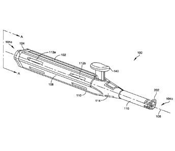
[0007] FIG. 1 illustrates an isometric view of a driver, in accordance
with some
embodiments.
[0008] FIG. 2 illustrates a front view of the driver of FIG. 1, in
accordance with some
embodiments.
[0009] FIG. 3 illustrates a rear view of the driver of FIG. 1, in
accordance with some
embodiments.
[0010] FIG. 4 illustrates a cross-sectional view of the driver of FIG. 1
taken along line A-
A in FIG. 1, in accordance with some embodiments.
2
CA 03057602 2019-09-23
WO 2019/005102 PCT/US2017/040278
[0011] FIG. 5 illustrates a strand locking mechanism of the driver of
FIG. 1 in a locked
position, in accordance with some embodiments.
[0012] FIG. 6 illustrates the strand locking mechanism of FIG. 5 in an
unlocked position,
in accordance with some embodiments.
[0013] FIG. 7 illustrates a cap of the driver of FIG. 1, in accordance
with some
embodiments.
[0014] FIG. 8 illustrates a cross-sectional view of a driver including a
compressible
locking mechanism, in accordance with some embodiments.
[0015] FIG. 9 illustrates the compressible locking mechanism of FIG. 8 in
a locked
position, in accordance with some embodiments.
[0016] FIG. 10 illustrates the compressible locking mechanism of FIG. 8
in an unlocked
position, in accordance with some embodiments.
[0017] FIG. 11 illustrates an isometric view of a needle construct
including a corkscrew
needle coupled to a flexible strand, in accordance with some embodiments.
[0018] FIG. 12 illustrates a side view of the needle construct of FIG.
11, in accordance
with some embodiments.
[0019] FIG. 13 illustrates a rear view of a needle of the needle
construct of FIG. 11, in
accordance with some embodiments.
[0020] FIG. 14 illustrates an isometric view of a strand retriever, in
accordance with
some embodiments.
[0021] FIG. 15 illustrates a top view of the strand retriever of FIG. 14,
in accordance
with some embodiments.
[0022] FIG. 16 is a cross-sectional view of the strand retriever taken
along line A-A in
FIG. 15, in accordance with some embodiments.
3
CA 03057602 2019-09-23
WO 2019/005102 PCT/US2017/040278
[0023] FIG. 17 illustrates the top view of the strand retriever of FIG.
14 having first and
second snares in a deployed position, in accordance with some embodiments.
[0024] FIG. 18 illustrates an isometric view of a clamping drill guide,
in accordance with
some embodiments.
[0025] FIG. 19 illustrates a side view of the clamping drill guide of
FIG. 18, in
accordance with some embodiments.
[0026] FIG. 20 illustrates a head of the clamping drill guide of FIG. 18,
in accordance
with some embodiments.
[0027] FIG. 21 illustrates a top view of the head of the clamping drill
guide of FIG. 18, in
accordance with some embodiments.
[0028] FIG. 22 illustrates a bottom view of the head of the clamping
drill guide of FIG.
18, in accordance with some embodiments.
[0029] FIG. 23 illustrates a drill trajectory guide configured to be
coupled to the
clamping drill guide of FIG. 18, in accordance with some embodiments.
[0030] FIG. 24 illustrates a strand retriever coupled to a clamping drill
guide, in
accordance with some embodiments.
[0031] FIG. 25 illustrates deployment of first and second snares of the
strand retriever of
FIG. 24 through the clamping drill guide, in accordance with some embodiments.
[0032] FIG. 26 illustrates a top view of a joint distractor, in
accordance with some
embodiments.
[0033] FIG. 27 illustrates a method of soft tissue repair, in accordance
with some
embodiments.
[0034] FIG. 28 illustrates a soft tissue release of a joint, in
accordance with some
embodiments.
4
CA 03057602 2019-09-23
WO 2019/005102 PCT/US2017/040278
[0035] FIG. 29 illustrates formation of an osteotomy in a first bone, in
accordance with
some embodiments.
[0036] FIG. 30 illustrates separation and temporary fixation of a first
bone fragment from
a second bone fragment, in accordance with some embodiments.
[0037] FIG. 31 illustrates distraction of a first bone from a second
bone, in accordance
with some embodiments.
[0038] FIG. 32 illustrates insertion of a flexible strand into a soft
tissue section, in
accordance with some embodiments.
[0039] FIG. 33 illustrates the flexible strand of FIG. 32 coupled to the
soft tissue section,
in accordance with some embodiments.
[0040] FIG. 34 illustrates a clamping drill guide coupled to a second
bone, in accordance
with some embodiments.
[0041] FIG. 35 illustrates insertion of a strand retriever through a
clamping drill guide
coupled to a second bone section, in accordance with some embodiments.
[0042] FIG. 36 illustrates the first and second snares of the strand
retriever deployed to a
plantar side of the second bone section through the clamping drill guide, in
accordance with
some embodiments.
[0043] FIG. 37 illustrates first and second flexible strands extending
from a plantar side
of the second bone to a dorsal side of the second bone after retrieval through
the clamping drill
guide, in accordance with some embodiments.
[0044] FIG. 38 illustrates tensioning of the first and second flexible
strands to position
the soft tissue section, in accordance with some embodiments.
[0045] FIG. 39 illustrates a knot formed in the first and second flexible
strands to couple
the soft tissue section to the second bone, in accordance with some
embodiments.
CA 03057602 2019-09-23
WO 2019/005102 PCT/US2017/040278
[0046] FIG. 40 illustrates a method of retracting a needle and flexible
strand after
deployment from a driver, in accordance with some embodiments.
[0047] FIG. 41 illustrates a driver coupled to a needle construct having
a needle and a
flexible strand in a deployed position, in accordance with some embodiments.
[0048] FIG. 42 illustrates the driver of FIG. 41 having a cap removed
from a proximal
end of the driver, in accordance with some embodiments.
[0049] FIG. 43 illustrates the driver of FIG. 42 having the flexible
strand decoupled from
the cap, in accordance with some embodiments.
[0050] FIG. 44 illustrates the driver of FIG. 43 after retraction of the
needle and flexible
strand of the needle construct, in accordance with some embodiments.
[0051] FIG. 45 illustrates the driver of FIG. 44 having a needle
construct coupled thereto
in a fixed relationship, in accordance with some embodiments.
DETAILED DESCRIPTION
[0052] This description of the exemplary embodiments is intended to be
read in
connection with the accompanying drawings, which are to be considered part of
the entire
written description. In the description, relative terms such as "lower,"
"upper," "horizontal,"
"vertical,", "above," "below," "up," "down," "top," "bottom," "proximal,"
"distal," "superior,"
"inferior," "medial," and "lateral" as well as derivative thereof (e.g.,
"horizontally,"
"downwardly," "upwardly," etc.) should be construed to refer to the
orientation as then described
or as shown in the drawing under discussion. For example, as used herein, the
terms "dorsal,"
"plantar," "superior," and "inferior" are used herein to refer to the
orientation of a bone in an
anatomical position. The terms "dorsal" and "superior" and the terms "plantar"
and "inferior"
are equivalent as used herein. Similarly, the terms "proximal" and "distal"
are used herein to
refer to a position of a surgeon when holding a tool and/or position
orientation of a bone in an
anatomical position. These relative terms are for convenience of description
and do not require
that the apparatus be constructed or operated in a particular orientation.
Terms concerning
attachments, coupling and the like, such as "connected," refer to a
relationship wherein structures
6
CA 03057602 2019-09-23
WO 2019/005102 PCT/US2017/040278
are secured or attached to one another either directly or indirectly through
intervening structures,
as well as both movable or rigid attachments or relationships, unless
expressly described
otherwise.
[0053] In various embodiments, a system and method for soft tissue repair
is disclosed.
The system includes one or more instruments for inserting, retrieving, and/or
manipulating a
flexible strand, such as a driver, a needle, a strand retriever, and/or
additional elements. The
driver includes a body extending along a longitudinal axis from a proximal end
to a distal end.
The body defines a cavity therein. A distal end of the body defines a needle
opening having a
non-circular cross-section that is sized and configured to receive a mating
portion of a needle
therein. A first portion of the body extends from the distal end towards a
neck and has a first
diameter and a second portion of the body extends from the neck to a proximal
end and has a
second diameter. A needle is inserted into the non-circular needle opening in
the distal end. The
needle includes a non-circular mating portion having a complimentary cross-
section with respect
to the needle opening in the distal end of the body. The non-circular cross-
section of the needle
opening and the non-circular mating portion of the needle maintain the needle
in a fixed position
with respect to the driver and transfer a torsional force from the driver to
the needle.
[0054] In various embodiments, the system includes a strand retriever.
The strand
retriever includes a body defining an inner cavity. A first longitudinal tube
and a second
longitudinal tube each extend from a first side of the body. A first snare and
a second snare are
slideably deployable from respective first and second longitudinal tubes. The
first and second
snares are deployable by a sliding deployment mechanism from a first position,
in which each of
the first snare and the second snare are substantially positioned within
respective first and second
longitudinal tubes, to a second position, in which the first snare and the
second snare are
substantially positioned distally of respective first and second longitudinal
tubes. The first and
second snares each include a loop sized and configured to receive a flexible
strand therethrough.
[0055] In various embodiments, a method of soft tissue repair is
disclosed. The method
includes accessing and distracting a joint, such as a metatarsophalangeal
(MTP) joint. A first
flexible strand is passed through a first tissue section by a first needle
coupled to a first driver.
The first flexible strand extends into a cavity defined in the longitudinal
body and is maintained
7
CA 03057602 2019-09-23
WO 2019/005102 PCT/US2017/040278
in a fixed position with respect to the longitudinal body of the first driver.
A second flexible
strand can be coupled to the soft tissue section by a second needle coupled to
a second driver. A
first hole and a second hole are formed through a bone of the joint. The first
flexible strand is
passed through the first hole and the second flexible strand is passed through
the second hole
using a strand retriever. The first tissue section is secured to the bone
using the first and second
flexible strands.
[0056] FIGS. 1-7 illustrate a driver 100, in accordance with some
embodiments. The
driver 100 includes a body 102 extending from a proximal end 104a to a distal
end 104b along a
longitudinal axis 106. In some embodiments, the body 102 includes a handle
portion 108
defined by a sidewall 110. The handle portion 108 can define one or more
grooves (or openings)
112a, 112b. The grooves 112a, 112b define gripping sections of the handle
portion 102. The
handle portion 108 can define any suitable shape, such as a triangular shape,
a circular shape, a
rectangular shape, and/or any other suitable shape. The handle portion 108 has
first radius (or
width).
[0057] In some embodiments, the body 102 includes a neck 114 and an
insertion portion
116. The neck 114 is coupled to the handle portion 108 at a proximal end and
is coupled to the
insertion portion 116 at a distal end. The insertion portion 116 extends from
the neck portion
along the longitudinal axis 106. The insertion portion 116 has a second radius
(or width). In
some embodiments, the second radius is less than the first radius. The neck
portion 114 can be
tapered from the first width at the proximal portion to the second width at
the distal portion. In
some embodiments, the neck 114 has a constant taper, although it will be
appreciated that the
neck 114 can have a variable taper and/or can be non-tapered. In some
embodiments, each of the
body 102, the neck 114, and the insertion portion 116 are concentric about the
longitudinal axis
106, although it will be appreciated that one or more of the body 102, neck
114, or insertion
portion 116 can be offset.
[0058] As shown in FIG. 4, in some embodiments, the body 102 defines an
internal
channel 118 (or cannula) extending from the proximal end 104a to the distal
end 104b of the
body 102. The channel 118 can include a first portion 118a having a first
diameter, a second
portion 118b having a second diameter, and a tapered portion 118c tapered from
the first
8
CA 03057602 2019-09-23
WO 2019/005102 PCT/US2017/040278
diameter to the second diameter. In some embodiments, the first portion 118a
is defined by the
handle portion 108, the second portion 118b is defined by the insertion
portion 116, and the
tapered portion 118c is defined by the neck 114. In some embodiments, the
first diameter is
greater than the second diameter.
[0059] A distal end 104b of the body 102 defines a needle opening 120.
The needle
opening 120 extends from an outer surface of the body 102 into the internal
channel 118. The
needle opening 120 is sized and configured to receive a mating portion 220 of
a needle 202
therein (as discussed in greater detail below). In some embodiments, the
needle opening 120 has
a non-circumferential cross-section configured to transfer a torsional force
to the needle 202.
For example, in some embodiments, the needle opening 120 includes an oval or
curved-rectangle
cross-section including sidewalls having a first length and top and bottom
walls having a second
length that is less than the first length. Although specific embodiments are
discussed herein, it
will be appreciated that the needle opening 120 can have any suitable shape
configured to
transition a driving force to a needle 202.
[0060] In some embodiments, a proximal end 104a of the body 102 defines a
cap
opening 122. The cap opening 122 extends from an outer surface of the body 102
into the first
portion 118a of the cavity 118. The cap opening 122 is sized and configured to
receive a cap 124
therein. The cap 124 can be coupled to the body 102 by any suitable coupling
mechanism, such
as an interference fit, a thread, and/or any other suitable coupling
mechanism. The cap 124 is
configured to be coupled to a flexible strand 204 extending from the needle
202. As discussed in
greater detail below, the cap 124 can be removed during surgery to retract a
suture after an initial
deployment of the needle and/or suture.
[0061] In some embodiments, a locking element 126 is disposed in the
inner cavity 118.
The locking element 126 is configured to maintain a needle in a fixed position
with respect to the
body 102. For example, the locking element 126 can include a locking body 127
configured to
apply a force to the flexible strand 204. In some embodiments, the locking
element 126 is
configured to maintain the suture in a fixed position through a friction or
interference fit between
the locking body 127 and an inner surface 128 of the cavity 118. As shown in
FIG. 5, in some
embodiments, the locking element 126 includes a locking body 127 having a
tapered head 130
9
CA 03057602 2019-09-23
WO 2019/005102 PCT/US2017/040278
sized and configured to be slideably received within the tapered portion 118c
of the cavity 118.
In some embodiments, the locking body 127 includes a semi-compressible or
fully-compressible
material disposed over at least a portion thereof. For example, in some
embodiments, the locking
body 127 includes a silicone and/or other material deposited over at least a
portion of the locking
body 127.
[0062] As shown in FIG. 6, when the locking body 127 is positioned
distally within the
tapered portion 118c, the tapered head 130 contacts the inner surface 128 of
the cavity 118. The
tapered head 130 and the inner surface 128 of the cavity 118 apply a
frictional locking force to
the flexible strand. The frictional locking force maintains the flexible
strand 204 (and by
extension the needle 202) in a fixed position with respect to the driver 100.
When the locking
body 127 is slidingly removed from the tapered portion 118c, the locking force
is removed from
the flexible strand and the flexible strand 204 and/or the needle 202 can be
decoupled from the
driver 100 through the needle opening 120.
[0063] In some embodiments, the locking element 126 includes a button 140
coupled to
the locking body 127. The button 140 is configured to transition the locking
body 127 from a
first (or distal) position to a second (or proximal) position. In some
embodiments, the button 140
includes a head 142 and a body 144 extending vertically therefrom. The body
144 defines a slot
148. The slot 148 extends through the body 140 at an angle between 0-90 with
a proximal end
148a of the slot 148 located above a distal end 148b of the slot 148. The slot
148 is sized and
configured to receive a pin 150. The pin 150 extends through the slot 148 and
into the locking
body 127. In some embodiments, the button 140 defines a channel extending
through the
vertical body 144 sized and configured to receive a portion of the locking
body 127 therethrough.
[0064] In some embodiments, the locking body 127 includes a tapered head
130 having a
complimentary taper with the tapered surface 118c of the cavity. The tapered
head 130 is
coupled to a longitudinal body 132 having a first portion 134a sized and
configured for insertion
through the channel formed in the button 140 and a second portion 134b sized
and configured to
abut a proximal surface of the button 140. Movement of button 140 in a
direction perpendicular
to the longitudinal axis of the locking body 127 (i.e., in an up/down
direction), causes the locking
body 127 to move longitudinally with respect to the driver body 102 such that
the tapered head
CA 03057602 2019-09-23
WO 2019/005102 PCT/US2017/040278
130 is advanced into and/or out of contact with the inner surface 128 of the
tapered portion 118c
of the cavity 118.
[0065] In some embodiments, the slot 148 exerts a force on the pin 150 to
translate the
locking body 127 from a first, or distal, position to a second, or proximal,
position. In the first
position, a portion of the locking body 127 positioned within the tapered
portion 118c of the
inner cavity 118 and is in contact with an inner surface 128. In the second
position, the locking
body 127 is positioned substantially within the first portion 118a of the
inner cavity 118 and is
not in contact with the inner surface 128. In some embodiments, translation of
the button from a
locked (or upper) position to an unlocked (or lower) position transitions the
locking body 127
from the first position to the second position. When the button 140 is in a
locked position, as
shown in FIG. 5, the pin 150 is positioned at a distal end 148a of the slot
148 and the head 130 of
the locking body 127 is positioned within the tapered portion 118c of the
cavity. The head 130 is
tapered to provide a taper-taper friction interface between the head 130 and
the tapered portion
118c to maintain the flexible strand 204 in a fixed position. When the button
140 is transitioned
to the second position, as shown in FIG. 6, the slot 148 drives the pin 150
proximally, which
moves the locking body 127 proximally. When the button 140 is fully depressed
(i.e., in the
second position), the pin 150 is located at a proximal end 148a of the slot
148 and the head 130
is spaced apart from the inner surface 128 of the cavity 118. The flexible
strand 204 is able to
freely move when the locking body 127 is in the second position (i.e.,
substantially located in the
first portion 118a of the cavity 118).
[0066] FIG. 7 illustrates a cap 124 sized and configured to be received
within the cap
opening 122 in a proximal end 104 of the body 102. The cap 124 includes a head
160 having a
locking surface 162 extending therefrom. The locking surface 162 can include a
conical and/or
circular locking surface configured to couple the cap 124 within the cap
opening 122 of the body
102. The locking surface 162 can be configured to provide a friction,
interference, threaded,
and/or other locking fit with the cap opening 122. The head 160 includes
projections 164a-164c
that extend circumferentially beyond the locking surface 162 and are
configured to abut the
proximal surface of the body 102 when the cap 124 is inserted into the cap
opening 122. In some
embodiments, the projections 164a-164c are configured to facilitate removal of
the cap 124 from
the cap opening 122.
11
CA 03057602 2019-09-23
WO 2019/005102 PCT/US2017/040278
[0067] In some embodiments, a boss 166 extends from the locking surface
162 and
defines a hole 168 sized and configured to receive a flexible strand 204
therethrough. A
proximal end of the flexible strand 204 is looped one or more times through
the hole 168. When
the needle 202 is deployed from the driver 100, the flexible strand 204 is
pulled through the hole
166 such that the flexible strand 204 deploys from distal opening 120 without
tangling. For
example, the flexible strand 204 can be maintained in a taught and/or semi-
taught state within the
cavity 118 until a proximal end 218 of the flexible strand 204 passes through
the hole 168. Once
the proximal end 218 passes through the strand hole 168, the flexible strand
204 can be removed
from the cavity 118 through the distal opening 120. In other embodiments, the
flexible strand
204 can be maintained within the cavity 118 by any suitable mechanism, such as
a spool, post,
anchor, and/or any other suitable mechanism and/or can be freely moveable
within the cavity
118.
[0068] In use, the driver 100 is configured to couple the needle 202 and
the flexible
strand 204 to a soft tissue section. During insertion of the needle 202, the
driver 100 transfers a
torsional force applied to the body 102 to the needle 202. The torsional force
is transferred by an
interaction between the non-circular proximal end 208 of the needle 202 and a
non-circular
cross-section of a needle opening 120 formed in the distal end 104a of the
body 102. The
torsional force drives the needle 202 through the soft tissue. As used herein,
the term non-
circular refers to any shape configured to prevent rotation of the needle 202
with respect to the
needle opening 120, such as, an oval, ellipsoid, rectangular, triangular, non-
regular, regular
polygon, and/or any other suitable shape. Although embodiments are discussed
herein including
a transfer of torsional force, it will be appreciated that the driver 100 can
be configured to
transfer any suitable penetrating force from the driver 100 to the needle 202,
such as a torsional
force, a longitudinal force, etc.
[0069] After insertion of the needle 202 through the soft tissue, the
button 140 is
transitioned from the locked position to the unlocked position to disengage a
locking element
126 from the flexible strand 204. After the locking element 126 is disengaged,
the needle 202
and the flexible strand 204 can be removed through the distal opening 120 of
the body 102. The
needle 202 and/or the flexible strand 204 are configured to couple the soft
tissue section to one
or more structures, such as a bone, other soft tissue, a plate, and/or any
other structure.
12
CA 03057602 2019-09-23
WO 2019/005102 PCT/US2017/040278
[0070] FIGS. 8-10 illustrate an embodiment of a driver 100a including a
compressible
locking element 126a, in accordance with some embodiments. As shown in FIG. 8,
the locking
element 126a includes a compressible body 136, such as a flexible tube,
configured to be
deformed or compressed against the flexible strand 204 and the inner surface
128 of the cavity
118. A compression force is applied to the compressible body 136 (for example,
by button 140a)
which causes compression and deformation of the flexible tube 136, as shown in
FIG. 9. The
compressible body 136 applies a locking force to the flexible strand and
maintains the flexible
strand in a fixed position. The compressible body 136 returns to an un-
compressed state (for
example, a circular state) when the compressive force is removed, allowing the
flexible strand to
move freely within the cavity 118.
[0071] A button 140a is configured apply the compressive force to the
compressible body
136 when in a locked position. For example, in some embodiments, the button
140a includes a
head 142a and a body 144a extending therefrom. The body 144a includes a first
vertical section
146a extending along a first vertical axis and a second vertical section 146b
extending along a
second vertical axis that laterally is offset from the first vertical axis.
[0072] In some embodiments, the button 140a is configured to be
transitioned from the
locked position to an unlocked position. In the locked position, the second
section 146b is in
contact with and compresses the compressible body 136, which applies the
locking force to the
flexible strand 204. The button 140a is transitioned from the locked position
(shown in FIG. 9)
to the unlocked position (shown in FIG. 10) to release the flexible strand 204
and/or an attached
needle 202 from the driver. In the locked position, the second section 146b of
the body 144a is
positioned below and out of contact with the compressible body 136. The first
section 146a is
offset from the second section 146b such that when the button 140a is in the
unlocked position,
the first section 146a is adjacent to and spaced apart from the compressible
body 136 at a
distance sufficient to allow the compressible body 136 assume a non-deformed
state.
[0073] In other embodiments, the locking element 126 can include a collet
or other
chuck-style locking element configured to lock the needle 202 both
rotationally and
longitudinally with respect to the body 102. The collet can include a three-
piece chuck
configured to lock the position of the needle 202. The collet can be
positioned in any suitable
13
CA 03057602 2019-09-23
WO 2019/005102 PCT/US2017/040278
portion of the driver 100, such as, for example, at the needle opening 120,
within the neck 114,
and/or at any other suitable portion of the driver 100.
[0074] FIGS. 11-13 illustrate a needle construct 200 including a
corkscrew needle 202
and a flexible strand 204 extending therefrom, in accordance with some
embodiments. The
corkscrew needle 202 includes a sharpened distal end 206, a non-circular
proximal end 208, and
a body 210 extending from the distal end 206 to the proximal end 208. The
sharpened distal end
206 includes a sharpened tip 214 configured to penetrate tissue, such as a
soft tissue section.
[0075] In some embodiments, the body 210 defines at least one helical
turn 212
configured for rotational (or torsional) insertion of the needle 202 through a
soft tissue section.
In some embodiments, each helical turn 212 is configured to minimize tissue
damage or trauma
during insertion of the needle 202. For example, in some embodiments, pitch,
arc length, and/or
other elements of each helical turn 212 is configured to provide insertion of
the needle 202
through a single hole formed in a soft tissue section by the sharpened distal
end 206. Rotation of
the needle 202, for example due to a torsional force applied to a driver 100,
causes the helical
turn 212 to pass through the single hole. Although embodiments are illustrated
including a
single helical turn, it will be appreciated that the needle 202 can include no
helical turns (e.g., is
straight and/or curved without a helical turn) or two or more helical turns in
other embodiments.
[0076] In some embodiments, the proximal end 208 of the needle 202
defines a non-
circular mating section 214. The non-circular mating section 214 is
complimentary to the cross-
section of the non-circular needle opening 120 formed in the driver 100. The
non-circular
mating section 214 is configured to receive a torsional force from the driver
100 and transfer the
torsional force to the needle 202 during insertion of the needle 202 into a
soft tissue section. The
non-circular mating section 214 can have any shape complementary to the non-
circular needle
opening 120, such as, for example, an oval shape, a rectangular shape, a
triangular shape, a
regular geometric shape, an irregular geometric shape, and/or any other
suitable shape
[0077] As shown in FIGS. 11-12, the needle construct 200 includes a
flexible strand 204
coupled to a proximal end 208 of the needle 202. The flexible strand 204 can
be coupled to the
needle 202 by any suitable coupling mechanism, such as a crimp engagement, a
knot, and/or any
other suitable coupling mechanism. For example, in the illustrated embodiment,
the flexible
14
CA 03057602 2019-09-23
WO 2019/005102 PCT/US2017/040278
strand 204 is coupled to the needle 202 by a crimp at a proximal end 208. In
some embodiments,
the coupling mechanism (such as a crimp) defines the non-circular mating
section 214 of the
needle 202.
[0078] The flexible strand 204 extends proximally from the needle 202.
The flexible
strand 204 can define a loop 216 extending from a distal end 216a to a
proximal end 216b and/or
can define one or more free ends at a proximal end 216b. In some embodiments,
a proximal end
216b of the flexible strand 204 is coupled to a cap 124 of a driver 100. The
flexible strand 204 is
maintained in a semi-taught or taught state when positioned within the driver
100 to prevent
knotting or other entanglement of the flexible strand 204 prior to insertion
and release of the
needle construct 200. The flexible strand 204 can include any suitable
material, such as a suture,
ribbon, wire, etc. In some embodiments, the flexible strand 204 can include a
plurality of strands
extending from a proximal end 208 of the needle 202 and each defining a free
end and/or a loop
configured to couple a soft tissue section to an anatomical and/or implanted
structure.
[0079] FIGS. 14-17 illustrate a strand retriever 300, in accordance with
some
embodiments. The strand retriever 300 includes a body 302 extending from a
proximal end 302a
to a distal end 302b. The body 302 further extends between an upper surface
304a and a lower
surface 304b and generally parallel side surfaces 306a, 306b. In some
embodiments, the upper
surface 304a defines a slot 308 sized and configured to receive a slideable
deployment
mechanism 310 therein. The slot 308 extends from a distal end 302b towards the
proximal end
302a. In some embodiments, the slot 308 extends through the upper surface 304a
to a cavity 312
defined by the body 302.
[0080] In some embodiments, at least one longitudinal tube 314a, 314b
extends from the
distal surface 302b of the body 302. The longitudinal tubes 314a, 314b each
define a lumen
extending from a proximal end 316a to a distal end 316b. In some embodiments,
the
longitudinal tubes 314a, 314b extend through the distal surface 302b of the
body 302 such that
the lumens defined therein are in communication with the cavity 312 defined by
the body 302.
In some embodiments, the longitudinal tubes 314a, 314b are parallel, although
it will be
appreciated that the longitudinal tubes 314a, 314b can be non-parallel in some
embodiments.
Although embodiments are illustrated having two longitudinal tubes 314a, 314b,
it will be
CA 03057602 2019-09-23
WO 2019/005102 PCT/US2017/040278
appreciated that the strand retriever 300 can include a greater and/or lesser
number of
longitudinal tubes, such as one longitudinal tube, three longitudinal tubes,
etc.
[0081] In some embodiments, a first snare 318a is positioned within and
slideably
deployable from the first longitudinal tube 314a and a second snare 318b is
positioned within
and slideably deployable from the second longitudinal tube 314b. The first
snare 318a and/or the
second snare 318b each include a loop 320a, 320b coupled to a respective
longitudinal shaft
322a, 322b extending proximally from the loop 320a, 320b. Each of the loops
320a, 320b can
have any suitable circular and/or non-circular shape, such as a
circumferential shape, a diamond
shape, a rectangular shape, a triangular shape, an irregular shape, etc. In
some embodiments, the
loops 320a, 320b and/or the longitudinal shafts 322a, 322b are formed of a
shape-memory
material, such as a shape-memory metal (e.g., nitinol), although it will be
appreciated that the
loops 320a, 320b and/or the longitudinal shafts 322a, 322b can be formed of
any resilient
material. In some embodiments, each of snares 318a, 318b are formed of a
single strand of
material that defines the loop 320a, 320b and is comingled (e.g., twisted,
braided, etc.) to form
the longitudinal shaft 322a, 322b. In other embodiments, the loop 320a, 320b
and the
longitudinal shaft 322a, 322b are formed of separate strands (or materials)
and are coupled
together.
[0082] The first snare 318a and the second snare 318b are each slideably
disposed within
respective first longitudinal tube 314a and second longitudinal tube 314b. The
first and second
snare 318a, 318b are slideably deployed from a distal end 316b of the
longitudinal tubes 314a,
314b by the sliding deployment mechanism 310. The first snare 318a and the
second snare 318b
are each formed of a resilient, shape-memory material configured to be
compressed within the
respective first and second longitudinal tubes 314a, 314b and that expands to
define a respective
loop 320a, 320b upon deployment from the longitudinal tubes 314a, 314b. The
loops 320a, 320b
are sized and configured to receive a needle 202 and/or a flexible strand 204
therethrough. In
some embodiments, the loops 320a, 320b are configured to be compressed such
that a first side
of the loop 320a, 320b is parallel with a second side of the loop 320a, 320b
in a compressed
state.
16
CA 03057602 2019-09-23
WO 2019/005102 PCT/US2017/040278
[0083] In some embodiments, the first snare 318a and/or the second snare
318b are
slideably deployable by the slideable deployment mechanism 310. The slideable
deployment
mechanism 310 includes a slider 330 positioned within the slot 308 and/or the
cavity 312 defined
in the body 302. The slider 330 is coupled to a body portion 324 defining a
plurality of female
slots 326 each configured to receive a crimped proximal end 340 of the
longitudinal shaft 322a,
322b of a respective snare 318a, 318b therein, although it will be appreciated
that any suitable
engagement between the longitudinal shafts 322a, 322b and the slider 330 can
be used.
Although embodiments are discussed herein including a crimped proximal end
340, it will be
appreciated that the longitudinal shafts 322a, 322b can be coupled to the
slider 330 using any
suitable method, such as, for example, crimping, over molding, injection
molding, plasma
welding, welding, adhesives, any other suitable coupling method and/or any
combination
thereof.
[0084] The slider 330 is longitudinally moveable within the slot 308 from
a first (or
proximal-most) position to a second (or distal-most) position. Translation of
the slider 330 from
the first position to the second position deploys the loops 320a, 320b of
first and second snares
318a, 318b from respective first and second longitudinal tubes 314a, 314b. In
some
embodiments, a portion of the longitudinal shaft 322 of each of the snares
318a, 318b is also
deployed from the first and second longitudinal tubes 314a, 314b such that the
loops 320a, 320b
are spaced apart from a proximal end 316a of a respective longitudinal tube
314a, 314b.
[0085] In some embodiments, the slot 308 includes a proximal locking
feature 336
configured to maintain the slider 330 in a fixed proximal position prior to
deployment of the
snares 318a, 318b. For example, in some embodiments, the proximal locking
feature 336
includes a proximal detent 342 formed in the slot 308. The locking feature 336
maintains the
slideable deployment mechanism 310 (and the attached snares 318a, 318b) in a
fixed proximal
position until a predetermined force is applied to the slider 330 to force the
slideable deployment
mechanism 310 beyond the locking feature 336. In other embodiments, the
locking feature 336
can include a spring-loaded locking feature, a hinged locking feature, and/or
any other suitable
locking feature. The locking feature 336 prevents deployment of the snares
318a, 318b prior to
positioning of the strand retriever 300 during surgery.
17
CA 03057602 2019-09-23
WO 2019/005102 PCT/US2017/040278
[0086] FIGS. 18-22 illustrate a clamping drill guide 400, in accordance
with some
embodiments. The clamping drill guide 400 is configured to be securely coupled
to (i.e.,
clamped to) a second bone. The clamping drill guide 400 is configured to guide
insertion of a
drill element to form one or more holes in the second bone. For example, in
some embodiments,
the clamping drill guide 400 is configured to guide formation of one or more
holes through the
second bone. The clamping drill guide 400 is further configured to guide
insertion of one or
more elements of a strand retriever 300, as discussed in greater detail below.
[0087] The clamping drill guide 400 includes a handle portion 402 and a
head 404
coupled to the handle portion 402 by a pivoting connection 420. The handle
portion 402
includes a first handle 406a and a second handle 406b coupled by the pivoting
connection 420.
Each of the handles 406a, 406b includes a finger loop 422a, 422b sized and
configured to allow a
user to grip and/or manipulate the handles 406a, 406b during surgery. The head
404 includes a
guide element 408 coupled to the first handle 406a and a clamping element 410
coupled to the
second handle 406b. The guide element 408 includes a body 412 defining at
least one guide hole
414a, 414b extending from a first side 412a to a second side 412b of the body
412. In some
embodiments, the guide holes 414a, 414b are parallel and extend through the
body 412 parallel
to a longitudinal axis of the body 412, although it will be appreciated that
each of the guide holes
414a, 414b can be angled with respect to the longitudinal axis and/or with
respect to the other
guide hole 414a, 414b. The guide holes 414a, 414b are sized and configured to
receive a hole
forming element therethrough. For example, in various embodiments, the guide
holes 414a,
414b can be configured to receive a drilling element (such as a drill bit), a
k-wire, a cutting
element, and/or any other suitable hole forming element therethrough.
[0088] The clamping element 410 includes a body 416 configured to apply a
clamping
force to a bone positioned between the clamping element 410 and the guide
element 408. The
body 416 defines an opening 418 aligned with the guide holes 414a, 414b such
that a hole
forming element inserted through the guide holes 414a, 414b passes through the
opening 418
when the head 404 is in a clamped configuration. In some embodiments, the body
416 defines a
first end 416a and a second end 416b spaced apart to define a slot, such as an
open crescent or
other open shape. The opening 418 is defined at least partially by a perimeter
of the body 416
extending between the first end 416a and the second end 416b. In other
embodiments, the body
18
CA 03057602 2019-09-23
WO 2019/005102 PCT/US2017/040278
416 defines a closed shaped and the opening 418 is defined entirely by an
internal perimeter of
the body 416, such as a circular closed shape, non-circular geometric shape,
etc.
[0089] In some embodiments, the guide element 408 and the clamping
element 410 are
coupled in a hinged arrangement and are configured to receive a portion of a
bone therebetween.
For example, in some embodiments, the guide element 408 and the clamping
element 410 define
an adjustable opening therebetween sized and configured to receive a portion
of a bone, such as a
proximal phalanx, although it will be appreciated that the guide element 408
and the clamping
element 410 can define a greater and/or lesser opening configured to receive
alternative and/or
additional bones.
[0090] In some embodiments, the clamping drill guide 400 includes a
locking element
424 configured to maintain a fixed position of the first handle 406a and the
second handle 406b
during clamping of a bone. The locking element 424 includes a first locking
element 426a
coupled to the first handle 406a and a second locking element 426b coupled to
the second handle
406b. The first and second locking elements 426a, 426b are coupled together to
provide a
variable clamping force to a bone and maintain the handles 406a, 406b in a
fixed position.
[0091] As illustrated in FIG. 22, in some embodiments, a bone-facing
surface 412b of the
guide element 408 defines a textured surface. The textured surface is
configured to provide
increase friction between the guide element 408 and the bone when the clamping
drill guide 400
is coupled to the bone. The textured surface can be any suitable textured
surface, such as a
plasma coated surface, a laser etched surface, a knurled surface, a surface
including one or more
protrusions (such as machined-in protrusions), and/or any other suitable
textured surface.
[0092] FIG. 23 illustrates a trajectory guide 450 configured to be
coupled to the head 404
of the clamping drill guide 400, in accordance with some embodiments. The
trajectory guide
450 includes a first post 452 extending from a body 454. The post 452 is
configured to indicate a
path of a hole forming element when the trajectory guide 450 is coupled to the
clamping drill
guide 400. For example, in some embodiments, when viewed sagittally, a post
452 indicates a
path of a drill guide 414a, 414b. The trajectory guide 450 indicates the path
of the drill guide
holes 414a, 414b to verify that a hole forming element inserted through the
guide element 408
does not violate undesirable soft tissue and/or bone surfaces, such as an
articular surface of a
19
CA 03057602 2019-09-23
WO 2019/005102 PCT/US2017/040278
phalanx. In some embodiments, the trajectory guide 450 includes one or more
pins 456a, 456b
sized and configured for insertion into the guide holes 414a, 414b formed in
the guide element
408. The pins 456 align the trajectory guide 450 with the guide element 408
and maintain the
trajectory guide 450 in a fixed position.
[0093] FIG. 24 illustrates a strand retriever 300 having longitudinal
tubes 314a, 314b
and/or the snares 318a, 318b inserted through the guide holes 414a, 414b of
the clamping drill
guide 400. The longitudinal tubes 318a, 318b have an outer diameter less than
a diameter of the
guide holes 414a, 414b. The longitudinal tubes 318a, 318b extend at least
partially into the
guide holes 414a, 414b. In some embodiments, the longitudinal tubes 318a, 318b
extend beyond
a distal end 412b of the guide body 412.
[0094] The snares 318a, 318b of the strand retriever 300 are deployed
from the
longitudinal tubes 314a, 314b after insertion of the longitudinal tubes 314a,
314b into the guide
holes 414a, 414b. In some embodiments, the longitudinal shaft 322a, 322b of
each of the snares
318a, 318b has a length sufficient to position the respective loops 320a, 320b
of each of the
snares 318a, 318b distal of the clamping element 410 of the clamping drill
guide 400. For
example, in the illustrated embodiment, the longitudinal shaft 322a, 322b of
each of the snares
318a, 318b positions a proximal end of the loop 320a, 320b beyond the clamping
element 410.
The loops 320a, 320b are fully expanded when positioned distal of the clamping
element 410.
Because the loops 320a, 320b are substantially contained within the
longitudinal tubes 314a,
314b during deployment, the flexibility of the loops 320a, 320b does not
hinder deployment
through the clamping drill guide 400.
[0095] In some embodiments, the guide holes 414a, 414b are configured to
deploy the
snares 318a, 318b through the opening 418 defined by the clamping element 410.
For example,
in some embodiments, the loops 320a, 320b and/or the longitudinal shafts 322a,
322b of each of
the snares 318a, 318b are configured to pass through the opening 418 when
deployed from the
strand retriever 300. The loops 320a, 320b are positioned distal of the
clamping element 410 and
expand after passing at least partially through the opening 418.
[0096] FIG. 26 illustrates a joint distractor 500, in accordance with
some embodiments.
The joint distractor 500 includes a proximal portion 502 and a distal portion
504. The proximal
CA 03057602 2019-09-23
WO 2019/005102 PCT/US2017/040278
portion 502 includes a first handle 506a and a second handle 506b coupled in a
pivoting
arrangement by pivoting element 524. The first handle 506a and the second
handle 506b each
define a finger loop 508 configured to allow a user to grip the hands 506a,
506b and/or actuate
the handles 506a, 506b.
[0097] In some embodiments, the proximal portion 504 includes a first
anchoring body
510a coupled to the first handle 506a and a second anchoring body 510b coupled
to the second
handle 506b. Each of the first and second anchoring bodies 510a, 510b define
at least one hole
512a-512d extending from a first end to a second end. For example, in the
illustrated
embodiment, a first hole 512a, 512c and a second hole 512b, 512d extend
through the anchoring
bodies 510a, 510b, although it will be appreciated that the anchoring bodies
510a, 510b can
define a greater and/or lesser number of holes 512a-512d. The holes 512a-512d
can extend
through each of the respective anchoring bodies 510a, 510b along parallel
and/or non-parallel
axes.
[0098] Each of the holes 512a-512d are sized and configured to receive an
anchoring
element therein. For example, in some embodiments, each of the holes 512a-512d
are sized and
configured to receive a k-wire therethrough, although it will be appreciated
that holes 512a-512d
can be configured to receive any suitable anchoring element, such as a k-wire,
a screw, a pin,
and/or any other suitable anchoring element. Anchoring elements inserted
through each of the
holes 512a-512d are configured to anchor the respective anchoring body 510a,
510b to a tissue
section, such as a bone section. After anchoring the anchoring bodies 510a,
510b to the bone
section, the handles 506a, 506b can be actuated to separate the anchoring
bodies 510a, 510b to
distract the bone sections coupled to the respective anchoring body 510a,
510b.
[0099] In some embodiments, the joint distractor 500 includes a locking
element 520.
The locking element 520 is configured to lock the handles 506a, 506b at a
selected
length/distance corresponding to a selected distraction of the anchoring
bodies 510a, 510b. In
some embodiments, the locking element 520 includes a ratcheting locking
element, although it
will be appreciated that any suitable locking element can be used. In some
embodiments, the
locking element 520 includes a release 522.
21
CA 03057602 2019-09-23
WO 2019/005102 PCT/US2017/040278
[0100] FIG. 27 illustrates a method 1000 of soft tissue repair and FIGS.
28-39 illustrate
various steps of the method 1000, in accordance with some embodiments. At step
1002, an
incision is formed to access a joint of a patient, such as a
metatarsophalangeal (MTP) joint.
Although embodiments are discussed herein with reference to an MTP joint, it
will be
appreciated that the method 1000 and/or the surgical instruments used during
the method 1000
can be used for various joints and/or soft tissue applications and is within
the scope of this
disclosure.
[0101] At step 1004, soft tissue and/or connective tissue is released
from a portion of the
exposed joint. For example, as illustrated in FIG. 28, one or more dorsal
capsular and/or
collateral ligaments can be released from a proximal phalanx of an MTP joint
1100. An elevator
1102 is used to release a soft tissue section, such as a plantar plate, from a
distal metatarsal of an
MTP joint. In other embodiments, alternative and/or additional connective
tissue (e.g.,
ligaments, tendons, etc.) and/or soft tissue can be released from any portion
of an exposed joint.
[0102] At step 1006, an osteotomy is formed in a first bone 1104 of the
joint 1100. For
example, as shown in FIG. 29, an osteotomy can be formed in a distal
metatarsal. The
osteotomy can provide for shortening of a first bone 1104 of the joint. In
some embodiments,
the osteotomy separates the first bone 1104 into a first bone fragment 1104a
and a second bone
fragment 1104b.
[0103] At step 1008, the first bone fragment 1104a is displaced
proximally from the
second bone fragment 1104b. In some embodiments, the first bone fragment 1104a
is a capital
fragment of a metatarsal formed as a result of the osteotomy of step 1006. In
the illustrated
embodiment, the first bone fragment 1104a and the second bone fragment 1104b
are temporarily
fixed using a k-wire 1108a, although it will be appreciated that any suitable
temporary fixation
device, such as a k-wire, a screw, a pin, etc. can be used to temporarily
fixate the first bone
fragment 1104a and the second bone fragment 1104b.
[0104] At step 1010, a distractor 500 is coupled to the first bone 1104
and the second
bone 1120. As shown in FIG. 31, in some embodiments, a first hole 512a formed
in a first
anchoring body 510a of the distractor 500 is coupled to the first k-wire 1108a
and a second k-
wire 1108b coupled to the second bone 1120. The second k-wire 1108b is
inserted through a
22
CA 03057602 2019-09-23
WO 2019/005102 PCT/US2017/040278
hole 512c defined in the second anchoring body 510b after coupling the first
anchoring body
510a to the first k-wire 1108a, although it will be appreciated that the
second k-wire 1108b can
be coupled to the second bone 1120 prior to coupling the first anchoring body
510a to the first k-
wire 1108a.
[0105] At step 1012, the first bone 1104 is distracted from the second
bone 1120. The
first and second bones 1104, 1120 can be distracted by actuating the handles
506a, 506b of the
distractor 500 to separate the first anchoring body 510a from the second
anchoring body 510b.
The first bone fragment 1104a and the second bone fragment 1104b of the first
bone 1104 are
moved together by the distractor 500, as the first bone fragment 1104a and the
second bone
fragment 1104b are coupled by the k-wire 1108a. As shown in FIG. 31, the first
bone 1104 and
the second bone 1120 are distracted to provide access to a soft tissue section
and allow insertion
of one or more surgical instruments to the soft tissue section, as discussed
in greater detail below.
[0106] At optional step 1014, additional fixation elements are inserted
through the
anchoring bodies 510a, 510b of the distractor 500 and/or the bones 1104, 1120
to provide
secondary fixation of the bones 1104, 1120. The additional fixation elements
can include any
suitable fixation element, such as a k-wire, a screw, a pin, and/or any other
suitable fixation
element. In other embodiments, the additional fixation elements are omitted
and the distractor
500 is coupled to the bones 1104, 1120 by the primary fixation elements 1108a,
1108b only.
[0107] At step 1016, a needle construct is coupled to the soft tissue
section 1114. As
illustrated in FIG. 32, a first needle construct 200a is coupled to and
implanted by a first driver
100b configured to apply an insertion force to a first needle 202a of the
first needle construct
200a. The first driver 100b is similar to the driver 100 discussed above, and
similar description
is not repeated herein. The first driver 100b transfers an insertion force,
such as a torsional
insertion force, from the body 102 to the first needle 202a, which penetrates
the soft tissue
section 1114. After insertion of the first needle 202 at least partially into
the soft tissue section
1114, the first needle construct 200a is released from the first driver 100b,
for example, by
depressing a button 140 to release a locking element 126 of the driver 100b.
[0108] At optional step 1018, the first needle construct 200a can be
retracted into the first
driver 100b and redeployed in the same and/or an alternative location. A
method of retracting
23
CA 03057602 2019-09-23
WO 2019/005102 PCT/US2017/040278
and redeploying the first needle construct 200a is discussed in greater detail
below with respect
to FIGS. 40-45.
[0109] At step 1020, the first flexible strand 204a of the first needle
construct 200a is
coupled to the soft tissue section 1114. In the embodiment shown in FIG. 33,
the first flexible
strand 204a defines a loop 216 and is coupled to the soft tissue section 1114
by passing the
needle 202a and the attached flexible strand 204 through the loop 216 to form
a knot 1112
around the tissue 1114. The knot 1112 can be any suitable knot, such as, for
example, a slip-knot
(luggage-tag style knot, hitch knot, etc.), a fixed knot, and/or any other
suitable knot. Although
embodiments are discussed herein including a luggage tag-style knot, it will
be appreciated that
any suitable knot, anchor, and/or any other suitable element can be used to
anchor the first
flexible strand 204a to the soft tissue section 1114.
[0110] In some embodiments, steps 1016-1020 can be repeated to couple one
or more
additional needle constructs to the soft tissue section 1114. For example, in
some embodiments,
a second needle construct (not shown) including a second needle 202b and a
second flexible
strand 204b are coupled to the soft tissue section 1114 at a second position.
Although
embodiments are discussed herein including two needle constructs, it will be
appreciated that any
suitable number of needle constructs 200b can be coupled to the soft tissue
section 1114.
[0111] At step 1022, the distractor 500 and/or the temporary fixation
elements 1108a,
1108b can be removed from the first bone 1104 and/or the second bone 1120. For
example, in
some embodiments, the distractor 500 is removed from the temporary fixation
elements 1108a,
1108b by sliding the anchor bodies 508a, 508b dorsally over the fixation
elements 1108a, 1108b.
The fixation elements 1108a, 1108b can be subsequently removed from the bones
1104, 1120.
[0112] At step 1024, a clamping drill guide 400 is coupled to a second
bone 1120, such
as a proximal phalanx. As discussed above, the clamping drill guide 400
includes a guide
element 408 and a clamping element 410 coupled in pivoting arrangement. The
second bone
1120 is positioned between the guide element 408 and the clamping element 410
and a force is
applied to the handle portion 402 to clamp the second bone 1120 between the
guide element 408
and the clamping element 410. In some embodiments, the clamping drill guide
400 includes a
24
CA 03057602 2019-09-23
WO 2019/005102 PCT/US2017/040278
locking element 424 configured to maintain the guide element 408 and the
clamping element 410
in a clamped position during surgery.
[0113] At step 1026, the trajectory of the guide holes 414a, 414b formed
in the guide
element 408 is confirmed. The trajectory of the guide holes 414a, 414b can be
confirmed by
coupling a trajectory guide 450 to the clamping drill guide 400. The
trajectory guide 450
includes a post 452 configured to indicate a path of the guide holes 414a,
414b when the
trajectory guide 450 is coupled to the clamping drill guide 400. For example,
in some
embodiments, when viewed sagittally, the post 452 indicates a path of the
drill guide holes 414a,
414b. The trajectory guide 450 is used to ensure that a hole forming element
inserted through
the drill guide holes 414a, 414b will not violate the articular surface of the
bone. The clamping
drill guide 400 can be adjusted as required to properly align the guide holes
414a, 414b.
[0114] At step 1028, at least one hole is formed through the second bone
1120. In some
embodiments, the at least one hole is formed by inserting a bit element, such
a k-wire or a drill
bit, through a guide hole 414a, 414b of the clamping drill guide 400. The bit
element penetrates
the second bone 1120 and extends through the opening 418 defined by the
clamping element 410
of the clamping drill guide 400.
[0115] At step 1030, a first longitudinal tube 314a and a second
longitudinal tube 314b of
a strand retriever 300 are inserted into respective first and second guide
holes 414a, 414b of the
clamping drill guide 400 on a dorsal (or first) side of the second bone 1120.
In some
embodiments, a portion of the longitudinal tubes 314a, 314b extends plantarly
beyond the guide
element 408 of the clamping drill guide and into one or more tunnels formed in
the second bone
1120, although it will be appreciated that the longitudinal tubes 314a, 314b
can extend partially
through the guide element 408.
[0116] At step 1032, a first snare 318a and a second snare 318b are
deployed from the
longitudinal tubes 314a, 314b to a plantar (or second) side of the second bone
1120. As shown
in FIG. 36, the snares 318a, 318b extend plantarly beyond the second bone
1120. After being
deployed, the snares 318a, 318b expand to define loops 320a, 320b sized and
configured to
receive a respective needle 202a, 202b and/or a respective flexible strand
204a, 204b
therethrough.
CA 03057602 2019-09-23
WO 2019/005102 PCT/US2017/040278
[0117] At step 1034, a flexible strand 204a of a first needle construct
200a is passed
through the loop 320a of the first snare 318a. In some embodiments, the needle
202a of the first
needle construct 202a is advanced through the loop 320a of the first snare
318a to pass the
flexible strand 204a therethrough. The flexible strand 204a can be passed
through the loop 320a
of the first snare 318a once and/or multiple times. As shown in FIG. 36, in
some embodiments, a
flexible strand 204b of a second needle construct 200b is passed through the
loop 320b of a
respective second snare 318b. The needles 202a, 202b can be removed from the
respective
flexible strands 204a, 204b prior to and/or after passing the flexible strands
204a, 204b through a
respective loop 320a, 320b. The needles 202a, 202b can be removed by cutting
or otherwise
disconnecting the needles 202a, 202b from the respective flexible strands
204a, 204b.
[0118] At step 1036, the flexible strands 204a, 204b are pulled through
the bone tunnels
formed in the second bone 1120 to a dorsal side of the second bone 1120 and
tensioned. The
flexible strands 204a, 204b can be retrieved by withdrawing the strand
retriever 300 dorsally,
which causes the loops 320a, 320b of each of the snares 318a, 318b to be
pulled through the
second bone 1120 and the guide element 408 of the clamping drill guide. The
snares 318a, 318b
pull the flexible strands 204a, 204b through the second bone 1120 and the
clamping drill guide
400 as they are withdrawn dorsally. In some embodiments, the entire strand
retriever 300 is
withdrawn dorsally from the second bone 1120. In other embodiments, the snares
318a, 318b
are withdrawn dorsally by actuating the slider 330 prior to withdrawing the
strand retriever 300
from the second bone 1120. FIG. 37 illustrates the first and second flexible
strands 204a, 204b
in a dorsal position after retrieval through the clamping drill guide 400. In
some embodiments,
knot 1114 is a self-tightening knot that is tensioned and/or tightened prior
to, during, and/or after
retrieval of the flexible strands 204a, 204b through the second bone 1120.
[0119] At step 1038, the bone fragments 1104a, 1104b are fixed in a
desired position. A
fixation device is inserted into one or more fragments 1104a, 1104b to anchor
the fragments
1104a, 1104b in a desired position. For example, in some embodiments, a snap-
off screw is
inserted into the fragments 1104a, 1104b, although it will be appreciated that
any fixation device
can be used to fix the position of the fragments 1104a, 1104b.
26
CA 03057602 2019-09-23
WO 2019/005102 PCT/US2017/040278
[0120] At step 1040, the soft tissue section 1114 is positioned by
applying tension to the
flexible strands 204a, 204b extending from the second bone 1120 to draw the
soft tissue section
1114 into contact with the second bone fragment 1104b. In some embodiments,
knot 1114 is a
self-tightening knot that is tensioned and/or tightened prior to, during,
and/or after positioning of
the soft tissue section 1114. As shown in FIGS. 38 and 39, a knot 1122 can be
formed in one or
more of the flexible strands 204a, 204b to anchor the soft tissue section 1114
in a predetermined
position with respect to the first bone 1104 and/or the second bone 1120. In
other embodiments,
a knotless anchor system can be used to secure one or more of the flexible
strands 204a, 204b.
[0121] FIG. 40 is a flowchart illustrating a method 1200 of retrieving
and redeploying a
needle construct 200c from a driver 100c and FIGS. 41-44 illustrate various
steps of the method
1200, in accordance with some embodiments. As shown in FIG. 41, at the start
of method 1200,
the needle construct 200c is in a deployed position with the needle 202c
released from and
positioned distally of the opening 120. The flexible strand 204c extends from
the needle 202c
and into the body 102 of the driver 100. At step 1202, a cap 124 is removed
from the body 102
of the driver 100. The cap 124 can be removed by applying a rotational and/or
longitudinal force
to the rear cap 124 to separate the cap 124 from the body 102.
[0122] At step 1204, the cap 124 is displaced proximally from body 102,
as shown in
FIG. 42. The cap 124 includes a boss 166 coupled to the flexible strand 204c.
For example, in
some embodiments, the boss 166 defines a hole 168 and the flexible strand 204c
is looped at
least once through the hole 168, although it will be appreciated that the
flexible strand 204c can
be coupled to the cap 124 using any suitable coupling mechanism.
[0123] At step 1206, the flexible strand 204 is disconnected from the cap
124. After
disconnecting the cap 124, a proximal end 208 of the flexible strand 204c
extends from a
proximal end 104b of the body 102.
[0124] At step 1208, a proximal force is applied to the flexible strand
204c to draw the
flexible strand 204c and the needle 202c proximally. The flexible strand 204c
is withdrawn
proximally until the non-circular mating portion 214 of the needle 202c is
positioned within the
opening 120 of the driver 100c. In some embodiments, the needle 202c is
rotated while applying
the proximal force to the flexible strand 204c to ensure a proper engagement
between the non-
27
CA 03057602 2019-09-23
WO 2019/005102 PCT/US2017/040278
circular mating portion 214 of the needle 202c and the non-circular needle
opening 120 of the
driver 100c.
[0125] At step 1210, the button 140 is returned to a locked position to
lock the flexible
strand 204c and the needle 202c in a fixed position with respect to the driver
100c. As discussed
above, a locking element 126 maintains the flexible strand 204c and/or the
needle 202c in a fixed
position when the button 140 is in a locked position. Because the flexible
strand 204c and/or the
needle 202c are maintained by the locking element 126, the driver 100c can be
used without
recoupling the cap 124 to the distal end 104a of the body 102, although in
some embodiments the
cap 124 can be recoupled to the body 102 prior to reinsertion of the needle
202c in a tissue
section. The driver 100c can be used to implant the needle 202c according to
one or more steps
of the method 1000 discussed above.
[0126] Although the devices, kits, systems, and methods have been
described in terms of
exemplary embodiments, they are not limited thereto. Rather, the appended
claims should be
construed broadly, to include other variants and embodiments of the devices,
kits, systems, and
methods, which may be made by those skilled in the art without departing from
the scope and
range of equivalents of the devices, kits, systems, and methods.
28