Note: Descriptions are shown in the official language in which they were submitted.
CA 03058461 2019-09-27
WO 2018/183987 PCT/US2018/025608
1
SYSTEMS AND METHODS FOR OCULAR LASER SURGERY AND THERAPEUTIC
TREATMENTS
FIELD OF THE INVENTION
[0001] The subject matter described herein relates generally to systems,
methods, therapies
and devices for laser scleral microporation, and more particularly for to
systems, methods and
devices for laser scleral microporation rejuvenation of tissue of the eye,
specifically regarding
aging of connective tissue, rejuvenation of connective tissue by ocular or
scleral rejuvenation.
BACKGROUND OF THE INVENTION
[0002] The eye is a biomechanical structure, a complex sense organ that
contains complex
muscular, drainage, and fluid mechanisms responsible for visual function and
ocular biotransport.
The accommodative system is the primary moving system in the eye organ,
facilitating many
physiological and visual functions in the eye. The physiological role of the
accommodation system
is to move aqueous, blood, nutrients, oxygen, carbon dioxide, and other cells,
around the eye organ.
In general, the loss of accommodative ability in presbyopes has many
contributing lenticular, as
well as extralenticular and physiological factors that are affected by
increasing age. Increasing
ocular rigidity with age produces stress and strain on these ocular structures
and can affect
accommodative ability which can impact the eye in the form of decreased
biomechanical
efficiency for physiological processes including visual accommodation, aqueous
hydrodynamics,
vitreous hydrodynamics and ocular pulsatile blood flow to name a few. Current
procedures only
manipulate optics through some artificial means such as by refractive laser
surgery, adaptive
optics, or corneal or intraocular implants which exchange power in one optic
of the eye and ignore
the other optic and the importance of preserving the physiological functions
of the accommodative
mechansim.
[0003] Additionally, current implanting devices in the sclera obtain the
mechanical effect upon
accommodation. They do not take into account effects of 'pores', `micropores',
or creating a
matrix array of pores with a central hexagon, or polygon in 3D tissue. As
such, current procedures
and devices fail to restore normal ocular physiological functions.
CA 03058461 2019-09-27
WO 2018/183987 PCT/US2018/025608
2
[0004] Accordingly, there is a need for systems and methods for restoring
normal ocular
physiological functions taking into account effects of 'pores' or creating a
lattice or matrix array
of pores with a central hexagon or polygon in three dimensional (3D) tissue.
SUMMARY OF THE INVENTION
[0005] Disclosed are systems, devices and methods for laser scleral
microporation for
rejuvenation of tissue of the eye, specifically regarding aging of connective
tissue, rejuvenation of
connective tissue by scleral rejuvenation. The systems, devices and methods
disclosed herein
restore physiological functions of the eye including restoring physiological
accommodation or
physiological pseudo-accommodation through natural physiological and
biomechanical
phenomena associated with natural accommodation of the eye.
[0006] In some embodiments, a system is provided to deliver microporation
medical
treatments to improve biomechanics, wherein the system includes a laser for
generating a beam of
laser radiation on a treatment-axis not aligned with a patient's visual-axis,
operable for use in
subsurface ablative medical treatments to create an array or lattice pattern
of micropores that
improves biomechanics. The system includes a housing, a controller within the
housing, in
communication with the laser and operable to control dosimetry of the beam of
laser radiation in
application to a target tissue. The system also includes a lens operable to
focus the beam of laser
radiation onto a target tissue, and an automated off-axis subsurface anatomy
tracking, measuring,.
and avoidance system. The array pattern of micropores is at least one of a
radial pattern, a spiral
pattern, a phyllotactic pattern, or an asymmetric pattern.
[0007] In some embodiments, the array pattern of micropores is a spiral
pattern of an
Archimedean spiral, a Euler spiral, a Fermat's spiral, a hyperbolic spiral, a
lituus, a logarithmic
spiral, a Fibonacci spiral, a golden spiral, a Bravais lattice a non Bravais
lattice or combinations
thereof.
[0008] In some embodiments, the array pattern of micropores has a
controlled asymmetry
which is an at least partial rotational asymmetry about the center of the
array pattern. The at least
partial rotational asymmetry may extend to at least 51 percent of the
micropores of the array
pattern. The at least partial rotational asymmetry may extend to at least 20
micropores of the array
pattern. In some embodiments, the array pattern of micropores has a random
asymmetry.
CA 03058461 2019-09-27
WO 2018/183987 PCT/US2018/025608
3
[0009] In some embodiments, the array pattern of micropores has a
controlled symmetry which
is an at least partial rotational symmetry about the center of the array
pattern. The at least partial
rotational symmetry may extend to at least 51 percent of the micropores of the
array pattern. The
at least partial rotational symmetry may extend to at least 20 micropores of
the array pattern. In
some embodiments, the array pattern of micropores may have a random symmetry.
[0010] In some embodiments, the array pattern has a number of clockwise
spirals and a number
of counter-clock wise spirals. The number of clockwise spirals and the number
of
counterclockwise spirals may be Fibonacci numbers or multiples of Fibonacci
numbers, or they
may be in a ratio that converges on the golden ratio.
[0011] In some embodiments, a method is provided for delivering
microporation medical
treatments to improve biomechanics. The method includes generating, by a
laser, a treatment beam
on a treatment-axis not aligned with a patient's visual-axis in a subsurface
ablative medical
treatment to create an array of micropores that improves biomechanics;
controlling, by a controller
in electrical communication with the laser, dosimetry of the treatment beam in
application to a
target tissue; focusing, by a lens, the treatment beam onto the target tissue;
monitoring, by an
automated off-axis subsurface anatomy tracking, measuring.,. and avoidance
system, an eye
position for application of the treatment beam; and wherein the array pattern
of micropores is at
least one of a radial pattern, a spiral pattern, a phyllotactic pattern, or an
asymmetric pattern.
BRIEF DESCRIPTION OF THE DRAWING(S)
[0012] The details of the subject matter set forth herein, both as to its
structure and operation,
may be apparent by study of the accompanying figures, in which like reference
numerals refer to
like parts. The components in the figures are not necessarily to scale,
emphasis instead being
placed upon illustrating the principles of the subject matter. Moreover, all
illustrations are
intended to convey concepts, where relative sizes, shapes and other detailed
attributes may be
illustrated schematically rather than literally or precisely. Illustrated in
the accompanying
drawing(s) is at least one of the best mode embodiments of the present
invention.
[0013] Figures 1A-1 to 1A-3 illustrate exemplary scleral laser rejuvenation
of viscoelasticity,
according to an embodiment of the disclosure.
CA 03058461 2019-09-27
WO 2018/183987 PCT/US2018/025608
4
[0014] Figures 1A-4 to 1A-7 illustrate exemplary posterior scleral
rejuvenation and ocular
nerve head decompression, according to an embodiment of the disclosure.
[0015] Figures 1B to 1E illustrate exemplary pore matrix arrays, according
to an embodiment
of the disclosure.
[0016] Figure 1E-1 illustrates an exemplary Pattern Speed Calculation,
according to an
embodiment of the disclosure.
[0017] Figure 1E-2 illustrates an exemplary Coagulation Zones, according to
an embodiment
of the disclosure.
[0018] Figure 1F illustrates an exemplary schematic projection of a basal
plane of the hcp unit
cell on close packed layers, according to an embodiment of the disclosure.
[0019] Figures 1G-1 to 1G-4 illustrate exemplary laser profiles, according
to an embodiment
of the disclosure.
[0020] Figure 1H illustrates exemplary pore structure characteristics,
according to an
embodiment of the disclosure.
[0021] Figures 2A-1 to 2A-2 illustrate an exemplary treatment pattern with
three critical zones,
according to an embodiment of the disclosure.
[0022] Figures 2B-1 to 2B-3 illustrate an exemplary treatment pattern with
five critical zones,
according to an embodiment of the disclosure.
[0023] Figures 2C-1 to 2C-4 illustrate exemplary laser scleral
uncrosslinking of scleral fibrils
and microfibrils, according to an embodiment of the disclosure.
[0024] Figures 2D-1 to 2D-4 illustrate exemplary effect of treatment on
ocular rigidity,
according to an embodiment of the disclosure.
[0025] Figure 2E illustrates another exemplary three critical zones of
significance, according
to an embodiment of the disclosure.
[0026] Figure 2F illustrates an exemplary matrix array of micro-excisions
in four oblique
quadrants, according to an embodiment of the disclosure.
CA 03058461 2019-09-27
WO 2018/183987 PCT/US2018/025608
[0027] Figure 2G illustrates an exemplary graphical representation of
treatment results,
according to an embodiment of the disclosure.
[0028] Figure 2H illustrates an exemplary box-and-whiskers plot of the
ocular rigidity,
according to an embodiment of the disclosure.
[0029] Figure 21 illustrates an exemplary box-and-whiskers plot of pre- and
post-operative
intraocular pressure, according to an embodiment of the disclosure.
[0030] Figure 2J illustrates exemplary charts showing uncorrected and
distance-corrected
visual acuity, according to an embodiment of the disclosure.
[0031] Figure 2K-1 illustrates an exemplary protocol execution, according
to an embodiment
of the disclosure.
[0032] Figures 2K-1-A to 2K-1-C illustrate exemplary protocol parameters
for three critical
zones, according to an embodiment of the disclosure.
[0033] Figures 2K-2 to 2K-17 illustrate exemplary views of various
protocols and their results,
according to an embodiment of the disclosure.
[0034] Figures 2K-18 to 2K-19 illustrate exemplary of microporation
patterns, according to
an embodiment of the disclosure.
[0035] Figure 2K-20 illustrates another exemplary pattern, according to an
embodiment of the
disclosure.
[0036] Figure 2K-21 illustrates another exemplary protocol and their
results, according to an
embodiment of the disclosure.
[0037] Figure 3A illustrates an exemplary laser treatment system, according
to an embodiment
of the disclosure.
[0038] Figure 3B illustrates another exemplary laser treatment system,
according to an
embodiment of the disclosure.
[0039] Figure 3C illustrates an exemplary camera correction system,
according to an
embodiment of the disclosure.
CA 03058461 2019-09-27
WO 2018/183987 PCT/US2018/025608
6
[0040] Figure 3D illustrates an exemplary flow diagram, according to an
embodiment of the
disclosure.
[0041] Figure 4A illustrates another exemplary laser treatment system,
according to an
embodiment of the disclosure.
[0042] Figures. 4A-(1-10) illustrate how microporation/nanoporation may be
used, according
to an embodiment of the disclosure.
[0043] Figure 4B illustrates another exemplary laser treatment system,
according to an
embodiment of the disclosure.
[0044] Figure 5 illustrates an exemplary flow diagram of OCT-based depth
control, according
to an embodiment of the disclosure.
[0045] Figure 6 illustrates an exemplary laser treatment system component
map, according to
an embodiment of the disclosure.
[0046] Figure 7 illustrates another exemplary laser treatment system,
according to an
embodiment of the disclosure.
[0047] Figure 7 illustrates another exemplary laser treatment system,
according to an
embodiment of the disclosure.
[0048] Figure 8 illustrates exemplary orthogonal projections, according to
an embodiment of
the disclosure.
[0049] Figure 9 illustrates exemplary 3D mapping, according to an
embodiment of the
disclosure.
[0050] Figure 10 illustrates exemplary design patterns, according to an
embodiment of the
disclosure.
[0051] Figure 11 illustrates exemplary models, according to an embodiment
of the disclosure.
[0052] Figure 12 illustrates an exemplary Schematized representation,
according to an
embodiment of the disclosure.
[0053] Figure 13 illustrates an exemplary graphical image, according to an
embodiment of the
disclosure.
CA 03058461 2019-09-27
WO 2018/183987 PCT/US2018/025608
7
[0054] Figures. 4A-(1-10) illustrate how microporation/nanoporation may
also be used,
according to an embodiment of the disclosure.
[0055] FIG. 14A illustrates an exemplary microporation pattern, according
to an embodiment
of the disclosure.
[0056] FIG. 14B is an exemplary illustration of a phyllotactic spiral
pattern, according to an
embodiment of the disclosure.
[0057] FIG. 14C is another exemplary illustration of a phyllotactic spiral
pattern, according to
an embodiment of the disclosure.
[0058] FIG. 14D is an exemplary illustration of the Vogel model, according
to an embodiment
of the disclosure.
[0059] FIGs. 15A-15F are exemplary illustrations of phyllotactic spiral
patterns, according to
an embodiment of the disclosure.
[0060] FIGs. 16A-16N are exemplary illustrations of exemplary microporation
derived from
icosahedron pattern shapes, according to an embodiment of the disclosure.
[0061] FIGs. 17A-17B are exemplary illustrations of microporation patterns
derived from
icosahedron pattern shapes, according to an embodiment of the disclosure.
[0062] FIG. 18 is an exemplary lens design, according to an embodiment of
the disclosure.
[0063] FIG. 19 illustrates an exemplary instrument and system, according to
an embodiment
of the disclosure.
[0064] FIGs. 20, 20(A-C), illustrate exemplary 'off axis' scanning
mechanism, according to
an embodiment of the disclosure.
[0065] FIG. 20D illustrates an exemplary scleral fixation component,
according to an
embodiment of the disclosure.
[0066] FIGs. 20(E-I) illustrate further the off axis features of the laser
system, according to an
embodiment of the disclosure.
[0067] FIGs. 20(G-I) illustrate different exemplary types of off axis
scanning, according to an
embodiment of the disclosure.
CA 03058461 2019-09-27
WO 2018/183987 PCT/US2018/025608
8
[0068] FIG 20J illustrates the aqueous flow within the eye.
[0069] FIGs. 20(K-L) illustrate how the systems would increase uveal
outflow, according to
an embodiment of the disclosure.
[0070] FIG. 20M illustrates an exemplary hand piece delivery system vs.
articulated arm,
according to an embodiment of the disclosure.
[0071] FIGs. 20(N-0) illustrate the treatment zones in the anterior and
posterior globe,
according to an embodiment of the disclosure.
[0072] FIG. 20P illustrates the choroid plexus drug and nutraceutical
delivery, according to an
embodiment of the disclosure.
[0073] FIGs. 20Q illustrate how the systems could be used for transcleral
drug delivery,
according to an embodiment of the disclosure.
[0074] FIG. 20R illustrates an exemplary opthacoil.
[0075] FIG. 20S illustrate in some embodiments drug delivery carriers,
according to an
embodiment of the disclosure.
[0076] FIG. 20T(1-3) illustrate an exemplary scleral wafer, according to an
embodiment of the
disclosure.
[0077] FIGS. 21A-21B illustrate an exemplary a nozzle guard, according to
an embodiment of
the disclosure.
[0078] FIG. 22 illustrates an exemplary nozzle guard being attached to a
nozzle, according to
an embodiment of the disclosure.
[0079] FIG. 23 illustrates the nozzle being fitted with disposable insert
and filter, according to
an embodiment of the disclosure.
[0080] FIG. 24 illustrates an exemplary workstation, according to an
embodiment of the
disclosure.
[0081] FIG. 25 illustrates an exemplary housing, according to an embodiment
of the
disclosure.
CA 03058461 2019-09-27
WO 2018/183987 PCT/US2018/025608
9
[0082] FIGS. 25A-25B illustrate the housing unit which is rotatable 360
degrees, according to
an embodiment of the disclosure.
[0083] FIG. 26-A illustrates an exemplary multilayer imaging platform,
according to an
embodiment of the disclosure.
[0084] FIGs. 26-B and 26-C illustrate an exemplary CCD camera, according to
an embodiment
of the disclosure.
[0085] FIG. 26-D illustrates an exemplary camera view using the CCD camera,
according to
an embodiment of the disclosure.
[0086] FIG. 26-1 illustrates an exemplary procedure, according to an
embodiment of the
disclosure.
[0087] FIG. 26-2 illustrates exemplary wavelengths with high water
absorption, according to
an embodiment of the disclosure.
[0088] FIG. 26-3 illustrates exemplary Off Axis Scanning: Treatment an
Angular, according
to an embodiment of the disclosure.
[0089] FIGs. 26-3A to 26-3A2 illustrate exemplary parameters, according to
an embodiment
of the disclosure.
[0090] FIG. 26-4 illustrates anatomy recognition, according to an
embodiment of the
disclosure.
[0091] FIG. 26-4-1 illustrates an exemplary effect of treatment density,
according to an
embodiment of the disclosure
[0092] FIG. 26-5 illustrates another exemplary workstation, according to an
embodiment of
the disclosure.
[0093] FIGs. 27(A-C) illustrate exemplary lens/mask, according to an
embodiment of the
disclosure.
[0094] FIGs. 28A-C and FIGS. 29A-B illustrate exemplary operation using a
speculum,
according to an embodiment of the disclosure.
CA 03058461 2019-09-27
WO 2018/183987 PCT/US2018/025608
[0095] FIG. 30 illustrates an exemplary test and anatomy avoidance in laser
section, according
to an embodiment of the disclosure.
[0096] FIGs. 31-32 illustrate exemplary further treatment parameters,
according to an
embodiment of the disclosure.
[0097] FIG. 33 illustrates exemplary different treatment region shapes,
according to an
embodiment of the disclosure.
[0098] FIG. 34 illustrates exemplary effect of shape treatment, according
to an embodiment of
the disclosure.
[0099] FIG. 35 illustrates exemplary effect of shape treatment, according
to an embodiment of
the disclosure.
[00100] FIGs. 35-36 illustrate exemplary therapy simulation methods, according
to an
embodiment of the disclosure.
[00101] FIGs. 37-39 illustrate exemplary therapy effects, according to an
embodiment of the
disclosure.
[00102] FIG. 40 illustrates another exemplary nozzle, according to an
embodiment of the
disclosure.
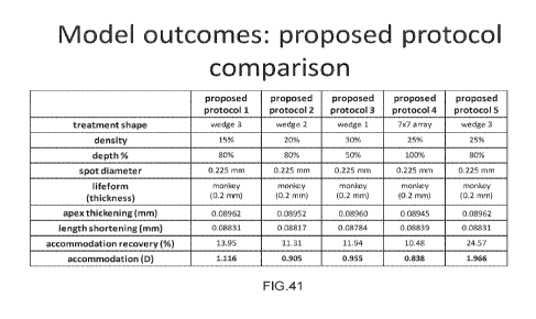
[00103] FIG. 41 illustrates further exemplary treatment patterns, according to
an embodiment
of the disclosure.
[00104] FIG. 42 illustrates exemplary model outcomes, according to an
embodiment of the
disclosure.
DETAILED DESCRIPTION
[00105] The below described figures illustrate the described invention and
method of use in at
least one of its preferred, best mode embodiment, which is further defined in
detail in the following
description. Those having ordinary skill in the art may be able to make
alterations and
modifications to what is described herein without departing from its spirit
and scope. While this
invention is susceptible of embodiment in many different forms, there is shown
in the drawings
and will herein be described in detail a preferred embodiment of the invention
with the
understanding that the present disclosure is to be considered as an
exemplification of the principles
CA 03058461 2019-09-27
WO 2018/183987 PCT/US2018/025608
11
of the invention and is not intended to limit the broad aspect of the
invention to the embodiment
illustrated. All features, elements, components, functions, and steps
described with respect to any
embodiment provided herein are intended to be freely combinable and
substitutable with those
from any other embodiment unless otherwise stated. Therefore, it should be
understood that what
is illustrated is set forth only for the purposes of example and should not be
taken as a limitation
on the scope of the present invention.
[00106] FIGS. 1 to 29 illustrate exemplary embodiments of systems and methods
for laser
scleral microporation for rejuvenation of tissue of the eye, specifically
regarding aging of
connective tissue, rejuvenation of connective tissue by scleral rejuvenation.
[00107] Generally, the systems and methods of the present disclosure take into
consideration
combination of pores filling technique and creating matrices of pores in three
dimensions (3D).
Pores with a particular depth, size and arrangement in a matrix 3D scaffold of
tissue produce plastic
behavior within the tissue matrix. This affects the biomechanical properties
of the scleral tissue
allowing it to be more pliable. It is known that connective tissues that
contain elastin are 'pliable'
and meant to have elasticity. The sclera in fact has natural viscoelasticity.
[00108] Influence of ocular rigidity and ocular biomechanics on the
pathogenesis of age-related
presbyopia is an important aspect herein. Descriptions herein are made to
modifying the structural
stiffness of the ocular connective tissues, namely the sclera of the eye using
the systems and
methods of the present disclosure.
[00109] In order to better appreciate the present disclosure, ocular
accommodation, ocular
rigidity, ocular biomechanics, and presbyopia will be briefly described. In
general, the loss of
accommodative ability in presbyopes has many contributing lenticular, as well
as extralenticular
and physiological factors that are affected by increasing age. Increasing
ocular rigidity with age
produces stress and strain on these ocular structures and can affect
accommodative ability.
Overall, understanding the impact of ocular biomechanics, ocular rigidity, and
loss of
accommodation could produce new ophthalmic treatment paradigms. Scleral
therapies may have
an important role for treating biomechanical deficiencies in presbyopes by
providing at least one
means of addressing the true etiology of the clinical manifestation of the
loss of accommodation
seen with age. The effects of the loss of accommodation has impact on the
physiological functions
of the eye to include but not limited to visual accommodation, aqueous
hydrodynamics, vitreous
CA 03058461 2019-09-27
WO 2018/183987 PCT/US2018/025608
12
hydrodynamics, and ocular pulsatile blood flow. Using the systems and methods
of the present
disclosure to restore more pliable biomechanical properties of ocular
connective tissue is a safe
procedure and can restore accommodative ability in aging adults.
[00110] Accommodation has traditionally been described as the ability of the
crystalline lens of
the eye to change dioptric power dynamically to adjust to various distances.
More recently,
accommodation has been better described as a complex biomechanical system
having both
lenticular and extralenticular components. These components act synchronously
with many
anatomical and physiological structures in the eye organ to orchestrate not
only the visual
manifestations that occur with accommodation, but also the physiological
functions integral to the
eye organ, such as aqueous hydrodynamics and ocular biotransport.
[00111] Biomechanics is the study of the origin and effects of forces in
biological systems.
Biomechanics has remained underutilized in ophthalmology. This biomechanical
paradigm
deserves to be extended to the anatomical connective tissues of the intricate
eye organ.
Understanding ocular biomechanics as it relates to accommodation can allow for
a more complete
picture of the role this primary moving system has on overall eye organ
function, while maintaining
optical quality for visual tasks.
[00112] The eye is a biomechanical structure, a complex sense organ that
contains complex
muscular, drainage, and fluid mechanisms responsible for visual function and
ocular biotransport.
The accommodative system is the primary moving system in the eye organ,
facilitating many
physiological and visual functions in the eye. The physiological role of the
accommodation system
is to move aqueous, blood, nutrients, oxygen, carbon dioxide, and other cells,
around the eye organ.
In addition, it acts as a neuroreflexive loop, responding to optical
information received through the
cornea and lens to fine tune focusing power throughout a range of vision, and
is essentially the
"heart" of the eye organ.
[00113] Biomechanics is particularly important to the complexity of
accommodative function
and dysfunction which occurs with age-related eye diseases (e.g., presbyopia,
glaucoma, age-
related macular degeneration (AMID) and myopia. Age-related changes in the
crystalline lens have
long been understood and reported. Recent endeavors have demonstrated how
stiffening ocular
tissues manifest as presbyopia. Ocular rigidity has been correlated with a
clinically significant loss
of accommodation with age, age-related macular degeneration, increased
intraocular pressure
CA 03058461 2019-09-27
WO 2018/183987 PCT/US2018/025608
13
(TOP), decreased ocular pulsatile blood, and certain forms of glaucoma and
cataracts. Stiffening of
the zonular apparatus and loss of elasticity of the choroid may also
contribute to accommodation.
[00114] Biomechanics plays a critical role in the pathophysiology of the eye
organ. In healthy
young eyes, this mechanism is biomechanically efficient and precisely achieves
the focusing of
objects at a particular distance. As we age, however, this biomechanical
mechanism is affected by
changes in material properties, anatomical relationships, and degradation of
healthy connective
tissue infrastructural relationships due to the aging process. These
biomechanical dysfunctions
result in a disruption of the functions of not only the accommodative
mechanism, which affect the
ability to dynamically focus the lens for ideal optical image quality, but
also the functions of other
physiologic mechanisms critical to the eye organ such as ocular biofluidics,
ocular blood flow, and
metabolic homeostasis. Thus, biomechanics plays a key role in the
pathophysiology that occurs
with aging, including glaucoma and AMD.
[00115] Presbyopia is a condition of sight traditionally defined as the
progressive loss of
accommodative ability with age. The loss of the ability to adjust the dioptric
power of the lens for
various distances, however, is only one consequence of this complex condition.
As the eye ages,
there are connective tissues changes in the eye organ or "oculus" that produce
significant but
reversible impacts on the biomechanical efficiencies of ocular function.
Studies using ultrasound
biomicroscopy (UBM) and endoscopy, optical coherence tomography (OCT), and
magnetic
resonance imaging (MM) have shown age-related changes in the vitreous
membrane, peripheral
choroid, ciliary muscle, and zonules. Age-related changes create biomechanical
alterations that
also manifest in the sclera, which bows inward with increasing age.
[00116] According to one model, during accommodation the ciliary muscle
contracts, releasing
tension on the zonules, which reduces tension on the lens and allows it to
curve and increase its
refractive power. The decrease in lens elasticity with age impedes the
deformation of the lens and
the lens refractive power will not increase enough to see objects at near.
Current approaches to
resolve the loss of near vision symptoms of presbyopia typically included
spectacles, multifocal
or monovision contact lenses, corneal procedures to induce monovision or
multifocality, lens
implants using multifocal lenses, corneal inlays, onlays, and accommodating
intraocular lenses.
However, none of these procedures restore true accommodation. Instead, these
procedures attempt
to improve near and intermediate vision by manipulating optics either in the
cornea or in the lens.
CA 03058461 2019-09-27
WO 2018/183987 PCT/US2018/025608
14
[00117] For true physiological accommodation to occur, the eye must modify its
focal length
to see objects clearly when changing focus from far to near or from near too
far. Generally, this is
thought to be caused primarily by the ciliary muscles, which contract and
force the lens into a more
convex shape. However, the accommodation process is far more complex.
Accommodation is also
influenced by corneal aberrations, and thus to see clearly, the lens must be
molded and undulated
to corneal aberrations, creating a balance of the optics between the lens and
the cornea before
exerting a focal response to accommodative stimulus. In addition, the zonular
tensions on the lens
and the elastic choroid contribute to the accommodative range and
biomechanical functionality of
the entire accommodation complex. The malfunction of these complex components
create a
biomechanical relationship dysfunction, which can affect the accommodative
amplitude, lens
deformation, and the central optical power generated from dynamic
accommodative forces.
[00118] Scleral surgery, e.g., as a treatment for presbyopia has used
corneal incisions to treat
myopia, a treatment known as radial keratotomy (RK). Anterior ciliary
sclerotomy (ACS)
procedure was developed, which utilized radial incisions in the sections of
the sclera overlaying
the ciliary muscle. The incisions were thought to increase the space between
the ciliary muscle
and the lens, allowing for increased 'working distance' for the muscles and
tightening of the
zonules to restore the accommodative ability in presbyopes. The long-term
results of ACS suggest
that the procedure was largely unsuccessful at restoring accommodation and the
effects were
eliminated completely as the scleral wounds healed very quickly. Laser
presbyopia reversal
(LAPR) followed from ACS, using lasers to perform radial sclerectomy. The
results of LAPR,
however, were mixed. Scleral implants attempt to lift the ciliary muscle and
the sclera, tightening
the zonules holding the lens, and restore accommodative ability. Their
effectiveness remains
controversial.
[00119] Accommodation loss and presbyopia have been used interchangeably.
However, it
should be emphasized that accommodation loss is just one clinical
manifestation of the
consequences of an aging (or presbyopic) eye. With increasing age, there are
numerous changes
to the lens and surrounding tissues, which may contribute to accommodation
loss. Research has
shown that the lens substance stiffens with age, decreasing its ability to
change shape (and
refractive power) during accommodation, and decreasing accommodative ability.
The softening of
the lens capsule, flattening of the lens, and lens movement anteriorly with
age may also contribute
to the loss of accommodative ability, however, accommodation is a complex
mechanism. Many
CA 03058461 2019-09-27
WO 2018/183987 PCT/US2018/025608
lenticular-based models fail to incorporate effects from the extralenticular
structures. To
understand accommodation fully, both lenticular and extralenticular components
need to be
considered together.
[00120] The amount of accommodation lost with age, which is related to
extralenticular factors
(primarily the zonules, choroid, and sclera) has only been relatively recently
investigated. The
circumlental space decreases with age. The ciliary body has been shown to
contract during
accommodation, and there is a decrease in the distance from scleral spur to
the ora serrata. Using
UBM, an attachment zone of the posterior zonules adjacent to the ora serrata
has been identified,
and contraction of these zonules is thought to be the etiology of the decrease
in distance found
with accommodation. This complex action of the zonules is suspected to be
reciprocal. While the
anterior zonules relax, reducing their tension on the lens such that the lens
changes shape
anteriorly, the posterior zonules contract, moving the posterior capsule
backward. This vitreal-
zonular complex stiffens with age, losing its elasticity. It is also now known
that the sclera becomes
less deformable during accommodation in the nasal area with age. The vitreous
has also been
suggested as an important factor to lens shape changes during accommodation
and may have a role
in presbyopia. New models suggest up to 3 diopters that might be contributed
by extralenticular
structures. The age-related changes in these structures and their
biomechanical interactions with
the ciliary-lens complex may contribute to presbyopia.
[00121] The ciliary muscle plays a critical role in many functions of the eye
organ including
accommodation and aqueous hydrodynamics (outflow/inflow, pH regulation, and
TOP). An
optically significant role of the ciliary muscles is to adjust the lens
dynamically to focus at various
distances (near, intermediate, and far). During accommodation, the ciliary
muscle contracts to
change the shape of the lens and, in basic terms, moves the lens forward and
inwards. This shape
deformation is caused by the release of tension on the anterior zonules and by
the aqueous fluid
moving in the posterior chamber. This allows the lens to change from a
relatively aspherical shape
to a more spherical shape, thereby increasing its refractive power for near
vision. Contraction of
the ciliary muscle is also important for spreading the trabecular meshwork and
aqueous drainage.
Inadequate drainage or a cause of perturbance to the normal flow of aqueous
drainage either by
uveal outflow pathway or Schlemm's canal can increase TOP and contribute to
the development of
certain types of ocular hypertension or glaucoma. Ciliary muscle contraction
during
accommodation lowers intraocular pressure (TOP). This is likely due to a
decrease in aqueous
CA 03058461 2019-09-27
WO 2018/183987 PCT/US2018/025608
16
outflow resistance during accommodation, caused by the ciliary muscle moving
inward and
anteriorly, which dilates Schlemm's canal and opens the trabecular meshwork.
[00122] FIGs. 1A-(1-3) illustrate, in some embodiments, exemplary scleral
laser rejuvenation
of viscoelasticity allowing compliance in the ciliary muscle. The ciliary
muscle and its components
include the meridional or longitudinal (1), radial or oblique (2), and
circular or sphincteric (3)
layers of muscle fibres, as displayed by successive removal towards the ocular
interior. The cornea
and sclera have been removed, leaving the canal of Schlemm (a), collecting
venules (b) and scleral
spur (c). The meridional fibres (1) often display acutely angled junctions (d)
and terminate in
epichoroidal stars (e). The radial fibres meet at obtuse angles (f) and
similar junctions, at even
wider angles (g), occur in the circular ciliary muscle.
[00123] The rigidity of a structure describes its resistance to deformation
and, in the case of a
confined structure with incompressible contents, rigidity is related to the
structure's volume and
the pressure of the contents. Ocular rigidity refers to the resistance of the
eyeball to stresses.
Increases in ocular rigidity have been correlated with increasing age, lending
support to the idea
that presbyopia and ocular rigidity share a common biomechanical factor. In
addition to affecting
accommodation, ocular rigidity may also hinder the accommodation apparatus to
return to a
disaccommodated state, following an accommodated state, by dampening the
elastic recoil of the
choroid posteriorly.
[00124] Ocular rigidity has been correlated with decreased ocular pulsatile
blood flow. The
blood vessels that support the health of the entire eye pass through the
sclera. An increase in ocular
rigidity could increase scleral resistance to venous outflow and decrease the
flow through choroidal
vessels.
[00125] Ocular rigidity has been correlated to the pathogenesis of macular
degeneration. An
increase in ocular rigidity could increase scleral resistance to venous
outflow and decrease the flow
through choroidal vessels. This may compromise Bruch' s membrane and lead to
choroidal
neovascularization. Decrease flow through the choroidal vessels may also
decrease perfusion,
which could lead to induced hypoxia and choroidal neovascularization.
[00126] Ocular rigidity has been correlated with certain forms of glaucoma.
Recent models
suggest that ocular rigidity affects the scleral response to increased
intraocular pressure. Reducing
ocular rigidity may decrease the mechanical strain that is transferred to the
optic nerve head with
CA 03058461 2019-09-27
WO 2018/183987 PCT/US2018/025608
17
elevated intraocular pressure due to age-related changes and ocular rigidity
in both the anterior
and posterior globe. During normal accommodation the retina and choroid are
pulled forward near
the optic nerve head when the ciliary muscle contracts. The ciliary muscle
retains its contractile
force with age, however increased rigidity of the sclera may affect ciliary
muscle motility, which
could increase the tensional forces on the optic nerve head during ciliary
muscle contraction.
[00127] FIGs 1A-(4-7) illustrate in some embodiments posterior scleral
rejuvenation and ocular
nerve head decompression.
[00128] Ocular rigidity or "stiffness" of the outer ocular structures of the
eye including the
sclera and the cornea, which occurs in the oculus with age, effects the
biomechanical functions of
all the internal anatomical structures, such as the extralenticular and
lenticular anatomy of the
accommodation complex as well as the trabecular meshwork, the choroid and the
retina. In
addition, ocular rigidity has a significant impact on the physiological
functions of the eye organ,
such as a change in the efficiency of aqueous dynamics and ocular pulsatile
blood flow. Increased
ocular rigidity affects other tissues as well, including ocular blood flow
through the sclera and
optic nerve. Ocular rigidity has been correlated to the pathogenesis many age-
related eye diseases.
Therefore, ocular rigidity may not only impact the loss of visual
accommodation but also have
more extensive clinical significance.
[00129] Ocular biomechanics is the study of the origin and effects of forces
in the eye. All
ocular tissues contain collagen, which provides them with viscoelastic
properties. Viscoelastic
substances contain the properties of both fluids and elastic materials. Fluids
tend to take the shape
of their container, while elastic materials can deform under a stress and
return to their original
form. When a stress is applied to viscoelastic materials, the molecules will
rearrange to
accommodate the stress, which is termed creep. This rearrangement also
generates back stresses
in the material that allow the material to return to its original form when
the stress is removed.
Thus, viscoelasticity is an important property that allows tissues to respond
to stresses.
[00130] Chronic stress that exceeds the healing ability of tissues can lead
to chronic
inflammation and eventual cell death, which technically describes the
pathophysiology of aging.
Ocular connective tissues are impacted, like all other connective tissues, by
age. The sclera
constitutes 5/6 of the oculus and is made up of dense irregular connective
tissue. It is comprised
primarily of collagen (50-75%), elastin (2-5%), and proteoglycans. The
connective tissues of the
CA 03058461 2019-09-27
WO 2018/183987 PCT/US2018/025608
18
eye stiffen with increasing age, losing their elasticity, largely due to the
crosslinking that occurs
with age. Crosslinks are bonds between polymer chains, such as those in
synthetic biomaterials or
the proteins in connective tissues. Crosslinking can be caused by free
radicals, ultraviolet light
exposure, and aging. In connective tissues, collagen and elastin can crosslink
to continuously form
fibrils and microfibrils over time. With increasing amounts of fibrils and
microfibrils, the sclera
stiffens, undergoing a sclerosclerosis', as well as a concomitant increase in
metabolic
physiological stress. As mentioned previously, age- and race-related increases
in collagen
crosslinks, along with loss of elastin-driven recoil, and/or collagen
microarchitectural changes,
may underlie the change in scleral material properties leading to loss of
compliance of scleral
tissue when stress is applied. As this pathophysiology progresses, the sclera
exerts compression
and loading stresses on underlying structures, creating biomechanical
dysfunction, specifically
those related to accommodation.
[00131] Age-related increased ocular rigidity also has an impact on the
ciliary muscle and the
biomechanics of the accommodation mechanism. For example, it is known that the
contractile
power of the ciliary muscle does not decrease with age, however, it may have a
decreased
capability to contract or exert substantial forces on the lens to create the
same dioptric changes as
those in a youthful system. A further explanation may be that ocular rigidity
affects the
biomechanical contributions of the ciliary muscle by relaxing zonular tension
and decreasing
accommodative ability.
[00132] Age-related material property changes within the sclera affects the
mobility of
connective tissues of the scleral fibers, directly leading to the loss of
compliance. This causes a
decrease in the normal maintenance and turnover of proteoglycans (PG) in the
sclera, leading to
the loss of PG and eventual tissue atrophy. However, if the compliance and
mobility of scleral
connective tissues are restored, this PG loss can be reversed.
[00133] As mentioned above, the systems and methods of the present disclosure
take into
consideration combination of pores filling technique and creating matrices of
pores in three
dimensions. Pores with a specific depth, size and arrangement in a matrix 3D
scaffold of tissue
produce plastic behavior within the tissue matrix. This affects the
biomechanical properties of the
scleral tissue allowing it to be more pliable. The plurality of pores may be
created in a matrix 3D
CA 03058461 2019-09-27
WO 2018/183987 PCT/US2018/025608
19
scaffold, in an array pattern or a lattice(s). Various microporation
characteristics may be supported.
These may include volume, depth, density, and so on.
[00134] It is advantageous to create a tetrahedral or central hexagon shape.
In order to create a
central hexagon within a matrix there must be a series of 'pores' with
specific composition, depth,
and relationship to the other 'pores' in the matrix and spatial tissue between
the pores in the matrix.
A substantial amount of depth (e.g., at least 85%) of the tissue is also
needed to gain the full effect
of the entire matrix throughout the dimensions of the polygon. The matrix
within the tissue
contains a polygon. The central angle of a polygon stays the same regardless
of the plurality of
spots within the matrix. This is an essential component of the systems and
methods of the present
disclosure since they take advantage of a matrix with a polygon which includes
the unique
relationship and properties of the pore pattern in the matrix or lattice.
[00135] The central angle of a polygon is the angle subtended at the center of
the polygon by
one of its sides. Despite the number of sides of the polygon, the central
angle of the polygon
remains the same.
[00136] Current implanting devices in the sclera obtain the mechanical effect
upon
accommodation. No current devices or methods take into account the effects of
'pores' or creating
a matrix array of pores with a central hexagon or polygon in 3D tissue. The
systems and methods
of the current disclosure may create a pore matrix array in biological tissue
to allow the change in
the biomechanical properties of the tissue itself to create the mechanical
effect upon biological
functions of the eye. A primary requirement of the 'pores' in the matrix is
the polygon.
[00137] A polygon by definition can have any number of sides and the area,
perimeter, and
dimensions of the polygon in 3D can be mathematically measured. In a regular
polygon case the
central angle is the angle made at the center of the polygon by any two
adjacent vertices of the
polygon. If one were to draw a line from any two adjacent vertices to the
center, they would make
the central angle. Because the polygon is regular, all central angles are
equal. It does not matter
which side one chooses. All central angles would add up to 360 (a full
circle), so the measure of
the central angle is 360 divided by the number of sides. Or, as a formula:
Central Angle = 360/n degrees, where n is the number of sides.
CA 03058461 2019-09-27
WO 2018/183987 PCT/US2018/025608
[00138] The measure of the central angle thus depends only on the number of
sides, not the size
of the polygon.
[00139] As used herein, polygons are not limited to "regular" or "irregular."
Polygons are one
of the most all-encompassing shapes in geometry. From the simple triangle, up
through squares,
rectangles, trapezoids, to dodecagons and beyond.
[00140] Types of polygons include regular and irregular, convex and concave,
self-intersecting
and crossed. Regular polygons have all sides and interior angles the same.
Regular polygons are
always convex. Irregular polygons include those where each side may have a
different length,
each angle may be a different measure and are the opposite of regular
polygons. Convex is
understood to mean all interior angles less than 180 , and all vertices 'point
outwards' away from
the interior. The opposite of which is concave. Regular polygons are convex.
Concave is
understood to mean one or more interior angles greater than 180 . Some
vertices push 'inwards'
towards the interior of the polygon. A polygon may have one or more sides
cross back over another
side, creating multiple smaller polygons. It is best considered as several
separate polygons. A
polygon that in not self-intersecting in this way is called a simple polygon.
[00141] Properties of all polygons (regular and irregular) include the
interior angles at each
vertex on the inside of the polygon and the angle on the outside of a polygon
between a side and
the extended adjacent side. The diagonals of a polygon are lines linking any
two non-adjacent
vertices. For regular polygons, there are various ways to calculate the area.
For irregular polygons
there are no general formulae. Perimeter is the distance around a polygon or
the sum of its side
lengths.
[00142] Properties of regular polygons include the apothem (inradius) which is
a line from the
center of the polygon to the midpoint of a side. This is also the inradius -
the radius of the incircle.
The radius (circumradius) of a regular polygon is a line from the center to
any vertex. It is also the
radius of the circumcircle of the polygon. The incircle is the largest circle
that will fit inside a
regular polygon. Circumcircle is the circle that passes through all the
vertices of a regular polygon.
Its radius is the radius of the polygon.
[00143] Some embodiments herein illustrate a plurality of polygons within the
matrix array.
Each can impact the CT (coherence tomography). They contain enough pores to
allow for a
'central hexagon'. A square/diamond shape may be apparent. As a formula:
CA 03058461 2019-09-27
WO 2018/183987 PCT/US2018/025608
21
where:
diagonal = E-2
s is the length of any side
which simplifies to:
where:
diaRathii =
s is the length of any side
[00144] A 'pore' described herein may have a specific form, shape, composition
and depth.
The creating of pores within a matrix array changing biomechanical properties
of connective tissue
is a unique feature of the current disclosure.
[00145] The 'pore matrix' used herein may be used to control wound healing. In
some
embodiments, it may include the filling of pores to inhibit scar tissue.
[00146] In some embodiments, pores may have at least 5%-95% depth through the
connective
tissue, and help create the intended biomechanical property change. They may
have a specific
composition, arrangement in the matrix and desirably have the mathematical
qualities of a
polygon. In three-dimensional (3D) space the intended change in the
relationship between the
pores in the matrix or lattice is the unique characteristic of the current
disclosure (see FIG. 1F).
The matrix or array can comprise of a 2D Bravais lattice, a 3D Bravais Lattice
or a Non-Bravais
lattice.
[00147] Referring to FIGs. 1(B-E), exemplary pore matrix arrays are
illustrated. The pore
matrix arrays herein are the basic building block from which all continuous
arrays can be
constructed. There may be a plurality of different ways to arrange the pores
on the CT in space
where each point would have an identical "atmosphere". That is each point
would be surrounded
by an identical set of points as any other point, so that all points would be
indistinguishable from
each other. The "pore matrix array" may be differentiated by the relationship
between the angles
between the sides of the "unit pore" and the distance between pores and the
'unit pore'. The "unit
pore' is the first "pore created" and when repeated at regular intervals in
three dimensions will
produce the lattice of the matrix array seen on the surface throughout the
depth of the tissue. The
"lattice parameter" is the length between two points on the corners of a pore.
Each of the various
lattice parameters is designated by the letters a, b, and c. If two sides are
equal, such as in a
tetragonal lattice, then the lengths of the two lattice parameters are
designated a and c, with b
CA 03058461 2019-09-27
WO 2018/183987 PCT/US2018/025608
22
omitted. The angles are designated by the Greek letters a, (3, and y, such
that an angle with a
specific Greek letter is not subtended by the axis with its Roman equivalent.
For example, a is the
included angle between the b and c axis.
[00148] A hexagonal lattice structure may have two angles equal to 90 , with
the other angle
(y) equal to 120 . For this to happen, the two sides surrounding the 120
angle must be equal (a =
b), while the third side (c) is at 90 to the other sides and can be of any
length.
[00149] Referring to FIG. 1F, an exemplary schematic projection of the basal
plane of the hcp
unit cell on the close packed layers is illustrated. Matrix array is defined
as the particular, repeating
arrangement of pores throughout a target connective tissue, e.g., the sclera.
Structure refers to the
internal arrangement of pores and not the external appearance or surface of
the matrix. However,
these are not entirely independent since the external appearance of a matrix
of pores is often related
to the internal arrangement. There may be a specific distance between each of
the pores in the
designated matrix to fulfill the mathematical characteristics and properties
of the polygon. The
pores created may also have a relationship with the remaining tissue within
the matrix thus
changing the biomechanical properties of the matrix.
[00150] Spatial relationships of the pores within the matrix have geometric
and mathematical
implications.
[00151] In some embodiments, the laser microporation system (see FIGs. 3) of
the present
disclosure generally includes at least these parameters: 1) a laser radiation
having a fluence
between about 1 - 3 poules/cm2 and about 2 Joules/cm2; > 15.0 J/cm2 on the
tissue; > 25.0 J/cm2
on the tissue; to widen treatment possibilities 2900 nm +/- 200 nm; around the
mid IR absorption
maximum of water; Laser repetition rate and pulse duration may be adjustable
by using pre-defined
combinations in the range of 100 ¨ 500 Hz and 50 ¨225 [is. This range may be
seen as a minimum
range > 15.0 J/cm2 on the tissue > 25.0 J/cm2 on the tissue; to widen
treatment possibilities; 2)
irradiated using one or more laser pulses or a series of pulses having a
duration of between about
1 ns and about 20 i.ts. Some embodiments can potentially have a up to 50W
version; 3) The range
of Thermal Damage Zone (TDZ)can be less than 201.tm in some embodiments or
between 20-501.tm
in some embodiments; 4) Parameters of pulse width from 101.tm-6001.tm can also
be included. (See
FIG. 1E-1)
CA 03058461 2019-09-27
WO 2018/183987 PCT/US2018/025608
23
[00152] The energy per pulses 1-3 microJoules may link to femtolasers and pico
lasers with
high rep rates, e.g., 500 Hz (Zeiss) up to several kilohertz (Optimedica). The
benefit of the femto-
lasers and pico-lasers are the small spot sizes (for example, 20 microns and
up to 50 microns) and
the energy densities are high for minimal thermal problems to surrounding
tissues. All this can
lead to an effective scleral rejuvenation. In some embodiments, the lasers may
produce
substantially round and conically shaped holes in sclera with a depth up to
perforation of sclera
and thermal damage from about 25 p.m up to about 90 p.m. The hole depth can be
controlled by
the pulse energy and the number of pulses. The hole diameter may vary by
motion artifacts and/or
defocusing. The thermal damage may correlate with the number of pulses. The
pulse energy may
be increased, which may lead to a decrease of number of pulses and with this
to a further decrease
of thermal damage. The increase of pulse energy may also reduce the
irradiation time. An
exemplary design of the laser system described allows for laser profiles
optimized for lower
thermal damage zone while preserving irradiation time thus maintaining a fast
speed of for optimal
treatment time, and chart showing the correlation between thermal damage zone
and pulse (see
FIG. 1E-2 and FIGs. 1G-(1-4)).
[00153] The nanosecond lasers for micro poring or micro tunneling, in some
embodiments
include the following specifications: wavelengths UV-Visible-Short infrared
350-355 nm; 520-
532nm; 1030-1064nm typical; -pulse lengths 0,1-500 nanoseconds, passive (or
active Q-
switching; pulse rep. rate 10Hz - 100kHz; peak energies 0,01- 10 milliJoules;
peak powers max.
over 10 Megawatts; free beam or fiber delivered.
[00154] Scleral rejuvenation can be performed with femto- or pico second
lasers and er:YAG
laser. Other preferred embodiments of laser energy parameters ideal for 2.94
ER:YAG laser or
other laser possibilities with ER:YAG preferred laser energy or other lasers
of different
wavelengths with high water absorption.
[00155] MilliJoules and energy densities for different spot sizes/shapes/pores
can include:
[00156] Spot size 50 microns: a) 0,5 mJoules pp is equal to 25 Joules/cm2;
b) 1,0 mJoule pp
is equal to 50 Joules/cm2 (possible with Er:YAG); 3) 2,0 mJoules pp is equal
to 100 Joules/cm2.
[00157] Spot size 100 microns (all these possible with ER:YAG): a) 2,0 mJoules
pp is equal to
25 Joules/cm2; b) 5,0 mJoules pp is equal to 62,5 Joules/cn2; c) 9,0 mJoules
pp is equal to 112,5
Joules/cm2.
CA 03058461 2019-09-27
WO 2018/183987 PCT/US2018/025608
24
[00158] Spot size 200 microns: a) 2,0 mJoules pp is equal to 6,8
Joules/cm2; b) 9,0 mJoules pp
is equal to 28,6 Joules/cm2; c) 20,0 mJoules pp is equal to 63,7 Joules/cm2.
[00159] Spot size 300 microns: a) 9,0 mJoules pp is equal to 12,8
Joules/cm2 - possible with
ER:YAG; b) 20,0 mJoules pp is equal to 28 Joules/cm2 - possible with DPM-
25/30/40/X; c) 30,0
mJoules pp is equal to 42,8 Joules/cm2 d) 40,0 mJoules pp is equal to 57
Joules/cm2 e) 50,0
mJoules pp is equal to 71 Joules/cm2.
[00160] Spot size 400 microns: a) 20 mJoules pp is equal to 16 Joules/cm2 -D
PM-
25/30/40/50/X; b) 30 mJoules pp is equal to 24 Joules/cm2; c) 40 mJoules pp is
equal to 32
Joules/cm2; d) 50 mJoules pp is equal to 40 Joules/cm2
[00161] It is noted that round or square pores or spots are possible as
well.
[00162] Regarding femto & picosecond lasers, some available wave lengths
include IR
1030nm; Green 512nm and UV 343nm. The peak energies can vary from nanoJoules
(at MHz rep
rate) via 5-50 microJoules up to several hundred microJoules in picosecond
region. Femtosecond
lasers having pulse length 100-900 femtosec; peak energies from a nanoJoules
to several hundred
microJoules, pulse rep. rates from 500Hz to several Megahertz (Ziemer LOV Z;
Ziemer AG,
Switzerland: nanoJoules peak energies at over 5 MHz rep. rate, beam
quality/density very good-
focuses in a small spot- 50 micron and under is possible).
[00163] The beam quality being so precise in the best femtolasers that, in
some embodiments,
femtolaser Micro Tunneling of sclera as micro pores using Erbium lasers can be
accomplished.
[00164] As used herein, nuclear pores can be defined as openings in the
nuclear envelope,
diameter about 10 nm, through which molecules (such as nuclear proteins
synthesize in the
cytoplasm) and ma must pass (see FIG. 1H). Pores are generated by a large
protein assembly.
Perforations in the Nuclear membrane which allow select materials to flow in
and out.
[00165] Formula for porosity in biological tissue: X(Xa, t) = qT"(X", t) =x* +
u"(X", t) , (1)
where qU is a continuously differentiable, invertible mapping from 0; to a,
and u" is the cY-
constituent displacement. The invertible deformation gradient for the a-
constituent, F", and its
Jacobian, .I", are defined as J" = det F" (3) where J" must be strictly
positive to prohibit self-
interpenetration of each continuum. The right Cauchy-Green tensor % and its
inverse, the Piola
CA 03058461 2019-09-27
WO 2018/183987 PCT/US2018/025608
deformation tensor B for the solid-constituent are defined as V=F`IF` (4) B =
F'-'F'+ where the
superscript t indicates transposition.
[00166] Current theoretical and experimental evidence suggests that creating
or maintaining
pores in connective tissue accomplishes three important tasks. First, it
transports nutrients to the
cells in the connective tissue matrix. Second, it carries away the cell waste.
Third, the tissue fluid
exerts a force on the wall of the sclera or outer ocular coat, a force that is
large enough for the cells
to sense. This is thought to be the basic mechanotransduction mechanism in the
connective tissue,
the way in which the ocular coat senses the mechanical load to which it is
subjected and the
response to the increase in intraocular pressure. Understanding ocular
mechanotransduction is
fundamental to the understanding of how to treat ocular hypertension, glaucoma
and myopia,
[00167] Deriving the physical properties of a porous medium (e.g., hydraulic
conductivity,
thermal conductivity, water retention curve) from parameters describing the
structure of the
medium (e.g., porosity, pore size distribution, specific surface area) is an
ongoing challenge for
scientists, whether in soft tissues or for porosities of bone tissue and their
permeabilities. To verify
the assumption of a porous medium having a self-similar scaling behavior,
fractal dimensions of
various features have been determined experimentally.
System Procedure and Mechanism of Action
[00168] While some current accommodative theory states that the lens is
primarily responsible
for the refractive change allowing us to read, all elements of the zonular
apparatus have been found
to be involved. Illumination of the role that extralenticular processes play
in accommodation
support the theory that scleral therapies, which modify biomechanical
properties by restoring
compliance to an otherwise rigid tissue, may influence accommodative ability
in presbyopes.
[00169] VisioDynamics theory in particular, argues that presbyopia is not a
refractive error or
simply the loss in the ability to focus on near objects. Instead, it is the
age-related consequences
on connective tissues of the eye organ or oculus, just as they occur
throughout the body. This
produces a significant but reversible impact on the biomechanical efficiencies
of ocular functions,
specifically accommodation, which potentially improves not only dynamic visual
focusing
capability but also ocular biotransport, and ocular metabolic efficiency.
VisioDynamics theory is
based on the fundamental and natural biological occurrences that occur with
age, and specifically
resonates on the effects of ocular rigidity to the accommodative structures
beneath the major outer
CA 03058461 2019-09-27
WO 2018/183987 PCT/US2018/025608
26
coat of the eye or sclera. The sclera undergoes a gradual "sclerosclerosis"
with age, which
represents the normal and gradual irreversible changes which occur in all
connective tissues. This
sclerotic process increases scleral compression, which imposes staggeringly
significant load,
stress, and strain upon underlying and related ocular and intraocular
structures. This ocular rigidity
or stress and strain upon the ciliary body and related structures which
control dynamic
accommodation, impact the biomechanics of the eye and compromises the eye's
ability to perform
its core organ functions.
[00170] In some embodiments, an ocular laser surgery and therapeutic
treatments system
provides an eye laser therapy procedure designed to alleviate the stresses and
strain that occur with
an increasingly rigid sclera with age by creating compliance in the scleral
tissue using a laser
generated matrix of micropores in the scleral tissue. The system aims to
facilitate biomechanical
property changes in the sclera, to alleviate compression of the subliminal
connective tissue, facial
tissue, and biophysiological structures of the eye, and restore accommodative
ability. The system
is specifically designed to alleviate stress and increase biomechanical
compliance over the ciliary
muscle, the accommodation complex, and key physiological anatomy that lies
directly beneath the
aging scleral tissue.
[00171] The laser therapy procedure targets specific treatment areas which are
in distinct
physiological zones covering critical anatomy inside the eye relative to eye
function. Although
examples of 3 or 5 physiological zones are described herein, other number of
physiological zones
may also be considered for treatments.
[00172] In some embodiments, a treatment pattern may be described as 3
critical zones in 3
distinct distances from the outer edge of the anatomical limbus (AL), not
touching any components
or relative tissues of the cornea. These zones are illustrated in FIGs. 2A-(1-
2). In some
embodiments, a treatment pattern may be described as 5 critical zones in 5
distinct distances from
the outer edge of the anatomical limbus (AL), not touching any components or
relative tissues of
the cornea, as illustrated in FIGs. 2B-(1-3).
[00173] The laser therapy procedure may use an erbium: yttrium¨aluminum¨garnet
(Er:YAG)
laser to create microspores in the sclera. These micropores may be created at
a plurality of depths
with preferred depth range, e.g., from 5%-95% of the sclera, up to the point
where the blue hue of
the choroid is just visible. The micropores may be created in a plurality of
arrays including a matrix
CA 03058461 2019-09-27
WO 2018/183987 PCT/US2018/025608
27
array, e.g., 5mm x 5mm, 7mm x 7mm, or 14mm x 14mm matrix array. These
microporation
matrices break bonds in the scleral fibrils and microfibrils having an
`uncrosslinking' effect in the
scleral tissue. A direct consequence of this matrix pattern is the creation of
areas of both positive
stiffness (remaining interstitial tissue) and negative stiffness (removed
tissue or micropores) in the
rigid sclera. These areas of differential stiffness allow the viscoelastic
modulus of the treated sclera
to be more compliant over the critical zones when subjected to force or
stress, such as contraction
of the ciliary muscles. Additionally, the treated regions of the sclera may
produce a dampening
effect in rigid scleral tissue when the ciliary muscles contract, due to
increased plasticity. This
enhances accommodative effort by directing unresisted forces inward and
centripetally toward the
lens or facilitating inward upward movement of the accommodative mechanism.
This is an
advantage over the model that postulates a net outward-directed force at the
lens equator. For
example, techniques which are directed at scleral expansion such as scleral
implants or surgical
laser radial ablations such as LAPR are all directed at increasing 'space' or
circumlental space to
allow the sclera to expand for the intention of giving the ciliary muscle room
. These techniques
are based on the 'lens crowding' theory and aim to induce the outward movement
rather than the
upward and inward movement of the sclera and ciliary mechanism. Overall, the
creation of the
micropore matrices in the scleral tissue induces an `uncrosslinking effect',
severing the fibrils and
microfibrils of the layers of the sclera allowing a more compliant response to
applied stress. Thus,
the proposed mechanism of action for the system is to increase plasticity and
compliance of scleral
tissue over critical zones of anatomical significance by creating these
regions of differential
stiffness over the ciliary complex, and thereby improve biomechanical function
and efficiency of
the accommodation apparatus. FIGs. 2C-(1-4) illustrate in some embodiments
laser scleral
uncrosslinking of scleral fibrils and microfibrils.
[00174] Referring to FIGs. 2D(1-4), using a novel model, the effect of the
procedure on ocular
rigidity has been investigated. Ocular connective tissues are impacted, like
all other connective
tissues, by age. The sclera constitutes 5/6 of the oculus and is made up of
dense irregular
connective tissue. It is comprised primarily of collagen (50-75%), elastin (2-
5%), and
proteoglycans. The connective tissues of the eye stiffen with increasing age
losing their elasticity
largely due to the crosslinking that occurs with age. Crosslinking creates an
"increase in
biomechanical stiffness" in connective tissues such as those in the eye.
Crosslinks are bonds
between polymer chains, such as those in synthetic biomaterials or the
proteins in connective
CA 03058461 2019-09-27
WO 2018/183987 PCT/US2018/025608
28
tissues. Crosslinking can be caused by free radicals, ultraviolet light
exposure, and aging. In
connective tissues, collagen and elastin can crosslink to continuously form
fibrils and microfibrils
over time. With increasing amounts of fibrils and microfibrils, the sclera
stiffens, undergoing a
sclerosclerosis', as well as a concomitant increase in metabolic physiological
stress. As this
pathophysiology progresses, the sclera exerts compression and loading stresses
on underlying
structures, creating biomechanical dysfunction, specifically those related to
accommodation. Laser
Scleral Microporation breaks scleral fibrils and microfibrils effectively
"uncrosslinking" bonds
thereby increasing scleral compliance and "decreasing biomechanical
stiffness".
[00175] In some exemplary operations, six freshly harvested porcine eyes were
modified by
crosslinking (0.8m1 of 2% glutaraldehyde for 10 minutes) to mimic the ocular
rigidity of an older
human eye (60 year old), based on the ocular rigidity coefficient model of
Pallikaris et al.. Seven
freshly harvested porcine eyes were left unmodified to mimic the ocular
rigidity of a young human
eye (30 years). Three of the eyes in each group received the treatment, while
the remaining eyes
were used as controls. In brief, the investigation used a pressure transducer
(up to 5 psi), a dosage
injector controller, a data computerized reader, and tissue holding frame to
which each porcine eye
was fixed, to generate an IOP versus injected volume curve for each eye. The
ocular rigidity
coefficient (K = d ln(P)/dV [in mmHg/ 1]) was then calculated as the slope of
ln(I0P) (from TOP
between 30-50 mmHg) versus injected volume. In the young eye, the treatment
resulted in a 10.8%
decrease in rigidity. In the older eye, the treatment resulted in a 30.1%
decrease in rigidity. Using
an analysis of variance (ANOVA) and Tukey honestly significant difference
(TukeyHSD) test, the
investigation found that The system significantly reduced ocular rigidity in
the old eyes and overall
(p = 0.0009; p = 0.0004). This decrease in ocular rigidity may be caused by
`uncrosslinking' aging
tissue.
[00176] In some exemplary operations, twenty-six subjects underwent the
treatment, and 21
completed 24 months of post-operative care. Five patients withdrew, due to
occupational travel
conflicts. The pre-operative (month 0) and post-operative TOP (determined by
pneumatic
tonometry) are shown in. There is an immediate 5% drop in TOP for the patient
eyes compared to
pre-operative TOP. Over the two years following the treatment, patient TOP
remains approximately
15% lower than pre-operative TOP. The immediate and sustained reduction in TOP
could be
demonstrative of an improvement in aqueous outflow following the treatment.
Using an ANOVA
CA 03058461 2019-09-27
WO 2018/183987 PCT/US2018/025608
29
and TukeyHSD test, these differences were statistically significant beginning
at post-operative
month 3 and continued through all subsequent months (p = 0.000063 at 24 months
postoperatively). This reduction in TOP may be indicative of enhanced ocular
mobility and a
decrease in ocular rigidity following the treatment.
[00177] The biomechanical improvements with the treatment may prove to
increase the
biomechanical efficiency of the accommodative apparatus. In some embodiments,
by creating
micropores in a matrix over four oblique quadrants, the treatment may restore
functional
extralenticular forces, and restore a minimum of 1-3 diopters of
accommodation. Our reported
results show an average of 1.5 diopters of accommodation post-operatively.
This significantly
improved the visual acuity of our patients. Data from a 24-month postoperative
follow-up of the
clinical study were presented in 2015 and show promising results. Visual
acuity was measured
using standard Early Treatment Diabetic Retinopathy Study (ETDRS) charts, and
statistical
analysis was done using an ANOVA and TukeyHSD test. The uncorrected monocular
near visual
acuity of the patients was 0.25 0.18 logMAR (mean standard deviation) at
24 months post-
operatively, compared to 0.36 0.20 logMAR (mean standard deviation) pre-
operatively (p <
0.00005).
[00178] In summary, utilizing innovative biometry and imaging technologies
that were not
previously available has illuminated that the loss of accommodative ability in
presbyopes has many
contributing lenticular, as well as extralenticular and physiological factors.
The lens, lens capsule,
choroid, vitreous, sclera, ciliary muscles, and zonules all play a critical
role in accommodation,
and are affected by increasing age. Increasing ocular rigidity with age
produces stress and strain
on these ocular structures and can affect accommodative ability.
[00179] Scleral therapies may have an important role in treating
biomechanical deficiencies in
presbyopes, by providing at least one means to address the true etiology of
the clinical
manifestation of the loss of accommodation seen with age. The treatment,
utilizing laser
microporation of the sclera to restore more pliable biomechanical properties,
is a safe procedure,
and can restore accommodative ability in aging adults. As a result, the
treatment improves dynamic
accommodative range as well as aqueous outflow. With the advent of improved
biometry, imaging,
and research focus, information about how the accommodation complex works and
how it impacts
the entire eye organ can be achieved.
CA 03058461 2019-09-27
WO 2018/183987 PCT/US2018/025608
[00180] Referring to FIG. 2E, exemplary three critical zones of significance
as measured from
the anatomical limbus (AL)) are shown. Zone 1) 0.5-1.1mm from the AL, over the
scleral spur at
the origin of the ciliary muscle; Zone 2) 1.1-4.9mm from the AL, over the mid
ciliary muscle body;
Zone 3) 4.9-5.5mm from the AL, over the insertion of the longitudinal muscle
fibers of the ciliary,
just anterior to the ora serrata at the insertion of the posterior vitreous
zonules. FIG. 2E(b)
illustrates exemplary restored mechanical efficiency and improved
biomechanical mobility.
[00181] In some embodiments, the laser scleral microporation procedure may
involve using the
laser described above to perform partial-thickness micro-ablations in the
sclera in a matrix in five
critical anatomic zones, 0-7.2mm from the anatomical limbus (AL). The five
zones may include:
Zone 0) 0.0-1.3mm from AL; distance from the AL to the superior boundary of
ciliary
muscle/scleral spur; Zone 1) 1.3-2.8mm from AL; distance from the sclera spur
to the inferior
boundary of the circular muscle; Zone 2) 2.8-4.6mm from AL; distance from the
inferior boundary
of the circular muscle to the inferior boundary of the radial muscle; Zone 3)
4.6-6.5mm from AL;
inferior boundary of the radial muscle to the superior boundary of the
posterior vitreous zonule
zone; and Zone 4) 6.5-7.2mm from AL; superior boundary of the posterior
vitreous zonule zone
to the superior boundary of the ora serrata.
[00182] FIG. 2F illustrates an exemplary matrix array of micro-excisions in
four oblique
quadrants.
[00183] FIG. 2G illustrates an exemplary graphical representation of restored
ocular
compliance, decreased scleral resistive forces, increased ciliary resultant
forces, and restored
dynamic accommodation following the treatment.
[00184] FIG. 2H illustrates an exemplary box-and-whiskers plot of the ocular
rigidity for
control (black) and treated (grey) porcine eyes. The upper and lower
extremities of the box
represent the 75th and 25th percentiles, the bar within the box represents the
median, and the
whiskers represent the full extent of the data ranges.
[00185] FIG. 21 illustrates an exemplary box-and-whiskers plot of pre- and
post-operative
intraocular pressure (TOP) for the patient eyes. The stars indicate a
significant difference from pre-
operative IOP. The upper and lower extremities of the box represent the 75th
and 25th percentiles,
the bar within the box represents the median, the whiskers represent the full
extent of the data
ranges, and the white circles represent outliers.
CA 03058461 2019-09-27
WO 2018/183987 PCT/US2018/025608
31
[00186] FIG. 2J illustrates exemplary charts showing uncorrected and distance-
corrected visual
acuity at distance 4 m, intermediate (60 cm), and near (40 cm) for a)
monocular and b) binocular
patient eyes. Error bars represent mean SD.
[00187] As described herein, accommodation of a human eye occurs through a
change or
deformation of the ocular lens when the eye transitions from distant focus to
near focus. This lens
change is caused by contraction of intraocular ciliary muscles (ciliary body),
which relieves
tension on the lens through suspensory zonule fibers and allows the thickness
and surface curvature
of the lens to increase. The ciliary muscle can have a ring-shaped and can be
composed of three
uniquely oriented ciliary fiber groups that contract toward the center and
anterior of the eye. These
three ciliary fiber groups are known as longitudinal, radial and circular.
Deformation of the ciliary
muscle due to the contraction of the different muscle fibers translates into
or otherwise causes a
change in tension to the surface of the ocular lens through zonule fibers,
whose complex patterns
of attachment to the lens and ciliary muscle dictate the resultant changes in
the lens during
accommodation. Ciliary muscle contraction also applies biomechanical strain at
the connection
locations between the ciliary muscle and the ocular sclera, known as the white
outer coat of the
eye. Additionally, biomechanical compression, strain or stress can be
caused during
accommodation can occur at connection locations between the ciliary muscle and
the choroid,
known as the inner connective tissue layer between the sclera and ocular
retina. Ciliary muscle
contraction can also cause biomechanical forces on the trabecular meshwork,
lamina cribrosa,
retina, optic nerve and virtually every structure in the eye.
[00188] Applying the techniques and models described with respect to the
various embodiments
herein using simulations can lead to outputs and results that fall within
known ranges of
accommodation of a young adult human.
[00189] 3D mathematical models can incorporate mathematics and non-linear
Neohookean
properties to recreate behavior of the structures of biomechanical,
physiological, optical and
clinical importance. Additionally, 3D (Finite Element Model) FEM models can
incorporate data
from imaging, literature and software relating to the human eye.
[00190] Visualization of accommodation structures during and after simulations
may be
included in addition to means for measuring, evaluating and predicting Central
Optical Power
(COP). These can be used to simulate and view age specific whole eye
structures, optics, functions
CA 03058461 2019-09-27
WO 2018/183987 PCT/US2018/025608
32
and biomechanics. Further, they can independently simulate properties of the
ciliary muscle,
extra-lenticular and lenticular movements of the ocular lens and functions on
the ocular lens.
Individual simulations of anatomical structures and fibers can reveal
biomechanical relationships
which would otherwise be unknown and undefined. Numerical simulation of the
patient's eye can
be created using 3D FEM meshing to accomplish these operations.
[00191] To elaborate, representative 3D geometry of resting ocular structures
can be
computationally defined based on extensive review of literature measurements
and medical images
of the anatomy of young adult eyes and through modeling. Specialized methods
implemented in
software, such as AMPS software (AMPS Technologies, Pittsburgh, PA), can be
used to perform
geometric meshing, material property and boundary conditions definitions, and
finite element
analysis during the modeling stage. Ciliary muscle and zonules can be
represented as a transverse
isotropic material with orientations specified to represent complex fiber
directions. Additionally,
computational fluid dynamic simulations can be performed in order to produce
fiber trajectories,
which can then be mapped to the geometric model.
[00192] Initially, a lens modeling can include a lens in a relaxed
configuration, before being
stretched by pre-tensioning zonule fibers to an unaccommodated position and
shape.
Unaccommodated lens position can be reached when zonules are shortened, e.g.,
to between 75%
and 80% of their starting length, and more particularly to about 77% of their
starting length. Then
accommodative motion can be simulated by performing active contraction of the
various fibers of
the ciliary muscle. In some embodiments, this can be accomplished using
previous models of
skeletal muscle that are modified to represent dynamics particular or
otherwise specific or unique
to the ciliary muscle. Model results representing lens and ciliary anterior
movement and deformed
ocular lens thickness at a midline and apex can be validated or otherwise
verified by comparing
them to existing medical literature measurements for accommodation. In order
to investigate
contributions of the various ciliary fiber groups to the overall action of the
ciliary muscle,
simulations can be performed for each fiber group by activating each in
isolation while others
remain passive or otherwise unchanged.
[00193] Various beneficial aspects of the embodiments described below are
described with
respect to simulations applying pre-tensioning zonules models and contracting
ciliary muscle
models.
CA 03058461 2019-09-27
WO 2018/183987 PCT/US2018/025608
33
[00194] With respect to the pre-tensioning zonules, modeling can include: 1)
Creation of 3D
material sheets oriented between measured zonular attachment points of
insertion on the lens and
origination on the ciliary/choroid; 2) specified fiber direction in the plane
of the sheet (i.e. fibers
directed from origin to insertion); and 3) Transversely isotropic constitutive
material with tension
development in the preferred direction. Further, with particular respect to
3), advantages have
been achieved, including: a) Time-varying tension parameter input regulates
the stress developed
in the material; b) Time-varying tension input is tuned to produce required
strain in the lens to
match measurements of the unaccommodated configuration; c) Age variation in
material
properties and geometries to produce age-related impact; and d) others.
[00195] With respect to the contracting ciliary muscle models, modeling can
include: 1)
Modified constitutive model to represent smooth and skeletal aspects of
ciliary mechanical
response; 2) 3 sets of specified fiber directions to represent physiological
orientation of muscle
cells and lines of action of force production; and 3) Transversely isotropic
constitutive material
with active force development in the preferred direction. Further, with
particular respect to 3),
advantages have been achieved, including: a) Activation parameter input
regulates the active stress
developed in the material; b) Activation input is tuned to produce appropriate
accommodative
response to match literature measurements; c) Activation of individual muscle
fiber groups can be
varied in isolation to assess contributions to lens strain/stress; d)
Activation of individual muscle
fiber groups can be varied in isolation to assess contributions to ocular
scleral strain/stress; e)
Activation of individual muscle fiber groups can be varied in isolation to
asses contributions to
choroidal strain/stress; and f) others.
[00196] In various embodiments, simulation results can be governed by
modification of
tensioning and activation inputs to the zonule and ciliary materials, as
opposed to performing an
applied displacement to external node(s) of a mesh.
[00197] Thereafter, systems, methods and devices for providing a predictive
outcome in the
form of a 3D Computer Model with integrated Artificial Intelligence (AI) can
be used to find
predictive best instructions for a therapeutic ophthalmic correction,
manipulation, or rehabilitation
of a patient's vision defects, eye disease, or age-related dysfunction are
disclosed. The predictive
best instruction can be derived from physical structural inputs, neural
network simulations, and
prospective therapeutic-outcome-influencing. New information can be analyzed
in conjunction
CA 03058461 2019-09-27
WO 2018/183987 PCT/US2018/025608
34
with optimized, historical therapeutic-outcome information in order to provide
various benefits.
The concepts herein can be used to performs a multitude of simulations and has
a knowledge-
based platform so that the system is able to improve its instruction response
as the database is
expanded.
[00198] In some embodiments, the stored instructions contemplated can
preferably be an
optimized, custom, photoablative algorithm for driving a photoablative,
photothermal laser. The
instructions can be provided along with an AT processor via direct
integration, stand-alone
importation or remotely via a Bluetooth enabled application or connection.
These instructions can
be performed a priori or intraoperatively.
[00199] In some embodiments, the stored instructions contemplated can
preferably be an
optimized custom ocular lens simulation algorithm used for simulating
manipulation of an
implantable intraocular lens in order to improve medical procedures and
understanding.
[00200] The instructions can also be set up as a 'stand-alone' system for use
as a Virtual Clinical
trial system or Research and Development system, whereby the instructions can
be provided with
independent research design inputs and outputs to test various conditions and
responses of the eye
to surgical manipulations, implantation devices, or other therapeutic
manipulations of the eye, in
order to optimize design and outcome response.
[00201] Additionally, these instructions can also include one or more of: an
algorithm for image
processing interpretation, expansion of ophthalmic imaging data platforms and
a companion
diagnostic to an imaging device.
[00202] As described herein, methods for improving ophthalmic treatments,
surgeries, or
pharmacological interventions can include obtaining topological,
topographical, structural,
physiological, morphological, biomechanical, material property, and optical
data for a human eye
along with applied physics and analyzing through mathematical simulations
using artificial
intelligence networks.
[00203] Virtual clinical applications using simulation can include techniques
executed via
devices, systems and methods for automated design of an ophthalmic surgical
procedure including
physical measurements and applied physics of a patient's whole eye are
obtained. Conventional
techniques can be used to obtain these measurements. The information measured
can be
CA 03058461 2019-09-27
WO 2018/183987 PCT/US2018/025608
interpolated and extrapolated to fit nodes of a finite element model (FEM) of
a human eye for
analysis, which can then be analyzed to predict an initial state of stress of
the eye and obtain pre-
operative conditions of the cornea, lens and other structures. Incision data
constituting an "initial"
surgical plan can be incorporated into the finite element analysis model. A
new analysis can then
be performed to simulate resulting deformations, biomechanical effects,
stresses, strains,
curvatures of the eye as well as dynamic movements of the eye, more
specifically the ciliary
muscles, lens and accommodative structures. These can be compared to original
values thereof
and to a vision objective. If necessary, a surgical plan can be modified and
resulting new ablation
data can be entered into the FEM and the analysis is repeated. This procedure
can be repeated as
desired or necessary until the vision objectives are met.
[00204] Artificial Intelligence (Al) software can use an artificial neural
network to conduct
machine learning, whereby the system can learn from the data, and therefore
has a learning
component based on the ongoing database expansion. It can be operative to
improve reliability as
the database is formulated and updated, heretofore unknown in the prior art of
3D predictive
modeling systems, methods and devices.
[00205] Simulation can include Age Progression simulation of a patient's eye,
having a
predictive capacity to simulate ophthalmic surgical outcomes, determine rates
of regression of
treatments, as well as execute predictive algorithms for future surgical or
therapeutic enhancement,
heretofore unknown in the prior art of 3D predictive modeling systems, methods
and devices.
[00206] Virtual Eye Simulation Analyzer can include integration of information
related to all
structures of an eye into a computer program for the purpose of simulating
biomechanical and
optical functioning of the eye, as well as age related simulations for
clinical application purposes.
[00207] Virtual Eye Simulation Analyzer systems, devices and methods can
include an output
display that can be viewed by users as a standalone or integrated display
system, along with other
equipment.
[00208] Information used as inputs for the simulator can include imaging
information for
Biometry (UBM, OCT and others). Dynamic Imaging can be performed using UBM,
OCT and
others. Anatomy information can include geometry, histology and others.
Physiological function
information can include dynamic accommodation, aqueous flow, intraocular
pressures, pulsatile
ocular blood flow, retinal performance or compromise and others. Material
Properties of tissues
CA 03058461 2019-09-27
WO 2018/183987 PCT/US2018/025608
36
of the eye, physics and biomechanical information related to relative
biomechanics can also be
used.
[00209] The simulator can incorporate mathematics and non-linear Neohookean
properties in
order to recreate behavior of the structures of biomechanical, physiological,
optical and others that
may be valuable or otherwise of clinical importance. The simulator can use
conventional methods
to input data incorporated into a 3D FEM with a patient's unique data based on
analysis of their
own individual eye or eyes. Further, the simulator can use conventional
methods to input data
and create a numerical simulation of the patient's eye using a 3D FEM meshing¨
essentially
creating a custom dynamic real-time "Virtual Eye," heretofore unknown in the
prior art of 3D
predictive modeling systems, methods and devices.
[00210] In some embodiments, the AT may be capable of learning via predictive
simulation and
can be operative to improve simulative predictions for surgical or therapeutic
manipulations of the
eye through artificial neural networks, e.g., in an "ABACUS" program. ABACUS
can also be
capable of providing instructions directly to a communicatively coupled
processor or processing
system to create and apply algorithms, mathematical sequencing, formula
generation, data
profiling, surgical selection and others. It can also be capable of providing
instructions directly to
a workstation, an image processing system, a robotic controller or other
device for implementation.
Further, it can be capable of providing instructions indirectly through a
Bluetooth or other remote
connection to a robotic controller, an image system or other workstation.
[00211] The models herein can have various applications for clinical,
research and surgical use,
including: 1) use of prior evaluation and simulation of accommodation
functions of the eye
(examples including Presbyopia indication-IOL design and use, extra-lenticular
therapeutics and
their uses); 2) use of prior evaluation and simulation of aqueous flow of the
eye, such as for
glaucoma indications; 3) virtual simulations and real time simulations of
efficacy of IOL' s,
therapeutic treatments and various biomechanical implications; 4) virtual
simulations using the AT
and CI to reproduce customized aging effects on an individual's biomechanical
and physiological
functions of their eye which have clinical importance; 5) Surgical Planning;
6) design model (such
as FEM) importation and simulation, such as for IOL' s and others; 7) Virtual
clinical trials and
analysis; 8) real-time intraoperative surgical analysis, planning and
execution; 9) Performance of
CA 03058461 2019-09-27
WO 2018/183987 PCT/US2018/025608
37
a crystalline lens of the eye as it relates to optical and biomechanical
dysfunction, cataract
formation and the like; and 10) others.
[00212] Additional components of simulators may include: 1) Eye Scanning; 2)
Optical inputs
such as a) Cornea optics, wavefronts, elastography, hysteresis, visual acuity
topography,
connective tissue macro and micro structure and b) lens optics such as
wavefront, visual acuity,
topography, lens opacity, light scatter, central optical power (COP) during
accommodation and
disaccommodation, elastography, viscoelastic properties and others; 3) Scleral
biomechanics,
viscoelastic, material properties, stress, strain mapping, connective tissue
macro/micro structure;
4) Trabecular meshwork material, viscoelastic, connective tissue macro and
micro structure; 5)
Lamina cribrosa material properties, stress, strain viscoelastic, connective
tissue macro and
microstructure; 6) Physiological Inputs including a) Aqueous outflow and
inflow, b) Intra Ocular
Pressure (TOP), c) Ocular pulsatile blood flow, d) Retinal activity and
others; 7) Surface
Spectroscopy; 8) Collagen Fibril characterization of the cornea, sclera, lens,
and others; and 9)
others.
[00213] Benefits of simulators in an accommodation embodiment may include: 1)
Measuring,
analyzing and simulating accommodation of an eye in real-time; 2)
demonstrating accommodation
biomechanics in real-time; 3) evaluating accommodation biomechanics; 4)
Visualization of
accommodation structures; 5) Measuring, evaluating and predicting Central
Optical Power; 6)
Simulating age progression of whole eye structures, functions and
biomechanics; and 7) others.
[00214] Major structural component inputs can be based on the sclera, cornea,
lens, trabecular
meshwork, lamina cribrosa, retina and others. For the sclera these can
include: Scleral rigidity,
viscoelasticity, Scleral thickness, Scleral depth, 3D surface topology, top
surface spectral
dimensions, 3D spectroscopy and others. For the cornea these can include:
Corneal Wavefront,
viscoelasticity, Topography, Keratotomy, Corneal thickness, 3D topology, K
readings, Corneal
stiffness, 3D spectroscopy and others. For the lens these can include:
Lenticular Wavefront,
Central optical power, Accommodative amplitude, Light scattering, Opacity and
others. For the
trabecular meshwork, these can include: elasticity, outflow, inflow and
others. For the lamina
cribrosa this can include: porosity, mechanical dependence, perfusion,
poroelasticity, cup floor
depth, and others.
CA 03058461 2019-09-27
WO 2018/183987 PCT/US2018/025608
38
[00215] Some of the various major optical profiles, properties, information
and visual acuity
information outputs for a cornea can include: Total aberrations, Visual Strehl
Ratio, Depth of
focus, MRSE, Visual acuity, lens scatter and others. Some of the various major
optical profiles,
properties, information and visual acuity information outputs for a lens can
include: Total
aberrations, VSOF, Depth of focus and others.
[00216] Also described are example embodiments of a creation of a 3D
Microporation Model
on a spherical surface.
[00217] Also described are example embodiments of Pantec Protocols Revised
Fibonacci
MatLab Pore Calculation for Whole Eye Patterns.
[00218] Referring to FIG. 2K-1, an example of Protocol Execution is now
described: Protocol
1.1: 225um (169 Total Pores @ 3% = 42.25 Pores/Quadrant). An example of Matlab
code used
for Protocol 1.1 may include: >> fibonacci spiral connected Pantec
('r',0.225,3,6.62,9.78).
Matlab code parameter breakdown may include: Parameter 1, 'r' = pore shape:
type in 'r' for
rectangular or 'c for circular pore shape. . Use r' for 'please' and 'c' for
`DPM25'*; Parameter 2,
225um (0.225) = r shape: length of the rectangular pore shape or the radius of
the circular pore
shape in [mm]; Parameter 3, 3% = D: pore density in [percent]; Parameter 4,
6.62mm (Radius of
the zone taken away from the pore calculation.) This is so there is no pores
calculated in the
corneal/limbus area (6.62mm) = r b: radius for the beginning of the circle in
[mm]; Parameter 5,
9.78mm (Radius to the end of the zone for pore calculation). The 6.62mm radius
will be subtracted
from the process of the pore calculation, thus allowing 6.62mm to 9.78mm
radius being the only
calculated area with pores = r e: radius for the ending of the circle in [mm].
Once the code
(('r',0.225,3,6.62,9.78)) is entered in Matlab, it will output the figure
generated specifically for this
pore protocol. It is how the title got its total pore number.
[00219] Therapy Manipulation protocols: The following are exemplary protocols
for Therapy
Manipulation which are 2 Manipulations per Protocol: a) First Manipulation of
entire quadrant
area; b) Second Manipulation of "Patch" area 5x5mm diamond, i) This diamond
having a length
of its diagonal = 5*V(2)=7.07mm, ii) The 5x5 matrix to place on the sphere we
have updated the
Fibonacci spirals can meet models.
[00220] Sphere comparison: Our "Patch" is 5x5 in some embodiments, so that
dimension is
used. Er:Yag laser with fiber optic probe; 600 p.m spot size; Nine micro-
excisions in the 4 oblique
CA 03058461 2019-09-27
WO 2018/183987 PCT/US2018/025608
39
quadrants; 10 min/eye treatment time; Micropores in the critical zones (e.g.,
3 or 5 zones) over the
ciliary complex; Creation of pliable matrix zones in the Sclera.
[00221] Procedure Objectives can include: 1) Improve compliance of sclera over
ciliary muscle
complex critical anatomy; 2) Restore mechanical efficiency of the natural
accommodative
mechanism; 3) Improve biomechanical mobility to achieve accommodative power;
and others.
[00222] An exemplary Fibonacci treatment pattern was generated through Matlab
or other
programs in two dimensions. When having correctly sized patches, such as
5x5mm, it may make
an actual treatment that may not fit in the critical zones (e.g., zones 1-3,
or 1-5). There is a way to
get an actual estimate from a 3D model to a 2D model. As illustrated in FIG.
2K-1, parameters
can include:
[00223] Baseline: 600[tm (92 Total Pores @ 16% = 23 Pores/Quadrant).
[00224] Spot Size: 600um; Depth: 80%; Density: 16%; Volume Removed: 1.16mm3;
Total
Pores Entirety: 92; Total Pores/Quadrant: 23.
[00225] Protocol 1.1: 225[tm (169 Total Pores @ 3% = 42.25 Pores/5.5mm Patch:
Validated)
Total Pores/5.5mm patch.
[00226] FIGs. 2K-1-A to 2K-1-C illustrate exemplary protocol parameters
producing a
diamond pattern for 3 critical zones.
[00227] In some embodiments, it can be important to know what is in each
protocol how many
pores are in the 5x5 patch on the 3D model pursuant to the changing density
and the changing spot
size. Once known, patch manipulations can be performed. FIGs. 2K-(2-17)
illustrate exemplary
views of various protocols used and their results. These protocols include:
[00228] Protocol 1.1: 225 m(96 Total Pores @ 3% = 24 Pores/Quadrant:
Validated)
[00229] Spot Size: 225um; Depth: 80%; Density: 3%; Volume Removed: 0.91mm3;
Total
Pores Entirety: 96; Total Pores/Quadrant: 24
[00230] Protocol 1.2: 225 m(161 Total Pores @ 5% = 40.25 Pores/Quadrant:
Validated)
[00231] Spot Size: 225um; Depth: 80%; Density: 5%; Volume Removed: 1.52mm3;
Total
Pores Entirety: 161; Total Pores/Quadrant: 40.25
[00232] Protocol 1.3: 225 m(257 Total Pores @ 8% = 64.25 Pores/Quadrant:
Validated)
CA 03058461 2019-09-27
WO 2018/183987 PCT/US2018/025608
[00233] Spot Size: 225um; Depth: 80%; Density: 8%; Volume Removed: 2.43mm3;
Total
Pores Entirety: 257; Total Pores/Quadrant: 64.25
[00234] Protocol 1.4: 225 m(565 Total Pores @ 10% = 141.25 Pores/Quadrant:
Validated)
[00235] Spot Size: 225um; Depth: 80%; Density: 10%; Volume Removed: 3.04mm3;
Total
Pores Entirety: 565; Total Pores/Quadrant: 141.25
[00236] Protocol 2.1: 250 m(100 Total Pores @3% = 25 Pores/Quadrant:
Validated)
[00237] Spot Size: 250um; Depth: 80%; Density: 3%; Volume Removed: 0.91mm3;
Total
Pores Entirety: 100; Total Pores/Quadrant: 25
[00238] Protocol 2.2: 250 m(166 Total Pores @ 5% = 41.5 Pores/Quadrant:
Validated)
[00239] Spot Size: 250um; Depth: 80% Density: 5%; Volume Removed: 1.52mm3;
Total
Pores Entirety: 166; Total Pores/Quadrant: 41.5
[00240] Protocol 2.3: 250 m(265 Total Pores @ 8% = 66.25 Pores/Quadrant:
Validated)
[00241] Spot Size: 250um; Depth: 80%; Density: 8%; Volume Removed: 2.43mm3;
Total
Pores Entirety: 265; Total Pores/Quadrant: 66.25
[00242] Protocol 2.4: 250 m(332 Total Pores @ 10% = 83 Pores/Quadrant:
Validated)
[00243] Spot Size: 250um; Depth: 80%; Density: 10%; Volume Removed: 3.04mm3;
Total
Pores Entirety: 332; Total Pores/Quadrant: 83
[00244] Protocol 3.1: 325 m(59 Total Pores @3% = 14.75 Pores/Quadrant:
Validated)
[00245] Spot Size: 325um; Depth: 80%; Density: 3%; Volume Removed: 0.91mm3;
Total
Pores Entirety: 59; Total Pores/Quadrant: 14.75
[00246] Protocol 3.2: 325 m(98 Total Pores @ 5% = 24.5 Pores/Quadrant:
Validated)
[00247] Spot Size: 325um; Depth: 80%; Density: 5%; Volume Removed: 1.52mm3;
Total
Pores Entirety: 98; Total Pores/Quadrant: 24.5
[00248] Protocol 3.3: 325 m(157 Total Pores @ 8% = 39.25 Pores/Quadrant:
Validated)
[00249] Spot Size: 325um; Depth: 80%; Density: 8%; Volume Removed: 2.43mm3;
Total
Pores Entirety: 157; Total Pores/Quadrant: 39.25
CA 03058461 2019-09-27
WO 2018/183987 PCT/US2018/025608
41
[00250] Protocol 3.4: 325 m(196 Total Pores @ 10% = 49 Pores/Quadrant:
Validated)
[00251] Spot Size: 325um; Depth: 80%; Density: 10%; Volume Removed: 3.04mm3;
Total
Pores Entirety: 196; Total Pores/Quadrant: 49
[00252] Protocol 4.1: 425 m(34 Total Pores @ 3% = 8.5 Pores/Quadrant:
Validated)
[00253] Spot Size: 425um; Depth: 80%; Density: 3%; Volume Removed: 0.91mm3;
Total
Pores Entirety: 34; Total Pores/Quadrant: 8.5;
[00254] Protocol 4.2: 425 m(57 Total Pores @ 5% = 14.25 Pores/Quadrant:
Validated)
[00255] Spot Size: 425um; Depth: 80%; Density: 5%; Volume Removed: 1.52mm3;
Total
Pores Entirety: 57; Total Pores/Quadrant: 14.25
[00256] Protocol 4.3: 425 m(92 Total Pores @ 8% = 23 Pores/Quadrant:
Validated)
[00257] Spot Size: 425um; Depth: 80%; Density: 8%; Volume Removed: 2.43mm3;
Total
Pores Entirety: 92; Total Pores/Quadrant: 23
[00258] Protocol 4.4: 425 m(115 Total Pores @ 10% = 28.75 Pores/Quadrant:
Validated)
[00259] Spot Size: 425um; Depth: 80%; Density: 10%; Volume Removed: 3.04mm3;
Total
Pores Entirety: 115; Total Pores/Quadrant: 28.75
Below are exemplary code references for the protocols:
[00260] fibonacci spiral connected Pantec('r',0.225,3,6.62,9.78)>>1.1
[00261] fibonacci spiral connected Pantec('r',0.225,5,6.62,9.78)>>1.2
[00262] fibonacci spiral connected Pantec('r',0.225,8,6.62,9.78)>>1.3
[00263] fibonacci spiral connected Pantec('r',0.225,10,6.62,9.78)>>1.4
[00264] fibonacci spiral connected Pantec('e,0.125,3,6.62,9.78)>>2.1
[00265] fibonacci spiral connected Pantec('e,0.125,5,6.62,9.78)>>2.2
[00266] fibonacci spiral connected Pantec('e,0.125,8,6.62,9.78)>>2.3
[00267] fibonacci spiral connected Pantec('e,0.125,10,6.62,9.78)>>2.4
[00268] fibonacci spiral connected Pantec('e,0.1625,3,6.62,9.78)>>3.1
CA 03058461 2019-09-27
WO 2018/183987 PCT/US2018/025608
42
[00269] fibonacci spiral connected Pantec('c',0.1625,5,6.62,9.78)>>3.2
[00270] fibonacci spiral connected Pantec(' c',0. 1625,8,6.62,9.78)>>3 .3
[00271] fibonacci spiral connected Pantec('c',0.1625,10,6.62,9.78)>>3 .4
[00272] fibonacci spiral connected Pantec(' c',0.2125,3 ,6.62,9.78)>>4. 1
[00273] fibonacci spiral connected Pantec('c',0.2125,5,6.62,9.78)>>4.2
[00274] fibonacci spiral connected Pantec('c',0.2125,8,6.62,9.78)>>4.3
[00275] fibonacci spiral connected Pantec('c',0.2125,10,6.62,9.78)>>4.4
[00276] As noted, the inputs include: Pore Diameter (ull); Pore Depth (ull); #
of Pores; Density
of Pores; Angle of the Zones of the pores; Position of the laser beam from the
surface and others
if desired or required.
[00277] Various inputs may be used for adequate and accurate modeling. These
can include
pore size in p.m, since pore size actually changes parameters and not just the
proportions of # spots
and pattern. Density should also be factored in, as well as surface area
formula, number of pores
as related to Pore Size as per the Power Calculations, angle and long arc in
each zone of the eye
sphere where each spot or row of spots will be placed are needed, angles that
will the laser spots
be in for each zone are using eye parameter inputs, and others.
[00278] In some embodiments, depth is fixed and at least two tests can be
simulated, such as
depth at 50% = 4541.tm or Depth at 80% = 70011.m.
[00279] Protocol Requirements for each Treatment Pattern can include: Spot
size; Depth;
Number Pores Whole Globe in All Quadrants; Number Pores/Quadrant; Number
Pores/5.5mm
patch; Volume removed; Density -- (How many spots). Performing therapy
manipulations can
include: whole quadrant vs. patch (surface area), where specific corneal
diameter of the shape
change eye can be important.
[00280] An example embodiment of applications of Artificial Intelligence,
Simulations and
Field Applications may include: 1) use for R&D of the Eye for various Modeling
implementations;
2) Virtual Clinical Trials; 3) Laser Integration as a diagnostic companion or
Robotics controller;
4) Performing virtual surgery on the eye for a "Smart Surgery" plan; 5)
Integration to Imaging
CA 03058461 2019-09-27
WO 2018/183987 PCT/US2018/025608
43
Devices improve image interpretation; 6) Integration to Surgical Microscope
for 'Real Time"
modification of surgery/therapy (for e.g. IOL surgery); and 7) others.
[00281] Functions of simulations can include: 1) Simulations of ideal
biomechanics for
optimizing Total visual function and best central optical power for
accommodation; 2) Simulations
of ideal biomechanics for optimizing total visual function and best optical
power of the cornea; 3)
Simulations of ideal biomechanics for optimizing decreased outflow of aqueous
from the
Trabecular meshwork; 4) Simulations of ideal biomechanics for optimizing
retinal decompression
of lamina cribrosa and parapapillary sclera; 5) Simulations for optimizing
scleral rejuvenation; 5)
Simulations for optimizing surgical outcomes of intra ocular lens surgery; 6)
Simulations for
optimizing surgical or therapeutic outcomes for corneal surgery; 7) Age
progression simulations
to evaluate long term effects of aging on eye function; 8) Age progression
simulations to evaluate
long term stability and outcomes of various surgical procedures of the eye; 9)
Simulations for
analyzing testing of applications, therapies, surgical manipulation,
implantation devices and
pharmacological treatments of the eye via Virtual clinical trials; and 10)
others.
[00282] Algorithms and other software used to implement the systems and
methods disclosed
herein are generally stored in non-transitory computer readable memory and
generally contain
instructions that, when executed by one or more processors or processing
systems coupled
therewith, perform steps to carry out the subject matter described herein.
Implementation of the
imaging, machine-learning, prediction, automated correcting and other subject
matter described
herein can be used with current and future developed medical systems and
devices to perform
medical procedures that provide benefits that are, to date, unknown in the
art.
[00283] In some embodiments, the described systems, methods and devices are
performed prior
to or contemporaneous with various medical procedures. In some embodiments,
they may be
implemented in their own systems, methods and devices, along with any required
components to
accomplish their respective goals, as would be understood by those in the art.
It should be
understood that medical procedures benefitting from the herein described
material are not limited
to implementation using the material described hereafter, but other previous,
currently performed
and future developed procedures can benefit as well.
[00284] FIG. 3A illustrates an exemplary laser treatment system according to
some
embodiments of the present disclosure. In some embodiments, a treatment laser
beam travels to
CA 03058461 2019-09-27
WO 2018/183987 PCT/US2018/025608
44
dichroic 208. At dichroic 208 the laser beam travels to Galvo Setup 320 which
consists of Galvol
210 and Galvo2 212. The beam then passes from Galvo Setup 320 to focusing
optics 216 and
ultimately to patient eye 140.
[00285] Also provided for in this embodiment is a control and monitoring
system which broadly
consists of a computer 310, video monitor 312, and camera 308. Camera 308
provides monitoring
of the laser beam at dichroic 208 via lens 306. Camera 308 transmits its feed
to computer 310.
Computer 310 is also operable monitor and control Galvo Setup 320. Computer
310 is also coupled
to video monitor 312 to provide a user or operator a live feed from camera
308.
[00286] In some embodiments of the invention a dual axis closed loop
galvanometer optics
assembly is used.
[00287] Since multiple lasers systems may be used for treatment in some
embodiments,
additional laser systems will now be described.
[00288] The laser system may include a cage mount galvanometer containing a
servo controller,
intelligent sensor, feedback system and mount assembly with an optical camera.
Some
embodiments may include use of a cage mount galvanometer optics assembly. Some
embodiments
may include ultra-high resolution nano-positioners to achieve sub-nanometer
resolution.
[00289] To expand, FIG. 3A shows more exemplary detail of a CCD (or CMOS)
camera-based
eye tracker subsystem. Dichroic 208 beam splitter is used to pick off visible
light, while allowing
the IR treatment beam to transmit. The beam splitter 208 is located in front
of the steering
elements, shown here as galvo mirrors 320. Lens 306 images the tissue plane
(eye) onto the
camera. Features in the image field (e.g. blood vessels, edge of the iris,
etc.) are identified by
image processing and their coordinates in the camera pixel field computed. If
the eye moves within
the pixel field frame-to-frame, the change in position of the reference
features can be computed.
An error function is computed from the change in reference feature position
and commands issued
to the galvo mirrors 320 to minimize the error function. In this
configuration, the optical line of
sight is always centered on the treatment spot, which is at a fixed coordinate
in the camera pixel
field. The apparent motion from repositioning the galvos 320 will be to move
the eye image
relative to the fixed treatment spot.
CA 03058461 2019-09-27
WO 2018/183987 PCT/US2018/025608
[00290] FIG. 3B illustrates an exemplary laser treatment system 303 according
to an
embodiment of the present disclosure. The laser treatment system 303 is
similar to FIG. 3A, except
that the eye tracking subsystem is located after galvo mirrors 320.
[00291] In this embodiment, a treatment laser beam travels to Galvo Setup 320
which consists
of Galvol 210 and Galvo2 212. The beam then passes from Galvo Setup 320 to
dichroic 208. At
dichroic 208 the laser beam travels to focusing optics 216 and ultimately to
patient eye 140.
[00292] Also provided for in this embodiment is a control and monitoring
system which broadly
consists of a computer 310, video monitor 312, and camera 308. Camera 308
provides monitoring
of the laser beam at dichroic 208 via lens 306. Camera 308 transmits its feed
to computer 310.
Computer 310 is also operable monitor and control Galvo Setup 320. Computer
310 is also coupled
to video monitor 312 to provide a user or operator a live feed from camera
308.
[00293] Here, the eye image is shown centered in the pixel field. When eye
motion is detected
within the pixel field, the galvos 320 are repositioned to move the treatment
spot to a new position
within the pixel field corresponding to the movement of the eye, and to a
desired fixed position
relative to the eye reference features.
[00294] With reference to the aforementioned biofeedback loop, eye tracking
includes in some
embodiments includes use of light source producing an infrared illumination
beam projected onto
an artificial reference affixed to an eye. The infrared illumination beam is
projected near the visual
axis of the eye and has a spot size on the eye greater than the reference and
covering an area when
the reference moves with the eye.
[00295] In some embodiments, the reference has a retro-reflective surface that
produces
backward scattering orders of magnitude stronger than backward scattering from
the eye would.
An optical collector may be configured and positioned a distance from the eye
to collect this
backward scattered infrared light in order to form a bright image spot of the
reference at a selected
image location.
[00296] The bright image spot appears over a dark background with a single
element
positioning detector positioned at the selected image location to receive the
bright image spot and
configured to measure a two-dimensional position of the bright image spot of
the reference on the
positioning detector. An electric circuit may be coupled to the positioning
detector to produce
CA 03058461 2019-09-27
WO 2018/183987 PCT/US2018/025608
46
positioning signals indicative of a position of the reference according to a
centroid of the bright
image spot based on the measured two-dimensional position of the bright image
spot on the
positioning detector.
[00297] FIG. 3C illustrates an exemplary camera correction system according to
an
embodiment of the present disclosure. In the example embodiment, the top row
illustrates the
camera focus location after galvos have been used and the bottom row
illustrates the camera focus
location before galvos. Various landmarks 392 may be seen in the example
embodiments
including capillaries, iris, pupil, etc. Treatment spot 394 may also be seen
in each embodiment.
[00298] As is shown in the example embodiment the top row of focus before the
galvos each
show the pupil of as the center pixel of each image. Compensation after galvos
in the bottom row
allows the treatment spot 394 to remain the focus of the camera's attention in
each image and
thereby allow the system to remain in position for the associated procedure.
[00299] FIG. 3D illustrates an exemplary flow diagram 330 of a camera-based
eye tracker
process according to an embodiment of the present disclosure.
[00300] Broadly put, the diagram represents the use of a CCD or CMOS camera to
capture an
image of eye. Image data is transmitted to a computer, where key features are
segmented/extracted
(e.g. blood vessels, iris features, edge of pupil). The image is stored as a
reference frame.
Subsequent images are then compared to reference frame. A shift is computed
after comparing
reference features in pixel coordinates. Conversion of pixel coordinates to
scanning system
coordinates then occurs before commanding the scanning system to deviate
treatment beam line
of site to restore relationship relative to reference features. If the shift
is too large or out of range
of scanning system, halt procedure and take steps to reacquire the target
image field.
[00301] As a more detailed explanation referencing each step, an
initialization or starting
sequence according to some embodiments requires capture image frame in step
332 before
processing the captured image frame in order to extract features in step 334.
This captured frame
with extracted features is then used to set a reference frame in step 336.
[00302] After a reference frame is set, step 338 consists of capturing an
additional image frame,
called a current frame. This image or current frame is processed in step 340
in order to extract
features. Step 342 consists of comparing the current frame to the reference
frame which was set
CA 03058461 2019-09-27
WO 2018/183987 PCT/US2018/025608
47
in step 336. An image shift is computed between the current frame and the
reference frame in
order to determine the difference between the frames. A comparison to a pre-
set threshold allows
the system to determine if the image shift exceeds the pre-set threshold and
stops the procedure at
this point by going to step 352.
[00303] If an image shift does not exceed the pre-set threshold and therefore
is not too large,
the system computes a compensation level in step 346 in order to compensate
for the change or
shift between the current frame and the reference frame. This compensation
level is computed
into physical coordinates used by a scanner in step 348. The scanner is then
commanded to
compensate using the coordinates in step 350. After this compensation step 338
occurs and another
current image frame is captured and the cycle continues.
[00304] FIG. 4A illustrates an exemplary laser treatment system 400 according
to an
embodiment of the present disclosure. In the example embodiment, laser
treatment system 400
consists of a treatment laser 202 emitting a laser beam which travels through
relay lens 204 to
dichroic or flip-in 208. Visible spotting laser 206 emits a laser beam which
also travels to dichroic
or flip-in 208. In some embodiments, the beams from treatment laser 202 and
visible spotting
laser 206 may meet simultaneously at first dichroic or flip-in 208. In other
embodiments, the
beams may reach first dichroic or flip-in 208 at staggered times.
[00305] The beam or beams leave first dichroic or flip-in 208 and travels to a
second dichroic
208. The beam or beams leave second dichroic 208 and travel to Galvo 210.
Galvol 210 may
consist of a mirror which rotates through a galvanometer set-up in order to
move a laser beam.
The beam or beams leave Galvo 210 and travel to Galvo2 212 which may be a
similar setup to
Galvol 210. The beam or beams leave Galvo2 212 and travel to dichroic
(visible/IR) 214.
Operator 160 may monitor the beam or beams at dichroic (visible/IR) 214 by
using a surgical
microscope 150. The beam or beams travel from dichroic (visible/IR) 214
through focusing optics
216 to patient eye 140.
[00306] In FIG. 4A, additional monitoring elements are provided for use by
operator 160 to aid
in medical procedures. Depth control subsystem 302 assists in controlling the
depth of ablation
procedures in accordance with the present invention and receives input from
second dichroic 208.
FIGs. 4A-(1-10)illustrate how microporation/nanoporation may be used to remove
surface,
subsurface and interstitial tissue and affect the surface, interstitial,
biomechanical characteristics
CA 03058461 2019-09-27
WO 2018/183987 PCT/US2018/025608
48
(e.g., planarity, surface porosity, tissue geometry, tissue viscoelasticity
and other biomechanical
and biorheological characteristics) of the ablated target surface or target
tissue.
[00307] Similarly, eye tracker 304 assists in tracking landmarks on patient
eye 140 during
medical procedures in accordance with the present invention and receives input
from second
dichroic 208. Another dichroic 208 is shown in the example embodiment
splitting the beam with
outputs to eye tracker 304 and depth control subsystem 302.
[00308] FIG. 4B illustrates an exemplary laser treatment system including
ablation pore depth
according to an embodiment of the present disclosure. FIG. 4B generally shows
a treatment laser
beam traveling to dichroic 208 before travelling to Galvol 210, then to Galvo2
212, through
focusing optics 216, and to patient eye 140. As shown above, FIGs. 4A-(1-
10)illustrate how
microporation/nanoporation may be used to remove surface, subsurface and
interstitial tissue and
affect the surface, interstitial, biomechanical characteristics (e.g.,
planarity, surface porosity, tissue
geometry, tissue viscoelasticity and other biomechanical and biorheological
characteristics) of the
ablated target surface or target tissue.
[00309] An OCT system 404 is an Optical Coherence Tomography system used to
obtain
subsurface images of the eye. As such, when coupled to computer 310 which is
coupled to video
monitor 312, OCT system 404 provides a user or operator the ability to see
subsurface images of
the tissue ablation; pore ablation can be between 5% and 95% of the sclera
thickness, with average
sclera thickness is 700[tm a typical pore depth could be magnitudes of order
larger than refractive
surface ablation at around 200um-300um deep. This is significantly greater
depth than other
surface refractive ablative procedures that are typically between 10um-45um[tm
in depth on
average and generally >120um.
[00310] In at least some embodiments OCT provides a real-time, intraoperative
view of depth
levels in the tissue. OCT may provide for image segmentation in order to
identify sclera interior
boundary to help better control depth. As shown above, FIGs. 4A-(1-10)
illustrate how
microporation/nanoporation may be used to remove surface, subsurface and
interstitial tissue and
affect the surface, interstitial, biomechanical characteristics (e.g.,
planarity, surface porosity, tissue
geometry, tissue viscoelasticity and other biomechanical and biorheological
characteristics) of the
ablated target surface or target tissue.
CA 03058461 2019-09-27
WO 2018/183987 PCT/US2018/025608
49
[00311] In some embodiments, the OCT system 404 uses an OCT measurement beam,
injected
into the treatment beam line of sight via a dichroic beam splitter 208,
located before the scanning
system. In this way, the OCT system line of sight is always centered on the
pore being ablated.
The OCT system is connected to a computer 310 for processing the images and
for control of the
laser.
[00312] In some embodiments of the invention an anatomy avoidance subsystem is
provided to
identify critical biological obstacles or locations during procedures (e.g.
blood vessels and others).
As such, subsurface visualization may be provided to identify obstacles such
as blood vessels or
anatomy that is desired to be avoided intraoperatively.
[00313] FIG 4A-5 and FIG. 4B show exemplary simple diagrams of an ablation
pore in the
sclera showing an example of the depth of an ablation in relation to the inner
boundary of the
sclera.
[00314] FIG. 5 illustrates an exemplary flow diagram of OCT-based depth
control 410
according to an embodiment of the present disclosure.
[00315] In general, the OCT system executes a repetitive B-scan, synchronized
with the laser.
The B-scan shows the top surface of the conjunctiva and/or sclera, the
boundaries of the pore being
ablated, and the bottom interface between the sclera and the choroid or
ciliary body. Automatic
image segmentation algorithms are employed to identify the top and bottom
surfaces of the sclera
(typically 400 - 1000 microns thick) and the boundaries of the ablated pore.
The distance from the
top surface of the sclera to the bottom surface of the pore is automatically
computed and compared
to the local thickness of the sclera. In some embodiments, this occurs in real
time. When the pore
depth reaches a predefined number or fraction of sclera thickness, ablation is
halted and the
scanning system indexed to the next target ablation location. In some
embodiments, images may
be segmented to identify interior sclera boundaries.
[00316] With reference to the steps in the figure, in the example embodiment a
starting or
initialization set of steps occurs first. This starting set of steps begins
with positioning to a pore
coordinate in step 412. AB-scan of the target region occurs in step 414. This
scan creates an
image which is processed in step 416 in order to segment and identify the
sclera boundary. A
distance is then computed in step 418 between the conjunctive surface and the
sclera boundary.
CA 03058461 2019-09-27
WO 2018/183987 PCT/US2018/025608
[00317] After completion of this starting set of steps ablation is
initiated in step 420. A laser
beam pulse is fired in step 422 followed by a B-scan in step 424. This B-scan
creates an image
that is then segmented in step 426 and pore depth and ablation rate are
computed from the image.
This pore depth and ablation rate are compared to the target depth in step
430. If the target depth
has not been reached, then the process loops back to step 422 and repeats.
Upon reaching a target
depth step 432 stops the ablation process and the starting process begins
again at step 434 with
positioning to a next pore coordinates. In some embodiments, the OCT system
can monitor
ablation depth during a single pulse and can stop the ablation as a risk
mitigation means, there may
also be other internal processes running that can end the ablation if the
process is out of range; eye
tracking operational limits exceeded, max preset # of pulses exceeded, laser
power monitoring is
not in limits. All of these are risk mitigation measures.
[00318] FIG. 6 illustrates an exemplary laser treatment system component map
600 showing
relation of related subsystems according to an embodiment of the present
disclosure.
[00319] In general laser treatment system component map 600 shows a laser 602,
a laser
delivery fiber 120, laser control system 604, monitoring system 608, and beam
control system 606.
[00320] Laser 602 is generally made up of several subsystems. In the example
embodiment,
these subsystems include system control electronics 104, Er:YAG laser head
612, laser cooling
system 108, HV power supply 110, and system power supplies 112. Foot pedal 114
provides some
control for the system user. Laser 602 transmits a laser beam via laser
delivery fiber 120 to beam
control system 606.
[00321] Beam control system 606 is generally made up of beam transport optics
624, red
spotting laser 626, galvo mirrors 628, beam delivery optics 630, and active
focus 632.
[00322] Laser control system 604 maintains a link to laser 602 via a laser
sync and to beam
control system 606 via power control position status. Laser control system 604
is generally made
up of a user interface 614, power supply 616, galvo controller 618, galvo
controller 620, and
microcontroller 622. Laser control system 604 is also manipulatable via
joystick 610.
[00323] Monitoring system 608 is generally made up of CCD camera 634 and
visual
microscope 636.
CA 03058461 2019-09-27
WO 2018/183987 PCT/US2018/025608
51
[00324] In some embodiments, a fiber laser is used which is composed of an
undoped cladding
and a doped core of higher refraction. The laser beam travels through the
fiber guided within the
fiber core and experiences a high amplification due to the length of
interaction. Fiber lasers are
considered advantageous to other laser systems because, among other qualities,
they have simple
thermal management properties, high beam quality, high electrical efficiency,
high optical
efficiency, high peak energy, in addition to being low cost, requiring low
maintenance, having
superior reliability, a lack of mirror or beam path alignment, and they are
lightweight and generally
compact.
[00325] In some embodiments of the invention spot arrays may be used in order
to ablate
multiple pores at once. These spot arrays may, in some cases, be created using
microlenses and
also be affected by the properties of the laser. A larger wavelength may lead
to a smaller number
of spots with increased spot diameter.
[00326] Turning to FIG. 7, an exemplary laser treatment system 700 is shown
according to an
embodiment of the present invention. Laser treatment system 700 is generally
made up of control
system 702, optics and beam controls.
[00327] Control system 702 includes monitor 704 and monitor2 706 as well as
keyboard 708
and mouse 710 to provide a user the ability to interact and control with a
host computer 724 running
computer programs. In many embodiments, the computer programs running on host
computer 724
include control programs for controlling visible spotting laser 712, laser
head 714, laser cooling
system 716, system power supplies 718, laser power supply 720, and beam
transport optics 722.
[00328] Also provided for in this embodiment are depth control subsystem 726,
galvo mirrors
728, CCD Camera 730, visual microscope 732, focus subsystem 734, and beam
delivery optics
736.
[00329] FIG. 7-1 illustrates another exemplary laser treatment system.
[00330] Preoperative measurement of ocular properties and customization of
treatment to an
individual patient's needs is beneficial in many embodiments. Preoperative
measurement of ocular
properties may include measuring intraocular pressure (TOP), scleral
thickness, scleral
stress/strain, anterior vasculature, accommodative response, and refractive
error. Measurement of
scleral thickness may include use of optical coherence tomography (OCT).
Measurement of
CA 03058461 2019-09-27
WO 2018/183987 PCT/US2018/025608
52
scleral stress/strain may include using Brillouin scattering, OCT
elastography, photoacoustics
(light plus ultrasound). Measurement of anterior vasculature may include using
OCT or Doppler
OCT. Measurement of refractive error may include using the products such as
the iTrace
trademarked product from Tracey Technologies Corp.
[00331] Intraoperative biofeedback loops may be important during the procedure
in order to
keep the physician informed on the progress of the procedure. Such feedback
loops may include
use of topographical measurements and monitoring "keep away" zones such as
anterior ciliary
arteries.
[00332] Biofeedback loops may include a closed-loop sensor to correct for
nonlinearity in the
piezo scanning mechanism. The sensor in some embodiments may offer real-time
position
feedback in a few milliseconds and utilizing capacitive sensors for real-time
position feedback.
Real-time position feedback may be communicated to a controller, and, upon
identification of
specific biological features based on tissue characteristics, may cease laser
operation
intraoperatively.
[00333] Sensor/feedback apparatus may also perform biological or chemical
"smart sensing" to
allow ablation of target tissue and protect or avoid surrounding tissue. In
some instances, this smart
sensing may be accomplished by using a biochip incorporation in a mask which
is activated by
light irradiation and senses location, depth, size, shape, or other parameters
of an ablation profile.
Galvo-optic assemblies are also contemplated in some embodiments and may be
used to gage
numerous parameters of laser steering and special function.
[00334] In some embodiments, the described systems, methods and devices may
include image
display transfer and GUI interface features that can include each image frame
taken and send
information to a video display after each firing inside the 3-dimension ¨ 7-
dimension micropore
before and after the firing of the laser in dynamic real time and surface
view. The GUI may have
integrated multiview system in 7-directionality for image capture including:
Surface, internal pore,
external pore, bottom of the micropore, whole globe eye view, target array
area.
[00335] In some embodiments, 7-cube may be a preferred projection for the
microprocessor but
other examples exists in dimensional Sphere Shape, integrated into the GUI and
microprocessor.
Orthogonal projections can include examples as shown in FIG. 8.
CA 03058461 2019-09-27
WO 2018/183987 PCT/US2018/025608
53
[00336] SVM pattern recognition is integrated into the AT (artificial
intelligence) network
directed to the microprocessor path. For the non-linear classification problem
the SVM will turn
the input space into a high dimensional space by a nonlinear mapping K(X).
Hence, the nonlinear
problem will turn into a linear problem and then the optimal separating
hyperplane will be
calculated in a new high dimensional space using Matlab or Mathematica
integrated programming.
As the optimization functions and classification functions involve only the
inner product between
samples (xi-xe) the transformed higher dimensional space is also just the
inner product (k(xi)-
k(xe)). If the kernel function (k(xi - k(xe) satisfies with Mercer condition,
it corresponds to a
transform space of inner product K (xi, x = ( k(xi)- k(x)). The common kernel
functions include
linear kernel polynomial kernel and radial bias kernel function. The use of
appropriate kernel
function can be an alternative to non-linear aping of high dimensional space,
which will achieve a
linear classification after nonlinear transformation. The corresponding
classification discriminant
function can be obtained as follows:
!L,
A:0
= sgti( rx;y0; = x) -I- 1?).
.1,ov
[00337] In some instances, mapping and optimization formulas for machine
learning may
include:
g(x) = sgn1 a* yi(xi* x) + b*)
g(x) = sgn1 a* yi(xi* x) + b*)
g(x) =sgn(1 aryiK(xi x x) + b*)
[00338] Instrument of the GUI interface & code may include multi-dimensional
scaling, linear
discriminant analysis and linear dimensional reduction processing as well as
locally linear
embedding and isometric maps (ISOMAP) are nonlinear dimensionality reduction
methods also
included.
CA 03058461 2019-09-27
WO 2018/183987 PCT/US2018/025608
54
[00339] A continuous mapping p : E B satisfying the homotopy lifting property
with respect
to any space may be used. Fiber bundles (over paracompact bases) constitute
important examples.
In homotopy theory, any mapping is 'as good as' a fibration¨i.e. any map can
be decomposed as
a homotopy equivalence into a "mapping path space" followed by a fibration
into homotopy fibers.
[00340] The fibers are by definition the subspaces ofE that are the inverse
images of
points b of B. If the base space B is path connected, it is a consequence of
the definition that the
fibers of two different points bi and b2 in B are homotopy equivalent.
Therefore, one usually
speaks of "the fiber" F.
[00341] Some embodiments can utilize a Serre fibration or Weak fibration. They
are able to
produce mapping of each cylinder micropore in the array and the total array
across the 3D surface
and interstitial mapping of pore arrays in cross section. An exemplary 3D
mapping 900 is shown
in FIG. 9.
[00342] FIG. 10 illustrates an exemplary design patterns that can be performed
as follows. Step
1001: Treatment design/ planning begins with tissue hierarchy which is
established using the 7
Sphere mathematical projection over entire sphere to establish congruent
treatment platform built
on 7 D shape and hyperbolic planar tessellation. Step 1002: Off Axis
mathematical algorithm
derived from tissue hierarchy and Fibonacci patterning is displayed as
mathematical imagery. Step
1003: Algorithmic Code is then implemented to develop customized microporation
patterns that
are reflective of the tissue biorheology including all inputs of rigidity,
viscoelastic modulus,
topology, topography, biometry etc. Step 1004 (not shown): Anatomy avoidance
software is
executed erasing or eliminating untargeted fields, arrays, regions. Step 1005
(not shown):
Surgeon/user can also manipulate the targeted or untargeted areas via touch
screen interface.
[00343] In some embodiments, the described systems, methods and devices may
include the
following features of laser user interface system delivery of treatment
algorithms. Real time
mathematical imagery is incorporated and displayed both in 3D mathematical
files which can also
be run in a GIF animation format to display apriori information regarding the
array effectiveness.
The workstation/algorithms work together with the VESA system in order to
produce the
mathematical imagery to the user/surgeon for ideal configuration of the 3 D
array on the eye. The
topological representation of the image is projected stereographically to the
display. The array is
prefixed formularies and in addition can be simulated in Fibonacci sequencing
with a plurality of
CA 03058461 2019-09-27
WO 2018/183987 PCT/US2018/025608
densities, spot sizes, micro and nano pore geometries and configurations. The
benefit of the
Fibonacci sequencing is to produce the most balanced array formulary which
corresponds to the
body's own natural tissue hierarchy both in macro and micro scales.
[00344] The array can also follow a hyperbolic geometry model or a uniform
(regular,
quasiregular, or semiregular) hyperbolic tiling which is an edge-to-edge
filling of the hyperbolic
plane which has regular polygons as faces and is vertex-transitive (transitive
on its vertices,
isogonal, i.e. there is an isometry mapping any vertex onto any other).
Examples are shown in
FIGs. 10 and 11. It follows that all vertices are congruent, and the tiling
has a high degree of
rotational and translational symmetry.
[00345] The uniform tilings can be identified by their vertex configuration, a
sequence of
numbers representing the number of sides of the polygons around each vertex.
One example below
represents the heptagonal tiling which has 3 heptagons around each vertex. It
is also regular since
all the polygons are the same size, so it can also be given the Schlafli
symbol.
[00346] The uniform tilings may be regular (if also face- and edge-
transitive), quasi-regular (if
edge-transitive but not face-transitive) or semi-regular (if neither edge- nor
face-transitive). For
right triangles (p q 2), there are two regular tilings, represented by
Schlafli symbol tp,q1 and
{ TP}.
[00347] Exemplary models are illustrated in FIG. 11.
[00348] In some embodiments, the described systems, methods and devices may
include
mechanism of creating an array of micropores the micropore array pattern
having a controlled non-
uniform distribution, or a uniform distribution, or a random distribution and
is at least one of a
radial pattern, a spiral pattern, a phyllotactic pattern, an asymmetric
pattern, or combinations
thereof. The phyllotactic spiral pattern may have clockwise and
counterclockwise parastichy
according to the present disclosure: FIG. 12 illustrates an exemplary
Schematized representation
1200 of creation of an asymmetrical controlled distribution of an array
algorithm pattern on an eye
with spiral phyllotaxis, where each array of micropore successively appear. Ro
is the radius of the
region that corresponds to the center of the meristem around which the
micropores are generated.
The big vertical arrow symbolizes vertical microporation expansion in the
array, while the laterally
depicted arrows indicate the spatial expansion of the system of new
micropores. i and j are pairs
of successive Fibonacci numbers, i.e. such a pair of successive Fibonacci
numbers is indicated
CA 03058461 2019-09-27
WO 2018/183987 PCT/US2018/025608
56
as (i, j). The symbols n, n - i, n - j, n - i - j stand for numbers indicating
the order of appearance of
micropores along the generative spiral during expansion of the array. However,
they may better
be symbolized by n, n + i, n +j, n + i +j. There are consecutive numbers in
one and the same
family of secondary spirals display a constant difference between them. So for
the anticlockwise
family: (n + i) - n = i, which is a Fibonacci number. (n + i +j) - (n +j) = i,
which is the same
Fibonacci number. For the clockwise family: (n +j) - n =j, which is the second
Fibonacci
number. (n + i +j) - (n + i) =j, which is the same Fibonacci number. So here
we have a case of (i,
j) phyllotaxis.
[00349] In some embodiments, the micropore array pattern is one of an
Archimedean spiral, a
Euler spiral, a Fermat's spiral, a hyperbolic spiral, a lituus, a logarithmic
spiral, a Fibonacci spiral,
a golden spiral, or combinations thereof.
[00350] In some embodiments, the described systems, methods and devices may
include
creation of a 3D microporation model on a spherical surface. FIG. 13
illustrates an exemplary
graphical image 1300 created on CAD program of an exemplary embodiment of a
microporation
with a pattern having a mechanism of creating the micropore array and
expanding the
microporation array in radial and lateral directions utilizing o phyllactic
spiral to expand the array
face to face and edge to edge while mainlining a non-uniform distribution
through divergence
angels consistent with the Vogel Model and Fibonacci sequence wherein X number
of micropores
at a plurality of densities, sizes and geometric shapes are created according
to the present invention.
Although this example embodiment is the anterior or posterior sclera of the
eye, it could also be
the cornea).
[00351] In some embodiments, the described systems, methods and devices may
include
utilization of Fibonacci and mathematical parameters to optimize surgical
execution, outcomes
and safety in a laser assisted microporation treatment array having a pattern
of
micropores/nanopores, wherein the pattern is a non-uniform distribution
pattern that is delivered
in cross sectional tissue in alignment with the existing tissue hierarchy on a
macro scale and
microscale so that there is a congruent rejuvenation effect of the treatment.
A treatment array or
lattice having a plurality of micropores/nanopores/ablations/incisions/targets
may be arranged in
a non-uniform distribution pattern, wherein the pattern is spiral or
phyllotactic. The patterns may
be described by the Vogel equation. Also, included is a plurality of other
geometries
CA 03058461 2019-09-27
WO 2018/183987 PCT/US2018/025608
57
/densities/depths and shapes having a spiral or phyllotactic patterns of flow
paths, such as in the
form of open channels or pores. The micropores/nanopores can be specifically
adapted to
correspond with any given contact lens, mask or other template material or
design having a non-
uniform distribution pattern. Alternatively, the microporation can be used in
conjunction with
conventional perforated coated or non-coated polymers such as hydrophilic or
hydrophobic types.
The array pattern having a non-uniform distribution pattern of micropores and
the lens or mask
can be used together as a treatment system
[00352] As shown above, FIGs. 4A-(1-10) and 26-3A illustrate how
microporation/nanoporation may be used to remove surface, subsurface and
interstitial tissue and
affect the surface, interstitial, biomechanical characteristics (e.g.,
planarity, surface porosity, tissue
geometry, tissue viscoelasticity and other biomechanical and biorheological
characteristics) of the
ablated target surface or target tissue. Additionally, the present disclosure
include various types of
automated processing systems to process the delivery of microporations of
various compositions
and configurations.
[00353] Tissue characteristics effected include, among others, porosity,
texture, viscoelasticity,
surface roughness, and uniformity. Surface characteristics, such as roughness
and gloss, are
measured to determine quality. Such microporation can also effect tissue
deformation, pliability
and flexibility and have an "orange peel" texture. Hence, the properties of
the tissue treated with
microporation/nanoporation will generally influence and/or enhance the tissue
quality by means
of restoring or rejuvenating the biomechanical pliability of the tissue when
at rest and under
stress/strain.
[00354] As shown below, microporation pattern may have a number of clockwise
spirals and a
number of counter-clock wise spirals, wherein the number of clockwise spirals
and the number of
counterclockwise spirals are Fibonacci numbers or multiples of Fibonacci
numbers.
[00355] FIG. 14A illustrates an exemplary embodiment of a microporation
pattern which can
be implemented directly on the target tissue or alternatively on a contact
lens, mask, or other such
template having an micropore pattern with a controlled non-uniform
distribution of the micropores
in the distribution of the Fibonacci sequence according to the present
disclosure.
[00356] FIG. 14B is an exemplary illustration of a phyllotactic spiral pattern
having clockwise
and counterclockwise parastichy according to the present disclosure.
CA 03058461 2019-09-27
WO 2018/183987 PCT/US2018/025608
58
[00357] FIG. 14C is another exemplary illustration of a phyllotactic spiral
pattern having
clockwise and counterclockwise parastichy according to the present disclosure.
[00358] FIG. 14D is an exemplary illustration of the Vogel model in accordance
with the
present disclosure. The Vogel model includes the pattern of florets. Briefly,
each floret is oriented
towards the next at 137.5 . The number of left spirals and the number of right
spirals are Fibonacci
numbers. In a typical sunflower, there are 34 in one direction and 55 in the
other.
[00359] FIGs. 15A-15F are exemplary illustrations of phyllotactic spiral
patterns conforming
to the Vogel model that have differing divergence angles according to the
present disclosure.
[00360] FIGs. 16A-16N are exemplary illustrations of exemplary embodiments of
microporation derived from icosahedron pattern shapes according to the present
disclosure
[00361] FIGs. 17A-17B, 2K-(18-19) are exemplary illustration of microporation
patterns
derived from icosahedron pattern shapes representing a fractal sphere and
icosahedron/tetrahedron
tessellations according to the present disclosure.
[00362] In some embodiments, the exemplary microporation patterns illustrated
in FIGs. 14A
to 17B above may be pre-drilled in to contact lens or mask. FIG. 18 disclosure
of contact lens/eye
mask that is co-operative with the microporation pattern of FIG. 18.
[00363] FIGs 2K1-2K17;Plus 3D eyes 2 slides illustrate exemplary embodiments
according to
the present invention of a microporation pattern of a plurality of micropores
with a plurality of
densities and a plurality of spot sizes
[00364] FIG. 2K-20 is an exemplary graphical image of an exemplary embodiment
of a
microporation with a pattern having 41 number of micropores according to the
present invention.
[00365] FIG. 14A-14D is an exemplary illustration of an exemplary embodiment
according to
the present invention. The sunflower pattern has been described by Vogel's
model, which is a type
of "Fibonacci spiral", or a spiral in which the divergence angle between
successive points is a fixed
Fibonacci angle that approaches the golden angle, which is equal to 137.508 .
[00366] The Vogel model as mentioned is (p=n *a, r=oln, where: n is the
ordering number of a
floret, counting outward from the center; y is the angle between a reference
direction and the
position vector of the nth floret in a polar coordinate system originating at
the center of the
capitulum, such that the divergence angle, a, between the position vectors of
any two successive
CA 03058461 2019-09-27
WO 2018/183987 PCT/US2018/025608
59
florets is constant, and with regard to the sunflower pattern, at 137.508'; r
is the distance from the
center of the capitulum and the center of the nth floret; and c is a constant
scaling factor. See FIG.
41.
[00367] In some embodiments, the micropore pattern is described by the Vogel
model or a
variation of the Vogel model. In some embodiments, the micropore pattern may
be described by
the Vogel model where: n is the ordering number of an micropore, counting
outward from the
center of the micropore pattern; y is the angle between a reference direction
and a position vector
of the nth micropore in a polar coordinate system originating at the center of
the micropore pattern,
such that the divergence angle between the position vectors of any two
successive micropores is a
constant angle a; r is the distance from the center of the micropore pattern
to the center of the nth
micropore; and c is a constant scaling factor.
[00368] In some embodiments, all, substantially all, or a portion of the
micropores of the
micropore pattern will be described by (i.e., conform to) the Vogel model. In
some embodiments,
all the micropores of the micropore pattern may be described by the Vogel
model. In some other
embodiments, at least 50%, at least 60%, at least 70%, at least 80%, at least
90%, at least 95%, or
at least 99% of the micropores may be described by the Vogel model.
[00369] Surface Area: The total target tissue surface area affects the amount
total tissue
material removed. Typically, as the amount of total tissue surface area is
increased, the amount of
surface material removed is increased. In some embodiments, the total
microporation surface area
of the target tissue is equal to the total potential surface of the
microporation system (i.e., the
microporation target area if there were no micropores) minus the total
micropore area (i.e., the sum
of the area of all the micropores). Thus, the amount of the total
microporation surface area can
range from 1% to about 99.5% of the total potential surface area, depending on
the amount of
desired micropore area. See FIG. 30.
[00370] Depth: Referring back to FIGs 4A-(5-10), they illustrate that the
total target tissue
depth affects the amount of total tissue material removed. Typically, as the
amount of total tissue
depth is increased, the amount of interstitial or subsurface tissue is removed
is increased. In some
embodiments, the depth of the tissue microporation removed is equal to the
total potential
subsurface and interstitial tissue of the microporation system (i.e., the
total interstitial and
subsurface tissue if there were no micropores) minus the total micropore cubic
volume (i.e., the
CA 03058461 2019-09-27
WO 2018/183987 PCT/US2018/025608
sum of the area of all the micropores). Thus, the amount of the total
microporation cubic volume
can range from 1% to about 95% of the total potential subsurface and
interstitial cubic volume of
the microporation tissue, depending on the amount of desired micropore cubic
volume.
[00371] Density of Micropores: The density of the micropore array influences
the total amount
of micropore area and the total amount of surface, subsurface, and
interstitial volume removed. It
also influences the total number of micropores and micropore distribution. A
plurality of density
configurations, micropore size and distribution of micropores are exemplary of
the current
invention. Micropores can be delivered randomly, uniformly, or singularly. See
FIGs. 2K-1-(A-C)
through 2K-17.
[00372] Number of Micropores: The number of micropores influences the total
amount of
micropore area and the amount of total surface, subsurface, and interstitial
volume removed.
Additionally, the number of micropores affects the density and distribution of
micropore coverage
on the surface of the microporation system, which in turn directly affects the
total volume
extraction of the microporation system. In an embodiment, the number of
micropores is at least
about 3, at least about 5, at least about 8; at least about 12, or at least
about 15. In another
embodiment, the number of micropores is at least about 45, at least about 96,
at least about 151,
and at least about 257. For parameters above and below, see also FIGs. 31-34,
37, 38, 39.
[00373] In some embodiments, the number of pores can range between 36 to
10,000 pursuant
to the size of the spot which can range from lnm-600um. The number of
micropores can be within
a range comprising any pair of the previous upper and lower limits. See FIG.
41.
[00374] Divergence Angle: In the microporation system method of delivering the
laser pulse
to the target tissue, increasing or decreasing the divergence angle a affects
how the micropores are
placed within the pattern and the shape of the clockwise and counter clockwise
spirals. The
divergence angle is equal to 360 divided by a constant or variable value,
thus the divergence angle
can be a constant value or it can vary. In some embodiments, the pattern has a
divergence angle in
polar co-ordinates that ranges from about 100 to about 170 . It has been
observed that small
changes in divergence angle can significantly alter the array pattern, and.may
show phyllotactic
patterns that differ only in the value of the divergence angle. The divergence
angle may be 137.3 .
The divergence angle may also be 137.5 , 137.6 . In some embodiments, the
divergence angle is
at least about 30 , at least about 45 , at least about 60 ; at least about 90
, or at least about 120 .
CA 03058461 2019-09-27
WO 2018/183987 PCT/US2018/025608
61
In other embodiments, the divergence angle is less than 180 , such as not
greater than about 150 .
The divergence angle can be within a range comprising any pair of the previous
upper and lower
limits. In some other embodiments, the divergence angle ranges from about 90
to about 179 ,
about 120 to about 150 , about 130 to about 140 , or about 135 to about 139
. In some
embodiments, the divergence angle is determined by dividing 360 by an
irrational number. In
some embodiments, the divergence angle is determined by dividing 360 by the
golden ratio. In
some embodiments, the divergence angle is in the range of about 137 to about
138 , such as about
137.5 to about 137.6 , such as about 137.50 to about 137.51 . In some
embodiments, the
divergence angle is 137.508 . See FIGs. 31-34.
[00375] Distance to the Edge of the Microporation Array: In some embodiments,
the overall
dimensions of the array pattern can be determined based on the geometry of the
microporation
system and intended usage. The distance from the center of the pattern to the
outermost micropores
can extend to a distance coterminous with the edge of the microporation
system. Thus, the edges
of the outermost micropores can extend to or intersect with the edge of the
microporation system.
Alternatively, the distance from the center of the pattern to the outermost
micropores can extend
to a distance that allows a certain amount of space between the edges of the
outermost micropores
and the edge of the microporation system to be free of micropores. The minimum
distance from
the edges of the outermost micropores can specified as desired. In some
embodiments, the
minimum distance from the edges of the outermost micropores to the outer edge
of the
microporation system is a specific distance, identified as a discreet length
or as a percentage of the
length of face of the microporation system upon which the array pattern
appears.
[00376] Size of Micropores: In some embodiments, the size of the micropores is
determined,
at least in part, by the desired total amount of array area for the
microporation system. The size of
the micropores can be constant throughout the pattern or it can vary within
the pattern. In some
embodiments, the size of the micropores is constant. In some embodiments, the
size of the
micropores varies with the distance of the micropores from the center of the
pattern. The size of
the pores can range from mm -600um. In some other embodiments, the size is 50
m, 100 m
125 m, 200 m, 250 m, 325 m, 425 m, and 600 m.
[00377] Shape of Micropores: Shape of micropores themselves created in
connective tissue
by electromagnetic irradiation has relative consequence on the tissue reaction
and wound healing.
CA 03058461 2019-09-27
WO 2018/183987 PCT/US2018/025608
62
Square shapes heal slower than round shapes. The microporation system is
capable of creating a
plurality of geometric individual micropore shapes. In some embodiments, the
ideal shape is
square.
[00378] Shape is also impactful in the micropore array. The amount of coverage
can be
influenced by the shape of the micropores. The shape of the micropores can be
regular or irregular.
In some embodiments, the shape of the micropores can be in the form of slits,
regular polygons,
irregular polygons, ellipsoids, circles, arcs, spirals, channels, or
combinations thereof. In some
embodiments, the micropore arrays have the shape of a circle. In some
embodiments, the shape of
the array may be in the form of one or more geometric patterns preferably
icosahedron or
tetrahedron tessellations, wherein multiple polygons intersect.
[00379] FIG. 16A-N show examples of such shaped micropore arrays. The
micropore arrays
are configured such that the patterns resemble polygons, which can have
slightly accurate edges.
Tissue removal in these configurations effect biomechanical properties in a
mathematically and
geometrically balanced way producing stability to the system.
[00380] For the various factors, see also FIGs. 31-35.
[00381] Design Factor: The design factor influences the overall placement of
the
microporation array or lattice in 3D tissue and relative to microporation
edges with relation to the
'atmosphere' within the tissue. The design of the microporation can be
adjusted depending on the
inherent shape of the tissue itself or around the intended physiological
anatomy or desired impact.
This can be a self-dual (infinite) regular Euclidean honeycombs, dual
polyhedron , 7 cube, 7
orthoplex or likewise simple lattice, Bravais lattis or non Bravais lattice;
[00382] Scaling Factor: The scaling factor influences the overall size and
dimensions of the
micropore array pattern. The scaling factor can be adjusted so that the edges
of the outermost
micropores are within a desired distance of the outer edge of the
microporation system.
Additionally, the scaling factor can be adjusted so that the inner edges of
the innermost micropores
are within a desired distance of the inner edge of the microporation system.
Duality can be
generalized to n-dimensional space and dual polytopes; in two dimension these
are called dual
polygons, or three dimensions or a plurality of dimensions containing
vertices, array's, or likewise
containing tessalations both isotropic or anisotropic.
CA 03058461 2019-09-27
WO 2018/183987 PCT/US2018/025608
63
[00383] Distance Between Nearest Adjacent Micropores: Along with consideration
for the
number and size of the micropores, the distance between the centers of the
nearest adjacent
micropores can be determined. The distance between the centers of any two
micropores is a
function of the other array design considerations. In some embodiments, the
shortest distance
between the center of any two micropores is never repeated (i.e., the pore-to-
pore spacing is never
the same exact distance). This type of spacing is also an example of
controlled asymmetry. In
another embodiment, the shortest distance between the center of any two
micropores is always
repeated (i.e., the pore-to-pore spacing is always the same exact distance).
This type of spacing is
also an example of controlled symmetry. In some embodiments, the distance
between two
micropores are randomly arranged (i.e. the pore-to pore spacing is random).
The system thus can
provide controlled asymmetry which is at least partial rotational asymmetry
about the center of the
array design or pattern, random asymmetry which is at least partial rotational
random about the
center of the array design or pattern, and controlled symmetry which is at
least partial rotational
about the center of the array design or pattern, and random symmetry which is
at least partial
rotational random about the center of the array design or pattern.
[00384] In some embodiments, the rotational asymmetry extends to at least 51%
of the
micropores of the pattern design. In some embodiments, the rotational
asymmetry extends to at
least 20 micropores of the array pattern design. In some embodiments, the
rotational symmetry
extends to at least 51% of the micropores of the pattern design. In some
embodiments, the
rotational symmetry extends to at least 20 micropores of the pattern design.
In some embodiments,
the rotational random pattern extends to at least 51% of the micropores of the
pattern design. In
some embodiments, rotational random pattern extends to at least 20 micropores
of the pattern
design.
[00385] In some embodiments, the 51% of the aperture pattern may be described
in polar co-
ordinates by the following equation: (p=n *a, r=c-Nin, where n is the ordering
number of an aperture,
counting outward from the center of the aperture pattern; y is the angle
between a reference
direction and a position vector of the nth aperture in a polar coordinate
system originating at the
center of the aperture pattern, such that the divergence angle between the
position vectors of any
two successive apertures is a constant angle a; r is the distance from the
center of the aperture
pattern to the center of the nth aperture; and c is a constant scaling factor.
CA 03058461 2019-09-27
WO 2018/183987 PCT/US2018/025608
64
[00386] Co-operative Eye Contact lens/Eye Mask: The co-operative Eye contact
lens/Eye
mask (see FIGs. 27A, 2700 and FIG. 40) can be flexible or rigid, soft or hard.
It can be made of
any number of various materials including those conventionally used as contact
lens or eye masks
such as polymers both hydrophilic, hydrophobic or soft gel or collagen or
dissolvable materials or
special metals. An exemplary flexible lens/ mask includes a pliable
hydrophilic ("water-loving")
plastic.
[00387] In some embodiments, microporation can comprise a plurality of
micropore paths
disposed in a pattern. The pattern of micropore paths can comprise regular
polygons, irregular
polygons, ellipsoids, arcs, spirals, phyllotactic patterns, or combinations
thereof The pattern of
micropore paths can comprise radiating arcuate paths, radiating spiral paths,
or combinations
thereof. The pattern of micropore paths can comprise a combination of inner
radiating spiral paths
and outer radiating spiral paths. The pattern of air flow paths can comprise a
combination of clock-
wise radiating spiral paths and counter clock-wise radiating spiral paths. The
micropore paths can
be discrete, or discontinuous, from each other. Alternatively, one or more of
the micropore paths
can be can be fluidly connected. The number of radiating arcuate paths
("arcs"), radiating spiral
paths, or combinations thereof can vary.
[00388] In some embodiments, microporation can comprise a pattern that is a
controlled
nonlinear distribution pattern, a controlled linear distribution pattern or a
random pattern. In some
embodiments, eye contact lens/eye mask can comprise a pattern of micropore
paths wherein the
pattern of micropore paths is generated from x and y co-ordinates of a
controlled non-uniform
distribution pattern. The controlled non-uniform distribution pattern used to
generate the eye
lens/eye mask micropore path can be the same or different than the array
pattern of the laser
microporation algorithm being used with the eye lens/ eye mask. In an
embodiment, the controlled
non-uniform distribution pattern is the same as the array pattern of the laser
microporation
algorithm being used with the eye lens/eye mask. In some embodiments, the
controlled non-
uniform distribution pattern is different than the array pattern of the laser
microporation algorithm
being used.
[00389] In some embodiments, the laser microporation system may have
phyllotactic patterns
according to the laser microporation algorithm embodiments described herein.
An eye lens/ eye
mask is co-operative with a laser microporation system having phyllotactic
patterns when the laser
CA 03058461 2019-09-27
WO 2018/183987 PCT/US2018/025608
microporation system includes a plurality of micropores, a plurality of
openings, a plurality of
cavities, a plurality of channels, plurality of passages, or combinations
thereof, that are configured
in a pattern designed to promote improvement of natural biological functions
such as fluid flow,
blood flow, muscular movement, as well as static and dynamic biological
function through the eye
lens/eye mask and tissue having a phyllotactic pattern. The micropores,
openings, cavities,
channels, passages, or combinations thereof can define biological flow paths
that are located along,
within, or though the back-up pad, or combinations thereof. In an embodiment,
the pattern of
micropores, openings, cavities, channels, passages or combinations thereof can
be in the form of
a regular polygons, irregular polygons, ellipsoids, arcs, spirals,
phyllotactic patterns, or
combinations thereof In another embodiment, the air-flow paths can be in the
form of a regular
polygons, irregular polygons, ellipsoids, arcs, spirals, phyllotactic
patterns, or combinations
thereof.
[00390] In some embodiments, a suitable spiral or phyllotactic pattern can be
generated from
the x and y co-ordinates of any phyllotactic array pattern of the
microporation system embodiments
described above. In an embodiment, the x and y co-ordinates of a spiral or
phyllotactic pattern are
transposed and rotated to determine the x' and y' co-ordinates of the spiral
or phyllotactic back-up
air flow pattern, wherein 0 is equal to 7c/n in radians and n is any integer
The (x' and y') can be
plotted, such as by the use of computer aided drafting (CAD) software, to
generate a suitable
pattern such as a spiral or phyllotactic pattern.
[00391] The patterns can then be used to define radiating accurate and spiral
channels, as well
as, annular channels that can intersect the arcuate and spiral channels, or
combinations thereof.
The annular, arcuate, spiral, or combination channels can produce shape
deformation, such as in
the form of grooves, cavities, orifices, passages, or other pathways to form.
Particular
embodiments of channel patterns that are based on transposed phyllotactic
patterns are shown
in FIG. 10, 13, 16. Additional embodiments based on transposed phyllotactic
patterns are shown
in FIGs. 14A-14D, 15A-15F, and 41.
[00392] In some embodiments, the described systems, methods and devices may
include
method and apparatus for treatment of sclera and neighboring ocular structures
and fractional
microporation and resurfacing, laser eye microporation for rejuvenation or
restoration of
physiological eye function, and/or alleviation of dysfunction or disease. In
various embodiments,
CA 03058461 2019-09-27
WO 2018/183987 PCT/US2018/025608
66
the arrays may take on a plurality of geometries, densities, configurations,
distributions, densities
and spot sizes and depths. They may also be preplanned and performed in
various time points. It
can also penetrate the epi sclera, sclera substantia, or lamina fusca at any
percentage of required
poration. Electromagnetic energy applications are also suitable.
[00393] Myeyes hydrophobic scleral lens customizable wafer, nano, pm etc.: In
various
embodiments, a hydrophobic scleral lens customizable wafer can have variable
sizes measured
generally in millimeters, micrometers or nanometers. Generally, it is a
scleral contact lens that
can contain a computer generated customized algorithm for a laser treatment on
a patient's sclera.
First, spots can be registered that are retreatable and the spots can be
profiled via the mask or lens.
The mask can be made of various materials including one or more hydrophobic
polymers or blends
of polymers that are impenetrable by the laser. This can provide an added
level of protection for
the surrounding tissue that is not going to be treated in addition to smart
mapping technology. A
corneal central contact lens can be tinted to protect the cornea from the
microscope light and from
the laser beam itself. In various embodiments, it can be disposable and not
reusable once the
pattern is on the eye. Additionally, it can be delivered prepackaged
sterilized containers.
[00394] This can be created by measuring biometry, morphology, anatomy,
topography,
keratotomy, scleral thickness, material properties, refractions, light
scatter, and other features and
qualities are each imported, uploaded or otherwise inputted into a three
dimensional (3D) dynamic
FEM module which is the platform for "Virtual Eye." The system of the
disclosure processes the
information of both cornea and lens and runs a plurality of algorithmic tests
once all of the optics
and information applies mathematical and physical scenarios aimed at enhancing
accommodative
power through manipulation of the scleral, and it also gives desirable Zernike
profiling of the
cornea which would produce maximum accommodative power in the event that there
are LVC
plus accommodated planning. Once complete the pattern is generated by ISIS
through Virtual Eye
and there is a visualization of said pattern.
[00395] The Myeyes wafer also stamps coordinates at the 12 and 6 o'clock
meridians for
orientation on the eye by the physician. The Myeyes wafer also stamps a unique
and different
coordinate at the 10/2/4/7 meridians for the treatment quadrant orientation
for the physician. The
My Eyes wafer/contact lens is produced by a corresponding 3 D printer which is
connected to the
mother board of ISIS. Once completed, the lens is sterilized prior to putting
on the patient's eyes.
CA 03058461 2019-09-27
WO 2018/183987 PCT/US2018/025608
67
[00396] In some exemplary operations, initially, a laser that can be coupled
with or contain an
eye tracker in some embodiments is calibrated or initiated and a lens is put
in place by the
physician. The wafer acts as both a mask and guide for the laser.
[00397] As illustrated in FIG. 18, the lens design is called "semiscleral-
contact" (SEQ). This
lens has as its starting point, a bearing edge of the sclera at the corneal
2.0mm part consists of three
curves. The SEQ lens features 10 fenestrations, which prevents the lens
getting stuck. Irregular
corneal surfaces are corrected using RGP contact lenses, corneal lenses
ranging in diameter from
8.0mm to 12.0mm. (Whether or not with a double) sclera lenses vary in diameter
from 22.0mm to
25.0mm.
[00398] To build up the lens and final fitting, formulas are used for the
calculation and
production of the lens. To narrow the whole range, it begins with a sagitta
fitting set of 2.70mm
extending to 4.10mm. Differences in the fitting set are similar to a fitting
set for RGP lenses with
a different radius of 0.05mm between a normal step.
[00399] The SEQ fitting set expires with sagittal 0.1mm height difference.
Despite the DK
value of 90 (at request 125), and 10 times fenestration of SEQ lens, an oxygen
supply problem
persists. Lenses adjusted in diameters larger than 12.0mm have a lot of
support that it is not moving
and thus no tear exchange can occur.
[00400] In some exemplary operations, 1) as the laser contains an eye tracker,
the lens it put in
place by a physician. The wafer acts as both a mask and guide for the laser.
2) This wafer guided
system is unique to the laser; the pattern is placed on the eye and through
the lens itself which is
perforated during the process creating a map receipt of the procedure and
registering all spots by
the scanner before and after the treatment. 3) ISIS retains this information
for this specific patient's
eye. 4) In the event that a retreatment is needed. All information (topo,
etc.) is imported back into
the patient's profile for ISIS to recalculate and reconfigure 'around' the
existing spots for further
maximization. 5) ISIS always calculates COP before and predictable COP after
applying the
simulation which can inform the patient and surgeon of the amount of COP
possible for any
particular patient with and without additional LVC. 6) ISIS also demonstrates
through use of the
FEM Virtual eye both the biomechanical functions, optical functions, as well
as a vision simulation
at all distances. 7) ISIS also demonstrates a post op COP, AA, Refractions,
Zernike profile changes
etc. and on the back end continues to capture all database information to come
up with future more
CA 03058461 2019-09-27
WO 2018/183987 PCT/US2018/025608
68
sophisticated and optimizing algorithms. 8) ISIS can also profile various
algorithms to enhance
the understand of the dual optic system and give changing scenarios based on
change of scleral
thickness and other biometry, geometry, optics etc. with age. The usefulness
of this is infinite but
a specific embodiment is that ISIS can generate an age-related treatment map
from the patient's
initial exam through cataract age. Therefore, ISIS can predict how many spots
and what pattern
should be used in advance so that the retreatment potential areas will be
'predetermined' by ISIS
upon the first wafer. This means that on subsequent visits, ISIS can alert the
physician when there
is a critical loss of COP and retreatment can start at any time (this would be
determined by the
physician, patient and ISIS output). 9) ISIS also has an audible interaction
and can also alert the
physician during treatment if there is a need for intervention, when it is
complete and guide the
physician at what exams should be evaluated for accuracy or for more
attention. ISIS can make
recommendations to the physician but the physician is in control of the
selection of programs ISIS
will perform 10) ISIS also has a reference list and can search for papers,
knowledge and recent
trends as well. 11) ISIS works like SirI look up the reference.
[00401] Laser features for some embodiments including a Er:Yag Ophthalmic
Laser Lasing
Medium can include an Er:YAG laser with 2.94 p.m wavelength; Pulse duration
approximately
250 sec; Rep rate is 3, 10, 15, 20, 25, 30, 40, 50 pps.
[00402] Various net absorption curves of various tissue components can be
important. At 2.94
p.m wavelength laser can be the closest wavelength in the near infrared
spectrum to the peak
absorption of H20 3.00 1_1111. This allows it to effectively evaporate H20
from the tissue (ablation
mechanism) with little thermal effect. Laser Tissue Interaction @ 2.94 m:
Early investigators
realized that 2.94 m would be a great wavelength for tissue ablation; 10 - 20
X better absorbed by
water than CO2 at 10.6 m; 3 X better absorbed by water than Er:YSGG at 2.79 m;
Ablation
threshold for water at 2.94 m about 1 J/cm2. The ablation occurs instantly and
may be a surface
effect only. This provides very precise ablation with little collateral tissue
damage.
[00403] Applications for Er:Yag ophthalmic systems can include a broad 510K
for excision,
incision, evaporization of ocular soft tissue therefore expansion of use is
inevitable after it is
adopted including in: Ptyerigium Surgery; Glaucoma Surgery; Ocular Nerve Head
Entrapment
(posterior sclera); Intra ocular capsulotomy; Extra Ocular soft tissue
surgery; AMD; and others.
CA 03058461 2019-09-27
WO 2018/183987 PCT/US2018/025608
69
[00404] Methods and apparatuses for treatment of sclera and neighboring ocular
structures and
fractional microporation and resurfacing are also contemplated.
[00405] As described herein, a system and method for performing fractional
resurfacing of a
target area of an eye, e.g., the sclera, using electromagnetic radiation are
provided. An
electromagnetic radiation is generated by an electromagnetic radiation source.
The
electromagnetic radiation is caused to be applied to a particular portion of a
target area of eye
preferably the sclera. The electromagnetic radiation can be impeded from
affecting another portion
of the target area of the eye by a mask or scleral lens. Alternatively, the
electromagnetic radiation
may be applied to portions of the target area of the sclera other than the
particular portion.
[00406] Additionally described herein is a method for modifying tissue with a
quasi-continuous
laser beam to change the optical properties of the eye comprises controllably
setting the volumetric
power density of the beam and selecting a desired wavelength for the beam.
Tissue modification
is accomplished by focusing the beam at a preselected start point in the
tissue and moving the
beam's focal point in a predetermined manner relative to the start point
throughout a specified
volume of the tissue or along a specified path in the tissue. Depending on the
selected volumetric
power density, the tissue on which the focal point is incident can be modified
either by
photoablation or by a change in the tissue's visco-elastic properties.
[00407] Ophthalmic laser system
[00408] In various embodiments, an ophthalmic laser system includes a laser
beam delivery
system and an eye tracker responsive to movement of the eye operable with the
laser beam delivery
system for ablating scleral material of the eye both anterior and/or posterior
through placement of
laser beam shot on a selected area of the sclera of the eye. The shots are
fired in a sequence and
pattern such that no laser shots are fired at consecutive locations and no
consecutive shots overlap.
The pattern is moved in response to the movement of the eye. Since the sclera
of the eye is 'off
axis' the scanning mechanism is novel in that it does not operate by fixation
of the beam over the
visual axis of the eye. Referring to FIGs. 20, Figures 20(A-C), rather the
'off axis' scanning
mechanism requires an eye docking system 2000 utilizing goniometric mirror or
guidance system
to ablate opposing quadrants of the sclera outside the visual axis. A closed
loop feedback system
is in place internally to the scanner and also between the eye docking system
in and the scanner in
the form of a magnetic sensor mechanism which both locks the laser head to the
eye docking
CA 03058461 2019-09-27
WO 2018/183987 PCT/US2018/025608
system and by virtue of biofeedback positioning of the eye to trigger both eye
tracking and beam
delivery.
[00409] In some embodiments, the laser apparatus for rejuvenating a surface
includes means to
select and control the shape and size of the area irradiated by each pulse of
laser energy without
varying the energy density of the beam. By varying the size of the irradiated
area between pulses,
some regions of the surface may be eroded more than others and so the surface
may be reprofiled.
The method and apparatus are suitable, inter alia, for removing corneal ulcers
and reprofiling the
cornea to remove refractive errors and also for reprofiling optical elements.
In one embodiment,
the beam from the laser enters an optical system housed in an articulated arm
and terminating in
an eyepiece having a suction cup for attachment to an eye. The optical system
includes a beam
forming arrangement to correct an asymmetric beam cross-section, a first relay
telescope, a beam
dimensional control system and a second relay telescope. The beam dimension
control system has
a stop with a shaped window or a shaped stop portion and movable axially along
a converging or
diverging beam portion. An alternative beam dimension control system has a
stop with a shaped
window and positioned between coupled zoom systems. Mirrors, adjustable slits
and refractive
systems may also be used. The laser can preferably be an ER:YAg laser in some
embodiments.
The apparatus may include a measurement device to measure the surface profile,
and a feedback
control system to control the laser operation in accordance with the measured
and desired profiles.
[00410] In some embodiments, the method, apparatus, and system for template-
controlled
precision laser interventions described herein improves the accuracy speed
range, reliability,
versatility, safety, and efficacy of interventions such as laser microsurgery,
particularly ophthalmic
surgery including ability to perform such laser surgery outside of the visual
axis. Turning back to
FIG. 19, FIG. 19 illustrates an exemplary instrument and system 1900 which are
applicable to
those specialties wherein the positioning accuracy of the laser treatment is
critical, wherever
accurate containment of the spatial extent of the laser treatment is
desirable, and/or whenever
precise operations on a target or series of targets subject to the movement
during the procedure are
to be affected. The system 1900 thus comprises of the following key
components: 1) a user
interface, consisting of a video display, microprocessor and controls, gui
interface, 2) an imaging
system, which may include a surgical video microscope with zoom capability, 3)
an automated 3D
target acquisition and tracking system that can follow the movements of the
subject issue, for
example and eye, during the operation, thus allowing the surgeon user to
predetermine his firing
CA 03058461 2019-09-27
WO 2018/183987 PCT/US2018/025608
71
pattern based on an image which is automatically stabilized over time, 4) a
laser, with which can
be focused so that only the precise treatments described by the user interface
are affected, 5) a
diagnostic system incorporating a mapping and topography, numerical data,
mathematical data,
geometrical data, imaging data, by means for measuring precise surface and 3D
shapes prior to,
during and subsequent to a procedure, said measurements to be executed online
within time scales
not limited to human response times, and can be real time, and 6) fast
reliable safety means,
whereby the laser firing is interrupted automatically, should any conditions
arise to warrant such
interruption of the procedure for example a safety concern.
[00411] FIGs. 20(E-I) illustrate further the off axis features of the laser
system. In some
embodiments, the system may provide 360 degree scanning. The laser delivery
may be nominally
positioned perpendicular to the surface of the eye for treatment. The
rotational symmetry axis is
the eye fixation point. Treatment areas for the laser preferably are not
hidden by eye lids and other
features of the patient. Eye fixation axis and the laser beam axis have a
fixed angle to expose pores
in defined zones. The laser beam delivery can be rotated around the eye, (3.
In some embodiments,
key elements may include: laser beam and OCT scan area are on same centerline,
and OCT scan
area and focal length is matched to laser spot size and focal length. Camera
is located just off laser
centerline. Zoom ability is provided to see entire eye or see bottom of pore.
Image provides features
for eye tracking system to lock on, off axis. Color image can be provided in
order to sense depth
by tissue coloration. Eye Fixation point is fixed angular relationship to the
laser delivery beam
180 from the laser delivery beam around (3. FIGs. 20(G-I) illustrate
different exemplary types of
off axis scanning.
[00412] In some embodiments, the system for use in ophthalmic diagnosis and
analysis and for
support of ophthalmic surgery may include 3D-7D mapping means for sensing
locations, shapes
and features on and in a patient's eye in three dimensions, and for generating
data and signals
representing such locations, shapes and features, display means receiving
signals from the 3D-7D
mapping means, for presenting to a user images representative of said
locations, shapes and
features of the eye, at targeted locations including display control means for
enabling a user to
select the target location and to display a cross section of portions of the
eye in real time both
during ablation and after each laser pulse, position analysis means associated
with and receiving
signals from the three dimensional mapping means, for recognizing the
occurrence of changes of
position of features of the eye, target tracking means associated with the
position analysis means,
CA 03058461 2019-09-27
WO 2018/183987 PCT/US2018/025608
72
for searching for a feature of target tissue and finding said features new
position after such a change
of position, and for generating a signal indicative of the new position, and
tracking positioning
means for receiving said signal from the target tracking means and for
executing a change in the
aim of the three dimensional mapping means to the new position of said feature
of the target tissue,
to thereby follow the feature and stabilize the images on the display means.
[00413] The display means can be a video display, and further including
surgical microscope
or digital monitor or smart device means directed at the patient's eye, for
taking video microscopic
images real time of target areas of the ocular tissue and for feeding video
image information to the
video display means to cause such video microscopic images to be displayed,
assisting the user in
diagnosis and analysis enabling display of different cross sections of the
patient's tissue as selected
by the user in real time.
[00414] A system wherein the tracking positioning means includes a turning
mirror under
automatic control, robotic control, blue tooth control and the system
including an objective lens
assembly associated with the mapping means and having a final focusing lens,
with the turning
mirror positioned within the objective lens assembly and movable with respect
to the final focusing
lens is an embodiment.
[00415] An instrument and system for high precision ophthalmic laser surgery
at a surgical site,
comprising, a laser pulsed source for producing an infrared to near infrared
light laser beam having
a power capable of effecting a desired type of surgery in an eye, laser firing
control means for
enabling a surgeon/user to control the aim, depth, and timing of the firing of
the laser to effect the
desired surgery, 3D-7D mapping means directed at a patient's eye, for
obtaining data representing
the location and shapes of features on and inside the eye, microprocessor
means for receiving data
from the three dimensional mapping means and for converting the data to a
format presentable on
a screen and useful to the surgeon/user in precisely locating features of the
eye and the aim and
depth of the laser beam within those features, and display means for
displaying microprocessor-
generated images representing the topography of the eye and the aim and depth
of the laser beam
before the next pulse of the laser is fired to the surgeon/user in preparation
for and during surgery,
with display control means for enabling the surgeon/user to select areas of
the eye for display,
including images of cross sections of portions of the eye.
CA 03058461 2019-09-27
WO 2018/183987 PCT/US2018/025608
73
[00416] An infrared or near infrared pulsed, free running, or continuous or Q-
Switched light
laser power source for generating a laser beam capable of effecting the
desired laser surgery in the
patient's tissue, including within transparent tissue of the patient, optical
path means for receiving
the laser beam and redirecting the laser beam and focusing it as appropriate
toward a desired target
in the tissue to be operated upon,
[00417] Laser housing positioned to intercept and direct the optical path
means, for taking
images of said target along the optical path means and for feeding video image
information to the
video display means, and tracking for tracking movements of the subject tissue
at which the system
is targeted without damaging the subject tissue before the next pulse of the
laser is fired and
shifting the optical path accordingly before the next pulse of the laser is
fired, such that information
and images generated by the three dimensional mapping mans and by the surgical
microscope
means, as well as the aiming and position of the laser beam, follow changes in
position of the
tissue. Each image frame takes sends information to the video display after
each firing inside the
3D-7D micropore before and after the firing of the laser in dynamic real time
and surface view.
GUI has integrated Multiview system in 7 directionalities for image capture
including: Surface,
internal pore, external pore, bottom of the micropore, whole globe eye view,
target array area.
[00418] In some embodiments, 7 cube is the preferred projection for the
microprocessor: but
other examples exist in dimensional Sphere Shape, Space is integrated into the
GUI and
microprocessor. Orthogonal projections can include examples shown in FIG. 8
above.
[00419] Instrument may include multi-dimensional scaling, linear discriminant
analysis and
linear dimensionality reduction processing as well as locally linear embedding
and isometric maps
(ISOMAP) are nonlinear dimensionality reduction methods also included.
[00420] In some embodiments, instruments can allow for a 1D, 2D, 3D, or 4D,
and up to 7D
conversion of the topological images or fibrations. The fibration is a
generalization of the notion
of a fiber bundle. A fiber bundle makes precise the idea of one topological
space, called a fiber,
being "parameterized" by another topological space, called a base. A fibration
is like a fiber bundle,
except that the fibers need not be the same space, nor homeomorphic; rather,
they are
just homotopy equivalent. Where the fibrationsis equivalent to the technical
properties of the
topological space in 3, 4, 5, 6, and 7 dimensional sphere spaces a continuous
mapping p : E B satisfying the homotopy lifting property with respect to any
space. Fiber
CA 03058461 2019-09-27
WO 2018/183987 PCT/US2018/025608
74
bundles (over paracompact bases) constitute important examples. In homotopy
theory, any
mapping is 'as good as' a fibration¨i.e. any map can be decomposed as a
homotopy equivalence
into a "mapping path space" followed by a fibration into homotopy fibers.
[00421] The laser workstation may be equipped with three programmable axes (X,
Y, Z; can be
expanded to 5 axes) has an automatic rotary table machine, programmable X, Y,
Z-axis and a 2-
station rotary table. It can include a Human Machine Interface (HMI) with
Security user access
level, diagnostic and data logging for validated processes and user friendly
operation, as well as a
sorter module adaptable for unique pulse modulation, where: hole diameter:
.11.tm - 1000 p.m; drill
depth max. 0.111m-20001.tm; Hole tolerances: 1 - 20 p.m
[00422] Operational features can also include networked computer connection,
iPad operations,
joy stick operations, touch screen operations, iPhone operations, remote or
Bluetooth operations,
digital camera integrated operations, video integrated operations, and others.
[00423] System and methods for laser assisted ocular drug delivery
[00424] FIG 20J illustrates the aqueous flow within the eye. Aqueous outflow
can be regulated
by active contraction of the ciliary muscle and trabecular cells. Contraction
of the ciliary muscle
expands the trabecular meshwork and increases outflow and decreases IOP.
Contraction of
trabecular cells decreases outflow and increases IOP. In some embodiments, the
described systems
would cause improvement in ciliary muscle dynamics would improve hydrodynamics
of aqueous
drainage.
[00425] The uveoscleral pathway is an alternative pathway for aqueous drainage
that may
account for 10-30% of all aqueous outflow. For the uveoscleral pathway,
aqueous enters the ciliary
body and passes between ciliary muscle fibres into supra-ciliary and
suprachoroidal spaces. FIGs.
20(K-L) illustrate how in some embodiments the described systems would
increase uveal outflow.
[00426] The sclera is 10 times more permeable than the cornea and half as
permeable as the
conjunctiva. Hence permeants can diffuse and enter the posterior segment via
the transcleral route.
With traditional drug delivery (such as eye drops), approximately 90% of drug
is lost due to nasal
lacrimal drainage, tear dilution and tear turnover leading to poor ocular
bioavailability, and less
than 5% of the topical drug ever reaches the aqueous humor.
[00427] In some embodiments, the described systems, methods and devices of the
disclosure
may be used for laser assisted ocular drug delivery, such as methods and
apparatuses for
CA 03058461 2019-09-27
WO 2018/183987 PCT/US2018/025608
phototherapeutically treating, e.g. by ablating, coagulating, and/or
phototherapeutically
modulating a target tissue, e.g., scleral tissue and other intraocular tissues
such as choroid,
subchoroidal space, neuroretina, or others. There is disclosed a method for
creating an initial
permeation surface (A) in a biological membrane (1) comprising: a) creating a
plurality of
individual micropores (2 1) in the biological membrane (1), each individual
micropore (2 1) having
an individual permeation surface (Ai); and b) creating such a number of
individual micropores
(2 1) and of such shapes, that the initial permeation surface (A), which is
the sum of the individual
permeation surfaces (Ai) of all individual micropores (2 1), having a desired
value. A microporator
performing the method is also disclosed. Biological surface may be an eye in
this case. In the case
of the eye: irradiating the area of the sclera such that the therapeutic agent
passes through the open
area created by the laser radiation and is thereby delivered to intraocular
target tissues in the
anterior or posterior globe such as the choroid, neuroretina, retinal
epithelium, uvea, vitreous , or
aqueous.
[00428] In some embodiments, the described systems, methods and devices of the
disclosure
may be used for laser assisted ocular drug delivery, such as methods and
apparatuses for a smart
activated polymer carrier, which could be light activated, light modified
poly(acrylamide)s, or
could be used to finely manipulate the pore size of nano/microporous materials
and demonstrate
its application for reversible color tuning of porous polymer photonic
crystals based on humidity
condensation.
[00429] Additionally, in some embodiments, the systems described herein can
include one or
more of an eye docking station, a scleral mask/nozzle guard, nozzle, novel
360dg jointed
articulated arm, novel off axis scanning, drug delivery system, depth control,
accessories,
Fibonacci algorithms, and others. Some options include hand held wands,
Fiberoptic hand pieces,
scanning automated laser applicator, workstation, remote control over
Bluetooth or others, hand
held tonometer for preoperative and post-operative ocular pressure
measurements, and others.
[00430] FIG. 20M illustrates an exemplary hand piece delivery system vs.
articulated arm.
[00431] For delivery purposes, the eye can be considered as consisting of two
segments. The
anterior segment comprises the cornea, conjunctiva, sclera and anterior uvea
while the posterior
segment includes the retina, vitreous and choroid. There are three main routes
for delivery of drugs
to the eye: topical, systemic, and intra-ocular injection. Controlled delivery
systems, such as
CA 03058461 2019-09-27
WO 2018/183987 PCT/US2018/025608
76
ocular inserts, minitablets and disposable lenses, can be applied to the
exterior surface of the eye
for treatment of conditions affecting the anterior segment of the eye.
Extended residence times
following topical application have the potential to improve bioavailability of
the administered drug
and additionally a range of strategies has been tested to improve penetration
including
cyclodextrins, liposomes and nanoparticles. Drug delivery strategies for
treatment of diseases of
the posterior segment of the eye will be discussed herein. The development of
therapeutic agents
that require repeated, long-term administration is a driver for the
development of sustained-release
drug delivery systems to result in less frequent dosing and less invasive
techniques.
[00432] Drug delivery to the eye is often for two main purposes. First, to
treat the exterior of
the eye for periocular conditions such as conjunctivitis, blepharitis and dry
eye and secondly to
treat intraocular disorders such as glaucoma, diabetic retinopathy, uveitis
and age-related macular
degeneration (AMD), retinal pathologies, and biomechanical compression,
restriction, or
interference with normal physiological functions of vessels, nerves, or
connective tissues under
the surface of the eye tissue. Under normal conditions drugs that are
administered to the eye as
aqueous eye drop solutions will rapidly be diluted and washed from the eye
surface by the constant
flow of tear fluid. Drug dilution on the eye surface also reduces drug flow
from the surface into
the eye. Consequently, eye drops must be administered frequently and at high
concentrations in
order to achieve therapeutic levels. The successful delivery of lipophilic
drugs in aqueous eye drop
suspensions has led to the development of delivery systems intended to
increase the residence
times of drug on the surface of the eye. By maintaining high levels of drug
within the tear fluid for
extended times it may be possible to increase uptake into the eye. This can
also be combined with
strategies to improve ocular penetration. Beyond the use of conventional
systems such as solutions,
suspensions, creams and gels, developments have been made using devices such
as inserts and
viscoelastic solutions.
[00433] In some embodiments, the described systems, methods and devices of the
disclosure
may be used for posterior ocular drug delivery in the posterior sclera
including but not limited to
the peripapillary sclera and lamina cribrosa. Currently, treatment of diseases
in the posterior globe
and hampered by poor efficacy of topical drugs and that there is no minimally
invasive way to
reach or to treat the posterior globe.
CA 03058461 2019-09-27
WO 2018/183987 PCT/US2018/025608
77
[00434] FIGs. 20(N-0) illustrate in some embodiments the treatment zones in
the anterior and
posterior globe.
[00435] In some embodiments, the described systems, methods and devices of the
disclosure
may be used for, but not limited to, the delivery of drugs, nutraceuticals,
grape seed extract, stem
cells, plasma rich proteins, light activated smart polymer carriers, and
matrix metalloproteinases.
FIG. 20P illustrates in some embodiments the choroid plexus drug and
nutraceutical delivery.
[00436] It is difficult to attain and retain effective drug concentrations
at the site of action. Only
about 5% of the applied dose from eye drops penetrates the cornea to reach the
ocular tissues and
residence times are around 2-5 minutes. Attempts to improve ocular
bioavailability include
extending drug residence time in the conjunctival sac and improving drug
penetration across the
cornea, the major pathway of drug entry into the internal eye. Delivery
systems for topical
administration include suspensions, gels, erodible and non-erodible inserts
and rods.
[00437] Increasing the viscosity of topical formulations can improve retention
in the eye and
combinations of viscosity-modifying agents may prove synergistic. Such
formulations are
particularly useful as artificial tears and ocular lubricants but may also be
utilized for the topical
delivery of drugs to the eye. Polyvinyl alcohol (PVA) and celluloses such as
hydroxypropylmethylcelluose are often used as viscosity modifiers as they have
a wide range of
molecular weights and demonstrate compatibility with many of the topically
applied active agents.
Specific combinations of polymers can be selected to obtain specific viscosity
or gelling
characteristics. in situ Gelling systems undergo a transition from a liquid
phase to a solid phase
forming a viscoelastic gel in response to a trigger such as change in pH,
temperature of the presence
of ions. Poloxamers, block copolymers of poly(oxyethylene) and
poly(oxypropylene) form
thermoreversible gels in the 25-35 C range and are generally well tolerated.
Cellulose acetate
phthalate (CAP) undergoes a phase transition triggered by change in pH [8].
Such systems however
require high polymer concentrations, which can result in discomfort to the
user. Gellan gum is an
anionic polysaccharide which forms a clear gel in the presence of mono or
divalent cations. Once
it is gelled the first controlled release ophthalmic delivery device was
launched in the mid-1970s
by Alza. It comprises the active pilocarpine and alginic acid contained within
a reservoir enclosed
by two release-controlling membranes made of ethylene-vinyl acetate copolymer
and enclosed by
a white retaining Like liposomes, polymeric microparticulate delivery systems
such as
microspheres and nanospheres have been investigated for topical delivery to
the eye. Particles in
CA 03058461 2019-09-27
WO 2018/183987 PCT/US2018/025608
78
the micrometer size range are termed microspheres whereas nanoparticles are
sub-micron in
diameter. .FIGs. 20Q illustrate how in some embodiments the described systems
could be used for
transcleral drug delivery and to improve drug intracellular release and
penetration. They can be
manufactured using a number of techniques including milling and
homogenization, spray-drying,
supercritical fluid technology and emulsion solvent evaporation. Incorporation
of microparticles
into viscous drops and gels would facilitate easier administration than
aqueous suspension
formulations Intraocular drug delivery via scleral vessels and aqueous. A
smart activated polymer
carrier could be incorporated, and could be light activated, light modified
poly(acrylamide)s, or
could be used to finely manipulate the pore size of nano/microporous materials
and demonstrate
its application for reversible color tuning of porous polymer photonic
crystals based on humidity
condensation. Improving penetration.
[00438] As a consequence, although topical ocular and subconjunctival sites
are therefore used
for anterior targets, intravitreal administration is desirable for posterior
targets. Local
administration of the drug will decrease the likelihood of side effects,
particularly with potent
molecules with severe side effects such as immune-suppressants. Alone, or in
combination, these
can be useful for alleviating conditions associated with dry eye. Effective
blood retinal barriers
prevent most systemically administered drugs from achieving therapeutic levels
in the vitreoretinal
space and side effects experienced following systemic administration of such
potent molecules
limit the usefulness of the route Sustained release may prolong the duration
of effective
concentration at the site of action as demonstrated by the current delivery
systems. Controlled
release formulations proposed for sustained intravitreal delivery include
liposomal formulations,
biodegradable microspheres, biodegradable and non-biodegradable implants.
Entrapping the drug
in a nanoparticle prior to incorporation into a contact lens is a strategy
that can be used to sustain
the release. Providing the nanoparticle size and loading are low, then the
lens should remain
transparent. Particulate polymeric delivery can include microspheres or
nanospheres that can be
manufactured in a number of ways including spray-drying, emulsification and
solvent evaporation
and precipitation
[00439] Microspheres may be useful for delivery to the anterior segment, to
adhere to the
surface of the eye and prolong release but they are also useful as sustained
release injectable
formulations. FIG. 20R illustrates anan exemplary opthacoil, which consists of
a drug-loaded
hydrogel embedded on a coiled wire designed to be placed in the conjunctival
fornix. Following
CA 03058461 2019-09-27
WO 2018/183987 PCT/US2018/025608
79
injection of nanoparticles into the vitreous, optional targeting of drug-
loaded sustained release
microspheres within the eye has also been explored by modifying surface of
particles to alter their
distribution within the eye. FIG. 20S illustrate in some embodiments drug
delivery carriers. FIG.
20T (1-3) illustrate in some embodiments the 360 scleral wafer.
[00440] In some embodiments, the drug delivery system includes a drug and a
lens disposed on
an eye having a back surface comprising: a central portion (corneal) and a
scleral portion having
an outer rim and an inner rim and a treatment portion consisting of an outer
rim, an inner room
and an interlocking carrier depot which has a plurality of tissue array sizes,
shapes and variations.
Corneal portion may be made of silicium carbide to protect the cornea and or
can be metallic.
Silicium carbide may be preferred. It is also opaque. The lens may be a
scleral lens covering at
least 18mm in diameter. The scleral portion of the lens may contact only the
sclera. The treatment
portion of the lens contacts only the sclera and the periphery of the cornea
including the
corneoscleral envelope and limbus.
[00441] In some embodiments, the haptic portion of the scleral lens further
defines a channel
that extends radially at least part of the distance between the outer rim and
the inner rim. The drug
may be selected from the group consisting of an antibiotic, an antiviral, an
antifungal, an
antiparasitic, a corticosteroid, a non-steroidal anti-inflammatory, a
mydriatic, cycloplegic, a
biologic, a drug that modifies neovascularization, a drug that increases
aqueous outflow, a drug
that reduces aqueous secretion, an antihistamine, a secretagogue, a mast cell
stabilizer, a tear
supplement, an anti-metabolite, and an immunomodulatory, VEGF, and other
posterior drugs such
as timoline, etc.
[00442] In some embodiments, the treated disease may include bacterial
infection, viral
infection, fungal infection, parasitic infection, inflammation,
neovascularization, ocular surface
disease, glaucoma, allergy, dry eye, dysplasia, neoplasm, and AMD.
[00443] In some embodiments, the treatment portion of the lens is made of
mesoporous silica.
Photoactivated moving parts based on the photoisomerization of azobenzene
derivatives have been
used in conjunction with mesoporous silica. The back and forth wagging motion
has been
demonstrated to act as a molecular impeller that regulates the release of
molecules from the pores
of silica nanoparticles under "remote control" upon photoexcitation.
Azobenzene-driven release,
unlike that regulated by many other nanomachines, can occur in aqueous
environments. Using
light-activated mesostructured silica (LAMS) nanoparticles, luminescent dyes
and ocular drugs
CA 03058461 2019-09-27
WO 2018/183987 PCT/US2018/025608
are only released inside of the target tissue array (e.g. sclera) that are
illuminated at the specific
wavelengths that activate the impellers. The quantity of molecules released is
governed by the
light intensity and the irradiation time. The irradiated target tissue array
is exposed to suspensions
of the particles and the particles are taken up by the cells. Cells containing
the particles loaded
with a particular drug are released from the particles inside of the cells
only when the impellers
are photoexcited by a particular wavelength. The ocular drug of choice which
is loaded into and
released from the particles inside the cells under light excitation, and
apoptosis is induced.
Intracellular release of molecules is sensitively controlled by the light
intensity, irradiation time,
and wavelength, and the anticancer drug delivery inside of cells is regulated
under external control.
[00444] The drug delivery system may be used within the
preoperative/perioperative/
postoperative state for any drug delivery needed for a plurality of eye
surgeries for use
prophylactically or post operatively.
[00445] In some embodiments, the transcleral delivery system for treating an
eye of a patient
includes an apparatus for facilitating transcleral delivery of a drug through
an area of the apparatus
and comprises an ablator that is configured to generate a microporation in the
area of the sclera of
the eye of the patient, and comprises a drug, wherein the drug effects at
least one of the biological
regulation of the target tissue. The drug may be administered transclerally or
intrasclerally to a site
of laser poration having a predetermined permeation surface overtime, wherein
the predetermined
permeation surface over time is effective to achieve a predetermined deposit
concentration of the
at least one drug to thereby treat the eye disease, further wherein the site
of laser poration comprises
a plurality of pores having different geometries. The drug may be
transclerally or intrasclerally
administered at a first location, and a plurality of drugs may be
transclerally or intrasclerally
administered at a different location. The drug may also be administered into
the suprachoroidal
space. The drug may be delivered either after or during the irradiation of the
target tissue array.
[00446] Turning back to FIGS. 20-20B, the system of the disclosure may include
an eye
docking station 2000. The eye docking station 2000 may be positioned above the
eye 2010 during
a medical operation. FIG. 20C illustrates an exemplary top view of the eye
docking station 2000.
The eye docking station 2000 may provide a view of the four quadrants. FIG.
20D illustrates an
exemplary scleral fixation component 2020 attachable to the eye docking
station 2000.
CA 03058461 2019-09-27
WO 2018/183987 PCT/US2018/025608
81
[00447] Turning to FIGS. 21A-21B, embodiments of a nozzle guard 2100 and 2110.
FIG. 22
illustrates, in some exemplary operations, the nozzle guard 2110 being
attached to a nozzle 2200.
FIG. 23 illustrates the nozzle 2200 being fitted with disposable insert and
filter 2310.
[00448] FIG. 24 illustrates and exemplary workstation 2400 of the laser
microporation, and
hand piece and related apparatus 2420 for laser surgery of the eye. The
workstation 2400 can
include the method, apparatus and system for template-controlled precision
laser interventions is
described above. As described above, the method, apparatus and system improve
the accuracy
speed range, reliability, versatility, safety, and efficacy of interventions
such as laser microsurgery,
particularly ophthalmic surgery including ability to perform such laser
surgery outside of the visual
axis.
[00449] Systems can include GUI interface which is touch screen or remotely
controlled.
The graphical user interface (GUI), is a type of user interface that allows
users to interact with
electronic devices through graphical icons and visual indicators such as
secondary notation, instead
of text-based user interfaces, typed command labels or text navigation.
[00450] The instrument workstation 2400 may include an articulating arm 2410;
a laser housing
unit 2500 (FIG. 25) including a self-contained laser housing unit which
includes: a CCD video
camera; Galvos Scanner capable of off axis scanning; aiming beam; and others.
[00451] FIGS. 25A-25B illustrate the housing unit 2500 which is rotatable 360
degrees.
[00452] The instrument and system can include three-dimensional mapping means,
at least one
communicatively coupled microprocessor, power supply, and the display means
include means for
presenting images to the surgeon/user indicating precise current location of
laser aim and depth in
computer generated views which comprise generally a plan view and selected
cross sectional views
of the eye representing features of the eye at different depths.
[00453] The instrument and system can also include an optical path with a
focusing lens capable
of controlling the focus of the laser beam on the eye tissue, and thus the
depth at which the laser
beam is effective, within about 5 microns, with depth control means for the
surgeon to vary the
focus of said lens to control the depth at which the laser beam is effective.
[00454] The instrument and systems may further include system program means
enabling the
surgeon/user to pre-program a pattern of lesions in the ocular tissue along
three axes in three
CA 03058461 2019-09-27
WO 2018/183987 PCT/US2018/025608
82
dimensions and to activate the laser to follow the preselected pre-programmed
path of surgery
automatically.
[00455] The instrument and system user interface can include equipment for
presenting
information to a surgeon/user and for enabling control of the surgical
procedure by the
surgeon/user, including video display means for presenting precise information
, patterns and
meridians of arrays to the surgeon/user relating to the location in a
patient's tissue at which the
system is targeted, and the three-7 -dimensional topography and contours of
features of the subject
tissue including imaging of cross sections of tissues, scanning surfaces and
areas and real time
dynamic control of the firing of the surgical laser beam by the user.
[00456] The instrument and system can contain or include an imaging system
connected to the
video display means, including three-dimensional to seven-dimensional mapping
means for
generating, reading, and interpreting data to obtain information regarding the
location in seven
dimensions of significant features of the tissue to be operated upon, and
including microprocessor
means for interpreting the data and presenting the data to the video display
means in a format
useful to the surgeon/user, This also includes an anatomy locator and eraser
technology which has
a chromophoric sensor to sense change in color, dimension, water content,
shape, spectral
properties, optical properties and has a reverse scanning biofeedback feature
which can outline
precise 3D-7D imagery of blood vessels, veins, and any other untargeted
anatomy. It is able to
signal the laser to avoid this untargeted anatomy. There is also an eraser
feature that the
user/surgeon can manually identify on the touch screen GUI interface to guide
the laser to avoid
erased areas/arrays/spots/regions.
[00457] The laser workstation can be equipped with three programmable axes (X,
Y, Z; can be
expanded to 5 axes) has an automatic rotary table machine, programmable X, Y,
Z-axis and a 2-
station rotary table Includes a Human Machine Interface (HMI) with Security
user access level,
diagnostic and data logging for validated processes and user-friendly
operation. A sorter module
with adaptable operational features: unique pulse modulation; hole diameter: -
11.tm - 800 p.m; drill
depth max. 0.11.tm-200011.m; Hole tolerances: .11.tm - 20 p.m.
Depth Control
[00458] In virtually all tissues, disease progression is accompanied by
changes in the
mechanical properties. Laser speckle rheology (LSR) is a new technique we have
developed to
CA 03058461 2019-09-27
WO 2018/183987 PCT/US2018/025608
83
measure the mechanical properties of tissue. By illuminating the sample with
coherent laser light
and calculating the speckle intensity modulations from reflected laser speckle
patterns, LSR
calculates T, the decay time constant of intensity decorrelation which is
closely associated with
tissue mechanical properties. The use of LSR technology can be validated by
measuring
mechanical properties of tissue. LSR measurements of T are performed on a
variety of phantom
and tissue samples and compared with the complex shear modulus G*, measured
using a
rheometer. In all cases, strong correlation is observed between T and G*
(r=0.95, p<0.002). These
results demonstrate the efficacy of LSR as a non-invasive and non-contact
technology for
mechanical evaluation of biological samples.
[00459] It is known that disease progression in major killers such as cancer
and atherosclerosis,
and several other debilitating disorders including neurodegenerative disease
and osteoarthritis, is
accompanied by changes in tissue mechanical properties. Most available
evidence on the
significance of biomechanical properties in evaluation of disease is obtained
using conventional
mechanical testing, ex vivo, which involves straining, stretching, or
manipulating the sample. To
address the need for mechanical characterization in situ, a new optical tool
can include a LSR.
[00460] When a turbid sample, such as tissue, is illuminated by a coherent
laser beam, rays
interact with tissue particles and travel along paths of different lengths due
to multiple scatterings.
Self-interference of the returning light creates a pattern of dark and bright
spots, known as laser
speckle. Due to thermal Brownian motion of scattering particles, light paths
can constantly
change, and speckle pattern fluctuates with time scales corresponding to the
mechanical properties
of the medium surrounding the scattering centers.
[00461] Open biofeedback loops can be used in various embodiments during
intraoperative
procedures using chromophore and other biofeedback processes. In chromophore
embodiments,
saturation of color can be measured with sensitivity to micron levels of
accuracy to determine
correct and incorrect tissues for surgical procedures. Pulse decisions can be
made based on various
preset color saturation levels. This is in contrast to prior art systems that
may use color or other
metrics only for feedback to imaging equipment and not to actual laser
application devices that are
applying treatments. Similarly, subsurface anatomy avoidance for predictive
depth calibration can
use tools to determine depth calculation in real-time to determine how close
extraction or other
treatment procedures are to completion, while also maintaining active
monitoring for undesirable
CA 03058461 2019-09-27
WO 2018/183987 PCT/US2018/025608
84
and unforeseen anatomical structures. As such, hydro- or other feature
monitoring is different
from older systems that may monitor surface levels for reflection but are
unable to effectively
measure depth in a tissue or other biological substance.
[00462] LSR exploits this concept and analyses the intensity decorrelation of
backscattered rays
to produce an estimate of tissue biomechanics. To this end, LSR calculates the
intensity
decorrelation function of speckle series, g20, and extracts its decay time
constant, T, as a measure
of biomechanical properties. The goal of this paper is to investigate the
relationship between LSR
measurements of 'r and conventional bulk mechanical testing measurements of
the shear complex
modulus G*.
Laser Speckle Rheology Bench
[00463] Bulk mechanical properties of tissue and substrates are measured using
a bench-top
LSR set-up. This set-up includes a laser of a plurality of coherence laser
lengths followed by a
linear polarizer and a beam expander. A focal length lens and a plane mirror
are used to focus the
illumination spot at the target tissue site. Laser speckle patterns are imaged
using a high-speed
CMOS camera. The image series are processed and the correlation between each
two frames is
calculated to determine the intensity decorrelation function, g2(t). Temporal
and spatial averaging
is applied over the image series pixels to reduce statistical errors. A single
exponential is fitted to
the resulting g2(t) curve to extract the time constant, T.
[00464] The sclera is a viscoelastic tissue and its complex shear modulus can
be adjusted
accurately by reshaping with a laser or selective fibril and /or microfibril
ablation thereby
modifying viscoelastic modulus and reducing biomechanical stiffness.
Measurement of
mechanical properties through biofeedback loop during the course of the laser
procedure enables
evaluation of LSR sensitivity to small gradual changes in mechanical
properties and therefore
tittering of the desired effect. Moreover, a preferred embodiment of this
invention is the simulation
through FEM (VESA) of changes in viscoelastic modulus through artificial
intelligence algorithm
predictions of desired patterns of reshaping and /or fibril/microfibril
selective ablations.
[00465] The scleral transparency or changes in opacity/transparency can create
scattering
features. The final volume fraction is measured to sufficiently identify
strong back-scattered
signal. LSR measurements are obtained followed by a conventional mechanical
frequency sweep
CA 03058461 2019-09-27
WO 2018/183987 PCT/US2018/025608
for a specified duration time. A final time point measurement is performed on
treated sclera using
both LSR and AR-G2 instrument.
[00466] As used herein, chromophore relates to water absorption spectrum to
quantify tissue
chromophore concentration changes in near-infrared spectroscopy.
[00467] Systems and methods herein can be used for measuring the differential
path length of
photons in a scattering medium utilizing the spectral absorption features of
water. Determination
of this differential path length is a prerequisite for quantifying chromophore
concentration changes
measured by near-infrared spectroscopy (NIRS). The quantification of tissue
chromophore
concentration measurements is used to quantify depth of ablation rates yielded
by water absorption
and time-resolved measurements through various layers of scleral tissue as it
relates to ablation
rate of absorption, pulse width and energy of the laser beam.
[00468] In some embodiments, a laser docking station may include the female
end to the laser
housing unit can be accomplished using magnetic sensors between the female and
male parts
which are in a closed feedback loop with the laser head. These sensors will
detect the spectral
reflection of the tissue which is differently absorbed by Er:YAg by the nature
of the ER:Yag
wavelength.
[00469] The number of pulses can be detected by the laser as well as the CCD
video camera
which can detect reflect light which is reflected differently through
different colors.
[00470] Water can also be used as a chromophore since the sclera is made 99%
of water,
therefore pulses per pore lasered in scleral tissue can be feedback to the
laser system and can be
utilized by how many pulses per pore and at which tissue level it is at since
there is tissue hierarchy
in the sclera.
[00471] Electrical vibrations can provide biofeedback.
The quantification of tissue
chromophore concentration measurements is performed through the galvos or
optics comparing
the differential path estimates yielded by water absorption and time-resolved
measurements, pulses
per pore. The sensor is also able to deliver andquantify the dynamic changes
in the absorption
coefficient of water as a function of incident fluence at 2.94 m.
[00472] Chromophore concentrations, absorption and scattering properties of
human in vivo
sclera absorption and reduced scattering coefficients of in-vivo human
connective tissue such as
CA 03058461 2019-09-27
WO 2018/183987 PCT/US2018/025608
86
the sclera of the eye provide critical information on non-invasive connective
tissue (sclera)
diagnoses for surgical and clinical purposes. To date, very few in-vivo
scleral optical properties
have been reported. As stated previously, absorption and scattering properties
of in-vivo skin in
the wavelength range from 650 to 1000nm using the diffusing probe in the
"modified two-layer
geometry." As disclosed herein, determination of the spectra of scleral
optical properties
continuously in the range from 500 to 1000nm. It was found that the
concentration of
chromophores, such as oxy-hemoglobin, deoxy-hemoglobin, and melanin,
calculated based on the
absorption spectra of eighteen subjects at wavelengths above and below 600nm
were distinct
because of the inherent difference in the interrogation region. The scattering
power, which is
related to the average scatterer' s size, demonstrates a clear contrast
between scleral phototypes,
scleral sites, and wavelengths. This invention uses the concentrations of oxy-
and deoxy-
hemoglobin assessed at wavelengths above and below 600nm to distinguish
between targeted
tissue (sclera) and adjacent anatomy (arteries/veins). For example, the sclera
is not vascularized
and would demonstrate a deoxy-hemoglobin response while the adjacent blood
vessel would
demonstrate an osy-hemoglobin response. The diffuse reflectance techniques
with the visible and
near infrared light sources can be employed to investigate the hemodynamics
and optical properties
of upper dermis and lower dermis.
[00473] The absorption coefficient i.ta, the scattering coefficient
and chromophore
concentrations of sclera are fundamental properties of tissue that can provide
essential information
for many surgical, therapeutic, and diagnostic applications such as monitoring
of skin blood
oxygenation, melanin concentration, detection of hydration with fluorescence,
laser surgery, and
photodynamic therapy.
[00474] Photon diffusion theory derived from the radiative transport equation
is usually
employed as a forward model to determine optical properties of in-vivo samples
at source-detector
separation longer than five mean-free-paths, where mean-free-path is defined
as 1/(p+11,1). This
has been proven to be a not adequate model for source-detector separations
longer than five mean-
free-paths, because boundary conditions and the assumption of multiple
scattering in a turbid
medium cannot be satisfied. In order to limit interrogation to superficial
tissue volumes, such as
sclera, source-detector separations shorter than five mean-free-paths are
incorporated. In-
vivo techniques can employ alternative forward models to determine optical
properties of sclera.
To determine optical properties of in-vivo sclera we use visible reflectance
spectroscopy with a
CA 03058461 2019-09-27
WO 2018/183987 PCT/US2018/025608
87
multi-layer scleral model and an optimization algorithm predetermined by use
of OCT, UBM or
CCD video camera guidance integrated using artificial intelligence FEM. A
multi-layer skin
model and a number of fitting parameters, such as layer thickness,
chromophores, and scattering
properties for each layer, and their corresponding ranges must be chosen
carefully in advance to
avoid non-uniqueness in the solution space.
[00475] The system model employed extracts optical properties from diffuse
reflectance spectra
collected from sclera in-vivo. The technique requires that all of the
chromophores contributing to
the measured signals are known in advance and the reduced scattering
coefficient has a linear
relation to the wavelength in order to separate absorption and reduced
scattering coefficients from
measured reflectance. All constituent chromophores are then determined and the
absorption
spectra is recovered. In addition, the reduced scattering coefficient produces
a linear dependence
on wavelength and the empirical mathematical model will recover tissue optical
properties
properly.
[00476] Further embodiments herein include the use of a probe design which has
been adjusted
into multiple source-detector pairs so that it can employ a white light source
to obtain continuous
spectra of absorption and reduced scattering coefficients. The advantages of
this multi-source-
detector separation probe include relative low instrument cost and self-
calibration in real time in
vivo for instrument response (by using the reflectance of one source detection
pair as the reference
and normalizing the reflectance of other source detector separation pairs to
the reference). The
normalized reflectance versus source-detector separation is then fit to a
diffusion model by a least
square minimization algorithm to determine the absorption and reduced
scattering spectra. The
recovered absorption spectra are fit linearly with known chromophore
absorption spectra to extract
chromophore concentrations, and the reduced scattering spectra are fit to a
scattering power law
to obtain the scattering power. The probe is used to determine the skin
optical properties sclera
and also extract the chromophore concentrations and the scattering power of
sclera It is found that
performing the "two-region chromophore fitting" to the absorption spectrum
would result in the
best fit with minimal residuals. By two-region chromophore fitting, we mean
that the sclera
absorption spectrum is fit to a set of known chromophore absorption spectra at
wavelengths
between 500 nm and 600 nm and again fit separately between 600 nm and 1000 nm.
The rationale
for performing the two-region fitting is that the sclera has very different
optical properties in the
visible and the NIR wavelength regions, and thus the sampling volumes at these
two regions are
CA 03058461 2019-09-27
WO 2018/183987 PCT/US2018/025608
88
quite different. Likewise, the best fittings for reduced scattering
coefficients of skin were obtained
when the reduced scattering spectra were fit in the region below and above
600nm separately. The
scattering power is not only dependent to anatomical location but also on
sclera layer. These
systems and methods are capable of studying in-vivo superficial tissue at
different depths
simultaneously. Significantly different hemoglobin concentration at the
targeted scleral tissue and
untargeted adjacent anatomy is also disclosed in various embodiments.
[00477] Instrumentation can include a diffusing probe used with multimodal
fibers for both
penetration and detectors. Reflectance can be measured through multiple
layers, plurality of
depths and capable of simultaneous depths. Diffuse reflectance spectroscopy as
a tool to measure
the absorption coefficient in sclera with integrated in-vivo imaging of tissue
absorption scattering
and hemoglobin concentration for means of injury prevention depth control and
anatomy
avoidance guidance for laser surgery and observation of in vivo micropore
biometry and ongoing
wound healing changes in tissue.
[00478] In a laser treatment, the optical properties (absorption and
scattering coefficients) are
important parameters. The melanin content of a tissue influences the
absorption of light in the
skin. A diffuse reflectance probe consisting of a ring of six light delivery
fibers and a central
collecting fiber system is proposed to measure the diffused reflected light
from sclera. The
absorption coefficient was calculated from these measurements. The system is
capable of real-time
in-vivo technique to determine the absorption coefficient of desired target
tissue in the sclera over
multiple layers of the sclera at multiple depths. Three sources of signals
that affect the intensity
of diffusely reflected light derive from characteristic of connective tissue.
(1) light scattering
changes, both fast (over 10 s of milliseconds) and slow (i.e., > ¨0.5 s) (2)
early (-0.5-2.5 s)
absorption changes from alterations in chromophore redox status, i.e., the
oxy/deoxy-hemoglobin
ratio (known as the "initial dip" period), and (3), slower (-2-10 s)
absorption changes due to blood
volume increase (correlated with the fIVIRI BOLD signal). Light scattering
changes have been
attributed to interstitial volume changes resulting from cellular hydration,
water content, water
movement, and capillary expansion.
[00479] Quantitative diffuse optical methods such as spatially-resolved
reflectance, diffuse
optical spectroscopy (DOS), and tomography (DOT), and diffuse correlation
spectroscopy (DCS)
possess exquisite sensitivity to functional and structural alterations in
connective tissue. Some
CA 03058461 2019-09-27
WO 2018/183987 PCT/US2018/025608
89
embodiments can utilize the near-infrared spectral region (600-1000 nm) to
separate and quantify
the multispectral absorption ([ta) and reduced scattering coefficients ([is'),
providing quantitative
determination of several important biological chromophores such as deoxy-
hemoglobin (HbR),
oxy-hemoglobin (Hb02), water (H20), and lipids. Concentrations of these
chromophores
represent the direct metrics of tissue function such as blood volume fraction,
tissue oxygenation,
and hydration. Additionally, the scattering coefficient contains important
structural information
about the size and density of scatterers and can be used to assess tissue
composition (extracellular
matrix proteins, cell nuclei, mitochondria) as well as follow the process of
tissue remodeling
(wound healing, etc.). The system utilizes a limited number of optical
wavelengths (e.g., 2-6) and
a narrow temporal bandwidth, but forms higher resolution images of subsurface
structures by
sampling a large number of source-detector "views." To achieve maximal spatial
resolution, the
ideal DOT design would employ thousands of source-detector pairs and
wavelengths. The system
further employs a non-contact quantitative optical imaging technology,
modulated imaging which
is capable of both separating and spatially-resolving optical absorption and
scattering parameters,
allowing wide-field quantitative mapping of tissue optical properties. It uses
spatially modulated
illumination for imaging of tissue constituents. Periodic illumination
patterns of various spatial
frequencies are projected over a large area of a sample. The diffusely
reflected image is modified
from the illumination pattern due to the turbidity of the sample. Typically,
sine-wave illumination
patterns are used. The demodulation of these spatially modulated waves
characterizes the
modulation transfer function (MTF) of the material and embodies the sample
optical property
information. Color coding is incorporated into the software to allow for color
assignment and
viewing of overlay on the displayed 3D converted image. Artificial
intelligence recognition of
color assigned anatomical distinctions is incorporated, thereby allowing for
real time identification
of tissue variance between targeted tissue and adjacent anatomy and
incorporation of color
assigned 3D integrated conversion display of image sample. Anatomy avoidance
technology
primarily focused on blood vessels and sub surface tissue via use of optical
properties of the tissues
using reflective spectroscopy, biofeedback loop and CCD video camera.
[00480] Referring to FIG. 26-A, a multilayer imaging platform 2600 is
illustrated, according to
some embodiments. The platform 2600 may include: HL¨Halogen Lamp; MS¨Mirror
system
DD¨digital Driver; L2¨projection lens; L3¨camera lens; LCTF¨liquid crystal
tunable filter;
and CCD VC¨CCD Video Camera. FIGs. 26-B and 26-C illustrate an exemplary CCD
camera
CA 03058461 2019-09-27
WO 2018/183987 PCT/US2018/025608
with nozzle. FIG. 26-D illustrates an exemplary camera view using the CCD
camera. In some
embodiments, the platform may include solid state laser wavelength Er:Yag 2.94
m, free running
system with scanning and long working distance platform, procedure performed
in slit lamp sitting
position, physician controlled / software dependent, procedure time several
minutes both eye, etc.
[00481] In some embodiments, a method for quantitatively mapping tissue
absorption and
scattering properties is provided, and thereby allowing local sampling of in
vivo concentrations of
oxy- and deoxy-hemoglobin can be used for selective identification and
distinguishing of target
tissues and untargeted tissues for purposes of surgical planning and laser
guidance for laser surgery
of the sclera. Consistent dynamic changes in both scattering and absorption
highlight the
importance of optical property separation for quantitative assessment of
tissue hemodynamics.
These systems and methods integrate general platforms of spatially modulated
structured
illumination using speckle correlation and fluorescence. The systems and
methods are then used
in an in vivo real-time intraoperative setting to provide feedback and
guidance for surgeons. 3D
conversion of the reconstructed image can be viewed simultaneously by CCD
video camera in
color code assignments to exploit anatomy avoidance software and targeted
treatments which can
be modified intraoperatively. Further use of the system postoperatively in
order to view
microporated tissue subsurface biometry, physiology, wound healing and
morphology for further
guidance and treatment implications.
[00482] Use of Fluorescence: The sclera has only 25% of the total GAG' s that
are present in
the cornea. Because the GAG' s attract water, the sclera is less hydrated than
the cornea (but not
75 % less; due to several structures that carefully maintain a lower hydration
level in the cornea).
The large variation in fibril size and the irregular spacing between scleral
components leads to
light scattering and opacity. The color of the sclera is white when healthy,
but can discolor over
time or due to illness (e.g. hepatitis). Internally, the sclera merges with
the choroidal tissue in the
suprachoroid layer. The innermost scleral layer is called the lamina fusca, as
described herein. All
of these have a specific fluorescence, spectral property and water content.
[00483] Fluorescence and diffuse reflectance spectroscopy are powerful tools
to differentiate
one connective tissue to the other based on the emissions from endogenous
fluorophores and
diffuse reflection of absorbers such as hemoglobin, melanin, water, protein
content etc. However,
separate analytical methods are used for the identification of fluorophores
and hemoglobin. The
CA 03058461 2019-09-27
WO 2018/183987 PCT/US2018/025608
91
estimation of fluorophores and hemoglobin simultaneously using a single
technique of auto
fluorescence spectroscopy can be performed. The diagnostic and real time
treatment selection of
targeted and untargeted in vivo tissues are important technical features
herein. Emissions from
prominent fluorophores collagen, flavin adenine dinucleotide, phospholipids,
and GAGS,
Proteoglycans are analyzed a priori and can also be assigned color tags. The
water concentration
can also be calculated from the ratio of fluorescence emissions at 500 and 570
nm. A better
classification of normal and tumor tissues is yielded for 410 nm excitation
compared to 320 nm
when diagnostic algorithm based on linear discriminant analysis is used.
Fluorescence
spectroscopy as a single entity can be used to evaluate the prominent
fluorophores as well as the
water concentration within gradient tissues and segregated tissue structure
and components.
[00484] Fluorescence spectroscopy is a tool used to differentiate targeted and
untargeted tissues
based on the emission spectral profile from endogenous fluorophores.
Fluorescence estimates the
concentration of fluorophores using auto fluorescence spectroscopy and to
utilize its diagnostic
inputs on in vivo tissues of clinical importance and to utilize that
information as laser guidance
software code platform via a real time biofeedback loop. Fluorescence
emissions of the scleral
tissues are recorded at excitation wavelengths of 320 and 410 nm. The emission
characteristics of
fluorophores such as collagen, nicotinamide adenine dinucleotide (NADH),
flavin adenine
dinucleotide (FAD), phospholipids and porphyrins, proteoglycans, GAGs,
Collagen extracellular
matrix and melanocytes of scleral tissues and adjacent anatomical tissues such
as blood vessels,
veins, nerves etc. are elicited. Exact tissue classification is then carried
out using the spectral
intensity ratio (SIR) and multivariate principal component analysis¨linear
discriminant analysis
(PCA¨LDA). The diagnostic algorithm based on PCA¨LDA provided better
classification
efficiency than SIR. Moreover, the spectral data based on an excitation
wavelength of 100nm to
700nm in particular are found to be more efficient in the classification than
320 nm excitation,
using PCA¨LDA. Better efficacy of PCA¨LDA in tissue classification was further
confirmed by
the receiver operator characteristic (ROC) curve method. The results of this
initial data capture
represent a system and method for using fluorescence spectroscopy based real
time tools for the
discrimination of various connective tissue components in this preferred
embodiment of the scleral
connective tissue of the eye from the adjacent untargeted tissue, which may
present a huge
challenge. This anatomy avoidance system can be reiterated using real time OCT
imaging sensors
as well as chromophore sensors (water, color etc.) or spectroscopy without
fluorescence.
CA 03058461 2019-09-27
WO 2018/183987 PCT/US2018/025608
92
[00485] There are many biological molecules which can absorb light via
electronic transitions.
Such transitions are relatively energetic and hence are associated with
absorption of ultraviolet,
visible and near-infrared wavelengths. The molecules generally have a string
of double bonds
whose pi-orbital electrons act similar to the electrons in a metal in that
they collectively behave as
a small antenna which can "receive" the electromagnetic wave of a passing
photon. If the
resonance of the pi-orbital structure matches the photon's wavelength then
photon absorption is
possible. The systems herein utilize these electrical vibrations to give
biofeedback to the laser
module thereby distinguishing not only targeted and untargeted tissues but
actual transitions in
tissue from one chromophore to the next creating an ultrasensitive ultra-
feedback loop. In
addition, in the field of infrared spectroscopy studies the variety of bonds
which can resonantly
vibrate or twist in response to infrared wavelengths and thereby absorb such
photons. Perhaps the
most dominant chromophore in biology which absorbs via vibrational transitions
is water. In the
infrared, the absorption of water is the strongest contributor to tissue
absorption and is described
in this invention. All other tissues which have color chromophores such as
blood vessels, veins,
or melanin are also described as providing biofeedback in their own specific
absorptions or
vibrational transitions and further defined as tissue characteristics which
are sensed by these laser
modules and other systems and combinations described herein.
[00486] In some embodiments color and chromophore sensing can be used to track
blood
vessels and other subsurface features in the sclera and other ocular
locations. Similarly, hydration
sensing can also be used. To elaborate, the amended claims include a
biofeedback sensor, a
scanner including a galvanometer and a CCD camera that provide biofeedback
that is used to
distinguish targeted and untargeted tissues in addition to the transitions
within tissues from one
chromophore to the next, in the form of a sensitive biofeedback loop. Such
transitions are
relatively energetic and hence are associated with absorption of ultraviolet,
visible and near-
infrared wavelengths. These concepts are not disclosed or taught in the prior
art, which uses simple
image facilitated feedback for the laser module it discloses. Since many
biological molecules can
absorb light via electronic transitions, sensing and monitoring them can be
useful generic imaging
capabilities.
[00487] It should be noted that chromophore sensing and monitoring, which is
the use of color
differences based on inherent light absorption by different materials as a way
to sense and monitor
and determine boundaries within a tissue, is an advantageous improvement of
the current
CA 03058461 2019-09-27
WO 2018/183987 PCT/US2018/025608
93
disclosure. Color sensing and monitoring provides an advantage in that it can
identify subtle
differences in tissue composition that can then be used for positional based
avoidance and a higher
degree of accuracy in targeting only those tissue locations desired.
[00488] Features of the laser system can include: Flash Lamp or Solid State
laser wavelength
Er:Yag 2.94[tm, or other wavelengths with high water absorption near peaks
shown on FIG. 26-2;
Fiber optic delivery system, with fiber core between 50um and 600um, with a
hand held probe &
eye contacting; Flash Lamp Pumped; Physician Dependent; No Eye Tracking;
Procedure time ¨
minutes per eye; Physician/ manual depth control.
[00489] An exemplary system functional diagram for a laser system of the
disclosure is
illustrated in FIG. 3B.
[00490] In some embodiments the features can include: solidsolid state laser
wavelength Er:
Yag 2.94[tm; Free space, short focal length, optic delivery system with a hand
held laser head, eye
contacting; Solid State laser wavelength Er: Yag 2.94 mdiode, or other
wavelengths with high
water absorption near peaks shown on FIG. 26-2; diode pumped; manual
positioning; 2D scanning
micro pore placement; spot 50 p.m to 425 p.m, scleral nozzle guard; w/
Physician/ manual depth
control; Performed semi-reclined; software controlled/ foot pedal; Monitor
visualization. An
exemplary system functional diagram FIG. 3A and FIG. 27 (A-C).
[00491] Engineering advantages can include: Light weight components, more
"space" in the
Hand piece than previous systems, and others. Engineering challenges can
include: Solid State
laser Source based in the base station, miniaturization of all components,
power/energy sufficient,
and others. Clinical advantages can include: easy to use, simple, less
technologic, and others.
Clinical challenges can include: patient eye movements, still surgeon
dependent, extra piece on
the eye, no eye tracking, depth control, requires means to hold eye lid open
(FIGs. 28 (A-C) and
FIGs 29 (A-B).
[00492] In some embodiments the features can include: solidsolid state laser
wavelength
Er:Yag 2.94 m; Free space, short focal length, optic delivery system with
manual control, eye
contacting; solidsolid state laser wavelength Er:Yag 2.941.tmdiode, or other
wavelengths with high
water absorption near peaks shown on FIG. 26-2; diode pumped; manual
positioning; 2D scanning
micro pore placement; spot 50 p.m to 425 p.m, scleral nozzle guard and foot
pedal; w/ Physician/
manual depth control; Performed semi-reclined; software controlled/ foot
pedal; with visualization
CA 03058461 2019-09-27
WO 2018/183987 PCT/US2018/025608
94
camera, an articulating arm with hand piece holder and camera and monitor
visualization
illustrated in FIG 26A and FIG 24.
[00493] Engineering advantages can include: Light weight components, more
"space" in the
Hand piece than previous systems, and others. Engineering challenges can
include: Solid State
laser Source based in the base station, miniaturization of all components,
power/energy sufficient,
stability of the articulating arm, CCD camera image zoom and resolution, and
others. Clinical
advantages can include: easy to use, simple, less technologic, and others.
Clinical challenges can
include: patient eye movements, still surgeon dependent, extra piece on the
eye, no eye tracking,
depth control.
[00494] In some embodiments the features can include: solidsolid state laser
wavelength
Er:Yag 2.94[tm; Free space, long focal length optic delivery system with a
automatic controls,
non-patient contacting; solidsolid state laser wavelength Er:Yag 2.94 m, or
other wavelengths
with high water absorption near peaks shown on FIG. 26-2; Diode Pumped;
Robotic positioning
for 6 axis; 2D scanning micro pore placement; 15 to 20 cm working distance w/
active Depth
Control; Laser power monitor sensor and controls; Performed semi-reclined;
Hands free/ software
controlled/ foot pedal; Eye Tracking; ; spot 50 p.m to 425 p.m; Eye Fixation
light source or LED
array, ablation debris removal system and Camera / Monitor visualization;
Procedure time ¨ few
minutes both eyes as illustrated in FIG 26.1,.
[00495] Engineering advantages can include: Automation of 6 axis laser
positioning, depth
control, eye tracking, eye fixation point, multiple treatment patterns,
ablation material removal,
reduced treatment times, surgeon hands free operation and others. Clinical
advantages can include:
easy to use, simple, faster, no patient eye contact, improved pore
repeatability and others. Clinical
challenges can include: automation, high accuracy beam deflection scanner,
patient eye tracking,
and depth control.
[00496] In some embodiments, the features could combine the features above of
the free space
optical delivery system with the features of the fiber delivery system as an
additional subsystem.
[00497] Engineering advantages can include: Integration of various subsystems,
controls,
displays and others. Clinical advantages can include: improved camera and
visualization, OCT
and depth verification, expanded treatment capability using advantages of
multiple beam delivery
CA 03058461 2019-09-27
WO 2018/183987 PCT/US2018/025608
systems and others. Clinical challenges can include: expanded controls and
functionality in
controls and software.
[00498] In some embodiments, the 2.94um Er:Yag laser may be substituted with
other
wavelengths that have high water absorbsion as shown on a wavelength vs water
absorbtion plot,
see FIG. 26-2, ie 2.0um and others.
[00499] In some embodiments, the 2.94um Er:Yag laser may be substituted with
other types of
Diode Pump Solid State (DPSS) lasers with single mode emmissions and higher
beam quality that
could product round, square or rectangular spots.
[00500] In some embodiments, the 2.94um Er:Yag laser may be substituted with
other types of
Diode Pump Solid State (DPSS) lasers that combine multiple sources to achive
equivalent fluence.
[00501] In some embodiments, the 2.94um Er:Yag Solid State laser may be
substituted with
other type of lasers with equivalent fluence specification that use shorter
pulse lengths. In some
embodiments the features can include: A Camera that will provide both high
resolution, color
images; a zoom range to see entire eye or the bottom of the pore for the
surgeon and allow them
to monitor the treatment protocol and have the opportunity to terminate and
shut off the laser if
needed; a electronic signal interface to allow the system to gain image data.
The camera will also
provide system controls when used with internal image processing and analysis
to provide eye
position and automatic centering of the patient eye for treatment, input for
eye tracking software,
background image to super impose treatment areas on the image of the patient's
eye. The Camera
can be positioned off the laser axis (see FIG. 20F) to enable field of view to
see the treatment area,
the entire eye and to see features of the patient eye to lock eye tracking to.
[00502] Engineering advantages can include: Integration of a CCD camera images
and analysis
with the Eye Tracking and Laser beam delivery systems and controls software.
These features
will mitigate potential risks; keep the doctor in control of the treatment.
Clinical advantages can
include: Improved surgeon visualization and overall control of the treatment,
risk mitigation of
eye movement, and others.
[00503] In some embodiments the features can include: Depth control can be
monitored by
OCT and other technologies and will control the remaining scalar thickness
below the bottom of
the pore without interrupting the treatment while insuring depth of the pore
limits are not exceeded.
CA 03058461 2019-09-27
WO 2018/183987 PCT/US2018/025608
96
The OCT sensor will be merged into the Laser beam axis and optical will match
the focal length
to the laser beam delivery system so the OCT system will work as a focal
sensor for the OCT and
laser system. The OCT will sample pore depth continually, the sample rate will
provide
verification between laser pulses or during laser pulses enabling the laser
emissions to be
immediately terminated (Refer back to FIG. 4B for an exemplary OCT system).
[00504] Engineering advantages can include: Integration of an OCT system with
the Laser
beam delivery system and controls software. Clinical advantages can include:
Reduce surgeon
dependences, risk mitigation for sclera perforation, improved pore depth and
repeatability, and
others.
[00505] In some embodiments, a long working distance system is preferred
because 1) it gives
more engineering flexibility to fully feature the procedure, including
improved: Eye tracking,
Depth Control, Positioning accuracy, Lighting & Visualization, Plume
evacuation, Cost
advantages; 2) Less invasive, no contact ¨ ultra minimally invasive; 3)
Automated control/
Reliable, predictable outcomes; 4) User and patient safety; 5) "No Touch"
procedure; and others.
[00506] In some embodiments the features can include: Robotics to position the
laser beam
delivery system centerline in 6 axis position to position the centerline of
the laser on the center of
the eye globe, at a distance to focus the beam spot on the surface of the
sclera; a means to rotate
the laser beam delivery system around the eye for 360 of rotation to perform
all treatment patterns
comprised of individual ablated pores (See examples shown in FIGs. 20(E, G,
H).
[00507] In some embodiments the features of the Robotics to position the laser
beam delivery
system can include: long focal length optical, 10-20cm, a galvanometer
scanners to position x and
y, an angular motion controls to scan in y only and then step x, an auto-focus
controls to correct z,
focus to an individual patients, means to ablate quadrants in sub quadrant
sections with
combination of x and y moves and reduced motion of the xy scanner beam motion.
The robot
could control 6 axis similar to a coordinate measuring machine; to laser beam
delivery system
could be mounted to a rotary mechanism on symmetrical axis of the patient's
eye controlling
various axis with an x,y scanner and focus mechanism and others (See example
shown in FIG.
201).
CA 03058461 2019-09-27
WO 2018/183987 PCT/US2018/025608
97
[00508] Other features include stability, speed, small angular precision in
the x,y scanner(s),
mass of the moving system. The Clinical advantages are hands off operation,
limited surgeon
training and manual skills, reduced treatment time, non-contact with patient
and others.
[00509] In some embodiments, patient may still move eyes to required position.
A fixation
target may shift to each of 4 quadrants, or sub-treatment areas (FIG 2B-2) in
a quadrant and robotic
or joy stick position has to track eye position, including: superior nasal;
superior temporal; inferior
nasal; inferior temporal. Visualization of each quadrant and laser ablation/
image of early work
with hand held system may be provided. Eye fixation position may be integral
to the positioning
of the treatment area on the eye based on the specifics of the patient. The
ability to shift the eye
fixation point can provide a means for vascular avoidance in shifting the
treatment area.
Movements in the fixation point provide a means to move the center of the
treatment position on
the eye. Also a means to break up a large treatment pattern into smaller
ablation areas, a mosaic
of the full treatment area, reducing the incident angle of the beam to the
surface of the eye at any
point and negating the need to move the laser beam delivery system.
[00510] In some embodiments, the fixation point would be comprised of a single
or multiple
illumination sources; selectively illuminated based on location relative to
the laser beam. The
illumination sources could move with the laser delivery system or have
multiple sources in
predefined locations. The illumination source could be an LED or array of
LED's, individually
addressable. The fixation point location could be fixed or controlled as part
of the eye tracking
system in combination with laser beam positioning.
[00511] A plurality of treatment simulations will be discussed. Zone treatment
simulations:
baseline model with sclera stiffness and attachment tightness altered in
individual full zones:
treated combinations of zones (with & w/o changing attachment): individually:
0, 1, 2, 3, 4;
combined: 1+2+3, 1+2+3+4, 0+1+2+3+4; effective stiffness: modulus of
elasticity (E) = 1.61
MPa, equivalent to ¨30 years old*; loose attachment between the sclera and the
ciliary/choroid
where values in original accommodation model are used. See FIG. 35.
[00512] Effect of zone treatment on ciliary deformation in accommodation may
include sclera
stiffness, sclera stiffness + attachment.
[00513] Different treatment region shapes may be applied to one sclera
quadrant with
reference to 5 critical zones baseline simulation: original model of healthy
accommodation with
CA 03058461 2019-09-27
WO 2018/183987 PCT/US2018/025608
98
"old" sclera: stiff starting sclera: modulus of elasticity (E) = 2.85 MPa,
equivalent to ¨50 years
old; tight attachment between the sclera and the ciliary/choroid, all other
parameters changed
(ciliary activation, stiffness of other components, etc.).
[00514] Shape treatment simulations: baseline model with regionally
"treated" sclera stiffness:
treated different area shapes (w/o changing attachment)¨ > treated stiffness:
modulus of elasticity
(E) = 1.61 MPa, equivalent to ¨30 years old; effective stiffness in each zone
may be determined
by amount of shape area in each zone and values in original accommodation
model.
[00515] Effect of shape treatment on ciliary deformation in accommodation may
include sclera
stiffness only.
[00516] Treated stiffness may depend on: pore volume fraction in the treated
region ¨> % sclera
volume removed by treatment; pore volume fraction is varied by changing
parameters of ablation
holes; and others. Resultant stiffness estimated as microscale mixture: holes
assumed to be parallel
evenly spaced/sized within volume = volume fraction (% of total sclera
volume); remaining
volume is "old" sclera (E = 2.85 MPa); need to remove ¨43.5% of volume to
change sclera
stiffness in the treated area from old (50 y.o.) to young (30 y.o.); protocols
(combinations of density
% & depth) allow for a maximum volume fraction of 13.7%, equivalent to a new
stiffness of 2.46
MPa; array size = side length of the square area of treatment (mm).
[00517] The following parameters are considered. (See FIGs. 26-3A, 26-3A1, 26-
3A2, 36).
[00518] Exemplary model outcomes are shown in FIG. 42.
[00519] Treated surface area = surface area of sclera where treatment is
applied (mmA2), where
treated surface area = array squared.
[00520] Thickness = thickness of sclera in the treated area (mm), assumed
uniform
[00521] Treated volume = volume of sclera where treatment is applied (mmA2)
treated volume = treated surface area * thickness = array2 * thickness
[00522] Density % = percent of treated surface area occupied by pores (%)
[00523] Spot size = surface area of one pore (mmA2)
[00524] # pores = number of pores in the treated region
CA 03058461 2019-09-27
WO 2018/183987 PCT/US2018/025608
99
density % * treated surface area density % * array2
[00525] # pores = _________________________________________________________
*round to nearest whole
spot size *100 spot size *100
number
[00526] Total pore surface area = total area within the treated surface area
occupied by pores
density % * treated surface area density % * array2
total pore surface area = spot size * pores
no no
[00527] Depth = depth of one pore (mm); dependent on pulse per pore (ppp)
parameter depth
dth
% = percent of the thickness extended into by the pore depth (%).
depth % ep = thickness * 100
[00528] As shown in figure 26 3-A, Total pore volume = total area within the
treated surface
area occupied by pores
total pore volume = total pore surface area * depth =
density % * treated surface are
spot size * pores * depth
no
[00529] Volume fraction = percent of treated volume occupied by pores (%),
i.e. percent of
total pore volume
sclera volume removed by the laser volume fraction = *
100
treated volume
density % * depth density % * depth%
thickness 100
[00530] Relationships between treatment parameters include: input parameters
of laser
treatment; properties of the sclera; input to calculate new stiffness.
[00531] Calculating new stiffness of sclera in the treated region
[00532] Volume fraction = percent of treated volume occupied by pores (%),
i.e. percent of
total pore volume
sclera volume removed by the laser . volume fraction = * 100
treated volume
density % * depth density % * depth%
thickness 100
[00533] Stiffness = modulus of elasticity of sclera before treatment (MPa)
[00534] Treated stiffness = modulus of elasticity of sclera after treatment
(MPa); estimated
volume fraction)
from microscale mixture model treated stiffness = (1 no *
stiffness
(1 density % * depth)
* stiffness = (1 density % * depth %'\
* stiffness
thickness * 100 10000
CA 03058461 2019-09-27
WO 2018/183987 PCT/US2018/025608
100
[00535] Input parameters of laser treatment: properties of the sclera, input
to calculate new
stiffness input to finite element model of treated zones, effect of volume
fraction on ciliary
deformation in
accommodation:
sclera stiffness only * *full zone region treated (region fraction = 1)
[00536] Protocols = range of possible combinations of density % and depth.
sclera in all zones
changed to treated stiffness corresponding with pore volume fraction
[00537] Effect of volume fraction on ciliary deformation in accommodation:
sclera stiffness + attachment **full zone region treated (region fraction = 1)
***healthy = original
accommodation model results
[00538] Protocols = range of possible combinations of density % and depth.
sclera in all zones
changed to treated stiffness corresponding with pore volume fraction effect
of volume fraction on
ciliary deformation in accommodation: sclera stiffness + treatment area shape
[00539] Protocols = range of possible combinations of density % and depth.
sclera in all zones
changed to treated stiffness corresponding with pore volume fraction and
region fraction of treated
area
[00540] J/cm2 calculation: J/cm2 x Hz (1/sec) x Pore size (cm2) = W; J/cm2 = W
/Hz/ pore
size. Example: PLEASE spot is actually a "square", therefore the area would be
based on square
calculation: 7.2 J/cm2 = 1.1 w/ 300 Hz / (225 p.m 104)2.
[00541]
Factors affect ablation depth % on living eyes in surgery: moisture content on
surface
and inside the tissue, tenon or conjuntiva layer, laser firing angle, thermal
damage, may consider
water spray, Cryo Spray/Refrigerated Eye Drops, Cryo hydrogel cartridge in the
laser disposable
system(peri operative medications such as antibiotics/steroids).
[00542] In some embodiments, the described systems, methods and devices of the
disclosure
may include following features.
[00543] Adjustable micropore density: dose and inflammation control could be
achieved thanks
to a variable number of micropores created per application area. Adjustable
micropore size; dose
and flexible patterning of microporation. Adjustable micropore thermal
profile: the system can
create micropores with adjustable thermal profiles that minimize creation of a
coagulation zone.
Adjustable depth with depth recognition: the system creates micropores in a
controlled manner
CA 03058461 2019-09-27
WO 2018/183987 PCT/US2018/025608
101
and prevent too deep ablation Anatomy recognition to avoid blood vessels. FIG.
26-4 illustrates
anatomy recognition. Laser security level: the device is a Laser Class lc
device, the system detects
the eye contact and the eye pod covers the cornea. Integrated smoke evacuation
and filtration:
fractional ablation can be conducted without any extra need in installing a
smoke evacuation
system, since smoke, vapor and tissue particles will be sucked out directly by
integrated systems.
Laser system will have an integrated CCD real time video camera (e.g., an endo
camera) with
biofeedback loop to Laser guidance system integrated with GUI display for
depth control/limit
control. See FIG. 26-4-1.
[00544] In some embodiments, the described systems, methods and devices of the
disclosure
may provide: Laser system biofeedback loop integrates chromophore recognition
of color change
using melanin content (computer integration of various micropore staging for
color change. A prior
depth information in the 3 zones of thickness). Laser system capable of
integrating a priori scleral
thickness mapping for communication with laser guidance planning and scleral
microporation.
Use of OCT or UBM or 3D tomography. Laser system programming release code with
controlled
pulses per procedure. Electronically linked to reporting to Ace Vision. Full
data report (calibration
data, and service data, statistics etc.). Laser system components are built in
modular fashion for
easy service maintenance and repair management. Self calibrating setup as well
as real time
procedure calibration prior to treatment, after treatment and before
subsequent treatment. All
calibrations are recorded in database. (Plug and Play service) Laser
communication port for online
(WIFI service trouble shooting, report generation, and communication to
Company (AVG). WIFI
access to diagnostic information (error code/parts requirement) and dispense
either trouble
shooting repair and maintenance or dispense an order for service by service
representative. Spare
Parts Service Kit is created for service maintenance and repair for onsite
repair. Laser System Key
Card integration with controlled pulses programming with time limitation
included. Aiming Beam
with flexible shape to set boundary conditions and also to trigger if the
laser nozzle is on axis, level
and positioning. Aiming beam coincident with alignment fixation beam to
trigger system Go No
Go for starting treatment ablation. Laser system requirements containing an
eye tracking system
and corresponding eye fixation system for safety of ablation to control for
eye movements. Laser
system requirements must have ability to go 'on axis' delivery through a gonio
mirror system to
deliver microporation on the sclera OR through a slit lamp application or free
space application.
These would require higher power, good beam quality as well as integration of
fixation target
CA 03058461 2019-09-27
WO 2018/183987 PCT/US2018/025608
102
and/or eye tracking system. Good Beam quality means: Laser system has to focus
down to 501.tm
and up to 42511.m. Laser System capable of doing a quick 360dg procedure
through galvos
scanning and use of robotics to change quadrant treatments within 40-45
seconds per whole eye
(4 quadrants in each eye about 10 seconds per quadrant; 1-2 seconds
repositioning laser to
subsequent quadrant). Laser System is a workstation with integration of foot
pedal, computer
monitor; OCT; CCD video camera and/or microscope system (if desired). Laser
System patient
positioning table/chair module is flexible from supine position; flexible
angle; or seated.
Motorized chair. (See FIG. 26-4, which illustrates anatomy recognition.)
[00545] In some exemplary operations, the described systems, methods and
devices of the
disclosure may include the following medical procedure: 1) The user manual may
give information
about the correct handling of the system. 2) Put the eye-applicator onto the
treatment area and
place the applicator unit on the eye-applicator. 3) The user can set the
treatment parameters. 4)
The user starts the treatment procedure. 5) The user may be informed about the
on-going state of
the treatment. 6) The user may be informed about the calibration of the energy
on the eye before
and after the treatment. 7) To prevent undesired odors, ablation smoke may be
prevented from
spreading. 8) The user may be informed about the visualization of the eye
during the treatment,
between quadrants and after the treatment.
[00546] In general, the system will have low maintenance. A system service, if
necessary, may
be conducted as fast as possible, leading to a minimal downtime. Furthermore,
service costs may
be lower than with common laser systems. The applicator unit, eye-applicator
and the disposable
insert may be easy and hygienic to handle, especially while attaching and
detaching. Software may
allow data exchange between the device and a PC.
[00547] Microporations ¨ Exemplary parameters
Procedure full eye - 4 quadrants
Treatment site Procedure: average area 300 cm2 (= mean value)
and size partial treatments: average area 50 cm2
Scenarios Maximal utilisation case Expected utilisation case
No. of
treatments per
day
CA 03058461 2019-09-27
WO 2018/183987 PCT/US2018/025608
103
Array size 5mm ( Variable between 5mm ( Variable between2mm-
2mm-14mm) 14mm)
"Standard" microporation (MP) parameters; based on preliminary experiments:
MP1 300 Hz repetition rate, 125 [Ls laser pulse duration, 5
pulses per
pore, 5 %
MP2 200 Hz repetition rate, 175 [Ls laser pulse duration, 5
pulses per
pore, 7 %
MP3 100 Hz repetition rate, 225 [Ls laser pulse duration, 7
pulses per
pore, 8 %
MP4 200 Hz repetition rate, 225 [Ls laser pulse duration, 5
pulses per
pore, 6 %
[00548] Service requirements may include: Max. every year or after 1000
procedures, whatever
occurs first. Max. every year or after 2000 procedures, whatever occurs first.
Max. every year or
after 3'000 procedures, whatever occurs first. Or others. Overall product life
time: all components
may be evaluated to withstand a product life time of at least 5 years.
Cleaning: Rubbing and
cleaning of the entire system with a soft cloth dampened but not soaked with a
standard hand
disinfection solution. System operation: trough pre-approved electronic key
card. Patient
position: Patient may be in horizontal position. Visualization required during
surgery: Lighting of
eye to aid visualization to be provided ¨ either external light source or
incorporated into laser
adaptor fixation device CCD Video Camera and GUI interface to computer monitor
is a required
module. Patient can be in horizontal or inclined or seated position. Shielding
for eye safety of
patient during procedures. Operation: The system may only allow activating the
laser when
applicator and insert are attached, on proper tissue contact and with verified
user access. Pore depth
monitor: max. depth monitored by end switch (optical or equal monitored) Depth
monitor /depth
control incorporated. Management of eye movement intra-procedure: Eye tracking
technology
with corresponding eye fixation targets for fully non-contact eye procedure.
Vasculature
avoidance: Scan/define ocular vasculature to avoid microporation in this area.
See FIGs. 4A(1-
10) which illustrate how microporation/nanoporation may be used to remove
surface, subsurface
and interstitial tissue and affect the surface, interstitial, biomechanical
characteristics (e.g.,
planarity, surface porosity, tissue geometry, tissue viscoelasticity and other
biomechanical and
biorheological characteristics) of the ablated target surface or target
tissue.
[00549] Performance requirements may include: Variable pore size, pore array
size and pore
location. Preparation time: 5 min from power-on of the device until start of
microporation process
CA 03058461 2019-09-27
WO 2018/183987 PCT/US2018/025608
104
(assuming average user reaction time). Robotics incorporation by quadrant to
achieve treatment
time requirements. Treatment time: < 60 45s for one procedure. Robotics
incorporation by
quadrant to achieve treatment time requirements. Diameter of micropores:
Adjustable between
501.tm-6001.tm. Tissue ablation rate: adjustable between 1 to 15 %.
Microporation array size: Area
adjustable up to between lmmxlmm and up to 14 x 14 mm, square shaped pore
custom shape
array. Multiple ablation pattern capability. Short press to activate and
deactivate laser: the actual
microporation process may be started by pressing a foot switch only for a
short amount of time,
instead of pressing it during the entire microporation. Stopping the laser can
be done identically.
Ablated hole depth: 5 % to 95 % of scleral thickness. Biocompatibility: All
tissue contact parts are
to be constructed with materials that are in compliance with medical device
requirements.
[00550] In some embodiments, the system may include: laser wavelength: 2900 nm
+/- 200 nm;
around the mid IR absorption maximum of water. Max laser fluency: > 15.0 J/cm2
on the tissue >
25.0 J/cm2 on the tissue; to widen treatment possibilities 2900 nm +/- 200 nm;
around the mid IR
absorption maximum of water. Laser setting combinations: Laser repetition rate
and pulse
duration may be adjustable by using pre-defined combinations in the range of
100 ¨ 500 Hz and
50 ¨ 225 [Ls. Said range may be seen as a minimum range > 15.0 J/cm2 on the
tissue > 25.0 J/cm2
on the tissue; to widen treatment possibilities. Aggressive treatments number
of pulses per pore:
"Aggressive" settings may also be selectable to create micropores far into the
dermis, e.g. with a
depth > 1 mm. As the depth is mainly fluence-controlled, a high number of
pulses per pores should
automatically lead to larger depth values. Therefore, the pulse per pore (PPP)
values may be
adjustable between: 1 - 15PPP Laser repetition rate and pulse duration may be
adjustable by using
pre-defined combinations in the range of 100 ¨ 500 Hz and 50 ¨ 225 [Ls. Said
range may be seen
as a minimum range. Shock and vibration: Device may withstand a lorry
transport within the
supplied single-use or multiple-use (in case of service or repair) packaging.
"Aggressive" settings
may also be selectable to create micropores far into the dermis, e.g. with a
depth > 1 mm. As the
depth is mainly fluence-controlled, a high number of pulses per pores should
automatically lead to
larger depth values. Therefore, the pulse per pore (PPP) values may be
adjustable between: 1 -15PPP. Prevention of odour spreading: A system to
reduce the spreading of unpleasant odour to
a minimum may be implemented. GUI: The user interface may be supported by a
reasonable
display size. Audible noise: The maximum noise generated by the system
(cooling and evacuation
system at 100 %) may not exceed 70 dBA or 50 Dba. Shock absorbance of the
unit: The unit may
CA 03058461 2019-09-27
WO 2018/183987 PCT/US2018/025608
105
tolerate a fall of certain height without any major damage which causes the
system to fail. System
connectivity with one or more of USB, LAN, WLAN, Bluetooth, Zigbee.
[00551] Physical requirements may include: Laser System may be incorporated
into a "Cart"
type workstation unit with lockable wheels and counter balanced/articulated
arm as to prevent
tipping of the cart during use or transport (See FIGs. 24 and 26-5).No Tilt
requirement. Weight:
Weight (Cart + counter balance / articulated arm): < 100 kg. Ancillary
equipment: video
monitoring system, e.g. used in conjunction with standard oculars, etc.
Temperature and Relative
humidity specifications for shipment and use: Humidity: <70 % RH, non-
condensing; Operating
temperature: 18 to 35 C; Humidity: < 70 % RH, non-condensing; Storage and
transport
temperature: -10 to 60 C.
[00552] Design and Usability: The usability of the design may fulfil the
general needs of the
targeted user groups, including lead users, doctors, and medical staff. Weight
balance: The weight
balance of the unit may achieve market acceptance. Shape of applicator unit:
The shape of the
unit may be optimized. Radius of action: The connection between the table-top
unit and the hand-
held unit may allow an action radius of at least 1.2 m. Good view to see
proper positioning of the
eye: The user may be able to verify the proper positioning of the laser on the
eye tissue. Convenient
handling of applicator and insert: Applicator and insert may be easily
attachable and detachable.
[00553] Permitted application areas on human body: Generally, the device may
be applied to
the eyes. Biocompatibility: All tissue contact parts are to be constructed
with materials that are in
compliance with medical device requirements.
[00554] Accessories may include: Applicator insert (disposable part): A
disposable part to
collect ablated tissue which establishes a hygienic interface between device
and tissue. Eye pod
(optional): The applicator may be reusable, easy to clean, bio-compatible, and
sterilisable. Foot
Switch: Foot switch operation for standard laser delivery.
[00555] Some embodiments described in this application include construction of
a system that
uses a pulsed, 2.94[im Er:YAG laser, along with a handheld probe, to ablate
holes in the sclera, to
modify the plasticity of a region of the sclera, in the treatment of
presbyopia and other eye
dysfunctions.
CA 03058461 2019-09-27
WO 2018/183987 PCT/US2018/025608
106
[00556] In some embodiments, the system includes parts of the PLEASETM
Platform and
additionally a 3mikronTM Class IV Er:YAG fractional laser system. The main
parts are: the
iGalaxy Module, a spherical shaped application (e.g. Saucer) module including:
3mikronTM DPM-
2 (Er:YAG), Scanning unit & eye tracking, a robotic stage for positioning,
touchscreen control
display, camera system, microscope, suction system, depth detection system,
lighting and laminar
air flow, aiming beam. A mobile cart module can include: power supply,
touchscreen control
display for non-surgical personal, control and cooling unit, DriConTM
Platform, wireless Foot
pedal, and others.
[00557] In some embodiments, some or all of the system can be easily
positioned over the
patients face. The iGalaxy Module (See also FIG. 26-1) allows establishing a
local sterile
environment utilizing laminar airflow inside. The iGalaxy module covers all
relevant parts of the
treatment procedure, such as the mechatronic motion system, that moves the
laser with high
precision to the selected treatment area on the sclera.
[00558] The system may include ability to assure control of ablation depth and
warning / control
feature that can reliably detect the depth of the tissue ablation and
ultimately the interface between
the sclera and choroid and effectively prevent ablation beyond the sclera,
ability of the system to
be ergonomically and clinically practical as well as acceptable for use by the
physician, high
reliability and controls to assure patient safety and re-producibility of the
procedure, ability to scan
with a larger working distance in order to produce a fast procedure.
[00559] In some embodiments, the system includes a display which included in
the iGalaxy
module to view the tissue area (doctors display), control & safety (see also
below) which includes
laser supply, electronics and motion control platform as well as safety,
direct interface to the iBase
station. The system may also include motion stage: Translation stage to
position the laser & optics
& scanner in the specific area, Laser and optics: 3mikron module and beam
forming optics, depth
control system to avoid too deep ablation, eye tracking module, suction and
laminar flow for
operator safety. Beam deflection synchronized with eye tracking for micropore
array generation.
Other components and features include: camera unit for vision, iBase-
intelligent moveable base
station, operator display for control and safety, distribution of power to
different modules, water
cooling of laser system, optional foot pedal, communication interface with
external world, debug,
CA 03058461 2019-09-27
WO 2018/183987 PCT/US2018/025608
107
updates, and other features, and main supply for wide range power supply for
international
operation.
[00560] As mentioned above, in some embodiments, the described systems,
methods and
devices of the disclosure may include creating a finite element model of the
accommodative
mechanism that includes seven major zonule pathways and three ciliary muscle
sections,
calibrating and validating the model through comparison to previously
published experimental
measurements of ciliary muscle and lens motion during accommodation, and using
the model to
investigate the influence of zonular anatomy and ciliary muscle architecture
on healthy
accommodative function. The model may include geometry of the lens and extra-
lenticular
structures and simulations utilized novel zonular tensioning and muscle
contraction driven
accommodation.
[00561] In some embodiments, the described systems, methods and devices of the
disclosure
may include a method to change the biomechanical properties of biological
tissue using a complex
of matrix formations consisting of perforations on said tissue where the
configuration is based on
a mathematical algorithm. The change in biomechanical properties of biological
tissue is related
to elasticity, shock absorption, resilience, mechanical dampening, pliability,
stiffness, rigidity,
configuration, alignment, deformation, mobility and/or volume of said tissue.
The matrix
formations of perforations allows for a non-monotonic force deformation
relationship on said
tissue with the range of isotropic elastic constant across the medium. Each
matrix formation creates
a linear algebraic relationship between row length and column length with each
perforation of said
tissue having continuous linear vector spaces with derivatives up to n. Where
N is an infinite
number. The complex creates a total surface area wherein each perforation has
a proportional
relationship to the total surface area of said tissue. The complex can also be
arranged to achieve
equilibrium of forces, stress and strain and reduce shearing effect the
between the matrix
formations and the perforation. Each perforation may be excised volume of
tissue which defines a
point lattice on said tissue where the preferred shape of excised volume is
cylindrical. The matrix
formation consists of tessellations with or without a repeating pattern
wherein the tessellations are
Euclidian, Non-Euclidean, regular, semi-regular, hyperbolic, parabolic,
spherical, or elliptical and
any variation therein. Each perforation may have a linear relationship with
the other perforations
within each matrix formation and the complex of matrices individually. The
tessellations directly
or indirectly relate to stress and shear strain atomic relationships between
tissues by computing the
CA 03058461 2019-09-27
WO 2018/183987 PCT/US2018/025608
108
mathematical array of position vectors between perforations. The atomic
relationship is a
predictable relationship of the volume removed by each perforation to the
change in biomechanical
properties seen as an element of the mathematical algorithm. The predictable
relationship of the
removed volume may be mutually exclusive. The tessellations may be a square
which can be
subdivided into a tessellation of equiangular polygons to derivative of n. In
some embodiments,
the mathematical algorithm uses a factor 4:1) or Phi to find the most
efficient placement of matrices
to alter the biomechanical properties of said tissue. The factor (I) or Phi
may be 1.618 (4 significant
digits) representing any fraction of a set of spanning vectors in a lattice
having the shortest length
relative to all other vectors' length. In some embodiments, the mathematical
algorithm of claim1
includes a nonlinear hyperbolic relationship between planes of biological
tissue and at any
boundary or partition of neighboring tissues, planes and spaces in and outside
of the matrix.
[00562] In some embodiments, the described systems, methods and devices of the
disclosure
may include a protection lens 2700 as illustrated in FIGs. 27A-C.
[00563] In some embodiments, the described systems, methods and devices of the
disclosure
may include speculum 2810/2820/2830 as illustrated in various embodiments in
FIGs. 28A-C.
FIGS. 29A-B illustrates an exemplary operation using the speculum 2830.
[00564] One or more of the components, processes, features, and/or functions
illustrated in the
figures may be rearranged and/or combined into a single component, block,
feature or function or
embodied in several components, steps, or functions. Additional elements,
components, processes,
and/or functions may also be added without departing from the disclosure. The
apparatus, devices,
and/or components illustrated in the Figures may be configured to perform one
or more of the
methods, features, or processes described in the Figures. The algorithms
described herein may also
be efficiently implemented in software and/or embedded in hardware.
[00565] Note that the aspects of the present disclosure may be described
herein as a process that
is depicted as a flowchart, a flow diagram, a structure diagram, or a block
diagram. Although a
flowchart may describe the operations as a sequential process, many of the
operations can be
performed in parallel or concurrently. In addition, the order of the
operations may be re-arranged.
A process is terminated when its operations are completed. A process may
correspond to a method,
a function, a procedure, a subroutine, a subprogram, etc. When a process
corresponds to a function,
its termination corresponds to a return of the function to the calling
function or the main function.
CA 03058461 2019-09-27
WO 2018/183987 PCT/US2018/025608
109
[00566] The enablements described above are considered novel over the prior
art and are
considered critical to the operation of at least one aspect of the disclosure
and to the achievement
of the above described objectives. The words used in this specification to
describe the instant
embodiments are to be understood not only in the sense of their commonly
defined meanings, but
to include by special definition in this specification: structure, material or
acts beyond the scope
of the commonly defined meanings. Thus if an element can be understood in the
context of this
specification as including more than one meaning, then its use must be
understood as being generic
to all possible meanings supported by the specification and by the word or
words describing the
element.
[00567] The definitions of the words or drawing elements described above are
meant to include
not only the combination of elements which are literally set forth, but all
equivalent structure,
material or acts for performing substantially the same function in
substantially the same way to
obtain substantially the same result. In this sense it is therefore
contemplated that an equivalent
substitution of two or more elements may be made for any one of the elements
described and its
various embodiments or that a single element may be substituted for two or
more elements in a
claim.
[00568] Changes from the claimed subject matter as viewed by a person with
ordinary skill in
the art, now known or later devised, are expressly contemplated as being
equivalents within the
scope intended and its various embodiments. Therefore, obvious substitutions
now or later known
to one with ordinary skill in the art are defined to be within the scope of
the defined elements. This
disclosure is thus meant to be understood to include what is specifically
illustrated and described
above, what is conceptually equivalent, what can be obviously substituted, and
also what
incorporates the essential ideas.
[00569] In the foregoing description and in the figures, like elements are
identified with like
reference numerals. The use of "e.g.," "etc," and "or" indicates non-exclusive
alternatives without
limitation, unless otherwise noted. The use of "including" or "includes" means
"including, but not
limited to," or "includes, but not limited to," unless otherwise noted.
[00570] As used above, the term "and/or" placed between a first entity and a
second entity
means one of (1) the first entity, (2) the second entity, and (3) the first
entity and the second entity.
Multiple entities listed with "and/or" should be construed in the same manner,
i.e., "one or more"
CA 03058461 2019-09-27
WO 2018/183987 PCT/US2018/025608
110
of the entities so conjoined. Other entities may optionally be present other
than the entities
specifically identified by the "and/or" clause, whether related or unrelated
to those entities
specifically identified. Thus, as a non-limiting example, a reference to "A
and/or B", when used
in conjunction with open-ended language such as "comprising" can refer, in one
embodiment, to
A only (optionally including entities other than B); in another embodiment, to
B only (optionally
including entities other than A); in yet another embodiment, to both A and B
(optionally including
other entities). These entities may refer to elements, actions, structures,
processes, operations,
values, and the like.
[00571] It should be noted that where a discrete value or range of values is
set forth herein (e.g.,
5, 6, 10, 100, etc.), it is noted that the value or range of values may be
claimed more broadly than
as a discrete number or range of numbers, unless indicated otherwise. Any
discrete values
mentioned herein are merely provided as examples.
[00572] Terms as used above may have the following definitions/
[00573] Cornea and sclera tissues have collagen building blocks, where most of
the sclera and
cornea are primarily connective tissue. Collagen is made up of 3 single
strands of alpha and/or
beta chains to form a triple helix. Collagen fibrils are 25-230 nm in diameter
and are arranged into
bundles of fibrils that are highly disorganized and variable in size in the
scleral stroma, and very
organized and uniform in size in the corneal stroma. Type 1 is the most common
collagen found
in the cornea and sclera. The random arrangement and the amount of
interweaving in the scleral
stroma probably contribute to strength and flexibility of the eye.
[00574] The intertwined helices in a collage molecule have non-helical
portions on the ends of
the strand. The individual molecules from natural linkages, creating long
assemblies of parallel
molecules that are the collagen fibrils. The structure of collagen fibrils is
created through
intermolecular cross-linking.
[00575] The collagen in the cornea and sclera is associated w/ polysaccharide
molecules called
glycosaminoglycans (GAGs). A proteoglycan is a core protein to which many GAG
are attached,
and they form a matrix around the collagen fibrils. The dominant GAGs in the
cornea and sclera
are dermatan sulfate and keratan sulfate. The collagen fibrils in the cornea
and sclera are then
surrounded by and embedded in proteoglycans.
CA 03058461 2019-09-27
WO 2018/183987 PCT/US2018/025608
111
[00576] GAGs are rather large molecules. They also have a very negative charge
and, therefore,
attract positively charged molecules such as sodium. Sodium comes along with
water, so tissues
with large amounts of GAGs will take up considerable water if left to their
own devices. The
combination of H20 creates a gel around the collagen fibrils creating the
ground substance. The
corneal stroma has a higher affinity for water, whereas the cornea has very
narrow limits because
it must remain transparent. In cornea spacing of collagen is key to its
transparency. Water content
needs to be maintained at a steady level to keep spacing of collagen regular.
[00577] In general, a sclera functions to maintain the shape of the eye and
resists deforming
forces both internal (TOP) and external. The sclera also provides attachment
points for extraocular
muscles and the optic nerve. The opacity of the sclera is due to many factors
including the number
of GAG' s (glycosaminoglycans --complex sugars that attach covalently to
collagen. (Fig. 8.3, p.
327)), the amount of water present, and the size and distribution of the
collagen fibrils.
[00578] The sclera has only 25% of the total GAG' s that are present in the
cornea. Because the
GAG' s attract water, the sclera is less hydrated than the cornea (but not 75
% less; due to several
structures that carefully maintain a lower hydration level in the cornea). The
large variation in
fibril size and the irregular spacing between scleral components leads to
light scattering and
opacity. The color of the sclera is white when healthy, but can discolor over
time or due to illness
(e.g. hepatitis). Internally, the sclera merges with the choroidal tissue in
the suprachoroid layer.
The innermost scleral layer is called the lamina fusca.
[00579] The sclera contains a number of holes where structures pass through or
interrupt the
expansion of the sclera. At the posterior pole of the eye the optic nerve
passes through the posterior
scleral layer. This area is bridged by a network of scleral tissue called the
lamina cribosa. The
lamina cribosa is the weakest part of the sclera. Elevated TOP could lead to a
bulging out at the
optic nerve and subsequent tissue damage. The scleral blood supply is very
limited, the tissue is
largely avascular. It contains no capillary beds, only a few small branches
from the episclera and
choroid, and branches of the long posterior ciliary arteries. Scleral
thickness varies from 1.0 mm
at the posterior pole to 0.3 mm behind rectus muscle insertions. The sclera
covers ¨5/6 of the
entire eye (about 85%).
[00580] The sclera consists of 3 layers: (1) episclera, consists of loose
vascularized connective
tissue. Branches of the anterior ciliary arteries form a capillary network
anterior to the rectus
CA 03058461 2019-09-27
WO 2018/183987 PCT/US2018/025608
112
muscle insertions. Surrounds the peripheral cornea and is physically linked to
Tenon's capsule
(see Orbit study guide) by connective tissue strands. The sclera thins towards
the back of the eye.
(2) scleral stroma thick dense connective tissue layer that is continuous with
the corneal stroma at
the limbus. (3) lamina fusca refers to the few pigmented cells that remain
adherent to sclera after
removal of choroids.
[00581] Tear layer consists of three layers that together are 7 i.tm thick.
The outer or most
anterior layer (1) is a lipid layer, the middle layer (2) is an aqueous layer
that originates from the
lacrimal gland. The mucous layer (3) is in contact with squamous cells
(posterior layer).
[00582] The cornea functions as the eye's primary refractive element. Most
important feature
is transparency. The cornea generally comprises about 1/6 of the outer layer
of the eye. Radius
of curvature of ¨8 mm; overall the cornea is 0.52-0.53 mm thick at the center
and 0.71 mm at the
periphery. Posterior side (inner surface) of cornea has smaller radius of
curvature than anterior.
[00583] The cornea is the major refractive component of the eye contributing
over 40 diopters.
It is avascular and transparent transmitting light very well. The anterior
portion of the cornea is
covered with tear film (see above). Optical zone is the circular region of the
cornea that is 4mm
around the corneal apex. Central radius of curvature and refractive power:
Air/tear interface +43.6
D; Tear/cornea +5.3 D; Cornea/aqueous -5.8 D; total central refractive power =
43.1 D.
[00584] The cornea consists of five layers. From anterior to posterior they
are: 1) Epithelial; 2)
bowman's; 3) stroma; 4) Decemet's; 5) endothelium.
[00585] Epithelial layer is the first corneal layer and most complex. The
epithelial cell layer is
made up of 6-8 rows of cells. The epithelial layer is about 501.tm thick. The
entire cornea is about
500-700 microns (p.m) thick (0.5 to 0.7 mm). Surface layer (anterior) consists
of squamous cells
that are non-pigmented and have a flattened appearance. The surface of these
cells consists of
many microvilli that serve to increase the surface area and stabilize the tear
film 'layer." The
squamous cells are connected through tight junctions i.e. Zonulae Occludens.
This creates an
effective barrier to exclude foreign material that might cause damage. As the
surface cells get
older their attachments are lost and the cell is sloughed off in the tear
film. New cells migrate
outward from the more internal rows of epithelial cells (bowman's) toward the
tear film layer.
CA 03058461 2019-09-27
WO 2018/183987 PCT/US2018/025608
113
[00586] The cornea epithelium is subdivided into 3 parts: 1) The squamous cell
layers at the
surface of the cornea, 2) wing cells that have an appearance of a wing, and 3)
columnar basal cells.
All the 3 cell types originally derive from the columnar basal cells. So,
cells are continually being
renewed along the basal surface and will ultimately (in about 10 days)
turnover an entire new cell
layer. Basal cells communicate through gap junctions. The middle layer of wing
cells is 2-3 layers
thick. These cells are polyhedral and have convex anterior surfaces and
concave posterior surfaces.
The most posterior cell layer consists of a single row of columnar basal
cells. Cells transform
from columnar to cuboidal to squamous. [Programmed cell death is called
apoptosis. This process
occurs throughout the body including corneal epithelium cells.] The cells are
connected to
adjacent cells by desmosomes and the basement membrane by hemidesmosomes. The
basement
membrane (Bowman's) is formed with secretions from the basal epithelial cells.
Newly born
epithelial cells are formed at the corneal periphery and then they migrate
toward the center of the
cornea. There are 325,000 nerve endings in epithelial layer of the cornea.
These nerve endings
arise from about 2000 nerves which arise from the medial and lateral long
ciliary nerves.
[00587] Bowman's layer (formerly Bowman's membrane) is the second corneal
layer. This
layer of cornea is about 10 p.m thick. It is a dense, acellular fibrous sheet
of interwoven collagen
fibers that are randomly arranged. Fibrils are 20-25 p.m in diameter. Bowman's
layer is a
transition layer between the basal epithelium and the stroma. This layer is
produced by the
epithelium; It does regenerate, but very slowly. Corneal nerves pass through
the layer losing their
Schwann cell covering and passing into overlying epithelium as unmylenated
fibers. The
Bowman's layer ends at the corneal periphery.
[00588] The corneal stroma layer is the third layer, also known as
substantia propria. It is 500
to 700 microns thick representing about 90% of the total cornea thickness. It
is comprised of
collagen fibrils and fibroblasts. The fibroblasts in the corneal stroma are
often called keratocytes
[old name, corneal corpuscles] and are specialized fibroblasts that produce
collagen fibrils during
development and maintain the connective tissue in the mature eye. Collagen
fibrils of the cornea
are 25-35nm in diameter and are grouped into flat bundles called lamellae.
There are 200-300
lamellae distributed throughout the corneal stroma. All the lamellae run
parallel to the surface of
the cornea. These stacked fibers account for 90% of the thickness and volume
of the cornea.
Adjacent lamellae lie at angles to one another; each lamellae extends across
the entire cornea; each
fibril runs from limbus to limbus. In the anterior 1/3 of stroma lamellae are
5-30 p.m wide and
CA 03058461 2019-09-27
WO 2018/183987 PCT/US2018/025608
114
0.2-1.2 p.m thick. Posterior 2/3 of the stroma is more regular and larger (100-
200 p.m). In the
innermost layer, adjacent to the next corneal layer Descemet's membrane the
collagen fibrils
interlace to form a dense but thin collagenous sheet which contributes to the
maintenance of the
attachment between the stroma & Descemet's membrane. Keratocytes in the stroma
produce
fibrils that make up the lamellae. In between the fibrils is the ground
substance that contains
proteoglycans (protein with the carbohydrate glycosaminoglycan (GAG). The GAGs
are
hydrophilic negatively charged that are located around specific sites around
each collagen fibril.
The hydrophilic nature of the GAGs serves to keep the stroma well hydrated
which helps to
maintain the spatial arrangement of the fibrils. Corneal hydration and the
regular arrangement of
the fibrils contributes to corneal transparency. So, proper hydration is
critical to maintain
transparency. Proper hydration is maintained by the actions of the epithelium
and endothelium to
maintain a balance (primarily by pumping water out of the cornea).
[00589] The fourth corneal layer is Descemet's membrane layer. It's function
is as a structure
and tough resistant barrier to perforation of the cornea. Secreted by
endothelium. It has 5 types
of collagen with Type VIII dominant. It is considered to be the basement
membrane of the
endothelium. The layer is constitutively adding new material so it becomes
thicker with age; it is
approximately 10 microns thick. It has an anterior portion that exhibits a
banded appearance like
a latticework of collagen fibrils. The posterior of Descemet's membrane is non-
banded and is
secreted by the endothelial cells throughout life.
[00590] Some terms may have definitions that vary in part or wholly from this
document. For
example, constrict has been defined to mean: to make narrow or draw together
<constrict the pupil
of the eye>, to subject (as a body part) to compression <constrict a nerve>,
to become constricted;
to become tighter and narrower, or to make something become tighter and
narrower, e.g. the drug
causes the blood vessels to constrict
[00591] Contracture has been variously defined to mean: a permanent shortening
(as of muscle,
tendon, or scar tissue) producing deformity or distortion; to shorten; to
become reduced in size; in
the case of muscle, either to shorten or to undergo an increase in tension; to
acquire by contagion
or infection; an explicit bilateral commitment by psychotherapist and patient
to a defined course
of action to attain the goal of the psychotherapy; To straighten a limb, to
diminish or extinguish
CA 03058461 2019-09-27
WO 2018/183987 PCT/US2018/025608
115
the angle formed by flexion; to place the distal segment of a limb in such a
position that its axis is
continuous with that of the proximal segment.
[00592] Extension has been defined to mean: additional piece, a piece that has
been or can be
added, or that can be pulled out, to enlarge or lengthen something.
[00593] Expansion has been defined in various circumstances as meaning: the
act or process of
expanding; the quality or state of being expanded; to increase in size, number
or importance, or to
make something increase in this way, process of becoming enlarged: the process
of increasing, or
increasing something, in size, extent, scope, or number.
[00594] Perforate has been defined in various forms to mean: to make a hole or
holes in
something; pierced with one or more holes.
[00595] In diagnostic or therapeutic radiology, a plate made of one or more
metals such as
aluminum and copper which, placed in the x- or gamma ray beam, permits passage
of a greater
proportion of higher-energy radiation and attenuation of lower-energy and less
desirable radiation,
raising the average energy or hardening the beam. A device used in
spectrophotometric analysis
to isolate a segment of the spectrum. A mathematical algorithm applied to
image data for the
purpose of enhancing image quality, usually by suppression or enhancement of
high spatial
frequencies. A passive electronic circuit or device that selectively permits
the passage of certain
electrical signals. A device placed in the inferior vena cava to prevent
pulmonary embolism from
low extremity clot. There are many variants.
[00596] Puncture is defined as to make a hole or holes in something; make
holes for tearing: to
make a line of small holes in paper to make tearing it easier; penetrate
something: penetrate or pass
through something; biology with small holes: dotted with small holes; biology
with transparent
spots: dotted with transparent spots.
[00597] Perforate: to drill, bore, drill, drive, hole, honeycomb,
penetrate, permeate, pierce, pit,
probe, punch, puncture, slit, stab, burrow, gouge, mine, penetrate, perforate,
pierce, pit, prick,
punch, puncture, ream, riddle, sink, tunnel
[00598] Crenellate has been defined as: to indent; to notch; as, a
crenelated leaf; having repeated
square indentations like those in a battlement; "a crenelated molding."
CA 03058461 2019-09-27
WO 2018/183987 PCT/US2018/025608
116
[00599] Compression: reduction in size, the reduction of the volume or mass of
something by
applying pressure, or the state of having been treated in this way.
[00600] Decompression: pressure decrease: a decrease in surrounding or
inherent pressure,
especially the controlled decrease in pressure that divers undergo to prevent
decompression
sickness; to reduce pressure in organ: a surgical procedure to reduce pressure
in an organ or part
of the body caused, for example, by fluid on the brain, or to reduce the
pressure of tissues on a
nerve; computing data expansion: the expansion to full size of compressed
computer data.
[00601] Flexible: susceptible to being led or directed; "fictile masses of
people ripe for
propaganda" able to adjust readily to different conditions; "an adaptable
person"; "a flexible
personality"; "an elastic clause in a contract" [elastic, flexible, pliant];
"a flexible wire"; "a pliant
young tree" [bendable, pliant]; [ductile, malleable, pliant, tensile,
tractile]
[00602] Pliable: capable of being bent or flexed or twisted without breaking;
capable of being
shaped or bent or drawn out; "ductile copper"; "malleable metals such as
gold"; "they soaked the
leather to made it pliable"; "pliant molten glass"; "made of highly tensile
steel alloy".
[00603] Diaphragm: muscular membranous partition separating the abdominal and
thoracic
cavities and functioning in respiration; also called midriff; a thin disk,
especially in a microphone
or telephone receiver, that vibrates in response to sound waves to produce
electric signals, or that
vibrates in response to electric signals to produce sound waves; a musculo-
membranous partition
separating the abdominal and thoracic cavities and functioning in respiration.
[00604] Pore as used herein means minute opening in tissue, as in the skin of
a human or an
animal, serving for example as an outlet for perspiration,.
[00605] Nuclear pores Openings in the membrane of a cell's nuclear envelope
that allow the
exchange of materials between the nucleus and the cytoplasm.
[00606] Nucleic acids can be defined as polymers composed of nucleotides;
e.g., DNA and
RNA.