Note: Descriptions are shown in the official language in which they were submitted.
DESCRIPTION
TITLE OF THE INVENTION
OLANEXIDINE AS ANTI-INFLAMMATORY AGENT
Technical Field
[0001]
The present invention relates to an anti-inflammatory
agent comprising olanexidine or a pharmacologically
acceptable salt thereof as an active ingredient.
Background Art
[0002]
Olanexidine is a compound, called 1-(3,4-
dichlorobenzy1)-5-octylbiguanide under chemical name,
having high bactericidal activity. Olanexidine gluconate,
which is a gluconate thereof, has a wide bactericidal
spectrum. Its bactericidal effect appears in a short time,
and further, the activity persists for a long time.
Moreover, an aqueous solution of olanexidine gluconate is
highly stable, can be preserved for a long period,
furthermore is low irritant or toxic to the skin, and is
also excellent in safety. In addition, the aqueous solution
of olanexidine gluconate is free from problems with color,
odor and taste and as such, is easily produced as a drug
formulation (patent document 1). Hence, olanexidine
gluconate is mainly used in the antisepsis of the skin at
an operation site (field of operation).
[0003]
However, it has not been known so far that olanexidine
or a salt thereof exhibits anti-inflammatory action.
1
Date Recue/Date Received 2021-03-09
Moreover, since olanexidine gluconate has irritancy to the
mucosa, the olanexidine gluconate is difficult to apply to
the mucosa such as the oral mucosa.
Prior Art Document
Patent Document
[0004]
Patent document 1: Japanese unexamined Patent Application
Publication No. 2005-289959
Summary of the Invention
Object to be Solved by the Invention
[0005]
An object of the present invention is to provide a
composition that can be used as a novel anti-inflammatory
agent.
Means to Solve the Object
[0006]
The present inventors have conducted diligent studies
to attain the object and consequently found that,
unexpectedly, olanexidine or a salt thereof exhibits anti-
inflammatory action. The
present inventors have further
found that olanexidine gluconate is applicable to the mucosa
such as the oral mucosa by using a composition comprising
the olanexidine gluconate and a poloxamer which is a block
copolymer consisting of a chain of polyoxypropylene (POP)
and two chains of polyoxyethylene (POE) flanking the POP,
leading to the completion of the present invention.
[0007]
Specifically, the present invention is as follows.
2
Date Recue/Date Received 2021-03-09
(1) A composition for amelioration and/or prevention of an
inflammation, comprising olanexidine or a pharmacologically
acceptable salt thereof.
(2) The composition according to (1), wherein the
olanexidine or the pharmacologically acceptable salt
thereof is olanexidine gluconate.
(3) The composition according to (1) or (2), further
comprising a poloxamer which is a block copolymer consisting
of a chain of polyoxypropylene (POP) and two chains of
polyoxyethylene (POE) flanking the POP.
(4) The composition according to any one of (1) to (3),
wherein the inflammation is selected from stomatitis, oral
mucositis, gingivitis, and pneumonia.
(5) The composition according to any one of (1) to (4),
wherein
the inflammation is oral mucositis due to treatment
of a cancer, and
the composition comprises
0.01 to 1.5% (W/V) of olanexidine gluconate, and
a poloxamer which is a block copolymer consisting of
a chain of polyoxypropylene (POP) and two chains of
polyoxyethylene (POE) flanking the POP.
(6) The composition according to any one of (3) to (5),
wherein the poloxamer is selected from polyoxyethylene (42)
polyoxypropylene (67) glycol (PluronicTM P-123),
polyoxyethylene (54) polyoxypropylene (39) glycol
(PluronicTM P-85), and polyoxyethylene
(196)
polyoxypropylene (67) glycol (PluronicTM F-127).
3
Date Recue/Date Received 2021-03-09
(7) The composition according to (6), wherein the poloxamer
is polyoxyethylene (42) polyoxypropylene (67) glycol
(PluronicTM P-123).
(8) The composition according to any one of (1) to (7),
wherein a concentration of the olanexidine gluconate is 0.05
to 0.5% (W/V).
(9) The composition according to any one of (3) to (8),
wherein a concentration of the poloxamer is 0.1 to 5.0%
(W/V).
(10) The composition according to any one of (1) to (9),
wherein the composition is in a form of a liquid or a gargle.
(11) The composition according to any one of (5) to (10),
wherein the treatment of the cancer is chemotherapy,
radiotherapy, or concurrent chemoradiotherapy.
[0008]
Other examples of the mode of carrying out the present
invention can include a method for ameliorating or
preventing (treating) an inflammation by administering the
composition for amelioration and/or prevention of an
inflammation of the present invention to a patient in need
of amelioration or prevention (treatment) of an inflammation,
a method for ameliorating or preventing (treating) oral
mucositis due to treatment of a cancer by administering the
composition for amelioration and/or prevention of oral
mucositis due to treatment of a cancer of the present
invention to a patient in need of amelioration or prevention
(treatment) of oral mucositis due to treatment of a cancer,
a composition comprising olanexidine or a pharmacologically
acceptable salt thereof for use in amelioration or
prevention (treatment) of an inflammation, a composition
4
Date Recue/Date Received 2021-03-09
comprising 0.01 to 1.5% (W/V) of olanexidine gluconate, and
a poloxamer which is a block copolymer consisting of a chain
of polyoxypropylene (POP) and two chains of polyoxyethylene
(POE) flanking the POP for use in amelioration or prevention
(treatment) of oral mucositis due to treatment of a cancer,
use of olanexidine or a pharmacologically acceptable salt
thereof for preparing the composition for amelioration
and/or prevention of an inflammation of the present
invention, and use of 0.01 to 1.5% (W/V) of olanexidine
gluconate, and a poloxamer which is a block copolymer
consisting of a chain of polyoxypropylene (POP) and two
chains of polyoxyethylene (POE) flanking the POP for
preparing the composition for amelioration and/or
prevention of oral mucositis due to treatment of a cancer
of the present invention.
Effect of the Invention
[0009]
The present invention provides a novel composition for
amelioration and/or prevention of an inflammation. The
composition of the present invention is applicable to a wide
range of inflammations such as stomatitis, oral mucositis,
gingivitis, and pneumonia. Moreover, the composition for
amelioration and/or prevention of an inflammation (anti-
inflammatory agent) of the present invention can ameliorate
and/or prevent oral mucositis in a patient who is receiving
chemotherapy, radiotherapy, or concomitant chemotherapy and
radiotherapy of a cancer, and thus, can prevent reduction
in QOL, such as inhibition of a communication function,
sleep disorder, pain, or dysphagia (decreased dietary
Date Recue/Date Received 2021-03-09
intakes), in a patient, or disturbance of dose conformity
of chemotherapy and/or radiotherapy.
Brief Description of Drawings
[0010]
[Figure 1] Figure 1 is a diagram showing results of
measuring the number of bacteria in the oral cavity by
aerobic culture in Example 1. The number of bacteria in
the ordinate was indicated by a logarithmic value.
[Figure 2] Figure 2 is a diagram showing results of
measuring the number of bacteria in the oral cavity by
culture in a streptococcus selective medium in Example 1.
The number of bacteria in the ordinate was indicated by a
logarithmic value.
[Figure 3] Figure 3 is a diagram showing results of
measuring the number of bacteria in the oral cavity by
anaerobic culture in Example 1. The number of bacteria in
the ordinate was indicated by a logarithmic value.
[Figure 4] Figure 4 is a diagram showing results of
measuring the number of bacteria in the oral cavity using a
bacterial counter in Example 1. The number of bacteria in
the ordinate was indicated by a logarithmic value.
[Figure 5] Figure 5 is a diagram showing results of a
comparison test between Olanedine(R) antiseptic solutions
(OPB) and other agents in Example 2. The number of bacteria
in the ordinate was indicated by a logarithmic value.
[Figure 6] Figure 6 is a diagram showing results of studying
the influence of an olanexidine concentration on
bactericidal efficacy in Example 3. The number of bacteria
in the ordinate was indicated by a logarithmic value.
6
Date Recue/Date Received 2021-03-09
[Figure 7] Figure 7 is a diagram showing change in body
weight of each group in Example 4.
[Figure 8] Figure 8 is a photograph of the cheek pouch of
each group on the final day in Example 4.
[Figure 9] Figure 9 is a diagram showing the number of
surviving bacteria in the hamster oral cavity in Example 6.
The number of bacteria in the ordinate was indicated by a
logarithmic value.
[Figure 10] Figure 10 is a diagram showing a stomatitis
grade in Example 6. The stomatitis grade is shown in the
ordinate.
[Figure 11] Figure 11 is a diagram showing a stomatitis
grade in Example 7. The stomatitis grade is shown in the
ordinate.
[Figure 12] Figure 12 is a diagram showing a stomatitis
grade in Example 8. The stomatitis grade is shown in the
ordinate.
[Figure 13] Figure 13 is a micrograph of a HE-stained
specimen in Example 9.
[Figure 14] Figure 14 is a diagram showing the inhibition
rate of SEAP expression by an Olanedine(R) antiseptic
solution (OPB) in Example 11.
[Figure 15] Figure 15 is a diagram showing the inhibition
of NO production by an Olanedine(R) antiseptic solution
(OPB) in Example 12.
Mode of Carrying Out the Invention
[0011]
The composition of the present invention is a
composition for amelioration and/or prevention of an
7
Date Recue/Date Received 2021-03-09
inflammation, comprising olanexidine or a pharmacologically
acceptable salt thereof. A salt pharmacologically known in
the art can be used as the pharmacologically acceptable salt
of olanexidine. Examples thereof can include hydrochloride,
carbonate, bicarbonate, citrate, gluconate, lactate,
acetate, gluceptate, and tartrate. Olanexidine gluconate
is preferred from the viewpoint of solubility in water.
[0012]
In the composition of the present invention,
olanexidine can be contained at a concentration that can
exhibit anti-inflammatory action.
Examples thereof can
include 0.001 to 20% (W/V), preferably 0.005 to 15% (W/V),
more preferably 0.01 to 10% (W/V), further preferably 0.1
to 5% (W/V), in terms of olanexidine gluconate. In the case
of applying the composition of the present invention to the
mucosa such as the oral mucosa, the concentration of
olanexidine is preferably 0.01 to 1.5% (W/V), more
preferably 0.05 to 0.5% (W/V), further preferably 0.1 to
0.3% (W/V), in terms of olanexidine gluconate. In the case
of applying the composition of the present invention to the
oral mucosa, it is not desirable that bactericidal efficacy
on oral bacteria cannot be sufficiently obtained if the
concentration of olanexidine gluconate is lower than 0.01%
(W/V), and irritation to the oral mucosa is too strong if
the concentration of olanexidine gluconate exceeds 1.5%
(W/V).
[0013]
The composition of the present invention may further
comprise one or more poloxamers in order to reduce
irritation to an application site. In this context, the
8
Date Recue/Date Received 2021-03-09
poloxamer is not particularly limited as long as the
poloxamer is a block copolymer consisting of a chain of
polyoxypropylene (POP) and two chains of polyoxyethylene
(POE) flanking the POP, and reduces irritation to an
application site. One or
more poloxamers selected from
polyoxyethylene (42) polyoxypropylene (67) glycol
(PluronicTM P-123), polyoxyethylene (54) polyoxypropylene
(39) glycol (PluronicTM P-85), and polyoxyethylene (196)
polyoxypropylene (67) glycol (PluronicTM F-127) are
preferred. Among
others, examples thereof can include
polyoxyethylene (42) polyoxypropylene (67) glycol
(PluronicTM P-123), polyoxyethylene (3) polyoxypropylene
(17) glycol (PluronicTM L-31), polyoxyethylene (20)
polyoxypropylene (20) glycol (PluronicTM L-
44),
polyoxyethylene (120) polyoxypropylene (40) glycol
(PluronicTM F-87), and polyoxyethylene
(160)
polyoxypropylene (30) glycol (PluronicTM F-68).
[0014]
Among the poloxamers described above, one or more
poloxamers selected from
polyoxyethylene (42)
polyoxypropylene (67) glycol (PluronicTM P-123),
polyoxyethylene (54) polyoxypropylene (39) glycol
(PluronicTM P-85), and polyoxyethylene
(196)
polyoxypropylene (67) glycol (PluronicTM F-127) are
preferred. Among them, polyoxyethylene (42)
polyoxypropylene (67) glycol (PluronicTM P-123) is more
preferred.
[0015]
Examples of the concentration of the poloxamer can
include, but are not particularly limited to, 0.1 to 5.0%
9
Date Recue/Date Received 2021-03-09
(W/V), preferably 0.1 to 4.0% (W/V), more preferably 0.1 to
3.0% (W/V), further preferably 0.1 to 2.0% (W/V), most
preferably 0.1 to 1.5% (W/V). The
concentration ratio
between olanexidine gluconate and the poloxamer is
preferably 1:2 to 1:20, more preferably 1:5 to 1:10. In
the case of applying the composition of the present
invention to the oral mucosa, irritation of the oral mucosa
by olanexidine gluconate is strong in a high concentration
range equal to or higher than an olanexidine gluconate
concentration of 0.3% (W/V). Therefore, a larger amount of
the poloxamer is more preferred for suppressing irritation
(increased keratosis) by olanexidine gluconate.
[0016]
In the present specification, the "inflammation" means
biological reaction causing a sign such as flare, a feeling
of warmth, swelling, or pain due to an internal factor such
as autoimmune disease, or an external factor such as
bacterial or viral infection, trauma, physical irritation
(heat, coldness, radiation, electricity, etc.), or a
chemical substance. The
inflammation according to the
present invention is not particularly limited as long as
the composition of the present invention can be applied to
the inflammation. Examples thereof can preferably include
an inflammation involving a Toll-like receptor, more
preferably an inflammation due to bacterial infection.
Examples of the inflammation site can include the brain,
the eye, the trachea, a vascular vessel, the lung, the liver,
the heart, the pancreas, the stomach, the intestine, the
mesenterium, the kidney, the skin, the nasal mucosa, the
oral mucosa, the gingiva and the joint. Specific examples
Date Recue/Date Received 2021-03-09
of the inflammation can include encephalitis, bronchitis,
angiitis, pneumonia, hepatitis, myocarditis, pancreatitis,
enteritis, gastritis, peritonitis, nephritis, stomatitis,
oral mucositis, gingivitis, arthritis, an inflammation
caused by reperfusion injury after ischemia, an inflammation
caused by immune rejection after transplantation, an
inflammation caused by burn or multiple organ failure,
inflammation developed after operation, and an inflammation
caused by arteriosclerosis. Among them, preferred examples
thereof can include stomatitis, oral mucositis, gingivitis,
and pneumonia. In the
present specification, the oral
mucositis refers to an inflammation developed in the oral
mucosa by treatment of a cancer, and the stomatitis refers
to an inflammation developed in the oral mucosa
independently of treatment of a cancer. Alternatively, the
composition of the present invention may be a composition
having a specific purpose of ameliorating and/or preventing
oral mucositis due to treatment of a cancer. In one aspect,
the present invention excludes a composition having a
purpose of ameliorating and/or preventing oral mucositis
due to treatment of a cancer.
[0017]
The composition of the present invention can be
applied to the skin, the oral mucosa, or the mucosa of the
gingiva, the gastrointestinal tract, the trachea, the lung,
or the like at an inflammation site.
Examples of the
administration method can include injection (intravenous,
intramuscular, subcutaneous,
intracutaneous,
intraperitoneal, etc.), oral administration, percutaneous
administration, inhalation, embrocation to the oral cavity,
11
Date Recue/Date Received 2021-03-09
embrocation to the gingiva, and gargling. Preparations can
be appropriately produced according to these administration
methods. A
selectable dosage form is not particularly
limited, and the dosage form can be widely selected from,
for example, an injection (a solution, a suspension, an
emulsion, a solid formulation for dissolution in use, etc.),
a tablet, a capsule, a granule, a powder, a liquid, a gargle,
a liposome formulation, an ointment, a gel, a power for
external use, a spray, and an inhalation powder. Also,
components usually used in medicaments, such as a common
excipient, stabilizer, binder, lubricant, emulsifier,
osmotic pressure adjuster, pH adjuster, colorant, and
disintegrant can be used for preparing these drug
formulations.
[0018]
In the case of applying the composition of the present
invention to the oral cavity, any dosage form suitable for
application to the oral cavity may be used.
Preferred
examples thereof can include a liquid and a gargle.
Alternatively, a solid composition gradually dissolving or
disintegrating in the mouth, such as lozenges, candies,
gummy candies, troches, or gums, may be used. Moreover, if
necessary, the composition of the present invention can
further contain various additives that are used for the
purpose of conferring flavor or coloring. Examples of the
additive for the purpose of conferring flavor can include a
synthetic fragrance, a natural fragrance, and a sweetener
such as aspartame, acesulfame potassium, sucralose, alitame,
neotame, a licorice root extract (glycyrrhizin), saccharin,
saccharin sodium, a stevia extract, and a stevia powder.
12
Date Recue/Date Received 2021-03-09
Examples of the additive for the purpose of coloring can
include caramel, a natural coloring agent, and a synthetic
coloring agent. Also,
the composition of the present
invention may contain an additive such as an emulsifier
(glycerin fatty acid ester, sorbitan fatty acid ester,
propylene glycol fatty acid ester, sucrose fatty acid ester,
lecithin, etc.), a stabilizer, or a preservative. These
additive agents may be used alone or in combination of two
or more thereof.
[0019]
In the case of administering the composition of the
present invention as a liquid or a gargle, a single dose
can be arbitrarily determined depending on a site where an
inflammation has been developed, or severity.
Examples
thereof can include 1 to 100 mL, preferably 2 to 50 mL, more
preferably 5 to 40 mL, most preferably 10 to 30 mL.
[0020]
The timing of administration of the composition of the
present invention can be arbitrarily determined depending
on a site where an inflammation has been developed, severity,
or the degree of amelioration of an inflammation. Examples
thereof can include after eating, after wake-up, and before
bedtime.
Alternatively, the composition of the present
invention may be administered at intervals of 2 to 8 hours,
preferably at intervals of 4 to 6 hours. Also,
the
composition of the present invention can prevent an
inflammation by administration to a patient before operation
or a patient after oral care.
[0021]
13
Date Recue/Date Received 2021-03-09
The administration period of the composition of the
present invention can be arbitrarily determined depending
on the degree of amelioration of an inflammation. Examples
thereof can include 1 week to 3 months, preferably 1 week
to 2 months, more preferably 1 week to 1 month, most
preferably 1 to 2 weeks.
[0022]
In the present invention, examples of the treatment
of the cancer can include chemotherapy, radiotherapy, and
concurrent chemoradiotherapy of the cancer. The
chemotherapy of the cancer refers to general treatment of
the cancer with an anticancer agent.
Examples of the
anticancer agent used in the present invention can include
a pyrimidine fluoride-based antimetabolite such as
fluorouracil (5-FU), tegafur/gimeracil/oteracil potassium
(S-1), and tegafur/uracil (UFT), a folate antagonist such
as methotrexate, an antitumor antibiotic such as
daunorubicin, doxorubicin, epirubicin,
bleomycin,
peplomycin, and actinomycin D, a vegetable alkaloid such as
paclitaxel, docetaxel, vincristine, and etoposide, and a
platinum-containing drug such as cisplatin, carboplatin,
and nedaplatin, which easily cause oral mucositis.
Particularly preferred examples thereof can include a
pyrimidine fluoride-based antimetabolite such as 5-FU.
These anticancer agents may be used alone or in combination
of two or more thereof.
[0023]
In the present invention, the radiotherapy is a
treatment for the purpose of suppressing proliferation of
cancer cells by irradiating a malignant tumor portion with
14
Date Recue/Date Received 2021-03-09
radiation.
Examples of the radiation for use in the
treatment include an X-ray and an electron beam. The
concurrent chemoradiotherapy refers to a treatment method
that enhances the effect of radiation by using radiation
therapy, which is a local cancer therapy, and an anticancer
agent in combination. The target site of the radiotherapy
is not particularly limited. Examples of the radiotherapy
can include radiotherapy in the head and neck portion,
particularly, in the oral cavity or the pharyngeal portion.
[0024]
Hereinafter, the present invention will be described
more specifically with reference to Examples. However, the
technical scope of the present invention is not limited by
these examples.
Example 1
[0025]
1. Test on bactericidal efficacy in oral cavity using
cynomolgus monkey
In this test, bactericidal efficacy on bacteria in the
oral cavity of cynomolgus monkeys was compared and studied
by using a simplified bacterial counter and a culture
technique in combination, and using test materials (0.1%
(w/v) olanexidine gluconate and 0.47% povidone-iodine as
bactericidal antiseptics, and saline as a negative control
drug).
[0026]
1-1 Test material
A test substance was prepared by diluting Olanedine(R)
Antiseptic Solution 1.5% (hereinafter, referred to as "1.5%
OPB", etc.; a solution containing 1.508% (w/v) of
Date Recue/Date Received 2021-03-09
olanexidine gluconate, manufactured by Otsuka
Pharmaceutical Factory, Inc.) 15-fold such that the
olanexidine gluconate concentration was 0.1% (w/v) (0.1%
OPB). A
control substance IsodineTM Gargle Solution 7%
(hereinafter, referred to as "7% PVP-I", etc.; manufactured
by Meiji Seika Pharma Co., Ltd.) was diluted 15-fold (0.47%
PVP-I), and saline (manufactured by Otsuka Pharmaceutical
Factory, Inc.) was used as it was.
[0027]
1-2 Test animal
Cynomolgus monkeys (male, produced in Cambodia,
manufactured by EveBioscience Co., Ltd.) which were 2 years
and 11 months to 3 years and 11 months old when used, were
used.
[0028]
1-2-1 Group configuration
The oral cavity of each animal was used as a test site.
Nine animals were used, and the number of test sites per
group was set to 3 in order to embrocate 0.1% OPB, 0.47%
PVP-I and saline.
Bacteria were collected a total of 4
times (before test material embrocation, and 10 minutes, 6
hours and 24 hours after embrocation).
[0029]
1-2-2 Animal number and sample number
Animal numbers and sample numbers were assigned as
shown in Table 1 below. The numbers of baseline bacteria
of animal Nos. 1 to 3 were measured, and the animals were
subjected to tests using saline, 0.1% OPB, and 0.47% PVP-I
in descending order of the number of bacteria. Likewise,
animal Nos. 4 to 6 were subjected to tests using 0.1% OPB,
16
Date Recue/Date Received 2021-03-09
0.47% PVP-I, and saline in descending order of the number
of bacteria, and animal Nos. 7 to 9 were subjected to tests
using 0.47% PVP-I, saline, and 0.1% OPB in descending order
of the number of bacteria.
[0030]
[Table 1]
Sample No. Animal No. Time point Sample No. Animal
No. Time point
1 baseline 21
baseline
2 1 10 min 22 6 10 min
3 6 hr 23 6 hr
4 24 hr 24 24 hr
baseline 25 baseline
6 2 10 min 26 10 min
7
7 6 hr 27 6 hr
8 24 hr 28 24 hr
9 baseline 29
baseline
10 min 30 8 10 min
3
11 6 hr 31 6 hr
12 24 hr 32 24 hr
13 baseline 33
baseline
14 10 min 34 10 min
4 9
6 hr 35 6 hr
16 24 hr 36 24 hr
17 baseline
18 10 min
5
19 6 hr
24 hr
[0031]
1-3 Testing method
[0032]
1-3-1 Anesthesia
The cynomolgus monkeys were systemically anesthetized
by intramuscularly injecting a 2:1 mixed solution of
KetalarTM (50 mg/mL in terms of ketamine, manufactured by
Daiichi Sankyo Propharma Co., Ltd.) and Seractal 2%
17
Date Recue/Date Received 2021-03-09
Injection Solution (2.0 g/100 mL in terms of xylazine,
manufactured by Bayer Yakuhin, Ltd.) at 0.5 mL per kg of
body weight.
[0033]
1-3-2 Embrocation
[1] 100 mL of each test material was poured to a container
(250 mL, manufactured by Corning Inc.) in which two Mouth
Pure Oral Care Sponges (manufactured by Kawamoto Corp.) were
placed.
[2] The air was evacuated from the sponges, and the
sponges were sufficiently soaked in the test material.
[3] The resultant was embrocated to the oral cavity for
approximately 2 minutes.
[0034]
1-3-3 Bacterial collection 1
[1] Bacteria were collected from the monkey oral cavity
using a sterilized glove and a sterile swab (both the
lateral walls in the oral cavity were scrubbed back and
forth twice).
[2] The swab was placed in 5 mL of a sampling solution
(10% (w/v) polysorbate 80, 0.04% (w/v) potassium dihydrogen
phosphate, 0.1% (w/v) TritonTm X-100, 1.01% (w/v) anhydrous
sodium monohydrogen phosphate, 2% (w/v) soybean lecithin,
5% (w/v) polyoxyethylene (20) cetyl ether, pH 7.8 to 7.9).
[0035]
1-3-4 Bacterial collection 2
[1] A swab of expendable supplies for measurement (DU-
ACO2NP-H, manufactured by Panasonic Healthcare Co., Ltd.)
was fitted into a constant-pressure sample collection
18
Date Recue/Date Received 2021-03-09
instrument (DU-AEO1NT-H, manufactured by Panasonic
Healthcare Co., Ltd.).
[2] The swab was pressured with constant pressure against
the monkey tongue, which was then scrubbed back and forth
three times at intervals of approximately 1 cm.
[0036]
1-3-5 Measurement of the number of bacteria in oral cavity
by plate culture technique
The agar plate pouring technique and the agar plate
surface smearing technique were carried out with reference
to New GMP Microbial Testing Methods and Standard Methods
of Analysis in Food Safety Regulation.
[1] Each sampling solution into which the bacteria were
recovered in 1-3-3 was vigorously stirred, and the resultant
was used as a recovered bacterial suspension.
[2] 0.5 mL of the recovered bacterial suspension was
diluted 10-fold, and dilution was further repeated by
similar manipulation to make 10-fold dilution series (5
scales).
[3] 1 mL each of the recovered bacterial suspension and
the serial dilutions was dispensed to each dish.
Approximately 15 mL of a measurement medium (TSA+) preserved
at approximately 47 C was added thereto to make pour plates.
Also, 100 pL each of the recovered bacterial suspension and
the serial dilutions was dispensed to each blood agar medium
or each MS agar medium, and the surface was smeared using a
bacteria spreader.
[4] After solidification of the measurement medium (TSA+),
the pour plates were inverted, and cultured until colony
counting was enabled. Also, the surface-smeared plates were
19
Date Recue/Date Received 2021-03-09
inverted, and cultured under anaerobic conditions until
colony counting was enabled.
[5]
Colonies that proliferated in the pour plates and the
surface-smeared plates were counted using a colony counter
(DC-3, AS ONE Corp.). A pour plate in which the number of
colonies was too many to distinguish the colonies was
regarded as TNTC (too numerous to count) without counting.
[0037]
1-3-6 Measurement of the number of bacteria in oral cavity
using bacterial counter
[1] A bacterial counter (DU-AA01, manufactured by
Panasonic Healthcare Co., Ltd.) was opened up.
[2] A sensor chip of expendable supplies for measurement
was fitted into the bacterial counter.
[3] A disposable cup of the expendable supplies for
measurement was loaded in the bacterial counter.
[4] A swab into which bacteria were collected was loaded
to the center of the disposable cup.
[5] The bacterial counter was closed.
[0038]
1-4 Results
[0039]
1-4-1 Measurement of the number of bacteria in oral cavity
by plate culture technique
[0040]
(1) Aerobic culture
The results are shown in Figure 1. The number of
baseline bacteria in the oral cavity was 1.73 x 10⁵ to 4.20
x 106. The
number of bacteria in the oral cavity after
saline embrocation was almost constant. The
number of
Date Recue/Date Received 2021-03-09
viable bacteria was 6.48 x 10⁵ CFU, 4.65 x 10³ CFU and 2.35
x 104 CFU 10 minutes after test material embrocation, 1.66
x 106 CFU, 1.47 x 10³ CFU and 4.93 x 10⁵ CFU 6 hours after
embrocation, and 4.67 x 10⁵ CFU, 5.58 x 10⁴ CFU and 1.22 x
106 CFU 24 hours after embrocation, for saline, 0.1% OPB and
0.47% PVP-I, respectively.
[0041]
(2) Culture in streptococcus selective medium
The results are shown in Figure 2. The number of
baseline bacteria in the oral cavity was 2.85 x 10⁵ to 7.60
x 107. The
number of bacteria in the oral cavity after
saline embrocation was almost constant. The
number of
viable bacteria was 1.26 x 10⁶ CFU, 5.97 x 10³ CFU and 7.33
x 104 CFU 10 minutes after test material embrocation, 5.51
x 106 CFU, 2.79 x 10⁴ CFU and 2.20 x 10⁶ CFU 6 hours after
embrocation, and 1.71 x 10⁶ CFU, 4.30 x 10⁵ CFU and 7.81 x
106 CFU 24 hours after embrocation, for saline, 0.1% OPB and
0.47% PVP-I, respectively.
[0042]
(3) Anaerobic culture
The results are shown in Figure 3. The number of
baseline bacteria in the oral cavity was 2.45 x 10⁵ to 1.65
x 107. The
number of bacteria in the oral cavity after
saline embrocation was almost constant. The
number of
viable bacteria was 2.70 x 10⁶ CFU, 1.33 x 10⁴ CFU and 7.33
x 104 CFU 10 minutes after test material embrocation, 3.22
x 106 CFU, 1.38 x 10⁴ CFU and 1.80 x 10⁶ CFU 6 hours after
embrocation, and 1.71 x 10⁶ CFU, 3.04 x 10⁵ CFU and 1.40 x
106 CFU 24 hours after embrocation, for saline, 0.1% OPB and
0.47% PVP-I, respectively.
21
Date Recue/Date Received 2021-03-09
[0043]
1-4-2 Measurement of the number of bacteria in oral cavity
using bacterial counter
The results are shown in Figure 4. The number of
baseline bacteria in the oral cavity was 1.29 x 10⁶ to >
1.00 x 10⁸. The number of viable bacteria was 2.84 x 10⁶
CFU, < 3.49 x 10⁵ CFU and 5.09 x 10⁵ CFU 10 minutes after
test material embrocation, 2.04 x 10⁷ CFU, 5.58 x 10⁵ CFU
and 8.98 x 10⁶ CFU 6 hours after embrocation, and 4.10 x 10⁷
CFU, 3.14 x 10⁷ CFU and 3.14 x 10⁷ CFU 24 hours after
embrocation, for saline, 0.1% OPB and 0.47% PVP-I,
respectively.
[0044]
Results having a similar tendency were obtained in
both the measurement of the number of bacteria by the plate
culture technique and the measurement of the number of
bacteria using a bacterial counter. Namely, the 0.1% OPB
group kept the number of bacteria at a low value up to 6
hours after embrocation, whereas the 0.47% PVP-I group
merely exhibited an effect up to 10 minutes after
embrocation. However, the number of bacteria made recovery
24 hours after embrocation in both the groups. No
persistency was observed in the 0.47% PVP-I group probably
because PVP-I is susceptible to inactivation by organic
matter in the oral cavity. 0.1% OPB was considered to have
a more persistent bactericidal antiseptic effect in the oral
cavity and be superior therein.
Example 2
[0045]
2. Bactericidal test in oral cavity using hamster - 1
22
Date Recue/Date Received 2021-03-09
This test was aimed at comparatively studying the
bactericidal efficacy of a gargle (bactericidal antiseptic)
on the mucosa in the oral cavity of normal hamsters with
other agents. In
order to study the persistency of
bactericidal activity, time points were established from
after test material gargling to 24 hours later (prior to,
immediately after, 8 hours after, and 24 hours after
gargling), and the number of bacteria was measured using a
bacterial counter and the culture technique.
[0046]
2-1 Test material
Test substances and control substances were
collectively used as test materials.
[0047]
2-1-1 Test substance 1
Designation: 0.1% OPB-1
Formula: olanexidine gluconate ... 0.10 w/v%
Pluronic L-44 ... 0.07 w/v%
Pluronic P-123 ... 1.0 w/v%
[0048]
2-1-2 Test substance 2
Designation: 0.1% OPB-2
Formula: olanexidine gluconate ... 0.10 w/v%
Pluronic L-44 ... 0.07 w/v%
Pluronic P-123 ... 1.0 w/v%
Lipidure(R) ... 1.0 w/v%
[0049]
2-1-3 Control substance 1
Designation/abbreviated name: base/Base
Formula: Pluronic L-44 ... 0.07 w/v%
23
Date Recue/Date Received 2021-03-09
Pluronic P-123 ... 1.0 w/v%
[0050]
2-1-4 Control substance 2
Designation/abbreviated name: Peridex(R)/0.12% CHG
Formula: chlorhexidine gluconate ... 0.12 w/v%
[0051]
2-1-5 Control substance 3
Designation/abbreviated name: Isodine Gargle Solution
0.47%/0.47% PVP-I
Formula: 15-fold dilution of Isodine Gargle Solution 7% (7%
PVP-I, manufactured by Meiji Seika Pharma Co., Ltd.)
[0052]
2-2 Animal used
Male Slc: Syrian hamsters which were 6 weeks old upon
receipt were used to conduct a test on 4 animals per group.
[0053]
2-3 Testing method
[0054]
2-3-1 Anesthesia
Gas anesthesia [induction of anesthesia: 3.0 L/min of
air with 3% isoflurane (manufactured by Mylan Seiyaku Ltd.),
the concentration of continuous anesthesia was
appropriately adjusted] was carried out.
[0055]
2-3-2 Test material administration
Each hamster was fixed in the supine position under
anesthesia, and 1 mL of each test material was injected to
one cheek pouch. Thirty seconds later, the test material
was eliminated, and a redundant test material was drawn out
of the cheek pouch using a sterile swab.
24
Date Recue/Date Received 2021-03-09
[0056]
2-3-3 Bacterial collection
Bacteria were collected from both the cheek pouches
under anesthesia using a sterile swab at a total of 4 time
points (before test material administration, 0 hr, 8 hr,
and 24 hr). The swab after the collection was dipped in 5
mL of a SCDLP medium, then stirred, and used as a sample
for bacterial counting.
[0057]
2-3-4 Measurement of the number of surviving bacteria
The agar plate pouring technique was carried out with
reference to New GMP Microbial Testing Methods 1) and
Standard Methods of Analysis in Food Safety Regulation 2).
[1] 500 pL of the sample for bacterial counting was
collected, and 10-fold dilution series from 101-fold to 106_
fold were made using 4.5 mL of a diluent solution.
[2] 1 mL each of the undiluted sample for bacterial
counting and the diluted bacterial suspensions was dispensed
to each sterile dish.
[3] 15 mL of a measurement medium (TSA+) incubated in a
thermostat bath set to approximately 47 C was rapidly
dispensed to the dish.
[4] After solidification of the measurement medium, the
resulting pour plates were inverted in an incubator, and
cultured at 35 C until colonies became able to be counted
(approximately 2 days).
[5] After the culture, colonies that proliferated in the
pour plates were visually counted. A pour plate in which
the number of colonies was too many to distinguish the
Date Recue/Date Received 2021-03-09
colonies was regarded as TNTC (too numerous to count)
without counting.
[6] The
number of colonies was multiplied by the dilution
ratio to calculate the number of surviving bacteria.
[0058]
2-4 Results
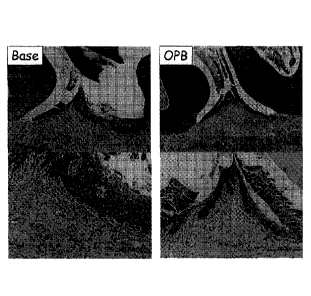
The results are shown in Figure 5 and Table 2.
[0059]
[Table 2]
The number of surviving bacteria in hamster oral cavity
The number of surviving bacteria {Mean SDILogio(CFU/swab)1}
Test material n
Baseline 0 hr 8 hrs 24 hrs
Base 4 6.09 0.87 5.26 0.50 5.55 0.52
5.86 0.53
0.1%0PB-1 4 6.13 0.40 3.13 0.52 3.98 1.03
5.57 0.69
0.1%0PB-2 4 6.16=60.27 3.24=60.34 4.52=60.65
6.24=60.22
Peridex 4 6.17 0.65 4.04 0.69 3.86 0.92
5.68 0.49
0.47% PVP-I 4 6.50 0.39 4.35 0.33 5.79 0.31
6.08 0.47
[ 0 0 6 0 ]
The number of bacteria in the oral cavity before test
material administration did not differ among the groups.
The bactericidal efficacy was 0.1% OPB-1 = 0.1% OPB-2 >
0.12% CHG > 0.47% PVP-I > Base immediately after test
material administration, was 0.1% OPB-1 = 0.1% OPB-2 = 0.12%
CHG > 0.47% PVP-I = Base 8 hours after administration, and
did not differ among the groups 24 hours after
administration. From
these results, the bactericidal
efficacy in the oral cavity was equivalent between 0.1% OPB
and 0.12% CHG, and 0.47% PVP-I had a weak immediate effect
with no persistent activity observed.
[0061]
26
Date Recue/Date Received 2021-03-09
The reason why the bactericidal activity of 0.47% PVP-
I was low in this test was that inactivation by proteins
and the like in the oral cavity probably made a significant
contribution thereto. 0.12%
CHG, as in 0.1% OPB, was
considered as a bactericidal antiseptic having persistent
activity in the oral cavity.
Example 3
[0062]
3. Bactericidal test in oral cavity using hamster - 2
This test was aimed at studying the influence of an
olanexidine concentration on the bactericidal efficacy of a
gargle (bactericidal antiseptic) on the mucosa in the oral
cavity of normal hamsters.
[0063]
3-1 Test material
Test substances and a control substance were
collectively used as test materials.
[0064]
3-1-1 Test substance 1
Designation: 0.1% OPB-1
Formula: olanexidine gluconate ... 0.10 w/v%
polyoxyethylene (20) polyoxypropylene (20)
glycol ... 0.07 w/v%
polyoxyethylene (160) polyoxypropylene (30)
glycol ... 0.10 w/v%
[0065]
3-1-2 Test substance 2
Designation: 0.1% OPB-2
Formula: olanexidine gluconate ... 0.10 w/v%
27
Date Recue/Date Received 2021-03-09
polyoxyethylene (20) polyoxypropylene (20)
glycol ... 0.07 w/v%
polyoxyethylene (160) polyoxypropylene (30) ...
1.00 w/v%
[0066]
3-1-3 Test substance 3
Designation: 0.5% OPB-3
Formula: olanexidine gluconate ... 0.50 w/v%
polyoxyethylene (20) polyoxypropylene (20) ...
0.36 w/v%
polyoxyethylene (160) polyoxypropylene (30) ...
5.00 w/v%
[0067]
3-1-4 Test substance 4
Designation: 1% OPB-2
Formula: olanexidine gluconate ... 1.00 w/v%
polyoxyethylene (20) polyoxypropylene (20)
glycol ... 0.72 w/v%
polyoxyethylene (160) polyoxypropylene (30)
glycol ... 10.00 w/v%
[0068]
3-1-5 Control substance
Designation: base
Formula: polyoxyethylene (20) polyoxypropylene (20)
glycol ... 0.07 w/v%
polyoxyethylene (160) polyoxypropylene (30)
glycol ... 0.10 w/v%
[0069]
3-2 Animal used
28
Date Recue/Date Received 2021-03-09
Male Slc: Syrian hamsters which were 6 weeks old upon
receipt were used to conduct a test on 3 animals per group.
[0070]
3-3 Testing method
[0071]
3-3-1 Anesthesia
Gas anesthesia [induction of anesthesia: 3.0 L/min of
air with 3% isoflurane (manufactured by Mylan Seiyaku Ltd.),
the concentration of continuous anesthesia was
appropriately adjusted] was carried out.
[0072]
3-3-2 Test material administration
Each hamster was fixed in the supine position under
anesthesia, and 1 mL of each test material was injected to
one cheek pouch. One minute later, the test material was
eliminated, and a redundant test material was drawn out of
the cheek pouch using a sterile swab.
[0073]
3-3-3 Bacterial collection
Bacteria were collected from both the cheek pouches
under anesthesia using a sterile swab at a total of 5 time
points (before test material administration, 0 hr, 1 hr, 3
hr, and 6 hr). The swab after the collection was dipped in
mL of a SCDLP medium, then stirred, and used as a sample
for bacterial counting.
[0074]
3-3-4 Measurement of the number of surviving bacteria
The number of surviving bacteria was measured in the
same way as in 2-3-4.
[0075]
29
Date Recue/Date Received 2021-03-09
3-4 Results
The results are shown in Figure 6. As is evident from
the results, 0.1% OPB can reduce the number of bacteria to
a low value persistently (up to 6 hours later). The
persistent activity was better at an OPB concentration of
0.5 w/v% or higher.
Example 4
[0076]
4. Oral mucosal irritancy test using hamster - 1
In this test, study drug formulations with varying
base formulas of 0.1% olanexidine gluconate were repeatedly
administered to the cheek pouches of hamsters for 14 days,
and comparatively studied for the degree of irritancy.
[0077]
4-1 Test substance
[0078]
4-1-1 Test substance 1
Designation: 0.1% OPB-1
Formula: olanexidine gluconate ... 0.10 w/v%
Pluronic L-44 ... 0.07 w/v%
[0079]
4-1-2 Test substance 2
Designation: 0.1% OPB-2
Formula: olanexidine gluconate ... 0.10 w/v%
Pluronic L-44 ... 0.07 w/v%
Pluronic L-31 ... 1.0 w/v%
[0080]
4-1-3 Test substance 3
Designation: 0.1% OPB-3
Formula: olanexidine gluconate ... 0.10 w/v%
Date Recue/Date Received 2021-03-09
Pluronic L-44 ... 0.07 w/v%
Pluronic P-123 ... 1.0 w/v%
[0081]
4-1-4 Test substance 4
Designation: 0.1% OPB-4
Formula: olanexidine gluconate ... 0.10 w/v%
Pluronic L-44 ... 0.07 w/v%
Pluronic P-85 ... 1.0 w/v%
[0082]
4-1-5 Test substance 5
Designation: 0.1% OPB-5
Formula: olanexidine gluconate ... 0.10 w/v%
Pluronic L-44 ... 0.07 w/v%
Pluronic F-127 ... 1.0 w/v%
[0083]
4-1-6 Test substance 6
Designation: 0.1% OPB-6
Formula: olanexidine gluconate ... 0.10 w/v%
Pluronic L-44 ... 0.14 w/v%
Pluronic F-68 ... 1.0 w/v%
[0084]
4-1-7 Test substance 7
Designation: 0.1% OPB-7
Formula: olanexidine gluconate ... 0.10 w/v%
Pluronic L-44 ... 0.07 w/v%
Trehalose ... 5.0 w/v%
[0085]
4-2 Animal used
Male Slc: Syrian hamsters which were 8 weeks old upon
receipt were used to conduct a test on 3 animals per group.
31
Date Recue/Date Received 2021-03-09
[0086]
4-3 Testing method
[0087]
4-3-1 Test substance application method
[0088]
(1) Amount applied
1 mL of each test substance was applied to the right
cheek pouch.
[0089]
(2) Application method
[1] Anesthesia was induced by gas anesthesia [induction
of anesthesia: 3.0 L/min of air with 3% isoflurane
(manufactured by Mylan Seiyaku Ltd.)].
[2] Each animal was fixed in the supine position under
maintenance of anesthesia (the concentration was
appropriately adjusted). The cheek pouch of the animal was
pulled using a swab, and the pulled cheek pouch was lightly
pinched with one hand.
[3] Foreign matter such as feed attached to the mucosa of
the cheek pouch was removed using saline and a swab for good
hygiene. Then, the cheek pouch was put back in place.
[4] 1 mL of each test substance was applied to the right
cheek pouch using a 1 mL syringe and a probe for oral
administration, and a vacant probe for oral administration
fitted into a 1 mL syringe was inserted to the left cheek
pouch, and decannulated.
[5] Thirty seconds after application, the animal was
reversed to the prone position so as to prevent the backflow
of the test substance into the respiratory tract, and the
32
Date Recue/Date Received 2021-03-09
test substance was eliminated. The whole redundant test
substance in the oral cavity was removed using a swab.
[6] The color tone and the like of the cheek mucosa at the
application site were observed and recorded. A collar for
hamsters was worn on the neck of the animal, and the animal
was then brought back to a cage.
[7] The manipulation described above was repeated twice a
day (morning and evening) for 14 days.
[0090]
4-3-2 Examination and observation
[0091]
(1) Observation of general status
The general status was observed as to all the animals
of each group before application of the test material and
at the completion of application in the application period
(Day 1 to Day 14). The observation was also performed on
the day following the end of the application period (Day
15).
[0092]
(2) Body weight measurement
The body weight was measured as to all the animals of
each group before application of the test material in the
application period (Day 1 to Day 14). The measurement was
also performed on the day following the end of the
application period (Day 15). However, the body weight was
not measured on Days 13 and 14 due to the breakdown of a
body weight scale.
[0093]
(3) Macroscopic observation method at application site
33
Date Recue/Date Received 2021-03-09
The status of the mucosa of the cheek pouch was
observed and scored as to the cheek pouches of all the
animals of each group before application of the test
material and at the completion of application in the
application period (Day 1 to Day 14). The observation was
also performed on the day (24 2 hours) following the end
of the application period (Day 15). The observation site
was set to the cheek mucosa at a site contacted with each
test material. As for the evaluation technique of
macroscopic observation, the degrees of erythema and eschar
formation were numerically graded (stomatitis grade)
according to the observation criteria and the numerical
grading described in Table 3 below (ISO 10993-10, Annex B.3
"Table B.2 Grading system for oral and penile reactions").
Other detected manifestations were also recorded. On the
basis of the obtained observation results, the respective
numerical grades for the mucosa of the animals of each group
were added for each test material, and the sum was divided
by the number of observations and the number of animals to
determine an average value (rounded to unit), which was used
as a reference material for comprehensive evaluation.
[0094]
[Table 3]
Table B.2 Grading system for oral and penile reactions
(Erythema and eschar formation) Numerical grading
No erythema ............................................ 0
Very slight erythema (barely perceptible) ............. 1
Well-defined erythema .................................. 2
Moderate erythema ...................................... 3
Severe erythema (beet-redness) to eschar formation
34
Date Recue/Date Received 2021-03-09
preventing grading of erythema ...................... 4
[0095]
(4) Pathological examination
Each animal was sacrificed by blood-letting under
isoflurane anesthesia after the completion of macroscopic
observation on the day following the end of the application
period, and the right and left cheek pouches were collected
and fixed in a 10% neutral buffered formalin solution. HE-
stained specimens were made according to a routine technique,
and pathological examination was carried out. As for the
evaluation technique of macroscopic observation,
manifestations or grades were recorded as to each item of
epithelium, leukocyte infiltration, hyperemia and edema
according to the criteria described in ISO 10993-10, Annex
B.3 "Table B.3 Grading system for microscopic examination
for oral, penile, rectal and vaginal tissue reaction".
Other observed manifestations were also recorded.
[0096]
(5) Comprehensive evaluation
The influence of each test material on the oral mucosa
was comprehensively evaluated on the basis of the degree of
reaction of each test material obtained from the macroscopic
observation results and the pathological observation
results about the cheek mucosa, with reference to
transitions in general status and body weight in the
observation period.
[0097]
4-4 Results
[0098]
Date Recue/Date Received 2021-03-09
4-4-1 General status
No abnormality was observed in any of the animals.
[0099]
4-4-2 Body weight
The results are shown in Figure 7. The body weight
of the 0.1% OPB-5 group was hardly changed. The average
values of the other groups were gradually increased.
[0100]
4-4-3 Macroscopic observation of application site
The results are shown in Table 10, and the cheek pouch
of each group on the final day is shown in Photos 1 to 8 of
Figure 8. Irritancy such as erythema was hardly observed
in all the drug formulations. However, a leukoplakia-like
symptom (increased keratosis or thickening) was observed in
0.1% OPB-1, -2, -6, -7 and -8. On the
other hand, no
abnormality was observed in 0.1% OPB-3, -4 and -5.
[0101]
4-4-4 Histopathological examination
The results are shown in Table 11. An
average
inflammation index of each individual and an average
inflammation index of each group were calculated by grading
of epithelium (cell degeneration, metaplasia and erosion),
leukocyte infiltration, hyperemia and edema according to
the evaluation criteria described in ISO 10993-10, Annex
B.3 "Table B.3 Grading system for microscopic examination
for oral, penile, rectal and vaginal tissue reaction".
Manifestations other than the evaluation criteria were also
recorded. As a result, no change was observed in the average
value of the 0.1% OPB-3 group. Cell degeneration of the
epithelium and minimum to moderate leukocyte infiltration
36
Date Recue/Date Received 2021-03-09
were observed in the other groups including the 0.1% OPB-1
group, and the inflammation index was evaluated as being
the minimum of 1 to 3. In
these groups, very slight
intercellular edema and very slight to slight hyperkeratosis
were observed as manifestations other than the evaluation
criteria.
[0102]
These results suggested that the base Pluronic P-123
used for 0.1% OPB-3 is particularly useful as a base for a
drug formulation for application of olanexidine gluconate
to the oral mucosa. The
results also suggested that
Pluronic P-85 and Pluronic F-127 used for 0.1% OPB-4 and -
5, which were found to be free from abnormality in
macroscopic observation, were also usable as bases for a
drug formulation for application of olanexidine gluconate
to the oral mucosa.
Example 5
[0103]
5. Oral mucosal irritancy test using hamster - 2
The irritancy test of Example 4 suggested that the
base Pluronic P-123 is useful as a base for a drug
formulation for application of olanexidine gluconate to the
oral mucosa. Accordingly, in this test, Pluronic P-123 was
adopted as a base for making a drug formulation having no
irritation, and subsequently, an OPB concentration and a
base concentration were studied. The
test system was
carried out by performing repeated administration to the
hamster cheek pouch, and prolonging the period from 2 weeks
to 4 weeks.
[0104]
37
Date Recue/Date Received 2021-03-09
5-1 Test substance
[0105]
5-1-1 Test substance 1
Designation: 0.1% OPB-1
Formula: olanexidine gluconate ... 0.10 w/v%
Pluronic L-44 ... 0.07 w/v%
Pluronic P-123 ... 0.50 w/v%
[0106]
5-1-2 Test substance 2
Designation: 0.1% OPB-2
Formula: olanexidine gluconate ... 0.10 w/v%
Pluronic L-44 ... 0.07 w/v%
Pluronic P-123 ... 1.0 w/v%
[0107]
5-1-3 Test substance 3
Designation: 0.1% OPB-3
Formula: olanexidine gluconate ... 0.10 w/v%
Pluronic L-44 ... 0.07 w/v%
Pluronic P-123 ... 0.50 w/v%
Lipidure(R) ... 1.0 w/v%
[0108]
5-1-4 Test substance 4
Designation: 0.1% OPB-4
Formula: olanexidine gluconate ... 0.10 w/v%
Pluronic L-44 ... 0.07 w/v%
Pluronic P-123 ... 1.0 w/v%
Lipidure(R) ... 1.0 w/v%
[0109]
5-1-5 Test substance 5
Designation: 0.3% OPB-1
38
Date Recue/Date Received 2021-03-09
Formula: olanexidine gluconate ... 0.30 w/v%
Pluronic L-44 ... 0.22 w/v%
Pluronic P-123 ... 1.50 w/v%
[0110]
5-1-6 Test substance 6
Designation: 0.3% OPB-2
Formula: olanexidine gluconate ... 0.30 w/v%
Pluronic L-44 ... 0.22 w/v%
Pluronic P-123 ... 3.0 w/v%
[0111]
5-1-7 Test substance 7
Designation: 0.3% OPB-3
Formula: olanexidine gluconate ... 0.30 w/v%
Pluronic L-44 ... 0.22 w/v%
Pluronic P-85 ... 1.50 w/v%
[0112]
5-1-8 Test substance 8
Designation: 0.3% OPB-4
Formula: olanexidine gluconate ... 0.30 w/v%
Pluronic L-44 ... 0.22 w/v%
Pluronic P-85 ... 3.0 w/v%
[0113]
5-1-9 Test substance 9
Designation: 0.5% OPB-1
Formula: olanexidine gluconate ... 0.50 w/v%
Pluronic L-44 ... 0.36 w/v%
Pluronic P-123 ... 2.50 w/v%
[0114]
5-1-10 Test substance 10
Designation: 0.5% OPB-2
39
Date Recue/Date Received 2021-03-09
Formula: olanexidine gluconate ... 0.50 w/v%
Pluronic L-44 ... 0.36 w/v%
Pluronic P-123 ... 5.0 w/v%
[0115]
5-1-11 Test substance 11
Designation: 0.5% OPB-3
Formula: olanexidine gluconate ... 0.50 w/v%
Pluronic L-44 ... 0.36 w/v%
Pluronic P-123 ... 2.50 w/v%
Lipidure(R) ... 1.0 w/v%
[0116]
5-1-12 Test substance 12
Designation: 0.5% OPB-4
Formula: olanexidine gluconate ... 0.50 w/v%
Pluronic L-44 ... 0.36 w/v%
Pluronic P-123 ... 5.0 w/v%
Lipidure(R) ... 1.0 w/v%
[0117]
5-2 Animal used
Male Slc: Syrian hamsters which were 8 weeks old upon
receipt were used to conduct a test on 3 animals per group.
[0118]
5-3 Testing method
[0119]
5-3-1 Test substance application method
[0120]
(1) Amount applied
1 mL of each test substance was applied to the left
cheek pouch.
[0121]
Date Recue/Date Received 2021-03-09
(2) Application method
[1] Anesthesia was induced by gas anesthesia [induction
of anesthesia: 3.0 L/min of air with 3% isoflurane
(manufactured by Mylan Seiyaku Ltd.)].
[2] Each animal was fixed in the supine position under
maintenance of anesthesia (the concentration was
appropriately adjusted). The cheek pouch of the animal was
pulled using a swab, and the pulled cheek pouch was lightly
pinched with one hand.
[3] Foreign matter such as feed attached to the mucosa of
the cheek pouch was removed using saline and a swab for good
hygiene. Then, the cheek pouch was put back in place.
[4] 1 mL of each test substance was applied to the left
cheek pouch using a 1 mL syringe and a probe for oral
administration, and a vacant probe for oral administration
fitted into a 1 mL syringe was inserted to the right cheek
pouch, and decannulated.
[5] Thirty seconds after application, the animal was
reversed to the prone position so as to prevent the backflow
of the test substance into the respiratory tract, and the
test substance was eliminated. The whole redundant test
substance in the oral cavity was removed using a swab.
[6] The color tone and the like of the cheek mucosa at the
application site were observed and recorded, and the animal
was then brought back to a cage.
[7] The manipulation described above was repeated twice a
day (morning and evening) for 28 days.
[0122]
5-3-2 Examination and observation
[0123]
41
Date Recue/Date Received 2021-03-09
(1) Observation of general status
The general status was observed as to all the animals
of each group before application of the test material and
at the completion of application in the application period
(Day 1 to Day 28). The observation was also performed on
the day following the end of the application period (Day
29).
[0124]
(2) Body weight measurement
The body weight was measured as to all the animals of
each group before application of the test material in the
application period (Day 1 to Day 28). The measurement was
also performed on the day following the end of the
application period (Day 29).
[0125]
(3) Macroscopic observation method at application site
The status of the mucosa of the cheek pouch was
observed and scored as to the cheek pouches of all the
animals of each group before application of the test
material in the application period (Day 1 to Day 28). The
observation was also performed on the day (24 2 hours)
following the end of the application period (Day 29). The
observation site was set to the cheek mucosa at a site
contacted with each test material. As for the evaluation
technique of macroscopic observation, the degrees of
erythema and eschar formation were numerically graded
(stomatitis grade) according to the observation criteria
and the numerical grading described in Table 3 above (ISO
10993-10, Annex B.3 "Table B.2 Grading system for oral and
penile reactions"). Other detected manifestations were also
42
Date Recue/Date Received 2021-03-09
recorded. On the basis of the obtained observation results,
the respective numerical grades for the mucosa of the
animals of each group were added for each test material,
and the sum was divided by the number of observations and
the number of animals to determine an average value (rounded
to unit), which was used as a reference material for
comprehensive evaluation.
[0126]
(4) Pathological examination
Each animal was sacrificed by blood-letting under
isoflurane anesthesia after the completion of macroscopic
observation on the day following the end of the application
period, and the right and left cheek pouches were collected
and fixed in a 10% neutral buffered formalin solution. HE-
stained specimens were made according to a routine technique,
and pathological examination was carried out. As for the
evaluation technique of macroscopic observation,
manifestations or grades were recorded as to each item of
epithelium, leukocyte infiltration, hyperemia and edema
according to the criteria described in ISO 10993-10, Annex
B.3 "Table B.3 Grading system for microscopic examination
for oral, penile, rectal and vaginal tissue reaction".
Other observed manifestations were also recorded.
[0127]
(5) Comprehensive evaluation
The influence of each test material on the oral mucosa
was comprehensively evaluated on the basis of the degree of
reaction of each test material obtained from the macroscopic
observation results and the pathological observation
results about the cheek mucosa, with reference to
43
Date Recue/Date Received 2021-03-09
transitions in general status and body weight in the
observation period.
[0128]
5-4 Results
[0129]
5-4-1 General status
No abnormality was observed in any of the animals.
[0130]
5-4-2 Body weight
The body weight was increased over time in all the
groups, and hardly differed among the groups.
[0131]
5-4-3 Macroscopic observation of application site
The results are shown in Table 12. Irritancy such as
erythema was not observed in all the drug formulations
(numerical grading: 0). However, a leukoplakia-like symptom
(increased keratosis or thickening) was observed in OPB
having a concentration of 0.3% or higher. On the other hand,
no abnormality was observed in ORB having a concentration
of 0.1%.
[0132]
5-4-4 Histopathological examination
The results are shown in Table 13. An
average
inflammation index of each individual and an average
inflammation index of each group were calculated by grading
of epithelium (cell degeneration, metaplasia and erosion),
leukocyte infiltration, hyperemia and edema according to
the evaluation criteria described in ISO 10993-10, Annex
B.3 "Table B.3 Grading system for microscopic examination
for oral, penile, rectal and vaginal tissue reaction".
44
Date Recue/Date Received 2021-03-09
Manifestations other than the evaluation criteria were also
recorded. As a
result, no inflammatory reaction was
observed in each group of 0.1% OPB. Degeneration of the
epithelium and leukocyte infiltration were observed in each
group of OPB having a concentration of 0.3% or higher, and
all the reactions were minimal with an inflammation index
of 1 to 3. Very slight to slight hyperkeratosis was observed
as manifestations other than the evaluation criteria in some
individuals of the 0.1% OPB group, and very slight to
moderate hyperkeratosis and very slight outgrowth of prickle
cells were observed in each group of OPB having a
concentration of 0.3% or higher. In
addition,
intraepidermal microabscess observed in the control group
(right cheek pouch: Sham-ope side) seemed to be a naturally
occurring lesion.
Example 6
[0133]
6. Efficacy test in 5-FU-induced hamster stomatitis model -
1
In this test, the efficacy of an OPB drug formulation
was tested in 5-FU-induced stomatitis models. Specifically,
measurements of the number of bacteria in the oral cavity
over time and stomatitis evaluation were performed by
gargling in the oral cavity with 0.1% OPB in 5-FU-induced
stomatitis models.
[0134]
6-1 Test material
A test substance and a control substance were
collectively used as test materials.
[0135]
Date Recue/Date Received 2021-03-09
6-1-1 Test substance
Designation: 0.1% OPB
Formula: olanexidine gluconate ... 0.10 w/v%
polyoxyethylene (20) polyoxypropylene (20)
glycol ... 0.14 w/v%
polyoxyethylene (160) polyoxypropylene (30)
glycol ... 0.10 w/v%
[0136]
6-1-2 Control substance
Designation: base
Formula: polyoxyethylene (20) polyoxypropylene (20)
glycol ... 0.07 w/v%
polyoxyethylene (160) polyoxypropylene (30)
glycol ... 0.10 w/v%
[0137]
6-2 Animal used
Male Slc: Syrian hamsters which were 6 weeks old upon
receipt were used to conduct a test on 5 animals per group.
[0138]
6-3 Testing method
[0139]
6-3-1 Anesthesia
Gas anesthesia [induction of anesthesia: 3.0 L/min of
air with 3% isoflurane (manufactured by Mylan Seiyaku Ltd.),
the concentration of continuous anesthesia was
appropriately adjusted] was carried out.
[0140]
6-3-2 Stomatitis model making
46
Date Recue/Date Received 2021-03-09
5-FU was intraperitoneally administered at 60 mg/kg
to the hamsters under anesthesia. The administration was
performed a total of twice on Day 0 and Day 2.
[0141]
On Day 4, the cheek pouch was pulled out from each
hamster under anesthesia. Feed and floor mat for laboratory
animals accumulated in the cheek pouch were removed, and
the cheek pouch was patted with a cotton pad saturated with
saline. The surface layer (horny layer) of the cheek pouch
was brushed with a precision wire brush (O2.34 mm,
manufactured by Sumflex. Co., Ltd.). The cheek pouch thus
brushed was brought back to the oral cavity.
[0142]
6-3-3 Test material administration
Each hamster was fixed in the supine position under
anesthesia, and 1 mL of each test material was injected to
one cheek pouch. Thirty seconds later, the test material
was eliminated, and a redundant test material was drawn out
of the cheek pouch using a sterile swab. This
administration by the gargling manipulation was performed
twice a day. The administration was not carried out after
the disorder of stomatitis reached the peak.
[0143]
6-3-4 Bacterial collection
On Days 0, 4, 7, 10, and 17, bacteria were collected
from both the cheek pouches under anesthesia using a sterile
swab at a total of 4 time points (before the first test
material administration, 0 hr, and 6 hr later). However,
on days 10 and 17 without test material application,
bacteria were collected only once. The
swab after the
47
Date Recue/Date Received 2021-03-09
collection was dipped in 5 mL of a SCDLP medium, then stirred,
and used as a sample for bacterial counting.
[0144]
6-3-5 Measurement of the number of surviving bacteria
The agar plate pouring technique was carried out with
reference to New GMP Microbial Testing Methods 1) and
Standard Methods of Analysis in Food Safety Regulation 2).
[1] 500 pL of the sample for bacterial counting was
collected, and 10-fold dilution series from 101-fold to 104-
fold were made using 4.5 mL of a diluent solution.
[2] 1 mL each of the undiluted sample for bacterial
counting and the diluted bacterial suspensions was dispensed
to each sterile dish.
[3] 15 mL of a measurement medium (TSA+) incubated in a
thermostat bath set to approximately 47 C was rapidly
dispensed to the dish.
[4] After solidification of the measurement medium, the
resulting pour plates were inverted in an incubator, and
cultured at 35 C until colonies became able to be counted
(approximately 2 days).
[5] After the culture, colonies that proliferated in the
pour plates were visually counted. A pour plate in which
the number of colonies was too many to distinguish the
colonies was regarded as TNTC (too numerous to count)
without counting.
[0145]
6-3-6 Calculation of the number of surviving bacteria
The number of colonies adopted on the basis of the
section 6-3-5 was divided by the dilution ratio to determine
the number of surviving bacteria (CFU/mL). The number of
48
Date Recue/Date Received 2021-03-09
colonies adopted was rounded off to one decimal place and
displayed. The number of surviving bacteria (CFU/swab) was
calculated according to the following expression.
A: the number of colonies adopted
The number of surviving bacteria (CFU/swab) = A x Dilution
ratio x Amount of the sample fluid (5 mL)
[0146]
Log reduction was further determined according to the
expression given below from the logarithmic value of the
number of surviving bacteria. The log reduction was rounded
off to two decimal places and displayed. When the number
of surviving bacteria was 1 or less, the logarithmic value
was set to 0.
B: logarithmic value of the number of viable bacteria at
the baseline
C: logarithmic value of the number of viable bacteria after
test material embrocation
Log reduction = B - C
[0147]
6-3-7 Statistical analysis
A mean and standard deviation were determined on the
number of viable bacteria (CFU/swab) of each group and its
logarithmic value. The
number of viable bacteria was
rounded to unit and displayed in integer. The logarithmic
value of the number of viable bacteria was rounded off to
two decimal places and displayed. When the number of viable
bacteria was 0, the logarithmic value of the number of
viable bacteria was set to 0. No assay was conducted because
of an exploratory test.
[0148]
49
Date Recue/Date Received 2021-03-09
6-3-8 Stomatitis evaluation
A stomatitis grade was evaluated on the basis of Table
4 below.
[0149]
[Table 4]
Grade Status
0 Neither erythema nor vasodilation
1 Erythema and vasodilation
2 Serious
erythema attended with superficial mucosal erosion
3 Mucosal ulceration (25%)
4 Mucosal ulceration (50%)
Mucosal ulceration (100%)
[0150]
6-4 Results
[0151]
6-4-1 The number of bacteria
The results are shown in Table 5 and Figure 9.
Decrease in the number of bacteria after administration was
marked in the 0.1% OPB group on Days 0, 4, and 10, whereas
the value of the decreased number of bacteria was very small
on Day 7 when increase in severity of stomatitis was marked.
[0152]
[Table 5]
The number of surviving bacteria in hamster oral cavity
The number of surviving bacteria {Mean SD [Logic, (CFU/swab)] }
Test n 17
0 day 4 day 7 day 10 day
material day
pre Oh 6h pre Oh 6h pre Oh 6h Pre Oh 6h pre
6.78 6.72 6.79 7.07 6.97 6.82 7.03 6.84 6.90 6.59 6.54 6.54 6.65
No
5
procedure
0.43 0.38 0.22 0.49 0.53 0.20 0.48 0.35 0.21 0.26 0.30 0.18 0.16
6.61 5.95 6.10 6.62 6.08 6.30 7.12 6.89 6.68 6.61 6.67 6.85 6.68
Base 5
0.52 0.39 0.39 0.42 0.09 0.20 0.07 0.29 0.23 0.59 0.57 0.31 0.56
Date Recue/Date Received 2021-03-09
6.86 4.69 4.78 6.78 5.32 6.31 7.02 6.36 6.84 6.60 5.82 6.54 6.50
0.1%0PB 5 +
0.42 0.15 0.68 0.30 0.25 0.55 0.19 0.44 0.35 0.35 0.27 0.32 0.27
[0153]
6-4-2 Stomatitis grade
The results are shown in Figure 10. The stomatitis
grade was markedly low in the 0.1% OPB group.
[0154]
In this test, the value of the decreased number of
bacteria was low on Day 7 in the 0.1% OPB group probably
because of reduction in bactericidal activity due to the
bacterial collection method or an excess of an effusion.
This test suggested that increase in severity of stomatitis
is mitigated by administering 0.1% OPB and thereby keeping
the oral cavity clean.
Example 7
[0155]
7. Efficacy test in 5-FU-induced hamster stomatitis model -
2
In this test, the stomatitis-mitigating effects of
0.1% olanexidine gluconate and 0.1% CHG were comparatively
studied in 5-FU-induced hamster stomatitis models.
[0156]
7-1 Test material
Test substances and a control substance were
collectively used as test materials.
[0157]
7-1-1 Test substance 1
Designation: 0.1% OPB
Formula: olanexidine gluconate ... 0.10 w/v%
51
Date Recue/Date Received 2021-03-09
Pluronic L-44 ... 0.07 w/v%
[0158]
7-1-2 Test substance 2
Designation: Peridex(R)/0.1% CHG
Manufacturer: 3M ESPE Dental Products
Formula: chlorhexidine gluconate ... 0.12 w/v%
[0159]
7-1-3 Control substance
Designation: base
Formula: polyoxyethylene (20) polyoxypropylene (20)
glycol ... 0.07 w/v%
[0160]
7-2 Bacterium used
In this test, Staphylococcus aureus (ATCC No: 6538,
manufactured by Microbiologics, Inc.) was used, which is a
normal inhabitant in the oral cavity.
[0161]
7-3 Animal used
Male Slc: Syrian hamsters which were 6 weeks old upon
receipt were used to conduct a test on 5 animals per group.
[0162]
7-4 Testing method
[0163]
7-4-1 Preparation of test bacterial suspension
[1] A stored vial containing bacterial pellets was taken
out and brought back to room temperature.
[2] One bacterial pellet was taken out of the vial and
transferred to a sterile tube.
[3] 0.5 mL of saline was added thereto.
52
Date Recue/Date Received 2021-03-09
[4] The bacterial pellet was squashed with a sterile swab
to prepare a suspended bacterial fluid.
[5] The suspended bacterial fluid was inoculated to a
round area of approximately 2 cm in diameter in a TSA plate
using a sterile swab, and streaked from the inoculation area
using a platinum loop.
[6] The streaked TSA plate was inverted, and cultured
until colonies were formed.
[7] A single colony was selected from among the formed
colonies, collected with a platinum needle, and stabbed to
a Casitone medium.
[8] The Casitone medium in which the inoculant was stabbed
was cultured until proliferation of bacteria became able to
be confirmed.
[9] After confirmation of the proliferation of bacteria,
the bacteria were refrigerated (set value: 2 to 8 C)
[10] A portion of the test bacteria refrigerated in the
Casitone medium was collected with a platinum needle,
transferred to a 14 mL sterile tube containing 5 mL of an
MHB medium, and static cultured until the bacteria
proliferated.
[11] After the culture, 10 pL of the culture solution was
collected with a sterile tip, transferred again to a 14 mL
sterile tube containing 5 mL of an MHB medium, and static
cultured until the bacteria proliferated.
[12] After the culture, approximately 5 mL of the test
bacterial culture solution subcultured in the MHB medium
was recovered into a 15 mL conical tube. After addition of
8 mL of saline, the mixture was gently stirred.
53
Date Recue/Date Received 2021-03-09
[13] The tube was centrifuged at 3000 rpm at 23 C for 10
minutes (cooled centrifuge 5800, rotor RS-720, manufactured
by Kubota Corp.), and the supernatant was discarded.
[14] The precipitated test bacteria were suspended by the
addition of 1 mL of distilled water (Otsuka Distilled Water,
manufactured by Otsuka Pharmaceutical Factory, Inc.).
[15] The suspended bacterial fluid was transferred to a 14
mL sterile tube, and the turbidity was determined using
McFarland Standard (product No. 70900, manufactured by
Sysmex-Biomerieux Co., Ltd."). The
concentration of the
bacterial suspension was adjusted by the addition of saline
so as to attain McFarland 5.
[16] The suspended bacterial fluid adjusted to McFarland 5
was used as a test bacterial suspension.
[0164]
7-4-2 Anesthesia
Gas anesthesia [induction of anesthesia: 3.0 L/min of
air with 3% isoflurane (Mylan Seiyaku Ltd.), the
concentration of continuous anesthesia was appropriately
adjusted] was carried out.
[0165]
7-4-3 Stomatitis model making
5-FU was intraperitoneally administered at 60 mg/kg
to the hamsters under anesthesia. The administration was
performed a total of twice on Day 0 and Day 2. On Day 4,
the cheek pouch was pulled out from each hamster under
anesthesia. Feed
and floor mat for laboratory animals
accumulated in the cheek pouch were removed, and the cheek
pouch was patted with a cotton pad saturated with saline.
The surface layer (horny layer) of the cheek pouch was
54
Date Recue/Date Received 2021-03-09
brushed with a precision wire brush (O2.34 mm, manufactured
by Sumflex. Co., Ltd.). The cheek pouch thus brushed was
brought back to the oral cavity.
[0166]
7-4-4 Test material administration
Each test material was embrocated twice a day to the
hamster cheek pouch under anesthesia using a swab (for 4
days from the grouping day). However, on the 4th day, the
administration was performed once (only in the morning).
[0167]
7-4-5 Test bacterial suspension embrocation
The test bacterial suspension prepared in 7-4-1 was
embrocated once a day before the test material
administration in the morning to the hamster cheek pouch
under anesthesia using a platinum loop (for 5 days from the
grouping day).
[0168]
7-4-6 Stomatitis evaluation
A stomatitis grade was evaluated on the basis of Table
4 above.
[0169]
7-4-7 Statistical analysis
A mean and standard deviation were determined on the
grade of each group, and a graph was created using only the
mean. No
assay was conducted because of exploratory
analysis.
[0170]
7-5 Results
The results are shown in Figure 11. A tendency to
mitigate increase in severity of stomatitis was seen in the
Date Recue/Date Received 2021-03-09
0.1% OPB group compared with the base and 0.1% CHG groups.
The grade was almost the same with no difference between
the base group and the 0.1% CHG group. From the effect of
mitigating increase in severity of stomatitis, the 0.1% OPB
was considered to be superior in bactericidal efficacy on
the embrocated bacteria or the normal inhabitant in the
mucosa of the cheek pouch to 0.1% CHG.
Example 8
[0171]
8. Efficacy test in hamster model of stomatitis induced by
using 5-FU and radiation irradiation in combination
In this test, a drug formulation of 0.1% OPB
containing Pluronic P-123 as a base was applied by the
gargling technique to hamster models of stomatitis induced
by using 5-FU and radiation irradiation in combination, and
comparatively studied for a stomatitis-mitigating effect.
[0172]
8-1 Test material
A test substance and a control substance were
collectively used as test materials.
[0173]
8-1-1 Test substance
Designation: OPB
Formula: olanexidine gluconate ... 0.10 w/v%
Pluronic L-44 ... 0.07 w/v%
Pluronic P-123 ... 1.0 w/v%
[0174]
8-1-2 Control substance
Designation/abbreviated name: base/Base
Formula: Pluronic L-44 ... 0.07 w/v%
56
Date Recue/Date Received 2021-03-09
Pluronic P-123 ... 1.0 w/v%
[0175]
8-2 Animal used
Male Slc: Syrian hamsters which were 6 weeks old upon
receipt were used to conduct a test on 8 animals per group.
[0176]
8-3 Testing method
[0177]
8-3-1 Anesthesia
[0178]
(1) At time of radiation irradiation
Somnopentyl(R) (manufactured by Kyoritsuseiyaku
Corp.) was intraperitoneally administered at 40 mg/kg.
[0179]
(2) At time of stomatitis evaluation and test material
administration
Gas anesthesia [induction of anesthesia: 3.0 L/min of
air with 3% isoflurane (manufactured by Mylan Seiyaku Ltd.),
the concentration of continuous anesthesia was
appropriately adjusted] was carried out.
[0180]
8-3-2 Stomatitis model making
[0181]
(1) Radiation irradiation
On Day 0, the cheek pouch was pulled out from each
hamster under anesthesia using a swab. Feed and floor mat
for laboratory animals accumulated in the cheek pouch were
removed, and the cheek pouch was patted with a cotton pad
saturated with saline. The body and the cheek pouch were
both fixed onto a molded acrylic plate. A region other than
57
Date Recue/Date Received 2021-03-09
the cheek pouch at an irradiation site was covered with lead,
and the cheek pouch was irradiated with radiation (40 Gy)
under conditions of [shelf board distance: 12.5 cm, tube
voltage: 160 kV, tube current: 6.2 mA]. However, the
irradiation was performed for one cheek pouch per individual,
and four animals with the left cheek pouch irradiated and
four animals with the right cheek pouch irradiated were
assigned to each group.
[0182]
(2) 5-FU administration
5-FU was intraperitoneally administered at 60 mg/kg
to the hamsters a total of three times on Days 0, 5, and 10.
[0183]
8-4-3 Test material administration
Each hamster was fixed in the supine position under
anesthesia, and 1 mL of each test material was injected to
one cheek pouch. Thirty seconds later, the test material
was eliminated, and a redundant test material was drawn out
of the cheek pouch using a sterile swab. This
administration by the gargling manipulation was performed
twice a day. The administration was not carried out after
the disorder of stomatitis reached the peak.
[0184]
8-4-4 Stomatitis evaluation
A stomatitis grade was evaluated on the basis of Table
4 above.
[0185]
8-4-5 Statistical analysis
58
Date Recue/Date Received 2021-03-09
A mean and standard deviation were determined on the
stomatitis grade of each group, and a graph was created.
No assay was conducted because of exploratory analysis.
[0186]
8-5 Results
The results are shown in Figure 12. The maximum value
of the stomatitis grade was 4.9 for the base group and 4.3
for the OPB group, and furthermore, the grade started to
rise earlier in the base group. The OPB group was evidently
cured earlier, though all the animals were not completely
cured because ulcer in some individuals was not healed even
on Day 40 due to the influence of large strength of the
models.
Example 9
[0187]
9. Efficacy test in rat gingivitis model
In this test, the therapeutic effect of 0.1%
olanexidine gluconate on gingivitis was studied in rat
gingivitis models.
[0188]
9-1 Test material
A test substance and a control substance were
collectively used as test materials.
[0189]
9-1-1 Test substance
Designation: OPB
Formula: olanexidine gluconate ... 0.10 w/v%
Pluronic L-44 ... 0.07 w/v%
Pluronic P-123 ... 1.0 w/v%
[0190]
59
Date Recue/Date Received 2021-03-09
9-1-2 Control substance
Designation/abbreviated name: base/Base
Formula: Pluronic L-44 ... 0.07 w/v%
Pluronic P-123 ... 1.0 w/v%
[0191]
9-2 Bacterium used
In this test, Porphyromonas gingivalis (ATCC No: 33277,
manufactured by Microbiologics, Inc.) was used, which is a
bacterium causative of gingivitis.
[0192]
9-3 Animal used
Male Jcl: Wistar rats which were 5 weeks old upon
receipt were used to conduct a test on 5 animals per group.
[0193]
9-4 Testing method
[0194]
9-4-1 Preparation of test bacterium (the whole manipulation
except for preservation was performed in an anaerobic
chamber)
[1] A stored vial containing bacterial pellets was taken
out and placed in an anaerobic chamber.
[2] One bacterial pellet was taken out of the vial and
transferred to a sterile tube.
[3] 0.5 mL of a prepared TSB medium was added thereto.
[4] The bacterial pellet was squashed with a sterile swab
to prepare a suspended bacterial fluid.
[5] The suspended bacterial fluid was inoculated to a
sheep blood agar medium for CDC anaerobes.
[6] Culture was performed for 3 to 4 days under anaerobic
conditions (37 C)
Date Recue/Date Received 2021-03-09
[7] A single colony was selected from among the formed
colonies, and similarly inoculated again to a sheep blood
agar medium for CDC anaerobes.
[8] After confirmation of proliferation of bacteria, 3 mL
of a prepared TSB medium was added thereto, and the bacteria
were suspended using a spreader to make a glycerol stock.
[9] The stock was cryopreserved.
[10] The stock was inoculated to a sheep blood agar medium
for CDC anaerobes and cultured under anaerobic conditions.
[11] After confirmation of proliferation of bacteria, the
bacteria were suspended in an appropriate amount of a
prepared TSB medium. A portion of the suspension was taken
out, and the turbidity was adjusted to McFarland 5 using
McFarland Standard. The dilution ratio was calculated, and
the concentration of the bacterial suspension was adjusted
to 1 x 10¹⁰ CFU/mL from the remaining suspension.
[12] The resultant was used as a test bacterial suspension.
[0195]
9-4-2 Anesthesia
[0196]
(1) At time of cotton sewing thread insertion and autopsy
An aqueous pentobarbital sodium solution was
intraperitoneally administered at 40 mg/kg.
[0197]
(2) Bacterial inoculation and test material administration
Gas anesthesia [induction of anesthesia: 1.0 L/min of
air with 5% isoflurane (Mylan Seiyaku Ltd.), the
concentration of continuous anesthesia was appropriately
adjusted] was carried out.
[0198]
61
Date Recue/Date Received 2021-03-09
9-4-3 Cotton sewing thread insertion
After anesthesia, each animal was fixed in the dorsal
position to a dedicated table, and a cotton sewing thread
was inserted to between the upper right first and second
molars with the lower jaw lifted.
[0199]
9-4-4 Bacterial inoculation
After anesthesia, 0.2 mL of the test bacterial
suspension was inoculated to the cotton sewing thread
insertion site. This manipulation was carried out every 2
hours.
[0200]
9-4-5 Test material administration
After anesthesia, 1 mL of each test material was
administered to cleanse the oral cavity. This manipulation
was performed twice a day.
[0201]
9-4-6 Observation and examination
[0202]
(1) General status
The general status was observed once a day from the
cotton sewing thread insertion day to the autopsy day.
[0203]
(2) Body weight measurement
The body weight was measured a total of twice from the
cotton sewing thread insertion day to the autopsy day.
[0204]
(3) Autopsy
62
Date Recue/Date Received 2021-03-09
Each animal was sacrificed by blood-letting from the
cut abdominal aorta under anesthesia, and autopsy was
performed.
[0205]
(4) Histopathological examination
The excised upper jaw was fixed in a 10 v/v% neutral
buffered formalin solution, degreased, and decalcified, and
HE-stained specimens were then made. Inflammatory change
was pathologically examined as to each specimen.
[0206]
9-4-7 Statistical analysis
A mean and standard deviation were calculated on the
body weight of each group. No assay was conducted.
[0207]
9-5 Results
No abnormality was observed in the general status, and
the body weight did not differ between the groups.
The pathological examination results are shown in
Table 14. The micrographs of the HE-stained specimens are
shown in Figure 13.
In the OPB administration group, the stratified
squamous epithelium of the gingiva was observed with
infiltration of neutrophils in 8 out of 10 cases,
intercellular edematization in 1 out of 10 cases, and ulcer
in 1 out of 10 cases, all of which were very slight. The
lamina propria of the gingiva was observed with infiltration
of neutrophils in 8 out of 10 cases and bleeding in 1 out
of 10 cases, all of which were very slight. On the other
hand, in the base administration group, the stratified
squamous epithelium of the gingiva was observed with
63
Date Recue/Date Received 2021-03-09
infiltration of neutrophils which was very slight in 8 out
of 10 cases and was slight in 2 out of 10 cases. The
stratified squamous epithelium of the gingiva was observed
with intercellular edematization in 2 out of 10 cases,
hyperkeratosis in 2 out of 10 cases, acanthosis in 2 out of
cases and ulcer in 1 out of 10 cases, all of which were
very slight. The lamina propria of the gingiva was observed
with infiltration of neutrophils which was very slight in 6
out of 10 cases and was slight in 3 out of 10 cases. The
lamina propria of the gingiva was observed with bleeding
and edematization, both of which were very slight in 1 out
of 10 cases.
[0208]
9-6 Discussion
All of infiltration of neutrophils, intercellular
edematization, hyperkeratosis and acanthosis in the
stratified squamous epithelium of the gingiva, and
infiltration of neutrophils and edematization in the lamina
propria were changes associated with an inflammation, and
were considered to occur due to procedures. All of these
changes tended to be low in terms of both frequency and
degree in the OPB administration group compared with the
base administration group.
Therefore, an inflammation-
mitigating effect brought about by OPB administration was
observed.
Example 10
[0209]
10. Efficacy test in rat pneumonia model
64
Date Recue/Date Received 2021-03-09
In this test, the therapeutic effect of 0.1%
olanexidine gluconate on pneumonia was studied in rat
aspiration pneumonia models.
[0210]
10-1 Test material
A test substance and a control substance were
collectively used as test materials.
[0211]
10-1-1 Test substance
Designation: OPB
Formula: olanexidine gluconate ... 0.10 w/v%
Pluronic L-44 ... 0.07 w/v%
Pluronic P-123 ... 1.0 w/v%
[0212]
10-1-2 Control substance
Designation/abbreviated name: base/Base
Formula: Pluronic L-44 ... 0.07 w/v%
Pluronic P-123 ... 1.0 w/v%
[0213]
10-2 Animal used
Male Crl: CD (SD) rats which were 7 weeks old upon
receipt were used to conduct a test.
[0214]
10-3 Group configuration
The body weight was measured in the morning on the
intratracheal administration day, and the animals were
assigned by stratified randomization to 4 groups (groups 1
to 4) shown in Table 6 below. Six animals excluded from
the assignment were assigned by stratified randomization to
Date Recue/Date Received 2021-03-09
2 groups (groups 5 and 6) shown in Table 6 below, and used
as individuals for saliva collection.
[0215]
[Table 6]
Group Administered Timing of BALF collection
No. substance (elapsed time after intratracheal n
administration)
Saliva (collected
1 6h 6
from group 5)
Saliva (collected
2 24h 6
from group 5)
Saliva (collected
3 6h 6
from group 5)
Saliva (collected
4 24h 6
from group 5)
Individual for BALF recovery
Oral cavity
Group
cleansing (test n
No.
material)
OPB 3
6 Base 3
Individual for saliva collection
[0216]
10-4 Testing method
[0217]
10-4-1 Anesthesia
[0218]
(1) Cleansing of oral cavity and saliva collection
Somnopentyl was intraperitoneally administered at 40
mg/kg.
[0219]
(2) At time of intratracheal administration and
bronchoalveolar lavage fluid (BALF) collection
Gas anesthesia [induction of anesthesia: 3.0 L/min of
air with 3% isoflurane (Mylan Seiyaku Ltd.), the
66
Date Recue/Date Received 2021-03-09
concentration of continuous anesthesia was appropriately
adjusted] was carried out.
[0220]
10-4-2 Cleansing of oral cavity
A sterile swab was impregnated with each test material,
which was then embrocated in a sufficient amount to the oral
cavity under anesthesia.
[0221]
10-4-3 Saliva collection
0.1% pilocarpine hydrochloride (5 mg/kg) was
intraperitoneally administered under anesthesia. Over-
secreted saliva was recovered. The recovered saliva was
added to a nutritive medium containing a neutralizing agent,
and left standing. Then, centrifugation (r.t., 3000 rpm,
min) was performed, and sediments were suspended in the
same amount of saline to prepare an intratracheal
administration solution.
[0222]
10-4-4 Intratracheal administration
After saliva collection, a tube for administration was
indwelled in the trachea under anesthesia using a
pharyngoscope, and 0.1 mL of saliva or saline was
administered thereto.
[0223]
10-4-5 BALF collection
A median incision was made under anesthesia 6 or 24
hours after intratracheal administration, and each animal
was euthanized by blood-letting from the incision in the
abdominal aorta. Then, the lung was exposed, and a catheter
was inserted to the origin of the bronchus. Lavage was
67
Date Recue/Date Received 2021-03-09
performed three times (infusion and recovery were repeated
twice for each time) with 8 mL of a PBS solution containing
0.1% BSA and 0.05 mM EDTA-2Na (hereinafter, referred to as
PBS) through the line, and a lavage fluid was collected
(BALF). BALF was centrifuged (200 g, 4 C, 10 min), and the
supernatant was separated into other preservation tubes and
used for LDH concentration measurement and measurement of
cytokines (ELISA). Sediments were suspended in 1 mL of PBS
and used for hematological examination.
[0224]
10-4-6 Handling of animal for saliva collection
After the completion of intratracheal administration,
each animal for saliva collection was euthanized by blood-
letting under excess anesthesia.
[0225]
10-4-7 Cytokine (TNF-a and IL-6) concentration measurement
The measurement was performed according to protocols
included in kits.
[0226]
10-4-8 Hematological examination
Hematological analysis was carried out on the
suspended sediments using an automatic blood cell counter
for multiple items.
[0227]
10-4-9 Biochemical examination
An LDH concentration in the collected BALF supernatant
was measured using an automatic analysis apparatus 7180
(Hitachi High-Technologies Corp.).
[0228]
10-4-10 Statistical analysis
68
Date Recue/Date Received 2021-03-09
No assay was conducted because of exploratory analysis.
[0229]
10-5 Results
The hematological examination and biochemical
examination results are shown in Table 15.
There was no difference between both the groups in the
hematological examination. The 24-hour value of IL-6 was
lower by 2.5 times in the OPB group. The value of TNF-a
was lower in the OPB group at both the time points.
These results suggested that inflammatory reaction in
the lung due to aspiration of saliva is lower in dealing of
the oral cavity with OPB.
Example 11
[0230]
11. Study on anti-inflammatory action of olanexidine using
TLR reporter cell line
Examples 6 to 10 indicated that olanexidine has anti-
inflammatory action on stomatitis, gingivitis, and
pneumonia. It has
been revealed that inflammations are
associated with immune response mediated by Toll-like
receptor 4 (TLR-4) and Toll-like receptor 2 (TLR-2), which
are receptors recognizing LPS or LTA (ChemMedChem. 2016 Jan
19; 11 (2): 154-65; Biotechnol Adv. 2012 Jan-Feb; 30 (1):
251-60; and J Dent Res. 2016 Jul; 95 (7): 725-33).
Olanexidine has the possibility of suppressing an
inflammation by antagonistic (antagonist-like) action on
TLR-4 and TLR-2. Accordingly, in this test, in order to
elucidate this, the antagonistic (antagonist-like) action
of olanexidine on TLR-4 and TLR-2 was confirmed by reporter
69
Date Recue/Date Received 2021-03-09
assay using human-derived cells stably expressing TLR-4,
TLR-2, and reporter (SEAP) genes.
[0231]
11-1 Test substance
Designation: 1.5% OPB
Formula: olanexidine gluconate ... 1.5 w/v%
[0232]
11-2 Cell
The cells described in Table 7 below were used.
[0233]
[Table 7]
Designation Catalog
Host cell Expressed gene Medium
(abbreviated name) No./supplier
DMEM (4.5 g/L glucose) + 10%
human Toll-like receptor 4
FBS + 4 mM L-glutamine + 1 mM
HEK293 (TLR4), human MD-2, human
sodium pyruvate + 100 unit/mL
TLR4/MD-2ICD14 (human CD14, secreted alkaline NBP2-
penicillin* + 100 ttg/mL
Reporter Cell Line embryonic phosphatase (SLAP) reporter 26503/Novus
streptomycin* + 10 ttg/mL
(HEK-TLR4) kidney- gene under the transcriptional
Biologicals, LLC
blastcidin*+ 2 ttg/mL puromycin"
derived) control of a NF-x13 response
element + 200 ttg/mL zeocin" + 500
ttg/mL
G418"
human Toll-like receptor 2 DMEM (4.5 g/L glucose) +
10%
HEK293
(human
(TLR2), secreted alkaline FBS 4 mM L-glutamine + 1
mM
NBP2-
TLR2 Reporter Cell phosphatase (SLAP) reporter sodium pyruvate + 100
unit/mL
embryonic 26274/Novus
Line (HEK-TLR2) gene under the transcriptional penicillin* + 100
ttg/mL
kidney- Biologicals, LLC
control of a NF-x13 response streptomycin* + 10 ttg/mL
derived)
element blastcidin" + 500 ttg/mL
G418"
*Added, if necessary
ISelection reagent
[0234]
11-3 Testing method
[0235]
11-3-1 Study on antagonistic (antagonist-like) action of
olanexidine on TLR-4
[0236]
- Cell used: HEK293 cells expressing TLR4
- Medium used
Date Recue/Date Received 2021-03-09
Preculture: DMEM + FBS (final concentration: 10%) +
penicillin (final concentration: 100 units/mL) +
streptomycin (final concentration: 100 pg/mL)
Sample administration and
culture after
administration: DMEM + FBS (final concentration: 5%)
- Activity measurement: SEAP assay kit (manufactured by
Novus Biologicals)
- Protein quantification: BCA protein assay (manufactured
by Funakoshi Co., Ltd.)
[0237]
[1] Cells were adjusted to 1.0 x 10⁵ cells/well/100 pL
with DMEM containing 10% FBS, seeded to a 96-well plate
(collagen-coated), and cultured for 40 hours (37 C, 5% CO2).
[2] 1.5% OPB was diluted to the concentrations shown in
Table 8 below using 5% FBS.
[0238]
[Table 8]
OPB concentration (pg/mL)
Preparation concentration 20, 10, 5, 2
Final concentration 10, 5, 2.5, 1
[0239]
[3] LPS was prepared at 20 ng/mL (final concentration: 10
ng/mL) using 5% FBS.
[4] After removal of the medium, media were added to the
cells in the order of 50 pL of the OPB medium and 50 pL of
the LPS medium, followed by culture for 8 hours (37 C, 5%
CO2).
[5] After the culture, 50 pL of the supernatant was
transferred to each well of another 96-well plate, and SEAP
assay was conducted.
71
Date Recue/Date Received 2021-03-09
[6] 50 pL of 0.1% SDS-0.1 N NaOH was added to each well
of the 96-well plate from which the remaining supernatant
was removed, and frozen overnight (-20 C). After thawing,
BCA protein assay was conducted.
[7] The expression level of the SEAP reporter gene was
calibrated with the protein concentration to calculate an
inhibition rate at each OPB concentration.
[0240]
11-3-2 Study on antagonistic (antagonist-like) action of
olanexidine on TLR-2
[0241]
- Cell used: HEK293 cells expressing TLR2
- Medium used
Preculture: DMEM + FBS (final concentration: 10%) +
penicillin (final concentration: 100 units/mL) +
streptomycin (final concentration: 100 pg/mL)
Sample administration and culture after
administration: DMEM + FBS (final concentration: 1%)
- Activity measurement: SEAP assay kit (manufactured by
Novus Biologicals)
- Protein quantification: BCA protein assay (manufactured
by Funakoshi Co., Ltd.)
[0242]
[1] Cells were adjusted to 1.0 x 10⁵ cells/well/100 pL
with DMEM containing 10% FBS, seeded to a 96-well plate
(collagen-coated), and cultured for 36 hours (37 C, 5% CO2).
[2] 1.5% OPB was diluted to the concentrations shown in
Table 8 above using 1% FBS.
[3] LTA was prepared at 2 pg/mL (final concentration: 1
pg/mL) using 1% FBS.
72
Date Recue/Date Received 2021-03-09
[4] After removal of the medium, media were added to the
cells in the order of 50 pL of the OPB medium and 50 pL of
the LTA medium, followed by culture for 12 hours (37 C, 5%
CO2).
[5] After the culture, 50 pL of the supernatant was
transferred to each well of another 96-well plate, and SEAP
assay was conducted.
[6] 50 pL of 0.1% SDS-0.1 N NaOH was added to each well
of the 96-well plate from which the remaining supernatant
was removed, and frozen overnight (-20 C). After thawing,
BCA protein assay was conducted.
[7] The expression level of the SEAP reporter gene was
calibrated with the protein concentration to calculate an
inhibition rate at each OPB concentration.
[0243]
11-4 Results
The results are shown in Figure 14.
The inhibition rate of SEAP was enhanced with
elevation in OPB concentration (IC50: approximately 10
pg/mL), demonstrating that OPB exhibits antagonistic
(antagonist-like) action on TLR4 and TLR2. These results
suggested that OPB has anti-inflammatory action by
inhibiting immune response mediated by TLR4 and TLR2.
Example 12
[0244]
12. Study on anti-inflammatory action of olanexidine using
mouse macrophage-like cell line RAW264.7
A mouse macrophage-like cell line RAW264.7, when
irritated with LPS or LTA, starts immune response via a
receptor recognizing it, to produce an inflammatory mediator
73
Date Recue/Date Received 2021-03-09
NO. Accordingly, whether olanexidine would have anti-
inflammatory action on an inflammation due to LPS irritation
was confirmed by using NO production from RAW264.7 cells as
an indicator.
[0245]
12-1 Test substance
Designation: 1.5% OPB
Formula: olanexidine gluconate ... 1.5 w/v%
[0246]
12-2 Cell
The cells described in Table 9 below were used.
[0247]
[Table 9]
Designation Animal species Tissue Catalog No./supplier
Medium
DMEM + 10% FBS + 100
RAW 264.7 Mouse, BALB/c Leukemic monocyte EC91062702-F0/DS Pharma. .
unit/mL penicillin* + 100
Biomedical Co., Ltd.
ug/mL streptomycin*
*Added, if necessary
[0248]
12-3 Testing method
[0249]
12-3-1 Study on anti-inflammatory action of olanexidine on
LPS irritation
[0250]
- Cell used: RAW264.7
- Medium used
Preculture: DMEM + FBS (final concentration: 10%) +
penicillin (final concentration: 100 units/mL)
streptomycin (final concentration: 100 pg/mL)
Sample administration and culture after
administration: DMEM + FBS (final concentration: 5%)
74
Date Recue/Date Received 2021-03-09
- Activity measurement: Nitrate/Nitrite Colorimetric Assay
Kit (manufactured by Griess Reagents)
- Protein quantification: not measured because the cells
were difficult to stain and destabilized values.
[0251]
[1] Cells were adjusted to 1.0 x 10⁵ cells/well/100 pL
with DMEM containing 10% FBS, seeded to a 96-well plate
(uncoated), and cultured for 24 hours (37 C, 5% CO2).
[2] 1.5% OPB was diluted to the concentrations shown in
Table 8 above using 5% FBS.
[3] LPS was prepared at 200 ng/mL (final concentration:
100 ng/mL) using 5% FBS.
[4] After removal of the medium, media were added to the
cells in the order of 50 pL of the OPB medium and 50 pL of
the LPS medium, followed by culture for 8 hours (37 C, 5%
CO2).
[5] After the culture, 50 pL of the supernatant was
transferred to each well of another 96-well plate, and
Nitrate/Nitrite colorimetric assay was conducted.
[0252]
12-3-2 Study on anti-inflammatory action of olanexidine on
E. coli (LPS-producing bacterium) irritation
[0253]
- Cell used: RAW264.7
- Medium used
Preculture: DMEM + FBS (final concentration: 10%) +
penicillin (final concentration: 100 units/mL) +
streptomycin (final concentration: 100 pg/mL)
Sample administration and culture after
administration: DMEM + FBS (final concentration: 1%)
Date Recue/Date Received 2021-03-09
- Activity measurement: Nitrate/Nitrite Colorimetric Assay
Kit (manufactured by Griess Reagents)
- Protein quantification: not measured because the cells
were difficult to stain and destabilized values.
[0254]
[1] Cells were adjusted to 1.0 x 10⁵ cells/well/100 pL
with DMEM containing 10% FBS, seeded to a 96-well plate
(uncoated), and cultured for 24 hours (37 C, 5% CO2).
[2] 1.5% OPB was prepared at the concentrations shown in
Table 8 above using 1% FBS.
[3] After removal of the medium, 50 pL of the OPB medium
was added to the cells, which were then left standing at
37 C under 5% CO2.
[4] E. coli cultured overnight in MHB was prepared at McF
1 and diluted 300-fold with 5% DMEM. Further, ampicillin
was added thereto so as to attain a final concentration of
50 ug/mL.
[5] The prepared bacterial culture medium was further
added at 50 p1/well to the plate supplemented with the OPB
medium, and cultured for 24 hours (37 C, 5% CO2) with the
plate hermetically sealed.
[6] After the culture, 50 pL of the supernatant was
transferred to each well of another 96-well plate, and
Nitrate/Nitrite colorimetric assay was conducted.
[0255]
12-4 Results
The results are shown in Figure 15.
NO production was decreased with elevation in OPB
concentration, demonstrating that OPB exhibits NO
production-inhibiting action (IC50: approximately 10 pg/mL).
76
Date Recue/Date Received 2021-03-09
These results suggested that OPB has anti-inflammatory
action.
Industrial Applicability
[0256]
The present invention provides a composition for
amelioration and/or prevention of an inflammation, which is
applicable to a wide range of inflammatory diseases.
Moreover, use of the composition of the present invention
as a composition for amelioration and/or prevention of oral
mucositis due to treatment of a cancer can prevent reduction
in QOL, such as inhibition of a communication function,
sleep disorder, pain, or dysphagia (decreased dietary
intakes), in a patient who is receiving chemotherapy and/or
radiotherapy, or disturbance of dose conformity of
chemotherapy and/or radiotherapy. Therefore, the present
invention has high industrial usefulness.
[Table 10] Table 10 is a table showing macroscopic
observation results in Example 4. The numeric values in
the table represent stomatitis grades, and the shaded areas
represent groups presenting with a leukoplakia-like symptom
(increased keratosis, thickening, etc.).
77
Date Recue/Date Received 2021-03-09
,
0
ce.-00000.00 1220..000..-0 i..8
It _____________________________________________ ..., =-'
WIC.-,0000000 W....Ø-000....,0 <X a
]1/4 0
Z
0 Z
WC
CO Cr
m-0000004, Ma..Ø00-0
el z (r CO
1 1 2
4 00000 oao <45-0aroewagilieneeoaaa
m...00=0000 ... M -a co.....a=a.aa
II ____________________________ 1 __________
1 . o _______
4-oa 00000 .... 4 -a ggaaapoo-sa
1¨ ___________________________ =¨.1a __________ .1.,
,.
r ............................... aa, r ..................... = 1s_Ø...
m 2
oCaoc=c000aco 4000000.4 ata.a000000
= 1 , = = I , A I
W0000,00e0 M OOOOO 000
Ma0000...-0
_______________________________________ ...¨. 1
! 1
w4C00000000 <00000000 4t0.01000--0
iµ ___________________________ 1 ___________
,
C 1 i _________
woo coo a = al = OOOOO = =
go - 0 0 0 0 - -O
0 . .
3400004,000 Z.(00000000 2.4...0000....0
.. . ,
M ............................. M ......... 7,131 ...... ..to
P
1
____________________________ 4 7 .
1 1_, .......... A
Ap0000ado a (44.1moo..41
_____________________________ , 4 __________
a 04
t '
a
im ......................................... = OOOOO = = 000Ø0
.r 1Ã 1-
1 I
< 40 OOOOO 00
ir....0012-c.
F = F f
W0000m000 M 000000 041
M.-000000-
P ___________________________ 4 1
14.2".004":000 Z.400000000 <-a00000-
..
g 1 ______________ 7.
4
0
CC0000 o o Ole M0000000a 1,32.-00000.-0
2
_ A
140....... i <00 000000 1 -....-aof000-fo
-'.....1aWa....a." -c.:(-4w,g,-/cc' ..t...*c-.i.r..4,...'a
,11111.1 I r I I I I I 11 I I . 8 i
m N I
i 1!!!!ffl I HIM!! 1 HMV OCCCCCCC CCCCCCCC CCCCCCCC
78
Date Recue/Date Received 2021-03-22
[Table 11] Table 11 is a table showing histopathological
examination results in Example 4.
mart draw 010P0-1 ais OPS- 2 016 0143-
3 Oil OP16.4 BIS OFII-5
1111/11a$492211321:41 Mini Na 3, 4 5 I lOt 11 21 II
13 14 15
40404414101) 3490.24$ 2 4 I 10 13 14 14 10 20 22
24 tt NI 30
Ffighi ctiedr poke% 0221 muocati
1/44.40..4ar.1100..411212104 inn* 0 0 1 0 1 1 0 0 3 0 1 0 I P1
liable& 1011004w 2 1 2 11 3 1 0 00 0 2 0 =1 1
11$42002or crlogeesor. a 0 0 0 0 0 0 00 0 0 0 00 0
Edema 0 0 0 0 0 0 0 0 # 0 0 0 0 6
mmation lei eir 1 1 3 1 4 2 0 00 0 3 0 0 2 2
Ayerage grade 2 2 0 1 1
Ott4er rranilestatf000
Intercelulat 44141110
Inc/eamid Karaite% is - - - .t
Oniim r fratemetiolt mitt/ ox11=4 041.444. 2 volt 41,44,1. 00%2M
o404ol4it, 394814o1
r:pareiteralootic
-g Greta 1 14 0011-0 010 CF11-7 al% 004-4
Artfra1110. if 12 le 10 20 21 22 2)24
40,-4P4rdro-44.- 3014=nes. 32 $4 24 31 40 42 44 44 41
R:;r11 :eel pouch or muoosa
044140 )411 *wok wmfoo4 !few,' I 1 I 1 1
111110cyl$ 1010412n 1 1 2 1 1 2 3 2
1,1424044r congas ton 0 0 0 0 0 a 0 0 0
Eden 4 0 D 0 0 0 0 0
'4144,x 2 2 2 ; 2 3 =3 3
*ono gtade 2 3
Olner manifestations
I ntetottels r edema
eficnatered loldebti% 4 4 4 * r 4 *
Geodeiiratioslikek -!7=10041 orttil 6=4424).$1 vor4v *Wks- Art 24-
,rnienete. 3+ tow*
; paraheratatic
Or. raga Ottereitttii
Hetsebotogical Anred lea 2 3 4 1 $ 7 13 io 11 lit 1 14 10
10 12 10 0 SS
manfetratet 2$4gli /01 1 3 6 7 0 11 13 16 1.2 14 21 0 0 27 0 21 0 20 37
31 (114 4
Rigraclusak ucwxyh, mucosa 0 0 0 0 0 0 0 0 0 00 0 0 4) 0 0 0 0 0 0 0 0 0
F44**4 44, *4444444444 .04004 .4 44440 0 0 0 0 9 0 0 0 000 0 0 0 2 1 0 0 0 00
0 0 2
Lacked:fie JAIN/ion 0 0 0 00 0 00 0 000 0 0 0000 0 0 0 0 0 0
'oescitar canalise 0 0 0 0 0 0 00 0 006 0 0 0000 0 0 0 0 0 0
Edema 0 0 0 00 0 000 006 0 0 00 0 0 0 0 0 0 0
irdarnrneiren rirdes Oa 0 00 0 00 0 000 0 0 2 II 00 0 0 0u et
Average grade
Other rmaniimhtet ions
In lateatelar edema - ----- - - -
111CM15a3 kOrainSiS
r1311 .'."11t41341100,041(fitc =r= mthm ham! I1t thrr *Mt, 40: MIK 414dimuts.
lhoMmOre
79
Date ecue/Date Received 2021-03-09
[Table 12] Table 12 is a table showing macroscopic
observation results in Example 5. The numeric values in
the table represent stomatitis grades, and the shaded areas
represent groups presenting with a leukoplakia-like symptom
(increased keratosis, thickening, etc.).
Date Recue/Date Received 2021-03-09
I 1-011016r0 0 010 0 0 0 0 00 . 1 it a
000 ' 0 00000 000 -,- 7 -
000'
,
r
. I ____________________ ..ono...= :,
===01.01.4
40000000000 4000000 000 00000 1 00110
1i00000000000
0000000 000 __ 000000000
1 * __________ i , , , , ,
40400000000 __ 4000000 v000 40000fr: i 1
f,0 000
ifr, ________
00.0 OOOOO 000 0000000 *00 =00000 ,000
ii I
rk
ir 1
40000000000 400000010000 40000000000
w 1 2 2 I
i*4' *-- = * - ¨* i ¨ ¨ ¨ ¨ ¨ i - H
= a 010i0.10 0 0 a co = co co
co a a a 0 a co o 00000000000
1 __________________________________ ,
T I 1
õII
40000000000 40000000000 40000041000
*
I
110 11
OOOOO 000000 =00000000000 00000000000
1_ _ I- - ill, IIIII
40000000000 40000000000 40000000004
0L , i V
i 7 i ,, ,11
II
00000000000 00000000000 00000000000
I - 1 __________________________________ .
a
ill r ,Ill km,
'' 1 j - rr
40000000000 40000000000 40000000000
0
O 0000 00000 0 i __________ 00000000000 0000000 0000 "
* ,i .1 3
..a._
1. ,
1
40000000000 40000000000 40000000000
' ta
1 _
= I
00000000000 00000000000 00000000000
r
i . __ ___ I . _ 7,1 _
a
40000000000 40000000000 4 40000000000
__________________________________ , . I
00000000000 00000000000 0000000000
r __ _ f'"
.1 .
400000000100 40000000000 40000000000
.4
.. .
. -¨---
00000000000 00000000000 00000000000
I r0
.; r
i
4000000...0 ....Ø0.1õ a o 0 0 orao 0000
= , .
Ililigglig rilgEggggg iiiiiiiii
_____________________________________________ z!!!!!ViU ;174#(7iod000
81
Date ecue/Date Received 2021-03-09
00000400000
I
1¨
.
40000$00000
Il
,
4 . ........
f
.0000000000 la
i¨
g
õ
4Ø0.00. 411.40..........
1 ,
, ________________________
t u
,
.000'0000,,010,0 000000 00000,
I III
4 0 0 0104 0 101100 1 40000000 r., 00
2
4 ¨ 1 11 n
0 0,0 00 0,0 0 C;;00 =0000000k-,0.0
õ I
,
I 1 I
4000000000011 4 0 0 0 0 0 , 10
;I 
f
***** 00000,0 00000,0000000
,
4 0 0 0 0 0 0 0 0,40,4 4 0 0 0 0 0; a 0 0 0
0
1
' I
00000000 00 00000000000
g
13 11111,
40000000 ,40 Oj 400001000000
1
1
1
000000000,00 000000000010
1
1
wa w
40000000000 40000000000
.7 :
1
al000000010,00 0000010001.0010
1
I I 1
I
40000000,0,00
I
1
400001007000
,
00000000000 1,00000000,000
1 V
I ^-
40000000000 40000000000
,
oi v
,....
1 .0000000,000 .00000000,00
r i
J ,
, 40000000,000 400000000.00
ggggg' gggg igggEggggg
______________________ 6600000000 0606,000000
82
Date ecue/Date Received 2021-03-09
[Table 13] Table 13 is a table showing histopathological
examination results in Example 5.
MEE!. AIMEE MUM Anna .3 Tr ___ linali
11I16,400002r.01 Mor d lui 1 ! 3 4 I 4 1 iii ,18 it 12 1
RI it II .14
rtmikstekin emv,A., r4# -it lit 11. 71.----t st et
176-$7117 la. $ ii_ III Pot AL /IL Mira'
Ftht awerpo.ch. Nal rnutrot6
404* :110****010.4=00.41, 'row( c 0 6 0 E 1: 0 0 0 0 0 C 1 1 1
I 1 0
1_0161.00/46 inflhalm, a a a 0 ^,..= a o 0 0 0 0 t 1 1 v
0 0 0
Valcut3r -DT gestor a a a 0 t c 0 0 0 0 0 e a 4 C
0 = a
Edema a o o a r a 0 0 P 0 9 9 0 4 0
0 4 cl
Intiaterrotark Irciti il 0 0 0 C C 0 0 6 e a 0 2 2 2
1 1 0
kotatee 01036 0 0 0
Other 06614(063200rd
Norco** adorn * ¨ * - * - 4 - - - * . 4 14
4 4 *
Miasma Wake* - - - - - - - - - _ _ ., - i
- ¨
Ilia-3606=31 niaaaalscess - ¨ - - - _ - - - - - - - - -
- -
01000 100200 600110041000 -; wilin !nomad 6**bmi, 0- very ;Ark *; 4104, 1*-
modwata 24"44# wit,.
000ort (3roup, 0.1i 0110-1 061 ire 1 0a" oeel,
$O40
Hweihita0p09 A04141 es 0; 4/ 34 31 31 36 37 01 /* 40 41 43
[3440114411143 5444443. N. 31L 34101. 191- 14L 394. 344- Th. sti.
rt 40i. 4U, 4*.
Rigiti chaek ipotice,lbil
filikmerR 3.41 ikerwato 40444144 4,4340 I I i 4 I 4 I I
I 0 k I
Lhelcosyle iniltialion ; ; / 0 0 0 I 1 1 0 I 1
V6 $4010r 00160106476 0 0 St 0 0 0 a 0 CI 0 0 e
Edema 0*0 000 000 000
M. fliti0m0000 index 3 1 3 1 1 1 2 2 2 0 2 2
Netrilfit 01801 1 1 2 1
Rumor RI St160610110n1
101140/2140r 00ems 14. 31* 3. 1- * * 2* 2+ 00 a 1* t.
bola** 160r010610 _t I - ¨ - * k - - I
Irtit60001116010100161010111 - - - , . - - - - ¨ -
ctra01 0 othor nvoilistakie -:*01* nrmil 0,40.0 0 wiry Art 4, thedt 14.1
Needesik ,34tioydriE
6"garl: Oral= ___________________________________________ ¨..-----
..õ---
,14*03*.toorco 41.60,6 We 1 1 3 I 5 6 3 1 li 14 1.11 11 13 16 11 14
1111 IS 40 10 71 21 31 It:
maatitavon ,..,.,,In02140-õ, 2R 30 *A IR '3 VI Is
10131M111.1,3000111010g11.41*1 110.1,16f
gight cheei poutA, old miLeaso 0 a a a a a c a a a 0 0 a a a a 0 a a a 0 3 a 0
F.041641.414...41.,,prolow, 'ff moan 6000000c 0 000400 c: 0 a 00400
l_etilkeepte I MilltNIC41 00001:1000131/0000,00:11000011
V11601AT conga/ton 00000000000000010 0 '100000
Edina 01100000066010060631...L.L.L.L.L.L
110.001kolveles 0004) 0000 0 0 0 0 0 0 0 es 4 11, I 41 4 111_ IM.,
kali0oluda ' ¨ 0
Olor 00041f0400/9ne
Neratildet aloe an. r r .............. r. r. ... r. .... r ..,
letwaselloiribtris
intrimilemiatiliovettaaegi - - I - I ..... ¨ ..................
Gram mow ddErmagens -: *lab 1661i611 106104 th: venr dr*, .7 dirt 14.
tisikrolos, DI- 44room
tVuan: Cita. Coro
.16korhappcot Amlmos Mix 11 12 61 14, 31 31 10 $2 32 42 41 4:
reilltiktWitt SiifflphY NI. MR SIR 339 14R311R31R27R an INC 4R411R 42ft
klfilt 0*(4 peoin, weIorricoss 0 6 0 0 0 6 0 0 0 0 0 0
Gooria. pill solawado. armor* .4 .4.ow 0 G CI CI D 0 0 0' 0 0 0 01
Li1414130110 infjtraice acacia 0 0 0 0 0 0 ti
Verscdisi ccagesten 0 ,;13 aooloaco 0 o
Ed* 00,0400044000
1611630400106 Mu 9000000011309
,oleowmeepod= 6
Other ma nifeetellane
letowelthir edema - AD 41. .., .... ..... m .
i****364119191914 . IP lir 11. .= .... ... ==
Ctai 1111471'4)=11. glint Millhell11.1 10011100" ;As; artht. .... Ate% ....
04000slii. 7 i. se* cm
83
Date ecue/Date Received 2021-03-09
[Table 14] Table 14 is a diagram showing pathological
examination results in Example 9.
Otgan! ¨
74.tv OPB t
ant N 11 'IT Na. 1 2SI 5 6 7. 6 B 1' q1
4 J5101111-117-7.i
It1IT.41
Frifiltratioi dt,I1
* ttliftIttlf
InterceluAr eartmtizzark ¨ ¨ * ; ¨
1-Iwt4 tA otos - - - ¨
,g14:antluzis
IEJos.ul * gla IQ al . ni J.
II ratrlina, girTiva
vein 2 le tit +*** 4
8"i4eiiing - - m me
Ed Malin
CrItth; : tekorits, :vory plett, diet, 2.0:rivelivire2 11-zwokyo
[Table 15] Table 15 is a diagram showing results of
hematological examination and biochemical examination in
Example 10.
84
Date Recue/Date Received 2021-03-09
o
m
5'
x
CD
K"
c
m
o
m
5'
x
CD
0
CD _
m Airg.r.gli. US11
0. kanr.n6cr T i ri e Animal at RA L F
YfilC MEM L04,1 L-S 11Wi 11010 NUT LDH L-6 1 N
F-1
F.) EU -
0 FIJI P.-AN/
solo pin: No - ...
F.)
2'filL) Wm 112111MEMI " rira - - mmirvpm up,:_ airm
(5
cr.., 11111111111: .. aim WEMIIWITM17.71ri 14107
I
o
to, -13 03+ c __25,31 .. ',I _ 1..1.1 V66
I 211 4 2051 RI1 1787.7 illi4
a ' '. 4 = i) = - i 1- 1 14 = - -
1 IIIMIIMMIniriallirSIVWEIRMUMNIIIiHri 1 1 o 0
6 225 4 45.0 505S 40.0 0.1111 2500,5 2.441
OPil
, __
7 115 142 41..f IV 0 4.18 15.4 $.1
B ... = 1 12 4 _ _tir a 2.51 IL) _ 7.1-
1 .; = 141 7i1.0 120
1 107,3 17,7 ,libi.
}th 1111M.7.11111 1511 1%01 asui Mg l'"u'r 1241 TN 1 NJ 4 a
cn 111:1111.11E iou itto 1.174 104.1 lingl
1.
simiiirm 1310 526 ail 4.0
_______________________________________________________________________________
__
1
- 11 20 27 r.5 111.-3 102.0
327 3111.7 4551,4
14 21.5 MINIM. /511 n re airs 20451.5
MN = - Sill Emilswilsi 1203 liff 3 As tatts owl
miimions 05 4 14 4 ,1, p ' 1 41 111 4 -410,1
26 MnIM.3 1193.7 5 c . 50 1 1 zcgia 8 EMT.
I MO 3330 2512 LMIlliallni Z311,EZI
, 111 MI.23 272.1 -Slii!0-0.0-0-201.3.1 -
211 20 2762 riaS 150 1 1.55 20.6 12 3
vi111
21 20 214 1 1 s, 5 1 1 5 n91 la rj 25D. AU
125 7 1113 ie 1
21 '....e4 ri-5 EIBMWRIVIRTMEDIMICEN
MR= ii) ,E,45 1$01 151 7)3 WS $u