Note: Descriptions are shown in the official language in which they were submitted.
CA 03058914 2019-10-02
WO 2018/213104 PCT/US2018/032188
ANALYZING A ROCK SAMPLE
PRIORITY APPLICATION
[0001] This application claims priority to and the benefit of U.S.
Provisional Patent
Application Serial No. 62/506,263, filed May 15, 2017, entitled "Analyzing a
Rock Sample," the
disclosure of which is incorporated herein by reference in its entirety.
TECHNICAL FIELD
[0002] This specification describes example processes for analyzing rock
samples, and
for outputting images based on the analyses.
BACKGROUND
[0003] Rock may contain hydrocarbons, such as oil or gas. Criteria used
to estimate the
existence and amount of hydrocarbons in rock include, for example, the types
of chemical
elements or minerals in the rock and the quantities of those chemical elements
and minerals in
the rock. To determine the existence and amount of organic material, such as
hydrocarbons or
kerogen, in rock, imaging techniques may be used to capture images of the
rock. The resulting
images can be analyzed to identify the existence, and amounts, of organic
material in the rock.
SUMMARY
[0004] This specification describes example processes that use
geochemical relationships
to determine a probable mineralogy, per pixel, of an image of a rock sample.
Examples of types
of images that may be analyzed to determine the probable minerology include,
but are not
limited to, images acquired using scanning electron microscopy (SEM). These
images integrate
elemental data and "Z" values measured and acquired using, for example, energy
dispersive
spectroscopy (ED S), back scatter electron (B SE) images, or wave dispersive
spectroscopy
(WDS). The output generated by the example processes may include a two-
dimensional (2D)
mineral map. This mineral map may be used for mineralogical assessments or for
constructing
three-dimensional (3D) focused ion beam-scanning electron microscope (FIB-SEM)
sections.
The mineral map may improve the ability to quantify reservoir properties for
hydrocarbons. The
1
CA 03058914 2019-10-02
WO 2018/213104 PCT/US2018/032188
example processes may also be used to quantify minerals in the rock sample
using micro-X-ray
fluorescence (micro-XRF) and may be used in combination with other techniques,
such as
Fourier transform infrared spectroscopy (FTIR).
[0005] In some implementations, mineral maps obtained using the example
processes
have resolutions, and quantifications of rock matrices, that are at the nano-
scale. In some
implementations, nano-scale may include pixels smaller than one micrometer (
m).
[0006] An example method comprises analyzing rock from an image of a
sample region
of the rock. The example method comprises accessing element maps of the sample
region in a
database, with each element map comprising an array of pixels, and with each
pixel having a
value that represents how closely the pixel correlates to a chemical element;
accessing a database
storing threshold values for multiple chemical elements including the chemical
element;
determining a presence of a substance in a portion of the sample region
corresponding to the
pixel by determining whether a value of the pixel in each of the element maps
is greater than, or
less than, a threshold value for a corresponding chemical element; labeling
the pixel based on the
presence of the substance in the pixel; and outputting data representing the
substance map for
rendering on a graphical interface. The example method may include one or more
of the
following features, either alone or in combination.
[0007] The image may be obtained using scanning electron microscopy
(SEM). At least
one element map may be generated based on a back scatter electron (B SE)
image, an energy
dispersive spectroscopy (EDS) image, a wave dispersive spectroscopy (WDS), or
micro-X-ray
fluorescence (micro-XRF) image. Each element map may be based on unprocessed
image data.
The chemical element may comprise at least one of: aluminum, calcium, carbon,
chlorine, iron,
oxygen, potassium, phosphorous, magnesium, sulfur, sodium, silicon, or
titanium. A resolution
of the substance map may be less than, or equal to, 250 nm per pixel.
[0008] Determining the presence of a substance in the portion of the
sample region may
comprise selecting an element map for a chemical element; comparing a value of
the pixel in the
element map to a first threshold; and detecting the presence of a substance by
determining if the
value of the pixel has a predetermined relationship with the first threshold.
If the value of the
pixel does not have the predetermined relationship with first threshold, the
method further
comprises repeating selecting, comparing, and determining for a different
chemical element.
2
CA 03058914 2019-10-02
WO 2018/213104 PCT/US2018/032188
[0009] The predetermined relationship may comprise the value of the pixel
being greater
than the first threshold, or the value of the pixel being less than the first
threshold.
[0010] The method may comprise selecting an element map for a first
chemical element;
comparing a value of the pixel in the element map to a first threshold;
determining that the value
of the pixel has a first predetermined relationship with the first threshold;
selecting an element
map for a second chemical element; comparing a value of the pixel in the
second element map to
a second threshold; determining that the value of the pixel has a second
predetermined
relationship with the second threshold; and labeling the pixel as a substance
based on the value
of the pixel having the first predetermined relationship with the first
threshold and based on the
value of the pixel having the second predetermined relationship with the
second threshold.
[0011] The substance may be a mineral, and the substance map may be a
mineral map.
[0012] The method may comprise receiving data representing the sample
region, with
the data being received from an imaging device and with the data representing
the pixel at a
nano-scale resolution. Determining the presence of a substance in the portion
of the sample
region may be based on the data received. The substance map may be at a
resolution that is
based on the nano-scale resolution. The method may comprise performing an
assessment of
substances in the substance map; and outputting data that is based on the
assessment. The data
that is based on the assessment may comprise a characterization of substances
in the substance
map. The method may further comprise determining a likelihood of hydrocarbons
in the rock
sample based on the characterization of the substances; and affecting
operation of a hydrocarbon
extraction process based on the likelihood of hydrocarbons in the rock sample.
[0013] Any two or more of the features described in this specification,
including in this
summary section, may be combined to form embodiments not specifically
described in this
specification.
[0014] All or part of the methods, systems, and techniques described in
this specification
may be implemented as a computer program product that includes instructions
that are stored on
one or more non-transitory machine-readable storage media, and that are
executable on one or
more processing devices. Examples of non-transitory machine-readable storage
media include,
for example, read-only memory, an optical disk drive, memory disk drive,
random access
memory, and the like. All or part of the methods, systems, and techniques
described in this
specification may be implemented as an apparatus, method, or system that
includes one or more
3
CA 03058914 2019-10-02
WO 2018/213104 PCT/US2018/032188
processing devices and memory storing instructions that are executable by the
one or more
processing devices to perform the stated operations.
[0015] The details of one or more implementations are set forth in the
accompanying
drawings and the description. Other features and advantages will be apparent
from the
description and drawings, and from the claims.
DESCRIPTION OF THE DRAWINGS
[0016] FIG. 1 shows an example process for generating a mineral map.
[0017] FIG. 2 shows an example mineral map.
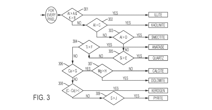
[0018] FIG. 3 is a flowchart showing an example process for performing a
mineralogical
analysis.
[0019] FIG. 4 is a flowchart showing an example process for performing a
mineralogical
analysis.
[0020] FIG. 5 shows an example mineral map and three EDS element maps.
[0021] FIG. 6 shows the mineral map of FIG. 5 compared to a low-
resolution mineral
map for a same sample.
[0022] FIG. 7A shows a refined version of the mineral map shown in FIG.
6B; and FIG.
7B shows an example low-resolution version of the mineral map shown in FIG.
6B.
[0023] FIG. 8 shows an example low-resolution mineral map of a sample
(top), and an
example higher-resolution mineral map of the same sample (bottom).
[0024] FIG. 9 shows an example mineral map and an example low-resolution
element
map for an iron sample (top), and an example mineral map and an example
element map for the
iron sample (bottom).
[0025] FIG. 10 shows an example mineral map.
[0026] FIG. 11 shows an example mineral map of a clastic rock sample
(top), and an
example mineral map of carbonate rock (bottom).
[0027] FIG. 12 shows an example mineral map (left), and the mineral map
overlaid with
a corresponding B SE image (right).
[0028] FIGS. 13A to 13C shows various types of SEM-derived images of a
same sample;
FIG. 13D shows a mineral map derived from the images in FIGS. 13A to 13C; and
FIG. 13E
shows a diagram of an interaction volume of a sample.
4
CA 03058914 2019-10-02
WO 2018/213104 PCT/US2018/032188
[0029] FIG. 14 shows an example mineral map generated from sample BSE
data.
[0030] FIG. 15A shows example element maps for aluminum, calcium, carbon,
chlorine,
iron, and oxygen; and FIG. 15B shows example element maps for potassium,
phosphorous,
magnesium, sulfur, sodium, silicon, and titanium.
[0031] FIG. 16 shows a graphical representation based on the images shown
in FIG. 15A
and FIG. 15B.
[0032] FIG. 17A shows an example mineral map for a sample; and FIG. 17B
shows the
mineral map of FIG. 17A overlaid with a BSE image.
DETAILED DESCRIPTION
[0033] This disclosure includes example processes ("the processes") for
generating pixel
maps of structures, such as rocks or minerals. In an example process, an image
of a rock is
captured. The pixels in the image are at a nano-scale resolution. In some
implementations,
nano-scale resolution images may include pixels with a length of an edge
smaller than one
micrometer ( m), for example 250 nanometers (nm). Each individual pixel of the
image is
analyzed to determine the chemical composition of the part of the rock that
the pixel represents.
Based on this analysis, the process determines the mineral composition of the
part of the rock.
Because the process performs the analysis at a nano-scale resolution, it may
be possible to
generate pixel maps that are more detailed than those that are generated using
lower-resolution
images.
[0034] Technologies that the example processes may employ include, but
are not limited
to, SEM imaging techniques, including FIB-SEM, EDS, BSE, and WDS. In an
example, SEM
includes scanning (or exciting) the surface of a sample using a focused beam
of electrons, and
generating an image based on the signals caused by excitation of the surface.
In an example,
FIB-SEM includes a system that is based on a working principle similar to SEM,
but that uses a
focused beam of ions instead of electrons to excite a sample. In an example,
EDS includes
detecting and measuring the characteristic X-ray excitation (photons) of a
sample. Because each
chemical element has a unique atomic structure, a unique set of peaks on the
electromagnetic
emission spectrum for each sample element can be detected. In an example, WDS
includes
detecting X-rays from different elements and separating them using
characteristic diffraction
patterns of an element (called Bragg diffraction). In an example, BSE includes
detecting
CA 03058914 2019-10-02
WO 2018/213104 PCT/US2018/032188
electrons reflected from a sample. There is a close relation between a BSE
signal and the atomic
number (the "Z" value): heavier chemical elements scatter the beam electrons
more strongly
than light elements. In a BSE image, heavier elements may appear brighter than
lighter
elements.
[0035] Mineralogy is used in the oil and gas industry to estimate the
quality and quantity
of rock deposits including, but not limited to, hydrocarbon deposits. For
example, the
mineralogy of shale is indicative of its susceptibility to (hydraulic)
fracturing (also known as
"fracking"). Analysis of shale with methods such as petrographic sections may
be challenging
because shale is largely composed of relatively fine grained minerals. High-
resolution imaging
techniques, such as SEM, can be useful to obtain qualitative, topological, and
quantitative
information from shale or other rock samples. For example, high-resolution
imaging techniques
enable imaging, at sub-micron resolutions, of mineral grain boundaries and
distribution in
organic matter, such as kerogen. Such imaging may allow for enhanced two-
dimensional (2D)
mineralogical mapping and three-dimensional (3D) reconstruction of rock
segments from
images, such as FIB-SEM images. Sub-micron resolution of mineral grain
boundaries may
enable a relatively detailed determination of mineralogy, lithology, organic
geochemistry and
petrophysics in a sample, such as a shale sample.
[0036] In some examples, the processes use elemental data and gray-scale
image data to
identify or to quantify, or both, minerals, organic matter, or both minerals
and organic matter, per
pixel within an SEM/EDS image alone or in combination with a BSE image. FIG. 1
shows an
example implementation of a process 100 for analyzing a rock sample from a
microscopic image
of a sample region of the rock sample. According to process 100, element maps
for a rock
sample are generated (101) by analyzing the rock sample using an EDS analysis,
a WDS
analysis, or both. In some implementations, an element map includes an array
of pixels. Each
pixel in the array has a gray-scale value that corresponds to the intensity of
an EDS, WDS or
BSE signal for a specific element. In this regard, because a rock sample may
contain more than
one element, multiple element maps may be generated for the same rock sample.
Each element
map may include the same pixel-by-pixel correlation between the element map
and content of
the rock sample such that the same pixel, from different element maps, may be
analyzed to
determine if that pixel represents one or more different chemical elements.
The element maps
may be stored in one or more appropriate databases or other storage
constructs.
6
CA 03058914 2019-10-02
WO 2018/213104 PCT/US2018/032188
[0037] In some implementations, to facilitate the determination of a rock
sample's
mineralogy, all element maps are loaded into a tensor model, in which the maps
are configured
as stacked pages. A corresponding BSE image may be added to help determine
organics and
porosity. FIG. 16 shows a graphical representation of an example data set.
[0038] To organize the image data, a three-index system may be used. The
first two
indices represent a spatial location of a pixel on the image in the form of
[row, column]. The
third index may be a depth index that reads the values of all the elemental
maps. Thus, using a
three-index system, any pixel in the stack can be located. If an XY location
in the tensor model
is called, the process returns an "elemental vector" containing all the values
of the pixels at the
specified location. This allows comparison of all the elements concurrently,
in some cases.
[0039] In some implementations, each element map includes an array of
pixels, and each
pixel has a value that represents how closely that pixel is representative of
a chemical element.
For example, that value may be a gray-scale value, for example, between 0 and
255, that is
greater or lesser than a pre-determined threshold value for that element.
Process 100 accesses
(102) a database to obtain the threshold values for chemical elements of
selected element maps.
Process 100 accesses, and selects, element maps and corresponding threshold
values for a set of
chemical elements. The chemical elements for which the threshold values and
elements maps
may be selected may be any appropriate set of predefined chemical values. For
example, a user
may have a list of chemical values for which the users wishes to test.
[0040] In this regard, because minerals have characteristic elemental
compositions, a
strong presence of certain elements that constitute a specific mineral can be
detected. A
threshold value can be established for each element. Each element responds
differently to an
electron beam excitation in EDS, so thresholds may not be universal between
elements. For
example, a value of "80" for iron may not mean the same as a value of "80" for
titanium.
Thresholds to determine a Boolean variable that would indicate the presence,
or absence, of an
element are established. In some implementations, by looking for the most
characteristic
minerals first, parameters may be tested sequentially in order to determine
the mineralogical
composition of a sample of rock represented by a pixel under consideration.
[0041] In some implementations, the selected element maps contain data
representing the
compositions of sedimentary rocks, although other types of maps may be
selected. That data
may be used to determine a probable mineralogy of the rock sample from SEM-
derived EDS or
7
CA 03058914 2019-10-02
WO 2018/213104 PCT/US2018/032188
BSE images, or both. For example, the data may be used to generate a substance
map, such as a
mineral map, showing the distribution of substances, such as minerals, and the
amounts of those
substances ¨ again, such minerals - present in the rock sample. An example of
a mineral map
generated by the example processes is shown in FIG. 2. In some
implementations, the
distribution and amount of minerals present in the rock sample may be
expressed in weight
percent (wt %) or volume percent (vol %) of the minerals relative to the
overall sample. To
determine a probable mineralogy of a rock sample, the process obtains
threshold values from the
database for chemical elements from the element maps.
[0042] Process 100 uses the obtained thresholds to determine (103) a
presence of a
substance, such as a mineral or organic material, in the rock sample. In some
implementations,
process 100 makes this determination by analyzing an image, such as an SEM
image, of the rock
sample on a pixel-by-pixel basis. FIGS. 3 and 4 show example processes for
performing such an
analysis. However, analyses other than those shown in FIGS. 3 and 4 may be
performed. In
some implementations, the analysis may include comparing values of the same
pixels from
different element maps to thresholds for each of a set of chemical elements,
and determining
whether a predefined relationship to the threshold is present for each
chemical element. For
example, the predefined relationship may include that the value exceeds the
threshold or that the
value is less than the threshold. Whether or not the chemical element is
present in a part of the
rock sample is determined based on whether the pixel value representing that
part of the rock
sample is greater or less than the threshold for that chemical element.
[0043] Based on the presence or absence (103) of a chemical element in
the rock sample,
process 100 generates (104) data representing a mineral map for the rock
sample. In some
implementations, the mineral map includes information representing the content
of the rock
sample. The mineral map may represent different minerals using different
colors, textures, or
other appropriate distinguishing indicia. As explained before, in some
implementations, mineral
maps generated using process 100 have resolutions, and quantifications of rock
matrices, that are
at the nano-scale. In some implementations, nano-scale may include pixels
smaller than one
micrometer ( m). In some implementations, mineral maps generated using process
100 may
have resolutions, and quantifications of rock matrices, that are greater than
a nano-scale or that
includes pixels smaller than one micrometer ( m).
8
CA 03058914 2019-10-02
WO 2018/213104 PCT/US2018/032188
[0044] Process 100 outputs (105) data representing the mineral map for
use in rendering
the mineral map on an appropriate graphical user interface, such as, but not
limited to, a
computer monitor, or the screen of a tablet computer or smartphone. The
mineral map is
rendered, based on the data, by an appropriate graphical processing device for
display to a user.
[0045] In some implementations of process 100, EDS and B SE data is
collected, for
example using SEM. This data is processed to obtain element information, such
as raw
elemental (spectral) data. The extracted and processed data may be used to
generate the element
maps described previously. In some implementations of the example processes,
including
process 100, data for each individual pixel is not converted to a chemical
composition and
subsequently matched to a minerals database. Instead, in some implementations,
the example
processes use raw elemental (spectral) data or other raw output data from an
electron or X-ray
detector of an SEM system. In some implementations, as noted, the raw output
data may be
normalized on a scale of, for example, 0 to 255. In some implementations, a
graphical user
interface (GUI) may be used to implement a real-time adjustment of the data's
acquisition
parameters to provide geologically consistent mineral maps. In some
implementations, the
acquisition parameters for the SEM system include the example settings shown
in Table 1.
Table 1
Accelerating voltage: 15kV (15,000 Volts)
Current -2.5nA (nano Amps)
Vacuum Pressure 40 Pascals
Working Distance 10.1 mm (millimeters)
Aperture 120 [tm (micro-meters)
Detectors used Electron Backscatter and Secondary Electron
Map size 300 [tm x 225 [tm
Counts 100,000 cps (Counts per Second)
Image filter Average filter of 5
Colors Maps Gray-scale 0-255
Acquisition Time 30 minutes
[0046] By adjusting the acquisition parameters, images can be obtained
that have
relatively smooth circular shapes for pyrite framboids, and images can be
obtained of diagenetic
dolomite crystals that are relatively sharp-edged rhomboids. In some
implementations, this
information can be useful for characterizing a rock sample since, for example,
shape and
9
CA 03058914 2019-10-02
WO 2018/213104 PCT/US2018/032188
orientation of pyrite framboids or dolomite rhomboids can indicate when and
how the
surrounding rock was formed. In this regard, accurate morphology may help to
differentiate
minerals visually. In addition, clearly defined boundaries, and thus surface
area, of each mineral
may increase quantitative and qualitative accuracy. The visual results,
obtained from the
combination of the relatively high-resolution imaging and parameter
flexibility facilitate accurate
determinations of the chemical composition, and thus the minerals, of a rock
sample. In some
implementations, for an SEM image with approximately 750,000 pixels, a
sufficiently large
number of determinations may be made to ensure a statistically-correct overall
mineral
composition for a rock sample under consideration. The results may be
displayed in a relatively
high-resolution mineral map.
[0047] In some implementations, process 100 includes a "rules-based"
process to
determine mineralogy on a pixel-by-pixel basis. In some implementations, the
pixels are on a
nano-scale, which may result in a mineral map having relatively high
resolution. Because the
computation time for such a process is proportional to the number of pixels,
larger images can, in
some cases, take a longer time to process. To reduce the amount of processing
time, the process
may be automated to operate in response to a single command. In an example
implementation,
the example process may be implemented using MATLAB produced by Mathworks of
1
Apple Hill Drive, Natick, Massachusetts.
[0048] As explained previously, process 100 uses thresholds to determine
(103) a
presence of a substance, such as a mineral or organic substance, in a rock
sample. FIG. 3 shows
an example process for analyzing a rock sample to make this determination. In
the example of
FIG. 3, values for one or more elements of interest are obtained from SEM
data. These values
correspond to how closely a pixel corresponds to a chemical element. In this
regard, each
element value of a pixel in an SEM element map corresponds to the presence of
that element in
the sample area represented by the pixel. In an example, an element value
corresponds to an
image gray-scale value that is normalized on a scale from 0 to 255. These
element values can be
used as inputs for use in analyzing element values in relation to a set of
threshold values,
examples of which are referred to as A, B, C, D, E, F, G, K, and J. In an
example, the process of
FIG. 3 performs an assessment of aluminum (in relation to threshold "A") and
potassium (in
relation to threshold "B") to determine whether the presence of illite is
probable (301). In some
implementations, the presence of a mineral is deemed probable if the
comparison indicates a
CA 03058914 2019-10-02
WO 2018/213104 PCT/US2018/032188
predefined relationship between the element value of the pixel and a
threshold. For example, the
presence of a mineral may be deemed probable if the element value is greater,
or less than, the
threshold value.
[0049] Continuing on with the FIG. 3 analysis, if aluminum and potassium
are each less
than a certain threshold value, then aluminum (in relation to thresholds "C" &
"D") is considered
to determine whether the pixel value corresponds to kaolinite (302) or
smectite (303). If these
criteria are not met, then silicon (in relation to threshold "E") followed by
titanium (in relation to
threshold "F") are evaluated to determine whether quartz (305) or anatase
(304) is probable. If
neither element meets the threshold criterion, then calcium (in relation to
threshold "G") and
magnesium (in relation to threshold "H") are considered to determine whether
the pixel
represents calcite (306) or dolomite (307). If the mineral is determined to be
neither calcite nor
dolomite, then the difference between carbon and calcium (in relation to
threshold "I") intensity
values provides a pathway to select either kerogen (308) or, upon evaluation
of sulfur (in relation
to threshold "J"), pyrite (309). The minerals and chemical compositions of
FIG. 3 are examples,
and other minerals and chemical compositions may be used in other
implementations.
[0050] In an example implementation of the FIG. 3 process, example
threshold values for
each element are in Table 2.
Table 2
A 26
50
22
20
35
[0051] As explained previously, in some implementations, each threshold
value may be,
for example, a gray-scale value between 0 and 255. These values may be chosen
or adjusted
based on factors such as a maximum intensity, a minimum intensity, or an
average intensity of
pixel data that is representative of a particular element in an EDS or B SE
image. Other threshold
values can be used, as appropriate. In addition or in the alternative, other
elements and minerals
11
CA 03058914 2019-10-02
WO 2018/213104 PCT/US2018/032188
can be used with the example process of FIG. 3, examples of which include, but
are not limited
to, aluminum, calcium, carbon, chlorine, iron, oxygen, potassium, phosphorous,
magnesium,
sulfur, sodium, silicon, and titanium.
[0052] As explained previously, process 100 uses thresholds to determine
(103) a
presence of a substance, such as a mineral or organic substance, in a rock
sample. FIG. 4 shows
an example process for analyzing a rock sample to make this determination. In
the example
process of FIG. 4, values corresponding to the presence of one or more
elements in pixels of
interest are obtained from appropriate image data, such as SEM image data. The
values may be
intensity values, which represents how closely a pixel correlates to a
chemical element. For
example, the greater the intensity value is for a particular chemical element,
the more likely it is
that a sample of the rock represented by that pixel contains that chemical
element. These values
can be used to analyze the sample to determine the mineralogical composition
of the sample. An
example set of threshold values (also called parameters) that may be used with
the process of
FIG. 4 is shown in Table 3. In some implementations, the determination of the
presence of a
specific mineral is based on the analysis of more than one element, for
example an analysis of
two, three, or more elements. Accordingly, example parameters 1, 2, and 3 are
shown in Table 3
for different example elements.
Table 3
Mineral Parameter 1 Parameter 2 Parameter 3
Kerogen BS < 50 C > 20
Pyrite Fe > 50 S > 40 Ca < 100
Sphalerite Zn > 100 S > 50
Albite Na > 100 Al > 60
Apatite P> 100
Chlorite Fe > 65 Mg > 50
Anatase Ti > 100
Dolomite Mg >100 Ca > 80
Anhydrite Ca > 50 S > 90
Calcite Ca > 100 S <80 Si < 100
12
CA 03058914 2019-10-02
WO 2018/213104 PCT/US2018/032188
K-Spar K>100
Illite K > 30 Al > 50
Kaolinite Al > 80
Smectite Al > 50
Quartz Si > 20
[0053] In this example, the process of FIG. 4 performs an assessment of a
BSE image,
followed by an analysis of EDS Data. If a pixel of the B SE image has a gray-
scale value less
than a threshold value, and if the corresponding pixel of an element map for
calcium has a value
greater than a certain threshold value, the pixel of the mineral map is
labeled as organics/pore
(401). If the corresponding pixel of an element map for iron has an element
value greater than a
certain threshold value, and if the corresponding pixel of an element map for
sulfur has an
element value greater than a certain threshold value, and if the corresponding
pixel of an element
map for calcium has an element value smaller than a certain threshold value,
the pixel of the
mineral map is labeled as pyrite (402). If the corresponding pixel of an
element map for zinc has
an element value greater than a certain threshold value, and if the
corresponding pixel of an
element map for sulfur has an element value greater than a certain threshold
value, the pixel of
the mineral map is labeled as sphalerite (403). If the corresponding pixel of
an element map for
sodium has an element value greater than a certain threshold value, and if the
corresponding
pixel of an element map for aluminum has an element value greater than a
certain threshold
value, the pixel of the mineral map is labeled as albite (404). If the
corresponding pixel of an
element map for phosphorus has an element value greater than a certain
threshold value, the
pixel of the mineral map is labeled as apatite (405). If the corresponding
pixel of an element
map for iron has an element value greater than a certain threshold value, and
if the corresponding
pixel of an element map for magnesium has an element value greater than a
certain threshold
value, the pixel of the mineral map is labeled as chlorite (406). If the
corresponding pixel of an
element map for titanium has an element value greater than a certain threshold
value, the pixel of
the mineral map is labeled as anatase (407). If the corresponding pixel of an
element map for
magnesium has an element value greater than a certain threshold value, and if
the corresponding
pixel of an element map for calcium has an element value greater than a
certain threshold value,
the pixel of the mineral map is labeled as dolomite (408). If the
corresponding pixel of an
13
CA 03058914 2019-10-02
WO 2018/213104 PCT/US2018/032188
element map for calcium has an element value greater than a certain threshold
value, and if the
corresponding pixel of an element map for sulfur has an element value greater
than a certain
threshold value, the pixel of the mineral map is labeled as anhydrite (409).
If the corresponding
pixel of an element map for calcium has an element value greater than a
certain threshold value,
and if the corresponding pixel of an element map for sulfur has an element
value smaller than a
certain threshold value, and if the corresponding pixel of an element map for
silicon has an
element value smaller than a certain threshold value, the pixel of the mineral
map is labeled as
calcite (410). If the corresponding pixel of an element map for potassium has
an element value
greater than a certain threshold value, the pixel of the mineral map is
labeled as KSpar (411). If
the corresponding pixel of an element map for potassium has an element value
greater than a
certain threshold value, and if the corresponding pixel of an element map for
aluminum has an
element value greater than a certain threshold value, the pixel of the mineral
map is labeled as
illite (412). If the corresponding pixel of an element map for aluminum has an
element value
greater than a certain threshold value and if potassium is absent, the pixel
of the mineral map is
labeled as kaolinite (413). If the corresponding pixel of an element map for
aluminum has an
element value greater than a certain threshold value, the pixel of the mineral
map is labeled as
smectite (414). If the corresponding pixel of an element map for silicon has
an element value
greater than a certain threshold value, the pixel of the mineral map is
labeled as quartz (415).
Otherwise, the pixel of the mineral map is labeled as unclassified (416).
Other threshold values
can be used, as appropriate. In addition or in the alternative, other elements
and minerals may be
used with the example process of FIG. 4 including, but not limited to, those
described before. In
some embodiments, the sequence of analysis may different depending on the
analyzed elements
or depending on the threshold parameters used.
[0054] As explained previously, process 100 uses thresholds to determine
(103) a
presence of a substance, such as a mineral or organic substance, in a rock
sample. Other
processes may be used for analyzing a rock sample to make this determination.
For example, if
the gray-scale value of a B SE image corresponding to a pixel is less than a
certain threshold, the
pixel of the mineral map may be labeled as organics/pore. If a sample area
corresponding to the
pixel is determined to contain iron and sulfur, the pixel of the mineral map
may be labeled as
pyrite. If a sample area corresponding to the pixel is determined to contain
potassium,
aluminum, silicon, and amounts of magnesium and iron less than a certain
threshold, the pixel of
14
CA 03058914 2019-10-02
WO 2018/213104 PCT/US2018/032188
the mineral map may be labeled as illite. If a sample area corresponding to
the pixel is
determined to contain aluminum (and, in some embodiments, contain amounts of
titanium
greater than a certain threshold), the pixel of the mineral map may be labeled
as smectite. If a
sample area corresponding to the pixel is determined to contain aluminum and
silicon, the pixel
of the mineral map may be labeled as kaolinite. If a sample area corresponding
to the pixel is
determined to contain magnesium and contains amounts of calcium less than a
certain threshold,
the pixel of the mineral map may be labeled as dolomite. If a sample area
corresponding to the
pixel is determined to contain amounts of phosphorous and calcium greater than
a certain
threshold, the pixel of the mineral map may be labeled as apatite. If a sample
area corresponding
to the pixel is determined to contain calcium and sulfur, the pixel of the
mineral map is labeled
may be anhydrite. If a sample area corresponding to the pixel is determined to
contain amounts
of titanium greater than a certain threshold, the pixel of the mineral map may
be labeled as
anatase. If a sample area corresponding to the pixel is determined to contain
amounts of silicon
greater than a certain threshold, the pixel of the mineral map may be labeled
quartz; and if the
pixel is determined to contain calcium, the pixel of the mineral map may be
labeled as calcite.
[0055] FIG. 5 shows an example mineral map 501 obtained using example
process 100.
Mineral map 501 is a composite of image data from three elements, namely iron
(Fe), carbon
(C), and magnesium (Mg). In this example, the process of FIG. 3 identifies, in
the subject rock
sample, the presence of each of these elements in an appropriate element map
502, 503, or 504.
Due to the presence of these elements, the process determines the
mineralogical composition of
the sample, including what minerals are in the sample and where they are
located. In this
example, mineral map 501 represents the different minerals using color;
however, any
appropriate distinguishing characteristic or attribute may be used to
represent different minerals.
For example, in FIG. 5, red shading is used to represent pyrite.
[0056] FIG. 6 shows the mineral map 501 of FIG. 5 compared to a low-
resolution
mineral map for a same sample 601. In this example, mineral map 501 is at a
nano-scale
resolution. As a result, mineral map 501 contains a relatively fine-grained
topology and
quantification for the rock sample. By contrast, low-resolution mineral map
601 does not
provide the same amount of detail as mineral map 501. Low-resolution mineral
map 601 is a
type of mineral map that may be generated using processes other than those
described in this
document.
CA 03058914 2019-10-02
WO 2018/213104 PCT/US2018/032188
[0057] In some implementations, mineral maps that are generated using the
example
processes can be further refined or updated in response to user input. For
example, FIG. 7A
shows a refined version 701 of mineral map 501. In the example of FIG. 7A, the
pyrite regions
702 and dolomite region 703 are updated relative to mineral map 501 of FIG. 5,
and are more
clearly delineated relative to the lower-resolution image shown in FIG. 7B.
[0058] FIG. 8 shows the contrast in resolution between a known low-
resolution mineral
map 801 and a mineral map 802 of the same section obtained using example
process 100. Note
that, in this example, the mineral map 802 shows a region of dolomite 803. As
shown, image
801 ¨ the known, low-resolutions image ¨ does not identify the dolomite region
803, but instead
labels the same region as carbonate, which refers to lithology instead of a
mineral.
[0059] FIG. 9 shows the contrast in resolution between sample image 901
for iron and a
known, low-resolution mineral map 902, and a sample image 903 for iron and a
mineral map 904
generated using example process 100. In this example, small regions of iron
905 are clearly
delineated in mineral map 904, while those same areas are ill-defined in image
902.
[0060] FIG. 10 shows a mineral map 1001 obtained using example process
100. In some
implementations, mineral amounts in mineral maps generated using process 100
are determined
by calculating an area occupied by a certain mineral in a material map
relative to a total area of
the mineral map. In some implementations, mineral locations in mineral maps
generated using
process 100 are determined by examining the topology of the mineral map. This
mineral map
shown in FIG. 10 includes a layered structure of the imaged rock sample, which
may have
implications for characterization and assessment of the rock, for example in
terms of the rock
sedimentation or mechanical properties of the rock.
[0061] FIG. 11 shows the contrast between a mineral map 1101 of elastic
rock and a
mineral map 1102 of carbonate rock obtained using example process 100.
[0062] FIG. 12 shows a mineral map 1201 obtained using example process
100, and the
same image with an overlay of a corresponding B SE image 1202. In some
implementations, this
overlay can enhance the definition of individual grains. An example of this
enhanced definition
is shown by the grain inside circled area 1203.
[0063] FIGS. 13A to 13C show various types of SEM-derived images that may
be used
in conjunction with example process 100. FIG. 13A shows a secondary electron
(SE) image
1301 of a surface of a rock sample. In this example, there are five pyrite
crystals, labeled 1 to 5.
16
CA 03058914 2019-10-02
WO 2018/213104 PCT/US2018/032188
Secondary electrons are emitted from the shallowest region of the interaction
volume, that is, the
volume of a sample emitting detectable signals when subjected to an electron
beam (see FIG.
13E). FIG. 13B shows a B SD image 1302 of the same region. Because
backscattered electrons
are emitted from a deeper region of the interaction volume, seven pyrite
crystals can be detected.
FIG. 13C shows an X-ray image (EDS image) 1303 of the same sample - in this
case, in the form
of an element map for iron (pyrite is formed from sulfur and iron). Because X-
rays are emitted
from an even deeper region of the interaction volume, nine pyrite crystals can
be detected. FIG.
13D shows a mineral map 1304 generated from EDS images using example process
100. In this
example, the input data was normalized on a scale from 0-255, with 255 being
the highest
intensity in an element map. Pyrite regions 6-9 have relatively low
intensities (element values)
and may be removed from the mineral map by controlling, for example, the
display threshold of
the mineral map. This effectively decreases the interaction volume. Thus, only
pyrite regions 1
to 5 are shown in the mineral map, which resembles more closely the secondary
electron map.
[0064] In some implementations, the example processes can also use BSE
images alone
or in combination with EDS and to identify minerals based on difference in
gray-scale and "Z"
(atomic number) values to further resolve grain boundaries and mineral spatial
relationships. For
example FIG. 14 shows an example mineral map 1401 generated solely from BSE
data, using
"Z" values to determine mineralogy.
[0065] FIG. 15A shows example element map images for aluminum 1501,
calcium 1502,
carbon 1503, chlorine 1504, iron 1505, and oxygen 1506. FIG. 15B shows example
element
map images for potassium 1507, phosphorous 1508, magnesium 1509, sulfur 1510,
sodium
1511, silicon 1512, and titanium 1513. A combined data set of the maps in FIG.
15 is shown in
FIG. 16.
[0066] FIG. 17A shows an example mineral map 1701. Because a mineral map
generally
does not show textures in the way that a BSE image does, a degree of
transparency can be added
to the mineral map, and the image may be overlaid with the BSE image. This may
provide more
details concerning shapes, and may be used to perform a 'sanity check' to
evaluate accuracy of
that the mineral map. The overlay map 1702 is shown in FIG. 17B. In FIG. 17B,
where the
shapes of the minerals correlate well with the features on the BSE. This may
indicate that the
chosen thresholds for determination of elemental presence were correct.
17
CA 03058914 2019-10-02
WO 2018/213104 PCT/US2018/032188
[0067] In addition to, or instead of, SEM imaging techniques, the example
processes may
also be used with a variety of other imaging techniques. For example, the
processes can be
applied to perform mineral quantification using images generated using micro-X-
ray
fluorescence (micro-XRF) or Fourier transform infrared spectroscopy (FTIR).
[0068] In some implementations, the processes can be used to determine a
likelihood of
hydrocarbons in the rock sample and can be used for characterization of the
substances and
affecting operation of a hydrocarbon extraction process based on the
likelihood of hydrocarbons
in the rock sample. For example, a certain mineral composition in the rock can
indicate
susceptibility of the rock to drilling or fracking, and may affect processes
for drilling or fracking
(for example, whether and where to perform those processes to extract
hydrocarbons).
[0069] In some implementations, an automated threshold determination
system may be
implemented, since thresholds and other parameters may vary between samples or
chemical
element detection methods. For example, an automated threshold determination
system may use
the minimum, maximum, or average intensity for each of the elemental maps to
guide the
determination of threshold values for the processes. In some implementations,
a neural network
may be used to guide the processes to recognize what thresholds should be used
in each case.
For example, the guidance may be based on thresholds used previously for
similar material. For
example, if the processes detect an image that resembles images of other
shales previously
analyzed, the processes can identify the image as 'shale' and use threshold
values from similar
images. The same principles could be applied to other types of materials, as
appropriate.
[0070] All or part of the processes described in this specification and
their various
modifications can be implemented, at least in part, via a computer program
product, for example
a computer program tangibly embodied in one or more information carriers, for
example in one
or more tangible machine-readable storage media, for execution by, or to
control the operation
of, data processing apparatus, for example a programmable processor, a
computer, or multiple
computers.
[0071] A computer program can be written in any form of programming
language,
including compiled or interpreted languages, and it can be deployed in any
form, including as a
stand-alone program or as a module, component, subroutine, or other unit
suitable for use in a
computing environment. A computer program can be deployed to be executed on
one computer
18
CA 03058914 2019-10-02
WO 2018/213104 PCT/US2018/032188
or on multiple computers at one site or distributed across multiple sites and
interconnected by a
network.
[0072] Actions associated with implementing the processes can be
performed by one or
more programmable processors executing one or more computer programs to
perform the
functions of the calibration process. All or part of the processes can be
implemented as special
purpose logic circuitry, for example an FPGA (field programmable gate array)
or an ASIC
(application-specific integrated circuit), or both.
[0073] Processors suitable for the execution of a computer program
include, by way of
example, both general and special purpose microprocessors, and any one or more
processors of
any kind of digital computer. Generally, a processor will receive instructions
and data from a
read-only storage area or a random access storage area or both. Components of
a computer
(including a server) include one or more processors for executing instructions
and one or more
storage area devices for storing instructions and data. Generally, a computer
will also include, or
be operatively coupled to receive data from, or transfer data to, or both, one
or more machine-
readable storage media, such as mass storage devices for storing data, for
example magnetic,
magneto-optical disks, or optical disks. Non-transitory machine-readable
storage media suitable
for embodying computer program instructions and data include all forms of non-
volatile storage
area, including by way of example, semiconductor storage area devices, for
example erasable
programmable read-only memory (EPROM), electrically erasable programmable read-
only
memory (EEPROM), and flash storage area devices; magnetic disks, for example
internal hard
disks or removable disks; magneto-optical disks; and CD-ROM and DVD-ROM disks.
[0074] Each computing device, such as a tablet computer, may include a
hard drive for
storing data and computer programs, and a processing device (for example a
microprocessor) and
memory (for example RAM) for executing computer programs. Each computing
device may
include an image capture device, such as a still camera or video camera. The
image capture
device may be built-in or simply accessible to the computing device.
[0075] Each computing device may include a graphics system, including a
display
screen. A display screen, such as a liquid crystal display (LCD) or a CRT
(Cathode Ray Tube)
displays, to a user, images that are generated by the graphics system of the
computing device.
As is well known, display on a computer display (for example a monitor)
physically transforms
the computer display. For example, if the computer display is LCD-based, the
orientation of
19
CA 03058914 2019-10-02
WO 2018/213104 PCT/US2018/032188
liquid crystals can be changed by the application of biasing voltages in a
physical transformation
that is visually apparent to the user. As another example, if the computer
display is a CRT, the
state of a fluorescent screen can be changed by the impact of electrons in a
physical
transformation that is also visually apparent. Each display screen may be
touch-sensitive,
allowing a user to enter information onto the display screen via a virtual
keyboard. On some
computing devices, such as a desktop or smartphone, a physical QWERTY keyboard
and scroll
wheel may be provided for entering information onto the display screen. Each
computing
device, and computer programs executed on such a computing device, may also be
configured to
accept voice commands, and to perform functions in response to such commands.
For example,
the process described in this specification may be initiated at a client, to
the extent possible, via
voice commands.
[0076] Components of different implementations described in this
specification may be
combined to form other implementations not specifically set forth in this
specification.
Components may be left out of the processes, computer programs, databases,
etc. described in
this specification without adversely affecting their operation. In addition,
the logic flows shown
in the figures do not require the particular order shown, or sequential order,
to achieve desirable
results. Various separate components may be combined into one or more
individual components
to perform the functions described here.
[0077] The patent or application file contains at least one drawing
executed in color.
Copies of this patent or patent application publication with color drawing(s)
will be provided by
the Office upon request and payment of the necessary fee.