Note: Descriptions are shown in the official language in which they were submitted.
CA 03059061 2019-10-03
WO 2018/195016
PCT/US2018/027872
X-RAY TOMOGRAPHY INSPECTION SYSTEMS AND METHODS
CROSS-REFERENCE
The present specification relies on United States Patent Provisional
Application Number
62/486,130, entitled "X-Ray Tomography Inspection Systems and Methods", filed
on April 17,
2017, for priority.
In addition, the present specification relates to United States Patent
Provisional
Application Number 62/597,155, entitled "X-Ray Tomography Inspection Systems
and
Methods", filed on December 11, 2017 which is herein incorporated by reference
in its entirety.
In addition, the present specification relates to United States Patent
Application Number
15/132,439 ("the '439 application"), entitled "X-Ray Sources" and filed on
April 19, 2016. The
'439 application is a continuation-in-part of U.S. Patent Application No.
14/635,814,
entitled "X-Ray Sources" and filed on March 2, 2015, which is a continuation
of U.S.
Patent Application No. 13/313,854, of the same title, and filed on December 7,
2011,
now issued U.S. Patent Number 9,001,973, which, in turn, is a continuation of
U.S.
Patent Application No. 12/478,757 (the '757 Application), filed on June 4,
2009, now
issued U.S. Patent Number 8,094,784, which is a continuation-in-part of U.S.
Patent
Application No. 12/364,067, filed on February 2, 2009, which is a continuation
of U.S.
Patent Application No. 12/033,035, filed on February 19, 2008, which is a
continuation
of U.S. Patent Application No. 10/554,569, filed on October 25, 2005, which is
a
national stage application of PCT/GB2004/001732, filed on April 23, 2004 and
which,
in turn, relies on Great Britain Patent Application Number 0309374.7, filed on
April 25,
2003, for priority. The '757 Application also relies on Great Britain Patent
Application
Number 0812864.7, filed on July 15, 2008, for priority.
All of the aforementioned applications are herein incorporated by reference in
their
entirety.
FIELD
The present specification relates to X-ray scanning systems. More
particularly, the
present specification relates to a stationary gantry X-ray inspection system
having a plurality of
1
CA 03059061 2019-10-03
WO 2018/195016
PCT/US2018/027872
X- ray sources positioned around a volume of inspection such that the sources
emit X-ray beams
having different beam angles.
BACKGROUND
X-ray computed tomography (CT) scanners have been used in security screening
in
airports for several years. A conventional system comprises an X-ray tube that
is rotated about an
axis with an arcuate X-ray detector which is also rotated, at the same speed,
around the same axis.
The conveyor belt on which the baggage is carried is placed within a suitable
aperture around the
central axis of rotation, and moved along the axis as the tube is rotated. A
fan beam of X-
radiation passes from the source through the object to be inspected and
subsequently to the X-ray
detector array.
The X-ray detector array records the intensity of X-rays passed through the
object to be
inspected at several locations along its length. One set of projection data is
recorded at each of a
number of source angles. From these recorded X-ray intensities, it is possible
to form a
tomographic (cross-sectional) image, typically by means of a filtered back
projection algorithm.
In order to produce an accurate tomographic image of an object, such as a bag
or package, there
is a requirement that the X-ray source pass through every plane through the
object. In the
arrangement described above, this is achieved by the rotational scanning of
the X-ray source, and
the longitudinal motion of the conveyor on which the object is carried.
In this type of system the rate at which X-ray tomographic scans can be
collected is
dependent on the speed of rotation of the gantry that holds the X-ray source
and detector array.
In a modern CT gantry, the entire tube-detector assembly and gantry will
complete two to four
revolutions per second. This allows up to four or eight tomographic scans to
be collected per
second, respectively.
As the state-of-the-art has developed, the single ring of X-ray detectors has
been replaced
by multiple rings of detectors. This allows many slices (typically 8) to be
scanned
simultaneously and reconstructed using filtered back projection methods
adapted from the single
scan machines. With a continuous movement of the conveyor through the imaging
system, the
source describes a helical scanning motion about the object. This allows a
more sophisticated
cone-beam image reconstruction method to be applied that can in principle
offer a more accurate
volume image reconstruction.
2
CA 03059061 2019-10-03
WO 2018/195016
PCT/US2018/027872
However, rotating gantry X-ray inspection systems are expensive to install,
have a large
footprint and consume a lot of power.
Some conventional CT scanners comprise non-rotating stationary gantry systems,
which
project X-ray beams from fixed, stationary sources at the subjects to be
scanned. These systems
include one or more spatially distributed X-ray sources for emitting X-rays
and one or more X-
ray detectors for detecting the X-rays. Multiple X-ray sources are required to
be activated at the
same time to produce a fan beam of X-rays in order to create a three-
dimensional scanned image
of an object. Stationary gantry systems may use anywhere from a dozen to a few
hundred X-ray
sources to produce a scanned image that varies in quality depending on the
number of X-ray
sources used. However, increasing the number of sources adds complexity to the
designs of
scanning systems and also increases their cost of manufacturing as well as
operation.
Additionally, traditional stationary gantry systems consume high amounts of
power and are
difficult to maintain.
Hence, what is needed is an improved X-ray inspection system that is efficient
in
detecting threat materials, is less expensive, has a smaller footprint and may
be operated using
regular line voltage power.
SUMMARY
The following embodiments and aspects thereof are described and illustrated in
conjunction with systems, tools and methods, which are meant to be exemplary
and illustrative,
not limiting in scope.
The present specification discloses an X-ray inspection system to scan an
object,
comprising: a housing enclosing a scanning volume; a conveyor to transport the
object through
the scanning volume for inspection; a multi-focus X-ray source having a
plurality of X-ray
source points arranged in a non-circular geometry around the scanning volume,
wherein a beam
angle of X-rays generated by each of the plurality of X-ray source points in
not uniform across
the plurality of X-ray source points; a detector array positioned between the
X-ray source and the
scanning volume, wherein said detector array has a plurality of multi-energy
detector modules
arranged in a non-circular geometry around the scanning volume to detect X-
rays transmitted
through the object during scanning; and a processor for analyzing sinogram
data and
reconstructed image data of the object being inspected to identify threat.
3
CA 03059061 2019-10-03
WO 2018/195016
PCT/US2018/027872
Optionally, the housing is substantially rectangular, wherein the housing has
a width
ranging from 800 mm to 1400 mm and a height ranging from 600 mm to 1500 mm.
Optionally, the non-circular geometry of the plurality of X-ray source points
is
rectangular.
Optionally, the non-circular geometry of the plurality of multi-energy
detector modules is
rectangular.
Optionally, the scanning volume has a width ranging from 500 mm to 1050 mm and
a
height ranging from 300 mm to 1050 mm.
Optionally, each of the plurality of multi-energy detector modules is
configured to
allocate detected photons into one of 2 to 64 energy bins.
Optionally, the multi-focus X-ray source has a plurality of X-ray source
points ranging
from 64 to 2048 X-ray source points, wherein the plurality of X-ray source
points are configured
in a plurality of groups, and wherein each of the plurality of groups has 4 to
32 X-ray source
points. A group may comprise eight X-ray source points. Optionally, a common
insulating
substrate supports each of the group of the plurality of groups.
Optionally, the conveyor has a speed ranging from 0.1 m/s to 1.0 m/s.
The present specification also discloses a method of scanning an object using
an X-ray
scanner having a scanning volume, comprising: transporting the object through
the scanning
volume using a conveyor; irradiating the object with X-rays generated by a
multi-focus X-ray
source, wherein the X-ray source has a plurality of X-ray source points
arranged in a first non-
circular geometry around the scanning volume, and wherein X-ray beam angles of
the plurality
of X-ray source points is not uniform; detecting X-rays transmitted through
the object using a
detector array positioned between the X-ray source and the scanning volume,
wherein said
detector array has a plurality of multi-energy detector modules arranged in a
second non-circular
geometry around the scanning volume; and analyzing sinogram data and
reconstructed image
data of the object being inspected to identify threat.
Optionally, the first non-circular geometry of said plurality of X-ray source
points is
rectangular.
Optionally, the second non-circular geometry of said plurality of multi-energy
detector
modules is rectangular.
4
CA 03059061 2019-10-03
WO 2018/195016
PCT/US2018/027872
Optionally, the first non-circular geometry is the same as the second non-
circular
geometry.
Optionally, the scanning volume has a width ranging from 500 mm to 1050 mm and
a
height ranging from 300 mm to 1050 mm.
Optionally, each of the plurality of multi-energy detector modules allocates
detected
photons into one of 2 to 64 energy bins.
Optionally, said multi-focus X-ray source has a plurality of X-ray source
points ranging
from 64 to 2048 X-ray source points, wherein said plurality of X-ray source
points are
configured in a plurality of groups, and wherein each of said plurality of
groups has 4 to 32 X-
ray source points.
Optionally, said conveyor has a speed ranging from 0.1 m/s to 1.0 m/s.
Optionally, each of said X-ray source points has a dwell time ranging from 50
.is to 500
.is per scan projection.
The present specification also discloses an X-ray inspection system to scan an
object,
comprising: a housing enclosing a scanning volume; a conveyor to transport the
object through
the scanning volume for inspection; a multi-focus X-ray source having a
plurality of X-ray
source points arranged in a non-circular geometry around the scanning volume,
wherein field of
views of X-ray beams generated by each of said plurality of X-ray source
points vary across said
plurality of X-ray source points; a first detector array positioned between
the X-ray source and
the scanning volume, wherein said first detector array has a plurality of
multi-energy detector
modules arranged in a non-circular geometry around the scanning volume to
detect X-rays
transmitted through the object during scanning; a second detector array
positioned between the
X-ray source and the scanning volume to detect X-rays diffracted from the
object during
scanning, wherein said second detector array has a plurality of energy
dispersive detector
modules located behind a plurality of associated collimators that are angled
to the X-ray beams;
and a processor for simultaneously generating a tomographic transmission image
using said X-
rays transmitted through the object and a tomographic diffraction image using
said X-rays
diffracted from the object in order to identify threat.
Optionally, said field of views range from approximately 60 degrees to 120
degrees.
Optionally, said collimators are angled at an angle ranging from 3 degrees to
10 degrees
to the direction of the X-ray beams.
5
CA 03059061 2019-10-03
WO 2018/195016
PCT/US2018/027872
Optionally, a portion of at least one of said first and second detector arrays
detect X-rays
backscattered from the object, wherein said processor also generates a
backscatter image of the
object that is also used to identify threat objects. The tomographic
diffraction image and/or said
backscatter image may be used to clear or confirm a threat raised by analysis
of said tomographic
transmission image.
The present specification also discloses an X-ray inspection system for
scanning items,
the system comprising: a stationary X-ray source extending around a
rectangular scanning
volume, and defining a plurality of source points from which X-rays can be
directed through the
scanning volume; an X-ray detector array also extending around the rectangular
scanning volume
and arranged to detect X-rays from the source points which have passed through
the scanning
volume; a conveyor arranged to convey the items through the scanning volume;
and at least one
processor for processing the detected X-rays to produce scanning images of the
items.
Optionally, each source point emits X-rays having a different beam angle.
Optionally, each source point is enclosed in one of a glass, metal, and
ceramic envelope.
Optionally, each source point comprises: an anode assembly comprising a target
coupled
with a high voltage power source; one or more slip coupling blocks for
accounting of thermal
expansion of the target; and a shield electrode for protecting the target and
power source from X-
rays; and a cathode assembly comprising at least a grid, a dispenser cathode,
a filament and a
primary focus electrode plugged into a printed circuit board, and a secondary
focus electrode for
protecting the cathode assembly from any flash of energy.
Optionally, the target is formed from a copper coolant tube providing coolant
to the
anode assembly.
Optionally, the coolant tube is molded into the target by using hydroforming.
Optionally, the secondary electrode is maintained at ground potential.
Optionally, the target is coated with silicon carbide and then patterned with
tungsten rich
tungsten carbide stripes.
Optionally, the target comprises a plurality of raised portions to define fan-
shaped
apertures.
The aforementioned and other embodiments of the present specification shall be
described in greater depth in the drawings and detailed description provided
below.
6
CA 03059061 2019-10-03
WO 2018/195016
PCT/US2018/027872
BRIEF DESCRIPTION OF THE DRAWINGS
These and other features and advantages of the present invention will be
further
appreciated, as they become better understood by reference to the detailed
description when
considered in connection with the accompanying drawings:
FIG. 1 is a longitudinal schematic view of a real time tomography security
scanning
system having a circular locus of source points, produced by conventional
systems;
FIG. 2A is a perspective view of a scanning unit, in accordance with an
embodiment of
the present specification;
FIG. 2B is a schematic diagram illustrating a plurality of views of the
scanning unit of
FIG. 2A in comparison to a plurality of corresponding views of a conventional
scanning unit;
FIG. 2C is a cross-sectional view of a housing/enclosure of the scanning unit
of FIG. 2A,
comprising a plurality of X-ray sources points and detectors, arranged in a
substantially
rectangular shape around a scanning volume, in accordance with an embodiment
of the present
specification;
FIG. 3A illustrates an X-ray source sealed within a glass envelope, in
accordance with an
embodiment of the present specification;
FIG. 3B illustrates an X-ray source sealed within a glass envelope, in
accordance with an
embodiment of the present specification;
FIG. 4A illustrates a cathode assembly, in accordance with an embodiment of
the present
specification;
FIG. 4B is an exploded view of the cathode assembly shown in FIG. 4A;
FIG. 4C is a top plan view of the cathode assembly shown in FIG. 4B;
FIG. 5A is a top plan view of a cathode array, in accordance with an
embodiment of the
present specification;
FIG. 5B is a bottom plan view of the cathode array shown in FIG. 5A, in
accordance with
an embodiment of the present specification;
FIG. 5C is another view of the cathode array shown in FIG. 5B;
FIG. 6 illustrates a grid control sequence of the elements of an X-ray source,
in
accordance with an embodiment of the present specification;
FIG. 7A is a plan view of an anode of an X-ray source assembly, in accordance
with an
embodiment of the present specification;
7
CA 03059061 2019-10-03
WO 2018/195016
PCT/US2018/027872
FIG. 7B is a plan view of a patterned anode surface, in accordance with an
embodiment
of the present specification;
FIG. 7C is a plan view of a patterned anode surface, in accordance with
another
embodiment of the present specification;
FIG. 8A is a cross-sectional view of the housing of the scanning unit of FIG.
2A,
comprising a plurality of X-ray source points and detectors, arranged in a
substantially
rectangular shape around a scanning volume, in accordance with another
embodiment of the
present specification;
FIG. 8B illustrates a plurality of electron gun source points, arranged in a
corner section
layout having an adjacent straight section layout on either side, representing
a portion of the
scanning unit shown in FIG. 8A, in accordance with an embodiment of present
specification;
FIG. 8C illustrates a multi-energy sensor in a 16 x 4 pixel array, in
accordance with an
embodiment of the present specification;
FIG. 8D illustrates a plurality of heat conductive and voltage supply
structures, in
accordance with embodiments of the present specification;
FIG. 8E is an expanded layout view of a plurality of X-ray source points or
electron guns,
of a multi-focus X-ray source, in accordance with some embodiments;
FIG. 9 is a cross-sectional view, through an imaging volume, of a scanning
unit in
accordance with embodiments of the present specification;
FIG. 10 is a cross-sectional view of an electron gun and detector array
assembly
positioned at one end of the imaging volume of the scanning unit of FIG. 9;
FIG. 11 illustrates a cross-sectional view through an imaging volume of a
scanning unit
combined with X-ray diffraction imaging system, in accordance with an
embodiment of the
present specification;
FIG. 12 is a flow chart describing a method of automatic threat detection and
clearance
using the combined X-ray transmission and X-ray diffraction systems shown in
FIG. 11;
FIG. 13 is a flow chart of a plurality of exemplary steps of a method of
manufacturing the
cathode assembly of FIG. 4A; and
FIG. 14 is a flow chart of a plurality of exemplary steps of a method of
manufacturing the
X-ray source or electron gun of FIG. 10.
8
CA 03059061 2019-10-03
WO 2018/195016
PCT/US2018/027872
DETAILED DESCRIPTION
In embodiments, the present specification provides an inspection system having
a
substantially rectangular or non-circular locus of source points used to scan
the scanning volume.
In an embodiment, the inspection system is a real-time tomography (RTT)
system. In an
embodiment, the source points are arranged in a non-circular or substantially
rectangular
geometry around the scanning volume. Due to the non-circular geometry of the X-
ray source
points, the inspection system is cost effective, has a smaller footprint and
may be operated using
regular line voltage to supply power to the high voltage power supply, which
is then used to
provide power to the X-ray source.
In various embodiments, the X-ray sources emit fan beams which have different
beam
angles based on the location of the X-ray source points with respect to the
imaging volume.
In an embodiment, both the anode and cathode of an X-ray tube generating X-
rays is
machine fabricated and installed onto a glass base. The base is then sealed
with a glass top by
using glass melting techniques, thereby resulting in an anode and a cathode
enveloped in a glass
vacuum envelope. Since glass provides a lower X-ray absorption (as it is a low
Z material) as a
transmission material, the inspection system of the present specification
provides improved
material discrimination. In an embodiment, the cathode comprises a secondary
electrode held at
ground potential that absorbs flashes of energy or short circuits within the
cathode.
The present specification is directed towards multiple embodiments. The
following
disclosure is provided in order to enable a person having ordinary skill in
the art to practice the
invention. Language used in this specification should not be interpreted as a
general disavowal
of any one specific embodiment or used to limit the claims beyond the meaning
of the terms used
therein. The general principles defined herein may be applied to other
embodiments and
applications without departing from the spirit and scope of the invention.
Also, the terminology
and phraseology used is for the purpose of describing exemplary embodiments
and should not be
considered limiting. Thus, the present invention is to be accorded the widest
scope encompassing
numerous alternatives, modifications and equivalents consistent with the
principles and features
disclosed. For purpose of clarity, details relating to technical material that
is known in the
technical fields related to the invention have not been described in detail so
as not to
unnecessarily obscure the present invention.
9
CA 03059061 2019-10-03
WO 2018/195016
PCT/US2018/027872
In the description and claims of the application, each of the words "comprise"
"include"
and "have", and forms thereof, are not necessarily limited to members in a
list with which the
words may be associated. It should be noted herein that any feature or
component described in
association with a specific embodiment may be used and implemented with any
other
embodiment unless clearly indicated otherwise.
FIG. 1 illustrates a conventional inspection system having a circular locus of
source
points. Referring to FIG. 1, a concourse baggage scanning system 6 comprises a
scanning unit 8
which includes a multi-focus X-ray source 10 and X-ray detector array 12. The
source 10
comprises a large number of source points 14 positioned in respective, spaced
locations on the
source, and arranged in a full 360 degree circular array about the X-X axis of
the system (which
is parallel to the conveyor belt 20). It will be appreciated that curved
arrays covering less than
the full 360 degree angle can also be used. The source 10 can be controlled to
produce X-rays
from each of the source points 14 in each of the source units individually
whereby X-rays from
each source point 14 are directed inwards through the scanning region 16
within the circular
.. source 10. The source 10 is controlled by a control unit 18 which controls
the applied electrical
potentials (to the grid wires) and hence controls the emission of X-rays from
each of the source
points 14.
The multi-focus X-ray source 10 allows the electronic control circuit 18 to be
used to
select which of the many individual X-ray source points 14 within the multi-
focus X-ray source
is active at any moment in time. Hence, by electronically scanning the multi-
focus X-ray tube,
X-ray source virtual "motion" is created with no actual physical movement of
mechanical parts.
In this case, the angular velocity of source rotation can be increased to
levels that simply cannot
be achieved when using conventional rotating X-ray tube assemblies. This rapid
rotational
scanning translates into an equivalently speeded up data acquisition process
and, as a result, fast
.. image reconstruction.
The detector array 12 is also circular and arranged around the axis X-X in a
position that
is slightly offset in the axial direction from the source 10. The source 10 is
arranged to direct the
X-rays it produces through the scanning region 16 towards the detector array
12 on the opposite
side of the scanning region. The paths 17 of the X-ray beams therefore pass
through the scanning
region 16 in a direction that is substantially, or almost, perpendicular to
the scanner axis X-X,
crossing each other near to the axis. The volume of the scanning region that
is scanned and
CA 03059061 2019-10-03
WO 2018/195016
PCT/US2018/027872
imaged is therefore in the form of a thin slice perpendicular to the scanner
axis X-X. The source
is scanned so that each source point emits X-rays for a respective period, the
emitting periods
being arranged in a predetermined order. As each source point 14 emits X-rays,
the signals from
the detectors 12, which are dependent on the intensity of the X-rays incident
on the detector, are
produced, and the intensity data that the signals provide are recorded in a
memory. When the
source has completed its scan the detector signals can be processed to form an
image of the
scanned volume.
A conveyor belt 20 moves through the imaging volume, from left to right, as
seen in FIG.
1, parallel to the axis X-X of the scanner. X-ray scatter shields 22 are
located around the
conveyor belt 20 upstream and downstream of the main X-ray system to prevent
operator dose
due to scattered X-rays. The X-ray scatter shields 22 include lead rubber
strip curtains 24 at the
open ends of the system such that the item 26 under inspection is conveyed
through one curtain
on entering the inspection region and another curtain upon leaving the
inspection region. In the
integrated system shown, the main electronic control system 18, a processing
system 30, a power
supply 32 and cooling racks 34 are shown mounted underneath the conveyor 20.
The conveyor
is arranged to be operated normally with a continuous scanning movement at
constant
conveyor speed, and typically has a carbon-fiber frame assembly within the
imaging volume.
It should be noted that the systems described throughout this specification
comprise at
least one processor (such as processing system 30) to control the operation of
the system and its
20 components. It should further be appreciated that the at least one
processor is capable of
processing programmatic instructions, has a memory capable of storing
programmatic
instructions, and employs software comprised of a plurality of programmatic
instructions for
performing the processes described herein. In one embodiment, the at least one
processor is a
computing device capable of receiving, executing, and transmitting a plurality
of programmatic
instructions stored on a volatile or non-volatile computer readable medium.
The present invention is directed towards multiple embodiments. The following
disclosure is provided in order to enable a person having ordinary skill in
the art to practice the
invention. Language used in this specification should not be interpreted as a
general disavowal
of any one specific embodiment or used to limit the claims beyond the meaning
of the terms used
therein. The general principles defined herein may be applied to other
embodiments and
applications without departing from the spirit and scope of the invention.
Also, the terminology
11
CA 03059061 2019-10-03
WO 2018/195016
PCT/US2018/027872
and phraseology used is for the purpose of describing exemplary embodiments
and should not be
considered limiting. Thus, the present invention is to be accorded the widest
scope encompassing
numerous alternatives, modifications and equivalents consistent with the
principles and features
disclosed. For purpose of clarity, details relating to technical material that
is known in the
technical fields related to the invention have not been described in detail so
as not to
unnecessarily obscure the present invention.
In the description and claims of the application, each of the words "comprise"
"include"
and "have", and forms thereof, are not necessarily limited to members in a
list with which the
words may be associated. It should be noted herein that any feature or
component described in
association with a specific embodiment may be used and implemented with any
other
embodiment unless clearly indicated otherwise.
For the purposes of this specification, a filtered back-projection method is
defined to
describe any transmission or diffraction tomographic technique for the partial
or complete
reconstruction of an object where a filtered projection is back-projected into
the object space; i.e.,
is propagated back into object space according to an inverse or approximate
inverse of the
manner in which the beam was originally transmitted or diffracted. The
filtered back-projection
method is usually implemented in the form of a convolution of filters and
directly calculates the
image in a single reconstruction step.
For the purposes of this specification an iterative reconstruction method
refers to iterative
algorithms (versus a single reconstruction algorithm) used to reconstruct 2D
and 3D images such
a computed tomography where an image must be reconstructed from projections of
an object.
In various embodiments of the present specification a non-circular locus of
source points
is used to scan a scanning volume as described above. This provides an
inspection system having
a smaller footprint with the same inspection volume as the scanning system
shown in FIG. 1.
Further, due to the smaller footprint, the power usage requirement is lower
and the scanner
described in the present specification can be operated with regular line
voltage, which is used to
supply power to the high voltage power supply, which is in turn used to
provide power to the X-
ray source, instead of the three-phase power required for conventional
scanning systems such as
that shown in FIG. 1.
In accordance with an embodiment of the present specification, FIG. 2A is a
perspective
view of a scanning unit 200, shown from a first side 245, comprising a
substantially rectangular
12
CA 03059061 2019-10-03
WO 2018/195016
PCT/US2018/027872
housing/enclosure 201 for housing a plurality of X-ray source points and
detectors. It should be
appreciated that, in alternate embodiments, the housing 201 may have a
quadrilateral shape, such
as, but not limited to, a square. An object under inspection is conveyed
through a first open end
or scanning aperture 203, enters an inspection region 206, and exits through a
second open end
(opposite to the first open end 203). In accordance with an embodiment, both
feed and return
conveyor loops pass through a space 216 just below the inspection region 206,
while space or
compartment 240 is reserved in the base of the scanning system (approximately
200 mm deep) to
accommodate automated return of trays when integrated with an automatic tray
return handling
system. The scanning unit 200 has an external body comprising the components
stated above
within said body. In embodiments, the body of unit 200 is shaped similar to a
large elongated
right rectangular prism, or a rectangular cuboid with curved corners. In some
embodiments, the
unit 200 is an extension of the shape of housing/enclosure 201. In
embodiments, the inspection
region 206 positioned within housing 201 is shaped similar to housing 201. In
some
embodiments, a narrow projection 290 encompasses three external surfaces of
the unit 200.
FIG. 2B illustrates a plurality of views of the scanning unit 200 of the
present
specification illustrating a system having a smaller footprint yet the same
inspection volume as a
conventional scanning system 205. The smaller footprint is accompanied with
the advantages of
reduced power usage and reduced noise. Referring now to FIG. 2B, view 241
illustrates a first
open end or scanning aperture 203 of the scanning system 200 for objects under
inspection to
enter the inspection region 206. In embodiments, the scanning aperture 203 and
the inspection
region 206 has a width ranging from 500 mm to 1050 mm and a height ranging
from 300 mm to
1050 mm. In some embodiments, the scanning aperture 203 and thus, inspection
volume 206,
has a width of 620 mm and a height of 420 mm. View 244 is the equivalent view
of the open end
of conventional scanning system 205. In various embodiments, scanning unit
seen in view 241
has width ranging from 800 mm to 1400 mm. Scanning system 205 seen in view 244
has a
relatively greater width than scanning unit 200. View 242 is a side view (as
seen from first side
245 of FIG. 2A) along a longitudinal direction of the scanning unit 200. View
246 is the
equivalent side view of conventional scanning system 205. View 243 is a top
view along the
longitudinal direction of scanning unit 200 and view 247 is the equivalent top
view of
conventional scanning system 205. It should be noted that the longitudinal
length of scanning
system 200 as shown in view 243 is longer than that of scanning system 205 as
shown in view
13
CA 03059061 2019-10-03
WO 2018/195016
PCT/US2018/027872
247 to accommodate for higher levels of X-ray scatter from the object under
inspection which is
caused by the higher beam current that is necessarily used to produce a clear
image. Views 241,
242 also illustrate the space 240 through which tray can pass when integrated
with an automatic
tray return handling system.
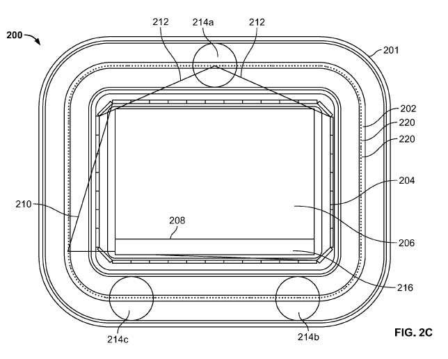
FIGS. 2C and 8A illustrate cross-sectional views of the housing 201 of the
scanning
units 200, 200' respectively, comprising a plurality of X-ray source points
and detectors arranged
in a substantially rectangular shape around a scanning volume, in accordance
with first and
second embodiments of the present specification. In various embodiments, the
rectangular
housing 201 has width ranging from 800 mm to 1400 mm and a height ranging from
600 mm to
1500 mm. In various embodiments, the housing 201 is configured to define an
imaging volume
or inspection tunnel 206, which is also rectangular, that has a width ranging
from 500 mm to
1050 mm and a height ranging from 300 mm to 1050 mm. It should be appreciated
that, in
alternate embodiments, the plurality of X-ray source points and detectors can
be arranged in
other quadrilateral shapes, such as, but not limited to, a square shape. It
should be appreciated
that the rectangular, quadrilateral, or square shape may also have rounded
edges and
encompasses shapes known as rounded rectangles, squircles, or rectellipses.
Referring now to FIGS. 2C and 8A simultaneously, the scanning units 200, 200'
respectively comprise a multi-focus X-ray source 202 and X-ray detector array
204 enclosed
within housing 201. The source 202 comprises a large number of source points
or electron guns
220 in locations spaced about the source 202, and arranged in a substantially
non-circular, such
as rectangular, geometry around an imaging or inspection volume 206, in
accordance with an
embodiment. In embodiments, the X-ray detector array 204 is positioned between
the X-ray
source points 220 and the imaging volume 206 such that the source points 220
and the detector
array 204 surround the imaging volume 206.
A conveyor belt 208 carries objects/luggage to be inspected through the
imaging volume
206 along a longitudinal axis of the scanning units 200, 200'. In an
embodiment, the conveyor
belt 208 has a speed of 0.5 m/s which is about twice the speed of conventional
X-ray systems
that typically operate at a speed of about 0.25 m/s and is about three times
the speed of
conventional rotating gantry systems that typically operate at a speed of
about 0.15 m/s. In
various embodiments, the conveyor belt 208 has a speed ranging from 0.1 m/s to
1.0 m/s. Both
feed and return conveyor loops pass through the base 216 of the imaging volume
206, having a
14
CA 03059061 2019-10-03
WO 2018/195016
PCT/US2018/027872
depth of approximately 50 mm while space 240 (approximately 200 mm deep and
having a
width equal to that of the base 216 of the imaging volume 206) is reserved in
the base of the
scanning units 200, 200', to accommodate automated return of trays when
integrated with an
automatic tray return handling system, in accordance with some embodiments.
The conveyor
.. and feed return loops both pass through base 216 of imaging volume 206. In
contrast, trays that
have been conveyed through the inspection or imaging volume 206 by the
conveyor 208 are
returned back through region 240, which ranges from 100 mm to 300 mm deep and
is preferably
200 mm deep.
In various embodiments, the rectangular housing 201 has width ranging from 800
mm to
1400 mm and a height ranging from 600 mm to 1500 mm. In embodiments, the
housing 201 has
a maximum width of 920 mm and a maximum height of 720 mm. In various
embodiments, the
housing 201 is configured to define an imaging volume or inspection tunnel
206, which is also
rectangular, that has a width ranging from 500 mm to 1050 mm and a height
ranging from 300
mm to 1050 mm. In some embodiments, the housing 201 is configured to define an
imaging
volume or inspection tunnel 206 that is approximately 620 mm in width and
approximately 420
mm in height.
In an embodiment, as shown in FIG. 2C, X-ray source 202 comprises 256 electron
guns
220, grouped in units of 16, substantially equidistantly spaced around the
imaging volume 206
on a 12 mm pitch (that is, a center-to-center spacing between adjacent
electron guns is 12 mm).
In various embodiments, the X-ray source 202 comprises 64 to 2048 electron
guns grouped in 4
to 32 units of electron guns. In various embodiments, the electron guns 220
are spaced on a pitch
ranging from 10 mm to 14 mm. In this configuration, every emission source
point has a different
field of view (FOV). In various embodiments, the X-ray sources emit fan beams
which have
different beam angles based on the location of the X-ray source points with
respect to the
-- imaging volume.
In another embodiment, as shown in FIG. 8A, the X-ray source 202 comprises 256
electron guns 220 spaced on a 12 mm pitch (that is, a center-to-center spacing
between adjacent
electron guns is 12 mm), grouped in units of 8, equidistantly spaced around
the imaging volume
206. In various embodiments, the X-ray source 202 comprises 64 to 2048
electron guns grouped
in 4 to 32 units of electron guns. In various embodiments, the electron guns
220 are spaced on a
pitch ranging from 10 mm to 14 mm. FIG. 8B illustrates a partial break-away
view of a corner
CA 03059061 2019-10-03
WO 2018/195016
PCT/US2018/027872
section layout 230 comprising a plurality of X-ray source points 220 flanked
on either side by an
adjacent straight section layout 235 of electron guns 220, grouped in units of
8 in accordance
with the embodiment shown in FIG. 8A. Each electron gun 220 of the X-ray
source 202 emits a
fan beam of X-rays having a different field of view (FOV). In various
embodiments, the X-ray
sources emit fan beams which have different beam angles based on the location
of the X-ray
source points with respect to the imaging volume.
Conventional RTT systems with the source points arranged in a circular
geometry have
the same X-ray beam angle or angle of coverage emanating from each source
point. In the
configurations shown in FIGS. 2C, 8A, and 8B, the beam angles are different
for different source
points, owing to the different distances from each source point to detector
element in the beam
path. In embodiments, a substantially rectangular field of view is
reconstructed for a rectangular
inspection tunnel region. Specifically, closer to the edges, the beam angle
made by the emitted
X-rays 210 is more narrow while closer to the middle of the scanning volume
206, the beam
angle made by the emitted X-rays 212 is broader. In some embodiments, the beam
angles range
from approximately 60 degrees to 120 degrees.
In an embodiment, as shown in FIG. 8A, the X-ray detector array 204 comprises
64
multi-energy detector modules (energy bins) or segments each having a 16 x 4
pixel sensor
configuration on a 2.5 mm pixel pitch (that is, a center-to center spacing
between adjacent
detector elements or sensors). FIG. 8C illustrates a 16 x 4 pixel multi-energy
sensor 225 in
accordance with an embodiment of the present specification. The sensor 225 is
capable of
detecting individual interacting photons with an intrinsic resolution of at
least 5 keV and incident
photon count rate of at least 5 Mcps/mm2/s. In embodiments, the detected
photons are allocated
into one of 2 to 64 programmable energy bins to provide accurate Z-effective
measurement in
the subsequent image reconstruction algorithms. In some embodiments, the
detected photons are
allocated into one of six programmable energy bins. In an embodiment, the
energy extents or
windows of each of the six programmable energy bins are, respectively, 25 keV,
40 keV, 55 keV,
65 keV, 100keV, 160keV. The energy extents or windows of each of the six
programmable
energy bins are customizable, in various embodiments, to optimize Z-effective
accuracy. In
various embodiments, the energy extents or windows of each of the six
programmable energy
bins ranges from 15 keV to 200 keV.
16
CA 03059061 2019-10-03
WO 2018/195016
PCT/US2018/027872
In an embodiment, each of the 64 multi-energy detector modules or segments of
the X-
ray detector array 204 has a length of 60 mm. The 64 multi-energy detector
modules or segments
form a rectangular ring or detector array 204 around the imaging volume 206,
as shown in FIG.
8A. In embodiments, the substantially square detector array 204 enables
reduction of the overall
height of the scanning unit 200' such that an operator and passenger can have
eye level contact
with each other while the passenger's baggage is being inspected.
As shown in FIG. 2C, a plurality of support means 214a, 214b, and 214c,
positioned at
points along the periphery of the imaging volume 206, are provided for
supporting the X-ray
source 202. In an embodiment, the support means 214b and 214c are also used to
provide
coolant and power to the X-ray source 202 and the scanning system 200,
respectively.
Referring back to FIG. 8A, a plurality of first structures 250 for enabling
heat dissipation
and at least one second structure 255 for enabling heat dissipation and for
providing voltage
supply is shown. FIG. 8D is a cross-sectional view of one of the plurality of
first structures 250
and the at least one second structure 255 along with respective top views 250'
and 255', in
accordance with embodiments of the present specification. Referring now to
FIGS. 8A, 8C and
8D, simultaneously, the plurality of first structures 250 include a thermally
conductive element
251 to dissipate heat from the anode region 252 (FIG. 8D). In embodiments, the
thermally
conductive element 251 is fabricated from ceramic. In embodiments, the first
structure 250 is
designed to maximize mechanical integrity and heat conductivity. The at least
one second
structure 255 comprises a thermally conductive element 253, also fabricated
from ceramic, to
dissipate heat from the anode region 254 and also a metal rod 256 that passes
through its center
to supply voltage. Both thermally conductive elements 251 and 253 include a
plurality of fins
258 along the height on either side to enable heat to dissipate into the air.
It should be
appreciated that, in some embodiments, the use of the first and second
structures 250, 255
obviates the need for circulating coolant to cool the electron guns. This in
turn, reduces overall
complexity and cost of manufacturing the electron guns.
In various embodiments, the thermally conductive elements 251, 253 are
manufactured
using AIN (Aluminum Nitride) ceramic electrical insulators to provide direct
thermal transfer
from the anode to ambient air. As shown in FIG. 8A, in some embodiments, the
plurality of first
structures 250 are strategically positioned along the perimeter of the source
202 such that each
first structure 250 pulls heat from a section of a plurality of electron guns
220. In some
17
CA 03059061 2019-10-03
WO 2018/195016
PCT/US2018/027872
embodiments, each first structure 250 pulls heat from 32 electron guns 220. In
different
embodiments, different combinations and numbers of first structure 250 and
second structure
255 are deployed. In one embodiment, a total of ten structures are employed
comprising nine
first structures 250 and one second structure 255. In an embodiment, the ten
structures are
equidistantly spaced about the source periphery. In embodiments, an equal
number of structures
are positioned along each side of the non-circular perimeter of the source. In
some embodiments,
the non-circular perimeter is a rectangle, with two equal and opposing sides
of a first length, and
the other two equal and opposite sides of a second length, where the first
length is greater than
the second length. In one embodiment, a greater number of structures 250, 255
is deployed
along the sides having a first length than the sides having a second length.
In an embodiment,
the first and second structures are welded into the housing 201. In accordance
with an
embodiment, each of the first and second structures is configured to dissipate
64 watts of heat
energy to air, on average.
FIG. 8E illustrates various views of at least a portion of the plurality of X-
ray source
points or electron guns 220 of the multi-focus X-ray source 202 (of FIGS. 2C
and 8A), in
accordance with some embodiments. As shown, view 270 illustrates first and
second adjoining
X-ray source modules 271, 272 each comprising a grouping or segment of 8
electron guns 220.
In one embodiment, 2 to 20 electron guns are integrally formed and positioned
on, or into, a
single common substrate 280. A top (vacuum side) view 274 of an emitter
assembly or X-ray
source module 271 shows a substrate comprising 8 individually controllable
electron guns 220.
A side section view 273 shows the same 8 electron guns 220 with an underlying
power bus bar
290 to supply power to the individual filaments in parallel. A bottom (air
side) view 275 for the
same module 271 shows the 8 electron guns 220 mounted into a common insulating
substrate
280. Views 282 and 283 are exploded views of one electron gun 220.
Referring now to views 275, 282 and 283, in embodiments, a focusing and grid
control
cup 278 is manufactured from nickel by stamping a sheet of nickel using a
power-press. A leg
279 extends downwards from the stamped nickel cup 278. The leg 279 is twisted
90 degrees to
lock the cup 278 in place once positioned into ceramic substrate 280. In some
embodiments, the
ceramic substrate 280 is brazed onto a nickel or copper ring. A filament 281,
comprised of
tungsten wire, is connected through the ceramic substrate 280. In embodiments,
glass frits 292
(that are finely powdered glasses that when re-heated sinter, soften, and flow
to form a seal or a
18
CA 03059061 2019-10-03
WO 2018/195016
PCT/US2018/027872
coating) are used to form metal to ceramic seals. The use of nickel for the
cup 278 and tungsten
for the filament 281 enables overall cost optimization of manufacturing the X-
ray source 202 (of
FIG. 8A). The modules, such as modules 271 and 272 are brazed onto the housing
201 of the
multi-focus X-ray source 202 (of FIG. 8A).
FIG. 3A illustrates a cross-section of an X-ray source sealed within a glass
envelope, in
accordance with some embodiments of the present specification. In an
embodiment, the anode
and cathode of an X-ray source generating X-rays are machine-built and
installed onto a glass
base. Next, the base is sealed with a glass top by using glass melting
techniques, thereby
resulting in an anode 302 and a cathode 304 enveloped in a glass vacuum
envelope 306. In an
embodiment, the cathode 304 is modular. In various embodiments, the thickness
of the glass
envelope 306 is uniform in all portions of the body of the glass envelope 306.
In an embodiment,
the thickness of the glass envelope 306 ranges between 0.5 mm to 5 mm. In an
embodiment, the
thickness of the glass envelope 306 is 1 mm +/- 0.3 mm. Since, glass as a
transmission material
provides a lower X-ray absorption (low Z material), the source design
illustrated in FIG. 3A
provides improved material discrimination. In embodiments the anode 302 is
supported in a
manner that accounts for differences in thermal expansion between the glass
envelope 306 and
the anode metal. Blocks 308 provided atop the anode 302 and shield electrode
318 are, in an
embodiment, a slip coupling that accounts for thermal expansion. The slip
coupling blocks 308
are attached directly to the glass envelope 306 just underneath a "depressed"
region 312, as
shown in FIG. 3A. As the glass envelope 306 changes shape/volume due to heat
generated by
the generation process of X-rays, the expansion is accounted for by movement
of the slip
coupling mechanism. The anode 302 is connected to a high voltage (HV) power
source via a
copper tube 314. In an embodiment, the slip coupling blocks 308 are each
respectively attached
to depressed region 312 at a distal end and to the anode 302 at a proximal
end. In an
embodiment, the slip coupling blocks 308 are also attached to the copper tube
314 along at least
one side as shown in FIG. 3A. The slip coupling blocks 308 have a three point
connection,
thereby allowing for expansion while not allowing swinging or lateral
movement. Ceramic or
glass tubes 316 act as insulating standoff elements for the shield electrodes
318 that shield the
HV connections and tubes from the generated X-rays.
FIG. 3B illustrates another view of the X-ray source sealed within a glass
envelope, in
accordance with an embodiment of the present specification. In this
embodiment, a ceramic
19
CA 03059061 2019-10-03
WO 2018/195016
PCT/US2018/027872
insulator 316' is used to seal the X-ray source once positioned on a glass
assembly. The ceramic
insulator 316' is a bulk insulator which is attached to the glass rather than
relying on the glass
envelope itself. This enables the X-ray source to be more tolerant to HV
breakdown while also
making the glass envelope more robust. As a result, the embodiments shown here
do not require
separate cooling channels.
FIG. 4A illustrates a cathode assembly, in accordance with an embodiment of
the present
specification. Cathode assembly 400 is enclosed in the glass envelope 306 as
shown in FIG. 3A
and comprises a printed circuit board 402 into which the elements of the
cathode are plugged.
Cathode assembly 400 further comprises a grid 404, a dispenser cathode 406 and
a filament 408
coupled with and partially encased by a primary focus electrode 410 via a
glass feed-through
element 412. The cathode assembly 400 also comprises a secondary focus
electrode 414.
In various embodiments, the cathode assembly 400 of the inspection system is
provided
with a secondary electrode 414, which absorbs any flash of energy within the
cathode 400. The
secondary electrode 414 is held at ground potential and acts as a trap or
buffer that captures
electrons that would otherwise leak in cases of a short circuit within the
cathode assembly 400.
The distance from the dispenser cathode 406 to grid 404 is calibrated to
maintain a predefined
gap exactly uniform across all cathodes. The dispenser 406 generates electrons
that form a cloud
around it. By keeping the gap small, an effective space charge limited region
is created, which
acts as the gate keeper, letting out only a predefined number of electrons at
a time.
In an embodiment, the cathode assembly 400 is precision manufactured in
volume. The
core cathode is built with high tolerances using robots. In an embodiment, the
secondary focus
electrode 414 is not part of the precision manufacturing and is added to the
assembly separately.
In an embodiment, a glass support for the cathode is brazed to glass envelope
306 (shown in FIG.
3A).
FIG. 4B is an exploded view of the elements of the cathode assembly shown in
FIG. 4A.
FIG. 4C is a top plan view of the elements of the cathode assembly shown in
FIG. 4B. Referring
to FIGS. 4A, 4B and 4C, electrical connections from each of the primary focus
electrode 410,
grid 404, filament 408 along with a ground connection 416 placed within the
glass feedthrough
element 412 are plugged into the circuit board 402.
FIG. 13 is a flow chart of a plurality of exemplary steps of a method of
manufacturing the
cathode assembly 400 of FIG. 4A. At step 1305, individual cathode pre-
manufactured assemblies
CA 03059061 2019-10-03
WO 2018/195016
PCT/US2018/027872
are inserted into a fixture that positions each cathode with respect to all
others to required
tolerance. Each cathode plugs into a printed circuit board. At step 1310, a
pre-cast glass envelope
half section with holes for each cathode element is located over the aligned
array of cathode
elements or assemblies. Thereafter, at step 1315, each cathode assembly is
brazed into the glass
envelope section and the glass support for the cathode is brazed to the glass
envelope. Finally, at
step 1320, the cathode section is ready to braze to a matching glass anode
half section.
FIG. 5A illustrates a top view of a cathode array, in accordance with an
embodiment of
the present specification. Array 500 comprises a plurality of cathode
assemblies 502, each
comprising a grid, a dispenser cathode and a filament coupled with and
partially encased by a
primary focus electrode via a glass feedthrough element, as shown in FIG. 4A.
FIG. 5B
illustrates a bottom view of the cathode array shown in FIG. 5A, in accordance
with an
embodiment of the present specification. FIG. 5C illustrates another view of
the cathode array
shown in FIG. 5B. The two lines 504 represent a secondary electrode, which in
an embodiment
is stamped out and laser cut with holes in the middle for accommodating the
cathode array 500.
FIG. 6 illustrates a grid control sequence of the elements of an X-ray source,
in
accordance with an embodiment of the present specification. Curves 602, 604,
606, and 608
represent the voltages maintained across a secondary electrode, a primary
electrode, a grid
aperture, and a cathode respectively. In various embodiments, the primary and
secondary
electrodes control the electron beam focusing on the target. As shown, the
secondary electrode is
maintained at a ground potential and the primary electrode is set to -5V to
mitigate against any
short circuit between the grid aperture and cathode. The grid aperture defines
a space charge
limited operating region around the cathode and sets the base electron gun
potential to ground
potential. The cathode is the main potential switched component and acts to
control overall
electron gun emission. As shown, the potential maintained across the cathode
in an off position
is +5V and ranges from -50V to -120V in an on position. Curve 610 represents
the X-ray
emission which follows the cathode potential. In embodiments, the X-ray
emission is inversely
proportional to the cathode potential. The more negative the cathode
potential, the greater the
beam current and the brighter the X-ray emission.
FIG. 7A illustrates an anode of an X-ray source assembly, in accordance with
an
embodiment of the present specification. In an embodiment, anode 702 is formed
from a copper
tube containing coolant within a stainless steel collimating structure. In an
embodiment, a
21
CA 03059061 2019-10-03
WO 2018/195016
PCT/US2018/027872
coolant tube (such as the copper tube 314 shown in FIG. 3A) is molded to form
an anode/target
in a source assembly of the inspection system of the present specification. In
an embodiment, the
coolant tube is molded by using hydroforming, wherein a large quantity of
water is pumped into
the tube to cause it to expand to the shape of a mold. A predefined material
is then introduced
into the beam of a flame and a fan is used to blow it onto the tube, to cause
a spray coating of the
material onto the interior walls of the tube. In an embodiment, in order to
obtain a coating of a
low Z material, the material used is silicon carbide. Further, in an
embodiment, Tungsten is used
to create predefined patterns on the silicon carbide coating.
In an embodiment, side walls 704 of electron entry path are coated with
graphite tubes or
a thick silicon carbide coating. In various embodiments, the surface of the
anode 702 which faces
the cathode is coated with a plurality of materials to obtain patterned anode
surfaces. FIG. 7B
illustrates a patterned anode surface, in accordance with an embodiment of the
present
specification. As shown in FIG. 7B, the anode surface 710 is coated with
silicon carbide and then
patterned with tungsten rich tungsten carbide stripes 712. FIG. 7C illustrates
a patterned anode
surface, in accordance with another embodiment of the present specification. A
raised portion
714 is added to the anode surface 710 to define fan-shaped apertures for the X-
ray beam 716 to
emerge from. The anode surface 710 combines the X-ray forming region with beam
forming
region to limit the radiation dose from the X-ray tube as well as to mitigate
against off-focal
radiation. In an embodiment, the entire anode assembly and not just the
surface facing the
cathode is coated with silicon carbide to minimize off-focus X-ray emission.
In another
embodiment, specific predefined regions of the anode are coated with tungsten
carbide to define
regions of X-ray emission.
Referring back to FIGS. 2A, 2C, 8A, it should be appreciated that in contrast
to
conventional rotating gantry systems, the firing pattern for the multi-focus X-
ray source 202 is
not constrained to move in a standard helical rotation about a baggage under
inspection. Thus, in
various embodiments, the source firing pattern may be fixed or random with
uniform or non-
uniform dwell time at each source point 220. In various embodiments, the dwell
time ranges
from 50 ps to 500 i.ts per scan projection. In some embodiments, the dwell
time is 200 i.ts per
scan projection.
In various embodiments, in order to determine substantially accurate measures
for Z-
Effective and Density in the reconstructed RTT images, both sinogram data (the
multi-energy
22
CA 03059061 2019-10-03
WO 2018/195016
PCT/US2018/027872
"raw" data produced by the X-ray detectors for each source projection) and the
reconstructed
image data from one or more multi-energy bins is used in determining threat
type for each object
segmented from the 3D image data. In embodiments, the reconstructed image is
available as
soon as the trailing edge of a conveyor tray leaves the RTT imaging region of
the scanning units
200,200'.
In accordance with some embodiments, the scanning units 200, 200' are
respectively
configured to achieve reconstructed image voxels of 0.8mm x 0.8mm x 0.8mm over
an
inspection tunnel size of 620mm wide x 420mm. This is equivalent to a slice
image size of 775
pixels (width) x 525 pixels (height). For a conveyor tray length of 0.8 m,
there will be 1,000
slices in each 3D image. In some embodiments, the RTT system spatial
resolution is 1.0 mm at
the center of the inspection tunnel. In embodiments, the RTT system is
configured to achieve Z-
effective resolution of +/- 0.2 atomic numbers with density resolution at the
center of the
inspection tunnel of +/- 0.5%.
FIG. 9 is a cross-sectional view, through the imaging volume, of the scanning
unit 900
(or the scanning unit 200' of FIG. 8A). Isolated electron guns or source
points 920 are illustrated
on opposite sides of the scanning or inspection volume 906. X-ray beams 915
enter the imaging
volume 906 and have a crossing angle that matches the length of the detector
904 on the opposite
side.
FIG. 10 is a cross-sectional view of an electron gun 1020 and detector array
1004
assembly positioned at one end of the imaging volume 906 of the scanning unit
900 of FIG. 9. In
an embodiment, the anode 1085 and cathode 1086 of the X-ray source 1020 are
machine built
and the cathode sections 1086 are installed into one of a glass, metal, or
ceramic base 1087.
Anode sections 1085 are installed into a top 1088 made with material (glass,
metal, or ceramic)
that is similar to the material of base 1087. Next, the base 1087 is sealed
with the top 1088 by
using suitable welding techniques, thereby resulting in the anode 1085 and the
cathode 1086
being enveloped in a vacuum envelope. In one embodiment, a metal housing is
used to create the
vacuum envelope of the electron gun 1020, wherein the metal housing includes a
window
fabricated from a low Z material. Block 1089 provided atop the anode 1085 and
shield electrode
1090 is a slip coupling that accounts for thermal expansion. In accordance
with an aspect of the
present specification, the slip coupling block 1089 are attached to a feed-
through thermally
conductive element 1051 (as also shown in FIG. 8D), to enable heat dissipation
from the anode
23
CA 03059061 2019-10-03
WO 2018/195016
PCT/US2018/027872
1085. In embodiments, the element 1051 comprises a plurality of fins or
extensions 1058 to
enable heat dissipation to ambient air. Use of the thermally conductive
element 1051 obviates a
need for circulating coolant to reduce anode temperature.
FIG. 14 is a flow chart of a plurality of exemplary steps of a method of
manufacturing the
X-ray source or electron gun 1020 of FIG. 10. At step 1405, the anode and
cathode of the X-ray
source are machine built. At step 1410, the anode section is installed into a
glass top. At step
1415, a slip coupling block is provided atop the anode and a shield electrode
to account for
thermal expansion. At step 1420, the slip coupling block is attached to a feed-
through thermally
conductive element to enable heat dissipation from the anode. Next, at step
1425, the cathode
section is installed into a glass base. Finally, at step 1430, the base is
sealed with the glass top by
using glass melting techniques, thereby resulting in the anode and the cathode
being enveloped
in a glass vacuum envelope.
Referring back to FIG. 10, X-rays emanating from the anode 1085 are
collimated, using
collimator 1091, to form the X-ray beam 1015. In various embodiments, the X-
ray beam 1015 is
a fan beam. In embodiments, the self-collimated anode 1085 minimizes off-focal
radiation. The
X-ray beam 1015 exits the electron gun 1020 through an opening 1092 in the
shield 1093
towards the inspection volume 1006. The opening 1092 is a radiologically thin
window to
preserve low energy content in the beam 1015. In alternate embodiments, where
a metal housing
is used to create the vacuum envelope of the electron gun 1020, the opening
1092 (in the metal
housing) is fabricated from a low Z material. The detector arrays 1004 are
positioned just below
a plane of the opening 1092 (on the same side as of the X-ray beam 1015) to
lie between the
electron gun 1020 and the inspection volume 1006. The detector arrays 1004 are
located within a
lead and composite material housing 1094 with easy service access from outside
the inspection
volume 1006 (using a plurality of screws 1095). The lead and composited
material housing 1094
minimizes radiation damage to various electronics.
Referring back to FIGS. 2C and 8A, in accordance with aspects of the present
specification, the amount of time that each individual X-ray source point 220
is 'on' can be
adjusted electronically, and in real-time, while during each source exposure
the source point is
fixed (rather than moving as is the case with a conventional rotating gantry
CT system). As
discussed earlier, in contrast to conventional rotating gantry CT systems, the
X-ray source firing
pattern for the multi-focus X-ray source 202 is not constrained to move in a
standard helical
24
CA 03059061 2019-10-03
WO 2018/195016
PCT/US2018/027872
rotation about an object under inspection. The ability of random source firing
as well as the
ability to use non-uniform dwell time at each X-ray source point 220, enables
combining an X-
ray diffraction (XRD) system with the transmission RTT system 900 (of FIG. 9)
using the same
X-ray beam 215 for both XRD and RTT systems. Thus, the use of non-uniform
dwell time at
each X-ray source point 220 enables increased sampling for the associated XRD
system ¨ using
the same X-ray beam 215 used for transmission RTT ¨ in suspected threat
regions for automated
in-line, real time alarm clearance.
FIG. 11 shows a cross-sectional view through the imaging volume 1115 of a
scanning
unit (such as the scanning unit 900 of FIG. 9 and 1000 of FIG. 10) combined
with an X-ray
diffraction imaging system, in accordance with an embodiment of the present
specification. In
the combined RTT and XRD system 1100 of FIG. 11, the system 1100 is configured
to function
as a primary scanning system using transmission RTT with multi-energy
detectors 1102 along
with an optional secondary alarm clearance system using in-line real-time X-
ray diffraction
(XRD). In embodiments, the X-ray diffraction imaging system comprises a
plurality of "venetian
.. blind" or "slot" collimators 1105 angled at a small angle, for example 5
degrees, to the
transmission RTT imaging beam 1107 (in the direction towards the beam 1107)
along with an
area array of energy dispersive X-ray detectors 1110 that are located behind
the collimators 1105.
The collimators 1105 provide a barrier that prevents X-rays from reaching each
detector of the
array 1110 unless it comes from a particular receiving direction. In various
embodiments, the
collimators 1105 are angled at an angle ranging from 3 to 10 degrees.
For each electron gun 1101, the energy dispersive X-ray detectors 1110, for
XRD
imaging, are positioned just above the plane of an opening 1150 from which the
transmission
RTT imaging beam 1107 emanates from the electron gun 1101. In various
embodiments, the
beam 1107 is a fan beam. The detectors 1110 are positioned within a lead and
composite
.. material housing 1152. The multi-energy detectors 1102, for transmission
RTT imaging, are
positioned just below the plane of the opening 1150 (as also described with
reference to FIGS. 8
and 9) within another lead and composite housing 1154. Housings 1152, 1154 are
easily
accessible (using screws) for servicing and maintenance of the detectors 1110,
1102. The
detectors 1102 and 1110 are positioned between the opening 1150 and the
inspection volume
1115 for each electron gun 1101 shown on either side of the inspection volume
1115.
CA 03059061 2019-10-03
WO 2018/195016
PCT/US2018/027872
In some embodiments, each of the detectors 1110 has sensitive area 2.5mm wide
x
2.5mm high and together they form a 60mm high "wall" of sensors that extend
around the whole
periphery of the imaging volume or inspection tunnel 1115. The large energy
sensitive
diffraction sensor area (of about 41,000 mm2) provides efficiency gains that
conventional
systems seek to achieve either through high beam flux or by open collimation
approaches.
As shown in FIG. 11, the lines 1120 show an extent of the diffraction field of
view
whereas lines 1125 and 1130 indicate scattering paths (containing X-ray
diffraction photons) at 5
degrees (relative to the transmission RTT beam 1107) from two, exemplary, 20mm
wide regions
1135, 1140 each 120mm from the center of the inspection/imaging volume or
inspection tunnel
1115. It should be appreciated that the collimated detector array 1110 can be
used to define
different inspection regions in an object under inspection.
Each photon counting, energy sensitive, pixel in the diffraction detector
array 1110
projects back to a small arc-shaped volume within the imaging volume 1115.
This arc is defined
by the intersection of X-rays from each individual sensor pixel back through
the 5 degree
collimator 1105 and to the fan-beam 1107 projecting from the electron gun or X-
ray source point
1101 through the imaging volume 1115. The use of this arc with one-dimensional
collimation
gives a much higher diffraction efficiency (that is, the number of diffracted
photons detected per
incident X-ray photon) than would be the case if two-dimensional collimation
were used. Thus,
every photon counting pixel in the diffraction detector array 1110 can measure
spatial location
and energy. By knowing position and energy of an interacting photon, the
location from which
the scattered photon has arrived and the elemental composition of the location
can be determined.
In order to form a reasonable coherent diffraction signal data is collected
for all source
points 801 within a tomographic scan and then the results are accumulated for
each sub-volume
of the imaging volume 1115. For a scanner with 256 source points, and an
average of four to five
coherent diffraction scatter results per sub-volume per scan, then following
accumulation of the
set of data, each sub-volume will have more than 1000 results associated with
it, corresponding
to 256 scattering events within that sub-volume. A typical sub-volume occupies
an area within
the imaging plane of a few square centimeters, with a volume thickness of a
few millimeters.
In accordance with an aspect, the diffraction signal (an energy spectrum) is
recorded for
every sensor in the diffraction imaging array 1110 for every X-ray source
point 1101. This data
set (comprising both energy dispersive and angular dispersive data) is then
converted into a 2D
26
CA 03059061 2019-10-03
WO 2018/195016
PCT/US2018/027872
set of slice images using an iterative back-projection method to create a 3D
diffraction
tomography image where each voxel in the image describes the diffraction
energy spectrum (and
hence material composition) of the object located within the imaging volume
1115 at that region
in space. The process occurs in real-time at the same time as the transmission
RTT image is
collected. Thus, a primary detection image (using the transmission RTT
imaging) and a
secondary clearance image (using the XRD imaging) are created at the same time
and can both
be used to create an overall threat detection capability for each item in an
object under inspection.
It should be appreciated that it is not required to slow or stop the conveyor
during the
screening process nor is it necessary to re-register the object under
inspection between the RTT
imaging and the diffraction imaging processes since both imaging processes are
conducted at the
same time. The 3D volume of each potential threat object is known from the RTT
volume data
and therefore in a second verification step, all the diffraction data
belonging to that one volume
(be it bulk or sheet) can be combined to maximize photon statistics and so
create a further,
accurate, data point in the threat detection capability.
It should be noted that those detectors not being used for forward X-ray
diffraction or
transmission RTT imaging will record the Compton backscatter signal which is a
sensitive
indicator of low-Z materials near the surface of the object under inspection.
In some
embodiments, this information may be reconstructed into a Compton scatter
image and further
correlated with the RTT scan data to create a separate item in the overall
threat detection
capability for the object under inspection.
FIG. 12 is a flow chart of a plurality of exemplary steps of a method of
automatic threat
detection and clearance using combined X-ray transmission RTT and XRD systems
of FIG. 11.
Referring now to FIGS. 11 and 12, at step 1205, a conveyor belt moves an
object under
inspection through the imaging volume 1115 parallel to a longitudinal axis of
the scanner 1100.
At step 1210, as each source point 1101 emits a fan beam 1107 of X-rays, the
tomographic
transmission scan signals from the detectors 1102, which are dependent on the
intensity of the X-
rays incident on the detector 1102, are produced, and the intensity data that
the signals provide
are recorded in memory. Simultaneously, as each source point 1101 emits a fan
beam 1107 of X-
rays, diffraction signals (an energy spectrum) are recorded for every sensor
in the diffraction
imaging array 1110 for every X-ray source point 1101.
27
CA 03059061 2019-10-03
WO 2018/195016
PCT/US2018/027872
At step 1215, once the scan is completed the tomographic transmission signals
from
detectors 1102 are processed using a filtered back-projection method to form a
tomographic
transmission image of the scanned volume. At step 1220, a threat detection
processor determines
whether the tomographic transmission image represents a threat. If no threat
is detected, then the
object is cleared at step 1225. However, if the object is flagged as a threat,
then at step 1230, the
diffraction signals recorded using detectors 1110 are converted into a 2D set
of slice images
using an iterative back-projection method to create a 3D diffraction
tomography image where
each voxel in the image describes the diffraction energy spectrum (and hence
material
composition) of the object located within the imaging volume 1115 at that
region in space.
For each scan, the tomographic transmission image data from the detectors 1102
produces data relating to the X-ray attenuation for each pixel of the image,
which in turn
corresponds to a respective sub-volume of the tomographic imaging volume. The
data from the
scatter detectors 1110 provides tomographic diffraction image data relating to
the amount of
coherent scattering within each sub-volume, and data relating to the amount of
incoherent
scattering within each sub-volume. At step 1235, the tomographic diffraction
image data, along
with the tomographic transmission image data, is analyzed using the threat
detection processor to
determine if the threat is benign or not. Examples of parameters that are
extracted from the
transmission and/or diffraction data are the ratio of coherent to incoherent
diffraction, material
types as determined from coherent diffraction data, material density as
determined from
incoherent diffraction data, correlation of tomographic transmission image
pixel values with
diffraction data. Thus, from the Bragg scattering data, for each detected
diffraction event, the
combination of the X-ray energy and the scatter angle can be used to determine
the inter-atomic
distance d of the material in which the diffraction event took place. In
practice, the scatter angle
can be assumed to be constant, and the energy used to distinguish between
different materials.
For the Compton scattering, the level of scattering from each volume of the
scanning volume
gives an indication of the density of the material in that volume. The ratio
of Compton to
coherent scatter can also be determined and used as a further parameter to
characterize the
material of the imaged object.
It shall be recognized by one skilled in the art that the objective of the
diffraction and
Compton backscatter imaging is to generate an alternative signature that can
be used to clear the
specific threat raised by analysis of the multi-energy transmission computed
tomography image
28
CA 03059061 2019-10-03
WO 2018/195016
PCT/US2018/027872
data. In this respect, the performance of the diffraction and backscatter
imaging does not need to
be of laboratory grade, since it is necessary only to clear a threat raised by
analysis of the higher
quality transmission X-ray imaging data. This facilitates diffraction imaging
at high conveyor
speed (ranging from 0.1m/s to 0.5m/s) where typically systems would be too
photon starved to
-- create a laboratory grade signal.
The above examples are merely illustrative of the many applications of the
system of
present specification. Although only a few embodiments of the present
invention have been
described herein, it should be understood that the present invention might be
embodied in many
other specific forms without departing from the spirit or scope of the
invention. Therefore, the
-- present examples and embodiments are to be considered as illustrative and
not restrictive, and
the invention may be modified within the scope of the appended claims.
29