Note: Descriptions are shown in the official language in which they were submitted.
CA 03059447 2019-13-08
WO 2018/195226
PCT/US2018/028206
1
ANTI-PD-Li ANTIBODY AND USE THEREOF
FIELD
The present disclosure pertains to the field of biomedicine
and relates to a fully human anti-PD-Li antibodies and
pharmaceutical uses thereof.
BACKGROUND
When T cells respond to an exogenous antigen, they need
antigen-presenting cells (APC) to provide two signals to resting
T lymphocytes: the first signal is generated when T cells
recognize antigen peptides bound to MHC molecules with the aid
of TCR, after which an antigen recognition signal is transmitted
via a TCR/CD3 complex; and the second signal is provided by a
series of costimulatory molecules; and in this way, the T cells
can be activated normally, which in turn produce a normal immune
response. These costimulatory molecules can be classified as
either positive costimulatory molecules or negative
costimulatory molecules depending on the effects produced by the
second signal, and regulation of the positive and negative
costimulatory signals as well as the relative balance between
said signals play an important regulatory role throughout the
body's entire immune response.
PD-1 is a member of the CD28 receptor family, and said
family also includes CTLA4, CD28, ICOS and BTLA. The initial
members of this family, CD28 and ICOS, were discovered when
monoclonal antibodies were added and observed as increasing T
cell proliferation (Hutloff et al. (1999) Nature 397: 263-266;
CA 03059447 2019-13-08
WO 2018/195226
PCT/US2018/028206
2
Hansen et al. (1980) Immunogenics 10: 247-260). Ligands of PD-1
include PD-Li and PD-L2, and study results have already shown
that binding of the receptor with a ligand downregulates T cell
activation and the secretion of related cytokines (Freeman et
al. (2000) J Exp Med 192: 1027-34; Latchman et al. (2001) Nat
Immunol 2: 261-8; Carter et al. (2002) Fur J Immunol 32: 634-43;
Ohlgashi, et al. (2005) Clln Cancer Res 11: 2947-53).
PD-Li (B7-H1) is a cell surface glycoprotein which belongs
to the B7 family and includes IgV- and IgC-like regions, a
transmembrane region and a cytoplasmic tail region. The
corresponding gene was first discovered and cloned in 1999 (Dong
H, et al. (1999) Nat Med 5: 1365-1369) and the glycoprotein
itself was determined to interact with the T cell receptor PD-1
and play an important role in the negative regulation of the
immune response. In addition to acting on PD-1 expressed on
T cells, PD-L1, when expressed on T cells, can interact with
CD80 on APCs to transmit negative signals, functioning as a T
cell inhibitor. In addition to being expressed on macrophage
lineage cells, PD-Li is also expressed at low levels in normal
human tissues, but the glycoprotein shows relatively high
expression in certain tumor cell lines, including, for example,
lung cancer, ovarian cancer, colon cancer and melanoma (Iwai et
al. (2002) PNAS 99: 12293-7; Ohigashi, et al. (2005) Clin Cancer
Res 11: 2947-53). Study results have suggested that increased
expression of PD-Li in tumor cells increases T cell apoptosis,
thereby playing an important role in allowing tumor cells to
evade an immune response. Researchers have found that PD-Li
gene-transfected P815 tumor cell lines can show in vitro
resistance to specific CTL lysis, and said cells are more highly
tumorigenic and invasive when inoculated into mice. These
biological properties can be reversed by blocking PD-Li. In PD-1
CA 03059447 2019-10-08
WO 2018/195226
PCT/US2018/028206
3
knockout mice, the PD-Ll/PD-1 pathway is blocked and inoculated
tumor cells are unable to form tumors (Dong H et al. (2002) Nat
Med 8: 793-800).
There remains a need for an anti-PD-Li antibody which is
capable of binding to PD-Li with high affinity and thus blocking
the binding of PD-1 and PD-Li.
SUMMARY
In certain aspects of the present invention a yeast display
system in conjunction with screening and affinity maturation was
utilized to obtain a fully human anti-PD-Li antibody which shows
good specificity and relatively high affinity and stability,
thereby completing the present invention.
The first aspect of the present invention pertains to an
anti-PD-Li antibody or an antigen-binding portion thereof, which
includes a group of CDR regions selected from one of the
following:
(1) heavy chain CDR1, CDR2 and CDR3 sequences which
correspond to SEQ ID NO: 1-3, respectively and light chain CDR1,
CDR2 and CDR3 sequences which correspond to SEQ ID NO: 4-6
respectively or sequences which are more than 70%, 80%, 85%, 90%
or 95% identical to one of the aforementioned sequences,
respectively;
(2) heavy chain CDR1, CDR2 and CDR3 sequences which
correspond to SEQ ID NO: 7-9, respectively and light chain CDR1,
CDR2 and CDR3 sequences which correspond to SEQ ID NO: 10-12
respectively or sequences which are more than 70%, 80%, 85%, 90%
or 95% identical to one of the aforementioned sequences,
respectively;
CA 03059447 2019-13-08
WO 2018/195226
PCT/US2018/028206
4
(3) heavy chain CDR1, CDR2 and CDR3 sequences which
correspond to SEQ ID NO: 13-15, respectively and light chain
CDR1, CDR2 and CDR3 sequences which correspond to SEQ ID NO: 16-
18 respectively or sequences which are more than 70%, 80%, 85%,
90% or 95% identical to one of the aforementioned sequences,
respectively;
(4) heavy chain CDR1, CDR2 and CDR3 sequences which
correspond to SEQ ID NO: 1, 2 and 19, respectively and light
chain CDR1, CDR2 and CDR3 sequences which correspond to SEQ ID
NO: 4-6 respectively or sequences which are more than 70%, 80%,
85%, 90% or 95% identical to one of the aforementioned
sequences, respectively;
(5) heavy chain CDR1, CDR2 and CDR3 sequences which
correspond to SEQ ID NO: 7, 20 and 9, respectively and light
chain CDR1, CDR2 and CDR3 sequences which correspond to SEQ ID
NO: 10-12 respectively or sequences which are more than 70%,
80%, 85%, 90% or 95% identical to one of the aforementioned
sequences, respectively;
(6) heavy chain CDR1, CDR2 and CDR3 sequences which
correspond to SEQ ID NO: 13-15, respectively and light chain
CDR1, CDR2 and CDR3 sequences which correspond to SEQ ID NO: 21,
17 and 18 respectively or sequences which are more than 70%,
80%, 85%, 90% or 95% identical to one of the aforementioned
sequences, respectively.
Any one of the anti-PD-Li antibodies or corresponding
antigen-binding portions constituted by the first aspect of the
present invention also includes a group of heavy chain variable
region framework regions selected from one of the following:
1) FR1, FR2, FR3 and FR4 sequences which correspond to SEQ
ID NO: 22-25, respectively or sequences which are more than 70%,
CA 03059447 2019-13-08
WO 2018/195226
PCT/US2018/028206
80%, 85%, 90%, 95% or 99% identical to one of the aforementioned
sequences, respectively;
2) FR1, FR2, FR3 and FR4 sequences which correspond to SEQ
ID NO: 30-33, respectively or sequences which are more than 70%,
5 80%, 85%, 90%, 95% or 99% identical to one of the aforementioned
sequences, respectively;
3) FR1, FR2, FR3 and FR4 sequences which correspond to SEQ
ID NO: 38-41, respectively or sequences which are more than 70%,
80%, 85%, 90%, 95% or 99% identical to one of the aforementioned
sequences, respectively;
4) FR1, FR2, FR3 and FR4 sequences which correspond to SEQ
ID NO: 30-33, respectively or sequences which are more than 70%,
80%, 85%, 90%, 95% or 99% identical to one of the aforementioned
sequences, respectively.
Any one of the anti-PD-Li antibodies or corresponding
antigen-binding portions constituted by the first aspect of the
present invention also includes a group of light chain variable
region framework regions selected from one of the following:
1) FR1, FR2, FR3 and FR4 sequences which correspond to SEQ
ID NO: 26-29, respectively or sequences which are more than 70%,
80%, 85%, 90%, 95% or 99% identical to one of the aforementioned
sequences, respectively;
2) FR1, FR2, FR3 and FR4 sequences which correspond to SEQ
ID NO: 30-33, respectively or sequences which are more than 70%,
80%, 85%, 90%, 95% or 99% identical to one of the aforementioned
sequences, respectively;
3) FR1, FR2, FR3 and FR4 sequences which correspond to SEQ
ID NO: 38-41, respectively or sequences which are more than 70%,
80%, 85%, 90%, 95% or 99% identical to one of the aforementioned
sequences, respectively;
CA 03059447 2019-13-08
WO 2018/195226
PCT/US2018/028206
6
4) FM, FR2, FR3 and FR4 sequences which correspond to SEQ
ID NO: 30-33, respectively or sequences which are more than 70%,
80%, 85%, 90%, 95% or 99% identical to one of the aforementioned
sequences, respectively.
Any one of the anti-PD-Li antibodies or corresponding
antigen-binding portions constituted by the first aspect of the
present invention includes a group of heavy chain variable
regions selected from one of the following:
1) sequences corresponding to SEQ ID NO: 47, 49, 51, 53 or
.. 54, or a sequence which is 70%, 80%, 85%, 90%, 95% or 99%
identical to one of the aforementioned sequences, respectively.
Any one of the anti-PD-Li antibodies or corresponding
antigen-binding portions thereof constituted by the first aspect
of the present invention includes a group of light chain
variable regions selected from the following:
1) sequences corresponding to SEQ ID NO: 48, 50, 52, 55 or
56, or a sequence which is 70%, 80%, 85%, 90%, 95% or 99%
identical to one of the aforementioned sequences, respectively.
Any one of the anti-PD-Li antibodies or corresponding
antigen-binding portions constituted by the first aspect of the
present invention corresponds to a whole antibody, a bispecific
antibody, scFv, Fab, Fab', F(ab')2 or Fv.
In any example of the present invention, when the invention
is constituted by an scFv, a connecting peptide is also included
between the heavy chain and light chain variable regions of the
aforementioned anti-PD-Li antibody or antigen binding portion
thereof.
In some specific examples of the present invention, the
sequence of the aforementioned connecting peptide is as shown in
SEQ ID NO: 67.
CA 03059447 2019-13-08
WO 2018/195226
PCT/US2018/028206
7
Any one example of the anti-PD-Li antibodies or
corresponding antigen-binding portions thereof constituted by
the first aspect of the present invention corresponds to a whole
antibody.
Any one example of the anti-PD-Li antibodies or
corresponding antigen-binding portions thereof constituted by
the first aspect of the present invention, wherein the heavy
chain constant region is selected from a group comprising IgG,
IgM, IgE, IgD and IgA.
In certain examples of the present invention, the heavy
chain constant region is selected from a group comprising IgGl,
IgG2, IgG3 and IgG4.
In specific examples of the present invention, the heavy
chain constant region corresponds to IgGl.
In certain specific examples of the present invention, the
IgG1 amino acid sequence is as shown in SEQ ID NO: 68.
Any one of the anti-PD-Li antibodies or corresponding
antigen-binding portions constituted by the first aspect of the
present invention, wherein the light chain constant region is a
K region or A region.
In certain specific examples of the present invention, the
amino acid sequence of the K light chain constant region is as
shown in SEQ ID NO: 70.
In certain specific examples of the present invention, the
amino acid sequence of the A light chain constant region is as
shown in SEQ ID NO: 72.
The second aspect of the present invention pertains to a
nucleic acid molecule which contains a nucleic acid sequence
encoding an antibody heavy chain variable region, wherein the
aforementioned antibody heavy chain variable region includes a
group of amino acid sequences selected from the following:
CA 03059447 21319-18
WO 2018/195226
PCT/US2018/028206
8
( ) SEQ ID NO: 1-3;
(ii) SEQ ID NO: 7-9;
(iii) SEQ ID NO: 13-15;
(iv) SEQ ID NO: 1, 2 and 19;
(v) SEQ ID NO: 7, 20 and 9;
Any one of the nucleic acid molecules constituted by the
second aspect of the present invention, wherein the
aforementioned antibody heavy chain variable region includes a
group of nucleic acid sequences which are selected from the
following: SEQ ID NO: 47, SEQ ID NO: 49, SEQ ID NO: 51, SEQ ID
NO: 53, SEQ ID NO: 54 or a sequence created by replacing one or
several of the amino acids contained in the frame region of one
of the aforementioned sequences.
In some examples of the present invention, the
aforementioned nucleic acid includes a sequence selected from
those shown in SEQ ID NO: 57-61.
In some examples of the present invention, the
aforementioned nucleic acid also contains a nucleic acid
sequence encoding an antibody heavy chain constant region,
wherein said heavy chain constant region is selected from a
group comprising IgG, IgM, IgE, IgD and IgA.
In some examples of the present invention, the heavy chain
constant region is selected from a group comprising IgGl, IgG2,
IgG3 and IgG4.
In a specific example of the present invention, the heavy
chain constant region corresponds to IgGl.
In a specific example of the present invention, the IgG1
nucleic acid sequence is as shown in SEQ ID NO: 69.
The third aspect of the present invention pertains to a
nucleic acid molecule which contains a nucleic acid sequence
capable of encoding an antibody light chain variable region,
CA 03059447 21319-18
WO 2018/195226
PCT/US2018/028206
9
wherein the aforementioned antibody light chain variable region
includes a group of amino acid sequences selected from the
following:
(1) SEQ ID NO: 4-6;
(ii) SEQ ID NO: 10-12;
(iii) SEQ ID NO: 16-18;
(iv) SEQ ID NO: 21, 17 and 18.
Any one of the nucleic acid molecules constituted by the
third aspect of the present invention, wherein the
aforementioned antibody light chain variable region includes a
group of nucleic acid sequences which are selected from the
following: SEQ ID NO: 48, SEQ ID NO: 50, SEQ ID NO: 52, SEQ ID
NO: 55, SEQ ID NO: 56 or a sequence created by replacing one or
several of the amino acids contained in the frame region of one
of the aforementioned sequences.
In some aspects of the present invention, the aforementioned
nucleic acid includes a sequence selected from those shown in
SEQ ID NO: 62-66.
In some aspects of the present invention, the aforementioned
nucleic acid also contains a nucleic acid sequence capable of
encoding an antibody light chain constant region, wherein said
light chain constant region is a K region or A region.
In a specific aspect of the present invention, the nucleic
acid sequence of the K light chain constant region is as shown
in SEQ ID NO: 70.
In a specific aspect of the present invention, the amino
acid sequence of the A light chain constant region is as shown
in SEQ ID NO: 72.
The fourth aspect of the present invention pertains to a
vector which contains any one of the nucleic acids constituted
by the second or third aspects of the present invention.
CA 03059447 2019-13-08
WO 2018/195226
PCT/US2018/028206
Any one of the vectors constituted by the fourth aspect of
the present invention contains any one of the nucleic acids
constituted by the second aspect of the present invention and
any one of the nucleic acids constituted by the third aspect of
5 the present invention.
The fifth aspect of the present invention pertains to a
host cell which contains any one of the nucleic acids
constituted by the second or third aspects of the present
invention or any one of the vectors constituted by the fourth
10 aspect of the present invention.
The sixth aspect of the present invention pertains to a
conjugate which contains any one of the anti-PD-Li antibodies or
corresponding antigen-binding portions constituted by the first
aspect of the present invention, as well as other biologically
active substances, wherein the aforementioned anti-PD-Li
antibody or corresponding antigen-binding portion is conjugated
to another biologically active substance, either directly or via
a connecting fragment.
In some aspects of the present invention, the
aforementioned additional biologically active substance is
selected from a group comprising chemicals, toxins,
polypeptides, enzymes, isotopes, cytokines or other individual
biologically active substances or mixtures thereof, which are
capable of directly or indirectly inhibiting cell growth or
killing cells, or otherwise inhibiting or killing cells via
activation of an immune response, such as Auristatin MMAE,
Auristatin MMAF, Maytansine DM1, Maytansine DM4, calicheamicin,
duocarmycin MGBA, doxorubicin, ricin, diphtheria toxin and other
related toxins, 1131, interleukins, tumor necrosis factors,
chemokines, nanoparticles, etc.
CA 03059447 2019-13-08
WO 2018/195226
PCT/US2018/028206
11
The seventh aspect of the present invention pertains to a
composition (such as a pharmaceutical composition), which
contains any one of the anti-PD-Li antibodies or corresponding
antigen-binding portions constituted by the first aspect of the
present invention, any one of the nucleic acids constituted by
the second or third aspects of the present invention, any one of
the vectors constituted by the fourth aspect of the present
invention, any one of the host cells constituted by the fifth
aspect of the present invention, or any one of the conjugates
constituted by the sixth aspect of the present invention, as
well as any pharmaceutically acceptable vector or excipient and
any other biologically active substance(s).
Any one of the compositions constituted by the seventh
aspect of the present invention (such as a pharmaceutical
composition), wherein the aforementioned additional biologically
active substances include, but are not limited to, other
antibodies, fusion proteins or drugs (e.g., anticancer drugs,
such as chemotherapy and radiotherapy drugs).
The present invention further pertains to a reagent or
reagent kit which contains any one of the anti-PD-Li antibodies
or corresponding antigen-binding portions constituted by the
first aspect of the present invention, wherein the
aforementioned detection reagent or reagent kit is used for
detecting the presence or absence of the PD-Li protein or
derivatives thereof.
The present invention further pertains to a diagnostic
reagent or reagent kit which contains any one of the anti-PD-Li
antibodies or corresponding antigen-binding portions constituted
by the first aspect of the present invention, wherein the
aforementioned diagnostic reagent or reagent kit is used in the
in vitro (e.g., cells or tissues) or in vivo (e.g., humans or
CA 03059447 21319-18
WO 2018/195226
PCT/US2018/028206
12
model animals) diagnosis of PD-Ll-related diseases (e.g., tumors
or viral infections, such as cases of viral infections showing
high PD-Li expression or tumors showing high PD-Li expression).
In some aspects of the present invention, the
aforementioned anti-PD-Li antibody or corresponding antigen-
binding portion is further coupled to a fluorescent dye,
chemical substance, polypeptide, enzyme, isotope, label, etc.
which can be used in detection or which can be detected by a
separate reagent.
In some aspects of the present invention, the
aforementioned tumors include, but are not limited to, lung
cancer, ovarian cancer, colon cancer, colorectal cancer,
melanomas, kidney cancer, bladder cancer, breast cancer, liver
cancer, lymphomas, hematologic malignancies, head and neck
cancer, gliomas, gastric cancer, nasopharyngeal cancer,
laryngeal cancer, cervical cancer, uterine cancer,
osteosarcomas, thyroid cancer and prostate cancer.
In some aspects of the present invention, the
aforementioned viral infections include, but are not limited to,
acute, subacute or chronic HBV, HCV or HIV infections.
The present invention further pertains to applications of
in which any one of the anti-PD-Li antibodies or corresponding
antigen-binding portions constituted by the first aspect of the
present invention, any one of the nucleic acids constituted by
the second or third aspects of the present invention, any one of
the vectors constituted by the fourth aspect of the present
invention, any one of the host cells constituted by the fifth
aspect of the present invention, any one of the conjugates
constituted by the sixth aspect of the present invention, or any
one of the compositions constituted by the seventh aspect of the
present invention is used to prepare a drug which is used in the
CA 03059447 2019-13-08
WO 2018/195226
PCT/US2018/028206
13
prevention or treatment of PD-Li-related diseases (e.g., tumors
or viral infections, such as cases of viral infections showing
high PD-Li expression or tumors showing high PD-Li expression).
In certain aspects of the present invention, the
aforementioned tumors refer to PD-Li-related tumors, such as
tumors showing a high level of PD-Li expression.
In specific aspects of the present invention, the
aforementioned tumors include, but are not limited to, lung
cancer, ovarian cancer, colon cancer, colorectal cancer,
melanomas, kidney cancer, bladder cancer, breast cancer, liver
cancer, lymphomas, hematologic malignancies, head and neck
cancer, gliomas, gastric cancer, nasopharyngeal cancer,
laryngeal cancer, cervical cancer, uterine cancer,
osteosarcomas, thyroid cancer and prostate cancer.
In some aspects of the present invention, the
aforementioned viral infections include, but are not limited to,
acute, subacute or chronic HBV, HCV or HIV infections.
The present invention further pertains to applications in
which any one of the anti-PD-Li antibodies or corresponding
antigen-binding portions constituted by the first aspect of the
present invention is used to prepare a reagent or reagent kit
for the diagnosis of PD-Li-related diseases (e.g., tumors or
viral infections, such as cases of viral infections showing high
PD-Li expression or tumors showing high PD-Li expression).
In some aspects of the present invention, the
aforementioned tumors refer to PD-Li-related tumors, such as
tumors showing a high level of PD-Li expression.
In specific aspects of the present invention, the
aforementioned tumors include, but are not limited to, lung
cancer, ovarian cancer, colon cancer, colorectal cancer,
melanomas, kidney cancer, bladder cancer, breast cancer, liver
CA 03059447 21319-18
WO 2018/195226
PCT/US2018/028206
14
cancer, lymphomas, hematologic malignancies, head and neck
cancer, gliomas, gastric cancer, nasopharyngeal cancer,
laryngeal cancer, cervical cancer, uterine cancer,
osteosarcomas, thyroid cancer and prostate cancer.
In some aspects of the present invention, the
aforementioned viral infections include, but are not limited to,
acute, subacute or chronic HBV, HCV or HIV infections.
In some aspects of the present invention, the
aforementioned anti-PD-Li antibody or corresponding antigen-
binding portion is further coupled to a fluorescent dye,
chemical substance, polypeptide, enzyme, isotope, label, etc.
which can be used in detection or which can be detected by a
separate reagent.
The present invention further pertains to applications in
which any one of the anti-PD-Li antibodies or corresponding
antigen-binding portions constituted by the first aspect of the
present invention is used to prepare a drug for the prevention
or treatment of CD80-related diseases.
In the context of the present invention, the CD80-related
diseases as referred to above include diseases which are related
to high CD80 expression.
The present invention further pertains to a method used to
prevent or treat PD-Li-related diseases (e.g., tumors or viral
infections, such as cases of viral infections showing high PD-Li
expression or tumors showing high PD-Li expression), wherein the
aforementioned method includes giving a subject an effective
prevention or treatment dose of any one of the anti-PD-Li
antibodies or corresponding antigen-binding portions constituted
by the first aspect of the present invention, any one of the
nucleic acids constituted by the second or third aspects of the
present invention, any one of the vectors constituted by the
CA 03059447 21319-18
WO 2018/195226
PCT/US2018/028206
fourth aspect of the present invention, any one of the host
cells constituted by the fifth aspect of the present invention,
any one of the conjugates constituted by the sixth aspect of the
present invention, or any one of the compositions constituted by
5 the seventh aspect of the present invention, in conjunction with
the administration of optional radiotherapy (such as X-ray
irradiation).
In some aspects of the present invention, the
aforementioned tumors refer to PD-Li-related tumors, such as
10 tumors showing a high level of PD-Li expression.
In specific aspects of the present invention, the
aforementioned tumors include, but are not limited to, lung
cancer, ovarian cancer, colon cancer, colorectal cancer,
melanomas, kidney cancer, bladder cancer, breast cancer, liver
15 cancer, lymphomas, hematologic malignancies, head and neck
cancer, gliomas, gastric cancer, nasopharyngeal cancer,
laryngeal cancer, cervical cancer, uterine cancer,
osteosarcomas, thyroid cancer and prostate cancer.
In some aspects of the present invention, the
aforementioned viral infections include, but are not limited to,
acute, subacute or chronic HBV, HCV or HIV infections.
The present invention further pertains to a method used to
prevent or treat CD80-related diseases, wherein the
aforementioned method includes giving a subject an effective
prevention or treatment dose of any one of the anti-PD-Li
antibodies or corresponding antigen-binding portions constituted
by the first aspect of the present invention.
In the context of the present invention, the CD80-related
diseases as referred to above include diseases which are related
to high CD80 expression.
CA 03059447 2019-13-08
WO 2018/195226
PCT/US2018/028206
16
The present invention is further described in the text
below:
In the context of the present invention, unless otherwise
indicated, scientific and technical terms used in this text
shall corresponded to their respective common meanings as
understood by a person skilled in the art. Furthermore, protein
and nucleic acid chemistry, molecular biology, cell and tissue
culture, microbiology and immunology-related terms, as well as
laboratory procedures used in the text all correspond to terms
and standard procedures which are widely employed in their
respective fields. However, definitions and explanations of
related terms are provided below in order to further clarify the
present invention.
In the context of the present invention, the term
"antibody" refers to an immunoglobulin molecule which usually
consists of two pairs of identical polypeptide chains (with each
pair having one "light" (L) chain and one "heavy" (H) chain).
Antibody light chains may be classified as either lc or A light
chains. Heavy chains can be classified as either p, 5, y, a, or
E and the respective corresponding antibody isotypes are defined
as being IgM, IgD, IgG, IgA, and IgE. For light and heavy
chains, the variable and constant regions are connected by
approximately 12 or more amino acid "J" regions, while heavy
chains also contain approximately 3 or more amino acid "D"
regions. Each heavy chain is composed of a heavy chain variable
region (VO and a heavy chain constant region (CO. The heavy
chain constant region is composed of three structural domains
(CH1, CH2 and CH3). Each light chain is composed of a light chain
variable region (VI) and a light chain constant region (CL). The
light chain constant region is composed of one structural domain
(CI). An antibody's constant region can mediate the binding of an
CA 03059447 2019-10-08
WO 2018/195226
PCT/US2018/028206
17
immunoglobulin to host tissues or factors, including the various
cells of the immune system (e.g., effector cells) as well as the
first component of the classical complement system (Clq). VH and
VL regions may be further subdivided into regions with high
variability (known as complementarity determining regions
(CDRs)), interspersed with more conserved regions, known as
framework regions (FRs). Each VH and VL is composed of 3 CDRs and
4 FRs which are arranged from the amino terminus to the carboxy
terminus in the following order: FR1, CDR1, FR2, CDR2, FR3, CDR3
and FR4. The variable regions (VH and VL) of each heavy
chain/light chain pair respectively form each of the antibody's
binding sites. Amino acid assignment to each region or
structural domain follows Kabat Sequences of Proteins of
Immunological Interest (National Institutes of Health, Bethesda,
Md (1987 and 1991)) or the definition given by Chothia & Lesk
(1987) J. Mol. Biol. 196: 901-917 and Chothia et al. (1989)
Nature 342: 878-883. The term "antibody" is not subject to any
particular limitations in terms of the method used to produce
the antibody. For example, it includes, in particular,
recombinant antibodies, monoclonal antibodies and polyclonal
antibodies. Antibodies can be antibodies of different isotypes,
including, for example, IgG (e.g., IgGl, IgG2, IgG3 or IgG4
subtypes), IgAl, IgA2, IgD, IgE, or IgM antibodies.
In the context of the present invention, the "antigen-
binding portion" of an antibody refers to one or more parts
along the entire length of the antibody, where said part
maintains the ability to bind to the same antigen to which the
antibody binds (e.g., PD-L1) and competes with intact antibodies
to specifically bind to a given antigen. See generally
Fundamental Immunology, Ch. 7 (Paul, W., ed., 2nd edition, Raven
Press, NY (1989), which is for all purposes incorporated herein
CA 03059447 2019-10-08
WO 2018/195226
PCT/US2018/028206
18
via full-text citation. Antigen-binding portions can be produced
via recombinant DNA techniques or via the enzymatic or chemical
breakdown of whole antibodies. In some instances, the antigen
binding portion includes a Fab, Fab', F(ab')2, Fd, Fv, dAb,
complementarity determining region (CDR) fragment, single chain
antibody (e.g., scFv), chimeric antibody, diabody and similar
polypeptides, which include at least a portion of an antibody
which is capable of imparting a polypeptide-specific antigen
binding capacity.
In the context of the present invention, the term "Fd
fragment" refers to an antibody fragment consisting of VH and CH1
structural domains; the term "Fv fragment" refers to an antibody
fragment consisting of the VL and VH structural domains of the
single arm of an antibody; the term "dAb fragment" refers to an
antibody fragment composed of a VH structural domain (Ward et
al., Nature 341: 544-546 (1989)); the term "Fab fragment" refers
to an antibody fragment composed of VL, VH, CL and C14 structural
domains; and the term "F(ab')2 fragment" refers to an antibody
fragment which includes two Fab fragments which are connected
via a disulfide bridge in the hinge region.
In some cases, the antigen-binding portion of the antibody
is a single chain antibody (e.g., scFv), where the VL and VH
structural domains form a monovalent molecule via pairing by
allowing it to be produced as a single polypeptide chain linker
(see, for example, Bird et al., Science 242: 423-426 (1988) and
Huston et al., Proc. Natl. Acad. Sci. USA 85: 5879-5883 (1988)).
Such an scFv molecule can have the general structure of: NH2-Vi-
connector-VH-COOH or NH2-VH-connector-VL-COOH. Suitable
conventional connectors (connecting peptides) are composed of
repeating GGGGS amino acid sequences or variants thereof. For
example, a connector with the amino acid sequence (GGGGS)4 can be
CA 03059447 2019-10-08
WO 2018/195226
PCT/US2018/028206
19
used, but variants can also be used (Holliger et al. (1993),
Proc. Natl. Acad. Sci. USA 90: 6444-6448). Other connectors
which can be used for the present invention are described in
Alfthan et al. (1995), Protein Eng. 8: 725-731, Choi et al.
(2001), Fur. J. Immunol. 31: 94-106, Hu et al. (1996), Cancer
Res. 56: 3055-3061, Kipriyanov et al. (1999), J. Mol. Biol. 293:
41-56 and Roovers et al. (2001), Cancer Immunol. In an aspect of
the present invention, the sequence of the aforementioned
connecting peptide is (GGGGS)3.
In some instances, the antibody is constituted by a
bispecific antibody which is capable of respectively binding two
different kinds of antigen or antigenic epitope and which
includes a light chain and heavy chain of an antibody which
specifically binds to a primary antigen, or an antigen-binding
portion thereof, as well as a light chain and heavy chain of an
antibody which specifically binds to a secondary antigen, or an
antigen-binding portion thereof. In some aspects of the present
invention, the light chain and heavy chain of an antibody which
specifically binds to a primary antigen, or an antigen-binding
portion thereof, included in the aforementioned bispecific
antibody can correspond to any one of the antibodies or
corresponding antigen-binding portions constituted by the
present invention, and the light chain and heavy chain of an
antibody which specifically binds to a secondary antigen, or an
antigen-binding portion thereof, included in the aforementioned
bispecific antibody can correspond to a different anti-PD-Li
antibody or corresponding antigen-binding portion, or an
antibody targeting a different antigen or corresponding antigen-
binding portion.
In some cases, the antibodies correspond to diabodies,
i.e., bivalent antibodies, wherein VH and VL structural domains
CA 03059447 2019-13-08
WO 2018/195226
PCT/US2018/028206
are expressed on a single polypeptide chain, but a linker which
is too short is used, which does not allow pairing between the
two structural domains on the same chain, thereby forcing the
structural domains to pair with complementary structural domains
5 of another chain and creating two antigen binding sites (see,
for example, Holliger P. et al., Proc. Natl. Acad. Sci. USA 90:
6444-6448 (1993), and Poljak R.J. et al., Structure 2: 1121-1123
(1994)).
Conventional techniques known by persons skilled in the art
10 (e.g., recombinant DNA techniques or enzymatic or chemical
cleavage) can be used to obtain the antigen-binding portion
(e.g., an antibody fragment as described above) from a given
antibody (such as the monoclonal antibody 2E12), and selectively
screen for antigen-binding portions of the antibody using the
15 same methods as those used for whole antibodies.
In the context of the present invention, the antigen
binding portions as referred to above include single chain
antibodies (scFv), chimeric antibodies, diabodies, scFv-Fc
bivalent molecules, dAb and complementarity determining region
20 (CDR) fragments, Fab fragments, Fd fragments, Fab' fragments and
Fv and F(ab')2 fragments.
In the context of the present invention, IgG1 heavy chain
constant regions as referred to above include allotypes such as
Glm(f), Glm(z), Glm(z,a) and Glm(z,a,x). In some aspects of the
present invention, the aforementioned IgG1 heavy chain constant
region corresponds to Glm(f).
In the context of the present invention, the aforementioned
K light chain constant region includes various allotypes, such
as Kml, Km1,2 and Km3. In some aspects of the present invention,
the aforementioned K light chain constant region corresponds to
a Km3 type region.
CA 03059447 2019-10-08
WO 2018/195226
PCT/US2018/028206
21
In the context of the present invention, the aforementioned
A light chain constant region includes various allotypes, such
as Al, All, XIII and AVI. In some aspects of the present
invention, the aforementioned A light chain constant region
corresponds to a All type region.
Antibody nucleic acids to which the present invention
pertains can also be obtained via conventional genetic
engineering recombinant techniques or chemical synthesis
methods. On the one hand, the sequences of antibody nucleic
acids to which the present invention pertains include anti-PD-Li
antibody heavy chain variable regions or partial nucleic acid
sequences belonging to antibody molecules. On the other hand,
the sequences of antibody nucleic acids to which the present
invention pertains also include anti-PD-Li antibody light chain
variable regions or partial nucleic acid sequences belonging to
antibody molecules. On yet another hand, the sequences of
antibody nucleic acids to which the present invention pertains
furthermore also include CDR sequences belonging to the heavy
chain and light chain variable regions. The complementarity
determining region (CDR) is a site which binds to an antigen
epitope and, within the context of the present invention, CDR
sequences are verified via IMGT/V-QUEST
(http://imgt.cines.fr/textes/vquest/). However, CDR sequences
obtained via different parsing methods are slightly different.
One aspect of the present invention pertains to nucleic
acid molecules which code for antibody B60-55, BII61-62, B50-6,
B60, BII61 and B50 heavy and light chain variable region
sequences. Nucleic acid molecules which code for antibody B60-
55, BII61-62, B50-6, B60, BII61 and B50 heavy chain variable
region sequences correspond to SEQ ID NO: 57, SEQ ID NO: 58, SEQ
ID NO: 59, SEQ ID NO: 60, SEQ ID NO: 61 and SEQ ID NO: 59,
CA 03059447 2019-13-08
WO 2018/195226
PCT/US2018/028206
22
respectively. Nucleic acid molecules which code for antibody
B60-55, B1161-62, B50-6, B60, B1161 and B50 light chain variable
region sequences correspond to SEQ ID NO: 62, SEQ ID NO: 63, SEQ
ID NO: 64, SEQ ID NO: 62, SEQ ID NO: 65 and SEQ ID NO: 66,
respectively. The present invention also pertains to variants or
analogs of nucleic acid molecules which code for antibody B60-
55, BII61-62, B50-6, B60, BII61 and B50 heavy and light chain
variable region sequences.
On the other hand, the present invention also pertains to
various separated nucleic acid molecule variants; specifically,
the sequence of said nucleic acid variants should show at least
70% similarity with the following nucleic acid sequences: SEQ ID
NO: 57, SEQ ID NO: 58, SEQ ID NO: 59, SEQ ID NO: 60, SEQ ID
NO: 61, SEQ ID NO: 59, SEQ ID NO: 62, SEQ ID NO: 63, SEQ ID
NO: 64, SEQ ID NO: 62, SEQ ID NO: 65 and SEQ ID NO: 66, with a
similarity reaching at least 75% being preferable, similarity
reaching at least 80% being more preferable, similarity reaching
at least 85% being even more preferable, similarity reaching at
least 90% being yet even more preferable and similarity reaching
at least 95% being most preferable.
The present invention further pertains to corresponding
separated nucleic acid molecules which code for antibody B60-55,
BII61-62, B50-6, B60, BII61 and B50 heavy chain variable region
sequences in the form of the amino acid sequences SEQ ID NO: 47,
49, 51, 53, 54 and 51. The present invention also pertains to
corresponding nucleic acid molecules which code for antibody
B60-55, BII61-62, B50-6, B60, BII61 and B50 light chain variable
region sequences in the form of the amino acid sequences SEQ ID
NO: 48, 50, 52, 48, 55 and 56.
The present invention pertains to a recombinant expression
vector which contains the aforementioned nucleic acid molecules
CA 03059447 2019-13-08
WO 2018/195226
PCT/US2018/028206
23
and furthermore pertains to a host cell which has been
transformed with said molecules. Furthermore, the present
invention pertains to methods which are used to culture host
cells which contain the aforementioned nucleic acid molecules
under specific conditions, followed by separation to obtain
antibodies as described by the invention.
Antibody Amino Acid Sequences
The amino acid sequences of monoclonal antibody mAb B60-55,
BII61-62, B50-6, B60, BII61 and B50 heavy and light chain
variable regions may be derived from the corresponding nucleic
acid sequences. The amino acid sequences of the antibody mAb
B60-55, B1161-62, B50-6, B60, BI161 and B50 heavy chain variable
regions correspond to SEQ ID NO: 47, 49, 51, 53, 54 and 51,
respectively. The amino acid sequences of the antibody mAb B60-
55, BI161-62, B50-6, B60, BI161 and B50 light chain variable
regions correspond to SEQ ID NO: 48, 50, 52, 48, 55 and 56,
respectively.
On the other hand, the amino acid sequences of the heavy
chain variable regions of antibodies provided by the present
invention should show at least 70% similarity with the sequences
given in SEQ ID NO: 47, 49, 51, 53, 54 and 51, with similarity
reaching at least 80% being preferable, similarity reaching at
least 85% being more preferable, similarity reaching at least
90% being even more preferable and similarity reaching at least
95% being most preferable.
On the other hand, the amino acid sequences of the light
chain variable regions of antibodies provided by the present
invention should show at least 70% similarity with the sequences
given in SEQ ID NO: 48, 50, 52, 48, 55 and 56, with similarity
reaching at least 80% being preferable, similarity reaching at
least 85% being more preferable, similarity reaching at least
CA 03059447 2019-13-08
WO 2018/195226
PCT/US2018/028206
24
90% being even more preferable and similarity reaching at least
95% being most preferable.
The CDR amino acid sequences for the heavy and light chain
variable regions of the antibodies B60-55, BII61-62, B50-6, B60,
BII61 and B50 are determined as follows:
The amino acid sequences for CDR1, CDR2 and CDR3 of the
heavy chain of the antibody B60-55 correspond to SEQ ID NO: 1-3,
respectively. The amino acid sequences for CDR1, CDR2 and CDR3
of the light chain of the antibody B60-55 correspond to SEQ ID
NO: 4-6, respectively.
The amino acid sequences for CDR1, CDR2 and CDR3 of the
heavy chain of the antibody BII61-62 correspond to SEQ ID NO: 7-
9, respectively. The amino acid sequences for CDR1, CDR2 and
CDR3 of the light chain of the antibody BII61-62 correspond to
SEQ ID NO: 10-12, respectively.
The amino acid sequences for CDR1, CDR2 and CDR3 of the
heavy chain of the antibody B50-6 correspond to SEQ ID NO: 13-
15, respectively. The amino acid sequences for CDR1, CDR2 and
CDR3 of the light chain of the antibody B50-6 correspond to SEQ
ID NO: 16-18, respectively.
On the other hand, an amino acid sequence contained in the
CDR of the heavy chain of an anti-PD-Li antibody or fragment
thereof may be obtained via one or more amino acid mutations,
additions or deletions of SEQ ID NO: 1-3, 7-9, 13-15, 19 and 20.
Preferably, the number of amino acids subject to mutation,
addition or deletion should not exceed three. More preferably,
the number of amino acids subject to mutation, addition or
deletion should not exceed two. Most preferably, the number of
amino acids subject to mutation, addition or deletion should not
exceed one.
CA 03059447 2019-13-08
WO 2018/195226
PCT/US2018/028206
On the other hand, an amino acid sequence contained in the
CDR of the light chain of an anti-PD-Li antibody or fragment
thereof may be obtained via one or more amino acid mutations,
additions or deletions of SEQ ID NO: 4-6, 10-12, 16-18 and 21.
5 Preferably, the number of amino acids subject to mutation,
addition or deletion should not exceed three. More preferably,
the number of amino acids subject to mutation, addition or
deletion should not exceed two. Most preferably, the number of
amino acids subject to mutation, addition or deletion should not
10 exceed one.
The FR amino acid sequences for the heavy and light chain
variable regions of the antibodies B60-55, BII61-62, B50-6, B60,
BII61 and B50 are determined as follows:
The FR1, FR2, FR3 and FR4 sequences of the heavy chain
15 variable regions of the antibodies B60-55 and B60 correspond to
SEQ ID NO: 22-25, respectively. The FR1, FR2, FR3 and FR4
sequences of the light chain variable regions correspond to SEQ
ID NO: 26-29, respectively.
The FR1, FR2, FR3 and FR4 sequences of the heavy chain
20 variable regions of the antibody BII61-62 correspond to SEQ ID
NO: 30-33, respectively. The FR1, FR2, FR3 and FR4 sequences of
the light chain variable regions correspond to SEQ ID NO: 34-37,
respectively.
The FR1, FR2, FR3 and FR4 sequences of the heavy chain
25 variable regions of the antibodies B50-6 and B50 correspond to
SEQ ID NO: 38-41, respectively. The FR1, FR2, FR3 and FR4
sequences of the light chain variable regions correspond to SEQ
ID NO: 42-45, respectively.
The FR1, FR2, FR3 and FR4 sequences of the heavy chain
variable regions of the antibody BII61 correspond to SEQ ID NO:
30-33, respectively. The FR1, FR2, FR3 and FR4 sequences of the
CA 03059447 2019-13-08
WO 2018/195226
PCT/US2018/028206
26
light chain variable regions correspond to SEQ ID NO: 34, 46,
36, 37, respectively.
On the other hand, an amino acid sequence contained in the
FR of the heavy chain variable region of an anti-PD-Li antibody
may be obtained via one or more amino acid mutations, additions
or deletions of SEQ ID NO: 22-46. Preferably, the number of
amino acids subject to mutation, addition or deletion should not
exceed three. More preferably, the number of amino acids subject
to mutation, addition or deletion should not exceed two. Most
preferably, the number of amino acids subject to mutation,
addition or deletion should not exceed one.
Variants which are obtained following the mutation,
addition or deletion of an amino acid contained in an
aforementioned antibody, CDR or frame region should still retain
the ability to bind specifically to human PD-Li. The present
invention also includes such variants of the antigen-binding
portion.
A variant of aforementioned antibodies is antibody B60-55-1
which has a complete heavy chain of SEQ ID NO: 85 and a complete
light chain of SEQ ID NO: 87, the terminal lysine residue at the
C-terminus of the heavy chain may be missing. The heavy chain of
B60-55-1 can be expressed by utilizing a nucleic acid sequence
of SEQ ID NO: 86. The nucleic acid sequence can be incorporated
into an expression vector for further incorporation into an
expression cell line. The light chain of B60-55-1 can be
expressed by utilizing a nucleic acid sequence of SEQ ID NO: 88.
The nucleic acid sequence can be incorporated into an expression
vector for further incorporation into an expression cell line.
B60-55-1 antibody can be formulated as a pharmaceutical
composition by adding a pharmaceutically acceptable excipient or
adjuvant. The composition may contain about 275 mM serine, about
CA 03059447 2019-13-08
WO 2018/195226
PCT/US2018/028206
27
mM histidine, and have a pH value of about 5.9. The
composition may contain about 0.05% polysorbate 80, about 1% D-
mannitol, about 120 mM L-prcline, about 100 mM L-serine, about
10 mM L-histidine-HC1, and having a pH of about 5.8.
5
Monoclonal antibody variants constituted by the present
invention can be obtained by conventional genetic engineering
methods. Those skilled in the art are fully aware of methods
which employ nucleic acid mutation to modify DNA molecules.
10 Additionally, nucleic acid molecules which code for heavy chain
and light chain variants can also be obtained via chemical
synthesis.
In the context of the present invention, examples of
algorithms which are used to determine the sequence identity and
sequence similarity percentage include BLAST and BLAST 2.0,
which are described in Altschul et al. (1977) Mud. Acid. Res.
25: 3389-3402 and Altschul et al. (1990) J. Mol. Biol. 215: 403-
410, respectively. Using, for example, parameters given in the
literature or the default parameters, BLAST and BLAST 2.0 can be
used to determine the percentage similarity of amino acid
sequences constituted by the present invention. Software capable
of performing a BLAST analysis can be obtained by any member of
the public via the National Center for Biotechnology
Information.
In the context of the present invention, amino acid
sequences which are at least 70% identical to a given amino acid
sequence as stated above include polypeptide sequences which are
fundamentally identical to said amino acid sequence, such as
sequences which are determined to be at least 70% identical to a
polypeptide sequence constituted by the present invention when
methods outlined in this text (e.g., BLAST analysis employing
CA 03059447 2019-13-08
WO 2018/195226
PCT/US2018/028206
28
standard parameters) are used, with sequences showing at least
75%, 80%, 85%, 86%, 87%, 88%, 89%, 90%, 91%, 92%, 93%, 94%, 95%,
96%, 97%, 98%, 99% or greater preferred.
In the context of the present invention the term "vector"
refers to a type of nucleic acid delivery vehicle which includes
a polynucleotide coding for a certain protein and which allows
said protein to be expressed. A vector allows for expression of
the genetic material component(s) which it carries within a host
cell following transformation, transduction or transfection of
said host cell. For example, the vectors include: plasmids;
phagemids; cosmids; artificial chromosomes such as a yeast
artificial chromosome (YAC), bacterial artificial chromosome
(BAf) or a P1-derived artificial chromosome (PAf);
bacteriophages such as a A phage or M13 phage and animal
viruses. Examples of animal viruses used as a vector include
retroviruses (including lentiviruses), adenoviruses, adeno-
associated viruses, herpes viruses (such as the herpes simplex
virus), poxviruses, baculoviruses, papilloma viruses and papova
viruses (e.g., SV40). A vector may contain several expression
control elements, including promoter sequences, transcription
initiation sequences, enhancer sequences, selection elements and
reporter genes. Furthermore, the vector may contain an origin of
replication. Vectors may also include components which
facilitate entry into a cell, such as viral particles, liposomes
or a protein coat, but said components are not limited to the
above substances.
In the context of the present invention, the term "host
cell" refers to a cell into which a vector is introduced,
comprising a number of different cell types, including
prokaryotic cells such as E. coli or B. subtilis, fungal cells
such as yeast cells or Aspergillus, insect cells such as
CA 03059447 2019-13-08
WO 2018/195226
PCT/US2018/028206
29
Drosophila S2 cells or Sf9, or animal cells such as fibroblasts,
CHO cells, COS cells, NSO cells, HeLa cells, BHK cells, HEK 293
cells or other human cells.
Antibody fragments constituted by the present invention can
be obtained via hydrolysis of whole antibody molecules (see
Morimoto et al., J. Biochem. Biophys. Methods 24: 107-117 (1992)
and Brennan et al., Science 229: 81 (1985)). Additionally, these
antibody fragments can also be directly produced by recombinant
host cells (reviewed in Hudson, Curr. Opin. Immunol. 11: 548-557
(1999); Little et al., Immunol. Today, 21: 364-370 (2000)). For
example, Fab' fragments can be directly obtained from E. coli
cells or chemically coupled to form F(ab')2 fragments (Carter
et al., Bio/Technology, 10: 163-167 (1992)). As another example,
F(ab')2 fragments can be obtained via connection using the GCN4
leucine zipper. Additionally, Fv, Fab or F(ab')2 fragments can
also be directly isolated from a recombinant host cell culture
medium. An ordinary person skilled in the art would be fully
aware of other techniques for the production of antibody
fragments.
In the context of the present invention, the term "specific
binding" refers to a non-random binding reaction between two
molecules, such as a reaction occurring between an antibody and
a corresponding antigen. Here, the binding affinity of an
antibody which binds a primary antigen for a secondary antigen
is very weak or undetectable. In certain aspects, an antibody
which is specific for a given antigen binds said antigen with an
affinity (KD) of 105 M (e.g., 10-6 M, 10-7 M, 10-8 M, 10-9 M or
10-1 M), where KD refers to the ratio of the dissociation rate
to the binding rate (koff/kon) and this quantity can be measured
via methods familiar to a person skilled in the art.
CA 03059447 2019-13-08
WO 2018/195226
PCT/US2018/028206
In some aspects of the present invention, an anti-PD-Li
antibody constituted by the present invention is capable of
specifically binding to human PD-Li and simultaneously also
binding to murine PD-L1, but does not bind to PD-L2 or B7H3.
5 In some aspects of the present invention, an anti-PD-Li
antibody constituted by the present invention is capable of
binding hPD-L1 competitively with respect to hPD-1.
In the context of the present invention, PD-Li-related
diseases include, for example, tumors and viral infections which
10 are linked to PD-L1, particularly tumors and viral infections
which are associated with a high level of PD-Li expression.
In some aspects of the present invention, the
aforementioned tumors include, but are not limited to, lung
cancer, ovarian cancer, colon cancer, colorectal cancer,
15 melanomas, kidney cancer, bladder cancer, breast cancer, liver
cancer, lymphomas, hematologic malignancies, head and neck
cancer, gliomas, gastric cancer, nasopharyngeal cancer,
laryngeal cancer, cervical cancer, uterine cancer,
osteosarcomas, thyroid cancer and prostate cancer.
20 In some aspects of the present invention, aforementioned
viral infections include, but are not limited to, acute,
subacute or chronic HBV, HCV or HIV infections.
In the context of the present invention, the twenty
conventional amino acids and their abbreviations follow
25 conventional usage. See Immunology - A Synthesis (2nd Edition,
E.S. Golub and D.R. Gren, Eds., Sinauer Associates, Sunderland,
Mass. (1991)), which is incorporated herein via citation.
Benefits of the Invention
The invention employs yeast display technology in
30 conjunction with screening and affinity maturation to obtain a
fully human anti-PD-Li antibody which shows good specificity and
CA 03059447 21319-18
WO 2018/195226
PCT/US2018/028206
31
relatively high affinity and stability, wherein said antibody is
capable of specifically binding to human PD-Li or simultaneously
also binding to murine PD-Li and does not bind to B7H3 or PD-L2;
and said antibody binds to activated T cells to further enhance
T cell activation and produces significant inhibition of tumor
growth.
BRIEF DESCRIPTION OF THE DRAWINGS
Figure 1: Inhibition of hPD-Ll/hPD-1 ligand-receptor
binding by purified anti-hPD-L1 scFv.
The X-axis represents the EGFP fluorescence intensity while
the Y-axis represents the SA-PE fluorescence intensity. A -
corresponds to a blank control, B - corresponds to a negative
control, C - corresponds to B50 scFv, D - corresponds to B60
scFv and E corresponds to BI161 scFv.
Figure 2: Yeast showing increased affinity for hPD-L1 yeast
following affinity maturation screening
Here, the X-axis represents the fluorescence intensity of
myc (myc-positive corresponding to yeast expressing whole
antibody fragments) and the Y-axis represents the fluorescence
intensity SA-APC, which indicates the antigen binding ability.
Figure 3: A comparison of the ability of antibodies
obtained following affinity maturation to bind hPD-L1 in
competition with hPD-1
Here, the horizontal axis corresponds to the antibody
concentration (units: ng/ml) and the vertical axis corresponds
to the OD value.
A) shows a comparison of BI161-62 and BII61, B) shows a
comparison of B50 and B50-6 and C) shows a comparison of B60 and
B60-55.
CA 03059447 2019-10-08
WO 2018/195226
PCT/US2018/028206
32
Figure 4: ELISA measurements of anti-hPD-L1 antibody and
hPD-L1 binding capacity
Here, the horizontal axis corresponds to the antibody
concentration (units: ng/ml) and the vertical axis corresponds
to the OD value.
Figure 5: Competitive ELISA measurement of anti-hPD-L1 and
hPD-1 competitive binding of hPD-L1
Here, the horizontal axis corresponds to the antibody
concentration (units: ng/ml) and the vertical axis corresponds
to the OD value.
Graph #5 corresponds to B1I61-62 mAb, Graph #2 corresponds
to B50-6 mAb and Graph #3 corresponds to B60-55 mAb.
Figure 6: Competitive ELISA measurement of anti-hPD-L1 and
CD80 competitive binding of hPD-L1
Figure 7: Detection of anti-hPD-L1 antibody specificity
Here, the X-axis represents the EGFP fluorescence
intensity, the Y-axis represents the fluorescence intensity of
the corresponding antibody binding, A - corresponds to a blank
control, B - corresponds to a negative control, C - corresponds
to B1161-62 mAb, D - corresponds to B60-55 mAb and E -
corresponds to B50-6 mAb;
(1) corresponds to a hPD-L1-EGFP protein, (2) corresponds
to hB7H3-EGFP and (3) corresponds to a hPD-L2-EGFP protein.
Figure 8: Anti-hPD-L1 antibody and mPD-L1 binding capacity
Here, the X-axis represents the EGFP fluorescence
intensity, the Y-axis represents the fluorescence intensity of
the corresponding antibody binding, A - corresponds to a blank
control, B - corresponds to a negative control, C - corresponds
to B60-55 mAb, D -corresponds to B1161-62 mAb and E corresponds
to B50-6 mAb;
CA 03059447 2019-10-08
WO 2018/195226
PCT/US2018/028206
33
(1) corresponds to a hPD-L1-EGFP protein and (2)
corresponds to a mPD-L1-EGFP protein.
Figure 9: Anti-hPD-L1 antibody and cynomolgus monkey PD-Li
binding capacity
Figure 10: Activation of CD4+1 cells by anti-hPD-L1
antibodies
Figure 11: Inhibitory activity of the anti-hPD-L1 antibody
B50-6 on tumor growth
Figure 12: Inhibitory activity of the anti-hPD-L1
antibodies B60-55 and B1161-62 on tumor growth
Here, A - corresponds to B1161-62 mAb and B60-55 inhibition
of tumor growth when a dose of 3 mg/kg is used; and B -
corresponds to the inhibitory effects of B1161-62 mAb on tumor
growth when different dosages are used.
Figure 13: A comparison of the stability of B60-55 and the
antibody 2.41H90P
Here, A - corresponds to the IC50 values of B60-55 and the
antibody 2.41H90P over time; B - corresponds to the proportion
of antibody dimers over time; and C -corresponds to the
competitive ELISA results obtained in B60-55 accelerated
stability testing.
Figure 14: Chromatography of B60-55-1 on CaPure-HA; B60-
55-1 retention time is about 45 min.
Figure 15: Size exclusion chromatography analysis of
purified B60-55-1 on TSKgel G3000SWxL(Tosoh) column.
Figure 16: Coomassie stained SDS-PAGE analysis of purified
B50-55-1: lane 1 - under reduced conditions, lane 2 - under non-
reducing conditions, lane 3 - molecular weight markers.
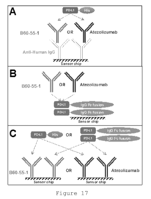
Figure 17: Alternative capturing approaches for SPR
measurements:
CA 03059447 2019-13-08
WO 2018/195226
PCT/US2018/028206
34
Panel A - Anti-human-IgG was immobilized on the chip as
capturing antibodies; B60-55-1 or atezolizumab were captured by
immobilized antibodies and various concentrations of PD-Li-His
ligand were applied.
Panel B - PD-Li-Fc fusion protein was directly immobilized
on the sensor chip and different concentrations of B60-55-1 or
atezolizumab were applied.
Panel C - to study interactions with both PD-Li-Fc fusion
protein and PD-Li-His, B60-55-1 or atezolizumab were directly
immobilized on the chip; a range of concentrations of PD-Li-His
tagged or PD-L1-Fc were applied.
Figure 18: Sensograms of binding of PD-Li-His tagged
ligand to immobilized comparator antibody atezolizumab or B60-
55-1; the approach is schematically shown in the left panel and
kinetic parameters are summarized in the table; anti-human
capturing antibodies were immobilized on a sensor chip and
atezolizumab or B60-55-1 were captured then followed by various
concentrations of PD-Li-His ligand:
Panel A - results for atezolizumab;
Panel B - results for B60-55-1.
Figure 19: Sensograms of binding of atezolizumab or B60-
55-1 to immobilized PD-Li-Fc fusion protein; the approach is
schematically shown on the left panel and kinetic parameters are
summarized in the table; various concentrations of B60-55-1 or
atezolizumab were applied to the chip:
Panel A - results for atezolizumab;
Panel B - results for B60-55-1.
Figure 20: Sensograms of binding of PD-Li-His or PD-Li-Fc
to immobilized B60-55-1; the approach is schematically shown in
the left panel and kinetic parameters are summarized in the
table.
CA 03059447 2019-13-08
WO 2018/195226
PCT/US2018/028206
Figure 21: Sensograms of binding of PD-Li-His or PD-L1-Fc
to immobilized atezolizumab; the approach is schematically shown
in left panel and kinetic parameters are summarized in the
table.
5 Figure 22: B60-55-1 and atezolizumab have no ADCC
activities compared to the control antibodies from the Promega
ADCC Reporter Bioassay Kit.
Figure 23: Evaluation of B60-55-1 and atezolizumab
binding to Clq.
10 Figure 24: Concentration dependent potencies of B60-55-1
and comparator antibodies on T cell activation in MLR assay.
Figure 25: Body weight change upon drug treatment; arrows
indicated the dosing time.
Figure 26: Tumor volume inhibition upon drug treatment;
15 arrows indicated the dosing time.
Figure 27: Individual tumor growth in three groups during
29 days' observation after grouping (n=8).
Figure 28: Tumor weight inhibition at day 29 posting
dosing.
20 Figure 29: Mean tumor volume in the three test groups from
experimental design shown in Table 7 below.
Figure 30: Mean tumor volume in the three test groups from
experimental design shown in Table 7 below at days 21 and 41;
three columns for each day correspond to group 1(1eft), two
25 (center) and 3 (right).
DETAILED DESCRIPTION
30 In the following section, the aspects of the present
invention are further illustrated via the following examples;
CA 03059447 2019-13-08
WO 2018/195226
PCT/US2018/028206
36
however, as should be understood by any person skilled in the
art, the following examples are only used to illustrate the
present invention and should not be construed as limiting the
scope of the present invention. Any conditions which are not
specified in the following examples should be set to be those
used conventionally or those recommended by the manufacturer.
Any reagents or instruments used for which a manufacturer is not
specified all correspond to standard products which can be
purchased commercially.
Example 1: Recombinant human PD-Li and PD-1 expression and
preparation of related EGFP cells.
The amino acid sequence of the extracellular domain of
human PD-Li was obtained based on an amino acid sequence of PD-
Li (Q9NZQ7) contained in the protein database Uniprot (i.e., the
sequence from Residue 1 to Residue 238 contained in Q9NZQ7); the
amino acid sequence of the structural domain of IgGl-Fc was
obtained based on an amino acid sequence of the constant region
of human immunoglobulin gammal (IgG1) (P01857) contained in the
protein database Uniprot (i.e., the sequence from Residue 104 to
Residue 330 contained in P01857); and the amino acid sequence of
the structural domain of IgGl-Fc was obtained based on an amino
acid sequence of the constant region of human immunoglobulin
gammal (IgG1) (P01868) contained in the protein database Uniprot
(i.e., the sequence from Residue 98 to Residue 324 contained in
P01868). The online tool DNAworks
(http://helixweb.nih.gov/dnaworks/) was used to design
corresponding encoding DNA sequences to obtain hPD-L1-Fc and
hPD-L1-muFc fusion protein genes, and the same method was used
to obtain a hPD-1-Fc gene. An amino acid sequence for enhanced
CA 03059447 2019-13-08
WO 2018/195226
PCT/US2018/028206
37
green fluorescent protein (EGFP) (C5MKY7) as well as an amino
acid sequence for human PD-Li (Q9NZQ7), an amino acid sequence
for murine PD-L1 (Q9EP73) and an amino acid sequence for human
PD-1 (Q15116) were obtained based on information contained in
the protein database Uniprot. The online tool DNAworks
(http://helixweb.nih.gov/dnaworks/) was used to design
corresponding encoding DNA sequences to obtain a PD-L1-EGFP
fusion protein gene, and the same method was used to obtain hPD-
1-EGFP and mPD-L1-EGFP genes. Corresponding DNA fragments were
obtained via artificial synthesis. Synthesized gene sequences
were double digested with Fermentas-made HindIII and EcoRI and
cloned into the commercial vector pcDNA4/myc-HisA (Invitrogen,
V863-20), after which sequencing was performed to verify that
the plasmid had been constructed accurately to obtain
recombinant plasmid DNA; i.e.: pcDNA4-hPD-Li-Fc, pcDNA4-hPD-Ll-
muFc, pcDNA4-hPD1-Fc, pcDNA4-hPD-L1-EGFP, pcDNA4-hPD1-EGFP and
pcDNA4-mPD-L1-EGFP.
Reverse transcription-polymerase chain reaction (RT-PCR)
was used to amplify human PD-L2 and B7H3 genes from lab-cultured
dendritic cells (DC cells) (wherein said DC cells were obtained
via TNF-a maturation of mononuclear cells isolated from PBMC)
and the gene amplification primers used were as follows:
PDL2-F HindIII: GCGCAAGCTTGCCACCATGATCTTCCTCCTGCTAATG (SEQ
ID NO: 74),
PDL2-R EcoI:
GCCGAATTCGATAGCACTGTTCACTTCCCTC (SEQ ID NO: 75);
hB7H3-F HindIII: GCGCAAGCTTGCCACCATGCTGCGTCGGCGGGGCAGC (SEQ
ID NO: 76);
CA 03059447 2019-13-08
WO 2018/195226
PCT/US2018/028206
38
hB7H3-R BamHT :
GCGCGAATTCGGCTATTICTTGTCCATCATCTTC (SEQ ID NO: 77).
The PCR product obtained was then double digested using
Fermentas HindITI and EcoRI and cloned into a pre-constructed
pcDNA4-hPD-L1-EGFP, after which sequencing was performed to
verify that the plasmid had been constructed accurately to
obtain recombinant plasmid DNA; i.e.: pcDNA4-hPD-L2-EGFP and
pcDNA4-hB7H3-EGFP.
A corresponding EGFP recombinant plasmid was transfected
into HEK293 (ATCC, CRL-1573TM) cells, and fluorescence-activated
cell sorting (FACS) was performed 48 hours after transfection to
verify the expression of hPD-L1, mPD-L1, hPD-L2 and hB7H3.
pcDNA4-hPD-L1-Fc, pcDNA4-hPD-L1-muFc and pcDNA4-hPD1-Fc
were transiently transfected into HEK293 cells for protein
production. The recombinant expression plasmid was diluted with
a Freestyle293 culture medium and added to a PEI
(polyethylenimine) solution required for transformation, after
which each plasmid/PET mixture was each separately added to a
cell suspension and left to culture at 37 C 10% CO2 and 90 rpm;
at the same time, a supplementary addition of 50 pg/L insulin-
like growth factor (IGF-1) was performed. Four hours thereafter,
a supplementary addition of EX293 culture medium, 2 mM glutamine
and 50 pg/L IGF-1 was performed and the culture was continued at
135 rpm. After a further 24 hours, 3.8 mM sodium valproate (VPA)
was added. After 5-6 days culturing, the supernatant of the
transient expression culture was collected and Protein A
affinity chromatography was used to initially purify and obtain
hPD-L1-Fc, hPD-L1-muFc and hPD-1-Fc protein samples for use in
the following examples. Protein samples thus obtained were
CA 03059447 21319-18
WO 2018/195226
PCT/US2018/028206
39
subject to preliminary testing using SDS-PAGE, and target bands
were clearly visible.
Example 2: Screening for anti-hPD-L1 antibodies in yeast
display library, cloning expression and identification.
Yeast display technology was used to screen for fully human
antibodies for PD-Li. Cloning of VH and VL genes contained in
spleen and lymph node IgM and IgG cDNA obtained from 21 healthy
human subjects was performed to construct an scFV yeast display
library (the connecting sequence between VH and VL was the
connecting peptide
GGGGSGGGGSGGGGS (SEQ ID NO: 67)
and the connecting peptide) storage capacity of the library
was 5 x 10⁸. A 10x capacity yeast library was revived and yeasts
were induced to express antibodies on their surface; 100 nM of
biotinylated hPD-L1 antigen magnetic beads were used to perform
two rounds of enrichment, after which a further two rounds of
enrichment were performed using an anti-myc antibody and
biotinylated hPD-L1 flow sorting. Yeasts thus obtained were
plated and single clones were picked. Monoclonal yeasts which
were subject to amplification and induction of expression were
further subjected to a staining analysis using an anti-myc
antibody as well as biotinylated hPD-L1 or the control antigen
hPD-1 and yeasts which were antigen-positive or control-negative
were assessed as being positive yeast.
FACS confirmed yeast clones were subject to yeast colony
PCR and sequencing using the following PCR primers:
CA 03059447 2019-13-08
WO 2018/195226
PCT/US2018/028206
pNL6-F:
GTACGAGCTAAAAGTACAGTG (SEQ ID NO: 78) ;
pNL6-R:
5 TAGATACCCATACGACGTTC (SEQ ID NO: 79) ;
wherein the sequencing primer used was pNL6-R. The sequence
results obtained after sequencing were subject to an alignment
analysis using the BioEdit software package.
10 The single-chain antibody scFv gene obtained as described
above and a previously obtained IgGl-Fc gene were fused and
cloned into the commercial vector pEE6.4 (Lonza) using a double
digest of Fermentas HindIII and EcoRI enzymes, after which
cloning and plasmid miniprep were performed in accordance with
15 standard molecular cloning procedures. Extracted plasmids were
transiently expressed in HEK293 cells and purified using a
protein A column.
hPD-L1-EGFP cells were resuspended in 0.5% PBS-BSA Buffer,
after which the aforementioned purified anti-hPD-L1 scFv
20 antibodies were added while at the same time, corresponding
controls were established with 2 ug of a hIgG1 protein used as a
negative control and hPD-1-Fc being added to the positive
control. The secondary antibody used was anti-hIg-PE from
eBioscience. Detection was performed via flow cytometry after
25 staining was completed. The above method was used to identify
antibodies capable of binding cell surface PD-Li antigens.
hPD-L1-EGFP cells were resuspended in 0.5% PBS-BSA Buffer,
after which the aforementioned purified anti-hPD-L1 scFv
antibodies were added while at the same time, a negative control
30 was established with 2 pg of a hIgG1 protein used as a negative
control; 0.3 pg of hPD-1-Fc-biotin was added to all samples and
CA 03059447 2019-13-08
WO 2018/195226
PCT/US2018/028206
41
SA-PE from eBioscience was used as a secondary antibody;
detection was performed via flow cytometry after staining was
completed, and the results are shown in Figure 1. The above
method was used to identify antibodies capable of blocking cell
surface PD-Li antigens and PD-1 binding.
After screening and identification three antibody strains
which showed favorable characteristics were obtained: B50, B60
and BII61. As can be seen in the results, all three anti-hPD-L1
antibody strains were able to block binding with the hPD-1
receptor.
The connecting peptide sequence
GGGGSGOGGSGGGGS (SEQ ID NO: 67)
was contained between the heavy chain and light chain
variable regions of the aforementioned antibody.
The amino acid sequence of the B50 heavy chain variable
region was:
QVQLQQSGPGLVKPSQTLSLICAISGDSVSSTKAAWYWIRQSPSRGLEWLGRTYFRSKW
YNDYADSVKSRLTINPDTSKNQFSLQLKSVSPEDTAVYYCARGQYTAFDIWGQGTMVTVSS
(SEQ ID NO: 51);
wherein the underlined sections constitute CDR1, 2 and 3
and correspond to SEQ ID NO: 13-15 respectively and the non-
underlined sections constitute FR1, 2, 3 and 4 and correspond to
SEQ ID NO: 38-41 respectively;
the corresponding DNA sequence was:
CAGGTACAGCTGCAGCAGTCAGGTCCAGGACTGGTGAAGCCCTCGCAGACCCTCTCACTCACCT
GTGCCATCTCCGGGGACAGTGTCTCTAGCACCAAGGCTGCTTGGTACTGGATCAGGCAGTCCCT
CA 03059447 2019-13-08
WO 2018/195226
PCT/US2018/028206
42
TCGAGAGGCCTTGAGTGGCTGGGAAGGACATACTTCCGGTCCAAGTGGTATAATGACTATGCCG
ACTCTGTGAAAAGICGATTAACCATCAACCCAGACACATCCAAGAACCAGTTCTCCCTGCAATT
AAGICIGTGAGICCCGAGGACACGGCTGTGTATTACTGTGCAAGAGGGCAATACACTGCTTTTG
ATATCTGGGGCCAAGGGACAATGGTCACCGTCTCTTCA (SEQ ID NO: 59);
the amino acid sequence of the light chain variable region
was:
QSALIQPASVSGSPGQSITISCTGTSSDVGGYDLVSWYQQYPGQAPRLIIYEVIKRPSGISDRF
SGSKSGNTASLTISGLQAEDEADYYCSSYAGRRLHGVFGGGTQLTVL (SEQ ID NO: 56);
wherein the underlined sections constitute CDR1, 2 and 3
and correspond to SEQ ID NO: 21, 17 and 18 respectively and the
non-underlined sections constitute FR1, 2, 3 and 4 and
correspond to SEQ ID NO: 42-45 respectively;
and the corresponding DNA sequence was:
CAGTCTGCTCTGATTCAGCCTGCCTCCGTGTCTGGGTCCCCTGGACAGTCGATCACTAT
CTCCTGTACTGGCACCAGTAGTGATGTTGGAGGTTATGACCTTGTCTCCTGGTACCAACAGTAC
CCGGCCAAGCCCCCAGACTCATCATTTATGAGGTCATTAAGCGGCCCTCAGGGATTTCTGATCG
CTTCTCTGGTTCCAAGTCTGGCAACACGGCCTCCCTGACAATCTCTGGGCTCCAGGCTGAGGAC
GAGCTGATTATTATTGCAGCTCATATGCAGGTAGACGTCTTCATGGTGTGTTCGGAGGAGGCAC
CCAGCTGACCGTCCTC (SEQ ID NO: 66).
The amino acid sequence of the B60 heavy chain variable
region was:
QVQLVQSGAEVKKPASSVKVSCTASGGSFSTYAISWVRQAPGQGLEWMGGIIPIFGTTKYAQRF
QGRVTITADESITTAYMELSSLISDDTALYYCTISRGFSYGWFDYWGQGTLVTVSS (SEQ ID
NO: 53);
CA 03059447 2019-13-08
WO 2018/195226
PCT/US2018/028206
43
wherein the underlined sections constitute CDR1, 2 and 3
and correspond to SEQ ID NO: 1, 2 and 19 respectively and the
non-underlined sections constitute FR1, 2, 3 and 4 and
correspond to SEQ ID NO: 22-25 respectively;
the corresponding DNA sequence was:
CAGGTCCAGCTIGTGCAGTCTGGAGCTGAGGIGAAGAAGCCTGCGTCCTCGGTCAAAGTCTCCT
GCACGGCTTCTGGCGGCTCCTTCAGCACCTATGCTATCAGTTGGGTGCGACAGGCTCCTGGACA
GGGCTTGAATGGATGGGCGGGATCATCCCCATCTTTGGTACAACTAAGTACGCACAGAGGTTCC
AGGGCAGGGTCACGATTACCGCGGACGAATCGACGACCACAGCCTACATGGAGCTGAGCAGCTG
ATATCTGACGACACGGCCCTGTATTATTGTACGACGTCTCGTGGATTCAGCTATGGCTGGTTTG
ACTACTGGGGCCAGGGTACCCTGGTCACCGTCTCCTCA (SEQ ID NO: 60);
the amino acid sequence of the light chain variable region
was:
EIVMTQSPATLSLSPGERATLSCRASQSVGIHLAWYQQKLGQAPRLLIYGASSRATGIPDRFSG
SGSGTDFTLTISRLEPEDFAVYYCQQYGSLPRTFGQGTKVEIK (SEQ ID NO: 48);
wherein the underlined sections constitute CDR1, 2 and 3
and correspond to SEQ ID NO: 4-6 respectively and the non-
underlined sections constitute FR1, 2, 3 and 4 and correspond to
SEQ ID NO: 26-29 respectively;
and the corresponding DNA sequence was:
GAAATTGTAATGACACAGICTCCAGCCACCCTGTCTTTGTCTCCAGGGGAAAGAGCCAC
CCTCTCCTGTAGGGCCAGTCAGAGTGTTGGCATACACTTAGCCTGGTACCAACAGAAACTTGGC
CAGGCTCCCAGGCTCCTCATCTATGGTGCATCCAGTAGGGCCACTGGCATCCCAGACAGGTTCA
GTGGCAGTGGGTCTGGGACAGATTCACTCTCACCATCAGCAGACTGGAGCCTGAAGATTTTGCA
GTGTATTACTGTCAGCAGTATGGTTCTTTACCTCGGACGTTCGGCCAAGGGACCAAGGTGGAAA
TCAAA (SEQ ID NO:62).
CA 03059447 2019-13-08
WO 2018/195226
PCT/US2018/028206
44
The amino acid sequence of the BII61 heavy chain variable
region was:
QVQLQQSGPGLVKPSQTLSLTCAISGDSVSSNSASWNWIRQSPSRGLEWLGRTYYRSKWYDDYA
VSVKSRISINPDTSKNQFSLQLNSVTPEDTAVYYCARSQGRYFVNYGMDVWGQGTTVTVSS
(SEQ ID NO: 54);
wherein the underlined sections constitute CDR1, 20 and 3
and correspond to SEQ ID NO: 7, 2 and 9 respectively and the
non-underlined sections constitute FR1, 2, 3 and 4 and
correspond to SEQ ID NO: 30-33 respectively;
the corresponding DNA sequence was:
CAGGTACAGCTOCAGCAGTCAGGICCAGGACTGOTGAAGCCCTCGCAGACCCTCTCACTCACCT
GTGCCATCTCCGGGGACAGTGTCTCTAGCAACAGTGCTTCTTGGAACTGGATCAGGCAGTCCCA
TCGAGAGGCCTIGAGIGGCTGGGAAGGACATATTACAGGICCAAATGGTATGATGATTATGCAG
TATCTGTGAAAAGICGAATCAGCATCAACCCAGACACATCCAAGAACCAGTICTCCCIGCAGTG
AACTCTGTGACTCCCGAGGACACGGCTGTGTATTACTGTGCAAGAAGCCAGGGACGATATTTTG
TCAACTACGGTATGGACGTCTGGGGCCAAGGGACCACGGTCACCGTCTCCTCA (SEQ ID
NO 61)
the amino acid sequence of the light chain variable region
was:
DIRLTQSPSSLSASVGDRITITCRASQSISSYLNWYQQKPGKAPKLLIYGASSLQSGVPSRFSG
SGSGTDFTLTISSLQPEDVATYYCQQSYFTPRGITFGPGTKVDIK (SEQ ID NO: 55);
wherein the underlined sections constitute CDR1, 2 and 3
and correspond to SEQ ID NO: 10-12 respectively and the non-
underlined sections constitute FR1, 2, 3 and 4 and correspond to
SEQ ID NO: 34, 46, 36 and 37 respectively;
and the corresponding DNA sequence was:
CA 03059447 2019-13-08
WO 2018/195226
PCT/US2018/028206
GACATCCGGTTGACCCAGTCTCCATCTTCCCTGTCTGCATCTGTAGGAGACAGAATCACCATCA
CT TGCCGGGCAAGTCAGAGCAT TAGCAGT TAT T TAAAT TGGTATCAACAGAAACCAGGGAAAGC
CCTAAGCTCCTGATCTATGGTGCATCCAGTTTGCAAAGTGGGGTCCCATCAAGGTTCAGTGGCA
5 GTGGATCTGGGACAGATTTCACTCTCACCATCAGCAGTCTGCAACCTGAAGATGTTGCAACTAC
TACTGTCAACAGAGTTACTTTACCCCCCGCGGGATCACTTTCGGCCCTGGGACCAAAGTGGATA
TCAAA (SEQ ID NO: 65).
Example 3: Construction of anti-hPD-L1 scFv- improved
10 affinity yeast library.
A standard PCR reaction was respectively performed using
pEE6.4-B50-Fc, pEE6.4-B60-Fc and pEE6.4-B1161-Fc plasmids as
templates, and
pEE6.4-F:
TCTGGTGGTGGTGGTTCTGCTAGC (SEQ ID NO: 80) and
cMyc-BBXhoI:
GCCAGATCTCGAGCTATTACAAGTCTTCTTCAGAAATAAGCTTTTGTTCTAGAATTCCG (SEQ
ID NO: 81)
as primers. PCR products were purified and cloned into the
commercial pC1302 vector commercial (addgene: #41845) using
Fermentas NheI and BglII, to obtain the recombinant plasmids
pCT302-B50, pCT302-B60 and pCT3C2-B1161. Next, error prone PCR
was used based on the method detailed in Ginger et al. (2006)
Nat Protoc 1(2):755-68 to obtain scEv randomly mutated PCR
products. The primers used were
17 proshort:
CA 03059447 2019-13-08
WO 2018/195226 PCT/US2018/028206
46
TAATACGACTCACTATAGGG (SEQ ID NO: 82) and
Splice 4 / L:
GGCAGCCCCATAAACACACAGTAT (SEQ ID NO: 83) .
The PCR products thus obtained were purified using a
Fermentas GeneJET DNA Purification Kit and then concentrated via
ethanol precipitation to a concentration greater than 1 pg/pl.
Fermentas NheI and BamHI were used to perform a double digestion
of the commercial vector pCT302 and at the same time, the
Fermentas FastAP dephosphorylation enzyme was used to perform
dephosphorylation of the vector, after which a Fermentas GeneJET
DNA Purification Kit was again used to perform purification and
ethanol precipitation was performed to concentrate the product
to a concentration greater than 1 ig/pl. Yeast electro-
transformation and in vivo recombination were performed in
accordance with the method described in Ginger et al. (2006)
Nat. Protoc. 1(2): 755-68 to obtain an affinity matured yeast
library.
Example 4: Screening for yeast expressing anti-hPD-L1 scFv
with improved affinity.
The affinity matured yeast library obtained as described
above was subjected to two rounds of flow sorting using 10 nM
and 1 nM of a hPD-L1-Fc protein, and yeast products thus
obtained were plated and monoclones were picked for
identification. Low concentration antigen staining was used to
perform flow staining to identify yeast monoclones which showed
increased affinity, by using previously obtained wildtype yeast
as a control.
CA 03059447 2019-13-08
WO 2018/195226
PCT/US2018/028206
47
Yeast clones which had passed FACS verification were
subject to yeast colony PCR and sequencing using the methodology
described above. The scFv gene obtained following affinity
maturation and a previously obtained IgGl-Fc gene were fused and
cloned into the commercial vector pEE6.4 using a double digest
of Fermentas HindIII and EcoRI enzymes, after which cloning and
plasmid miniprep were performed in accordance with standard
molecular cloning procedures. Extracted plasmids were
transiently expressed in HEK293 cells and purified using a
protein A column.
The antibody binding capacity and blocking capacity were
measured using the method described in Example 2.
See Figure 2 for the results of the binding capacity
testing; the results show that the three antibody strains
obtained following affinity maturation had significantly
increased affinity.
See Figure 3 for the results of the blocking capacity
testing; the results show that the IC50 values for competitive
binding to PD-Li in competition with PD-1 obtained for the three
antibody strains obtained following affinity maturation were
0.837 ig/m1 for BII61-62 (0.884 ig/m1 for BII61), 4.56 pg/m1 for
B50-6 (5.63 ig/m1 for B50) and 1.14 ig/m1 for B60-55 (16.8 pg/m1
for B60), respectively.
Following affinity maturation, three anti-hPD-L1 scFv
antibody sequences showing increased affinity, i.e. B50-6, B60-
55 and BI161-62, were obtained. Compared with B50, B50-6 showed
an amino acid mutation from D to N in its VL CDR1; compared with
B60, B60-55 showed an amino acid mutation from S to N in its VH
CDR3; and compared with BII61, BII61-62 showed an amino acid
mutation from S to G in its VH CDR2 as well as an amino acid
CA 03059447 21319-18
WO 2018/195226
PCT/US2018/028206
48
mutation from I to V in its VL FR2. The connecting peptide
sequence
GGGGSGGGGSGGGGS (SEQ ID NO: 67)
was contained between the heavy chain and light chain
variable regions of the aforementioned antibody.
The amino acid sequence of the B50-6 heavy chain variable
region was:
QVQLQQSGPGLVKPSQTLSLTCAISGDSVSSTKAAWYWIRQSPSRGLEWLGRTYFRSKWYNDYA
DSVKSRLTINPDTSKNQFSLQLKSVSPEDTAVYYCARGQYTAFDIWGQGTMVTVSS (SEQ ID
NO: 51);
wherein the underlined sections constitute CDR1, 2 and 3
and correspond to SEQ ID NO: 13-15 respectively and the non-
underlined sections constitute FR1, 2, 3 and 4 and correspond to
SEQ ID NO: 38-41 respectively;
the corresponding DNA sequence was:
CAGGTACAGCTGCAGCAGTCAGGTCCAGGACTGGTGAAGCCCTCGCAGACCCTCTCACTCACCT
GTGCCATCTCCGGGGACAGTGTCTCTAGCACCAAGGCTGCTTGGTACTGGATCAGGCAGTCCCT
TCGAGAGGCCTTGAGTGGCTGGGAAGGACATACTTCCGGTCCAAGTGGTATAATGACTATGCCG
ACTCTGTGAAAAGICGATTAACCATCAACCCAGACACATCCAAGAACCAGTTCTCCCTGCAATT
AAGTCTGTGAGTCCCGAGGACACGGCTGTGTATTACTGTGCAAGAGGGCAATACACTGCTTTTG
ATATCTGGGGCCAAGGGACAATGGTCACCGTCTCTTCA (SEQ ID NO: 59);
the amino acid sequence of the light chain variable region
was:
CA 03059447 2019-13-08
WO 2018/195226
PCT/US2018/028206
49
QSALIQPASVSGSPGQSITISCTGTSSNVGGYDLVSWYQQYPGQAPRLIIYEVIKRPSG
ISDRFSGSKSGNTASLTISGLQAEDEADYYCSSYAGRRLHGVFGGGTQLTVL (SEQ ID NO:
52);
wherein the underlined sections constitute CDR1, 2 and 3
and correspond to SEQ ID NO: 16-18 respectively and the non-
underlined sections constitute FR1, 2, 3 and 4 and correspond to
SEQ ID NO: 42-45 respectively;
the corresponding DNA sequence was:
CAGTCTGCTCTGATTCAGCCTGCCTCCGTGTCTGGGTCCCCTGGACAGTCGATCACTATCTCCT
GTACTGGCACCAGTAGTAATGTTGGAGGTTATGACCTTGTCTCCTGGTACCAACAGTACCCGGG
CCAAGCCCCCAGACTCATCATITATGAGGICATTAAGCGOCCCTCAGGGATTTCTGATCGCTTC
TCTGGTTCCAAGTCTGGCACACGGCCTCCCTGACAATCTCTGGGCTCCAGGCTGAGGACGAGGC
TGATTATTATTGCAGCTCATATGCAGGTAGACGTCTTCATGGTGTGTTCGGAGGAGGCACCCAG
CTGACCGTCCTC(SEQ ID NO: 64);
the amino acid sequence of the B60-55 heavy chain variable
region was:
QVQLVQSGAEVKKPASSVKVSCIASGGSFSTYAISWVRQAPGQGLEWMGGIIPIEGTTK
YAQRFQGRVTITADESTTTAYMELSSLISDDTALYYCTTSRGFNYGWFDYWGQGTLVTVSS
(SEQ ID NO: 47);
wherein the underlined sections constitute CDR1, 2 and 3
and correspond to SEQ ID NO: 1-3 respectively and the non-
underlined sections constitute FR1, 2, 3 and 4 and correspond to
SEQ ID NO: 22-25 respectively;
the corresponding DNA sequence was:
CAGGTCCAGCTIGTGCAGTCTGGAGCTGAGGIGAAGAAGCCTGCGTCCTCGGTCAAAGTCTCCT
CA 03059447 2019-13-08
WO 2018/195226
PCT/US2018/028206
GCACGGCTTCTGGCGGCTCCTTCAGCACCTATGCTATCAGTTGGGTGCGACAGGCTCCTGGACA
GGGCTTGAATGGATGGGCGGGATCATCCCCATCTTTGGTACAACTAAGTACGCACAGAGGTTCC
AGGGCAGGGICACGAT TACCGCGGACGAATCGACGACCACAGCCTACATGGAGCTGAGCAGCTG
ATATCTGACGACACGGCCCTGTATTATTGTACGACGTCTCGTGGATTCAACTATGGCTGGTTTG
5 ACTACTGGGGCCAGGGTACCCTGGTCACCGTCTCCTCA (SEQ ID NO: 57);
the amino acid sequence of the light chain variable region
was:
10
EIVMTQSPATLSLSPGERATLSCRASQSVGIHLAWYQQKLGQAPRLLIYGASSRATGIP
DRFSGSGSGTDFTLTISRLEPEDFAVYYCQQYGSLPRTFGQGTKVEIK (SEQ ID NO:
48);
wherein the underlined sections constitute CDR1, 2 and 3
15 and correspond to SEQ ID NO: 4-6 respectively and the non-
underlined sections constitute FR1, 2, 3 and 4 and correspond to
SEQ ID NO: 26-29 respectively;
the corresponding DNA sequence was:
20
GAAATTGTAATGACACAGICTCCAGCCACCCTGTCTTTGTCTCCAGGGGAAAGAGCCAC
CCTCTCCTGTAGGGCCAGTCAGAGTGTTGGCATACACTTAGCCTGGTACCAACAGAAACTTGGC
CAGGTCCCAGGCTCCTCATCTATGGTGCATCCAGTAGGGCCACTGGCATCCCAGACAGGTTCAG
TGGCAGTGGGTCTGGGACAGACTTCACTCTCACCATCAGCAGACTGGAGCCTGAAGATTTTGCA
GTGATTACTGTCAGCAGTATGGTTCTTTACCTCGGACGTTCGGCCAAGGGACCAAGGTGGAAAT
25 CAAA (SEQ ID NO: 62);
the amino acid sequence of the B1161-62 heavy chain
variable region was:
CA 03059447 2019-13-08
WO 2018/195226
PCT/US2018/028206
51
QVQLQQSGPGLVKPSQTLSLTCAISGDSVSSNSASWNWIRQSPSRGLEWLGRTYYRSKW
YDDYAVSVKGRISINPDTSKNQFSLQLNSVTPEDTAVYYCARSQGRYFVNYGMDVWGQGTTVTV
SS (SEQ ID NO: 49);
wherein the underlined sections constitute CDR1, 2 and 3
and correspond to SEQ ID NO: 7-9 respectively and the non-
underlined sections constitute FR1, 2, 3 and 4 and correspond to
SEQ ID NO: 30-33 respectively;
the corresponding DNA sequence was:
CAGGTACAGCTGCAGCAGTCAGGTCCAGGACTGGTGAAGCCCTCGCAGACCCTCTCACT
CACCTGTGCCATCTCCGGGGACAGTGTCTCTAGCAACAGTGCTTCTTGGAACTGGATCAGGCAG
TCCCATCGAGAGGCCT T GAGT GGCT GGGAAGGACATAT TACAGGTCCAAAT GGTAT GAT GAT TA
TGCAGTATCTGTGAAAGGTCGAATCAGCATCAACCCAGACACATCCAAGAACCAGTTCTCCCTG
CAGTGAACTCTGTGACTCCCGAGGACACGGCTGTGTATTACTGTGCAAGAAGCCAGGGACGATA
TTTTGTCAACTACGGTATGGACGTCTGGGGCCAAGGGACCACGGTCACCGTCTCCTCA
(SEQID NO: 58);
the amino acid sequence of the light chain variable region
was:
DIRLTQSPSSLSASVGDRITITCRASQSISSYLNWYQQKPGKAPKLLVYGASSLQSGVPSRFSG
SGSGTDFTLTISSLQPEDVATYYCQQSYFTPRGITFGPGTKVDIK (SEQ ID NO: 50);
wherein the underlined sections constitute CDR1, 2 and 3
and correspond to SEQ ID NO: 10-12 respectively and the non-
underlined sections constitute FR1, 2, 3 and 4 and correspond to
SEQ ID NO: 34-37 respectively;
and the corresponding DNA sequence was:
CA 03059447 2019-13-08
WO 2018/195226
PCT/US2018/028206
52
GACATCCGGTTGACCCAGTCTCCATCTTCCCTGTCTGCATCTGTAGGAGACAGAATCAC
CATCACT TGCCGGGCAAGTCAGAGCAT TAGCAGT TAT T TAAATTGGTATCAACAGAAACCAGGG
AAAGCCCTAAGCTOCTGGTOTATGGIGCATCCAGTITGCAAAGTGGGGTCCCATCAAGGTTCAG
TGGCAGTGGATCTGGGACAGATTTCACTCTCACCATCAGCAGTCTGCAACCTGAAGATGTTGCA
ACTACTACTGTCAACAGAGTTACTTTACCCCCCGCGGGATCACTTTCGGCCCTGGGACCAAAGT
GGATATCAAA (SEQ ID NO: 63).
Example 5: Formatting of scFv antibody to IgG antibody.
A human IgG1 constant region amino acid sequence was
obtained based on the amino acid sequence of the constant region
of human immunoglobulin gammal (IgG1) contained in the Uniprot
protein database (P01857). The online tool DNAworks
(http://helixweb.nih.gov/dnaworks/) was used to design
corresponding encoding DNA sequences to obtain a human IgG1
constant region gene and the VH sequences of the heavy chain
variable regions of B50-6, B60-55 and BII61-61 obtained via
screening were spliced together with the human IgG1 constant
region gene while at the same time, the following signal peptide
sequence was added to the 5' end of the VH:
ATGGAGACAGACACACTCCTGCTATGGGTACTGCTGCTCTGGGTTCCAGGTTCCACCGGT
(SEQ ID NO: 84);
the spliced gene was synthesized and double digestion was
performed using Fermentas HindITI and EcoRI enzymes to clone the
gene into the vector pEE6.4 to obtain pEE6.4-B50-6HC; pEE6.4-
B60-55HC; and pEE6.4-B1161-62HC. A human Kappa light chain
constant region amino acid sequence was obtained based on the
amino acid sequence of the constant region of human
immunoglobulin Kappa contained in the Uniprot protein database
CA 03059447 2019-13-08
WO 2018/195226
PCT/US2018/028206
53
(P01834). The online tool DNAworks
(http://helixweb.nih.gov/dnaworks/) was used to design
corresponding encoding DNA sequences to obtain a human Kappa
light chain constant region gene and the VL sequences of the
heavy chain variable regions of B60-55 and B1161-61 obtained via
screening were spliced together with the human Kappa light chain
constant region gene while at the same time, the following
signal peptide sequence was added to the 5' end of the VL:
ATGGAGACAGACACACTCCTGCTATGGGTACTGCTGCTCTGGGTTCCAGGTTCCACCGGT
(SEQ ID NO: 84);
the online tool DNAworks
(http://helixweb.nih.gov/dnaworks/) was used to design
corresponding encoding DNA sequences to obtain a human lambda
(A) light chain constant region gene and the VL sequence of the
light chain variable region of B50-6 obtained via screening were
spliced together with the human lambda light chain constant
region gene while at the same time the following signal peptide
sequence was added to the 5' end of the VL:
ATGGAGACAGACACACTCCTGCTATGGGTACTGCTGCTCTGGGTTCCAGGTTCCACCGGT
(SEQ ID NO: 84);
and the spliced gene was synthesized and double digestion
was performed using Fermentas HindIII and EcoRI enzymes to clone
the gene into the vector pEE12.4 (Lonza) to obtain pEE12.4-B50-
6LC; pEE12.4-B60-55LC; and pEE12.4-B1161-62LC.
Heavy chain and light chain plasmids obtained as described
above were prepared using an AidLab Maxiprep Kit (PL14).
Recombinantly constructed light and heavy chain plasmids were
CA 03059447 2019-10-08
WO 2018/195226
PCT/US2018/028206
54
co-transfected into HEK293 cells to express the antibody. The
recombinant expression plasmid was diluted with a Freestyle293
culture medium and added to a PEI (polyethylenimine) solution
required for transformation, after which each plasmid/PEI
mixture was each separately added to a cell suspension and left
to culture at 37 C, 10% 002 and 90 rpm; at the same time, a
supplementary addition of 50 pg/IGF-1 was performed. Four hours
thereafter, a supplementary addition of EX293 culture medium,
2 mM glutamine and 50 pg/L IGF-1 was performed and the culture
was continued at 135 rpm. After a further 24 hours, 3.8 mM VPA
was added. After 5 - 6 days culturing, the supernatant of the
transient expression culture was collected and Protein A
affinity chromatography was used to purify and obtain anti-hPD-
Ll B50-6, B60-55 and BII61-62 mAb antibodies.
the IgG1 chain constant region amino acid sequence was:
ASTKGPSVFPLAPSSKSTSGGTAALGCLVKDYFPEPVTVSWNSGALTSGVHTFPAVLQS
SGLYSLSSVVTVPSSSLGTQTYICNVNHKPSNTKVDKKVEPKSCDKTHTCPPCPAPELLGGPSV
FLFPPKPKDTLMISRTPEVTCVVVDVSHEDPEVKFNWYVDGVEVHNAKTKPREEQYNSTYRVVS
VLTVLHQDWLNGKEYKCKVSNKALPAPIKTISKAKGQPREPQVYTLPPSRDELTKNQVSLTCLV
KGFYPSDIAVEWESNGQPENNYKTTPPVLDSDGSFFLYSKLTVDKSRWQQGNVFSCSVMHEALH
NHYTQKSLSLSPGK (SEQ ID NO: 68);
the IgG1 chain constant region nucleic acid sequence was:
GCCAGCACTAAGGGGCCCTCTGTGTTTCCACTCGCCCCTTCTAGCAAAAGCACTTCCGG
AGGCACTGCAGCACTCGGGTGTCTGGTCAAAGATTATTTCCCTGAGCCAGTCACCGTGAGCTGG
AACTTGGCGCCCTCACCTCCGGGGTTCACACCTTTCCAGCCGTCCTGCAGTCCTCCGGCCTGTA
CTCCCTGAGCAGCGTCGTTACCGTGCCATCCTCTTCTCTGGGGACCCAGACATACATCTGCAAT
GTCACCATAAGCCTAGCAACACCAAGGTGGACAAAAAGGTCGAGCCAAAGAGCTGCGATAAGAC
ACACACCTGCCCTCCATGCCCCGCACCTGAACTCCTGGGCGGGCCTTCCGTTTTCCTGTTTCCT
CCAAGCCCAAGGATACACTGATGATTAGCCGCACCCCCGAAGTCACTTGCGTGGTGGTGGATGT
CA 03059447 2019-13-08
WO 2018/195226
PCT/US2018/028206
GAGCCATGAAGATCCAGAAGTTAAGTTTAACTGGTATGTGGACGGGGTCGAGGTGCACAATGCT
AAACAAAGCCCAGGGAGGAGCAATATAAC T CCACATACAGAGT GGT GT CCGT T C T GACAGT CCT
GCACCAGGAC T GGC T GAACGGGAAGGAATACAAGT GCAAGGT GT CTAATAAGGCAC T GC CAGC C
CCATAGAGAAGACAATCTCTAAAGCTAAAGGCCAACCACGCGAGCCTCAGGTCTACACACTGCC
5 AC
CAT CCAGGGAC GAAC T GAC CAAGAAT CAGGT GAGCC T GAC T T GT CT CGT CAAAGGAT T
CTAC
CAAGCGACATCGCCGTGGAGTGGGAATCCAACGGCCAACCAGAGAACAACTACAAGACCACCCC
ACCAGTCCTGGACTCTGATGGGAGCTTTTTCCTGTATTCCAAGCTGACAGTGGACAAGTCTCGT
GGCAACAGGGCAACGTGTTCAGCTGCTCCGTGATGCATGAAGCCCTGCATAACCACTATACCCA
GAAAAGCCTCAGCCTGTCCCCCGGGAAATAATGA (SEQ ID NO: 69);
the Kappa chain constant region amino acid sequence was:
RTVAAPSVFIFPPSDEQLKSGTASVVCLLNNFYPREAKVQWKVDNALQSGNSQESVTEQ
DSKDSTYSLSSILTLSKADYEKHKVYACEVTHQCLSSPVIKSFNRGEC (SEQ ID NO:
70);
the Kappa chain constant region nucleic acid sequence was:
CGTACGGTGGCTGCACCATCTGTCTTCATCTTCCCGCCATCTGATGAGCAGTTGAAATC
TGGTACCGCTAGCGTTGTGTGCCTGCTGAATAACTTTTATCCACGGGAGGCTAAGGTGCAGTGG
AAAGGGACAATGCCCTCCAGAGCGGAAATAGCCAAGAGTCCGTTACCGAACAGGACTCTAAAGA
CTCTACATACTCCCTGTCCTCCACACTGACCCTCTCCAAGGCCGACTATGAGAAACACAAGGTT
TACCATGCGAGGTCACACACCAGGGACTCTCCTCTCCCGTGACCAAGAGCTTCAACCGGGGAGA
ATGC (SEQ ID NO: 71);
the B50-6 light chain (lambda) constant region amino acid
sequence was:
GQPKAAPSVILFPPSSEELQANKATLVCLVSDFYPGAVTVAWKADGSPVKVGVETTKPSKQSNN
KYAASSYLSLTPEQWKSHRSYSCRVTHEGSTVEKTVAPAECS (SEQ ID NO: 72);
and the B50-6 light chain (lambda) constant region nucleic
acid sequence was:
CA 03059447 2019-10-08
WO 2018/195226
PCT/US2018/028206
56
GGTCAGCCCAAGGCTGCCCCCTCGGTCACTCTGTTCCCACCCTCCTCTGAGGAGCTTCAAGCCA
ACAAGGCCACACTGGTGTGTCTCGTAAGTGACTTCTACCCGGGAGCCGTGACAGTGGCCTGGAG
GCAGAIGGCAGCCCCGTCAAGGTGGGAGTGGAGACCACCAAACCCTCCAAACAAAGCAACAACA
AGTATGCGGCCAGCAGCTACCTGAGCCTGACGCCCGAGCAGTGGAAGTCCCACAGAAGCTACGC
TGCCGGGTCACGCATGAAGGGAGCACCGTGGAGAAGACAGTGGCCCCTGCAGAATGCTCT
(SFQ ID NO: 73).
Example 6: Verification of anti-hPD-L1 mAb properties.
Measurement of purified anti-hPD-L1 antibody and hPD-L1
binding capacity (ELISA method):
A coating buffer (50 mM carbonate-bicarbonate buffer,
pH 9.6) was used to dilute hPD-Ll-muFc to 2 pg/ml after which
the solution was aliquoted at 100 pL/well and left to stand at 4
C overnight. Liquid on the plate was then thrown off and
washing was performed using PBST (pH 7.4, 0.05% Tween-20, V/V)
and the sample was sealed in 3% BSA-PBS for 1 hour. The
antibodies B50-6mAb, B60-55mAb and B1161-62mAb were each subject
to twofold serial dilution starting from 2,000 ng/ml, for a
total of 11 different concentrations with diluent (1% BSA-PBS)
used as a control, and incubation was performed for 2 hours at
37 C. Goat anti-human IgG-HRP (goat anti-human IgG-HRP
conjugated) was then added and incubation was performed for 1
hour. Soluble single-component TMB chromogenic substrate
solution was then added and each sample was developed at room
temperature in a dark environment for 5 - 10 minutes. 2 N H2SO4
was added at 50 pL/well to terminate the development reaction.
Each sample was then placed on an MD SpectraMax P1us364
microplate reader and OD450nm-650nm values were read, after
which the SoftMax Pro v5.4 software package was used to perform
CA 03059447 2019-10-08
WO 2018/195226
PCT/US2018/028206
57
data processing and mapping analysis; the results are shown in
Figure 4.
Using the above method, the antigen-binding EC50 values of
the three antibody strains were determined to be 40 pg/m1 (B60-
55 mAb), 18.3 pg/m1 (B1161-62 mAb), and 28.1 pg/m1 (B50-6 mAb).
Measurement of purified anti-hPD-L1 antibody and hPD-L1
binding kinetics (SPR):
Measurements of the binding kinetics of the anti-PD-Li
antibodies B50-6 mAb, B1161-62 mAb and B60-55 mAb with respect
to recombinant human PD-Li were measured using surface plasmon
resonance (SRP) conducted using the Biacore X100. Recombinant
hPD-Li-Fc was directly coated onto a CM5 biosensor chip in order
to obtain approximately 1000 response units (RU). For kinetics
measurements, the antibody was diluted via a threefold serial
dilution in HBS-EP+lxbuffer (GE, Cat#: BR-1006-69) (from 1.37 nm
to 1000 nm), sampling was performed at 25 C for 120 seconds,
with a dissociation time of 30 minutes, and regeneration was
performed with 10 mM glycine-HC1 (pH 2.0) for 120 seconds. A
simple one-to-one Languir binding model (Biacore Evaluation
Software Version 3.2) was used to calculate the association rate
(kon) and dissociation rate (koff). The equilibrium dissociation
constants (kD) was computed as the ratio of koff/kon.
See Table 1 for the measured anti-PD-Li binding affinity
values.
Table 1. Measurement of anti-hPD-L1 antibody and hPD-L1
binding kinetics
Designation Kon (1/Ms) Koff (1/s) KD (M)
B50-6mAb 1.672E+5 1.370E-2 8.193E-8
B60-55mAb 1.295E+6 2.222E-4 1.716E-10
BII 61-62mAb 9.795E+4 4.264E-4 4.353E-9
CA 03059447 2019-10-08
WO 2018/195226
PCT/US2018/028206
58
Measurement of purified anti-hPD-L1 antibody binding
capacity for hPD-L1 in competition with hPD-1:
A coating buffer (50 mM carbonate-bicarbonate buffer, pH
9.6) was used to dilute hPD-L1-hIgG to 5 pg/ml after which the
solution was left to stand at 4 C overnight. Washing was
performed using PBST (pH 7.4, 0.05% Tween-20, V/V) and the
sample was sealed in 3% BSA-PBS for 1 hour. The concentration of
anti-hPD-L1 mAb awaiting measurement was diluted to 100 pg/ml,
after which a 1:6 serial dilution was performed using 1% BSA-
PBST-0.05% Tween-20 (containing 10 pg/ml of hPD-1-hIgG-biotin)
for a total of 9 different dilutions, and the dilutions were
left to stand for 2 hours at 37 C. After the plate was washed,
horseradish peroxidase-conjugated streptavidin (SA-HRP) was
added and the sample was allowed to incubate at room temperature
for 1.5 hours. Soluble single-component TMB chromogenic
substrate solution was then added and each sample was developed
at room temperature in a dark environment for 5 - 10 minutes,
after which 2 N H2SO4 was added to terminate the development
reaction. Each sample was then placed on an MD SpectraMax
Plus384 microplate reader and OD450nm-650nm values were read,
after which the SoftMax Pro v5.4 software package was used to
perform data processing and mapping analysis; and the antibody
competitiveness was analyzed based on measured data and IC50
values and the results are shown in Figure 5.
Using the above method, the competitive antigen-binding
IC50 values for PD-Li of the three antibody strains with respect
to PD-1 were determined to be 0.255 pg/ml 1.7 nM (B60-55),
0.24 pg/ml 1.6 nM (BII61-62), and 1.76 pg/ml 11.7 nM (B50-6).
Measurement of purified anti-hPD-L1 antibody binding
capacity for hPD-L1 in competition with CD80:
CA 03059447 2019-10-08
WO 2018/195226
PCT/US2018/028206
59
Three antibody strain obtained via screening, B60-55,
BII61-62 and B50-6, were evaluated to determine whether or not
they were capable of blocking PD-Li and CD80 binding via a
competitive ELISA method. The specific method used was as
follows: a coating buffer (50 mM carbonate-bicarbonate buffer,
pH 9.6) was used to dilute hPD-Li-hFc to 5 pg/ml after which the
solution was left to stand at 4 C overnight. Washing was
performed using PBST (pH 7.4, 0.05% Tween-20, V/V) and the
sample was sealed in 3% BSA-PBS for 1 hour. The concentration of
anti-hPD-L1 mAb awaiting measurement was diluted to 100 pg/ml,
after which a 1:6 serial dilution was performed using 1% BSA-
PBST-0.05% Tween-20 (containing 100 pg/ml of hCD80-hFc-biotin,
R&D: 140-B1-100) for a total of 9 different dilutions, and the
dilutions were left to stand for 2 hours at 37 C. After the
plate was washed, horseradish peroxidase-labeled streptavidin-
biotin (SA-HRP conjugated) was added and the sample was allowed
to incubate at room temperature for 1.5 hours. Soluble single-
component TMB chromogenic substrate solution was then added and
each sample was developed at room temperature in a dark
environment for 5 - 10 minutes, after which 2 N H2504 was added
to terminate the development reaction. Each sample was then
placed on an MD SpectraMax Plus384 microplate reader and
OD450nm-650nm values were read, after which the SoftMax Pro v5.4
software package was used to perform data processing and mapping
analysis; and the antibody competitiveness was analyzed based on
measured data and IC50 values and the results are shown in
Figure 6.
Using the above method, the competitive antigen-binding
IC50 values for PD-Li of the three antibody strains with respect
to CD80 were determined to be 0.543 pg/ml (B60-55), 0.709 pg/ml
(BII61-62), and 0.553 pg/ml 11.7 nM (B50-6).
CA 03059447 2019-13-08
WO 2018/195226
PCT/US2018/028206
Verification to determine whether or not PD-Li is
specifically recognized: binding of purified anti-hPD-L1 with
hPD-L1, hPD-L2 and hB7H3
HEK293 cells containing hPD-L1-EGFP, hB7H3-EGFP and hPD-L2-
5 EGFP which were constructed in Example 1 were suspended in a
0.5% PBS-BSA buffer, after which anti-hPD-L1 mAb protein was
added (with hIgG Fc used as a negative control) and incubation
over ice was performed for 20 minutes. After washing, the
eBioscience secondary antibody anti-hIg-PE was added and the
10 samples were left to stand on ice for 20 minutes. After washing,
cells were resuspended in 500 pl of a 0.5% PBS-BSA Buffer and
subject to measurement in a flow cytometer.
The results are shown in Figure 6. As shown in the results, the
three antibody strains were all able to bind with hPD-L1-EGFP
15 cells but were unable to bind with hB7H3-EGFP and hPD-L2-EGFP
cells, demonstrating good specificity.
Binding of purified anti-hPD-L1 with murine PD-Li (mPD-L1):
HEK293 cells containing hPD-L1-EGFP and mPD-L1-EGFP which
were constructed in Example 1 were suspended in a 0.5% PBS-BSA
20 buffer, after which target anti-hPD-L1 mAb was added (with hIgG
Fc used as a negative control) and incubation over ice was
performed for 20 minutes; washing was then performed, the
eBioscience secondary antibody anti-hIg-PE was added and the
samples were left to stand on ice for 20 minutes. After washing,
25 cells were resuspended in a 0.5% PBS-BSA Buffer and subject to
measurement in a flow cytometer. The results are shown in Figure
7. As shown in the results, B50-6 mAb was capable of binding
with murine PD-Li (mPD-L1), while B60-55 and BII61-62 were not
able to bind with mPD-L1.
30 Binding of purified anti-hPD-L1 with cynomolgus monkey PD-
Li:
CA 03059447 2019-13-08
WO 2018/195226
PCT/US2018/028206
61
Cynomolgus monkey PBMCs were separated using a human
lymphocyte separation medium (Tianjin Hao Yang) and cells were
resuspended in RPMI complete medium, after which cell density
was adjusted to 1 million cells/ml; subsequently, 2 million
cynomolgus monkey PBMCs were added to a 24-well plate while
phytohaemagglutinin (PHA) was simultaneously added to a final
concentration of 2 pg/ml; cells were stimulated for 48 hours,
after which they were collected, washed in a FACS buffer and
subject to antibody staining. Isotype ctrl (anti-KLH) was used
as a negative control and commercial PE-labeled anti-human PD-Li
antibodies (Biolegend: 329705) were used as a positive control.
Using our in-house antibodies as primary antibodies, antibody
staining was performed using anti-hIg-PE as a secondary antibody
after washing was performed. Each staining step was followed by
incubation at 4 C for thirty minutes, and after staining was
performed, a FACS buffer was used to wash cells twice via
centrifugation, after which secondary antibodies were added or
cells were fixed directly in 2% paraformaldehyde followed by an
analysis using Guava. The results are shown in Figure 8. The
results showed that cynomolgus monkey T cells expressed PD-Li
after being stimulated with PHA and the three antibody strains
which were produced were capable of binding with activated
cynomolgus monkey T cells.
Example 7: Measurement of PD-Li antibody activation of CD4+
T cells in a dendritic cell-T cell mixed lymphocyte reaction.
Human lymphocyte separation medium (Tianjin Mao Yang) was
used to separate out peripheral blood mononuclear cells (PBMCs)
from enriched peripheral white blood cells obtained from healthy
donors via density gradient centrifugation. Next, said cells
CA 03059447 2019-10-08
WO 2018/195226
PCT/US2018/028206
62
were resuspended in serum-free RPMI1640 and cultured in a 10 cm
dish for 1 - 2 hours, after which non-adherent cells were
removed and cells were cultured in RPMI containing 10% FBS.
Cytokines were added at final concentrations of 250 ng/ml for
GM-CSF (Shanghai Primegene: 102-03) and 100 ng/ml for IL-4
(Shanghai Primegene: 101-04) and a fresh cytokine-containing
medium was thereafter added every 2 - 3 days. On day 6 of the
culture, 50 ng/ml TNF-alpha (Shanghai Primegene: 103-01) was
used to induce cell maturation and cells were incubated for a
further 24 hours. Mature dendritic cells were harvested and
stained with HLA-DR antibody to verify maturation. Cells were
then resuspended in a RPMI complete medium at a concentration of
200,000 cells/ml. 50 pl of the resulting suspension was added to
each well of a 96-well U-bottom plate (Costar: 3799) and the
cells were left to culture in an incubator.
A magnetic bead isolation kit (Miltenyi Biotec: 130-096-
533) was used to isolate CD4lT cells from PBMCs obtained from
another donor according to the instructions provided. Cells were
counted and resuspended in RPMI complete medium at a
concentration of 2 million cells/ml, after which they were added
to the 96-well U-bottom plate containing dendritic cells, with
50 pl being added to each well. 100 p1 of PD-Li antibodies which
had been serially diluted in RPMI complete medium were added to
each well to obtain final antibody concentrations
of 100, 10, 1, 0.1, 0.01, 0.001 and 0 ig/ml. Cells were then
cultured for five days, the supernatant was taken, and an IFN- y
ELISA detection kit (eBioscience) was used to detect IFN- y
levels in the supernatant. The results are shown in Figure 9.
The results show that PD-Li antibodies can enhance CD4- T cell
secretion of y-IFN in a mixed lymphocyte reaction; that is to
say, PD-Li antibodies enhanced T cell activation. The EC50 value
CA 03059447 2019-13-08
WO 2018/195226
PCT/US2018/028206
63
obtained for BII61-62 was 0.078 pg/m1 (equivalent to 0.5 nM) and
the EC50 value obtained for B60-55 was 0.189 pg/m1 (equivalent
to 1.2 nM).
Example 8: Inhibitory activity of anti-hPD-L1 antibodies on
tumor growth.
It is already clear that many tumors express PD-1 ligands
as a way of weakening the body's anti-tumor T cell responses.
Characteristic increased expression levels of PD-Li was been
discovered in tumors and tumor infiltrating leukocytes in many
different subjects, and said increased PD-Li expression is often
associated with poor prognosis. Murine tumor models have shown
similar increases in PD-Li expression in tumors and have also
demonstrated the role of the PD-1/PD-L1 pathway in inhibiting
tumor immunity.
Here we have provided experimental results which show that
blocking PD-Li affects tumor growth for MC38 cells (murine
colorectal cancer cells) found in syngeneic C57B6 mice.
At Day 0, 1 million MC38 cells (generously provided by
Professor Yangxin Fu of the University of Chicago) were
inoculated subcutaneously in C57B6 mice; mice were then subject
to a 10 mg/kg anti-PD-Li (B50-6) or PBS intraperitoneal
injection on days 0, 3, 7 and 10. Tumor dimensions were
measured on day 3 and the tumor volume was computed to draw a
tumor growth curve (see Figure 10); the results show that anti-
PD-L1 (B50-6) is capable of significantly inhibiting tumor
growth.
Immunodeficient NOD/SCID (non-obese diabetic/severe
combined immunodeficiency) mice were used to study the in vivo
activity of the PD-Li antibodies B60-55 and BII61-62, which were
CA 03059447 2019-13-08
WO 2018/195226
PCT/US2018/028206
64
incapable of recognizing murine PD-Li. Experiments using the
melanoma cell line A375 (ATCC, CRL_1619TM) which expresses human
PD-Li when subdermally transplanted into NOD/SCID mice and human
peripheral blood mononuclear cells (PBMCs) were used to achieve
the above objective. A375 cells and PBMCs were mixed at a ratio
of 5:1 prior to injection and a subcutaneous injection with a
total volume of 100 pl (containing 5 million A375 cells and 1
million PBMCs) was performed; antibodies were administered
intraperitoneally on days 0, 7, 14, 21 and 28 following tumor
inoculation (the antibody dose was 3 mg/kg for Figure 11-A and
the antibody doses are shown directly in Figure 11 for
Figure 11-B), with PBS used as a negative control. Each
experimental group consisted of 4 - 6 mice. Tumor formation was
observed twice per week, dimensions were measured using Vernier
calipers and the tumor volume was computed to draw a tumor
growth curve (see Figure 11); the results show that the
antibodies B60-55 and BII-61-62 are capable of significantly
inhibiting tumor growth.
Example 9: A comparison of the stability of B60-55 and the
antibody 2.41H9OP (Medimmune).
An accelerated stability test of the anti-PD-Li antibody
B60-55 and MedImmune LLC's antibody 2.41H90P was performed at 45
C and the specific test procedure used was as follows: the
anti-PD-Li antibody B60-55 and MedImmune LLC's anti-PD-Li
antibody 2.41H9OP (prepared according to the method for
preparing 2.14H9 given in US Patent No. 20130034559, after which
the antibody was renamed as 2.41H90P) were enriched to a
concentration of 10 mg/ml, after which 100 pg of antibody was
added to a 200 pg PCR tube and placed in a 45 C batch; samples
CA 03059447 2019-13-08
WO 2018/195226
PCT/US2018/028206
were taken on days 0, 10, 20 and 30, after which competitive
ELISA and SE-HPLC analysis tests were performed, wherein the
competitive ELISA method used was the same as that described in
Example 6, to obtain IC50 values. SE-HPLC was performed using a
5 Shimadzu LC LC20AI HPLC chromatograph; samples were concentrated
to 1 mg/ml and samples were loaded at a flow rate of 0.5 ml/min,
for a total sample volume of 50 pg; and isocratic elution was
performed for 30 minutes following sample loading and the
results shown in Figure 12.
10 In Figure 12, A shows a graphical comparison of IC50 values
over time for B60-55 and the antibody 2.41H90P, and the data
indicates that there were no significant changes in sample
competitiveness at different time points; B shows the proportion
of antibody dimers over time, and the data indicates that the
15 dimer ratio decreased over time for both B60-55 and 2.41H90P;
however, the rate at which 2.41H90P showed a decrease was faster
than B60-55, indicating that B60-55 is more stable; and C shows
the competitive ELISA curve obtained for B60-55 accelerated
stability testing and the data show that B60-55 is capable of
20 maintaining relatively good activity and stability.
Example 10: Scaled up preparation and formulation stability
of antibody variant B60-55-1.
25
To evaluate potential for antibody preparation scale up, an
example antibody variant B50-55-1 was cloned essentially as
described in the foregoing disclosure. The amino acid sequence
of B60-55-1 complete heavy chain was:
30 QVQLVQSGAEVKKPASSVKVSCTASGGSFSTYAISWVRQAPGQGLEWMGGIIPIFGTTKYAQRF
QGRVTITADESTTTAYMELSSLISDDTALYYCTTSRGFNYGWFDYWGQGTLVTVSSASTKGPSV
CA 03059447 2019-13-08
WO 2018/195226
PCT/US2018/028206
66
FPLAPSSKSTSGGTAALGCLVKDYFPEPVTVSWNSGALTSGVHTFPAVLQSSGLYSLSSVVTVP
SSSLGTQTYICNVNHKPSNTKVDKKVEPKSCDKTHTCPPCPAPELLGGPSVFLFPPKPKDTLMI
SRTPEVTCVVVDVSHEDPEVKFNWYVDGVEVHNAKTKPREEQYASTYRVVSVLTVLHQDWLNGK
EYKOKVSNKALPAPIEKTISKAKGQPREPQVYTLPPSREEMTKNQVSLTOLVKGFYPSDIAVEW
ESNGQPENNYKTTPPVLDSDGSFFLYSKLTVDKSRWQQGNVFSCSVMHEALHNHYTQKSLSLSP
GK
(SEQ ID NO: 85);
the corresponding DNA sequence was:
CAGGTCCAGCTIGTGCAGTCTGGAGCTGAGGTGAAGAAGCCTGCGTCCTCGGTCAAAGTCTCCT
GCACGGCTTCTGGCGGCTCCTTCAGCACCTATGCTATCAGTTGGGTGCGACAGGCTCCTGGACA
AGGOCTTGAATOGATOGGCOGGATCATCCCCATCTITOGTACAACTAAGTACGCACAGAGGTTC
CAGGGCAGGGTCACGATTACCGCGGACGAATCGACGACCACAGCCTACATGGAGCTGAGCAGCC
TGATATCTGACGACACGGCCCTGTATTATTGTACGACGTCTCGTGGATTCAACTATGGCTGGTT
TGACTACTGGGGCCAGGGTACCCTGGTCACCGTCTCCTCAGCCAGCACTAAGGGGCCCTCTGTG
TITCCACTCGCCCCTICTAGCAAAAGCACTTCCGGAGGCACTGCAGCACTCGGGTGTCTGGTCA
AAGATTATTTCCCTGAGCCAGTCACCGTGAGCTGGAACTCTGGCGCCCTCACCTCCGGGGTTCA
CACCTTTCCAGCCGTCCTGCAGTCCTCCGGCCTGTACTCCCTGAGCAGCGTCGTTACCGTGCCA
TCCTCTTCTCTGGGGACCCAGACATACATCTGCAATGTCAACCATAAGCCTAGCAACACCAAGG
TGGACAAAAAGGTCGAGCCAAAGAGCTGCGATAAGACACACACCTGCCCTCCATGCCCCGCACC
TGAACTCCTGGGCGGGCCTTCCGTTTTCCTGTTTCCTCCCAAGCCCAAGGATACACTGATGATT
AGCCGCACCCCCGAAGTCACTTGCGTGGTGGTGGATGTGAGCCATGAAGATCCAGAAGTTAAGT
TTAACTGGTATGTGGACGGGGICGAGGIGCACAATGCTAAAACAAAGCCCAGGGAGGAGCAATA
TGCCTCCACATACAGAGTGGTGTCCGTTCTGACAGTCCTGCACCAGGACTGGCTGAACGGGAAG
GAATACAAGTGCAAGGTGTCTAATAAGGCACTGCCAGCCCCCATAGAGAAGACAATCTCTAAAG
C TAAAGGCCAACCACGCGAGCC T CAGGT C TACACAC T GCCACCAT CCAGGGAGGAAAT GACCAA
GAATCAGGTGAGCCTGACTIGICTCGTCAAAGGATICTACCCAAGCGACATCGCCGTGGAGTGG
GAAT CCAACGGCCAAC CAGAGAACAAC TACAAGAC CACCCCAC CAGT CCT GGAC T C T GAT GGGA
GC T T T T T CC T GTAT T CCAAGC T GACAGT GGACAAGT C T CGGT GGCAACAGGGCAACGT GT
TCAG
CA 03059447 2019-13-08
WO 2018/195226
PCT/US2018/028206
67
CIGCTCCGTGATGCATGAAGCCCIGCATAACCACTATACCCAGAAAAGCCTCAGCCTGTCCCCC
GGGAAATAATGA
(SEQ ID NO: 86);
the amino acid sequence of the complete light chain was:
EIVMTQSPAILSLSPGERATLSCRASQSVGIHLAWYQQKPGQAPRLLIYGASSRATGIPDRFSG
SGSGTDFTLTISRLEPEDFAVYYCQQYGSLPRTFGQGTKVEIKRTVAAPSVFIFPPSDEQLKSG
TASVVCLLNNFYPREAKVQWKVDNALQSGNSQESVIEQDSKDSTYSLSSTLTLSKADYEKHKVY
ACEVTHQGLSSPVTKSFNRGEC
(SEQ ID NO: 87);
the corresponding DNA sequence was:
GAAATTGTAATGACACAGTCTCCAGCCACCCTGTCTTTGTCTCCAGGGGAAAGAGCCACCCTCT
CCTGTAGGGCCAGTCAGAGTGTTGGCATACACTTAGCCTGGTATCAACAGAAACCTGGCCAGGC
TCCCAGGCTCCTCATCTATGGTGCATCCAGTAGGGCCACTGGCATCCCAGACAGGTTCAGTGGC
AGTGGGTCTGGGACAGACTTCACTCTCACCATCAGCAGACTGGAGCCTGAAGATTTTGCAGTGT
ATTACTGTCAGCAGTATGGTTCTTTACCTCGGACGTTCGGCCAAGGGACCAAGGTGGAAATCAA
ACGTACGGTGGCTGCACCATCTGTCTTCATCTTCCCGCCATCTGATGAGCAGTTGAAATCTGGT
ACCGCTAGCGTIGIGTGCCIGCTGAATAACTITTATCCACGGGAGGCTAAGGTGCAGTGGAAAG
TGGACAATGCCCTCCAGAGCGGAAATAGCCAAGAGICCGITACCGAACAGGACTCTAAAGACTC
TACATACTCCCTGTCCTCCACACTGACCCTCTCCAAGGCCGACTATGAGAAACACAAGGTTTAC
GCATGCGAGGTCACACACCAGGGACTCTCCTCTCCCGTGACCAAGAGCTTCAACCGGGGAGAAT
GC
(SEQ ID NO: 88);
B60-55-1 were produced in CHO cells grown in a bioreactor
using either ActiCHO (GE) or Dynamis (Thermo Fisher Scientific)
media. Initially, B60-55-1 were purified from clarified cell
culture fluid using Protein A affinity chromatography resin
CA 03059447 2019-13-08
WO 2018/195226
PCT/US2018/028206
68
MabSelect Sure LX, GE followed by two other chromatography steps
-anion exchange chromatography on Q-adsorber (GE) membrane in a
flow through mode and column chromatography on hydroxyapatite
resin (CaPure-HA, Tosoh) which was the final polishing step.
The observed step yield of B60-55-1 purification on the
Protein A resin was about 95-98%. The observed step yield for
Q-adsorber chromatography was about 93%-95%. The final
purification step of B60-55-1 at which dimers, oligomers, and
aggregates of B60-55-1, traces of residual DNA, and Protein A
that leaks from Protein A column are removed is polishing
chromatography on CaPure-HA which also serves as a good viral
clearance step. The final hydroxyapatite step yield was about
77%-85%. Chromatogram of B60-55-1 purification on CaPure-HA is
shown on Figure 14.
Homogeneity of B60-55-1 after chromatography on CaPure-HA,
as assessed by size exclusion HPLC, was not lower than 99%.
Analytical size exclusion chromatogram is shown on Figure 15.
Polyacrylamide gel electrophoresis in the presence of SDS
(SDS-PAGE) under reduced and non-reducing conditions also
demonstrated high purity of B60-55-1 preparation. Images of
Coomassie-stained gels are shown in Figure 16.
LC-MS tryptic peptide mapping analysis of purified B50-55-1
showed that the heavy chain of the purified antibody is missing
the C-terminal lysine residue, which does not affect the antigen
biding properties of the purified antibody (see Example 11).
Several liquid formulations developed for B60-55-1 were
tested in stressed stability studies. During these studies
sterile samples of different B60-55-1 formulations with B60-55-1
at concentration of about 50 mg/mL were incubated at 40 C for 6
weeks. Samples were pooled and analyzed at seven time points
during the incubation: 0, 1 week, 2 weeks, 3 weeks, 4 weeks, 5
CA 03059447 2019-13-08
WO 2018/195226
PCT/US2018/028206
69
weeks, and 6 weeks. Subsequent testing was performed for pooled
samples measuring protein concentration (concentration of B60-
55-1), purity, integrity, turbidity, and osmolality. Protein
concentration was measured by absorbance at 280 nm, protein
identity and integrity were assessed by SDS-PAGE, turbidity was
measured by A600, osmolality was measured by calibrated
osmometer. Based on the results of the stresses stability
experiments the following formulation was used for subsequent
studies: 275 mM serine, 10 mM histidine, pH 5.9. In this
formulation, after incubation at 40 C for 5 weeks the purity of
B60-55-1 exceeded 95%. Additionally, the following formulation
produced substantially similar protein stability: 0.05%
polysorbate 80, 1% D-mannitol, 120 mM L-proline, 100 mM L-
serine, 10 mM L-histidine-HCl, pH 5.8 .
Example 11: Purified B60-55-1 and hPD-L1 binding kinetics
studies by SPR.
The purpose of the study was comparative evaluation of
binding parameters of B60-55-1 versus atezolizumab interaction
with human PD-Li using SPR method. The assay was carried out
using several approaches and two versions of human PD-Li were
used, PD-Li-His tagged and PD-Li-Fc fusion protein. Series of
different concentrations of PD-Li ligands were used for
calculating dissociation constants (Kd). The following equipment
was utilized: R75000DC, plasmon resonance spectrometer, Reichert
Technologies, Instrument # 00478-1115 with SPRAutolink Control
and TraceDrawer Evaluation Software packages. Sensor Chip SR7000
Gold Sensor Slide, 500 kDa Carboxymethyl dextran, Reichert, Inc,
Prt No: 13206066
CA 03059447 2019-13-08
WO 2018/195226
PCT/US2018/028206
Following were the reagents used: B60-55-1 Stock Solution 32
mg/ml in 1% p-mannitol, 10 mM Na-Acetate, pH 5.4 with;
atezolizumab (Tecentriq), 60 mg/ml, in 20 mM histidine, 14 mM
acetic acid, 0.04% polysorbate 20, 4 % sucrose, Lot 3109904,
5 Genentech Inc; PD-Li-His tagged, Human recombinant, HEK293-
derived, Phe19-Thr239, Accession 4 Q9NZQ7, R&D systems, Cat 4
9049-B7-100, Lot 4 DDIW0116081; PD-L1-Fc, human IgG Fc fusion
protein, Human recombinant, HEK293-derived, Phe19-Thr239,
Accession # Q9NZQ7, R&D systems, Cat 4 156-B7-100, Lot 4
10 DKL2116031; Human Antibody Capture Kit, GE Healthcare, Cat 4 BR-
1008-39, Lot 4 10247121; Running buffer: lx PBS supplemented
with 0.005% Tween-20, degassed and filtered through 0.2u filter.
One of the standard approaches for measuring binding
parameters is immobilization of capturing antibodies on a chip
15 with subsequent loading of the test antibodies followed by
ligand application. However, due to the presence on human IgG
Fc fragment in PD-Li-Fc fusion protein, capturing mediated by
anti-human antibodies could not be used for this ligand.
Therefore, for PD-Li-Fc, an alternative approach was utilized
20 illustrated in Figure 17. For PD-Li-His tagged version of the
ligand, antibody capturing approach illustrated in Figure 17,
panel A, was used. To test for binding of PD-L1-Fc fusion
protein, two alternative methods were used: (1) direct
immobilization of PD-L1-Fc itself illustrated in Figure 17,
25 panel B, and (2) immobilization of the test antibodies, B60-55-1
and comparator atezolizumab illustrated in Figure 17, panel C.
All proteins were covalently attached to the chip using the
same chemistry and protocol. The proteins conjugated to the
chip included monoclonal anti-human IgG antibodies, PD-L1-Fc
30 ligand, B60-55-1 and atezolizumab. Anti-human IgG and PD-L1-Fc
were used in buffers compatible with the conjugation procedure
CA 03059447 2019-13-08
WO 2018/195226 PCT/US2018/028206
71
whereas B60-55-1 and atezolizumab preparations were extensively
dialyzed against 0.1x PBS before coupling. SR7000 Gold Sensor
Slide was placed into the instrument and primed with Running
Buffer, lx PBS supplemented with 0.005% Tween 20, for 5 min at
250 pl/min, then allowed to stabilize at 25 pl/min. All steps
were carried out at 25 C. Protein preparations were diluted
using Immobilization Buffer (10 mM Na-acetate pH 5.0) to a final
concentration of 25 pg/ml. Reagents for immobilization procedure
were prepared as follows: EDC/NHS activation agent consisting
of EDC (1-ethyl-3-(3-dimethylaminopropy1)-carbodiimide) at 40
mg/ml and NHS (N-hydroxysuccinimide) at 10 mg/ml in water, 1 M
ethanolamine-HC1, pH 8.5 in water. Activation: EDC/NHS
activation agent was injected into the chip at 10 ul/min for 8
min followed by 5 min wash with Running Buffer. Immobilization:
anti-Human IgG at a final concentration of 25 pg/m1 was injected
into the chip at 10 pl/min for 8 min. Deactivation: unreacted
active groups on the chip surface were blocked by injection of 1
M ethanolamine-HC1 at 10 pl/min for 7 min. After antibody
conjugation, the chip was washed with Running Buffer for 15 min
at 25 pl/min.
To study interaction of PD-Li-His tagged ligand with B60-
55-1 and atezolizumab, antibody capturing approach was used.
Anti-human IgG were covalently attached to the chip and used for
capturing test antibodies as illustrated in Figure 17, panel A.
Chip with immobilized anti-human IgG was equilibrated with
Running Buffer at a flow rate of 25 pl/min for 10-15 min. Test
antibodies, B60-55-1 or atezolizumab, were loaded at 25 pl/min
for 2 min, then the chip was washed for 3 min to remove unbound
antibodies. PD-Li-His ligand 2-fold dilutions were prepared
using Running Buffer starting from 100 nM concentration. Seven
concentrations were used: 100, 50, 25, 12.5, 6.25, 3,125 and
CA 03059447 2019-13-08
WO 2018/195226
PCT/US2018/028206
72
1.56 nM. The ligand was loaded at 25 pl/min for 3 min. After
ligand loading, dissociation phase of the experiment was carried
out using Running Buffer at 25 pl/min flow rate for 5 min.
Dissociation of protein complexes bound by immobilized anti-
human IgG was carried out by 3 M MgC12 running though the chip at
25 pl/min for 30 sec. Series of sensograms for captured B60-55-1
or atezolizumab at different PD-Li-His ligand concentrations
were generated as shown Figure 18 and used for analysis. A
kinetic evaluation of 1:1 binding model was used for the
analysis of PD-Ll-His interaction with the test antibodies. The
following Kd values were obtained: B60-55-1 Kd = 40.2 nM;
atezolizumab Kd = 0.67 nM
The results of study showed that the binding affinity of
monomeric PD-Ll for the comparator atezolizumab was about 2-log
higher than for B60-55-1, 0.67 nM vs. 40.2 nM, respectively.
The lower affinity of B60-55-1-PD-L1-His interaction was due to
a higher rate of dissociation, whereas the association phase for
B60-55-1 and atezolizumab were essentially identical as follows
from the table in Figure 18.
To investigate the binding properties of PD-L1-Fc ligand
with B60-55-1 and its comparator atezolizumab, PD-L1-Fc fusion
protein was directly immobilized on the chip as illustrated in
Figure 17, panel B. To identify conditions for effective
regeneration of the chip, scouting experiments were carried out.
It was found that 3 M MgCl2 did not dissociate bound antibodies
(neither B60-55-1 nor atezolizumab) from immobilized PD-Li-Fc.
Several regeneration conditions were tested including 10 mM
glycine-HC1 buffers with pH 3.0, pH 2.5, pH 2.0, and 10 mM NaOH.
It was determined that pH 3.0 and pH 2.5 buffers did not
effectively remove bound antibodies, whereas NaOH treatment
inactivated ligand, resulting in loss of binding. It was
CA 03059447 2019-13-08
WO 2018/195226
PCT/US2018/028206
73
subsequently concluded that glycine-HC1, pH 2.0 was suitable for
these series of experiments.
PD-Li-Fe ligand was immobilized on chip as described
earlier in this example, and series of concentrations of B60-55-
1 or atezolizumab were applied. Two-fold dilutions of B60-55-1
or atezolizumab were prepared using Running Buffer starting from
100 nM concentration. Seven concentrations were used: 100, 50,
25, 12.5, 6.25, 3,125 and 1.56 nM. The ligand was loaded at 25
pl/min for 3 min. After ligand loading, dissociation phase of
the experiment was carried out using Running Buffer at 25 pl/min
flow rate for 5 min. Series of sensograms for immobilized PD-Li-
Fe at different concentrations of B60-55-1 or atezolizumab were
generated (shown in Figure 19) and used for analysis. A kinetic
evaluation of 1:1 binding model was used for the analysis of
immobilized PD-Li-Fe interactions with the test antibodies. The
following Kd values were obtained: B60-55-1 Kd = 0.66 nM;
atezolizumab Kd = 0.26 nM
The results of study showed that binding affinity of
immobilized dimeric PD-Li-Fe were similar for B60-55-1 and for
comparator atezolizumab, 0.6 nM vs. 0.26 nM, respectively as
shown in the table in Figure 19. The observed similarity of
affinities of both antibodies reflects interactions with the
dimeric ligand, which apparently were different from the
interactions with the monomeric His-tagged version of the
ligand.
To further evaluate binding properties of the test
antibodies, B60-55-1 or atezolizumab were covalently cross-
linked on the chip as illustrated in Figure 17, panel C. This
approach enabled direct comparison of both versions of PD-Li
ligand, His-tagged and Fe-fusion proteins. Regeneration
conditions of this binding system were re-evaluated and it was
CA 03059447 2019-13-08
WO 2018/195226
PCT/US2018/028206
74
found that 10 mM glycine-HC1, pH 2.0 provided sufficient
recovery. B60-55-1 and atezolizumab were immobilized on separate
sensor chips as describe earlier in this example and various
concentration of PD-Li-His or PD-Li-Fc fusion proteins were
sequentially applied on immobilized antibodies. Two-fold
dilutions of PD-Li-His or PD-L1-Fc were prepared using Running
Buffer starting from 100 nM concentration. Seven concentrations
were used: 100, 50, 25, 12.5, 6.25, 3,125 and 1.56 nM. The
ligands were loaded at 25 pl/min for 3 min. After ligand
loading, dissociation phase of the experiment was carried out
using Running Buffer at 25 pl/min flow rate for 5 min. Series of
sensograms for immobilized B60-55-1 or atezolizumab at different
concentrations of PD-Li-His or PD-Li-Fc fusion protein were
generated, as shown in Figure 20 and 21, and used for analysis.
A kinetic evaluation of 1:1 binding model was used for the
analysis of immobilized B60-55-1 interaction with both versions
of ligands, PD-Li-His and PD-Li-Fc. The following Kd values
were obtained for B60-55-1: for monomeric PD-Li-His ligand Kd =
14.3 nM; for dimeric PD-Li-Fc ligand Kd = 0.45 nM; for
atezolizumab: for monomeric PD-Li-His ligand Kd = 0.62 nM while
for dimeric PD-Li-Fc ligand Kd = 0.19 nM.
Thus, comparison of binding affinities of monomeric PD-L1-
His and dimeric PD-Li-Fc to B60-55-1 and its comparator
atezolizumab, revealed that B60-55-1 exhibits about 2-log higher
affinity to PD-Li-Fc that to PD-Li-His, while atezolizumab has
similar affinity towards PD-Li-His and PD-L1-Fc. The latter
indicates that atezolizumab cannot distinguish between monomeric
and dimeric forms of the ligand.
Evaluation of the binding properties of B60-55-1 and
atezolizumab unexpectedly revealed B60-55-1 can substantially
differentiate between a dimeric and a monomeric forms of its
CA 03059447 2019-13-08
WO 2018/195226
PCT/US2018/028206
cognate target PD-L1, as opposed to a comparator antibody which
is presently in clinical use.
Example 12: Comparability of effector functions of B60-55-1
5 antibody and atezolizumab.
This example discloses further analysis and comparison of
the effector functions of B60-55-1 antibody with a comparator
antibody atezolizumab. The present disclosure includes
10 evaluations of binding to Fc gamma receptors: CD16a, CD32a, and
CD64; antibody-dependent cell-mediated cytotoxicity (ADCC)
activity using PD-Li positive cells; complement-induced
cytotoxicity (CDC) activity, Clq binding, and FoRn binding
evaluations.
15 In addition to their role in binding antigen, antibodies
can regulate immune responses through interacting with Fc gamma
receptors via interactions with the Fc region of the antibody.
These interactions with receptors present on natural killer (NK)
and other myeloid cells, induce these cells to release cytokines
20 such as IFNy and cytotoxic granules containing perforin and
granzymes, which culminates in ADCC.
The conducted studies revealed that B60-55-1 antibody
exhibited no detectable binding to CD16a receptor while
atezolizumab Kd for CD16a was 1.6E-5 M; B60-55-1 did not
25 demonstrate detectable binding to CD32a receptor, while
atezolizumab Kd for CD32a was 4.1E-5 M; B60-55-1 has a ten-fold
lower binding to the CD64 receptors compared to other IgG1
antibodies, however it has a similar binding to CD64 as compared
to atezolizumab.
30 Antibody-dependent cell-mediated cytotoxicity (ADCC) is a
mechanism of action of antibodies through which virus-infected
CA 03059447 2019-13-08
WO 2018/195226
PCT/US2018/028206
76
or other diseased cells are targeted for destruction by
components of the cell-mediated immune system, such as natural
killer cells. The ADDC reporter Bioassay Core Kit from Promega
(Cat #G7014) is a bioluminescent reporter assay for quantifying
ADCC. The assay combines effector cells expressing FcyRIIIa
receptors on the cell surface that bind Fc fragments of test
antibodies bound to the surface of the cells expressing the
target receptor. The bridging of target cells to the effector
cells through the biologic results in the activation of gene
transcription through the NFAT pathway in effector cells,
driving the expression of firefly luciferase, which can be
quantified by luminescence. Since B60-55-1 did not show any
binding to CD16a and CD32a, the molecule was not expected to
demonstrate any ADCC activity. The assay was conducted using PD-
Li positive cell line A2058. The ADCC activity of B60-55-1 and
atezolizumab was compared to ADCC of rituximab, an antibody
known to exhibit strong ADCC activity.
As expected for this engineered IgG1 antibody, B60-55-1 did
not exhibit a substantial ADCC activity as compared to rituximab
(control in Figure 22), while it exhibited a comparable ADCC
activity to atezolizumab.
B60-55-1 and atezolizumab are antibodies targeting PD-L1,
the binding of both antibodies to Clq was compared. An antigen
binding two-site ELISA was employed to examine the affinity with
which both anti-PD-Li antibodies interact with Clq. In this
assay both antibodies were coated onto the plate at 25, 20, 15,
10, 8, 4, 2, 1, 0.5 and 0 lag/mL overnight at 4o C. The plate was
then washed and blocked with SuperBlock solution, followed by
addition of Clq (Sigma, Cat #C1740) at 2 pg/mL in binding buffer
and incubated for 1 hour at room temperature. The plate was then
washed and anti-Clq-HRP conjugate (Thermo, Cat. # PA1-84324) was
CA 03059447 2019-13-08
WO 2018/195226
PCT/US2018/028206
77
added to the plate at 1:250 dilution in binding buffer (100
IlL/well) for 1 hour at room temperature. Unbound HRP-conjugated
antibody was removed by washing with Wash Buffer. The HRP
activity was detected by using chromogenic substrate TMB. The
color reaction was stopped by adding sulfuric acid, and the
plate was read at 450 nm. Following a 3-parameter curve fit,
EC50 was calculated for sample and for reference standard. The
reportable value is % EC50 of reference standard EC50 relative
to EC50 of sample, such that a higher % means a higher potency
for the sample. The purpose of these experiments was to
determine the binding of atezolizumab and B60-55-1 to Clq using
ELISA format.
ELISA assay results are shown in Figure 23. It was
determined that EC-50 of atezolizumab binding to Clq was 14.9
pg/mL, while EC-50 of binding of B60-55-1 to Clq was 6.9 pg/mL.
Therefore these binding characteristics are comparable.
Further, the ability to induce CDC on PD-Li positive cells
(A2058 cells) was compared between B60-55-1 and atezolizumab. In
this assay cell lysis, 'cell ghosts' (lysed cells) can be
observed microscopically and quantitated via the luminescent
CytoTox-Glo reagent added to the cells for 1 hour.
Both products exhibited very low CDC activity. For
atezolizumab EC50 was 0.09 pg/ml, while EC50 of B60-55-1 was
0.05 pg/mi.
IgG half-life is dependent on the neonatal Fc receptor
(FcRn), which among other functions, protects IgG from
catabolism. FcRn binds the Fc domain of IgG at an acidic pH
ensuring that endocytosed IgG will not be degraded in lysosomal
compartments and will then be released into the bloodstream.
B60-55-1 and atezolizumab were compared for binding to FcRn
receptor that was stably expressed by CHO cells.
CA 03059447 2019-10-08
WO 2018/195226
PCT/US2018/028206
78
The study showed that B60-55-1 binds to FcRn at Kd of 4.7e-
7 M, which is typical for antibodies, while atezolizumab showed
slightly higher affinity to FcRn with Kd of 1E-7 M.
Example 13: Comparative evaluation of potencies of B60-55-1
antibody, atezolizumab and pembrolizumab by Mixed Lymphocyte
Reaction.
Mixed lymphocyte reaction (MLR) assay was performed to
evaluate the potencies of B60-55-1 and atezolizumab on T cell
activation. The T cell activation was measured by the
concentration of interleukin 2 (IL-2) secreted by T cells.
Dendritic cells (DC) and CD4+ T cells were isolated from human
Peripheral blood mononuclear cells (PBMC). The potency of
pembrclizumab on T cell activation in MLR was used as the
internal control to monitor the assay performance. Half maximal
effective concentration (EC50) values were analyzed with the
Sigmoidal dose-response non-linear regression fit by GraphPad
Prism.
Reagents and Materials
RPMI 1640: Gibco,Invitrogen (Cat#22400); FBS, Gibco,
(Cat#10099); Penicilin-Streptomycin (P/S): Gibco, Invitrogen
(Cat 410378); Phosphate-Buffered Saline (PBS): Gibco, Invitrogen
(Cat 410010-023); QC antibodies for dendritic cells: Anti-CD1a
[HI149] (FITC), Abcam (ab18231), Anti-CD83 [HB15e] (FITC), Abcam
(ab134491), Anti-CD86 [BU63] (FITC), Abcam (ab77276), Anti-HLA
DR [GRB1] (FITC), Abcam (ab91335); CD4+ T Cell Isolation Kit:
Miltenyi Biotec, (Cat # 130-096-533); Pan Monocyte Isolation
Kit: Miltenyi Biotec, (Cat # 130-096-537).
CA 03059447 2019-10-08
WO 2018/195226
PCT/US2018/028206
79
Cell line
Dendritic cells, prepared from freshly isolated human blood
(over 20 healthy donors); CD4+ T cell, prepared from freshly
isolated human blood (over 20 healthy donors).
Assay kit
Human IL2 HTRF kit (Cisbio, Cat* 64IL2PEB).
Detection device
PHERAstarPlus, BMG Labtech.
Cell Preparation
CD4+ T cells were purificated by CD4+ T Cell Isolation Kit.
PBMCs were prepared with density gradient centrifugation using
Lymphoprep, the cells maintained in complete medium at 37 C /
5% CO2 according to protocol from GenScript.
Dendritic cells were purificated by Pan Monocyte Isolation
Kit. PBMCs were prepared with density gradient centrifugation
using Lymphoprep, the cells maintained in complete medium at 37
C / 5% CO2 according to protocol from GenScript. Purity of
dendritic cells were validated by their surface markers by FACS
(CD1a, CD83, CD86, and HLA-DR).
Antibodies preparation
The samples were delivered in dry shipper and stored at 4 C
before testing. The samples were diluted with RPM' 1640 and
applied to the tests.
Mixed lymphocyte reaction for antibody testing
- Harvesting effector cells (CD4+ T cells) by centrifugation at
1000 rpm for 3 min;
CA 03059447 2019-10-08
WO 2018/195226
PCT/US2018/028206
- Serial dilution of testing samples with assay buffer;
- Seeding the effector cell stock to 96-well assay plate and add
test sample;
- Harvesting target cells (dendritic cells) by centrifugation at
5 1000 rpm for 3 min;
- Adding the target cells to initiate the reaction and mix
gently;
- Incubating the plate at 37 C/5% CO2 incubator for 3 days;
- Performing Human IL-2 test and read the plate;
10 - Test concentration range for B60-55-1 and atezolizumab:
starting from 300 nM, 3-fold dilution, 10 points in
triplicates;
- Test concentration range for pembrolizumab: Starting from 10
jig/ml, 5-fold dilution, 6 points in triplicates.
Mixed lymphocyte reaction (MLR) assay
The results of the MLR assay are shown in Figure 24, B60-
55-1 and atezolizumab were able to activate T cells in MLR with
different IL-2 secretions. The T cell activation data for
pembrolizumab used as control was consistent with historic data.
The analysis of the MLR data is shown in Table 2. The EC50
values for B60-55-1 and atezolizumab in the MLR assay were
0.4665 nM and 21.53 nM. Thus B60-55-1 activates T cells in the
MLR assay with substantially higher potency.
Table 2. Best fit value summary for MLR
pembrolizumab B60-55-1 pembrolizumab atezolizumab
Bottom 60.62 49.49 68.18 55.2
Top 164.2 94.34 161.3 86.13
LogEC50 -0.8871 -0.3311 -0.7364 1.333
CA 03059447 2019-10-08
WO 2018/195226
PCT/US2018/028206
81
FINSlope 0.7036 1.097 0.9005 1.356
EC50 0.1297 pg/ml 0.4665 nM 0.1835 pg/ml 21.53 nM
EC50 (nM) 0.8705 0.4665 1.2315 21.53
Example 14: Evaluation of B60-55-1 Efficacy in the
Treatment of Subcutaneous MC38-hPD-L1 Murine Colon Carcinoma
Model in Humanized PD-Li Mice
The purpose of this study was testing in vivo efficacy of
B60-55-1 and its comparator atezolizumab, both dosed at 10
mg/kg, in the treatment of subcutaneous MC38-hPD-L1 murine colon
carcinoma engrafted into humanized PD-Li mice.
Reagents and equipment
Dulbecco's Modified Eagle's medium (DMEM): Cellgro,
Catalog No. 10-013-CVR, stored at 4 C Fetal Bovine Serum (FBS):
Excell, Catalog No.FSP500, stored at -20 C Phosphate buffer
saline (PBS): Gibco, Catalog No. 20012027, stored at 4 C.
Balance: Shanghai Shun Yu Heng Ping Science and Equipment Co.
Ltd, Catalog No. MP5002. Caliper: Hexagon Metrolog, Catalog
No.00534220 .
Test and control articles
Antibody B50-55-1 was stored in PBS at 50 mg/ml
concentration; negative control IVIG: Guang Dong Shuang Lin BIO-
Pharmacy Co. Ltd, Lot No 20160407, stored in PBS at 50 mg/ml;
positive control antibody Atezolizumab: Genentech/Roche, Lot No
3109904, at 60 mg/ml was stored in a buffer containing glacial
acetic acid (16.5 mg), L-histidine (62 mg), polysorbate 20 (8
mg) and sucrose (821.6 mg).
CA 03059447 21319-18
WO 2018/195226
PCT/US2018/028206
82
Dosing Solution Preparation
Test and control articles were diluted with PBS before
dosing, stored at 2-8 C temporarily, and used at room
temperature within 4 hours. Remaining test and control articles
that had not been diluted were stored at 2-8 C.
Animals
Fourty male B-hPD-L1 humanized mice strain C57BL/6 were
supplied by Beijing Biocytogen Co. Ltd. (quality certificate
No.: 201716816)
Animal housing management
Animals were housed in a specific pathogen free barrier at
Animal center of Beijing Biocytogen Co., Ltd. with 5 animals per
individual ventilated cage (IVC). Animals were acclimated for
three days to one week after arrival.
Temperature was maintained at 20-26 C and humidity was
maintained at 40-70%. Cages were made of polycarbonate, their
size was 300 mmx180 mmx150 mm. The bedding material was pressure
sterilized soft wood, which was changed once per week. The
identification labels for each cage contained the following
information: number of animals, sex, strain, date received,
treatment, group number, and the starting date of the treatment.
Animals had free access to autoclaved dry granule food and water
during the entire study period. Food was SPF grade and purchased
from Beijing Keao Xieli Feed Co., Ltd. Water was purified by
ultrafiltration. Animals were marked by ear coding.
Experimental Methods and Procedures
CA 03059447 2019-10-08
WO 2018/195226
PCT/US2018/028206
83
The parental MC38 murine colon carcinoma cell line was
purchased from Shunran Shanghai Biological Technology Co.Ltd.
MC38-hPD-L1 cell line was constructed by replacing mouse PD-Li
with human PD-L1 by Biocytogen Co, Ltd. The cells were
maintained in monolayer culture in DMEM supplemented with 10%
heat inactivated FBS and were subcultured twice weekly. Cells
growing in an exponential growth phase were harvested and
counted for tumor inoculation.
Each mouse was subcutaneously injected with MC38-hPD-L1
tumor cells (5 x 10⁵) with 0.1 mL PBS in the right front flank
for tumor development. Tumor-bearing animals were randomly
enrolled into three study groups when the mean tumor size
reached approximately 100 mm3. Each group consisted of eight
mice. The test and control articles were administrated to the
tumor-bearing mice according to predetermined regimens as shown
below.
Dosing Regiment
Working Dosin
Dosages
No. of
Schedul
Groups Treatment conc.
animals (mg/kg)
(mg/mI) Route
1 IVIG 8 10 1 i.p.
BIWx8
2 Positive 8 10 1 i.p.
BIWx8
control
3 B60-55-1 8 10 1 i.p.
BIWx8
Notes: (1) Dosing volume was administrated based on body weight
(10 uL/g).
(2) i.p. refers to intraperitoneal.
(3) BIWx8 refers to a dosing frequency of twice a week and 8
times doses.
CA 03059447 2019-10-08
WO 2018/195226
PCT/US2018/028206
84
If body weight loss of a mouse exceeded 10%, treatment
schedule was adjusted and dosing volume was reduced accordingly,
alternatively the animal was suspended from the study.
After completing dosing, monitoring of the tumor volume and
body weight was continued twice a week for up to 2 weeks.
Animals were euthanized with CO2, followed by marrow
breaking to confirm the euthanasia.
Tumor measurements index
Tumor size was measured twice weekly in two dimensions
using a caliper, and the volume was expressed in mm3 using the
formula: V = 0.5 axb2 where a and b were the long and short
diameters of the tumor, respectively.
Animals were weighed before tumor inoculation and animal
grouping, then twice a week during the experiment, and finally
before animals were euthanized at the end point of the
experiment. Animals were weighed when accidental death happened
or animals were on the verge of death.
During the entire period of the experiment, animals were
checked twice a day (morning and afternoon) for their behavior
and status, including but not limited to appearance of tumor
ulcers, animal mental status, visual estimation of food and
water consumption, and so on.
Tumors were collected and weighed at the time of study
termination. Pictures were taken for both euthanized animals and
collected tumors, and were attached in the report later.
Drug evaluation index
CA 03059447 2019-10-08
WO 2018/195226
PCT/US2018/028206
Relative tumor growth inhibition (TGI%): TGI% = (1-T/C) x
100%. T and C refer to the mean relative tumor volume (RTV) of
the treated and vehicle groups, respectively, on a given day.
T/C% stands for the relative tumor proliferation rate", and the
5 equation is: T/C % =TRaw/Cmw x 100% (TRaw: mean RTV of the treated
groups; CRTv: mean RTV of the vehicle group; RTV=Vt/Vo, Vo refers
to the tumor volume when grouping, Vt refers to the tumor volume
measured at each indicated time points following treatment.)
Inhibition ratios of tumor weight (IRTI#96): At the endpoint,
10 the tumors of animals were weighed, average tumor weight in each
group was determined, and the 'Ram% was calculated by formula:
'Rive% - control group ¨ W treatment group) / W control group x
100. W
refers to the mean tumor weigh.
Data were analyzed using Student t-test/two way ANOVA, and
15 P<0.05 was considered to be statistically significant. Both
statistical analysis and biological observations were taken into
consideration.
Results
No obvious clinical signs were observed during the entire
20 experiments. Body weights of most animals were gradually
increased during the study. The mean body weight and mean
percent body weight changes over time were shown in Figure 25
and Table 3. Animals in B60-55-1 group showed no statistical
difference on body weight compared with those in the control
25 groups, (P>0.05).
CA 03059447 2019-10-08
WO 2018/195226
PCT/US2018/028206
86
Table 3. Body weight changes on humanized B-hPD-L1 mice with
murine colon cancer MC38-hPD-L1 tumor
graft.
Body Weight
Body
Body Weight (g)a
Animal (g) a
Weight
Groups Treatment 23 days post
Number. Before
Change
Ph
grouping
grouping (g)
1 IVIG 10 22.7 0.5 27.2 1.0
+4.5
2 Positive 10 22.9 0.7 28.3 1.2 0.8
+5.4
control
3 B60-55-1 10 23.3 0.7 28.2 1.1 0.9
+4.9
Note:
a: Mean SEM.
b: Statistical analysis via independent sample T-test on mean
body weight of the treatment group versus vehicle group on day
23 post grouping.
All mice were closely monitored for tumor growth during the
entire experiment, with tumor size measured and recorded twice
per week. The tumor growth inhibition (TGI%) was calculated and
analyzed at the best therapeutic point (23 days post grouping).
The statistical analysis results are shown in table 4 and 5.
Individual mouse tumor growth in three groups were plotted in
Figure 26 and Figure 27. Reduced tumor growth rate were both
CA 03059447 2019-10-08
WO 2018/195226
PCT/US2018/028206
87
observed after atezolizumab and B60-55-1 administration.
Distinct tumor regression in atezolizumab and B60-55-1 group was
separately observed in 2/8 and 1/8 mice.
Table 4. Tumor growth inhibition of B60-55-1 on day 23 post
grouping
Tumor volume
(rirro)a
TGI
Animal ________________________________________________
Groups
number Before 23 days
groupi after
ng grouping (%)
G1:IVIG 10 119 4 2078 459
G2:Positive
119 4 1046 336 52.7 0.10
control
G3:B60-55-1 10 120 5 1022 552 53.9 0.17
Note:
a: Mean SEN.
b: Statistical analysis via pooled standard deviation
10 t-test on mean tumor volume of the treatment
group versus vehicle group on day 23 post grouping.
Table 5. The statistical analysis of tumor volume among various
groups of B60-55-1
Groups 3
2: Positive
0.970
control
CA 03059447 2019-10-08
WO 2018/195226
PCT/US2018/028206
88
3: B60-55-1
Note:
Statistical analysis via pooled standard deviation
t-test on relative tumor volume on day 23 post grouping.
All tumors were dissected out from sacrificed mice,
photographed and weighed on day 29 post grouping. The
statistical analysis results of tumor weights are shown in Table
6 and Figure 28. As the tumors were still growing after dosing
completion, tumor growth inhibition rate (TGITv%) was compromised
compared to that at day 23. Thus, tumor weights in treated
groups at the endpoint of the study (day 23) had no significant
differences from the vehicle group (P>0.05).
Table 6. Tumor weight inhibition of B60-55-1 on day 29 after
starting dosing
Animal Tumor weight Tumor weight
Groups inhibition Pb
number (g) a
IRITA
Gl: IVIG 10 4.653 1.009
G2: Positive 2.596 0.860
10 44.2 0.193
control
G3: B60-55-1 10 3.173 1.570 31.8
0.447
Note:
CA 03059447 2019-13-08
WO 2018/195226
PCT/US2018/028206
89
a: Mean SEM.
b: Statistical analysis via independent sample T-test on
mean tumor weight on day 29 post grouping
In this study body weights of most animals were gradually
increased. Animals in B60-55-1 group showed no statistical
difference on body weight compared with those in the control
groups (P>0.05), indicating that B60-55-1 is safety at the
present dosage. Reduced tumor growth rates were observed both
after atezolizumab and B60-55-1 administration. At the best
tumor growth inhibition point (day 23 post grouping), the mean
tumor volume of vehicle control group was 2078 459 mm3, while in
positive control treated groups, the mean tumor volume was
1046 336 mm3, and in the B60-55-1 treated groups, the mean tumor
volume was 1022 552 mm3. The tumor growth inhibition TGID/96 was
52.7% and 53.9%, respectively. At the endpoint of this study
(day 29 post grouping), distinct tumor regression in
atezolizumab and B60-55-1 group was observed in 2/8 and 1/8
mice, and tumor weight inhibition IRrw% was 44.2% and 31.8%,
respectively. Comparing to the control group, tumor volumes of
animals in B60-55-1 group the compound had anti-tumor activity
but without significant difference.
Thus, in this study B60-55-1 showed comparable anti-tumor
efficacy to atezolizumab at dose levels of 10 mg/kg without
negatively affecting the animal body weight or inducing any
abnormal clinical observations.
Example 15: Evaluation of B60-55-1 in a Xenograft Model for
Breast Cancer Using Humanized NSGTM Mice.
CA 03059447 2019-10-08
WO 2018/195226
PCT/US2018/028206
Excluding skin cancer breast cancer is the most prevalent
form of cancer in women, affecting about 7% of women by the time
they reach 70 years of age (CDC). According to The American
5 Cancer Society's estimates, there will be 252,710 newly
diagnosed cases and 40,610 deaths in the United States in 2017.
The 5-year relative survival rate during 2006-2012 was
approximately 90% for all stages combined. Triple-negative breast
cancer is a unique, aggressive subtype of breast cancer that is
10 clinically negative for expression of estrogen and progesterone
receptors and HER2 protein. Currently there are no targeted
therapies to address this form of breast cancer. Developing a
mouse model of primary human cancers is relevant to human
disease as it represents a clinically relevant cancer model in
15 mice that recapitulates the human disease. The Jackson
Laboratory has established patient-derived xenograft (PDX) breast
cancer models as well as cell line xenograft models in the
highly immunodeficient NSGTM mouse strain as well as NSGTh-derived
strains such as NSGTm-SGM3. The NSGTM (NOD.Cg-Prkdcscid
20 Il2rgtm1Wjl/SzJ) mouse was developed for its ability to
efficiently engraft human cells and tissues. Engraftment
efficiency is significantly improved over other mouse strains
due to the innate deficiencies in the immune system. Humanized
NSGTM (hu-CD34 NSGTM) mice are NSGTM mice injected with human CD34+
25 hematopoietic stem cells and have become important tools to
study human immune function in vivo. These mice provide a strong
preclinical platform for the application of
novel
immunotherapies, particularly those that are human specific and
do not cross-react well with mouse. In addition, these models are
30 used for genomic profiling of disease and/or for preclinical
CA 03059447 2019-10-08
WO 2018/195226
PCT/US2018/028206
91
drug development. In this study, MDA-MB-231 cell line xenograft
model for breast cancer established in humanized NSGTM mice was
used to evaluate a novel antibody.
Mice and Housing
Female hu-CD34 NSGTM mice engrafted with human CD34+ cells
that had >25% human CD45+ cells in the peripheral blood 16 weeks
post engraftment were used for this study. Cohorts of hu-CD34
NSGTM mice engrafted with CD34+ cells from two donors were used.
Mice were housed in individually ventilated polysulfone cages
with HEPA filtered air at a density of up to 5 mice per cage.
The animal room was lighted entirely with artificial fluorescent
lighting, with a controlled 12 h light/dark cycle (6 am to 6 pm
light). The normal temperature and relative humidity ranges in
the animal rooms were 22-26 C and 30-70%, respectively. The
animal rooms were set to have up to 15 air exchanges per hour.
Filtered tap water, acidified to a pH of 2.5 to 3.0, and
standard rodent chow was provided ad libitum.
Methods and Records
Thirty eight (38) hu-CD34 NSGTM mice from two individual
donors were implanted in the mammary fat pad with MDA-MB-231
cells at 5x10⁶ in 1:1 mixture with Matrigel. Body weights and
clinical observations were recorded 1X-2X weekly post
implantation and digital caliper measurements were used to
determine tumor volume 2X weekly once the tumors became
palpable. Mice were randomized based on tumor volumes when the
tumor volumes reached -62-98 mm3 and dosed according to Table 7
starting on Day 0. Body weights, clinical observations and
digital caliper measurements were recorded 2X weekly post dose
initiation. Animals that reached a body condition score of 2, a
CA 03059447 2019-10-08
WO 2018/195226
PCT/US2018/028206
92
body weight loss of .20% or a tumor volume >2000 mm3 were
euthanized before study terminus. Animals with ulcerated tumors
were also euthanized before study terminus. All remaining
animals were euthanized by CO2 asphyxiation on Study Day 41.
Table 7. Experimental Design
Dosing Dosing
Group N Compound Dose (mg/kg) Route** Frequency***
N/A
(Volume
Twice weekly for
1 10 Vehicle* equivalent) IV 6 weeks
(on Day 0)
Twice weekly for
2 10 Pembrolizumab 5 (thereafter) IV 6 weeks
Twice weekly for
3 11 B60-55-1 25 IV 6 weeks
* The same vehicle was used to formulate B60-55-1.
** The dosing route was switched to IP when it was not
possible to do IV injection via tail vein due to swelling. One
animal from Group 3 was dosed IP on Day 35, and one animal from
Group 1 and two from Group 3 were dosed IP on Day 38.
*** Animals were dosed on Days 0, 3, 7, 10, 14, 17, 21, 24,
28, 31, 35 and 38.
CA 03059447 2019-10-08
WO 2018/195226
PCT/US2018/028206
93
Results
Results of the study are summarized in Figure 29 and Figure
30. The results indicate that the antibody B60-55-1 exhibits
comparable efficacy to that of Pembrolizumab in the xenograft
model for breast cancer used in the study.
Tecentriq is a registered trademark of Genentech USA, Inc.
Although specific embodiments of the present invention have
been described here in detail, those skilled in the art will
appreciate that it is possible to perform various modifications
and substitutions of the more detailed aspects based on already
published guidelines and teachings; and said changes all fall
within the scope of the present invention. The full scope of the
invention is given by the appended claims and any equivalent
documentation.