Note: Descriptions are shown in the official language in which they were submitted.
CA 03059593 2019-10-09
WO 2018/217669
PCT/US2018/033747
AN INSULIN-LIKE GROWTH FACTOR-CHEMOTHERAPEPUTIC
CONJUGATE FOR TREATING MYELODYSPLASTIC SYNDROME
Background
Myelodysplastic syndrome (MDS) is a hematopoietic disorder derived from an
abnormal multipotent progenitor cell, characterized by ineffective
hematopoiesis, bone
marrow failure, peripheral blood cytopenias, and reduced survival. It includes
chronic
myelomonocytic leukemia (CMML) as a subtype of MDS. And it often progresses to
oligoblastic acute myeloid leukemia (0-AML). MDS, CMML, and O-AML may be
considered types of myeloproliferative disorders. CMML and O-AML may also be
considered subtypes of MDS diseases closely related to MDS. [Schanz J,
Tilchler H,
Sole F, et al. New comprehensive cytogenetic scoring system for primary
myelodysplastic syndromes (MDS) and oligoblastic acute myeloid leukemia after
MDS
derived from an international database merge. J Clin Oncol 2012; 30:820-829.
http://www.clevelandclinicmeded.com/medicalpubs/
diseasemanagement/hematology-oncology/myelodysplastic-syndromes/[.
Only three drugs are approved for treatment of MDS in the United States. The
primary two are the demethylating agents azacitidine (5-AZA, VIDAZA), and
decitabine (DACOGEN). These hypomethylating agents purportedly work by
reactivating tumor suppressor genes and through direct cytotoxic mechanisms,
and may
induce hematopoietic progenitor cell differentiation. In a recent phase III
trial in higher-
risk patients, responses occurred in approximately 50% of patients, and
survival was
doubled at 2 years of follow-up in patients receiving azacitidine, compared to
those
treated with conventional care, including best supportive care or
chemotherapy.
Decitabine has been investigated in a multicenter phase III study, in which
responses
were achieved in 30% of patients, with a complete and partial remission rate
in 17%,
though no overall survival advantage has been demonstrated compared to best
supportive care. Lenalidomide (REVLIMID), related to thalidomide, is also
approved
for MDS and has shown efficacy in some but not all types of MDS.
Standard chemotherapy drugs that interfere in some way with DNA replication
are not generally used in MDS because they reduce blood cell counts ¨ cause
cytopenia
¨ and MDS patients already have cytopenia as a consequence of their disease
and
1
CA 03059593 2019-10-09
WO 2018/217669
PCT/US2018/033747
cannot tolerate further decreases in blood cell counts. Lenalidomide also has
the side
effect of causing cytopenia, limiting its utility in MDS.
The existing drugs for MDS, O-AML, and CMML are insufficiently effective.
Life expectancy at diagnosis with MDS remains at 4-85 months, with a median of
about 24 months [Bennett JM, Catovsky D, Daniel MT, et al. Proposals for the
classification of the myelodysplastic syndromes. Br J Haematol 1982; 51:189-
199.
http://www.clevelandclinicmeded.com/medicalpubs/diseasemanagement/hematology-
oncology/myelodysplastic-syndromesa New drugs and therapies for MDS, O-AML,
and CMML are needed.
Summary
We describe here a conjugate (IGF-MTX) of insulin like growth factor-1 (IGF-1
or IGF), or a variant of IGF such as 765IGF, covalently attached to
methotrexate
(MTX). The conjugate, like methotrexate, is cytotoxic to cancer cells in
vitro. The
IGF-MTX conjugate, like MTX, inhibits dihydrofolate reductase. The IGF-MTX
conjugate has a higher IC50 (50% inhibitor concentration) for inhibiting the
growth of
cancer cells in vitro than free MTX. But it is more than 6-fold more effective
than
MTX in inhibiting tumor growth in vivo in a mouse model. That is, IGF-MTX is
more
effective than MTX even at a 6-fold lower dose in terms of moles of MTX groups
dosed per kg.
We now show that in a human clinical trial 765IGF-MTX was effective in
delaying tumor growth in some solid tumor patients at a surprisingly low dose
of 0.2
microEq per kg. One recurrent Hodgkin's lymphoma patient was also found to be
cancer free after weekly treatment with 0.2 microEq/kg of 765IGF-MTX. No dose
limiting toxicity was seen in humans in this clinical trial at up to 0.8
microEq/kg. No
cytopenia at all was seen in the trial at any dose, including the highest dose
tested of 0.8
microEq/kg. In contrast, cytopenia is universal with treatment with
methotrexate and is
probably the most serious common adverse event seen with methotrexate.
In a 5-week repeat dose study in dogs, with weekly dosing of 0.5, 2.0, and 4.0
microEq/kg of 765IGF-MTX, a slight reduction of erythrocyte mass was seen,
although
not to levels below the normal range, but no reduction of lymphocytes or
neutrophils
was seen. In a repeat-dose study in rats dosed weekly for 6 weeks at 0.5, 5,
or 8
2
CA 03059593 2019-10-09
WO 2018/217669
PCT/US2018/033747
microEq/kg, no cytopenia was seen at 0.5 microEq/kg, and at 5 microEq/kg there
was
only a marginal decrease in erythrocyte mass and white blood cells, which
reverted to
normal after a 3-week recovery period.
The lack of cytopenia with IGF-MTX in humans and in animal toxicology
studies makes IGF-MTX attractive for treating leukemias, particularly myeloid
leukemias, including acute myeloid leukemia (AML), because cytopenia is a
consequence of these diseases. The lack of cytopenia with IGF-MTX in humans
and
animal toxicology makes it especially attractive for treating myelodysplastic
syndrome
(MDS) (including the closely related diseases oligoblastic acute myeloid
leukemia (0-
AML) and chronic myelomonocytic leukemia (CMML)), which is characterized by
growth of a myeloid clone in the bone marrow, which crowds out production of
other
blood cells, resulting in strong cytopenia. The patients become dependent on
blood
transfusions.
We show here that an MDS cell line and three AML cell lines are all sensitive
to 765IGF-MTX in vitro at about the same concentrations as MCF7, which is also
sensitive to IGF-MTX in vivo. We also show that the MDS cell line expresses
the type
1 IGF membrane receptor (IGF-1R) at a high level similar to MCF7. This
suggests that
MDS in humans will be sensitive to IGF-MTX and that MDS patients can be
treated
with IGF-MTX at doses that are effective but cause no cytopenia.
Accordingly, one embodiment of the invention provides a method of treating a
patient for oligoblastic acute myelogenous leukemia (0-AML) or myelodysplastic
syndrome (MDS) or chronic myelomonocytic leukemia (CMML) comprising:
administering to a patient in recognized need of treatment for 0-AML, MDS, or
CMML an agent comprising: an insulin-like growth factor type 1 receptor (IGF-
1R)
ligand conjugated to an anti-cancer chemotherapy drug. 0-AML and MDS are
recognized as very similar diseases to MDS.
Another embodiment provides a method of treating a patient for acute myeloid
leukemia (AML), chronic myeloid leukemia (CML), 0-AML, CMML, or MDS
comprising: administering to a patient in recognized need of treatment for
AML, CML,
0-AML, CMML, or MDS (a) a hypomethylating agent (e.g., azacitidine or
decitabine)
and (b) an agent comprising: an insulin-like growth factor type 1 receptor
(IGF-1R)
3
CA 03059593 2019-10-09
WO 2018/217669
PCT/US2018/033747
ligand conjugated to methotrexate; wherein the IGF-1R ligand is insulin-like
growth
factor 1 (IGF-1) or a variant thereof or insulin.
It has been found that 765IGF-MTX, insulin-MTX, and longR3IGF-MTX each
tend to have low solubility in phosphate buffered saline, or any neutral pH
solution
containing salt, for instance over about 50 mM NaCl. Accordingly, 765IGF-MTX
is
currently stored as a 4 mEq/L solution in 10 mM HC1. For infusion into
patients, it is
diluted into 250 ml of 5% dextrose or 10% dextrose in water. IGF-MTX causes
hypoglycemia, so delivering it as an infusion in 5% dextrose has the benefit
of
administering dextrose to counteract the expected mild hypoglycemia. And
765IGF-
MTX is completely soluble in water with any concentration of dextrose, whereas
it
tends to precipitate in about 150 mM NaCl at neutral pH.
Accordingly, another embodiment of the invention provides a pharmaceutical
composition that is a solution for infusion comprising: (a) an agent
consisting of a
covalent conjugate an IGF-1R ligand covalently conjugated to methotrexate,
wherein
the IGF-1R ligand is insulin-like growth factor 1 (IGF-1) or a variant thereof
or insulin;
dissolved in (b) 100 ml to 1 liter of 5% to 10% (w/v) dextrose in water,
wherein the
composition does not comprise more than 5 mM NaCl or more than 2 mM phosphate;
wherein the solution is in an infusion bag and has a volume of 100 ml to 1
liter.
Another embodiment provides a method of administering an agent consisting of
a covalent conjugate an IGF-1R ligand covalently conjugated to methotrexate,
wherein
the IGF-1R ligand is insulin-like growth factor 1 (IGF-1) or a variant thereof
or insulin;
the method comprising: diluting the agent into a diluent consisting
essentially of a
volume of 100 ml to 1 liter of 5% to 10% dextrose (w/v) in water to make a
solution of
the agent in the diluent; and infusing the solution into a patient.
Preferably, the
infusion occurs over a time of 30 minutes to 2 hours.
Another embodiment provides a composition comprising: an agent comprising:
an insulin-like growth factor type 1 receptor (IGF-1R) ligand conjugated to an
anti-
cancer chemotherapy drug, for use in a method of treating oligoblastic acute
myelogenous leukemia (0-AML) or myelodysplastic syndrome (MDS) or chronic
myelomonocytic leukemia (CMML).
4
CA 03059593 2019-10-09
WO 2018/217669
PCT/US2018/033747
Another embodiment provides a device comprising: (a) an infusion bag capable
of holding a maximum volume of 100 ml to 2 liters, filled with (b) a solution
of 5% or
10% (w/v) dextrose and dissolved in the solution (c) an agent consisting of a
covalent
conjugate an IGF-1R ligand covalently conjugated to methotrexate, wherein the
IGF-
1R ligand is insulin-like growth factor 1 (IGF-1) or a variant thereof or
insulin, the
solution having a volume of 100 ml to 1 liter (more preferably 100 ml to 500
ml, 150
ml to 500 ml, or about 250 m1).
Brief Description of the Drawings
FIG. 1. Competition binding assay of 765IGF and longR3-IGF to IGF1R on
MCF7 cells versus I-125-labeled IGF1.
FIG. 2. Competition binding assay of 765IGF to IGF1R on MCF7 cells versus
I-125-labeled IGF1.
FIG. 3 shows a plot of MCF7 cell growth inhibition by 765IGF-MTX used to
determine an IC50 of 765IGF-MTX for growth inhibition.
FIG. 4 shows the results of an assay for inhibition of dihydrofolate reductase
(DI-11-R) by 765IGF-MTX.
FIG. 5. Competition binding assay of 765IGF-MTX to IGF1R on MCF7 cells
versus I-125-labeled IGF1.
FIG. 6. is of plot of MDS-L cell growth inhibition by 765IGF-MTX.
FIG. 7. Western blot of MCF7 (left 2 stained lanes) and MDS-L (right stained
lane)
FIG. 8. Inhibition of proliferation of MCF7 cells by 765IGF-MTX.
FIG. 9. In vivo tumor growth inhibition by IGF-MTX and free MTX of MCF7
tumors in nu/nu mice.
FIG. 10. In vivo tumor growth inhibition by IGF-MTX and free MTX of
MCF7-L tumors in nu/nu mice.
FIG. 11. In vivo tumor growth inhibition by IGF-MTX and free MTX of
LNCaP tumors in nu/nu mice.
FIG. 12. Flow cytometry of hemolyzed blood from a healthy volunteer
detecting CD34 and IGF1R (CD221).
5
CA 03059593 2019-10-09
WO 2018/217669
PCT/US2018/033747
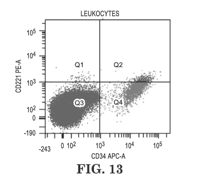
FIG. 13. Flow cytometry of hemolyzed blood from a healthy volunteer (100 ul)
mixed with 10 ul of 1 million/ml MDS-L cells detecting CD34 and IGF1R (CD221).
FIG. 14. Flow cytometry of MDS-L cells (1 million/m1) detecting CD34 and
IGF1R (CD221).
Detailed Description
Definitions:
The term "anti-cancer chemotherapeutic drug or agent" refers to a synthetic,
biological, or semi-synthetic compound that is not an enzyme and that kills
cancer cells
or inhibits the growth of cancer cells while having less effect on non-
cancerous cells. It
does not include antibodies or molecules naturally made by mammals, such as
growth
factors and cytokines.
The term "treating cancer" includes, e.g., preventing metastasis, inhibiting
growth of a cancer, stopping the growth of cancer, or killing cells of a
cancer.
The term "binding affinity" of a ligand for a particular receptor refers to
the
association constant KA (the inverse of the dissociation constant KD) or to
experimentally determined approximations thereof.
The term "anti-metabolite" refers to an anti-cancer chemotherapeutic agent
that
bears a structural similarity to a naturally occurring substance, interacts
with enzymes
as an inhibitor or a substrate, and interferes with cellular processes.
Examples include
methotrexate, fluorouracil, floxuridine, fludarabine, mercaptopurine,
thioguanine,
cytarabine, azacytidine, cladribine, and pentostatin.
The "IGF-1 receptor" is also known in the literature as the type 1 IGF
receptor
and is abbreviated herein as IGF-1R.
"Containing" as used herein is open-ended; i.e., it allows the inclusion of
other
unnamed elements and has the same meaning as "comprising."
The term "leader sequence" as used herein refers to an amino acid sequence a
the N-terminus of a protein. It is not cleaved off after synthesis of the
protein but is
part of the mature protein.
Description:
6
CA 03059593 2019-10-09
WO 2018/217669
PCT/US2018/033747
One embodiment of the invention provides a method of treating a patient for
oligoblastic acute myelogenous leukemia (0-AML) or myelodysplastic syndrome
(MDS) or chronic myelomonocytic leukemia (CMML) comprising: administering to a
patient in recognized need of treatment for O-AML, MDS, or CMML an agent
comprising: an insulin-like growth factor type 1 receptor (IGF-1R) ligand
conjugated to
an anti-cancer chemotherapy drug. O-AML and CMML are recognized as very
similar
diseases to MDS.
Another embodiment provides a method of treating a patient for acute myeloid
leukemia (AML), or chronic myeloid leukemia (CML), or O-AML, or CMML, or MDS
comprising: administering to a patient in recognized need of treatment for
AML, CML,
O-AML, CMML, or MDS (a) a hypomethylating agent (e.g., azacitidine or
decitabine)
and (b) an agent comprising: an insulin-like growth factor type 1 receptor
(IGF-1R)
ligand conjugated to methotrexate; wherein the IGF-1R ligand is insulin-like
growth
factor 1 (IGF-1) or a variant thereof or insulin. Typically, the
hypomethylating agent
and the agent comprising an IGF-1R ligand conjugated to methotrexate are
administered on the same day.
It has been found that 765IGF-MTX, insulin-MTX, and longR3IGF-MTX each
tend to have low solubility in phosphate buffered saline, or any neutral pH
solution
containing salt, for instance over about 50 mM NaCl. Accordingly, 765IGF-MTX
is
currently stored as a 4 mEq/L solution in 10 mM HC1. For infusion into
patients, it is
diluted into 250 ml of 5% dextrose or 10% dextrose in water. IGF-MTX causes
hypoglycemia, so delivering it as an infusion in 5% dextrose has the benefit
of
administering dextrose to counteract the expected mild hypoglycemia. And
765IGF-
MTX is completely soluble in water with any concentration of dextrose, whereas
it
tends to precipitate in about 150 mM NaCl at neutral pH.
Accordingly, another embodiment of the invention provides a pharmaceutical
composition that is a solution for infusion comprising: (a) an agent
consisting of a
covalent conjugate an IGF-1R ligand covalently conjugated to methotrexate,
wherein
the IGF-1R ligand is insulin-like growth factor 1 (IGF-1) or a variant thereof
or insulin;
dissolved in (b) 100 ml to 1 liter of 5% to 10% (w/v) dextrose in water,
wherein the
7
CA 03059593 2019-10-09
WO 2018/217669
PCT/US2018/033747
composition does not comprise more than 5 mM NaC1 or more than 2 mM phosphate;
wherein the solution is in an infusion bag and has a volume of 100 ml to 1
liter.
Another embodiment provides a method of administering an agent consisting of
a covalent conjugate an IGF-1R ligand covalently conjugated to methotrexate,
wherein
the IGF-1R ligand is insulin-like growth factor 1 (IGF-1) or a variant thereof
or insulin;
the method comprising: diluting the agent into a diluent consisting
essentially of a
volume of 100 ml to 1 liter of 5% to 10% dextrose (w/v) in water to make a
solution of
the agent in the diluent; and infusing the solution into a patient.
Preferably, the
infusion occurs over a time of 30 minutes to 2 hours.
The IGF-1R ligand may be an antibody that specifically binds to IGF-1R. In
other embodiments it is insulin or IGF-1 (SEQ ID NO:3) or a variant of IGF-1.
A
preferred specific variant is 765IGF (SEQ ID NO:2).
In specific embodiments, the anti-cancer chemotherapy drug is methotrexate,
chlorambucil, or bendamustine. In a specific embodiment it is methotrexate.
In a specific embodiment, the chemotherapy drug is a small molecule
(molecular weight smaller than 2000 daltons) that is not a protein or peptide
and that
contains a free carboxyl group. These can be conjugated to a protein IGF-1R
ligand by
reaction with EDC to conjugate the carboxyl to amino groups of the protein.
A specific embodiment of the methods of treating patients described herein
involve administration of an IGF-1R ligand conjugated to methotrexate,
preferably
765IGF-MTX, at a dose of 0.1 to 2.5 microEq per kg patient body weight. In
other
embodiments the dose is 0.2 to 2.5, 0.4 to 2.5, or 0.4 to 1.6, about 0.2,
about 0.4, about
0.8, or about 1.6, or about 2.5 microEq/kg. Dosing is preferably once weekly,
but may
be twice weekly or once per 2 weeks or once per 3 weeks. In one embodiment,
dosing
is once per week for 3 weeks, followed by one week off, in a 28-day cycle.
Dosing is
preferably by intraveneous infusion. In one embodiment, the conjugate (an IGF-
1R
ligand conjugated to an anti-cancer chemotherapy drug) is administered by
intravenous
infusion in 5% to 10% dextrose in a volume of 100 ml to 1 liter (more
preferably about
200 ml to about 500 ml, and in other embodiments 100 to 500 ml, 150 to 500 ml,
200 to
500 ml, or about 250 ml, or about 500 m1).
The IGF-1R ligand in some embodiments is covalently attached to the
8
CA 03059593 2019-10-09
WO 2018/217669
PCT/US2018/033747
chemotherapy drug. In other embodiments, it may be conjugated by non-covalent
attachment, for instance by embedding the chemotherapy drug and IGF-1R ligand
together in nanoparticles, similarly to the way ABRAXANE is a nanoparticle of
paclitaxel associated with albumin.
In specific embodiments, the IGF-1R ligand is insulin-like growth factor-1
(IGF-1), or a variant thereof, or insulin. The variants of IGF-1 preferably
have reduced
binding affinity for soluble IGF binding proteins as compared to native IGF-1.
Soluble
IGF binding proteins are soluble proteins in the blood that binding to IGF-1,
as opposed
to the IGF-1R membrane receptor that is a membrane protein through which IGF-1
exerts its biological action. As much as 99% of IGF-1 in vivo is bound to the
soluble
IGF binding proteins, and when it is bound to the soluble IGF binding proteins
it is
unavailable for binding to IGF-1R. Below specific variants of IGF-1 that have
reduced
binding affinity for the soluble IGF binding proteins are described, as well
as an assay
for determining binding affinity to the soluble IGF binding proteins.
We have expressed in E. coli, from a recombinant vector with expression
controlled by a T7 promoter and induced with IPTG, a fusion protein having the
sequence of SEQ ID NO:2. This protein has the sequence at its N-terminus of
SEQ ID
NO:1, which provides a polyhis tag for purification and several additional
lysine
residues. The C-terminal of the protein is residues 19-88 and corresponds to
R3-IGF,
which is human wild type IGF-1 sequence with an arginine at position 21 of SEQ
ID
NO:2 that replaces the native glutamic acid at position 3 of wild-type IGF-1
(SEQ ID
NO:3).
R3-IGF (SEQ ID NO:6) is a variant IGF-1, as discussed below.
765IGF (SEQ ID NO:2) comprising SEQ ID NO:1 as an N-terminal sequence
followed by R3-IGF expressed at a high yield and purified at a higher yield
than other
IGF fusion protein constructs comprising different leader sequences. It was
more
stable to storage than IGF132, another variant of IGF-1. It also refolded with
almost
100% yield of active form, and it displaced more wild-type IGF-1 from its
receptor on
MCF7 cells than did long-R3-IGF, another variant of IGF-1.
The SEQ ID NO:1 leader also provides five lysine residues. A 765IGF-
methotrexate conjugate was prepared by covalently attaching methotrexate
through one
of its carboxyl groups by amide bond to amino groups on 765IGF. 765IGF has
nine
9
CA 03059593 2019-10-09
WO 2018/217669
PCT/US2018/033747
amino groups, including eight lysine side chains (five of these in the SEQ ID
NO:1
leader) and the amino terminal alpha-amino group. The 765IGF-MTX had an
average
of about 8 methotrexate groups attached per IGF monomer. Conjugates to longR3-
IGF
and IGF132 had fewer methotrexate groups per IGF monomer. So this was another
advantage of the SEQ ID NO:1 leader.
R3-IGF is a variant IGF-1 in a fusion protein with SEQ ID NO:1 in SEQ ID
NO:2. It is a variant that activates the IGF receptor (IGF-1R) but has reduced
binding
affinity for the soluble IGF binding proteins (as compared to wild-type IGF-1)
(Francis,
G.L., et al.1992, J. Mol. Endocrinol. 8:213-223; Tomas, F.M. et al., 1993, J.
Endocrinol. 137:413-421). Soluble IGF binding proteins are natural serum
proteins
that bind to IGF-1, holding it in circulation and extending its biological
half-life. But
when IGF-1 is bound to the IGF binding proteins it cannot bind to the membrane
IGF
receptor (IGF-1R). (Clemons, D.R., 1998, Mol. Cell. Endocrinol. 140:19-24.)
For that
reason, variants of IGF-1 that have reduced binding to the soluble IGF binding
proteins
are more active in vivo than wild-type IGF-1 and more rapidly target the IGF
receptor.
Binding affinity for IGF binding proteins can be tested with rat L6-myoblast-
conditioned medium. The medium from growth of rat L6 myoblasts (0.2 ml) is
mixed
with 8,000 cpm1251-1GF-1 (approximately 0.05 uCi) in 0.3 ml final volume of 50
mM
sodium phosphate, pH 6.5, 0.25% bovine albumin and test competitor (wild type
IGF-1
or an IGF variant) at 0.1 nM to 1 uM final concentration. After incubation 90
minutes
at room temperature, to separate bound and free tracer an ice cold rapidly
stirred
suspension of charcoal at 5mg/m1 in assay buffer containing 0.2 mg/ml
protamine
sulfate is added to the sample, and after 8 minutes on ice, the mixture is
centrifuged 20
minutes at 5,000 x g. Radioactivity in the supernatant is counted in a gamma
counter.
The binding affinity of a variant can be compared to that of wild-type IGF to
determine
whether a variant has reduced binding affinity for the soluble IGF binding
proteins.
Some specific variants of IGF-1 with reduced binding affinity to the soluble
IGF binding proteins include IGF132 (SEQ ID NO:4) (disclosed in U.S. Patent
No.
4,876,242), LONG-R3-IGF (SEQ ID NO:5), R3-IGF (SEQ ID NO:6), and des(1-
3)IGF1 (SEQ ID NO:7), which lacks the first three residues of wild-type IGF-1.
(LongR3-IGF, R3-IGF, and des(1-3)IGF1, are described in Francis, G.L., et
al.1992, J.
CA 03059593 2019-10-09
WO 2018/217669
PCT/US2018/033747
Mol. Endocrinol. 8:213-223; Tomas, F.M. et al., 1993, J. Endocrinol. 137:413-
421).
Thus, in particular embodiments, the polypeptide that is a variant IGF-1 with
reduced
binding to the soluble IGF-1 binding proteins comprises any one of SEQ ID
NOS:4-7.
The IGF receptor may be targeted in cancer with conjugates comprising (a) an
anti-cancer chemotherapeutic agent covalently coupled to (b) an IGF receptor
ligand
such as IGF-1 or the IGF variants described herein. Because insulin has
affinity for
IGF-1R, the IGF-1R ligand may also be insulin.
Preferably, the IGF-1 receptor ligand with reduced affinity for soluble IGF-1
binding proteins has at least 5-fold, more preferably at least 10-fold, more
preferably
still at least 100-fold lower binding affinity for soluble IGF-1 binding
proteins than
wild-type IGF-1. Binding affinity for the soluble IGF-1 binding proteins can
be
measured by a competition binding assay against labeled IGF-1 (e.g., 1251 IGF-
1), using
a mixture of purified IGF-1 binding proteins or rat L6 myoblast-conditioned
medium (a
naturally produced mixture of IGF-1 binding proteins), as described in
Francis, G.L., et
al. (1992, J. Mol. Endocrinol. 8:213-223); Szabo, L. et al. (1988, Biochem.
Biophys.
Res. Commun. 151:207-214); and Martin, J.L. et al. (1986, J. Biol. Chem.
261:8754-
8760). Preferably, the variant IGF-1 has an IC50 in a competition binding
assay against
labeled wild-type IGF-1 for binding to soluble IGF-1 binding proteins in L6
myoblast-
conditioned medium of greater than 10 nM, more preferably greater than 100 nM.
Preferably, the IGF-1R ligand, such as the variant IGF-1 variant with reduced
affinity for soluble IGF-1 binding proteins, has affinity for the IGF-1
receptor that is
close to wild-type IGF-1 (e.g., less than 30-fold greater than wild-type IGF-
1, more
preferably less than 10-fold greater than wild-type IGF-1). In specific
embodiments,
the variant IGF-1 has an KD in a competition binding assay against labeled
wild-type
IGF-1 for binding to IGF-1 receptors (e.g., on MCF-7 cells) of less than 50
nM, more
preferably less than 10 nM, more preferably still less than 5 nM, more
preferably still
less than 3 nM). This assay is described in Ross, M. et al. (1989, Biochem. J.
258:267-
272) and Francis, G.L., et al. (1992, J. Mol. Endocrinol. 8:213-223), and in
Example 4
herein.
11
CA 03059593 2019-10-09
WO 2018/217669
PCT/US2018/033747
In a specific embodiment of the invention the IGF-1 variant comprises IGF-1
(SEQ ID NO:3) or comprises a segment at least 90% identical to any one of SEQ
ID
NOS:3 and 4.
In specific embodiments, the anti-cancer chemotherapeutic drug may be one
with a free carboxyl group, such as methotrexate, chlorambucil, or
bendamustine.
In particular embodiments, the chemotherapeutic agent conjugated to the IGF-
1R ligand is mechlorethamine, cyclophosphamide, ifosfamide, melphalan,
chlorambucil, thiotepa, hexamethylmelamine, busulfan, carmustine, lomustine,
semustine, streptozocin, decarbazine, vincristine, vinblastine, etoposide,
teniposide,
paclitaxel, docetaxel, daunorubicin, idarubicin, doxorubicin, epirubicin,
dactinomycin,
plicamycin, mitomycin C, bleomycin, mitoxantrone, methotrexate, fluorouracil,
floxuridine, fludarabine, mercaptopurine, thioguanine, cytarabine,
azacytidine,
cladribine, pentostatin, cisplatin, carboplatin, mitotane, procarbazine, or
amsacrine.
In specific embodiments of the methods, the IGF-1R ligand is not IGF-1 (SEQ
ID NO:3) and is or comprises 765IGF (SEQ ID NO:2), IGF132 (SEQ ID NO:4), long-
R3-IGF (SEQ ID NO:5), R3-IGF (SEQ ID NO:6), or des(1-3)IGF1 (SEQ ID NO:7), or
a variant at least 90% identical to IGF-1.
In other embodiments, the IGF-1R ligand is an antibody against IGF-1R.
In specific embodiments where the IGF-1R ligand is conjugated to
methotrexate, the method comprises dosing the patient with the agent at a dose
of 0.1 to
2.5 microEq/kg, 0.1 to 2.5, 0.4 to 2.5, 0.4 to 1.6, about 0.2, about 0.4,
about 0.8, about
1.6, or about 2.5 microEq/kg. A microEq is a micromole of methotrexate groups
(conjugated to the ligand).
In the methods comprising adiministering a hypomethylating agent, the
hypomethylating agent is preferably azacitidine or decitabine, more preferably
azacitidine.
One embodiment provides a pharmaceutical composition that is a solution for
infusion comprising: (a) an agent consisting of a covalent conjugate an IGF-1R
ligand
covalently conjugated to methotrexate, wherein the IGF-1R ligand is insulin-
like
growth factor 1 (IGF-1) or a variant thereof or insulin; dissolved in (b) 100
ml to 1 liter
of 5% to 10% (w/v) dextrose in water, wherein the solution does not comprise
more
12
CA 03059593 2019-10-09
WO 2018/217669
PCT/US2018/033747
than 5 mM NaC1 or more than 2 mM phosphate; wherein the solution is in an
infusion
bag and has a volume of 100 ml to 1 liter.
In more specific embodiments, the solution has a volume of 100 ml to 500 ml,
150 ml to 500 ml, 200 ml to 500 ml, or about 250 ml, or about 500 ml.
In a specific embodiment, the agent is 765IGF-MTX.
In more specific embodiments, the composition comprises less than 1 mM NaCl
and less than 1 mM phosphate.
Another embodiment provides a method of administering an agent consisting of
a covalent conjugate an IGF-1R ligand covalently conjugated to methotrexate,
wherein
the IGF-1R ligand is insulin-like growth factor 1 (IGF-1) or a variant thereof
or insulin;
the method comprising: diluting the agent into a diluent consisting
essentially of a
volume of 100 ml to 1 liter of 5% to 10% dextrose (w/v) in water to make a
solution of
the agent in the diluent; and infusing the solution into a patient.
In specific embodiment, the step of infusing the solution into a patient
occurs
over a time of 20 minutes to 2.5 hours, or over 30 minutes to 2 hours, or over
45
minutes to 1.5 hours, or over 1 to 2 hours.
Another embodiment provides a composition comprising: an agent comprising:
an insulin-like growth factor type 1 receptor (IGF-1R) ligand conjugated to an
anti-
cancer chemotherapy drug, for use in a method of treating oligoblastic acute
myelogenous leukemia (0-AML) or myelodysplastic syndrome (MDS) or chronic
myelomonocytic leukemia (CMML) or acute myeloid leukemia (AML) or chromic
myeloid leukemia (CML).
Another embodiment provides a device comprising: (a) an infusion bag capable
of holding a maximum volume of 100 ml to 2 liters, filled with (b) a solution
of 5% or
10% (w/v) dextrose and dissolved in the solution (c) an agent consisting of a
covalent
conjugate an IGF-1R ligand covalently conjugated to methotrexate, wherein the
IGF-
1R ligand is insulin-like growth factor 1 (IGF-1) or a variant thereof or
insulin, the
solution having a volume of 100 ml to 1 liter (more preferably 100 ml to 500
ml, 150
ml to 500 ml, or about 250 m1).
The device may further comprise (d) tubing connected to the infusion bag, and
(e) a hypodermic needle connected to the tubing.
In one embodiment, the agent is 765IGF-MTX.
13
CA 03059593 2019-10-09
WO 2018/217669
PCT/US2018/033747
In one embodiment of the device the solution comprises at least 10 microEq of
the agent and no more than 250 microEq of the agent.
In specific embodiments of the device the solution comprises no more than 5
mM NaCl (preferably no more than 1 mM NaCl) and no more than 2 mM phosphate
(preferably no more than 1 mM phosphate).
In specific embodiments of the device, the solution comprises no more than 5
mM NaCl (preferably no more than 1 mM NaCl, more preferably no NaCl).
Guidelines for coupling anti-cancer chemotherapeutic agents to receptor
ligands
The natural ligands to the insulin and IGF-1 receptors are proteins, namely
insulin, IGF-1, and IGF-2. Chemotherapeutic agents are typically coupled to
proteins
through the reactive groups present on proteins. These include the N-terminal
alpha-
amino group, the C-terminal alpha-carboxyl group, the side-chain amino group
of
lysine, the side-chain carboxyl groups of aspartic acid and glutamic acid, the
side chain
thiol of cysteine, and the side chain of arginine. Other reactive side chains
found on
proteins are the side-chain hydroxyl of serine and threonine, the hydroxyaryl
of
tyrosine, the imidazole of histidine, and the methionine side chain.
Many of the same reactive groups are found on chemotherapeutic agents and on
non-proteinaceous ligands of the insulin and IGF-1 receptors. Thus, many of
the
principles of modification and cross-linking of proteins discussed herein also
apply to
modification and cross-linking of chemotherapeutic agents and non-
proteinaceous
ligands.
The chemistry and principles of protein conjugation and cross-linking are
described in Wong, Shan S., Chemistry of Protein Conjugation and Cross-
Linking,
1991, CRC Press, Boca Raton, Florida. Other sources for information on this
chemistry include the Pierce Biochemistry catalog; and Greene, T.W., and Wutz,
P.G.M., Protecting Groups in Organic Synthesis, second edition 1991, John
Wiley &
Sons, Inc., New York, and references cited therein.
The strongest nucleophile of amino acid side chains is the thiol of reduced
cysteine side chains. The thiol reacts with most protein modifying reagents.
Alpha-
haloacetamides and maleimides are considered to react specifically with
cysteine
14
CA 03059593 2019-10-09
WO 2018/217669
PCT/US2018/033747
residues, particularly at pH 7.0 and below. Thiols also react by disulfide
interchange
with disulfide reagents.
Hello
CA 03059593 2019-10-09
WO 2018/217669
PCT/US2018/033747
0 0
R-SH + II
CI-CH2-C-NHR1 ¨1 P" R-S-CH2-C-NHR1
0 0
"AI
R-SH
R-S
0 0
Amino groups are the next-strongest nucleophiles found on proteins.
Aldehydes react with amino groups to form Schiff bases. The Schiff bases are
hydrolyzable, which can be an advantage in the present invention. With uptake
into
cancer cells of a ligand-chemotherapeutic agent conjugate, in some cases it is
necessary
that the chemotherapeutic agent is cleaved from the conjugate for it to be
active. This
is better accomplished if the chemotherapeutic agent is linked to the ligand
by a
cleavable linkage, such as a hydrolyzable linkage. Cleavable linkages can be
cleaved
spontaneously or by enzymes in the cell. For instance, amide bonds are cleaved
by
certain enzymes, including proteases. A Schiff base linkage spontaneously
hydrolyzes
at an appreciable rate. A disulfide linkage is expected to be reductively
cleaved in the
intracellular reducing environment of a cancer cell.
0
R-NH2
R1 R-N=C-R1
The Schiff base formed by reaction of an amino group with an aldehyde can be
stabilized by reduction with, for instance, sodium borohydride or pyridine
borane.
Pyridine borane has the advantage of not reducing disulfides, which are found
in
insulin, IGF-1, and IGF-2 and are essential for the structure of those
proteins.
16
CA 03059593 2019-10-09
WO 2018/217669
PCT/US2018/033747
Sugars or other moieties having hydroxyl groups on adjacent carbons, which are
found in some chemotherapeutic agents, can be modified to react with amino
groups by
oxidizing the sugars with, for instance, periodate. This cleaves between the
carbons
and produces a dialdehyde. The aldehyde groups will react with amino groups.
A dialdehyde, such as glutaraldehyde, will cross-link two molecules having
amino groups.
Other amino reagents include activated carbonyls, such as N-
hydroxysuccinimide esters, p-nitrophenyl esters, or acid anhydrides (e.g.,
succinic
anhydride).
0
0
II 0
R¨NH2 + Ri¨C-0¨N ¨OP- II
R¨NH¨CRi
0
0
0
II
R¨NH2 0 ¨00- RNH¨C¨CH2CH2COOH
0
Amino groups also react with sulfonyl halides and aryl halides (e.g, 2,4-
dinitrofluorobenzene).
17
CA 03059593 2019-10-09
WO 2018/217669
PCT/US2018/033747
O 0
R¨NH2 Ri¨S¨CI ¨No" RNH¨S¨Ri
O 0
R¨NH2 F = NO2 -11111'- RNH 41, NO2
0
02N 2N
Amino groups also react with isocyanates and isothiocyanates to form urea or
thiourea derivatives.
R¨NH2 R1¨N=C=S I I
R¨N¨C¨NHRi
Imidoesters are the most specific acylating agents for amino groups.
Imidoesters react specifically with amines to from imidoamides at pHs between
about 7
and 10. This reaction has the advantage of maintaining charge stability by
generating a
positively charged group, the imidoamide, at the former amino group.
Imidoamides
also slowly hydrolyze at pHs above neutrality, which can also be an advantage
in that
the hydrolysis can release free chemotherapeutic agent in the cancer cell.
R¨NH2
R1¨C-0¨R2 R¨NH¨C¨Ri
Carboxyl groups react specifically with diazoacetate and diazoacetamide under
mild acid conditions, e.g., pH 5.
O 0 0
RCOOH R1C¨CH=N2
RC-0¨CH2¨CR1
The most important chemical modification of carboxyls uses carbodiimides,
such as 1-cyclohexy1-3-(2-morpholiny1-4-ethyl)carbodiimide (CMC) and 3-(3-
dimethylaminopropyl)carbodiimide (EDC). In the presence of an amine,
carbodiimides
18
CA 03059593 2019-10-09
WO 2018/217669
PCT/US2018/033747
form an amide bond to the carboxyl in two steps. In the first step, the
carboxyl group
adds to the carbodiimide to form an 0-acylisourea intermediate. Subsequent
reaction
with an amine yields the corresponding amide.
0 N¨Ri
RCOOH Ri¨N=C=N¨Ri fl--0¨C
/\ NH
Ri
0 I
II R2NH2
R¨C¨NHR2
A particularly important carbodiimide reaction is its use in activating
carboxyls
with N-hydroxysuccinimide to form an N-hydroxysuccinimide ester.
19
CA 03059593 2019-10-09
WO 2018/217669
PCT/US2018/033747
0 0
0
+ Ri-NH2
0 0
0 (c1\1-0)S'SN)
1<1=)
R1-NH 1'''S" \ ____________ / 0
+ R2-NH2
DTT
0
0 R1-NH SH ,SJ
R2-NH)CS =) /
)C
0 0
R1-NHS¨SNH-R2
Arginine reacts with vicinal dialdehydes or diketones, such as glyoxal, 2,3-
butanedione, and 1,2-cyclohexanedione. Borate may stabilize the adduct, if
stabilization is desired.
20
CA 03059593 2019-10-09
WO 2018/217669
PCT/US2018/033747
NH 0 0
II II
Protein¨NH¨C¨NH2 R¨C¨C¨R
OH
HO OH HO /
B03- /
HN
\NH+
NH HN
Protein
NH
Protein
The reactive groups can also be interchanged with other reactive groups by
some of the above reactions. For instance, modification of an amino group with
an acid
anhydride such as succinic anhydride, replaces the positively charged amino
group with
a free carboxyl group. Likewise, reaction of a carboxyl group with a
carbodiimide and
a diamine, such as ethylene diamine, replaces the carboxyl group with a free
amino
group.
Cross-linking: Reagents containing two of the reactive groups described above,
for instance two amino-reactive groups or an amino-reactive and a thiol-
reactive group,
can be used to cross-link a chemotherapeutic agent containing one of the
appropriate
groups to an insulin or IGF-1 receptor ligand containing the other appropriate
group. In
addition, a carboxyl (of, e.g., a chemotherapeutic agent) activated with a
carbodiimide
or a carbodiimide and N-hydroxysuccinimide can react with an amino group (of,
e.g., a
protein ligand) to form an amide bond cross-link.
21
CA 03059593 2019-10-09
WO 2018/217669
PCT/US2018/033747
0
RCOOH + Ri¨N=C=N¨Ri
NH
Ri
0
0
/ 0 I
N-0¨fl
0
0
The activated carboxyl is stable enough to be isolated, but will then readily
react
with amino groups to form an amide bond.
Succinimides such as N-succinimidy1-342-pyridyldithiolpropionate (SPDP) can
be used to couple two compounds through amino groups. (See Pierce
Biotechnology
catalog, and Thorpe, P.E. et al. 1982, Immunol. Rev. 62:119-158.)
Examples
Example 1
Plasmids were synthesized by DNA 2.0 (Menlo Park, California) encoding
these proteins with nucleotide sequences optimized for expression in E. coli,
and under
the control of a T7 promoter:
Protein encoded Description Sequence
403IGF His6-IGF SEQ ID NO:8
764IGF His6-K5-IGF132 SEQ ID NO:11
765IGF His6-K5-R3IGF SEQ ID NO:2
784IGF mutTrx-R3IGF SEQ ID NO:9
785IGF mutTrx-IGF132 SEQ ID NO:10
E. coli BL21(DE3) was transformed with each of the plasmids and transformants
isolated. 10 ml of the transformed BL21(DE3) culture of each was used to seed
500 ml
22
CA 03059593 2019-10-09
WO 2018/217669
PCT/US2018/033747
of LB media with 50 ug/ml kanamycin (LB-kan) in a 2 L baffled flask. These
were
induced with 0.4 mM final IPTG at an O.D. 600 nm of 0.6 and grown overnight at
25
degrees C.
The cells were resuspended in 50 mM Tris-HC1 pH 8.0 and frozen. They were
thawed and incubated at 5% wet weight/volume cell weight in 50 mM Tris-HC1 pH
8.0,
0.2% Triton-X100, 0.5 mg lysozyme per g cell paste, for 30 minutes at room
temperature. They were then sonicated to break cells. MgCl2 was added to 3 mM
final
concentration and 250 ul of BENZONASE was added per liter of culture. This was
incubated a further 1 hour at room temperature.
Inclusion bodies were isolated by centrifugation. Soluble fraction was
retained.
Inclusion bodies were solubilized in 7 M urea, 0.5 M NaCl, 20 mM phosphate
pH 7.8.
The solubilized inclusion bodies were loaded onto 1 ml of Ni-nitrolito-
triacetic
acid (Ni-NTA) resin in a column. The column was washed with Ni-A buffer and
eluted
with Ni-B buffer.
Ni-A 6 M urea, 0.5 M NaCl, 20 mM sodium phosphate, 20 mM imidazole, pH 7.3.
Ni-B 6 M urea, 0.5 M NaCl, 20 mM sodium phosphate, 0.4 M imidazole, pH 7.3.
The protein yields were:
403IGF eluate 3.6 mg
764IGF eluate 16 mg
765IGF eulate 24 mg
784IGF eluate 6.7 mg
785IGF eluate 1.9 mg
SDS-PAGE was run of the eluates and of the crude insoluble and soluble
fractions. It
appeared that 784IGF and 785IGF had about half of the IGF in the soluble
fraction and
half in the insoluble. 403IGF, 764IGF, and 765IGF appeared to have nearly all
of the
IGF in the insoluble fraction.
From this data, the best yield was with 765IGF. Those with the SEQ ID NO:1
leader sequence (764IGF and 765IGF) gave better yields than those with a
simple Met-
His6 leader (403IGF) or with thioredoxin leader sequences (784IGF and 785IGF).
And
the constructs with the R3IGF mutant for the IGF portion (765IGF and 784IGF)
gave
23
CA 03059593 2019-10-09
WO 2018/217669
PCT/US2018/033747
better yields than the corresponding constructs with the IGF132 mutant for the
IGF
portion of the fusion protein (764IGF and 785IGF).
Example 2
Refolding and binding assay
2 ml of each of the original Ni eluates from Example 1 was mixed with about an
equal
volume of 100 mM glycine, 6 M urea, pH 9.5, concentrated by ultrafiltration in
a
CENTRICON 3 kDa filter unit, then brought up again in that buffer and
concentrated to
about 420 ul. Then they were diluted to 2 mg/ml for 403IGF, 764IGF, and
765IGF,
and 4 mg/ml for 784IGF and 2.4 mg/ml for 785IGF.
200 ul of each of these was mixed rapidly with 1.8 ml of refold buffer. Refold
buffer was 1.4 M urea, 100 mM glycine, 0.5 M NaCl, 19% ethanol, 0.5 mM GSSG, 4
mM GSH, pH 9.5. They were refolded at room temperature for 3 hours, and then
tested
in a binding assay for competition binding to IGF receptors against 1-132
radioactive
wild type IGF (Perkin Elmer, Inc.) For comparison, commercial Long-R3-IGF
(LR3IGF) was also tested.
The approximate binding constants (KDs) in this experiment were these:
LR3IGF 1 nM
403IGF 2 nM
764IGF 100 nM
765IGF 10 nM
784IGF 3 nM
785IGF 40 nM
The fusion proteins containing the R3IGF mutant (LR3IGF, 765IGF, and 784IGF)
had
lower KDs than those containing the IGF132 mutant (403IGF, 764IGF and 785IGF).
Example 3
Purification and Yield of 765IGF
A plasmid encoding 765IGF with optimized codon usage for E. coli, with the
765IGF
gene under the control of a T7 promoter, was synthesized by DNA 2.0 (Menlo
Park,
CA, USA). E. coli B121(DE3) was transformed with the plasmid and grown in
fermentor culture and induced with IPTG.
24
CA 03059593 2019-10-09
WO 2018/217669
PCT/US2018/033747
765IGF was purified under denaturing conditions by ion exchange
chromatography and Nickel affinity chromatography. The yield of purified
765IGF
was about 60 mg per liter of culture.
765IGF was refolded by a procedure similar to that of Example 2 and then the
refolded protein was purified by ion exchange chromatography on a DEAE resin
and
affinity chromatography on a nickel resin.
Example 4
765IGF Binding Assay to IGF-1 Receptor
Method:
Theory of assay: Radioactive 1251 labeled insulin-like growth factor-1 (IGF-1)
competes with a test ligand for binding to type 1 IGF receptors that are
abundant on
MCF7 cells (a human breast cancer cell line) in vitro. The tested ligands
include our
765IGF variant of insulin-like growth factor-1 (IGF-1) and our novel covalent
conjugates that contain the antifolate drug methotrexate coupled to 765IGF, as
well as
commercially available long-R3-IGF-1 (Sigma Aldrich, St. Louis, MO, USA) as a
comparison and positive control.
MCF7 cell media: 500 mL MEM, 0.01 mg/mL bovine insulin; 5 mL sodium
pyruvate, 5 mL non-essential amino acids, 10 mL sodium bicarbonate, 10 mL
fetal
bovine serum, 5 mL penicillin/streptomycin.
MCF7 cells (ATCC HTB-22) were plated at 20,000 cells per well in a volume
of 0.5 mL/well in a 48-well tissue culture plate (flat bottom with low
evaporation lid)
and placed in a cell culture incubator set at 37 C with 5% CO2. After 2-3
days in
culture the plates were washed 2x with 0.5 mL per well of cold binding assay
buffer
(100 mM Hepes-NaOH, pH 7.2; 120 mM NaCl; 5 mM KC1; 1.2 mM MgSO4; 0.1 %
BSA). After the final wash, 0.5 mL of binding assay buffer was added to each
well and
the plates are placed at 4 C for 2 to 6 hours.
Test ligands were prepared at a concentration of 10 micromolar (long-R3-IGF)
or 20 micromolar (765IGF and IGF-MTX) in 5 mM HC1 in a volume of 200 ul. To
determine the concentration, the molecular weight of 765IGF (9742 daltons) and
long-
R3-IGF (9111 daltons) are used. For long-R3, the lyophilized commercial
material is
dissolved at 1.0 mg/ml in 10 mM HC1 and this is diluted to a concentration of
91 ug/ml
for a 10 uM solution.
CA 03059593 2019-10-09
WO 2018/217669
PCT/US2018/033747
The 765IGF and long-R3-IGF were diluted into binding buffer in the wells at
concentrations of 2000 nM to 1 nM.
Next, 25 uCi lot of 1-125 IGF (Perkin Elmer Radiochemicals, Waltham,
Massachussetts, USA) was dissolved in 1 ml of water. An appropriate dilution
into
binding buffer ws made, and then 50 ul of diluted radioactive IGF is added to
each
well, to add 0.03 uCi or more per well. For fresh 1-125 IGF, per plate used
100 ul of
the 1 ml solution of 1-125 IGF in water can be added to 2.6 ml of binding
buffer per
plate used, and 50 ul added per well.
The plates were then incubated overnight at 4 C. Then the liquid was
withdrawn from each well with a micropipettor and the wells were washed twice
in
binding buffer. Cells were lysed with 0.5 mL 300 mM NaOH, 1% SDS and the
lysates
were counted on a gamma counter.
Results:
The result of an IGF-1 receptor binding assay for 765IGF and commercially
available
long-R3-IGF are shown in FIG. 1. At high concentrations, 765IGF consistently
displaced more radioactivity than long-R3-IGF, suggesting it may bind to IGF-1
binding sites on the membranes that long-R3-IGF does not. The KD of 765IGF in
this
assay was less than 1 nM, while the KD of long-R3-IGF was about 3 nM. A second
binding assay with a different lot of 765IGF is shown in FIG. 2 and gave a KD
of 3.5
nM.
Example 5
Conjugation of Methotrexate to 765IGF
The protein was buffer exchanged into pH 7.3 conjugation buffer and adjusted
to a
concentration of 2.5 mg/ml.
pH 7.3 conjugation buffer: 25 mM sodium phosphate, 10 mM NaCl, 6 M urea,
pH 7.3.
pH 6.3 conjugation buffer is the same buffer at pH 6.3.
Methotrexate was dissolved at 20 mg/ml in pH 6.3 conjugation buffer, and the
pH
adjusted to pH 6.3 with NaOH.
26
CA 03059593 2019-10-09
WO 2018/217669
PCT/US2018/033747
1-ethyl-3-l3-dimethylaminopropyllcarbodiimide hydrochloride (EDC) was
freshly dissolved in pH 6.3 conjugation buffer at 75 mg/ml.
One volume of EDC solution was added to 1 volume of MTX solution and
incubated 30 seconds at room temperature and then this mixture was added to 8
volumes of 2.5 mg/ml protein solution in pH 7.3 conjugation buffer.
The mixture was mixed and then reacted overnight at room temperature. Then
6 M HC1 was added to the reaction mixture to 60 mM final concentration. Then
the
reaction mixture was buffer exchanged into 10 mM HC1.
Result:
The amount of methotrexate conjugated per mole of protein was determined by
measuring absorbance of the conjugate at 305 nm in 100 mM HC1, using a molar
extinction coefficient for methotrexate groups of 21.6 per mM (Chamberlin et
al.
Analytical Profiles of Drug Substances, 1976, 5:283-306.) The protein
concentration
was determined by quantitative amino acid analysis. By this, the molar ratio
of MTX
groups to IGF in the 765IGF-MTX conjugate was approximately 8.
Example 6
765IGF-MTX In Vitro Cytotoxicity Assay
Cytotoxicity Assay. This potency assay is an assay for inhibition of
proliferation of
MCF-7 tumor cells in vitro by incubation with the 765IGF-MTX.
Method
Day 0. Five-thousand MCF7 cells were plated per well in a 96-well test plate
in 100 ul
of rich media on day 0.
Day 1. A shadow plate was made for each test plate, with each well of the
shadow plate
containing media or 3X the intended final concentration of test agent in media
in each
well. As a negative control, media is used. As a positive control, free
methotrexate at
3 uM is used.
After making the shadow plate, 50 ul is transferred from each well of the
shadow plate to the corresponding well of the test plate to generate the final
concentrations of test agent in the wells of the test plate.
Day 5. Cell proliferation is determined by adding Dojindo CCK-8 reagent and
incubating and measuring absorbance of the dye according to the manufacturer's
instructions.
27
CA 03059593 2019-10-09
WO 2018/217669
PCT/US2018/033747
Result:
Results of a representative cytotoxicity assay with 765IGF-MTX are shown in
FIG. 3.
The IC50 (Concentration needed for 50% inhibition of cell proliferation) of
765IGF-
MTX was 249 nEquivalents per L. (A nanoEquivalent is a nanomole of
methotrexate
groups conjugated to 765IGF.) For comparison, in the same assay, the IC50 of
free
methotrexate was measured as 88 nM.
Example 7
Inhibition of dihydrofolate reductase by methotrexate and IGF-methotrexate
conjugates
Method:
The experiments were done with the dihydrofolate reductase assay kit from
Sigma-
Aldrich (St. Louis, MO, USA), according to the manufacturer's instructions. In
the
assay dihydrofolate reductase is mixed with pH 7.5 buffer. Next the inhibitor
¨
methotrexate or an IGF-methotrexate conjugate ¨ is added and the solution
mixed. It
was incubated for 30 seconds to allow inhibitor binding. Then NADPH is added
to 50
uM final concentration, and then dihydrofolic acid is added to 60 uM final
concentration. The reaction is monitored by measuring absorbance at 340 nm.
Results:
The tested conjugates were:
765IGF-MTX prepared as described in Example 3. 765IGF has 9 amino groups
available to conjugate to methotrexate (8 lysines and the N-terminal amino
group).
This batch had a MTX:protein molar ratio of 7.5.
765IGF-MTX 1/3. This conjugate was prepared with 1/3 of the usual
concentrations of MTX and EDC in the conjugation reaction. It produced a
conjugate
with a MTX:protein molar ratio of 1.2.
LR3IGF-MTX. In this case, the version of IGF is long-R3-IGF. This has 4
available amino groups for conjugation (3 lysine side chains and the N-
terminal amino
group). This conjugate had a MTX:protein ratio of 2.8.
In addition, free methotrexate was tested.
The conjugates were exhaustively ultrafiltered to remove any free methotrexate
28
CA 03059593 2019-10-09
WO 2018/217669
PCT/US2018/033747
before their use in the inhibition assay.
A plot of the inhibition data for 765IGF-MTX is shown in FIG. 4.
The IC50s of methotrexate and the conjugates were these:
Competitor IC so MTX:IGF ratio
Methotrexate 5.3 nM N.A.
765IGF-MTX 95 nEq/L 7.5
1/3 765IGF-MTX 90 1.2
LR3IGF-MTX 99 2.8
The IC50in nEq/L was approximately the same for all three of the IGF-MTX
conjugates, despite having different numbers of MTX groups conjugated per IGF
protein monomer. This shows that each conjugated methotrexate group acts as an
independent inhibitor of the enzyme. If the additional methotrexate groups on
a
conjugate monomer were sterically unable to bind to and inhibit a DHFR enzyme
once
one group is bound to a DHFR enzyme, then one would expect that the IC50 for
the
conjugates would be the same in terms of nM protein concentration for each of
the
conjugates, instead of being the same in terms of nEq/L MTX group
concentration, as
is observed. Because the inhibition is proportional to MTX groups, 765IGF-MTX,
with
its higher MTX loading, has an inhibition constant in terms of protein
concentration of
13 nM (95 nEq/L divided by 7.5 MTX per IGF gives 13 nM IGF), whereas LR3IGF-
MTX has an inhibition constant in terms of protein concentration of 35 nM.
Thus, with
the higher loading of MTX, less 765IGF protein needs to be used to achieve the
same
inhibition of DHFR, and by inference the same level of killing of tumor cells.
The data show that the protein-conjugated MTX groups inhibit DHFR, but a
higher concentration is needed for inhibition as compared to free MTX.
Example 8
Binding of 765IGF-MTX to IGF-1R on MCF7 cells.
Several competition binding assays of 765IGF-MTX conjugate with MCF7 against
radiolabeled IGF-1 have been conducted, as described in Example 4. The results
are a
29
CA 03059593 2019-10-09
WO 2018/217669
PCT/US2018/033747
KD of about 20 nM 765IGF-MTX. (To explain, this is nM of the protein
conjugate, not
nEq/L of MTX groups. Since there are about 8 MTX per 765IGF, 20 nM 765IGF-
MTX is about 160 nEq/L 765IGF-MTX). In the particular binding assay shown in
Fig.
5, the KD was 13.4 nM.
Example 9
In Vivo Toxicology Studies
MTD is based on both non-rodent and rodent studies. Formal GLP toxicity in
vivo studies were completed in rats and Beagle dogs, in which the 765IGF-
MTX conjugate was administered intravenously by a single 30 minutes infusion
on study Days 1 and 8 as shown in Table 1 for the dogs:
Table 1.
Preparation'
Target Amount of 4
Concentration peconi (4 mm
Group / Dose ueq/m1 MTX groups) D5W-Added Total Volume
(umol MTX 765IGF-MTX (mL) (mL)
groups/mL) Stock
Solution (mL)
1. 765IGF-MTX
0.2 peq/kg
0.04 1.25 123.75 125
(0.2 ul MTX groups
/kg)
2. 765IGF-MTX
2.0 usqlkg
0.4 10 90 100
(2.0 umol MTX
groups/kg)
3. 765IGF-MTX
0.5 peq/kg
0.1 3.125 121.875 125
(0.5 mol MTX
groups/kg)
4. 765IGF-MTX
6.0 peq/kg
1.2 37.5 87.5 125
(6.0 mol MTX
groups/kg)
CA 03059593 2019-10-09
WO 2018/217669
PCT/US2018/033747
Analysis of all generated data, including clinical observations, serial blood
glucose
determinations, and clinical pathology revealed no drug/treatment-related
significant
toxicity in dogs treated by intravenous infusion with 765IGF-MTX conjugate in
5%
dextrose at 0.2 and 0.5 ueq/kg. In the 0.2 ueq/kg group there was only
transient
dyspnea and passivity noted, while in the animals treated at 0.5 ueq/kg a
single episode
of vomiting and diarrhea were noted, and there was also mild reddening and
swelling of
the skin on the head of the female dog. Treatment of dogs in these two groups
was well
tolerated, and any reaction to the treatment was transient and resolved by
itself.
In animals dosed at 2 ueq/kg the reactions to the treatment included mild to
moderate
anaphylactoid and hives-type reactions, transient anorexia and weight loss,
and
hypoglycemia. Reactions to the treatment in the dogs dosed at 6 ueq/kg were
similar to
the reactions at 2 umol/kg, but they were more severe and lasted longer. Thus,
a MTD
of 765IGF-MTX conjugate in Beagle dogs in this study may be considered to be 6
ueq/kg, by a single infusion over 30 minutes.
Recovery from anaphylactoid reactions and hypoglycemia in the female dog dosed
at 2
Eq/kg and in both animals dosed at 6 Eq/kg, was assisted by treatments with
diphenhydramine and dextrose.
Hypoglycemia in the higher dose groups was an exaggerated pharmacological
effect of
IGF, which was mitigated by the use of 5% dextrose as a vehicle for delivery
of
765IGF-MTX conjugate. The pathogenesis of the anaphylactoid reactions was not
clear, but may have been caused by either methotrexate, IGF or the combination
thereof. Vomiting, diarrhea, anorexia and weight loss are known reactions to
methotrexate.
The highest non-severely toxic dose in beagles was 0.5 ueq/kg. Using the
conversion
from doses in dogs in uEq/kg to equivalent human dose in Eq/kg, the
equivalent
human dose to 0.5 Eq/kg in dogs is 0.27 Eq/kg in humans.
IGF-MTX does not cause significant cytopenia
Cytopenia is a particular concern for MDS, CMML, and O-AML since it is a
principal
sequela of these diseases. In both rat and dog repeat dose GLP toxicology
testing IGF-
31
CA 03059593 2019-10-09
WO 2018/217669
PCT/US2018/033747
MTX caused almost no cytopenia, even at the highest doses tested. In both rat
and dog
toxicology studies IGF-MTX caused a slight dose-dependent reduction in
erythrocyte
mass, but even at the highest doses tested erythrocyte mass was within normal
ranges.
In rats but not dogs neutrophils were also slightly decreased by IGF-MTX but
remained
within normal ranges even at the highest doses. No other hematological
parameters
were affected by IGF-MTX.
In the completed Phase I dose-escalation study in human solid tumor patients,
a dose of
0.8 Eq/kg was found to be tolerated without any serious adverse events. Since
MDS
patients have greater cytopenia than solid tumor patients, and for safety of
MDS
patients, we are conducting a new dose escalation in this study beginning at a
dose level
of 0.2 Eq/kg administered on days 1, 8, and 15. See the schema on page 6 for
the
dose escalation schema.
Example 10
A 6-DOSE, ONCE WEEKLY, INTRAVENOUS
TOXICITY STUDY WITH
IGF-METHOTREXATE CONJUGATE
IN SPRAGUE-DAWLEY RATS, FOLLOWED BY
A 14-DAY RECOVERY PERIOD
This repeated dose study examined the systemic toxic potential and target
organs for
toxicity of 765IGF-MTX, a conjugate of a variant of insulin-like growth factor
designated
765IGF with methotrexate (MTX). The IGF-MTX conjugate was administered once a
week, for 6 weeks, by intravenous slow bolus injection. Three groups of
Sprague-
Dawley rats were dosed intravenously at dose levels of 0.5, 2 and 5 Eq/kg (
Eq is a
umol of methotrexate groups) for two doses. Since no toxicity was observed at
the
high-dose group, starting from third dose, the dose level in the mid-dose
group was
increased from 2 to 5 Eq/kg and this remained until the end of treatment
(i.e. the
animals received a total of 4 doses at 5 Eq/kg). In the high-dose group, the
dose was
first increased from 5 to 10 Eq/kg (third dose) and then due to severe
toxicity, the
dose level was adjusted to 8 Eq/kg for the remaining 3 doses. The control
group of
rats was dosed with 5% Dextrose Injection USP (D5W) which was used as the
diluent
for the IGF-MTX preparations.
32
CA 03059593 2019-10-09
WO 2018/217669
PCT/US2018/033747
Four groups of rats were used in this study (1 control and 3 test). Each Main
Study test
and control group consisted of 10 male rats [Strain: Crl:CD (SD) BR-Sprague-
Dawley
(Charles River Canada Inc., Canada)]. There were also 5 rats per sex included
in the
Recovery groups in the control, mid and high-dose groups; and 3 rats per sex
(control)
and 6 rats per sex per group (test groups) in CBC/glucose subgroups. An
additional 3
rats per sex were allocated to the control group and 9 rats per sex to each
test group for
toxicokinetics blood collection.
The dose volume was 4 mL/kg, for all groups including the control group.
Although
some adjustments to the dose levels were made for the purpose of this report,
the doses
used in the report will be referred as 0.5, 5 and 8 Eq/kg.
Examinations of all animals included daily clinical observations, and daily
food and
water monitoring. Animals also received detailed physical examinations on a
weekly
basis. Body weights were recorded initially and then on Days 7, 14, 21, 28, 35
and 41,
and prior to necropsy on Day 42 (Main Study animals), and additionally on Days
37,
42, and 49 and prior to necropsy on Day 50 (Recovery animals). Food
consumption
was recorded weekly. Complete blood count (CBC) was performed 24 hours before
the
second dose, and 24 hours prior to each subsequent dose. Complete clinical
pathology
was performed at the end of the Main Study and Recovery Periods.
Ophthalmoscopy was performed initially (before the initiation of treatment),
and at the
end of the Main Study.
Blood samples were also collected as per the protocol schedule for
toxicokinetics,
however the samples were not analyzed. Six days after the end of the sixth
dose, 10
male and 10 female Main Study animals from each group were euthanized and
submitted for gross necropsy and histopathology examinations. The remaining 5
male
and 5 female Recovery rats in the control group, and the groups dosed at 5 and
8
Eq/kg were euthanized 14 days after the last dose and were submitted for
necropsy
and histopathological examinations.
33
CA 03059593 2019-10-09
WO 2018/217669
PCT/US2018/033747
Three rats died and/or were euthanized in the high-dose group. On Day 15 (Dose
3)
when the dose was increased from 5 to 10 Eq/kg one male (TK group) and one
female
(Main Study group) died immediately after the dosing was completed.
It was suspected that the low pH of the test article (pH ¨ 2.3) may have
caused
metabolic acidosis after the dose level was increased to 10 Eq/kg and/or that
the test
article precipitated intravenously. Histopathological evaluation found
numerous
variably sized round greenish-gray amorphous particles, some with central
densities in
lungs of both animals which died acutely. One of these rats had these
particles also in
the heart. In both animals there was also acute thromboembolism in lungs, and
acute
intravenous coagulation in the heart. It appears that the injected material
precipitated
and initiated intravascular platelet aggregation with micro embolism and
obstruction of
small blood vessels.
In the third rat that was euthanized on Day 24, ascending pyelonephritis with
ischemic
necrosis and parenchimal atrophy were found. These changes were severe and
explained the clinical deterioration. This condition was considered incidental
and not
related to the treatment with the test article.
With the exception of the rats that died, all other rats from all groups
received the
specified treatment and survived to the scheduled euthanasia and necropsy
dates.
There were no treatment-related systemic toxic effects in any rat treated with
IGX-
MTX conjugate at 0.5 Eq/kg, or in rats dosed at 2 Eq/kg (for the first 2
doses), and
then at 5 Eq/kg for the remaining 4 doses.
In the high dose group occasional haematuria was observed in some rats after
the dose
was increased to 10 Eq/kg within one hour after dosing. This was attributed
to the
low pH (pH=-2.3) of the test formulation.
Ophthalmology results did not identify any abnormal findings, and food
consumption
and body weight gains did not identify any significant differences between the
test and
control groups.
34
CA 03059593 2019-10-09
WO 2018/217669
PCT/US2018/033747
Pathology findings that were most likely treatment-related in the groups dosed
at 5 and
8 Eq/kg, but were expected and common adverse reactions to MTX
administration,
were as follows:
= The general trend in CBC parameters monitored during the study was a
slight dose-dependent decrease in erythrocyte mass (RBC's, Hb, Hct), with
a compensatory increase in reticulocytes. Before Dose 6, erythrocyte mass
in the group treated at 5 Eq/kg was reduced by approximately 8-15%,
(gender combined), and there was approximately 15-19% decrease in the
group dosed at 8 Eq/kg. At the same time, reticulocyte counts increased
47-102% and 2.2-2.4-fold, (gender combined), in the groups which received
the IGF-MTX conjugate at 5 and 8 Eq/kg, respectively. Neutrophil counts
were also decreased in both these groups.
= Reduction in erythrocyte mass was also observed at the end of the Main
Study in animals of both genders in the groups treated with IGF-MTX
conjugate at 5 and 8 Eq/kg (mean decreases of 6-7%, and 10-14%, for the
groups that were dosed at 5 and 8 Eq/kg, respectively). Compensatory
reticulocytosis was noted in both of these groups.
= WBC's were also decreased in these 2 groups (neutrophils, lymphocytes and
monocytes were all affected). All cell types appeared about equally
affected. The reduction in neutrophils was about 5-35% (gender combined)
in the group treated at 5 Eq/kg, and in the group that was dosed at 8
Eq/kg, the decrease ranged between 50-57%, when compared to the
controls. Platelets were also significantly decreased in females dosed at 8
Eq/kg (approximately 38% decrease relative to the control females). In the
recovery animals, all these parameters were either back to normal or were
showing the tendency of normalization indicating recovery.
= The mean weight of the spleen was increased in both males and females
dosed at 5 and 8 Eq/kg. The increases ranged on average between 17 to
38%, relative to the mean weight of the spleen in the control rats.
CA 03059593 2019-10-09
WO 2018/217669
PCT/US2018/033747
The changes that were observed in treatment groups, and which were considered
treatment-related, but were expected exaggerated pharmacological effects of
IGF,
were as follows:
= In the group dosed at 0.5 Eq/kg (gender combined) mean glucose levels
were increased after dosing. The increases ranged between 0.3 0.5 to 4.0
5.0 mmol/L, over 6 doses. Occasionally, in some individual animals there
was a decrease in blood glucose levels in this group.
= In the group dosed at 5 Eq/kg, the mean increases or decreases in blood
glucose levels over 6 doses, ranged between -1.8 7 to 7.5 3.4 mmol/L,
and in the group dosed at 8 Eq/kg, the increases/decreases ranged between
-2.1 1.3 to 2.6 3.1 mmol/L. On one occasion (3rd dose - 10 Eq/kg)
glucose levels in some animals decreased below 2.8 mmol/L, and thus all
animals in this group were dosed with 10% Dextrose, I.P. at 2 mL/rat. It
should be noted that decreases in glucose blood levels appeared dose
dependent.
Histopathological evaluations identified some changes in the lungs of animals
dosed at
8 Eq/kg that may be of possible toxicological significance and these were as
follows:
= In the lungs, there was increased prominence of small blood vessels with
infiltrates of granulocytes and lymphocytes in the perivascular interstitium
in many animals in the study. These perivascular inflammatory cell
infiltrates were observed in 5/20 controls and in 19/19 in the group dosed at
8 Eq/kg animals in the Main Study. The severity of this response was
higher in the group dosed at 8 Eq/kg. The response was seen in 4/10
controls and 0/10 high-dose animals in the Recovery groups.
= In one animal in the high-dose Recovery Group, there were wedge-shaped
areas of parenchymal loss with replacement fibrosis consistent with
ischemic infarcts. These are the residuum of an earlier focal ischemic event.
36
CA 03059593 2019-10-09
WO 2018/217669
PCT/US2018/033747
It is unknown whether the infarcts were related to the treatment, because
such changes could occur as a sporadic condition unrelated to the protocol.
In conclusion, analysis of all generated data, including clinical observations
ophthalmology, gross necropsy and histopathology revealed no drug/treatment-
related
significant toxicity in rats that were treated intravenously with IGF-MTX
conjugate at
0.5 and 5 Eg/kg weekly, for 6 weeks. At these two dose levels, the treatments
were
well tolerated by animals.
At a dose level of 0.5 Eg/kg, the only finding that was occasionally noted in
some rats
was a marginal decrease in blood glucose levels. This finding was an expected
pharmacological effect of IGF, and thus under the conditions of this
experiment, the no
observed effect level in this study was considered to (NOEL) be equal to 0.5
Eq/kg
dosed weekly, for 6 weeks.
At a dose level of 5 Eq/kg, besides the decrease in blood glucose levels
which were
more pronounced than in the animals dosed at 0.5 Eg/kg, there was also a
marginal
decrease in erythrocyte mass (about 8 - 15% before Dose 6, and about 6 - 7% at
the end
of the Main Study). WBC's were also marginally reduced in this group at the
end of
the Main Study (about 5 - 35%). At the end of recovery both RBC's and WBC's
were
within the normal ranges in this group, indicating the reversibility of these
changes.
There was also a marginal increase in the weight of spleens in this group,
which
showed a tendency for normalization in the Recovery animals. These findings
were
known effects of MTX on hematopoiesis. Thus, under the condition of this
experiment,
the no observe adverse effect level (NOAEL) in this study was considered to be
equal
to 5 Eg/kg.
Example 11
A 5-DOSE, ONCE WEEKLY, INTRAVENOUS
INFUSION TOXICITY STUDY WITH
IGF-METHOTREXATE CONJUGATE
IN BEAGLE DOGS, FOLLOWED BY
A 21-DAY RECOVERY PERIOD
37
CA 03059593 2019-10-09
WO 2018/217669
PCT/US2018/033747
This repeated dose study examined the systemic toxic potential and target
organs for
toxicity of 765IGF-MTX, a conjugate of a variant of insulin-like growth factor
designated 765IGF with methotrexate (MTX). The IGF-MTX conjugate was
administered once a week, for 5 weeks, by intravenous (IV) infusion. Three
groups of
Beagle dogs were dosed intravenously at dose levels of 0.5, 2 and 4 pEq-kg (a
pEq is a
pmole of methotrexate groups) in 5% dextrose (D5W), and the fourth control
group of
dogs was dosed with D5W which was used as the diluent for the IGF-MTX
conjugate
preparations.
Four groups of dogs were used in the study (1 control and 3 test groups). The
control
group, mid-dose and high-dose groups consisted of 10 dogs (5 males and 5
females),
and the low-dose group consisted of 6 dogs (3 males and 3 females), breed:
Beagle,
Ridglan Farms. The test and control articles were administered by an IV
infusion at a
dose volume of 5 mL/kg/hour over 1 hour.
Examinations of all animals included daily clinical observations, and daily
food and
water monitoring. Animals also received detailed physical examinations on a
weekly
basis. Body weights were recorded initially and then on Days 8, 15, 22, 29 and
34 and
prior to necropsy on Day 35 (Main Study animals), and additionally on Days 30,
37,
44, 49 and prior to necropsy on Day 50 (Recovery animals). Food consumption
was
recorded daily. Complete blood count (CBC) was performed 24 hours after the
first
dose, and prior to each subsequent dose. Complete clinical pathology was
performed
before study initiation, at the end of the Main Study and Recovery Periods.
All animals received the specified treatments at doses of 0.5 to 4 pEq/kg and
no
mortalities were associated with the treatments.
Ophthalmology and ECG's (including QT) findings were found to be within the
normal
physiological limits in all test groups.
In the group dosed at 0.5 pEq/kg, clinical reactions to the treatment were
mild and
consisted of mild to moderate passivity and injected sclera in one male dog,
vomiting in
38
CA 03059593 2019-10-09
WO 2018/217669
PCT/US2018/033747
two female dogs, and mild dyspnoea in another female dog. Mild edema around
the
eyes was also observed in one female animal. All of the above observations
were single
clinical events, which occurred only once during the 5 dose periods, were
transient and
resolved themselves. Any decrease in blood glucose levels after dosing in
these animals
was negligible. At the end of study, there was a minimal reduction in
erythrocyte mass
noted in this group (about 10% reduction, compared to the controls). However,
Red
Blood Cells (RBC's), Hematocrit (Hct) and Hemoglobin (Hb) in this group were
well
within the normal ranges. These findings were not considered clinically
relevant as they
were of low magnitude, transient, resolved themselves and were the expected
side
effects of methotrexate therapy.
Clinical findings, clinical pathology, and gross necropsy which were treatment-
related
in the groups dosed at 2 and 4 pEq/kg that were clinically relevant, but were
expected
and common adverse reactions to MTX administration, were as follows:
0 Post-dosing anorexia was observed in all dogs, with subsequent reduction in
body weights, and/or body weight loss. In dogs dosed at 2 pEq/kg, anorexia was
observed usually on the second day after each dosing, which would last for a
couple of days, and then the animals would completely or partially recover by
the next dose. By the end of the Main Study, the body weight gain in these
dogs
was negligible, male animals gained only about 1.2% compared to 8.4% for the
control male dogs, over the 34-day period, and females gained 2.8% in
comparison to 11.1% for the control females. Food consumption in this group
was approximately 22% lower (gender combined) than the food consumption of
the control dogs.
0 In animals dosed at 4 pEq/kg, anorexia was more severe than in the dogs
dosed at 2 pEq/kg. This resulted in approximately 36-38% reduction in food
consumption over the treatment period, when compared to the controls, and the
total average weight loss in dogs was 1.3 kg for males and 0.9 kg for females.
During the 3-week Recovery Period, dogs from this group regained their body
weights, indicating reversibility.
39
CA 03059593 2019-10-09
WO 2018/217669
PCT/US2018/033747
O Occasionally nausea (retching), diarrhoea and vomiting were observed in
some animals dosed at 2 pEq/kg and in all dogs dosed at 4 pEq/kg. These were
usually observed on days when dosing was performed.
0 Reduction in erythrocyte mass (RBC's, Hb, Hct) and
anisocytosis/macrocytosis was mild by the end of the Main Study. The
reduction was approximately 13% and 15%, for dogs dosed at 2 and 4 pEq/kg,
respectively, when compared to the control dogs. By the end of the Recovery
Period, the erythrocyte mass was still below the pre-study levels in these two
groups. However, the reduction in RBC's, Hb and Hct levels never exceeded
the lower limits of the normal ranges.
O The mean total protein and albumin levels were reduced slightly below the
limit of normal ranges in animals dosed at 4 pEq/kg. After the Recovery
Period,
these levels were within the normal ranges. Reduced albumin levels were most
likely the result of anorexia and body mass loss as reported in these dogs.
O In 2 out of 10 dogs dosed at 2 pEq/kg, ALT activity was slightly
increased
above the upper limit of normal ranges (on average 11% increase). In dogs
dosed at 4 pEq/kg, 4 dogs were affected and increases were approximately 1-
fold for males and about 52% for females, on average. In the absence of
histopathological findings indicating hepatocellular injury, the increases of
ALT
most likely represented sublethal injury with alterations in hepatocyte
permeability, and an increase in ALT.
O Glucose level decreases in the group dosed at 2 pEq/kg (initial blood
glucose
level minus blood glucose level immediately after the end of infusion) ranged
between -2.5 1.3 to -0.7 1.3 mmol/L (mean decrease over 5 doses, gender
combined). In the group dosed at 4 pEq/kg, the decreases ranged between -3.9
1.7 to -1.6 0.6 mmol/L. The decreases in blood glucose levels were an
expected pharmacological effect of IGF.
Clinical and pathology findings that were considered dosing and treatment-
related, and
CA 03059593 2019-10-09
WO 2018/217669
PCT/US2018/033747
which were clinically relevant and toxicologically significant in the groups
dosed at 2
and 4 pEq/kg, are described below. It should be noted that the following
adverse
reactions are less common, but are known and reported reactions in humans
receiving
MTX for various conditions. However, it cannot be completely excluded that the
following reactions were not augmented by the presence of IGF. These reactions
were
as follows:
O Anaphylactoid (angioedema), type of reactions presented as swelling
(edema)
and reddening (erythema) of the skin of head (around the eyes, lips, ears),
throat, neck and forelimbs, were noted mostly during the infusion. The total
number of dogs that had these reactions (all groups combined) during dosing
were: 5 dogs (the first dose); 3 dogs (the second dose); 4 dogs (the third
dose);
14 dogs (the fourth dose) and 6 dogs (the fifth dose). It should be noted that
on
the fourth day of dosing, there was a spike in the number of dogs with these
reactions and in the severity of the reactions, however, during the following
dose (dose five), the reactions were comparable to the reactions seen on
dosing
Days 1, 2 and 3.
O There did not appear to be a difference (qualitative or quantitative) in
anaphylactoid reactions between the groups which were dosed at 2 or 4 pEq/kg
of IGF-MTX conjugate.
O Two dogs from the group dosed at 2 pEq/kg, had neurological reactions
which consisted of a seizure in one male dog during the second dose and a
transient loss of consciousness and postural tone during the third dose in one
female dog. Both animals received subsequent treatment without further
neurological incidents.
One plausible explanation for the seizure in one dog would be that the blood-
brain-
barrier (BBB) was disrupted by the release of inflammatory mediators (this
animal had
moderate anaphylactoid reactions to the treatment and had to be treated with
antihistamines before the seizure started), and/or the BBB was disrupted by
the
extramedullary haematopoiesis in the choroid plexus as found histologically in
this dog.
The latter is reported predisposing condition for seizures in dogs. The second
dog with
a minor neurological incident was also found with the extramedullary
haematopoiesis
in the choroid plexus. Secondly, the dog with the seizure had an unexpectedly
high
spike of MTX in the blood during infusion. The MTX level in this dog on Day 1
was at
41
CA 03059593 2019-10-09
WO 2018/217669
PCT/US2018/033747
least 2-7 fold higher than in any other dog dosed at 2 pEq/kg and it was at
least 2-fold
higher than in the dogs which received the test article at 4 pEq/kg. Thus, the
most likely
explanation for the seizure in this animal was a probable disruption of the
BBB and a
high spike of MTX in the blood.
Histopathologically there were several findings of potential toxicological
significance
and they were as follows:
0 Thymic atrophy due to depletion of cortical and medullary lymphocytes
occurred in several dogs with increasing frequency and severity with treatment
dose. Thymic atrophy was pronounced, dose-related through the 0.5-4.0
pEq/kg/week dose range, and persistent through the recovery period. While this
might have occurred as a direct effect of the test article on proliferating
thymic
lymphocytes, thymic atrophy is best explained as an indirect stress response.
Thymic atrophy was reported in humans as a response to MTX chemotherapy.
0 The animal in the group dosed at 2 pEq/kg that had a seizure had several
findings that were not seen in other animals in the study. These included
increased lymphocytic infiltrates of the superficial mucosa of the cecum and
rectum, unilateral focal degeneration and mineralization of the
corticomedullary
region of one adrenal gland, and prominent hyperplasia with mixed leukocyte
infiltrates in the respiratory epithelium of the trachea and major bronchi.
Toxicokinetic (TK) parameters for free MTX were estimated from plasma
concentration-time data arising from a 5-dose once weekly intravenous infusion
study
with insulin-like growth factor (IGF) MTX conjugate at low (0.5 pEq/kg), mid
(2.0
pEq/kg) and high (4.0 pEq/kg) MTX equivalent dose levels. No consistent
differences
in the plasma levels of MTX were observed between animals of different genders
and
thus data from males and females were combined. Plasma levels of MTX increased
with time both during and following a 1-hour intravenous infusion of the IGF-
MTX
conjugate. On Day 1 and based on the mean plasma concentrations of 6 - 10
dogs, the
Tmax was 2 hr from start of infusion for for all dose levels. Cmax values
ranged from
71.6 - 511.9 ng/mL. Both the AUCGo and Cmax values were dose proportional. The
plasma terminal half-life and mean residence time of MTX ranged from 4.6-5.5
hrs and
6.1-7.6 hrs, respectively, with a low apparent clearance (based on liberation
of free
42
CA 03059593 2019-10-09
WO 2018/217669
PCT/US2018/033747
MTX from the conjugate) and large apparent volume of distribution ranging from
0.34-
0.46 L/hr/kg and 2.24-2.81 Ukg, respectively. The pharmacokinetics of MTX
following Day 29 of dosing were similar to those following Day 1 of dosing.
These findings suggest that free MTX is liberated in a time-dependent manner
due to
metabolism of the conjugate. Thus, the resulting TK for MTX are dependent on
both
the liberation of MTX from the conjugate and the elimination of free MTX over
time.
Following intravenous administration of the IGF-MTX conjugate in the dose
range of
0.5-4.0 pEq/kg to Beagle dogs, serum levels of the IGF-MTX conjugate decreased
quickly. There was no gender bias and an increase in serum exposure was
observed on
Day 29 compared to Day 1. The apparent clearance of the IGF-MTX conjugate
which
was small and ranged from 0.02-0.05 L/kg/hr, decreased by 25-50% on Day 29
compared to Day 1, while the apparent volume of distribution, also small and
which
ranged from 0.07-0.20 L/kg, did not display any trends. The terminal half-life
of the
IGF-MTX conjugate which ranged from 2.5-4.6 hrs was marginally longer on Day
29
compared to Day 1, while the mean residence time which ranged from 1.4-5.3
hrs, did
not display any trend. Together these findings suggest that following
intravenous
administration of the IGF-MTX conjugate, it was eliminated with a low
clearance from
a small volume of distribution consistent with its large molecular size. The
elimination
of the IGF-MTX conjugate was marginally slower with successive doses,
resulting in
an increased plasma exposure. The elimination of the IGF-MTX conjugate
preceded the
development of maximal MTX concentrations.
In conclusion, analysis of all generated data, including clinical
observations,
ophthalmology, electrocardiography, gross necropsy and histopathology revealed
no
drug/treatment-related significant toxicity in dogs that were treated
intravenously with
IGF-MTX conjugate at 0.5 pEq/kg weekly, for 5 weeks.
At this dose level of 0.5 pEq/kg, the only findings that were most likely
treatment-
related were mild to moderate passivity and injected sclera in one animal,
vomiting in
two dogs and mild dyspnoea in another dog. Mild edema around the eyes was also
observed in one dog. At the end of the study, there was also a mild reduction
in
erythrocytes (about 10% reduction, relative to the control group). These
findings were
43
CA 03059593 2019-10-09
WO 2018/217669
PCT/US2018/033747
not considered clinically relevant as they were of low magnitude, transient,
resolved
themselves and were expected side effects of methotrexate therapy.
Therefore, under condition of this experiment, the no observed adverse effect
level
(NOAEL) in this study was considered to be equal to 0.5 pEq/kg dosed weekly,
for 5
weeks.
In the groups dosed at 2 and 4 pEq/kg, the side effects which were also
associated with
the MTX administration included anorexia with reduced body weight gain or body
weight loss, occasional diarrhea and vomiting. There was also a mild reduction
in
erythrocyte mass, (about 13-15%) and reduced albumin levels, and a slight
increase in
ALT activity. These observations appeared to be dose-dependent and with the
exception of RBC reduction, there was a full recovery of all animals by the
end of the
recovery period. In these two groups, thymic atrophy and an increased number
of
necrotic cells in the duodenum were also noted at histopathology.
There were also two types of adverse reactions noted in this study, which
although
undesirable, were not completely unexpected as they were reported adverse
reactions to
MTX administration. In the group dosed at 2 pEq/kg, in two dogs there were
neurological incidents. The first dog had a seizure during the second dose. In
this
animal, the seizure may have been the result of a disruption in the blood-
brain-barrier,
induced by inflammatory mediators and/or extramedullary haematopoiesis, and a
high
spike of free MTX. In the second dog, there was one episode of loss of
consciousness
and postural tone. In the groups dosed at 2 and 4 pEq/kg there were incidences
of
anaphylactoid reactions.
For the neurological and anaphylactoid reactions, although most likely induced
by
MTX, it cannot be completely excluded that IGF did not play any role in these
events.
It has to be noted that both of these adverse reactions were easily controlled
by the
appropriate therapy (antihistamines and diazepam).
Example 12
Phase I Study of IGF-Methotrexate Conjugate in the Treatment of Advanced
Tumors
Expressing IGF-1R
44
CA 03059593 2019-10-09
WO 2018/217669
PCT/US2018/033747
The primary objective of this study was to determine the maximum tolerated
dose
(MTD) of 765IGF-MTX by evaluation of toxicity during treatment of advanced,
previously treated malignancies that express IGF-1R. One inclusion criterion
was that a
subject's tumor (tissue, bone marrow, or blood) must express IGF-1R, defined
as >10%
of tumor cells expressing IGF-1R by immunohistochemistry (IHC). Patients were
enrolled with solid tumors or lymphomas.
Nineteen subjects were enrolled in this dose escalation study. 765IGF-MTX was
administered as an IV infusion over 1 hour on days 1, 8 and 15 of a 28 day
cycle in 250
ml of 5% dextrose or at the physician's discretion 10% dextrose.. Treatment
continued
until disease progression, unacceptable toxicity, or patient refusal.
Assessment of
response was confirmed with imaging studies performed at the end of cycle 2 +/-
7
days, and every 2 cycles thereafter. The table below shows, for each dose
level tested,
the subjects that were tested, the type of malignancies they had, number of
cycles they
completed, whether they experienced a dose limiting toxicity (DLT), and, if
applicable,
their reason for study discontinuation. One patient with Hodgkin Lymphoma at
dose
level 3 was treated with 22 doses with apparently stable disease by CT scan,
but
treatment was discontinued when a lymph node biopsy showed no evidence of
cancer.
Dose Dose Subjec Malignancy DLT # Reason for
Leve ( Eq/kg t # (YIN dose discontinuatio
I ) ) s n
1 0.05 1 Colon Ca N 6 Progression
2 0.10 2 Met N 6 Progression
adamantinom
a
3 0.20 3 Colorectal, N 6 Progression
met
3 0.20 4 Endometrial Y 1 not evaluable
Ca
3 0.20 5 Endometrial N 18 stable disease
Ca
3 0.20 6 Pancreatic Ca N 2 not evaluable
3 0.20 7 Thymic Ca N 6 Progression
3 0.20 8 Hodgkin Y 6 Progression
Lymphoma
3 0.20 9 Hodgkin N 22* stable disease
Lymphoma or complete
response
4 0.40 10 Colon Ca N 5 Progression
4 0.40 11 Thymoma N 6 Progression
CA 03059593 2019-10-09
WO 2018/217669
PCT/US2018/033747
4 0.40 12 Colon Ca N 9 stable disease
0.80 13 mCRC N 3 not evaluable
5 0.80 14 Pancreatic Ca N 6 Progression
5 0.80 15 Colon Ca N 3 not evaluable
5 0.80 16 Endometrial N 4 not evaluable
Ca
5 0.80 17 Colon Ca N 6 Progression
5 0.80 18 Breast Ca N 6 Progression
5 0.80 19 Basal cell Ca N 6 partial response.
with lung
mets.
* At time of this report is tumor-free by lymph node biopsy.
Adverse Events
5 Adverse events experienced during the prior solid tumor Phase I on
solid tumor patients
that were graded as possibly or probably related to the study drug are shown
in the
Table below.
Adverse events possibly related to study drug
Event Grade
Hypotension 3
Fever 2
Seizure 2
Sinus tachycardia 2
Dyspnea 2
Abdominal pain or cramps 2
Nausea, Vomiting 1
Hypoglycemia 1
Dizziness 1
None of the adverse events were seen in all subjects or all drug
administrations in a
single subject. The most common adverse events were hypoglycemia, which is an
expected consequence of the study drug, chills, and fever. When these
occurred, they
usually resolved within 2 hours after the end of the infusion. One subject had
grade 2
fever beginning with the infusion and lasting overnight. One subject had grade
3
hypotension beginning during the infusion and lasting overnight. Nausea and
vomiting
were common, but occurred during the infusion, not with a several hour delay
as is
typical with chemotherapy. In all cases they resolved within 1 hour after the
end of the
infusion. Abdominal pain or cramping was common but appeared limited to
patients
with colon carcinoma, so it may have been associated with the study drug
binding to
46
CA 03059593 2019-10-09
WO 2018/217669
PCT/US2018/033747
and targeting their tumor tissue. The one seizure that was seen occurred
during the
infusion in a subject during cycle 5, day 1 (the patient's 16th dose of drug)
at dose level
0.4 uEq/kg, and resolved in 2 minutes. This patient also experienced the grade
3
hypotension event at the same time and was hospitalized overnight for it.
Absence of cytopenia. Remarkably, there was no evidence of any cytopenia in
any
subject treated.
Example 13
Cytotoxicity in vitro against MDS and AML cell lines and synergy with
azacitidine.
IGF-MTX is cytotoxic in vitro against an MDS cell line.
765IGF-MTX was tested in vitro in a tritium incorporation assay. The MDS-L
cell line
was licensed from Dr. Kaoru Tohyama of Kawasaki Medical School, Japan. Cells
were
plated at 15,000 cells per well in 150 ul medium in a 96-well plate on Day 1.
On day 2,
IGF-MTX was added at concentrations ranging from 78 nEq/L to 15 microEq/L. On
day 6, one microCi of tritiated thymidine was added per well, and after 6
hours the
wells were harvested and counted. The result is shown in FIG. 6. The IC50 was
359
nEq/L. One nEq is defined as 1 nmole of MTX groups. There are approximately 8
MTX groups per 765IGF.
IGF-MTX is cytotoxic in vitro against myeloid cancer cell lines and is
synergistic with
azacytidine
Three AML cell lines were tested in our laboratory for sensitivity to IGF-MTX.
The
three cell lines were HL-60, HL-60/54, and Kasumi-1. All three were sensitive
to IGF-
MTX with IC50s of 458-668 nEq/L (nM of methotrexate groups in the IGF-MTX
conjugate). This is about the same as the IC50 against MCF7 breast cancer cell
line
and LNCaP prostate cancer cell line, both of which are sensitive to IGF-MTX in
mouse
xenografts.
The effect of IGF-MTX was also additive or synergistic with Azacitidine on at
least the
Kasumi-1 and HL-60 cell lines (it was not tested the combination on HL-60/54).
In the
presence of a concentration of Azacitidine about 1/3 of its IC50, the IC50 of
IGF-MTX
47
CA 03059593 2019-10-09
WO 2018/217669
PCT/US2018/033747
decreased from 668 to 398 nEq/L in Kasumi-1 cell line and decreased from 466
to 409
nEq/L in HL-60.
Example 14
An MDS cell line has a high level of IGF-1R
MDS-L cells were grown in RMPM1640/10% fetal calf serum / 50 ng/ml IL-3,
50 micoM beta-mercaptoethanol. They were harvested when they were at a density
of
about 5 x 10^5 cells per ml ar resuspended at 20 million cells per ml in 25 mM
Tris,
150 mM NaCl, pH 7.5 (TBS), which was at 0.5 mg/ml protein.
MCF7 was grown in Eagle's Minimum Essential Medium, 10% 1-13S, 0.1 mg/ml
human insulin. MCF7 were harvested in exponential phase into TBS at 1.0 mg/ml
protein.
Non-reducing SDS-PAGE was run on 4-20% Tris glycine wedge gels.
The samples were diluted with 4x LDS (65 ul sample, 20 ul LDS buffer).
Lanes
1. markers 8 ul NEB P7710s prestained
2,3, blank.
4. 15 ul MCF7 sample (12 ug).
5. 3.75 ul MCF7 sample (3 ug)
6. blank.
7. 20 ul MDS sample (8 ug)
The gel was electrotransferred onto a 0.2 micron PVDF filter (Invitrogen
LC2002) in
25 mM Tris, 192 mM glycine, 0.1% SDS, 20% methanol (v/v) transfer buffer.
The membrane was blocked in TBST (TBS with 0.1% Tween-20) plus 5% dry milk for
hour at 23C, then probed overnight in 0.2 microg/ml of BAF391 biotynylated
mouse
monoclonal antibody- against IGF-1R (RnD systems) at 4C. The next day it was
washed 3x for 12 minutes each in TBST, then incubated in 1/2,000 diluted
Pierce high
sensitivity streptavidin-HRP conjugate, thermo sci cat. No. 21130, diluted in
30 ml of
48
CA 03059593 2019-10-09
WO 2018/217669
PCT/US2018/033747
TBST+ 5% milk. Tt was rocked in that for 60 minutes at room temp. Then I
rinsed
briefly in TBST twice and then rocked in TBST for 12 minutes 3 times.
Then it was put in ECL Prime western blot reagent for 5 minutes without
rocking, and
put in an camera system and read for 10 minute
Result:
The Western blot result is shown in Fig. 7.. The left two lanes that have
visible bands
are MCF7 are and have 12 ug and 3 ug respectively of total protein loaded
(Lanes 4 and
5). The right stained lane is MDS-L (lane 7) and by protein assay has 8 ug
protein
loaded. But by commassie staining of a gel run in parallel it appeared the
MCF7 in
lane 4 actually had about half as much protein as the MDS-L in lane 7. The
stained
IGF-1R band in MDS-L lane 7 runs above 200 kDa. The two stained bands in the
MCF7 lanes run at about between 80 and 150 kDa. IGF-1R is a dimer of predicted
molecular weight of 205 kl), where each monomer contains a 81 kDa and a 20 kDa
polypeptide. So the MDS-L bands are consistent with a 205 kDa dimer and the
MCF7
bands are consistent with the 81 kDa polypeptide and 102 kDa monomer.
MCF7 is the standard cell line for high expression of IGF-1R, and MDS-L has
approximately the same amount of IGF-1R. So MDS-L has high expression of IGF-
Example 15
IGF-MTX inhibits proliferation of solid tumor prostate and breast cancer cell
lines LNCaP and MCF7
In vitro proliferation assays
LNCaP cells were plated in a 96-well plate at 5,000 cells per well in RPMI +
glutamine
+ 10% FCS medium in 100 1. After 24 hours, 100 ul fresh medium was added
containing no drug (control), IGF-MTX or MTX at the indicated concentration.
After
48 hours of further incubation, cell proliferation was assayed with the Cell
Counting
Kit-8 (Dojindo Molecular Technologies, Kumamoto, Japan) according to the
manufacturer's instructions.
MCF7 was cultured in Eagle's Minimum Essential Medium, with 0.01 mg/ml
human recombinant insulin, 10% fetal bovine serum, and pen/strep. For in vitro
proliferation assays it was plated at 8,000 cells per well in a 96-well plate.
After 1 day
49
CA 03059593 2019-10-09
WO 2018/217669
PCT/US2018/033747
765IGF-MTX or MTX was added to the indicated concentration. After 5 more days,
1
uCi tritiated thymidine was added per well in 50 ul fresh medium. Six hours
later the
cells were harvested and radioactivity counted.
In vitro tumor inhibition
To evaluate effects on cell proliferation, the IGF-MTX conjugate and free MTX
were
incubated with LNCaP tumor cells in vitro. Both agents inhibited proliferation
of
LNCaP cells compared to untreated control cells. At the highest tested
concentration of
2000 nM, free MTX caused significantly greater inhibition than IGF-MTX
(P=0.003).
Inhibition of proliferation by free MTX at 500 nM did not differ significantly
from that
of IGF-MTX at 2000 nM. The IC50 for longR3-IGF-MTX was about 1000 nEq/L (nM
methotrexate groups) (McTavish, H. et al., Translational Research 2009;153:275-
282)
In this case, the IGF portion of the conjugate was long-R3-IGF.
The results of a proliferation assay of 765IGF-MTX with MCF7 cells is shown
in Fig. 8. In this case, the IC50 of 765IGF-MTX was 715 nEq/m1 (Fig. 8). The
IC50
for free methotrexate against MCF7 in a parallel assay was 17 nM (data not
shown).
Example 16
IGF-MTX inhibits tumor growth in vivo of a prostate cancer cell line in mice
at
more effectively than free MTX, even at a 6-fold lower dose of IGF-MTX
IGF-MTX conjugate synthesis, analysis and quantification
Long-R3-IGF-1 was purchased from Novozymes GroPep (Novozymes BioPharma AU,
Thebarton, Australia). MTX was purchased from Sigma (St. Louis, Missouri,
USA).
Long-R3-IGF-1 (20 mg) was dissolved in 3.0 ml, 10 mM HC1. Sodium phosphate
(2.5
ml, 200 mM, pH 7.4) and solid urea (1.625 g) were added to the solution. The
solution
was dialyzed (3500 m.w. cut-off) against 20 mM sodium phosphate, pH 7.4, 5 mM
NaCl, 6.5 M urea (urea dialysis buffer) overnight at 4 C. MTX hydrate (14.8
mg)
neutralized with 1.4 mole equivalents of NaOH dissolved in 0.4 ml urea
dialysis buffer
was added to the long-R3 IGF solution in the dialysis bag. Long-R3-IGF-1 and
MTX
were coupled by incubation with 1-ethyl-3- 3-dimethylaminopropyllcarbodiimide
hydrochloride (EDC). EDC is a zero-length cross-linker that produces direct
amide
bonds between protein amine groups and the carboxyl group on MTX. EDC (60 mg)
was freshly dissolved in urea dialysis buffer (0.6 ml) and then added to the
dialysis bag,
CA 03059593 2019-10-09
WO 2018/217669
PCT/US2018/033747
which was sealed and stored in a dish for 2 hours at room temperature. The
reaction is
schematically shown in Fig. 1.
After 2 hours, the bag was placed in urea dialysis buffer and dialyzed 3.5
hours
at 4 C. The dialysis buffer was changed to 2 mM HC1, and dialysis was
continued
overnight. Long-R3-IGF-1 has 3 lysine residues and an amino terminal for a
total of 4
amino groups available for conjugation. To determine the extent of saturation,
the MTX
concentration in the conjugated long-R3-IGF-1 protein was determined by
optical
absorption at pH 11 using Ã372 nm = 6.47 mM-1. The conjugated protein is
hereafter
referred to as IGF-MTX (for long-R3-IGF-1-methotrexate).
In vivo tumor growth assays
MCF7 cells (human breast adenocarcinoma cell line) were grown in Eagle's
minimal
essential medium supplemented with 0.1 mg/ml insulin and 10% FCS. The estrogen-
dependent MCF7-L cell line was a gift from Deepali Sachdev of the University
of
Minnesota. MCF7-L cells were grown in modified IMEM medium (Invitrogen,
Carlsbad, California, USA) supplemented with 0.1 mg/ml insulin. LNCaP cells
(metastatic human prostatic adenocarcinoma) were grown in RPMI supplemented
with
glutamine and 10% FCS (Invitrogen, Carlsbad, California, USA). Cells were
grown at
37 C in a 5% CO2 humidified atmosphere. In each case, cells were grown to
approximately two-thirds confluence, harvested by trypsinization, washed with
rich
medium and then washed twice with PBS and resuspended in phosphate buffered
saline
in BD matrigel matrix (Becton Dickinson, Franklin Lakes, New Jersey, USA).
Cells
were injected intradermally in mice on the back. An estrogen pellet (0.5 mg
estradiol,
60-day release, Innovative Research of America, Sarasota, Florida, USA) was
implanted subcutaneously between the shoulder blades two days before
implanting
MCF7 and MCF7-L cells. MCF7 and MCF7-L cells were implanted in 8-week-old
female nu/nu mice. LNCaP cells were implanted in 8-week-old male nu/nu mice.
The
IGF-MTX conjugate was administered in 2 mM HC1, 1% glycerol. MTX was
dissolved in PBS. Untreated vehicle controls received 2 mM HC1, 1% glycerol.
Drug
was administered intravenously by tail-vein injection in a volume of 12.5 :1
per gram
mouse weight. All studies were approved by the University of Minnesota Animal
Care
and Use Committee and conformed to relevant ethical guidelines.
51
CA 03059593 2019-10-09
WO 2018/217669
PCT/US2018/033747
Results: Xenograft tumor growth inhibition in mice
Three in vivo studies were performed to assess the targeting of MTX with long-
R3-
IGF-1. In the initial preliminary study, breast cancer MCF7 cells were
implanted
intradermally in the backs of nude mice. When tumors in 15 mice became
palpable
(approximately 5x5 mm), the mice were randomly distributed into three groups
(n=5
per group). After randomization, mice were treated on days 0, 4 and 8 with
intravenous
tail vein injection of vehicle, free MTX at 40 nmol/g or IGF-MTX at 10 nmol of
MTX/g. Even by one day after the first treatment, tumors in the IGF-MTX
conjugate-
treated group were smaller than those in the other groups (Fig. 9). For the 12
days of
observation, tumors continued to grow in the free MTX and untreated control
groups,
whereas tumors treated with IGF-MTX showed no signs of tumor growth on
average.
There was approximately an 8-fold difference in tumor volume on average
between the
IGF-MTX conjugate-treated group and the MTX-treated group at day 12, which was
found to be statistically significant (P=0.048, unpaired t test). The tumor
volume in
mice treated with the IGF-MTX conjugate was lower even though the conjugate
was
used at a 4-fold lower dose of MTX than the dose of free MTX. These data
indicate that
the IGF-MTX conjugate is more effective than free MTX at controlling the
growth of
MCF7 tumors in vivo even when used at a quarter of the dose.
The second in vivo study was conducted using an estrogen-dependent MCF7
strain, MCF7-L. Tumor cells were implanted, and mice were monitored for tumor
growth. Nine days after tumor implantation, 15 mice with visible tumors were
sorted
into three groups with equal average tumor size. Mice were then injected by
tail vein
on days 0 and 5 with vehicle, free MTX (40 nmol/g) or the IGF-MTX conjugate
(10
nmol of MTX/g). Tumor growth was inhibited about equally in animals treated
with
IGF-MTX or free MTX at day 22 (Fig. 10). However, the dose of IGF-MTX
conjugate
was 4-fold lower than the dose of free MTX. The difference in tumor volume at
day 22
between the IGF-MTX group and untreated controls was significant (P=0.008).
These
data again suggest that a lower dose of IGF-MTX is equally effective as higher
doses of
free MTX at inhibiting tumor growth in vivo.
In a final in vivo study, prostate cancer LNCaP cells were implanted
intradermally on day 0 in mice, which were then randomized to different
treatment
groups. Mice received a single tail vein injection on day 5 (before tumors
were visible)
with MTX or the IGF-MTX conjugate at various concentrations (Fig. 11). Tumor
size
was much smaller in the groups treated with 8 nmol/g or 3.2 nmol/g of the IGF-
MTX
52
CA 03059593 2019-10-09
WO 2018/217669
PCT/US2018/033747
conjugate (dosage expressed as moles of each MTX molecule) compared to mice
treated with higher doses of free MTX (50, 20 or 8 nmol/g). The lowest dose of
IGF-
MTX conjugate tested, 1.28 nmol/g, did not inhibit tumor growth. The
difference in
tumor growth in animals receiving 8 nmol/g of IGF-MTX compared to 50 nmol/g of
free MTX at the conclusion of the study (day 98) was significant (P=0.04, two-
tailed t
test). There was also a significant difference between the pooled results for
the two
highest IGF-MTX concentrations (8 nmol/g and 3.2 nmol/g) and the highest free
MTX
concentration (50 nmol/g) (P=0.011). In addition, the difference between the
pooled
results for two highest IGF-MTX concentrations (8 nmol/g and 3.2 nmol/g) and
free
MTX concentrations (20 nmol/g and 50 nmol/g) was significant (P=0.029). Based
on
these data, it is reasonable to conclude that the IGF-MTX conjugate was more
effective
than free MTX against tumor growth in vivo, even at a 6.25-fold lower dose (8
nmol/g
IGF-MTX vs. 50 nmol/g MTX).
Discussion:
IGF-MTX (LR3IGF-MTX) was at least 6-fold more effective than free MTX in
inhibiting tumor growth in the LNCaP model in the sense that a 6-fold lower
molar
dose of methotrexate groups in IGF-MTX (8 nEq/kg) was more effective than the
6-
fold higher molar dose of free MTX (50 nmoles/kg). Likewise, in the MCF7
models
IGF-MTX was at least as effective as free MTX even at a 4-fold lower molar
dose of
IGF-MTX than free MTX. This contrasts with in vitro results, where with MCF-7
cells
the IC50 of MTX is about 50-fold lower than IGF-MTX (17 nM MTX versus 715
nEq/L for IGF-MTX). This indicates there is tremendous targeting of the IGF-
MTX to
the tumor cells in vivo.
Example 17
Flow cytometry of blood cells and MDS-L show health blood cells have almost no
CD34 or IGF-1R and MDS-L cells have high levels of CD34 and IGF-1R
A flow cytometry assay was developed to test for CD34, a marker of blood stem
cells, and IGF-1R. The IGF-1R (referred to as CD221) was detected with CD221
clone
1H7 from BD Biosciences.
53
CA 03059593 2019-10-09
WO 2018/217669 PCT/US2018/033747
Flow cytometry was done on hemolyzed whole blood from healthy donors and
the blood mixed with varying volumes of 1 million/ml MDS-L cells that were
viably
frozen.
The results from whole blood alone is shown in Fig. 12, from 100 ul whole
blood mixed with 10 ul MDS-L cells in Fig. 13, and the result with 75 ul of
MDS-L
cells alone in Fig. 14. The percentage of cells in Q1 (CD221+/CD34-), Q2
(CD221+/CD34+), and Q4 (CD221-/CD34+) are summarized in Table 2.
Table 2.
MDSL cells
100 ul hemolyzed blood, with only
0 uL 10 uL 50 uL
MDSL MDSL MDSL 75 uL
Q1 %Parent (CD221+/CD34-) 0 0.1 0.3 0.7
Q2 %Parent (CD221+/CD34+) 0 0.4 1.8 9.9
Q4 %Parent (CD221-/CD34+) 0.1 2.6 12.5 86.9
In a healthy donor's blood, almost no leukocytes were positive for either CD34
or
IGF1R (CD221). In MDS-L cells, almost all cells were positive for either CD34
or
IGF1R, and 9.9% were positive for both. This ssuggests MDS cells are almost
the only
cells in blood positive for IGF1R or for CD34 or for both.
Example 18
AML-03: Pilot Study of IGF-Methotrexate Conjugate in the Treatment of
Myelodysplastic Syndrome, CMML and Oligoblastic AML.
Synopsis
Primary Obiectiye:
The primary objective of this study is to determine the safety and
tolerability of
utilizing the insulin-like growth factor-l-methotrexate conjugate, 765IGF-MTX
for the treatment of advanced, previously treated myelodysplastic syndrome
(MDS), chronic myelomonocytic leukemia (CMML) and oligoblastic acute
myelogenous leukemia (oligoblastic AML or O-AML), including determining the
maximum tolerated dose (MTD).
Secondary Obiectiye:
54
CA 03059593 2019-10-09
WO 2018/217669
PCT/US2018/033747
The secondary objective of this study is to determine the clinical benefit of
765IGF-MTX as measured by response rate, progression-free survival, and
overall survival in patients with advanced, previously treated MDS, CMML, or
O-AML.
Patient Population:
= Diagnosis of MDS, CMML or O-AML that is refractory to or intolerant of
standard therapy and is no longer likely to respond to such therapy
= Patient must have recovered from the acute toxic effects grade 1 CTCAE
v.4.0) of previous anti-cancer treatment prior to study enrollment
= Age 18 years or older
= ECOG performance status 0, 1, or 2 (appendix III)
Study Design:
This pilot study will evaluate use of IGF-Methotrexate conjugate (765IGF-MTX)
in patients with advanced, previously treated MDS, CMML and O-AML.
765IGF-MTX at a dose of 0.20 to 2.5 equivalents per kg is administered as
an IV infusion over 1.5 hours on days 1, 8 and 15 of a 28 day cycle. Treatment
continues until disease progression, as assessed after 2 cycles, unacceptable
toxicity, or patient refusal. Assessment of response will be confirmed by bone
marrow studies performed at the end of cycles 2, 4, and 6 (each +/- 3 days).
Once a final maximum tolerated dose level (MTD) is determined, the
pharmacokinetics (PK) of 765IGF-MTX will be assessed on days 1 and 2 and
days 15 and 16 of cycle 1 at the MTD in three patients.
Study Schema
Patients are screened, and then if entered into the trial treated on days 1,
8,
and 15 of a 28-day cycle. Patients are treated until disease progression,
unacceptable toxicity, or patient elects withdrawal. Bone marrow samples are
taken within 28 days before the first dose, and after cycles 2, 4, and 6.
Pharmacodynamic samples are taken on day 1 of cycle 1, days 1 and 15 of
cycle 2, and day 15 of subsequent cycles. At the maximum tolerated dose,
CA 03059593 2019-10-09
WO 2018/217669
PCT/US2018/033747
pharmackonetic samples are taken over 2 days on days 1-2 and days 15-16 of
cycle 1.
Phase I Dose Levels
Dose Number of
765IGF-MTX Dose
Level Patients*
1 0.20 equivalents per kg 1-9
2 0.40 equivalents per kg 1-9
3 0.80 equivalents per kg 1-9
4 1.6 equivalents per kg 1-9
2.5 equivalents per kg 1-9
5
*dose escalation cohorts between 1-9 patients; total of 9 patients in
MTD cohort. At the discretion of the principal investigator, intra-
patient dose escalation between cycles is also allowed.
1. Objectives
1.1. Primary Objective
The primary objective is to determine the safety and tolerability of 765IGF-
MTX when used for the treatment of advanced, previously treated MDS,
CMML or O-AML, including determining the MTD of IGF-MTX.
1.2. Secondary Objectives
2.2.1 Evaluate clinical benefit of 765IGF-MTX by evaluating the overall
response rate (ORR; = CR and PR), progression-free survival (PFS),
cumulative incidence of progression (CIP), and overall survival (OS) in
patients with advanced, previously treated O-AML, CMML or MDS.
1.3. Correlative Objectives
1.3.1. Characterize pharmacokinetics (PK) of 765IGF-MTX, 765IGF,
methotrexate, and 7-0H methotrexate
1.3.2. Assess potential for QT prolongation
1.3.3. Assess Pharmacodynamic (PD) effects of 765IGF-MTX on
soluble IGF-1 and IGF-1R levels
56
CA 03059593 2019-10-09
WO 2018/217669
PCT/US2018/033747
1.3.4. Assess formation of antibodies against 765IGF-MTX
1.3.5. Assess formation of neutralizing antibodies
1.3.6. Assess level of diseased cell IGF-1R expression
2. Endpoints
2.1. Primary Endpoint
The primary endpoint is safety and tolerability of 765IGF-MTX. This will be
assessed by evaluation of adverse effects (AEs) as defined by CTCAE
v.4Ø
2.2. Secondary Endpoints
2.2.1. Evaluate clinical benefit of 765IGF-MTX by assessment of ORR,
PFS, CIP, OS.
2.2.2. Complete remission (CR), CRi, PR as defined by the following
response criteria for hematologic malignancies (see Appendix I Tables 5
and 6, and Appendix II):
2.2.2.1. Acute leukemia: 2015 SWOG Manual Chapter 11A44
(Appendix I); European LeukemiaNet criteria45 and 2003 IWG
criteria46
2.2.2.2. MDS: 2006 IWG criteria 47 (Appendix II)
2.3. Correlative Endpoints
3.3.1 Pharmacokinetic (PK) parameters as defined by AUC from start
of infusion to time of last quantifiable plasma concentration, AUC from
start of infusion to infinity, maximum observed plasma concentration,
time of maximum plasma concentration, terminal elimination constant of
both free MTX and IGF-MTX for 765IGF-MTX, 765IGF, methotrexate,
and 7-0H methotrexate.
3.3.2 Evaluation of potential of 765IGF-MTX for QT prolongation
3.3.3 Pharmacodynamic parameters (PD) as defined here by plasma
IGF-1 and plasma IGF-1R concentration, and standard of care systemic
PD variables (cell count, differential)
3.3.4 Plasma 765IGF Level and 765IGF-MTX toxicity/response
57
CA 03059593 2019-10-09
WO 2018/217669
PCT/US2018/033747
3.3.5 Serum and blood IGF-1R level and 765IGF-MTX
Toxicity/Response
3.3.6 Assess formation of antibodies against 765IGF-MTX
3.3.7 Assess formation of neutralizing antibodies
3.3.8 IGF-1R expression level in diseased tissue in bone marrow, as
measured by IHC, and flow cytometry, and in blood cells as measured
by flow cytometry.
3. Overall Design and Study Plan
This pilot study will evaluate the safety and clinical benefit of 765IGF-MTX
in
patients with advanced, previously treated MDS, CMML or O-AML. 765IGF-
MTX is administered as an IV infusion over 1.5 hours on days 1, 8 and 15 of a
28 day cycle. Treatment continues until disease progression, unacceptable
toxicity, or patient refusal. Assessment of response will be confirmed with
bone
marrow studies performed at the end of cycle 2, and every 8 weeks +/- 7 days
(2 cycles) thereafter up to the end of cycle 6, and at physician's discretion
thereafter.
= Dose Finding Component: Up to 5 dose levels will be tested (refer to
schema on page 6). The maximum tolerated dose (MTD) will be determined
using the modified toxicity probability interval design with dose limiting
toxicity (DLT) estimated at 0.33.
Our modification involves using cohorts of size 1 for the initial doses, which
allows rapid escalation through the initial dose levels, and expanding to
cohorts of size 3 once any grade 2 or higher toxicity is observed that is
considered related to study drug (with the exception of alopecia, nausea, or
diarrhea). Additional patient cohorts will not be enrolled until 1 of 1 (with
no
grade 2 toxicity), 3 of 3, 5 of 6, or 7 of 9 patients at the current dose
level
complete all planned treatment for cycle 1 (defined as 3 doses of 765IGF-
MTX without DLT and are able to start cycle 2 with no more than a 2 week
delay. At the discretion of the principal investigator, intra-patient dose
escalation between cycles is also allowed but only after 2 cycles at one
58
CA 03059593 2019-10-09
WO 2018/217669
PCT/US2018/033747
dose level, and patients who were intra-patient dose escalated cannot be
the only patients treated at the higher dose level.
Dose escalation will be done based on the modified toxicity probability
interval design found and after consultation with the study statistician. If
none of the dose levels are acceptable at study completion, an optimal dose
level will not be identified and the drug does not warrant further
investigation.
= Maximum Tolerated Dose (MTD) Cohort with pharmacokinetics (PKs) and
pharmacodynamics (PD): The MTD will be defined as the highest dose
associated with DLT in less than or equal to 33% of patients treated. Once
the MTD is determined, enrollment will continue until 9 patients total are
accrued at the MTD. For this group, pharmacokinetics will be performed on
at least 3 patients before and for up to 48 hours after drug administration on
days 1 and 15 of cycle 1. Pharmacodynamic samples will be assessed on
day 1 of cycle 1, and days 1 and 15 of cycle 2 before the infusion of
756IGF-MTX, and one sample will also be drawn within the fourth week of
each treatment cycle.
If none of the dose levels are acceptable at study completion, an optimal dose
level will not be identified and the drug does not warrant further
investigation.
Dose limiting toxicity (DLT) for a patient is defined as one of the following
events occurring during cycle 1:
= Grade 4 or greater treatment related hematologic toxicity for > 7 days
during the first cycle (28 days) of therapy
= Grade 3 or greater treatment related clinical non-hematological toxicity
(excluding grade 3 nausea, vomiting, or diarrhea without maximal
medical intervention and/or prophylaxis) during the first cycle (28 days)
of therapy
= Febrile neutropenia during the first cycle (28 days) of therapy
59
CA 03059593 2019-10-09
WO 2018/217669
PCT/US2018/033747
= Platelets less than 10 x 109/1_ with clinically significant bleeding
during
the first cycle (28 days) of therapy
Additional patient cohorts will not be enrolled until 1 of 1 (with no grade 2
toxicity), 3 of 3, 5 of 6, or 7 of 9 patients at the current dose level
complete all
planned treatment for cycle 1 (defined as 3 doses of 765IGF-MTX without DLT
and are able to start cycle 2 with no more than a 2 week delay.
The MTD will be defined as a dose level at which fewer than 33% of patients
treated experience a DLT.
A minimum of 9 patients are to be treated at the MTD to assure safety and
765IGF-MTX based pharmacokinetics will be performed on at least 3 patients
in total.
Pharmacokinetics will be performed before and for up to 24 hours after drug
administration on days 1 (for 24 hrs) and 15 (for 24 hrs) of cycle 1.
Pharmacodynamic samples will be assessed pre-dosing on day 1 of cycle 1,
pre-dosing on days 1 and 15 of cycle 2, and pre-dosing on day 15 of all
subsequent cycles.
4. Selection of Patients
Study entry is open to adults 18 years
regardless of gender or ethnic
background. While there will be every effort to seek out and include women and
minorities, the patient population is expected to be no different than that of
other studies performed at the Mayo Clinic.
4.1. Inclusion Criteria
CA 03059593 2019-10-09
WO 2018/217669
PCT/US2018/033747
5.1.1 Diagnosis of O-AML that is refractory to or intolerant to standard
therapy and is no longer likely to respond to such therapy (at least
one line of therapy); or
Diagnosis of MDS/CMML that is refractory to or intolerant to
standard therapy and is no longer likely to respond to such
therapy (at least one line of therapy)
5.1.2 Confirmed histologic diagnosis on bone marrow biopsy and
aspirate within 14 days of trial entry prior to starting cycle 1.
5.1.3 Platelets > 10 x 109/L
5.1.4 Age 18 years
5.1.5 ECOG performance status of 0, 1 or 2 (appendix ill).
5.1.6 Prior systemic chemotherapy, immunotherapy, or biological
therapy, radiation therapy and/or surgery are allowed; prior use of
systemic methotrexate > 1 month prior to study entry is allowed.
Intrathecal methotrexate is allowed prior to and during treatment
per investigator discretion.
Time since prior therapy and the first dose of study drug:
= At least 2 weeks since prior radiation, non cytotoxic small
molecule drugs, prior major surgery (defined as a surgery
involving a risk to the life of the patient; specifically: an
operation upon an organ within the cranium, chest, abdomen,
or pelvic cavity), prior systemic FDA-approved therapy
5.1.7 Patient must have recovered from the acute toxic effects grade
1 CTCAE v.4.0) of previous anti-cancer treatment prior to study
enrollment; the only exception is that grade 2 neuropathy is
permitted
5.1.8 Adequate organ function within 14 days of study registration
defined as:
61
CA 03059593 2019-10-09
WO 2018/217669
PCT/US2018/033747
System Laboratory Values
Hematologic
Platelets > 10 x 109/L
Hepatic
Bilirubin total 1.5 x ULN
Alkaline Phosphatase, AST X ULN (< 5
x ULN is
and ALT acceptable
if liver has
tumor involvement)
Renal
Serum Creatinine, or 1.5 x ULN
Creatinine Clearance, or > 60 mL/min
GFR, or > 60 mL/min
24 hour urine creatinine
clearance > 50 mL/min
5.1.9 Negative urine or serum pregnancy test in females. Male and
female patients with reproductive potential must use an approved
contraceptive method if appropriate (for example, abstinence, oral
contraceptives, implantable hormonal contraceptives, or double
barrier methods) during, and for 3 months after the last dose of
765IGF-MTX.
5.1.10 Voluntary written informed consent before performance of any
study-related procedure not part of normal medical care, with the
understanding that consent may be withdrawn by the patient at
any time without prejudice to future medical care.
6 Study Parameters
6.1 Standard of Care Procedures
62
CA 03059593 2019-10-09
WO 2018/217669
PCT/US2018/033747
Baseline, Every
3
within 28 8 0 days ( 1
week) after final
days of Each treatment cycle weeks
weeks
dose of 765IGF-
enrollmen ( 1
MTX1
week)
Day 1 2 Day 8 Day15
( 1 day) ( 1 ( 1
day) day)
Signed consent
(Registration/
Enrollment must
occur within 30
days.
Medical history x X X
Review of prior
therapy
Bone marrow
biopsy and aspirate x3
confirming disease
and blast count3
Physical exam X X X
Vital signs X X x x X
Height, weight X X X
Concomitant meds X x x X
review
Performance status x X X
Symptom and X x x x3
toxicity
CBC w/ diff4 X X x x X
CMP (with: calcium,
glucose, sodium,
potassium, CO2,
chloride, BUN,
creatinine, albumin, x x x X
ALT, AST, bilirubin,
alkaline
phosphatase, total
protein)4
Uric acid (cycle 1
only)
LDH
UA for protein X5 X X
HbA1C for diabetic
patients
Serum pregnancy
test for females of
child-bearing
potential
ECG6
765IGF-MTX
administration
a Subsequent cycles beyond cycle 1 must meet the criteria found in section 7.3
and may begin
1 day earlier or up to 2 days later to accommodate scheduling issues.
63
CA 03059593 2019-10-09
WO 2018/217669
PCT/US2018/033747
1 For patients who leave treatment with a response, repeat appropriate disease
assessment
every 6-12 weeks until progression or start of a new treatment.
2 For cycle 1 only, tests and procedures do not need to be repeated if done
within 3 days of day
1.
3 Bone marrow biopsy should be at end of end of cycle 2, and every 2 cycles
thereafter to cycle
6. Patients who achieve response (CR) could have repeat bone marrow biopsies
per MD
discretion.
4 CBC and complete metabolic panel (CMP) will be performed weekly, or more
frequently, as
clinically necessary.
5 Within 1 week of study enrollment. If urinalysis is abnormal then a 24-hour
urine for protein
must demonstrate 1 gm protein in 24 hours to allow participation in the study
6 ECG performed only in cycle 1 and at the end of study visit 30 days after
the final dose.
6.2 Research Related Procedures
Baselin C1D1 Cl C2 C2 Every 8 Cycles 4
015 D1 015 weeks ( 1 and 6,
(within week) up to 015
28 days week 24
of Cycle
1, D1
PKs per section x1 x1
9.1
ECG (QT study) X2 X2
done during PK
blood collection
per section 9.3.
PK patients only.2
Serum IGF-1 x3 X3 X3 X3
level. All subjects.
Serum and blood x X3 X3 X3
IGF-1R level. All
subjects.
Anti-765IGF-MTX x3 X3 X3 X3
antibody assay. All
subjects.
Neutralizing x3 X3 X3 X3
antibodies. All
subjects.
IGF-1R X4 X4
expression in
bone marrow
biopsy and
aspirate by IHC
and flow
cytometry. All
subjects.
64
CA 03059593 2019-10-09
WO 2018/217669
PCT/US2018/033747
IGF-1R x5 x5
expression in
blood cells by flow
cytometry. All
subjects.
1 Done only in the PK patients in the MID. Pre-765IGF-MTX infusion (at the
time of
am labs if am labs are within one hour of 765IGF-MTX infusion), 5 min before
the
infusion ends (+/- 5 minutes), and at the following time points after
completion of the
infusion: 30 min (+/- 5 minutes), 60 min (+/- 15 minutes), 2 h (+/- 15
minutes), 4 h
(+/- 15 minutes), 6 h (+/- 15 minutes), 10 h (+/- 15 minutes and 24 h (+/- 2
hours)).
The 10 hour time point will be collected on day 1 but not on day 15.
2 Done only in PK patients in the MID. Pre-765IGF-MTX infusion(+/- 5 minutes),
and
30 min (+/- 5 minutes) after starting the infusion, at the following time
points after
completion of the infusion: 60 min (+/- 15 minutes) and 3 hours (+/- 15
minutes). In
all patients, ECG is done at baseline, on day 1, cycle 1 (before the
infusion), and 30
days ( 1 week) after the final dose (see section 8.1).
3 Blood draw prior to infusion
4 Bone marrow biopsy and aspirate will be collected at baseline and every 8
weeks ( 1
week) up to week 24, i.e., after cycles 2, 4, and 6, before the first dose of
the next
cycle. If no bone marrow aspirate viably frozen or freshly collected for flow
cytometry is available from a patient in the 28-day baseline period before
cycle 1,
day 1, the patient may still enroll and no new bone marrow aspirate will be
required
for a baseline sample.
5 Whole blood collection in EDTA tubes.
7 Correlative Studies
Information on the collection of blood samples for the correlative studies
(collection tubes to be used, volume of blood drawn, collection processing,
aliquot procedure and storage, and particular assay to be used) can be found
in
Appendix VII.
7.1 Pharmacokinetics
7.1.1 Pharmacokinetic Sample Collection
The pharmacokinetics of 765IGF-MTX will be examined following the
doses administered on days 1 and 15 of cycle 1. On these days the
765IGF-Methotrexate will be infused in the morning as described in
section 7.1.1. The research team will record the start and stop times of
the infusion and volume of the 765IGF-MTX solution infused. Whole
blood (6 mL) will be collected from an inserted butterfly needle on the
opposite arm or from a peripheral site if the patient has a central venous
CA 03059593 2019-10-09
WO 2018/217669
PCT/US2018/033747
catheter immediately prior to the 765IGF-MTX infusion, 5 min before the
infusion ends, and at the following time points after completion of the
infusion: 30 min, 60 min, 2 h, 4 h, 6 h, 10 h, and 24 h, (windows as
above [Research-Related Procedures table, footnote 1). The 10 h time
point will be collected on day 1 but not day 15. IGF-MTX and MTX
unbound to IGF toxicokinetic analysis will be performed. These time
points are based on pharmacokinetics in dogs. Please see Appendix VII
for PK sample collection procedures.
7.1 .2 Pharmacokinetic Sample Processing
Blood samples (6 mL) will be collected in 6-mL EDTA (purple top) tubes.
The tube should be gently inverted a few times for complete mixing with
the anticoagulant. The exact time of sample collection should be
recorded on the tube label and Pharmacokinetics Data Form provided.
The tube should be kept on wet ice until centrifugation. Within 120
minutes of blood collection, centrifuge each blood sample at
approximately 3,000 X g for 5-10 minutes at 4 C. Aliquots,
approximately 0.5 mL each, will be pipetted into 4 separate plastic
centrifuge tubes and frozen at -80 C until analysis. Please see Appendix
VII for PK sample collection procedures.
765IGF-MTX, 765IGF, methotrexate and 7-0H-methotrexate plasma
concentrations will be determined using validated assays under GLP
conditions at the Toxicology Research Laboratory, University of Illinois at
Chicago.
7.1 .3 Pharmacokinetic Parameter Determination
The pharmacokinetics of the 765IGF-Methotrexate, 765IGF,
methotrexate and 7-0H methotrexate will be analyzed by compartmental
and noncompartment approaches. Non-compartmental analysis of the
plasma concentration-time data for each of the compounds of interest
will be performed using WinNonlin 6.3 (Pharsight, St Louis, MO).
Pharmacokinetic parameters to be estimated include: 1). area under the
drug plasma concentration-time curve from the start of the infusion to the
66
CA 03059593 2019-10-09
WO 2018/217669
PCT/US2018/033747
time of the last quantifiable plasma concentration (AUCo_t), 2) AUC from
the start of the infusion to infinity (AUCo--), 3). maximum observed
plasma concentration (Cmax), 4). time of maximum plasma concentration
(Tmax) and 5). terminal elimination constant (Az).
Plasma concentration versus time data for the compounds of interest will
be fit individually and simultaneously to appropriate models using
nonlinear mixed effects modeling as implemented in NONMEM
(version7.3). Data will be modeled with individual and population
approaches. One, two, and three-compartment models incorporating
parent and metabolite disposition will be evaluated. First order
conditional estimation, Monte Carlo expectation maximization and Monte
Carlo Bayesian methods will be explored for estimating the maximum
likelihood.
If anti-765IGF antibodies are detected in some or all patients, the effect
of the presence of these antibodies on the pharmacokinetic parameters
will be investigated. This will be done, for instance, by comparing
pharmacokinetic parameters in the first dose, when no anti-drug
antibodies could be present, with the parameters in later doses after
anti-765IGF antibodies have been shown to have developed, and by
comparing the parameters in patients who have anti-765IGF antibodies
with those that do not.
7.1.4 Statistical Analysis for Pharmacokinetics
The parameters for 765IGF-MTX, 765IGF, methotrexate, and 7-0H
methotrexate will be expressed by descriptive statistics (geometric
mean, median, standard deviation and coefficient of variation). The
primary pharmacokinetic parameters investigated for each compound
will be AUCo_t, AUC0-.., Cmax and A.
Descriptive statistics will be
calculated for the demographic data. Graphs and correlations will be
used to examine the distribution of values and bivariate relationships.
67
CA 03059593 2019-10-09
WO 2018/217669
PCT/US2018/033747
7.2 Pharmacodynamic Assessment
Pharmacodynamic samples will be assessed on D1 of cycle 1, D1 and D15
of cycle 2, and D15 of each subsequent cycle.
Systemic responses are defined as plasma concentrations of IGF-1 and
blood and serum concentrations of IGF-1R, which will be measured for all
subjects and used to assess whether 765IGF-MTX has affected the
production of these biological markers. Measures of toxicity (e.g., changes
WBC counts, differential cell populations, platelets, etc.) are also
considered
systemic PD variables.
Pharmacodynamic data will be fit to an appropriate model, using maximum
likelihood estimation. To determine whether any relationship exists between
systemic activity of drug and biomarkers, the individual baseline corrected
maximum biomarker concentrations will be plotted against individual
765IGF-MTX pharmacokinetic values, and Pearson's correlation coefficients
will be calculated.
7.3 Evaluation of potential of 765IGF-MTX for QT prolongation
QT evaluation will be performed in the PK subjects only during PK collection
times by ECG immediately prior to the 765IGF-MTX infusion, 30 minutes
after starting the infusion, and at the following time points after completion
of the infusion: 60 minutes and 3 hours. In all subjects, QT evaluation will
be performed by ECG at baseline (within 14 days of enrollment), at cycle 1
day 1 (before the infusion) and at 30 days ( 1 week) after the final dose of
765IGF-MTX.
7.4 Plasma IGF Level and 765IGF-MTX Toxicity/Response
Blood samples from before the infusion on D1 of cycle 1, and before the
infusion on D1 and D15 of cycle 2 will be collected from each patient to
determine if pre- and during treatment plasma soluble IGF-1 level is
associated with 765IGF-MTX toxicity and/or response. Quantification of
IGF-1 in plasma will be performed by Quest Diagnostics, test code 16293
by LC/MS. . A descriptive analysis will be done between clinical response
68
CA 03059593 2019-10-09
WO 2018/217669
PCT/US2018/033747
and marker levels. Please see Appendix VII for biomarker collection
procedures.
7.5 Serum and blood IGF-1R Level and 765IGF-MTX Toxicity/Response
Serum and blood samples from before the infusion on D1 of cycle 1, and
before the infusion on D1 and D15 of cycle 2 will be collected from each
patient to determine if pre- and within-treatment serum and blood IGF-1R
level is associated with 765IGF-MTX toxicity and/or response.
Quantification of IGF-1R in plasma will be performed by IGF Oncology using
western blotting. A descriptive analysis will be done between clinical
response and marker levels. Please see Appendix VII for biomarker
collection procedures.
7.6 Formation of anti-7651G-MTX antibodies.
Serum samples from before the infusion on D1 of cycle 1, and before the
infusion on D1 and D15 of cycle 2 will be collected from each patient and
analyzed for anti-drug antibodies by an assay the sponsor has used for
detecting anti-7651G-MTX antibodies in preclinical test dogs and rats. The
assay is a sandwich ELISA involving plating serum in 96-well plates, adding
drug to the wells of the plates to bind to any anti-drug antibodies that may
be present in the serum, and then detecting bound 765IGF-MTX drug with
an anti-IGF-HRP conjugate antibody from R&D Systems, Quantikine human
IGF-1 ELISA kit.
That sandwich assay detects antibodies against 765IGF-MTX. The same
assay will be performed where 765IGF protein, instead of 765IGF-MTX is
added to the plates. This will detect antibodies against 765IGF protein.
The serum samples will also be analyzed for the presence of neutralizing
antibodies. In this assay, serum will be mixed with 765IGF-MTX in an in
vitro assay for killing of human MCF7 breast cancer cells to determine
whether the addition of patient serum affects the minimum inhibitory
concentration of 765IGF-MTX in inhibiting growth of MCF7 cells. This
assay will be performed on the same serum samples collected before
69
CA 03059593 2019-10-09
WO 2018/217669
PCT/US2018/033747
treatment on Cycle 2, days 1 and 15 and on day 15 of cycles 4 and 6.
Please see Appendix VII for biomarker collection procedures.
Risk assessment. The risk to patients from assaying for anti-765IGF
antibodies is minimal and arises only from an additional blood draw before
the infusion on D1 of cycle 1, and D1 and D15 of cycle 2. A small amount of
blood (7.5 mUdraw) will be taken, which will have no effects on patient
health. The risk to patients from possibly developing anti-765IGF antibodies
is also small and would be lessened by our knowledge as to whether they
are developing these antibodies. First, none of the dogs and none of the
rats developed anti-drug antibodies in preclinical testing, so it appears
unlikely that patients will develop anti-765IGF antibodies. The risk to
patients from developing the antibodies, if it occurs, would be that the
antibodies would be expected to possibly reduce the effectiveness of the
drug, and would raise a risk of an anaphylactic reaction to administration of
the drug. Anaphylactic reactions occur with some biologic medicines, such
as Rituximab, and can usually be managed with antihistamines, such as
diphenhydramine. Formation of antibodies against 765IGF may also
potentially cause similar side effects of antibodies against IGFR1 receptor,
such as the possibility of development of hyperglycemia. Monitoring blood
sugar levels will be routinely performed in this study.
7.7 Neutralizing antibodies and 765IGF-MTX Toxicity/Response
Serum samples from before the infusion on D1 of cycle 1 and before the
infusion on D1 and D15 of cycle 2, and on D15 of cycles 4 and 6 will be
analyzed for the presence of neutralizing antibodies. In this assay, serum
will be mixed with 765IGF-MTX in an in vitro assay for killing of human
MCF7 breast cancer cells to determine whether the addition of patient
serum affects the minimum inhibitory concentration of 765IGF-MTX in
inhibiting growth of MCF7 cells. Please see Appendix VII for biomarker
collection procedures.
A descriptive analysis will be done correlating neutralizing antibody
presence or levels with clinical response to, and toxicity of, 765IGF-MTX.
CA 03059593 2019-10-09
WO 2018/217669
PCT/US2018/033747
7.8IGF-1R expression level in diseased cells of bone marrow aspirate via
IHC and flow cytometry
When bone marrow aspirates are collected from patients, a portion will be
clotted, fixed, and paraffin embedded; and a second portion will be held at
room temperature and shipped overnight the same day. A pathology report
will be prepared for the fixed sample. All samples will be held at Mayo
Clinic and shipped together at the end of the study with their pathology
reports to Quest Diagnostics for IGF-1R expression level testing by IHC with
their test code 19429X.
The fresh viable bone marrow aspirate will be shipped overnight the same
day to Charles River, Inc., for testing of IGF-1R and CD34 expression by
flow cytometry. This assay will be similar to that of He et al.28
7.9IGF-1R expression level in diseased cells of whole blood via flow
cytometry
Whole blood will be collected in EDTA tubes (6 ml) and shipped overnight
the same day in an insulated container at room temperature to Charles
River, Inc., for testing of IGF-1R and CD34 expression by flow cytometry.
This assay will be similar to that of He et al.28 This collection and testing
will
be at baseline and every 8 weeks after treatment is begun.
Results:
To date, 2 subjects have been enrolled, both diagnosed with O-AML.
Subject 101 previously had MDS that has transformed to O-AML.
The bone marrow aspirate blast counts and the complete blood counts with
differential for both subjects at baseline before treatment and at 8 weeks,
after two cycles of treatment (6 doses) are shown in Table 3. Both were
dosed at dose level 1 (0.2 microEq/kg). Both were males over 80 years old
(80 and 83 years old).
71
CA 03059593 2019-10-09
WO 2018/217669
PCT/US2018/033747
Table 3. Bone marrow and hematology results of first two subjects enrolled.
normal
Subject 101 102 range
8
Time point baseline 8 week baseline weeks
bone marrow parameter
bone marrow blast
percentage 22 5 17 17 0-5
hematology parameters
Leukocytes x10(9)/L 0.8 1.8 0.8 2.9 4.5-11
red blood cell count x10(12)/L 1.92 2.38 2.72 2.79 4.1-
5.5
hemoglobin (g/dL) 7.3 8.9 9.7 9.9 12-16
hemotocrit % 20.5 25.4 28.1 28.5 37-47
platelets x10(9)/L 12 24 85 39 150-450
neutrophils x10(9)/L 0.04 0.75 0.38 1.29 2-7
lymphocytes x10(9)/L 0.71 0.87 0.38 1.40 1-3
Monocytes x10(9)/L 0.03 0.11 0.03 0.12 0.2-1
Eosinophils x10(9)/L 0.03 0.03 0.03 0.04 0.02-0.5
The bone marrow blast cell percentage is the key parameter used to
evaluate MDS and the related diseases O-AML, and CMML. It is
substantially improved in subject 101 and stable in subject 102. The next
key measurement is leukocytes, and that is substantially improved for both
subjects. The next important parameters are neutrophils and platelets.
Neutrophils are substantially improved for both subjects, and platelets are
improved for subject 101 and worse for subject 102. Every blood parameter
measured was improved or stable in both subjects except for platelets in
subject 102.
The clinical evaluation of both subjects after 8 weeks was stable
disease. Stable disease for 8 weeks, with almost all parameters improving,
is evidence of efficacy because both of these subjects had an estimated life
expectancy when they began treatment of only 3 months, according to the
physician principal investigator of the clinical trial.
72
CA 03059593 2019-10-09
WO 2018/217669
PCT/US2018/033747
Sequences
SEQ ID NO:1 MVKGKHHHHHHNGKGKSK
SEQ ID NO:2 (765IGF)
MVKGKHHHHH HNGKGKSKGP RTLCGAELVD ALQFVCGDRG FYFNKPTGYG
_
SSSRRAPQTG
IVDECCFRSC DLRRLEMYCA PLKPAKSA
SEQ ID NO:3 (human IGF-1)
GPETLCGAEL VDALQFVCGD RGFYFNKPTG YGSSSRRAPQ TGIVDECCFR
SCDLRRLEMY
CAPLKPAKSA
SEQ ID NO:4 (IGF132)
FVNQHLCGSHLVEALYL VCGDRG FYFNKPTGYG SSSRRAPQTG
IVDECCFRSCDLRR LEMYCAPLKPAKSA
SEQ ID NO:5 (long-R3-IGF)
MFPAMPLSSLFVN GPRTL CGALVDALQ FVCGDRGFYF NKPTGYGSSS
RRAPQTGIVD ECCFRSCDLR RLEMYCAPLK PAKSEA
SEQ ID NO:6 (R3-IGF)
GPRTLCGAELVD ALQFVCGDRG FYFNKPTGYG SSSRRAPQTG
_
IVDECCFRSC DLRRLEMYCA PLKPAKSA
SEQ ID NO:7 des(1-3)IGF1
TLCGAELVD ALQFVCGDRG FYFNKPTGYG SSSRRAPQTG
IVDECCFRSC DLRRLEMYCA PLKPAKSA
SEQ ID NO:8, 403IGF
MTSGHHHHHHSAGVNG FVNQHLCGSHL VEALYLVCGD RGFYFNKPTG
YGSSSRRAPQ TGIVDECCFR SCDLRRLEMY CAPLKPAKSA
SEQ ID NO:9, 784IGF
MVKQIESKTAFQEALDAAGDKLVVVDFSATWCGHCKMIKPFFHSLSEKYSNVIFLE
73
CA 03059593 2019-10-09
WO 2018/217669
PCT/US2018/033747
VDVDDSQDVASESEVKSMPTFQFFKKGQKVGEF SGANKEKLEATINELVGSKSGHHHH
HH
SAKGGPRTLCGAELVDALQFVCGDRGF YFNKPTGYGS S SRRAPQTGIVDECCF RS CDL
RR
LEMYCAPLKPAKSA
SEQ ID NO:10, 785IGF
MVKQIESKTAFQEALDAAGDKLVVVDF SATWCGHCKMIKPFFHSLSEKYSNVIFLE
VDVDDSQDVASESEVKSMPTFQFFKKGQKVGEF SGANKEKLEATINELVGSKSGHHHH
HH
SAKGFVNQHLCGSHLVEALYLVCGDRGFYFNKPTGYGSS SRRAPQTGIVDECCFRSCD
LR
RLEMYCAPLKPAKSA
SEQ ID NO : 11, 764IGF
MVKGKHHHHHHNGKGKSKFVNQHLCGSHLVEALYLVCGDRGFYFNKPTGYGSS SRR
APQTGIVDECCFRSCDLRRLEMYCAPLKPAKSA
All patents, patent documents, and other references cited are incorporated by
reference.
74