Note: Descriptions are shown in the official language in which they were submitted.
CA 03059910 2019-10-11
WO 2018/195210 PCT/US2018/028184
METHODS AND COMPOSITIONS FOR TREATING SKELETAL MUSCULAR
DYSTROPHY
CROSS-REFERENCE TO RELATED APPLICATIONS
[0001] This application claims priority to U.S. Provisional Application
No. 62/487393,
filed April 19, 2017, U.S. Provisional Application No. 62/487402, filed April
19, 2017, and U.S.
Provisional Application No. 62/487408, filed April 19, 2017, and U.S.
Provisional Application
No. 62/535672, filed July 21, 2017. This application also claims priority to
U.S. Provisional
Application No. 62/569,440, filed October 6, 2017 and 62/614,753, filed
January 8, 2018. All of
the foregoing applications are hereby incorporated by reference in their
entireties.
STATEMENT REGARDING FEDERALLY-SPONSORED RESEARCH
[0002] This invention was made with government support under Grant No.
HL124074
awarded by the National Institutes of Health. The government has certain
rights in the invention.
BACKGROUND
Field
[0003] Some embodiments relate to the use of cardiosphere-derived cells
and
extracellular vesicles derived therefrom (e.g., exosomes, etc.), as well as
the isolated molecular
cargo thereof (e.g., nucleic acids, short non-coding RNAs, microRNAs, and/or
mutants and
synthetic analogs thereof), for treating dystrophinopathy (muscular dystrophy,
Duchenne muscular
dystrophy and Becker muscular dystrophy), and symptoms or disease states
associated therewith
(including skeletal muscle myopathy associated with Duchenne muscular
dystrophy).
Background
[0004] Duchenne muscular dystrophy (DMD) afflicts ¨20,000 boys and
young men in
the USA. The central cause is a genetic abnormality in the dystrophin complex,
with secondary
damage to skeletal muscle and heart tissue. Dystrophin is a large, rod-shaped,
sarcolemmal protein
that provides a physical link between the intracellular cytoskeleton and the
extracellular matrix.
With dystrophin deficiency, the sarcolemma is destabilized and the muscle
fibers are susceptible
to physical damage with repeated contraction. This devastating X-linked muscle
wasting disease
has no specific treatment. Affecting 1 in 3500 male births, DMD accounts for
80% of all cases of
1
CA 03059910 2019-10-11
WO 2018/195210 PCT/US2018/028184
muscular dystrophy. Dystrophic muscle undergoes myopathy (cell membrane damage
in muscle
fiber), leading to loss of ambulation at a very early age, with subsequent
respiratory muscle
weakness and cardiac failure. In pediatric subjects, skeletal muscle weakness
starts 3-5 years from
onset, progressive weakness occurs, with wheelchair dependency at
approximately 13 years from
onset. Importantly, cardiomyopathy is observed to take hold in 1/3 of patients
from less than 13
years from onset, increasing to 1/2 of patients less than 18 years from onset,
and in all patients
after 18 years. Heart failure resulting from and/or secondary to DMD (HF-DMD),
particularly at
later stages, presents significant exclusionary comorbidities, wherein cell,
tissue, heart or
mechanical transplantation may not be an option for late stage heart failure
with over symptomatic
or advanced heart failure (HF). Patients may further suffer from smooth muscle
myopathy
including vascular dysfunction, further including gastrointestinal and urinary
tract systems
involvement. Common prognosis is death from respiratory insufficiency or
cardiomyopathy.
[0005] Underlying these clinical features is a dystrophin gene mutation
(deletion)
wherein loss of dystrophin results in cellular membrane damage and leakage of
extracellular Ca2+
into the cell. Elevated intracellular calcium levels ultimately result in
increased oxidative and/or
nitrosative stress and inflammation, and activation of calpain. The
combination of these effects
results in muscle proteolysis and apoptosis, leading to the degradative
features described above.
Current treatment is limited to the use of corticosteroids, and
cardioprotective medications to ease
the effects of the disease, but does not treat or slow down the progression of
the disease itself.
Accordingly, there still exists a great need in the art for treatments,
including pediatric subjects
where early intervention would ward off emergence of late stage comorbidities.
SUMMARY
[0006] Described herein are methods of treating a dystrophinopathy
and/or one or more
disease states associated therewith, by administering a therapeutically
effective amount of
cardiosphere-derived cells (CDCs), exosomes derived from CDCs (CDC-X0s),
and/or
combinations thereof to a patient suffering from a dystrophinopathy and/or a
disease state
associated therewith. In some embodiments, the dystrophinopathy is one or more
of Duchenne's
muscular dystrophy (DMD) and/or Becker muscular dystrophy. In some
embodiments, the disease
state that is treated is a skeletal myopathy (e.g., skeletal DMD or skeletal
Becker muscular
dystrophy). In some embodiments, administration of CDCs and/or CDC-X0s delays
the onset of
muscular dysfunction (including in skeletal muscle dysfunction) and/or
maintains, improves,
2
CA 03059910 2019-10-11
WO 2018/195210 PCT/US2018/028184
and/or restores muscular function and integrity (including in skeletal
muscles) in the subject having
a dystrophinopathy. In some embodiments, dystrophic skeletal muscles of the
patient that are
treated include one or more of the diaphragm, the limb muscles (e.g., in the
arms and/or legs),
and/or torso muscles.
[0007] For brevity, several embodiments are disclosed with reference to
CDC-X0s and
CDCs specifically. It should be understood, however, that one or more of the
treatments disclosed
herein can be achieved with extracellular vesicles derived from CDCs (referred
to herein as CDC-
EVs, which may include CDC-derived microvesicles (CDC-MVs)), the isolated
molecular cargo
of CDC-X0s or CDC-EVs, and combinations thereof. Thus, in some embodiments,
the methods
of treatment described herein can be performed using one or more of CDC-X0s,
CDCs, CDC-
EVs, isolated and/or purified molecular cargo of CDC-X0s, isolated and/or
purified molecular
cargo of CDC-EVs, and/or combinations thereof.
[0008] In some embodiments, the methods of treatment comprise
administering to the
subject (e.g., a patient suffering from dystrophinopathy or a disease state
associated therewith) a
therapeutically effective amount of CDCs, CDC-X0s, and/or CDC-EVs. In some
embodiments,
the CDCs, CDC-X0s, and/or CDC-EVs are autologous or allogeneic to the subject
(e.g., derived
from their own tissue, from another subject's tissue, and/or from the tissue
of another animal
species). In some embodiments, the methods of treatment comprise administering
to the subject a
therapeutically effective amount of molecular cargo from CDC-X0s and/or CDC-
EVs (including
CDC-derived microvesicles (CDC-MVs)). In some embodiments, molecular cargo of
CDC-X0s
or CDC-EVs is isolated and/or synthesized and that molecular cargo (e.g.,
particular molecules
and/or combinations of different molecules, including RNA polynucleotides
and/or short non-
coding RNAs) is administered to the subject in need thereof (e.g., a subject
having a
dystrophinopathy and/or a disease state thereof). In some embodiments, the
method of treatment
comprises administering to the subject a therapeutically effective amount of
an isolated RNA
polynucleotide or a vector encoding (and/or containing) a RNA polynucleotide
found in CDC-
X0s and/or CDC-EVs.
[0009] In some embodiments, the CDCs, CDC-EVs, and/or CDC-X0s are
delivered to
the subject systemically. In some embodiments, the CDCs, CDC-EVs, and/or CDC-
X0s are
delivered to the subject systemically and locally. In some embodiments, the
CDCs, CDC-EVs,
and/or CDC-X0s are delivered to the subject systemically but not locally. In
some embodiments,
3
CA 03059910 2019-10-11
WO 2018/195210 PCT/US2018/028184
the CDCs, CDC-EVs, and/or CDC-X0s are delivered to the subject systemically
locally. In some
embodiments, the CDCs, CDC-EVs, and/or CDC-X0s are delivered to the subject
locally but not
systemically. In some embodiments, non-limiting examples of a methods to
administer a
therapeutically effective amount of CDCs, CDC-EVs, and/or CDC-X0s include
systemic
administration (e.g., intravenous, intra-arterial, intraventricular, intra-
aortic, and/or intraperitoneal
injection and/or infusion). In some embodiments, the CDCs, CDC-EVs, and/or CDC-
X0s are
injected or infused intravenously. In some embodiments, a therapeutically
effective amount of
CDCs, CDC-EVs, and/or CDC-X0s is administered to a patient by intramuscular
injection and/or
infusion. In some embodiments, a therapeutically effective amount of CDCs, CDC-
EVs, and/or
CDC-X0s is administered to a patient by infusion directly at a local site
(e.g., into or near a
dystrophic skeletal muscle and/or a target site where treatment is desired).
In some embodiments,
an effective amount of CDCs, CDC-EVs, and/or CDC-X0s is delivered systemically
via injection
and/or infusion at an area of the body that is not in the heart. In some
embodiments, the intravenous
administration of CDCs, CDC-EVs, and/or CDC-X0s includes jugular and/or
femoral vein
injection and/or infusion.
[0010] In some embodiments, the administration of CDCs, CDC-EVs, and/or
CDC-
X0s to a subject in need thereof includes a single dose and/or multiple doses
(e.g., 2, 4, 6, 8, 10,
or more doses). In some embodiments, where multiple doses are used, the
administration of CDCs,
CDC-EVs, and/or CDC-X0s is performed daily, weekly, biweekly, every three
weeks, monthly,
every six months, or every year. In some embodiments, the dosing schedule is
performed over a
period of, for example, 2 weeks, 1 month, 2 months, 3 months, 5 months, 6
months, a year, 5 years,
or ranges including and/or spanning the aforementioned values. For
illustration, in some
embodiments, the interval includes the administration of 2-10 doses at
intervals of 1-5 months. In
some embodiments, the dosing schedule is 3 doses with about 3 months between
each dose. In
some embodiments, the dosing schedule is 5 doses with about 1 week separating
each dose. In
some embodiments, the dosing schedule is 3 administrations (e.g., 3 single
doses at different times)
at weeks 0, 6 and 9. In some embodiments, an interval schedule is used, where
there are periods
of dosing and periods of rest between dosing periods (e.g., weekly doses for a
month followed by
a rest period of 5 months, followed by weekly doses for a month and so on). In
some embodiments,
a single dose comprises a therapeutically effective amount of CDCs, CDC-X0s,
and/or CDC-EVs.
In some embodiments, the dosing periods and/or interval schedule is performed
throughout the life
4
CA 03059910 2019-10-11
WO 2018/195210 PCT/US2018/028184
of the patient. In some embodiments, multiple administrations of each single
dose are provided to
the subject. In various embodiments, as disclosed elsewhere herein, the
administration can be in
repeated doses, such as two, three, four, four or more sequentially-applied
doses.
[0011] In some embodiments, a therapeutically effective amount of CDCs
includes at
least about 75 x 106 to 500 x 106 CDCs. In some embodiments, a therapeutically
effective amount
of CDCs includes greater than or equal to about: 75 x 106 CDCs, 150 x 106
CDCs, 300 x 106 CDCs,
400 x 106 CDCs, 500 x 106 CDCs, or ranges including and/or spanning the
aforementioned values.
In some embodiments, a therapeutically effective amount of CDCs includes less
than or equal to
about: 75 x 106 CDCs, 150 x 106 CDCs, 300 x 106 CDCs, 400 x 106 CDCs, 500 x
106 CDCs, or
ranges including and/or spanning the aforementioned values.
[0012] In some embodiments, the number of CDC-EVs or CDC-X0s
administered in
each dose (where a single or multiple doses are used) and/or over the course
of a treatment regimen
is equal to or at least about: 1 x 10⁶, 1 X 10⁷, 1 X 10⁸, 1 x 10⁹, 1 x 10¹⁰, 1
x 1011, 1 x 10¹², or ranges
including and/or spanning the aforementioned values. In some embodiments, the
quantities of
CDC-EVs or CDC-X0s administered in each dose (where a single or multiple doses
are used)
and/or over the course of a treatment regimen ranges from 1 x 10⁶ to 1 x 10⁷,
1 x 10⁷ to 1 x 10⁸, 1
x 108 to 1 x 10⁹, 1 x 10⁹ to 1 x 10¹⁰, 1 x 10¹⁰ to 1 x 10¹¹, 1 x 10" to 1 x
1012, 1 x 10¹² or more.
[0013] In some embodiments, the number of CDC-X0s (or CDC-EVs)
delivered to the
subject in a dose (or dosing regimen) is determined based on the number of
CDCs that would be
used in a clinically effective dose in a cell-based therapy method. For
example, in some
embodiments, where 75-500 x 106 CDCs is an effective dose for therapeutic
treatment of skeletal
myopathy, using the equivalent amount of CDC-X0s or CDC-MVs that would be
released by
those CDCs in vivo would be administered to a patient in a "cell-free" method
of treatment. In
other words, CDC equivalent doses of CDC-X0s and/or CDC-MVs can be used. As an
illustration,
in some embodiments, 3 mL /3 x 108 CDCs, is capable of providing therapeutic
benefit. Therefore,
a plurality of CDC-X0s as would be derived from that number of CDCs over the
time course of
those CDCs' residence in the body is used. In some embodiments, the amount of
CDC-X0s or
CDC-EVs delivered to the patient is the amount of CDC-X0s or CDC-EVs that
would be released
via an injection of equal to or at least about: 75 x 106 CDCs, 150 x 106 CDCs,
300 x 106 CDCs,
400 x 106 CDCs, 500 x 106 CDCs, or ranges including and/or spanning the
aforementioned values.
In some embodiments, the number of CDCs administered in any single dose is 1 x
105, 1 x 10⁶, 1
CA 03059910 2019-10-11
WO 2018/195210 PCT/US2018/028184
x 107, 1 x 10⁸, 1 x 10⁹, 1 x 10¹⁰, 1 x 10¹¹, 1 x 10¹² (or ranges including
and/or spanning the
aforementioned values). In some embodiments, the amount of CDC-X0s or CDC-EVs
delivered
to the patient is the amount of CDC-X0s or CDC-EVs that would be released via
an injection of
equal to or at least about: 1 x 10⁵ CDCs, 1 x 10⁶ CDCs, 1 x 10⁷ CDCs, 1 x 10⁸
CDCs, 1 x 10⁹
CDCs, 1 x 10¹⁰ CDCs, 1 x 10¹¹ CDCs, 1 x 10¹² CDCs, or ranges including and/or
spanning the
aforementioned values. In some embodiments, a dose of CDCs ranges between
about 10 and 90
million CDCs, including about 10 to about 20 million, about 20 to about 30
million, about 30 to
about 50 million, about 50 to about 60 million, about 60 to about 70 million,
about 70 to about 75
million, about 75 million to about 80 million, about 80 million to about 90
million, and ranges
including and/or spanning the aforementioned values. Some such does are
particularly favorable
for coronary delivery. In several embodiments, the dose of CDCs ranges from
about 30 million to
about 1.5 billion CDCs, including about 30 million to about 45 million, about
40 million to about
50 million, about 50 million to about 50 million, about 60 to about 75
million, about 75 to about 1
billion, about 90 million to about 1.1 billion, about 1 billion to 1.25
billion, about 1.25 billion to
about 1.5 billion, and ranges including and/or spanning the aforementioned
values. Without being
bound to a particular theory, when injected, it is believed that CDCs are
transient residents in the
subject. Depending on the embodiment, the degree of CDC retention varies. For
example, in
several embodiments, the retention rate is between about 0.01% and 10%,
including about 0.01%
to about 0.05%, about 0.05% to about 0.1%, about 0.1% to about 0.5%, about
0.5% to about 1.0%,
about 1.0% to about 2.5%, about 2.5% to about 5%, about 5% to about 10%, and
ranges including
and/or spanning the aforementioned values. Thus, in some embodiments, the
equivalent amount
of CDC-X0s or CDC-EVs delivered to the patient is calculated as the amount of
CDC-X0s or
CDC-EVs that would be released via an administration (e.g., injection or
infusion) of the disclosed
amounts CDCs over a given time of CDC residence in the body of about 1 week,
about 2 weeks,
about 3 weeks, or more. In certain instances, the dosage may be prorated to
body weight (range
100,000-1M CDCs/kg body weight total CDC dose). In some embodiments, for
injection into the
heart, the number of administered CDCs includes 25 million CDCs per coronary
artery (i.e., 75
million CDCs total) as another baseline for XO or EV dosage quantity.
[0014] In some embodiments, the CDC, CDC-XO, and/or CDC-EV quantity
delivered
to the patient (e.g., the dose) may be measured by weight (in mg) of CDCs, CDC-
X0s, and/or
CDC-EVs (e.g., where the solution and/or milieu surrounding the CDCs, CDC-X0s,
and/or CDC-
6
CA 03059910 2019-10-11
WO 2018/195210 PCT/US2018/028184
EVs has been removed or substantially removed). For instance, in some
embodiments, a dose of
CDCs, CDC-X0s, and/or CDC-EVs may comprise equal to or at least about the
following weights
in mg: about 0.001 to about 0.005, about 0.005 to about 0.01, about 0.01 to
about 0.05, about 0.05
to about 0.1, about 0.1 to about 0.5, about 0.5 to about 1, about 1 to about
10, about 10 to about
25, about 25 to about 50, about 50 to about 75, about 75 to about 100, or
ranges including and/or
spanning the aforementioned values. As discussed in additional detail herein,
those masses are
representative, of the number of CDCs, CDC-X0s or CDC-EVs that are dosed to a
subject,
depending on the embodiment. For example, in several embodiments, the number
of CDCs in a
dose can range from about 5 x 10⁴ to about 2 x 10⁹, including about 5 x 10⁴ to
about 1 x 10⁵, about
1 x 10⁵ to about 2.5 x 10⁵, about 2.5 x 10⁵ to about 1 x 10⁶, about 1 x 10⁶ to
about 1 x 10⁷, about 1
x 107 to about 1 x 10⁸, about 1 x 10⁸ to about 1 x 10⁹, about 1 x 10⁹ to about
2 x 10⁹, about 2 x 10⁹
to about 5 x 10⁹, and ranges including and/or spanning the aforementioned
values. Likewise,
depending on the embodiment, the number of exosomes or particles (e.g.,
vesicles) dosed to a
subject can range from about 1 x 10⁹ to about 2 x 10¹⁴, including about 1 x
109 to about 2 x 10⁹,
about 2 x 10⁹ to about 4 x 10⁹, about 4 x 10⁹ to about 1 x 10¹⁰, about 1 x
1010 to about 1 x 10¹¹,
about 1 x 10¹¹ to about 1 x 10¹², about 1 x 10¹² to about 2 x 10¹², about 2 x
1012 to about 2 x 10¹³,
about 2 x 10¹³ to about 1 x 10¹⁴, about 1 x 10¹⁴ to about 2 x 10¹⁴, and ranges
including and/or
spanning the aforementioned values. In some embodiments, the CDC, CDC-XO,
and/or CDC-EV
quantity delivered to the patient may be measured by protein weight (in mg)
and/or by total cell or
vesicle weight (e.g., where water has been removed from the area outside the
cells or vesicles). In
some embodiments, the CDC, CDC-XO, and/or CDC-EV quantity delivered to the
patient is equal
to 1-10, 10-25, 25-50, 50-75, 75-100, or 100 or more mg protein. In some
embodiments,
administering a therapeutically effective amount of a composition includes
about 1 to about 100
mg XO and/or EV protein in a single dose.
[0015] In some embodiments, a formulation or a composition comprising
CDCs, CDC-
EVs, and/or CDC-X0s is provided. In some embodiments, the formulation and/or
composition
includes a pharmaceutically acceptable carrier. In some embodiments, the
carrier is water at
physiologic pH and/or isotonicity. In some embodiments, the formulation or
composition is used
in the treatment of dystrophinopathy (e.g., skeletal muscular dystrophy,
dystrophic
cardiomyopathy, etc.) according to the aforementioned methods. In some
embodiments, the
formulation or composition is used to effectively and/or safely treat
dystrophinopathy in a subject
7
CA 03059910 2019-10-11
WO 2018/195210 PCT/US2018/028184
in need thereof wherein a formulation and/or composition comprising a
therapeutically effective
amount of CDCs, CDC-EVs, and/or CDC-X0s is delivered to a targeted dystrophic
skeletal
muscle.
[0016] In some embodiments, as disclosed elsewhere herein, method of
treatment is
for a subject (e.g., patient) afflicted with myopathy. In some embodiments,
the muscle myopathy
includes one or more of cell membrane degradation, interstitial inflammation,
fatty replacement,
and fibrosis, one or more of which is treated and/or substantially alleviated
during the treatment as
disclosed herein.
[0017] In some embodiments, as disclosed elsewhere herein, method of
treatment is
for a subject (e.g., patient) afflicted with cardiomyopathy. In some
embodiments, the subject is
afflicted with cardiomyopathy, but not heart failure. In some embodiments, the
subject is
diagnosed with cardiomyopathy. In some embodiments, the subject is diagnosed
with
cardiomyopathy, but not heart failure. In some embodiments, the cardiomyopathy
includes one or
more of left ventricle posterobasal fibrosis, conduction abnormalities that
are intra-atrial, including
SVT with abnormal AV nodal conduction, one or more of which is treated and/or
substantially
alleviated by the treatment as disclosed herein. In various embodiments,
cardiomyopathy includes
advanced stages of ventricle enlargement, dyspnea, peripheral edema and liver
enlargement, one
or more of which is treated and/or substantially alleviated by the treatment
as disclosed herein. In
various embodiments, heart failure (HF) includes asymptomatic abnormalities in
cardiac structure
and function wherein heart function is depressed (stage B), overt symptomatic
HF (stage C), to
advanced HF (stage D), one or more of which is treated and/or substantially
alleviated by the
treatment as disclosed herein.
[0018] In various embodiments, subject is afflicted with skeletal
muscle myopathy,
smooth muscle myopathy including vascular dysfunction, further including GI
and urinary tract
systems involvement. In some embodiments, one or more of these disease states
is treated and/or
substantially alleviated by the methods as disclosed elsewhere herein. In some
embodiments, the
myopathy includes one or more of cell membrane degradation, interstitial
inflammation, fatty
replacement, and fibrosis, one or more of which is treated and/or
substantially alleviated by the
treatment as disclosed herein.
[0019] In some embodiments, treatment of the subject further includes
assessing
functional improvement in the subject, including functional improvement in
skeletal muscle tissue.
8
CA 03059910 2019-10-11
WO 2018/195210 PCT/US2018/028184
In some embodiments, the methods disclose herein result in functional
improvement of muscle
tissue. In some embodiments, the methods disclose herein result in functional
improvement in, for
example, voluntary muscle contraction. In some embodiments, the functional
improvement
includes one or more of increased contractile strength, improved ability to
walk, improved ability
to stand from a seated position, improved ability to sit from a recumbent or
supine position, and
improved manual dexterity such as pointing and/or clicking a mouse. In some
embodiments,
treatment of the subject further includes assessing cognition in response to
treatment of neural
damage, blood-oxygen transfer in response to treatment of lung damage, and
immune function in
response to treatment of damaged immunological-related tissues.
[0020] In some embodiments, said subject in need of treatment for
dystrophinopathy
is a human subject. In some embodiments, the human subject is a pediatric
subject at the age of
less than or equal to about: 3, 8, 11, 12, 15, 18, or ranges including and/or
spanning the
aforementioned values. In some embodiments, the human subject is a pediatric
subject at the age,
for example, about 3 to about 11 years old, or about 12 to about 18 years old.
In some embodiments,
the subject is categorized by one or more of the above characteristics, such
as one of the recited
age groups, and/or is afflicted and/or diagnosed with one or more of the above
disease states (e.g.,
myopathy, cardiomyopathy and/or heart failure). In some embodiments, the
patient suffers from
one or more of the disease states disclosed above, but not others. For
example, a subject that is 3-
11 years old, afflicted with and/or diagnosed with cardiomyopathy, but not
heart failure. As
another illustration, the subject may be 8-15 years old and afflicted with
skeletal muscle myopathy
but not cardiomyopathy or heart failure.
[0021] In some embodiments, as disclosed elsewhere herein, infusion can
be intra-
arterial or intravenous. The arteries and veins can include those in a limb,
in the torso (e.g., at or
around the lung), the neck, etc. In some embodiments, infusion delivers a
therapeutically effective
dose of CDC-X0s, CDC-EVs, and/or CDCs to one or more locations in the body
(e.g., locations
at the infusion site or away from the infusion site). In some embodiments,
infusion delivers a
therapeutically effective dosage of exosomes to smooth or skeletal muscle
tissue. In some
embodiments, administering a therapeutically effective amount of a composition
includes
injection. In some embodiments, the injection includes injection into the
heart, including
intramyocardial injection, cavities and chambers of the heart, vessels
associated thereof. In some
embodiments, injection into the heart, cavities and chambers of the heart,
vessels associated
9
CA 03059910 2019-10-11
WO 2018/195210 PCT/US2018/028184
thereof, is capable of delivering a therapeutically effective dosage of
exosomes to smooth or
skeletal muscle tissue. In some embodiments, injection results in and/or is
performed to achieve
systemic delivery. In some embodiments, injection delivers a therapeutically
effective dose of
CDC-X0s, CDC-EVs, and/or CDCs to one or more targeted locations in the body
(e.g., locations
that may be at the injection site or away from the injection site). In some
embodiments, the
injection includes skeletal muscle injection (into the skeletal muscle). In
some embodiments, the
injection includes intraperitoneal injection. In some embodiments, the
injection includes
percutaneous injection.
[0022] According to several embodiments, there are provided herein
methods of
treating muscular dystrophy (e.g., a dystrophinopathy) in a subject in need
thereof, the method
comprising administrating to the subject a therapeutically effective amount of
cardiosphere-
derived cells (CDCs). In several embodiments, there are also provided methods
of treating
cardiomyopathy in a subject in need thereof, the method comprising
systemically administering to
the subject a therapeutically effective amount of CDCs. In several
embodiments, the
cardiomyopathy is dystrophic cardiomyopathy, with some embodiments, wherein
the dystrophic
cardiomyopathy is heart failure secondary to a chronic muscular dystrophy. In
several
embodiments, the methods employ exosomes derived from CDCs, in place of, or in
addition to
CDCs themselves. In several embodiments, methods of treating a
dystrophinopathy are provided,
the methods, comprising administering a therapeutically effective amount of
exosomes to a
pediatric subject afflicted with a dystrophinopathy, thereby treating the
subject. In several
embodiments, the plurality of the exosomes is isolated from cardiosphere-
derived cells (CDCs)
grown in serum-free media. In several embodiments, there are provided methods
of treating a
dystrophic skeletal muscle, comprising administering cardiosphere-derived
cells (CDCs) and/or
CDC-derived exosomes (CDC-X0s) to a subject afflicted with a dystrophinopathy,
thereby
treating the dystrophic skeletal muscle, wherein the CDCs and/or CDC-X0s are
administered to
the subject at a site that is not the heart and wherein the dystrophic
skeletal muscle is a targeted
dystrophic skeletal muscle and wherein the targeted dystrophic skeletal muscle
receives a
therapeutically effective amount of CDCs and/or CDC-X0s. In one embodiment,
there is provided
a method of treating skeletal muscular dystrophy in a subject in need thereof,
the method
comprising administering to the subject a first dose of a composition
comprising a therapeutically
effective amount of cardiosphere-derived cells (CDCs), wherein the
therapeutically effective
CA 03059910 2019-10-11
WO 2018/195210 PCT/US2018/028184
amount of the first dose ranges from about 1 x 10⁷ to about 1 x 10⁹ CDCs,
waiting a first period of
time after administration of said first dose, wherein said first period of
time is between about 1 and
6 months, administering to the subject a second dose of a composition
comprising a therapeutically
effective amount of CDCs, wherein the therapeutically effective amount of the
second dose ranges
from about 1 x 10⁷ to about 1 x 10⁹ CDCs, waiting a second period of time
after administration of
said second dose, wherein said second period of time is between about 1 and 6
months,
administering to the subject at least one additional dose of a composition
comprising a
therapeutically effective amount of CDCs, wherein the therapeutically
effective amount of the at
least one additional dose ranges from about 1 x 10⁷ to about 1 x 10⁹ CDCs,
waiting at least one
additional period of time after administration of said at least one additional
dose, wherein said
second period of time is between about 1 and 6 months, wherein said
administrations result in an
improvement in exercise capacity or muscle function, wherein said CDCs are
allogeneic with
respect to said subject, wherein said administrations do not induce a
significant immune response
in the subject, and wherein said administrations comprise systemic
administration. In several
embodiments, the administration of CDCs (one or more times) alters expression
of one or more
markers of T cell activation or proliferation, the markers comprising CD69
and/or HLA-DR.
[0023] In several embodiments, the therapeutically effective amount of
CDCs is
sufficient to treat a dystrophic skeletal muscle of the subject, which
according to some
embodiments, is afflicted by Duchenne muscular dystrophy (DMD) or Becker
muscular dystrophy,
each involving dystrophinopathy of a skeletal muscle. Any skeletal muscle may
be affected,
however, according to several embodiments, the dystrophic skeletal muscle is a
skeletal muscle of
the diaphragm, the arm, or the leg.
[0024] Administration routes can vary, depending on the embodiment. For
example,
in several embodiments, the CDCs are administered to the subject via
intramuscular injection at a
dystrophic skeletal muscle (e.g., a local administration). In several
embodiments, the CDCs are
administered to the subject systemically, of which several routes are
optional. For example, in
several embodiments, systemic administration is via intravenous injection or
infusion. In several
embodiments, systemic administration via injection into the right ventricle,
whereas in additional
embodiments, systemic administration is via injection into the left ventricle.
[0025] In some embodiments, administration of the CDCs is via a single
dose, while
in some embodiments, two or more doses are administered. In several
embodiments, with multiple
11
CA 03059910 2019-10-11
WO 2018/195210 PCT/US2018/028184
doses, the doses are given at intervals of about 3 weeks to about three
months, for example, about
3-4 weeks, 4-5 weeks, 5-6 weeks, 6-8 weeks, 8-12 weeks, or any time there
between, including
endpoints. In several embodiments, the subsequent doses are given at 6 and 12
weeks after an
initial CDC dose is administered. Depending on the embodiment, the number of
CDCs and/or the
location of administration can vary over the repeated doses. Alternatively,
the dosing regimen can
use constant CDC numbers and locations across a regimen.
[0026] By way of example, the methods disclosed herein can employ dose
(e.g., a
therapeutically effective amount of CDCs) of at least, about 75 x 106 CDCs.
More specifically, in
several embodiments, the dose is at least about 150 x 106 CDCs, at least about
300 x 106 CDCs, at
least about 350 x 106 CDCs, at least about 400 x 106 CDCs, at least about 450
x 106 CDCs, at least
about 500 x 106 CDCs, at least about 550 x 106 CDCs, at least about 600 x 106
CDCs, or any
number there between. In those embodiments employing exosomes, some
embodiments, comprise
a dose of about 1 to about 100 mg exosome protein in a single dose.
[0027] In several embodiments the CDCs or exosomes are allogeneic with
respect to
the subject receiving the CDCs.
[0028] In several embodiments, the administration of CDCs or exosomes
results in
increased dystrophin expression (e.g., increased over 'normal' dystrophin
expression, for example
a control population or an earlier time point in a disease). In several
embodiments, the increase in
dystrophin is detectable in for example, the skeletal muscle and/or the
diaphragm.
[0029] In several embodiments, the methods further comprise
administering (e.g.,
separately or co-administering) a steroid with the CDCs.
[0030] In several embodiments, the methods, uses and compositions
disclosed herein
result in an improvement in muscle function, or a decrease in muscle fibrosis
or tissue damage. In
several embodiments, the improvements are with respect to skeletal muscle. In
some
embodiments, improvements are with respect to cardiac muscle.
[0031] Also provided herein is the use of a composition comprising CDCs
and/or
CDC-exosomes, wherein the composition is suitable for systemic administration
to a subject
having a muscular dystrophy, and wherein the administration of the composition
treats said
muscular dystrophy (e.g., skeletal muscle is treated).
[0032] Further provided, in several embodiments, is a composition
comprising an
isolated RNA polynucleotide derived from a CDC, a CDC-XO, or a CDC-derived
extracellular
12
CA 03059910 2019-10-11
WO 2018/195210 PCT/US2018/028184
vesicle (CDC-EV) or a vector encoding the RNA polynucleotide, wherein the RNA
polynucleotide
comprises a short non-coding RNA. In several embodiments, the RNA
polynucleotide sequence
comprises at least about 80%, 85%, 90%, 95%, 96%, 97%, 98%, or 99% percentage
identity to
short non-coding RNA from DMD (srDMD). In several embodiments, the short non-
coding RNA
comprises srDMD. In several embodiments, the short non-coding RNA comprises a
microRNA.
Depending on the embodiment, the microRNA may comprise GCG on the 5' end or 3'
end. In
several embodiments, the RNA polynucleotide comprises at least about 80%, 85%,
90%, 95%,
96%, 97%, 98%, 99% percentage identity to miR-148a. In one embodiment, the
microRNA
comprises miR-148a. In several embodiments, the vector is a virus (e.g., a
parvovirus, a retrovirus,
lentivirus, etc.). In one embodiment, the adenovirus or adeno-associated
virus.
[0033] In some embodiments, the methods disclosed herein achieve one or
more
desired patient outcomes. In some embodiments, the treatment of a subject
results in an increase
in dystrophin expression. In some embodiments, increase in dystrophin
expression occurs in
skeletal muscle. In some embodiments, the increase in dystrophin expression in
the skeletal
muscles includes skeletal muscle in limbs (e.g., the arms or legs), such as a
soleus muscle. In some
embodiments, the increase in dystrophin expression occurs in the diaphragm. In
some
embodiments, treatment of the subject results in decreased fibrosis, decreased
inflammation,
and/or increased mitochondrial function. In some embodiments, decreased
fibrosis includes a
reduction in collagen accumulation. In some embodiments, collagen includes
collagen I and/or
collagen III. In some embodiments, decreased inflammation includes an increase
in cytoplasmic
nuclear factor (erythroid-derived 2)-like 2 (Nrf2), reduction in fatty acid
peroxidation end
products, reduced numbers of inflammatory cells, and/or upregulated expression
of antioxidants.
In some embodiments, upregulated antioxidants include one or more of heme
oxygenase-1 (HO-
1), catalase, superoxide dismutase-2 (SOD-2), and glutamate-cystein ligase
catalytic (GCLC)
subunit. In some embodiments, down regulated inflammatory cells include CD68+
macrophages
and CD3+ T-cells. In some embodiments, increased mitochondrial function
includes increased
mitochondrial ultrastructure and/or increased mitochondrial biogenesis. In
some embodiments,
increased mitochondrial function includes increased nuclear PPAR-y co-
activator-1 (PGC-1)
expression.
[0034] In some embodiments, as disclosed elsewhere herein, therapeutic
compositions
comprising one or more isolated components of the molecular cargo of CDC-X0s
are used in the
13
CA 03059910 2019-10-11
WO 2018/195210 PCT/US2018/028184
methods disclosed herein. In some embodiments, the therapeutic compositions
comprise CDC-XO
RNAs. In some embodiments, the RNAs can be isolated from CDCs, CDC-X0s, and/or
CDC-
MVs and re-combined (e.g., mixed and matched) to provide therapeutic mixtures
for use in
methods of treatment as disclosed elsewhere herein. In some embodiments, a
therapeutic mixture
of RNA can include a single RNA or multiple RNAs (e.g., 2, 3, 4, 5, 6, 7, 8,
9, 10 or more RNAs),
including non-coding RNAs. In some embodiments, the non-coding RNAs include
tRNAs, Y
RNAs, rTNAs, mirRNAs, lncRNAs, piRNAs, snRNAs, snoRNAs, further including
fragments
thereof, among others. In some embodiments, the therapeutic mixture includes
one or more
microRNAs selected from the group consisting of: microRNAs miR-146a, miR-148a,
miR-22,
miR-24, miR-210, miR-150, miR-140-3p, miR-19a, miR-27b, miR-19b, miR-27a, miR-
376c,
miR-128, miR-320a, miR-143, miR-21, miR-130a, miR-9, miR-185, miR-23a, miR-
215, miR-
33a, miR 204, miR-376c, miR4532, miR-4742, miR-582, miR-629, miR-223, miR-
3125, miR-
3677, miR-376b, miR-4449, miR-4773, miR-4787, miR-491, miR-495, miR-500a, miR-
548ah,
miR-550, miR-548ah, miR-550a, miR-551n, miR-5581, miR-616, or any other
microRNAs
depicted as enriched in Figure 29, and/or a polynucleotide having at least
about 80%, 85%, 90%,
95%, 96%, 97%, 98%, or 99% percentage identity to any of the foregoing. In
some embodiments,
the therapeutic mixture can include one or more of miR-148a, miR-148-5p, miR-
148-39, srDMD,
and/or a polynucleotide having at least about 80%, 85%, 90%, 95%, 96%, 97%,
98%, or 99%
percentage identity to any of the foregoing. In some embodiments, the
therapeutic mixture
microRNA includes miR-148a-3p, and/or a polynucleotide having at least about
80%, 85%, 90%,
95%, 96%, 97%, 98%, or 99% percentage identity to any of the foregoing. In
some embodiments,
the microRNA includes miR-148a-3p, and/or a polynucleotide having at least
about 80%, 85%,
90%, 95%, 96%, 97%, 98%, or 99% percentage identity to any of the foregoing.
In various
embodiments, the exosomes include a small non-coding RNA from DMD, srDMD,
and/or a
polynucleotide having at least about 80%, 85%, 90%, 95%, 96%, 97%, 98%, or 99%
percentage
identity to any of the foregoing.
[0035] In some embodiments, the methods as disclosed elsewhere herein,
can be
accomplished using non-coding RNAs isolated from CDC-X0s. Without being bound
to a
particular theory, it is believed that non-coding RNAs appear to be well-
suited for regulatory roles
that require highly specific nucleic acid recognition, including short non-
coding RNA genes have
been identified and designated as microRNAs. In some embodiments, the isolated
RNA
14
CA 03059910 2019-10-11
WO 2018/195210 PCT/US2018/028184
polynucleotide is selected from one or more of miR-148a, miR-148-5p, miR-148-
39, srDMD,
and/or a polynucleotide having at least about 80%, 85%, 90%, 95%, 96%, 97%,
98%, or 99%
percentage identity to miR-148a, miR-148-5p, miR-148-39, or srDMD. In some
embodiments, the
nucleotide sequence of miR-148a is:
5' GAGGCAAAGUUCUGAGACACUCCGACUCUGAGUAUGAUAGAAGUCAGUG
CACUACAGAACUUUGUCUC3' (SEQ ID NO: 1);
the nucleotide sequence of miR-148-5p is:
5'AAAGUUCUGAGACACUCCGACU3' (SEQ ID NO: 2);
the nucleotide sequence of miR-148-3p is:
5'UCAGUGCACUACAGAACUUUGU3' (SEQ ID NO: 3); and
the nucleotide sequence of srDMD is:
5'UGUACACAGAGGCUGAUCGAUUCUCCCUGAACAGCCUAUUACGGAGGCA
CUGCAGAUCAAGCCCGCCUGGAGAGGUGGAGUUUCAAGAGUCCCUUCCUGGUUCA
CCGUCUCCUUU3' (SEQ ID NO: 4).
[0036] In some embodiments, the one or more isolated components of the
molecular
cargo of CDCs, CDC-X0s, and/or CDC-MVs are delivered to the cell using viral
or non-viral
vectors. In some embodiments, the vector is a virus. In various embodiments,
the virus is
adenovirus or adeno-associated virus.
[0037] In some embodiments, a formulation or a composition comprising
miR-148a,
miR-148-5p, miR-148-39, srDMD, and/or a polynucleotide having at least about
80%, 85%, 90%,
95%, 96%, 97%, 98%, or 99% percentage identity to miR-148a, miR-148-5p, miR-
148-39, or
srDMD, for use in the treatment of skeletal muscular dystrophy and/or
dystrophic cardiomyopathy
according to the aforementioned method of effectively and/or safely treating
dystrophinopathy in
a subject in need thereof. In some embodiments, a use of the aforementioned
formulation and/or
composition for treating skeletal muscular dystrophy and/or dystrophic
cardiomyopathy according
to the aforementioned methods of effectively and/or safely treating
dystrophinopathy in a subject
in need thereof are provided.
[0038] In some embodiments, the CDCs are generated from a biopsy sample
cultured
into an explant, further cultured into an explant derived cell, additionally
cultured as cardiosphere
forming cells, thereafter cultured as cardiospheres, and subsequently cultured
as CDCs from which
X0s and EVs are isolated. In some embodiments, the CDCs are human. In various
embodiments,
CA 03059910 2019-10-11
WO 2018/195210 PCT/US2018/028184
the CDCs are generated from a biopsy sample obtained the subject afflicted
with dystrophinopathy.
In some embodiments, the CDCs are cultured under hypoxic conditions (e.g., 2%
02) for a period
of about 24 hours. In some embodiments, the CDCs are cultured under serum-free
conditions.
BRIEF DESCRIPTION OF THE DRAWINGS
[0039] Figures 1A-J. CDC transplantation into mdx hearts. Function,
survival,
antioxidant pathways, inflammation, mitochondrial dysfunction and dystrophin
expression
improved by CDC transplantation into mdx mice. Fig. 1A: Ejection fraction (EF)
in CDC-injected
mdx mice (Mdx+CDC) and vehicle-injected mdx mice (Mdx+Vehicle) in response to
injections at
baseline (10 months of age) and 3 months later (CTL: n=7; Mdx+Vehicle &
Mdx+CDC: n=12
each). Fig. 1B: Exercise capacity in mice subjected to weekly high-intensity
treadmill exercise,
starting 3 weeks after single-dose CDC or vehicle administration (CTL: n=7;
Mdx+Vehicle &
Mdx+CDC: n=11 each). Cardiac and treadmill experiments were performed
separately on different
groups of experimental mice. Fig. 1C: Kaplan-Meier analysis of survival in the
same animals as
Fig. 1C shows lower survival in vehicle-treated mdx mice than in CDC-treated
mdx mice or wild-
type controls (p<0.001, log rank test); the latter two groups, however, were
statistically
comparable. Fig. 1D: Immunohistochemical images of Nrf2 in mdx mouse hearts 3
weeks after
administration of vehicle or CDCs. Age-matched wild-type mice (CTL) served as
control. The
hearts are stained for inflammatory cell markers CD68, CD20, and CD3. Black
arrows point to
CD68+ (upper row), CD20+ (middle row), and CD3+ (lower row) cells. Fig. 1E:
Malondialdehyde
protein adducts in mdx mouse hearts 3 weeks after administration of vehicle or
CDCs (WT, n = 4;
Mdx + vehicle, n=6; and Mdx + CDC, n = 6). Fig. 1F: Western blots and pooled
data for protein
abundance of phospho-Akt (AktTT308, Akt-pS473), cytoplasmic phospho-Nrf2
(Nrf2_pS40),
and
nuclear Nrf2. Fig. 1G: Western blots and pooled data for protein abundance of
nuclear p65, p-IKB
(NF-KB pathway) in mdx mouse hearts. Fig. 1H: Western blots and pooled data
for protein
abundance of Nrf2 downstream gene product, heme oxygenase-1 (H0-1). Fig. 11:
Western blots,
pooled data, and bar graph representing protein abundance of MCP1 (monocyte
chemoattractant
protein 1) and average number of indicated inflammatory cells and in mdx mouse
hearts. Fig. 1J:
Immunohistochemical images of Nrf2 in mdx mouse hearts 3 weeks after
administration of vehicle
or CDCs. Pooled data are means SEM; CM: cardiomyocytes; *p<0.05; #p<0.005;
p<0.05;
p<0.002; scale bars: 10 Mm.
16
CA 03059910 2019-10-11
WO 2018/195210 PCT/US2018/028184
[0040] Figures 2A-B. Restoration of mitochondrial integrity. Fig. 2A:
Transmission
electron microscopy (TEM) images from mdx mouse hearts 3 weeks after
administration of vehicle
or CDCs. Age-matched WT mice served as control. Scale bars: 5 jim.
Mitochondrial structures
displayed a clear restoration of organized structure. Fig. 2B: Western blots
and pooled data for
mitochondrial respiratory chain subunits in WT and vehicle/CDC mdx heart
tissues and oxygen
consumption rate (OCR) of mitochondria isolated from the hearts of WT and CDC-
or vehicle-
treated mdx mice 3 weeks after treatment (WT, n = 3; Mdx + vehicle and Mdx +
CDC, n = 8 each).
Substrates (pyruvate, malate, and ADP), a selective uncoupler (FCCP) and
blockers (oligomycin;
antimycin and rotenone) of oxidative phosphorylation were applied when
indicated.
[0041] Figures 3A-B. Repopulation with stable competent mitochondria.
Fig. 3A:
Initial turnover of damaged mitochondria was followed by repopulation with
healthy
mitochondria. Fig. 3B: Numbers of mitochondria from TEM images, wherein the
same
mitochondrial number between groups existed, and mitochondrial DNA copy
numbers per nuclear
genome in mdx heart tissue.
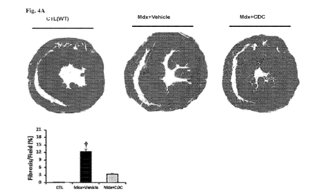
[0042] Figures 4A-B. Reduced cardiac collagen content and fibrosis.
Fig. 4A:
Diminished cardiac fibrosis. Representative Masson trichrome images of a wild-
type heart, an mdx
heart that had been vehicle-injected and an mdx heart that had been CDC-
injected, and pooled data
for morphometric analysis. Fig. 4B: Western blots and pooled data for
myocardial cardiac collagen
IA1 and IIIA 1, 3 weeks after CDC injection in mdx hearts. Data are means
SEM; tp<0.05;
#p<0.05.
[0043] Figures 5A-B. Cardiomyogenesis. Enhanced cardiomyogenesis 3
weeks after
CDC injection in mdx mice is evident from representative immunohistochemical
images and
pooled data. Fig. 5A: Immunohistochemical images (wild type, vehicle-treated
and CDC-treated
mdx mouse hearts stained for Ki67 and Aurora B; n=4-6 per group). Arrows point
to Ki67+ (upper
row) and Aurora B (lower row) cardiomyocytes. Fig. 5B: Pooled data for
morphometric analysis
of Aurora a' and ki67+ staining. Data are means SEM; 1- p<0.05; scale bars:
10 pm.
[0044] Figure 6. Depiction of the various mechanisms unpinning muscular
dystrophy
pathogenesis involving myocyte loss, fibrosis, oxidative stress, inflammation,
mitochondrial
inefficiency/loss, apoptosis, fibrosis, etc.
[0045] Figures 7A-B. Restoration of dystrophin expression. Fig. 7A:
Immunohistochemical images, western blots, and pooled data for protein
abundance of dystrophin
17
CA 03059910 2019-10-11
WO 2018/195210 PCT/US2018/028184
isoforms: dp427, dp260, dp140, dpi i6, dp71, dp40 in mdx mouse hearts 3 weeks
after
administration of vehicle or CDCs. CDC injection in mdx hearts resulted in
restoration of
dystrophin expression across all measured isoforms. Fig. 7B: Additional
representative depiction.
[0046] Figure 8. Schematic of pathophysiological mechanisms operative
in muscular
dystrophy and the cellular mechanisms recruited by CDCs and CDC-X0s involving
myocyte loss,
fibrosis, oxidative stress, inflammation, mitochondrial inefficiency/loss,
apoptosis, fibrosis, etc.
[0047] Figures 9A-D. CDC-X0s recap effects of CDCs. Intramyocardial
injection of
CDC-X0s reduces collagen to nearly the same levels as wild-type. Fig. 9A:
Western blots and
pooled data for cardiac collagen IA and IIIA. WGA (wheat germ agglutinin) was
applied for
staining and delineation of cell membrane. Fig. 9B: Immunohistochemical images
and pooled data
(wild type, n =4; vehicle-treated and CDC-XO-treated, n = 6 each) from mdx
mouse hearts stained
for Ki67 and Aurora B. Arrows point to Ki67+ (upper row) and Aurora B (lower
row)
cardiomyocytes. Fig. 9C: Western blots and pooled data for protein abundance
of dystrophin
isoforms: dp427, dp260, dp140, dpi i6, dp71, dp40 in mdx mouse hearts 3 weeks
after
administration of vehicle, CDCs or CDC-X0s (n=4-6). Fig. 9D: Injection of CDC-
X0s into mdx
hearts retarded progressive decrease in ejection fraction (n=11). Data are
means SEM; *p<0.05;
p<0.02; *p<0.01. Scale bar: 10 jim.
[0048] Figures 10A-B. Disproportional increase in cardiac function and
exercise
capacity in CDC-treated mdx mice. This could be due to CDCs themselves,
secreted mediators
(exosomes, EVs, proteins, etc.) from engrafted CDCs, modulated cardiac
secretome, and/or
improved systemic hemodynamics. Fig. 10A: Disproportional increase in cardiac
function and Fig.
10B: exercise capacity in CDC-treated mdx mice.
[0049] Figures 11A-N. Intraventricular injection of CDC-X0s.
Administration of
CDC-X0s demonstrated similar beneficial results. Fig. 11A: Systemic
biodistribution of CDC-
X0s after intraventricular injection in mdx mice. CDC-X0s were stained with
fluorescent lipid
dye and tracked 6 hours later using bioluminescence imaging. Fig. 11B: CDC-X0s
modulated
gene expression in a manner minoring CDCs. Fig. 11C: Dimensional hierarchical
clustering using
genes from hearts of non-treated mdx mice and of mdx mice treated
intramyocardially with CDCs
or intraventricularly with CDC-X0s. Genes with at least 2-fold differences
with corresponding
transcripts in non-treated mdx mice were included. Fig. 11D: Ejection fraction
improved with
intraventricular injection of CDC-X0s. Fig. 11E: Exercise capacity improved
with intraventricular
18
CA 03059910 2019-10-11
WO 2018/195210 PCT/US2018/028184
injection of CDC-X0s. Fig. 11F: Correlation of fold changes in expression of
the same genes in
the diaphragm 3 weeks after intramyocardial CDC injection or intraventricular
CDC-XO injection.
Fig. 11G: 2-Dimensional hierarchical clustering using genes from the diaphragm
of non-treated
mdx mice and of mdx mice treated intramyocardially with CDCs or
intraventricularly with CDC-
X0s. Genes with at least 2-fold differences with corresponding genes in
nontreated mdx mice were
included. Fig. 11H: Diaphragm contractile properties 3 weeks after
intraventricular CDC-XO
injection. Both twitch and specific force improved with intraventricular CDC-
XO injection. Fig.
111: These results were further observed in the soleus, as shown for gene
expression results. Fig.
11J: Dimensional hierarchical clustering. Fig. 11K: Contractile properties
from the soleus 3 weeks
after intraventricular CDC-XO injection. Both twitch and specific force
improved with
intraventricular CDC-XO injection. Dystropin levels shown for Fig. 11L: heart,
Fig. 11M:
diaphragm, Fig. 11N: soleus. Data are means SEM; *p<0.05; 1-P<0.05.
[0050] Figure 12. Biodistribution after intraventricular CDC-XO
injection. Figure 12
shows distribution of CDC-X0s stained with fluorescent lipid dye in mdx mice.
[0051] Figures 13A-J. Intramuscular injection of CDC-X0s resulted in
muscle growth
and reversal of pathophysiological abnormalities of muscular dystrophy. Fig.
13A: H&E and
immunohistochemical images of the soleus stained for MyoD (wild type, vehicle-
treated and CDC-
XO-treated mdx mouse soleus). Arrows in H&E images point to the linearly
arranged nuclei (left
column) and myofibers (right column). In the immunohistochemistry, linearly
arranged nuclei
were positive for MyoD (middle column). Figs. 13B & 13C: Frequency
distribution of myofiber
sizes and number of myoblasts (MyoD) 3 weeks after vehicle and CDC-XO
injection in mdx
soleus (n=59). Figs. 13D-13F: Western blots and pooled data for protein
abundance of Fig. 13D:
MyoD and myogenin, Fig. 13E: IGF1 receptor, and Fig. 13F: cytoplasmic p-p65 in
mdx soleus 3
weeks after intrasoleus vehicle and CDC-XO injection (n=4-6). Fig. 13G: CDC-XO
microRNA
reads as a measure of myogenesis. Fig. 13H: Representative Masson trichrome
images and
morphometric analysis in mdx soleus 3 weeks after administration of vehicle
and CDC-X0s into
mdx soleus (n=5-9). Fig. 131: Immunohistochemical images of dystrophin in mdx
mouse soleus 3
weeks after intrasoleus injection of vehicle and CDC-X0s (n=4-6). Age-matched
wild-type mice
served as control. Western blots and pooled data for protein abundance of
dystrophin isoform
dp427 in mdx mouse soleus 3 weeks after administration of vehicle and CDC-X0s
(n=4-6). Fig.
13J: Ex vivo measurement of soleus contractile properties: twitch force and
absolute force 3 weeks
19
CA 03059910 2019-10-11
WO 2018/195210 PCT/US2018/028184
after vehicle and CDC-XO injection into mdx soleus. Pooled data are means
SEM; *p<0.05;
1p<0.05; *p<0.002; scale bars: 5 jim (Fig. 13A, right column), 10 jim (Fig.
13A, middle column),
50 jim (Fig. 13A, left column), 200 jim (Fig. 13H), 20 pm (Fig. 131).
[0052] Figures 14A-C. Fig. 14A: CDC-XO injection was capable of
modulating
transcriptome of diaphragm. Fig. 14B: Western blots and pooled data for
protein abundance of
dystrophin isoforms in human Duchenne cardiomyocytes (DMD CM) one week after
priming with
CDC-X0s. Calcium transients from normal and DMD CM measured during 1Hz burst
pacing.
Duchenne cardiomyocytes were primed with vehicle or CDC-X0s 1 week before
assessment. Bar
graphs are of calcium transient alternans (variation in beat-to-beat calcium
transient amplitude)
and time to peak. Western blots and pooled data for protein abundance of
dystrophin isoforms:
dp427, dp260, dp140, dp116, dp71, dp40 in mdx mouse hearts after 3 weeks. Fig.
14C: Oxygen
consumption rate (OCR) in DMD CM primed with CDC-X0s or EVs derived from
normal human
dermal fibroblasts (NHDF-X0s) 1 week before OCR measurement. Normal and non-
treated DMD
CM were studied in parallel.
[0053] Figure 15. Left ventricular end-diastolic (LV EDV) and end-
systolic (LV ESV)
volumes after CDC administration. CDC transplantations resulted in a sustained
improvement of
LV EDV and LV ESV for 3 months after both first and second (3 months interval)
injections in
mdx mice, relative to placebo. Data are means SEM; n=12 in each group;
#p<0.05.
[0054] Figure 16. Percentage engraftment of CDCs in the heart 1, 2 and
3 weeks after
transplantation. Percentage engraftment of CDCs at 1 week was ¨8% and <1% at 2
weeks. By 3
weeks, no surviving CDCs could be detected. n=3 at each time point.
[0055] Figures 17A-D. Changes in mdx heart transcriptome 3 weeks after
CDC
treatment. Fig. 17A: 2-Dimensional hierarchical clustering using 560 genes
with at least 2-fold
differences between vehicle-treated and CDC-treated mdx hearts. Each column
represents an mdx
heart and each row a gene. Probe set signal values were normalized to the mean
across mdx hearts.
The relative level of gene expression is depicted from the lowest (green) to
the highest (red),
according to the scale shown on the top. Examples of fold changes of
transcripts for genes involved
in the various pathways of interest are plotted here, including Fig. 17B:
mitochondrial integrity,
Fig. 17C: oxidative stress, and Fig. 17D: inflammation.
CA 03059910 2019-10-11
WO 2018/195210 PCT/US2018/028184
[0056] Figure 18. Western blots and pooled data for protein abundance.
Measurements
including catalase, superoxide dismutase-2 (SOD-2), and catalytic subunit of
glutamate-cysteine
ligase (GCLC) in mdx mouse hearts 3 weeks after administration of vehicle or
CDCs.
[0057] Figure 19. IPA analysis of differentially expressed genes.
Depicted are genes
involved in inflammation in CDC-treated and vehicle-treated mdx hearts,
denoting inhibition of
inflammatory response concomitantly with reduced migration of inflammatory
cells in mdx hearts
3 weeks after CDC treatment. The blue color represents inhibition of
function/response and the
red and green colors represent up and downregulation, respectively.
[0058] Figures 20A-C. Immunohistochemical images. Depicted are mdx
hearts stained
for inflammatory cell marker CD3 with blowups of the boxed areas, including
Fig. 20A: vehicle-
treated mdx heart, Fig. 20B: CDC-treated mdx heart, and Fig. 20C: wild type
heart as control.
[0059] Figures 21A-B. Mitochondria. Fig. 21A: Numbers of mitochondria
from TEM
images. Fig. 21B: Mitochondrial DNA copy numbers per nuclear genome in the
heart tissue 3
weeks after treatment.
[0060] Figure 22. XO analysis. Isolated X0s obtained by
ultracentrifugation were
analyzed by nanoparticle tracking, using the NanoSight NS300 system (NanoSight
Ltd, UK).
Videos were collected and analyzed using NTA-software (version 2.3), with the
minimal expected
particle size, minimum track length, and blur setting all set to automatic.
Camera shutter speed
was fixed at 30.01 ms and camera gain was set to 500. Camera sensitivity and
detection threshold
were set close to maximum (15 or 16) and minimum (3 or 4), respectively, to
reveal small particles.
Ambient temperature was recorded manually, ranging from 24 C to 27 C. For each
sample, five
videos of 60 seconds duration were recorded, with a 10-second delay between
recordings,
generating five replicate histograms that were averaged. Representative five
replicate histograms
depicting size/concentration. Standard error of the mean concentration,
calculated from 5
replicates, is shown in red.
[0061] Figure 23. LV end-diastolic (LV EDV) and end-systolic (LV ESV)
volumes
after CDC-XO administration. CDC-XO transplantation resulted in a sustained
improvement of
LV EDV and LV ESV for 3 months after both first and second (3 months interval)
injections in
mdx mice, relative to placebo. Data are means SEM; n=11 in each group;
#p<0.05.
21
CA 03059910 2019-10-11
WO 2018/195210 PCT/US2018/028184
[0062] Figure 24. Immunoglobin serum level. IgG serum levels 6 months
after the first
injection and 3 months after repeat injection of mouse CDCs, human CDC-X0s,
and vehicle in
mdx mice. Circulating anti-donor IgG antibodies were screened by flow
cytometry.
[0063] Figure 25. Clathrin-dependent myocardial uptake of X0s.
Distribution of
intramyocardially injected CDC-X0s in the mdx mouse heart with and without
chlorpromazine
(CPZ) pretreatment. CPZ is an inhibitor of clathrin-dependent endocytosis.
Fluorescent-labeled
X0s (XenoLight DiR, 5pM, overnight incubation; Caliper Life Sciences,
Hopkinton, MA) were
injected intramyocardially into the apex of mdx mouse hearts; 6 hours later,
the hearts were
harvested, fixed and sectioned for evaluation of XO distribution. The average
number of labelled
X0s in the interior of cardiomyocytes (verified by co-staining for sarcomeric
a-actinin [green] and
DAPI [blue]) was calculated by counting intracardiomyocyte X0s in 10 fields
from each of 10
sections selected randomly from the apical (3 sections; 50pm interval), middle
(4 sections; 50pm
interval) and basal (3 sections; 50pm interval) regions of each heart. The
presence of fluorescently
labeled X0s in the interior of the cardiomyocytes is a measure of endocytic
uptake; pretreatment
with CPZ (50pg/g, i.p., single dose, 1 hour before XO injection), resulted in
marked reduction in
intracellular presence of X0s, indicating participation of clathrin-mediated
uptake in
internalization of CDC-X0s in mdx cardiomyocytes, among others. Bar graph
depicts the number
of labeled X0s (purple) in the interior of cardiomyocytes with and without CPZ
administration,
expressed as the number of intracardiomyocyte labelled X0s divided by the
total number of
cardiomyocytes per high-power field (HPF). Arrows point to fluorescent-labeled
exosomes.
Pooled data are means SEM; 1p<0.001.
[0064] Figure 26. Contractile properties. Depicted are extensor
digitorum longus
(EDL) contractile properties after intramyocardial CDC injection: In situ
measurement of EDL
contractile properties, absolute twitch and maximum tetanic force, 3 weeks
after CDC/vehicle
treatment of mdx hearts.
[0065] Figure 27. IPA analysis of differentially expressed genes.
Depicted are genes
involved in inflammation in the liver of mdx mice treated intramyocardially
with CDC or vehicle,
denoting inhibition of the NF--03 inflammatory pathway in mdx livers 3 weeks
after
intramyocardial CDC injection. The blue color represents inhibition of
function/response and the
red and green colors represent up and downregulation, respectively.
22
CA 03059910 2019-10-11
WO 2018/195210 PCT/US2018/028184
[0066] Figure 28. Fold change of mitochondrial-related microRNAs in X0s
from
hypoxically-cultured CDCs relative to X0s from CDCs grown under normoxia.
Depicted is 2-
dimensional hierarchical clustering using microRNAs with -6 to 6 times 1og2
fold change (230
microRNAs). The relative 1og2 fold change of microRNAs is represented from the
lowest (red
(bottom), -6) to the highest (green (top), +6), according to the scale shown
at the top. Each column
represents an XO preparation and each row a microRNA species. Among 389
detected microRNAs
in hypoxic X0s, 248 were previously reported to be mitochondria-related
microRNAs.
[0067] Figure 29. Fold changes of microRNAs under different culturing
conditions.
Depicted are properties of CDC-X0s isolated from hypoxic conditioned media (2%
02) compared
to CDC-X0s isolated from normoxic conditioned media; fold change >10 and <-20
were included.
NEBNext Small RNA Library Prep kit (New England BioLabs, Ipswich, MA) was used
for
miRNA-seq library preparation of extracted small RNAs from the X0s. RNAs were
extracted from
X0s using miRNeasy Serum/Plasma Kit (QIAGEN, Germantown, MD).
[0068] Figures 30A-B. Physiological properties following CDC-XO and miR-
148
administration. Fig. 30A: LV ejection fraction at baseline and 3 weeks after
intramyocardial
injection of CDC-X0s and NHDF-X0s in mdx mice. Fig. 30B: LV ejection fraction
at baseline
and 3 weeks after intramyocardial injection of miR-148 and microRNA mimic
control in mdx
mice. Data are means SEM; n=5 per group.
[0069] Figures 31A-B. Age-related changes in dystrophin expression in
mdx hearts.
Fig. 31A: Dystrophin expression in young (8 weeks) and old (10 months) mdx
hearts. Fig. 31B:
Western blot of dystrophin protein in wild-type control mouse heart and mdx
mouse hearts 3 weeks
and 3 months after first intramyocardial CDC injection and 3 months after
second (repeat) CDC
injection into myocardium. All hearts were from mice 10 months old at
baseline. CS: citrate
synthase.
[0070] Figures 32A-D. Non-cardiac manifestations CDC or CDC-XO
injections. Fig.
32A: Ingenuity pathway analysis of differentially expressed genes involved in
inflammation in the
liver of mdx mice injected intramyocardially with CDCs or vehicle, showing
inhibition of the NF-
-03 inflammatory pathway in mdx livers 3 weeks after intramyocardial CDC
injection. The blue
color represents inhibition of function/response and the red and green colors
represent up and
downregulation, respectively. Fig. 32B: Bioluminescence imaging of mdx mouse
organs after
systemic injection of dyed human CDC-X0s. 6 hours after injection of X0s
systemically into the
23
CA 03059910 2019-10-11
WO 2018/195210 PCT/US2018/028184
mdx mouse left ventricular cavity, the indicated organs were dissected and
imaged using IVIS
molecular imaging systems (Caliper Life Sciences, Hopkinton, MA, USA). Fig.
32C: Western blot
of dystrophin protein in wild type mouse heart and mdx mouse hearts 1 week, 3
weeks and 3
months after first intraventricular CDC-XO injection and 3 months after second
(repeat) CDC-XO
injection. Fig. 32D: Western blot showing protein content of dystrophin in
wild type control and
in mdx mouse heart, hypothalamus, diaphragm, soleus, tibialis anterior, and
extensor digitorum
longus, 3 weeks after systemic CDC-XO delivery by intraventricular injection.
CS: citrate synthase
loading control. Although no dystrophin expression is evident in the EDL,
contractile force was
increased in EDL after intramyocardial CDC injection, suggesting that
dystrophin re-expression
may not be the sole mechanism of benefit in skeletal muscle.
[0071] Figure 33. Dystrophin expression and its consequences. Absence
of dystrophin
in X0s. Wild type heart lysate was used as a positive control for probing
dystrophin.
[0072] Figures 34A-D. Verification that the bioactivity of the X0s
studied here are
attributable to exosomes characterized. Exosomes were floated on a linear
iodixanol density
gradient, which demonstrated vesicles by transmission electron microscopy
(TEM) and the
presence of membrane proteins, and showed that the biological effect is
vesicle associated. Fig.
34A: TEM images of sequentially-centrifuged exosomes with (Exol, left) and
without (Exo2,
right) purification with linear iodixanol density gradient show vesicles in
both conditions. The
vesicles are variable in size and morphology, consistent with previous work.
Fig. 34B: Western
blot on lysed exosomes for key proteins characteristic of exosomes: CD63, CD81
and TSG. Fig.
34C, Fig. 34D: Biological activity of Exol and Exo2 were compared by injection
into mdx soleus
and evaluation of mdx soleus transcriptome 3 weeks after injection. Fig. 34C:
Changes in mdx
soleus transcriptome 3 weeks after Exol and Exo2 injection. 2-Dimensional
hierarchical clustering
using 332 genes with at least 2-fold differences between vehicle/Exol and
vehicle/Exo2 in mdx
soleus. Fig. 34D: Correlation of fold changes in expression of the same genes
3 weeks after Exol
and Exo2 injection in mthc soleus. The similarity of the effects of Exol and
Exo2 support the notion
that the bioactivity of the vesicles isolated by the default protocol is
genuinely due to exosomes,
and not to other types of vesicles that might have been co-purified by
ultracentrifugation. Scale
bars: 50 pm (Exol); 100 nm (Exo2).
[0073] Figures 35A-C. miR-148a-3p and srDMD transplantation into mdx
heart. Fig.
35A: Differential expression of miR-148a-3p and srDMD in CDC-X0s isolated from
hypoxic
24
CA 03059910 2019-10-11
WO 2018/195210 PCT/US2018/028184
conditioned media (2% 02) compared to CDC-X0s isolated from normoxic
conditioned media
(n=2), along with depiction of apparent secondary structure of srDMD. Fig.
35B: Western blots
and pooled data for protein abundance of dystrophin isoforms: dp427, dp260,
dp140, dpi i6, dp71,
dp40 in mdx mouse hearts 3 weeks after intramyocardial injection of vehicle,
CDCs, CDC-X0s
(n=4-6), miR-148a-3p, or srDMD. Fig 35C: Western blots and pooled data for
protein abundance
of dystrophin isoforms: dp427, dp260, dp140, dpi i6, dp71, dp40 in mdx mouse
hearts 3 weeks
after intramyocardial injection of vehicle, CDCs, CDC-X0s (n=4-6), miR-148a-
3p, and srDMD.
[0074] Figures 36A-B. Exon skipping/alternative splicing excluded. Fig.
36A: miR-
148a results in decreases in both NFKB p65 and phospho-Akt levels. Fig. 36B:
RT-PCR using
primers that flank the exon 23 of dystrophin. It was used to assess exon 23
inclusion in expressed
dystrophin in mdx hearts from vehicle, miR-148a-3p, and srDMD-treated mice
(n=4-6). Sashimi
plots of RNA-seq data for dystrophin from vehicle, miR-148a-3p, or srDMD-
treated mdx hearts
depict no junction read that span exon 23. All data are means SEM; ip<0.03.
[0075] Figures 37A-B. Western blot detection of dystrophin. Fig. 37A:
Western blot
depicting protein content of dystrophin in wild type mouse hearts and srDMD-
treated mdx mouse
hearts 3 weeks after intramyocardial injection of srDMD. Fig. 37B: Percentage
increase relative
to vehicle (PBS) in dystrophin/eGFP expression after treatment with CDC-X0s,
miR-148a-3p, or
srDMD in transfected HEK293 NT cells with dual reporter constructs harboring a
point mutation
in exon 23 of dystrophin gene or deletion of exon 50 of dystrophin gene.
[0076] Figures 38A-B. Dystrophin expression and its consequences. Fig.
38A: Ejection
fraction at baseline and 3 weeks after intramyocardial injection of miR-148a-
3p or microRNA
mimic control in mdx mice. Wild type EF values also shown for reference; n=5
per group. Fig.
38B: Western blot depicting protein content of dystrophin in wild type mouse
hearts and in vehicle-
mutant srDMD, or srDMD-injected mdx mouse hearts 3 weeks after intramyocardial
injection.
[0077] Figures 39A-B. Fig. 39A: Plasmid map of synthetic DNA constructs
cloned into
mammalian expression vectors. Full length human dystrophin was cloned into the
ORF, either as
wild-type or as one of two mutants: UAA premature termination codon in exon 23
(PTC), or exon
50 deletion (Exon 50 A). The construct creates a fusion protein of full-length
dystrophin in frame
with eGFP, such that green fluorescence can be taken as a reporter of
dystrophin expression.
Constitutive luciferase expression (driven independently by an 5V40 promoter)
was used to
normalize for transfection efficiency. Fig. 39B: Dystrophin/eGFP expression in
HEK-293NT cells
CA 03059910 2019-10-11
WO 2018/195210 PCT/US2018/028184
transfected with full-length (WT), PTC or Exon 50 A constructs. Fluorescence
and luminescence
of total cell lysates were quantified on a well-by-well basis in a 96-well
spectrophotometer;
fluorescence in each well was also quantified with nontransfected cells at an
equivalent seeding
density and lysis volume.
[0078] Figures 40A-D. Ten-to-twelve month old mdx mice were treated
with the
following: a single dose of vehicle (mdx), 2.5 x 10⁵ syngeneic CDCs, or 2.0 x
109 human CDC-
exosomes (CDC-X0s) via intravenous injection into the femoral vein. Fig. 40A
shows maximal
exercise capacity (n=8-10 per group) and Fig. 40B in vivo cardiac ejection
fraction (n=6-8 per
group) before (baseline) and 3 weeks following treatment. Fig. 40C shows
Masson's trichrome
micrographs of hearts from vehicle- (top panel), CDC- (middle panel), or X0-
(bottom panel)
treated mdx mice. Fig. 40D shows pooled data analyzing area of blue staining
(collagen) relative
to red staining (cytoplasm) as a marker of cardiac fibrosis (n=5-6 per group).
Data are represented
as mean SEM. * indicates statistically different from vehicle treatment.
Statistical significance
was set to P<0.05.
[0079] Figures 41A-D. Animals were treated as described in Figure 40,
Fig. 41A shows
whole transcriptome analysis of hearts from RNA-sequencing data. Transcripts
with a 2-fold or
higher change with P<0.05 were considered differentially expressed and
represented in the
heatmap in panel A. The mdx column was compared to an age-matched wild-type
mouse heart,
while the CDCs and X0s columns were each compared to mdx mouse hearts. Fig.
41B shows
representative Western blot and pooled data probing for phosphorylated NFKB
protein levels (n=5-
6 per group) in the hearts from wild-type (WT), vehicle (mdx), CDC, or XO
treated mice. Fig. 41C
shows pooled data from CD68 immunofluorescent images (n=3 per group) Fig. 41D
from mdx,
CDC, or XO treated hearts. Data are represented as mean SEM. * indicates
statistically different
from vehicle treatment. Statistical significance was set to P<0.05.
[0080] Figures 42A-D. Animals were treated as described in Fig. 40.
Fig. 42A. Pooled
data from Western blot analysis of mitochondrial electron transport chain
complexes (n=6 per
group) from WT, mdx, CDC, or XO treated hearts. Fig. 42B. Protein-carbonyl
adduct formation
(n=8-10 per group) in WT, mdx, CDC, or XO treated hearts. Fig. 42C. Pooled
data from Ki-67
immunofluorescent images (n=3 per group). Fig. 42D from mdx, CDC, or XO
treated hearts. Data
are represented as mean SEM. * indicates statistically different from
vehicle treatment. Statistical
significance was set to P<0.05.
26
CA 03059910 2019-10-11
WO 2018/195210 PCT/US2018/028184
[0081] Figures 43A-F. Animals were treated as described in Fig. 40.
Fig. 43A. Force-
frequency relationship of solei (n=5-8 per group) from WT (circle), mdx
(square), CDCs (upward
triangle), or X0s (downward triangle). Fig. 43B Twitch and Fig. 43C tetanic
force developed by
solei from WT, mdx, CDCs, or XO treated mice. Fig. 43D Force-frequency
relationship of
diaphragms (n=5-6 per group) from WT (circle), mdx (square), CDCs (upward
triangle), or CDC-
X0s (downward triangle). Fig. 43E Twitch and Fig. 43F tetanic force developed
by diaphragms
from WT, mdx, CDCs, or CDC-XO treated mice. Fig. 43G Masson's trichrome
micrographs from
mdx (left panel), CDC- (middle panel), or CDC-X0- (right panel) treated mice.
Fig. 43H Pooled
data analyzing area of blue staining (collagen) relative to red staining
(cytoplasm) as a marker of
skeletal muscle fibrosis in the soleus (n=5-6 per group). Fig. 431
Quantification of the number of
myofibers per whole muscle section of the soleus (n=5-6 per group). Data are
represented as mean
SEM. * indicates statistically different from vehicle treatment. Statistical
significance was set to
P<0.05.
[0082] Figures 44A-D. Animals were treated as described in Fig. 40.
Fig. 44A. Whole
transcriptome analysis of hearts from RNA-sequencing data. Transcripts with a
2-fold or higher
change with P<0.05 were considered differentially expressed and represented in
the heatmap in
panel A. The mdx column was compared to an age-matched wild-type mouse soleus,
while the
CDCs and CDC-X0s columns were each compared to mdx mouse solei. Fig. 44B.
Kyoto
Encyclopedia of Genes and Genomes analysis of a CDC-XO treated soleus.
Pathways listed are
considered upregulated relative to mdx soleus ¨ many are involved in
inflammation. Fold change
in genes known to be involved in Fig. 44C TNF and Fig. 44D NFKB signaling
altered in the soleus
by CDC and CDC-XO treatment.
[0083] Figures 45A-C. Animals were treated as described in Fig. 40.
Fig. 45A
Representative Western blot and pooled data probing for phosphorylated NFKB
protein levels (n=3
per group) in the soleus from wild-type (WT), vehicle (mdx), CDC, or CDC-XO
treated mice. Fig.
45B Pooled data from CD68 immunofluorescent images (n=3 per group). Fig. 45C
from mdx,
CDC, or CDC-XO treated hearts. Data are represented as mean SEM. * indicates
statistically
different from vehicle treatment. Statistical significance was set to P<0.05.
[0084] Figures 46A-B. Animals were treated as described in Fig. 40.
Western blot
probing for full-length (427 kDa) dystrophin in the soleus Fig. 46A and
diaphragm Fig. 46B from
27
CA 03059910 2019-10-11
WO 2018/195210 PCT/US2018/028184
vehicle (mdx), CDC, GW4869 treated CDCs, and CDC-XO treated mice. The left 2
columns
represent a relative level of WT dystrophin (e.g., 5% and 1%).
[0085] Fig. 47 depicts a schematic protocol that was used for
evaluating the efficacy
of escalating doses of intravenous administration of CDCs to improve exercise
capacity using a
mouse model of DMD (mdx mice), wherein mdx mice received treatment or control
vehicle in
Week 0, exercised every week, and were sacrificed in Week 6.
[0086] Figs. 48A-48B graphically show effects of intravenous (IV)
administration of
CDCs on the exercise capacity of mdx mice injected with 75,000, 150,000, or
250,000 (referred to
as "75K," "150K," and "250K," in the figure) CDCs versus phosphate buffered
saline (PBS)
control.
[0087] Fig. 49 graphically shows the effects of jugular IV
administration of CDCs on
the diaphragm muscle function of mice injected with 75K-250K CDCs versus PBS
control.
[0088] Figs. 50A and 50B graphically show the effects of jugular IV
administration of
CDCs on the body weight of mdx mice injected with 37K-150K CDCs versus PBS
control.
[0089] Fig. 51 shows that CDCs administered in accordance with several
embodiments
disclosed herein reduce fibrosis in the hearts of mdx mice (shown is collagen
staining, an indicator
of fibrosis).
[0090] Fig. 52A depicts a schematic protocol that was used for
evaluating effects of
CDC treatment on cardiac ejection fraction using echocardiography.
[0091] Fig. 52B graphically shows the effects of jugular IV
administration of CDCs on
the cardiac ejection fraction of mdx mice injected with 150K CDCs versus PBS
control.
[0092] Fig. 53 depicts histology data of Masson's trichrome staining of
heart tissue
sections of mdx mice injected with 150K CDCs via jugular IV injection, or 250K
CDCs via femoral
IV injection, versus PBS control.
[0093] Fig. 54 graphically shows the effects of IV administration on
the cardiac (left
ventricular) ejection fraction in a mouse model of myocardial infarction with
300K CDCs via
systemic injection (femoral IV injection or 100K CDCs via injection into the
right ventricle) versus
100K CDCs via intramyocardial injection versus PBS control.
[0094] Fig. 55A graphically shows that ciPCR performed on purified
human CDC
DNA was validated.
28
CA 03059910 2019-10-11
WO 2018/195210 PCT/US2018/028184
[0095] Fig. 55B graphically shows standard curves of human CDC DNA
spiked into
mouse tissue DNA prepared for each tissue to be tested.
[0096] Fig. 56A depicts a schematic protocol for determining the
biodistribution of
human CDCs in wild type (WT) mice after jugular vein administration by a human
Alu sequence
qPCR method.
[0097] Fig. 56B graphically shows human CDC biodistribution in WT mice
10 minutes
and 24 hours after jugular vein injection, in the lung, liver, blood, heart,
soleus, diaphragm, and
spleen tissues.
[0098] Fig. 57A depicts a schematic protocol for determining the
biodistribution and
clearance of human CDCs in severe combined immunodeficiency (SCID) mice after
jugular vein
administration by human Alu sequence qPCR method.
[0099] Fig. 57B graphically shows human CDC biodistribution in SCID
mice 24 hours,
1 week, and 3 weeks after jugular vein injection, in the lung tissue.
[0100] Fig. 57C graphically shows human CDC biodistribution in SCID
mice 24 hours,
1 week, and 3 weeks after jugular vein injection, in the liver, blood, heart,
soleus, diaphragm,
spleen, and testes tissue.
[0101] Fig. 58 schematically illustrates lung tissue sample collection for
histopathological analysis to evaluate safety of high dose of CDCs in a
porcine model of acute
myocardial infarction.
[0102] Fig. 59 graphically shows changes in serum troponin I (TnI)
levels in pigs
treated with 50, 100 or 200 million ("50M," "100M," or "200M," respectively)
CDCs.
[0103] Fig. 60 graphically shows average area at risk (AAR) in pigs
treated with 50,
100 or 200 million CDCs.
[0104] Fig. 61 graphically shows average ratio between no reflow
(unstained) and area
at risk, indicating myovascular obstruction (MVO) in pigs treated with 50, 100
or 200 million
CDCs.
[0105] Fig. 62 graphically shows average ratio of triphenyl tetrazolium
chloride (TTC)
stain and area at risk, indicating scar size, in pigs treated with 50, 100 or
200 million CDCs.
[0106] Fig. 63 graphically shows AEF calculated for treated and
untreated animals, in
pigs treated with 50, 100 or 200 million CDCs.
29
CA 03059910 2019-10-11
WO 2018/195210 PCT/US2018/028184
[0107] Fig. 64 graphically shows the expression of immune molecules
involved in T
and natural killer (NK) immune response by steady state CDCs. The percentage
of positive cells
and geometric mean fluorescence intensity are indicated.
[0108] Fig. 65 shows representative images of tailored-MLR cultures.
[0109] Fig. 66 graphically shows the activation of T cells by CDC.
Expression of
CD69 (left panel) and HLA-DR (right panel) by CD4+ (black) and CD8+ T (white)
cells by three
different PBMC (Donor A, C, and D). Results are mean values SD of
triplicates. *p<0.05;
**p<0.01; ***p<0.001; ****p<0.0001.
[0110] Figs. 67A-67C graphically show the proliferation of T cells from
unfractionated
PBMC in response to CDCs; Fig. 67A shows the representative dot plots (from
Donor A); Fig.
67B shows the proliferation of T cells from three different PBMC donors; Fig.
67C shows the
proliferation of T cells from 3 different donors. For Fig. 67B, results are
mean values SD from
triplicates. For Fig. 67C, results are mean values SD obtained from three
different donors each
in triplicates.
[0111] Fig. 68 graphically shows the purified T cells activation and
proliferation in
response to CDCs. Results are mean values SD obtained from responses
(duplicates) for each
donor (upper and middle panels) and as mean values SEM obtained from both
donors each in
duplicate (lower panel).
[0112] Fig. 69 graphically shows the immune modulation of
phytohemagglutinin
(PHA)-induced T cells proliferation by CDCs. Representative cultures and
histograms (upper
panel) and results presented as mean percentage values SD from 3 different
donors each done in
triplicates.
[0113] Fig. 70 graphically shows the modulation of PHA-induced CD69 and
HLA-DR
expression and PHA-induced T cells proliferation by CDCs. Results for each
donor are presented
as mean values SD obtained from triplicates. Lower panel presents mean
values SD of the
percentages of decrease (CD69) or increase (HLA-DR) in expression of
activation markers (left
and middle panels) and percentage of proliferation inhibition obtained from
both donors each done
in triplicates.
[0114] Fig. 71 is a western blot showing CDC-EVs' contents of
recognized exosomes
markers.
CA 03059910 2019-10-11
WO 2018/195210 PCT/US2018/028184
[0115] Fig. 72 graphically shows the expression of immune molecules
involved in T
and natural killer (NK) immune response on CDC-EVs.
[0116] Figs. 73A-73B show the activation of T cells by CDCs and CDC-
EVs; Fig. 73A
shows the representative images of tailored-MLR cultures; Fig. 73B shows the
expression of CD69
and HLADR by CD4+ and CD8+ T cells.
[0117] Figs. 74A-74B graphically show T cells proliferation in response
to CDC and
EVs; Fig. 74A shows the representative dot plots; Fig. 74B shows the
proliferation of T cells from
3 different donors presented as mean values SD from triplicates.
[0118] Fig. 75 graphically shows the purified T cells activation and
proliferation in
response to CDCs and CDC-EVs. Results are presented as mean values SD
obtained from
responses (duplicates) from 2 different donors.
[0119] Fig. 76 shows the IDC cultures in the presence and absence of
EVs (upper
panel) and expression of relevant immune molecules (lower panel).
[0120] Fig. 77 graphically shows the purified T cells activation and
proliferation by
CDC-EVs presented by iDC. Results are presented as mean values from
triplicates.
[0121] Fig. 78 shows the mDC cultures in the presence and absence of
EVs (upper
panel) and expression of relevant immune molecules (lower panel).
[0122] Fig. 79 graphically shows the purified T cells activation and
proliferation by
mDC and mDC-EVs. Results are presented as mean values from triplicates.
[0123] Fig. 80 shows the immune modulation of PHA-induced T cells
proliferation by
CDCs and CDC-EVs. Results are mean percentage values SEM from 3 different
donors each
done in triplicates.
[0124] Fig. 81 graphically shows the down-regulation of PHA-induced
CD69 and/or
HLA-DR expression by CDCs and CDC-EVs. Results are mean percentage values SD
obtained
from triplicates.
[0125] Fig. 82 graphically shows that CDCs and CDC-EVs down regulate
PHA-
induced T cells proliferation. Results are mean values SD obtained from
triplicates.
[0126] Fig. 83 graphically shows that Immune modulation of PHA-induced
CD4+ and
CD8+ T cells proliferation by CDC and CDC-EVs. Results are mean percentage
values SEM
from 2 different donors each done in triplicates.
31
CA 03059910 2019-10-11
WO 2018/195210 PCT/US2018/028184
[0127] Fig. 84A is a graphical depiction of changes from baseline in
exercise capacity
with multiple administrations of syngeneic CDCs (from C57BL/10 mice) or a PBS
control. 84B is
a graphical depiction of changes from baseline in exercise capacity after
multiple administrations
of allogeneic CDCs (from C3H mice) or a PBS control. Fig. 84C is a combination
of Figs. 84A
and 84B into a single graph.
[0128] Fig. 85A is a graphical depiction of changes from baseline in
exercise capacity
with two administrations of allogeneic CDCs (from C3H mice), a steroid, and/or
PBS vehicle. Fig.
85B is a graph of the percent of cells positive for various genetic markers.
Fig. 85C is a graph
indicating the amount of IgG antibodies.
[0129] Fig. 86 shows data related to evaluation of antibody production
in response to
administration of CDCs (as compared to PBS).
DETAILED DESCRIPTION
[0130] Some embodiments disclosed herein pertain to methods of treating
disease,
disease states, and/or symptoms of disease using CDCs, CDC-X0s, CDC-EVs, the
isolated
molecular cargo of CDCs (e.g., individual molecules or combinations of
molecules derived from
CDCs, CDC-X0s, and/or CDC-EVs), and/or combinations of the forgoing. In some
embodiments,
the disease is a dystrophinopathy. In some embodiments, the disease state is a
dystrophic disorder.
In some embodiments, the dystrophinopathy includes one or more of Duchenne
muscular
dystrophy (DMD) and/or Becker muscular dystrophy. In some embodiments, the
disease state is a
myopathy. In some embodiments, the myopathy is a skeletal muscle myopathy. In
some
embodiments, the method includes administering a therapeutically effective
amount of CDCs,
CDC-X0s, CDC-EVs, the molecular cargo of CDC-X0s or CDC-EVs, and/or
combinations of
the forgoing to a subject (e.g., a patient) suffering from the disease,
thereby treating the disease
and/or its symptoms. Some embodiments of the methods and compositions provided
herein are
based on the surprising discovery that, inter alia, despite the finding that
intravenous
administration of CDCs to mdx mice resulted in accumulation of the at least a
portion of
administered CDCs in their lungs, functional improvements at dystrophic
skeletal muscles were
achieved as demonstrated by the various data presented herein, thereby
enabling an effective
treatment of a human subject suffering from skeletal muscular dystrophy, e.g.,
Duchenne muscular
32
CA 03059910 2019-10-11
WO 2018/195210 PCT/US2018/028184
dystrophy (DMD), by administering a therapeutically effective amount of CDCs
to a human
subject suffering from skeletal muscular dystrophy.
[0131] As used herein, "and/or" refers to and encompasses any and all
possible
combinations of one or more of the associated listed items, as well as the
lack of combinations
when interpreted in the alternative ("or").
[0132] "Treat" or "treating" or "treatment" refers to any type of
action that imparts a
modulating effect, which, for example, can be a beneficial effect, to a
subject afflicted with a
disorder, disease or illness, including preventing the manifestation of
disease states associated with
the condition, improvement in the condition of the subject (e.g., in one or
more symptoms or in
the disease), delay or reduction in the progression of the condition, and/or
change in clinical
parameters, disease or illness, curing the illness, etc.
[0133] The term "therapeutically effective amount," as used herein,
refers to an amount
of the therapeutic (e.g., CDC-X0s, CDC-EVs, CDCs, molecular cargo of X0s and
EVs, or
combinations thereof) that imparts a modulating effect, which, for example,
can be a beneficial
effect, to a subject afflicted with a disorder, disease or illness, including
improvement in the
condition of the subject (e.g., modulating one or more symptoms), delay or
reduction in the
progression of the condition, prevention or delay of the onset of the
disorder, and/or change in
clinical parameters, disease or illness, etc. For example, in some
embodiments, an effective amount
can refer to the amount of a composition, compound, or agent that improves a
condition in a subject
by at least 5%, e.g., at least 10%, at least 15%, at least 20%, at least 25%,
at least 30%, at least
35%, at least 40%, at least 45%, at least 50%, at least 55%, at least 60%, at
least 65%, at least 70%,
at least 75%, at least 80%, at least 85%, at least 90%, at least 95%, or at
least 100%. Actual dosage
levels of active ingredients and agents in an active composition of the
disclosed subject matter can
be varied so as to administer an amount of the active agent(s) that is
effective to achieve the desired
response for a particular subject and/or application. The selected dosage
level will depend upon a
variety of factors including, but not limited to, the activity of the
composition, formulation, route
of administration, combination with other drugs or treatments, severity of the
condition being
treated, and the physical condition and prior medical history of the subject
being treated.
Determination and adjustment of an effective dose, as well as evaluation of
when and how to make
such adjustments, are contemplated herein. The term "a therapeutically
effective amount" can
mean an amount of CDC-X0s, CDC-EVs, CDCs, and/or molecular cargo X0s and EVs
sufficient
33
CA 03059910 2019-10-11
WO 2018/195210 PCT/US2018/028184
to reverse dystrophinopathy through dystrophin re-expression and/or to durably
(e.g., substantially
irreversibly) restore skeletal muscle function at a targeted dystrophic
skeletal muscle.
[0134] The term "a targeted dystrophic skeletal muscle" as used herein
is the delivery
of an amount of CDC-X0s, CDC-EVs, CDCs, a molecular cargo CDC-X0s and CDC-EVs,
and/or
combinations thereof at the site of a dystrophic skeletal muscle. In some
embodiments, targeted
delivery does not include incidental, accidental, or inadvertent delivery CDC-
X0s, CDC-EVs,
CDCs, and/or molecular cargo CDC-X0s and CDC-EVs to a target site. In some
embodiments,
targeted delivery does not include systemic delivery. In some embodiments,
targeted delivery does
not include incidental, accidental, or inadvertent delivery CDC-X0s, CDC-EVs,
CDCs, and/or
molecular cargo of CDC-X0s and CDC-EVs in an amount that would be insufficient
to treat
dystrophinopathy at the site of a dystrophic skeletal muscle.
[0135] The term "dystrophic" as used herein is a lack of or deficiency
of dystrophin
(e.g., in skeletal and/or heart muscles).
[0136] Cells release into the extracellular environment diverse types
of extracellular
vesicles (EVs) of endosomal and plasma membrane origin called exosomes (X0s)
and
microvesicles (MVs). EVs represent an important mode of intercellular
communication and serve
as vehicles for the transfer of molecular cargo (e.g., one or more of
cytosolic proteins, lipids, and
RNA) between cells and through cell membranes. X0s, secreted lipid vesicles
containing a rich
milieu of biological factors, provide powerful paracrine signals by which stem
cells potentiate their
biological effects to neighboring cells, including diseased or injured cells.
Through the
encapsulation and transfer of proteins, bio-active lipid and nucleic acid
cargo, these natural
delivery devices can induce significant phenotypic and functional changes in
recipient cells that
lead to activation of regenerative programs. Administration of X0s has been
demonstrated to treat
heart failure in mdx mice in W02016/05491, which is herein incorporated by
reference in its
entirety.
[0137] Some embodiments disclosed herein pertain to the use of CDCs and
CDC-X0s
in methods of therapeutic use. In some embodiments, described herein are
methods of treating a
dystrophic disorder, including a step of administering a therapeutically
effective amount of CDCs
and/or CDC-X0s to a subject afflicted or having dystrophinopathy, thereby
treating the subject.
In some embodiments, the subject is a pediatric subject afflicted with a
dystrophinopathy. In some
embodiments, the X0s are isolated from CDCs. In some embodiments, the CDCs are
grown in
34
CA 03059910 2019-10-11
WO 2018/195210 PCT/US2018/028184
serum-free media. In some embodiments, the dystrophinopathy is Duchenne
muscular dystrophy.
In other embodiments, the dystrophinopathy is Becker muscular dystrophy. In
some embodiments,
CDC-X0s, CDC-EVs, molecular cargo of CDC-X0s or CDC-EVs, CDCs producing X0s
and
EVs, and/or combinations of the forgoing are used in a method for achieving
dystrophin re-
expression. In some embodiments, the CDC-X0s, CDC-EVs, molecular cargo of CDC-
X0s or
CDC-EVs, CDCs producing X0s and EVs, and/or combinations upon systemic
intraventricular
injection of CDC-EVs, as well as upon direct intramuscular injection of CDC-
EVs into skeletal
muscles of mdx mice, whereby the present inventors conceived of the novel
treatment method as
described for the first time herein, e.g., a method of treating skeletal DMD
by administering a
therapeutically effective amount of CDCs and/or CDC-EVS in a single or
multiple systemic
administrations. In some embodiments, the disease is muscular dystrophy.
[0138] The data and experiments disclosed herein demonstrate an
unexpected
advantage of CDCs, CDC-X0s, and/or CDC-EVs in inducing dystrophin expression.
As shown
elsewhere herein, injection of CDCs into the hearts of mdx mice boosts full-
length dystrophin
protein levels in both heart and skeletal muscle, dramatically and durably
improving cardiac
function, ambulatory capacity and survival. Similar results are demonstrated
with human
Duchenne cardiomyocytes. Positive factors appear to exist in cellular XO's
produced by CDCs,
which are the lipid bilayer nanovesicles secreted by cells when multivesicular
endosomes fuse
with the plasma membrane.
[0139] In some embodiments, X0s (and EVs) secreted by human CDCs are
demonstrated to reproduce the benefits of CDCs in mdx mice and in human
Duchenne
cardiomyocytes. In some embodiments, the delivery of noncoding RNA species
found in CDC-
X0s (e.g., miR-148a) mimics the ability of CDCs, CDC-X0s, and/or CDC-EVs to
increase
dystrophin protein levels, without affecting transcript length or exon/intron
junctions. In some
embodiments, CDC-XO mediated transfer of noncoding RNAs ameliorates DMD by
restoring
dystrophin in heart and skeletal muscle.
[0140] In some embodiments, the results described herein demonstrate
CDCs and their
X0s (and/or EVs) as a therapeutic option for dystrophinopathy. CDCs and their
secreted X0s
(and/or EVs) robustly increase dystrophin levels in heart and skeletal muscle.
In some
embodiments, the increasing dystrophin levels in the heart and skeletal muscle
leads to major
durable systemic benefits after injection of CDCs, CDC-X0s, and/or CDC-EVs
into the body (e.g.,
CA 03059910 2019-10-11
WO 2018/195210 PCT/US2018/028184
systemically or locally, including locally into the skeletal muscle). In some
embodiments, as
disclosed herein, CDCs, CDC-X0s, and/or CDC-EVs are not only regenerative, but
also anti-
inflammatory and anti-fibrotic. CDCs secrete diffusible factors that promote
angiogenesis, recruit
endogenous progenitor cells, and coax surviving heart cells to proliferate;
transplanted CDCs also
suppress maladaptive remodeling, and apoptosis. In some embodiments, CDCs
operate through
indirect pathways (via CDC-X0s and/or CDC-EVs); they work indirectly via the
secretion of
CDC-X0s and/or CDC-EVs laden with noncoding RNA including microRNAs
(constituents of
the molecular cargo). In some embodiments, while allogeneic CDCs are cleared
completely within
several weeks, but their functional and structural benefits persist at least 6
months. These diverse
mechanisms are mediated via the secretion of CDC-X0s and/or CDC-EVs laden with
noncoding
RNA including microRNAs.
[0141] Without being bound to a particular theory, the above mechanisms
afford
CDCs, CDC-EVs, or CDC-X0s the capacity to treat DMD, with application to
similar muscular
dystrophies such as Becker muscular dystrophy. In some embodiments, CDCs, CDC-
X0s, and/or
CDC-EVs replace dystrophin, and offset the pathophysiological consequences of
dystrophin
deletion, by recruiting regenerating cells, reversing fibrosis and targeting
inflammation. In some
embodiments, reversing the central deficits of DMD in pediatric patients, the
methods herein are
capable of forestalling or preventing progression of the disease, allowing
those patients to avoid
comorbidities which may otherwise significantly limit options for therapeutic
intervention.
[0142] While the disclosed methods herein include those involving the
delivery of
CDCs to a patient, in some embodiments, using CDC-EVs (e.g., CDC-X0s) secreted
by CDCs,
and not cells, may provide advantages when compared to transplant and delivery
of cells
themselves. In some embodiments, CDC-EVs and CDC-X0s, including those produced
by CDCs,
can provide a potent and rich source for developing "cell-free" therapies. CDC-
XO-based, "cell-
free" therapies, in contrast to cell therapy, provide one or more of the
following advantages in
regenerative medicine. In some embodiments, as non-viable entities, with
reduced or non-existent
immunogenic or tumorigenic potential, these features significantly obviate
certain safety issues.
In some embodiments, stem cell-derived exosomes (and/or CDC-X0s) can be less
immunogenic
than parental cells, as a result of a lower content of membrane-bound
proteins, including MHC
complex molecules. In some embodiments, CDC-XO encapsulation of bioactive
components in
lipid vesicles allows protection of contents from degradation in vivo, thereby
potentially negating
36
CA 03059910 2019-10-11
WO 2018/195210 PCT/US2018/028184
obstacles often associated with delivery of soluble molecules such as
cytokines, growth factors,
transcription factors and RNAs. In some embodiments, the ease of
administration (and/or storage)
for CDC-X0s and/or CDC-EVs can ultimately allow for repeated and sustained
delivery to
patients, thereby maximizing the potential for regeneration and repair of
diseased and/or
dysfunctional tissue.
[0143] In some embodiments, CDCs and/or CDC-X0s can be used to
stimulate
numerous cellular, tissue and physiological processes, including immune
modulating processes,
angiogenesis, and migration of endothelial cells. As disclosed herein, based
on the
pathophysiology of DMD patients, including an environment of increased
oxidative and/or
nitrosative stress, elevated inflammation, pro-apoptotic and remodeling
states, therapeutic
approaches involving CDCs and/or CDC-X0s secreted by cells, provide
significant benefits in
reversing the course of the disease (and one or more of the aforementioned
disease states and/or
manifestations). In some embodiments, CDCs, CDC-X0s, and/or CDC-EVs promote
anti-
oxidative, anti-inflammatory, anti-apoptotic, anti-remodeling effects. In some
embodiments,
CDCs, CDC-X0s, and/or CDC-EVs enhance regenerative capacity of diseased cells
and tissues.
In some embodiments, CDC, CDC-XO, and/or CDC-EV administration is beneficial
in retarding
and/or reversing DMD, and exosome populations derived from CDCs allow for
these benefits to
be delivered. Early therapeutic intervention in pediatric subjects provides
durable and systemic
benefits that will prevent or ward off comorbidities in late stage disease,
such as heart failure. In
some embodiments, durable benefits are those lasting equal to or at least
about: 3 months, 6
months, 12 months, or ranges including and/or spanning the aforementioned
values.
[0144] Some Embodiments of Exosomes. X0s are lipid bilayer vesicles
that are
enriched in a variety of biological factors, including cytokines, growth
factors, transcription
factors, lipids, and coding and non-coding nucleic acids. X0s are found in
blood, urine, amniotic
fluid, interstitial and extracellular spaces. These exocytosed vesicles of
endosomal origin can range
in size between 30-200 nm, including sizes of 40-100 nm, and possess a cup-
shaped morphology,
as revealed by electron microscopy. Their initial formation begins with inward
budding of the cell
membrane to form endosomes, which is followed by invagination of the limiting
membrane of late
endosomes to form multivesicular bodies (MVB). Fusion of the MVB with the
plasma membrane
results in the release of the internal vesicles to the extracellular space,
through the formation of
vesicles thereafter known as exosomes. In some embodiments, X0s as described
herein are those
37
CA 03059910 2019-10-11
WO 2018/195210 PCT/US2018/028184
extracellular vesicles that are exocytosed and/or are of endosomal origin. In
some embodiments,
XO as described herein can have diameters of less than or equal to about: 30
nm, 50 nm, 100 nm,
150 nm, 200 nm, or ranges including and/or spanning the aforementioned values.
[0145] As described herein, the "cargo" contents of X0s reflect their
parental cellular
origin, as containing distinct subsets of biological factors in connection
with their parent cellular
origin, including the cell regulatory state of the parental cells when formed.
The rich biological
milieu of different proteins, including cytokines and growth factors, lipids,
coding and noncoding
RNA molecules, within exosomes are all necessarily derived from their parental
cells. In addition
to containing a rich array of cytosolic derivatives, exosomes further express
the extracellular
domain of membrane-bound receptors at the surface of the membrane.
[0146] In some embodiments, the described encapsulation and formation
processes
create heterogeneity in XO compositions based on parental cellular origin and
regulatory state at
time of formation. Nevertheless, in some embodiments, generic budding
formation and release
mechanisms establish a common set of features as a consequence of their
origin, such as
endosome-associated proteins (e.g., Rab GTPase, SNAREs, Annexins, and
flotillin), proteins that
are known to cluster into microdomains at the plasma membrane or at endosomes
(four
transmembrane domain tetraspanins, e.g., CD63, CD81, CD82, CD53, and CD37),
lipid raft
associated proteins (e.g., glycosylphosphatidylinositol- anchored proteins and
flotillin),
cholesterol, sphingomyelin, and hexosylceramides.
[0147] In some embodiments, in addition to components reflecting their
vesicle origin,
X0s contain both mRNA and microRNA associated with signaling processes, with
both cargo
mRNA being capable of translation in recipient cells, or microRNA functionally
degrading target
mRNA in recipient cells. In some embodiments, other noncoding RNAs, capable
for influencing
gene expression, may also be present in X0s. While the processes governing the
selective
incorporation of mRNA or microRNA populations into X0s is not entirely
understood and without
being bound to any particular theory, it is believed that RNA molecules are
selectively, not
randomly incorporated into X0s, as demonstrated by the enrichment of X0s cargo
RNAs when
compared to the RNA profiles of other exosomes and their originating cells. In
some embodiments,
in view of RNA molecules potential a role in disease pathogenesis and
regenerative processes,
without being bound by a theory, the presence of RNA molecules in X0s and
apparent potency in
38
CA 03059910 2019-10-11
WO 2018/195210 PCT/US2018/028184
affecting target recipient cells allow X0s and their molecular cargo
therapeutically effective as
disclosed elsewhere herein.
[0148] In some embodiments, the natural bilayer membrane encapsulation
of
exosomes also provides a protected and controlled internal microenvironment
that allows cargo
contents to persist or migrate in the bloodstream or within tissues without
degradation. In some
embodiments, the later release of this cargo into the extracellular
environment allows for
interaction with recipient cells via adhesion to the cell surface mediated by
lipid-ligand receptor
interactions, internalization via endocytic uptake, or by direct fusion of the
vesicles and cell
membrane. These processes lead to the release of exosome cargo content into
the target cell.
[0149] In some embodiments, XO-cell interactions can modulate genetic
pathways in
the target recipient cell, as induced through any of several different
mechanisms including antigen
presentation, the transfer of transcription factors, cytokines, growth
factors, nucleic acid such as
mRNA and microRNAs.
[0150] Isolation and Preparation of Exosomes. In some embodiments, XO
isolation
can be accomplished using their generic biochemical and biophysical features
for separation and
analysis. In some embodiments, differential ultracentrifugation can be used as
a technique wherein
secreted X0s are isolated from the supernatants of cultured cells. In some
embodiments, this
approach allows for separation of X0s from nonmembranous particles, by
exploiting their
relatively low buoyant density. In some embodiments, size exclusion allows for
their separation
from biochemically similar, but biophysically different MVs, which possess
larger diameters of
up to 1,000 nm. In some embodiments, MVs are also included in therapeutic
mixtures with X0s
(where EVs encompass both X0s and MVs) and/or MVs are not removed from X0s. In
other
embodiments, X0s can be isolated from MVs so that the X0s are enriched and/or
substantially
free of MVs. In some embodiments, differences in flotation velocity further
allows for separation
of differentially sized exosomes. In some embodiments, X0s sizes will possess
a diameter ranging
from 30-200 nm, including sizes of 40-100 nm. In some embodiments, the
disclosed MVs and EVs
have sizes (in nm) of greater than or equal to about: 1000, 750, 500, 400,
300, 250, 200, or ranges
including and/or spanning the aforementioned values.
[0151] In some embodiments, further purification of X0s may be
performed based on
specific properties of the particular exosomes of interest. This includes, for
example, use of
39
CA 03059910 2019-10-11
WO 2018/195210 PCT/US2018/028184
immunoadsorption with a protein of interest to select specific vesicles with
exoplasmic or outward
orientations.
[0152] In some embodiments, while any one of differential
centrifugation,
discontinuous density gradients, immunoaffinity, ultrafiltration and high
performance liquid
chromatography (HPLC) may be used to isolate X0s (and/or MVs), differential
ultracentrifugation
is used. In some embodiments, this technique utilizes increasing centrifugal
force from 2000xg to
10,000xg to separate the medium- and larger-sized particles and cell debris
from the exosome
pellet at 100,000xg. While centrifugation alone allows for significant
separation/collection of X0s
from a conditioned medium, in some embodiments, ultracentrifugation may also
remove various
protein aggregates, genetic materials, particulates from media and cell debris
that are common
contaminants. In some embodiments, enhanced specificity of exosome
purification may deploy
sequential centrifugation in combination with ultrafiltration, or equilibrium
density gradient
centrifugation in a sucrose density gradient, to provide for the greater
purity of the exosome
preparation (flotation density 1.1-1.2g/m1) or application of a discrete sugar
cushion in preparation.
[0153] In some embodiments, ultrafiltration can be used to purify
exosomes without
compromising their biological activity. In some embodiments, membranes with
different pore
sizes - such as molecular weight cut-off (MWCO) less than or equal to about:
200 kDa, 100 kDa,
75 kDa, 50 kDa, or ranges including and/or spanning the aforementioned values.
In some
embodiments, gel filtration can alternatively or also be used to eliminate
smaller particles. In some
embodiments, membrane (e.g., dialysis, ultrafiltration, etc.) and/or gel
filtration is performed using
a substantially physiological pH and/or at substantially physiological salt
concentrations (e.g.,
avoiding the use of a nonneutral pH or non-physiological salt concentration).
In some
embodiments, tangential flow filtration (TFF) systems used. In some
embodiments, TFF systems
are scalable (to >10,000L), allowing one to not only purify, but concentrate
the XO fractions. In
some embodiments, such approaches are advantageously less time consuming than
differential
centrifugation. In some embodiments, HPLC is used to purify the X0s. In some
embodiments,
HPLC can also be used to purify exosomes to homogeneously sized particles and
preserve their
biological activity as the preparation is maintained at a physiological pH and
salt concentration.
[0154] In some embodiments, chemical methods are used to isolate X0s.
In some
embodiments, these chemical methods include separation by differential
solubility in precipitation
techniques. In some embodiments, a precipitation reagent is added to a
solution of X0s to purify
CA 03059910 2019-10-11
WO 2018/195210 PCT/US2018/028184
the X0s. In some embodiments, these chemical methods include separation by
addition to volume-
excluding polymers (e.g., polyethylene glycols (PEGs), etc.). In some
embodiments, these
chemical methods can be combined with additional rounds of centrifugation or
filtration, etc. In
some embodiments, for example, a precipitation reagent, ExoQuick , is added to
a conditioned
cell media to quickly and rapidly precipitate a population of exosomes. In
some embodiments,
flow field-flow fractionation (F1FFF) is an elution-based technique that is
used to separate and
characterize macromolecules (e.g., proteins) and nano- to micro-sized
particles (e.g., organelles
and cells) and which is successfully applied to fractionate exosomes from
culture media.
[0155] In some embodiments, beyond the techniques disclosed elsewhere
herein,
relying on biochemical and biophysical features of the X0s, focused techniques
may be applied to
isolated specific exosomes of interest. In some embodiments, antibody
immunoaffinity is used to
recognize XO-associated antigens. In some embodiments, X0s express the
extracellular domain
of membrane-bound receptors at the surface of the membrane of the parent
cells. In some
embodiments, this expression allows isolating and segregating X0s in
connections with their
parental cellular origin, based on a shared antigenic profile. In some
embodiments, conjugation to
magnetic beads, chromatography matrices, plates or microfluidic devices,
and/or combinations of
such techniques with other techniques disclosed herein allows isolating of
specific XO or MV
populations of interest (e.g., as may be related to their production from a
parent cell of interest or
associated cellular regulatory state). Other affinity-capture methods use
lectins which bind to
specific saccharide residues on the XO surface.
[0156] Exosome-Based Therapies. In some embodiments, as disclosed
elsewhere
herein, XO-based therapy advantageously allows potential "cell-free" therapies
(e.g., where CDCs
etc. are separated from CDC-X0s, etc.). The use of a "cell-free" therapy holds
potential benefits
of cellular therapeutics with reduced risks and/or can be used in scenarios in
which cell therapy
would be unavailable (and/or impossible). In some embodiments, as described
elsewhere herein,
the therapeutic benefits of cell-based therapies such as CDCs may occur
through indirect
mechanisms involving regenerated tissue arising from endogenous origin. In
some embodiments,
cellular X0s produced by CDCs may allow for production and delivery of growth
factors,
transcription factors, cytokines and nucleic acids for new therapeutic
approaches in a manner that
not only ameliorates progression of the disease, but repairs and regenerates
disease and/or
dysfunctional tissue. In this regard, CDC-derived exosomes can effectively
address a major unmet
41
CA 03059910 2019-10-11
WO 2018/195210 PCT/US2018/028184
medical need, by recruiting synergistic mechanisms to attract endogenous stem
cells to sites of
myocardial injury, promote cellular differentiation, reversing chronic disease
pathophysiology,
such as Duchenne muscular dystrophy. In some embodiments, CDCs can be used as
XO (and/or
EV) factories, advantageously providing a lasting source of X0s throughout the
time of residence
of the CDC in the patient.
[0157] In some embodiments, particularly for chronic conditions, such
as DMD,
repeated and sustained delivery to patients of CDC-X0s or CDCs that produce
X0s may enhance
the potential for regeneration and repair of diseased and/or dysfunctional
tissue, in a manner that
would be easier and potentially safer than when using a cell-based therapy.
Dosing regimens and
schedules are disclosed in additional detail elsewhere herein.
[0158] In some embodiments, as disclosed elsewhere herein, the
administration
methods and amounts of X0s and/or CDCs provided to a patient can be provided
in a variety of
ways to deliver a therapeutic dose. In some embodiments, for example,
administering a
composition and/or solution for administration of X0s includes about 1 to
about 100 mg CDC-
XO protein in a single dose. In some embodiments, a dose of CDC-EVs (e.g., CDC-
X0s) may
comprise a weight of EVs or X0s (in mg) of equal to or at least about: 1, 10,
25, 50, 75, 100, 200,
or ranges including and/or spanning the aforementioned values. In some
embodiments, the
administration method includes multiple administrations of each single dose to
the subject. In some
embodiments, administering a composition (e.g., a composition including CDC-
X0s, CDC-EVs,
CDCs, or combinations thereof) includes injection. In some embodiments,
injection includes
skeletal muscle injection. In some embodiments, injection includes
intraperitoneal injection. In
some embodiments, administering a composition includes intra-arterial or
intravenous infusion. In
some embodiments, treatment of the subject (e.g., by delivery of a dose or
doses of CDC-X0s
and/or CDCs that release CDC-X0s) results in increased dystrophin expression.
In some
embodiments, increased dystrophin expression occurs in skeletal muscle in a
limb (e.g., one or
more of an arm or leg). In some embodiments, increased dystrophin expression
occurs in the
diaphragm. In some embodiments, patient undergoing therapy as disclosed
elsewhere herein is a
pediatric subject afflicted with cardiomyopathy. In some embodiments, the
pediatric subject is
diagnosed with cardiomyopathy. In some embodiments, the pediatric subject is
afflicted with
cardiomyopathy, but not heart failure. In some embodiments, the pediatric
subject is 3-11 years
old. In other embodiments, the pediatric subject is 12-18 years old. In some
embodiments, the
42
CA 03059910 2019-10-11
WO 2018/195210 PCT/US2018/028184
human subject is a pediatric subject at the age of less than or equal to
about: 3,6, 11, 12, 15, 18,
or ranges including and/or spanning the aforementioned values.
[0159] In some embodiments, as disclosed elsewhere herein,
administering a
composition (e.g., one including CDCs, CDC-X0s, or CDC-EVs) includes
injection. In some
embodiments, the injection includes skeletal muscle injection. In some
embodiments, the injection
includes intraperitoneal injection. In some embodiments, administering a
composition includes
intra-arterial or intravenous infusion. In some embodiments, treatment of the
subject results in
increased dystrophin expression. In some embodiments, increased dystrophin
expression occurs
in skeletal muscle in a limb. In other embodiments, the increased dystrophin
expression occurs in
the diaphragm. In other embodiments, the subject is afflicted with
cardiomyopathy. In other
embodiments, the subject is diagnosed with cardiomyopathy. In other
embodiments, the subject is
afflicted with cardiomyopathy, but not heart failure. In other embodiments,
the subject is 3-11
years old. In other embodiments, the subject is 12-18 years old.
[0160] Described herein are compositions and methods providing
significant benefits
in the repair or regeneration of damaged or diseased tissues via CDCs and CDC-
X0s. Certain
supporting techniques are described in, for example, U.S. App. No. 11/666,685,
12/622,143,
12/622,106, 14/421,355, PCT App. No. PCT/US2013/054732, PCT/US2015/053853,
PCT/US2015/054301 and PCT/US2016/035561, which are fully incorporated by
reference herein.
[0161] In some embodiments, described herein is a method of treating a
skeletal
myopathy disease including administering a therapeutically effective amount of
CDCs and/or
CDC-X0s to a subject, thereby treating the subject. Further described herein
is a method of treating
a skeletal myopathy disease including administering a therapeutically
effective amount of a
composition including CDCs and/or CDC-X0s to a subject, thereby treating the
subject. In other
embodiments, the composition includes a pharmaceutically acceptable carrier.
Further described
herein is a method of treating a chronic muscular disease including
administering a therapeutically
effective amount of a composition including a plurality of CDCs and/or CDC-X0s
to a subject,
thereby treating the subject. In various embodiments, the plurality of the
CDCs and/or CDC-X0s
are isolated from CDCs grown in serum-free media. In various embodiments, the
exosomes have
a diameter of about 90 nm to about 200 nm and are CD81+, CD63+, or both. In
other embodiments,
the chronic muscular disease includes a dystrophinopathy. In various
embodiments, the
dystrophinopathy includes Duchenne muscular dystrophy. In various embodiments,
the
43
CA 03059910 2019-10-11
WO 2018/195210 PCT/US2018/028184
dystrophinopathy includes Becker muscular dystrophy. In various embodiments,
the subject is a
pediatric patient of less than 18 years old. In various embodiments, the
subject is a prepubescent
patient of less than 13 years old. In various embodiments, the subject is a
prepubescent patient of
less than 12 years old. In various embodiments, the subject is a prepubescent
patient of less than
11 years old. In various embodiments, the subject is a prepubescent patient of
less than 10 years
old. In various embodiments, the subject is 3-11 years old. In various
embodiments, the subject is
12-18 years old.
[0162] In some embodiments herein is a method of treating a skeletal
myopathy disease
including administering a therapeutically effective amount of CDC-X0s, CDC-
EVs, and/or X0-
releasing CDCs to a subject, thereby treating the subject. Some embodiments,
pertain to a method
of treating a skeletal myopathy disease including administering a
therapeutically effective amount
of a composition including CDC-X0s, CDC-EVs, and/or XO-releasing CDCs to a
subject, thereby
treating the subject. In some embodiments, the composition includes a
pharmaceutically
acceptable carrier. Further described herein is a method of treating a chronic
muscular disease
including administering a therapeutically effective amount of a composition
including CDC-X0s,
CDC-EVs, and/or XO-releasing CDCs to a subject, thereby treating the subject.
In some
embodiments, the chronic muscular disease includes a dystrophinopathy. In
other embodiments,
the dystrophinopathy is Duchenne muscular dystrophy. In some embodiments, the
dystrophinopathy includes Becker muscular dystrophy. In various embodiments,
the subject is a
pediatric patient of less than 18 years old. In various embodiments, the
subject is a prepubescent
patient of less than 13 years old. In various embodiments, the subject is a
prepubescent patient of
less than 12 years old. In various embodiments, the subject is a prepubescent
patient of less than
11 years old. In various embodiments, the subject is a prepubescent patient of
less than 10 years
old. In various embodiments, the subject is 3-11 years old. In various
embodiments, the subject is
12-18 years old.
[0163] In various embodiments, the subject is afflicted with
cardiomyopathy. In
various embodiments, the subject is afflicted with cardiomyopathy, but not
heart failure. In various
embodiments, the subject is diagnosed with cardiomyopathy. In various
embodiments, the subject
is diagnosed with cardiomyopathy, but not heart failure.
[0164] In various embodiments, the cardiomyopathy includes one or more
of cell
membrane degradation, interstitial inflammation, fatty replacement and
fibrosis. In various
44
CA 03059910 2019-10-11
WO 2018/195210 PCT/US2018/028184
embodiments, cardiomyopathy includes left ventricle posterobasal fibrosis;
conduction
abnormalities that are intra-atrial, including SVT with abnormal AV nodal
conduction. In various
embodiments, cardiomyopathy includes advanced stages of ventricle enlargement,
dyspnea,
peripheral edema and liver enlargement. In various embodiments, heart failure
(HF) includes
asymptomatic abnormalities in cardiac structure and function wherein heart
function is depressed
(stage B), overt symptomatic HF (stage C), to advanced HF (stage D). In
various embodiments,
subject is afflicted with smooth muscle myopathy including vascular
dysfunction, further
including GI and urinary tract systems involvement.
[0165] In some embodiments, the subject is one or more of the above,
such as one of
the recited age groups, afflicted and/or diagnosed with cardiomyopathy and/or
heart failure. This
includes for example, a subject that is 3-11 years old, afflicted with and/or
diagnosed with
cardiomyopathy, but not heart failure.
[0166] In other embodiments, administering a therapeutically effective
amount of a
composition includes about 1 x 10⁵ to about 1 x 10⁸ or more CDCs in a single
dose. In another
example, the number of administered CDCs includes 25 million CDCs per coronary
artery (i.e.,
75 million CDCs total) as another baseline for exosome dosage quantity. In
various embodiments,
the numbers of CDCs includes 1 x 10⁵, 1 x 10⁶, 1 x 10⁷, 1 x 10⁸, 1 x 10⁹ CDCs
in a single dose as
another baseline for exosome dosage quantity. In certain instances, this may
be prorated to body
weight (range 100,000-1M CDCs/kg body weight total CDC dose). In various
embodiments, the
administration can be in repeated doses, such as two, three, four, four or
more sequentially-applied
doses.
[0167] In other embodiments, administering a therapeutically effective
amount of a
composition includes infusion, including intra-arterial and intravenous
infusion. In other
embodiments, infusion results in systemic delivery. In other embodiments,
infusion is capable of
delivering therapeutically effective dosages of exosomes to one or more
locations in the body. In
other embodiments, infusion is capable of delivering a therapeutically
effective dosage of
exosomes to smooth or skeletal muscle tissue. In other embodiments,
administering a
therapeutically effective amount of a composition includes injection. In other
embodiments, the
injection includes injection into the heart, including intramyocardial
injection, cavities and
chambers of the heart, and vessels associated thereof. In other embodiments,
injection into the
heart, cavities and chambers of the heart, vessels associated thereof, is
capable of delivering a
CA 03059910 2019-10-11
WO 2018/195210 PCT/US2018/028184
therapeutically effective dosage of exosomes to smooth or skeletal muscle
tissue. In other
embodiments, the injection includes skeletal muscle injection. In other
embodiments, the injection
includes intraperitoneal injection. In other embodiments, the injection
includes percutaneous
injection.
[0168] In other embodiments, treatment of the subject results in an
increase in
dystrophin expression. In other embodiments, increase in dystrophin expression
occurs in skeletal
muscle. This includes skeletal muscle in limbs, such as a soleus muscle. In
other embodiments,
increase in dystrophin expression occurs in the diaphragm. In other
embodiments, treatment of the
subject results in decreased fibrosis, decreased inflammation, and/or
increased mitochondrial
function. In other embodiments, decreased fibrosis includes a reduction in
collagen accumulation.
In other embodiments, collagen includes collagen I and/or collagen III. In
other embodiments,
decreased inflammation includes an increase in cytoplasmic nuclear factor
(erythroid-derived 2)-
like 2 (Nrf2), reduction in fatty acid peroxidation end products, reduced
numbers of inflammatory
cells, and/or upregulated expression of antioxidants. In other embodiments,
antioxidants include
heme oxygenase-1 (H0-1), catalase, superoxide dismutase-2 (SOD-2), and
glutamate-cystein
ligase catalytic (GCLC) subunit. In other embodiments, inflammatory cells
include CD68+
macrophages and CD3+ T-cells. In other embodiments, increased mitochondrial
function includes
increased mitochondrial ultrastructure and/or increased mitochondrial
biogenesis. In other
embodiments, increased mitochondrial function includes increased nuclear PPAR-
y co-activator-
1 (PGC-1) expression.
[0169] In various embodiments, the CDCs are generated from a biopsy
sample cultured
into an explant, further cultured into an explant derived cell, additionally
cultured as cardiosphere
forming cells, thereafter cultured as cardiospheres, and subsequently cultured
as. In other
embodiments, the CDCs are human. In various embodiments, the CDCs are
generated from a
biopsy sample obtained the subject afflicted with a dystrophinopathy.
[0170] In other embodiments, treatment of the subject further includes
assessing
functional improvement in the subject, including functional improvement in
skeletal muscle tissue.
In various embodiments, functional improvement includes one or more of
increased contractile
strength, improved ability to walk, improved ability to stand from a seated
position, improved
ability to sit from a recumbent or supine position, and improved manual
dexterity such as pointing
and/or clicking a mouse. In other embodiments, treatment of the subject
further includes assessing
46
CA 03059910 2019-10-11
WO 2018/195210 PCT/US2018/028184
cognition in response to treatment of neural damage, blood-oxygen transfer in
response to
treatment of lung damage, and immune function in response to treatment of
damaged
immunological-related tissues.
[0171] In some embodiments, described herein is a method including
isolating a biopsy
specimen from a subject, culturing the biopsy specimen as an explant,
generating explant derived
cells (EDCs), culturing the EDCs into cardiospheres, and inducing formation of
cardiosphere-
derived cells (CDCs). In other embodiments, the method includes administering
CDCs to a subject.
In other embodiments, the method includes isolating exosomes from the CDCs and
administering
CDC-derived exosomes to a subject. In various embodiments, culturing the
biopsy specimen as an
explant includes mincing the biopsy specimen and culturing on a fibronectin
coated vessel. In
various embodiments, generating EDCs includes isolating cells from the
explant. In various
embodiments, isolated cells from the explant include loosely adherent cells
and/or stromal-like
cells. In various embodiments, culturing the EDCs into cardiospheres includes
culturing of EDCs
on poly-D-lysine dishes. In various embodiments, formation of CDCs includes
culturing detached
cardiospheres on a fibronectin coated vessel. Further examples and embodiments
for CDC
generation are described in U.S. Pat. Pub. No. 2008/0267921, which is fully
incorporated by
reference herein. In various embodiments, isolating CDC-derived exosomes
includes use of any
of the techniques described herein. In various embodiments, administering CDCs
to a subject
includes use of any of the techniques described herein. In various
embodiments, administering
CDCs or CDC-derived exosomes to a subject includes use of any of the
techniques described
herein. In various embodiments, the biopsy specimen is isolated from the same
subject that is
administered the CDCs or CDC-derived exosomes. In various embodiments, biopsy
specimen is
isolated from a different subject that the subject that is administered the
CDC-derived exosomes.
In various embodiments, the subject is afflicted with a chronic muscular
disease. In other
embodiments, the chronic muscular disease includes dystrophinopathy. In other
embodiments, the
dystrophinopathy is Duchenne muscular dystrophy. In other embodiments, the
dystrophinopathy
includes Becker muscular dystrophy. In various embodiments, the subject
afflicted with a chronic
muscular disease is a pediatric subject less than 18 years old. In various
embodiments, the subject
is a prepubescent subject less than 12 years old.
[0172] In some embodiments, delivery of noncoding RNA species found in
CDC-
derived exosomes (e.g., miR-148a-3p, or srDMD, a small 115-nucleotide RNA of
previously
47
CA 03059910 2019-10-11
WO 2018/195210 PCT/US2018/028184
unknown function) mimics the ability of CDCs and CDC-derived exosomes to
increase dystrophin
protein levels, without affecting transcript length or exon/intron junctions.
In some embodiments,
these noncoding RNAs ameliorate Duchenne muscular dystrophy by restoring
dystrophin in heart
and skeletal muscle. In some embodiments, disclosed herein are factors capable
of replacing
dystrophin, and offsetting the pathophysiological consequences of dystrophin
deletion. In some
embodiments, the factors include one or more of miR-148a-3p and srDMD that,
transferred into
recipient cells to influence dystrophin expression, establishing for the first
time, nucleic acids as a
therapeutic option for DMD.
[0173] In some embodiments, a RNA polynucleotide is whose sequence is
at least
80%, 85%, 90%, 95%, 96%, 97%, 98%, 99% or 100% identical to any of the
microRNAs or short
non-coding RNAs mentioned elsewhere herein are used. As used herein, the term
"identical" (i.e.,
"sequence identity") means that two polynucleotide sequences are the same
(i.e., on a nucleotide-
by-nucleotide basis) over a window of comparison. When referring a percentage
of sequence
identity, (i.e., sequences that are "X% identical", "percent identity"), the
percentage of "identical"
is calculated by comparing two aligned sequences, including optimally aligned
sequences, over a
window of comparison, determining the number of positions at which the
identical nucleic acid
base (e.g., A, T, C, G, U, or I) occurs in both sequences to yield the number
of matched positions,
dividing the number of matched positions by the total number of positions in
the window of
comparison (i.e., the window size), and multiplying the result by 100 to yield
the percentage of
sequence identity. In some embodiments, a comparison window of at least 15
nucleotide positions,
frequently over a window of at least 15-50, 50-100, or 100 or more
nucleotides, wherein the
percentage of sequence identity is calculated by comparing a reference
sequence to a
polynucleotide sequence of interest. In some embodiments, one or more
comparison windows
between reference and polynucleotide sequence of interest, including
discontiguous segments in
the polynucleotide sequence of interest, may be added together to calculate
percentage of sequence
identity to account for translocations. In some embodiments, the
polynucleotide sequence of
interest may include deletions or additions which total 20 percent or less of
the reference sequence
over the window of comparison. In some embodiments, microRNA of the invention
may include
additional nucleotides at the 5', 3', or both 5' and 3' ends of at least, at
most or about 1, 2, 3, 4, 5,
6, 7, 8, 9, 10 or more nucleotides. This includes, for example, addition of
GCG-modified miR-
148a with GCG added on the 5' end or on the 3' end.
48
CA 03059910 2019-10-11
WO 2018/195210 PCT/US2018/028184
[0174] In some embodiments, the one or more polynucleotides is encoded
in one or
more vectors as disclosed elsewhere herein. In some embodiments, the one or
more vectors is
introduced into the cell via a gene delivery vehicle. In some embodiments, the
delivery vehicle
includes a viral vector such as an adenoviral vector and (e.g., an adeno-
associated virus vector). In
some embodiments, the delivery vehicle includes expression vectors and
delivery vehicles. In
some embodiments, polynucleotides are capable of acting on release factors or
on the ribosome
itself. In some embodiments, the one or more polynucleotides is capable of
enhancing read-
through of dystrophin transcript.
[0175] While in some embodiments, the therapeutic compositions can
include CDCs,
CDC-X0s, and/or CDC-EVs, in other embodiments, a therapeutic composition can
lack CDCs
and/or vesicles and instead includes a composition with an effective amount of
RNA
polynucleotide or vector encoding RNA polynucleotide. In some embodiments, an
effective
amount of an RNA therapeutic ranges between 0.1 and 20 mg/kg, 0.5 and 10
mg/kg. In some
embodiments, the therapeutically effective amount is a single unit dose. In
some embodiments,
an effective amount includes concentration at a range between 0.1 nM and 10M.
In some
embodiments, the concentration ranges between 0.3 to 400 nM, and/or between 1
to 200 nM. In
some embodiments, an effective amount includes an amount capable of increasing
dystrophin
expression in one or more tissues, including for example, cardiac and skeletal
muscle tissue. In
some embodiments, the short non-coding RNA and microRNA include length of an
RNA
polynucleotide that is at least, at most, or about 6, 7, 8, 9, 10, 11, 12, 13,
14, 15, 16, 17, 18, 19, 20,
21, 22, 23, 24, 25, 50, 100, 150 or 200 nucleotides, including all integers or
ranges derivable there
between, and ranges including and/or spanning the aforementioned values. In
some embodiments,
as with any administration disclosed herein, administration of the
therapeutically effective amount
in a dosage regime depends on the subject to be treated and can be
extrapolated based on patient
size (increased for larger patients, decreased for smaller patients,
extrapolated from mouse models
(such as multiplying a mouse dose by a factor of equal to or at least about:
1500, 2000 2500, 3000,
and/or ranges including and/or spanning the aforementioned values, etc.). In
some embodiments,
administration in a dosage regime may be a single dose, or multiple
administrations of dosages
over a period of time spanning 10, 20, 30, 40, 50, 60 minutes, and/or 1, 2, 3,
4, 5, 6, 7, 8, 9, 10, 11,
12, 13, 14, 15, 16, 17, 18, 19, 20, 21, 22, 23, 24 or more hours, and/or 1, 2,
3, 4, 5, 6, 7, days or
49
CA 03059910 2019-10-11
WO 2018/195210 PCT/US2018/028184
more. In some embodiments, administration may be through a time release or
sustained release
mechanism, implemented by formulation and/or mode of administration.
[0176] In some embodiments, the one or more RNA polynucleotides possess
biological
activity. In some embodiments, biological activity can include enhanced
translation readthrough
of a peptide or protein of interest. This includes for example, assessment of
biological activity
using peptide or protein expression in a heterologous expression system. In
some embodiments,
using a heterologous expression fusion protein system, such as dystrophin-
eGFP, wild-type or
mutant protein can be transfected into cells as a measure of enhanced
translation readthrough to
assess biological activity. In some embodiments, biological activity can be
assessed as a
percentage of fluorescence normalized against vehicle only, when compared to
wild-type or
mutant proteins. In some embodiments, G418 can serve as a positive control. In
some
embodiments, percentage of fluorescence to assess biological activity includes
an increase of about
10-25%, 25-50%, 50-75%, 75-100% or 100% more increase in fluorescence signal
compare to a
mutant. In some embodiments, percentage of fluorescence to assess biological
activity includes a
0-25%, 25-50%, 50-75%, 75-100% or 100% of fluorescence of wild-type peptide or
protein
expression, or G418 positive control.
[0177] In some embodiments, administering an RNA polynucleotide
composition
includes infusion, including intra-arterial, intravenous, and myocardial
infusion. In some
embodiments, administering a composition includes injection. In some
embodiments, the injection
includes injection into the heart, including intramyocardial injection,
cavities and chambers of the
heart, vessels associated thereof. In some embodiments, the injection includes
skeletal muscle
injection. In some embodiments, the injection includes intraperitoneal
injection. In some
embodiments, the injection includes percutaneous injection. In some
embodiments, administering
a composition includes inhalation.
[0178] In some embodiments, treatment of the subject with an RNA
polynucleotide
composition results in an increase in dystrophin expression. In some
embodiments, the increase
in dystrophin expression occurs in skeletal muscle. In some embodiments, this
includes skeletal
muscle in limbs, such as a soleus muscle. In some embodiments, the increase in
dystrophin
expression occurs in the diaphragm. In some embodiments, treatment of the
subject results in
decreased fibrosis, decreased inflammation, and/or increased mitochondrial
function. In some
embodiments, decreased fibrosis includes a reduction in collagen accumulation.
In some
CA 03059910 2019-10-11
WO 2018/195210 PCT/US2018/028184
embodiments, collagen includes collagen I and/or collagen III. In some
embodiments, decreased
inflammation includes an increase in cytoplasmic nuclear factor (erythroid-
derived 2)-like 2
(Nrf2), reduction in fatty acid peroxidation end products, reduced numbers of
inflammatory cells,
and/or upregulated expression of antioxidants. In some embodiments,
antioxidants include heme
oxygenase-1 (H0-1), catalase, superoxide dismutase-2 (SOD-2), and glutamate-
cysteine ligase
catalytic (GCLC) subunit. In some embodiments, inflammatory cells include
CD68+ macrophages
and CD3+ T-cells. In some embodiments, increased mitochondrial function
includes increased
mitochondrial ultrastructure and/or increased mitochondrial biogenesis. In
some embodiments,
increased mitochondrial function includes increased nuclear PPAR-y co-
activator-1 (PGC-1)
expression.
[0179] In some embodiments, treatment of the subject with an RNA
polynucleotide
further includes functional improvement in the subject, including functional
improvement in
skeletal muscle tissue. In some embodiments, functional improvement includes
one or more of
increased contractile strength, improved ability to walk, improved ability to
stand from a seated
position, improved ability to sit from a recumbent or supine position, and
improved manual
dexterity such as pointing and/or clicking a mouse. In some embodiments,
treatment of the subject
further includes improved cognition in response to treatment of neural damage,
blood-oxygen
transfer in response to treatment of lung damage, and immune function in
response to treatment of
damaged immunological-related tissues.
[0180] In some embodiments, as disclosed elsewhere herein, described
herein is an
RNA polynucleotide composition including one or more RNA polynucleotides, such
as 2, 3, 4, 5,
6, 7, 8, 9, 10 or more RNA polynucleotides. In some embodiments, the
composition includes one
or more RNA polynucleotides such as 2, 3, 4, 5, 6, 7, 8, 9, 10 or more RNA
polynucleotides. In
some embodiments, the RNAs include non-coding RNAs. In some embodiments, the
non-coding
RNAs include tRNAs, yRNAs, rTNAs, mirRNAs, lncRNAs, piRNAs, snRNAs, snoRNAs,
further
including fragments thereof, among others. In some embodiments, the one or
more RNA
polynucleotides are microRNAs. In some embodiments, the microRNAs are selected
from the
group consisting of miR-148a, miR-215, miR-33a, miR 204, miR-376c, miR4532,
miR-4742,
miR-582, miR-629, miR-223, miR-3125, miR-3677, miR-376b, miR-4449, miR-4773,
miR-4787,
miR-491, miR-495, miR-500a, miR-548ah, miR-550, miR-548ah, miR-550a, miR-551n,
miR-
5581, miR-616, or any other microRNAs depicted as enriched in Figure 29. In
some embodiments,
Si
CA 03059910 2019-10-11
WO 2018/195210 PCT/US2018/028184
the microRNAs are selected from the group consisting of: microRNAs miR-146a,
miR148a, miR-
22, miR-24, miR-210, miR-150, miR-140-3p, miR-19a, miR-27b, miR-19b, miR-27a,
miR-376c,
miR-128, miR-320a, miR-143, miR-21, miR-130a, miR-9, miR-185, and miR-23a. In
some
embodiments, the microRNA includes miR-148a-3p. In some embodiments, the
exosomes include
a small non-coding RNA from DMD, srDMD. In some embodiments, the one or more
polynucleotides are capable of increasing dystrophin expression in a subject.
In some
embodiments, the one or more polynucleotides or a vector including one or more
polynucleotides
can be incorporated into a pharmaceutically active mixture or composition by
adding a
pharmaceutically acceptable carrier. In some embodiments, the pharmaceutical
composition
includes one or more polynucleotides and/or a viral-based vector encoding the
one or more
polynucleotides and a pharmaceutically acceptable carrier. In some
embodiments, the
pharmaceutical composition including the one or more polynucleotides and/or a
vector encoding
the one or more polynucleotides, and a pharmaceutical acceptable carrier or
excipient, includes
excipients capable of forming complexes, vesicles and/or liposomes that
deliver the one or more
polynucleotides, and/or an oligonucleotide complexed or trapped in a vesicle
or liposome through
a cell membrane. In some embodiments, excipients can include one or more of
polyethylenimine
and derivatives, or similar cationic polymers, including polypropyleneimine or
polyethylenimine
copolymers (PECs) and derivatives, synthetic amphiphils, Lipofectin'TM, DOTAP
and/or viral
capsid proteins that are capable of self-assembly into particles that can
deliver such one or more
polynucleotides
[0181] In some embodiments, concentration of the one or more
polynucleotides ranges
between 0.1 nM and 10M. In various embodiments, the concentration ranges
between 0.3 to 400
nM, even more and/or between 1 to 200 nM. In some embodiments, the one or more
polynucleotides may be used at a dose which is ranged between 0.1 and 20
mg/kg, and/or 0.5 and
mg/kg. In some embodiments, the one or more polynucleotides include
concentrations that refer
to the total concentration of polynucleotides or the concentration of each
polynucleotides added.
[0182] In some embodiments, as described elsewhere herein, the RNA
polynucleotide
is a microRNA (and/or combination of microRNAs). In some embodiments, the
microRNA
includes miR-148a. In some embodiments, the miR-148a microRNA has the
following sequence
5' GAGGCAAAGUUCUGAGACACUCCGACUCUGAGUAUGAUAGAAGUCAGUGCACU
ACAGAACUUUGUCUC3' [SEQ ID NO: 1]. In some embodiments, a microRNA can be
52
CA 03059910 2019-10-11
WO 2018/195210 PCT/US2018/028184
designated by a suffix "5P" or "3P", with "5P" indicating that the mature
microRNA derives from
the 5 end of the precursor and a corresponding "3P" indicates that it derives
from the 3' end of the
precursor. In some embodiments, the microRNA comprises miR-148-5p, whose
sequence is
5'AAAGUUCUGAGACACUCCGACU3' [SEQ ID NO: 2]. In some embodiments, the
microRNA comprises miR-148a-3p, whose sequence is
5'UCAGUGCACUACAGAACUUUGU3' [SEQ ID NO: 3]. In various embodiments, the
microRNA includes an RNA polynucleotide whose sequence is at least 80%, 85%,
90%, 95%,
96%, 97%, 98%, 99% or 100% identical to miR-148a, miR-148-5p, and/or miR-148a-
3p and/or
fragments of any of the foregoing. In some embodiments, for example, a
comparison window of
at least 15 nucleotide positions, frequently over a window of at least 15-50,
50-100, or 100 or more
nucleotides, wherein the percentage of sequence identity is calculated by
comparing a reference
sequence to a polynucleotide sequence of interest. In some embodiments, one or
more comparison
windows between reference and polynucleotide sequence of interest, including
discontiguous
segments in the polynucleotide sequence of interest, may be added together to
calculate percentage
of sequence identity to account for translocations. In some embodiments, the
polynucleotide
sequence of interest may include deletions or additions which total 20 percent
or less of the
reference sequence over the window of comparison. In some embodiments,
microRNA of the
invention may include additional nucleotides at the 5', 3', or both 5' and 3'
ends of at least, at most
or about 1, 2, 3, 4, 5, 6, 7, 8, 9, 10 or more nucleotides. This includes, for
example, addition of
GCG-modified miR-148a with GCG added on the 5' end or on the 3' end.
[0183] In
some embodiments, the RNA polynucleotide is a short non-coding RNA
from Duchenne muscular dysrophy (DMD), srDMD, whose sequence is
5'UGUACACAGAGGCUGAUCGAUUCUCCCUGAACAGCCUAUUACGGAGGCACUGC
AGAUCAAGCCCGCCUGGAGAGGUGGAGUUUCAAGAGUCCCUUCCUGGUUCACCGU
CUCCUUU3' [SEQ ID NO: 4]. In some embodiments, the short non-coding RNA
includes an
RNA polynucleotide whose sequence is at least 80%, 85%, 90%, 95%, 96%, 97%,
98%, 99% or
100% identical to srDMD and/or fragments thereof. This includes, for example,
a 113-nucleotide
length variant of srDMD (srDMD variant) whose sequence is
5'UGUACACGGUGGAGUUUCAAGAGUCCCUUCCUGGUUCACCGUCUCCUUUAGAG
GCUGAUCGAUUCUCCCUGAACAGCCUAUUACGGAGGCACUGCAGAUCAAGCCCGC
CUGGA3' [SEQ ID NO: 5]. Another example includes a srDMD mutant whose sequence
is
53
CA 03059910 2019-10-11
WO 2018/195210 PCT/US2018/028184
5'UCCCCACAGAGGCUGAUCGAUUCUCCCUGAACAGCCUCCUCCGGAGGCACUGCA
GAUCAAGCCCGCCUGGAGAGGUGGAGUUUCAAGAGUCCCUUCCUGGUUCACCGUC
UCCUUU3' [SEQ ID NO: 6].
[0184] In
some embodiments, the short non-coding RNA, including microRNAs,
include lengths that are, are at least, or are at most 12, 13, 14, 15, 16, 17,
18, 19, 20, 21, 22, 23,
24, 25, 26, 27, 28, 29, 30, 31, 32,33, 34,35, 36, 37, 38, 39, 40, 41, 42,43,
44, 45,46, 47, 48,49, 50,
51, 52, 53, 54, 55, 56, 57, 58, 59, 60, 61, 62, 63, 64, 65,66, 67,68, 69, 70,
71,72, 73,74, 75, 76,
77,78, 79,80, 81, 82.83, 84,85, 86, 87, 88, 89, 90,91, 92,93, 94, 95, 96,
97,98, 99, 100, 101, 102,
103, 104, 105, 106, 107, 108, 109, 110, 111, 112, 113, 114, 115, 116, 117,
118, 119, 120, 121,
122, 123, 124, 125, 126, 127, 128, 129, 130, 140, 145, 150, 160, 170, 180,
190, 200 or more
residues in length, including any integer or any range there between. In some
embodiments, short
non-coding RNAs, including microRNAs, refers to a length of an RNA
polynucleotide that is at
least, at most, or about 6, 7, 8, 9, 10, 11, 12, 13, 14, 15, 16, 17, 18, 19,
20, 21, 22, 23, 24, 25, 50,
100, 150 or 200 nucleotides, including all integers or ranges derivable there
between.
[0185] In
some embodiments, the RNA polynucleotide possesses biological activity.
In some embodiments, biological activity can include enhanced translation
readthrough of a
peptide or protein of interest. This includes for example, assessment of
biological activity using
peptide or protein expression in a heterologous expression system. For
example, using a
heterologous expression fusion protein system, such as dystrophin-eGFP, wild-
type or mutant
protein can be transfected into cells as a measure of enhanced translation
readthrough to assess
biological activity. In various embodiments, biological activity can be
assessed as a percentage of
fluorescence normalized against vehicle only, when compared to wild-type or
mutant proteins. In
various embodiments, G418 can serve as a positive control. In various
embodiments, percentage
of fluorescence to assess biological activity includes an increase of about 10-
25%, 25-50%, 50-
75%, 75-100% or 100% more increase in fluorescence signal compare to a mutant.
In various
embodiments, percentage of fluorescence to assess biological activity includes
a 0-25%, 25-50%,
50-75%, 75-100% or 100% of fluorescence of wild-type peptide or protein
expression, or G418
positive control.
[0186] In
some embodiments, the RNA polynucleotide is synthetic. For example,
nucleic acids can be synthesized by phosphotriester, phosphite, or
phosphoramidite chemistry and
solid phase techniques. In various embodiments, the RNA polynucleotide is
produced in a
54
CA 03059910 2019-10-11
WO 2018/195210 PCT/US2018/028184
recombinant method. For example, this includes the use of vectors (viral and
non-viral), plasmids,
cosmids, and other vehicles for delivering a nucleic acid to a cell, such as a
host cell (to produce
large quantities of the desired RNA molecule).
[0187] In some embodiments, the vector encoding an RNA polynucleotide
is a viral
vector such as an adenoviral vector (e.g., an adeno-associated virus vector).
In various
embodiments, the vector is a non-viral expression vector.
[0188] In some embodiments, an effective amount of RNA ranges between
0.1 and 20
mg/kg, and/or 0.5 and 10 mg/kg. In some embodiments, the therapeutically
effective amount is a
single unit dose. In some embodiments, an effective amount of RNA includes
concentration at a
range between 0.1 nM and 10M. In some embodiments, the concentration of RNA
ranges between
0.3 to 400 nM, or between 1 to 200 nM. In some embodiments, the RNA
polynucleotide or vector
includes concentrations that refer to the total concentration of RNA
polynucleotide or vector
added. In some embodiments, an amount of a RNA polynucleotide or vector is
provided to a cell
or organism is an effective amount for a particular result, which refers to an
amount needed to
achieve a desired goal, such as inducing a particular cellular
characteristic(s). In various
embodiments, an effective amount of RNA polynucleotide includes an amount
capable of
increasing dystrophin expression in one or more tissues, including for
example, cardiac and
skeletal muscle tissue.
[0189] In some embodiments, the RNA polynucleotide or a vector encoding
the RNA
polynucleotide are incorporated into a pharmaceutically active mixture or
composition by adding
a pharmaceutically acceptable carrier or excipients. In some embodiments, the
pharmaceutical
composition includes the RNA polynucleotide and/or a viral-based vector
encoding the RNA
polynucleotides and a pharmaceutically acceptable carrier or excipient. In
some embodiments, the
pharmaceutical composition including the RNA polynucleotide and/or a vector
encoding the RNA
polynucleotide, and a pharmaceutical acceptable carrier or excipient, includes
excipients capable
of forming complexes, vesicles and/or liposomes that deliver the RNA
polynucleotide, and/or an
oligonucleotide complexed or trapped in a vesicle or liposome through a cell
membrane. Many of
these excipients are known to one of skill in the art, including
polyethylenimine and derivatives,
or similar cationic polymers, including polypropyleneimine or polyethylenimine
copolymers
(PECs) and derivatives, synthetic amphiphils, Lipofectin'TM, DOTAP and/or
viral capsid proteins
that are capable of self-assembly into particles that can deliver such RNA
polynucleotide. In other
CA 03059910 2019-10-11
WO 2018/195210 PCT/US2018/028184
embodiments, the RNA polynucleotide is contained within an exosome. In some
embodiments,
the RNA polynucleotide contained within an exosome is enriched compared to the
RNA
polynucleotide within an exosome derived from a cell. In some embodiments,
enrichment can
include 10-100%, 100-200%, 200-400, 400-1000% greater levels of the RNA
polynucleotide when
compared to that RNA polynucleotide within an exosome derived from a cell.
[0190] In some embodiments, a method of treating a chronic muscular
disease
including administering an RNA polynucleotide or vector encoding a RNA
polynucleotide is
provided. In some embodiments, the administration of the composition treats a
chronic muscular
disease in the subject. In some embodiments, the chronic muscular disease is a
dystrophinopathy.
In some embodiments, the dystrophinopathy is Duchenne muscular dystrophy. In
some
embodiments, the dystrophinopathy is Becker muscular dystrophy. In some
embodiments, the
RNA polynucleotide is a microRNA. In various embodiments, the microRNA
includes miR-148a
[SEQ ID NO: 1]. In some embodiments, the microRNA includes miR-148-5p [SEQ ID
NO: 2],
and/or miR-148a-3p [SEQ ID NO:3]. In some embodiments, the microRNA includes
an RNA
polynucleotide whose sequence is at least 80%, 85%, 90%, 95%, 96%, 97%, 98%,
99% or 100%
identical (i.e., percentage identity) to miR-148a [SEQ ID NO:1] and/or
fragments thereof (e.g.,
[SEQ ID NO:2], [SEQ ID NO:3]. In some embodiments, microRNA may include
additional
nucleotides at the 5', 3', or both 5' and 3' ends of at least, at most or
about 1, 2, 3, 4, 5, 6, 7, 8, 9,
or more nucleotides. This includes, for example, addition of GCG-modified miR-
148a with
GCG added on the 5' end or 3' end. In some embodiments, the RNA polynucleotide
is a short non-
coding RNA from Duchenne muscular dysrophy (DMD), srDMD. In some embodiments,
the
short non-coding RNA includes an RNA polynucleotide whose sequence is at least
80%, 85%,
90%, 95%, 96%, 97%, 98%, 99% or 100% identical to srDMD [SEQ ID NO: 4] and/or
fragments
thereof. In some embodiments, the short non-coding RNA includes srDMD variant
[SEQ ID
NO:5] and/or srDMD mutant [SEQ ID NO:6].
[0191] In some embodiments, administering the composition includes a
composition
with an effective amount of RNA polynucleotide or vector encoding RNA
polynucleotide. In
some embodiments, an effective amount of RNA polynucleotide or vector encoding
RNA
polynucleotide ranges between 0.1 and 20 mg/kg, and/or 0.5 and 10 mg/kg. In
some embodiments,
the therapeutically effective amount is a single unit dose. In some
embodiments, an effective
amount includes concentration at a range between 0.1 nM and 10M. In some
embodiments, the
56
CA 03059910 2019-10-11
WO 2018/195210 PCT/US2018/028184
concentration ranges between 0.3 to 400 nM, and/or between 1 to 200 nM. In
some embodiments,
an effective amount includes an amount capable of increasing dystrophin
expression in one or
more tissues, including for example, cardiac and skeletal muscle tissue. In
some embodiments,
the short non-coding RNA and microRNA include length of an RNA polynucleotide
that is at least,
at most, or about 6,7, 8,9, 10, 11, 12, 13, 14, 15, 16, 17, 18, 19, 20, 21,
22, 23, 24, 25, 50, 100,
150 or 200 nucleotides, including all integers or ranges derivable there
between.
[0192] In
some embodiments, administration of the therapeutically effective amount
in a dosage regime depends on the subject to be treated. In some embodiments,
administration in
a dosage regime may be a single dose, or multiple administrations of dosages
over a period of time
spanning 10, 20, 30, 40, 50, 60 minutes, and/or 1, 2, 3, 4, 5, 6, 7, 8, 9, 10,
11, 12, 13, 14, 15, 16,
17, 18, 19, 20, 21, 22, 23, 24 or more hours, and/or 1, 2, 3, 4, 5, 6, 7, days
or more. Moreover,
administration may be through a time release or sustained release mechanism,
implemented by
formulation and/or mode of administration.
[0193] In
some embodiments, administering a composition includes infusion,
including intra-arterial, intravenous, and myocardial infusion. In
some embodiments,
administering a composition includes injection. In some embodiments, the
injection includes
injection into the heart, including intramyocardial injection, cavities and
chambers of the heart,
vessels associated thereof. In some embodiments, the injection includes
skeletal muscle injection.
In some embodiments, the injection includes intraperitoneal injection. In some
embodiments, the
injection includes percutaneous injection. In some embodiments, administering
a composition
includes inhalation.
[0194] In
some embodiments, treatment of the subject results in an increase in
dystrophin expression. In some embodiments, increase in dystrophin expression
occurs in skeletal
muscle. In some embodiments, this includes skeletal muscle in limbs, such as a
soleus muscle. In
other embodiments, increase in dystrophin expression occurs in the diaphragm.
In some
embodiments, treatment of the subject results in enhanced readthrough
translation of a protein,
including for example, dystrophin. In some embodiments, treatment of the
subject further includes
assessing functional improvement in the subject, including functional
improvement in skeletal
muscle tissue. In some embodiments, functional improvement includes one or
more of increased
contractile strength, improved ability to walk, improved ability to stand from
a seated position,
improved ability to sit from a recumbent or supine position, and improved
manual dexterity such
57
CA 03059910 2019-10-11
WO 2018/195210 PCT/US2018/028184
as pointing and/or clicking a mouse. In some embodiments, treatment of the
subject further
includes assessing cognition in response to treatment of neural damage, blood-
oxygen transfer in
response to treatment of lung damage, and immune function in response to
treatment of damaged
immunological-related tissues.
EXAMPLES
[0195] Embodiments herein demonstrate that CDCs and CDC-derived X0s can
be
used to reverse key pathophysiological hallmarks of Duchenne muscular
dystrophy in mdx mice.
Exosomes secreted by human CDCs reproduce the benefits of CDCs in mdx mice,
and reverse
abnormalities of calcium cycling and mitochondrial respiration in human
Duchenne
cardiomyocytes. Both CDCs and their exosomes improve heart function in mdx
mice; a single
injection of CDCs suffices to increase maximal exercise capacity and improve
survival. Delivery
of noncoding RNA species found in CDC-derived exosomes (e.g. miR-148a) mimics
the ability
of CDCs and CDC-derived exosomes to increase dystrophin protein levels,
without affecting
transcript length or exon/intron junctions. Thus, CDCs and CDC-derived
exosomes ameliorate
features of Duchenne muscular dystrophy via exosome-mediated transfer of
signaling molecules.
Example 1
Animal study
[0196] The Inventors studied mdx mouse model of DMD (C57BL/10ScSn-
Dmdmdx/J)
and wild-type strain matched mouse (C57BL/10ScSnJ wild type mouse heart)
(Jackson
Laboratory, USA) from 10 months of age. To optimize the process of CDC
transplantation,
preliminary dose-response experiments were performed, which identified 1 x10⁵
cells in first
injection and lx 104 cells in second injection (3 months after first
injection) as effective doses,
consistent with prior dose ranging experiments in ischemic and non-ischemic
mouse models. A
total of lx105 cells/40 L phosphate-buffered saline (PBS; first injection) or
lx104 cells/40 L PBS
(second injection) or PBS alone were injected into left ventricular (LV)
myocardium divided
equally among 4 sites as described. The LV was visually divided into three
zones: basal, middle,
and apical, with one injection in the basal, two in the middle and one in the
apical zone. Ten-month
old CDC/mdx and vehicle/mdx mice were injected with CDCs (Mdx+CDC, n=12) or
vehicle
[placebo: Mdx+Vehicle (PBS), n=12] twice (3 months interval), respectively.
Injections were
during open-chest thoracotomy via a 281/2 gauge-needle. All surgical
procedures were carried out
58
CA 03059910 2019-10-11
WO 2018/195210 PCT/US2018/028184
while the animals were under general anesthesia (Dexmedetomidine
(0.5mg/kg)/Ketamine
(75mg/kg); IP; once before surgery). Similar protocols were used for injection
of CDC-derived
exosomes, NHDF-derived exosomes (as control), miR-148a-3p (Sigma-Aldrich
Catalog No.
HMI0237), microRNA mimic control (Sigma-Aldrich Catalog No. HMC0002), srDMD,
and
mutant srDMD. Intraventricular single injection of CDC-derived exosomes
11(10.32 3.28)
x109/150 ML PBS] or PBS alone into LV cavity were performed during open-chest
thoracotomy
via a 281/2 gauge-needle. Intraaortic injections of CDCs ( lx104 cells/40 L
PBS) or PBS were
conducted using PE-10 catheter (ALZET; Cupertino, CA) via neck carotid artery.
Intramuscular
injection of exosomes into soleus (SOL) muscles were performed at a single
site at the lower 1/3
of the muscle using a 25 jil Hamilton syringe (with 0.5 jil marks) with a 31
gauge needle. The
needle was advanced up to the upper 1/3 of the muscle and then slowly
retracted through the belly
as exosomes [(20.64 2.12)x107/3 L] were injected.
Example 2
CDC, CDC-derived exosome, NHDF-derived exosome, miR-148a-3p, miR mimic
control, srDMD
and mutant srDMD
[0197] Mouse CDCs were expanded from wild-type strain-matched mouse
hearts
(C57BL/10ScSnJ wild type mouse heart) as described. Briefly, ventricular
tissues were minced
into ¨1 mm explants, partially digested enzymatically and plated on adherent
(fibronectin coated)
culture dishes. These explants spontaneously yield outgrowth cells (explant-
derived cells) which
were harvested once confluent and plated in suspension culture (105 cells/mL
on poly-D-lysine¨
coated dishes) to enable self-assembly of three-dimensional cardiospheres.
Subsequent replating
of cardiospheres on adherent culture dishes yielded CDCs which were used in
all experiments at
passage one. CDC-derived exosome: Exosomes were isolated from serum-free media
conditioned
overnight (24 hr) by cultured human CDCs. (CDC-derived exosome) [or normal
human dermal
fibroblasts (NHDF) as a control] in hypoxia (2% 02; default condition) or
normoxia (20% 02,
solely for studies comparing RNA content of exosomes). Ultracentrifugation
(100,000g for 1 hr)
was used to isolate exosomes from conditioned media after sequential
centrifugations at 300g
(10min) and 10,000g (30min) and filtration with 0.22 micron filters. Isolated
exosomes were re-
suspended in PBS (for in vivo and in vitro experiments) and the ratio of
exosome to protein was
measured using Nanosight particle counter and Micro BCA Protein Assay Kit
(Life technologies,
Grand Island, NY), respectively. Preliminary dose-response studies identified
[(2.24 1.34)x107]
59
CA 03059910 2019-10-11
WO 2018/195210 PCT/US2018/028184
and [6.19 3.68 x10⁸] exosomes from hypoxic CDCs as effective doses for in
vitro and in vivo
(intramyocardial CDC-derived exosome injection) experiments, respectively.
[0198] A miR-148a-3p mimic and miR mimic control (hsa-miR-148a-3p &
miRNA
negative control 1; 2jig each; Sigma-Aldrich, St. Louis, MO), short non-coding
RNA, srDMD, or
srDMD mutant (12jig each; GE Dharmacon, Lafayette, CO) mixed with RNAiMAX
transfection
reagent (life technologies, Grand Island, NY) for 30 mm at room temperature at
a total volume of
40 jil were injected into 4 points per heart as described above.
[0199] The nucleotide sequence of srDMD mutant is
5'UCCCCACAGAGGCUGAUCGAUUCUCCCUGAACAGCCUCCUCCGGAGGCACUGCA
GAUCAAGCCCGCCUGGAGAGGUGGAGUUUCAAGAGUCCCUUCCUGGUUCACCGUC
UCCUUU3' (SEQ ID NO: 6).
Example 3
Echocardiography
[0200] Echocardiographic studies were performed two days before
(Baseline) and 3
weeks, 2 and 3 months after first CDC/CDC-derived exosome or vehicle injection
and 3 weeks, 2
and 3 months after second CDC/CDC-derived exosome or vehicle injection (when
applicable)
using the Vevo 770 Imaging System (VisualSonics, Toronto, Canada). The same
imaging system
was used to perform echocardiographic studies at baseline (2 days before) and
3 weeks after
selected RNA (or control) injection. After induction of light general
anesthesia, the heart was
imaged at the level of the greatest LV diameter. LV ejection fraction (LVEF)
was measured with
VisualSonics version 1.3.8 software from 2-dimensional long-axis views.
[0201] Changes in left ventricular (LV) end diastolic and systolic
volumes after CDC
injection. First and second CDC transplantation resulted in a sustained
improvement of LV end-
diastolic (LV EDV) and end-systolic (LV ESV) volumes in mdx mice, relative to
placebo, for at
least 6 months.
Example 4
Treadmill exercise testing and survival analysis
[0202] Exercise capacity was assessed weekly with Exer-3/6 open
treadmill
(Columbus Instruments, Columbus, OH), beginning 1 week pre-operatively and 3
weeks after
CDC/vehicle injection (exercise capacity measured in a subset of mdx mice 1
week pre-operatively
was equivalent to that measured 3 weeks post-operatively in the Mdx+Vehicle
group). After an
CA 03059910 2019-10-11
WO 2018/195210 PCT/US2018/028184
acclimation period (10 m/min for 20 min), stepwise increases in average speed
(1 m/min) were
applied every two minutes during treadmill exercise until the mouse became
exhausted (spending
>10 seconds on shocker; continuous nudging was used during treadmill to help
mice stay on the
track). Subsequently, the mouse was returned to the cage and the total
distance recorded. The
treadmill protocol conformed to guidelines from the American Physiological
Society. After 3
months of weekly exercise, CDC/vehicle mdx mice along with wild-type age-
matched mice were
followed for assessment of mortality (Fig. 1C).
Example 5
In vitro isometric contractile properties of skeletal muscle
[0203] Mice were deeply anesthetized with Ketamine/Xylazine (80 mg/kg
and 10
mg/kg body weight IP), the soleus (SOL) and/or extensor digitorum longus (EDL)
and/or
diaphragm (DIA) muscles were rapidly excised, and the animal was euthanized.
Briefly, following
a lateral midline skin incision of the lower leg the SOL and/or EDL muscle was
dissected and
isolated and its tendons of origin and insertion were tightened with silk
suture (3-0) and rapidly
excised. The SOL or EDL muscle was vertically mounted in a tissue bath
containing a mammalian
Ringer's solution of the following composition: (in mM) 137 NaCl, 5 KC1, 2
CaCl2, 1 MgSO4, 1
NaH2PO4, 24 NaHCO3, 11 glucose. The solution was constantly aerated with 95%
02 and 5% CO2
with pH maintained at 7.35 and temperature kept at 24 C. For studies of the
diaphragm, following
a left costal margin skin and muscle incision, a section of the midcostal
hemidiaphragm was
transferred to a preparatory Sylgar-lined dish containing cold Ringer's and a
narrow 3-4 mm wide
strip of diaphragm was isolated maintaining fiber attachments to the rib and
central tendon intact
which were tighten with silk suture and mounted vertically in the tissue bath.
One end of the SOL,
EDL or DIA was secured to a clamp at the bottom of the dish and one end was
attached to a
calibrated force transducer (Cambridge Technology Model 300B, Watertown, MA).
A
micromanipulator linked to the system was used to adjust muscle length.
Platinum plate electrodes
placed on each side of the muscle were used for direct muscle stimulation
(Grass Model S88
stimulator; Quincy, MA) using 0.2 msec duration monophasic rectangular pulses
of constant
current delivered at supramaximal intensity. Muscle length was adjusted until
maximum isometric
twitch force responses were obtained. Isometric contractile properties were
determined at optimal
length (Lo). Peak twitch force (Pt) was determined from a series of single
pulses. Force/frequency
relationships were measured at stimulus frequencies ranging from 5-150 pulses
per second (pps).
61
CA 03059910 2019-10-11
WO 2018/195210 PCT/US2018/028184
The stimuli were presented in trains of 1 sec duration with an interval of at
least 1 mm intervening
between each stimulus train. Muscle forces generated, including Pt and maximum
tetanic force
(Po), were normalized for the estimated physiological cross-sectional areas
(CSA) of the muscle
segment (CSA = muscle weight/1.056 x Lo; where 1.056 g/cm3 represents the
density of muscle)
and expressed in Newtons (N)/cm2. For the SOL and EDL muscle Lo was also
normalized for
muscle fiber length (0.71 and 0.44 of Lo, respectively) in estimating muscle
specific force.
Absolute muscle forces generated by the SOL and EDL are also reported (mN).
Example 6
iPSC derived cardiomyocytes
[0204] Urine-derived cells were seeded onto Matrigel (BD, San Jose,
California)
coated 12 well plates at 50,000 cells/well and allowed to attach overnight
(day 0). On day two,
cells were transduced with high-titer OSKM viral supernatants in the presence
of 8 ug/m1
polybrene for three hours. Viral supernatants were replaced with fresh USC
medium and after
three days, replaced with mTeSR1 medium (StemCell Technology, Vancouver, BC)
and changed
daily. As iPSC-like colonies appeared over time, they were picked using glass
Pasteur pipettes
under a stereo dissection microscope (Leica M205C, Buffalo Grove, IL) and
transferred to new
Matrigel-coated plates for further expansion. Urine-derived iPSCs were
differentiated to
cardiomyocytes following an established protocol with modifications. Briefly,
iPSC colonies were
detached by 10 minute incubation with Versene (Life technologies, Carlsbad,
CA), triturated to a
single-cell suspension and seeded onto Matrigel-coated plastic dishes at a
density of
250,000ce11s/cm2 in mTeSR1 medium and cultured for 4 more days.
Differentiation was then
initiated by switching the medium to RPMI-1640 medium supplemented with 2%
insulin reduced
B27 (Life Technologies) and fresh L-glutamine.
Example 7
Histology
[0205] Mice were sacrificed 3 weeks (CTL: n=4; Mdx+Vehicle: n=6;
Mdx+CDC/Mdx+CDC-derived exosome: n=6 each) or 3 months (CTL: n=4; Mdx+Vehicle:
n=6;
Mdx+CDC/Mdx+CDC-derived exosome: n=6) after first CDC/CDC-derived exosome
injections
and 3 weeks after miR-148 injection (n=6). Paraffin-embedded sections from
apical, middle and
basal parts of each heart were used for histology. Masson's trichrome staining
(HT15 Trichrome
Stain [Masson] Kit; Sigma-Aldrich, St. Louis, MO) was performed for evaluation
of fibrosis. T
62
CA 03059910 2019-10-11
WO 2018/195210 PCT/US2018/028184
cells, B cells and macrophages were assessed by immunostaining with antibodies
against mouse
CD3, CD20 and CD68, respectively, and the average number of cells was
calculated by counting
cells in 10 fields from each of 10 sections selected randomly from the apical
(3 sections; 50 m
interval), middle (4 sections; 50 m interval) and basal (3 sections; 50 m
interval) regions of each
heart. The data were presented as number of cells/mm2 field. Actively-cycling
(Ki67 ) and
proliferating (Aurora B ) cardiomyocytes and the cardiomyocytes positive for
dystrophin were
counted in the same manner, and the cycling and proliferating fractions and
the dystrophin positive
cardiomyocytes were expressed as the number of Ki67 , Aurora a and dystrophin+
cardiomyocytes divided by the total number of cardiomyocytes per high-power
field (HPF),
respectively, as described. Measurements were averaged for each heart.
[0206] Immunofluorescence staining: Heat-induced epitope retrieval in
low or high pH
buffer (DAKO, Carpinteria, CA) was followed by 2 hours
permeabilization/blocking with Protein
Block Solution (DAKO, Carpinteria, CA) containing 1% saponin (Sigma, St.
Louis, MO; Protein
Block Solution contained 3% saponin was applied for immunofluorescence
staining of Ki67).
Subsequently, primary antibodies in Protein Block Solution were applied
overnight in 4 C for
immunofluorescence staining of 5- m sections from apical, middle and basal
parts of each heart.
After 3x wash with PBS, each 10 minutes, Alexa Fluor secondary antibodies
(Life Technologies,
Grand Island, NY) were used for detection. Images were taken by a Leica TCS
5P5 X confocal
microscopy system. Immunofluorescence staining was conducted using antibodies
against mouse
dystrophin (1 g/m1; Thermo Fisher Scientific, Fremont, CA), Ki-67 (5P6; 1:50;
Thermo Fisher
Scientific, Fremont, CA), WGA (Wheat germ agglutinin; 1:200; Life
Technologies, Grand Island,
NY), Nrf2 (C20; 1:50; Santa Cruz Biotechnology, Santa Cruz, CA), aurora B
(1:250; BD
Biosciences, San Jose, CA).
[0207] Immunoperoxidase staining: Immunohistochemical detection of CD3,
CD20
and CD68 was performed on 5- m sections using prediluted rabbit monoclonal
antibodies from
Ventana Medical System (Tuscon, AZ; CD68) and Cell Marque (Rocklin, CA; CD3,
CD20).
Staining was conducted on the Leica Bond-Max Ventana automated slide stainer
(Chicago, IL)
using onboard heat-induced epitope retrieval method in high pH ER2 buffer
(Leica Biosystems,
Buffalo Grove, IL). Staining was visualized using the Dako Envision' rabbit
detection System and
Dako DAB (Carpinteria, CA). The slides were subsequently counterstained with
Mayer's
hematoxylin for 1 minute and coverslipped.
63
CA 03059910 2019-10-11
WO 2018/195210 PCT/US2018/028184
[0208] Electron microscopy: Apical (1 cube), middle (3 cubes from
right, middle and
left subparts) and basal (3 cubes from right, middle and left subparts) parts
of posterior wall from
each heart (CTL: n=3; Mdx+Vehicle: n=3; Mdx+CDC: n=3) were fixed by immersion
of 1mm3
cubes in 2% glutaraldehyde, postfixed in osmium, and embedded in epon.
Sections were cut at
silver thickness, stained with uranyl acetate and lead citrate, and viewed
with JEOL 1010 equipped
with AMT digital camera system.
Example 8
Western blots
[0209] Western blot analysis was performed to compare myocardial
abundance of
_µ
dystrophin and target proteins contributing to Nrf2 signaling [Nrf2, phospho-
Nrf2 (Nrf2ps40 ) and
Nrf2 downstream gene products: heme oxygenase-1 (H0-1), catalase, superoxide
dismutase-2
(SOD-2), and catalytic subunit of glutamate-cysteine ligase (GCLC)], Nrf2
phosphorylation
[phosphoAkt(Akt-p308)], oxidative phosphorylation [CI (NDUFB8 subunit), CII
(SDHB subunit),
CIV (MTC01 subunit), CIII (UQCRC2 subunit) and CV (ATPSA subunit)],
mitochondrial
biogenesis (PGC-1), mitophagy (PINK1), inflammation (NF--03 and MCP-1) and
fibrosis
(Collagen IA1 and collagen IIIA1). Myocardial density of malondialdehyde
protein adducts, a
marker of oxidative stress, was also measured by Western blotting (WB).
Samples from apical,
middle and basal parts of each heart (each 1 mm-thick transverse section) were
mixed and
homogenized, and nuclear and cytoplasmic fractions were extracted per
manufacturer's
instructions (CelLytic NuCLEAR Extraction Kit, Sigma-Aldrich, St. Louis, MO).
Mitochondria
were extracted from fresh whole hearts (CTL: n=3; Mdx+Vehicle: n=8; Mdx+CDC:
n=8) as
described in respirometry section. Cytoplasmic, nuclear and mitochondrial
extracts for WB
analysis were stored at ¨80C . The protein concentrations in extracts were
determined by the
Micro BCA Protein Assay Kit (Life technologies, Grand Island, NY). Target
proteins in the
cytoplasmic, nuclear and mitochondrial fractions were measured by Western blot
analysis using
the following antibodies: antibodies against mouse Nrf2, HO-1, catalase, SOD-
2, GCLC, collagen
IAL and collagen IIIAL and PGC-1 were purchased from Santa Cruz Biotechnology
(Santa Cruz,
CA), phospho-Nrf2 (Nrf2_ps40; Biorbyt, San Francisco, CA), respiratory chain
subunits (Total
OXPHOS Rodent WB Antibody Cocktail antibody), malondialdehyde, citrate
synthase and TBP
(Abcam, Cambridge, MA), Akt and AktTT308 ; I-03-a, p-I-03-a, (Cell Signaling
Technology,
Denver, CO), PINK1, MCP-1 and NF--03 p65 (Sigma-Aldrich, St. Louis, MO)
antibodies were
64
CA 03059910 2019-10-11
WO 2018/195210 PCT/US2018/028184
purchased from the cited sources. Antibodies to TBP (TATA binding protein) and
citrate synthase
were used for measurements of the housekeeping proteins for nuclear (TBP),
cytosolic and
mitochondrial (citrate synthase) target proteins.
[0210] Western blot methods: Briefly, aliquots containing 20 pg
proteins were
fractionated on 8, 10 and 4-12% Bis-Tris gel (Life technologies, Grand Island,
NY) at 120 V for
2 h and transferred to a PVDF membrane (Life technologies, Grand Island, NY).
The membrane
was incubated for 1 h in blocking buffer (lx TBS, 0.05% Tween-20 and 5% nonfat
milk) and then
overnight in the same buffer containing the given antibodies at optimal
dilutions. The membrane
was washed 3 times for 5 mm in lx TBS, 0.05% Tween-20 before a 2-h incubation
in a buffer (lx
TBS, 0.05% Tween-20 and 3% nonfat milk) containing horseradish peroxidase-
linked anti-rabbit
IgG, anti-mouse IgG (Cell Signaling Technology, Denver, CO) and anti-goat IgG
(Sigma-Aldrich,
St. Louis, MO) at 1:1000-3000 dilution. The membrane was washed 3 times for 5
mm in lx TBS,
0.05% Tween20 and developed by autoluminography using the ECL chemiluminescent
agents
(Super Signal West Pico Chemiluminescent Substrate; Life Technologies, Grand
Island, NY).
Citrate synthase and TBP were used as housekeeping proteins against which
expressions of the
proteins of interest were normalized. Phosphorylated Akt, Nrf2 and IKB-a were
normalized to total
Akt, Nrf2 and I-03-a. Western blot analyses of collagen I and collagen III
were conducted under
nonreducing, non-denaturing conditions.
Example 9
Statistical analysis
[0211] All pooled data are presented as mean SEM, except results for
alternate data
which are presented as mean SD. Normality and equality of variances of data
sets were first tested
using Kolmogorov-Smirnov and Levene's tests, respectively. If both were
confirmed, t-test or
analysis of variance followed by Bonferroni's post hoc test were used for
determination of
statistical significance; if either normality or equality of variances was not
assured, nonparametric
tests (Wilcoxon test or Kruskal-Wallis test followed by Dunn's post-test) were
applied (SPSS II,
SPSS Inc., Chicago, Illinois). No preliminary data were available for a power
analysis. Results
from a pilot project allowed us to power subsequent studies. The study
followed preclinical
reporting standards, as described. Age-matched mice were randomly allocated to
experimental
groups using computer generated randomization schedules. Conduct of
experiments and analysis
of results and outcomes were performed in a blinded manner (allocation
concealment and blinded
CA 03059910 2019-10-11
WO 2018/195210 PCT/US2018/028184
assessment of outcome). There was no post-hoc exclusion of mice or data after
the analysis before
unblinding.
[0212]
Ejection Fraction Data: Preliminary data were collected from a pilot study
of 5 animals per group measuring ejection fraction at baseline, and again 3
weeks after treatment
with cells or vehicle control in mdx and corresponding wild-type mice
(C57BL/10ScSnJ). The
measured treatment effect was approximately 4 units, with a time effect of
approximately 1 unit,
with group standard deviations of 3.5 units. The Inventors anticipated larger
differences between
groups over later time points with possible increase in measured variance.
Therefore, with 12
animals per treatment group in the each of the mdx groups, and 7 wild-type
control animals, the
study had at least 80% power to detect a difference of 4.5 units or greater in
treatment effect and
1.4 units or greater in time effect in a study design with 6 measurements per
animal over time
assuming a compound symmetry covariance structure, a correlation of 0.7
between measurements
within animals over time, and a two-sided alpha of 0.05. (Power computed via
PASS v. 11Ø)
[0213]
Treadmill Data: Preliminary data were collected from a pilot study of 5
animals
per group measuring treadmill distance (i.e., the distance ambulated before
exhaustion, as
described below) at baseline, and again 3 weeks after treatment with cells or
vehicle control in
mdx animals and corresponding wild-type animals. The measured treatment effect
was
approximately 150 meters, with limited differences observed over time in
untreated groups. Group
standard deviations were approximately 75 meters, with more variation observed
after treatment.
The Inventors anticipated larger differences between groups over later time
points with possible
increase in measured variance. Therefore, with 11 animals per treatment group
in the each of the
transgenic groups, and 7 wild-type control animals, the study had at least 80%
power to detect a
difference of 100 meters or greater in treatment effect and changes of at
least 30 meters over time
in a study design with 12 measurements per animal over time assuming a
compound symmetry
covariance structure, a correlation of 0.7 between measurements within animals
over time, and a
two-sided alpha of 0.05. (Power computed via PASS v. 11Ø).
Example 10
Assessment of CDC engraftment by real-time polymerase chain reaction
[0214]
Quantitative polymerase chain reaction (PCR) was performed 1, 2 and 3 weeks
after CDC injection to assess cell engraftment. Male CDCs were injected into
female mdx mice to
enable detection of the SRY gene located on the Y chromosome as a marker of
engraftment using
66
CA 03059910 2019-10-11
WO 2018/195210 PCT/US2018/028184
the TaqMan assay (Applied Biosystems, Foster City, CA). The whole mouse heart
was harvested,
weighed, and homogenized. A standard curve was generated with multiple
dilutions of genomic
DNA isolated from the injected CDCs. All samples were spiked with equal
amounts of genomic
DNA from non-injected mouse hearts as a control. For each reaction, 50 ng of
genomic DNA was
used. Real-time PCR was performed in triplicate.
[0215] Engraftment was quantified from the standard curve. Percentage
engraftment
of CDCs at 1 week was ¨8% and <1% at 2 weeks. By 3 weeks, no surviving CDCs
could be
detected.
Example 11
Respirometry
[0216] Mice were sacrificed via cervical dislocation after isofluorane
anesthesia.
Hearts were immediately excised, rinsed in PBS and homogenized via polytron in
lmL ice cold
HES buffer (250mM sucrose, 1mM EDTA, 10mM HEPES, pH 7.4). Lysates were spun
down at
1000g for 5min at 4 C to remove unbroken cells and large debris. Supernatant
was then spun down
at 7000g for 10min at 4 C to separate mitochondria-enriched fraction from
crude cytosol. Pellet
was resuspended in lmL HES buffer (A subportion in lysis buffer for WB).
Protein quantification
was performed and adjustment with HES buffer to obtain sample containing 10jig
protein in 50 L
buffer which was loaded into a 24-well Seahorse cell culture plate, which was
spun down at 2000g
for 20min at 4 C to allow mitochondria to adhere to the plate surface. 4500_,
MAS buffer (70mM
sucrose, 220mM mannitol, 5mM KH2PO4, 5mM MgCl2, 1mM EGTA, 0.2% fatty acid-free
BSA,
pH 7.4) was then added prior to Seahorse XF24 mitochondria stress test.
5mM/5mM
pyruvate/malate and 0.25mM ADP was used to stimulate mitochondrial oxidative
phosphorylation
followed by 1 M oligomycin, 1 M FCCP, and a mixture of 1 M antimycin, 500nM
rotenone.
Citrate synthase activity was measured in sample lysates to normalize for
actual amount of
mitochondria loaded for test. Seahorse respirometry on normal and Duchenne
iPSC cell-derived
cardiomyocytes was performed using SeahorseTM XF96 Extracellular Flux analyzer
as described.
Example 12
Bioluminescence imaging of mdx mouse organs after systemic injection of
fluorescently-labeled
CDC-derived exosomes
[0217] 6 hours after injection of fluorescently-labeled CDC-derived
exosomes
systemically into the mdx mouse left ventricular cavity, the mice sacrificed
and the organs
67
CA 03059910 2019-10-11
WO 2018/195210 PCT/US2018/028184
dissected and imaged using IVIS molecular imaging systems (Caliper Life
Sciences, Hopkinton,
MA, USA).
[0218] Intracellular Ca2+ recordings: iPSC-derived cardiomyocytes were
loaded for 30
mm with 5 pM of the fluorogenic calcium-sensitive dye, Cal-520 (AAT Bioquest,
Sunnyvale, CA)
and paced via field stimulation at a frequency of 1 Hz using an Ion-Optix
Myopacer (IonOptix
Corp) delivering 0.2 ms square voltage pulses with an amplitude of 20 V via
two platinum wires
placed on each side of the chamber base (-1 cm separation). The Inventors used
the xyt mode (2D)
of a Leica TCS-5P5-II (Leica Microsystems Inc.; Wetzlar, Germany) to image
intracellular Ca2 .
Cal 520 was excited with a 488 nm laser and its emission (>505 nm) was
collected with a 10X
objective (Leica: N PLAN 10x/0.25) at scan speeds ranging from 36 to 7 ms per
frame depending
on the field size. The fluorescence intensity (F) proportional to Ca2+
concentration was normalized
to baseline fluorescence, FO (F/FO). Time to peak and Ca2+ transient amplitude
(F/FO) were
analyzed with the software Clampfit (ver. 10.2, Molecular Devices, Inc.). Beat-
to-beat alternans
in each group calculated over the 5-10 sec interval of pacing at 1 Hz. The
amplitude of each
transient from each cell (n=10 cells in each group) was measured during pacing
and mean and
standard deviation were calculated and compared among groups.
[0219] RNA sequencing and 2-Dimensional hierarchical clustering: Nugen
Ovation
RNA-Seq System V2 kit was used to generate the double-stranded cDNA using a
mixture of
random and poly (T) priming. Kapa LTP library kit (Kapa Biosystems, Wilmington
MA) was used
to make the sequencing library. The workflow consists of fragmentation of
double stranded cDNA,
end repair to generate blunt ends, A-tailing, adaptor ligation and PCR
amplification. Different
adaptors were used for multiplexing samples in one lane. Sequencing was
performed on Illumina
HiSeq 2500 for a pair read 100 run. Data quality check was done on Illumina
SAY. Demultiplexing
was performed with Illumina CASAVA 1.8.2. The reads were first mapped to the
latest UCSC
transcript set using Bowtie2 version 2.1.0 and the gene expression level was
estimated using
RSEM v1.2.15. TMM (trimmed mean of M-values) was used to normalize the gene
expression.
Differentially expressed genes were identified using the edgeR program. Genes
showing altered
expression with p<0.05 and more than 2 fold changes were considered
differentially expressed.
The pathway and network analyses were performed using Ingenuity (IPA). IPA
computes a score
for each network according to the fit of the set of supplied focus genes.
These scores indicate the
likelihood of focus genes to belong to a network versus those obtained by
chance. A score > 2
68
CA 03059910 2019-10-11
WO 2018/195210 PCT/US2018/028184
indicates ¨99% confidence that a focus gene network was not generated by
chance alone. The
canonical pathways generated by IPA are the most significant for the uploaded
data set. Fischer's
exact test with FDR option was used to calculate the significance of the
canonical pathway. 2-
Dimensional hierarchical clustering used genes with at least 2 times fold
change difference (10g2)
between vehicle/CDC or vehicle/CDC-derived exosome (intraventricular
injection) mdx hearts,
diaphragms, soleus and EDL muscles. Each column represents an mdx analyzed
tissue and each
row a gene. Probe set signal values were normalized to the mean across mdx
analyzed tissues. The
relative level of gene expression is depicted from the lowest (green) to the
highest (red), according
to the scale shown; examples of fold changes of transcripts for genes involved
in the various
pathways of interest are plotted.
[0220] Cardiac mitochondria after intramyocardial CDC injection: TEM
images of
sections from apical, middle and basal parts of each heart were used for
calculating the average
numbers of mitochondria in CTL (wild type) and CDC/vehicle mdx mouse hearts.
Extracted DNAs
(QIAamp DNA Mini Kit, QIAGEN, Germantown, MD) from whole heart tissue were
used to
measure mitochondrial to nuclear DNA ratio using PCR format per manufacturer's
instructions
(NovaQUANTTm Mouse Mitochondrial to Nuclear Ratio kit, EMD Millipore,
Billerica, MA).
Example 13
CDC transplantation in mdx hearts
[0221] Following intramyocardial injections of CDCs, improvements were
observed
in cardiac function as shown in Fig. 1A, increased exercise capacity, as shown
in Fig. 1B, and
increased survival rate as shown in Fig. 1C. Oxidative stress & inflammation
were also confirmed
as major players in DMD. CDC administration resulted in decreased inflammatory
cell infiltration,
as shown in Figure 1D, and reduction in oxidative stress as shown in Fig. 1E,
Fig. 1F, and Fig. 1G.
[0222] These results further included restoration of mitochondrial
integrity.
Mitochondrial structures displayed a clear restoration of organized structure
as shown in Fig. 2A
and confirmed by subunit measurements as shown in Fig. 2B. Repopulation with
stable competent
mitochondria was further observed. Initial turnover of damaged mitochondria
was followed by
repopulation with healthy mitochondria, as shown in Fig. 3A. The same
mitochondrial number
between groups existed as shown in Fig. 3B.
69
CA 03059910 2019-10-11
WO 2018/195210 PCT/US2018/028184
[0223] In addition, reductions in cardiac collagen content and fibrosis
was observed as
shown in microscopic imaging as shown in Fig. 4A, and confirmed in collagen
protein detection
as shown in Fig. 4B. Further improvements in cardiomyogenesis were observed,
as shown in Fig.
5A and via AuroraB+ and ki67+ staining in Fig. 5B.
[0224] In this aspect, CDCs are shown as effective in improving key
features of DMD,
including skeletal myopathy, cardiomyopathy resulting in myocyte loss,
fibrosis, oxidative stress,
inflammation, mitochondrial inefficiency/loss, apoptosis and fibrosis.
[0225] More specifically, intramyocardial injection of first and second
(lower) doses
of CDCs into the hearts of mdx mice improved left ventricular function (as
manifested by ejection
fraction [EF]) and volumes, relative to placebo, for at least 6 months. The
CDC-induced
improvement in EF persisted beyond the point at which no surviving CDCs were
detectable in mdx
hearts (3 weeks after CDC delivery). In addition to improving EF, CDC
injection enhanced
ambulatory function. Ten-month-old wild-type mice (CTL) and mdx mice (distinct
from the mdx
mice studied in other experiments) were subjected to weekly high-intensity
treadmill exercise,
starting 3 weeks after single-dose CDC or vehicle administration. CDC-treated
mdx mice showed
a substantial increase in maximal exercise capacity, relative to vehicle-
treated mdx mice, over the
3 mos that exercise capacity was measured; survival also differed in the two
groups. By ¨23 mos
of age, all vehicle-treated mdx mice had died, whereas >50% of CDC-treated mdx
mice remained
alive. In investigating mechanism, the Inventors first studied the anti-
oxidative, anti-inflammatory,
anti-fibrotic, and cardiomyogenic effects of CDCs. Injection of CDCs led to
major changes in the
expression of genes related to oxidative stress, inflammation and
mitochondrial integrity. The Nrf2
anti-oxidant pathway was activated in CDC-treated mdx heart. Nrf2 is normally
repressed by
Keapl, but oxidative stress (as well as Nrf2 phosphorylation by protein
kinases such as Akt) causes
dissociation of the Nrf2-Keap 1 complex, culminating in nuclear translocation
of Nrf2 and
transcriptional activation of antioxidant enzymes. In mdx hearts, levels of
phosphorylated Akt,
total Nrf2 and nuclear Nrf2 were high (as expected in response to oxidative
stress); CDC treatment
further increased their protein levels and those of downstream gene products
(heme oxygenase-1
[H0-1], catalase, superoxide dismutase-2 [SOD-2], and the catalytic subunit of
glutamate-cysteine
ligase [GCLC]). Concomitantly, oxidative stress was attenuated, as evidenced
by a profound
reduction of malondialdehyde adducts. Histological analysis revealed extensive
fibrosis in vehicle-
treated mdx hearts, but much less in CDC-treated mdx hearts (comparable to an
age-matched wild-
CA 03059910 2019-10-11
WO 2018/195210 PCT/US2018/028184
type [WT] control. Likewise, CDC treatment largely reversed the accumulation
of collagens I and
III in mdx heart tissue 3 weeks after treatment. CDCs inhibited the
inflammation and mitochondrial
dysfunction characteristic of mdx cardiomyopathy. NficB, the master regulator
of pro-
inflammatory cytokines and chemokines, was activated in vehicle mdx hearts.
Increases in
phosphorylated Ixl3 and nuclear p65 were accompanied by upregulation of MCP1
(monocyte
chemoattractant protein 1 ) and accumulation of CD68+ macrophages and CD3+ T
cells. CDC
treatment reversed activation of NFO3 and decreased the number of inflammatory
cells in mdx
hearts 3 weeks after CDC injection. Mitochondrial structure and function are
abnormal in muscular
dystrophy-associated heart failure. Whole-transcriptome analysis revealed
major changes in the
expression of genes related to mitochondrial integrity in mdx hearts.
Consistent with this finding,
CDCs restored mitochondrial ultrastructure, increased mitochondrial DNA copy
numbers (but not
mitochondrial number), augmented levels of respiratory chain subunits and
normalized the
deficient respiratory capacity of isolated mdx mitochondria. Of note, the
improved mitochondrial
integrity and decreased mitochondrial turnover observed 3 weeks after CDC
treatment in mdx
mouse hearts were associated with upregulation of antioxidant enzymes and
reductions of
oxidative stress and inflammation. The Inventors also probed the effects of
CDCs on
cardiomyogenesis. Vehicle-treated mdx hearts exhibited a modest increase in
the numbers of
cycling (Ki67 ) and proliferating (aurora B ) cardiomyocytes, presumably as a
compensation for
ongoing cardiomyocyte loss. CDCs are known to increase endogenous
cardiomyogenesis in
ischemic and non-ischemic models. Similar effects were seen in the mdx heart:
CDC treatment
promoted cardiomyocyte cycling and proliferation, as evidenced by a marked
increase in Ki67+
and aurora a' cardiomyocytes.
[0226] Interestingly, the Inventors found, appreciable dystrophin
staining by
immunohistochemistry (IHC) in CDC-treated mdx hearts (19.8 2.7% dystrophin
positive
cardiomyocytes). Western blotting (using an antibody against the C-terminal of
dystrophin) further
revealed a virtual absence of dystrophin in vehicle-treated mdx hearts, but
much higher levels after
CDC injection. All of the naturally-occurring isoforms of dystrophin were
augmented by CDCs;
the physiologically-relevant full-length isoform was restored, on average, to
20.1 0.8 % of control
levels by western blot densitometry. The values for dystrophin restoration,
measured either by IHC
or the more quantitatively reliable immunoblots, are notable, as CRISPR/Cas 9-
mediated
restoration of dystrophin expression in this range and even lower suffices to
produce substantial
71
CA 03059910 2019-10-11
WO 2018/195210 PCT/US2018/028184
functional benefit. Intramyocardial CDC injection (LV 4 injection sites),
resulted in an increase in
expression of dystrophin as shown in Fig. 7A, including across all measured
isoforms as shown in
Fig. 7B.
Example 14
CDC-derived exosome transplantation in mdx hearts
[0227] Consistent with reports related to CDC mediating their
therapeutic effects via
secreted vesicle exosomes, a depiction of role of CDCs and CDC-derived
exosomes in retarding
or reversing Duchenne muscular dystrophy is shown in Figure 8. CDCs prevent
myocyte loss,
reduce apoptosis, fibrosis, and inflammation as mediated via CDC-derived
exosomes.
Interestingly, intramyocardial exosomes recap effects of CDCs. Intramyocardial
CDC-derived
exosome injection reduces collagen, to nearly the same levels as wild-type as
shown in Fig. 9A.
Moreover, intramyocardial exosomes recap effects of CDCs as shown in Fig. 9B
and Fig. 9C.
Injection of exosomes was able to retard progressive decrease in ejection
fraction as shown in Fig.
9D.
[0228] A disproportional increase in cardiac function and exercise
capacity in CDC-
treated mdx mice. This could be due to CDCs themselves, secreted mediators
(exosomes, ECV,
proteins, etc.) from engrafted CDCs, modulated cardiac secretome, and/or
improved systemic
hemodynamics. Disproportional increase in cardiac function as shown in Fig.
10A and exercise
capacity as shown in Fig. 10B in CDC-treated mdx mice.
[0229] Exosomes secreted by CDCs (i.e., CDC-derived exosomes) mimic the
functional and structural benefits of CDCs in a murine model of myocardial
infarction. In mdx
mice, likewise, the benefits of CDCs were reproduced by exosomes isolated from
media
conditioned by hypoxic CDCs (-30-200 nm in diameter). Two repeat doses of
human CDC-
derived exosomes (separated by 3 months) led to sustained improvement in EF,
relative to vehicle
injection, with a minimal but detectable humoral response in the non-
immunosuppressed mdx
mice. Collagen I and III levels decreased while the numbers of cycling (Ki67 )
and proliferating
(aurora B ) cardiomyocytes increased in CDC-derived exosome-injected mdx
hearts. The effects
of CDC-derived exosomes were mediated at least in part via clathrin-mediated
endocytosis by the
surrounding myocardium. As with the parent CDCs, intramyocardial CDC-derived
exosome
injection increased dystrophin expression in mdx hearts. The extent of
dystrophin protein
upregulation was comparable after treatment with CDCs or CDC-derived exosomes.
72
CA 03059910 2019-10-11
WO 2018/195210 PCT/US2018/028184
Example 15
Systemic CDC-derived exosome injection
[0230] To further evaluate the potential of exosomes to mediate
systemic benefits, the
Inventors injected CDC-derived exosomes into the left ventricular cavity of
mdx hearts.
[0231] Intraventricular injection of CDC-derived exosomes demonstrated
similar
beneficial results in the heart as shown in Fig. 11A. CDC-derived exosomes
were capable of
modulating gene expression in a manner minoring CDCs themselves as shown in
Fig. 11B, and
with a high degree of correlation as shown in Fig. 11C. Moreover, both
ejection fraction and
distance improved with CDC-derived exosome injection, as shown in Fig. 11D and
Fig. 11E,
respectively. These results were further observed in diaphragm, as shown for
gene expression
results in Fig. 11F and Fig. 11G. Both twitch and specific force improved with
CDC-derived
exosome injection as shown in Fig. 11H. These results were further observed in
soleus, as shown
for gene expression results in Fig. 111 and Fig. 11J. Both twitch and specific
force improved with
CDC-derived exosome injection as shown in Fig. 11K. Biodistribution after
intraventricular CDC-
derived exosome injection showed wide distribution across many tissue types.
[0232] Six hours post-injection, fluorescently- labeled CDC-derived
exosomes were
evident not only in the heart and skeletal muscle, but also in brain, liver,
lung, spleen, gut and
kidneys. Changes in mdx heart, diaphragm and soleus 3 weeks after
intraventricular CDC-derived
exosome injection mimicked the modifications seen in these organs after
intramyocardial CDC
injection. In the mdx heart 3 weeks after injection of CDC-derived exosomes,
the Inventors found
major transcriptomic changes which mirrored the changes seen after
intramyocardial CDC
injection. Meanwhile, cardiac dystrophin levels increased, EF improved and
exercise capacity was
augmented. diaphragm similarly showed extensive transcriptomic changes which
correlated well
with those seen in mdx diaphragm after intramyocardial CDC injection, as well
as increased
dystrophin levels. The function of diaphragm was virtually normalized 3 weeks
after
intraventricular CDC-derived exosome injection. Likewise, the soleus exhibited
characteristic
changes in gene expression, robust restoration of dystrophin, and enhanced
muscle function. The
results collectively implicate CDC-derived exosomes as mediators of
intramyocardial CDC
injection.
73
CA 03059910 2019-10-11
WO 2018/195210 PCT/US2018/028184
Example 16
CDC-derived exosome injection into mdx skeletal muscle
[0233] To investigate primary effects on skeletal muscle, the Inventors
injected CDC-
derived exosomes directly into the soleus in mdx mice. The above results
indicated that the
observed effects in skeletal tissue effect is mediated at least in part via
CDC-derived exosomes.
Results of Direct CDC-derived exosome injection into soleus is shown in Fig.
13A, Fig. 13B, and
Fig. 13C. Further improvements in MyoD and Myogenin levels are shown in Fig.
13D. Levels of
IGF1R and p-p65 reaching nearly the same as wild-type levels in Fig. 13F and
Fig. 13G. Visible
improvements were observed in soleus mass as shown in Fig. 13H, and dystrophin
expression and
distribution as shown in Fig. 131. These improvements were further measured in
improvements in
twitch and absolute force as shown in Fig. 13J.
[0234] In an intra-aortic arch injection of CDCs in mdx mice. CDC-
derived exosome
injection was capable of modulating transcriptome of diaphragm as shown in
Fig. 14A. When
evaluating Human Duchenne cardiomyocytes derived from iPSC cells, similar
improvements in
dystrophin protein expression was observed, as shown in Fig. 14B and Fig. 14C.
[0235] Histological analysis revealed a paucity of surviving myofibers
in vehicle
injected mdx soleus relative to wild-type controls, and those that remained
were hypertrophic.
CDC-derived exosomes markedly increased the total number of myofibers and
shifted the size
distribution to smaller diameters, indicative of myofiber proliferation 3
weeks after injection.
Consistent with this interpretation, the number of MyoD cells was augmented
after CDC-derived
exosome injection, with increased tissue levels of MyoD and myogenin, the
major transcription
factors orchestrating myoblast and myofiber differentiation. In physiological
muscle growth, IGF-
1 is commonly implicated as an upstream signal, but the effects of CDC-derived
exosomes on mdx
soleus muscle were independent of IGF-1 receptors. Along with enhanced muscle
regeneration,
intrasoleus CDC-derived exosome injection decreased inflammation and fibrosis
while increasing
expression of dystrophin protein in mdx soleus muscle (evident by both
immunohistochemistry
and western blotting). The net effect was complete restoration of contractile
force in the soleus
muscles that had been injected with CDC-derived exosomes.
74
CA 03059910 2019-10-11
WO 2018/195210 PCT/US2018/028184
Example 17
CDC-derived exosomes in human Duchenne cardiomyocytes derived from iPSC cells
[0236] Demonstration of efficacy in multiple models of DMD would
bolster the notion
that CDC-derived exosomes may be viable therapeutic candidates. Duchenne human
induced
pluripotent stem cell (iPSC)-derived cardiomyocytes (DMD CMs) exhibit a number
of phenotypic
deficits characteristic of DMD, including decreased oxygen consumption rate
(OCR) reminiscent
of that observed in mdx heart mitochondria, and abnormal calcium cycling.
Priming DMD CMs
with CDC-derived exosomes one week earlier increased dystrophin expression
(here to 27.2
1.1% of control levels, even greater than in mdx hearts), suppressed beat-to-
beat calcium transient
alternans during 1Hz burst pacing (a measure of arrhythmogenicity) and
normalized OCR. The
congruence of experimental findings in the two DMD models is noteworthy: the
mdx mouse has a
missense mutation in exon 23 of the murine dystrophin gene, while the DMD
patient whose iPSC
cells were studied here has a fundamentally different genetic lesion in the
dystrophin gene (exon
50 deletion with frame shift). Thus, the active principle of CDC-derived
exosomes is not specific
for a single dystrophin mutation or for a single class of dystrophin
mutations.
Example 18
CDC-derived exosomes prepared under serum-free hypoxic conditions
[0237] As shown in Fig. 28, microRNAs in exosomes from hypoxically-
cultured CDCs
are enriched relative to exosomes from CDCs grown under normoxia. Depicted is
2-Dimensional
hierarchical clustering using microRNAs with -6 to 6 times 1og2 fold change
(230 microRNAs.
Among 389 detected microRNAs in hypoxic exosomes (derived from CDCs cultured
for 24 hours
in serum-free hypoxic medium), 248 were previously reported to be mitochondria-
related
microRNAs. Further depiction of exosomes of interest are shown in Fig. 29. In
this aspect,
culturing of CDCs under serum-free hypoxic exosomes may heighten potency and
improve
salutary benefits for exosomes derived therefrom when compared to alternative
culturing
conditions, such as normoxic conditions.
Example 19
Heterologous expression system
[0238] HEK-293NT cells were grown with 10% FBS in DMEM (without sodium
pyruvate) supplemented with MEM-NEAA and 10mM L-glutamine. Cells were
harvested and
plated at passage 3 at a density of 3.5 x 10⁵ cells per well of a 6-well
tissue culture-treated plate.
CA 03059910 2019-10-11
WO 2018/195210 PCT/US2018/028184
The cells were allowed to adhere overnight, then the following day they were
transfected using the
Roche HP DNA Transfection Reagent according to the manufacturer's protocol.
Briefly, all
reagents were brought to room temperature. Then, for each well, 1 pg of
plasmid DNA was
suspended in 100p L of Opti-MEM, and 4p L of transfection reagent was added to
the solution. This
reaction was allowed to incubate at room temperature for 30 minutes and then
100pL was added
to each well in a dropwise fashion. The cells were incubated with the
transfection solution for 24
hours at 30 C to stimulate protein translation, and then experimental
treatments were added
directly to each well. Treatments consisted of: lmg G418 sulfate (Gibco),
125ng miR-148a mimic
or 1.25pg srDMD reconstituted in UltraPure Distilled Water (DNase and RNase
free). Vehicle
treatments consisted of equivalent volumes of PBS corresponding to the volume
used for each
treatment listed above. Following a 24-hour treatment period, the cells were
harvested for GFP
fluorescence and luciferase activity analysis. Briefly, the 6-well plate was
placed on ice and each
well was washed twice with ice-cold PBS. Next, lmL of ice-cold non-denaturing
lysis buffer
(20mM Tris HC1 pH 8, 137mM NaCl, and 1% Triton X-100 in PBS) was added to each
well and
incubated on ice for 15 minutes. Cell lysates were then transferred to
microcentrifuge tubes using
cell scrapers and centrifuged at 12,000 RPM for 1 minute at 4 C. Supernatants
were transferred to
new, pre-chilled microcentrifuge tubes and kept on ice. For each sample, 200pL
of cell lysate was
transferred to one well of a black/clear-bottom 96-well plate. This plate was
used to measure GFP
fluorescence on a SpectraMax M5 plate reader. Then, 20pL of cell lysate was
taken from each of
those wells and transferred to a new black/clear bottom 96-well plate. Room
temperature-
equilibrated luciferase substrate (Sigma-Aldrich: LUC-1) was added to each
well according to the
manufacturer's protocol and luminescence was measured on the SpectraMax M5
plate reader (top
read, is integration time). The luciferase measurements were done in shifts to
ensure that no more
than 20 seconds elapsed between adding the luciferase substrate and measuring
luminescence. For
each experiment, the raw GFP fluorescence measurements (in RFUs) for non-
transfected controls
were subtracted from the fluorescence measurements for all of the transfected
samples. These
corrected values were then divided by the corresponding luciferase activity
measurements (in
RLUs). Finally, the normalized values were transformed using the exponential
function.
76
CA 03059910 2019-10-11
WO 2018/195210 PCT/US2018/028184
[0239] Raw GFP fluorescence measurements were corrected by
corresponding
luciferase activity and then transformed using the exponential function
(Equation 1). On the Y
axis, 1 is the fluorescence level of an untransfected well. Raw GFP
fluorescence measurements
were corrected by corresponding luciferase activity and then transformed using
the exponential
function (Equation 1). On the Y axis, 1 is the fluorescence level of an
untransfected well.
e(LGurcPx woo.)
Normalized GFP =
The nonlinear relationship between basal levels of PTC and Exon 50 A
expression versus WT was
best described by a saturating function, consistent with the presumption that
degradation of the
full-length fusion protein increases with increasing expression (e.g., due to
endoplasmic reticulum
stress)3. PTC: vehicle (n = 15), G418 (n = 7), miR-148a-3p (n = 4), and srDMD
(n = 4); Exon 50
A: vehicle (n = 8), G418 (n = 3), miR-148a-3p (n = 4), and srDMD (n = 3); *
P<0.05 vs vehicle.
Wild type: human DMD variant Dp427m [BC111587.2] in mammalian expression
vector with
CMV promoter + C-eGFP tag + SV40-firefly luc (no neomycin and no stop codon
before C-eGFP)
PTC: human DMD variant Dp427m [BC111587.2, G to U mutation at pos. 6863 based
on
transcript sequence to introduce UAA stop codon] in mammalian expression
vector with CMV
promoter + C-eGFP tag + SV40-firefly luc (no neomycin and no stop codon before
C-eGFP) Exon
50 A: human DMD variant Dp427m [BC111587.2, del exon 50] in mammalian
expression vector
with CMV promoter + C-eGFP tag + SV40-firefly luc (no neomycin and no stop
codon before C-
eGFP)
Example 20
miR-148a-3p and srDMD transplantation into mdx heart
[0240] In Fig. 35A, differential expression of miR-148a-3p and srDMD in
CDC-
derived exosomes isolated from hypoxic conditioned media (2% 02) was observed
when compared
to CDC-derived exosomes isolated from normoxic conditioned media, along with
depiction of
apparent secondary structure of srDMD. Further results of changes under
culturing conditions as
shown in Figures 28 and 29. As shown in Fig. 35B, western blots and pooled
data for protein
abundance of dystrophin isoforms: dp427, dp260, dp140, dp116, dp71, dp40 in
mdx mouse hearts
3 weeks after intramyocardial injection of vehicle, or mimics of miR-148a-3p
or srDMD. Further,
in Fig 35C, western blots and pooled data for protein abundance of dystrophin
isoforms: dp427,
77
CA 03059910 2019-10-11
WO 2018/195210 PCT/US2018/028184
dp260, dp140, dpi i6, dp71, dp40 in mdx mouse hearts 3 weeks after
intramyocardial injection of
vehicle, mimics of miR-148a-3p or srDMD and levels of dystrophin expression.
Example 21
Exon skipping/alternative splicing excluded
[0241] In Fig. 36A, miR-148a-3p results in decreases in both NFKB p65
and phospho-
Akt levels. NFKB and Akt are known targets of miR-148a-3p. In Fig. 36B, RT-PCR
using primers
that flank the exon 23 of dystrophin. It was used to assess exon 23 inclusion
in expressed
dystrophin in mdx hearts from vehicle, miR-148a-3p or srDMD-treated mice (n=4-
6). Sashimi
plots of RNA-Seq data for dystrophin from vehicle, miR-148a-3p or srDMD-
treated mdx hearts
depict no junction read that span exon 23. All data are means SEM. t P<0.002
vs. miR-148a-3p
and srDMD; # P<0.03 vs. miR-148a-3p and CTL (Wild type).
[0242] In Fig. 37B, percentage increase [relative to vehicle (PBS)] in
dystrophin/eGFP
expression after treatment with miR-148a-3p or srDMD in transfected HEK293 NT
cells with dual
reporter constructs harboring a point mutation in exon 23 of dystrophin gene
(PTC) or deletion of
exon 50 of dystrophin gene (Exon 50 A).
Example 22
Dystrophin expression and its consequences
[0243] In Fig. 38A, ejection fraction (EF) at baseline and 3 weeks
after intramyocardial
injection of miR-148a-3p or microRNA mimic control [miRMimic(CTL)] in mdx
mice. Wild type
(WT) EF values also shown for reference, n=5 per group. In Fig. 38B, western
blot depicting
protein content of dystrophin in wild type (WT) mouse hearts and in vehicle-
(Veh.), mutant
srDMD- and srDMD-injected (srDMD) mdx mouse hearts 3 wks after intramyocardial
injection.
Example 23
Mechanistic study using heterologous expression
[0244] As shown in Fig. 39A, full length human dystrophin was cloned
into the ORF,
either as wild-type or as one of two mutants: UAA premature termination codon
in exon 23 (PTC),
or exon 50 deletion (Exon 50 A). The construct creates a fusion protein of
full-length dystrophin
in frame with eGFP, such that green fluorescence can be taken as a reporter of
dystrophin
expression. Constitutive luciferase expression (driven independently by an
5V40 promoter) was
used to normalize for transfection efficiency.
78
CA 03059910 2019-10-11
WO 2018/195210 PCT/US2018/028184
[0245] As shown in Fig. 39B, dystrophin/eGFP expression in HEK-293NT
cells
transfected with full-length (WT), PTC or Exon 50 A constructs. Fluorescence
and luminescence
of total cell lysates were quantified on a well-by-well basis in a 96-well
spectrophotometer;
fluorescence in each well was also quantified with nontransfected cells at an
equivalent seeding
density and lysis volume. The responses mimic those to the aminoglycoside
G418, and are
qualitatively similar for both mutations. Without being bound by any
particular theory, these
findings support the idea that short non-coding RNAs act on release factors or
on the ribosome
itself.
Example 24
miR-148a-3p and srDMD as effectors of dystrophin re-expression
[0246] The Inventors utilized heterologous expression of novel dual-
reporter
constructs (wild-type and mutant dystrophins fused in-frame to eGFP, and
independently-
expressed luciferase) to explore mechanism. The responses mimic those to the
aminoglycoside
G418, and are qualitatively similar for both mutations. Given the efficacy on
both types of
mutations, short non-coding RNAs are most likely increase dystrophin
expression indirectly, by
acting on release factors or on the ribosome itself to enhance recoding.
[0247] More specifically, read-through of PTCs and ribosomal
frameshifting are
natural "recoding" processes which increase translational efficacy in certain
genetic errors; both
are enhanced by aminoglycoside antibiotics (albeit at concentrations that can
be toxic in vivo). To
quantify translation, The Inventors created dual-reporter plasmids expressing
full-length human
dystrophin fused in-frame with eGFP, and luciferase independently coexpressed
to assay
transfection efficiency. The Inventors compared green fluorescence, seen only
when dystrophin-
eGFP was translated, in HEK-293NT cells transfected with plasmids encoding
wild-type
dystrophin or each of two mutants: a point mutation in exon 23 introducing a
PTC to mimic the
mdx mutation, and another with deletion of exon 50, reproducing the human DMD
mutation.
Normalized fluorescence, expressed as percent enhancement over vehicle only,
showed
appropriate increases with the aminoglycoside G418 as a positive control in
both mutants.
Application of miR-148a-3p mimic or srDMD likewise enhanced dystrophin eGFP
expression in
both mutants.
[0248] There is firm evidence that the effects of exosomes are
attributable to their RNA
payloads. Dystrophin transcripts were absent by RNA-seq and undetectable by
quantitative PCR
79
CA 03059910 2019-10-11
WO 2018/195210 PCT/US2018/028184
in CDC-derived exosomes, so dystrophin restoration is not due to cell-cell
transfer of its mRNA.
Nevertheless, regulatory RNA may act directly or indirectly to increase
dystrophin expression, by
splicing to remove defective exons or by readthrough of premature stop codons.
RNA-seq of CDC-
derived exosomes grown under the Inventors' conditions (24 hours of serum-free
hypoxic
medium) revealed major differences, including 144- and 337-fold augmentation,
respectively, of
miR-148a-3p and a small RNA from DMD (srDMD) samples of unknown function as
compared
to normoxic CDC-derived exosomes. Among sequenced small RNAs (25-200 bp) in
the exosomes,
miR-148a-3p seemed worthy of investigation given its enrichment. In addition
to this
consideration, srDMD caught the Inventors' attention as it had cognate
sequences with UAA (the
premature stop codon in exon 23 of dystrophin in the mdx mice), suggesting
that it might function
as a nonsense suppressor RNA to promote readthrough. Intramyocardial injection
of miR-148a-3p
or srDMD restored expression of dystrophin in mdx hearts 3 weeks after
administration. The
unexpected bioactivity of miR-148a-3p on dystrophin protein levels occurred in
parallel to known
effects of miR-148a-3p (decreases in both NF-03 p65 and phosphoAkt levels).
The effects of
srDMD were striking insofar as this short RNA has no known function. Mutation
of srDMD to
alter the cognate UAA site rendered srDMD ineffective. While consistent with
nonsense
suppressor activity, these findings do not suffice to prove that mechanism.
The Inventors did,
however, exclude exon skipping as a contributory factor: junction read
analysis of sequenced
dystrophin mRNAs from miR-148a-3p or srDMD-injected mdx hearts revealed no
read spanning
exon 23. The evidence against alternative splicing leaves, by exclusion,
enhanced readthrough as
a likely mechanism underlying the increased dystrophin expression seen with
miR-148a-3p or
srDMD administration. Figures 28 and 29 list a variety of other RNA
polynucleotides enriched
under hypoxic conditions and possible candidates for therapeutic agents.
[0249] To compare possible exosome contents responsible for the
aforementioned
effects, miR-148a-3p was measured as compared to srDMD oligomer, both of which
displayed
similar levels of activity, and levels of dystrophin expression.
[0250] The Inventors utilized heterologous expression of novel dual-
reporter
constructs (wild-type and mutant dystrophins fused in-frame to eGFP, and
independently-
expressed luciferase) to explore mechanism. The data support the idea that
exosomes increase
translation of dystrophin mutants, as do their individual constituents miR-
148a-3p and srDMD.
The responses mimic those to the aminoglycoside G418, and are qualitatively
similar for both
CA 03059910 2019-10-11
WO 2018/195210 PCT/US2018/028184
mutations. Given the efficacy on both types of mutations, CDC exosomes and
their contents most
likely increase dystrophin expression indirectly, by acting on release factors
or on the ribosome
itself to enhance recoding.
[0251] More specifically, read-through of PTCs and ribosomal
frameshifting are
natural "recoding" processes which increase translational efficacy in certain
genetic errors; both
are enhanced by aminoglycoside antibiotics (albeit at concentrations that can
be toxic in vivo). To
quantify translation, we created dual-reporter plasmids expressing full-length
human dystrophin
fused in-frame with eGFP, and luciferase independently coexpressed to assay
transfection
efficiency. We compared green fluorescence, seen only when dystrophin-eGFP was
translated, in
HEK-293NT cells transfected with plasmids encoding wild-type dystrophin or
each of two
mutants: a point mutation in exon 23 introducing a PTC to mimic the mdx
mutation, and another
with deletion of exon 50, reproducing the human DMD mutation. Normalized
fluorescence,
expressed as percent enhancement over vehicle only, showed appropriate
increases with the
aminoglycoside G418 as a positive control in both mutants. Application of CDC-
exosomes (X0),
miR-148a-3p mimic or srDMD likewise enhanced dystrophin eGFP expression in
both mutants.
Given the efficacy on both types of mutations, CDC exosomes and their contents
most likely
increase dystrophin expression indirectly, by acting on release factors or on
the ribosome itself. In
contrast, the observed augmentation of dystrophin-eGFP translation in non-
dystrophic HEK-
293NT cells argues against translational derepression by relief of oxidative
stress. The expression
vectors here used an open reading frame for dystrophin containing no introns,
further excluding
splicing as a mechanism of benefit. The data support the idea that exosomes
themselves increase
translational efficacy in dystrophin mutants, as do their constituents miR-
148a-3p and srDMD.
Example 25
Identification of further short non-coding RNAs of interest, validation
platform
[0252] The creation of a dual-reporter system using eGFP and luciferase
provides a
robust platform for identifying additional short non-coding RNAs that may
possess bioactivity
enhancing translation efficiency. In this aspect, RNA profiling of cells
possessing therapeutic
activity can be compared against inert cells to identify enriched RNAs.
Alternatively, the same
cells with therapeutic activity can be compared against variable culture
conditions enhancing or
diminishing therapeutic activity, again to identify enriched RNAs. Short non-
coding RNAs
identified by these approaches can then be validated by contact with cells
expressing the dual-
81
CA 03059910 2019-10-11
WO 2018/195210 PCT/US2018/028184
report system. Specifically, by measuring green fluorescence, seen when a
fused in-frame protein
(e.g., dystrophin-eGFP) is translated. By further comparison to aminoglycoside
as control,
bioactivity of short non-coding RNAs that enhance translation can be
identified.
Example 26
Remote effects of CDC transplantation in mdx heart
[0253] Intramyocardial injection of CDCs and their exosomes improved
Duchenne
cardiomyopathy by increasing dystrophin and reversing key pathophysiological
processes in the
mdx mouse heart. These changes were associated with a substantial increase in
exercise capacity
which seemed disproportionate to the CDC-related improvement in cardiac
function: EF increased
by <10%, while ambulatory capacity doubled. To further evaluate the mechanism
of enhanced
exercise capacity in CDC-treated mdx mice, the Inventors isolated and examined
three distinct
skeletal muscles: the diaphragm (DIA, a key respiratory muscle), and two limb
muscles (soleus
and extensor digitorum longus [EDL], representative of slow and fast twitch
muscles, respectively)
3 weeks after intramyocardial injection of CDCs or vehicle.
[0254] To understand the contribution of CDC-derived exosomes in the
above effects,
the Inventors assessed skeletal muscles, diaphragm and soleus, 3 weeks after
intramyocardial CDC
injection. Secondary effect on diaphragm gene expression after intramyocardial
CDC injection
demonstrated differences in Ca2 . Additional results were observed in
inflammatory pathway and
response. Intramyocardial CDC-derived exosome injection resulted in decrease
of oxidative stress
marker, MDA to nearly the same levels as wild-type as shown. Further decreases
in inflammatory
markers p65 and 1kB were observed. Reduction in fibrosis was observed as well
as a reduction in
inflammatory cells. Improvements in diaphragm force production and soleus
muscle was observed.
Similarly, soleus and EDL showed notable improvements at both transcriptomic
and functional
levels; soleus contractile force was fully normalized. Changes in gene
expression were
significantly correlated in diaphragm and soleus.
[0255] To further evaluate the potential of exosomes to mediate
systemic benefits, the
Inventors injected CDC-derived exosomes into the left ventricular cavity of
mdx hearts.
Intraventricular injection of CDC-derived exosomes demonstrated similar
beneficial results in the
heart as shown in. CDC-derived exosomes were capable of modulating gene
expression in a
manner minoring CDCs themselves. Moreover, both ejection fraction and distance
improved with
CDC-derived exosome injection. These results were further observed in
diaphragm and both twitch
82
CA 03059910 2019-10-11
WO 2018/195210 PCT/US2018/028184
and specific force improved with CDC-derived exosome injection. These results
were further
observed in soleus, as shown for gene expression and again both twitch and
specific force
improved with CDC-derived exosome injection.
Example 27
Animals and Injections
[0256] All animal procedures were approved by the Cedars-Sinai Medical
Center
Institutional Animal Care and Use Committee. Ten ¨ twelve-month-old mdx
(C57BL10/ScSn-
DMDmdx/J) and wild-type strain-matched (C57BL10/ScSn/J) animals were used in
this study. Mice
were housed under pathogen-free conditions in a temperature controlled room
with a 12-hour
photoperiod. Baseline measurements of maximal exercise capacity and in vivo
cardiac function
were recorded prior to injection. CDCs (2.5x10⁵) and CDC-exos (2x10⁹) were
suspended in 100
ML of DPBS and injected into the femoral vein of mdx mice. Vehicle-treated mdx
mice received
an equal volume of DPBS injected into the femoral vein. Mice were reassessed
for maximal
exercise capacity and in vivo cardiac function 3 weeks post-injection, then
tissues were harvested
and processed for muscle physiology experiments, histology and
immunohistochemistry, or frozen
in liquid nitrogen and stored at -80 C.
Cardiosphere-Derived Cell Culture and Exosome Purification
[0257] Mouse CDCs were expanded from an 8-week-old strain-matched wild-
type
donor. The ventricles were cut into fragments < lmm3, washed, and partially
digested with trypsin
(0.05%; Gibco). These fragments were individually seeded onto fibronectin
(Corning) coated
culture dishes and cultured in growth media [Iscove's Modified Dulbecco's
Medium (GIBCO),
20% fetal bovine serum (Atlas Biologicals), 1% penicillin/streptomycin
(GIBCO), and 1X 2-
mercaptoethanol (GIBCO)]. After a variable period of growth, a monolayer of
cells emerged from
the explants, which phase bright cells proliferated. The loosely adherent
cells surrounding the
explants (termed explant derived cells) were harvested using mild enzymatic
digestion (TrypLE;
GIBCO) and plated on poly-D-lysine coated culture flasks (ultra-low adherent)
for three days. In
suspension culture, explant-derived cells spontaneously form three-dimensional
clusters termed
cardiospheres, which were harvested and plated in fibronectin coated culture
flasks. In adherent
culture, as disclosed elsewhere herein, cardiospheres form a monolayer of
cells termed CDCs.
CDCs were expanded to passage 3-5, which were used for all experiments. To
block exosome
biosynthesis, confluent CDCs were washed with DPBS and the media was
supplanted with serum-
83
CA 03059910 2019-10-11
WO 2018/195210 PCT/US2018/028184
free media. CDCs to be used for in vivo experiments were washed, enzymatically
dissociated from
the adherent culture dishes, counted, and suspended in DPBS. To generate
exosomes, human
CDCs were cultured until confluency at passage 5. The cells were washed with
DPBS, and the
media was supplanted to serum-free media. CDCs were then cultured in
physiologically low
oxygen (2% 02) for 24 hours. The conditioned media was then collected, sterile
filtered using a
0.45 mm filter, and frozen for later use. Later, the conditioned media was
thawed and the exosomes
were purified and concentrated by ultrafiltration via centrifugation using 3
kDa centrifugal filters
(EMD Millipore). Exosome concentration of the filtrate was measured by
nanoparticle tracking
analysis (NanoSight NS300). Exosomes were then aliquoted in ready-to-use
tubes, frozen, and
stored at -80 C until later use.
Treadmill Exercise Testing
[0258] Mice were placed inside an Exer-3/6 rodent treadmill (Columbus
Instruments)
equipped with a shock plate. During the acclimatization period, the belt speed
was set to 10 m/min
with the shock plate inactivated, and mice were undisturbed for 20 minutes to
acclimate to the
environment. After the acclimatization period, the exercise protocol engaged
(shock plate activated
at 0.15 mA at a frequency of 1 shock/sec). The protocol is intended to induce
volitional exhaustion
by accelerating the belt speed by 1 m/min per minute. Mice that rest on the
shock plate for >10 sec
with nudging are considered to have reached their maximal exercise capacity
(their accumulated
distance traveled is recorded) and the exercise test is terminated.
In vitro Isolated Skeletal Muscle Physiology
[0259] Mice were deeply anesthetized with isoflurane inhalation and the
soleus or
diaphragm muscles were rapidly excised. Briefly, following a lateral midline
skin incision of the
lower leg the soleus was dissected and isolated and its tendons of origin and
insertion were
tightened with silk suture (3-0) and rapidly excised. The soleus muscle was
vertically mounted in
a tissue bath containing a mammalian Ringer's of the following composition:
(in mM) 137 NaCl,
KC1, 2 CaCl2, 1 MgSO4, 1 NaH2PO4, 24 NaHCO3, 11 glucose. The solution was
constantly
aerated with 95% 02 and 5% CO2 with pH maintained at 7.35 and temperature kept
at 24 C.
Following a left costal margin skin and muscle incision a section of the
midcostal hemidiaphragm
was transferred to a preparatory Sylgar-lined dish containing the aerated cold
Ringer's and a
narrow 3-4 mm wide strip of diaphragm was isolated maintaining fiber
attachments to the rib and
central tendon intact which were tighten with silk suture and mounted
vertically in the tissue bath.
84
CA 03059910 2019-10-11
WO 2018/195210 PCT/US2018/028184
One end of the soleus or diaphragm was secured to a clamp at the bottom of the
dish and one was
attached to a calibrated force transducer (Cambridge Technology Model 300B,
Watertown, MA).
A micromanipulator linked to the system was used to adjust muscle length.
Platinum wire
electrodes placed on each side of the muscle were used for direct muscle
stimulation (Grass Model
S88 stimulator; Quincy, MA) using 0.2 msec duration monophasic rectangular
pulses of constant
current (Mayo Engineering, Rochester, MN) delivered at supramaximal intensity.
Muscle preload
was incrementally adjusted until the optimal muscle length for maximum
isometric twitch force
(Lo) was reached. Lo was measured at 0.1 mm accuracy using a digital caliper
(Mitutoyo, Japan).
Isometric contractile properties were then determined at this Lo. Peak twitch
force (Pt), contraction
time (i.e., time to Pt) and half-relaxation time (i.e., time for Pt to fall
one-half maximum) were
determined from a series of single pulses. Force-frequency relationships were
measured at stimulus
frequencies ranging from 5 to 180 pulses per second. The stimuli were
presented in trains of 1 sec
duration with an interval of at least 1 min intervening between each stimulus
train. Muscle forces
generated, including Pt and maximum tetanic force (Po), were normalized for
the estimated
physiological cross-sectional area(CSA) of the muscle segment (CSA = muscle
weight/1.056 x
Lo; where 1.056 g/cm3 represents the density of muscle) and expressed in
Newtons (N)/cm2. For
the soleus muscle, Lo was also normalized for muscle fiber length (0.71 of Lo)
in estimating
muscle specific force. Absolute muscle maximum forces generating are also
reported (mN).
[0260] Figure 40 demonstrates functional improvements to the
cardiorespiratory
system by a single intravenous dose of syngeneic cardiosphere-derived cells
(CDCs) and human
CDC-derived exosomes (CDC-X0s) in mdx mice. Ten-twelve-month-old wild-type
(WT) and mdx
mice were subjected to baseline assessment of maximal exercise capacity and in
vivo cardiac
function by echocardiography. At this age, mdx mice have a markedly reduced
ability to tolerate
exercise concomitant with impaired left ventricular ejection fraction (Fig.
40A). A single
intravenous dose of CDCs or CDC-X0s dramatically improved the maximal exercise
capacity of
mdx mice 3 weeks after treatment. In addition to improving exercise capacity,
CDC and CDC-XO
treatment boosted left ventricular function (as evidenced by ejection
fraction) relative to vehicle
treated mdx mice (Fig. 40B). The robust improvements in cardiac function were
mirrored by a
global decrease in histopathology of mdx hearts (Fig. 40C; vehicle: top panel,
CDCs: middle panel,
and CDC-X0s: bottom panel) concomitant with a significant reduction in
interstitial fibrosis (Fig.
40D).
CA 03059910 2019-10-11
WO 2018/195210 PCT/US2018/028184
Example 28
[0261] Animals were treated as described in Example 27. Figures 41 & 42
reveal the
effects of CDCs and CDC-X0s on the transcriptome, inflammation, oxidant
stress, and
regeneration of mdx hearts. Whole transcriptome analysis of RNA-sequencing
data demonstrates
that CDCs and CDC-X0s partially reverse the transcriptomic profile of mdx
hearts, skewing gene
expression toward WT hearts (Fig. 41A). Kyoto Encyclopedia of Genes and
Genomes (KEGG)
enrichment analysis of the 772 differentially regulated genes show a
significant upregulation in
cytokine-receptor interaction, complement and coagulation cascades, and
several pathways
involved in inflammation, such as NF-KB (data not shown). Therefore, the
activation
(phosphorylation) of NK-KB, a master transcriptional regulator of a host of
pro-inflammatory
genes, was probed. In mdx hearts, NF-KB is potently activated (Fig. 41B).
Conversely, CDC and
CDC-XO treatment decreased the protein levels of phosphorylated NF-KB (Fig.
41B), indicating
a reduction in pro-inflammatory signaling. To determine if decreased NF-KB
signaling had a
physiological effect on inflammation in mdx hearts, cryosections of mdx hearts
from vehicle
(control; labeled mdx), CDC, and CDC-XO treated mice were immunostained for
CD68, an
activated macrophage marker, and visualized immunofluorescence by confocal
microscopy.
Relative to vehicle-treated mdx hearts, CDC and CDC-XO-treated mdx hearts
contained
significantly few CD68+ macrophages (Fig. 41C&D) demonstrating a direct effect
of CDCs and
CDC-X0s to modulate inflammation in mdx hearts. Because hearts from mdx mice
have been
previously described to have mitochondrial dysfunction, assays for the protein
expression of
complexes involved in electron transport and oxidative phosphorylation were
performed.
Consistently, a modest, but significant, decrease in most electron transport
chain complexes and
ATP synthase (complex V) was demonstrated (Fig. 42A). In contrast, CDC and CDC-
XO
treatment restored protein expression of the electron transport chain
complexes and ATP synthase
(Fig. 42A). Mitochondrial dysfunction is associated with increases in cellular
oxidant stress. The
formation of protein-carbonyl adducts, an irreversible oxidative modification
to proteins caused
by severe oxidant stress, was tested. Treatment with CDCs and CDC-X0s reduced
carbonylated
protein accumulation to a level consistent with WT hearts (Fig. 42B). Lastly,
testing was
performed to determine if CDCs or CDC-X0s could induce cardiomyocyte
proliferation, a marker
of cardiac regeneration, when delivered intravenously. Compared to vehicle
treated mdx hearts,
86
CA 03059910 2019-10-11
WO 2018/195210 PCT/US2018/028184
CDC and CDC-XO-treated mdx hearts 2.5-3-fold more Ki-67+ cardiomyocytes, a
protein
exclusively expressed during cell division (Fig. 42C&D).
Example 29
[0262] Animals were treated as described in Example 27. Figure 43 shows
the
therapeutic benefits of intravenous delivery of CDCs and CDC-X0s are not
exclusive to mdx
hearts, they are also efficacious at improving skeletal muscle function. Given
that skeletal muscles
of mdx mice share common pathophysiological processes with mdx hearts, whether
systemic
delivery of CDCs and CDC-X0s would benefit skeletal muscles of mdx mice was
tested. Vehicle-
treated mdx mice exhibit a marked reduction isometric twitch and tetanic force
of the diaphragm
(Fig. 43A-C) and soleus (Fig. 43D-F), key respiratory and locomotor muscles
respectively.
Intravenous delivery of CDCs and CDC-X0s potently boosted isometric force
produced by the
diaphragm and soleus (Fig. 43A-F). Like mdx hearts, these improvements were
mirrored by a
decrease in histopathology (Fig. 43G; vehicle: left panel, CDCs: middle panel,
and CDC-X0s:
right panel) and related fibrosis (Fig. 43H). In parallel, CDC and CDC-XO
treatment boosted the
number of myofibers comprising the soleus of mdx mice (Fig. 431).
Example 30
[0263] Animals were treated as described in Example 27. Figures 44&45
reveal the
effects of CDCs and CDC-X0s on the transcriptome, and inflammation in the
solei of mdx mice.
Like mdx hearts, whole transcriptome analysis demonstrates that CDCs and CDC-
X0s partially
reverse the transcriptomic profile in the mdx mouse soleus (Fig. 44A). KEGG
enrichment show a
dramatic upregulation of pathways involved in inflammation in CDC (data not
shown) and CDC-
XO (Fig. 44B) treated mdx soleus. Fold change (relative to vehicle treated mdx
soleus) of genes
due to CDC and CDC-XO treatment involved in TNF and NF--03 signaling are
depicted in Fig.
43C and Fig. 43D, respectively. Consistent, with vehicle treated mdx hearts,
phosphorylated NF-
-03 was significant greater in vehicle treated mdx solei than WT solei (Fig.
45A). Next, we probed
for CD68 immunohistochemistry on vehicle, CDC, and CDC-XO treated mdx solei
cyrosections.
Like vehicle treated mdx hearts, their soleus muscles are also infiltrated
with CD68+ macrophages.
However, unlike CDC and CDC-XO treated mdx hearts, these treatments appear to
boost CD68+
macrophage accumulation in the soleus, an observation consistent with RNA-
sequencing data (Fig.
45B&C). A careful inspection of the fascicular arrangement in these muscles (a-
sarcomeric actin
[green] channel in Fig. 45C) reveal that the increased accumulation of CD68+
macrophages due to
87
CA 03059910 2019-10-11
WO 2018/195210 PCT/US2018/028184
CDC and CDC-XO treatment do not appear pathological. Indeed, these treatments
boost
contractile function of this muscle (Fig. 43D-F), and attenuate protein-
carbonyl adducts (data not
shown).
[0264] Figure 46 demonstrates the ability of CDCs and CDC-X0s (when
delivered
intravenously) to modestly increase the protein expression of the full-length
dystrophin isoform in
the soleus (Fig. 46A) and diaphragm (Fig. 46B) 3 weeks after a single dose.
Additional Background and Examples
[0265] As discussed above, several embodiments of the methods and
compositions
provided herein are based on the surprising discovery that, despite the
finding that intravenous
administration of cardiosphere-derived cells (CDCs) to mdx mice resulted in
accumulation of the
majority of CDCs in their lungs, functional improvements at dystrophic
skeletal muscles were
achieved as demonstrated by the various data presented herein, thereby
enabling an effective
treatment of a human subject suffering from muscular dystrophy, e.g., DMD, by
administering a
therapeutically effective amount of CDCs to a human subject suffering from
skeletal muscular
dystrophy.
[0266] The CDCs accumulated in the lungs may have released
extracellular vesicles
(EVs) including exosomes and microvesicles, as well as paracrine factors that
through direct
interactions with dystrophic skeletal muscle at, e.g., the leg, or through an
indirect mechanism
(e.g., immunomodulatory response and reducing chronic inflammation), reached a
therapeutically
effective amount to treat a subject in need thereof. As such, in this context
and not wishing to be
bound by theory, what is meant by "a therapeutically effective amount of CDCs"
is a sufficient
amount of CDCs administered to a subject to result in delivery of a sufficient
amount of EVs to a
targeted dystrophic skeletal muscle in a subject to increase and/or restore
skeletal muscle function
in the subject and to immune-modulate chronic inflammatory immune response.
[0267] Accordingly, one aspect of some embodiments provides a method of
safely
treating skeletal muscular dystrophy in a subject in need thereof, the method
comprising
administering to the subject a therapeutically effective amount of autologous
or allogeneic CDCs
and/or EVs, e.g., exosomes and microvesicles. In particular, said
therapeutically effective amount
of CDCs and/or EVs is sufficient to treat or alleviate a targeted dystrophic
skeletal muscle of the
subject. What is meant by "a targeted dystrophic skeletal muscle" in this
context is that a
therapeutically effective amount of CDCs and/or EVs is sufficient to treat or
alleviate
88
CA 03059910 2019-10-11
WO 2018/195210 PCT/US2018/028184
dystrophinopathy and/or restore skeletal muscle function, of a particular
dystrophic skeletal
muscle in a subject at the site of a dystrophic skeletal muscle, rather than
an accidental or
inadvertent delivery of CDCs and/or EVs that might be secreted from CDCs that
might not be in
a sufficient amount to treat dystrophinopathy at the site of a dystrophic
skeletal muscle.
[0268] Non-limiting examples of said skeletal muscular dystrophy
include DMD and
Becker muscular dystrophy, wherein one or more skeletal muscles of, e.g., the
diaphragm, the arm
and/or the leg is/are dystrophic. Non-limiting examples of a means to
administer a therapeutically
effective amount of CDCs and/or EVs in this context include intramuscular
injection or infusion
directly at a dystrophic skeletal muscle and systemic administration, in a
single dose or multiple
doses.
[0269] Another aspect provides a method of safely treating dystrophic
cardiomyopathy, the method comprising systemically administering to the
subject a
therapeutically effective amount of CDCs. In particular, said therapeutically
effective amount of
CDCs is sufficient to treat or alleviate the subject's dystrophic heart
muscle. Non-limiting
examples of said dystrophic cardiomyopathy include heart failure secondary to,
or associated with,
an acute or chronic muscular dystrophy, e.g., DMD or Becker muscular
dystrophy.
[0270] As discussed above, dystrophic tissues includes lack of, or
deficient, dystrophin
in skeletal and/or heart muscle.
[0271] In some embodiments, said subject is a mammal such as a human.
Non-limiting
examples of said systemic administration of CDCs include intravascular
administration (e.g.,
intravenous or intra-arterial injection or infusion), intra-aortic
administration, intraventricular
administration (e.g., injection or infusion into the right or left ventricle
or atrium), intrathecal
administration, and intraperitoneal administration. Non-limiting examples of
said intravenous
administration of CDCs include jugular and/or femoral vein injection and/or
infusion. Non-
limiting examples of said administration of CDCs in multiple doses include
administration of 2-
doses at intervals of 1-5 months, e.g., 3 doses at intervals of about 3
months, or 5 doses at
interval of about 1 week. Non-limiting examples of said administration of CDCs
in multiple doses
include three administrations at weeks 0, 6 and 12. Non-limiting examples of
said therapeutically
effective amount of CDCs include at least about 75-500 x 106 CDCs, e.g., about
75 x 106 CDCs,
about 150 x 106 CDCs, about 300 x 106 CDCs, 400 x 106 CDCs, and 500 x 106
CDCs.
89
CA 03059910 2019-10-11
WO 2018/195210 PCT/US2018/028184
[0272] Some embodiments provide a formulation comprising CDCs for use
in the
treatment of skeletal muscular dystrophy and/or dystrophic cardiomyopathy
according to the
aforementioned methods.
[0273] Some embodiments use the aforementioned formulation for treating
skeletal
muscular dystrophy and/or dystrophic cardiomyopathy according to the
aforementioned methods.
Cardiospheres
[0274] In some embodiments, cardiospheres are derived from cardiac
tissue and
include undifferentiated cardiac cells that grow as self-adherent clusters as
described in WO
2005/012510, and Messina et al., "Isolation and Expansion of Adult Cardiac
Stem Cells From
Human and Murine Heart," Circulation Research, 95:911-921(2004), the
disclosures of which are
herein incorporated by reference in their entirety.
[0275] Briefly, heart tissue can be collected from a patient during
surgery or cardiac
biopsy. The heart tissue can be harvested from the left ventricle, right
ventricle, septum, left atrium,
right atrium, crista terminalis, right ventricular endocardium, septal or
ventricle wall, atrial
appendages, or combinations thereof. A biopsy can be obtained, e.g., by using
a percutaneous
bioptome as described in, e.g., U.S. Patent Application Publication Nos.
2009/012422 and
2012/0039857, the disclosures of which are herein incorporated by reference in
their entirety. The
tissue can then be cultured directly, or alternatively, the heart tissue can
be frozen, thawed, and
then cultured. The tissue can be digested with protease enzymes such as
collagenase, trypsin and
the like. The heart tissue can be cultured as an explant such that cells
including fibroblast-like cells
and cardiosphere-forming cells grow out from the explant. In some instances,
an explant is cultured
on a culture vessel coated with one or more components of the extracellular
matrix (e.g.,
fibronectin, laminin, collagen, elastin, or other extracellular matrix
proteins). The tissue explant
can be cultured for about 1, 2, 3, 4, or more weeks prior to collecting the
cardiosphere-forming
cells. A layer of fibroblast-like cells can grow from the explant onto which
cardiosphere-forming
cells appear. Cardiosphere-forming cells can appear as small, round, phase-
bright cells under phase
contrast microscopy. Cells surrounding the explant including cardiosphere-
forming cells can be
collected by manual methods or by enzymatic digestion. The collected
cardiosphere-forming cells
can be cultured under conditions to promote the formation of cardiospheres. In
some aspects, the
cells are cultured in cardiosphere-growth medium comprising buffered media,
amino acids,
nutrients, serum or serum replacement, growth factors including but not
limited to EGF and bFGF,
CA 03059910 2019-10-11
WO 2018/195210 PCT/US2018/028184
cytokines including but not limited to cardiotrophin, and other cardiosphere
promoting factors
such as but not limited to thrombin. Cardiosphere-forming cells can be plated
at an appropriate
density necessary for cardiosphere formation, such as about 20,000-100,000
cells/mL. The cells
can be cultured on sterile dishes coated with poly-D-lysine, or other natural
or synthetic molecules
that hinder the cells from attaching to the surface of the dish. Cardiospheres
can appear
spontaneously about 2-7 days or more after cardiosphere-forming cells are
plated.
Cardiosphere-derived cells (CDCs)
[0276] In some embodiments, CDCs include a population of cells
generated by
manipulating cardiospheres in the manner as described in, e.g., U.S. Patent
Application Publication
No. 2012/0315252, the disclosures of which are herein incorporated by
reference in their entirety.
For example, CDCs can be generated by plating cardiospheres on a solid surface
which is coated
with a substance which encourages adherence of cells to a solid surface of a
culture vessel, e.g.,
fibronectin, a hydrogel, a polymer, laminin, serum, collagen, or gelatin, and
expanding same as an
adherent monolayer culture. CDCs can be repeatedly passaged, e.g., passaged
two times or more,
according to standard cell culturing methods.
Extracellular vesicles (EVs)
[0277] In some embodiments, EVs, including exosomes and microvesicles,
include
vesicles formed via a specific intracellular pathway involving multivesicular
bodies or endosomal-
related regions of the plasma membrane of a cell. EVs can range in size, for
example, from
approximately 20-150 nm in diameter. In some cases, they have a characteristic
buoyant density
of approximately 1.1-1.2 g/mL, and a characteristic lipid composition. Their
lipid membrane may
be rich in cholesterol and contain sphingomyelin, ceramide, lipid rafts and
exposed
phosphatidylserine. EVs express certain marker proteins, such as integrins and
cell adhesion
molecules, but generally lack markers of lysosomes, mitochondria, or caveolae.
In some
embodiments, the EVs contain cell-derived components, such as but not limited
to, proteins, DNA
and RNA (e.g., microRNA and noncoding RNA). In some embodiments, EVs can be
obtained
from cells obtained from a source that is allogeneic, autologous, xenogeneic,
or syngeneic with
respect to the recipient of the exosomes.
[0278] In some embodiments, certain types of RNA, e.g., microRNA
(miRNA), are
carried by EVs. miRNAs function as post-transcriptional regulators, often
through binding to
91
CA 03059910 2019-10-11
WO 2018/195210 PCT/US2018/028184
complementary sequences on target messenger RNA transcripts (mRNAs), thereby
resulting in
translational repression, target mRNA degradation and/or gene silencing. For
example, as
described in WO 2014/028493, miR146a exhibits over a 250-fold increased
expression in CDCs,
and miR210 is upregulated approximately 30-fold, as compared to the EVs
isolated from normal
human dermal fibroblasts.
[0279] Examples of EVs derived from cardiospheres and CDCs are
described in, e.g.,
WO 2014/028493, the disclosures of which are herein incorporated by reference
in their entirety.
Methods for preparing EVs can include the steps of: culturing cardiospheres or
CDCs in
conditioned media, isolating the cells from the conditioned media, purifying
the EVs by, e.g.,
sequential centrifugation, and optionally, clarifying the EVs on a density
gradient, e.g., sucrose
density gradient. In some instances, the isolated and purified EVs are
essentially free of non-
exosome components, such as components of cardiospheres or CDCs. EVs can be
resuspended in
a buffer such as a sterile PBS buffer containing 0.01-1% human serum albumin.
The EVs may be
frozen and stored for future use.
Example 31: Mouse CDC preparation
[0280] Some embodiments of the compositions and methods provided herein
include
CDCs prepared from a mammal such as a mouse or a human. In examples where
mouse CDCs
were used, mouse CDCs were expanded from wild-type strain-matched mouse hearts
(C57BL/10ScSnJ wild type mouse heart) as described in, e.g., Smith, R. R. et
al., Regenerative
potential of cardiosphere-derived cells expanded from percutaneous
endomyocardial biopsy
specimens, Circulation 115, 896-908 (2007). Briefly, ventricular tissues were
minced into ¨1 mm
explants, partially digested enzymatically and plated on adherent (fibronectin-
coated) culture
dishes. These explants spontaneously yielded outgrowth cells (explant-derived
cells) which were
harvested once confluent and plated in suspension culture (105 cells/mL on
poly-D-lysine-coated
dishes) to enable self-assembly of three-dimensional cardiospheres. Subsequent
replating of
cardiospheres on adherent culture dishes yielded CDCs which were used at
passage 3, 4 or 5.
Example 32: Exercise capacity of mdx mice
[0281] As shown in Fig. 47, before treatment, a baseline measure of
left ventricular
ejection fraction in 8-10 month old mdx mice was obtained by echocardiography,
and exercise
92
CA 03059910 2019-10-11
WO 2018/195210 PCT/US2018/028184
capacity was measured using treadmill exercise. CDC treatment or vehicle
control was given at
t=0 weeks. The CDC treatment included one of three doses of CDCs: 75,000 cells
(injected
intravenously into the jugular vein), 150,000 cells (injected intravenously
into the jugular vein
unless otherwise indicated), or 250,000 cells (injected intravenously into the
femoral vein). The
vehicle control included PBS (injected intravenously into the jugular vein).
Left ventricular
ejection fraction was measured 3 weeks after treatment. Exercise capacity was
measured every
week for 6 weeks following treatment. At the study conclusion, mice were
sacrificed, isolated
muscle function was measured on each mouse's soleus and diaphragm, and heart
tissue was
analyzed by Masson's trichrome staining to measure collagen deposition. The
experimental
protocol shown in Fig. 47 was used to generate the data in Figs. 48A, 48B, 49,
and 50A-54, and
described in Examples 32A-36B.
[0282] In one experiment, mdx mice received CDCs intravenously into
either the
jugular veins or femoral vein, to determine whether the route of
administration had an effect on
exercise capacity. Mice were treated with 150,000 CDCs in either the jugular
(n=4) or femoral
(n=4) vein, or received PBS vehicle without CDCs (n=10), at 0 weeks. Exercise
capacity was
assessed weekly, and is shown in Fig. 48A. Exercise capacity was assessed with
an Exer-3/6 open
treadmill (Columbus Instruments, Columbus, OH). For each mouse, after an
acclimation period
(10 m/min for 20 min), stepwise increases in average speed (2 m/min) were
applied every two
minutes during treadmill exercise until the mouse became exhausted (spending
>10 seconds on a
shocker; continuous nudging was used during treadmill to help mice stay on the
track).
Subsequently, the mouse was returned to its cage and the total distance
traveled on the treadmill
was recorded. Both treatment routes (jugular and femoral) resulted in similar
increases in exercise
capacity during the 6-week study period. * = p<0.05 versus control. Thus, in
some embodiments,
CDC treatment by systemic administration improves exercise capacity in a
subject with a muscular
dystrophy, such as DMD or Becker muscular dystrophy, involving
dystrophinopathy of a skeletal
muscle. A therapeutically effective administration includes intravenous
injection into a blood
vessel or vein such as the jugular vein or femoral vein.
[0283] A set of experiments was performed to determine effects of
various CDC doses
on exercise capacity, muscle function, body weight, and cardiac fibrosis,
structure, and function
(described in Examples 32-36B). Mice were treated with IV administration of
75,000 CDCs (n=8),
150,000 (n=8) CDCs, or 250,000 CDCs (n=4), or PBS vehicle (n=12), at 0 weeks
and exercise
93
CA 03059910 2019-10-11
WO 2018/195210 PCT/US2018/028184
capacity was assessed weekly with Exer-3/6 open treadmill. For each mouse,
after an acclimation
period (10 m/min for 20 min), stepwise increases in average speed (2 m/min)
were applied every
two minutes during treadmill exercise until the mouse became exhausted
(spending >10 seconds
on shocker; continuous nudging was used during treadmill to help mice stay on
the track).
Subsequently, the mouse was returned to the cage and the total distance
traveled by the mouse was
recorded. The results are shown graphically in Fig. 48B. After an initial
increase in exercise
capacity 1-3 weeks after treatment, the exercise capacity of mice treated with
75K CDCs returned
to that of PBS-treated mice. Mice treated with 150K and 250K CDCs showed
increased exercise
capacity over the course of the 6 week study compared to mice treated with 75K
or PBS, indicating
a dose response. * = p<0.05 versus control. All of these results indicate that
in some embodiments,
doses of about 75,000, 100,000, 125,000, 150,000, 200,000 250,000 CDCs, or
more, such as about
500,000 or 1 x 10⁶ CDCs, are therapeutically effective for improving exercise
capacity in a subject
with muscular dystrophy, such as DMD or Becker muscular dystrophy, involving
dystrophinopathy of a skeletal muscle. These results also indicate that in
some embodiments,
ranges including and/or spanning the aforementioned numbers of CDCs, are
therapeutically
effective for improving exercise capacity in a subject with muscular
dystrophy, such as DMD or
Becker muscular dystrophy, involving dystrophinopathy of a skeletal muscle.
These results also
indicate that in some embodiments, a dose of about 150,000, 200,000 250,000,
500,000, or 1 x 10⁶
CDCs, or more CDCs may be even more effective than a dose of 75,000 CDCs.
Thus, in some
embodiments, systemic administration of about 75,000 to about 250,000 CDCs, or
of about
150,000 to about 250,000 CDCs may be used to improve a subject's exercise
capacity, including
running capacity.
[0284] The therapeutically effective doses exemplified here and in the
other examples
may be increased or adjusted in accordance with the size and/or body weight of
the subject to be
treated. For example, where a dose of about 75,000 to about 250,000 CDCs is
therapeutically
effective for a mouse, a therapeutically effective dose for a human may also
be about 75,000 to
about 250,000 CDCs, but may also be adjusted in accordance with the body
weight of an average
human to include a dose such as about 1.86 x 10⁸ to about 6.2 x 10⁸ CDCs (to
adjust from a typical
mouse weight of 25 g to an average human body weight of 62 kg).
94
CA 03059910 2019-10-11
WO 2018/195210 PCT/US2018/028184
Example 33: In vitro isolated muscle function
[0285] Effects on muscle function of various doses of CDCs administered
systemically
were also determined. The same mice that were used to generate the data shown
in Fig. 48B were
deeply anesthetized with ketamine/xylazine (80 mg/kg and 10 mg/kg body weight
IP). For each
mouse, the diaphragm muscle was rapidly excised, and the animal was
euthanized. Following a
left costal margin skin and muscle incision, a section of the midcostal
hemidiaphragm was
transferred to a preparatory Sylgar lined dish containing cold Ringer's and a
narrow 3-4 mm wide
strip of diaphragm was isolated maintaining fiber attachments to the rib and
central tendon intact
which were tightened with a silk suture and mounted vertically in the tissue
bath. One end of the
diaphragm was secured to a clamp at the bottom of the dish and one end was
attached to a calibrated
force transducer (Cambridge Technology Model 300B, Watertown, MA). A
micromanipulator
linked to the system was used to adjust muscle length. Platinum plate
electrodes placed on each
side of the muscle were used for direct muscle stimulation (Grass Model S88
stimulator; Quincy,
MA) using 0.2 msec duration monophasic rectangular pulses of constant current
delivered at
supramaximal intensity. Muscle length was adjusted until maximum isometric
twitch force
response measurements were obtained. Isometric contractile properties were
determined at
optimal length (Lo). Peak twitch force (Pt) was determined from a series of
single pulses.
Force/frequency relationships were measured at stimulus frequencies ranging
from 5-150 pulses
per second (pps). The stimuli were presented in trains of 1 sec duration with
an interval of at least
1 min intervening between each stimulus train. Muscle forces generated,
including Pt and
maximum tetanic force (Po), were normalized for the estimated physiological
cross-sectional areas
(CSA) of the muscle segment (CSA = muscle weight/1.056 x Lo; where 1.056 g/cm3
represents
the density of muscle) and expressed in Newtons (N)/cm2. As shown in Fig. 49,
diaphragm muscle
function tended to be increased in mice treated with 75K (n=8), 150K (n=8)
compared to PBS
vehicle (n=6). 250K (n=4) CDCs had a higher impact on diaphragm muscle
function compared to
150K CDCs and 75K CDCs, indicating a dose response. The data for the 250K dose
was
statistically significant versus the PBS control treatment (p<0.05). Thus, in
some embodiments,
systemic administration of CDCs improves muscle function, including skeletal
muscle function,
in a subject with muscular dystrophy, such as DMD or Becker muscular
dystrophy, involving
dystrophinopathy of a skeletal muscle. Therapeutically effective doses for
improving muscle
CA 03059910 2019-10-11
WO 2018/195210 PCT/US2018/028184
function include but are not limited to about 75,000 to about 250,000 CDCs,
about 150,000 to
about 250,000 CDCs, or about 250,000 or more CDCs.
Example 34: Mdx mouse body weight
[0286] Mice treated with CDCs were weighed weekly immediately after
exercise to
determine whether CDC treatment had any effect on body mass. Body weight data
are shown in
Figs. 50A-50B. No difference in body weight was observed between groups. Thus,
in some
embodiments, a therapeutically effective dose of CDCs may be systemically
administered without
affecting a subject's body mass or weight.
Example 35: Masson's trichrome stain of mdx mouse hearts from PBS or CDC-
treated mice
[0287] As described in Example 31, mice treated with CDCs were
sacrificed 6 weeks
after treatment. Paraffin-embedded sections of each heart were used for
histology to identify the
effect of CDC treatment on cardiac fibrosis. Masson's trichrome staining (HT15
Trichrome Stain
[Masson] Kit; Sigma-Aldrich, St. Louis, MO) was performed for evaluation of
fibrosis. As shown
in Fig. 51, left ventricular heart tissue from PBS-treated mice exhibited more
fibrosis and collagen
deposition compared to mice treated with 150K CDCs as shown by the decrease in
blue in the
CDC-treated mouse heart sections. Thus, in some embodiments, systemically
administering CDCs
decreases or prevents fibrosis, including cardiac or left ventricular fibrosis
in a subject with
muscular dystrophy, such as DMD or Becker muscular dystrophy, involving
dystrophinopathy of
a skeletal muscle. A therapeutically effective dose for decreasing or
preventing cardiac fibrosis
includes at least 150,000 CDCs.
[0288] The histology slides used to generate the images in Fig. 51 were
recut and
restained with Masson's trichrome. Whole-heart sections of the recut and
restained slides are
shown in Fig. 53. Similar results were seen in the whole-heart sections shown
in Fig. 53 as for the
images shown in Fig. 51. Accordingly, in some embodiments, systemic
administration of CDCs
prevents or decreases fibrosis throughout the whole heart. Therapeutically
effective doses for
preventing or decreasing fibrosis throughout the whole heart include about
75,000 to about
250,000 CDCs, about 150,000 to about 250,000 CDCs, or about 250,000 CDCs.
Additionally, no
adverse effects on overall cardiac structure were seen in the hearts of mice
treated with CDCs.
Accordingly, in some embodiments, a therapeutically effective amount of CDCs
does not
adversely affect a subject's cardiac structure.
96
CA 03059910 2019-10-11
WO 2018/195210 PCT/US2018/028184
Example 36: Change in ejection fraction of mdx mice from baseline to 3 weeks
after injection
[0289] As shown in Fig. 52A (and also Fig. 47), echocardiographic
studies were
performed 1-3 days before treatment and 3 weeks after treatment using the Vevo
3100 Imaging
System (VisualSonics, Toronto, Canada) to determine effects of CDCs on cardiac
function. After
induction of light general anesthesia, the heart was imaged at the level of
the greatest left
ventricular (LV) diameter. LV ejection fraction (EF) was measured with
VisualSonics version
3Ø0 software from 2-dimensional long-axis views. Treatment with 150,000 CDCs
did not
decrease the ejection fraction (Fig. 52B). Therefore, in some embodiments, a
therapeutically
effective dose of CDCs may be administered to a subject with muscular
dystrophy, such as DMD
or Becker muscular dystrophy, involving dystrophinopathy of a skeletal muscle,
without adversely
affecting the subject's heart function.
Example 37: Change in ejection fraction of SCID mice with permanent LAD
ligation
[0290] To determine whether various routes of administration could
beneficially affect
heart function, human CDCs were administered to SCID mice by three separate
administration
routes: intramuscular (IM), femoral vein, or right ventricle. Administration
by all three routes
resulted in a positive change in left ventricular ejection fraction (Fig. 54).
The change was
statistically significant in all three groups compared to mice that received a
control treatment.
Intramuscular and IV administration routes were equally effective, indicating
efficacy with IV
administration. Accordingly, in some embodiments, treatment with human CDC
improves cardiac
function in a subject with SCID. A therapeutically route of administration
includes intramuscular
injection, systemic intravenous injection into the femoral vein, or cardiac
injection such as right
ventricular injection.
Example 38: Biodistribution of CDCs after jugular vein administration in wild
type mice
evaluated using human Alu sequence OCR method
[0291] One purpose of the study in this example was to determine the
biodistribution
of CDCs after systemic delivery. Human CDCs were administered systemically to
wild-type mice
via intravenous injection into the jugular vein. Making the biodistribution
determination included
measuring the abundance of DNA containing the human Alu sequence, a
transposable element
abundant in most human DNA and generally absent from mouse DNA. The abundance
of DNA
97
CA 03059910 2019-10-11
WO 2018/195210 PCT/US2018/028184
containing the human Alu sequence was determined using qPCR on tissues
collected 10 minutes
and 24 hours after CDC administration.
CDC preparation
[0292] Human CDCs were obtained in a manner similar to mouse CDCs as
described
hereinabove. After a flask was rinsed with volume of culture medium equal to
the amount of
culture medium in a cell solution, the cell solution was centrifuged at 1000
rpm (197 x g) for 5
minutes to pellet cells in the cell solution. CDCs were resuspended in
Iscove's Modified
Dulbecco's Media (IMDM) with no phenol red and no additional supplementation,
then counted
using a iNCYTO C-chip disposable hemocytometer. CDCs were diluted to 1.5 x 10⁶
cells/mL in
IMDM with no phenol red and no additional supplementation. CDCs were kept on
ice prior to
injection, or the cell pellet was frozen at -20 C for tissue spiking studies.
ciPCR method validation
[0293] Genomic DNA was isolated from 1 x 10⁶ CDCs at passage 5 using a
DNeasy
Blood and Tissue Kit (Qiagen). Ten-fold serial dilutions of the CDC DNA were
prepared in sterile
water and qPCR was performed using Taqman Fast Advanced Master Mix
(ThermoFisher) with
custom Alu primers and a custom Alu probe (from ThermoFisher). The DNA
sequences of the
probe and primers are as follows:
[0294] Forward: 5 '-GTCAGGAGATC GAGACCATCCT-3 ',
[0295] Reverse: 5'-AGTGGCGCAATCTCGGC-3',
[0296] Probe: 5'-6-FAM-AGCTACTCGGGAGGCTGAGGCAGGA-MGB-3'
[0297] The qPCR reactions were performed in a on a QuantStudio 6 Flex
Real-Time
PCR (RT-PCR) system (ThermoFisher). Ct values were plotted versus the log of
the number of
CDCs in each qPCR sample. Linear regression analysis was performed using
GraphPad Prism 5
and the slope of the line was used to calculate the efficiency of the qPCR
using the equation: %
efficiency = -1+10(-1/s10Pe) x 100.
Tissue spiking curves
[0298] Dilutions of DNA isolated from human CDCs were spiked into DNA
isolated
from naïve 8-12 week old C57BL/6J mouse tissue. qPCR was run in triplicate on
a QuantStudio
6 Flex RT-PCR system with 1 ML spiked DNA. The qPCR reactions included Taqman
Fast
Advanced Master Mix and the same primers/probe as described above under "qPCR
method
validation" for the Alu sequence, and a mouse fl-actin (ThermoFisher,
Mm00607939_s1) Taqman
98
CA 03059910 2019-10-11
WO 2018/195210 PCT/US2018/028184
primer was used as a housekeeping gene for normalizing Ct values. ACt was
calculated by
subtracting the Ct value for [I-actin from the Ct value for Alu from each
sample. ACt was plotted
against the known number of cells in the 1 ML ciPCR sample. Linear regression
analysis was done
using GraphPad Prism 5.
Mouse injection and tissue harvest
[0299] C57BL/6J mice (8-12 weeks old, Jackson Laboratory) were injected
with 100
ML human CDCs in IMDM (1.5 x 10⁶ cells/mL) in the jugular vein under
anesthesia with inhaled
isofluorane. This dose of CDCs utilized had been effective for mdx mice when
administered by
jugular vein injection. After 10 minutes (n=8) or 24 hours (n=8), mice were
sacrificed by cervical
dislocation. Blood was collected from the submandibular vein, followed by
removal of the heart,
lungs, spleen, liver, diaphragm, and soleus muscle. Tissues were also
collected from two control
mice that did not undergo cell injection. Tissues were washed in PBS before
being frozen at -
80 C. EDTA was added to blood as an anticoagulant at a final concentration of
0.05 M prior to
freezing at -80 C.
Alu and 11-actin aPCR
[0300] Tissue samples were thawed, weighed, and cut into small pieces
for
homogenization. Average tissue weights, amounts of tissue used for
homogenization, and amounts
of tissue used for DNA isolation are listed in Table 1. DNA was isolated using
a DNeasy Blood
and Tissue Kit (Qiagen), per the instructions from the DNeasy Kit
manufacturer. DNA was eluted
from the DNeasy column with 100 ML elution buffer. ciPCR was performed as
described above
for tissue spiking curves.
Data analysis
ACt was calculated by subtracting the Ct value of [I-actin from the Ct value
of Alu for each
sample. The slope and y-intercept from a standard curve were used to calculate
the log of the
number of cells in the ciPCR sample. The number of cells per gram of tissue
was calculated by
multiplying the number of cells in the ciPCR sample by a factor specific to
the tissue, accounting
for the amount of tissue used for the DNA isolation and the final volume of
eluted DNA (Table
1). Triplicates were averaged and cells per gram of tissue were graphed for
each organ at each
time point. Significance was determined using a 1-tailed student's T-test with
p < 0.05.
[0301] Referring to Table 1, the weight of each tissue was measured and
the average
was taken to approximate tissue weight. In most cases, the whole tissue was
homogenized, except
99
CA 03059910 2019-10-11
WO 2018/195210
PCT/US2018/028184
for the liver which was larger than the other tissues. From the homogenized
tissue, an equivalent
of 10-25 mg was taken for DNA isolation. The factor used to calculate CDCs per
gram of tissue
is based on 1 L used for qPCR out of 100 L purified DNA and the amount of
tissue used for
DNA isolation.
Table 1: Tissue weights and DNA isolation information
Approximate Amount used for Factor to
Amount
Tissue tissue weight DNA isolation
calculate CDCs
homogenized
(mg) (mg) per g tissue
Lung 175 whole tissue 25 4000
Liver 1300 250 mg 25 4000
Heart 130 whole tissue 25 4000
Soleus 10 whole tissue whole tissue 10000
Diaphragm 40 whole tissue 25 4000
Spleen 80 whole tissue 15 6666
qPCR method validation
[0302] To validate the qPCR primers and assess the linear range of the
assay, human
CDC DNA was isolated from a known number of cells. Serial dilutions were
prepared and qPCR
was performed using the Alu primer. As shown in Fig. 55A, this assay is linear
in the range of
0.0025 to 2500 cells per qPCR sample, which can encompass all study samples
and can detect
DNA from less than 1 cell.
Tissue spiking curves - results
[0303] Spiking studies were done in each tissue of interest to remove
any tissue-
specific variability due to fl-actin levels in each tissue. As shown in Fig.
55B, standard curves
were prepared in the lungs, liver, heart, spleen, diaphragm, blood, and soleus
muscle, by spiking a
known amount of CDC DNA into naive tissue DNA, and Table 2 summarizes the
slope, intercept,
and R2 for each line. These standard curves were used to calculate the amount
of CDC DNA in
the qPCR of study samples.
100
CA 03059910 2019-10-11
WO 2018/195210 PCT/US2018/028184
Table 2: Summary of tissue spiking curves
Tissue Slope Intercept R2
Lung -3.72 1.36 0.9932
Liver -3.471 1.02 0.9982
Blood -3.601 -3.011 0.9979
Heart -3.662 -0.5318 0.997
S oleus -3.582 -0.1151 0.9958
Diaphragm -3.574 0.4034 0.9977
Spleen -3.588 3.678 0.9975
CDC biodistribution in WT mice 10 minutes and 24 hours after jugular vein
administration
[0304] C57BL/6J mice were injected with 150,000 human CDCs by jugular
vein
administration. At 10 minutes (n=8) or 24 hours (n=8) after injection, each
mouse was euthanized,
and tissues were removed (Fig. 56A). Tissues were homogenized, DNA was
isolated, and qPCR
was performed using the Alu sequence and mouse [I-actin primers described
above.
[0305] As shown in Fig. 56B, the majority of human CDCs were found in
the lungs
(155,000 12,500 cells/g tissue in 10 minutes). Less than 1% of infused CDCS
were found
distributed among all other tissues, with 120 47 CDCs/g tissue in the liver
10 minutes after CDC
infusion. Blood, heart, and soleus muscle also contained CDCS above background
levels (67
15, 19 4, 14 2 cells/g tissue, respectively), but were not as high as in
the lungs. 24 hours after
administration, ¨23% of the CDCs in the lung remained (36,000 7,900 cells/g
tissue). Low
levels of CDCs remained in the liver, blood, heart, spleen, and soleus (31
4, 15 6, 11 4, 17
5, and 23 11 cells/g tissue, respectively). Rapid clearance of cells may be
due in part to immune
system clearance by WT mice, but more studies would be needed to confirm this
hypothesis.
[0306] After jugular vein administration, human CDCs were rapidly
trapped in mouse
lungs with less than 1% of injected cells remaining in the rest of the tested
tissues. CDCs were
cleared rapidly with only ¨22-26% of the cells in the lungs, liver or blood at
10 minutes remaining
after 24 hours. A lower CDC clearance ratio was observed in the heart (58% of
the cells found 10
minutes after administration remain 24 hours later) and in the soleus. In
fact, more cells per gram
of tissue were found in the soleus 24 hours after cell delivery than after 10
minutes. These results
indicate that in some embodiments, even though systemic administration of CDCs
leads to the
majority of the CDCs entering the lungs, some CDCs do arrive at the heart and
skeletal muscles
such as the soleus and diaphragm. Therefore, in some embodiments, at least
some therapeutic
101
CA 03059910 2019-10-11
WO 2018/195210 PCT/US2018/028184
effects on the heart and skeletal muscle of systemic CDC administration may be
due to direct
effects on the heart and skeletal muscle within those tissues.
Example 39: Biodistribution and clearance of CDCs after jugular vein
administration in
SCID mice evaluated using human Alu sequence ("MR method
[0307] One purpose of the study in this example was to determine the
biodistribution
and clearance of CDCs in severe combined immunodeficiency (SCID) mice after
systemic
delivery to the jugular vein. The biodistribution of human CDCs was determined
by measuring the
human Alu sequence. Biodistribution and clearance of CDCs were determined
using qPCR on
tissues collected 24 hours, 1 week, and 3 weeks after jugular vein
administration in the SCID mice
(Fig. 57A). SCID mice were chosen for this study because their compromised
immune system
would limit immune reaction against human CDCs, so longer time points could be
studied if an
immune response would otherwise have cleared the CDCs from the body sooner.
[0308] The methods relating to CDC preparation, qPCR method validation,
and tissue
spiking curves for this study were performed in the same manner as described
in Example 39.
Mouse injection and tissue harvest
[0309] Male 8-12-week-old SCID mice (Jackson Laboratory) were injected
with 100
ML CDCs in IMDM (1.5 x 10⁶ cells/mL) into the jugular vein under anesthesia
with inhaled
isofluorane. After 24 hours (n=4), 1 week (n=8), or 3 weeks (n=8), blood was
collected from the
submandibular vein, followed by removal of the heart, lungs, spleen, liver,
diaphragm, soleus
muscle, and testes. Tissues were also collected from two control mice that did
not undergo cell
injection. Tissues were washed in PBS before freezing at -80 C. EDTA was added
to blood as an
anticoagulant to a final concentration of 0.05 M prior to freezing at -80 C.
[0310] The methods relating to Alu and [I-actin qPCR and data analysis
for this study
were performed in the same manner as described in Example 38, Figs. 55A-55B,
and Table 2.
Table 3 is the same as Table 1, except that data relating to testes is
included in Table 3 but not
Table 1.
102
CA 03059910 2019-10-11
WO 2018/195210
PCT/US2018/028184
Table 3: Tissue weights and DNA isolation information
Approximate Amount used for Factor to
Amount
Tissue tissue weight DNA isolation
calculate CDCs
homogenized
(mg) (mg) per g tissue
Lung 175 whole tissue 25 4000
Liver 1300 250 mg 25 4000
Heart 130 whole tissue 25 4000
Soleus 10 whole tissue whole tissue 10000
Diaphragm 40 whole tissue 25 4000
Spleen 80 whole tissue 15 6666
Testes 190 whole tissue 25 4000
CDC biodistribution in SCID mice 24 hours, 1 week, and 3 weeks after jugular
vein
administration
[0311] As described above, SCID mice were injected with 150,000 human
CDCs by
jugular vein administration. 24 hours (n=4), 1 week (n=8), and 3 weeks (n=8)
after injection, mice
were sacrificed and their tissues were removed. Tissues were homogenized, DNA
was isolated,
and qPCR was performed using primers for the Alu sequence and mouse fl-actin
described in
Example 38.
[0312] As shown in Figs. 57B and 57C, the majority of CDCs were found
in the lungs
(217,000 71,000 CDCs/g tissue in 24 hours). Less than 1% of infused CDCs
were found
distributed among all other tissues. Liver and blood contained CDCs above
background (50 15,
39 8 cells/g tissue, respectively) 24 hours after CDCs administration. 4% of
the CDCs found in
the lungs at 24 hours remained (8,600 1900 cells/g tissue) 1 week after
administration.
Interestingly, more CDCs tended to be found in the heart (7,700 5,000 at 1
week vs. 110 58 at
24 hours), diaphragm (107 82 vs. 42 18), and spleen (170 80 vs. 107
37) 1 week after
administration than were found in those tissues 24 hours after administration.
The increase in
CDCs found in those tissues 1 week after administration suggests that in some
embodiments, cells
are freed from the lungs to redistribute to other tissues, and become notably
lodged in the heart,
the first organ that would be encountered after exiting the lung via the
pulmonary vein. None of
the tissues tested had a statistically significant number of cells present 3
weeks after CDC
administration compared to vehicle, suggesting most CDCs are cleared within 3
weeks after
delivery.
103
CA 03059910 2019-10-11
WO 2018/195210 PCT/US2018/028184
[0313] This study confirms the findings of the biodistribution study
performed in WT
mice (see Example 38), wherein CDCs were trapped in the lungs and relatively
few CDCs
distributed to other tissues. Compared to 24 hours post administration in WT
mice, SCID mice do
not clear CDCs as rapidly (36,000 7,900 cells/g tissue in WT mice vs.
218,000 71,000 in SCID
mice present at 24 hours), likely due to the immunocompromised nature of SCID
mice.
[0314] This study shows that CDCs are trapped in the lungs 24 hours
after cell
administration by the jugular vein. A similar CDC biodistribution was
observed in
immunodeficient SCID mice and in WT C57BL/6 mice at 24 hours, suggesting that
although the
immune system could be responsible for the faster clearance observed in immune-
competent mice,
it does not have a significant impact on cell distribution.
[0315] Around 4% of the CDCs found in the lungs 24 hours after jugular
vein
administration, remained 1 week later. This study shows a possible
redistribution within 1 week
after cell delivery with more cells found in the heart, spleen, and diaphragm
at 1 week versus 24
hours. Thus, the results indicate that in some embodiments, even though
systemic administration
of CDCs leads to a portion of the CDCs entering the lungs, according to
several embodiments,
some CDCs arrive at the heart and skeletal muscles (by way of non-limiting
example, the soleus
and diaphragm). Therefore, in some embodiments, at least some therapeutic
effects on the heart
and skeletal muscle of systemic CDC administration may be due to effects on
the heart and skeletal
muscle due to CDCs that are localized within those tissues.
Example 40: Dose-dependent safety and efficacy of CDCs in an acute myocardial
infarction
(AMI) porcine model using intravenous administration
[0316] The purpose of this study was to investigate a maximum tolerable
dose (MTD)
for CDCs and a dose efficacy response of CDCs. In this non-limiting example,
intravenous
administration was used in an AMI pig model, which is a widely used model for
AMI.
[0317] CDC preparation
[0318] Sinclair mini pig CDCs (pCDCs) were produced and formulated in a
manner
similar to mouse CDCs and human CDCs (hCDCs) as described hereinabove. Pig
hearts were
harvested and dissected. Pieces of both the atria and the septum were isolated
and minced into
explant pieces. These explants were plated on cell bind 1-stacks with 20%
growth media. After
3-4 days, explant-derived cells (EDCs) began to grow around each explant. EDCs
were harvested
104
CA 03059910 2019-10-11
WO 2018/195210 PCT/US2018/028184
and frozen in CS10 in 2mL cryovials until ready to be used. The EDCs were
thawed at 37 C until
only a small amount of ice remained in the vial. The cell solution was added
dropwise to a small
volume of 20% media (-10mLs). The cells were centrifuged at ¨280G for 5
minutes to remove
any residual CS10. The cells were resuspended, counted, and plated on
fibronectin-coated Nunc
triple flasks at approximately 3-6 x 10⁶ cells per flask. pCDCs were grown in
20% media
supplemented with hyclone serum up to P5 and P6. Cells were lifted and frozen
in CryoStor CS10.
Frozen cells were thawed at 37 C until only a small amount of ice remained in
the vial. The cells
were resuspended in the following administration buffer:
= CryoStor CS10 (22.5 mL), heparin (2.5 mL), nitroglycerin (250 L); and
= 5% human serum albumin (103 mL), HypoThermosol (13.5 mL), CS10 (13.5 mL)
(This concentration is proportional to the human equivalent dose. The volume
was
changed after an optimal volume of 130 mL was determined for IV infusion.).
[0319] Cells were administered over 45 minutes for most doses to keep
cell
concentrations relatively consistent. Cells delivered in a volume of 130 mL
were also delivered
over 45 minutes.
Animal model
[0320] Myocardial infarction was induced in Yucatan mini pigs by a 90
minute
occlusion of the left anterior descending (LAD) artery using an angioplasty
balloon, followed by
30 minutes of reperfusion as previously described (Kanazawa, Tseliou et al.
2015). Animals then
underwent a baseline left ventriculogram (LV gram) to assess changes in
cardiac function (as
indicated by change in ejection fraction), and were then infused with vehicle
(CryoStor CS10,
n=10) or allogeneic CDCs (n=18). Three CDC doses were administered
sequentially: 50 x 106
(n=8), 100 x 106 (n=3), and 200 x 106 (n=3). Infusions took place using a Swan-
Ganz catheter (6-
8 French) placed in the right ventricular outflow tract (RVOT). An additional
group (n=4) animals
were injected with 200 x 106 CDCs using femoral vein infusion. Two days after
infusion, animals
underwent a follow-up LV gram. Troponin I (TnI) levels were assessed by
chemiluminescence
with an Abbot Architect i2000SR, at baseline and 48 hours to identify any
myocardial tissue
damage. Briefly, the blood samples were spun down to collect plasma, and once
the plasma was
collected, it was analyzed with the Abbot Architect i2000SR. Animals underwent
physical exams
24 and 48 hours after administration. One animal was administered 200 x 106
CDCs via femoral
105
CA 03059910 2019-10-11
WO 2018/195210 PCT/US2018/028184
vein infusion and then followed up two weeks later. This animal was intended
to show the long-
term effects of CDCs.
Histopathology analysis
[0321] Heart and lung tissues were subjected to histology. Gentian
Violet and
Thioflavin T dyes were injected into the left atrium prior to animal sacrifice
to assess area at risk
(AAR) and microvascular obstruction (MVO) in the heart. Excised hearts were
sliced and stained
with triphenyl tetrazolium chloride (TTC) to measure infarct size (IS). Lungs
from two pigs RVOT
injected with vehicle or 200M CDCs were collected and infused with 4%
paraformaldehyde. After
48 hours in 4% PFA, tissue samples were collected from the anterior and
posterior areas, as
illustrated in Fig. 58). Fifteen samples were collected from each lung, and
paraffin embedded. 5
jim tissue slices on slides were stained with H&E.
Results
[0322] The first animal infused at the 50 x 106 dose was infused over
the course of 15
minutes as opposed to the 45 minutes used for all subsequent animals. This pig
experienced a
persistent decrease in oxygen saturation during infusion (Sp02 decreased from
100% to 76%).
During a physical exam 24 hours post-infusion, thoracic auscultation revealed
normal lung sounds.
At the 48 hour endpoint, this animal continued to display a decreased Sp02
from baseline (85%).
As a result, the infusion time was increased to 45 minutes for all subsequent
animals. One animal
infused with the 100 x 106 dose displayed a transient decrease in Sp02 (98% to
81%), which
returned to baseline 20 minutes into the infusion. This animal was normal on a
follow-up physical
exam. One animal infused with the 200 x 106 dose displayed a slight transient
decrease in Sp02
(100% to 94%) but remained within the range of normal for Sp02. Cardiac enzyme
(i.e. TnI)
increases were moderate and similar for vehicle and CDC treated animals. As
shown in Fig. 59,
TnI increases did not statistically correlate with CDC dose. Instead, there
was a trend for CDC
treatment to decrease TnI, indicating that in some embodiments, systemic CDC
administration
does not cause cardiac tissue damage, and may prevent or decrease cardiac
tissue damage. As
shown in Figs. 60-62, AAR, NR/AAR, and TTC/AAR were similar among groups. This
provides
further evidence that the degree of myocardial damage 48 hours post-infusion
was not negatively
impacted by CDC treatment in this study. As shown in Fig. 63, EF trended
toward improvement
when CDCs were administered at the 100 x 106 and 200 x 106 doses. However,
femoral vein
administration of 200 x 106 cells showed a decrease in EF. Overall, these
results indicate that in
106
CA 03059910 2019-10-11
WO 2018/195210 PCT/US2018/028184
some embodiments, systemic administration of CDCs into, for example, the RVOT
or femoral
vein, does not cause cardiac tissue damage or dysfunction, and may prevent or
decrease cardiac
tissue damage or dysfunction.
[0323] Lungs from a pig injected with vehicle or 200 x 106 CDCs into
the RVOT were
collected 48 hours after the ischemia/reperfusion event and product
administration, and fixed in
paraformaldehyde for histological analysis. 32 samples were obtained, paraffin
embedded, sliced
and analyzed after H&E stain. Lung surfaces as well as section surfaces
revealed lack of evident
lesions. 68 slides from 2 pigs RVOT injected with vehicle or with 200 x 106
CDCs were analyzed
by an independent pathologist. H&E slides analyzed from pig injected with
vehicle or injected
with CDCs, displayed normal lung structures without evident histological
abnormalities. No
CDCs were found in blood vesicles or other lung areas. Thus, in some
embodiments, systemic
administration of CDCs does not adversely affect lung tissue.
[0324] Of the different doses of cells administered, the minimum
effective dose when
CDCs were delivered through RVOT was 100 x 106, as a higher dose (200 x 106)
did not add any
apparent additional benefit. Both doses showed similar effects on ejection
fraction of the AMI
model. Scar size was similar across all conditions. However, pigs that were
injected with 200 x
106 CDCs into the femoral vein showed a limited EF improvement. This suggests
that in some
embodiments, administration via different routes may have different
efficacies.
[0325] Histological analysis of the lung samples showed no tissue
damage in pigs with
200 x 106 cells administered via RVOT or using the femoral vein route.
Administration of the
cells via the RVOT is a more direct path from the heart to the lungs than, for
example, the femoral
vein, and administration of CDCs by that route would be expected in some
embodiments to show
a greater effect CDC administration by the femoral vein.
[0326] Results from this study illustrate that systemic administration
of CDCs is
reasonably safe (i.e., not generally associated with more than a few, mild,
transient adverse events
during infusion) up to a human equivalent dose of at least 400 x 106 (200 x
106 dose in pigs).
[0327] This study showed that by systemically delivering 100 x 106
cells shows an
efficacious improvement in EF in an AMI porcine model. No significant
differences in terms of
efficacy were observed between pigs delivered with 100 x 106 or 200 x 106
cells. Accordingly, in
some embodiments, a therapeutically effective dose includes 1 x 10⁵, 1 x 10⁶,
1 x 10⁷, 1 x 10⁸, 2 x
107
CA 03059910 2019-10-11
WO 2018/195210 PCT/US2018/028184
108, 5 x 10⁸, 1 x 10⁹, or 1 x 10¹⁰ CDCs, and prevents or decreases cardiac
dysfunction, and tissue
damage in the heart and/or lungs of a subject undergoing a cardiac injury.
[0328] The evidence in the studies in this example supports the
conclusion that in some
embodiments, a maximum effective dose may be between 100 x 106 and 200 x 106
CDCs (human
equivalent does of between 200 x 106 and 400 x 106 CDCs). Further,
administering these doses
did not result in any toxicological effect on the lung tissue of the subjects
to whom the doses were
administered.
Example 41: CDC interaction with human T cells
[0329] One purpose of the study in this example was to determine the
immunological
activity of CDCs linked to human allogeneic T cells. The study was conducted
with HLA-
genotyped human peripheral blood mononuclear cells (PBMC) (n=3) and human CD3+
T cells
(n=2) isolated from PBMC. CDCs were prepared in a manner described above.
CDC immune phenotype
[0330] CDCs (105 cells) at steady state were stained with antibodies
specific for
immune relevant molecules or their respective isotype controls. Cells were
acquired on a Canto
II BD FACS and analyzed by FlowJo software. All of the cells expressed
significant levels of
HLA class I molecules but were negative for HLA II molecules (Fig. 64).
[0331] A modest percentage of cells displayed a dim expression of non-
classical HLA
I molecules HLA-E and HLA-G. A good proportion of cells expressed moderate
levels of co-
stimulatory CD86 and co-stimulatory/regulatory CD274 (PD-L1) molecules while
few cells
displayed dim expression of co-stimulatory CD80 and co-stimulatory/regulatory
CD275 (ICOS-
L) molecules. Nearly 65% of CDCs also showed considerable expression of NK
cells activating
receptor NKG2D ligands (ULBP and MIC-A/B). Thus, in some embodiments, at least
some
immune-stimulatory or immune-modulatory markers may be present in CDCs.
The capacity of CDCs to stimulate T cells in allogeneic setting
[0332] Tailored one-way mixed lymphocyte cultures was used to
investigate the
capacity of CDCs to stimulate allogeneic T cells. Briefly, human HLA-
mismatched PBMC were
prepared from blood samples of 3 different healthy donors by centrifugation on
a Ficoll-Hypaque
density gradient. Responding unfractionated PBMC (1x10⁵) labeled with
carboxyfluorescein
succinimidyl ester (CFSE) (2.5pM for 10 minutes) were co-cultured in RPMI-10%
FBS in U-
108
CA 03059910 2019-10-11
WO 2018/195210 PCT/US2018/028184
bottom 96-wells plates with either HLA-mismatched mitomycin-C-treated
stimulatory PBMC
( 1 x10⁵) (AlloPBMC) used as positive-control or mitomycin-C-treated CDCs ( 1
x104).
[0333] At the end of 5-days co-cultures, the expression of two T-cell
activation
markers, CD69 and HLA-DR, was monitored by staining with conjugated anti-CD3,
anti-CD4,
anti-CD8-APC, anti-CD69, anti-HLA-DR; T-cell proliferation by loss of CFSE
labeling; and cell
death by 7AAD staining. The results in Fig. 66 show slight staining in CDCs,
but not as
pronounced as for the AlloPBMC positive control. Accordingly, in some
embodiments, CDCs may
have a slight capacity to stimulate allogeneic T cells.
[0334] As seen in Fig. 66, both CD4+ and CD8+ T cells up-regulated,
more or less, at
least one of the two activation markers in response to allogeneic CDCs. The up-
regulation of
HLA-DR was more pronounced than CD69. Thus, in some embodiments, CDCs can
activate T
cells in unfractionated PBMC. However, compared to allogeneic PBMC-induced
activation
(positive control), the observed up-regulation of these markers is in general
very weak albeit for
expression of HLA-DR by Donor A.
[0335] It was then determined whether this activation would be able to
result in T cell
proliferation. By monitoring CFSE, it was observed that CDCs might be able to
elicit a weak
CD4+ T cells proliferation, however in a donor dependent manner. The observed
proliferation
seems to be in line with expression of activation markers as shown in Fig. 66.
For Fig. 67B, only
T cells from unfractionated PBMC Donor A demonstrated significant expression
of both CD69
and HLA-DR and were able to proliferate in response to CDC. T cells from
unfractionated PBMC
Donor C only showed significant expression of HLA-DR, whereas Donor D
expressed neither
CD69 nor HLA-DR, and both did not demonstrate any significant proliferation in
response to
CDCs. In contrast, CDCs did not elicit any substantial response in CD8+ T
cells, as shown in Figs.
67A-67B. Taken together, in some embodiments, CDCs seem to induce a weak
response, much
weaker than that observed with an allogeneic PMBC control, in CD4+ T cells.
While the response
can vary to some degree among donors, as shown in Fig. 67C, according to some
embodiments,
systemic administration of a therapeutically effective amount of CDCs does not
activate CD4+ or
CD8+ T cells, or any significant immune response.
[0336] The CDC-induced activation and proliferation of purified CD3+ T
cells (Donor
C and A) were evaluated using the same experimental settings described above.
Briefly,
responding allogeneic T ( 1 x10⁵) labeled with CFSE (2.5pM for 10 minutes)
were co-cultured in
109
CA 03059910 2019-10-11
WO 2018/195210 PCT/US2018/028184
RPMI-10% FBS in U-bottom 96-wells plates with either HLA-mismatched mitomycin-
C-treated
stimulatory PBMC ( 1 x10⁵) (AlloPBMC) or CDC ( 1 x10⁴) and five days later,
their activation
(expression of CD69 and HLA-DR) and proliferation (CFSE loss) were monitored.
CD4+ and
CDS+ T cells from both donors up-regulated, more or less, the expression of
HLA-DR activation
marker rather than CD69. However, this activation did not result in any
significant proliferation
of either CD4 or CD8 T cells, as shown in Fig. 68. Thus, in some embodiments,
the administration
of CDCs results in a surprisingly weak or absent immune response, or does not
activate an immune
response, despite being re-exposed to allogeneic CDCs.
Immune-modulation by CDCs
[0337] The capacity of CDCs to modulate an ongoing immune response in
an
allogeneic setting was then investigated because a lack of activation of an
immune response may
indicate an improved safety profile with decreased side effects in response to
CDC treatment. To
this end, HLA-mismatched unfractionated CFSE-labeled PBMC ( 1 x10⁵) were
stimulated with
PHA (1 ug/m1) in the absence or presence of CDCs (1x10⁴) to see whether the
CDCs would
enhance the PHA stimulation, in U-bottom 96 well plates and allogeneic T cell
proliferation was
evaluated by monitoring CFSE. CDC considerably down regulated PHA-induced
proliferation of
both CD4+ and CDS+ T cells, as shown in Fig. 69. These results indicate that
in some embodiments,
CDCs surprisingly decrease immune stimulation, and do not enhance it.
[0338] Therefore, similar experiments were conducted using purified
CD3+ T cells
from two donors (Donor C and A). The modulation of PHA-induced CD69 and HLA-DR
expression on these HLA-mismatched T cells, as well as the modulation of their
PHA-induced
proliferation, were evaluated.
[0339] While significantly down-regulating the PHA-induced CD69, CDCs
significantly increased the expression of HLA-DR on both CD4+ and CDS+ T
cells. This
modulation of activation markers resulted in strong inhibition of both CD4+
and CDS+ T cell
proliferation. The inhibition of CD4+ T cell proliferation was more pronounced
than that observed
with CDS+ T cells. Thus, despite the inter-donor variability and within the
limit of results obtained
with two donors only, CDCs appear to be potent immunomodulators. HLA-DR is a
marker of
effector regulatory T cells (Treg). Therefore the observed increase in HLA-DR
expression might
suggest an eventual expansion of regulatory T cells induced by the presence of
CDC, which might
explain the strong inhibition of observed on-going T cells proliferation.
These results are in line
110
CA 03059910 2019-10-11
WO 2018/195210 PCT/US2018/028184
with the above data analyzing the induction of allogeneic T cells activation
and proliferation by
CDC, as shown in Fig. 70. Overall, these results indicate that in some
embodiments, a
therapeutically effective dose of allogeneic CDCs can surprisingly
downregulate an immune
response.
Example 42: CDC-derived extracellular vesicles (CDC-EVs) interaction with T
cells
[0340] One purpose of the study in this example was to determine the
immunological
activity of CDC-EVs linked to T cells activation and regulation.
Characterization of CDC-EVs & immune phenotype
[0341] The expression of exosome informative markers was analyzed in
CDC-EVs
using western blotting. CDC-EVs (20p1= 10pg) were lysed using RIPA buffer and
loaded in 10%
SDS-Page gels, then transferred to nitrocellulose membrane. Membranes were
blocked with 5%
BSA, then hybridized with specific antibodies against HSP70, CD81, CD63, ALM
HLA II and
0-actin. CDCs and dendritic cells (DC) lysates as well as exosome-free
supernatant (SN) were
used as controls. CDC-EVs expressed expected exosome markers CD81, CD63 and
ALM while
SN was completely negative (Fig. 71).
[0342] CDC-EVs were next analyzed for the surface expression of immune
relevant
markers. CDC-EVs (30p1=15pg) were coupled to Sul Latex beads (4pm). CDC-
EVs/beads were
treated successively with 100 mM glycine and 2% BSA buffers in order to block
any eventual non-
specific binding of these EVs/beads with antibodies or with beads. After
washing, EVs/beads
were stained with specific antibodies against relevant immune molecules
acquired on Canto II BD
Facs and analyzed by FlowJo software. Beads incubated with the same amount of
respective
antibodies were used as a control.
[0343] Compared to a bead-antibody control, CDC-EVs expressed HLA class
I
molecules and CD86, but were negative for HLA II and CD80 molecules (Fig. 72).
CDC-EVs
seem to express the co-stimulatory PD-L1 molecule but not the ICOS-L. NK
activating receptor
ligands were remarkably expressed on the CDC-EVs. Significant expression of
both EV markers
CD81 and CD63 was detected. Thus, in some embodiments, at least some immune-
stimulatory or
immune-modulatory markers may be present in CDC-EVs.
111
CA 03059910 2019-10-11
WO 2018/195210 PCT/US2018/028184
The capacity of CDCs and CDC-EVs to activate T cells in allogeneic setting
[0344] PBMC were prepared from blood samples of 3 different healthy
donors by
centrifugation on a Ficoll-Hypaque density gradient and cryopreserved for use
in different
experiments. T cell activation and proliferation in response to CDC and CDC-
EVs were
investigated by monitoring the expression of two T cells activation markers,
CD69 and HLA-DR,
and the level of CFSE by flow cytometry, respectively. Briefly, responding
PBMC (1x10⁵) labeled
with CFSE (2.5pM for 10 minutes) were co-cultured in RPMI-10% FBS in U-bottom
96-wells
plates with either HLA-mismatched mitomycin-C-treated stimulatory PBMC ( 1
x105) or HLA-
mismatched CDC ( 1 x10⁴) or with CDC-EVs at different doses as indicated, as
shown in Fig. 73A.
At the end of 5-days co-cultures staining with conjugated anti-CD3, anti-CD4,
anti-CD8-APC,
anti-CD69, anti-HLA-DR and 7AAD monitored the activation, proliferation, and
cell death of T
cells. The results show some staining in CDC-EVs, but not as much as for the
AlloPBMC positive
control. Accordingly, in some embodiments, CDC-EVs may be capable of
stimulating allogeneic
T cells.
[0345] As shown in Fig. 74, compared to control, CDCs and CDC-EVs seem
to be able
to elicit weak CD4+ T cell proliferation but did not elicit any substantial
response in CD8+ T cells.
The observed response was much weaker than that observed with allogeneic PMBC
control.
[0346] Accordingly, the CDC- and CDC-EV-induced activation and
proliferation of
purified CD3+ T cells (2 different donors) were evaluated using the same
experimental settings
described above. Briefly, responding allogeneic T ( 1 x10⁵) labeled with CFSE
(2.5pM for 10
minutes) were co-cultured in RPMI-10% FBS in U-bottom 96-wells plates with
either HLA-
mismatched mitomycin-C-treated stimulatory PBMC ( 1 x10⁵) or CDCs ( 1 x10⁴) or
with CDC-EVs
at different doses as indicated. A weak activation and proliferation of CD4+ T
cells was only
observed with CDC-EVs but not with CDCs, as shown in Fig. 75. However, the
magnitude of
both activation and proliferation compared to that obtained when
unfractionated-PBMC were used
was fairly lower. Thus, in some embodiments, the administration of CDCs and/or
CDC-EVs
results in a surprisingly weak or absent immune response, or does not activate
an immune response.
[0347] Together these results also indicate that in some embodiments,
the observed
CDC-EV-induced activation and proliferation of T cells occurs mainly via an
indirect pathway
that may involve antigen presenting cells such as monocyte/macrophages and
dendritic cells (DC).
112
CA 03059910 2019-10-11
WO 2018/195210 PCT/US2018/028184
Indirect T cells activation and proliferation in response to allogeneic EVs
[0348] Monocytes were isolated from blood samples obtained from two
different
healthy donors. Isolated-monocytes were then stimulated for 6 days with a
combination of GM-
CSF (20 ng/ml) and IL4 (20 ng/ml) to allow their differentiation to dendritic
cells (DC).
Differentiation of monocytes with GM-CSF+IL4 generates immature DCs (iDC)
marked by
moderate expression of HLA II, CD80, and CD86 molecules, absence of CD16
(marker of
monocytes/macrophages), and low expression of TLR-2. These monocyte-derived
iDC were then
incubated overnight with HLA-mismatched CDC-EVs.
[0349] iDC cultured with EVs displayed features of mature DC (mDC);
they up-
regulated their HLA II, CD80 and CD86 molecules, which is recognized as mDC
properties, as
shown in Fig. 76. Then lx104 of iDCs or iDCs that were in contact with EVs
(iDC-EVs) were co-
cultured with autologous T cells (1x10⁵) in U-bottom 96 wells plates for
another 6 days.
Autologous T cells co-cultured with iDC or iDC-EVs where analyzed for their
expression of CD69
and HLA-DR, and for their proliferation. Although the response of T cells from
two different
donors was variable, as a whole iDC-EV were more potent in activating and
inducing the
proliferation of T cells than iDCs alone, as shown in Fig. 77. Compared to
direct CDC-EV-induced
T cell proliferation, the magnitude of indirect CDC-EV-induced T cells
proliferation was fairly
higher.
[0350] The capacity of CDC-EVs to stimulate T cells when presented by
mature DC
(mDC) was evaluated. Given that mDC have a very low phagocytic activity and to
ensure
appropriate uptake of EVs and based on previous experience of phagocytizing
apoptotic bodies,
the maturation of DC was induced by treating iDC and iDC-EVs overnight with
IFNy (500 IU/ml),
which is a recognized inducer of DC maturation. Compared to iDC, these mDC
showed higher
expression of HLA II, CD80 and CD86, and higher expression of TLR-2, which are
features of
mDC. The presence of CDC-EVs during iDC maturation to mDC further up-regulated
HLA II,
CD86, CD80, and TLR-2 molecules, as shown in Fig. 78, indicating that CDC-EVs
may enhance
DC maturation.
[0351] mDC or mDC-EVs were then co-cultured with autologous T cells
(1x10⁵) fin
U-bottom 96-wells plates or 6 days and their activation (expression of CD69
and HLA-DR), and
proliferation was analyzed. Again the responses from two donors, were variable
but as a whole
mDC-EVs were more potent in activating and inducing the proliferation of T
cells than mDCs
113
CA 03059910 2019-10-11
WO 2018/195210 PCT/US2018/028184
alone. Compared to iDC-EVs-induced response, mDC-EV-induced T cell activation
and
proliferation for the same donor was higher.
[0352] The ensemble of these results in regards to T cells activation
and proliferation
suggests that CDC-EVs can activate and induce the proliferation of T cells
through the indirect
pathway without ruling out at least some activation of the direct pathway.
Thus, in several
embodiments, an indirect pathway is activated. In other embodiments, the
direct pathway is
partially activated. In still additional embodiments, a combination of direct
and indirect pathways
is activated.
Immune-modulation by CDCs and CDC-EVs
[0353] Although CDCs or CDC-EVs may enhance DC maturation, this does
not mean
that they would necessarily results in an adverse reaction within a subject.
To evaluate this, the
capacity of CDCs and CDC-EVs to modulate an ongoing immune response in an
allogeneic setting
was investigated. HLA-mismatched unfractionated CFSE-labeled PBMC (1x10⁵) were
stimulated
with PHA (1 ng/m1) in the absence or presence of CDCs (1x10⁴) or various doses
of CDC-EVs as
shown in the upper panel of Fig. 80. The experiment was conducted in U-bottom
96 well plates,
and allogeneic T cell proliferation was evaluated by monitoring CFSE. Both
CDCs and CDC-EVs
were able to down regulate PHA-induced CD4+ and CD8+ T cell proliferation. CDC-
EVs-induced
down regulation of T cells proliferation was dose dependent, and at the
highest used dose (20x109
particles) CDC-EVs were more potent in down regulating ongoing response than
their parental
cells, as shown in Fig. 80.
[0354] The CDC- and CDC-EV-induced immune modulation is likely through
direct
effects since similar results were obtained when purified CD3+ T cells were
used instead of PBMC
within the same experimental settings. Indeed, CDCs and CDC-EVs were able to
down regulate
PHA-induced expression of CDC69 and/or HLA-DR on T cells obtained from 2
different donors,
as shown in Fig. 81.
[0355] As shown in Fig. 82, down-modulation of T cells activation
markers by CDCs
and CDC-EVs resulted in a pronounced down regulation of PHA-induced CD4+ and
CD8+ T
proliferation. Similarly, as shown in Fig. 83, despite inter-donor variability
and within the limit of
results obtained with two donors only, both CDCs and CDC-EVs are potent immune
modulators.
114
CA 03059910 2019-10-11
WO 2018/195210 PCT/US2018/028184
[0356] These studies of capacity of CDC-EVs to induce immune-modulation
demonstrated that they are potent immune-modulators. Accordingly, in some
embodiments, the
administration of CDCs and/or CDC-EVs surprisingly suppresses an immune
response, overall.
Example 43: Additional improvement with multiple administrations of allogeneic
CDCs
compared with single administration
[0357] In some cases, because CDCs and CDC-EVs may suppress an immune
response, they may be administered repeatedly to a subject without an immune
response severely
dampening their therapeutic effects or resulting in an inflammatory response.
One purpose of the
study in this example was to evaluate whether multiple systemic
administrations of allogeneic
CDCs could result in an additive or enhanced effect compared to a single dose.
For example,
multiple administrations may promote an additional improvement in muscle
activity and exercise
capabilities in subjects with muscular dystrophy, as is modeled in mdx mice.
Thus, a goal was to
analyze immune responses after multiple administrations of allogeneic CDCs.
[0358] Cells derived from heart explants were cultured in ultra-low
adherent plates to
obtain cardiospheres, and then seeded in fibronectin coated plates to produce
CDCs. Aged mdx
mice (8-10 months of age) were IV treated with three doses of CDCs, six weeks
apart (systemic
administrations via the jugular vein at weeks 0, 6 and 12), and their exercise
capacity was analyzed
weekly for 14 weeks. The dose used in this experiment was 150,000 syngeneic
CDCs (n=8),
150,000 allogeneic CDCs (n=6), or PBS as a control (n=6). The presence or
absence of allo-
antibodies was also analyzed to determine the extent of immune activation that
the CDCs may
have caused.
[0359] The repeated systemic dosing of syngeneic or allogeneic CDCs
resulted in an
increase in exercise capability after each administration in mdx mice, as
shown in Figs. 84A-84C.
These results indicate that in some embodiments, a therapeutically effective
dose delivered in
multiple systemic administrations enhances the therapeutic benefit when
compared to a single
administration. Surprisingly, CDC administration resulted in a low immunogenic
profile with
weak production of allo-antibodies, indicating that in some embodiments,
multiple administrations
of syngeneic and/or allogeneic CDCs does not result in an immune response. In
some
embodiments, the enhanced benefit of multiple administrations compared to a
single
administration results from the low immunogenic profile and/or weak production
of allo-
115
CA 03059910 2019-10-11
WO 2018/195210 PCT/US2018/028184
antibodies because the low immunogenic profile and weak production of allo-
antibodies allows
more CDCs to remain in the body, and/or allows the CDCs to remain in the body
longer so the
greater number or duration of CDCs can exert their beneficial effects to a
greater extent. For
example, a strong immune response might have otherwise prevented the second
dose from
enhancing the therapeutic effect because the immune response might have
destroyed the CDCs in
the second administration before they could exert their beneficial effects. In
some embodiments,
several systemic administrations of CDCs are given, where at least one of the
additional
administrations enhances the benefit of the first administration of CDCs.
[0360] In another example, multiple administrations of syngeneic and/or
allogeneic
CDCs exert a greater beneficial effect on the heart, such as reducing cardiac
fibrosis, cardiac tissue
damage and cardiac remodeling, and improving cardiac or ventricular function,
compared to a
single CDC administration, at least partially as a result of lack of or
relatively weak immune
response to the CDCs.
[0361] To further assess the interaction of an immune response with
multiple CDC
administrations, another experiment was included in which CDCs were
administered with or
without a corticosteroid to suppress the immune system and inflammation. 8-10-
month-old mdx
mice were given PBS vehicle (n=6), PBS vehicle + steroid (n=7), CDCs (n=7), or
CDCs + steroid
(n=8). The CDCs were administered at a dose of 150,000 CDCs per mouse per
administration.
Steroid (prednisone, 1 mg/kg/day) was given for 5 days during each week of CDC
and/or PBS
administration. The steroid, CDCs, and/or vehicle were administered twice,
once at week 0, and
the second time at week 6. Each administration included a systemic intravenous
injection into the
jugular vein. Steroid administration did not impact the CDCs efficacy, and a
similar improvement
in exercise capability was observed with or without steroids, as shown in Fig.
85A. Allo-antibodies
against CDCs in blood were measured by flow cytometry. As shown in Figs. 85B
and 85C, CDCs
had a low immunogenic profile with and without steroids (see also Fig. 86).
These results are in
line with a lack of an immune response to systemic CDC administration because
if an immune
response prevented or decreased the therapeutic benefits of one or more CDC
administrations, then
the steroid would have been expected to enhance the CDCs' therapeutic benefits
by reducing that
immune response. Thus, in some embodiments, a lack of an immune response or a
weak immune
response enables additional CDCs to be administered multiple times, and
thereby exert enhanced
116
CA 03059910 2019-10-11
WO 2018/195210 PCT/US2018/028184
therapeutic effects, with a minimized or non-existent immune response to the
CDCs, as compared
to a single administration.
[0362] These data demonstrate that repeat dosing of CDCs is effective
in producing
additional exercise improvement in mdx mice. Although allogeneic CDCs are
recognized by the
immune system, their low immunogenic profile and immune-regulatory
capabilities allow them to
be effectively administered multiple times.
[0363] Although the foregoing has been described in some detail by way
of
illustrations and examples for purposes of clarity and understanding, it will
be understood by those
of skill in the art that modifications can be made without departing from the
spirit of the present
disclosure. Therefore, it should be clearly understood that the forms
disclosed herein are
illustrative only and are not intended to limit the scope of the present
disclosure, but rather to also
cover all modification and alternatives coming with the true scope and spirit
of the embodiments
of the invention(s).
[0364] It is contemplated that various combinations or subcombinations
of the specific
features and aspects of the embodiments disclosed above may be made and still
fall within one or
more of the inventions. Further, the disclosure herein of any particular
feature, aspect, method,
property, characteristic, quality, attribute, element, or the like in
connection with an embodiment
can be used in all other embodiments set forth herein. Accordingly, it should
be understood that
various features and aspects of the disclosed embodiments can be combined with
or substituted for
one another in order to form varying modes of the disclosed inventions. Thus,
it is intended that
the scope of the present inventions herein disclosed should not be limited by
the particular
disclosed embodiments described above. Moreover, while the invention is
susceptible to various
modifications, and alternative forms, specific examples thereof have been
shown in the drawings
and are herein described in detail. It should be understood, however, that the
invention is not to
be limited to the particular forms or methods disclosed, but to the contrary,
the invention is to
cover all modifications, equivalents, and alternatives falling within the
spirit and scope of the
various embodiments described and the appended claims. Any methods disclosed
herein need not
be performed in the order recited. The methods disclosed herein include
certain actions taken by
a practitioner; however, they can also include any third-party instruction of
those actions, either
expressly or by implication. For example, actions such as "administering an
antigen-binding
protein" include "instructing the administration of an antigen-binding
protein." In addition, where
117
CA 03059910 2019-10-11
WO 2018/195210 PCT/US2018/028184
features or aspects of the disclosure are described in terms of Markush
groups, those skilled in the
art will recognize that the disclosure is also thereby described in terms of
any individual member
or subgroup of members of the Markush group.
[0365] The ranges disclosed herein also encompass any and all overlap,
sub-ranges,
and combinations thereof. Language such as "up to," "at least," "greater
than," "less than,"
"between," and the like includes the number recited. Numbers preceded by a
term such as "about"
or "approximately" include the recited numbers. For example, "about 90%"
includes "90%." In
some embodiments, at least 95% homologous includes 96%, 97%, 98%, 99%, and
100%
homologous to the reference sequence. In addition, when a sequence is
disclosed as "comprising"
a nucleotide or amino acid sequence, such a reference shall also include,
unless otherwise
indicated, that the sequence "comprises", "consists of' or "consists
essentially of' the recited
sequence.
[0366] Terms and phrases used in this application, and variations
thereof, especially in
the appended claims, unless otherwise expressly stated, should be construed as
open ended as
opposed to limiting. As examples of the foregoing, the term 'including' should
be read to mean
'including, without limitation,' including but not limited to,' or the like.
[0367] The indefinite article "a" or "an" does not exclude a plurality.
The term "about"
as used herein to, for example, define the values and ranges of molecular
weights means that the
indicated values and/or range limits can vary within 20%, e.g., within 10%.
The use of "about"
before a number includes the number itself. For example, "about 5" provides
express support for
"5". Numbers provided in ranges include overlapping ranges and integers in
between; for example
a range of 1-4 and 5-7 includes for example, 1-7, 1-6, 1-5, 2-5, 2-7, 4-7, 1,
2, 3, 4, 5, 6 and 7.
118