Note: Descriptions are shown in the official language in which they were submitted.
CA 03060190 2019-10-16
WO 2018/197502
PCT/EP2018/060488
T cell redirectina bispecific antibodies for the treatment of EGFR positive
cancers
Technical Field
The present invention relates to bispecific antibodies which bind to CD3 and
EGFR
simultaneously. This class of antibody has been demonstrated by the inventors
to be
useful in the treatment of EGFR tumors by redirecting T cells and forming an
immune
synapse between activated T cells and EGFR expressing tumor cells, leading to
increased levels of killing of EGFR expressing tumor cells.
Background of Invention
Targeting epidermal growth factor receptor (EGFR) overexpressed by many
epithelial-
derived cancer cells with anti-EGFR monoclonal antibodies (mAb) has been
demonstrated to inhibit their growth, leading to positive clinical outcomes.
Cancer immunotherapy or immune-oncology is as the fourth antitumor modality
and
has undergone a period of growth following in some cases encouraging and in
others
remarkable data regarding its clinical efficacy.
Clinical responses in patients treated with an anti-EGFR mAb, have been
variable
however and may reflect variability in EGFR expression, signaling in
neoplastic cells,
adaptive mechanisms used by cancer cells to evade therapy or likely some
combination of all these factors.
One well elucidated mechanism by which cancer cells can become resistant to
anti-
EGFR mAb therapy, is by mutation of the Kirsten ras (KRAS) oncogene homolog
from
the mammalian ras gene family. Somatic KRAS mutations are found at high rates
in
leukemias, colorectal cancer, pancreatic cancer and lung cancer. KRAS mutation
is
predictive of a very poor response to the approved anti-EGFR mAb therapies
panitumumab (Vectibixe) and cetuximab (Erbitux0) in colorectal cancer. Studies
show patients whose tumors express the mutated version of the KRAS gene will
not
respond to cetuximab or panitumumab. The emergence of KRAS mutations is a
frequent driver of acquired resistance to anti-EGFR mAb therapies in
colorectal and
other cancers.
1
CA 03060190 2019-10-16
WO 2018/197502
PCT/EP2018/060488
Summary of the invention
To address the problems associated with the treatment of EGFR cancers, the
inventors
have generated a new set of anti-tumor medicaments which are suitable for
treating
EGFR overexpressing cancers and overcome the problems of existing therapies.
The present invention relates to a bispecific antibody which binds to epitopes
upon
CD& and EGFR.
Wherein the CD& binder is preferably SP34 or OKT3 or derived therefrom.
Wherein the EGFR binder is preferably panitumumab and cetuximab.
In accordance with the present invention the CD3xEGFR bispecific antibody
comprises
at least one FAB and one scFv binding portion.
In particular the present invention relates to binding portions from protein
based target
specific binding molecules such as antibodies, DARPins, Fynomers, Affimers,
variable
lymphocyte receptors, anticalin, nanofitin, variable new antigen receptor
(VNAR), but
is not limited to these.
In particular the binding portions are taken or derived from an antibody such
as a Fab,
Fab', Fab'-SH, Fd, Fv, dAb, F(ab')2, scFv, Fcabs, bispecific single chain Fv
dimers,
diabodies, triabodies. In preferred embodiments the agonist comprises binding
portions taken or derived from Fab, ScFv and dAb.
In accordance with the present invention the CD3xEGFR bispecific antibody
comprises
at least one FAB and one scFv portion concatenated to each other.
In particular the binding portions maybe genetically fused to a scaffold
comprising the
same or a different antibody Fc or a portion thereof. In accordance with this
aspect of
the present invention, a first full length antibody such as an IgG may form
the basis of
a CD3xEGFR bispecific antibody according to the present invention and a second
set
of binding portions may be grafted onto the starting antibody in accordance
with the
present invention.
Preferably the two binding portions are concatenated such that the second
binding
portion is located distally to the variable portion of the immunoglobulin
heavy chain.
2
CA 03060190 2019-10-16
WO 2018/197502
PCT/EP2018/060488
Alternatively the two binding portions are concatenated such that the second
binding
portion is located proximal to the variable portion of the immunoglobulin
heavy chain.
Preferably the two binding portions are concatenated such that the second
binding
portion is located distally to the variable portion of the immunoglobulin
light chain.
Alternatively the two binding portions are concatenated such that the second
binding
portion is located proximal to the variable portion of the immunoglobulin
light chain.
In accordance with the present invention the two concatenated binding portions
may
be separated by a peptide linker.
In accordance with the present invention the CD3xEGFR bispecific antibody is
selected from the group comprising CD3xEGFR_SF1 (SEQ ID NOs: 4, 5 and 6),
CD3xEGFR _5F3 (SEQ ID NOs: 7, 2 and 8), CD3xEGFR _5F4 (SEQ ID NOs: 4, 5 and
9), CD3xEGFR SD1 (SEQ ID NOs: 1,2 and 10) and CD3xEGFR _5D2 (SEQ ID NOs:
11, 10 and 2).
In accordance with another aspect of the present invention relates to an
antibody or
fragment thereof that binds to domain 4 of human EGFR and which comprises a
heavy
and light variable sequence selected from the group: SEQ ID NOs: 23 and 24,
SEQ ID
NOs: 25 and 26, SEQ ID NOs: 31 and 33, SEQ ID NOs: 32 and 34, SEQ ID NOs: 36
and 38, SEQ ID NOs: 37 and 39 or derived therefrom.
The present invention also relates to the use of the CD3xEGFR bispecific
antibody
according to the present invention as a medicament.
The present invention also relates to the use of CD3xEGFR bispecific antibody
according to the present invention as a medicament for the treatment of cancer
or other
disease characterised or exacerbated by over expression of EGFR.
The present invention also relates to a method of treating a patient suffering
from
cancer, involving administering to the patient an effective amount of the
CD3xEGFR
bispecific antibody.
The present invention also relates to a method of treating a patient suffering
from
cancer, involving administering to the patient an effective amount of the
CD3xEGFR
bispecific antibody and one or more other agents, such as small molecule or
biological
medicines to further modulate the immune system of the patient. Examples of
such
3
CA 03060190 2019-10-16
WO 2018/197502
PCT/EP2018/060488
agents include anti-PD-1 antibodies and antineoplastic small molecules such as
multikinase inhibitors.
Further the present invention relates to the co-administration of the CD3xEGFR
bispecific antibody according to the present invention and another medicament
to a
patient, wherein the other medicament has a synergistic or additive effect.
In accordance with another aspect of the present invention there is provided a
method
of treating an EGFR expressing cancer by administering a therapeutic amount of
a
CD3xEGFR bispecific antibody according to the present invention to a patient
in need.
In accordance with another aspect of the present invention there is provided a
CD3xEGFR bispecific antibody according to the present invention for use as a
medicament.
In accordance with another aspect of the present invention there is provided a
CD3xEGFR bispecific antibody according to the present invention for use as a
treatment of EGFR expressing cancer.
In accordance with another aspect of the present invention the EGFR expressing
cancer further comprises provided a one or more KRAS or B-Raf mutation.
Unless otherwise defined, scientific and technical terms used in connection
with the
present invention shall have the meanings that are commonly understood by
those of
ordinary skill in the art. Further, unless otherwise required by context,
singular terms
shall include pluralities and plural terms shall include the singular.
Generally,
nomenclatures utilized in connection with, and techniques of, cell and tissue
culture,
molecular biology, and protein and oligo- or polynucleotide chemistry and
hybridization
described herein are those well-known and commonly used in the art. Standard
techniques are used for recombinant DNA, oligonucleotide synthesis, and tissue
culture and transformation (e.g., electroporation, lipofection). Enzymatic
reactions and
purification techniques are performed according to manufacturer's
specifications or as
commonly accomplished in the art or as described herein. The foregoing
techniques
and procedures are generally performed according to conventional methods well
known in the art and as described in various general and more specific
references that
are cited and discussed throughout the present specification. See e.g.,
Sambrook et
al. Molecular Cloning: A Laboratory Manual (2d ed., Cold Spring Harbor
Laboratory
4
CA 03060190 2019-10-16
WO 2018/197502
PCT/EP2018/060488
Press, Cold Spring Harbor, N.Y. (1989)). The nomenclatures utilized in
connection
with, and the laboratory procedures and techniques of, analytical chemistry,
synthetic
organic chemistry, and medicinal and pharmaceutical chemistry described herein
are
those well-known and commonly used in the art. Standard techniques are used
for
chemical syntheses, chemical analyses, pharmaceutical preparation,
formulation, and
delivery, and treatment of patients.
The basic antibody structural unit is known to comprise a tetramer. Each
tetramer is
composed of two identical pairs of polypeptide chains, each pair having one
"light"
(about 25 kDa) and one "heavy" chain (about 50-70 kDa). The amino-terminal
portion
of each chain includes a variable region of about 100 to 110 or more amino
acids
primarily responsible for antigen recognition. The carboxy-terminal portion of
each
chain defines a constant region primarily responsible for effector function.
In general,
antibody molecules obtained from humans relate to any of the classes IgG, IgM,
IgA,
IgE and IgD, which differ from one another by the nature of the heavy chain
present in
the molecule. Certain classes have subclasses (also known as isotypes) as
well, such
as IgG1, IgG2, and others. Furthermore, in humans, the light chain may be a
kappa
chain or a lambda chain.
The term "monoclonal antibody" (MAb) or "monoclonal antibody composition", as
used
herein, refers to a population of antibody molecules that contain only one
molecular
species of antibody molecule consisting of a unique light chain gene product
and a
unique heavy chain gene product. In particular, the complementarity
determining
regions (CDRs) of the monoclonal antibody are identical in all the molecules
of the
population. MAbs contain an antigen binding site capable of immunoreacting
with a
particular epitope of the antigen characterized by a unique binding affinity
for it.
The term "antigen-binding site" or "binding portion" refers to the part of the
immunoglobulin molecule that participates in antigen binding. The antigen
binding site
is formed by amino acid residues of the N-terminal variable ("V") regions of
the heavy
("H") and light ("L") chains. Three highly divergent stretches within the V
regions of the
heavy and light chains, referred to as "hypervariable regions," are interposed
between
more conserved flanking stretches known as "framework regions," or "FRs".
Thus, the
term "FR" refers to amino acid sequences which are naturally found between,
and
adjacent to, hypervariable regions in immunoglobulins. In an antibody
molecule, the
5
CA 03060190 2019-10-16
WO 2018/197502
PCT/EP2018/060488
three hypervariable regions of a light chain and the three hypervariable
regions of a
heavy chain are disposed relative to each other in three-dimensional space to
form an
antigen-binding surface. The antigen-binding surface is complementary to the
three-
dimensional surface of a bound antigen, and the three hypervariable regions of
each
of the heavy and light chains are referred to as "complementarity-determining
regions,"
or "CDRs." The assignment of amino acids to each domain is in accordance with
the
definitions of Kabat Sequences of Proteins of Immunological Interest (National
Institutes of Health, Bethesda, Md. (1987 and 1991)), or Chothia & Lesk J.
Mol.
Bio1.196:901-917 (1987), Chothia et al. Nature 342:878- 883 (1989).
The single domain antibody (sdAb) fragments portions of the fusion proteins of
the
present disclosure are referred to interchangeably herein as targeting
polypeptides
herein.
As used herein, the term "epitope" includes any protein determinant capable of
specific
binding to/by an immunoglobulin or fragment thereof, or a T-cell receptor. The
term
"epitope" includes any protein determinant capable of specific binding to/by
an
immunoglobulin or T-cell receptor. Epitopic determinants usually consist of
chemically
active surface groupings of molecules such as amino acids or sugar side chains
and
usually have specific three dimensional structural characteristics, as well as
specific
charge characteristics. An antibody is said to specifically bind an antigen
when the
dissociation constant is 1 mM, for example, in some embodiments, 1 pM; e.g.,
100 nM, 10 nM or 1 nM.
As used herein, the terms "immunological binding," and "immunological binding
properties" refer to the non-covalent interactions of the type which occur
between an
immunoglobulin molecule and an antigen for which the immunoglobulin is
specific. The
strength, or affinity of immunological binding interactions can be expressed
in terms of
the dissociation constant (KD) of the interaction, wherein a smaller KD
represents a
greater affinity. Immunological binding properties of selected polypeptides
can be
quantified using methods well known in the art. One such method entails
measuring
the rates of antigen-binding site/antigen complex formation and dissociation,
wherein
those rates depend on the concentrations of the complex partners, the affinity
of the
interaction, and geometric parameters that equally influence the rate in both
directions.
Thus, both the "on rate constant" (kon) and the "off rate constant" (koff) can
be
6
CA 03060190 2019-10-16
WO 2018/197502
PCT/EP2018/060488
determined by calculation of the concentrations and the actual rates of
association and
dissociation (See Nature 361:186-87 (1993)). The ratio of koff /kon enables
the
cancellation of all parameters not related to affinity, and is equal to the
dissociation
constant KD (See, generally, Davies et al. (1990) Annual Rev Biochem 59:439-
473).
An antibody of the present disclosure is said to specifically bind to an
antigen, when
the equilibrium binding constant (KD) is 1 mM, in some embodiments 1 pM, 100
nM, 10 nM, or 100 pM to about 1 pM, as measured by assays such as radioligand
binding assays, surface plasmon resonance (SPR), flow cytometry binding assay,
or
similar assays known to those skilled in the art.
The term "isolated protein" referred to herein means a protein of cDNA,
recombinant
RNA, or synthetic origin or some combination thereof, which by virtue of its
origin, or
source of derivation, the "isolated protein" (1) is not associated with
proteins found in
nature, (2) is free of other proteins from the same source, e.g., free of
marine proteins,
(3) is expressed by a cell from a different species, or (4) does not occur in
nature.
The term "polypeptide" is used herein as a generic term to refer to native
protein,
fragments, or analogs of a polypeptide sequence. Hence, native protein
fragments,
and analogs are species of the polypeptide genus.
The term "naturally-occurring" as used herein as applied to an object refers
to the fact
that an object can be found in nature. For example, a polypeptide or
polynucleotide
sequence that is present in an organism (including viruses) that can be
isolated from
a source in nature and which has not been intentionally modified by man in the
laboratory or otherwise is naturally-occurring.
The term "sequence identity" means that two polynucleotide or amino acid
sequences
are identical (i.e., on a nucleotide-by-nucleotide or residue-by-residue
basis) over the
comparison window. The term "percentage of sequence identity" is calculated by
comparing two optimally aligned sequences over the window of comparison,
determining the number of positions at which the identical nucleic acid base
(e.g., A,
T, C, G, U or I) or residue occurs in both sequences to yield the number of
matched
positions, dividing the number of matched positions by the total number of
positions in
the comparison window (i.e., the window size), and multiplying the result by
100 to
yield the percentage of sequence identity. The terms "substantial identity" as
used
herein denotes a characteristic of a polynucleotide or amino acid sequence,
wherein
7
CA 03060190 2019-10-16
WO 2018/197502
PCT/EP2018/060488
the polynucleotide or amino acid comprises a sequence that has at least 85
percent
sequence identity, for example, at least 90 to 95 percent sequence identity,
more
usually at least 99 percent sequence identity as compared to a reference
sequence
over a comparison window of at least 18 nucleotide (6 amino acid) positions,
frequently
over a window of at least 24-48 nucleotide (8-16 amino acid) positions,
wherein the
percentage of sequence identity is calculated by comparing the reference
sequence to
the sequence which may include deletions or additions which total 20 percent
or less
of the reference sequence over the comparison window. The reference sequence
may
be a subset of a larger sequence.
As used herein, the twenty conventional amino acids and their abbreviations
follow
conventional usage. See Immunology - A Synthesis (2nd Edition, E.S. Golub and
D.R.
Gren, Eds., Sinauer Associates, 5under1and7 Mass. (1991)). Stereoisomers
(e.g., D-
amino acids) of the twenty conventional amino acids, unnatural amino acids
such as
a-, a - disubstituted amino acids, N-alkyl amino acids, lactic acid, and other
unconventional amino acids may also be suitable components for polypeptides of
the
present disclosure. Examples of unconventional amino acids include: 4
hydroxyproline,
y-carboxyglutamate, c-N,N,N- trimethyllysine, -N-acetyllysine, 0-
phosphoserine, N-
acetylserine, N-formylmethionine, 3-methylhistidine, 5-hydroxylysine, a-N-
methylarginine, and other similar amino acids and amino acids (e.g., 4-
.. hydroxyproline). In the polypeptide notation used herein, the left-hand
direction is the
amino terminal direction and the right-hand direction is the carboxy-terminal
direction,
in accordance with standard usage and convention.
Similarly, unless specified otherwise, the left-hand end of single-stranded
polynucleotide sequences is the 5' end the left-hand direction of double-
stranded
polynucleotide sequences is referred to as the 5' direction. The direction of
5' to 3'
addition of nascent RNA transcripts is referred to as the transcription
direction
sequence regions on the DNA strand having the same sequence as the RNA and
which are 5' to the 5' end of the RNA transcript are referred to as "upstream
sequences", sequence regions on the DNA strand having the same sequence as the
RNA and which are 3' to the 3' end of the RNA transcript are referred to as
"downstream sequences".
8
CA 03060190 2019-10-16
WO 2018/197502
PCT/EP2018/060488
As applied to polypeptides, the term "substantial identity" means that two
peptide
sequences, when optimally aligned, such as by the programs GAP or BESTFIT
using
default gap weights, share at least 80 percent sequence identity, for example,
at least
90 percent sequence identity, at least 95 percent sequence identity, or at
least 99
.. percent sequence identity.
In some embodiments, residue positions which are not identical differ by
conservative
amino acid substitutions.
Conservative amino acid substitutions refer to the interchangeability of
residues having
similar side chains. For example, a group of amino acids having aliphatic side
chains
is glycine, alanine, valine, leucine, and isoleucine; a group of amino acids
having
aliphatic-hydroxyl side chains is serine and threonine; a group of amino acids
having
amide- containing side chains is asparagine and glutamine; a group of amino
acids
having aromatic side chains is phenylalanine, tyrosine, and tryptophan; a
group of
amino acids having basic side chains is lysine, arginine, and histidine; and a
group of
amino acids having sulfur- containing side chains is cysteine and methionine.
Suitable
conservative amino acids substitution groups are: valine-leucine-isoleucine,
phenylalanine-tyrosine, lysine-arginine, alanine valine, glutamic- aspartic,
and
asparagine-glutamine.
As discussed herein, minor variations in the amino acid sequences of
antibodies or
immunoglobulin molecules are contemplated as being encompassed by the present
disclosure, providing that the variations in the amino acid sequence maintain
at least
75%, for example, at least 80%, 90%, 95%, or 99%. In particular, conservative
amino
acid replacements are contemplated. Conservative replacements are those that
take
place within a family of amino acids that are related in their side chains.
Genetically
encoded amino acids are generally divided into families: (1) acidic amino
acids are
aspartate, glutamate; (2) basic amino acids are lysine, arginine, histidine;
(3) non-polar
amino acids are alanine, valine, leucine, isoleucine, proline, phenylalanine,
methionine, tryptophan, and (4) uncharged polar amino acids are glycine,
asparagine,
glutamine, cysteine, serine, threonine, tyrosine. The hydrophilic amino acids
include
.. arginine, asparagine, aspartate, glutamine, glutamate, histidine, lysine,
serine, and
threonine. The hydrophobic amino acids include alanine, cysteine, isoleucine,
leucine,
methionine, phenylalanine, proline, tryptophan, tyrosine and valine. Other
families of
9
CA 03060190 2019-10-16
WO 2018/197502
PCT/EP2018/060488
amino acids include (i) serine and threonine, which are the aliphatic-hydroxy
family; (ii)
asparagine and glutamine, which are the amide containing family; (iii)
alanine, valine,
leucine and isoleucine, which are the aliphatic family; and (iv)
phenylalanine,
tryptophan, and tyrosine, which are the aromatic family. For example, it is
reasonable
to expect that an isolated replacement of a leucine with an isoleucine or
valine, an
aspartate with a glutamate, a threonine with a serine, or a similar
replacement of an
amino acid with a structurally related amino acid will not have a major effect
on the
binding or properties of the resulting molecule, especially if the replacement
does not
involve an amino acid within a framework site. Whether an amino acid change
results
in a functional peptide can readily be determined by assaying the specific
activity of
the polypeptide derivative. Assays are described in detail herein. Fragments
or analogs
of antibodies or immunoglobulin molecules can be readily prepared by those of
ordinary skill in the art. Suitable amino- and carboxy-termini of fragments or
analogs
occur near boundaries of functional domains. Structural and functional domains
can
be identified by comparison of the nucleotide and/or amino acid sequence data
to
public or proprietary sequence databases. In some embodiments, computerized
comparison methods are used to identify sequence motifs or predicted protein
conformation domains that occur in other proteins of known structure and/or
function.
Methods to identify protein sequences that fold into a known three-dimensional
structure are known. Bowie et al. Science 253:164 (1991). Thus, the foregoing
examples demonstrate that those of skill in the art can recognize sequence
motifs and
structural conformations that may be used to define structural and functional
domains
in accordance with the invention.
Suitable amino acid substitutions are those which: (1) reduce susceptibility
to
proteolysis, (2) reduce susceptibility to oxidation, (3) alter binding
affinity for forming
protein complexes, (4) alter binding affinities, and (4) confer or modify
other
physicochemical or functional properties of such analogs. Analogs can include
various
muteins of a sequence other than the naturally-occurring peptide sequence. For
example, single or multiple amino acid substitutions (for example,
conservative amino
acid substitutions) may be made in the naturally-occurring sequence (for
example, in
the portion of the polypeptide outside the domain(s) forming intermolecular
contacts.
A conservative amino acid substitution should not substantially change the
structural
characteristics of the parent sequence (e.g., a replacement amino acid should
not tend
CA 03060190 2019-10-16
WO 2018/197502
PCT/EP2018/060488
to break a helix that occurs in the parent sequence, or disrupt other types of
secondary
structure that characterizes the parent sequence). Examples of art-recognized
polypeptide secondary and tertiary structures are described in Proteins,
Structures and
Molecular Principles (Creighton, Ed., W. H. Freeman and Company, New York
(1984));
Introduction to Protein Structure (C. Branden and J. Tooze, eds., Garland
Publishing,
New York, N.Y. (1991)); and Thornton et al. Nature 354:105 (1991).
The term "polypeptide fragment" as used herein refers to a polypeptide that
has an
amino terminal and/or carboxy-terminal deletion, but where the remaining amino
acid
sequence is identical to the corresponding positions in the naturally-
occurring
sequence deduced, for example, from a full length cDNA sequence. Fragments
typically are at least 5, 6, 8 or 10 amino acids long, for example, at least
14 amino
acids long, at least 20 amino acids long, at least 50 amino acids long, or at
least 70
amino acids long. The term "analog" as used herein refers to polypeptides
which are
comprised of a segment of at least 25 amino acids that has substantial
identity to a
portion of a deduced amino acid sequence and which has specific binding to
CD47,
under suitable binding conditions. Typically, polypeptide analogs comprise a
conservative amino acid substitution (or addition or deletion) with respect to
the
naturally- occurring sequence. Analogs typically are at least 20 amino acids
long, for
example, at least 50 amino acids long or longer, and can often be as long as a
full-
length naturally-occurring polypeptide.
Peptide analogs are commonly used in the pharmaceutical industry as non-
peptide
drugs with properties analogous to those of the template peptide. These types
of non-
peptide compound are termed "peptide mimetics" or "peptidomimetics". Fauchere,
J.
Adv. Drug Res.15:29 (1986), Veber and Freidinger TINS p.392 (1985); and Evans
et
al. J. Med. Chem.30:1229 (1987). Such compounds are often developed with the
aid
of computerized molecular modeling. Peptide mimetics that are structurally
similar to
therapeutically useful peptides may be used to produce an equivalent
therapeutic or
prophylactic effect. Generally, peptidomimetics are structurally similar to a
paradigm
polypeptide (i.e., a polypeptide that has a biochemical property or
pharmacological
activity), such as human antibody, but have one or more peptide linkages
optionally
replaced by a linkage selected from the group consisting of: -- CH2NH--, --
CH2S-, --
CH2- CH2--, --CH=CH--(cis and trans), --COCH2--, CH(OH)CH2--, and -CH2S0--, by
methods well known in the art. Systematic substitution of one or more amino
acids of
11
CA 03060190 2019-10-16
WO 2018/197502
PCT/EP2018/060488
a consensus sequence with a D-amino acid of the same type (e.g., D-lysine in
place
of L-lysine) may be used to generate more stable peptides. In addition,
constrained
peptides comprising a consensus sequence or a substantially identical
consensus
sequence variation may be generated by methods known in the art (Rizo and
Gierasch
Ann. Rev. Biochem.61:387 (1992)); for example, by adding internal cysteine
residues
capable of forming intramolecular disulfide bridges which cyclize the peptide.
The term "agent" is used herein to denote a chemical compound, a mixture of
chemical
compounds, a biological macromolecule, and/or an extract made from biological
materials.
As used herein, the terms "label" or "labeled" refers to incorporation of a
detectable
marker, e.g., by incorporation of a radiolabeled amino acid or attachment to a
polypeptide of biotinyl moieties that can be detected by marked avidin (e.g.,
streptavidin containing a fluorescent marker or enzymatic activity that can be
detected
by optical or calorimetric methods). In certain situations, the label or
marker can also
be therapeutic. Various methods of labeling polypeptides and glycoproteins are
known
in the art and may be used. Examples of labels for polypeptides include, but
are not
limited to, the following: radioisotopes or radionuclides (e.g., 3H, 140, 15N,
35S, 90Y,
99Tc, 111In, 1251, 1311), fluorescent labels (e.g., FITC, rhodamine,
lanthanide
phosphors), enzymatic labels (e.g., horseradish peroxidase, p-galactosidase,
luciferase, alkaline phosphatase), chemiluminescent, biotinyl groups,
predetermined
polypeptide epitopes recognized by a secondary reporter (e.g., leucine zipper
pair
sequences, binding sites for secondary antibodies, metal binding domains,
epitope
tags). In some embodiments, labels are attached by spacer arms of various
lengths to
reduce potential steric hindrance. The term "pharmaceutical agent or drug" as
used
herein refers to a chemical compound or composition capable of inducing a
desired
therapeutic effect when properly administered to a patient.
The term "antineoplastic agent" is used herein to refer to agents that have
the
functional property of inhibiting a development or progression of a neoplasm
in a
human, particularly a malignant (cancerous) lesion, such as a carcinoma,
sarcoma,
lymphoma, or leukemia. Inhibition of metastasis is frequently a property of
antineoplastic agents.
12
CA 03060190 2019-10-16
WO 2018/197502
PCT/EP2018/060488
As used herein, the terms "treat," treating," "treatment," and the like refer
to reducing
and/or ameliorating a disorder and/or symptoms associated therewith. By
"alleviate"
and/or "alleviating" is meant decrease, suppress, attenuate, diminish, arrest,
and/or
stabilize the development or progression of a disease such as, for example, a
cancer.
It will be appreciated that, although not precluded, treating a disorder or
condition does
not require that the disorder, condition or symptoms associated therewith be
completely eliminated.
Other chemistry terms herein are used according to conventional usage in the
art, as
exemplified by The McGraw-Hill Dictionary of Chemical Terms (Parker, S., Ed.,
McGraw-Hill, San Francisco (1985)).
As used herein, "substantially pure" means an object species is the
predominant
species present (i.e., on a molar basis it is more abundant than any other
individual
species in the composition), and in some embodiments, a substantially purified
fraction
is a composition wherein the object species comprises at least about 50
percent (on a
molar basis) of all macromolecular species present.
Generally, a substantially pure composition will comprise more than about 80
percent
of all macromolecular species present in the composition, for example, more
than
about 85%, 90%, 95%, and 99%. In some embodiments, the object species is
purified
to essential homogeneity (contaminant species cannot be detected in the
composition
by conventional detection methods) wherein the composition consists
essentially of a
single macromolecular species.
In this disclosure, "comprises," "comprising," "containing," "having," and the
like can
have the meaning ascribed to them in U.S. and/or European Patent law and can
mean
"includes," "including," and the like; the terms "consisting essentially of"
or "consists
essentially" likewise have the meaning ascribed in U.S. Patent law and these
terms
are open-ended, allowing for the presence of more than that which is recited
so long
as basic or novel characteristics of that which is recited are not changed by
the
presence of more than that which is recited, but excludes prior art
embodiments.
By "effective amount" is meant the amount required to ameliorate the symptoms
of a
disease relative to an untreated patient. The effective amount of active
compound(s)
used to practice the present invention for therapeutic treatment of a disease
varies
depending upon the manner of administration, the age, body weight, and general
13
CA 03060190 2019-10-16
WO 2018/197502
PCT/EP2018/060488
health of the subject. Ultimately, the attending physician or veterinarian
will decide the
appropriate amount and dosage regimen. Such amount is referred to as an
"effective"
amount.
By "subject" is meant a mammal, including, but not limited to, a human or non-
human
mammal, such as a bovine, equine, canine, rodent, ovine, primate, camelid, or
feline.
The term "administering," as used herein, refers to any mode of transferring,
delivering,
introducing, or transporting a therapeutic agent to a subject in need of
treatment with
such an agent. Such modes include, but are not limited to, oral, topical,
intravenous,
intraperitoneal, intramuscular, intradermal, intranasal, and subcutaneous
administration.
There follows a brief summary of the figures.
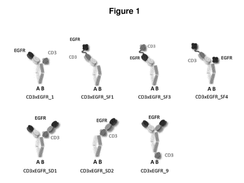
Figure 1: The panitumumab anti-EGFR binder (black) and the humanized 5P34 anti-
CD3 binder (grey) where assembled in various different BEAT architectures.
Figure 2: Flow cytometry analysis of 3A6 and 10E6 hybridoma candidates on BAF
cells
expressing membrane-bound EGFR. This figure shows the FAGS profiles of
parental
3A6 and 10E6 hybridoma supernatants to membrane-bound EGFR expressed on BAF
cells. One hundred pl harvested from both hybridoma clones were incubated with
100u1
of EGFR-transfected BAF cells diluted at 106 cells/ml. As a negative control,
a purified
mouse IgG isotype was used diluted at 10pg/ml. Antibody binding was detected
with
goat anti-mouse IgG-PE.
Figure 3: 3A6Al2B5 and 10E6F5 bind specifically to extracellular domain IV of
the
EGFR receptor. This figure shows the ELISA results in which several
concentrations
(ranging from 10 to 0.01pg/m1) of purified 3A6Al2F5 and 10E6F5 hybridoma
subclones were tested against immobilized recombinant soluble EGFR (A) or EGFR-
Her3 chimeric molecules (B and C) or single domain IV of EGFR (D). Vectibix0
was
also tested in the assay.
Figure 4: CD3-EGFR_5 and CD3-EGFR_8 display a killing activity of EGFR+A549
target cells. A CD3-redirected killing assay against EGFR+ A549 cells (Target
cells, T)
was performed using PBMCs from 3 healthy donors as effector cells (E), at an
E:T
ratio of 10:1, during 48h0ur5. The histograms show the average percentage of
specific
14
CA 03060190 2019-10-16
WO 2018/197502
PCT/EP2018/060488
killing calculated from the 3 individual donors. The two BEAT molecules were
used at
10nM in the assay.
Figure 5: (A) KD measurement for the chimeric 3A6 antibody. (B) KD measurement
for
the chimeric 10E6 antibody.
Figure 6: (A) KD measurement for the 10E6-best-fit antibody. (B) KD
measurement for
the 10E6-stable antibody.
Figure 7: (A) Sensorgrams of binding tests with 3A6 chimeric antibody, (B)
Sensorgrams of binding tests with 10E6 chimeric antibody. (C) Sensorgrams of
the
control experiment using the polyclonal goat anti-EGFR antibody.
Figure 8: (A) Thermogram for 10E6-best-fit antibody. The first peak
corresponds to the
IgG1 CH2-CH3 domains and shows a Tm of 71.7 C, the second peak corresponds to
the Fab. (B) Thermogram for 10E6-stable antibody. The first peak corresponds
to the
IgG1 CH2-CH3 domains and shows a Tm of 71.8 C, the second peak corresponds to
the Fab.
Figure 9: CD3xEGFR_1 has no efficacy in A549 tumors.
Figure 10: CD3xEGFR-SF1 and CD3xEGFR-SF3 have the same efficacy in A549
tumors.
Figure 11: CD3xEGFR-SF3 displays a better potency than Vectibix in SNU-216
tumors.
Figure 12: Dexamathasone impact on CD3xEGFR-SF3 anti-tumor activity in
xenograft
models. (A) The graph shows the mean tumor size (in mm3) SEM. (B) The graph
shows the tumor growth per mouse at day 37.
Figure 13: Simple binding ELISA format schematic for EGFR (A) and 0D3 (B).
Figure 14: Dual binding ELISA format schematic.
Figure 15: Detection of CD3xEGFR-5F3 in mice serum by a simple EGFR binding
ELISA.
Figure 16: Detection of CD3xEGFR-5F3 in mice serum by a simple 0D3 binding
ELISA.
CA 03060190 2019-10-16
WO 2018/197502
PCT/EP2018/060488
Figure 17: Detection of CD3xEGFR-SF3 in mice serum by a dual CD3 and EGFR
binding ELISA.
Figure 18: Pharmacokinetic profile of CD3xEGFR-SF3 in Sprague-Dawley rats
serum.
The pharmacokinetics of CD3xEGFR-SF3 was evaluated in male Sprague-Dawley rats
.. (n=4) following a single intravenous injection at a dose of 1 mg/kg body
weight. The
blood samples for pharmacokinetic (PK) assessment were collected at pre-
specified
time points of 0.25, 1, 6, 24, 48, 96, 168, 336, 530, 672, 840 and 1008 hours
post dose
over a period of 42 days (six weeks). The concentrations of CD3xEGFR-SF3 in
these
serum samples were quantified using a suitable ELISA method. Data
representative
of four animals tested (N=1).
Figure 19. Detection of CD3xEGFR-SF3 binding by ELISA. A dose response of
CD3xEGFR-SF3 and control antibodies were incubated on coated human CD3-Fc
(huCD3-Fc, A), human EGFR domain I-IV his-tagged (huEGFR-His; B) or huEGFR-
His (C), then detected with either an anti-human IgG Fab coupled with HRP (A
and B)
or huCD3-biotin followed by HRP-coupled streptavidin (C). The graphs show the
sigmoidal dose-response binding curves (absorbance at 450 nM) for each
treatment.
Each data point is the mean SEM of duplicates values from three independent
replications.
Figure 20. Detection of CD3xEGFR-SF3 binding by flow cytometry. A dose
response
of CD3xEGFR-SF3 and control antibodies were incubated on either PBMCS (A-C) or
the squamous cancer cell line NCI-H1703 (D) and detected with a PE-labelled
anti-
human IgG (Fc-y). For the PBMCs, the cells were also labelled with anti-CD4 or
anti-
CD8 antibodies. The graphs show the nonlinear sigmoidal regression binding
curves
of the mean fluorescent intensity (MFI) for each treatment. Each data point is
the mean
SEM of duplicates values from three independent replications.
Figure 21. CD3xEGFR-SF3 induces the redirected lysis of EGFR-expressing human
cancer cell lines. Target cancer cells (T) and effector cells (E; PBMCs) were
incubated
at an E:T ratio of 1:10 in the presence of a dose response of CD3xEGFR-SF3 or
control
antibodies and the redirected lysis of the cancer cells was determined by a
cytotoxic
assay (MTS). The ECK values were extracted from the sigmoidal dose-response
curves of specific killing. The error bars represent the mean SEM. Cell
lines
redirected lysis was statistically different (one-way ANOVA; F=5,6; p<0.0001).
16
CA 03060190 2019-10-16
WO 2018/197502
PCT/EP2018/060488
Figure 22. CD3xEGFR-SF3 has a low antibody-dependent cell-mediated
cytotoxicity
potential. Antibody-dependent cell-mediated cytotoxicity (ADCC) of CD3xEGFR
was
evaluated in the EGFR+ carcinoma cell lines A-431 and A549 (A) as well as in
CD3+
HPB-ALL cells (B) and represented by the ECK, values that were extracted from
the
sigmoidal dose-response curves of specific killing. The error bars represent
means
SEM from two independent experiments. Effect of the treatment was
statistically
significant for EGFR+ carcinoma cells (Least Square model, F = 29, p<0.0001)
and
CD3+ HPB-ALL cells (T test, t = 3, p<0.05). Statistically significant
differences (p<0.05)
are represented by asterisks (*).
lo Figure 23. CD3xEGFR-5F3 has no complement-dependent cytotoxicity. Specific
complement-dependent cytotoxicity (CDC) was evaluated in the EGFR+ carcinoma
cells A549 (A) as well as in CD3+ HPB-ALL cells (B) and the sigmoidal dose-
response
curves of specific CDC are represented.
Figure 24. Effects of CD3xEGFR-5F3 on the proliferation of PBMCs. PBMCs were
incubated for 48h in presence of increasing doses of CD3xEGFR or controls. The
graph shows the results of 3H-thymidine incorporation from six independent
experiments. AE042, P1069, and TRS represent different batches of CD3xEGFR-5F3
and 0.0005, 0.005, 0.05, 0.5 and 5 the concentrations in ug/ml. For the
different
treatments, "c" stands for coated and "s" for soluble. The error bars
represent means
SEM.
Figure 25. Statistical analysis of the effects of CD3xEGFR-5F3 on the
proliferation of
PBMCs. Data from Figure 24 were analyzed by fit least square model followed by
a
Dunnett's comparison (a=0,05) to compare the means against the no mAb control
(A)
and against the isotype control (B). Significant differences of the mean are
shown as
the needles bars outside of the decision limits (95% CI interval for each
treatment; gray
area).
Figure 26. Non-specific CD4+ T cell activation in response to CD3xEGFR-5F3.
PBMCs
were incubated for 24h or 48h in presence of increasing doses of CD3xEGFR or
controls. Activation of CD4+ T cell was measured as the expression of the
activation
marker 0D69 by flow cytometry. AE042, P1069, and TRS represent different
batches
of CD3xEGFR-5F3 and 0.0005, 0.005, 0.05, 0.5 and 5 the concentrations in
ug/ml. For
17
CA 03060190 2019-10-16
WO 2018/197502
PCT/EP2018/060488
the different treatments, "c" stands for coated and "s" for soluble. The error
bars
represent means SEM from six independent experiments.
Figure 27. Statistical comparison of the non-specific CD4+ T cell activation
between
CD3xEGFR-5F3 and no mAb condition. Data from Figure 26 were analyzed by fit
least
square model followed by a Dunnett's comparison (a=0,05) to compare the means
against the no mAb control at 24h (A) and 48h (B). Significant differences of
the mean
are shown as the needles bars outside of the decision limits (95% CI interval
for each
treatment; gray area).
Figure 28. Statistical comparison of the non-specific CD4+ T cell activation
between
lo CD3xEGFR-5F3 and the isotype control. Data from Figure 26 were analyzed by
fit
least square model followed by a Dunnett's comparison (a=0,05) to compare the
means against the isotype control at 24h (A) and 48h (B). Significant
differences of the
mean are shown as the needles bars outside of the decision limits (95% CI
interval for
each treatment; gray area).
Figure 29. Non-specific CD8+ T cell activation in response to CD3xEGFR-5F3.
PBMCs
were incubated for 24h or 48h in presence of increasing doses of CD3xEGFR-5F3
or
controls. Activation of CD8+ T cell was measured as the expression of the
activation
marker 0D69 by flow cytometry. AE042, P1069, and TRS represent different
batches
of CD3xEGFR-5F3 and 0.0005, 0.005, 0.05, 0.5 and 5 the concentrations in
ug/ml. For
the different treatments, "c" stands for coated and "s" for soluble. The error
bars
represent means SEM from six independent experiments.
Figure 30. Statistical comparison of the non-specific CD8+ T cell activation
between
CD3xEGFR-5F3 and no mAb condition. Data from Figure 29 were analyzed by fit
least
square model followed by a Dunnett's comparison (a=0,05) to compare the means
against the no mAb control at 24h (A) and 48h (B). Significant differences of
the mean
are shown as the needles bars outside of the decision limits (95% CI interval
for each
treatment; gray area).
Figure 31. Statistical comparison of the non-specific CD8+ T cell activation
between
CD3xEGFR-5F3 and the isotype control. Data from Fig. 29 were analyzed by fit
least
square model followed by a Dunnett's comparison (a=0,05) to compare the means
against the isotype control at 24h (A) and 48h (B). Significant differences of
the mean
18
CA 03060190 2019-10-16
WO 2018/197502
PCT/EP2018/060488
are shown as the needles bars outside of the decision limits (95% CI interval
for each
treatment; gray area).
Figure 32. Non-specific T cell cytokine responses to CD3xEGFR-SF3 at 24h.
PBMCs
were incubated for 24h in presence of increasing doses of CD3xEGFR-SF3 or
controls,
and the levels of IL-2, IL-6, TNF-a, and IFN-y released were measured by
Luminex in
the supernatant. AE042 and P1069, represent different batches of CD3xEGFR-SF3
and 0.0005, 0.005, 0.05, 0.5 and 5 the concentrations in ug/ml. For the
different
treatments, "c" stands for coated and "s" for soluble. The error bars
represent means
SEM from six independent experiments.
Figure 33. Statistical comparison of the non-specific T cell cytokine
responses between
CD3xEGFR-SF3 and the no mAb condition at 24h. Data from Fig.32 were analyzed
by
fit least square model followed by a Dunnett's comparison (a=0,05) to compare
the
means against the no mAb control of IL-2 (A), IL-6 (B), IFN-y (C), and TNF-a
(D).
Significant differences of the mean are shown as the needles bars outside of
the
decision limits (95% CI interval for each treatment; gray area).
Figure 34. Statistical comparison of the non-specific T cell cytokine
responses between
CD3xEGFR-5F3 and the isotype control at 24h. Data from Fig.32 were analyzed by
fit
least square model followed by a Dunnett's comparison (a=0,05) to compare the
means against the isotype control of IL-2 (A), IL-6 (B), IFN-y (C), and TNF-a
(D).
Significant differences of the mean are shown as the needles bars outside of
the
decision limits (95% CI interval for each treatment; gray area).
Figure 35. Non-specific T cell cytokine responses to CD3xEGFR-5F3 at 48h.
PBMCs
were incubated for 48h in presence of increasing doses of CD3xEGFR-5F3 or
controls,
and the levels of IL-2, IL-6, TNF-a, and IFN-y released were measured by
Luminex in
the supernatant. AE042, P1069, and TRS represent different batches of CD3xEGFR-
SF3 and 0.0001, 0.0005, 0.001, 0.005, 0.01, 0.05, 0.1, 0.5, 1, 5, and 10 the
concentrations in ug/ml. For the different treatments, "c" stands for coated
and "s" for
soluble. The error bars represent means SEM from six independent experiment.
Figure 36. Statistical comparison of the non-specific T cell cytokine
responses between
CD3xEGFR-5F3 and the no mAb condition at 48h. Data from Fig.35 were analyzed
by
fit least square model followed by a Dunnett's comparison (a=0,05) to compare
the
means against the no mAb control of IL-2 (A), IL-6 (B), IFN-y (C), and TNF-a
(D).
19
CA 03060190 2019-10-16
WO 2018/197502
PCT/EP2018/060488
Significant differences of the mean are shown as the needles bars outside of
the
decision limits (95% CI interval for each treatment; gray area).
Figure 37. Statistical comparison of the non-specific T cell cytokine
responses between
CD3xEGFR-5F3 and the isotype control at 48h. Data from Fig.35 were analyzed by
fit
.. least square model followed by a Dunnett's comparison (a=0,05) to compare
the
means against the isotype control of IL-2 (A), IL-6 (B), IFN-y (C), and TNF- a
(D).
Significant differences of the mean are shown as the needles bars outside of
the
decision limits (95% CI interval for each treatment; gray area).
Figure 38. CD3xEGFR-5F3 does not induce a non-specific T cell cytokine
response in
a high density PBMC assay. PBMCs were incubated for 48h at high density (107
cells/ml). The cells were then plated at a normal density (106 cells/ml), and
cultured for
24h in presence of increasing doses of CD3xEGFR-5F3 or controls, and the
levels of
IL-2, IL-6, TNF-a, and IFN-y released were measured by Luminex in the
supernatant.
AE042 and TRS represent different batches of CD3xEGFR-5F3 and 0.0001, 0.001,
0.01, 0.1, 1 and 10 the concentrations in ug/ml. The error bars represent
means SEM
from four independent experiment.
Figure 39. Statistical comparison of the non-specific T cell cytokine
responses between
CD3xEGFR-5F3 and the no mAb condition in a high density PBMC assay. Data from
Fig.38 were analyzed by fit least square model followed by a Dunnett's
comparison
(a=0,05) to compare the means against the no mAb control of IL-2 (A), IL-6
(B), IFN-y
(C), and TNF-a (D). Significant differences of the mean are shown as the
needles bars
outside of the decision limits (95% CI interval for each treatment; gray
area).
Figure 40. Statistical comparison of the non-specific T cell cytokine
responses between
CD3xEGFR-5F3 and the isotype control in a high density PBMC assay. Data from
Fig.38 were analyzed by fit least square model followed by a Dunnett's
comparison
(a=0,05) to compare the means against the isotype control of IL-2 (A), IL-6
(B), IFN-y
(C), and TNF-a (D). Significant differences of the mean are shown as the
needles bars
outside of the decision limits (95% Cl interval for each treatment; gray
area).
Figure 41. CD3xEGFR-5F3 does not induce a cytokine response in a whole blood
assay. Whole blood from healthy volunteers was cultured for 24h in presence of
increasing doses of CD3xEGFR-5F3 or controls and the levels of IL-2, IL-6, TNF-
a,
and IFN-y were measured by Luminex in the serum. AE042 and TRS represent
CA 03060190 2019-10-16
WO 2018/197502
PCT/EP2018/060488
different batches of CD3xEGFR-SF3 and 0.001, 0.01, 0.1, and 1 the
concentrations in
ug/ml. The error bars represent means SEM from four independent experiments.
Figure 42. Statistical comparison of the cytokine responses between CD3xEGFR-
5F3
and the no mAb condition in a whole blood assay. Data from Fig.41 were
analyzed by
fit least square model followed by a Dunnett's comparison (a=0,05) to compare
the
means against the no mAb control of IL-2 (A), IL-6 (B), IFN-y (C), and TNF-a
(D).
Significant differences of the mean are shown as the needles bars outside of
the
decision limits (95% CI interval for each treatment; gray area).
Figure 43. Statistical comparison of the cytokine responses between CD3xEGFR-
5F3
and the isotype control in a whole blood assay. Data from Fig.41 were analyzed
by fit
least square model followed by a Dunnett's comparison (a=0,05) to compare the
means against the isotype control of IL-2 (A), IL-6 (B), IFN-y (C), and TNF-a
(D).
Significant differences of the mean are shown as the needles bars outside of
the
decision limits (95% CI interval for each treatment; gray area).
Figure 44. Efficacy of CD3xEGFR-5F3 therapeutic treatment in NOD SCID
xenografted mouse model. The expression level of EGFR on A549 cells was
determined by sABC before the graph. A mix of tumor cells (target cells, T)
and PBMCs
(effector cells, E) were injected s.c. at an E:T ratio of 2:1 into the right
flank area of
NOD.CB17/AlhnRj-Prkdcscid/Rj (NOD/SCID) mice (n = 4 to 5 per group per PBMC
.. donor). CD3xEGFR-5F3 was administered i.v. at 2mg/kg once a week starting
on day
2 for 3 weeks. Tumor growth was determined by external caliper measurements.
The
graphs show the mean tumor size (in mm3) SEM. 2 PBMC donors were included.
Name of the study: A549_15.
Figure 45. A549 tumor volume comparison between CD3xEGFR-5F3 in therapeutic
treatment and control group at day 41. The data showed per group the tumor
volume
of each animal at day 41. Data are extracted from Figure 44. Name of the
study:
A549_15.
Below is provided a set of non-exhaustive examples relating to the present
invention.
Example 1: Engineering of CD3xEGFR bispecific antibodies in different formats
.. Hombach et al. (2007) demonstrated that the position of the targeted
epitope within a
target molecule has a major impact on the efficacy of T cell activation and
that
21
CA 03060190 2019-10-16
WO 2018/197502
PCT/EP2018/060488
shortening the distance between the T-cell and the target cell membrane can
increase
the cytotoxic potential of a bispecific antibody. Our hypothesis was, that re-
arranging
the binding domains in a CD3xEGFR BEAT bispecific antibody could change the
distance between the redirected T-cells and the EGFR expressing cancer cells
and
thus modulate the cytotoxic potential of our molecule.
The panitumumab anti-EGFR binder and the humanized SP34 anti-CD3 binder were
engineered into a number of different BEAT formats as described below.
Construction of alternative BEAT architectures
Alternative BEAT architectures were designed by altering the position of the
panitumumab anti-EGFR binder and the humanized SP34 anti-CD3 binder (Figure
1).
Depending on the architecture, the binders were formatted as single-chain
fragment
(scFv) or Fab. Binders in scFv format were fused via Gly4Ser or Gly4Thr
linkers (SEQ
ID NOs: 13 and 14) to confer flexibility. When a Fab was fused to a scFv, a
Gly4Ser
linker was added in between. Coding DNAs (cDNAs) encoding the different
polypeptide chains in part or in full were first gene synthetized by GENEART
AG
(Regensburg, Germany) and modified using standard molecular biology
techniques.
PCR products were digested with appropriate DNA restriction enzymes, purified
and
ligated in a modified pcDNA3.1 plasmid (Invitrogen AG, Zug, Switzerland)
carrying a
CMV promoter and a bovine hormone poly-adenylation (poly(A)) previously
digested
with the same DNA restriction enzymes. All polypeptide chains were
independently
ligated in this expression vector where secretion was driven by the murine
VJ2C leader
peptide. Polypeptide chain A (see Figure 1) generally contained in addition to
the
variable domain, an IgG1 hinge followed by an IgG3 CH2 domain with both L234A
and
L235A substitutions (EU numbering) and an IgG3 CH3 domain containing the BEAT
(A) substitutions. To prevent Protein A binding due to the VH3 type framework
of the
humanized 5P34 anti-CD3 binder, Protein A binding abrogating mutations N82aS
and/or G655 were added when 5P34 was placed on chain A. Polypeptide chain B
generally contained an IgG1 hinge followed by an IgG1 CH2 domain with both
L234A
and L235A substitutions and an IgG1 CH3 domain containing the BEAT (B)
substitutions. When a binder was present in Fab format, an IgG1 CH1 domain was
also part of the polypeptide chain. The following molecules were constructed:
CD3xEGFR 1 (SEQ ID NOs: 1, 2 and 3), CD3xEGFR SF1 (SEQ ID NOs: 4, 5 and 6),
22
CA 03060190 2019-10-16
WO 2018/197502
PCT/EP2018/060488
CD3xEGFR SF3 (SEQ ID NOs: 7, 2 and 8), CD3xEGFR _5F4 (SEQ ID NOs: 4, 5 and
9), CD3xEGFR SD1 (SEQ ID NOs: 1,2 and 10), CD3xEGFR _5D2 (SEQ ID NOs: 11,
and 2) and CD3xEGFR _9 (SEQ ID NOs: 1,2 and 12).
Production of CD3xEGFR in alternative BEAT architectures
5 For transient expression, equal quantities of each engineered chain
vector were co-
transfected into suspension-adapted HEK293-EBNA cells (ATCC-LGL standards,
Teddington, UK; Cat. No: CRL-10852) using polyethyleneimine (PEI; Sigma,
Buchs,
Switzerland). Typically, 100 ml of cells in suspension at a density of 0.8-1.2
million cells
per ml are transfected with a DNA-PEI mixture. When recombinant expression
vectors
10 encoding the respective chains are introduced into the host cells, the
immunoglobulin
construct is produced by further culturing the cells for a period of 4 to 5
days to allow
for secretion into the culture medium (EX-CELL 293, HEK293-serum-free medium
(Sigma), supplemented with 0.1% pluronic acid and 4mM glutamine). Cell-free
culture
supernatants containing the secreted proteins were prepared by centrifugation
followed by sterile filtration. BEATs were then purified from cell-free
supernatant using
Protein A affinity resin (Repligen). Clarified supernatants were adjusted to
pH 6.0 with
NaH2PO4 at 0.2 M and loaded on Protein A by gravity flow. Columns were washed
with
CV of 0.2 M citrate phosphate buffer pH 6Ø Proteins were eluted with 16 CV
of 20
mM sodium acetate at pH 4.1 and neutralized with 0.1 volume of 1 M Tris pH 8.0
20 (Sigma). Samples were buffer exchanged into PBS pH 7.4 using Illustra
NAP-10
columns (GE Healthcare). Exceptionally, CD3xEGFR_5F3 cell-free supernatants
were
loaded onto a 1 ml HiTrapTm MabSelect SuRe TM Protein A column pre-
equilibrated in
0.2 M citrate phosphate buffer pH 6.0 and operated on an AKTApurifierTm
chromatography system (both from GE Healthcare Europe GmbH; column Cat. No:
11-0034-93) at a flow rate of 1 ml/min. Running buffer was 0.2 M citrate
phosphate
buffer pH 6Ø Washing buffer was 0.2 M citrate phosphate buffer pH 5Ø
Elution was
performed using 20 mM sodium acetate buffer pH 4.1. Elution was followed by OD
reading at 280 nm; fractions containing CD3xEGFR antibodies were pooled and
neutralized with 0.1 (:)/0 volume of 1 M Tris pH 8Ø Samples were buffer
exchanged into
PBS pH 7.4 using Illustra NAP-10 columns (GE Healthcare Europe GmbH,
Glattbrugg,
Switzerland).
23
CA 03060190 2019-10-16
WO 2018/197502
PCT/EP2018/060488
Example 2: Generation and characterization of anti-EGFR antibodies specific
for
domain IV of EGFR.
Immunization
Female BALB/c mice, 7 weeks of age (Harlan) were used to generate antibodies
against the extracellular domain 4 of EGFR. The mice were immunized three
times by
the intraperitoneal (i.p) and the subcutaneous (s.c.) routes with a mixture of
either 50
pg human EGFR His-tagged protein (SEQ ID NO: 15) or 50 pg extracellular domain
IV
EGFR His-tagged protein (SEQ ID NO: 16), in combination with 100 pl of
adjuvant.
The presence of circulating anti-EGFR antibodies specific to domain IV in the
immunized mouse sera was evaluated by direct ELISA using plates coated with
the
recombinant human EGFR his or domain IV His proteins. Mouse sera were serially
diluted (from 1:100 to 1:109) and added to 96-well ELISA plates and the bound
antibodies were detected using a goat anti-mouse molecule-HRP (Jackson
Immunoresearch). A final intravenous boost with 10 pg of antigen without
adjuvant was
performed in animals displaying the best anti-EGFR domain IV IgG serum titer
three
days before sacrifice. Animals were euthanized and the spleens were harvested
for
fusion.
Fusion protocol
1 ml of warm PEG1500 was slowly added to the cell slurry over the course of 1
min
while swirling. The cells were gently mixed for a further 2 minutes. 4 ml of
warm SFM
was then added over a period of 4 min. 10 ml of warm SFM was slowly added and
the
cells were incubated for 5 min in a water bath at 37 C. The cells were
centrifuged at
1000 rpm for 5 min and re-suspended in 200m1 of complete medium. For the
fusion,
the cells were plated at 200 p1/well in ten 96 flat-bottom well plates.
Screening of hybridoma supernatants on membrane-bound EGFR by flow
cytometry
Approximately 1900 wells from two fusions were analyzed by ELISA for their
content
in murine IgG specific for recombinant human EGFR. Positive hybridoma
supernatants
were further screened against recombinant domain IV of human EGFR, immobilized
on 96-well ELISA plates. Among all the tested clones, two parental candidates,
3A6
and 10E6, were identified and tested by flow cytometry on BAF cells
transfected with
24
CA 03060190 2019-10-16
WO 2018/197502
PCT/EP2018/060488
membrane-bound EGFR. In this assay, 105 cells/well were incubated with 100u1
of
supernatants at 4 C for 1 hour. Following this primary incubation, cells were
centrifuged at 1300rpm, 2min and pellets were resuspended with 100u1 of PE-
labeled
goat anti-mouse secondary antibody diluted at 1/100 in FAGS buffer. Cells were
then
incubated for 30 minutes at 4 C and washed twice, the supernatants removed and
the
cells resuspended in 150 pl of FAGS buffer. The samples were analyzed by flow
cytometry. As shown in Figure 2, the results from the flow cytometry
experiment show
that both 3A6 and 10E6 hybridoma candidates recognize the membrane-bound human
EGFR receptor expressed on BAF cells, compared to the isotype control used at
bug/ml. These two hybridoma candidates were expanded and subcloned.
Screening of hybridoma supernatants on soluble EGFR by ELISA
The subclones 3A6Al2B5 and 10E6F5 were derived from 3A6 and 10E6 parental
clones, respectively. Supernatants from both subclones were harvested and
purified
using a LC-kappa mouse affinity matrix (Life technologies), according to the
manufacturer's instructions. These purified antibodies were tested by ELISA on
96-
well plates coated with either soluble human EGFR or recombinant EGFR-Her3
chimeric constructs. These molecules were diluted at 2ug/m1 in PBS and
immobilized
overnight at 4 C on a high binding 96-well plates. The plates were blocked
with PBS
2% Bovine Serum Albumin (BSA) and incubated for 1 hour with a serial dilution
of
either 3A6Al2B5 or 10E6F5. As a control, Panitumumab (Vectibix0) was used at
the
same concentrations. The plates were then washed with PBS 0.01% Tween and
incubated for 1 hour with 100u1of either goat anti-mouse IgG (to detect
3A6Al2B5 and
10E6F5) (Jackson ImmunoResearch Europe Ltd, Newmarket, UK) or goat anti-human
IgG, F(ab')2 fragment specific-HRP (to detect Panitumumab). Following this
incubation, the plates were washed and incubated with 100u1 of TMB substrate.
The
reaction was stopped by adding 100 pl of H2504 2N and the absorbance was read
at
450 nm on a Synergy HT2 spectrophotometer (Biotek, USA; distributor: WITTEC
AG,
Littau, Switzerland). Results from Figure 3 show that purified 3A6Al2B5 and
10E6F5
antibodies recognize in a dose dependent manner the soluble EGFR molecule (A)
and
bind also the chimeric Her31-III-EGFR IV (C) and EGFR IV (D) molecules.
Conversely,
none of these two candidates recognize the chimeric EGFR I-111 Her3 IV
molecule (B).
These results show that both 3A6Al2B5 and 10E6F5 antibodies bind EGFR, and
specifically to domain IV.
CA 03060190 2019-10-16
WO 2018/197502
PCT/EP2018/060488
Redirected lysis assay (RDL)
This assay was performed following the procedure described in the example.
Figure 4
shows that both BEAT CD3-EGFR_5 and CD3-EGFR_8 molecules display a killing
potential against EGFR+ A549 target cells.
Example 3: Humanization of anti-EGFR domain IV antibodies 3A6 and 10E6
3.1 Human EGFR and chimeric human EGFR-ErbB3 proteins used for mouse
immunization and antibody characterization
Coding DNA (cDNA) encoding the polypeptide chain of human EGFR soluble
extracellular region (UniProt accession No: P00533 residues 25-638, referred
to herein
as hEGFR, SEQ ID NO: 15) with a C-terminal poly-histidine tag was synthetized
by
GENEART AG (Regensburg, Germany) and modified using standard molecular
biology techniques. PCR products were digested with appropriate DNA
restriction
enzymes, purified and ligated in a modified pcDNA3.1 plasmid carrying a CMV
promoter and a bovine hormone poly-adenylation (poly(A)) signal previously
digested
with the same DNA restriction enzymes.
The following EGFR domain IV only constructs were PCR amplified from the hEGFR
construct described above and restriction ligated into the expression plasmid
mentioned above: hEGFR-IV_505-638 (505-638 indicates residue range, this
construct additionally carried the mutation W516A to increase solubility,
mutation was
added by standard overlapping PCR using primers including the appropriate
mutation),
hEGFR-IV_556-638 and hEGFR-IV 580-638 (SEQ ID NO: 16, 17 and 18).
Cynomolgus EGFR-IV_556-638 and 580-638 (herein referred to as cEGFR-IV_556-
638 and 580-638) were generated by adding the mutations A566V (only for the
construct encompassing residues 556-638), P637A and T638R to the human
construct
by overlapping PCR (SEQ ID NO: 19 and 20).
The following chimeric human EGFR-ErbB3 constructs (human ErbB3, UniProt
accession No: P21860) were designed and cDNA encoding their polypeptide chains
were synthesized by Eurofins Genomics: hEGFR-1-11-111_hErbB3-IV and hErbB3-1-
11-
III hEGFR-IV (SEQ ID NO: 21 and 22).
.. For transient expression of the above described EGFR constructs, the
appropriate
expression vectors were transfected into suspension-adapted HEK-EBNA cells
26
CA 03060190 2019-10-16
WO 2018/197502
PCT/EP2018/060488
(ATCC-CRL-10852) using polyethyleneimine (PEI). Typically, 100 ml of cells in
suspension at a density of 0.8-1.2 million cells per ml are transfected with a
DNA-PEI
mixture containing 100 pg of expression vector. When recombinant expression
vectors
are introduced into the host cells, proteins are produced by further culturing
the cells
for a period of 4 to 5 days to allow for secretion into the culture medium (EX-
CELL 293,
HEK293-serum-free medium; Sigma, Buchs, Switzerland), supplemented with 0.1
(:)/0
pluronic acid, 4 mM glutamine). EGFR was then purified from cell-free
supernatant
using Ni sepharose Excel (GE Healthcare Europe GmbH, Glattbrugg, Switzerland)
and
used for further analysis.
3.2 Chimeric 3A6 and 10E6
Using standard molecular biology techniques, VH and VL sequences extracted
from
antibodies from hybridoma cells were re-formatted into mouse-human chimeric
IgG1
antibodies. Mouse 3A6 and 10E6 VH domains were fused to human IgG1 Fc (CH1-
hinge-CH2-CH3) and the corresponding VL domains were fused to IgG1 constant
kappa. The resulting constructs were ligated into the modified pcDNA3.1
plasmid
described above (3A6 chimeric antibody SEQ ID NO: 23 and 24, 10E6 chimeric
antibody SEQ ID NO: 25 and 26).
For transient expression of the chimeric antibodies, the recombinant vectors
for the
heavy- and the light chain were transfected at a 1:1 molar ratio into
suspension-
adapted HEK-EBNA cells using PEI. Antibodies were then purified from cell-free
supernatant using Protein A affinity resin (Repligen, Waltham MA, USA).
Clarified
supernatants were loaded on Protein A by gravity flow. Proteins were eluted
with 0.1
M glycine pH 3Ø Samples were buffer exchanged into PBS pH 7.4 using Illustra
NAP-
10 columns (GE Healthcare Europe GmbH, Glattbrugg, Switzerland).
3.3 Humanization of mouse monoclonal 3A6
Humanization of the anti-human EGFR mouse antibody 3A6 including selection of
human acceptor frameworks that substantially retain the binding properties of
human
CDR-grafted acceptor frameworks is described herein. Two grafts were prepared,
one
using the best-fit framework and another using a stable framework.
Homology matching was used to choose human best-fit acceptor frameworks to
graft
3A6 CDRs. Databases e.g. a database of germline variable genes from the
27
CA 03060190 2019-10-16
WO 2018/197502
PCT/EP2018/060488
immunoglobulin loci of human and mouse (the IMGT database, supra) or the
VBASE2
(Retter I et al, (2005) Nucleic Acids Res. 33, Database issue D671-D674) or
the Kabat
database (Johnson G et al, (2000) Nucleic Acids Res. 28: 214-218) or
publications
(e.g., Kabat EA et al, supra) may be used to identify the human subfamilies to
which
the murine heavy and light chain V regions belong and determine the best-fit
human
germline framework to use as the acceptor molecule. Selection of heavy and
light chain
variable sequences (VH and VL) within these subfamilies to be used as acceptor
may
be based upon sequence homology and/or a match of structure of the CDR1 and
CDR2 regions to help preserve the appropriate relative presentation of the six
CDRs
after grafting.
For example, use of the IMGT database indicates good homology between the 3A6
heavy chain variable domain framework and the members of the human heavy chain
variable domain subfamily 4. Highest homology and identity of both CDRs and
framework sequences were observed for germline sequence IGHV4-4*08 (SEQ ID
NO: 27) which had sequence identity of 59.4% for the whole sequence up to
CDR3.
Using the same approach, 3A6 light chain variable domain sequence showed good
homology to the members of the human light chain variable domain kappa
subfamily
6. Highest homology and identity of both CDRs and framework sequences were
observed for germline sequence IGKV6-21*02 (SEQ ID NO: 28) with sequence
identity
of 69.5%.
Selection of human heavy and light chain variable sequences (VH and VL) to be
used
as acceptor may be based upon germlines with good biophysical properties (as
documented in Ewert Set al., (2003) J.Mol.Biol, 325, 531-553) and/or pairing
as found
in natural antibody repertoire (as documented in Glanville J et al., (1999)
Proc Natl
Acad Sci U S A, 106(48):20216-21; DeKosky BJ et al., (2015) Nat Med, 21(1):86-
91).
Framework sequences known in the field for good paring and/or stability are
the human
IGHV3-23*04 (SEQ ID NO: 29) and IGKV1-39*01 (SEQ ID NO: 30) frameworks which
were used as acceptor frameworks for the 3A6 stable graft humanization.
Two humanized antibodies of human gamma one isotype were prepared. The
antibodies encompassed a human-mouse hybrid heavy chain variable domain and a
human-mouse hybrid light chain variable domain. The first hybrid heavy chain
variable
domain was based on the human heavy chain variable domain IGHV4-4*08 wherein
28
CA 03060190 2019-10-16
WO 2018/197502
PCT/EP2018/060488
germline CDRH1 and H2 where respectively replaced for 3A6 CDRH1 and CDRH2.
Best matching JH segment sequence to the human acceptor framework was
identified
from the !MGT database using homology search. To accommodate CDRs on to the
human acceptor framework key positions were modified by substituting human
residues to mouse residues. This process is called back-mutation and is the
most
unpredictable procedure in the humanization of monoclonal antibodies. It
necessitates
the identification and the selection of critical framework residues from the
mouse
antibody that need to be retained in order to preserve affinity while at the
same time
minimizing potential immunogenicity in the humanized antibody. The resulting
human-
mouse hybrid heavy chain variable sequence having human IGHV4-4*08 framework
regions, 3A6 mouse CDRs, key human to mouse framework back-mutations and best
matching JH to human acceptor is referred herein as heavy chain variable
domain 3A6-
best-fit-VH with SEQ ID NO: 31. The second hybrid heavy chain variable domain
was
based on the human heavy chain variable domain IGHV3-23*04 wherein germline
CDRH1 and H2 where respectively replaced for 3A6 CDRH1 and CDRH2. Best
matching JH segment sequence to the human acceptor framework was identified
from
the !MGT database using homology search. The resulting human-mouse hybrid
heavy
chain variable sequence having human IGHV3-23*04 framework regions, 3A6 mouse
CDRs, key human to mouse framework back-mutations and best matching JH to
human acceptor is referred herein as heavy chain variable domain 3A6-stable-VH
with
SEQ ID NO: 32.
Similarly, the first human-mouse hybrid light chain variable domain used for
this first
humanized antibody candidate had human IGKV6-21*02 framework regions, 3A6
mouse CDRs and best matching JK to human acceptor, and is referred herein as
light
chain variable domain 3A6-best-fit-VL with SEQ ID NO: 33 (no back-mutations in
the
framework were required in this case as all key positions were the same in the
mouse
and human framework). The first humanized antibody encompassing 3A6-best-fit-
VH
and 3A6-best-fit-VL is abbreviated herein as 3A6-best-fit antibody. The second
human-
mouse hybrid light chain variable domain used for the second humanized
antibody
candidate had human IGKV1-39*01 framework regions, 3A6 mouse CDRs, key human
to mouse framework back-mutations and best matching JK to human acceptor, and
is
referred herein as light chain variable domain 3A6-stable-VL with SEQ ID NO:
34. The
29
CA 03060190 2019-10-16
WO 2018/197502
PCT/EP2018/060488
second humanized antibody encompassing 3A6-stable-VH and 3A6-stable-VL is
abbreviated herein as 3A6-stable antibody.
3.4 Humanization of mouse monoclonal 10E6
Humanization of the anti-human EGFR mouse antibody 10E6 including selection of
.. human acceptor frameworks that substantially retain the binding properties
of human
CDR-grafted acceptor frameworks is described herein. Two grafts were prepared,
one
using the best-fit framework and another using a most stable framework.
Homology matching was used as described above to choose human best-fit
acceptor
frameworks to graft 10E6 CDRs. The IMGT database indicates good homology
.. between the 10E6 heavy chain variable domain framework and the members of
the
human heavy chain variable domain subfamily 4. Highest homology and identity
of
both CDRs and framework sequences were observed for germline sequence IGHV4-
30-4*01 (SEQ ID NO: 35) which had sequence identity of 73.2% for the whole
sequence up to CDR3.
Using the same approach, 10E6 light chain variable domain sequence showed good
homology to the members of the human light chain variable domain kappa
subfamily
6. Highest homology and identity of both CDRs and framework sequences were
observed for germline sequence IGKV6-21*02 (SEQ ID NO: 14) with sequence
identity
of 69.5%.
Stable frameworks were chosen as described above. Human IGHV3-23*04 (SEQ ID
NO: 29) and IGKV1-39*01 (SEQ ID NO: 30) were used as acceptor frameworks for
the
stable graft humanization.
Two humanized antibodies of human gamma one isotype were prepared. The
antibodies encompassed a human-mouse hybrid heavy chain variable domain and a
human-mouse hybrid light chain variable domain. The first hybrid heavy chain
variable
domain was based on the human heavy chain variable domain IGHV4-30-4*01
wherein germline CDRH1 and H2 where respectively replaced for 10E6 CDRH1 and
CDRH2. Best matching JH segment sequence to the human acceptor framework was
identified from the IMGT database using homology search. The resulting human-
mouse hybrid heavy chain variable sequence having human IGHV4-30-4*01
framework regions, 10E6 mouse CDRs, key human to mouse framework back-
CA 03060190 2019-10-16
WO 2018/197502
PCT/EP2018/060488
mutations and best matching JH to human acceptor is referred herein as heavy
chain
variable domain 10E6-best-fit-VH with SEQ ID NO: 36. The second hybrid heavy
chain
variable domain was based on the human heavy chain variable domain IGHV3-23*04
wherein germline CDRH1 and H2 where respectively replaced for 10E6 CDRH1 and
CDRH2. Best matching JH segment sequence to the human acceptor framework was
identified from the !MGT database using homology search. The resulting human-
mouse hybrid heavy chain variable sequence having human IGHV3-23*04 framework
regions, 10E6 mouse CDRs, key human to mouse framework back-mutations and best
matching JH to human acceptor is referred herein as heavy chain variable
domain
10E6-stable-VH with SEQ ID NO: 37.
Similarly, the first human-mouse hybrid light chain variable domain used for
this first
humanized antibody candidate had human IGKV6-21*02 framework regions, 10E6
mouse CDRs, key human to mouse framework back-mutations and best matching JK
to human acceptor, and is referred herein as light chain variable domain 10E6-
best-fit-
VL with SEQ ID NO: 38. The first humanized antibody encompassing 10E6-best-fit-
VH
and 10E6-best-fit-VL is abbreviated herein as 10E6-best-fit antibody. The
second
human-mouse hybrid light chain variable domain used for the second humanized
antibody candidate had human IGKV1-39*01 framework regions, 10E6 mouse CDRs,
key human to mouse framework back-mutations and best matching JK to human
acceptor, and is referred herein as light chain variable domain 10E6-stable-VL
with
SEQ ID NO: 39. The second humanized antibody encompassing 10E6-stable-VH and
10E6-stable-VL is abbreviated herein as 10E6-stable antibody.
3.5 Production of humanized 3A6 and 10E6 antibodies
Coding DNA sequences (cDNAs) for best-fit VH and VL and stable VH and VL were
synthesized by Eurofins Genomics (Ebersberg, Germany) and modified using
standard
molecular biology techniques. VH domains were fused to a human IgG1 CH1-hinge-
CH2-CH3 portion and restriction ligated into the expression vector described
above.
Similarly, genes for the VL domains were fused to the human constant kappa
domain
and ligated into a separate expression vector. The resulting antibodies were
3A6-best-
fit (SEQ ID NO: 40 and 41), 3A6-stable (SEQ ID NO: 42 and 43), 10E6-best-fit
(SEQ
ID NO: 44 and 45) and 10E6-stable (SEQ ID NO: 46 and 47).
31
CA 03060190 2019-10-16
WO 2018/197502
PCT/EP2018/060488
For transient expression of antibodies, the recombinant vectors for the heavy-
and the
light chain were transfected at a 1:1 molar ratio into suspension-adapted HEK-
EBNA
cells using PEI. Antibodies were then purified from cell-free supernatant
using Protein
A affinity resin. Clarified supernatants were loaded on Protein A by gravity
flow.
Proteins were eluted with 0.1 M glycine pH 3Ø Samples were buffer exchanged
into
PBS pH 7.4 using Illustra NAP-10 columns.
3.6 Kinetic binding affinity constants of the chimeric and humanized
antibodies
for human EGFR by surface plasmon resonance (SPR)
Kinetic binding affinity constants (KD) were measured. Measurements were
conducted
on a BlAcore T200 (GE Healthcare -BlAcore, Uppsala, Sweden) at room
temperature,
and analyzed with the Biacore T200 Evaluation software. A Series S CM5 sensor
chip
was covalently coupled with Protein G and 100 RUs of antibody of interest was
captured. hEGFR was injected at concentrations ranging from 19.53-2500 nM for
3A6
and 2.4-312.5 nM or 1-250 nM for 10E6 based antibodies at a flow rate of 30
pl/min in
HBS-EP+ buffer (Figure 5 and 6, data are expressed as number of response units
(abbreviated RU; Y axis) vs. time (X axis)). After each binding event, the
surface was
regenerated with 10 pl of glycine buffer pH 1.5. Dissociation time was 4 min.
Experimental data were processed using a 1:1 Langmuir model with global Rmax.
3.7 Production of 3A6 and 10E6 bispecific antibodies in combination with SP34
In order to generate bispecific BEAT antibodies combining 3A6 and 10E6 binders
with
humanized 5P34, the VH fragments mentioned above were fused to a human IgG1
CH1-hinge followed by an IgG1 CH2 and an IgG1 CH3 domain containing the BEAT
(A) substitutions. The CH2 domain contained both L234A and L235A substitutions
(EU
numbering). In-house humanized 5P34 in scFv format followed by a short five
amino
acid linker (Gly4Thr) was fused to a human IgG3 CH2 followed by an IgG3 CH3
domain
containing the BEAT (B) substitutions. The CH2 domain contained both L234A and
L235A substitutions. Constructs were ligated into the expression vector as
described
above. The resulting molecules were CD3xEGFR_5 (SEQ ID NO: 48, 24 and 49),
CD3xEGFR 6 (SEQ ID NO: 50, 26 and 49), CD3xEGFR 7 (SEQ ID NO: 48, 35 and
49) and CD3xEGFR 8 (SEQ ID NO: 52, 36 and 47).
For transient expression of the BEAT antibodies, the recombinant vectors for
the
heavy- and the light chain and the scFv-Fc chain were transfected at a 1:1:1
molar
32
CA 03060190 2019-10-16
WO 2018/197502
PCT/EP2018/060488
ratio into suspension-adapted HEK-EBNA cells using PEI as described above.
Cell-
free supernatants were loaded onto a 1 ml HiTrapTm MabSelect SuReTM Protein A
column pre-equilibrated in 0.2 M citrate phosphate buffer pH 6.0 and operated
on an
AKTApurifierTm chromatography system (both from GE Healthcare Europe GmbH;
column Cat. No: 11-0034-93) at a flow rate of 1 ml/min. Running buffer was 0.2
M
citrate phosphate buffer pH 6Ø Washing buffer was 0.2 M citrate phosphate
buffer pH
5Ø Elution was performed using 20 mM sodium acetate buffer pH 4.1. Elution
was
followed by OD reading at 280 nm; fractions containing CD3xEGFR antibodies
were
pooled and neutralized with 0.1 % volume of 1 M Tris pH 8Ø
3.8 Epitope Mapping and cross-reactivity with cynomolgus monkey
Surface plasmon resonance was used to evaluate the binding of 3A6 and 10E6
antibodies to various human and cynomolgus EGFR domain IV constructs with the
aim
to narrow down the epitope within EGFR domain IV and to demonstrate cross-
reactivity
with cynomolgus EGFR.
Measurements were performed on a Biacore 2000 instrument (GE). A CMS sensor
chip was covalently coupled with Protein G and 100 RUs of 3A6 or 10E6 chimeric
antibody were captured. Various human and cynomolgus EGFR constructs were
injected as analyte at a concentration of 200 nM at a flow rate of 30 pl/min
in HBS-EP
buffer. Dissociation time was 4 min. After each binding event, the surface was
regenerated with 10 pl of glycine buffer pH 1.5. To verify the integrity of
the EGFR
domain IV constructs, a polyclonal goat anti-EGFR known to us to contain at
least one
anti EGFR domain IV binder (AF231, Bio-Techne AG, Zug, Switzerland) was
immobilized as described above and human and cynomolgus EGFR constructs were
injected as described above. Results are shown in Figure 7. Both, 3A6 and 10E6
antibodies were able to bind human and cynomolgus EGFR-IV_556-638. EGFR-
IV_ 580-638 was not bound by either of the antibodies. Thus we conclude that
the
binding epitope of 3A6 and 10E6 antibodies is within EGFR domain IV and more
precisely within residues 556-638. Furthermore, residues 556-580 contain a
required
part of the epitope. Additionally, we demonstrate that both, 3A6 and 10E6
antibodies
are cross-reactive to cynomolgus EGFR.
33
CA 03060190 2019-10-16
WO 2018/197502
PCT/EP2018/060488
3.9 Thermostability of 10E6-best-fit and 10E6-stable antibodies
The thermal stability of 10E6-best-fit and 10E6-stable antibodies in PBS
buffer was
investigated by differential scanning calorimetry (DSC). Calorimetric
measurements
were carried out on a VP-DSC Capillary differential scanning microcalorimeter
(Malvern Instruments Ltd, Malvern, UK). The cell volume was 0.128 ml, the
heating
rate was 1 C/min and the excess pressure was kept at 64 p.s.i. All samples
were used
at a concentration of 1 mg/ml in PBS (pH 7.4). The molar heat capacity of each
protein
was estimated by comparison with duplicate samples containing identical buffer
from
which the protein had been omitted. The partial molar heat capacities and
melting
curves were analyzed using standard procedures. Thermograms were baseline-
corrected, concentration normalized, and further analyzed using a Non-Two
State
model using the Origin v7.0 software (supplied by Malvern Instruments Ltd).
Results
are shown in Figure 8. Melting temperatures (Tm) for 10E6-best-fit Fab and
10E6-
stable Fab were determined and were 82.8 C and 84.9 C respectively.
Example 4: CD3-EGFR_1 efficacy in A549 tumors xenografted in s.c.
MATERIAL AND METHODS
4.1. Cell line culture conditions
Cells were cultured in standard media in a humidified atmosphere of 5% CO2 at
37 C.
The cells were passaged 2 to 3 times per week to maintain them at optimal
confluency
and were routinely tested for mycoplasma contamination using the MycoAlert
detection
kit (LT07-318, Lonza). The cells consistently tested mycoplasma contamination
free.
4.2. Effector cells: human Peripheral Blood Mononuclear Cells (PBMC)
Peripheral blood mononuclear cells (PMBCs) were harvested from blood filters
obtained from La Chaux-de-Fonds Transfusion Center. For blood filter
processing, 40
ml of PBS supplemented with 1% Liquemine (Drossapharm) was injected into the
blood filter and the solution containing the PBMCs was collected in 50m1 blood
separation tubes (Chemie Brunschwig, PAA535710) previously loaded with ficoll
(GE
Healthcare, 17-1440-03). The ficoll tubes were centrifuged for 20 min at 800g
at room
temperature (RT) without the brake and the PBMC "buffy coat" ring was
harvested and
transferred into 50 ml falcon tubes containing 30 ml of PBS. The PBMCs were
washed
three times in PBS before being processed.
34
CA 03060190 2019-10-16
WO 2018/197502
PCT/EP2018/060488
4.3. Animal husbandry
In vivo experiments were performed in female 7-week-old immunodeficient
NOD.CB17/AlhnRj-Prkdcscid/Rj (NOD/SCID) mice characterized by T cell, B cell,
and
natural killer cell deficiency (Harlan, Guana, France). The mice were
maintained under
standardized environmental conditions in rodent micro-isolator cages (20 1 C
room
temperature, 50 10% relative humidity, 12 hours light dark cycle). Mice
received
irradiated food and bedding and 0.22 pm-filtered drinking water. All
experiments were
performed according to the Swiss Animal Protection Law with previous
authorization
from the cantonal and federal veterinary authorities. In compliance with the
Animal
Protection Law, mice were euthanized when tumors induced by subcutaneous
(s.c.)
xenografts reached 2000 mm3.
4.4. Xenograft experiments
A mix of tumor cells (target cells, T) and PBMCs (effector cells, E) were
injected s.c. at
an E:T ratio of 2:1 into the right flank area of NOD.0B17/AlhnRj-Prkdcscid/Rj
(NOD/SCID) mice (n = 5 per group per PBMC donor).
Protocols were prophylactic, the treatments were administered intravenously
(i.v.) 3
hours post cell implantation.
CD3xEGFR_1 was administered i.v. once a week for 3 weeks.
The tumor size quantification was performed by caliper measurement. The tumor
volumes were calculated using the following formula:
Tumor volume (mm3) = 0.5 x length x width2
SUMMARY AND RESULTS
With reference to Figure 9, the expression level of EGFR on A549 cells was
determined
by sABC. A549 tumor cells and PBMCs, obtained from healthy human donors, were
injected s.c. into the right flank of female NOD/SCID mice. A total of 5 mice
were
grafted for each PBMC donor. CD3xEGFR_1 was administered i.v. once a week
starting on day 0 for 3 weeks. Tumor growth was determined by external caliper
measurements as described in Section 4.4. Control mice were treated with PBS.
Statistical analysis performed: one-way analysis of variance (ANOVA) followed
by
CA 03060190 2019-10-16
WO 2018/197502
PCT/EP2018/060488
Dunnett's post hoc for multiple comparisons. For each condition (control or
CD3xEGFR_1), 2 PBMC donors were included (n=10 animals per condition).
In conclusion, CD3xEGFR_1 showed no efficacy in A549 tumors.
Example 5. Comparison of CD3-EGFR-SF1 and CD3-EGFR-SF3 efficacy in A549
tumors xenografted subcutaneously
MATERIAL AND METHODS
5.1. Cell line culture conditions
Cells were cultured in standard media in a humidified atmosphere of 5% CO2 at
37 C.
The cells were passaged 2 to 3 times per week to maintain them at optimal
confluency
and were routinely tested for mycoplasma contamination using the MycoAlert
detection
kit (LT07-318, Lonza). The cells consistently tested free of mycoplasma
contamination.
5.2. Effector cells: human Peripheral Blood Mononuclear Cells (PBMC)
Peripheral blood mononuclear cells (PMBCs) were harvested from blood filters
obtained from La Chaux-de-Fonds Transfusion Center. For blood filter
processing, 40
ml of PBS supplemented with 1% Liquemine (Drossapharm) was injected into the
blood filter and the solution containing the PBMCs was collected in 50m1 blood
separation tubes (Chemie Brunschwig, PAA535710) previously loaded with ficoll
(GE
Healthcare, 17-1440-03). The ficoll tubes were centrifuged for 20 min at 800g
at room
temperature (RT) without the brake and the PBMC "buffy coat" ring was
harvested and
transferred into 50 ml falcon tubes containing 30 ml of PBS. The PBMCs were
washed
three times in PBS before being processed.
5.3. Animal husbandry
In vivo experiments were performed with female 7-week-old immunodeficient
.. NOD.CB17/AlhnRj-Prkdcscid/Rj (NOD/SCID) mice characterized by T cell, B
cell, and
natural killer cell deficiency (Harlan, Guana, France). The mice were
maintained under
standardized environmental conditions in rodent micro-isolator cages (20 1 C
room
temperature, 50 10% relative humidity, 12 hours light dark cycle). Mice
received
irradiated food and bedding and 0.22 pm-filtered drinking water. All
experiments were
performed in accordance with Swiss Animal Protection Laws with previous
36
CA 03060190 2019-10-16
WO 2018/197502
PCT/EP2018/060488
authorization from the Swiss cantonal and federal veterinary authorities. In
compliance
with the Animal Protection Law, mice were euthanized when tumors induced by
subcutaneous (s.c.) xenografts reached 2000 mm3.
5.4. Xenograft experiments
A mix of tumor cells (target cells, T) and PBMCs (effector cells, E) were
injected s.c. at
an E:T ratio of 1:1 into the right flank area of NOD.CB17/AlhnRj-Prkdcscid/Rj
(NOD/SCID) mice (n = 5 per group per PBMC donor).
Protocols were prophylactic, the treatments were administered intravenously
(i.v.) 3
hours post cell implantation.
= CD3xEGFR-SF1 was administered i.v. once a week for 3 weeks.
= CD3xEGFR-5F3 was administered i.v. once a week for 3 weeks.
The tumor size quantification was performed by caliper measurement. The tumor
volumes were calculated using the following formula:
Tumor volume (mm3) = 0.5 x length x width2
5.5. Statistical treatment
Data were analyzed using Graphpad Prism 5 software; the data are presented as
the
mean SEM. Statistical analysis was performed by one-way analysis of variance
(ANOVA) followed by Dunnett's post hoc for multiple comparisons. P<0.05 was
considered as statistically significant.
SUMMARY AND RESULTS
With reference to Figure 10, CD3xEGFR-SF1 and CD3xEGFR-5F3 has the same
efficacy in A549 tumors. The expression level of EGFR on A549 cells was
determined
by sABC. Equal numbers (10x106) of A549 tumor cells and PBMCs obtained from
healthy human donors were injected s.c. into the right flank of female
NOD/SCID mice.
A total of 5 mice were grafted for each PBMC donor. CD3xEGFR-SF1 and
CD3xEGFR-5F3 were administered i.v. once a week starting on day 0 for 3 weeks.
Tumor growth was determined by external caliper measurements as described in
Section 5.4. Control mice were treated with PBS. Statistical analysis
performed: one-
way analysis of variance (ANOVA) followed by Dunnett's post hoc for multiple
37
CA 03060190 2019-10-16
WO 2018/197502
PCT/EP2018/060488
comparisons. For each condition (control, CD3xEGFR-SF1 or CD3xEGFR-SF3), 3
PBMC donor were included (n=15 animals per condition).
In conclusion CD3xEGFR-SF1 and CD3xEGFR-SF3 have the same efficacy in
xenographed A549 tumors.
Adjusted
Dunnett's multiple comparisons test Significant Summary
P Value
CD3xEGFR-SF1 - 0.2mg/kg vs. Control Yes ***
<0.001
CD3xEGFR-SF1 - 0.04mg/kg vs. Control Yes ***
<0.001
CD3xEGFR-SF3 - 0.2mg/kg vs. Control Yes ***
<0.001
CD3xEGFR-SF3 - 0.04mg/kg vs. Control Yes ***
<0.001
Table 1: Statistical analysis of Figure 10. Statistical analysis performed:
one-way
analysis of variance (ANOVA) followed by Dunnett's post hoc for multiple
comparisons.
Example 6: CD3xEGFR-5F3 displays a better in vivo anti-cancer potency than
other EGFR targeting therapies (i).
MATERIAL AND METHODS
6.1. Cell line culture conditions
Cells were cultured in standard media in a humidified atmosphere of 5% CO2 at
37 C.
The cells were passaged 2 to 3 times per week to maintain them at optimal
confluency
and were routinely tested for mycoplasma contamination using the MycoAlert
detection
kit (LT07-318, Lonza). The cells consistently tested negative for mycoplasma
contamination.
6.2. Effector cells: human Peripheral Blood Mononuclear Cells (PBMC)
Peripheral blood mononuclear cells (PMBCs) were harvested from blood filters
obtained from La Chaux-de-Fonds Transfusion Center. For blood filter
processing, 40
ml of PBS supplemented with 1% Liquemine (Drossapharm) was injected into the
blood filter and the solution containing the PBMCs was collected in 50m1 blood
separation tubes (Chemie Brunschwig, PAA535710) previously loaded with ficoll
(GE
Healthcare, 17-1440-03). The ficoll tubes were centrifuged for 20 min at 800g
at room
temperature (RT) without the brake and the PBMC "buffy coat" ring was
harvested and
38
CA 03060190 2019-10-16
WO 2018/197502
PCT/EP2018/060488
transferred into 50 ml falcon tubes containing 30 ml of PBS. The PBMCs were
washed
three times in PBS before being processed.
6.3. Animal husbandry
In vivo experiments were performed in female 7-week-old immunodeficient
NOD.CB17/AlhnRj-Prkdcscid/Rj (NOD/SCID) mice characterized by T cell, B cell,
and
natural killer cell deficiency (Harlan, Guana, France). The mice were
maintained under
standardized environmental conditions in rodent micro-isolator cages (20 1 C
room
temperature, 50 10% relative humidity, 12 hours light dark cycle). Mice
received
irradiated food and bedding and 0.22 pm-filtered drinking water. All
experiments were
performed according to the Swiss Animal Protection Law with previous
authorization
from the cantonal and federal veterinary authorities. In compliance with the
Animal
Protection Law, mice were euthanized when tumors induced by subcutaneous
(s.c.)
xenografts reached 2000 mm3.
6.4. Xenograft experiments
A mix of tumor cells (target cells, T) and PBMCs (effector cells, E) were
injected s.c. at
an E:T ratio of 1:1 into the right flank area of NOD.0B17/AlhnRj-Prkdcscid/Rj
(NOD/SCID) mice (n = 5 per group per PBMC donor).
Protocols were prophylactic, the treatments were administered intravenously
(i.v.) 3
hours post cell implantation.
= CD3xEGFR-5F3 was administered i.v. once a week for 3 weeks.
= Vectibix was administered i.v. twice a week for 6 weeks.
The tumor size quantification was performed by caliper measurement. The tumor
volumes were calculated using the following formula:
Tumor volume (mm3) = 0.5 x length x width2
6.5. Statistical treatment
Data were analyzed using Graphpad Prism 5 software; the data are presented as
the
mean SEM. Statistical analysis was performed by one-way analysis of variance
(ANOVA) followed by Dunnett's post hoc for multiple comparisons. P<0.05 was
considered as statistically significant.
39
CA 03060190 2019-10-16
WO 2018/197502
PCT/EP2018/060488
SUMMARY AND RESULTS
With reference to Figure 11, the expression level of EGFR on SNU-216 cells was
determined by sABC. Equal numbers (10x106) of SNU-216 tumor cells and PBMCs
obtained from healthy human donors were injected s.c. into the right flank of
female
NOD/SCID mice. A total of 5 mice were grafted for each PBMC donor. CD3xEGFR-
SF3 was administered i.v. once a week starting on day 0 for 3 weeks. Vectibix
was
administered i.v. twice a week starting on day 0 for 6 weeks. Tumor growth was
determined by external caliper measurements as described in Section 1.4. The
graphs
show the mean tumor size (in mm3) SEM. Control mice were treated with PBS. 1
PBMC donor was included (n=5 animals per condition). Name of the study: SNU_2
In conclusion CD3xEGFR-5F3 displays a better potency than Vectibix in SNU-216
tumors.
Example 7
CD3XEGFR-5F3 displays a better in vivo anti-cancer potency than other EGFR
targeting therapies (ii).
MATERIAL AND METHODS
7.1. Cell line culture conditions
Cells were cultured in the media in a humidified atmosphere of 5% CO2 at 37 C.
The
cells were passaged 2 to 3 times per week to maintain them at optimal
confluency and
were routinely tested for mycoplasma contamination using the MycoAlert
detection kit
(LT07-318, Lonza). The cells consistently tested negative mycoplasma
contamination
free.
7.2. Effector cells: human Peripheral Blood Mononuclear Cells (PBMC)
Peripheral blood mononuclear cells (PMBCs) were harvested from blood filters
obtained from La Chaux-de-Fonds Transfusion Center. For blood filter
processing, 40
ml of PBS supplemented with 1% Liquemine (Drossapharm) was injected into the
blood filter and the solution containing the PBMCs was collected in 50m1 blood
separation tubes (Chemie Brunschwig, PAA535710) previously loaded with ficoll
(GE
Healthcare, 17-1440-03). The ficoll tubes were centrifuged for 20 min at 800g
at room
temperature (RT) without the brake and the PBMC "buffy coat" ring was
harvested and
CA 03060190 2019-10-16
WO 2018/197502
PCT/EP2018/060488
transferred into 50 ml falcon tubes containing 30 ml of PBS. The PBMCs were
washed
three times in PBS before being processed.
7.3. Animal husbandry
In vivo experiments were performed in female 7-week-old immunodeficient
NOD.CB17/AlhnRj-Prkdcscid/Rj (NOD/SCID) mice characterized by T cell, B cell,
and
natural killer cell deficiency (Harlan, Guana, France). The mice were
maintained under
standardized environmental conditions in rodent micro-isolator cages (20 1 C
room
temperature, 50 10% relative humidity, 12 hours light dark cycle). Mice
received
irradiated food and bedding and 0.22 pm-filtered drinking water. All
experiments were
performed according to the Swiss Animal Protection Law with previous
authorization
from the cantonal and federal veterinary authorities. In compliance with the
Animal
Protection Law, mice were euthanized when tumors induced by subcutaneous
(s.c.)
xenografts reached 2000 mm3.
7.4. Xenograft experiments
A mix of tumor cells (target cells, T) and PBMCs (effector cells, E) were
injected s.c. at
an E:T ratio of 1:1 into the right flank area of NOD.0B17/AlhnRj-Prkdcscid/Rj
(NOD/SCID) mice (n = 5 per group per PBMC donor).
Protocols were prophylactic, the treatments were administered intravenously
(i.v.) 3
hours post cell implantation.
= CD3xEGFR-5F3 was administered i.v. once a week for 3 weeks.
= Dexamethasone was administered i.p. three times a week for 3 weeks.
The tumor size quantification was performed by caliper measurement. The tumor
volumes were calculated using the following formula:
Tumor volume (mm3) = 0.5 x length x width2
.. 7.5. Statistical treatment
Data were analyzed using Graphpad Prism 5 software; the data are presented as
the
mean SEM. Statistical analysis was performed by one-way analysis of variance
(ANOVA) followed by Dunnett's post hoc for multiple comparisons. P<0.05 was
considered as statistically significant.
41
CA 03060190 2019-10-16
WO 2018/197502
PCT/EP2018/060488
SUMMARY AND RESULTS
With reference to Figure 12. Dexamethasone impact on CD3xEGFR-SF3 anti-tumor
activity in xenograft models. The expression level of EGFR on A549 cells was
determined by sABC. Equal numbers (10x106) of A549 tumor cells and PBMCs
obtained from healthy human donors were injected s.c. into the right flank of
female
NOD/SCID mice. A total of 5 mice were grafted for each PBMC donor. CD3xEGFR-
SF3 was administered i.v. once a week starting on day 0 for 3 weeks.
Dexamethasone
was administered i.p. three times a week starting on day 0 for 3 weeks. Tumor
growth
was determined by external caliper measurements as described in Section 7.4.
Control
mice were treated with PBS. Statistical analysis performed: one-way analysis
of
variance (ANOVA) followed by Dunnett's post hoc for multiple comparisons. For
each
condition (control, CD3xEGFR-5F3, Dexamethasone or the combo), 1 PBMC donor
was included (n=5 animals per condition). (A) The graph shows the mean tumor
size
(in mm3) SEM. (B) The graph shows the tumor growth per mouse at day 37. Name
of the study: A549_10.
Adjusted
Dunnett's multiple comparisons test Significant Summary
P Value
Control vs. CD3xEGFR-5F3 - 0.05mg/kg Yes ****
0.0001
Control vs. CD3xEGFR-5F3 - 0.05mg/kg +
Yes ***
0.0002
DEX - 0.5mg/kg
Control vs. CD3xEGFR -SF3 - 0.05mg/kg +
Yes ***
0.0005
DEX - 5mg/kg
Control vs. DEX - 5mg/kg Yes **
0.0066
Table 2: Statistical analysis Figure 12. Statistical analysis performed: one-
way
analysis of variance (ANOVA) followed by Dunnett's post hoc for multiple
comparisons.
In conclusion, administration of dexamethasone reduced the CD3XEGFR-5F3 anti-
tumor activity in xenograft models of A549 cells.
42
CA 03060190 2019-10-16
WO 2018/197502
PCT/EP2018/060488
Example 8: In vivo stability of CD3xEGFR-SF3 in mice serum
MATERIAL AND METHODS
8.1. Cell line culture conditions
Cells were cultured in the media in a humidified atmosphere of 5% CO2 at 37 C.
The
cells were passaged 2 to 3 times per week to maintain them at optimal
confluency and
were routinely tested for mycoplasma contamination using the MycoAlert
detection kit
(LT07-318, Lonza). The cells consistently tested negative for mycoplasma
contamination.
8.2. Effector cells: human Peripheral Blood Mononuclear Cells (PBMC)
Peripheral blood mononuclear cells (PMBCs) were harvested from blood filters
obtained from La Chaux-de-Fonds Transfusion Center. For blood filter
processing, 40
ml of PBS supplemented with 1% Liquemine (Drossapharm) was injected into the
blood filter and the solution containing the PBMCs was collected in 50m1 blood
separation tubes (Chemie Brunschwig, PAA535710) previously loaded with ficoll
(GE
Healthcare, 17-1440-03). The ficoll tubes were centrifuged for 20 min at 800g
at room
temperature (RT) without the brake and the PBMC "buffy coat" ring was
harvested and
transferred into 50 ml falcon tubes containing 30 ml of PBS. The PBMCs were
washed
three times in PBS before being processed.
8.3. Animal husbandry
In vivo experiments were performed in female 7-week-old immunodeficient
NOD.CB17/AlhnRj-Prkdcscid/Rj (NOD/SCID) mice characterized by T cell, B cell,
and
natural killer cell deficiency (Harlan, Guana, France). The mice were
maintained under
standardized environmental conditions in rodent micro-isolator cages (20 1 C
room
temperature, 50 10% relative humidity, 12 hours light dark cycle). Mice
received
irradiated food and bedding and 0.22 pm-filtered drinking water. All
experiments were
performed according to the Swiss Animal Protection Law with previous
authorization
from the cantonal and federal veterinary authorities. In compliance with the
Animal
Protection Law, mice were euthanized when tumors induced by subcutaneous
(s.c.)
xenografts reached 2000 mm3.
43
CA 03060190 2019-10-16
WO 2018/197502
PCT/EP2018/060488
8.4. Xenograft experiments
- In the experiment named: IVS_3 (simple ELISA binding - EGFR arm) injected
with
the batch P1027
15 NOD.CB17/AlhnRj-Prkdcscid/Rj (NOD/SCID) mice with no tumor xenografted were
injected once with 2mg/kg of CD3XEGFR-SF3 molecules in i.v.
Several time points of blood samples were performed after CD3XEGFR-5F3
injections: t=Oh (one week before injection), t=6h, t-24h, --------------------
t-48h, t-96h, t-148h. For
each time point, 3 mice were bled.
Blood samples were centrifuged 10min at 14'00ORPM, mice serums were collected
and frozen at -20 C.
- In the experiment named: IVS_3 bis (simple ELISA binding - CD3 arm & dual
ELISA
binding - CD3 and EGFR arm) injected with the batch P1069
Mice used for the IVS_3 study were re-used 3 weeks after the IVS_3 study.
Detection
of CD3xEGFR-5F3 before injection was performed to confirm that they were no
more
CD3xEGFR-5F3 left in the animal's blood. NOD.CB17/AlhnRj-Prkdcscid/Rj
(NOD/SCID) mice with no tumor xenografted were injected once with 2mg/kg of
CD3xEGFR-5F3 molecules in i.v.
Several time points of blood samples were performed after CD3xEGFR-5F3
injections:
t=Oh (one week before injection), t=6h, t=24h, t=48h, t=96h, t=148h. For each
time
point, 3 mice were bled.
Blood samples were centrifuged 10min at 14'00ORPM, mice serums were collected
and frozen at -20 C.
- In the experiment named: IVS_4 (simple ELISA binding - EGFR arm) injected
with
the batch P1027
A mix of tumor cells HT29 (target cells, T) and PBMCs (effector cells, E) were
injected
s.c. at an E:T ratio of 2:1 into the right flank area of 20 NOD.CB17/AlhnRj-
Prkdcscid/Rj
(NOD/SCID) mice (n = 10 per group per PBMC donor).
44
CA 03060190 2019-10-16
WO 2018/197502
PCT/EP2018/060488
The tumor size quantification was performed by caliper measurement. The tumor
volumes were calculated using the following formula: Tumor volume (mm3) = 0.5
x
length x width2
When tumor reached 150-200mm3, CD3xEGFR-SF3 was administered i.v. once at
2mg/kg.
Several time points of blood samples were performed after CD3xEGFR-5F3
injections:
t=6h, t-24h, -- t-48h, t-96h, t-148h. For each time point, 4 mice were bled (2
mice in
control group and 2 mice in CD3xEGFR-5F3 treated group).
Blood samples were centrifuged 10min at 14'000 RPM, mice serums were collected
and frozen at -20 C.
8.5. Simple ELISA binding
Figure 13 shows the assay format.
Human EGFR-1-1V-C-His-tagged or Hs CD3 1-26 N-term peptide-Fc-tagged protein
was diluted in lx PBS to 2 pg/ml and 100p1 was added to each well of a 96 well
plate
and incubated overnight at 4 C.
The following day the supernatant was removed and the plate was blocked with
200 pl
PBS 2% BSA/well for 1 hr at room temperature (RT). The supernatant was
removed.
CD3xEGFR-5F3 was diluted to a range of concentrations in PBS 2% BSA or in PBS
2% BSA spike with mouse serum at 1/100 + 1/200 + 1/400 + 1/800 + 1/1600 +
1/3200
and 100p1 of antibody dilution was added to each well according to the plate
layout.
For the samples (mouse treated serum -1V53-1V54) were diluted at 1/100 + 1/200
+
1/400 + 1/800 + 1/1600 + 1/3200 and 100p1 of serum dilution was added to each
well
according to the plate layout and incubated for 1 hr at RT. The plate was
washed x5
with PBS 0.01% Tween. One hundred pl goat anti-human Fc HRP (1/1000 in PBS 2%
BSA) or 100 pl goat anti-human Fab HRP (1/1000 in PBS 2% BSA) was added to
each
well and the plate was incubated for 1 hr at RT. The plate was washed x5 times
with
PBS 0.01% Tween and 100p1 of 3,3',5,5'-tetramethylbenzidine (TMB) solution was
added to each well. The reaction was stopped after 5 min by the addition of
100p1
H2504 2N /well. The absorbance was read at 450 nm.
Data were then plotted and analyzed using Prism (GraphPad) software. To obtain
a
STD curve: Used GraphPad Prism 5, Transform X values using X=Log(X), Analysis
CA 03060190 2019-10-16
WO 2018/197502
PCT/EP2018/060488
Nonlinear Regression, Equation: Sigmoidal Dose Response. To obtain a
concentration
in ug/ml of the antibody concentration into the samples: use the following way
from the
Transform X values using X=Log(X) use again the Analysis Nonlinear Regression,
Equation: Sigmoidal Dose Response and Interpolated the unknown values with the
.. STD curve.
8.6. Dual binding ELISA on CD3 and EGFR
A dual binding enzyme-linked immunosorbent assay (ELISA) assay, quantifying
the
binding of CD3XEGFR-SF3 to its targets CD3 and EGFR has been developed to
confirm CD3XEGFR-SF3 biological activity. Figure 14 shows the assay format.
High binding 96-well flat-bottom plates were coated with anti panitumumab at
2pg/m1
and the plates were incubated overnight at 4 C. The plates were then blocked
with
PBS-2% BSA for 1 hour at RT. CD3xEGFR-SF3 was diluted to a range of
concentrations in PBS 2% BSA or in PBS 2% BSA spike with mouse serum at 1/100
+ 1/200 + 1/400 + 1/800 + 1/1600 + 1/3200 and 100p1 of antibody dilution was
added
to each well according to the plate layout. For the samples (mouse treated
serum ¨
IVS3-1VS4) were diluted at 1/100 + 1/200 + 1/400 + 1/800 + 1/1600 + 1/3200 and
100p1
of serum dilution was added to each well according to the plate layout and
incubated
for 1 hr at RT. The plates were then washed 5x with PBS 0.01`)/0 Tween. One
hundred
pl of anti Id 5P34 biotinylated was added at 0.1 pg/ml and the plates were
incubated
for 1 hour at RT.
Streptavidin-HRP solution at 1/4000 dilution in PBS-2% BSA was added and the
plates
incubated 1 hour at RT. The plates were then washed 5x with PBS 0.01% Tween.
Finally, 100plof SuperSignal West Femto Maximum Sensitivity Substrate solution
was
added to each well. Luminescence was measured (with Gain 100, Optic position
Top,
Emission Hole, Integration time 1 sec, Read Height 1.00mm) with a Synergy HT2-
Spectrophotometer.
Data were then plotted and analyzed using Prism (GraphPad) software. To obtain
a
STD curve: Used GraphPad Prism 5, Transform X values using X=Log(X), Analysis
Nonlinear Regression, Equation: Sigmoidal Dose Response. To obtain a
concentration
in ug/ml of the antibody concentration into the samples: use the following way
from the
Transform X values using X=Log(X) use again the Analysis Nonlinear Regression,
46
CA 03060190 2019-10-16
WO 2018/197502
PCT/EP2018/060488
Equation: Sigmoidal Dose Response and Interpolated the unknown values with the
STD curve.
SUMMARY AND RESULTS
With reference to Figures 15, 16 and 17 detection of CD3XEGFR-SF3 in mice
serum
by a simple and dual binding ELISA was performed and CD3xEGFR-SF3 was detected
up to one week after the injection.
Example 9. Pharmacokinetic profile of CD3XEGFR-5F3 in Sprague-Dawley rats
serum.
The pharmacokinetics of CD3xEGFR-SF3 was evaluated in male Sprague-Dawley rats
(n=4) following a single intravenous injection at a dose of 1 mg/kg body
weight. The
blood samples for pharmacokinetic (PK) assessment were collected at pre-
specified
time points of 0.25, 1, 6, 24, 48, 96, 168, 336, 530, 672, 840 and 1008 hours
post dose
over a period of 42 days (six weeks).
The concentrations of CD3xEGFR-SF3 in these serum samples were quantified
using
an ELISA method. In this method Human a-Pan itumumab antibody was used as the
capturing antibody and biotinylated anti-id biotin SP34 as the detecting
antibody. LLOQ
of the assay was 6.25ng/m1 in undiluted SD rat serum. The serum concentrations
vs
time profiles were subjected to non-compartmental analysis (NCA) using Phoneix
WinNonlinO version 7.0 to estimate PK parameters.
Following intravenous bolus injection, the maximum concentration (Cmax) was
observed at the initial time points of 0.25hr5 post dose except for animal #M2
at 1hr.
The serum concentrations were above LLOQ (6.25 ng/mL) for 35 days post
injection
in all animals except #M3. In animal #M3, the serum concentration was
quantifiable
only up to 672 hrs. The serum concentration was below LLOQ in all animals at
1008hrs
(day 42).
Intravenous pharmacokinetics profiles were comparable across all the four rats
(Figure
18). The serum concentration profile appeared to follow a bi-exponential
disposition
with an initial distribution phase followed by a longer terminal elimination
phase.
CD3xEGFR-5F3 showed slow clearance and limited volume of distribution. The
47
CA 03060190 2019-10-16
WO 2018/197502
PCT/EP2018/060488
terminal elimination half-life (t1/2) of CD3xEGFR-SF3 in Sprague-Dawley rats
was
estimated to be approximately 4 days.
Mean PK parameters Unit Mean (%CV)
Cmax (ug/mL) 19
(12.9)
AUCO-1008 (hrug/mL) 842.7
(11)
AUCO-inf (hrug/mL) 844.4
(11)
*Tmax (hr) 0.25 (0.25-1)
t1/2 (hr) 98.0
(8.2)
Vz (mL/kg) 168.5 (10.1)
Vss (mL/kg) 119.6 (11.4)
CL (mL/hr/kg) 1.195
(11)
MRTINF (hr) 100.11 (3.5)
*: Median (Range)
Table 3. Mean pharmacokinetic parameters of CD3XEGFR-SF3 in Sprague-
Dawley rats serum. Cmax: The peak plasma concentration of a drug after
administration. AUC: area under the curve, the integral of the concentration-
time curve.
Tmax: Time to reach Cmax. T1/2, the time required for the concentration of the
drug
to reach half of its original value. Vz: Volume of distribution during
terminal phase after
intravenous administration. Vss: Apparent volume of distribution at
equilibrium
determined after intravenous administration. CL: clearance, the volume of
plasma
cleared of the drug per unit time. MRTINF: mean residence time infinity.
Example 10: Further investigations of in vitro pharmacology
MATERIAL AND METHODS
10.1 Simple ELISA binding
High binding 96-well flat-bottom plates (Corning) were coated overnight at 4 C
with
either human EGFR-I-IV-his or human CD3 1-26 N-term peptide (2ug/m1 in 0.01M
PBS). Plates were blocked with PBS + 2% BSA for 1 hour at room temperature
(RT).
Serial dilutions of CD3xEGFR-5F3 (starting at bug/ml, 1/3 dilutions) and
control
antibodies (bug/m1) were prepared in PBS + 2% BSA and 100u1 was transferred
into
the assay plate and incubated for 1 hour at RT. The plates were then washed 5x
with
PBS + 0.01% Tween 20, and anti-human IgG (Fab) HRP (1/2000) was added for 1
hour at RT. The plates were washed 5x with PBS + 0.01% Tween 20, 100u1 of TMB
substrate solution (3,3',5,5'-Tetramethylbenzidine) was added to each well and
the
reaction was stopped between 1 to 10 min by adding 100u1 of H2504 (2N).
Optical
density was measured at 450nm with a Synergy HT2-Spectrophotometer.
48
CA 03060190 2019-10-16
WO 2018/197502
PCT/EP2018/060488
10.2 Dual binding ELISA on CD3 and EGFR
A dual binding enzyme-linked immunosorbent assay (ELISA) assay, quantifying
the
binding of CD3xEGFR-SF3 to its targets CD3 and EGFR has been developed to
confirm CD3xEGFR-SF3 biological activity. For this, high binding 96-well flat-
bottom
plates (Corning) were coated overnight at 4 C with human recombinant EGFR-Fc
(2ug/ml, 0.01 M PBS). Plates were washed 5x with PBS + 0.01% Tween 20 and
blocked with PBS + 2% BSA for 1 hour at room temperature (RT). Serial
dilutions of
CD3xEGFR-5F3 (starting at 4 ug/ml, 1/4 dilution) and control antibodies were
done in
PBS + 2% BSA, and 100u1 was transferred into the assay plate and incubated for
1
hour at RT. The plates were then washed 5x with PBS + 0.01% Tween 20, and
biotinylated anti-human CD3c (0.5 ug/ml) was added for 1 hour at RT prior to
incubation
for 1h at RT with Streptavidin-HRP (1/1000). The plates were then washed 5x
with
PBS + 0.01% Tween 20, 100u1 of TMB substrate solution (3,3',5,5'-
Tetramethylbenzidine) was added to each well and the reaction was stopped
between
1 to 10 min by adding 100u1 of H2504 (2N). Optical density was measured at
450nm
with a Synergy HT2-Spectrophotometer.
10.3 Simple FACS binding
FAGS simple binding was performed using PBMCs (to assess binding to CD3) or
NCI-
H1703 squamous cancer cells (to assess binding to EGFR). Cells were
resuspended
at 106 cells/ml in FAGS buffer (1X PBS + 10% Versene + 2% FBS), and 100u1 were
added to U-bottom 96-well plates which were then centrifuged at 350g for 3min.
Serial
dilutions of CD3xEGFR-5F3 (bug/ml, 1/3 dilution) and control antibodies were
added
to the cells and incubated 30min at 4 C. The cells were washed in FAGS buffer
and
stained with the following antibodies (ThermoFisher): anti-human CD4 PE-eF610
(1/100), anti-human CD8a APC (1/100) and anti-human IgG (Fc-gamma specific) PE
(1/200) for 20min 4 C. Cells were washed with FAGS buffer and resuspended in
FAGS
buffer containing Sytox green viability dye (1/200), for 20min at 4 C and
acquired on a
CytoFlex (Beckman Coulter).
10.4 Antibody-dependent cell-mediated cytotoxicity (ADCC) assays
For effector cells, PBMCs were harvested from whole blood filters using ficoll
gradient
purification. Briefly, PBS containing Liquemin (Drossapharm) was injected into
the
filters, collected into 50m1 blood separation tubes (SepMate-50; Stemcell)
loaded with
49
CA 03060190 2019-10-16
WO 2018/197502
PCT/EP2018/060488
ficoll, and centrifuged at 1200g for 10min. The PBMCS were harvested and
washed
three times with PBS before isolation of NK cells using the NK Cell Isolation
Kit
(eBiosciences) according to the manufacturer's protocol. Isolated NKs were
resuspended at 106 cells/mL and incubated overnight at 37 C with IL-2 (100U/m1
Peprotech). For the target cells, HPB-ALL, A-431 and A549 were washed in ADDC
media (RPM! + 2% FCS + 1% Glut + 1% NEAA + 1% NaPyr + 1% P/S) and
resuspended at 0.2 x 10⁶ cells/ml in CDC media. Serial dilutions of CD3xEGFR-
5F3
(80nM, 1/10 dilution) and control antibodies were added to the target cells
(1:1 ratio)
and incubated 15-20min at 37 C. Spontaneous killing (lower baseline) was
obtained
1.0 using untreated target cells. Maximum killing (upper baseline) was
obtained using
heat-shocked cell (cells were frozen at -80 C, and thawed 3x). NK cells
(50'000 cells)
were then added (E:T ratio of 5:1) and the samples were incubated for 4.5h at
37 C.
For A549 and A-413, the samples were centrifuged 3min at 350g, the supernatant
was
harvested and analyzed for cytotoxicity (LDH release) using the CytoTox 96
Non-
Radioactive Cytotoxicity Assay (Promega) according to the manufacturer's
protocol.
Briefly, the supernatants were incubated with LDH substrate solution for 20-
60min
before stopping with 50u1 of stop solution and the plates were read at 490nM
with a
Synergy HT2- Spectrophotometer. For the HPB-ALL cells, the cells were
resuspended
in 1X PBS + 10% Versene + 2% FBS containing 7-AAD (1/100) for analysis on a
CytoFlex (Beckman Coulter).
10.5 Complement-dependent cytotoxicity (CDC) assays
Target cells (A549 or HPB-ALL) were washed in CDC media (RPM! + 2% FCS + 1%
Glut + 1% NEAA + 1% NaPyr + 1% P/S) and resuspended at 106 cells/mL in CDC
media. Serial dilutions of CD3xEGFR-5F3 (100nM, 1/5 dilution) and control
antibodies
were added to the target cells (1:1 ratio) and incubated 15min at 37 C, before
adding
15% of baby rabbit complement. Spontaneous killing (lower baseline) was
obtained
using untreated target cells. Maximum killing (upper baseline) was obtained by
heat-
shocked cell (cells were frozen at -80 C, and thawed 3x). Samples were
incubated for
4.5h at 37 C and centrifuged 3min at 350g. For the A549 cells, the supernatant
was
harvested and analyzed for cytotoxicity (LDH release) using the CytoTox 96
Non-
Radioactive Cytotoxicity Assay (Promega) according to the manufacturer's
protocol.
Briefly, the supernatants were incubated with LDH substrate solution for 20-
60min
before stopping with 50u1 of stop solution and the plates were read at 490nM
with a
CA 03060190 2019-10-16
WO 2018/197502
PCT/EP2018/060488
Synergy HT2-Spectrophotometer. For the HPB-ALL cells, the cells were
resuspended
in 1X PBS + 10% Versene + 2% FBS containing 7-AAD (1/100) for acquisition on a
CytoFlex (Beckman Coulter). Data were analyzed using FlowJo (BD).
10.6 Normal density PBMC assay
.. PBMCs were harvested from whole blood filters using ficoll gradient
purification.
Briefly, PBS containing Liquemin (Drossapharm) was injected into the filters,
collected
into 50m1 blood separation tubes (SepMate-50; Stemcell) loaded with ficoll,
and
centrifuged at 1200g for 10min. The PBMCS were harvested, washed three times
with
PBS, resuspended at 106 cells/ml, seeded in 96-well plates and incubated for
24h or
48h at 37 C in the presence of serial dilutions of CD3xEGFR-5F3 (bug/ml, 1/3
dilution) and control antibodies.
Activation markers were assessed at 24h and 48h by flow cytometry. The cells
were
stained for 20min at 4 C with the following antibodies (ThermoFischer): anti-
human
CD4 PE-eF610, CD8 AF700, 0D25 PE and 0D69 PeCy7, then washed 1X PBS + 10%
Versene + 2% FBS, centrifuged 3min at 350g and resuspended in 1X PBS + 10%
Versene + 2% FBS containing Sytox green viability dye (1/2000), for 20min at 4
C and
acquired on a CytoFlex (Beckman Coulter). Data were analyzed using FlowJo
(BD).
For cytokine release at 24h and 48h, plates were centrifuged at 350g for 5min,
supernatants were harvested and cytokines were quantified by Luminex according
to
the manufacturer's protocol.
To assess the proliferation, 3H-thymidine (0.5uCu/well) was added after 30h of
incubation and harvested at the 48h time-point user filtermate filters to
which
scintillation fluid is added before reading the counts per million (cpm) using
a beta
scintillation counter.
10.7 Non-specific T cell activation in a high density PBMC assay
PBMCs were harvested from whole blood filters using ficoll gradient
purification.
Briefly, PBS containing Liquemin (Drossapharm) was injected into the filters,
collected
into 50m1 blood separation tubes (SepMate-50; Stemcell) loaded with ficoll,
and
centrifuged at 1200g for 10min. The PBMCS were harvested and washed three
times
with PBS and seeded in 24-well plates at 107 cells/ml for incubation at 37 C
for 48h.
The cells were then centrifuged 5min at 350g, resuspended at a normal density
of 106
51
CA 03060190 2019-10-16
WO 2018/197502
PCT/EP2018/060488
cells/ml in 96-well plates and incubated for 24h at 37 C in the presence of
serial
dilutions of CD3xEGFR-SF3 (bug/ml, 1/3 dilution) and control antibodies. To
measure
the cytokines released, plates were centrifuged at 350g for 5min, supernatants
were
harvested and cytokines were quantified by Luminex according to the
manufacturer's
protocol.
10.8 Whole blood assay (WBA)
Fresh whole blood was harvested from healthy volunteers and 0.5m1/well were
seeded
in a 48-well plate. The blood was incubated for 24h at 37 C in the presence of
serial
dilutions of CD3xEGFR-SF3 (bug/ml, 1/10 dilution) and control antibodies. The
samples were centrifuged for 5min at 3000g and supernatants were harvested,
diluted
1/2 and cytokines released were quantified by Luminex according to the
manufacturer's
protocol.
10.9 Redirected lysis assay (RDL)
A range of cell lines were obtained from ATCC for use as target cells and
passaged 2-
3x/week with trypsin to maintain them at optimal confluency in the media
recommended by the supplier. The cells were routinely tested for mycoplasma
contamination using the Mycoalert Detection Kit (LT07-318, Lonza) and were
consistently negative. Prior to each assay, the cells were assessed for
specific
Antibody Binding Capacity (sABC; QIFIKITC) to verify the surface EGFR
expression.
PBMCs (effector cells) were harvested from whole blood filters using ficoll
gradient
purification. Briefly, PBS containing Liquemin (Drossapharm) was injected into
the
filters, collected into 50m1 blood separation tubes (SepMate-50; Stemcell)
loaded with
ficoll, and centrifuged at 1200g for 10min. The PBMCS were harvested, washed
three
times with PBS and resuspended at 2x10⁶ cells/ml.
For the redirected lysis, target cells (T; 1x10⁴ cells/well) and effector
cells (E; 1x10⁶
cells/well) (E:T ratio 10:1) were plated in 96-well flat bottom plates
incubated for 48h
at 37 C in the presence of serial dilutions of CD3xEGFR-5F3 (10nM, 1/10
dilution) and
control antibodies. The viability of the target cells was assessed at 48h by
MTS assay
using the CellTiter 96 AQueous One solution cell proliferation assay
(Promega)
according to the manufacturer's protocol. Briefly, the plates were washed 3
times and
then the MTS solution was added into the wells. Plates were read at 490nm on a
52
CA 03060190 2019-10-16
WO 2018/197502
PCT/EP2018/060488
Synergy HT2- Spectrophotometer. The plates were considered valid when a
sufficient
difference between maximum killing (target only that were killed using a Lysis
solution)
and spontaneous killing (wells with target only) was observed.
10.10 Data and statistical analysis
Dose response analysis: Data were plotted and analyzed using Prism (GraphPad).
Data were first transformed using X = Log(X). Using transformed data a 4
parameters
logistic regression (4PL) fitting was applied resulting in a sigmoidal dose-
response
curve (Hillslope fixed to 1). ECF values (F = 20, 50 and 80) corresponding to
the
percentage F of the maximum efficacy of the tested sample were obtained
according
to the curve fitting.
Flow cytometry data: Data were analyzed using FlowJo (BD) and either mean
fluorescence intensity (MFI), percentage of specific cells population or
events by ul
were extracted. Data were then processed for each experiment.
Luminex Data: Luminex data were analyzed using ProcartaPlex Analyst
(eBioscience).
Cytokines concentration were normalized to the upper and lower limit of
quantification.
Percentage of specific ADCC killing formula:
Sample ¨ RS
% Specific Killingsampie =RM ¨ RS ______________________ x 100
Where sample corresponds to the killing measured in a sample, RS corresponds
to
the spontaneous killing and RM corresponds to the maximum killing. Percentages
of
specific killing were then analyzed using the dose response analysis method.
Percentage of specific CDC killing formula:
% Specific CDCSample = % Specific Killingsampie ¨ % Specific killing No
Antibody
Where "Yo Specific Killingsampie corresponds to the specific killing measured
for a
sample, (:)/0 Specific killingNo Antibody corresponds to the specific killing
for an untreated
target. Percentages of specific killing were then analyzed using the dose
response
analysis method.
53
CA 03060190 2019-10-16
WO 2018/197502
PCT/EP2018/060488
Percentage of specific killing formula in a RDL:
Abs490nm (Spontaneous Killing)-Abs490nm (Sample)
% Specific killing (Sample) = x 100
Abs490nm (Spontaneous Killing)- Abs490nm (Maximum killing)
Where Abs490nm (Sample) correspond to the OD obtain for a sample, Abs490nm
(Spontaneous Killing) correspond to the OD obtain for the mean of target only
wells
and Abs490nm (Maximum Killing) corresponds to the mean OD obtained for the
lysed
target cells only. Percentages obtained this way were further analyzed using
the dose
response analysis method described above.
Donor exclusion: Donor exclusion was performed using JMP software.
RDL donor exclusion: Donors were excluded when the fitting of the dose
response
curve had an R2 < 0.7, or when the no mAb samples had a specific killing
higher than
40%.
Safety experiment donor exclusion: Readout data (Activation percentage,
proliferation
or cytokines concentration) were fitted against the treatment for each donor
separately.
A Dunnett's comparison test was then performed using the no mAb condition as
the
control. Donors were excluded when there was no statistical differences
between the
no mAb condition and at least one of the positive controls for that donor.
Statistical analysis: Statistical comparisons were performed using JMP. Fit
Least
Square nested models were performed to compare the effect of the treatment,
the
concentration of the treatment and the batch of the treatment. After the model
fitting,
a Dunnett's comparison was performed using the no mAb condition and the IgG
Isotype condition as controls. These comparisons were performed after donor
exclusion and for each time point (if applicable) separately.
SUMMARY AND RESULTS
To confirm CD3xEGFR-5F3 biological activity and to quantify the binding of
CD3xEGFR-5F3 to its targets CD3 and EGFR, ELISA assays have been performed.
In particular, a dose response of CD3xEGFR-5F3 and control antibodies were
incubated on coated human CD3-Fc or human EGFR his-tagged then detected with
either an anti-human IgG Fab coupled with HRP (single EGFR or CD3; Figure 19 A
and B, respectively) or huCD3-biotin followed by HRP-coupled streptavidin
(dual
54
CA 03060190 2019-10-16
WO 2018/197502
PCT/EP2018/060488
binding; Figure 190). Table 4 represents the EC20, 50 and 80 values extracted
from
the sigmoidal dose-response binding curves of three independent replications
(Figure
19).
ELISA Binding EC20 (ug/ml) EC50 (ug/ml) ECso (ug/ml)
Single EGFR (A) 0.0132 0.004 0.053 0.016 0.212
0.063
Single CD3 (B) 0.023 0.001 0.091 0.005 0.363
0.022
Dual Binding (C) 0.043 0.01 0.171 0.04 0.685
0.18
Table 4. EC values of CD3xEGFR-SF3 binding ELISA. The values represent the
mean SEM.
To further assess CD3xEGFR-5F3 binding to CD3 and to EGFR, FAGS simple binding
was performed using PBMCs or NCI-H1703 squamous cancer cells, respectively. In
particular a dose response of CD3xEGFR-5F3 and control antibodies were
incubated
on either PBMCS (Figure 20 A-C) or the squamous cancer cell line NCI-H1703
(Figure
20D) and detected with a PE-labelled anti-human IgG (Fc-y). For the PBMCs, the
cells
were also labelled with anti-CD4 or anti-CD8 antibodies. Table 5 represents
the EC20,
50 and 80 values extracted from the non-linear sigmoidal regression binding
curves of
three independent replications (Figure 20).
FAGS binding / EC20 (ug/ml) ECK, (ug/ml) ECK, (ug/ml)
T cells (A) 1.48 0.32 5.93 1.29 23.74
5.162
CD3 CD4+ T cells (6)1.51 0.29 6.05 1.16 24.22
4.67
CD8+ T cells (C)0.976 0.43 3.91 1.75 15.617 6.99
EGFR NCI-H1703 (D) 0.046 0.01 0.182 0.05 0.730
0.2
Table 5. EC values of CD3xEGFR-SF3 binding ELISA. The values represent the
.. mean SEM.
To assess the ability of CD3xEGFR-5F3 to induce the redirected lysis of
various
EGFR-expressing human cancer cell lines, target cancer cells (T) and effector
cells (E;
PBMCs) were incubated at an E:T ratio of 1:10 in the presence of a dose
response of
CD3xEGFR-5F3 or control antibodies. The redirected lysis of the cancer cells
was
CA 03060190 2019-10-16
WO 2018/197502
PCT/EP2018/060488
determined by a cytotoxic assay (MTS). The ECK, values were extracted from the
sigmoidal dose-response curves of specific killing (Figure 21). Cell lines
redirected
lysis was statistically different (one-way ANOVA; F=5, 6; p<0.0001). In
conclusion,
CD3xEGFR-SF3 induces the redirected lysis of all of the EGFR-expressing human
cancer cell lines tested.
Antibody-dependent cell-mediated cytotoxicity (ADCC) of CD3xEGFR-SF3 was
evaluated in the EGFR+ carcinoma cell lines A-431 and A549 (Figure 22 A) as
well as
in CD3+ HPB-ALL cells (Figure 22 B) and represented by the ECK, values that
were
extracted from the sigmoidal dose-response curves of specific killing.
Treatment with
CD3xEGFR-SF3 reduced the ADCC as compared to Erbitux in both A-431 and A549
(Figure 22A; Least Square model, F = 29, p<0.0001) and as compared to human
anti-
5P34 antibody in HPB-ALL cells (Figure 22B; T test, t = 3, p<0.05). In
conclusion,
CD3xEGFR-5F3 does not induce ADCC in the EGFR- or CD3-expressing cell lines
tested.
Specific complement-dependent cytotoxicity (CDC) was evaluated in the EGFR+
carcinoma cells A549 (Figure 23A) as well as in CD3+ HPB-ALL cells (Figure
23B) and
the ECK, values were extracted from the sigmoidal dose-response curves of
specific
CDC. For both A549 and HPB-ALL cells, CD3xEGFR-5F3 does not induce any
specific
complement-dependent cytotoxicity.
To evaluate the effects of CD3xEGFR-5F3 on non-specific cellular
proliferation,
PBMCs from healthy donors (n=19) were incubated for 48h in presence of
increasing
doses of CD3xEGFR-5F3 or controls. Proliferation was assessed by measuring the
incorporation of 3H-thymidine at 48h (Figure 24). Statistical analysis of the
proliferation
was done using a Fit Least Square model followed by a Dunnett's comparison to
compare to means either against the no mAb control (Figure 25A) or against the
isotype control (Figure 25B).
In comparison to the no mAb condition, CD3xEGFR-5F3 induced a slight
proliferation
at high concentration of the batches of CD3xEGFR-5F3 AE042 and P1069 which is
not observed in the most recent bulk drug substance the TRS batch. When
compared
to the isotype control, CD3xEGFR-5F3 only induced statistically significant
proliferation in the AE042 batch at the highest concentration (5ug/m1). The
other
batches of CD3xEGFR-5F3 in either aqueous or wet coated form did not induce
any
56
CA 03060190 2019-10-16
WO 2018/197502
PCT/EP2018/060488
significant increase in proliferation as compared to the isotype control. In
conclusion,
the TRS batch of CD3xEGFR-SF3 does not induce any proliferation in a PBMC
assay.
To evaluate whether CD3xEGFR may induce a non-specific activation of CD4+ T
cells,
PBMCs from healthy donors (n=23) were incubated for 24h or 48h in presence of
increasing doses of CD3xEGFR-SF3 or controls. Activation of CD4+ T cell was
measured as the expression of the T cell activation marker 0D69 by flow
cytometry
(Figure 26). Statistical analysis of the CD4+ T cell activation was performed
for each
time-point using a Fit Least Square model followed by a Dunnett's comparison
to
compare to means either against the no mAb control (Figure 27A and B) or
against the
1.0 .. isotype control (Figure 28 A and B).
When compared to the no mAb condition (i.e. the most stringent comparison),
the
coated and aqueous TRS batch of CD3xEGFR-5F3 at high concentration (1-
1Oug/m1),
and aqueous only AE042 batch of CD3xEGFR-5F3 at 5ug/m1 induced a non-specific
activation of CD4+ T cells at 24h and 48h, and the aqueous but not coated
CD3xEGFR-
SF3 batch P1069 (at concentrations starting at 0.005ug/m1) induced CD4+ T
cells
activation at 48h. When compared to the isotype control, none of the batches
of
CD3xEGFR-5F3 tested induced a non-specific CD4+ T cell activation at 24h, and
at
48h only the highest concentration (5ug/m1) of the aqueous form of the batches
AE042
and P1069 but not the TRS batch of CD3xEGFR-5F3 induced activation of CD4+ T
cells. In conclusion, as compared to an isotype antibody the TRS batch of
CD3xEGFR-
SF3 does not induce statistically significant CD4+ T cell activation in a non-
specific
PBMC.
To evaluate whether CD3xEGFR may induce a non-specific activation of CD8+ T
cells,
PBMCs from healthy donors (n=23) were incubated for 24h or 48h in presence of
increasing doses of CD3xEGFR-5F3 or controls. Activation of CD8+ T cell was
measured as the expression of the activation marker 0D69 by flow cytometry
(Figure
29). Statistical analysis of the CD8+ T cell activation was performed for each
time-point
using a Fit Least Square model followed by a Dunnett's comparison to compare
to
means either against the no mAb control (Figure 30A and B) or against the
isotype
control (Figure 31 A and B).
When compared to the no mAb condition (i.e. the most stringent comparison),
the
different batches of CDxEGFR-5F3 either aqueous or coated at high
concentration (1 -
57
CA 03060190 2019-10-16
WO 2018/197502
PCT/EP2018/060488
bug/m1) induced CD8+ T cell activation at 24h and 48h in a PBMC assay. When
compared to the isotype control, at 24h only the coated TRS batch of CD3xEGFR-
SF3
at lug/mL, but not bug/m1 induced CD8+T cell activation, and at 48h, only the
highest
concentrations of the different batches induced CD8+ T cell activation. In
conclusion,
as compared to an isotype antibody CD3xEGFR-SF3 does not induce statistically
significant CD8+ T cell activation in a non-specific PBMC assay at low doses.
To predict any potential cytokine release in a clinical setting, PBMCs are
routinely used
in pre-clinical testing for cytokine release assays (Stebbings et al. J
Immunol 179:3325-
3331 (2007)). To evaluate whether CD3xEGFR-SF3 may induce a non-specific T
cell
cytokine response, PBMCs from healthy donors (n=23) were incubated for 24h in
presence of increasing doses of CD3xEGFR-SF3 or controls, and the levels of IL-
2,
IL-6, TNF-a, and IFN-y released were measured by Luminex in the supernatant
(Figure
32). Statistical analysis of the cytokines released was performed for each
time-point
using a Fit Least Square model followed by a Dunnett's comparison to compare
to
means either against the no mAb control (Figure 33A-D) or against the isotype
control
(Figure 34A-D).
When compared to the no mAb condition (i.e. the most stringent comparison),
none of
the batches of CD3xEGFR-5F3 induced the release of IL-2, only the aqueous
AE042
batch of CD3xEGFR-5F3 induced the release of IL-6, and only the higher doses
(0.5
and 5ug/m1) of coated AE042 and P1069 batches of CD3xEGFR-5F3 induced IFN-y
and TNF-a release in a non-specific T cell assay. When compared to the isotype
control, CD3xEGFR-5F3 did not induce the release of either IL-2 or IFN-y at
24h, IL-6
was only induced in presence of aqueous AE042 batch of CD3xEGFR-5F3 at the
highest concentrations, and TNF-a only with the coated P1069 batch of CD3xEGFR-
SF3 at the highest concentrations. In summary, CD3xEGFR-5F3 only induces the
release of IL-6 and TNF-a, but not IL-2 or IFN-y at the highest concentrations
tested,
in a batch-dependent manner following 24h incubation with PBMCs.
PBMCs from healthy donors (n=23) were incubated for 48h in presence of
increasing
doses of CD3xEGFR-5F3 or controls, and the levels of IL-2, IL-6, TNF-a, and
IFN-y
released were measured by Luminex in the supernatant (Figure 35) . Statistical
analysis of the cytokines released was performed for each time-point using a
Fit Least
58
CA 03060190 2019-10-16
WO 2018/197502
PCT/EP2018/060488
Square model followed by a Dunnett's comparison to compare to means either
against
the no mAb control (Figure 36A-D) or against the isotype control (Figure 37A-
D).
When compared to the no mAb condition (i.e. the most stringent comparison),
none of
the batches of CD3xEGFR-5F3 induced the release of IL-2 or IFN-y, only coated
CD3xEGFR-5F3 at high concentrations induced the release of TNF-a, and IL-6
release
was much more variable depending on the batch and concentration of CD3xEGFR-
SF3. When compared to the isotype control, CD3xEGFR-5F3 did not induce any
release of either IL-2 or IFN-y, coated CD3xEGFR-5F3 induced the release of
TNF-a
at high concentrations only and IL-6 release was induced with the aqueous but
not
coated AE042 batch of CD3xEGFR-5F3 (0.05, 0.5, and 5ug/m1), and in coated TRS
at
0.01 and 0.1ug/m1 but not at any higher concentrations. In summary, CD3xEGFR-
5F3
only induces the release of IL-6 and TNF-a, but not IL-2 and IFN-y at high
concentrations, in a batch-dependent manner following 48h incubation with
PBMCs.
High density pre-culture of PBMCs followed by incubation with soluble mAbs is
used
as a cytokine release assay method to evaluate the pre-clinical safety of mAbs
(R6mer
et al. Blood 118:6772-6782 (2011)). PBMCs from healthy donors (n=16) were pre-
incubated for 48h at high density (107 cells/ml). The cells were then plated
at a normal
density (106 cells/ml), and cultured for 24h in presence of increasing doses
of aqueous
CD3xEGFR-5F3 or controls, and the levels of IL-2, IL-6, TNF-a, and IFN-y
released
were measured by Luminex in the supernatant (Figure 38). Statistical analysis
of the
cytokines released was performed using a Fit Least Square model followed by a
Dunnett's comparison to compare to means either against the no mAb control
(Figure
39A-D) or against the isotype control (Figure. 40A-D). CD3xEGFR-5F3 did not
induce
any significant increase in the levels of IL-2, IL-6, TNF-a, or IFN-y as
compared to
either the untreated (no mAb) condition or the isotype control in a high
density PBMC
assay.
Whole blood assays are widely used as a risk-assessment method to identify to
potential release of cytokines in a clinical setting following the infusion
with
monoclonal antibody therapies (Vessillier et al. Immunol Methods 424:43-52
(2015)).
To assess whether CD3xEGFR-5F3 may induce cytokine production, whole blood
from healthy volunteers (n=16) was cultured for 24h in presence of increasing
doses
of CD3xEGFR-5F3 or controls and the plasma levels of IL-2, IL-6, TNF-a, and
IFN-y
were measured by Luminex in the supernatant (Figure 41). Statistical analysis
of the
59
CA 03060190 2019-10-16
WO 2018/197502
PCT/EP2018/060488
cytokines released was done using a Fit Least Square model followed by a
Dunnett's
comparison to compare to means either against the no mAb control (Figure 42A-
D)
or against the isotype control (Figure 43A-D). CD3xEGFR-5F3 did not induce any
significant increase in the levels of IL-2, IL-6, TNF-a, or IFN-y as compared
to either
the untreated (no mAb) condition or the isotype control in a whole blood
assay.
Example 11. Further in vivo characterization of CD3xEGFR_SF3 in NOD SCID
xenografted mouse model
MATERIAL AND METHODS
11.1 Cell line culture conditions
A549 cells were cultured in the media in a humidified atmosphere of 5% CO2 at
37 C.
The cells were passaged 2 to 3 times per week to maintain them at optimal
confluency
and were routinely tested for mycoplasma contamination using the MycoAlert
detection
kit. The cells consistently tested negative.
11.2 Effector cells: human Peripheral Blood Mononuclear Cells (PBMC)
Peripheral blood mononuclear cells (PMBCs) were harvested from blood filters
obtained from La Chaux-de-Fonds Transfusion Center. For blood filter
processing, 40
ml of PBS supplemented with 1% Liquemine (Drossapharm) was injected into the
blood filter and the solution containing the PBMCs was collected in 50m1 blood
separation tubes (Chemie Brunschwig) previously loaded with ficoll (GE
Healthcare).
The ficoll tubes were centrifuged for 20 min at 800g at room temperature (RT)
without
the brake and the PBMC "buffy coat" ring was harvested and transferred into 50
ml
falcon tubes containing 30 ml of PBS. The PBMCs were washed three times in PBS
before being processed.
11.3 Animal husbandry
In vivo experiments were performed in female 6-7-week-old immunodeficient
NOD.CB17/AlhnRj-Prkdcscid/Rj (NOD/SCID) mice characterized by T cell, B cell,
and
natural killer cell deficiency (Envigo). The mice were maintained under
standardized
environmental conditions in rodent micro-isolator cages (20 1 C room
temperature,
50 10% relative humidity, 12 hours light dark cycle). Mice received
irradiated food
and bedding and 0.22 pm-filtered drinking water. All experiments were
performed
CA 03060190 2019-10-16
WO 2018/197502
PCT/EP2018/060488
according to the Swiss Animal Protection Law with previous authorization from
the
cantonal and federal veterinary authorities.
11.4 Xenograft experiments
A mix of tumor cells (target cells, T) and PBMCs (effector cells, E) were
injected s.c. at
an E:T ratio of 2:1 into the right flank area of NOD.CB17/AlhnRj-Prkdcscid/Rj
(NOD/SCID) mice (n = 4 to 5 per group per PBMC donor). Protocol was
therapeutic,
the treatment was administered intravenously (i.v.) day 2 post cell
implantation.
CD3xEGFR-5F3 (P1069) was administered i.v. once a week for 3 weeks at 2mg/kg.
The tumor size quantification was performed by caliper measurement. The tumor
volumes were calculated using the following formula: Tumor volume (mm3) = 0.5
x
length x width2.
11.5 Statistical Analysis
Data were analyzed using Graphpad Prism 5 software; the data are presented as
the
mean SEM. Statistical analysis was performed by a Mann-Whitney test. P<0.05
was
considered as statistically significant.
SUMMARY AND RESULTS
Mann Whitney test
P value 0,0184
Exact or approximate P value? Exact
P value summary * _____
Significantly different (P < 0.05)? Yes
Table 6. Statistical analysis of Figure 45 (Mann Whitney test).
At day 41, in CD3xEGFR-5F3 treated group, the mean of the tumor volume was 488
mm3 compared to 1059mm3 in the control group (see Figures 44 and 45). CD3xEGFR-
SF3 induced a significant A549 tumor grow reduction.
61