Note: Descriptions are shown in the official language in which they were submitted.
CA 03060309 2019-10-16
WO 2018/200717
PCT/US2018/029428
1
WEARABLE IMAGE MANIPULATION AND CONTROL
SYSTEM WITH CORRECTION FOR VISION DEFECTS AND
AUGMENTATION OF VISION AND SENSING
BACKGROUND OF THE INVENTION
Cross Reference.
[0001] This application is based on and claims priority to U.S. Provisional
Patent
Application No. 62/489,801 filed April 25, 2017 and U.S. Utility Patent
Application No.
15/962,661 filed April 25, 2018. This application is also a continuation in
part of U.S. Patent
Application No. 15/073,144 filed March 17, 2016, which will issue on May 1,
2018 as U.S.
Patent No. 9,955,862 and which claims the benefit of U.S. Provisional Patent
Application No.
62/134,422 filed March 17, 2015. All are incorporated herein by reference.
Field of the Invention.
[0002] The present invention relates generally to improvements in Augmented
Reality
(AR) glasses including using such glasses for medical purposes for the
correction of vision
defects, and more particularly to a system and methods for compensating for
visual defects, for
detecting the vision defects, capturing an image, modifying the image for
correcting the visual
defect, and displaying a modified image for that correction, and also for the
correction of what
prescription glasses would otherwise do. This present invention also
incorporates novel hardware
and software applications related to the invention, including the application
of smart contact
lenses.
Description of the Related Art.
[0003] Macular degeneration (AMD), macular hole, and other FOV (Field of
Vision)
related blindness or vision defect conditions, such as central macular scar,
histoplasmosis, end-
stage glaucoma, Stargardt's disease, central serous retinopathy, myopic
macular degeneration,
diabetic macular edema, cystoid macular edema, macular holes, macular atrophy,
central
macular scar, histoplasmosis, macular hole, anterior ischemic optic
neuropathy, and retinitas
pigmentosa, are often irreversible. The impact to a patient's life due to the
loss of a portion of
their vision is enormous, including degraded and loss of the ability to read,
watch TV, and see
computer screens. Some of the conditions can be halted, and fortunately leaves
some of the
vision intact, and in the case of macular hole or macular degeneration, the
peripheral vision
remains intact; while in the case of retinitas pigmentosa the peripheral
vision is lost and only
"tunnel vision" remains. In each of these cases, augmentation of a projected
image with pixel
CA 03060309 2019-10-16
WO 2018/200717
PCT/US2018/029428
2
manipulation together with real world visual information, "mixed reality" can
aid the patient in
recovering some or all of their sight.
[0004] There have been previous attempts to augment the sight of a patient
whose other
sight is defective or otherwise impaired, or otherwise compensate for the
patient's damaged or
impaired sight. For instance, previous efforts have focused on devices that
increase the intensity
or contrast of the patient's sight and/or increase the magnification of the
image seen by the
patient while wearing Virtual Reality goggles, which block all other external
sight. These
attempts have not been very effective, are bulky and expensive, and are
presented only in an
immersive, occluded, ensconced virtual reality (VR) type of viewing
environment, meaning that
the patient's existing real-world sight is restricted and the patient can only
see what is projected
onto that display, while everything else is blocked out. Thus, the patent
using these VR type
goggles loses the ability to see what is actual around him or her with any
remaining sight. This
is a disadvantage because a person wearing VR type googles and some AR
glasses, which use
wave guides that mechanically necessarily restrict the peripheral view, cannot
completely see
how to move in their environment, walk, or navigate steps or the immediate
environment around
him or her, so that the display is only potentially useful when sitting or
remaining stationary.
This causes any user to have to remove the goggles from their eyes to be able
to receive actual
visual clues from the real-world environment; a serious limitation of this
type of application.
Another limitation with these type VR goggles or AR glasses is that they do
not bear an accurate
relation to the real world a person might see, as the field of view is too
small, and a patient
wearing these type of VR goggles may experience motion sickness versus real
world vision, due
to blur, whirr, and latency.
[0005] Since the peripheral receptors in the retina are usually still
functioning, it is the
purpose of this invention, in one embodiment for the medical application of AR
glasses, then, to
stretch, skew, and manipulate the image being projected on the eye to avoid
the macula, and be
directed to the retina's peripheral receptors. In this way, the entire image
is projected on the
functioning retinal receptors, and any involvement of the macula is avoided.
The method taught
in this invention is how to create a matrix distortion of the entire image and
project it onto the
periphery of the eye, while avoiding the macula.
[0006] However, by combination of hardware, software, and firmware, as taught
herein,
the patient, by using "see through" glasses or lenses that provide a wide
field of vision, upon
which an augmented image can also be displayed, can have both real world and
augmented
CA 03060309 2019-10-16
WO 2018/200717
PCT/US2018/029428
3
visual information which corrects for the vision defect suffered delivered to
the eyes. This is an
improvement to the existing art and a new "Mixed Reality" wearable invention.
[0007] Under the teaching herein, the visually impaired patent can be
introduced to both
real world visual information and augmented information, at the same time,
such that together
the two separate inputs provide a "mixed reality" vision. This can be
accomplished, as taught
herein, with virtually no latency, such that the augmented enhances the
user/patient's remaining
real-world experience eyesight. Under this patent, the patient can still see
some real world visual
information with their peripheral eyesight so that the patient can move, walk,
and navigate his or
her immediate surroundings with as much surety and safety as the patent would
otherwise have,
and at the same time rely on the augmented reality of an augmented pixel/image
moved video
feed.
[0008] The present invention is aimed at one or more of the problems
identified above.
SUMMARY OF THE INVENTION
[0009] In general, in a first aspect, the invention relates to a vision
corrective wearable
device which, in its preferred embodiment, uses Mixed Reality type of
glasses/lenses together
with new software and hardware to achieve the desired effect. This patent
teaches to manipulate
an image or video to avoid unsighted areas, such as the damaged areas that
result with macular
degeneration or macular hole, and project the image on the glasses lenses
where it can be viewed
by the next nearest sighted areas of the eye. It also teaches to merge such
augmented video back
into real world images which can be viewed alongside the real-world images
received without
video by, typically, the periphery of the naked eye. It also teaches to
correct for nearsightedness
and farsightedness at the same time as the correction of the central vision.
[0010] It must be remembered that the entire retina is the light and color
sensitive tissue
that lines the inside of the eye. As such, the retina functions in a manner
similar to film in a
camera, hence this invention supplements the retina's camera effect by
providing an augmented,
Mixed Reality duality of vision to the patient using both external camera(s)
and display, as well
as the eye's natural vision. Because it is important to make the augmented
video or image hit as
many cones as possible, the higher the rate of resolution, the better. In
addition, the preferred
embodiment of the invention the display would cover at least 50 degrees of the
Field of Vision
(FOV) or greater. Although, the invention will work with a lesser FOV also.
[0011] Thus, in one aspect of the invention the image to be displayed covers
over the
entire 120 degrees of normal eye vision, while in another aspect of the
invention, the image is
CA 03060309 2019-10-16
WO 2018/200717
PCT/US2018/029428
4
displayed on 90 degrees, 80 degrees, or 50 degrees FOV. The greater the FOV of
the
manipulated video display, the better reintegration of the real world in the
exterior periphery of
the eye's vision.
[0012] The image to be displayed is intended to be displayed on all or a
portion of the
lenses of Mixed Reality glasses, goggles, or other display techniques, where
is extant both video
and normal vision.
[0013] Part of the duality of the vision is the real-world vision that the
patient sees where
there is no augmented modified video, typically on the periphery of the lenses
of the glasses and
beyond that, simply the user's own unrestricted vision. The other portion of
the duality of vision
is the augmented, modified video or picture which is typically, in the case of
macular
degeneration, focused on the portion of the eye closest to the central vision,
concentrating
manipulated pixels and images onto areas that are still sighted, and avoiding
areas that are
unsighted. Together, these make up a Mixed Reality augmented reality vision
which helps
correct for the defect of eye diseases like macular degeneration (all of which
eye diseases are
referred herein sometimes as "defects" or "deficits").
[0014] In its natural state, the optical elements in the eye focus an image
onto the retina
of the eye, using the lens, initiating a series of chemical and electrical
events within the retina.
Nerve fibers within the retina receive these signals and send electrical
signals to the brain, which
then interprets these signals as visual images. In fact, all of us "see" an
image upside down, since
the eye bends the image through the lens, and the brain has the unique ability
to "upright" the
image in brain implemented natural simulation. This invention uses this
natural "simulation"
created by the brain to "see" a whole picture or video, without any part
missing, when in
actuality there is a portion of the lens, which does not display an image.
[0015] Thus, this invention also employs the "brain-stitching" theory behind
the natural
blind spot, scotoma, or punctum caecum, which naturally exist in every human's
eye. This
naturally occurring "hole" is the place in the visual field that corresponds
to the lack of light-
detecting photoreceptor cells on the optic disc of the retina where the optic
nerve passes through
the optic disc. Because there are no cells to detect light on the optic disc,
this part of the eye's
Field of Vision (FOV) is naturally occurring as unsighted and invisible to the
human eye, as no
visual information can be captured there. However, it has been recognized for
a long time that
some process in our brains interpolates the blind spot based on surrounding
detail and
information from the same eye or the other eye, and "fills-in" the blind spot
with visual
information so similar that we do not normally perceive the blind spot.
CA 03060309 2019-10-16
WO 2018/200717
PCT/US2018/029428
[0016] This invention teaches that by removing and displacing pixels or images
of
pictures or video from a non-sighted portion of a defective macula to the area
just surrounding
the damaged portion of the macula, the brain will interpret the image as a
whole, and dismiss the
actual hole that is cut into the picture or video. Computing software and
chips create a modified
5 cameral generated display image which corrects for the missing macular
portion of the retina by
not projecting any video or picture on the unsighted areas, and instead
displaying the entire
image or video on all remaining sighted areas.
[0017] This invention has discovered a new concept for the correction of
defects like
macular degeneration which supposes and enables the brain-stitching/natural
brain simulation
theory. It has been proven on one notable patient, Brig. Gen. Richard C.
"Dick" Freeman
(U.S.A.F. Ret.) who is one of the inventors here and one of the inventors who
first invented
streaming mobile video. General Freeman had macular degeneration, and upon
wearing a device
using the invention and its augmentations, could instantly "see" a nose on a
face, which, due to
the macular degeneration, has not been visible for years. The brain-stitching
was, in his case,
instant, and did not need to be "learned" by the brain.
[0018] Thus, in one embodiment of this invention, there are up to four
distinct "phases"
of visual images a user would experience. These Four Phases are called the
Image Manipulation
Techniques (IN/IT) herein. In actuality, the invention works with less than
the four, but the most
suitable involves all these four. For instance, with Virtual Reality googles,
only the First and
Second Phases would be necessary. Just these two steps could be applied to
Mixed Reality and
Augmented Reality hardware. However, looking at the preferred embodiment, the
example of
the Four Phases is explained.
[0019] The First Phase of the Image Manipulation Techniques is the "hole" of
diverse
shapes and sizes, resembling as closely as possible the user/patient's own
defect which is
virtually "cut" into the picture or video through software techniques, to be
displayed to the lenses
for the eyes to view. Here, in this First Phase, there is no video or image
display, except what the
user might see with the naked eye and with the existing defect.
[0020] The Second Phase IMT is the augmented reality video display which
contains the
Pixel Mapping, Interpolation, and Synthesis. This is the area where the pixels
which have been
"cut out" of the video or image are repositioned to the nearest adjacent area
of the eye. These
pixels and subpixels are repositioned on the area directly around the
defective area of the eye,
and the brain, like the case of the punctum caecum fills in the "hole" with
the visual information
added to the surrounding area. In another embodiment of the invention, the
image is displayed
CA 03060309 2019-10-16
WO 2018/200717
PCT/US2018/029428
6
directly onto the eyes through techniques like retinal projection. And again,
in another
embodiment of the invention, the display is directly on the eye by virtue of
Smart Contact
Lenses, which can create a display on a contact lens covering the eye.
[0021] This manipulation of pixels or image, whether of a picture or video, of
course,
presents itself with more than 100% of the visual information which must be
displayed onto the
immediate adjoining areas of the eye. One method to have more than 100% of the
image or
video displayed on 100% screen is to interleave the video, rather than have it
display
progressively, where on one scan the original image is shown, and on the
alternative scan the
repositioned pixels are shown. In another embodiment, a simple reduction of
the image occurs.
This is necessary because a part of the image or video has been "cut out" in
software and
repositioned on the next adjacent space to the deficit in the eye. In another
embodiment, the
method to displace and replace more than 100% of information is accomplished
with pixel
mapping and replacement. This pixel mapping and replacement occurs after the
camera has
acquired the image or video and the buffering begins. This manipulation
typically takes place in
the Central Processing Unit (CPU) of a micro circuit; and more specifically in
the Graphics
Processing Unit (GPU) occasionally called the Visual Processing Unit (VPU).
These GPU
"chips" are specialized electronic circuits designed
to rapidly manipulate and
compress/decompress video and alter memory to accelerate the creation of
images in a frame
buffer intended for output to a display device. Speed is key here, as any
latency will be evident
in the display to the eye. With proper software, most of the modern GPU's can
be configured to
have only a 1 millisecond delay, between acquisition of the image or video,
manipulation of the
pixels and the display of the video, which the eye can easily accommodate and
absorb in the
display with little or no affect. However, to accomplish the requisite video
compression and
manipulation both the CPU and the GPU may need to be used and functions
separated and an
ASIC, which is an Application Specific Integrated Circuit, may be used to help
combine the
necessary CPU and GPU functions. The CPU and the GPU work together, however,
to
accomplish the task and may need other parts on a circuit or circuit board to
fully perform, such
as capacitors, resistors, input/output connectors, circuitry, and the like.
[0022] It can be recognized that in many instances, since the area of defect
is typically
not expressed in a standard form, like an oval or circle, that there also must
be algorithms which
instantly measure how far away a pixel would have to be moved to go upwards,
downwards, to
the left side or to the right side, or transversely from the original area in
which the pixel resides.
Thus, a measurement may be taken from the area of defect (non-sighted) to
determine which way
CA 03060309 2019-10-16
WO 2018/200717
PCT/US2018/029428
7
to move the pixels, up, down, to the left or right sideways, or transversely
such as up and left or
down and right. The software and algorithms may be programmed to move the
pixels to the
closest original place where there is sight, whichever way the need to me
moved. Thus, two
pixels or parts of an image which were originally exactly adjacent to one
another on any axis
up/down, sideways, or transverse, may be moved together one way, or, if one
pixel or part of an
image is closer to one border than to the other, the pixels may be split with
one pixel or image
going to its closest border and the other pixel going to its closest border
which is the essence of
corrective subpixel mapping and modification.
[0023] The cutting of the "hole" and repositioning of the video or image may
be
accomplished primarily by stretching the pixels to a larger overall area than
the original captured
image (i.e. 1000 stretches to 120 overall space, but the center 10 is cut
out). In this method all
the pixels are still there, in relatively the same size and shape, as
originally captured and buffered
by the camera(s), except either the far edge boundary has been extended or
cropped. This
method works well with Virtual Reality goggles, but not as well with Mixed
Reality
improvements in the technique. Thus, the preferred method in Mixed Reality
Corrective Glasses
(MRCG) is to use Pixel Mapping, Interpolation, and Synthesis (PMIS). Under
this method the
pixels in the area of the display to be avoided are mapped, in real or near
real time, within or
without a buffer, and software algorithms keep the same shape of the image,
but reduce the size
of the pixels to subpixels, such an image which was, for instance, shown on
four pixels, is now
shown on three, two, or just one. The resulting display has all the visual
information, just
displayed using a fewer number of pixels and subpixels. Under this method
pixels have been
reduced to subpixels, which have been moved in the video according to the
software
implementation and the shape of the defect. In this way the pixels and the
image that are moved
do not necessarily have to have a specific "boundary" like an oval or a
circle, but the pixels can
be removed from any defect area, no matter how irregular and repositioned to a
sighted area just
adjacent. Thus, the idea is not just one where boundaries are created, but
where the image or
video pixels are moved one by one out of the non-seeing, defect area to
another location as close
to that unsighted area as possible with the remaining image being likewise
transposed to make
room for the removed and replaced pixels and image. Thus, the area to be
avoided may be very
irregular and complex, which makes no difference, as once it is mapped, pixels
are removed
from the space where no sight is and placed as closely adjacent to the place
on the pixel map as
possible, which is described herein as subpixel mapping and placement.
CA 03060309 2019-10-16
WO 2018/200717
PCT/US2018/029428
8
[0024] Pixels as used herein are perceived spaces where subpixel mapping is a
recently
developed technology involving algorithms to obtain and map the spatial
distribution
information of area covered within mixed pixels and then reposition them on a
smaller or
different scale. See, Figure 25. Algorithms can be applied to pixel mapped
video or image
content, and images moved from one location in the video to another wherein
the shape may not
be a homogenous shape like a circle or oval. In some instances, the pixels or
subpixels must be
"distorted" in order to have 100% of the image included into 100% of the
display space. In this
case the pixels or image take on a shape which is not a typical pixel square,
but can be something
besides a square, and often more like a tetrahedron or polyhedron, or shapes
like triangles or
parallelograms.
[0025] Under this method, the classification on a per pixel basis is
established and then
reconstituted in a pixel/subpixel format to achieve subpixel mapping for
modification. By
applying known pixel and subpixel mapping techniques, as well as the ones
invented by the
inventors here, an image or video can be displayed with augmented
pixel/subpixel manipulation
and stitching so that a whole image exists, just not in the original place as
the camera input
originally assigned.
[0026] Next is the Third Phase, where video is faded back into reality video
through
"stitching" or similar techniques which are used to merge combine the Second
Phase with the
Third Phase in steps where the Second Phase is "phased out" and the Third
Phase of real world
.. captured video dominates. In this Third Phase, direct camera input is a
phased-in re-engagement
of the real world projected image. In the Third Phase, the Second Phase Image
Manipulation
Technique merges with the Third Image Manipulation Technique to phase out the
100% pixel
manipulation. This Third Phase works the other way to reintroduce the image or
video back to
100% of what the camera actually acquires as an image. However, in this Phase,
the video may
still be manipulated so as to correct for line-of-sight (to correct for the
what the eye sees versus
the camera captured images) and to correct for the epipolar geometry effect of
the eyes moving
inward and outward/straight.
[0027] This Third Phase software/hardware stitching is akin to the techniques
commonly
utilized in 3D video stitching software. It is in Phase Three where the
augmented video is then
returned to an un-modified video of what the user would actually "see" if the
cameras were
projecting and displaying raw, unmodified video or images. This "raw" video is
projected or
displayed on the retina, contacts or lenses of glasses where only a portion of
the Field of Vision
is used for Phases One through Three and the rest of the display area is
reserved for Phase Four
CA 03060309 2019-10-16
WO 2018/200717
PCT/US2018/029428
9
video, where it can be merged by the eye and brain with the real-world vision
which is external
to Phase Four.
[0028] Further, Phase Four is where the user sees with his or her peripheral
vision the
real world and upon which either the sight through the lenses or beyond the
lenses, no video is
displayed. This Phase also includes any extra peripheral vision that is extant
outside of the
glasses, lenses, contacts, or retinal projection, and provides the user with
additional real-world
ques and images.
[0029] Thus, by using Phases One through Four, a user experiences four
distinct image
sets, all of which merge through the brain's natural simulations to create one
Mixed Reality view
of the world, which is corrected for the defect. Thus, on a display of see
through glasses there is
projected an augmented video, which could be as large as 30-50 degrees Field
of Vision or more.
This could be greater or smaller depending on the type of defect and the
amount of correction.
Outside that augmented video display on the lens is displayed a video of what
the eyes would
ordinarily see, but augmented in a phase-in/phase-out of the augmented video.
[0030] In another embodiment of the invention, an implanted lens, or lenses,
akin to an
implanted intraocular lens, performs some or all of the pixel manipulation by
diverting pixels
away from the damaged areas of the macula. This could be done with dual lenses
like those used
in Intraocular Lens for Visually Impaired Patients (IOLVIP or IOL-VIP) which
is an intraocular
lens system aiming to treat patients with poor central vision due to age
related macular
degeneration. The IOLVIP procedure involves the surgical implantation of a
pair of lenses that
magnify and divert the image using the principals of the Galilean telescope.
By arranging the
lenses, it is possible to direct the image to a different part of the eye than
the fovea. In this way
the glasses, frame and headgear (GFH) and external display would be calculated
to coordinate
with the implanted lenses to cut out the image ordinarily displayed where the
defect exists and
project the full image on the display, which is then diverted by the implanted
lenses and becomes
a full image. This is unlike the IOLVIP lenses are used now, which only carry
a portion of the
actual image information.
[0031] In addition, in one embodiment, this invention comprises a system
having a
database, a CPU, a model controller, a camera intake, a display controller,
and a display unit.
The model controller, which may be hardware, firmware, software, memory,
microcontroller,
state machine, or a combination of any of the forgoing, is coupled to the
database and is
configured to establish a reference to a visual model associated with a
patient's visual defect;
then the camera(s), one or more, take a picture or video of the actual image
and the software
CA 03060309 2019-10-16
WO 2018/200717
PCT/US2018/029428
makes corrections for the patient's visual defect and then the
corrected/modified image is
displayed which has been corrected for the patient's visual defect.
[0032] In summary of the invention, one or more cameras and lenses are enabled
to assist
the patient in identifying one or more of his or her visual impairment
boundaries, and then
5
transferring this information into the Visual Modification Program which
augments the displayed
video to displace video and picture images to displace the part of the image
within the vision
impaired boundaries and replace it to the nearest sighted area. In one
embodiment of the
invention, the Visual Modification Program also re-introduces real world
images captured by a
Camera Input System (CIS) so that an augmented video segment is displayed on
the lenses,
10
wherein the augmented video segment is phased back to a real-world, un-
modified video, so that
the "edges" of the displayed system are in sync or near sync with the real-
world vision seen by
the eyes. The invention also includes a method to store the modified visual
model in the database
and to project it on a display. The invention also includes a Diagnostic
Impairment Mapping
(DIM) System and method to capture information about the area and location of
the eye defect.
An example of this would be mapping an area where macular degeneration has
occurred and
little or no sight or vision remains. The corrected visual model includes data
related to the quality
of the patient's vision and the manipulation of images and/or pixels or other
visual portions of a
video or recorded image or images which correct for that patient's visual
defect. In one
embodiment, the corrected image is not a manipulation of pixels, but a mapping
of pixels in
software/firmware including a step of correction for the patient's visual
defect through
repositioning of the image onto other pixels or subset of pixels which are
then projected onto the
sighted areas of the eye, such that a whole picture or video is shown, but the
portion of the eye
that is defective is left with no image/video projection. As used herein when
the word or term
picture, image, or video is used it shall mean all or any one of the same.
BRIEF DESCRIPTION OF THE DRAWINGS
[0033] Non-limiting and non-exhaustive embodiments of the present invention
are
described with reference to the following figures, wherein like reference
numerals refer to like
parts throughout the various views, unless otherwise specified.
[0034] FIG. 1 is a block diagram of a system to augment a patient's vision,
according to
an embodiment of the present invention;
[0035] FIG. 2 is a diagrammatic illustration of a patient's vision without a
defect;
[0036] FIG. 3 is a diagrammatic illustration of a patient's vision with a
defect;
CA 03060309 2019-10-16
WO 2018/200717
PCT/US2018/029428
11
[0037] FIG. 4A is an illustration of a sample visual model, according to an
embodiment
of the present invention;
[0038] FIG. 4B is an alternative view of the sample visual model of FIG. 4B;
[0039] FIG. 4C is an illustration of first and second boundaries, according to
an
embodiment of the present invention;
[0040] FIG. 4D is an illustration of first and second boundaries, according to
another
embodiment of the present invention;
[0041] FIG. 5 is an illustration of a complex boundary, according to an
embodiment of
the present invention;
[0042] FIG. 6 is an illustration of a simple boundary comprised from one of a
plurality of
predefined shapes;
[0043] FIG. 7 is an illustration of a patient's vision with a more complex
defect;
[0044] FIG. 8 is an illustration of a boundary associated with the
illustration of FIG. 7;
[0045] FIG. 9 is a diagrammatic illustration used in establishing a retinal
map, according
to an embodiment of the present invention;
[0046] FIG. 10 is a diagrammatic illustration used in establishing a retinal
map,
according to an embodiment of the present invention;
[0047] FIG. 11 is a diagrammatic illustration used in establishing a retinal
map,
according to another embodiment of the present invention;
[0048] FIG. 12 is a diagrammatic illustration of a head mounted display unit,
according
to an embodiment of the present invention;
[0049] FIG. 13 is a second diagrammatic illustration of the head mounted
display unit of
FIG. 12;
[0050] FIG. 14 is a diagrammatic illustration of a heads up display unit,
according to an
embodiment of the present invention;
[0051] FIG. 15 is a flow diagram of a method for augmenting the vision of a
patient,
according to an embodiment of the present invention;
[0052] FIG. 16 is a graphical illustration of a first example of a
manipulation of
prescribed retinal interface, according to an embodiment of the present
invention;
[0053] FIG. 17 is a graphical illustration of a second example of a
manipulation of
prescribed retinal interface, according to an embodiment of the present
invention;
[0054] FIG. 18 is a flow diagram of a process for establishing a digital field
of vision
map, according to an embodiment of the present invention;
CA 03060309 2019-10-16
WO 2018/200717
PCT/US2018/029428
12
[0055] FIG. 19 is a graphical illustration of a first portion of the process
of FIG. 18;
[0056] FIG. 20 is a graphical illustration of a second portion of the process
of FIG. 18;
[0057] FIG. 21 is a graphical illustration of a third portion of the process
of FIG. 18;
[0058] FIG. 22 is a graphical illustration of an Amsler map of a patient with
normal
vision and an Amsler map of a patient with AMID;
[0059] FIG. 23 is an illustration of a smart contact lens;
[0060] FIG. 24 is an illustration of the patient's macula;
[0061] FIG. 25 is an illustration of subpixel mapping;
[0062] FIG. 26 is a graphical illustration of the corrected field of vision,
showing the area
of pixel manipulation;
[0063] FIG. 27 is a further illustration of the corrected field of vision,
showing the area
of pixel manipulation;
[0064] FIG. 28 is an illustration of the system with remote camera (top) and
contact lens
camera (bottom);
[0065] FIG. 29 is a flow chart of the process;
[0066] FIG. 30 is an illustration demonstrating dynamic opacity;
[0067] FIG. 31 is an illustration of lens layers; and
[0068] FIG. 32 is an illustration of a micro display configuration.
[0069] Other advantages and features will be apparent from the following
description and
from the claims.
DETAILED DESCRIPTION OF THE INVENTION
[0070] The devices and methods discussed herein are merely illustrative of
specific
manners in which to make and use this invention and are not to be interpreted
as limiting in
scope.
[0071] While the devices and methods have been described with a certain degree
of
particularity, it is to be noted that many modifications may be made in the
details of the
construction and the arrangement of the devices and components without
departing from the
spirit and scope of this disclosure. It is understood that the devices and
methods are not limited
to the embodiments set forth herein for purposes of exemplification. It will
be apparent to one
having ordinary skill in the art that the specific detail need not be employed
to practice the
present invention. In other instances, well-known materials or methods have
not been described
in detail in order to avoid obscuring the present invention.
CA 03060309 2019-10-16
WO 2018/200717
PCT/US2018/029428
13
[0072] Reference throughout this specification to "one embodiment", "an
embodiment",
"one example", or "an example" means that a particular feature, structure or
characteristic
described in connection with the embodiment or example is included in at least
one embodiment
of the present invention. Thus, appearances of the phrases "in one
embodiment", "in an
embodiment", "one example", or "an example" in various places throughout this
specification
are not necessarily all referring to the same embodiment or example.
Furthermore, the particular
features, structures or characteristics may be combined in any suitable
combinations and/or sub-
combinations in one or more embodiments or examples. In addition, it is
appreciated that the
figures provided herewith are for explanation purposes to persons ordinarily
skilled in the art,
and that the drawings are not necessarily drawn to scale.
[0073] Embodiments in accordance with the present invention may be embodied as
an
apparatus, method, or computer program product. All of the Systems and
Subsystems may exist
or portions of the Systems and Subsystems may exist to form the invention.
Accordingly, the
present invention may take the form of an entirely hardware embodiment, an
entirely software
embodiment (including firmware, resident software, micro-code, etc.), or an
embodiment
combining software and hardware aspects that may all generally be referred to
herein as a "unit",
"module" or "system." Furthermore, the present invention may take the form of
a computer
program product embodied in any tangible media of expression having computer-
usable program
code embodied in the media.
[0074] Any combination of one or more computer-usable or computer-readable
media (or
medium) may be utilized. For example, a random-access memory (RAM) device, a
read-only
memory (ROM) device, an erasable programmable read-only memory (EPROM or Flash
memory) device, a portable compact disc read-only memory (CDROM), an optical
storage
device, and a magnetic storage device. Computer program code for carrying out
operations of the
present invention may be written in any combination of one or more programming
languages.
Further, the intelligence in the main circuitry may be software, firmware or
hardware, and can be
a microcontroller based or included in a state machine. The invention may be a
combination of
the above intelligence and memory and this can exist in a Central Processing
Unit or a multiple
of chips including a central graphics chip. The computer portion of the
invention typically also
includes a Model View Controller.
[0075] The flowchart and block diagrams in the flow diagrams illustrate the
architecture,
functionality, and operation of possible implementations of systems, methods,
and computer
program products according to various embodiments of the present invention. In
this regard,
CA 03060309 2019-10-16
WO 2018/200717
PCT/US2018/029428
14
each block in the flowchart or block diagrams may represent a module, segment,
or portion of
code, which comprises one or more executable instructions for implementing the
specified
logical function(s). It will also be noted that each block of the block
diagrams and/or flowchart
illustrations, and combinations of blocks in the block diagrams and/or
flowchart illustrations,
may be implemented by special purpose hardware-based systems that perform the
specified
functions or acts, or combinations of special purpose hardware and computer
instructions. These
computer program instructions may also be stored in a computer-readable media
that can direct a
computer or other programmable data processing apparatus to function in a
particular manner,
such that the instructions stored in the computer-readable media produce an
article of
manufacture, including instruction means which implement the function/act
specified in the
flowchart and/or block diagram block or blocks.
[0076] Several (or different) elements discussed herein and/or claimed, are
described as
being "coupled", "in communication with", "integrated" or "configured to be in
communication
with" or a "System" or "Subsystem" thereof. This terminology is intended to be
non-limiting,
and where appropriate, be interpreted to include without limitation, wired and
wireless
communication using any one or a plurality of a suitable protocols, as well as
communication
methods that are constantly maintained, are made on a periodic basis, and/or
made or initiated on
an as needed basis.
[0077] The disclosure particularly describes a system, a method and computer
program
instructions stored in media, that augment the sight of an individual or
patient whose sight has
been damaged or is otherwise defective. In general, the present invention
provides techniques
that may be implemented in systems, methods, and/or computer-executable
instructions that (1)
map the defective areas of the patient's sight, (2) establish one or more
boundaries that delineate
between the effective and defective areas of the patient's eye(s), (3) capture
an image (or series
of images) using a camera associated with the patient, (4) map the capture
image (or series of
images) and generate a corrected image (or series of images), and (5) present
the correct
image(s) to the patient's eye(s).
[0078] With reference to FIG. 1, an exemplary system 10, according to one
embodiment
of the present invention, is illustrated. The system 10 includes a database
12, a model controller
14, a display controller 16, and a display unit 18. As will be discussed in
more detail below, a
data gathering unit 20 is used to gather data that may be used to develop a
visual model of the
patient's eyesight. The data used to establish the visual model, the visual
model and other data is
stored in the database 12. Since the peripheral receptors, in the macular
degeneration case, in the
CA 03060309 2019-10-16
WO 2018/200717
PCT/US2018/029428
retina are usually still functioning, the present invention stretches, skews
and/or otherwise
manipulates the image(s) presented to the eye(s) of the patient to avoid the
macula or the
damaged portions of the macula. Thus, the entire image is presented to, or
onto, the functioning
retinal receptors. As explained in more detail below, the present invention
creates a distortion
5 map of the image and displays it, or projects it onto the periphery of
the eye(s), while avoiding
the (damaged portion of the) macula. The distorted image is presented to,
projected onto, the eye
using (high definition) goggles, glasses, a "smart" contact lens, or a photon
projection (using a
virtual retina display) of the image directly onto the periphery of the eye.
[0079] In general, the model controller 14 is coupled to the database 12 and
is configured
10 to establish the visual model associated with a patient and to store the
visual model in the
database. The visual model includes data related to a quality of the patient's
vision. The model
controller 14 is further configured to establish a boundary as a function of
data associated with
the visual model. This process is discussed in further detail below. The
boundary is indicative of
an area to be corrected within the patient's vision. The model controller is
further configured to
15 establish a retinal map as a function of the boundary and to store the
retinal map in the database.
[0080] The display controller 16 is configured to receive and to store the
retinal map. The
display controller 16 is further configured to receive an image (or series of
images) from a
camera, such as a video camera, (see below) associated with the patient and to
apply corrections
to the image(s) based on the retinal map and responsively generate corrected
image(s).
[0081] In one aspect of the present invention, one or more macular or retinal
maps may
be generated. These maps may be associated with predefined settings, for
examples, day time,
night time, reading, or watching television. The correct retinal map may be
automatically
selected for specific conditions and/or may be user selectable to fit changing
conditions.
[0082] The display unit 18 is coupled to the display controller 16 and is
configured to
receive the corrected image(s) and to present the corrected image(s) to the
eye of the patient. It
should be noted that the present invention may be configured to present
corrected video, as a
series of images, to the eye of the patient.
[0083] In general, the model controller 14 and database 12 may be an
embodiment, in a
computer, specific or specifically designed hardware or apparatus, and
application specific
integrated circuit (ASIC) server, or servers operating independently, or in a
networked
environment. The data gathering unit 20 (described in further detail below)
may be linked, at
least temporarily, or may be data transferred over a network, electronically,
or through a physical
media.
CA 03060309 2019-10-16
WO 2018/200717
PCT/US2018/029428
16
[0084] In one aspect of the present invention, the retinal map may be
established
automatically and adjusted (with or without the patient's specific update
permission) at or by the
model controller and then transferred electronically to the display
controller.
[0085] In another aspect of the present invention, the model controller 14 may
establish a
plurality of retinal maps that vary in either the parameters used to generate
the retinal map and/or
the method used to generate the retinal map. The plurality of retinal maps may
be stored at the
display controller 16. The patient may then cycle through the retinal maps and
select, for use,
one of the retinal maps that works best. For instance, a particular retinal
map may work best for
the instant conditions. Thus, the patient may select a retinal that works best
for the conditions
.. which currently exist.
[0086] As discussed more fully below, the display controller 16 and the
display unit 18
may be embodied in a head mounted display, goggles, or glasses that are
mounted to, or worn by
the patient. Alternatively, the display controller 16 and display unit 18 may
be embodied in a unit
that is separated from, i.e., not worn by, the patient. One or more sensors
(not shown) may be
utilized to find the location and distance of the patient relative to the
display unit 18 such that the
image may be displayed properly.
[0087] Each eye of the patient is different and typically has a unique defect.
For instance,
one eye of the patient may have a specific defect (having a specific shape,
size and location),
while the other eye of the patient may not have a defect or may have a defect
having a different
shape and size. Thus, each eye of the patient will generally be mapped and a
respective visual
model of each eye established. A border of the defect of each eye will be
generated and an
associated retinal map generated. In one embodiment, separate cameras will
generate a separate
set of images for each eye and the display controller 16 will generate a
respective series of
images to be presented to each eye. Cameras should be of very high quality and
4K or 8K
cameras and projection will provide the best results.
[0088] With reference to FIG. 2, a graphic 22A representing the vision of a
patient's eye
without a defect is shown for purposes of comparison. With reference to FIG.
3, a graphic 22B
representing the vision of a patient's eye with a defect is shown. The defect
is represented by the
dark shape 24 shown in the center of the graphic 22B.
[0089] In one aspect of the present invention, the visual model may be
established using
the data gathering unit 20. The data gathering unit 20 may include at least
one of (1) a field of
vision ophthalmological instrument, (2) a portable mobile field of vision test
apparatus, and (3) a
CA 03060309 2019-10-16
WO 2018/200717
PCT/US2018/029428
17
computer-based system. The process of gathering data using the data gathering
unit 20 is
discussed in more detail below.
[0090] With reference to FIG. 4A, a simplified example of field of vision
(FOV) data 26
is shown. The FOV data 26 is used to create the visual model. The FOV data 26
includes a
plurality of cells 28 arranged in a grid 30. Each cell 28 has an associated
value associated with
the quality of the patient's vision. The values may be based on an absolute or
representative scale
that is indicative of the quality of vision. Alternatively, the values may be
a deviation from a
standard value, or a value of an associated cell. For purposes of explanation,
in the exemplary
FOV data 26 of FIG. 4A, the values in the grid utilize a scale of 0-9, where 0
represents no
defect, 9 represents a defect and the values 1-8 represent a quality of vision
between 0 and 9. It
should be noted that a scale of 0-9 is for discussion purposes only. The scale
utilized may be any
suitable scale, for example, 0-99, 0-255, -30 to 30, or any suitable scale.
Furthermore, the
illustrated grid having 12 rows and 20 columns. The shape of the grid may be
used to
approximate the shape of an eye and may be different between the left and the
right eye.
However, the size and the shape of the grid may be based on a 12 x 20 grid,
however, any size
grid may be utilized. The size of the grid may be dependent upon the data
gathering process, or
data gathering unit 20 and/or the display unit 18. In another embodiment, the
FOV data may be
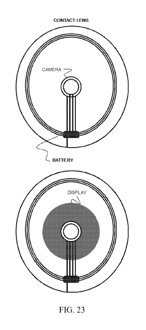
represented by a contour, polygon or morphological operator.
[0091] The boundary may be established as a function of the values associated
with the
cells in the grid. In one embodiment, the values in the grid values are
compared with a threshold
to establish the boundary. For example, in the above example, the threshold
may be set to 7.
Thus, any cell 28 having a value of 7 or greater is within the boundary and
any cell 28 having a
value of 0 is outside of the boundary. A modified view of the FOV data 26 is
shown in FIG. 4B,
in which the cells 28 meeting the above threshold are highlighted.
[0092] Alternatively, the FOV data 26 could be used to create a contour. The
visual
model emerges from interpreting the raw data and is not necessarily a point-by-
point
transformation of the raw data. The intent is to put the removed pixels as
close to where they
ordinarily would have been, thus, the algorithms in the software determine,
based on (i) the
whole of the defect, (ii) the distance of the specific pixel or ray from the
border of the defect, (iii)
whether a pixel is a new image or a part of an existing image (meaning whether
the pixel is a part
of an image or on the border of an image change), (iv) the other options for
the pixel to move
another way, and (v) where the adjacent pixels to be adjusted are being moved,
exactly where to
move such pixels/rays.
CA 03060309 2019-10-16
WO 2018/200717
PCT/US2018/029428
18
[0093] In another embodiment of the invention, vector images are used. For the
purpose
of this patent, vector images and pixels are used interchangeably. However, in
practice unlike
digital images which are made up of (usually) millions of tiny squares or
other shapes known
as pixels, while vector images are made from mathematical points connected
together by lines
and curves to create different shapes. Since they are based on math and
algorithms, not merely
pre-placed pixels, vector shapes are extremely flexible and do not suffer from
the same
limitations as pixels.
[0094] There are five (5) major Systems, and a number of Subsystems which are
a part of
the complete invention. One or more of the Systems or Subsystems may be
combined, omitted,
or integrated.
[0095] The first major system is the glasses, frame and headgear ("GFH"),
which
typically is worn on the head of a user and positioned over the eyes and nose
like typical glasses.
The GFH houses the cameras, the microcontrollers, the connectors, and
Subsystems which are
comprised of sensors, such as motion sensors, six or nine Degrees of Freedom
sensors (up/down;
back/forward; left/right; pitch/roll/yaw), gesture recognition sensors,
fiducial marker sensors,
accelerometer sensor, infrared sensors, motion sensors, alert sensors (which
would alert a user to
a danger), gyroscope technology and related sensors, positional tracking
sensors (including Wi-
Fi location systems, mobile locations systems, and RFID location based
systems), sound sensors,
and optical sensor technologies. The sensor array also can include mechanical
linkages,
magnetic sensors, optical sensors, acoustic sensors, and inertial sensors.
This list is not
exhaustive, but illustrative of the type of sensors located on the GFH. The
GFH also houses
virtual environment (VE) Subsystems such as: (1) head and eye tracking for
augmenting visual
displays; (2) hand and arm tracking for haptic interfaces to control virtual
objects and aid in the
diagnostic tools; (3) body tracking for locomotion and visual displays; (4)
environment mapping
interfaces to build a digitized geometrical model for interaction with
sensors, diagnostics, and
simulations. Other sensor technologies typically housed on the GFH are the
digital buttons,
which would include the power buttons and a D Pad or Control Pad for accessing
and controlling
functions by the user. The sensors listed above include their operating
systems and output. The
GFH also houses the connectors such as power connection for recharging a
battery or for direct
connection to and AC source, as well as other connectors for HDMI, sound, and
other
input/outputs, such as additional image overlay display, or for a diagnostics
protocol for
upgrading the system.
CA 03060309 2019-10-16
WO 2018/200717
PCT/US2018/029428
19
[0096] The GFH also houses the Microprocessor(s) Control Circuits (MCC) which
are
described below.
[0097] The GFH may also include a strap and counterweight or other headgear to
balance
the GFH and maintain its position on the head. In addition, the GFH may
include a "dongle"
whereby one or more of the Systems or Subsystems are connected via wire or
wireless to another
device, such as could be worn on a belt or carried in a pocket to reduce the
overall weight of the
GFH.
[0098] In one embodiment, the GFH is connected to another device which is
providing
power, while in an alternative embodiment, the GFH has its own power from the
Mains or from
wireless power transmission or from a battery. Further, in another embodiment,
the GFH houses
the cameras, the microcontrollers, the connectors, Central Processing Unit,
Graphics Processing
Unit, software, firmware, microphone, speakers, and subsystems.
[0099] In another embodiment, the GFH contains an RFID reader to read signals
from
RFID tags. In another embodiment, the GFH contains optical character
recognition/reader
sensors to read information from the real world.
[0100] Alternatively, some parts of the system mentioned herein are in a
dongle attached
to the GFH via wire or wireless connection. Alternatively, some portions of
the system
mentioned herein are contained in a connected device, like a laptop, smart
phone, or WiFi router.
Alternatively, some parts of the system mentioned herein are contained in a
remote location and
accessed by the GFH via Radio Frequency (i.e. cellular bands) or other
wireless frequencies or
via wireline. Thus, in one embodiment of the invention, multiple heads-up
displays on the same
headgear or on the headgear of multiple wearers are connected through a wire
or wireless
network in order to develop or control information which can be shared with
the other users.
This would be accomplished by having the GFH gather information from the
cameras or sensors
processing the information through preset filters and distributing the
information to all the other
GFH would have the ability to control the information or share the information
with all the other
GFH connected to the network. In another embodiment of the invention the
information could be
gathered from a remote location or library and shared with other HDC through
an intermediate
source like a smart phone or laptop.
[0101] The GFH also contains the battery and receipt charging DC Subsystem or
alternatively, an AC input and converter to connect directly to an AC source;
as well as the wire
and wireless Subsystems to connect or pair the device to other systems, such
as sound, alert
systems, fall monitoring systems, heart monitoring, other vital sign
monitoring, and various
CA 03060309 2019-10-16
WO 2018/200717
PCT/US2018/029428
APPs programs, cloud computing and data storage. Other Subsystems in the GFH
are a
microphone/speaker and amplifier system, an integrated Inertial Measuring Unit
(IMU)
containing a Three Axis Accelerometer, a Three Axis Gyroscope and a Three Axis
Magnetometer, or things like an Auxiliary port for custom sensors such as
range finder, thermal
5 camera, etc.
[0102] Other Subsystems like Bluetooth for near connectivity to cell phones,
tablets,
automobiles, and the like can be included as well as Global Positioning
Systems or interior
tracking systems like RFID, Wi-Fi, or Cellular tracking location based
directional travel. Other
communication systems can also be included based on either wire or wireless
connectivity of the
10 GFH. The GFH can also be connected wired or wirelessly to a main monitoring
data system
which would track the health, whereabouts, and condition of the user to be
displayed to another
person such as a caretaker or a health care provider.
[0103] In another aspect of this invention, there is no manipulation of the
pixels for eye
correction, rather, an AR headset which provides a computer mediated video
shown on a display
15 screen such that the wearer sees both the real world and the augmented
video at the same time.
In this aspect of the invention, such features as voice/speech recognition,
gesture recognition,
obstacle avoidance, an accelerometer, a magnetometer, gyroscope, GPS, special
mapping (as
used in simultaneous localization and mapping (SLAM), Cellular Radio
Frequencies, WiFi
frequencies, Bluetooth and Bluetooth light connections, infrared cameras,
other light, sound,
20 movement, and temperature sensors are employed, as well as infrared
lighting, eye-tracking, and
Dynamic Opacity are employed as set out following.
[0104] With what the inventors call Dynamic Opacity, one aspect of this
invention solves
the typical "heads-up" reflected display problem of visualization in bright
light or sunlight
conditions. In this instance, the GFH uses a bright display, typically for the
highest resolution it
could be a Quad HD AMOLED display, which is reflected onto the surface of a
lens for the user
to see the "virtual" portion of the display. In using a high-resolution AMOLED
reflected
display, the brightness can be adjusted up or down depending on ambient light.
Alternatively, the
adjustment can be in the system controller and automatically adjust depending
on what the
sensors say the brightness of the ambient light is, which would typically be
brighter when in
brighter exterior light. The AMOLED, OLED, or similar display can be one
display or two
displays, one for each eye as reflected on the lens.
CA 03060309 2019-10-16
WO 2018/200717
PCT/US2018/029428
21
[0105] In one aspect of the invention, a reflective coating is applied to the
clear lens to
enhance the reflectivity of the virtually displayed image. In another aspect
of the invention, the
reflective coating is not necessary because of the operation of the Dynamic
Opacity subsystem.
[0106] The clear lens upon which the high-resolution display which may be a
plastic like
Lexan or other clear polycarbonate or glass or any other clear material, may
or may not have a
reflector integrated into the lens to improve visibility of the reflected
display. In any case, the
outside of the lens would also be bonded to a layer containing a Liquid
Crystal Display (LCD) or
Transparent OLED display which operates to obscure the outside light to
provide greater acuity
for the wearer viewing the virtual information displayed in high lighting
conditions (Dynamic
Opacity Display or DOD). An OLED transparent display can be quite clear, which
makes
reading fine details or text on objects behind the display possible until
something is displayed on
the screen in "virtual mode," meaning something from the streaming video
reflected display is
shown on the display/lens. Alternatively, a transparent/translucent LCD can be
used as an outer
layer or middle layer of the otherwise clear lenses, and either bonded
together with the clear lens
upon which the reflected display is to be projected, to create the Dynamic
Opacity. Dynamic
Opacity senses where the image is being projected on the interior of the lens
and obscures from
one percent or less to up to 100 percent of the otherwise clear lens. In this
aspect, the clear lens
may or may not be also coated with a reflective layer. See Figure 30. The
clear lenses can also
have reflective material on the inside to increase reflectivity of the
projected image, such that the
base lens is not exactly clear, but is some percentage of obscured by the
reflective film, paint, or
other embedded reflectivity. See Figure 31.
[0107] The Dynamic Opacity subsystem is controlled by the display controller
and works
in tandem with the information displayed. The display controller creates an
image buffer for the
projected virtual display, and this information is shared with the Dynamic
Opacity controller,
.. which then activates the pixels which correspond with the exact or near
exact location where the
display controller is projecting the virtual image, so as to make the portion
of the reflective lens
upon which the image display is being projected is likewise made opaque on the
exterior of the
reflective lens, so that the image displayed appears to be brighter due to the
backlighting or light
filtering provided by the Dynamic Opacity. The Dynamic Opacity subsystem works
because the
transparent LCD or translucent OLED contain some resolution of pixels, which
in the instance of
Dynamic Opacity can be a lower resolution than the projected display, and each
pixel is
controllable by the Dynamic Opacity controller, which gets its information of
which pixels to
activate from the display controller. In the OLED the activation of the pixels
would be turning
CA 03060309 2019-10-16
WO 2018/200717
PCT/US2018/029428
22
on the individual OLED RGB pixels in order to achieve the correct level of
opacity to
compensate for existing brightness for the condition experienced by the user.
In this instance, the
RGB pixels can be activated to create a "shadow" effect or depending on the
type of light which
is extant, an emphasis on either Red, Green, or Blue, or combinations of the
three. In addition,
the Dynamic Opacity subsystem can be pre-programmed to provide a user with
various options
from warm color to cold (amber to green) for a sunglass effect on the exterior
of the reflective
lens. In the case of the LCD the activation of the pixels is one or more
phases and changing the
polarization of the pixels to achieve opacity on the exterior of the glasses
for the same effect. In
this instance, typically, an LCD unit would be employed which does not include
a RGB
component, as just outside ray blocking is needed.
[0108] Alternatively, with Dynamic Opacity, any other transparent material
which
provides electronic control of pixels or areas inside the transparency to
create an opaqueness can
be used. In either case, the outer layer would typically be transparent to the
user providing a "see
through" lens to the real world, until some virtual information was displayed
on the Head
Mounted Display Unit reflective lens, such as a hologram, a 2D image like a
movie, or other 3D
image or information, including text.
[0109] In this embodiment of the invention, a controller, like Model View
Controller
(MVC), would control the Dynamic Opacity Display through corresponding data
input
information about where the reflective display is projecting information. In
this instance the
MVC would identify in the buffer or elsewhere, in digital format, where the
images are going to
be displayed on the reflective display, and the MCV would anticipate these
locations and turn on
pixels, including RGB pixels in the transparent LCD or OLED, and "cloud" or
rather make more
opaque the portions of the lens corresponding to the areas of the lens where
the virtual image is
being displayed on the inside or other layers of the reflective display. In
this fashion, the
Dynamic Opacity provides a "backdrop" or "background" display corresponding to
the pixels
where the virtual image is displayed making the contrast of the virtual
display greater to the eye,
so that brightness like natural sunlight can be minimized, which would
otherwise compete with
the reflected display and cause it to be hard to see. With the Dynamic
Opacity, the reflected
display has a buffer between it and exterior light, which gives the reflected
display greater
brightness to the eye.
[0110] The Dynamic Opacity could be in either a course or fine mode, meaning
that the
opacity from the Transparent OLED or LCD would either appear in the general
area of the
virtual display or for fine applications would appear in almost or the exact
same pixels which
CA 03060309 2019-10-16
WO 2018/200717
PCT/US2018/029428
23
correspond to the image pixels being displayed or reflected on the interior of
the lens. In another
aspect of the invention, the Dynamic Opacity can work with wave guide displays
or prism type
displays with equal effect. Likewise, the Dynamic Opacity described here can
be used with a
micro-mirror type display with equal effect.
[0111] There are many methods to identify exactly where the coarse or fine
opaqueness
should appear, but one embodiment would use the same eye-tracking as the
primary display/lens
and the MVC would know exactly where the eye gaze is and how far each way on a
six way axis
the virtual display is centered and extends, so that the opaqueness mimics the
same space as the
virtual display according to where the eyes are gazing as identified by the
eye-tracking software.
In this way, the reflected image display overcomes the issue of not being
bright enough in
daylight and other high light conditions. Likewise, the Dynamic Opacity
includes transparent
OLED or LCD overlay or layer of the lens can also act as "sun glasses" for the
display and "tint"
the entire display to compensate for bright lights, like on a sunny day. Or
with similar effect, a
light valve can be used with the same effect in a similar manner. A light
valve (LV) is a known
device for varying the quantity of light from a source, which reaches a
target. Examples of
targets are computer screen surfaces, or a wall screen or in this case the
coarse or fine coverage
of the virtual display on the glasses lens.
[0112] In the Dynamic Opacity technology, the MCV can be pre-programmed or
programmed to automatically compensate for external brightness and act as
instant "transition"
lenses and can be either used on the AR glasses display or with computer
intelligence can be
used on typical corrective lenses. In this case, the entire exterior layer of
Transparent OLED or
LCD would tint much like a light valve to balance the bright external light,
and still provide
additional opaqueness on the portion of the lens where the virtual video or
picture or image is
being displayed.
[0113] In another aspect of the invention, the display can be a small display
like OLED-
on-Silicon micro-displays. Such a display device consists of two key elements:
the silicon
backplane that contains circuitry to drive the OLED pixels, and the OLED
emissive frontplane
layer. With a small micro-display which is only 1 inch by 1 inch but contains
2.5K by 2.5K
resolution, with as bright a display as possible (1,000 NITS) one can use two
displays, one for
each eye, to be the projector on to a reflective or semi-reflective lens. In
this case, the micro-
displays can serve as the projector for a reflected display which the eyes of
the wearer would see.
The correction or fine tuning is offered by keystone corrections contained
within or on the GFH
and the correction for projection of the reflected display.
CA 03060309 2019-10-16
WO 2018/200717
PCT/US2018/029428
24
[0114] In another aspect of the invention, one or more small micro displays
like the those
offered by TSMC, which is a 1 inch by 1 inch, 2.5K by 2.5K resolution
display(s) can be used to
project an image onto a clear lens connected to the a head mounted display
that contains
computer intelligence through a CPU and can be known as a Smart Head Mounted
Display
(SmartHMD) or GFH. In another aspect of the invention, there would also be
either or both a
layer of reflective film on the lens or the outer layer of the lens contains
the Dynamic Opacity
technology as explained above. In this instance, a corrective lens or lenses
can be affixed to very
small micro-displays, which are bright enough to provide a reflected image
onto the reflective
lens. In this instance, the micro-displays in order to correct and fine tune
the image for displaying
on an ultra-short throw between the display and the inside of the reflective
lens can utilize one or
more image correcting lenses and can even be combined with a middle layer of a
wave guide or
polarization, which provides enhanced image resolution and guides the image's
rays to exactly
where it is to be displayed on the reflective lens.
[0115] In one embodiment of the invention, two corrective lenses sandwich a
wave guide
or polarization layer. The image projection source is a small display, as
shown in Figure 32, that
is rotated to achieve the greatest clarity and field of view. The image source
(OLED) is then
passed through a circular polarizer. The circular polarized image is then
passed through a lens
with a positive diopter to focus the light through a linear polarizer. This
linearly polarized light is
then passed through a negative diopter lens, and possibly multiple negative
diopter lenses to
achieve the necessary projection size required. The purpose of the polarizing
films used either in
combination with other correcting lenses or not, is to retard the light that
may be reflected back
onto the micro-display and to focus the light rays on the specific part of the
reflective lens as is
desired. After a passing through the lens curvatures which will provide the
correct size of
projection, the image is then reflected into the eye using a spherical lens,
possibly coated with a
semi-reflective or reflective surface. In this aspect of the invention, the
angle of the display and
lens combination to the angle of the spherical reflection surface will be
adjustable to provide
focus for eye location, which can be monitored using eye-tracking technologies
combined with
the control of the projected image. Further, an adjustment can be permitted on
the corrective
lens which is correlated to the micro-display, and thereby one can change the
closeness of the
lens to the micro-display, which would permit the user to adjust the reflected
lens display closer
or further from the user's face, to better allow room for the user's own
corrective glasses or large
facial features, like a large nose or other gear worn on the face like an
oxygen mask or filter
mask (i.e. like for a fighter pilot or in a HAZMAT situation).
CA 03060309 2019-10-16
WO 2018/200717
PCT/US2018/029428
[0116] The Eye-Tracking subsystem works through hardware and software. The
software is connected to the system's GPU working in connection with the
systems model
controller. The eye-tracking is captured by infrared (IR) light being
projected onto the eye,
which creates a glint or reflection, which is then captured by an IR sensitive
camera. Typically,
5 an eye-tracking system captures the glint from the eye from 30 frames per
second to 500 frames
per second. This information is stored in real-time in the Model Controller,
which can be a
MVC, and them processes this information into a virtual space represented by
XY or Cartesian
coordinates. These coordinates provide the system with the information about
where the user's
gaze is in relation to the reflective lens. When used for medical applications
like AMD, the eye-
10 tracking information is correlated with the buffered information about
the person's eye visual
defect such that when the manipulated image is displayed, it is in sync with
the user's gaze. This
is necessary because the eye scanning and eye movement necessitates that the
buffered and
manipulated area of the video be moved to correspond to the user's eye gaze so
that the buffered
"hole" and the user's defect align and remain in sync. All this processing
happens in real-time
15 and keeps up with the movement of the user's eye. Latency is important
and keeping the latency
to less than 10 milliseconds will aid in preventing the user from feeling
dizzy and preventing
whirr.
[0117] In another embodiment of the invention, a computerized worm gear or
drive is
used, or non-computerized mechanical device such as a worm gear or gear can be
used to move
20 the micro-displays on the GFH such that the displays can be aligned with
a person's own Inter
Pupillary Distance or IPD. In the case of a computerized worm gear, this gear
can get its
information about how far to move in one to four directions from the eye-
tracking subsystem,
which can measure the distance from the gleam detected in each of the person's
eyes and
transmit measurement data into movement data so that the worm drive aligns the
micro-display
25 in the GFH to the perfect position for the persons own IPD and relative
height vis-à-vis the way
the GFH is worn, so that alignment side to side and up and down is
accomplished. Alignment of
the user's eyes on a four axis is necessary because this ensures the sharpest
reflected image for
each individual user in combination with how the user wears the GFH.
[0118] In another aspect of the invention, the GFH can be made where it is
locked on a
user so that in institutional environments it cannot be easily removed. In
this aspect, people,
such as inmates of some type would be required to wear such GFH headgear, so
that if there is
trouble or emergencies, a manager could either cut off the video feed leaving
the user with only
limited sight resources with which to navigate. This may reduce the desire to
become aggressive
CA 03060309 2019-10-16
WO 2018/200717
PCT/US2018/029428
26
or provide information for emergency exit. In this embodiment, the display
screen is subject to
the command of an outside operator, and could display, for instance, peaceful
pictures, and
soothing music to calm the user experiencing a fit. Or the display could
become opaque and
deny the user the ability to see. Or the display could be used to heighten
awareness with
magnification, color enhancements and sharper contrasts of images and sound.
The GFH could
also be used to dispense smells to either enhance a pleasurable experience,
permit a focus on
identification of a person, or thing, or for training purposes, like to give a
user an artificial
experience like would exist in a simulation or another not currently existent
real-world situation.
[0119] In another aspect of the invention, the GFH is more like a helmet or
the display
more like a face shield than lenses.
[0120] In another aspect of the invention, the GFH is more like a band and the
reflective
display is like two partial spherical clear lenses, one partial sphere over
each eye.
[0121] In another aspect of the invention the real world is not displayed, but
videos,
television shows, emails, or other online or prepackaged information is
displayed, either with or
without the macular degeneration type pixel manipulation, so that a user could
experience other
forms of entertainment, training, learning, or task accomplishment with the
Mixed Reality
Glasses than just a real-world projection onto the display. The GFH can also
be fitted with
night-vision, infrared, or other types of cameras so that the experience is
hyper real world. Thus,
any kind of camera can be used to make a display. In this embodiment of the
invention, the GFH
can be programmed to act as a host for other devices utilizing technologies
like Apple Airplay,
which permits the GFH to be "paired" with other devices, like a phone or smart
watch. In this
instance, one would refer to the GFH as a smart head mounted display or
SmartHMD and all the
applications (apps) on a person's cell phone or tablet can be transferred to
the GFH seamlessly.
So, in this instance, a user could begin a Hulu or Netflix movie on a cell
phone or tablet, and
then it could be directed or transferred to the GFH for continued viewing,
which would free up
the cell phone for other use. In this aspect of the invention the GFH is
connected to the internet
via cellular or WIFI or other radio frequencies or wireline or wireless
frequencies and acts like a
router with other devices which can attach themselves to the GFH, much like
computers acquire
and connect to a typical internet router. This provides the GFH with the
ability to access the
internet.
[0122] In another aspect of the invention, the GFH is loaded with Artificial
Intelligence,
like the Google virtual assistant, Sin, or Alexis. In this instance, the GFH
can be programmed
with a virtual assistant virtual image and be able to show a visual virtual
assistant (VVA), not
CA 03060309 2019-10-16
WO 2018/200717
PCT/US2018/029428
27
just a voice like Sin i or Alexis. Technology like invented by a group from
the University of
Washington, where researchers have created a new tool that takes audio files,
converts them into
realistic mouth movements, and then grafts those movements onto existing
video. In this instance
the AT software neural nets are trained to change a video of a speaking mouth
to other words can
be used to create a VVA with a minimum of actual videos taken of the live
subject which is to be
the VVA.
[0123] Under this embodiment, the GFH can include either speakers which would
be
controllable or have incorporated into its sound system earbud type speakers,
which are either
attached via wire or via a wireless network in the GFH like Bluetooth light.
[0124] In another aspect of the invention the cameras can be used to not only
display an
image in real time to the user, but to record the image that a camera captures
for replay later.
Thus, a user, if sleepy, could activate a "record" button, causing the CPU and
GPU to record the
real-world images, for instance from a football game, and the user, when
awakened, could then
enable the recorded display to show on the lenses of the GFH. This feature
could also be used to
recall real world experiences, for instance to record a university lecture for
playback and
contemplation at another time. The playback can be in real time, slow motion,
freeze framed,
stopped, and fast forwarded or reversed. In this aspect of the invention the
GFH has a Subsystem
which permits storing data and replaying data and menus to identify the stored
information, or to
recall an instruction previously given. In this instance the user could
activate the record when
taking medication and the CPU would log such information and be able to
respond to visual, text
input or auditory requests, like, "did I take my medicine today" to which the
GFH would respond
yes or no or not known, depending on whether the recorded information was
available. In
another aspect of the invention the record function can be configured to
automatically record
certain functions, like image recognition software which could activate the
recording of taking
medicine, convert that to data base information, and be able to play back the
correct information
to the user. The GFH could also become Bluetooth enabled when in the proximity
of other
devices, like a pulse oximeter or blood pressure cuff, and automatically
record this information
and store it in the data base to be replayed, recorded for later use, or sent
to a third party, which
might be a caretaker or health care provider or store for recall by the user.
[0125] In another aspect of the invention other meaningful information can be
displayed
along with either the real-world information or non-real-world information
(such as TV or a
movie) where a user can be altered or amended by text information or sound to
conduct a certain
time-based task, like, for instance, an alert to take medicine, check on a
pet, or answer a phone
CA 03060309 2019-10-16
WO 2018/200717
PCT/US2018/029428
28
call or email. In this instance the GFH would permit the user to use the D-
Pad, Fiducial Marker,
or other controller to switch from a real world or non-real-world experience
on the display to a
task-based experience, such as an email or phone call or video phone call.
These examples are
but a few of the many tasks that would need the user to switch the input of
the display, and all
examples used herein are by way of illustration and not limitation, in this
specific case, the main
idea being that the GFH would be akin to a wearable computer, and permit a
change in the user's
environment and display to correspond with the task or undertaking necessary
at the time,
whether to see the real world, to see the non-real world, or use the GFH as a
wearable computer,
online device, Wi-Fi device, RFID device, Near Field Communication device, or
other
communication device, learning device, or a smart device, like one that would
clock elapsed
time.
[0126] In another aspect of the invention, the GFH acting as a wearable
computing
device could process a credit card payment or undertake some other task that
the physical
limitations of the user would otherwise prohibit or would enhance.
[0127] In another aspect of the invention, the GFH does not provide specific
correction
for eye diseases like macular degeneration which requires repositioning pixels
or vectored
images but does contain all these Subsystems which exist to inform the user
and show a user
how to reach a certain waypoint, or prioritize travel, all displayed on the
lens display of the GFH.
And in another aspect of the invention, the pixel manipulation is used, but
not to correct for eye
defects like macular degeneration, but to reposition a display onto a certain
portion of the lenses,
so that a user can see both the display and the real time world at the same
time.
[0128] Thus, the GFH can contain other wearables technology to monitor,
report, and
track or direct the user. This can be done by audio, or within the display or
as a separate display,
where, for instance, the real-world environment is displayed, and a text is
also shown of
directions, or alerts or any kind of useful information to the user. Alerts
could also be signaled
by vibrations from the GFH. The GFH can also signal messages to people
external to the GFH,
and, for instance, to alert third parties that an impaired sighted person is
passing. Or alert third
parties that the person has some sort of authority, like a siren, or flashing
light, in the case of
police officers or emergency personnel.
[0129] The GFH also contains the Image Projection and Lenses (IPL) System
which is
the combination of the projector and lenses upon which the image or corrected
image is to be
displayed, along with their connectors and integration with the other Systems
and Subsystems.
CA 03060309 2019-10-16
WO 2018/200717
PCT/US2018/029428
29
[0130] The GFH also contains connectors for a patient diagnostics programming,
and
computer interface, for wearable computing functions and other Subsystems,
explained herein.
The examples above are designated herein as "subsystems" or "Subsystems" of
the invention
which also is understood to include all powering, connectivity, computing,
display, and
integration of the Subsystems. The computing and patient diagnostic
programming can be
resident in the system or external through a connector. Thus, for instance,
the patient diagnostics
programming can be in the circuitry and intelligence of the system, the GFH or
accessed
externally through wire or wireless connections to a device like a tablet,
laptop, computer, or
mainframe. The GFH may all be worn on the head, or be like a helmet, or be
dispersed on other
parts of the body as auxiliary wearables.
[0131] The second major System is the Camera Input System (CIS), which
typically
includes one or more cameras and their lenses, connectors, and operating
systems. As mentioned
above the cameras can be of a typical video or still camera or can be of a
specialized nature like
night vision, infrared, 360 cameras, thermal imaging, magnification, color,
black and white, or
3D cameras with each their own distinctive displays. One or more of each of
these different type
cameras can be incorporated into the CIS System.
[0132] In a typical medical correction configuration the GFH would contain one
or more
camera and camera systems for capturing the real word visuals that the user
would ordinarily
see; and also can contain one or more cameras which monitor eye movement so
that corrective
software can receive this eye positioning information and approximate the
epipolar geometry of
the eyes (eyes moving inwards or outwards, left or right, transversely) and
calculate for the same
as well as the offset of the line of sight of the cameras versus the actual
eye position so that the
display shows nearly what the user's eye would ordinarily see.
[0133] In another embodiment of the invention, the CIS may be partially or
completely
embedded on smart contact lenses, where the cameras, in the instance of
macular degeneration,
are positioned on the smart contact lens (SCL) in the exact location where no
sight exists, being
typically in the most central 15% of the eye. In the instance of the SCL it
may contain is own
battery, sensors, communication, and charging apparatus including
communicating via methods
such as backscatter, Interscatter, Bluetooth, Wi-Fi, ZigBee, RFID, and other
antenna
transmissions. In these instances, the GFH provides the energy to be harvested
by the SCL and
the communication network and protocols, for wireless communication, all of
which are a
Subsystem of the GFH. Thus, if SCL were worn by themselves they would need
another device
CA 03060309 2019-10-16
WO 2018/200717
PCT/US2018/029428
to harvest energy and communication reference from, here the GFH System
provides the
necessary energy and communication link and are synced together.
[0134] In another embodiment of the invention, one or more cameras per eye are
used to
create monocular or binocular vision. In this instance the GFH System would
also have a
5 method to monitor the movement of at least one eye, like a camera in the
GFH facing back
towards one or more eye to monitor the eye movement, for line of sight
augmentations to the
projected image, and for epipolar geometry corrections for the movement of the
eyes focusing on
far away versus close items. One Subsystem and method for monitoring the eye
in another
camera, one or more which is directed at least one eye. This camera would
utilize eye tracking
10 software to provide to the IMP the information necessary for an
adjustment in the display so that
the image displayed as nearly as possible represented the real-world images,
thus, there would be
correction for epipolar geometry and line of sight at least in the software.
[0135] In another embodiment of the invention one cameras is used, creating
monocular
vision to be displayed to one or both eyes. In this instance, the monocular
vision can be
15 corrected per eye, so that the "cut outs" are different for each eye,
such that the correction best
suits each eye differently.
[0136] In another embodiment there are one or two or more cameras per eye,
receiving
real world input. In the instance of using two cameras per eye, it is
recommended that they
would be offset towards each other, so that each camera's FOV intersects the
other. This is
20 because when capturing a wide Field of Vision, the cameras themselves
interject a certain
amount of distortion. A typical camera lens, which does not introduce a great
degree of
distortion is only up to about 75 degrees FOV. Thus, to capture more than 75
degrees FOV,
which is often necessary under this patent's teaching, two cameras are
recommended to avoid
wide-angle lenses, which introduce distortion, and avoid the most distortion
from camera lenses
25 that attempt wide FOV. However, by using the joint image from two cameras,
and then
"stitching" the image together as one in software, less distortion is
introduced into the actual
image to be manipulated and a higher degree of pixel accuracy is maintained
from the camera
input to the Image Manipulation Program(s) (IMP).
[0137] The third major System is the Microcontroller Control Circuits. This
group of
30 chips, parts, circuits and circuit boards include one or more
microprocessors, its circuit board and
parts, and typically a specialized Application Specific Integrated Circuit
(ASIC) which may be a
separate chip or housed in one of the other chips in the microprocessor
circuit board. The MCC
does the main functions of the invention and receives the input from the CIS
and sensors, runs
CA 03060309 2019-10-16
WO 2018/200717
PCT/US2018/029428
31
the routines and programs for collecting sensor data and visual images, and
then corrects for the
macular defect of the user and controls the display. Portions of the MCC
System are controllable
by the user, especially related to the Macular Degeneration Diagnostic Program
(MDDP)
Subsystem. This MDDP Subsystem contains the software and firmware for the
patient
application defect mapping program which establishes the boundaries, one or
more, per eye, of
the defect area, as well as the boundaries of the area of projection. The MCC
also houses the
Video Manipulation Programs (VMP) which collects the camera input and
repositions the image
and pixels, for corrected vision display. The MCC also houses the Application
Program
Interfaces as well as the Graphic User Interfaces (GUI) and routines. The MCC
also houses the
controllers for all of the sensors, inputs and outputs and user control.
[0138] As stated above the VMP may be any number of kinds as described
previously, or
could be a Pixel Manipulation Scheme or Vector math like taking the image from
the real world
such as the Pixel Interpolation and Simulation, Image Stretching, or other
software video
distorting application.
[0139] In one embodiment, the flat picture as sent to the buffer by the camera
and is
turned in to a "fisheye" or "barrel" distortion where the middle is larger and
then the image is
squeezed at the edge. In this instance, the central image, which is as near as
possible to the
deficit of the person's disease, is removed and the image is stretched and
displayed. In the
instance of the goggles, the edge is not critical, and may simply be "cropped"
to permit the
central portion of the video to be displayed without the edges, which have
been pushed out by
cutting the central portion out. In another embodiment of the invention the
edges are important,
like in the case of the Mixed Reality macular degeneration glasses where Phase
Two distorted
images must be remerged into Phase Three video images.
[0140] Thus, this invention teaches that one camera can be used for monoscopic
image
capture and display. In addition, this invention teaches that you can use two
cameras to simulate
on the goggle/glasses display true stereoscopic vision, wherein the IN/ID
model includes factor
correction for epipolar curves, guided by the epipolar geometry so that stereo
vision, generated
by two or more cameras, can be employed and be displayed, and seen, as one PRI
image.
[0141] The invention uses computer aided video images which are skewed and
stretched
in a matrix distortion or other similar fashion to put the most or the
entirety of the image onto the
peripheral vision of the patent by opening up the center of the image and
manipulating it to the
peripheral cones of the eyes, as seen by the patent in the projected image, in
order to project the
video captured images on the peripheries of the cones in the eyes where vision
is still active. One
CA 03060309 2019-10-16
WO 2018/200717
PCT/US2018/029428
32
of the benefits of this invention is that no invasive procedures are necessary
and as the patient's
macular degeneration changes the software can be adjusted so that the image is
now correctly
skewed.
[0142] In the fashion taught by this invention, the viewed experience may make
it nearly
impossible for the user to distinguish between what is actually seen and the
image that is created
by the PRI.
[0143] Thus, the spreading and/or multi-lateral skewing of the image reflects
the
corrected image onto 3D or High Definition goggles and/or glasses worn by the
patient. The
image is skewed via the IMD module to avoid projection to the area of the eye
which involves
the macula, but still has all the image information. To imagine this process,
think of a picture
which is printed onto a stretchable and compactable substance. A hole is cut
into the middle of
the image and stretched open. The image compress into the sides of the
picture. Thus, all of the
information of the picture is still there, it is just rearranged where a hole
is in the middle and the
image is moved each way to the side, top, and bottom. This "hole-cutting" is
done via algorithms
and computer software/firmware technology, for instance, using a technology
like Matrix
Distortion as above mentioned.
[0144] Matrix Distortion of a camera and Matrix Calibration, which is the
correction of
the distortion are commonly known areas of camera calibration and have been
used for a long
time. Often times cameras display significant distortion. However, the
distortion is constant like
on a matrix, and with a calibration and some remapping the distortion can be
corrected. Typical
distortion correction takes into account the radial and tangential factors.
For the radial factor one
uses the following formula:
Xcarrated = X- (1 4- 1(112 + +K3 H
=Igo+ r2 r4 k3r46)i
[0145] So for an old pixel point at (x,y) coordinates in the input image, its
position on the
corrected output image will be (x {corrected} y {corrected}). This corrects
for the presence of
the radial distortion which manifests in form of the "barrel" or "fish-eye"
effect.
[0146] Tangential distortion occurs because the image taking lenses are not
perfectly
parallel to the imaging plane. It can be corrected via the formulas:
1,41
= f7 :AI
X,correttai ¨ X XII pAr. 1-=
ibwrected + [Pi (r2 + 42)
+ 21,24
[0147] However, for this invention a type of reverse methodology is employed,
that
would not normally be thought of Thus, once typical distortions in the camera
have been fixed,
CA 03060309 2019-10-16
WO 2018/200717
PCT/US2018/029428
33
then, it is the teaching of this invention that an intentional distortion is
introduced. In one
embodiment the IMD model stretches a center pixel to the points at which an
individual cannot
see, and compresses everything else to fit in the remaining peripheral portion
of the goggles. In
this fashion a "hole" is artificially cut into the image by computer and
software/firmware aided
manipulation such that a pixel which was formerly in the center of an image is
squeezed to the
outside so that the entire image in projected around the "hole" in the center
which is artificially
created. Only the matrix distortion portion of the model is shown here, as the
other pieces are not
directly related to the IMD model. There are other substantive parts of this
program for
projecting the image once the IMD model is applied. As shown the IMD
distortion model is
shows as a value to the "webGL"1, a program which can be used with
"renderingContext"2.
[0148] There are other substantive parts of this program for projecting the
image once the
IMD model is applied. As shown the IMD distortion model is shown as a value to
the
"webGL"1, a program which can be used with "renderingContext"2.
[0149] The fourth major System is the Image Projection and Lenses System. The
IPL
projector and lenses may employ such technologies for display such as wave
guides, mirrors,
prisms or other technologies, such as transparent rear projection film, to
correctly display the
image on the glasses (lenses) or on a portion of the lenses. Alternatively, a
"heads-up" type
display may be used, such as a transparent shield or facemask. In practice,
the lenses may be one
of any of a number of types of see-through displays, like Augmented Reality or
Mixed Reality
glasses, or can be immersive, and not transparent like Virtual Reality
goggles. Some examples
of organic light emitting diodes (OLED) which can be employed are Passive-
matrix OLED,
Active-matrix OLED, Transparent OLED, Top-emitting OLED, Foldable OLED, Lucius
Prism
OLED, White OLED, Quantum dot light emitting diode (QLED), ultra LED (ULED)
and Ultra
HD 3840x2160 pixel resolution, also called 4K, which is twice the resolution
of Full HD and has
4 times the number of pixels. A recommended combination is transparent Active
Matrix OLED
(AMOLED) with the evolution of technology as it is now because AMOLED's are
thin, have
fast refresh rates, are less complex from an electronics standpoint, offer a
lot of control over
individual pixels, and consume low amounts of energy, especially sense they
produce their own
light; and they have high resolution and produce sharp colors, which is needed
for the invention
__ to work at its optimum. In another configuration, lenses, such as Corning's
transparent display
technology and features Corning Gorilla Glass could be used. The application
of a special
functional film on the thin, durable Gorilla Glass surface creates a
transparent display that is
acceptable for displaying real time augmented video onto the GFH lenses. In
addition, the
CA 03060309 2019-10-16
WO 2018/200717
PCT/US2018/029428
34
application of technologies such as LG Display's N Pixel technology can assist
the invention by
making the pixels clearer from any viewing by the eyes. Further, technologies
such as retinal
projection can be used, and would be housed in the GFH.
[0150] The fifth major System is the Diagnostics Impairment Mapping (DIM)
System
and tools, which include virtual simulations and tools, a user manipulated
method of viewing a
grid and using hand gesture sensors or tools like fiducial markers, or a
connected mouse to
identify the area and boundaries where no vision exists, so that this mapping
can be obtained
from the real "analog" world and transferred into digital coordinates for
correction by the Video
Manipulation Program. In this instance the user would select "Diagnostics"
setting, and an
Amsler grid would appear on the lenses one at a time, while one lens was being
evaluated, the
other lens would be opaque to not let the user be distracted by "see through."
The user would
trace where the edges of the border of the sight is which is then transposed
by the MCC to
specific mathematical coordinates which create a border where the image is to
be removed and
replaced elsewhere. The Diagnostic Test could be employed as often as the user
desires to refine
.. and re-correct for the advance of the disease.
[0151] In another embodiment of the invention, the display screen on the GFH
is curved
slightly, so as to reduce the reflections of ambient light from the display,
thus improving image
contrast, and focusing more of the image on the eye peripheries. The slight
curvature also
reduces the optical distortion (keystone) in the screen image geometry,
especially farther away
from the central portion of the display, were no or little image is displayed
in the case of macular
degeneration.
[0152] In another embodiment of the invention, normal corrective
glasses/lenses are used
and a film, like 3M translucent rear projection film is used and simply
affixed to the corrective
lenses, or the corrective glasses are affixed to the OLED material so that the
patient has both his
correction and the pixel manipulation in the same set of lenses.
[0153] In another embodiment of the invention, the correction for typical non-
retinal
problems of the eye like astigmatisms, myopia, hyperopia, or presbyopia is
done in the MCC.
Pixel corrections can be combined with the pixel manipulation techniques so
that that the
displayed video image corrects and compensates for that person's native other
visual
impairments, by using algorithms that adjust for the myopia or hyperopia
through techniques like
increased focus, increased contrast and enlargement of the video with known
techniques like
fixed parallax barriers, lenticular lenses, pre-filtered light display,
switchable liquid crystal
barrier or display, multilayer display, diopter adjustment with independent
eye focus, or pre-
CA 03060309 2019-10-16
WO 2018/200717
PCT/US2018/029428
filtered light field display and the deployment of self-illuminating pixel
technologies in the
display and specialized lenses on the camera to correct for the non-macular
problems of the eye
astigmatisms, myopia, hyperopia, or presbyopia. In this way the invention
replaces corrective
optics to correct vision, with computations within the software and other
aids. In another
5 embodiment, the camera lenses have the correction needed or that works
with the computed
correction in software.
[0154] If the camera lenses are not corrective, then the image correction is
made in the
software, firmware, or hardware, so that the device corrects for both the loss
of sight, like in
macular degeneration, and also for problems like myopia. In this fashion, a
person wearing the
10 GFH system would obtain two types of correction in the same display, (i)
one for the macular
degeneration, and (ii) another for the nearsightedness or farsightedness. In
this situation, the
invention teaches that by pre-filtering, the video on the display computes a
pre-filtered light
field, or uses other similar technologies, which results in a desired
projection of the displayed
image on the retina of a user or patient which corrects for their exact eye
problem. By
15 eradicating the rays which do not directly hit the retina at the precise
angle necessary for the best
correction, a user's eye prescription can be obtained without the need for
corrective glasses. In
other words, the correction which is computed into the video, can be adjusted
on the fly, or in
real time by the user via a fiducial marker, D Pad, or Control Pad ("focus
controller"). An
adjustment on the control pad would automatically correspond with a change in
the filtering so
20 that a more precise image is displayed on the lens and on the retina of
the patient's eye. This
correction can be done for each eye, so that the display on one eye is
different than the display on
the other eye and each eye display can be adjusted independently by the focus
controller. Also,
the problem of scanning or eye-tracking is solved by having the cameras needed
for the
correction on the smart contact lenses, which then permits the cameras input
and displayed
25 images to match that of the movement of the eyes.
[0155] In another embodiment, the augmented video may be displayed on the
lenses and
include the central 10 to 60 degrees FOV, for example, or any other desired
FOV. This
displayed video would encompass Phases One and Two. Then the stitching
techniques would be
employed on the "edges" of Phase Two the augmented video, here, in this
example, beginning at
30 60 degrees FOV and using, there would be projected/displayed, for
example, on another 20
degrees FOV to re-interpolated and phase back into real-world, non-adjusted
video. Pixel
mapping techniques can help retain image edge features better and produce
higher accuracy of
integration of a real-world image projection. Thus, a user would have his or
her central most
CA 03060309 2019-10-16
WO 2018/200717
PCT/US2018/029428
36
vision augmented via the projected video, while the video further from the
central vision is
reintegrated into the real world non-adjusted video, and then there is no
video on the outermost
peripheral areas where actual vision is used.
[0156] In one aspect of the present invention, the data comprising the visual
model may
be filtered or transformed to eliminate noise or other undesirable effects
within the data prior to
the boundary (or boundaries) being established. This process may be performed
automatically
using a set of predefined operations, or may be performed under the control of
an operator of the
model controller 14. For instance, the data may be filtered using one or more
morphological
transformations. Possible morphological transformations or operations may
include, but are not
limited to: erosion, dilation, opening, morphological gradient, top hat,
and/or black hat. An initial
boundary may be established using pre-filtered data and a secondary boundary
may be
established after the data has been filtered or transformed. The initial and
secondary boundary
may be compared automatically or by the operator to optimize the boundary
used. Alternatively,
Boolean operations may be used to filter the visual model and/or combining
boundaries.
[0157] In another aspect of the invention the pre-filtering can also include
the pixel
manipulation which by using a parallax filter or other filter permits only the
pixels which are
rays that are at such an angle to miss the area of defect are utilized to be
projected.
[0158] In one aspect of the present invention, the threshold is adjustable,
either at the
model controller 14 or at the display controller 16. If performed at the model
controller 14, this
would provide control to the operator. In adjusting the threshold, the
operator could optimize the
boundary. If performed at the display controller 16, control would be provided
to the patient.
This would allow the patient to adjust the boundary to optimize the boundary
for current
conditions.
[0159] One method for making sure that the digital pixel manipulation exactly
replicates
that of the analog eye, a fiducial marker is connected to the diagnostic
system resident in the
GFH. A fiducial marker is an object placed in the field of view of an imaging
system which
appears in the image produced, for use as a point of reference or a measure
merging the analog
world with the digital world. Its applications are often seen in commercial
products like virtual
games. It may be either something placed into or on the imaging subject, or a
mark or set of
marks, as is preferable in this instance, in the reticle of an optical
instrument, which is the
measured camera and display. This diagnostic system is combined with the pixel
manipulation
system such that the input of the diagnostic system causes the pixels
identified by the user as
non-sighted or defective to me moved to a different location as is more fully
explained below. In
CA 03060309 2019-10-16
WO 2018/200717
PCT/US2018/029428
37
the diagnostic state, Amsler Grid has been included in the software to be
projected onto the
lenses. A sample Amsler grid of a person with normal vision and a sample
Amsler grid of a
person with AMID are shown in FIG. 22.
[0160] The fiducial marker, or mouse or other similar device is connected to
the software
so that a location on the visual grid the user sees corresponds to the virtual
grid resident in the
software. The user then looks through the glasses at the grid and utilizes the
fiducial marker to
identify the exact edges of the non-sighted space, which is then converted or
identified by the
fiducial marker software or firmware as the space from which pixels and images
must be moved
and manipulated. In another embodiment, the output of a wearable FOV test is
used. For
example, the embodiment may use an automated program embedded in the wearable
HMD/HUD
display device 50, 60. An initial start-up and mapping routine would be
performed by
observation, such as looking at an Amsler grid or moving objects to check the
UFOV, or both,
utilizing an existing FOV map to modify and optimize. Eye tracking technology
may be used to
ensure more accurate FOV mapping, and validating fixation. Since eye movements
can be as fast
as 600 deg/s. and the smallest time constant for saccades is around 50 ms; and
the smallest
saccades could be completed in 60 milliseconds, thus, it is possible for the
"reverse cameras"
which are a part of the CIS System looking at the eyes to sample eye movements
at a rate of 1
kHz which will allow sufficient precision of tracking of the eyes to let the
system know how to
modify the output in near real time for epipolar geometry and line of sight
offsets. This result is
immediately usable directly as the digital input for the UFOV for the Matrix
Mapping
Technology.
[0161] In another embodiment of the present invention, the boundary 32 may be
adjusted
or replaced with a simpler form (boundary 32', see FIG 6). For instance, the
boundary 32 may be
replaced with a boundary established as a function of one or more predesigned
shapes and the
visual model. The model controller 14 may utilize a set of predefined set of
shapes, for example,
rectangles, triangles, ovals that are sized to include the affected area. The
model controller 14
may select one or more shapes automatically, or the process may be performed
by, or with the
assistance of, the operator.
[0162] With reference to FIG. 7, the shape of the defect or damaged area 24'
may be
more complex. A complex boundary may be established using the threshold
process identified
above, or by some other method. Alternatively, the initial boundary may be
replaced
automatically, or with operator input using one or more of the predefined
shapes, sized to cover
CA 03060309 2019-10-16
WO 2018/200717
PCT/US2018/029428
38
the defect or with the results of the user using the fiducial marker. In the
example of FIG. 8, two
shapes 34A, 34B are used. The boundary may be formed by the outer edge of the
joined shapes.
[0163] With reference to FIGS. 9 and 10, in one aspect of the present
invention, the
image data inside the boundary 32 is shifted outside of the boundary 32. In
the example shown in
FIG. 9, first a center point 36 is established. The center point 36 may be an
actual center of the
boundary if the shape of the boundary is regular, or it may be defined by
finding or estimating
the center of the shape defined by the boundary or the center point is ignored
and the other items
as described above are used to determine how a pixel is moved. In one
embodiment, image data
along a plurality of rays 37 starting at the center point and extending
outward is shifted outside
of the boundary. It should be noted that in the above examples, the areas
inside the boundary or
boundaries are defective. However, in some situations, for example, where
peripheral vision is
affected, the area inside a boundary may be associated with good vision and
the areas outside of
a boundary may be associated with poor vision.
[0164] In one embodiment, the retinal map includes a series of data points
which overlay
the digital model. The data points are laid out in a grid in a regular pattern
approximating the
Amsler Grid. Each data point is defined by a set of X, Y coordinates relative
to the image data.
As explained in detail below, each data point is assigned a set of coordinate
transformation
values (AX, AY), which is used to transform the image data. Each data point
lies on a single ray
and one or more pixels which extends outward from the center point 36. For
each data point, the
associated ray is found and a set of coordinate transformation values (AX, AY)
are established
based on a set of predetermined rules. The coordinate transformation values
(AX, AY) are used
as coefficient values in the transformation equations below.
[0165] In one embodiment, visual information in the image from the camera is
radially
shifted from a central point. For instance, in one embodiment the image data
from the center
point 36 to the edge of the image 38 is compressed (in the corrected image)
from the boundary
32 to the edge of the image 38. Thus, the coordinate transformation values
(AX, AY) for any data
point lying on the ray may be calculated based on the length of the distance
from the center point
36 to the boundary 32, and the length from the center point 36 to the
respective edge of the
image 38. This works better in an immersive environment where the concern for
the moved
"edges" is non-existent.
[0166] In an alternative embodiment, the coordinate transformation value (AX,
AY) is
calculated such that the visual information is disproportionally shifted from
the center point. For
example, with respect to FIG. 11, visual information from the center point 36
to the boundary 32
CA 03060309 2019-10-16
WO 2018/200717
PCT/US2018/029428
39
may be shifted to a segment of the ray defined by the boundary 32 and a point
32'. The length
between the boundary 32 and point 32' may be equal to or different than the
length between the
center point and the boundary 32. In this embodiment, the visual information
between the
boundary and the edge of the image 38 may be compressed between point 32' and
the edge of
the image 38. Not only can the visual information be shifted out towards the
periphery, but can
also be accomplished in reverse and the visual information can be shifted
inward as well.
[0167] Once coordinate transformation values are established, the retinal map
is stored in
the database 12 and transferred to the display controller 16. In use, the
retinal map is then used to
transform the image(s) received from the camera and generate the corrected
image(s). The
corrected image(s) may then be displayed in real-time via the display unit 18.
[0168] In one aspect of the present invention, the visual information is
transformed (or
moved) at each data point. The visual information between the data points may
be transformed
using a spline function, e.g., a B spline function, to interpolate the visual
information between
the data points. In another aspect of the invention, the pixels relating to
the data portion of the
image which is moved are reduced to smaller pixels, such that the moved pixels
and the pre-
existing pixels occupy the same space on the display. Or, the removed and
replaced pixels may
be interlaced into a video frame consisting of two sub-fields taken in
sequence, each sequentially
scanned at odd then even lines of the image sensor. In another aspect of the
invention, the pixels
may be manipulated by fixed parallax barriers, pre-filtered light display, or
switchable liquid
crystal barrier or display. The parallax barrier will cancel out the pixels
which have an
undesirable angle and permit the ray bearing pixels which do have the correct
angle of projection
onto the retina to pass. Likewise, the other technologies will only let
certain rays through to the
retina, which can be used for the cut-out and repositioning of the pixels. In
another embodiment
of the invention the prescription for the use is included in each camera
lenses so that the
correction is done at the lens stage with lenticular lenses, progressive
lenses, bifocal or trifocal
lenses, and the like before or at the same time as the other modifications
identified in this patent.
[0169] The display controller, in generating the corrected image, shifts
visual information
within the corrected image in a first area inside the boundary to a second
area outside of the
boundary as a function of the series of data points. The coordinate
transformation values are used
to shift image data that exists inside the boundary to an area outside of the
boundary. In the
above example, the second area is defined as any area in the image that is
outside of the
boundary.
CA 03060309 2019-10-16
WO 2018/200717
PCT/US2018/029428
[0170] In another embodiment, the second area may be defined based on the data
in the
visual model. For example, a second boundary may be established as a function
of the data in the
visual model. In one example, the second boundary may be established based on
the visual
model that meets predefined criteria. For example, an area within the visual
model may be
5 established cells 28 in the grid 30 that have a value that meets
predefined criteria. In the example
above, for instance, the second boundary may encompass an area of the grid 30
in which the
cells 28 have a value of 3 (or some other threshold) or less. In this
embodiment, the information
inside the first boundary 32 is shifted (proportionally or disproportionally)
into the area defined
by the second boundary. Examples of an area defined by a first area 32A and an
area defined by
10 a second area 32C are shown in FIGS. 4C and 4D. In both examples, visual
information in one of
the areas 32A or 32C may be shifted towards or into the other one of the areas
32A, 32C. In the
illustrated examples, the second boundary in FIG. 4C has been replaced with a
simpler
shape/form in FIG. 4D.
[0171] In one aspect of the present invention, the display controller 16 and
the display
15 unit 18 may be implemented in a suitable user wearable device, such as
smart glasses or head
mounted displays (HMDs). In all cases, these hardware wearable platforms all
contain wearable
glasses that contain one or two forward mounted cameras, and onboard
microprocessor, display
technologies for viewing by the eye. Furthermore, these are usually battery
powered, as well as
able to plug into a PC in order to upload information via a USB cable etc.
and/or for charging.
20 This may also include HUD (Heads Up Displays), for example, the offering
from Meta can be
worn over a patient's existing glasses with prescription lenses 62 in order to
facilitate moving
between the two modes of normal vision and the augmented IDM (Image Distortion
Map)
vision. Alternatively, a virtual retina display maybe used to project photons
directly onto the
retina, or a "smart" contact lens can project the image that is worn on the
eye. Any suitable
25 method or device to present the correction image or images to or onto
the eye(s) may be used.
Alternatively, the image or images presented to the patient may be otherwise
opaque such that
the outside world is not visible.
[0172] With reference to FIGS. 12 and 13, in one embodiment, the display
controller 16
and the display unit 18 are embodied in an exemplary head mountable display
(HMD) device 50
30 .. that is worn by the patient. In the illustrated embodiment, the HMD
device 50 includes a set of
wearable glasses 52 that contains one or two forward mounted cameras 54. The
display
controller 16 may be mounted to an HMD frame 58 and include an onboard
microprocessor. The
display unit 18 includes a suitable display technology for viewing by the eye.
One or more input
CA 03060309 2019-10-16
WO 2018/200717
PCT/US2018/029428
41
or control buttons may be provided that work in conjunction with suitable
menus, and software
controls display on the display unit 18 to allow the patient/user to change
options. The HMD
device 50 may be battery powered and may include a USB cable or suitable port
62 to connect
to, e.g., a computer to transfer data and software and/or for charging the
battery.
[0173] With reference to FIG. 14, the display controller 16 and the display
unit 18 may
also be embodied in a Heads Up Displays (HUD) display device 60, for example,
the offering
from Meta Vision, that can be worn over a patient's existing glasses with
prescription lenses in
order to facilitate moving between the two modes of normal vision and
augmented IMD vision.
The HUD display device 60 are head mountable and may include different display
technology
such as separate LCD or LED type of display. The HUD display device 60 may
embed a display
on the actual lenses of the glasses themselves that overlay the image to view
the augmented
display in conjunction with the outside world.
[0174] With reference to FIG. 15, in another aspect of the present invention,
a method
M10 according to one embodiment of the present invention is provided. In a
first step S10, a
visual model associated with a patient is established, by the model controller
14 and stored in the
database 12. The visual model includes data related to a quality of the
patient's vision. In a
second step S20, at least one boundary is established, by the model controller
14, as a function of
data associated with the visual model. At least one boundary is indicative of
an area to be
corrected within the patient's vision. In a third step S30, the model
controller 14 establishes a
retinal map as a function of the boundary and stores the retinal map in the
database 12. The
database may be incorporated into a semiconductor chip, which may also be
existing space in a
camera chip.
[0175] In a fourth step S40, an image from one or more cameras associated with
the
patient is received by a display controller 16. Corrections to the image based
on the retinal map
are applied to the image and a corrected image is generated in a fifth step
S50. In a sixth step
S60, the corrected image is received at the display unit 18 and presented to
the eye of the patient.
[0176] The system 10 and method M10, in general, remap portions of the
image(s)
captured by the camera(s) which would be viewed by the effected portions of
the patient's eye(s)
to the periphery or unaffected portions of the patient's vision, or
alternatively to another portion
of the patient's retina. With this mapping correctly, executed the patient's
brain adapts quickly
and effective central (or periphery) vision is mimicked. This is accomplished
with the forward-
looking cameras as the sensor that captures the real world image. The system
10 and method
M10 of the present invention shift the pixels to form a corrected image or
series of images which
CA 03060309 2019-10-16
WO 2018/200717
PCT/US2018/029428
42
are displayed on the micro-displays on a head mounted device, such as readily
available
augmented reality and virtual reality glasses. This process is all non-
invasive and depends only
on the processor in the glasses, the remapping software, and the patient's
brain processing power
through direct observation of the micro-display. The display device utilized
may be implemented
in head mounted devices, suitable examples of which are these offered by
companies such as
Sony, Epson, Facebook, Google, etc., utilize a variety of display
technologies, such as LED,
LCD, OLED, Photon Retinal Display, Virtual Retinal Displays, and Heads Up
Displays.
Field of Vision Mapping
[0177] In order to correctly enable the pixel remapping technology of the
present
.. invention for enhancement of central vision (for the macular degeneration
case) and other
blindness conditions, the initial mapping of the UFOV (Usable Field of Vision)
must be digitally
generated. It should be noted that the present invention is not limited to
mapping from a center
area to a peripheral area. In some cases, peripheral vision is affected and
the mapping may be
from the peripheral area to the center. There are a multitude of methods to
accomplish this task.
In all cases the initial examination, mapping and calibration must be
converted to a digital file.
This digital file is then used to construct the boundaries of the UFOV. The
UFOV is treated as a
sharp outline where peripheral or useable vision is clear, and not degraded.
However, this
boundary may be a result of evaluation and determination of the gradation of
the partial vision,
then interpreted to construct the UFOV boundary. This UFOV border is then
utilized as the
baseline for the IN/IA (Image Mapping Algorithm) to determine the area where
the effective
central vision can be mapped into, along with the existing effective
peripheral vision. There are
numerous ways to construct the initial UFOV boundary conditions, both through
direct digital
means and by manual approaches that can be then converted to a digital file.
In some of these
cases, the FOV test may be administered by a trained medical professional such
as an optometrist
or ophthalmologist in the doctor's office. In other cases, an automated FOV
test may be self-
administered with the proper digital technology. In the third case, a trained
professional can
manually administer an FOV mapping test to generate the UFOV. Any, and all, of
these cases
can be utilized to generate the UFOV as outlined.
[0178] With respect to FIG. 18, the general process is embodied in a method
M20. The
general process is as follows:
1. The wearable GFH is placed on the patient's head and would be
put into
"Diagnostic" mode for FOV mapping. (Step S70)
CA 03060309 2019-10-16
WO 2018/200717
PCT/US2018/029428
43
2. The wearable GFH is connected (via external cable or wireless
communication
mode) to a patient feedback device, such as a PC with a mouse, tablet, and
mobile
phone. (Step S80) or voice recognition technology where the patient gives
verbal
feedback to the system, which recognized commands, clues and instructions, and
accomplishes the FOV mapping automatically.
3. The auto mapping routine is initialized. (Step S90)
4. Eye tracking and fixation are monitored throughout the FOV mapping
process in
order to determine valid results. Given that macular degeneration attacks the
central vision, it is important that the fixation and focal point test is
administered
through markers or objects in the peripheral vision, as well. The valid
results can
be driven with a secondary feedback loop by constantly monitoring fixation and
using only valid visual data points for the mapping of the UFOV, and retesting
as
necessary to develop the entire UFOV map. (Step S170)
5. The FOV mapping test is administered first for the left eye (or right
eye) through
use of visually moving along an Amsler grid to see where images are warped or
straight. (Steps S100 and 5110). Alternatively, a flashing object is generated
to
show at different points in the patient's vision in order to determine visual
acuity
through the feedback device. This is performed at different level intensities
to
verify level of degradation of vision. See FIGS. 19 and 20. Alternatively, an
object is moved through a series of sequences and with feedback, determined
when the object becomes clear from blurry to unviewable, effectively creating
gradations of the sight map. See FIG. 21. Alternatively, a constantly
expanding
sphere is displayed until the edges become clearly visible to the patient. The
edges
are manipulated through the feedback device until the edge of the UFOV is
determined. The latter two cases offer the advantage of a faster approach to
FOV
mapping for utilization with the wearable later. With a quicker mapping
procedure, the system is less likely to cause fixation errors due to lack of
concentration from the patient. This also offers quicker calibration for more
frequent tweaks to the UFOV map to optimize the performance. The further
advantage that can be realized with the patient's ability to manipulate the
FOV
edge is to better personalize the calibration to their particular affliction
(Step
S120).
6. The same test is then administered for the other eye (Steps S130, S140,
S150).
CA 03060309 2019-10-16
WO 2018/200717
PCT/US2018/029428
44
7. The results are validated or invalidated based on verifying eye tracking
and
fixation, which is done concurrently while administering the eye tests (Step
170).
8. The Digital FOV map is then generated (Step 160). The auto-mapping and
Digital
FOV map can be created using voice recognition technology where the patient
gives verbal feedback to the system, which recognized commands, clues and
instructions, and accomplishes the FOV mapping automatically.
[0179] This invention teaches the use of one or more cameras to capture the
approximate
line of sight of the user and display a corrected pixel manipulated version of
the real world onto
see through glasses or lenses though which the user looks. When the line of
sight is not exact,
then software is used to realign the picture or video so that it most closely
approximates the
actual line of sight of the eyes. Alternatively, Smart Contact Lenses are worn
with the cameras
place in the center of the lenses.
[0180] Further, software is used for correction for the epipolar geometry
correction, so
that the image is corrected for when the eye is looking at long distances
versus looking at
something close. In these instances, a camera looking at the eyes or one eye
tracks the position
of the eye and sends information to the control subsystem.
[0181] In one embodiment of the invention, smart contact lenses are used in
connection
with glasses. The smart contact lenses (Fig. 23, 26) have the camera placed in
the area where the
vision has been impaired or is non-existent. In this fashion, the image which
is to be displayed
on the lenses has the same or near similar aspect as the rest of the normal
vision because the
cameras move with each eyeball and, when projected with a corrected image, can
approximate
the real-world vision.
[0182] In another aspect of the invention, more than two cameras may be used.
The two
or more cameras may be used to create stereoscopic vision or to simply project
the same
corrected image to both eyes. The reason that more than one camera per eye may
be used is
because each camera institutes its own distortion, and the larger the FOV that
the camera
captures, the more distortion. Thus, less distortion may be introduced in the
example of one
corrected image displayed for both eyes, captured by two cameras to create the
entire FOV of
over from less than 100% FOV to over 200% FOV. This is because it is easier to
use simple
existing programs for "blending" or "seaming" the images from two cameras
together than to use
one camera that must originally capture an image which is up to 220% FOV and
then correct for
the lens distortion. This method may also be employed with the method
described below for the
employment of smart contact lenses, where the smart contact lenses may use one
camera for a
CA 03060309 2019-10-16
WO 2018/200717
PCT/US2018/029428
corrected display to both eyes, or may utilize one camera for each eye for a
dual corrected
display, or more than one camera for each eye/contact lens for a display to
each eye or to both
eyes. In addition to the positioning of the cameras, one or more, the
invention teaches that
software/firmware can be used to correct the projected image for eye view
aspect ratio, meaning
5 to make the projected image look as though it was captured in the line of
sight of the eyes. The
use of smart contact lenses with camera(s) placed in the central vision non-
sighted portion of the
patient's vision (the Central Vision or Macular Vision, see Fig. 23), also
corrects the displayed
image for triangulation and Epipolar Geometry so that a mono or stereoscopic
image can be
accurately displayed on the glasses/lenses or directly into the retina and be
in aspect with the
10 patient's own vision.
[0183] Irrespective of where the camera or cameras are located, either on
smart contact
lenses, or on the person or on the glasses or glasses frame, the image of the
real world is
captured, then modified in accordance with the corrective modification
software/hardware which
is then displayed on the glasses or a portion of the Field of Vision of the
glasses. This can be
15 done on one lens or on both lenses. In this fashion, the user is looking
at the real-world vision
through the glasses while simultaneously an augmented manipulated and
corrected (for that
patient/user) version is also displayed onto a portion of the glasses or
lenses, where only the
portion of the Field of View which needs to be adjusted is modified. The goal
of the new
inventions in this patent is to ensure that there remains some peripheral
vision where real world
20 images are reintroduced to the patients FOV, which is unmodified looking
through the glasses
and around the glasses/lenses so a person can use this peripheral vision to
avoid hazards, ensure
near navigation and be able to manage steps or other obstacles or see hazards.
[0184] The corrected display onto the glasses, lenses or retina can be
accomplished with
glasses or lenses using such technology as transparent OLED material, or such
as Apple's
25 Retina HiDPI mode display, where the user interface image is doubled in
width and height to
compensate for the smaller pixels. In this invention where the word pixels are
used it also means
a subpart of an image and light emitted rays of information which is to be
broadcast to the eye
and retina.
[0185] In addition, see-through technologies which project opaque images via
the use of
30 wave guided images upon lenses, or the use of mirrors to project an
image upon clear lenses, or
technology such as clear rear projection film affixed to a person's
prescription lenses are also
suitable. In addition, technologies which project images directly into the
retina can also be
employed. The goal of all of this is to remove the image from the non-sighted
portion of the
CA 03060309 2019-10-16
WO 2018/200717
PCT/US2018/029428
46
patient's vision within the damaged macula, as shown on Fig. 24, which
comprises about 18'%
of the central FOV and move those images to the near peripheral which
comprises about the next
60% of the FOV (minus the 18% macular vision), and then reintroduce non-
manipulated images
into the Mid-peripheral vision, which comprises about 120% FOV, and leave no
project on the
Far-peripheral, which comprises the outermost 220% FOV, all of which combine
in the mind to
create one homogeneous image.
[0186] In this fashion only the 30% to 60% FOV which needs to be manipulated
is
augmented with pixel manipulated video, superimposed over the see-through
lenses, leaving the
actual real-world images for the patient's Mid and Far Peripheral vision to
see, so that a patient
can see where to step, walk, move, and negotiate his or her real-world
environment. While the
estimate of 30% to 60% of the FOV being manipulated is stated here, in
actuality anywhere from
below 1% to over 100% of the FOV may need to be manipulated, depending on the
patient's
impaired or missing FOV vision, and the adjustments to the FOV which need to
be made to
correct for that defect. Likewise, the de-modification of the image can occur
in the Near, Mid, or
Far Peripheral vision of the patient, as necessary to get the best vision.
[0187] It is the teaching of this invention that merging the augmented and
manipulated
pixel video information is superimposed onto some type of see through lenses
or directly onto
the retina. This augmented video display which is attempted to be constrained
into the Near-
Peripheral Vision, as much as possible, contains more FOV visual information
(pixelated or
otherwise) than originally exists in the real world. This is augmented video
display is then
merged with non-manipulated real-world information, which is already available
through the
see-through lenses.
[0188] In the instance of merging, the augmented video, which is the video
which has
had the pixels manipulated to show more FOV information than would otherwise
exist in the real
world, is merged with real world visual information to create a "mixed
reality" display, so that
the patient sees augmented video with the manipulated images on the display of
the glasses,
lenses, or retina, which is then slowly merged back into a real world video
matched as closely as
possible with the real world unmodified vision of the patient, all of which
combine in the mind to
create one homogeneous corrected image.
[0189] In another aspect of this invention, the glasses or lenses are not used
and the
image is displayed upon smart contact lenses which receive the video from a
remote source
which has received the video, manipulated the image and re-projected the
modified image onto
the smart contact lenses for the patient to see.
CA 03060309 2019-10-16
WO 2018/200717
PCT/US2018/029428
47
[0190] In another aspect of this invention, the lenses, such as Wave Guide
projected
lenses, Mirror projected lenses, transparent OLED lenses, or film applied to
lenses, such as 3M
reverse projection transparent film, upon which the video or images are to be
displayed may be
glued or similarly affixed to the patient's corrective lenses, such that the
patient sees both the
prescription corrected real world images along with the video projected
augmented images, all of
which combine in the mind to create one homogeneous image.
[0191] In another aspect of the invention, pixel algorithms are used to use
the outer
boundary of the projected FOV to intersperse augmented visual information
which by skipping
some, but not all pixels, to permit real world information to be viewed
through the see-through
glasses or lenses, a merging effect "mixed reality" is created which merges
the real-world images
to the eye with the augmented video.
[0192] In another aspect of this invention, the prescriptive corrective lenses
may be worn
together with the "mixed reality" see-through lenses, without the same being
glued or directly
affixed. In this case they corrective lenses would have a mechanism to "snap
in" or otherwise
hold the corrective lenses within a close proximity to the augmented "mixed
reality" lenses.
[0193] In another aspect of the invention, contact lenses, upon which
augmented images
can be viewed can be used together with the patient's own prescription glasses
and/or lenses.
[0194] In another embodiment of the invention, this manipulated video of the
real world
would be displayed on see through glasses, and improvement over the enclosed
goggles which
previously existed, in order to merge manipulated video information with real
world visuals.
[0195] The model controller is further configured to establish a border
somewhere in the
FOV as a function of data associated with the augmented visual model. The
boundary is
indicative of an area to be corrected within the patient's vision, wherein the
area to be corrected
includes more visual information than would originally exist in that same FOV
in the real world.
In other words, to correct for the patient's limited FOV, the image or pixels
from the area where
the patent cannot see are included into the FOV where the patient can see.
[0196] In one embodiment of the invention, this occurs with reducing the
overall size of
the pixels to be able to include the manipulated pixels. In another aspect of
the invention, the
pixels are the same size but are manages pixel by pixel to include additional
visual information.
[0197] In one embodiment of the invention, for instance in the case of
correction and
merging of augmented video with real world vision, a macular degeneration
patient would use
interlaced video rather than progressive video protocols, and the removed
pixels reside in the
alternate interlace.
CA 03060309 2019-10-16
WO 2018/200717
PCT/US2018/029428
48
[0198] The model controller is further configured to establish a retinal map
as a function
of the boundary and to store the retinal map in the database. The display
controller is configured
to receive and to store the retinal map. The display controller is further
configured to receive an
image from a camera or cameras from associated with the patient and to apply
corrections to the
image based on the retinal map and responsively generate a corrected image.
The display unit is
coupled to the display controller and is configured to receive the corrected
image to present the
corrected image to the eye of the patient.
[0199] In other embodiments, a method is provided. The method includes the
steps of
establishing, by a model controller, a visual model associated with a patient
and storing the
visual model in the database. The visual model includes data related to a
quality of the patient's
vision. The method further includes the step of establishing, by the model
controller, a boundary
as a function of data associated with the visual model, the boundary being
indicative of an area to
be corrected within the patient's vision into which corrected FOV where the
additional pixels
removed from the non-visual area of the patients FOV are added.
[0200] The method also includes the steps of establishing, by the model
controller, a
retinal map as a function of the boundary and storing the retinal map in the
database, receiving,
at a display controller, an image from a camera or cameras associated with the
patient, applying
corrections to the image based on the retinal map, and responsively generating
a corrected image.
Further, the method includes the steps of receiving, at a display unit, the
corrected image and
presenting the corrected image to the eye of the patient.
[0201] In still other embodiments, one or more non-transitory computer-
readable storage
media have computer-executable instructions embodied thereon. When executed by
at least one
processor, the computer-executable instructions cause the at least one
processor to establish, by a
model controller, a visual model associated with a patient and storing the
visual model in the
database. The visual model includes data related to a quality of the patient's
vision. A boundary
is established as a function of data associated with the visual model, the
boundary being
indicative of an area to be corrected within the patient's vision. A retinal
map is established as a
function of the boundary. An image from a camera or cameras associated with
the patient is
received at a display controller. Corrections are applied to the image based
on the retinal map,
and a corrected image is generated. The corrected image is presented to the
eye of the patient.
Industrial Applicability
[0202] With reference to the drawings and in operation, the present invention
provides
systems, and methods to stretch, skew, and manipulate the image being
projected on the eye to
CA 03060309 2019-10-16
WO 2018/200717
PCT/US2018/029428
49
avoid the vision impaired or unsighted portions of the macula, and be directed
to the remaining
central vision, sighted macular vision, and the near peripheral vision. The
findings of the
inventors being that the displaced pixels or images should be removed but
replaced as near the
original position as possible. In this instance, the Central Vision area
typically is said to
comprise the central 5 degrees FOV of the eye, with the Paracentral area being
the most central 8
degrees of the eye's vision and the Macular Vision being the central 18
degrees of the eye's
vision. Typically with an AMD patient, the eye defect lies within these areas.
On the outside of
the Macular Vision is what is called the Near Peripheral area of the eye which
comprises the next
30 degrees of the FOV of the eye. If possible, since the receptors of the eye
are the most similar
to the central portion of the eye, the displacement of the pixels or image
should be to the nearest
possible Near Peripheral Field of Vision of the eye.
[0203] The whole foveal area including foveal pit, foveal slope, parafovea,
and perifovea
is considered the macula of the human eye. This is what is destroyed with
macular degeneration.
Familiar to ophthalmologists is a yellow pigmentation to the macular area
known as the macula
lutea. The macula lutea is thought to act as a short wavelength filter,
additional to that provided
by the lens. The fovea is the most essential part of the retina for human
vision and contains short-
wavelength receptors cells, medium-wavelength receptor cells, and long-
wavelength receptor
cells. Thus, the central approximate 10 degrees of the eye's FOV projects onto
approximately
the central 3 mm of retina, or a region within 1.5 mm radius of the fovea
centralis positioned at
0 eccentricity. This is a slightly larger area than the region that contains
the yellow macular
pigments, which is 4-6 in diameter (macula lutea) or the Macula. The foveola
approximately
coincides with the area of peak cone density in the photoreceptor layer, and
in general is centered
within a small region devoid of retinal vessels ¨ the 'foveal avascular zone'
(FAZ). Thus, the
repositioning of pixels or images must be concentrated onto the remaining non-
defect areas of
this region, as much as possible, as the cones in this region are so densely
packed that they look
almost like rods. Also, the relationship to the cellular structure and ganglia
are on par with a
more one-to-one basis than any other area in the eye, so that just making a
"hole" bigger, if it
ignores sighted portions of the foveolar centralis makes a far less crisp
picture.
[0204] For this reason, the software must not just "cut a hole" as a
homogeneous looking
space, like an oval or a circle, but the software must as precisely as
possible remove the pixels
and images from the non-sighted areas and replace them in the next closest
sighted areas, despite
the highly irregular pattern this might demand. Figure 25 depicts how this is
to be accomplished.
CA 03060309 2019-10-16
WO 2018/200717
PCT/US2018/029428
In this way the remaining sighted portions of the foveolar centralis and
macula are used to
project the modified image to make the best use of this specialized region of
the eye.
[0205] If the disease has progressed, where there are no remaining sighted
portions of the
macula or foveolar centralis, then the image is to be displaced and projected
on the closest
5 nearest area with the highest concentration of cones which exists.
[0206] The distribution of rods and cones across the surface of the retina
also has
important consequences for correction for macular degeneration. Typically, the
total number of
rods in the human retina, approximately 91 million, which exceeds the number
of cones at
approximately 4.5 million. As a result, there is a higher density of rods
throughout most of the
10 retina, while the cones are more concentrated in the central vision
portion of the eye. Since
daytime vision and acuity is highly dependent on cone-mediated vision,
transference of the
modified picture and video is preferred to any remaining areas that contain
the most cones for the
best augmented acuity.
[0207] Since the relationship of cones and rods changes dramatically in the
fovea
15 (macula), a highly specialized region of the central retina that
measures about 1.2 millimeters in
diameter, this is the area of first focus for the repositioned augmented
pixels and image. In the
fovea, cone density increases almost 200-fold, reaching, at its center, the
highest receptor packing density anywhere in the retina. The increased density
of cones in the
fovea is accompanied by a sharp decline in the density of rods. In fact, the
central 300 p.m of the
20 fovea, called the foveola, is totally rod-free. Thus, an important
aspect of this invention is to
displace the pixels or image to as similar an area of the eye as possible, so
that perception of the
image by the eye is projected onto an area which is as close to the same as
the damaged area, in
terms of rods and cones, as possible.
[0208] To accommodate this specific displacement area, up to 15 degrees
(typically a
25 patient does not have the entire macular area defective, at least in the
early stages, so 15 degrees
is usually an outside range with 5 to 8 degrees being more typical) additional
pixels and images
must be placed within the closest 30 degrees FOV to the unsighted area.
[0209] Alternatively, if no area exists where there is a concentration of
cones, then the
image must be moved to the next best place which is the Near Periphery and the
retina's
30 peripheral receptors. Alternatively, the image can be skewed to
immediately adjacent portions of
the retina in an irregular fashion that best approximates the area of defect.
In this way, the entire
image is projected on the functioning retinal receptors, and any involvement
of the macula is
avoided. The systems and methods, according to embodiments of the present
invention, create a
CA 03060309 2019-10-16
WO 2018/200717
PCT/US2018/029428
51
distortion map of the entire image and project it onto the periphery of the
eye, while avoiding the
macula. This can be done by the use of computer aided 90-degree 3D or similar
High Definition
goggles or glasses, or by photon projection with a virtual retina display of
the image directly
onto the retina of the eye.
[0210] In some embodiments of the invention, the method and manner of the
skewed
projection relies on external lenses, with up to 2 million pixels, a
resolution seen only otherwise
on ultra-high-definition TVs and tablet computers, which provide the
resolution needed to put
the entire image on the peripheral retina receptors in sufficient detail to be
analyzed by the
optical nerve and brain.
[0211] Also, for the introduction of perspective, two cameras can to be used,
and the
modern goggles and glasses can accept more than one image interface and/or
signal. Thus, the
computed manipulated images are captured in real-time and displayed in real-
time for the
patient.
[0212] In addition, the goggles and/or glasses could be used to house a
technology like
virtual retina display, retina scan display projection, and/or a retinal
projector technology which
all use photon on retina projection, which in this case would be modulated by
the IDM (Image
Distortion Map) to the person's specific Retinal Map so that an intentionally
distorted image
would be projected onto the areas of the eye which have the best visual
reception. In this fashion,
you can project the image directly into the portion of the peripheral retina
which is still active in
a macular degeneration patient via photons, utilizing a technology such as a
virtual retinal
display (VRD), also known as a retinal scan display (RSD) or retinal projector
(RP), is used.
When combined with these technologies, the person's specific retinal map,
modulated by the
image distortion map, would be displayed by the technology which draws a
raster display (like
a television) directly onto the retina of the eye, and in this case on to the
usable portions of the
retina of the eye. With the VRD, RSD, or RP, the patient user sees what
appears to be a
conventional display floating in space in front of them, which is corrected
for the loss of macula,
but still provides the patient with the ability to see other peripheral
obstacles, such as steps in
front of the patient which the camera is not yet focused on. In addition, the
goggles and/or
glasses could be used to house a technology like virtual retina display,
retina scan display
projection, and/or a retinal projector technology which all use photon on
retina projection, which
in this case would being modulated by the pixel manipulation according to the
person's specific
loss of sight. In this fashion, you can scan the manipulated image directly
into the portion of the
peripheral retina which is still active in a macular degeneration patient via
photons. These
CA 03060309 2019-10-16
WO 2018/200717
PCT/US2018/029428
52
photons may be projected by cameras in the glasses or by Smart Contact Lenses,
which may or
may not receive its information, energy, and connection from the GFH.
[0213] Another advantage is that these types of wide field-of-vision goggles
or glasses
can be used in conjunction with one or more cameras, which are typically head
mounted.
Another advantage of these types of glasses is that they can be combined with
proximity sensors,
motion sensors, head and eye tracking, a feature which is advantageous for
understanding a
user's specific field of vision for adjustments, and to measure distance
through triangulation. For
instance, in human eyes there is a convergence of the image when it comes
closer to the face,
meaning that the image captured by each eye begins to overlap the other eye's
image. In 3D
camera applications, this convergence is not always taken into account, and
the sensors can also
be used to automatically change the field of view presented to the retina,
i.e., a virtual zoom to
determine facial features when in proximate distance of another person. When
used in
conjunction with a user interface, the zoom, skew or other manipulation
features can be selected
in a straightforward method chosen by the user to gain visual acuity in
various environments. A
differential adjustment may also be chosen with regard to each eye.
Alternatively software
derived proximity and motion sensing can be employed by utilizing comparative
techniques on
sequential camera images.
[0214] Thus, this invention teaches that, one camera can be used for
monoscopic image
capture and display. In addition, this invention teaches that you can use two
cameras to simulate
on the goggles/glasses display true stereoscopic vision, wherein the IDM
(Image Distortion Map)
model includes factor correction for epipolar curves, guided by the epipolar
geometry so that
stereoscopic vision, generated by two or more cameras, can be employed and be
displayed, and
seen.
[0215] The invention uses computer aided video images which are skewed and
stretched
in a matrix distortion or other similar fashion to put the most or the
entirety of the image onto the
peripheral vision of the patient by opening up the center of the image and
manipulating it to the
peripheral cones of the eyes, as seen by the patient in the projected image,
in order to project the
video captured images on the peripheries of the cones in the eyes where vision
is still active. The
benefits of this invention are that no invasive procedures are necessary and
as the MD changes,
the software can be adjusted so that the image is now correctly skewed. It is
an additional
advantage of this invention that live feedback can be provided.
CA 03060309 2019-10-16
WO 2018/200717
PCT/US2018/029428
53
[0216] In the fashion taught by this invention, the viewed experience makes it
nearly
impossible for the user to distinguish between what is actually seen and the
image that is created
by the distortion map.
[0217] Thus, the spreading and/or multi-lateral skewing of the image which
reflects the
corrected image onto 3D or High-Definition goggles and/or glasses worn by the
patient. The
image is skewed via the IDM (Image Distortion Map) module to avoid projection
to the area of
the eye which involves the macula, but still has all the image information. To
imagine this
process, think of a picture which is printed onto a stretchable and
compactable substance. A hole
is cut into the middle of the image and stretched open. This makes the image
compress into the
sides of the picture. Thus, all of the information of the picture is still
there, it is just rearranged
where a hole is in the middle and the image is moved each way to the side, top
and bottom. This
"hole-cutting" is done via algorithms and computer software/firmware
technology, for instance,
using a technology like Image Distortion Mapping as above mentioned.
[0218] In one embodiment, the process maps each pixel in the two dimensional
image (or
video) from the camera(s) and maps the pixel to a new pixel location on the
display. In another
embodiment, only the data points are remapped. The other image data is
transformed using a
predefined function that interpolates the data between the data points.
[0219] The IDM model takes vector values (numbers) that describe the lens
center of the
goggle device (per eye, on the oculus rift) (called "lCr"), as well as field
of view of the display,
and returns the vector object that defines how to distort the image to make it
more viewable by
someone with macular degeneration. The key element is to define the mapping
between image
(pixel) coordinates and 3D rays in the camera(s) coordinates as a linear
combination of nonlinear
functions of the image coordinates. This allows a linear algorithm to estimate
nonlinear models,
and creates a method to distort the image such that there is typically a
(circular) "hole(s)" or a
"cut-out(S)", or a geometrically distorted area in the center of the image
accomplished by
moving the pixel coordinates so that the entire image is distorted and mapped
around the hole
which is cut-out or to compensate for the geometric distortion caused by
leaking vessels. How
this image is exactly cut-out and the pixels rearranged is accomplished
through testing with the
subject so that it is attempted to use as many peripheral retina receptors as
that subject has active.
This Image Distortion Map ("IDM") model thus becomes that person's Prescribed
Retinal
Interface ("PRI").
[0220] This invention has great benefits in that it is non-invasive, can be
worn or not
worn, and is easier to adjust and keep fine-tuned because it is external, and
image and algorithms
CA 03060309 2019-10-16
WO 2018/200717
PCT/US2018/029428
54
which stretch and skew the image to the PRI can be adjusted in real-time based
on MD Patient
feedback in adjustments.
[0221] In another embodiment of the invention, the active retinal receptors
are identified
through evaluation with the system or by known prescription whereby the lowest
number of
receptors in the retina required to affect the desired mental and visual
impression of the image
are used to increase the apparent refresh rate, by actually increasing the
refresh rate by displaying
the image on less than all of the receptors.
[0222] In another aspect of the present invention, various FOV maps are stored
and/or
analyzed or tracked in a database. The database could be stored in the cloud.
A knowledge base
and decision tree based formula can be used to analyze the FOV maps, and one
or more of the
FOV maps could be used as a starting point for a patient. The selected FOV map
could be fine-
tuned using one or more of the methods described above. A FOV from the
database may be
chosen as a starting point based on patient visual models, common trends and
outliers within the
data. The FOVs models could be sorted and/or chosen based on identified common
boundaries.
The output of the different FOV maps, i.e., the resultant corrected images
could be analyzed,
with patient input, utilizing a process of comparison and elimination while
viewing desired real
world images, i.e., a face chart, text chart or the like.
[0223] A controller, computing device, server or computer, such as described
herein,
includes at least one or more processors or processing units and a system
memory, which may be
an embodiment in a personal computer, server, or other computing device. The
controller
typically also includes at least some form of computer-readable media. By way
of example and
not limitation, computer-readable media may include computer storage media and
communication media. Computer storage media may include volatile and
nonvolatile, removable
and non-removable media implemented in any method or technology that enables
storage of
information, such as computer readable instructions, data structures, program
modules, or other
data. Communication media typically embody computer-readable instructions,
data structures,
program modules, or other data in a modulated data signal, such as a carrier
wave or other
transport mechanism and include any information delivery media. Those skilled
in the art should
be familiar with the modulated data signal, which has one or more of its
characteristics set or
changed in such a manner as to encode information in the signal. Combinations
of any of the
above are also included within the scope of computer-readable media.
[0224] The order of execution or performance of the operations in the
embodiments of
the invention illustrated and described herein is not essential, unless
otherwise specified. That is,
CA 03060309 2019-10-16
WO 2018/200717
PCT/US2018/029428
the operations described herein may be performed in any order, unless
otherwise specified, and
embodiments of the invention may include additional or fewer operations than
those disclosed
herein. For example, it is contemplated that executing or performing a
particular operation
before, contemporaneously with, or after another operation is within the scope
of aspects of the
5 invention.
[0225] In some embodiments, a processor or controller, as described herein,
includes any
programmable system including systems and microcontrollers, reduced
instruction set circuits
(RISC), application specific integrated circuits (ASIC), programmable logic
circuits (PLC), and
any other circuit or processor capable of executing the functions described
herein. The above
10 examples are exemplary only, and thus are not intended to limit in any
way the definition and/or
meaning of the term "processor."
[0226] Whereas, the devices and methods have been described in relation to the
drawings
and claims, it should be understood that other and further modifications,
apart from those shown
or suggested herein, may be made within the spirit and scope of this
invention.