Note: Descriptions are shown in the official language in which they were submitted.
CA 03060383 2019-10-18
WO 2018/201253
PCT/CA2018/050526
SYSTEM AND METHOD FOR MEASURING CARD TORE SP IRATORY RESPONSE
TECHNICAL FIELD
[0001] The present invention relates to optical coherence
tomography (OCT). More specifically, the present
invention relates to methods and systems for using OCT
to determine sympathetic nervous system activity in
living humans or other animal species.
BACKGROUND
[0002] Most organs and blood vessels in the body receive
inputs from the sympathetic nervous system. Altered
sympathetic nervous system activity occurs as a
consequence of drug administration/exposure,
psychological and physiological stress (including
hemorrhage) and disease. For example, alteration in
sympathetic nervous system activity occurs during
anaesthesia, in obesity, diabetes, hypertension,
hypotension, asthma, chronic obstructive pulmonary
disease (COPD), inflammation, anxiety/depression,
sleep apnea, and other major cardiorespiratory and
metabolic diseases. The consequences of sympathetic
activity extend to all aspects of autonomic control
and include direct hemodynamic effects such as changes
in heart rate, blood pressure and/or regional
distribution of blood flow to various vascular beds
and organs, or other pathways involving inflammation,
growth factors such as insulin and VEGF that
contribute to vascular remodelling and
atherosclerosis. In addition, most blood vessels are
capable of autoregulation, adjusting their calibre
- 1 -
CA 03060383 2019-10-18
WO 2018/201253
PCT/CA2018/050526
according to their immediate environment.
Autoregulation is subject to many of the same diseases
and conditions above, but measurements of
autoregulation in vivo are made difficult by the
effects of sympathetic inputs. As such, assessment of
sympathetic nervous system activity, and disentangling
the effects of sympathetic activity from
autoregulation on vascular beds, are potentially
warranted for treatment of all diseases that involve
altered sympathetic activity, all phases of drug
discovery and use, and assessment of physiological and
psychological stress.
[0003] Currently, measurement of sympathetic nervous system
activity in biopharmaceutical research and development
and in clinical settings is difficult to do and
difficult to interpret. An effective and time-
sensitive method for assessing sympathetic nervous
system activity is urgently needed. The simplest of
measurements of sympathetic nervous system activity
may involve measuring the variation of beat-to-beat
cardiac R-R intervals. However, the dual
parasympathetic and sympathetic innervation of cardiac
tissue clouds the ability to differentiate
parasympathetic or sympathetic influence. Other
techniques such as measurement of norepinephrine
concentrations in blood or urine lack sensitivity and
temporal resolution of moment-by-moment changes in
hemodynamic function, and reproducibility may be
compromised due to tissue re-uptake and clearance of
noradrenaline. While these drawbacks have been
overcome by the use of radiolabeled techniques and the
direct delivery into specific tissues (cardiac and
renal), these modifications further complicate routine
- 2 -
CA 03060383 2019-10-18
WO 2018/201253
PCT/CA2018/050526
use. One other option is to directly record nervous
system activity. Indeed, the direct recording of
sympathetic nervous system activity from sympathetic
nerves using microneurography has long been the gold
standard of sympathetic nervous system measurement.
This technique possesses good sensitivity and
reproducibility to measure real-time changes in
sympathetic nervous system activity within subjects.
However, the amplitude of nerve recordings cannot be
easily compared between subjects due to differences in
needle proximity within the nerve and inter-subject
burst amplitude. In addition, discomfort associated
with needle placement is likely to affect sympathetic
nervous system activity itself and, in some cases,
induce a strong vaso-vagal response that again
compromises interpretation. Given the caveats
associated with these techniques, their invasive
nature, mixed success rate, and the need for
additional measures to corroborate the effect of
sympathetic nervous system activity, a feasible
alternative to these techniques is needed.
[0004] There is therefore a need for systems and/or methods
which address the above issues and which mitigate if
not overcome such issues.
SUMMARY
[0005] The present invention provides systems and methods for
use in measuring sympathetic nervous system activity.
The choroid plexus in the human eye is accurately
imaged and, using the resulting image, the vascular
perfusion density (VPD) in the choroid is measured.
- 3 -
CA 03060383 2019-10-18
WO 2018/201253
PCT/CA2018/050526
VPD provides a measurement that is directly related to
sympathetic nervous system activity. The effect of
stimuli on sympathetic nervous system activity can be
measured by comparing pre-stimuli VPD measurements
with post-stimuli measurements. Quantifying VPD can
be performed by determining pixel/voxel density within
specific regions of the choroid plexus image.
Heightened sympathetic nervous system activity can be
detected in a subject by comparing that subject's VPD
measurements over time and/or to baseline VPD
measurements from healthy individuals.
[0006] In a first aspect, the present invention provides a
system for measuring an effect of a stimulus to an
autonomic nervous system of a human, the system
comprising:
- an imaging device for imaging at least one portion
of a human eye, said imaging device producing at least
one image of said at least a portion of said human
eye;
- a data storage device for storing said image;
wherein
- said data storage device stores said at least one
image from said imaging device;
- said at least one portion comprises choroid
vasculature of said human eye;
- said system is used to obtain at least one first
measurement in said choroid vasculature prior to an
application of said stimulus to said human and to
obtain at least one second measurement in said choroid
- 4 -
CA 03060383 2019-10-18
WO 2018/201253
PCT/CA2018/050526
vasculature subsequent to said application of said
stimulus;
- a comparison of said first measurement to said
second measurement indicating an effect of said
stimulus on said autonomic nervous system.
[0007] In a second aspect, the present invention provides a
system for determining a level of autonomic nervous
system activity in a human, the system comprising:
- an imaging device for imaging at least one portion
of a human eye, said imaging device producing at least
one image of said at least a portion of said human
eye;
- a data storage device for storing said image;
wherein
- said data storage device stores said at least one
image from said imaging device;
- said at least one portion comprises choroid
vasculature of said human eye, said at least one image
allowing for a measurement in said choroid
vasculature;
- said system is used to obtain at least one
measurement in said choroid vasculature;
- said at least one measurement is compared with at
least one previously obtained measurement to determine
a level of said autonomic nervous system activity.
[0008] In a third aspect, the present invention provides a
system for determining sympathetic nervous system
activity in a human, the system comprising:
- 5 -
CA 03060383 2019-10-18
WO 2018/201253
PCT/CA2018/050526
- an imaging device for imaging at least one portion
of a human eye, said imaging device producing at least
one image of said at least one portion of said human
eye;
- a data storage device for storing said image;
wherein
- said data storage device stores said at least one
image from said imaging device;
- said at least one portion comprises choroid
vasculature of said human eye;
- properties of said at least one image is indicative
of vascular perfusion density in said choroid
vasculature such that said properties is directly
related to said sympathetic nervous system activity.
[0009] In a fourth aspect, the present invention provides a
method for determining an amount of autonomic nervous
system activity in a human, the method comprising:
- obtaining at least one image of at least one portion
of an eye of said human;
- determining a measurement of a characteristic in
said eye from said image;
- comparing said measurement to a previously obtained
measurement to determine if said autonomic nervous
system activity is increased or decreased.
[0010] In a fifth aspect, the present invention provides a
method for determining an amount of autoregulation in
a human, the method comprising:
- 6 -
CA 03060383 2019-10-18
WO 2018/201253
PCT/CA2018/050526
- obtaining at least one image of at least one portion
of a human eye;
- determining measurements of two characteristics in
said human eye from said image, said two
characteristics being subject to different levels of
autoregulation and sympathetic regulation; and
- comparing said measurements to determine if said
autoregulation is increased or decreased.
BRIEF DESCRIPTION OF THE DRAWINGS
[0011] The embodiments of the present invention will now be
described by reference to the following figures, in
which identical reference numerals in different
figures indicate identical elements and in which:
FIGURE 1 is a block diagram of a system according to
one aspect of the invention;
FIGURE 2 is an image of the choroid plexus subsequent
to the Valsalva manoeuvre;
FIGURE 3 is an image of a choroid plexus prior to any
stimuli or provocation to the sympathetic nervous
system;
FIGURES 4A-4B are images of the choroid plexus
subsequent to different stimuli and illustrating an
increased VPD;
FIGURE 5A-5D are images of the choroid plexus
subsequent to different stimuli and illustrating a
decreased VPD;
- 7 -
CA 03060383 2019-10-18
WO 2018/201253
PCT/CA2018/050526
FIGURE 6 are graphs demonstrating the divergent
responses of the choroid and the retinal layer to
stimuli which increase and decrease sympathetic
activity;
FIGURE 7 is a block diagram of a system for use in
processing choroid images;
FIGURE 8A is a flowchart detailing the steps in a
method according to another aspect of the invention;
FIGURE 8B show raw images and processed images of an
individual's retina during baseline and various
cardiorespiratory stimuli/challenges;
FIGURE 9A is a plot showing the change from baseline
of retina VPD due to different stimuli;
FIGURE 9B is a plot showing the change from baseline
of choroid VPD due to different stimuli;
FIGURE 9C is a plot showing the change in muscle
sympathetic nervous due to different stimuli;
FIGURES 10A-10D show the relationships between choroid
VPD, retina VPD, muscle sympathetic nerve activity
(MSNA), and mean arterial pressure (MAP);
FIGURES 11A-11C are plots of choroid VPD and retina
VPD against MSNA and against each other; and
FIGURES 12A-12C are plots detailing the relationship
between R-R intervals to muscle sympathetic nervous
activity (MSNA).
- 8 -
CA 03060383 2019-10-18
WO 2018/201253
PCT/CA2018/050526
DETAILED DESCRIPTION
[0012] As is well-known, the choroid plexus is richly
Innervated by the sympathetic nervous system and vaso-
regulation is mediated by both a- and p-adrenergic
receptors (this is in contrast to blood vessels in the
retina that receives weak sympathetic inputs). The
choroid vasculature appears to be reactive to
isometric exercise, postural changes and hypoxia, all
of which increase activity of the sympathetic nervous
system. Importantly, since the sympathetic nervous
system regulates blood flow, vessel diameter, and
cardiac output, then by measuring blood flow, one may
attain an altered index of sympathetic activity.
Vascular perfusion density (VPD) is a static
measurement of total blood volume in the choroid (-ml)
and in the choroid, sympathetic activity dominates
autoregulation in determining blood vessel diameter.
Therefore, measuring VPD (which is dictated solely by
vessel diameter) provides an indirect measurement of
sympathetic nervous system activity that is minimally
affected by competing signals or other physiological
indications. VPD can be measured by imaging the human
eye and then isolating or focusing on the choroid
plexus. A section of the choroid plexus can then be
further analyzed by determining voxel/pixel properties
(e.g., intensity) in the image. These voxel/pixel
properties provide a measure of VPD and can be
compared with the voxel/pixel properties of other
regions in the image and with regions in similar
images. Such a comparison effectively compares VPD
between subjects and, in effect, compares sympathetic
nervous system activity between different subjects.
Similarly, by comparing the VPD for the same subject
- 9 -
CA 03060383 2019-10-18
WO 2018/201253
PCT/CA2018/050526
at different instances in time, the effect of
treatments or of circumstances on a subject's
sympathetic nervous system activity can also be
compared. By comparing different regions of the eye,
differentially affected by autoregulation and
sympathetic innervation in this way, the effects of
autoregulation and sympathetic activity can be
disentangled: Sympathetic activity and/or
autoregulation corrected for sympathetic activity can
be assessed.
[0013] It should be noted that using stimuli to increase or
decrease sympathetic activity specifically isolates
changes due to sympathetic nervous system activity.
The imaging of choroid vasculature in combination with
physiologic, psychologic and/or pharmacologic stimuli
to stimulate the sympathetic nervous system creates an
accurate, reliable, and time-sensitive tool which can
be used by researchers and clinicians to measure
sympathetic nervous system reactivity.
[0014] In one embodiment, the present invention involves the
use of optical coherence tomography (OCT) to image
choroid vasculature in the eyes of an individual. The
acquired images are analyzed and, by comparing the
analyzed images with previously acquired images, one
can quantify the reactivity of sympathetic nervous
system to cardiorespiratory relevant stimuli or to
other interventions. In one implementation, optical
coherence tomography equipment is repurposed for non-
invasive, expedient yet comprehensive, and low-cost
human sympathetic nervous system monitoring and
cardiovascular risk assessment. This new use for the
OCT platform may have a significant impact on drug
research and development in general, while
- 10 -
CA 03060383 2019-10-18
WO 2018/201253
PCT/CA2018/050526
revolutionizing biopharmaceutical research and
development for diseases involving altered sympathetic
nervous system activity and/or autoregulation.
[0015] Referring to Figure 1, a block diagram of one aspect
of the invention is provided. An imaging device 10 is
used to capture images of a human eye 20. These images
are then stored in a storage medium 30. As noted
above, the imaging device is, preferably, a device
which uses optical coherence tomography technology for
imaging the eye. Other technologies which are based
on optical interferometry may also be used. Other
types of OCT-based technologies may also be used with
the present invention. Specifically, OCT¨EDI, swept¨
source OCT (SS¨OCT) and image averaging OCT may also
be used with the present invention.
[0016] In one implementation, a human eye was imaged using
OCT equipment. The equipment used was a ZEISS Cirrus
HD-OCT 4000 (Cirrus HD-OCT or Cirrus) device which
enabled examination of the posterior and anterior of
the eye at an extremely fine spatial scale, without
surgical biopsy or even any contact with the eye.
Using spectral domain optical coherence tomography,
the Cirrus HD-OCT device acquires OCT data about 70
times faster (27,000 vs. 400 A-scans per second) and
with better resolution (5 pm vs. 10 pm axial
resolution in tissue), compared to earlier generation
OCT technology such as a Stratus OCT device, also
manufactured by Carl Zeiss Meditec. Cirrus acquires
whole cubes of OCT image data, composed of hundreds of
line scans, in about the same time as a Stratus OCT
device acquires a six-line scan. These data cubes can
be viewed in three planes or through three dimensions,
thereby providing access to an extensive amount of
- 11 -
CA 03060383 2019-10-18
WO 2018/201253
PCT/CA2018/050526
retinal image data in one scan. Experimental results
indicate that Fourier Domain OCT devices are eminently
suited for use in the provision of highly reliable, 3D
ocular images such that their spatial and temporal
resolutions are sufficient to measure changes in
choroid microvascular responses to sympathetic nervous
system provocation or stimuli. The system and method
of the present invention can be used for routine
assessment of the choroid microvasculature and its
sympathetic regulation in humans.
[0017] To demonstrate the effectiveness of the present
invention, images of the choroid plexus from two males
were taken before and after sympathetic nervous system
provocation (see Figure 2). One subject (35 years old
and an extreme athlete) had high choroid VPD, whereas
the other (48 years old, sedate and newly-diagnosed as
hypertensive) had low VPD. Two repeats for the 48-year
old subject indicates that OCT yields stable
measurements of VPD. After 15s of a specific stimulus
(Valsalva manoeuvre-mediated sympathetic provocation),
both males exhibited pronounced decreases in choroid
VPD (meaning an increase in sympathetic nervous
activity).
[0018] Referring to Figure 3, an image of a choroid plexus
obtained using OCT is illustrated. This image
provides a baseline for comparison with the other
choroid plexus images noted below. In addition to the
image of the choroid plexus, a plot of the sympathetic
nervous system activity during the period the image
was taken is provided at the right of Figure 3. The
image of the choroid plexus in Figure 3 was taken
prior to any stimuli or provocation to the sympathetic
nervous system.
- 12 -
CA 03060383 2019-10-18
WO 2018/201253
PCT/CA2018/050526
[0019] Referring to Figures 4A and 4B, illustrated are images
of a choroid plexus subsequent to stimuli or
provocations that are known to reduce sympathetic
nervous system activity. As with Figure 3, to the
right of each Figure is a plot of the sympathetic
nervous system activity during the period when the
image of the choroid plexus was taken. Figure 4A
illustrates the microvasculature of the choroid plexus
after hyperoxia (high oxygen) while Figure 4B
illustrates the microvasculature of the choroid plexus
after hyperventilation. As can be seen,
qualitatively, the amount of dark areas in Figure 3 is
less than in Figures 4A and 4B. Concurrent qualitative
decreases in sympathetic activity (reduction of upward
deflections) have occurred as can be seen from the
traces in the Figures.
[0020] Referring to Figures 5A-5D, illustrated are images of
a choroid plexus subsequent to other stimuli or
provocations. Figures 5A-5D are images of the choroid
plexus after stimuli that are known to increase
sympathetic nervous system activity and thereby
decrease choroid VPD. As with the previous Figures,
to the left of the image of Figures 5A-5D are plots of
sympathetic nervous system activity during the period
when the images were taken. Figure 5A illustrates the
choroid plexus after an end-expiratory breath hold
where the subject holds his/her breath (known to
stimulate the sympathetic nervous system). Figure 5B
illustrates the choroid plexus after hypoxia (low
oxygen). Figure 50 illustrates the choroid plexus
after hypercapnia (high carbon dioxide), and Figure 5D
is an image of the choroid plexus after a cold pressor
(pain-based) provocation to the sympathetic nervous
- 13 -
CA 03060383 2019-10-18
WO 2018/201253
PCT/CA2018/050526
system. As can be seen, qualitatively, the image of
Figure 3 is darker than those of Figures 5A-5D. For
greater clarity, stimuli and provocations (and
protocols using these stimuli) which may be used on
the sympathetic nervous system to confirm results are
as follows:
1) Hyperoxia/hypoxia (high/low oxygen): hypoxia
stimulates the sympathetic nervous system by exciting
the chemical sensors in the carotid body (peripheral
chemoreflex), leading to lower choroid VPD. Hyperoxia
diminishes sympathetic nervous activity (and thereby
increases choroid VPD) by the same mechanism but in
the opposite direction. To obtain the necessary post
stimulus scans, imaging of the choroid plexus would be
obtained during the last minute of a 3-5 minute bout
of hyperoxic/hypoxic breathing.
2) Hypercapnia (high carbon dioxide) stimulates the
chemical sensors in the brainstem (central
chemoreflex). This stimulus increases the sympathetic
nervous system activity, leading to lower choroid VPD.
Scans of the choroid plexus are taken in the last
minute of a 3-5 minute bout of hypercapnia.
3) Hyperventilation: The only way to expel carbon
dioxide is through hyperventilation. This stimulus
serves as a control experiment to hypercapnia. Each
subject will hyperventilate for 1-2 minutes and scans
of the subject's eye will be made prior to
hyperventilation and during the last 30 seconds of
hyperventilation. As the opposite of hypercapnia,
hyperventilation decreases sympathetic nervous system
activity, leading to increased choroid VPD.
- 14 -
CA 03060383 2019-10-18
WO 2018/201253
PCT/CA2018/050526
4) Hand grip exercise stimulates the muscle metabolite
and mechanical sensors and, due to the sustained
contraction, acts as a strong sympathetic nervous
system stimulus independent of changes in blood gases.
For the testing protocol, subjects produce a sustained
contraction (e.g. 30% of predetermined maximal
voluntary contraction). Subjects will hold this for a
set duration (e.g. 3 minutes). Scans of the subjects'
eyes will be taken prior to contraction and during the
last 30 seconds of contraction. Since this stimulus
leads to increased sympathetic nervous system
activity, it produces reduced choroid VPD.
5) The cold pressor test is a general sympathetic
stimulus which stimulates pain receptors. Subjects
will immerse their non-dominant hand in an ice cold
(1-2 C water bath) for 5 minutes. Scans will be taken
prior to and during the last 30 seconds of hand
immersion. This stimulus increases sympathetic
nervous system activity and, as such, leads to lower
choroid VPD.
6) Antagonist drugs. Using a combination of receptor
antagonist drugs, the effects of the sympathetic
nervous system on different receptors can be isolated.
This way, an analysis of the sympathetic nervous
system's effects on the choroid can be performed to
best characterize how the sympathetic nervous system
modulates VPD in the choroid.
[0021] The differences in tissue beds which are predominantly
under autoregulatory control versus tissues which are
dominated by the sympathetic nervous system are
demonstrated in Figure 6. The measurements in Figure
6 were obtained by microneurography in awake humans
- 15 -
CA 03060383 2019-10-18
WO 2018/201253
PCT/CA2018/050526
from the right fibular (peroneal) nerve, posterior to
the fibular head. A fine tungsten need electrode is
inserted into the nerve and maneuvered to discriminate
sympathetic activity. A second needle electrode is
used as a reference. The needles are connected to a
high impedance amplifier and activity recorder on a
computer. MSNA activity was analyzed using custom
software.
[0022] Figure 6 illustrates that the choroid shows a
reduction in VPD in response to hypoxia (increase in
sympathetic activity) and an increase in VPD in
response to hyperoxia (reduced sympathetic activity).
In comparison, the retinal layer demonstrates the
opposite effect such that the metabolic need (a driver
of autoregulatory responses, i.e. the surrounding
environment) dictates epiretinal VPD. During hypoxia
(a state of heightened metabolic demand) epiretinal
VPD increases. In contrast, during hyperoxia (a
state of reduced metabolic demand) retinal VPD is
reduced.
[0023] In one embodiment the present invention involves using
OCT to simultaneously image vasculature in two or more
regions of the eye. The vasculature can then be
measured. A mathematical combination of such
measurements can then be calculated. Simple examples
of this combination include calculating the ratio of
the measurements or the difference of the
measurements. This mathematical combination of the
measurements can be compared before and after relevant
stimuli or other interventions to characterize
sympathetic nervous system activity or autoregulation.
Of particular convenience is the OCT measurement in
- 16 -
CA 03060383 2019-10-18
WO 2018/201253
PCT/CA2018/050526
the choroid and the epiretinal layers because they can
be simultaneously measured with the same apparatus.
[0024] It should be noted that, for the images illustrated in
the Figures, the data initially collected were static
images of the eye. A 6(width)x 6(height) x 2(depth)
mm cube of the eye was attained with a 15-pm transverse
and a 5-pm axial resolution with 1024 data points per
scan using the Cirrus HD OCT device noted above. The
cube is bordered with vitreous superficial to the
retina and the posterior choroid and is centered on
the fovea.
[0025] In terms of viewing the image, the mode used with the
device noted above is the scan mode, utilizing the
advanced visualization parameter and RPE Slice. Slice
thickness and distance from retina/optic disk are
determined from vessel borders within the choroid
slice. The analysis of the slice is then completed by
thresholding the image to black and white and
attaining pixel density. As an internal control the
RGB pixel density and taken as a percentage of blue
over all colors confirms thresholded images.
[0026] An automated process may be used when comparing
baseline choroid images with post stimuli choroid
images. For such a process, each post stimulus image
may need to undergo image processing steps to ensure
that the post stimulus image is suitable for
comparison with the baseline image. Accordingly, image
processing steps, which may include image translation,
image rotation, image reduction, image enlargement,
and image registration (i.e. ensuring that the post
stimulus image registers with the baseline image so
that similar areas are represented in the images) may
- 17 -
CA 03060383 2019-10-18
WO 2018/201253 PCT/CA2018/050526
be taken. In addition, image enhancement or color
enhancement steps, including contrast adjustment,
contrast or color enhancement, and/or color switching
may also be taken. Once the post stimulus images have
been processed, color and/or pixel depth as well as
color or pixel density may be measured for both the
baseline and the post stimulus images. The pixel
depth and/or the pixel density may be used as the
point of comparison between the baseline and the post
stimulus images. As noted above, color images may
have their colors converted/adjusted or the color
images may be converted to black and white images, if
necessary, to determine pixel density. Once pixel
densities for the baseline image and for the post
stimulus images have been calculated, these numbers
may be compared to determine choroid VPD. Of course,
a darker post stimulus image (i.e. having a higher
pixel density than the baseline image) would indicate
that the stimulus produces higher VPD due to lower
sympathetic nervous system activity. Similarly, a
lighter post stimulus image (i.e. having a lower pixel
density than the baseline image) would indicate that
the stimulus produces lower choroid VPD due to higher
sympathetic nervous system activity.
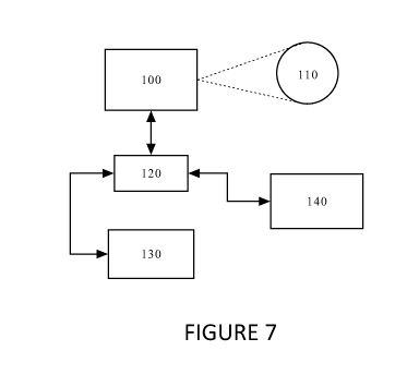
[0027] For the above automated process, a system such as that
illustrated in Figure 7 may be used. As can be seen
in Figure 7, the system may include the imaging device
100 that images the human eye 110. Once an image of
the eye has been produced, the image is then stored in
a storage device 120. The image can then be processed
by an image processing block 130. Image processing
can take the form of image translation, rotation,
enhancement, color adjustment, color substitution, as
- 18 -
CA 03060383 2019-10-18
WO 2018/201253
PCT/CA2018/050526
well as other image processing steps. Preferably, the
image processing steps are used to ensure that the
image and its contents are suitable for within image
comparison and measurement and for comparison with a
baseline image or with images previously acquired. The
processed image is then stored again in the storage
device 120. From the storage device 120, an image
comparison block 140 can retrieve the processed image
as well as a processed baseline image or other
previously processed and previously acquired images.
The image comparison block 140 can then extract data
from the retrieved images so that the extracted data
sets can be compared. As noted above, one data set
from the processed image may be the pixel density of
the choroid plexus area as a representation of the
vascular perfusion density for the area. The
extracted data sets (e.g. the pixel densities of the
choroid plexus area for the baseline image and the
acquired image) can then be compared to non-choroid
regions and/or to each other to determine whether the
newly acquired image indicates an increase or a
decrease in sympathetic nervous system activity or if
the newly acquired image indicates little or no change
in the sympathetic nervous system activity.
[0028] In another aspect, the invention may take the form of
a method for determining the effects of a stimulus on
an individual using that individual's choroid plexus.
Referring to Figure 8A, a block diagram illustrating
the steps in this method is illustrated. The method
may be initiated with the image capture of the
individual's eyeball, with particular attention being
paid to the choroid plexus of the eye (step 200). Once
the image has been captured, the image is then
- 19 -
CA 03060383 2019-10-18
WO 2018/201253
PCT/CA2018/050526
analyzed and the parameters regarding the eye (and in
particular the choroid plexus) are derived (step 210).
This step, as noted above, may include performing
image processing steps on the image to assist in the
extraction of data from the processed image. Once the
image has been processed, the data can then be
extracted. In one example, the data may include a
measure of the vascular perfusion density for the
choroid plexus. In another example, the data may
include a measure of the vascular perfusion density
for the choroid plexus and non-choroid plexus region
that are differentially regulated by the sympathetic
nervous system and autoregulation. The next step in
the process may be the application of one or more
stimuli on the individual (step 220). The stimulus
can be physical, physiological, mental, and/or
psychological. After the stimulus has been applied,
an image of the individual's eye is once again taken
(step 230). As with step 200, particular attention is
given to the choroid plexus of the eye. The post
stimulus image is then processed and data is extracted
in much the same way as in step 210 (step 240). With
data extracted from the processed post stimulus image,
this extracted data is then compared with the data
extracted in step 210 (step 250). The differences in
the data sets can be used to determine what effects
the stimulus had on the individual's physiology as
evidenced by changes to the individual's choroid
plexus in reference to itself, or to other parts of
the eye that have a difference balance of
autoregulatory and sympathetic control. Optionally,
the data extracted in step 240 can also be compared
with other data sets previously extracted from other
individuals.
- 20 -
CA 03060383 2019-10-18
WO 2018/201253
PCT/CA2018/050526
[0029] The steps in the above method can be used to screen
patients for sympathetic nervous activity and/or
autoregulation. In a clinical setting, the steps in
the method can form part of a pre-screening process to
identify patients with abnormal sympathetic nervous
activity and/or abnormal autoregulation. With disease
history and knowledge of sympathetic activity,
patients may be better treated with personalized
medicine.
[0030] The present invention may also be used as part of
pharmaceutical research and development as a method to
conveniently monitor sympathetic activity/autonomic
regulation of subjects enrolled in research projects
and/or clinical trials. The use of such a method may
reduce the number of drugs that proceed to late-stage
trials only to fail because of unexpected
cardiovascular complications. As well, the present
invention may be used to conduct "sympathetic
/autoregulatory phenotyping" as this increases the
prospect of personalized medicine, furthering research
and development into medications that may be
beneficial to a specific phenotype but detrimental to
others. These and other benefits may be the result of
the present invention as the present invention allows
for quick and easy assessment of key cardiorespiratory
determinant.
[0031] Further studies and analysis have shown the
correlation between different stimuli and VPD. For
these studies, both choroid and retina VPD (noted as
being a static index of perfusion within the image)
were calculated for participants before and after
stimuli were applied. For the retina, images of the
retina were attained from the 3D visualization mode
- 21 -
CA 03060383 2019-10-18
WO 2018/201253
PCT/CA2018/050526
and a 2D flattened image was selected in order to
attain the aggregate vasculature of the retinal
circulation. The results of these further studies can
be seen in the Figures described below.
[0032] Referring to Figure 8B, illustrated are raw images
(left column) and processed images (right column) for
an individual participant for the retina during
baseline and various cardiorespiratory
stimuli/challenges (noted next to the relevant image).
Vascular perfusion is false colored in the raw images
where blue and red represent high and low vascular
perfusion, respectively. Retina VPD is demarcated by
black pixels in the processed black and white images.
[0033] Referring to Figure 9A, the plot shows the change from
baseline of choroid VPD due to different stimuli. As
can be seen, for these tests, the stimuli were
hyperoxia, hypoxia, hyperpnoea, hypercapnia, breath
hold, and cold pressor test. All the data in Figure
9A are presented as a percentage change from baseline.
Figure 9B shows the change from baseline of retina VPD
after the same set of stimuli used for Figure 9A.
Figure 9C shows the muscle sympathetic nervous
activity (MSNA) in response to the same stimuli used
for Figures 9A and 9B.
[0034] In addition to the above, Figures 10A-10D show the
relationships between choroid VPD, retina VPD, muscle
sympathetic nerve activity (MSNA), and mean arterial
pressure (MAP). For these Figures, each data point
represents a participant's response to hyperoxia
(green), hypoxia, (blue), hyperpnoea (magenta),
hypercapnia (gray), breath hold (cyan), and cold
pressor test (red). Figure 10A shows the relationship
- 22 -
CA 03060383 2019-10-18
WO 2018/201253
PCT/CA2018/050526
between choroid VPD and MSNA. Figure 10B shows the
relationship between choroid VPD and MAP. Figure 100
shows the relationship between retina VPD and MSNA
while Figure 10D shows the relationship between retina
VPD and MAP. For clarity, Pearson R correlation
coefficients with accompanying p-values are indicated
for each panel. For relationships involving the
retina, values in red text are inclusive of results
for the cold pressor test and values in solid text
exclude results for the cold pressor test.
[0035] Referring to Figures 11A-11C, choroid VPD and retina
VPD are plotted against MSNA when the stimuli are
hypoxia (blue data points) and hyperoxia (green data
points). These plots show that the divergent vascular
regulatory mechanisms of the choroid (sympathetically
regulated) and retina (local vascular regulation) are
underscored by the divergent relationships with
sympathetic activity. For clarity, the plots are
based on percentage change of MSNA from baseline.
Figure 11A plots choroid VPD against MSNA while Figure
11B plots retina VPD against MSNA. Figure 110 shows
the relationship between retina VPD and choroid VPD
and it can be seen that this plot supports the use of
choroid VPD as a surrogate measure of MSNA.
[0036] Referring to Figures 12A-12C, illustrated are plots
detailing the relationship between R-R intervals to
muscle sympathetic nervous activity (MSNA). For these
plots, each data point represents a participant's
response to hyperoxia (green data points), hypoxia
(blue data points), hyperpnoea (magenta data points),
hypercapnia (gray data points), breath hold (cyan data
points), and the cold pressor test (red data points)
as a percentage change from baseline. Figure 12A
- 23 -
CA 03060383 2019-10-18
WO 2018/201253
PCT/CA2018/050526
shows the relationship between root mean square of
standard deviation R-R interval (RMSSD) to MSNA in
response to sympathetic provocations (i.e. stimuli).
Figure 12B shows the relationship of low frequency R-R
interval attained from Fast Fourier Transform (LF) to
muscle sympathetic nervous activity (MSNA) in response
to sympathetic provocations/stimuli. Figure 120 shows
the relationship of low frequency to high frequency R-
R interval ratio attained from Fast Fourier Transform
(LF/HF) to muscle sympathetic nervous activity (MSNA).
[0037] While the above description focuses on optical
coherence tomography for imaging an individual's eye,
other technologies and techniques which allow for
similar imaging results may also be used. As long as
a technology or a technique allows for the imaging
and/or characterization of an individual's choroid
vascular system, it may be used with the present
invention. The present invention may also be used
with any manoeuvre or intervention (such as a drug
treatment) that can activate or suppress the
sympathetic nervous system or alter autoregulation.
The method of the invention may also be used to
identify and characterize manoeuvres and/or
interventions previously unknown for its effect on the
sympathetic nervous system and autoregulation. If
such a manoeuvre or intervention with a previously
unknown effect on the sympathetic nervous
system/autoregulation is found, the present invention
can also be used to identify and characterize
manoeuvres or interventions that can counter or
aggregate the effect of this manoeuvre or intervention
with the previously unknown effect on the sympathetic
nervous system/autoregulation.
- 24 -
CA 03060383 2019-10-18
WO 2018/201253
PCT/CA2018/050526
[0038] The present invention may also be used for the
development of new drugs targeting sympathetic
abnormalities to treat hypertension, hypotension,
COPD, asthma and other cardiorespiratory diseases. The
development of personalized therapies, targeted to
specific cardiorespiratory phenotypes, enhanced
subject selection, and phenotyping prior to clinical
trials may also benefit from the use of the present
invention. Finally, the present invention may be used
for earlier detection of unforeseen deleterious
cardiorespiratory effects during clinical trials.
[0039] The embodiments of the invention may be executed by a
computer processor or similar device programmed in the
manner of method steps, or may be executed by an
electronic system which is provided with means for
executing these steps. Similarly, an electronic memory
means such as computer diskettes, CD-ROMs, Random
Access Memory (RAM), Read Only Memory (ROM) or similar
computer software storage media known in the art, may
be programmed to execute such method steps. As well,
electronic signals representing these method steps may
also be transmitted via a communication network.
[0040] Embodiments of the invention may be implemented in any
conventional computer programming language. For
example, preferred embodiments may be implemented in a
procedural programming language (e.g."C") or an
object-oriented language (e.g."C++", "java", "PHP",
"PYTHON" or "C#"). Alternative embodiments of the
invention may be implemented as pre-programmed
hardware elements, other related components, or as a
combination of hardware and software components.
- 25 -
CA 03060383 2019-10-18
WO 2018/201253
PCT/CA2018/050526
[0041] Embodiments can be implemented as a computer program
product for use with a computer system. Such
implementations may include a series of computer
instructions fixed either on a tangible medium, such
as a computer readable medium (e.g., a diskette, CD-
ROM, ROM, or fixed disk) or transmittable to a
computer system, via a modem or other interface
device, such as a communications adapter connected to
a network over a medium. The medium may be either a
tangible medium (e.g., optical or electrical
communications lines) or a medium implemented with
wireless techniques (e.g., microwave, infrared or
other transmission techniques). The series of computer
instructions embodies all or part of the functionality
previously described herein. Those skilled in the art
should appreciate that such computer instructions can
be written in a number of programming languages for
use with many computer architectures or operating
systems. Furthermore, such instructions may be stored
in any memory device, such as semiconductor, magnetic,
optical or other memory devices, and may be
transmitted using any communications technology, such
as optical, infrared, microwave, or other transmission
technologies. It is expected that such a computer
program product may be distributed as a removable
medium with accompanying printed or electronic
documentation (e.g., shrink-wrapped software),
preloaded with a computer system (e.g., on system ROM
or fixed disk), or distributed from a server over a
network (e.g., the Internet or World Wide Web). Of
course, some embodiments of the invention may be
implemented as a combination of both software (e.g., a
computer program product) and hardware. Still other
embodiments of the invention may be implemented as
- 26 -
CA 03060383 2019-10-18
WO 2018/201253
PCT/CA2018/050526
entirely hardware, or entirely software (e.g., a
computer program product).
[0042] A person understanding this invention may now conceive
of alternative structures and embodiments or
variations of the above all of which are intended to
fall within the scope of the invention as defined in
the claims that follow.
- 27 -