Note: Descriptions are shown in the official language in which they were submitted.
CA 03060518 2019-10-18
WO 2018/200411 PCT/US2018/028939
METHODS FOR INDUCING CHONDROGENESIS
CROSS-REFERENCE
[0001] This application claims the benefit of US Provisional Application
Serial Number 62/489,397 filed
April 24, 2017, the entirety of which is hereby incorporated by reference
herein.
BACKGROUND OF THE INVENTION
[0002] Osteoarthritis (OA) represents the most common musculoskeletal
disorder. Approximately 40
million Americans are currently affected and this number is predicted to
increase to 60 million within the
next twenty years as a result of the aging population and an increase in life
expectancy, making it the fourth
leading cause of disability. OA is characterized by a degenerative breakdown
of the joint including both the
articular cartilage (containing the cells and matrix which produce lubrication
and cushioning for the joint)
and the subchondral bone underlying the articular cartilage. Current OA
therapies include pain relief with
oral NSAIDs or selective cyclooxygenase 2 (COX-2) inhibitors, intra-articular
(IA) injection with agents
such as corticorsteroids and hyaluronan, and surgical approaches.
[0003] Mesenchymal stem cells (MSCs) are present in adult articular cartilage
and upon isolation can be
programmed in vitro to undergo differentiation to chondrocytes and other
mesenchymal cell lineages. In part
it is regulated by growth factors (TGF s, BMPs), serum conditions and cell-
cell contact.
SUMMARY OF THE INVENTION
[0004] Provided herein is a method for ameliorating arthritis or joint injury
in a subject, the method
comprising administering to the joint space of a knee of the subject from
about 10 fig to about 1000 fig of N-
(4-(2-methoxyethyl)pheny1)-2-(methylsulfonamido)benzamide, or a
pharmaceutically acceptable salt, or
solvate thereof
[0005] The method for ameliorating arthritis or joint injury in a subject may
comprise administering to the
joint space of a knee from about 10 fig to about 400 lag of N-(4-(2-
methoxyethyl)pheny1)-2-
(methylsulfonamido)benzamide, or a pharmaceutically acceptable salt, or
solvate thereof.
[0006] The method for ameliorating arthritis or joint injury in a subject may
comprise administering to the
joint space of a knee from about 50 fig to about 400 lag of N-(4-(2-
methoxyethyl)pheny1)-2-
(methylsulfonamido)benzamide, or a pharmaceutically acceptable salt, or
solvate thereof
[0007] Provided herein is a method for ameliorating arthritis or joint injury
in a subject, the method
comprising administering to the joint space of a knee of the subject no more
than about 1000 fig of N-(4-(2-
methoxyethyl)pheny1)-2-(methylsulfonamido)benzamide, or a pharmaceutically
acceptable salt, or solvate
thereof
[0008] The method for ameliorating arthritis or joint injury in a subject may
comprise administering to the
joint space of a knee no more than about 400 lag of N-(4-(2-
methoxyethyl)pheny1)-2-
(methylsulfonamido)benzamide, or a pharmaceutically acceptable salt, or
solvate thereof
- 1 -
CA 03060518 2019-10-18
WO 2018/200411 PCT/US2018/028939
[0009] Provided herein is a method for inducing differentiation of mesenchymal
stem cells into
chondrocytes in a subject, the method comprising administering to the joint
space of a knee of the subject
from about 10 lag to about 1000 lag of N-(4-(2-methoxyethyl)pheny1)-2-
(methylsulfonamido)benzamide, or a
pharmaceutically acceptable salt, or solvate thereof.
[0010] The method for inducing differentiation of mesenchymal stem cells into
chondrocytes in a subject
may comprise administering to the joint space of a knee from about 10 lag to
about 400 lag of N-(4-(2-
methoxyethyl)pheny1)-2-(methylsulfonamido)benzamide, or a pharmaceutically
acceptable salt, or solvate
thereof
[0011] The method for inducing differentiation of mesenchymal stem cells into
chondrocytes in a subject
may comprise administering to the joint space of a knee from about 50 lag to
about 400 lag of N-(4-(2-
methoxyethyl)pheny1)-2-(methylsulfonamido)benzamide, or a pharmaceutically
acceptable salt, or solvate
thereof
[0012] Disclosed herein is a method for inducing differentiation of
mesenchymal stem cells into
chondrocytes in a subject, the method comprising administering to the joint
space of a knee of the subject
not more than about 1000 lag of N-(4-(2-methoxyethyl)pheny1)-2-
(methylsulfonamido)benzamide, or a
pharmaceutically acceptable salt, or solvate thereof.
[0013] The method for inducing differentiation of mesenchymal stem cells into
chondrocytes in a subject
may comprise administering to the joint space of a knee no more than about 400
lag of N-(4-(2-
methoxyethyl)pheny1)-2-(methylsulfonamido)benzamide, or a pharmaceutically
acceptable salt, or solvate
thereof
[0014] In the method for ameliorating arthritis or joint injury or for
inducing differentiation of
mesenchymal stem cells into chondrocytes in a subject, N-(4-(2-
methoxyethyl)pheny1)-2-
(methylsulfonamido)benzamide, or a pharmaceutically acceptable salt, or
solvate thereof may be
administered to the subject annually.
[0015] In the method for ameliorating arthritis or joint injury or for
inducing differentiation of
mesenchymal stem cells into chondrocytes in a subject, N-(4-(2-
methoxyethyl)pheny1)-2-
(methylsulfonamido)benzamide, or a pharmaceutically acceptable salt, or
solvate thereof may be
administered to the subject every eleven months.
[0016] In the method for ameliorating arthritis or joint injury or for
inducing differentiation of
mesenchymal stem cells into chondrocytes in a subject, N-(4-(2-
methoxyethyl)pheny1)-2-
(methylsulfonamido)benzamide, or a pharmaceutically acceptable salt, or
solvate thereof may be
administered to the subject every ten months.
[0017] In the method for ameliorating arthritis or joint injury or for
inducing differentiation of
mesenchymal stem cells into chondrocytes in a subject, N-(4-(2-
methoxyethyl)pheny1)-2-
(methylsulfonamido)benzamide, or a pharmaceutically acceptable salt, or
solvate thereof may be
administered to the subject every nine months.
- 2 -
CA 03060518 2019-10-18
WO 2018/200411
PCT/US2018/028939
[0018] In the method for ameliorating arthritis or joint injury or for
inducing differentiation of
mesenchymal stem cells into chondrocytes in a subject, N-(4-(2-
methoxyethyl)pheny1)-2-
(methylsulfonamido)benzamide, or a pharmaceutically acceptable salt, or
solvate thereof may be
administered to the subject every eight months.
[0019] In the method for ameliorating arthritis or joint injury or for
inducing differentiation of
mesenchymal stem cells into chondrocytes in a subject, N-(4-(2-
methoxyethyl)pheny1)-2-
(methylsulfonamido)benzamide, or a pharmaceutically acceptable salt, or
solvate thereof may be
administered to the subject every seven months.
[0020] In the method for ameliorating arthritis or joint injury or for
inducing differentiation of
mesenchymal stem cells into chondrocytes in a subject, N-(4-(2-
methoxyethyl)pheny1)-2-
(methylsulfonamido)benzamide, or a pharmaceutically acceptable salt, or
solvate thereof may be
administered to the subject every six months.
[0021] In the method for ameliorating arthritis or joint injury or for
inducing differentiation of
mesenchymal stem cells into chondrocytes in a subject, N-(4-(2-
methoxyethyl)pheny1)-2-
(methylsulfonamido)benzamide, or a pharmaceutically acceptable salt, or
solvate thereof may be
administered to the subject every five months.
[0022] In the method for ameliorating arthritis or joint injury or for
inducing differentiation of
mesenchymal stem cells into chondrocytes in a subject, N-(4-(2-
methoxyethyl)pheny1)-2-
(methylsulfonamido)benzamide, or a pharmaceutically acceptable salt, or
solvate thereof may be
administered to the subject every four months.
[0023] In the method for ameliorating arthritis or joint injury or for
inducing differentiation of
mesenchymal stem cells into chondrocytes in a subject, N-(4-(2-
methoxyethyl)pheny1)-2-
(methylsulfonamido)benzamide, or a pharmaceutically acceptable salt, or
solvate thereof may be
administered to the subject every three months.
[0024] In the method for ameliorating arthritis or joint injury or for
inducing differentiation of
mesenchymal stem cells into chondrocytes in a subject, N-(4-(2-
methoxyethyl)pheny1)-2-
(methylsulfonamido)benzamide, or a pharmaceutically acceptable salt, or
solvate thereof may be
administered to the subject every two months.
[0025] In the method for ameliorating arthritis or joint injury or for
inducing differentiation of
mesenchymal stem cells into chondrocytes in a subject, N-(4-(2-
methoxyethyl)pheny1)-2-
(methylsulfonamido)benzamide, or a pharmaceutically acceptable salt, or
solvate thereof may be
administered to the subject monthly or weekly.
[0026] In the method for ameliorating arthritis or joint injury or for
inducing differentiation of
mesenchymal stem cells into chondrocytes in a subject, about 25 lag of N-(4-(2-
methoxyethyl)pheny1)-2-
(methylsulfonamido)benzamide, or a pharmaceutically acceptable salt, or
solvate thereof may be
administered.
- 3 -
CA 03060518 2019-10-18
WO 2018/200411 PCT/US2018/028939
[0027] In the method for ameliorating arthritis or joint injury or for
inducing differentiation of
mesenchymal stem cells into chondrocytes in a subject, about 50 lag of N-(4-(2-
methoxyethyl)pheny1)-2-
(methylsulfonamido)benzamide, or a pharmaceutically acceptable salt, or
solvate thereof may be
administered.
[0028] In the method for ameliorating arthritis or joint injury or for
inducing differentiation of
mesenchymal stem cells into chondrocytes in a subject, about 100 lag of N-(4-
(2-methoxyethyl)pheny1)-2-
(methylsulfonamido)benzamide, or a pharmaceutically acceptable salt, or
solvate thereof may be
administered.
[0029] In the method for ameliorating arthritis or joint injury or for
inducing differentiation of
mesenchymal stem cells into chondrocytes in a subject, about 150 lag of N-(4-
(2-methoxyethyl)pheny1)-2-
(methylsulfonamido)benzamide, or a pharmaceutically acceptable salt, or
solvate thereof may be
administered.
[0030] In the method for ameliorating arthritis or joint injury or for
inducing differentiation of
mesenchymal stem cells into chondrocytes in a subject, about 200 lag of N-(4-
(2-methoxyethyl)pheny1)-2-
(methylsulfonamido)benzamide, or a pharmaceutically acceptable salt, or
solvate thereof may be
administered.
[0031] In the method for ameliorating arthritis or joint injury or for
inducing differentiation of
mesenchymal stem cells into chondrocytes in a subject, about 250 lag of N-(4-
(2-methoxyethyl)pheny1)-2-
(methylsulfonamido)benzamide, or a pharmaceutically acceptable salt, or
solvate thereof may be
administered.
[0032] In the method for ameliorating arthritis or joint injury or for
inducing differentiation of
mesenchymal stem cells into chondrocytes in a subject, about 300 lag of N-(4-
(2-methoxyethyl)pheny1)-2-
(methylsulfonamido)benzamide, or a pharmaceutically acceptable salt, or
solvate thereof may be
administered.
[0033] In the method for ameliorating arthritis or joint injury or for
inducing differentiation of
mesenchymal stem cells into chondrocytes in a subject, about 350 lag of N-(4-
(2-methoxyethyl)pheny1)-2-
(methylsulfonamido)benzamide, or a pharmaceutically acceptable salt, or
solvate thereof may be
administered.
[0034] In the method for ameliorating arthritis or joint injury or for
inducing differentiation of
mesenchymal stem cells into chondrocytes in a subject, about 400 lag of N-(4-
(2-methoxyethyl)pheny1)-2-
(methylsulfonamido)benzamide, or a pharmaceutically acceptable salt, or
solvate thereof may be
administered.
[0035] In the method for ameliorating arthritis or joint injury or for
inducing differentiation of
mesenchymal stem cells into chondrocytes in a subject, N-(4-(2-
methoxyethyl)pheny1)-2-
(methylsulfonamido)benzamide, or a pharmaceutically acceptable salt, or
solvate thereof may be
administered in a volume of from about 1 mL to about 5 mL.
- 4 -
CA 03060518 2019-10-18
WO 2018/200411 PCT/US2018/028939
[0036] In the method for ameliorating arthritis or joint injury or for
inducing differentiation of
mesenchymal stem cells into chondrocytes in a subject, N-(4-(2-
methoxyethyl)pheny1)-2-
(methylsulfonamido)benzamide, or a pharmaceutically acceptable salt, or
solvate thereof may be
administered in a volume about or no more than about 5 mL.
BRIEF DESCRIPTION OF THE DRAWINGS
[0037] FIG. 1 shows shows the substantial cartilage degeneration width
following treatment with
Compound A at 10 uM.
[0038] FIG. 2 shows the combined cartilage degeneration widths following
treatment with compound A
once every two weeks at 10 M.
[0039] FIG. 3 shows the total joint scores without the femur of animals
treated with compound A as
compared to vehicle treated animals.
[0040] FIG. 4 shows the schematic of histological analysis of osteoarthritic
lesions in the tibial plateau and
femur of the dog.
[0041] FIG. 5 shows the cartilage degeneration width thllowing treatment with
compound A.
[0042] FIG. 6 shows the depth of cartilage lesions in the femur following
treatment with compound A.
[0043] FIG. 7 shows the levels of bone sclerosis following treatment with
compound A.
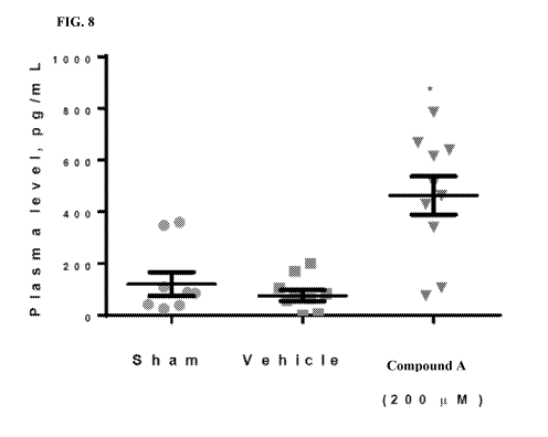
[0044] FIG. 8 shows the circulating levels of collagen formation marker PIINP
following treatment with
compound A.
[0045] FIG. 9 shows the in vitro compound A binding to FLNA.
[0046] FIG. 10 shows the induction of CBFI3 nuclear localization through
compound A.
INCORPORATION BY REFERENCE
[0047] All publications, patents, and patent applications mentioned in this
specification are herein
incorporated by reference to the same extent as if each individual
publication, patent, or patent application
was specifically and individually indicated to be incorporated by reference.
DETAILED DESCRIPTION OF THE INVENTION
[0048] Osteoarthritis (OA) is characterized by progressive breakdown of
articular cartilage, and ultimately
leads to functional failure of synovial joints [Reginster, J.Y. and N.G.
Khaltaev, Introduction and WHO
perspective on the global burden of musculoskeletal conditions. Rheumatology
(Oxford), 2002. 41 Supp 1:
p. 1-21. OA is mediated by several pathogenic mechanisms including enzymatic
degradation of extracellular
matrix, deficient new matrix formation, cell death, and abnormal activation
and hypertrophic differentiation
of cartilage cells [Goldring, M.B. and S.R. Goldring, Articular cartilage and
subchondral bone in the
pathogenesis of osteoarthritis. Ann N Y Acad Sci, 2010. 1192(1): p. 230-71.
The only current therapeutic
options for OA are pain management and surgical intervention [Hunter, D.J.,
Pharmacologic therapy for
osteoarthritis-the era of disease modification. Nat Rev Rheumatol, 2011. 7(1):
p. 13-221.
- 5 -
CA 03060518 2019-10-18
WO 2018/200411 PCT/US2018/028939
[0049] Mesenchymal stem cells (MSCs), residing in bone marrow and most adult
tissues, are capable of
self-renewal and differentiation into a variety of cell lineages including
chondrocytes, osteoblasts and
adipocytes [Pittenger, M.F., et al., Multilineage potential of adult human
mesenchymal stem cells. Science,
1999. 284(5411): p. 143-71. Recent studies found that adult articular
cartilage contains MSCs
(approximately 3% of the cells) that are capable of multi-lineage
differentiation. In OA cartilage, the number
of these cells approximately doubles. These resident stem cells still retain
the capability to differentiate into
chondrocytes and thus the capacity to repair the damaged cartilage [Grogan,
S.P., et al., Mesenchymal
progenitor cell markers in human articular cartilage: normal distribution and
changes in osteoarthritis.
Arthritis Res Ther, 2009. 11(3): p. R85; Koelling, S., et al., Migratory
chondrogenic progenitor cells from
repair tissue during the later stages of human osteoarthritis. Cell Stem Cell,
2009. 4(4): p. 324-351.
[0050] The present invention is based, in part, on the discovery that the
compounds of the present invention
stimulate chondrocyte differentiation in mesenchymal stem cells. Accordingly,
the present invention
provides for methods of induction of mesenchymal stem cell differentiation
into chondrocytes. Further, the
present invention provides for administration of compounds and compositions of
the present invention to
prevent or ameliorate arthritis or joint injury by administrating the compound
or composition into a joint, the
vertebrae, vertebral disc or systemically. In particular, the compounds of the
present disclosure are
administered intra-articularly into the knee at a dosage of about 10 ug to
about 1000 us. The compounds
may be administered as a single dose or as a course of up to four doses.
Dosing may be repeated, for
example, every week, two weeks, monthly, or every 3-12 months. As a non-
limiting example, dosing is
weekly for no more than five weeks. As used herein, administration to the knee
or the joint of the knee refers
to administration to one knee. However, both knees may be administered with
the compounds herein. For
example, each knee is dosed with about 10 ug to about 1000 ug of a compound
provided herein.
Definitions
[0051] In the following description, certain specific details are set forth in
order to provide a thorough
understanding of various embodiments. However, one skilled in the art will
understand that the invention
may be practiced without these details. In other instances, well-known
structures have not been shown or
described in detail to avoid unnecessarily obscuring descriptions of the
embodiments. Unless the context
requires otherwise, throughout the specification and claims which follow, the
word "comprise" and
variations thereof, such as, "comprises" and "comprising" are to be construed
in an open, inclusive sense,
that is, as "including, but not limited to." Further, headings provided herein
are for convenience only and do
not interpret the scope or meaning of the claimed invention.
[0052] Reference throughout this specification to "one embodiment" or "an
embodiment" means that a
particular feature, structure or characteristic described in connection with
the embodiment is included in at
least one embodiment. Thus, the appearances of the phrases "in one embodiment"
or "in an embodiment" in
various places throughout this specification are not necessarily all referring
to the same embodiment.
Furthermore, the particular features, structures, or characteristics may be
combined in any suitable manner in
one or more embodiments. Also, as used in this specification and the appended
claims, the singular forms
- 6 -
CA 03060518 2019-10-18
WO 2018/200411 PCT/US2018/028939
"a," "an," and "the" include plural referents unless the content clearly
dictates otherwise. It should also be
noted that the term "or" is generally employed in its sense including "and/or"
unless the content clearly
dictates otherwise.
[0053] The term "patient", "subject" or "individual" are used interchangeably.
As used herein, they refer to
individuals suffering from a disorder, and the like, encompasses mammals and
non-mammals. None of the
terms require that the individual be under the care and/or supervision of a
medical professional. Mammals
are any member of the Mammalian class, including but not limited to humans,
non-human primates such as
chimpanzees, and other apes and monkey species; farm animals such as cattle,
horses, sheep, goats, swine;
domestic animals such as rabbits, dogs, and cats; laboratory animals including
rodents, such as rats, mice
and guinea pigs, and the like. Examples of non-mammals include, but are not
limited to, birds, fish and the
like. In some embodiments of the methods and compositions provided herein, the
individual is a mammal. In
preferred embodiments, the individual is a human.
[0054] The terms "treat," "treating" or "treatment," and other grammatical
equivalents as used herein,
include alleviating, abating or ameliorating a disease or condition or one or
more symptoms thereof,
preventing additional symptoms, ameliorating or preventing the underlying
metabolic causes of symptoms,
inhibiting the disease or condition, e.g., arresting the development of the
disease or condition, relieving the
disease or condition, causing regression of the disease or condition,
relieving a condition caused by the
disease or condition, or stopping the symptoms of the disease or condition,
and are intended to include
prophylaxis. The terms further include achieving a therapeutic benefit and/or
a prophylactic benefit. By
therapeutic benefit is meant eradication or amelioration of the underlying
disorder being treated. Also, a
therapeutic benefit is achieved with the eradication or amelioration of one or
more of the physiological
symptoms associated with the underlying disorder such that an improvement is
observed in the individual,
notwithstanding that the individual is still be afflicted with the underlying
disorder. For prophylactic benefit,
the compositions are administered to an individual at risk of developing a
particular disease, or to an
individual reporting one or more of the physiological symptoms of a disease,
even though a diagnosis of this
disease has not been made.
[0055] The terms "administer," "administering", "administration," and the
like, as used herein, refer to the
methods that may be used to enable delivery of compounds or compositions to
the desired site of biological
action. These methods include, but are not limited to oral routes,
intraduodenal routes, parenteral injection
(including intravenous, subcutaneous, intraperitoneal, intramuscular,
intravascular or infusion), topical and
rectal administration. Those of skill in the art are familiar with
administration techniques that can be
employed with the compounds and methods described herein. In preferred
embodiments, the compounds and
compositions described herein are administered orally.
[0056] The terms "effective amount", "therapeutically effective amount" or
"pharmaceutically effective
amount" as used herein, refer to a sufficient amount of at least one agent or
compound being administered
which will relieve to some extent one or more of the symptoms of the disease
or condition being treated. The
result can be reduction and/or alleviation of the signs, symptoms, or causes
of a disease, or any other desired
- 7 -
CA 03060518 2019-10-18
WO 2018/200411 PCT/US2018/028939
alteration of a biological system. For example, an "effective amount" for
therapeutic uses is the amount of
the composition comprising N-(4-(2-methoxyethyl)pheny1)-2-
(methylsulfonamido)benzamide required to
provide a clinically significant decrease in a disease. An appropriate
"effective" amount may differ from one
individual to another. An appropriate "effective" amount in any individual
case may be determined using
techniques, such as a dose escalation study.
[0057] The term "acceptable" as used herein, with respect to a formulation,
composition or ingredient,
means having no persistent detrimental effect on the general health of the
individual being treated.
[0058] The term "pharmaceutically acceptable" as used herein, refers to a
material, such as a carrier or
diluent, which does not abrogate the biological activity or properties of N-(4-
(2-methoxyethyl)pheny1)-2-
(methylsulfonamido)benzamide, and is relatively nontoxic, i.e., the material
may be administered to an
individual without causing undesirable biological effects or interacting in a
deleterious manner with any of
the components of the composition in which it is contained.
[0059] The term "pharmaceutically acceptable salt" as used herein, refers to
salts that retain the biological
effectiveness of the free acids and bases of N-(4-(2-methoxyethyl)pheny1)-2-
(methylsulfonamido)benzamide
and that are not biologically or otherwise undesirable. N-(4-(2-
methoxyethyl)pheny1)-2-
(methylsulfonamido)benzamide may react with inorganic or organic bases, and
inorganic and organic acids,
to form a pharmaceutically acceptable salt. These salts can be prepared in
situ during the final isolation and
purification, or by separately reacting the purified compound in its free base
form with a suitable organic or
inorganic acid, and isolating the salt thus formed.
[0060] The term "pharmaceutical composition," as used herein, refers to a
biologically active compound,
optionally mixed with at least one pharmaceutically acceptable chemical
component, such as, though not
limited to carriers, stabilizers, diluents, dispersing agents, suspending
agents, thickening agents, excipients
and the like.
[0061] The term "carrier" as used herein, refers to relatively nontoxic
chemical compounds or agents that
facilitate the incorporation of a compound into cells or tissues.
[0062] The terms "pharmaceutical combination", "administering an additional
therapy", "administering an
additional therapeutic agent" and the like, as used herein, refer to a
pharmaceutical therapy resulting from
the mixing or combining of more than one active ingredient and includes both
fixed and non-fixed
combinations of the active ingredients. The term "fixed combination" means
that N-(4-(2-
methoxyethyl)pheny1)-2-(methylsulfonamido)benzamide, and at least one co-
agent, are both administered to
an individual simultaneously in the form of a single entity or dosage. The
term "non-fixed combination"
means that N-(4-(2-methoxyethyl)pheny1)-2-(methylsulfonamido)benzamide, and at
least one co-agent, are
administered to an individual as separate entities either simultaneously,
concurrently or sequentially with
variable intervening time limits, wherein such administration provides
effective levels of the two or more
compounds in the body of the individual. These also apply to cocktail
therapies, e.g. the administration of
three or more active ingredients.
- 8 -
CA 03060518 2019-10-18
WO 2018/200411 PCT/US2018/028939
[0063] The terms "co-administration", "administered in combination with" and
their grammatical
equivalents or the like, as used herein, are meant to encompass administration
of the selected therapeutic
agents to a single individual, and are intended to include treatment regimens
in which the agents are
administered by the same or different route of administration or at the same
or different times. In some
embodiments N-(4-(2-methoxyethyl)pheny1)-2-(methylsulfonamido)benzamide will
be co-administered with
other agents. These terms encompass administration of two or more agents to an
animal so that both agents
and/or their metabolites are present in the animal at the same time. They
include simultaneous administration
in separate compositions, administration at different times in separate
compositions, and/or administration in
a composition in which both agents are present. Thus, In some embodiments, N-
(4-(2-
methoxyethyl)pheny1)-2-(methylsulfonamido)benzamide and the other agent(s) are
administered in a single
composition. In some embodiments, N-(4-(2-methoxyethyl)pheny1)-2-
(methylsulfonamido)benzamide and
the other agent(s) are admixed in the composition.
[0064] "Western Ontario and McMaster Universities Arthritis Index" or "WOMAC"
refers to a widely
used, proprietary set of standardized questionnaires used by health
professionals to evaluate the condition of
patients with osteoarthritis of the knee and hip, including pain, stiffness,
and physical functioning of the
joints. The WOMAC has also been used to assess back pain, rheumatoid
arthritis, juvenile rheumatoid
arthritis, systemic lupus erythematosus, and fibromyalgia. It can be self-
administered and was developed at
Western Ontario and McMaster Universities in 1982. The WOMAC measures five
items for pain (score
range 0-20), two for stiffness (score range 0-8), and 17 for functional
limitation (score range 0-68). Physical
functioning questions cover everyday activities such as stair use, standing up
from a sitting or lying position,
standing, bending, walking, getting in and out of a car, shopping, putting on
or taking off socks, lying in bed,
getting in or out of a bath, sitting, and heavy and light household duties.
Compound A
[0065] Provided herein is N-(4-(2-methoxyethyl)pheny1)-2-
(methylsulfonamido)benzamide, or a
pharmaceutically acceptable salt or solvate thereof:
0
S, 0N H 0
0
140:1
Further Forms of Compound A
Isomers
[0066] In some embodiments, compound A described herein exists as geometric
isomers. In some
embodiments, compound A described herein possesses one double bond. Compound A
described herein
include all cis, trans, syn, anti, entgegen (E), and zusammen (Z) isomers as
well as the corresponding
mixtures thereof
- 9 -
CA 03060518 2019-10-18
WO 2018/200411 PCT/US2018/028939
Labeled compounds
[0067] In some embodiments, compound A described herein exists in its
isotopically-labeled forms. In
some embodiments, the methods disclosed herein include methods of treating
diseases by administering such
isotopically-labeled compounds. In some embodiments, the methods disclosed
herein include methods of
treating diseases by administering such isotopically-labeled compound A as
pharmaceutical compositions.
Thus, In some embodiments, compound A disclosed herein includes isotopically-
labeled compound A,
which is identical to compound A, but for the fact that one or more atoms are
replaced by an atom having an
atomic mass or mass number different from the atomic mass or mass number
usually found in nature.
Examples of isotopes that can be incorporated into compounds of the invention
include isotopes of
hydrogen, carbon, nitrogen, oxygen, phosphorous, sulfur, fluorine and
chloride, such as 2H, 3H, 13C, 14C, 15N,
180, 170, 31p, 32p, 35s, 18-=-,,
and 36C1, respectively. Compound A described herein which contain the
aforementioned isotopes and/or other isotopes of other atoms are within the
scope of this disclosure. Certain
isotopically-labeled compound A, for example those into which radioactive
isotopes such as 3H and 14C are
incorporated, are useful in drug and/or substrate tissue distribution assays.
Tritiated, i. e., 3H and carbon-14,
i. e.,
u isotopes are particularly preferred for their ease of preparation and
detectability. Further,
substitution with heavy isotopes such as deuterium, i.e., 2H, produces certain
therapeutic advantages
resulting from greater metabolic stability, for example increased in vivo half-
life or reduced dosage
requirements. In some embodiments, the isotopically labeled compound A, or
pharmaceutically acceptable
salt or solvate thereof is prepared by any suitable method.
[0068] In some embodiments, compound A described herein is labeled by other
means, including, but not
limited to, the use of chromophores or fluorescent moieties, bioluminescent
labels, or chemiluminescent
labels.
Pharmaceutically acceptable salts
[0069] In some embodiments, compound A described herein exists as a
pharmaceutically acceptable salt. In
some embodiments, the methods disclosed herein include methods of treating
diseases by administering such
pharmaceutically acceptable salts. In some embodiments, the methods disclosed
herein include methods of
treating diseases by administering such pharmaceutically acceptable salts as
pharmaceutical compositions.
[0070] Examples of pharmaceutically acceptable salts include those salts
prepared by reaction of compound
A with a mineral, organic acid or inorganic base, such salts including,
acetate, acrylate, adipate, alginate,
aspartate, benzoate, benzenesulfonate, bisulfate, bisulfite, bromide,
butyrate, butyn-1,4-dioate, camphorate,
camphorsulfonate, caproate, caprylate, chlorobenzoate, chloride, citrate,
cyclopentanepropionate, decanoate,
digluconate, dihydrogenphosphate, dinitrobenzoate, dodecylsulfate,
ethanesulfonate, formate, fumarate,
glucoheptanoate, glycerophosphate, glycolate, hemisulfate, heptanoate,
hexanoate, hexyne-1,6-dioate,
hydroxybenzoate, y-hydroxybutyrate, hydrochloride, hydrobromide, hydroiodide,
2-hydroxyethanesulfonate,
iodide, isobutyrate, lactate, maleate, malonate, methanesulfonate, mandelate
metaphosphate,
methanesulfonate, methoxybenzoate, methylbenzoate, monohydrogenphosphate, 1-
napthalenesulfonate, 2-
napthalenesulfonate, nicotinate, nitrate, palmoate, pectinate, persulfate, 3-
phenylpropionate, phosphate,
- 10 -
CA 03060518 2019-10-18
WO 2018/200411 PCT/US2018/028939
picrate, pivalate, propionate, pyrosulfate, pyrophosphate, propiolate,
phthalate, phenylacetate,
phenylbutyrate, propanesulfonate, salicylate, succinate, sulfate, sulfite,
succinate, suberate, sebacate,
sulfonate, tartrate, thiocyanate, tosylate undeconate and xylenesulfonate.
[0071] Further, compound A described herein can be prepared as
pharmaceutically acceptable salts formed
by reacting the free base form of the compound with a pharmaceutically
acceptable inorganic or organic
acid, including, but not limited to, inorganic acids such as hydrochloric
acid, hydrobromic acid, sulfuric
acid, nitric acid, phosphoric acid metaphosphoric acid, and the like; and
organic acids such as acetic acid,
propionic acid, hexanoic acid, cyclopentanepropionic acid, glycolic acid,
pyruvic acid, lactic acid, malonic
acid, succinic acid, malic acid, maleic acid, fumaric acid, p-toluenesulfonic
acid, tartaric acid, trifluoroacetic
acid, citric acid, benzoic acid, 3-(4-hydroxybenzoyl)benzoic acid, cinnamic
acid, mandelic acid, arylsulfonic
acid, methanesulfonic acid, ethanesulfonic acid, 1,2-ethanedisulfonic acid, 2-
hydroxyethanesulfonic acid,
benzenesulfonic acid, 2-naphthalenesulfonic acid, 4-methylbicyclo42.2.21oct-2-
ene-1-carboxylic acid,
glucoheptonic acid, 4,4'-methylenebis-(3-hydroxy-2-ene-1 -carboxylic acid), 3-
phenylpropionic acid,
trimethylacetic acid, tertiary butylacetic acid, lauryl sulfuric acid,
gluconic acid, glutamic acid,
hydroxynaphthoic acid, salicylic acid, stearic acid and muconic acid. In some
embodiments, other acids,
such as oxalic, while not in themselves pharmaceutically acceptable, are
employed in the preparation of salts
useful as intermediates in obtaining the compounds of the invention and their
pharmaceutically acceptable
acid addition salts.
[0072] In some embodiments, those compounds described herein which comprise a
free acid group react
with a suitable base, such as the hydroxide, carbonate, bicarbonate, sulfate,
of a pharmaceutically acceptable
metal cation, with ammonia, or with a pharmaceutically acceptable organic
primary, secondary, tertiary, or
quaternary amine. Representative salts include the alkali or alkaline earth
salts, like lithium, sodium,
potassium, calcium, and magnesium, and aluminum salts and the like.
Illustrative examples of bases include
sodium hydroxide, potassium hydroxide, choline hydroxide, sodium carbonate,
1\1 (C1_4 alky1)4, and the like.
[0073] Representative organic amines useful for the formation of base addition
salts include ethylamine,
diethylamine, ethylenediamine, ethanolamine, diethanolamine, piperazine and
the like. It should be
understood that the compounds described herein also include the quaternization
of any basic nitrogen-
containing groups they contain. In some embodiments, water or oil-soluble or
dispersible products are
obtained by such quaternization.
Solvates
[0074] In some embodiments, compound A exists as a solvate. The invention
provides for methods of
treating diseases by administering such solvates. The invention further
provides for methods of treating
diseases by administering such solvates as pharmaceutical compositions.
[0075] Solvates contain either stoichiometric or non-stoichiometric amounts of
a solvent, and, in some
embodiments, are formed during the process of crystallization with
pharmaceutically acceptable solvents
such as water, ethanol, and the like. Hydrates are formed when the solvent is
water, or alcoholates are
formed when the solvent is alcohol. Solvates of the compounds described herein
can be conveniently
-11-
CA 03060518 2019-10-18
WO 2018/200411 PCT/US2018/028939
prepared or formed during the processes described herein. By way of example
only, hydrates of compound
A can be conveniently prepared by recrystallization from an aqueous/organic
solvent mixture, using organic
solvents including, but not limited to, dioxane, tetrahydrofuran or methanol.
In addition, the compounds
provided herein can exist in unsolvated as well as solvated forms. In general,
the solvated forms are
considered equivalent to the unsolvated forms for the purposes of compound A
and methods provided
herein.
Tautomers
[0076] A "tautomer"as used herein refers to a proton shift from one atom of a
molecule to another atom of
the same molecule. Compound A presented herein may exist as a tautomer.
Tautomers are compounds that
are interconvertible by migration of a hydrogen atom, accompanied by a switch
of a single bond and
adjacent double bond. In bonding arrangements where tautomerization is
possible, a chemical equilibrium of
the tautomers will exist. All tautomeric forms of compound A disclosed herein
are contemplated. The exact
ratio of the tautomers depends on several factors, including temperature,
solvent, and pH. In some cases,
0
S, 0
cr NH OH I.
Compound A may exist as:
Methods
[0077] Provided herein is a method of treating arthritis in a mammal, the
method including administering
about 10 ug to about 1000 ug of compound A, or a pharmaceutically acceptable
salt or solvate thereof, via
intra-articular injection to a joint of the mammal. For example, compound A,
or a pharmaceutically
acceptable salt or solvate thereof, is injected into an articulation. For
example, compound A, or a
pharmaceutically acceptable salt or solvate thereof, is injected into the
knee. In some cases, compound A is
not systemically absorbed about 1 hr, about 2hr, about 3hr, about 4hr, about
5hr, about 6hr, about 7hr, about
8hr, about 9hr, or about 10hr after admistration. In some cases, from about 10
ug to about 800 ug, from
about 10 ug to about 600 ug, from about 10 ug to about 400 ug, from about 10
ug to about 200 ug, from
about 10 ug to about 100 ug of compound A, or a pharmaceutically acceptable
salt or solvate thereof, is
administered. Compound A, or a pharmaceutically acceptable salt or solvate
thereof, may be administered as
a single dose or as a course of up to four doses. The dosing may be repeated,
for example, weekly, bi-
weekly, monthly, or every 3-12 months. As a non-limiting example, dosing is
repeated every 3, 4, 5, 6, 7, 8,
9, 10, 11 or 12 months. As another non-limiting example, dosing is weekly for
no more than five weeks. As
another non-limiting example, dosing is biweekly.
[0078] Provided herein is a method of treating osteoarthritis in a mammal, the
method including
administering about 10 ug to about 1000 ug of compound A, or a
pharmaceutically acceptable salt or solvate
thereof, via intra-articular injection to a joint of the mammal. For example,
compound A, or a
pharmaceutically acceptable salt or solvate thereof, is injected into an
articulation. For example, compound
- 12 -
CA 03060518 2019-10-18
WO 2018/200411 PCT/US2018/028939
A, or a pharmaceutically acceptable salt or solvate thereof, is injected into
the knee. In some cases,
compound A is not systemically absorbed about 1 hr, about 2hr, about 3hr,
about 4hr, about 5hr, about 6hr,
about 7hr, about 8hr, about 9hr, or about 10hr after admistration. In some
cases, from about 10 lag to about
800 lag, from about 10 pg to about 600 lag, from about 10 lag to about 400
lag, from about 10 lag to about
200 lag, from about 10 pg to about 100 lag of compound A, or a
pharmaceutically acceptable salt or solvate
thereof, is administered. Compound A, or a pharmaceutically acceptable salt or
solvate thereof, may be
administered as a single dose or as a course of up to four doses. The dosing
may be repeated, for example,
weekly, bi-weekly, monthly, or every 3-12 months. As a non-limiting example,
dosing is repeated every 3,
4, 5, 6, 7, 8, 9, 10, 11 or 12 months. As another non-limiting example, dosing
is weekly for no more than
five weeks. As another non-limiting example, dosing is biweekly.
[0079] Provided herein is a method of ameliorating arthritis or joint injury
in a mammal, the method
including administering about 10 lag to about 1000 lag of compound A, or a
pharmaceutically acceptable salt
or solvate thereof, via intra-articular injection to a joint of the mammal.
For example, compound A, or a
pharmaceutically acceptable salt or solvate thereof, is injected into an
articulation. For example, compound
A, or a pharmaceutically acceptable salt or solvate thereof, is injected into
the knee. In some cases,
compound A is not systemically absorbed about 1 hr, about 2hr, about 3hr,
about 4hr, about 5hr, about 6hr,
about 7hr, about 8hr, about 9hr, or about 10hr after admistration. In some
cases, from about 10 lag to about
800 pg, from about 10 lag to about 600 lag, from about 10 lag to about 400
lag, from about 10 lag to about
200 pg, from about 10 lag to about 100 lag of compound A, or a
pharmaceutically acceptable salt or solvate
thereof, is administered. Compound A, or a pharmaceutically acceptable salt or
solvate thereof, may be
administered as a single dose or as a course of up to four doses. The dosing
may be repeated, for example,
weekly, bi-weekly, monthly, or every 3-12 months. As a non-limiting example,
dosing is repeated every 3,
4, 5, 6, 7, 8, 9, 10, 11 or 12 months. As another non-limiting example, dosing
is weekly for no more than
five weeks. As another non-limiting example, dosing is biweekly.
[0080] Provided herein is a method of inducing differentiation of mesenchymal
stem cells into
chondrocytes, the method including exposing mesenchymal stem cells by intra-
articular injection to about 10
lag to about 1000 lag of compound A, or a pharmaceutically acceptable salt or
solvate thereof, in a subject in
need thereof, thereby inducing differentiation of the stem cells into
chondrocytes. For example, compound
A, or a pharmaceutically acceptable salt or solvate thereof, is injected into
an articulation. For example,
compound A, or a pharmaceutically acceptable salt or solvate thereof, is
injected into the knee. In some
cases, compound A is not systemically absorbed about 1 hr, about 2hr, about
3hr, about 4hr, about 5hr, about
6hr, about 7hr, about 8hr, about 9hr, or about 10hr after admistration. In
some cases, from about 10 lag to
about 800 lag, from about 10 lag to about 600 lag, from about 10 lag to about
400 lag, from about 10 lag to
about 200 lag, from about 10 lag to about 100 lag of compound A, or a
pharmaceutically acceptable salt or
solvate thereof, is administered. Compound A, or a pharmaceutically acceptable
salt or solvate thereof, may
be administered as a single dose or as a course of up to four doses. The
dosing may be repeated, for example,
weekly, bi-weekly, monthly, or every 3-12 months. As a non-limiting example,
dosing is repeated every 3,
- 13 -
CA 03060518 2019-10-18
WO 2018/200411 PCT/US2018/028939
4, 5, 6, 7, 8, 9, 10, 11 or 12 months. As another non-limiting example, dosing
is weekly for no more than
five weeks. As another non-limiting example, dosing is biweekly.
[0081] In some embodiments, the mammal does not have, but is at increased risk
for, arthritis or joint
injury. It is contemplated that the compounds, compositions, and methods of
the present invention may be
used to ameliorate any type of arthritis or joint injury. It is further
contemplated that the compounds,
compositions, and methods of the present invention may be used to ameliorate
various cartilagenous
disorders. In some embodiments, the compounds and compositions of the present
invention are administered
to prevent arthritis or joint injury, for example where there is a genetic or
family history of arthritis or joint
injury or prior or during joint surgery or other circumstances where there is
an increased risk of arthritis or
joint injury. Exemplary conditions or disorders to be treated or prevented
with the compounds,
compositions, and methods of the invention, include, but are not limited to
systemic rheumatoid arthritis,
juvenile chronic arthritis, osteoarthritis, degenerative disc disease,
spondyloarthropathies, and systemic
sclerosis (scleroderma). In some embodiments of the invention, the compounds,
compositions, and methods
of the present invention may be used to treat osteoarthritis. In some
embodiments, the arthritis can be
osteoarthritis, trauma arthritis, degenerative disc disease, dupuytren
disease, or tendon disease.
[0082] In some embodiments, the compounds, compositions, and methods of the
present invention provide
a method for stimulating chondrocyte proliferation and cartilage production in
cartilagenous tissues that
have been damaged due to traumatic injury or chondropathy. Traumatic injury
can include, but is not limited
to, blunt trauma to the joint, or damage to ligaments such as tearing the
anterior cruciate ligament, medial
collateral ligament, or a meniscal tear. Examples of tissues that exhibit
articulated surfaces, and thus are
particularly susceptible to treatment include, but are not limited to, spine,
shoulder, elbow, wrist, joints of
the fingers, hip, knee, ankle, and the joints of the feet. Examples of
diseases that may benefit from treatment
include osteoarthritis, rheumatoid arthritis, other autoimmune diseases, or
osteochondritis dessicans. In
addition, cartilage malformation is often seen in forms of dwarfism in humans
suggesting that the
compounds, compositions, and methods would be useful in these patients.
[0083] It is contemplated that the compounds, compositions, and methods of the
present invention may be
used to treat a mammal. As used herein a "mammal" refers to any mammal
classified as a mammal,
including humans, domestic and farm animals, and zoo, sports or pet animals,
such as cattle (e.g. cows),
horses, dogs, sheep, pigs, rabbits, goats, cats, etc. In some embodiments, the
mammal can be a human, a
dog, a cat, or a horse. In some embodiments of the invention, the mammal is a
human. In some
embodiments, the mammal is a dog, a cat, or a horse. In some embodiments, the
mammal is cattle, sheep,
pig, goat, or rabbit. In some embodiments, the mammal is a domesticated animal
or livestock. In further
embodiments, the domesticated animal or livestock is a dog, cat, or horse. In
some embodiments, the
mammal is a companion animal. As used herein, "companion animal" refers to
dog, cat, rodent, and rabbit.
In some embodiments, the mammal is a companion animal or livestock. In some
embodiments, the mammal
is livestock.
- 14 -
CA 03060518 2019-10-18
WO 2018/200411 PCT/US2018/028939
[0084] The compounds of the present invention are also useful for inducing
differentiation of mesenchymal
stem cells (MSCs) into chondrocytes. In some embodiments, the present
invention provides a method of
inducing differentiation of mesenchymal stem cells into chondrocytes, the
method including exposing
mesenchymal stem cells to a sufficient amount of a compound of the present
invention, thereby inducing
differentiation of the stem cells into chondrocytes.
[0085] MSCs are multipotent stem cells that can differentiate into several
different types of cells including,
but not limited to, osteoblasts, chondrocytes and adipocytes. Differentiation
is the process by which a
specialized cell type is formed from a less specialized cell type, for
example, a chondrocyte from a MSC. In
some embodiments, the method is performed in vitro. In some embodiments, the
method is performed in
vivo in a mammal and the stem cells are present in the mammal. In certain
embodiments, the mammal is a
human, a dog, a cat, or a horse. In certain embodiments, the mammal is a
human. In certain embodiments,
the mammal is a dog, a cat, or a horse.
[0086] The mammal may be diagnosed or identified as having moderate to severe
symptomatic
osteoarthritis. For example, the mammal may be diagnosed or identified as
having moderate to severe
symptomatic knee osteoarthritis. In some embodiments, the mammal has grade 1
(or KL-1) osteoarthritis, as
determined by the Kellgren-Lawrence system. In some embodiments, the mammal
has grade 2 (or KL-2)
osteoarthritis, as determined by the Kellgren-Lawrence system. In some
embodiments, the mammal has
grade 3 (or KL-3) osteoarthritis, as determined by the Kellgren-Lawrence
system. In some embodiments, the
mammal has grade 4 (or KL-4) osteoarthritis, as determined by the Kellgren-
Lawrence system. In some
embodiments, a mammal is administered compound A as a preventative measure,
for example, a mammal
with grade 1 osteoarthritis.
[0087] In some embodiments, the mammal has unilateral osteoarthritis of the
knee. In some embodiments,
the mammal has bilateral osteoarthritis of the knees.
[0088] In some embodiments, the mammal is overweight or obese. In some
embodiments, the mammal has
a body mass index (BMI) of between about 25 and about 30, for example, a BMI
of 25, 26, 27, 28, or 29. In
some embodiments, the mammal has a BMI of 30 or greater, such as 30, 31, 32,
33, 34, 35, 40, or greater
than 40.
[0089] One method of monitoring the progression and/or treatment of
osteoarthritis involves measuring the
joint space. As cartilage deteriorates or wears away, narrowing of the joint
space of the affected joint can be
observed (joint space narrowing). Given the difficulty in measuring cartilage,
joint space width (JSW)
measurements are often considered a surrogate for articular cartilage
thickness as such measurements
involve determining the distance between two bones (e.g., using X-ray
techniques). Without being bound by
any theory, an increase in the JSW is an indicator of cartilage growth.
Methods of measurement of JSW can
be completed following radiographic imaging of the affected joint.
Measurements can be either manual
using calipers or a simple graduated ruler and a micrometric eyepiece or
semiautomated using computer
software. In some embodiments, JSW measurements can involve radiographic
images (e.g., X-ray) taken of
the knee. For example, one or more of metatarsophalangeal, fixed flexion,
semiflexed anteroposterior (AP)
- 15 -
CA 03060518 2019-10-18
WO 2018/200411 PCT/US2018/028939
and Lyon-Schuss radiographs can be used to obtain the measurement. In some
embodiments, the subject is
imaged while standing. For example, standing, fixed-flexion (Synaflexer),
posterior-anterior (PA)
radiographs.
[0090] The methods provided herein may result in an increase in the joint
space width in the joint
surrounding the point of injection in a mammal of compound A. The methods
provided herein may result in
an increase in the joint space width in the joint surrounding the point of
injection in a mammal of about 5%
to about 50%. For example, an increase in the joint space width in the joint
surrounding the point of
injection of about 5%, about 6%, about 7%, about 8%, about 9%, about 10%,
about 11%, about 12%, about
13%, about 14%, about 15%, about 16%, about 17%, about 18%, about 19%, about
20%, about 21%, about
22%, about 23%, about 24%, about 25%, about 26%, about 27%, about 28%, about
29%, about 30%, about
31%, about 32%, about 33%, about 34%, about 35%, about 36%, about 37%, about
38%, about 39%, about
40%, about 41%, about 42%, about 43%, about 44%, about 45%, about 46%, about
47%, about 48%, or
about 50%. In some embodiments, the methods provided herein exhibit
substantially no change in the joint
space width at the joint surrounding the point of injection. Such a result can
be indicative of an arrest of
symptoms of the disease as no further loss in the joint space width is
observed. The methods provided herein
may result in an increase in the joint space width in the joint surrounding
the point of injection in a mammal
of compound A of about 0.05 mm to about 2 mm. The methods provided herein may
result in an increase in
the joint space width in the joint surrounding the point of injection in a
mammal of about 0.05 mm; about
0.1 mm; about 0.15 mm; about 0.2 mm; about 0.25 mm; about 0.3 mm; about 0.35
mm; about 0.4 mm; about
0.45 mm; about 0.5 mm; about 0.55 mm; about 0.6 mm; about 0.65 mm; about 0.7
mm; about 0.75 mm;
about 0.8 mm; about 0.85 mm; about 0.9 mm; about 0.95 mm; about 1 mm; about
1.05 mm; about 1.1 mm;
about 1.15 mm; about 1.2 mm; about 1.25 mm; about 1.3 mm; about 1.35 mm; about
1.4 mm; about 1.45
mm; about 1.5 mm; about 1.55 mm; about 1.6 mm; about 1.65 mm; about 1.7 mm;
about 1.75 mm; about 1.8
mm; about 1.85 mm; about 1.9 mm; about 1.95 mm; or about 2 mm. The methods
provided herein may
result in an increase in the joint space width in the joint surrounding the
point of injection in a mammal one
week after administration, or two weeks after administration, or after three
weeks after administration, or
after four weeks after administration, or after five weeks after
administration, or after six weeks after
administration, or after seven weeks after administration, or after eight
weeks after administration, or after
nine weeks after administration, or after 10 weeks after administration, or
after 11 weeks after
administration, or after twelve weeks after administration, or or after 24
weeks after administration.
[0091] The methods provided herein may result in an increase in the cartilage
thickness in the joint
surrounding the point of injection in a mammal of compound A. The methods
provided herein may result in
an increase in the cartilage thickness in the joint surrounding the point of
injection in a mammal of about 5%
to about 50%. For example, an increase in the cartilage thickness in the joint
surrounding the point of
injection of about 5%, about 6%, about 7%, about 8%, about 9%, about 10%,
about 11%, about 12%, about
13%, about 14%, about 15%, about 16%, about 17%, about 18%, about 19%, about
20%, about 21%, about
22%, about 23%, about 24%, about 25%, about 26%, about 27%, about 28%, about
29%, about 30%, about
- 16 -
CA 03060518 2019-10-18
WO 2018/200411 PCT/US2018/028939
31%, about 32%, about 33%, about 34%, about 35%, about 36%, about 3'7%, about
38%, about 39%, about
40%, about 41%, about 42%, about 43%, about 44%, about 45%, about 46%, about
4'7%, about 48%, or
about 50%. In some embodiments, the methods provided herein exhibit
substantially no change in the
cartilage thickness at the joint surrounding the point of injection. Such a
result can be indicative of an arrest
of symptoms of the disease as no further loss in the cartilage thickness is
observed. The methods provided
herein may result in an increase in the cartilage thickness in the joint
surrounding the point of injection in a
mammal compound A of about 0.05 mm to about 2 mm. The methods provided herein
may result in an
increase in the cartilage thickness in the joint surrounding the point of
injection in a mammal of about 0.05
mm; about 0.1 mm; about 0.15 mm; about 0.2 mm; about 0.25 mm; about 0.3 mm;
about 0.35 mm; about 0.4
mm; about 0.45 mm; about 0.5 mm; about 0.55 mm; about 0.6 mm; about 0.65 mm;
about 0.7 mm; about
0.75 mm; about 0.8 mm; about 0.85 mm; about 0.9 mm; about 0.95 mm; about 1 mm;
about 1.05 mm; about
1.1 mm; about 1.15 mm; about 1.2 mm; about 1.25 mm; about 1.3 mm; about 1.35
mm; about 1.4 mm; about
1.45 mm; about 1.5 mm; about 1.55 mm; about 1.6 mm; about 1.65 mm; about 1.7
mm; about 1.75 mm;
about 1.8 mm; about 1.85 mm; about 1.9 mm; about 1.95 mm; or about 2 mm. The
methods provided may
herein result in an increase in the cartilage thickness in the joint
surrounding the point of injection in a
mammal one week after administration, or two weeks after administration, or
after three weeks after
administration, or after four weeks after administration, or after five weeks
after administration, or after six
weeks after administration, or after seven weeks after administration, or
after eight weeks after
administration, or after nine weeks after administration, or after 10 weeks
after administration, or after 11
weeks after administration, or after twelve weeks after administration, or or
after 24 weeks after
administration.
[0092] The methods provided herein may result in a decrease in WOMAC total
score in a subject. The
methods provided herein may result in a decrease in WOMAC total score in the
subject from baseline. For
example, a decrease in WOMAC total score in the subject of at least 15 points
from baseline; a decrease in
WOMAC total score of at least 20 points from baseline; or a decrease in WOMAC
total score of at least 25
points from baseline. The methods provided herein may result in a decrease in
WOMAC total score one
week after administration, or two weeks after administration, or after three
weeks after administration, or
after four weeks after administration, or after five weeks after
administration, or after six weeks after
administration, or after seven weeks after administration, or after eight
weeks after administration, or after
nine weeks after administration, or after 10 weeks after administration, or
after 11 weeks after
administration, or after twelve weeks after administration, or or after 24
weeks after administration.
[0093] The WOMAC score can be broken down into individual pain, function, and
stiffness scores.
[0094] The methods provided herein may result in a decrease in WOMAC function
score in a subject. The
methods provided herein may result in a decrease in WOMAC function score in
the subject from baseline.
For example, a decrease in WOMAC function score in the subject of at least 5
points from baseline; a
decrease in WOMAC function score in the subject of at least 10 points from
baseline; a decrease in
WOMAC function score of at least 15 points from baseline; a decrease in WOMAC
function score of at least
- 17 -
CA 03060518 2019-10-18
WO 2018/200411 PCT/US2018/028939
20 points from baseline; a decrease in WOMAC function score in the subject of
at least 25 points from
baseline; a decrease in WOMAC function score in the subject of at least 30
points from baseline; a decrease
in WOMAC function score in the subject of at least 35 points from baseline; or
a decrease in WOMAC
function score in the subject of at least 40 points from baseline. The methods
provided herein may result in a
decrease in WOMAC function score from baseline, such as, for example, a
decrease in WOMAC function
score of about 10% from baseline; a decrease in WOMAC function score of about
15% from baseline; or a
decrease in WOMAC function score of about 20% from baseline; a decrease in
WOMAC function score of
about 25% from baseline; or a decrease in WOMAC function score of about 30%
from baseline; a decrease
in WOMAC function score of about 35% from baseline; or a decrease in WOMAC
function score of about
40% from baseline; a decrease in WOMAC function score of about 45% from
baseline; or a decrease in
WOMAC function score of about 50% from baseline. The methods provided herein
may result in a decrease
in WOMAC function score one week after administration, or two weeks after
administration, or after three
weeks after administration, or after four weeks after administration, or after
five weeks after administration,
or after six weeks after administration, or after seven weeks after
administration, or after eight weeks after
administration, or after nine weeks after administration, or after 10 weeks
after administration, or after 11
weeks after administration, or after twelve weeks after administration, or or
after 24 weeks after
administration.
[0095] The methods provided herein may result in a decrease in WOMAC pain
score in a subject. The
methods provided herein may result in a decrease in WOMAC pain score in the
subject from baseline. For
example, a decrease in WOMAC pain score in the subject of at least 6 points
from baseline; a decrease in
WOMAC pain score in the subject of at least 8 points from baseline; a decrease
in WOMAC pain score of at
least 10 points from baseline; a decrease in WOMAC pain score in the subject
of at least 12 points from
baseline; or a decrease in WOMAC pain score in the subject of at least 14
points from baseline. The methods
provided herein may result in a decrease in WOMAC pain score from baseline,
such as, for example, a
decrease in WOMAC pain score of about 10% from baseline; a decrease in WOMAC
pain score of about
15% from baseline; or a decrease in WOMAC pain score of about 20% from
baseline; a decrease in
WOMAC pain score of about 25% from baseline; or a decrease in WOMAC pain score
of about 30% from
baseline; a decrease in WOMAC pain score of about 35% from baseline; or a
decrease in WOMAC pain
score of about 40% from baseline; a decrease in WOMAC pain score of about 45%
from baseline; or a
decrease in WOMAC pain score of about 50% from baseline. The methods provided
herein may result in a
decrease in WOMAC pain score one week after administration, or two weeks after
administration, or after
three weeks after administration, or after four weeks after administration, or
after five weeks after
administration, or after six weeks after administration, or after seven weeks
after administration, or after
eight weeks after administration, or after nine weeks after administration, or
after 10 weeks after
administration, or after 11 weeks after administration, or after twelve weeks
after administration, or or after
24 weeks after administration.
- 18 -
CA 03060518 2019-10-18
WO 2018/200411 PCT/US2018/028939
[0096] The methods provided herein may result in a decrease in WOMAC stiffness
score in a subject. The
methods provided herein may result in a decrease in WOMAC stiffness score in
the subject from baseline.
For example, a decrease in WOMAC stiffness score in the subject of at least 2
points from baseline; a
decrease in WOMAC stiffness score in the subject of at least 3 points from
baseline; a decrease in WOMAC
stiffness score of at least 4 points from baseline; or a decrease in WOMAC
stiffness score of at least 5 points
from baseline. The methods provided herein may result in a decrease in WOMAC
stiffness score from
baseline, such as, for example, a decrease in WOMAC stiffness score of about
10% from baseline; a
decrease in WOMAC stiffness score of about 15% from baseline; or a decrease in
WOMAC stiffness score
of about 20% from baseline; a decrease in WOMAC stiffness score of about 25%
from baseline; or a
decrease in WOMAC stiffness score of about 30% from baseline; a decrease in
WOMAC stiffness score of
about 35% from baseline; or a decrease in WOMAC stiffness score of about 40%
from baseline; a decrease
in WOMAC stiffness score of about 45% from baseline; or a decrease in WOMAC
stiffness score of about
50% from baseline. The methods provided herein may result in a decrease in
WOMAC stiffness score one
week after administration, or two weeks after administration, or after three
weeks after administration, or
after four weeks after administration, or after five weeks after
administration, or after six weeks after
administration, or after seven weeks after administration, or after eight
weeks after administration, or after
nine weeks after administration, or after 10 weeks after administration, or
after 11 weeks after
administration, or after twelve weeks after administration, or or after 24
weeks after administration.
[0097] The methods provided herein may result in a decrease in WORMS score
(Whole-Organ Magnetic
Resonance Imaging Score) in a subject. The methods provided herein may result
in a decrease in WORMS
score in the subject from baseline. For example, a decrease in WORMS score in
the subject of at least 10
points from baseline; a decrease in WORMS score in the subject of at least 15
points from baseline; a
decrease in WORMS score of at least 20 points from baseline; or a decrease in
WORMS score of at least 25
points from baseline; or a decrease in WORMS score of at least 30 points from
baseline; or a decrease in
WORMS score of at least 35 points from baseline; or a decrease in WORMS score
of at least 40 points from
baseline; or a decrease in WORMS score of at least 45 points from baseline; or
a decrease in WORMS score
of at least 50 points from baseline; or a decrease in WORMS score of at least
55 points from baseline; or a
decrease in WORMS score of at least 60 points from baseline; or a decrease in
WORMS score of at least 65
points from baseline; or a decrease in WORMS score of at least 70 points from
baseline; or a decrease in
WORMS score of at least 75 points from baseline; or a decrease in WORMS score
of at least 80 points from
baseline; or a decrease in WORMS score of at least 85 points from baseline; or
a decrease in WORMS score
of at least 90 points from baseline; or a decrease in WORMS score of at least
95 points from baseline; or a
decrease in WORMS score of at least 100 points from baseline. The methods
provided herein may result in a
decrease in WORMS score from baseline, such as, for example, a decrease in
WORMS score of about 10%
from baseline; a decrease in WORMS score of about 15% from baseline; or a
decrease in WOMAC score of
about 20% from baseline; a decrease in WORMS score of about 25% from baseline;
or a decrease in
WORMS score of about 30% from baseline; a decrease in WORMS score of about 35%
from baseline; or a
- 19 -
CA 03060518 2019-10-18
WO 2018/200411 PCT/US2018/028939
decrease in WORMS score of about 40% from baseline; a decrease WORMS score of
about 45% from
baseline; or a decrease in WORMS score of about 50% from baseline. The methods
provided herein may
result in a decrease in WORMS score one week after administration, or two
weeks after administration, or
after three weeks after administration, or after four weeks after
administration, or after five weeks after
administration, or after six weeks after administration, or after seven weeks
after administration, or after
eight weeks after administration, or after nine weeks after administration, or
after 10 weeks after
administration, or after 11 weeks after administration, or after twelve weeks
after administration, or or after
24 weeks after administration.
[0098] The methods provided herein may result in an increase of the N-
propeptide of type IIA collagen
(PIIANP) serum level. Type II collagen is the most abundant protein of
cartilage matrix and alterations in
turnover of this molecule are believed to play a role in the progressive loss
of cartilage in osteaoarthritis.
Type II procollagen is synthesized in two splice forms, type IIA and type JIB.
The N-propeptide of type IIA
collagen (PIIANP) can be specifically measured and may represent a biological
marker of phenotypic
changes of chondrocytes. It has been shown that serum levels of type IIA
procollagen amino terminal
propeptide (PIIANP) are decreased in patients with knee osteoarthritis. Serum
levels of PIIANP may be used
as a potential biomarker for type II collagen synthesis.
[0099] The methods provided herein may result in an increase in N-propeptide
of type IIA collagen
(PIIANP) serum level, such as, for example, an increase in N-propeptide of
type IIA collagen (PIIANP)
serum level between about 5% and about 50%, or an increase in N-propeptide of
type IIA collagen (PIIANP)
serum level of about 5% from baseline; or an increase in N-propeptide of type
IIA collagen (PIIANP) serum
level of about 10% from baseline; or an increase in N-propeptide of type IIA
collagen (PIIANP) serum level
of about 15% from baseline; or an increase in N-propeptide of type IIA
collagen (PIIANP) serum level of
about 20% from baseline; or an increase in N-propeptide of type IIA collagen
(PIIANP) serum level of about
25% from baseline; or an increase in N-propeptide of type IIA collagen
(PIIANP) serum level of about 30%
from baseline; or an increase in N-propeptide of type IIA collagen (PIIANP)
serum level of about 35% from
baseline; or an increase in N-propeptide of type IIA collagen (PIIANP) serum
level of about 40% from
baseline; or an increase in N-propeptide of type IIA collagen (PIIANP) serum
level of about 45% from
baseline; or an increase in N-propeptide of type IIA collagen (PIIANP) serum
level of about 50% from
baseline. The methods provided herein may result in an increase in N-
propeptide of type IIA collagen
(PIIANP) serum level one week after administration, or two weeks after
administration, or after three weeks
after administration, or after four weeks after administration, or after five
weeks after administration, or after
six weeks after administration, or after seven weeks after administration, or
after eight weeks after
administration, or after nine weeks after administration, or after 10 weeks
after administration, or after 11
weeks after administration, or after twelve weeks after administration, or or
after 24 weeks after
administration.
-20-
CA 03060518 2019-10-18
WO 2018/200411 PCT/US2018/028939
Preparation of Compound A
[00100] Described herein is compound A, or a pharmaceutically acceptable salt
or solvate thereof, for
inducing differentiation of mesenchymal stem cells into chondrocytes and for
ameliorating arthritis or joint
injury in a mammal, and processes for the preparation of this compound.
Pharmaceutical compositions
comprising compound A or a pharmaceutically acceptable salt or solvate of such
compound, and a
pharmaceutically acceptable excipient are also provided.
[00101] Compound A described herein may be synthesized using standard
synthetic reactions known to
those of skill in the art or using methods known in the art. The reactions can
be employed in a linear
sequence to provide compound A or they may be used to synthesize fragments
which are subsequently
joined by the methods known in the art.
[00102] The starting material used for the synthesis of compound A may be
synthesized or can be obtained
from commercial sources, such as, but not limited to, Aldrich Chemical Co.
(Milwaukee, Wisconsin),
Bachem (Torrance, California), or Sigma Chemical Co. (St. Louis, Mo.).
compound A, and other related
compounds having different substituents can be synthesized using techniques
and materials known to those
of skill in the art, such as described, for example, in March, ADVANCED
ORGANIC CHEMISTRY 4th Ed.,
(Wiley 1992); Carey and Sundberg, ADVANCED ORGANIC CHEMISTRY 4th Ed., Vols. A
and B (Plenum 2000,
2001); Green and Wuts, PROTECTIVE GROUPS IN ORGANIC SYNTHESIS 3rd Ed., (Wiley
1999); Fieser and
Fieser's Reagents for Organic Synthesis, Volumes 1-17 (John Wiley and Sons,
1991); Rodd's Chemistry of
Carbon Compounds, Volumes 1-5 and Supplementals (Elsevier Science Publishers,
1989); Organic
Reactions, Volumes 1-40 (John Wiley and Sons, 1991); and Larock's
Comprehensive Organic
Transformations (VCH Publishers Inc., 1989). (all of which are incorporated by
reference in their entirety).
General methods for the preparation of compound as disclosed herein may be
derived from known reactions
in the field, and the reactions may be modified by the use of appropriate
reagents and conditions, as would
be recognized by the skilled person, for the introduction of the various
moieties found in the formulae as
provided herein.
[00103] The products of the reactions may be isolated and purified, if
desired, using conventional
techniques, including, but not limited to, filtration, distillation,
crystallization, chromatography and the like.
Such materials may be characterized using conventional means, including
physical constants and spectral
data.
Pharmaceutical Compositions/Formulations
[00104] In another aspect, provided herein are pharmaceutical compositions
comprising compound A, or a
pharmaceutically acceptable salt or solvate thereof, and a pharmaceutically
acceptable excipient.
[00105] In some embodiments, compound A, or a pharmaceutically acceptable salt
or solvate thereof, is
formulated into pharmaceutical compositions. Pharmaceutical compositions are
formulated in a conventional
manner using one or more pharmaceutically acceptable inactive ingredients that
facilitate processing of the
active compounds into preparations that can be used pharmaceutically. Proper
formulation is dependent
-21-
CA 03060518 2019-10-18
WO 2018/200411 PCT/US2018/028939
upon the route of administration chosen. A summary of pharmaceutical
compositions described herein can
be found, for example, in Remington: The Science and Practice of Pharmacy,
Nineteenth Ed (Easton, Pa.:
Mack Publishing Company, 1995); Hoover, John E., Remington's Pharmaceutical
Sciences, Mack
Publishing Co., Easton, Pennsylvania 1975; Liberman, H.A. and Lachman, L.,
Eds., Pharmaceutical Dosage
Forms, Marcel Decker, New York, N.Y., 1980; and Pharmaceutical Dosage Forms
and Drug Delivery
Systems, Seventh Ed. (Lippincott Williams & Wilkins1999), herein incorporated
by reference for such
disclosure.
[00106] Provided herein are pharmaceutical compositions that include compound
A, or a pharmaceutically
acceptable salt or solvate thereof, and at least one pharmaceutically
acceptable inactive ingredient. In some
embodiments, the compounds described herein are administered as pharmaceutical
compositions in which a
compound described herein is mixed with other active ingredients, as in
combination therapy. In other
embodiments, the pharmaceutical compositions include other medicinal or
pharmaceutical agents, carriers,
adjuvants, preserving, stabilizing, wetting or emulsifying agents, solution
promoters, salts for regulating the
osmotic pressure, and/or buffers. In yet other embodiments, the pharmaceutical
compositions include other
therapeutically valuable substances.
[00107] A pharmaceutical composition, as used herein, refers to a mixture of
compound A, or a
pharmaceutically acceptable salt or solvate thereof, with other chemical
components (i.e. pharmaceutically
acceptable inactive ingredients), such as carriers, excipients, binders,
filling agents, suspending agents,
flavoring agents, sweetening agents, disintegrating agents, dispersing agents,
surfactants, lubricants,
colorants, diluents, solubilizers, moistening agents, plasticizers,
stabilizers, penetration enhancers, wetting
agents, anti-foaming agents, antioxidants, preservatives, or one or more
combination thereof The
pharmaceutical composition facilitates administration of the compound to an
organism. In practicing the
methods of treatment or use provided herein, therapeutically effective amounts
of compounds described
herein are administered in a pharmaceutical composition to a mammal having a
disease, disorder, or
condition to be treated. In some embodiments, the mammal is a human, a dog, a
cat, or a horse. In some
embodiments, the mammal is a human. In some embodiments, the mammal is a dog,
a cat, or a horse. A
therapeutically effective amount can vary widely depending on the severity of
the disease, the age and
relative health of the subject, the potency of the compound used and other
factors. The compounds can be
used singly or in combination with one or more therapeutic agents as
components of mixtures.
[00108] The pharmaceutical composition described herein may be in the form of
a liquid for intra-articular
injection. The pharmaceutical composition described herein my comprise
compound A, or a
pharmaceutically acceptable salt thereof in an amount between about 0.05 g to
about 3 g per 1L of intra-
articular liquid formulation. The amount of compound A, or a pharmaceutically
acceptable salt thereof may
be about 0.05 g, about 0.06 g, about 0.07 g, about 0.08 g, about 0.09 g, about
0.1 g, about 0.2 g, about 0.3 g,
about 0.4 g, about 0.5 g, about 0.6 g, about 0.7 g about 0.8 g, about 0.9 g,
about 1 g, about 1.1 g, about 1.2 g,
about 1.3 g, about 1.4 g, about 1.5 g, about 1.6 g, about 1.7 g about 1.8 g,
about 1.9 g, about 2 g, about 2.1 g,
- 22 -
CA 03060518 2019-10-18
WO 2018/200411 PCT/US2018/028939
about 2.2 g, about 2.3 g, about 2.4 g, about 2.5 g, about 2.6 g, about 2.7 g
about 2.8 g, about 2.9 g, or about 3
g per per 1L of intra-articular liquid formulation.
[00109] The pharmaceutical composition described herein may be in the form of
a liquid for intra-articular
injection and may further comprise a pharmaceutically acceptable carrier. The
carrier may be an aqueous
carrier.
[00110] The pharmaceutical composition described herein may be in the form of
a liquid for intra-articular
injection and may further comprise a pharmaceutically acceptable excipient.
Pharmaceutically acceptable
excipient may include solvents, co-solvents, surfactants, buffers,
solubilizers, tonicity agents, stabilizers,
preservatives, viscosity enhancers, and anti-foaming agents or any
combinations thereof. Methods of
preparing such pharmaceutical composition are known, or will be apparent, to
those skilled in the art; for
example, see Remington: The Science and Practice of Pharmacy, 22nd Edition
(Pharmaceutical Press,
London, UK. 2012).
[00111] The pharmaceutical composition described herein may be in the form of
a liquid for intra-articular
injection and may further comprise a solvent. The pharmaceutical composition
described herein may be in
the form of a liquid for intra-articular injection and may further comprise
multiple solvents. The solvents
may be selected from polyethylene glycols and alcohols. The solvents may be
selected from PEG 3350 and
benzyl alcohol.
[00112] The pharmaceutical composition described herein may be in the form of
a liquid for intra-articular
injection and may further comprise a co-solvent. The co-solvents may be
selected from glycols such as
polyethylene glycol (PEG 200, PEG 300, or PEG 400) or propylene glycol;
alcohols such as isopropanol,
propanol, or ethanol; N,N-dimethylacetarnide (DMA); N-methyl-2-pyrro1idone
(NMP);
polyvinylpyrrolidone (PVP), dimethylsulphoxide (DMS0); and any combinations
thereof.
[00113] The pharmaceutical composition described herein may be in the form of
a liquid for intra-articular
injection and may further comprise a surfactant. Non-limiting examples of
surfactants include polysorbates
such as polysorbate 20, polysorbate 40, polysorhate 60, polysorbate 80, and
polysorbate 85; polyoxyethylene
hydrogenated castor oils such as polyoxyethylene hydrogenated castor oil 60
and polyoxyl 35 castor oil;
sorbitan fatty acid esters; sucrose fatty acid esters; polyoxyethylene
pol,yoxypropylcue glycols;
polyoxyethylene fatty acid ethers; polyoxyl stearates; and other surfactants,
including, but not limited to, 1,2-
dimyristoy1-sn-glycero-3-(phospho-s-0- glycerol)), 1,2-dioleoyl-sn-glycero-3-
phosphocholine, I,2-
dipalmitoyl-sii-glycero-3- (phosplio-rae-(1 -glycerol)), 1,2-distearoyi-sii-
glycero-3-(phospho-rac-(1 -
glycerol)), 11,2- distearoyl-sti-glycero-3-phosphocliolnc., deoxycholic acid,
dipalmitoylphosphatidylglyccrol
(di), distearoylphosphatidylcholine (di), docusate sodium, egg phospholipids,
glyceryl. palmitostearate,
glyceryl trioleate, hydrogenated soybean lecithin, hydrolyzed soy protein
(enzymatic; 2000 inw),
liydroxyethylpiperazine ethane sulfonic acid, lecithin, miripirium chloride, n-
(carbonyl-
methoxypolyethylene glycol .2000)-i,2-distearo2,71-sn-glycero-:3-p1uv, oleic
acid, palmitic acid., peg vegetable
oil, peg-20 sorb i tan isostearate, peg-40 castor oil, phospholipid, poloxamer
188, polyethylene glycol 200,
polyethylene glycol 300, polyethylene glycol 3350, polyethylene glycol 400,
polyethylene glycol 4000,
-23-
CA 03060518 2019-10-18
WO 2018/200411 PCT/US2018/028939
polyethylene glycol 600, polyoxyethylene fatty acid esters, sodium
clicilestery-1 sulfate, sodium deoxycholate,
sodium n-(carborryl- methoxypoly ethylene glycol 2000)-1õ2-distearoyl-sn-glyc,
sodium oleate, sorbitan
monolaurate, sorbitan inonopalmitate, stearic acid, tricaprylin, or mixtures
thereof.
[00114] The pharmaceutical composition described herein may be in the form of
a liquid for intra-articular
injection and may further comprise a buffer to maintain the pH between about 5
and about 9. The pH may be
adjusted with the addition of an acid such as hydrochloric acid. The pH may be
maintained at a pH suitable
for injection. The pH may be maintained at a pH of about 5, about 5.1, about
5.2, about 5.3, about 5.4, about
5.5, about 5.6, about 5.7, about 5.8, about 5.9, about 6, about 6.1, about
6.2, about 6.3, about 6.4, about 6.5,
about 6.6, about 6.7, about 6.8, about 6.9, about 7, about 7.1, about 7.2,
about 7.3, about 7.4, about 7.5,
about 7.6, about 7.7, about 7.8, about 7.9, about 8, about 8.1, about 8.2,
about 8.3, about 8.4, about 8.5,
about 8.6, about 8.7, about 8.8, about 8.9, or about 9. Examples of buffer
agents include, but are not limited
to, acetic acid, acetic anhydride, adipic acid, alanine, albumin, alcohol,
alfadex, ammonia, ammonium
acetate, ammonium sulfate, anhydrous citric acid, anhydrous dextrose,
anhydrous lactose, anhydrous
trisodium citrate, arginine, ascorbic acid, aspartic acid, benzenesulfonic
acid, benzoic acid, calcium chloride,
calcium gluceptate, calcium hydroxide, calcium, caprylic acid, carbon dioxide,
citric acid monohydrate,
dibasic potassium phosphate, diethanolamirie, disodium citrate sesquihydrate,
disodium hydrogen citrate,
edetate calcium disodium, edetate disodium, edetate sodium, edetic acid,
ethanolamine hydrochloride, ferric
chloride, gluceptate sodium, alycnie hydrochloride, glycine, guanidine
hydrochloride, histidine,
hydrochloric acid, isoleucine, lactic acid, lactobionic acid, 'elicit-3e,
lysine acetate, lysine, lysine
monohydrate, magnesium chloride, magnesium stearate, maleic acid,
metaphosphoric acid, methanesulfbnic
acid, nitric acid, phosphate ion, phosphoric acid, potassium chloride,
potassium hydroxide., potassium
phosphate (rionobasic), sodium acetate, sodium ascorbate, sodium benzoate,
sodium thcarbonate, sodium
bisulfate, sodium carbonate, sodium citrate, sodium hydroxide, sodium h-
ypochlorite, sodium phosphate
dihydrate, sodium phosphate, sodium phosphate dibasic dihydrate, sodium
phosphate dibasic dodecahydrate,
sodium phosphate dibasic, sodium phosphate dibasic (anhydrous), sodium
phosphate dibasic hepiahydrate,
sodium phosphate monobasic (anhydrous), sodium phosphate monobasic dihydrate,
sodium phosphate
mc.mobasic monohydrate, sodium phosphate monobasic, sodium sulfate
(anhydrous), sodium sulfate, sod um
thioglycolate, sodium thiomalate, sodium thiosulfate, SUCCillie acid, sulfuric
acid, tartaric acid, tartaric acid
(dl), trifluoroacetic acid, tromantadine, and tromethamine.
[00115] The pharmaceutical composition described herein may be in the form of
a liquid for intra-articular
injection and may further comprise a solubilizer. Examples of solubilizers
include, but are not limited to,
acetyltryptophan (dl), alanine, albumin (aggregated), alcohol, alfadex
intracavitary powder, ammonia,
anhydrous dextrose, anhydrous lactose, anhydrous trisodium citrate, arginine,
ascorbic acid, aspartic acid,
benzenesulfonic acid, benzyl alcohol, benzyl benzoate, benzyl chloride,
betadex sulfobutyl ether sodium,
butanol (mixed isomers), caprylic acid, carboxymethylcellulose,
carboxymethylcellulose sodium, castor oil,
cholesterol, corn oil, cottonseed oil, creatine, creatinine, croscarmellose
sodium, crospovidone, cysteine
hydrochloride, cysteine, cysteine (dl), dextran 40, dextran, diacetylated
monoglycerides, diethanolamine,
- 24 -
CA 03060518 2019-10-18
WO 2018/200411 PCT/US2018/028939
dimethyl sulfoxide, ethanolamine hydrochloride, ethyl acetate, ethylene-vinyl
acetate copolymer (15% vinyl
acetate), gamma cyclodextrin, gelatin, gentisic acid ethanolamide, gentisic
acid, gluconolactone, glucuronic
acid, glycerin, he tastarch, human albumin microspheres, hyaluronate sodium,
hydroxypropyl betadex
intramuscular injection, hypromellose, isopropyl alcohol, methylcellulose,
methylpyrrolidone,
microcrystalline cellulose, N,N- dimethylacetamide, niacinamide, oleic acid,
palmitic acid, peanut oil, peg
vegetable oil, peg-20 sorbitan isostearate, peg-40 castor oil, phenylethyl
alcohol, polyethylene glycol 200,
polyethylene glycol 300, polyethylene glycol 3350, polyethylene glycol 400,
polyethylene glycol 4000,
polyethylene glycol 600, polypropylene glycol, polyvinyl alcohol, poppy seed
oil, povidone k12, povidone
k17, povidone, proline, propyl gallate, propylene glycol, sesame oil, soybean
oil, starch, stearic acid,
trimethylsilyl treated dimethiconol/trimethylsiloxysilicate crosspolymer, and
yellow wax, and combinations
thereof.
[00116] The pharmaceutical composition described herein may be in the form of
a liquid for intra-articular
injection and may further comprise a tonicity agent. Examples of tonicity
agents include, but are not limited
to, dextrose monohydrate, dextrose solution, dextrose, dimethyl sulfoxide,
fructose, gluconolactone,
glucuronic acid, glycerin, glycine hydrochloride, glycine, guanidine
hydrochloride, histidine, hydrochloric
acid, hypertonic sodium chloride solution, isoleucine, isopropyl alcohol,
isotonic sodium chloride solution,
lactic acid (dl), lactobionic acid, lactose monohydrate, lactose, leucine,
lysine acetate, lysine, lysine
monohydrate, magnesium chloride, magnesium stearate, maleic acid, mannitol,
meglumine, methionine,
methylboronic acid, polypropylene glycol, potassium chloride, potassium
hydroxide, potassium phosphate
(monobasic), proline, propyl gallate, propylene glycol, saccharin sodium,
serine, sodium acetate, sodium
ascorbate, sodium benzoate, sodium bicarbonate, sodium bisulfate, sodium
carbonate, sodium chloride,
sodium citrate, sodium gluconate, sodium hydroxide, sodium hypochlorite,
sodium lactate, sodium
phosphate dihydrate, sodium phosphate, sodium phosphate dibasic dihydrate,
sodium phosphate dibasic
dodecahydrate, sodium phosphate dibasic, sodium phosphate dibasic (anhydrous),
sodium phosphate dibasic
heptahydrate, sodium phosphate monobasic (anhydrous), sodium phosphate
monobasic dihydrate, sodium
phosphate monobasic monohydrate, sodium phosphate monobasic, sodium sulfate
(anhydrous), sodium
sulfate, sodium thioglycolate, sodium thiomalate, sodium thiosulfate,
sorbitol, succinic acid, sucrose,
sulfuric acid, tartaric acid, tartaric acid (dl), threonine, trehalose,
trifluoroacetic acid, trisodium citrate
dihydrate, tromethamine, tryptophan, tyrosine, urea, urethane, and valine and
combinations thereof
[00117] The pharmaceutical composition described herein may be in the form of
a liquid for intra-articular
injection and may further comprise a stabilizer. Examples of stabilizers
include, but are not limited to,
acetyltryptophan (dl), alanine, albumin (aggregated), alcohol, alfadex
intracavitary powder, ammonia,
anhydrous dextrose, anhydrous lactose, anhydrous trisodium citrate, arginine,
ascorbic acid, aspartic acid,
benzenesulfonic acid, benzyl alcohol, benzyl benzoate, benzyl chloride,
betadex sulfobutyl ether sodium,
boric acid, butanol (mixed isomers), caprylic acid, carboxymethylcellulose,
carboxymethylcellulose sodium,
castor oil, cholesterol, creatine, creatinine, croscarmellose sodium,
crospovidone, cysteine hydrochloride,
cysteine, cysteine (dl), dextran 40, dextran, ethylene-vinyl acetate copolymer
(15% vinyl acetate), gelatin,
-25-
CA 03060518 2019-10-18
WO 2018/200411 PCT/US2018/028939
gentisic acid ethanolamide, gentisic acid, hetastarch, human albumin
microspheres, hyaluronate sodium,
hypromellose, meglumine, methionine, methylboronic acid, methylcellulose,
methylpyrrolidone,
microcrystalline cellulose, miripirium chloride, N- (carbonyl-methoxypoly
ethylene glycol 2000)-1,2-
distearoyl-sn-glycero-3-phiv, N,N- dimethylacetamide, niacinamide,
phenylalanine, polyvinyl alcohol,
povidone K12, povidone K17, povidone, serine, sodium citrate, sodium
gluconate, sodium lactate, starch,
threonine, trehalose, tricaprylin, trimethylsilyl treated
dimethiconol/trimethylsiloxysilicate crosspolymer, tri
sodium citrate dihydrate, tryptophan, tyrosine, urea, and valine and
combinations thereof
[00118] The pharmaceutical composition described herein may be in the form of
a liquid for intra-articular
injection and may further comprise a preservative. Examples of preservatives
include, but are not limited to,
acetone sodium bisulfite, alpha-tocopherol, benzalkonium chloride, benzyl
alcohol, benzyl benzoate, benzyl
chloride, boric acid, butylated hydroxyanisole, butylated hydroxytoluene,
butylparaben, chlorobutanol,
chlorobutanol hemihydrate, cresol, diethyl pyrocarbonate, edetate calcium
disodium, edetate disodium,
edetate sodium, edetic acid, hexylresorcinol, metacresol, methylparaben,
miripirium chloride,
monothioglycerol, nitrogen, phenol, phenylethyl alcohol, phenylmercuric
nitrate, potassium bisulfite,
potassium metabi sulfite, propylparaben, sodium ascorbate, sodium benzoate,
sodium bisulfate, sodium
chlorate, sodium dithionite, sodium formaldehyde sulfoxylate, sodium iodide,
sodium metabi sulfite, sodium
sulfite, sodium tartrate, sulfur dioxide, sulfurous acid, and thimerosal and
combinations thereof
[00119] The pharmaceutical composition described herein may be in the form of
a liquid for intra-articular
injection and may further comprise a viscosity enhancer. Examples of viscosity
enhancers include, but are
not limited to, carboxymethylcellulose, carboxymethylcellulose sodium,
croscarmellose sodium,
crospovidone, ethylene-vinyl acetate copolymer (15% vinyl acetate), gelatin,
hetastarch, human albumin
microspheres, hyaluronate sodium, hypromellose, methylcellulose,
methylpyrrolidone, microcrystalline
cellulose, polyvinyl alcohol, povidone K12, povidone K17, povidone, starch,
and trimethylsilyl treated
dimethiconol/trimethylsiloxysilicate crosspolymer and combinations thereof.
[00120] The pharmaceutical composition described herein may be in the form of
a liquid for intra-articular
injection and may further comprise an anti-foaming agent. Examples of anti-
foaming agents include, but are
not limited to, dimethicone, polysiloxane, silicone, and simethicone, and
combinations thereof.
Stability
[00121] The compositions described herein are stable in various storage
conditions including refrigerated,
ambient and accelerated conditions. Stable as used herein refer to
formulations having about 95 % of the
compound of compound A and about 5 % or less total impurities or related
substances at the end of a given
storage period. Stability is assessed by HPLC or any other known testing
method. The stable formulations
may have about 5 %, about 4 %, about 3 %, about 2.5 %, about 2 %, about 1.5 %,
about 1 %, or about 0.5 %
total impurities or related substances. The stable formulations may have about
5 % total impurities or related
substances. The stable formulations may have about 4 % total impurities or
related substances. The stable
formulations may have about 3 % total impurities or related substances. The
stable formulations may have
-26-
CA 03060518 2019-10-18
WO 2018/200411 PCT/US2018/028939
about 2 % total impurities or substances. The stable formulations may have
about 1 % total impurities or
related substances. The stable formulations may have about 95 %, about 96 %,
about 97 %, about 98 % or
about 99 % of compound A at the end of a given storage period.
[00122] At refrigerated and ambient conditions, the formulations described
herein are stable for at least 1
month. At refrigerated and ambient conditions, the formulations described
herein are stable for at least 30
days, at least 29 days, at least 28 days, at least 27 days, at least 26 days,
at least 25 days, at least 24 days, at
least 23 days, at least 22 days, at least 21 days, at least 20 days, at least
19 days, at least 18 days, at least 17
days, at least 16 days, at least 15 days, at least 14 days, at least 13 days,
at least 12 days, at least 11 days, at
least 10 days, at least 9 days, at least 8 days, at least 7 days, at least 6
days, at least 5 days, at least 4 days, at
least 3 days, at least 2 days, or at least 1 day. In some instances, a
refrigerated condition is at about 2 C,
about 3 C, about 4 C, about 5 C, about 6 C, about 7 C or about 8 C. In
other instances, a refrigerated
condition is at about 4 C.
[00123] At accelerated conditions, the formulations described herein are
stable for at least 1 month. At
accelerated conditions, the formulations described herein are stable for at
least 30 days, at least 29 days, at
least 28 days, at least 27 days, at least 26 days, at least 25 days, at least
24 days, at least 23 days, at least 22
days, at least 21 days, at least 20 days, at least 19 days, at least 18 days,
at least 17 days, at least 16 days, at
least 15 days, at least 14 days, at least 13 days, at least 12 days, at least
11 days, at least 10 days, at least 9
days, at least 8 days, at least 7 days, at least 6 days, at least 5 days, at
least 4 days, at least 3 days, at least 2
days, or at least 1 day. Accelerated conditions include temperature and/or
relative humidity (RH) that are
above ambient levels (e.g. 25 5 C; 55 10% RH). In some instances, an
accelerated condition is at about 30
C, about 35 C, about 40 C, about 45 C, about 50 C, about 55 C or about 60
C. In other instances, an
accelerated condition is above 65 % RH, about 70 % RH, about 75 % RH or about
80 % RH. In further
instances, an accelerated condition is about 40 C or 60 C at ambient
humidity. In yet further instances, an
accelerated condition is about 40 C at 75 5 % RH humidity. Ambient conditions
include temperature
and/or relative humidity (RH) that are at ambient levels (e.g. 25 5 C; 55 10%
RH). In some instances, an
ambient condition is at about 20 C, about 21 C, about 22 C, about 23 C,
about 24 C, about 25 C, about
26 C, about 27 C, about 28 C, about 29 C, or about 30 C. In other
instances, an ambient condition is
about 45 % RH, about 50 % RH, about 55 % RH, about 60 % RH or about 65 % RH.
Refrigerated
conditions include temperature and/or relative humidity (RH) in typical
refrigeration units (e.g., 5 3 C).
Doses
[00124] The amount of pharmaceutical composition administered comprising
compound A, or a
pharmaceutically acceptable salt or solvate thereof, will firstly be dependent
on the mammal being treated.
In the instances where pharmaceutical compositions are administered to a human
individual, the daily
dosage will normally be determined by the prescribing physician with the
dosage generally varying
according to the age, sex, diet, weight, general health and response of the
individual, the severity of the
individual's symptoms, the precise indication or condition being treated, the
severity of the indication or
-27-
CA 03060518 2019-10-18
WO 2018/200411 PCT/US2018/028939
condition being treated, time of administration, the disposition of the
composition, rate of excretion, drug
combination, and the discretion of the prescribing physician. Preferably, the
pharmaceutical composition is
administered by intra-articular injection into a joint, for example, the knee.
In some instances, treatment may
be initiated with smaller dosages which are less than the optimum dose and
thereafter, the dosage may be
increased by small amounts until the optimum effect under the circumstances is
reached. The amount and
frequency of administration of the composition herein, and if applicable other
therapeutic agents and/or
therapies, will be regulated according to the judgment of the attending
clinician (physician) considering such
factors as described above. Thus the amount of pharmaceutical composition to
be administered may vary
widely.
[00125] Intra-articular administration of a composition comprising compound A,
or a pharmaceutically
acceptable salt or solvate thereof, herein may occur in an amount of about 10
lag to about 1000 [lg. A
particular therapeutic dosage can include, e.g., from about 10 lag to about 50
lag, from about 10 lag to about
100 lag, from about 10 lag to about 200 lag, from about 10 lag to about 300
lag, from about 10 lag to about
400 lag, from about 10 lag to about 500 lag, from about 10 lag to about 600
lag, from about 10 lag to about
700 lag, from about 10 lag to about 800 lag, from about 10 lag to about 900
lag, from about 10 lag to about
1000 lag, from about 50 lag to about 100 lag, from about 50 lag to about 200
lag, from about 50 lag to about
300 lag, from about 50 lag to about 400 lag, from about 50 lag to about 500
lag, from about 50 lag to about
600 lag, from about 50 lag to about 700 lag, from about 50 lag to about 800
lag, from about 50 lag to about
900 lag, from about 50 lag to about 1000 lag, from about 100 lag to about 200
lag, from about 100 lag to about
300 lag, from about 100 lag to about 400 lag, from about 100 lag to about 500
lag, from about 100 lag to about
600 lag, from about 100 lag to about 700 lag, from about 100 lag to about 800
lag, from about 100 lag to about
900 lag, from about 100 lag to about 1000 lag, from about 100 lag to about 150
lag, from about 150 lag to
about 200 lag, from about 200 lag to about 250 lag, from about 250 lag to
about 300 lag, from about 300 lag to
about 350 lag, from about 350 lag to about 400 lag, from about 400 lag to
about 450 lag, from about 500 lag to
about 550 lag, from about 550 lag to about 600 lag, about 600 lag to about 650
lag, from about 650 lag to
about 700 lag, about 700 lag to about 750 lag, from about 750 lag to about 800
lag, about 800 lag to about 950
lag, from about 950 lag to about 1000 lag, or any range there between. In some
cases, from about 10 lag to
about 100 ug is administered per joint. In some cases, from about 50 lag to
about 200 ug is administered per
joint. In some cases, from about 200 lag to about 800 ug is administered per
joint. In some cases, the
therapeutic dose is less than about 1000 lag, less than about 900 lag, less
than about 800 lag, less than about
700 lag, less than about 600 lag, less than about 500 lag, less than about 400
lag, less than about 300 lag, less
than about 200 lag, or less than about 100 [lg. In some cases, the therapeutic
dose does not exceed about
1000 lag, does not exceed about 900 lag, does not exceed about 800 lag, does
not exceed about 700 lag, does
not exceed about 600 lag, does not exceed about 500 lag, does not exceed about
400 lag, does not exceed
about 300 lag, does not exceed about 200 lag, or does not exceed about 100 lag
per joint. A particular
therapeutic dosage per joint can include, e.g., from about 10 lag to about 50
lag, from about 10 lag to about
100 lag, from about 10 lag to about 200 lag, from about 10 lag to about 300
lag, from about 10 lag to about
-28-
CA 03060518 2019-10-18
WO 2018/200411 PCT/US2018/028939
400 lag, from about 10 lag to about 500 lag, from about 10 lag to about 600
lag, from about 10 lag to about
700 lag, from about 10 lag to about 800 lag, from about 10 lag to about 900
lag, from about 10 lag to about
1000 lag, from about 50 lag to about 100 lag, from about 50 lag to about 200
lag, from about 50 lag to about
300 lag, from about 50 lag to about 400 lag, from about 50 lag to about 500
lag, from about 50 lag to about
600 lag, from about 50 lag to about 700 lag, from about 50 lag to about 800
lag, from about 50 lag to about
900 lag, from about 50 lag to about 1000 lag, from about 100 lag to about 200
lag, from about 100 lag to about
300 lag, from about 100 lag to about 400 lag, from about 100 lag to about 500
lag, from about 100 lag to about
600 lag, from about 100 lag to about 700 lag, from about 100 lag to about 800
lag, from about 100 lag to about
900 lag, from about 100 lag to about 1000 lag, from about 100 lag to about 150
lag, from about 150 lag to
about 200 lag, from about 200 lag to about 250 lag, from about 250 lag to
about 300 lag, from about 300 lag to
about 350 lag, from about 350 lag to about 400 lag, from about 400 lag to
about 450 lag, from about 500 lag to
about 550 lag, from about 550 lag to about 600 lag, about 600 lag to about 650
lag, from about 650 lag to
about 700 lag, about 700 lag to about 750 lag, from about 750 lag to about 800
lag, about 800 lag to about 950
lag, from about 950 lag to about 1000 lag, or any range there between. In some
cases, from about 10 lag to
about 100 ug is administered per mammal. In some cases, from about 50 lag to
about 200 ug is administered
per mammal. In some cases, from about 200 lag to about 800 ug is administered
per mammal. In some cases,
the therapeutic dose is less than about 1000 ng, less than about 900 ng, less
than about 800 ns, less than
about 700 lag, less than about 600 lag, less than about 500 lag, less than
about 400 lag, less than about 300 lag,
less than about 200 lag, or less than about 100 lag. In some cases, the
therapeutic dose does not exceed about
1000 lag, does not exceed about 900 lag, does not exceed about 800 lag, does
not exceed about 700 lag, does
not exceed about 600 lag, does not exceed about 500 lag, does not exceed about
400 lag, does not exceed
about 300 lag, does not exceed about 200 lag, or does not exceed about 100 lag
per mammal. A particular
therapeutic dosage per mammal can include, e.g., from about 10 lag to about 50
lag, from about 10 lag to
about 100 lag, from about 10 lag to about 200 lag, from about 10 lag to about
300 lag, from about 10 lag to
about 400 lag, from about 10 lag to about 500 lag, from about 10 lag to about
600 lag, from about 10 lag to
about 700 lag, from about 10 lag to about 800 lag, from about 10 lag to about
900 lag, from about 10 lag to
about 1000 lag, from about 50 lag to about 100 lag, from about 50 lag to about
200 lag, from about 50 lag to
about 300 lag, from about 50 lag to about 400 lag, from about 50 lag to about
500 lag, from about 50 lag to
about 600 lag, from about 50 lag to about 700 lag, from about 50 lag to about
800 lag, from about 50 lag to
about 900 lag, from about 50 lag to about 1000 lag, from about 100 lag to
about 200 lag, from about 100 lag to
about 300 lag, from about 100 lag to about 400 lag, from about 100 lag to
about 500 lag, from about 100 lag to
about 600 lag, from about 100 lag to about 700 lag, from about 100 lag to
about 800 lag, from about 100 lag to
about 900 lag, from about 100 lag to about 1000 lag, from about 100 lag to
about 150 lag, from about 150 lag
to about 200 lag, from about 200 lag to about 250 lag, from about 250 lag to
about 300 lag, from about 300 lag
to about 350 lag, from about 350 lag to about 400 lag, from about 400 lag to
about 450 lag, from about 500 lag
to about 550 lag, from about 550 lag to about 600 lag, about 600 lag to about
650 lag, from about 650 lag to
-29-
CA 03060518 2019-10-18
WO 2018/200411 PCT/US2018/028939
about 700 pg, about 700 pg to about 750 pg, from about 750 pg to about 800 pg,
about 800 pg to about 950
pg, from about 950 pg to about 1000 pg, or any range there between.
[00126] Intra-articular administration of a composition comprising compound A,
or a pharmaceutically
acceptable salt or solvate thereof, may occur in an amount of about 10 pg,
about 15pg, about 20ps, about
25ps, about 30pg, about 35pg, about 40pg, about 45pg, about 50ps, about 55pg,
about 60pg, about 65ps,
about 70pg, about 75ps, about 80pg, about 85pg, about 90ps, about 95pg, about
100pg, about 110ps, about
120ps, about 130pg, about 140pg, about 150ps, about 160ps, about 170pg, about
180pg, about 190ps,
about 200pg, about 210ps, about 220ps, about 230pg, about 240pg, about 250ps,
about 260ps, about
270ps, about 280pg, about 290pg, about 300ps, about 3 lOps, about 320pg, about
330pg, about 340ps,
about 350pg, about 360ps, about 370ps, about 380pg, about 390pg, about 400ps,
about 450ps, about
500pg, about 550pg, about 600ps, about 650pg, about 700pg, about 750ps, about
800ps, about 850ps,
about 900ps, about 950pg, or about 1000p.g. In some cases, the therapeutic
dose does not exceed about 1000
pg, does not exceed about 900 pg, does not exceed about 800 pg, does not
exceed about 700 pg, does not
exceed about 600 pg, does not exceed about 500 pg, does not exceed about 400
pg, does not exceed about
300 pg, does not exceed about 200 pg, or does not exceed about 100 pg. In some
cases, the therapeutic dose
is administered in 1, 2, 3, 4, 5, 6, 7, 8, 9, or 10 injections. For example,
the therapeutic dose is administered
once. As another example, the therapeutic dose is administered 2, 3, or 4
times. In some instances, dosage
levels below the lower limit of the aforesaid range may be more than adequate,
while in other cases still
larger doses may be employed without causing any harmful side effect, e.g. by
dividing such larger doses
into several small doses. The therapeutic dosing of the composition described
in the examples may also be
used for treatment.
[00127] In some embodiments, a composition comprising compound A, or a
pharmaceutically acceptable
salt or solvate thereof, is administered in a single or plurality of doses
once per: week, two weeks, three
weeks, four weeks, two months, three months, four months, five months, six
months, seven months, eight
months, nine months, ten months, eleven months, or twelve months. In some
cases, the composition is
administered once per three months. In some cases, the composition is
administered once per four months.
In some cases, the composition is administered once per five months. In some
cases, the composition is
administered once per six months. In some cases, the composition is
administered once per seven months. In
some cases, the composition is administered once per eigth months. In some
cases, the composition is
administered once per nine months. In some cases, the composition is
administered once per ten months. In
some cases, the composition is administered eleven per three months. In some
cases, the composition is
administered once per twelve months. In some cases, the composition is
administered weekly for no more
than five weeks.
[00128] In some embodiments, a composition comprising compound A, or a
pharmaceutically acceptable
salt or solvate thereof, is administered in a total volume from about 0.5 mL
to about 10 mL in a single or a
plurality of doses. In some cases, the composition is administered in a total
volume from about 0.5 mL to
about 10 mL, from about 0.5 mL to about 9 mL, from about 0.5 mL to about 8 mL,
from about 0.5 mL to
- 30 -
CA 03060518 2019-10-18
WO 2018/200411 PCT/US2018/028939
about 7 mL, from about 0.5 mL to about 6 mL, from about 0.5 mL to about 5 mL,
from about 0.5 mL to
about 4 mL, from about 0.5 mL to about 3 mL, from about 0.5 mL to about 2 mL,
from about 0.5 mL to
about 1 mL, from about 1 mL to about 9 mL, from about 1 mL to about 8 mL, from
about 1 mL to about 7
mL, from about 1 mL to about 6 mL, from about 1 mL to about 5 mL, from about 1
mL to about 4 mL, from
about 1 mL to about 3 mL, from about 1 mL to about 2 mL, from about 1.5 mL to
about 5 mL, from about
1.5 mL to about 4 mL, from about 1.5 mL to about 3 mL, from about 1.5 mL to
about 2 mL, from about 2
mL to about 5 mL, from about 2 mL to about 4 mL, or from about 2 mL to about 3
mL. In some cases, the
total volume of composition does not exceed about 10 mL, does not exceed about
9 mL, does not exceed
about 8 mL, does not exceed about 7 mL, does not exceed about 6 mL, does not
exceed about 5 mL, does
not exceed about 4 mL, does not exceed about 3 mL, does not exceed about 2 mL,
or does not exceed about
1 mL. In some cases, the total volume of composition is about 10 mL, is about
9 mL, is about 8 mL, is about
7 mL, is about 6 mL, is about 5 mL, is about 4 mL, is about 3 mL, is about 2
mL, or is about 1 mL.
[00129] In some embodiments, a composition comprising compound A, or a
pharmaceutically acceptable
salt or solvate thereof, is administered at a concentration of compound from
about 50 ug/mL to about 1000
ug/mL, from about 50 ug/mL to about 900 ug/mL, from about 50 ug/mL to about
800 ug/mL, from about
50 ug/mL to about 700 ug/mL, from about 50 ug/mL to about 600 ug/mL, from
about 50 ug/mL to about
500 ug/mL, from about 50 ug/mL to about 400 ug/mL, from about 50 ug/mL to
about 300 ug/mL, from
about 50 ug/mL to about 200 ug/mL, from about 50 ug/mL to about 100 ug/mL,
about 100 ug/mL to about
1000 ug/mL, from about 100 ug/mL to about 900 ug/mL, from about 100 ug/mL to
about 800 ug/mL, from
about 100 ug/mL to about 700 ug/mL, from about 100 ug/mL to about 600 ug/mL,
from about 100 ug/mL
to about 500 ug/mL, from about 100 ug/mL to about 400 ug/mL, from about 100
ug/mL to about 300
ug/mL, or from about 100 ug/mL to about 200 ug/mL of the compound. In some
cases, the composition is
administered at a composition of compound of at least about 100 ug/mL, about
150 ug/mL, about 200
ug/mL, about 250 ug/mL, about 300 ug/mL, about 350 ug/mL, about 400 ug/mL,
about 450 ug/mL, or
about 500 ug/mL of the compound.
Combination Treatment
[00130] The compounds and compositions of the present invention can be used in
combination with other
components suitable for ameliorating arthritis or joint injury. In some
embodiments, the composition can
further comprise an additional compound which is therapeutically effective for
the treatment of arthritis or
joint injury and/or the symptoms associated with arthritis or joint injury in
a mammal. In some
embodiments, the composition can also include a non-steroidal anti-
inflammatory drug (NSAID), an
analgesic, a glucocorticoid, an angiopoietin-like 3 protein (ANGPTL3) or
chondrogenic variant thereof, oral
salmon calcitonin, SD-6010 (iNOS inhibitor), vitamin D3 (choliecalciferol),
collagen hydrolyzate, FGF18,
BMP7, avocado soy unsaponifiables (ASU) or hyaluronic acid. ANGPTL3 is
described in more detail in
W0201 1/008773 (incorporated herein in its entirety). In some embodiments, the
composition includes an
agent with anti-inflammatory activity. In some embodiments, the composition
includes an apoptosis
-31-
CA 03060518 2019-10-18
WO 2018/200411 PCT/US2018/028939
modulator. In certain embodiments, the apoptosis modulator is a caspase
inhibitor. One non-limiting
example of an apoptosis/caspase inhibitor is emricasan. In some embodiments,
the composition includesan
iNOS inhibitor. One non-limiting example of an iNOS inhibitor is SD-6010.
[00131] NSAIDS include, but are not limited to, aspirin, diflunisal,
salsalate, ibuprofen, dexibuprofen,
naproxen, fenoprofen, ketoprofen, dexketoprofen, flurbiprofen, oxaprozin,
loxoprofen, indomethacin,
tolmetin, sulindac, etodolac, ketorolac, nabumetone, diclofenac, piroxicam,
meloxicam, tenoxicam,
droxicam, lornoxicam, isoxicam, mefenamic acid, meclofenamic acid, flufenamic
acid, tolfenamic acid,
celecoxib, parecoxib, etoricoxib, lumiracoxib, and firocoxib.
[00132] Analgesics include, but are not limited to, acetaminophen and opioids
(narcotics). Opioids include,
but are not limited to, dextropropoxyphene, codeine, tramadol, tapentadol,
anileridine, alphaprodine,
pethidine, hydocodone, morphine, oxycodone, methadone, diamorphine,
hydromorphone, oxymorphone,
levorphanol, 7-hydroxymitragynine, buprenorphine, fentanyl, sufentanil,
bromadol, etorphine,
dihydroetorphine, and carfentanil.
[00133] Glucocorticoids include, but are not limited to, hydrocortisone,
cortisone, prednisone, prednisolone,
methylprednisolone, dexamethasone, betamethasone, triamcinolone,
beclometasone, or fludrocortisones.
[00134] Compound A, or a pharmaceutically acceptable salt or solvate thereof,
may be used in combination
with one or more compounds which are therapeutically effective for the
treatment of arthritis or joint injury
and/or the symptoms associated with arthritis or joint injury. Such additional
compounds may be
administered, by a route and in an amount commonly used therefore,
contemporaneously or sequentially
with a compound disclosed herein. When a compound disclosed herein is used
contemporaneously with one
or more such additional compounds, a pharmaceutical composition in unit dosage
form containing such
other drugs and the compound of the present invention is preferred. However,
the combination therapy may
also include therapies in which the compound disclosed herein and one or more
additional compounds are
administered on different overlapping schedules. It is also contemplated that
when used in combination with
one or more additional compounds, the compounds may be used in lower doses
than when each is used
singly.
[00135] The above combinations include combinations of compound A, or a
pharmaceutically acceptable
salt or solvate thereof not only with one compound which is therapeutically
effective for the treatment of
arthritis or joint injury and/or the symptoms associated with arthritis or
joint injury, but also with two or
more such compounds. Likewise, compounds disclosed herein, either in
combination with a compound
which is therapeutically effective for the treatment of arthritis or joint
injury and/or the symptoms associated
with arthritis or joint injury or by themselves, may be used in combination
with other drugs that are used in
the prevention, treatment, control, or amelioration of osteoarthritis or joint
injury or conditions associated
with osteoarthritis or joint injury. Such other drugs may be administered, by
a route and in an amount
commonly used therefore, contemporaneously or sequentially with a compound
disclosed herein. When
compound A disclosed herein is used contemporaneously with one or more other
drugs, a pharmaceutical
composition containing such other drugs in addition to the compound of the
present invention is preferred.
- 32 -
CA 03060518 2019-10-18
WO 2018/200411 PCT/US2018/028939
Accordingly, the pharmaceutical compositions of the present invention also
include those that also contain
one or more other active ingredients, in addition to a compound disclosed
herein. The weight ratio of the
compound disclosed herein to the second active ingredient may be varied and
will depend upon the effective
dose of each ingredient. Generally, an effective dose of each will be used.
Administration of Pharmaceutical Composition
[00136] In one aspect, a composition comprising compound A, or a
pharmaceutically acceptable salt or
solvate thereof is administered by intra-articular injection. In some cases,
the composition is administered to
the knee. In some embodiments, excess fluid is aspirated from the knee prior
to injection of the composition.
Ultrasound may be used to guide the procedure as necessary. The route of IA
injection may differ per joint,
i.e. in the case of IA injection into the knee the synovial volume and
mobility of the knee joint will differ
from the hip or spinal joints.
EXAMPLES
EXAMPLE 1: Human chondrocyte differentiation assay
[00137] Human MSCs (50,000) were plated into each well of a 96-well plate and
cultured overnight.
Compound A (in DMSO solution) was added to the cells at a final concentration
of 104, and the cells were
cultured for 7 days at 5% CO2, 37 C. The cells were fixed with 10% formalin
solution at room temperature
for 10 min, and immunostained using antibodies specific for type II collagen
(Abcam), Sox9 (Santa Cruz)
and cartilage oligomeric matrix protein (COMP, Santa Cruz), and fluorescently
labeled secondary antibodies
(Li-Cor). The total intensity of the staining was measured using Oddyssey CLx
imaging system (Li-Cor).
Vehicle (DMSO) was used as control to determine the basal level of chondrocyte
differentiation. The result
is shown in Table 1 [A: >50% increase in staining intensity compared to
vehicle control; B: 30-50% increase
in staining intensity compared to vehicle control].
TABLE 1
Structure Biological Activity
HN, "0
A
Compound A
EXAMPLE 2: Compound A Cell Viability Assay
[00138] Human MSCs, chondrocytes, osteoblasts and synoviocytes are plated into
384-well plates at 10,000
cells per well. Compound A is added at a final concentration of 10004. The
cells are cultured for 48 h. Cell
viability is analyzed by Cell Titer-Glo (Promega) assay using EnVision plate
reader (PerkinElmer).
- 33 -
CA 03060518 2019-10-18
WO 2018/200411 PCT/US2018/028939
Apoptosis activity is analyzed by Caspase 3/7-Glo (Promega) assay using
EnVision plate reader
(PerkinElmer).
EXAMPLE 3: Compound A PK Study Via Intra-Articular Injection in Rats
[00139] A 30 1-compound A solution (100 [tM in PBS containing 0.1% DMSO) is
injected into the articular
space of the right knee of each rat. The animals are bled at 1, 3, 4, 6, 7, 8,
9, and 10 hours post-injection. The
animals are terminated at 2 or 12 hours post-dose. Plasma and joint lavage of
the injected knees are
collected. The quantities of the injected compounds are analyzed using LCMS.
EXAMPLE 4: Rat Medial Meniscal Tear (MMT) Osteoarthritis (OA) Models
[00140] The medial meniscus of the right knee of each animal is surgically
torn to induce OA. Dosing of the
compound A solution (30 [d of 100 [tM in PBS containing 0.1% DMSO) is begun 7
days post-surgery at one
dose per week for three weeks. Body weights and gait deficits are monitored
weekly right before dosing.
Animals are terminated at day 28 post-surgery. The joints of the operated
knees are processed and
histochemically stained for cartilage, and the cartilage is evaluated.
[00141] Following 4-6 days in 5% formic acid decalcifier, the operated joints
are cut into two approximately
equal halves in the frontal plane and embedded in paraffin. Three sections are
cut from each operated right
knee (g1-8) at approximately 200 p.m steps and stained with toluidine blue.
Left knees of group 1 and right
knees from group 9 have a single section prepared and stained with toluidine
blue.
[00142] All three sections of each operated knee are analyzed microscopically.
The worst-case scenario for
the two halves on each slide is determined for general cartilage degeneration,
proteoglycan loss, collagen
damage, and osteophyte formation. The values for each parameter are then
averaged across the three
sections to determine overall subjective scores.
[00143] In addition, for some parameters (noted below), regional differences
across the tibial plateau are
taken into consideration by dividing each section into three zones (1-outside,
2-middle, 3-inside). In the
surgical OA model, the outside (zl) and middle (z2) thirds are most severely
affected, and milder changes
are present on the inside third (z3). When zones are scored individually,
scores are assigned based on
percent area of the zone affected. Zone areas are delineated using an ocular
micrometer.
[00144] The following parameters are measured and/or scored:
[00145] General cartilage degeneration includes the important parameters of
chondrocyte death/loss,
proteoglycan loss, and collagen loss or fibrillation. Cartilage degeneration
in the tibia is scored none to
severe (numerical values 0-5) for each zone using the following criteria:
= 0 = no degeneration
= 1 = minimal degeneration, within the zone 5-10% of the matrix appears non
viable as a result of
significant chondrocyte loss (greater than 50% of normal cell density). PG
loss is usually present in
these areas of cell loss and collagen matrix loss may be present.
- 34 -
CA 03060518 2019-10-18
WO 2018/200411 PCT/US2018/028939
= 2 = mild degeneration, within the zone 11-25% of the matrix appears non
viable as a result of
significant chondrocyte loss (greater than 50% of normal cell density). PG
loss is usually present in
these areas of cell loss and collagen matrix loss may be present.
= 3 = moderate degeneration, within the zone 26-50% of the matrix appears
non viable as a result of
significant chondrocyte loss (greater than 50% of normal cell density). PG
loss is usually present in
these areas of cell loss and collagen matrix loss may be present.
= 4 = marked degeneration, within the zone 51-75% of the matrix appears non
viable as a result of
significant chondrocyte loss (greater than 50% of normal cell density). PG
loss is usually present in
these areas of cell loss and collagen matrix loss may be present.
= 5 = severe degeneration, within the zone 76-100% of the matrix appears
non viable as a result of
significant chondrocyte loss (greater than 50% of normal cell density). PG
loss is usually present in
these areas of cell loss and collagen matrix loss may be present.
In some cases, image analysis may be used to determine the exact % of matrix
viability and/or loss in each
zone or in selected zones so that absolute % rather than scores (0-5) can be
compared. A 3-zone sum for
cartilage degeneration is calculated in addition to expressing the data for
each zone.
[00146] The same process is applied to evaluation of the femoral cartilage
with the exception that lesions are
not analyzed based on zones since the lesions are not generally distributed
over the surface in a zonal
pattern. The total width of the load-bearing surface (approximately 2000 um
for the femur) is determined
and the above criteria is applied to the most severely affected 1/3, 2/3 or
3/3. For example, if 1/3 of the total
area (lesion may be in the center of the plateau covering about 667 um) has
minimal degeneration (5-10% of
total area has loss of chondrocytes and/or matrix), a score of 1 is assigned.
If that minimal degeneration
extends over the entire surface (3/3) then the score is 3. If the entire
femoral cartilage is absent as a result of
severe diffuse degeneration, then the score is 15.
[00147] In addition to this overall cartilage degeneration score, collagen
matrix damage is scored separately
in order to identify more specific effects of agents. Collagen damage across
the medial tibial plateau (most
severely affected section of the two halves) is quantified by measuring the
total width of the following:
= Any damage (fibrillation ranging from superficial to full thickness
loss).
= Severe damage (total or near total loss of collagen to tidemark, >90%
thickness)
= Marked damage (extends through 61-90% of the cartilage thickness)
= Moderate damage (extends thru 31-60% of the cartilage thickness)
= Mild damage (extends through 11-30% of the cartilage thickness)
= Minimal damage (very superficial, affecting upper 10% only)
[00148] In addition to the above subjective general cartilage scoring, two
cartilage degeneration width
measurements are taken:
= Total Tibial Cartilage Degeneration Width (um) is a micrometer
measurement of total extent of
tibial plateau affected by any type of degeneration (cell loss, proteoglycan
loss or collagen damage).
- 35 -
CA 03060518 2019-10-18
WO 2018/200411 PCT/US2018/028939
This measurement extends from the origination of the osteophyte with adjacent
cartilage
degeneration (outside 1/3) across the surface to the point where tangential
layer and underlying
cartilage appear histologically normal.
= Substantial Cartilage Degeneration Width (i.J.m) reflects areas of tibial
cartilage degeneration in
which both chondrocyte and proteoglycan loss extend through greater than 50%
of the cartilage
thickness. In general, the collagen damage is mild (25% depth) or greater for
this parameter but
chondrocyte and proteoglycan loss extend to at least 50% or greater of the
cartilage depth.
1001491A micrometer depth of any type of lesion (both chondrocyte and
proteoglycan loss, but may have
good retention of collagenous matrix and no fibrillation), expressed as a
ratio of depth of changed area vs.
depth to tidemark, is taken in the area of greatest lesion severity in each of
the three zones across the tibial
surface at the midpoint of the zone. This measurement is the most critical
analysis of any type of
microscopic change present. The denominator can serve as an average measure of
cartilage thickness in each
of the three zones for comparison of anabolics when measures are taken at the
midpoint of the zone.
[00150] Scoring of the osteophytes and categorization into small, medium and
large is done with an ocular
micrometer. Marginal zone proliferative changes have to be >200 p.m in order
to be measured and
designated as osteophytes. Scores are assigned to the largest osteophyte in
each section (typically found in
the tibia) according to the following criteria:
= 1 = small up to 299 lam
= 2 = moderate 300-399 lam
= 3 = large 400-499 lam
= 4 = very large 500-599
= 5 = very large >600
[00151] The actual osteophyte measurement (tidemark to furthest distance point
extending toward
synovium) is also recorded.
[00152] The femoral cartilage degeneration score and the three-zone sum of the
tibial cartilage degeneration
scores (mean of three levels) are summed to create a total cartilage
degeneration score. The mean osteophyte
score for each joint is added to this value to produce a total joint score.
Image analysis
[00153] In order to quantify and compare the cartilage matrix preservation,
cartilage area measurements are
taken from the most severely affected section of each animal. Photomicrographs
are taken with a
CoolSNAP-Pro microscope camera and loaded into ImagePro Plus software. The
following measurements
are taken from tracings of these photomicrographs, four per page, which are
included in the report:
= Total area from the tidemark to the surface (or projected surface in
degenerated areas) over 9 cm
(photomicrograph) of the tibial plateau, measured from the inner edge of the
osteophyte
= Area of non-viable matrix (cartilage with less than 50% chondrocytes,
proteoglycan, and intact
collagen) and no matrix within the total area
= Area of no matrix within the total area
- 36 -
CA 03060518 2019-10-18
WO 2018/200411 PCT/US2018/028939
The area of non-viable matrix is subtracted from the total area to get the
area of viable matrix, and the area
of no matrix is subtracted from the total area to get the area of any matrix
(collagen matrix with or without
chondrocytes and proteoglycan). These two values are then compared back to the
total area to derive the
percent viable matrix area and the percent any matrix area, which are compared
between groups. Five left
knees from the vehicle group are included in this process as normal controls.
This process may be used to
analyze the entire surface or selected zones depending on lesion severity and
apparent treatment effects.
[00154] Synovial reaction, if abnormal, is described (should be mainly
fibrosis) and characterized with
respect to inflammation type and degree but is not included in the OA score.
[00155] Damage to the calcified cartilage layer and subchondral bone (worst
case scenario for all sections) is
scored using the following criteria:
= 0 = No changes
= 1 = Increased basophilia at tidemark, no fragmentation of tidemark, no
marrow changes or if present
minimal and focal
= 2 = Increased basophilia at tidemark, minimal to mild focal fragmentation
of calcified cartilage of
tidemark, mesenchymal change in marrow involves 1/4 of total area but
generally is restricted to
subchondral region under lesion
= 3 = Increased basophilia at tidemark, mild to marked focal or multifocal
fragmentation of calcified
cartilage (multifocal), mesenchymal change in marrow is up to 3/4 of total
area, areas of marrow
chondrogenesis may be evident but no major collapse of articular cartilage
into epiphyseal bone
(definite depression in surface)
= 4 = Increased basophilia at tidemark, marked to severe fragmentation of
calcified cartilage, marrow
mesenchymal change involves up to 3/4 of area and articular cartilage has
collapsed into the
epiphysis to a depth of 250 um or less from tidemark (see definite depression
in surface cartilage)
= 5 = Increased basophilia at tidemark, marked to severe fragmentation of
calcified cartilage, marrow
mesenchymal change involves up to 3/4 of area and articular cartilage has
collapsed into the
epiphysis to a depth of greater than 250 um from tidemark
In addition, measurements are made of the thickness of the medial
synovial/collateral ligament repair in a
non-tangential area of the section.
[00156] Growth plate thickness is measured in all knees on medial and lateral
sides (2 measures/joint) at the
approximate midpoint of the medial and lateral physis (assuming a non
tangential area of the section).
EXAMPLE 5: Extraction and Quantitation of Compound A in Joint and Plasma Rat
Samples
[00157] LC-MS/MS analysis for compound A, or a pharmaceutically acceptable
salts or solvates thereof,
were performed using an API 3000 equipped with an Agilent 1100 HPLC and a Leap
Technologies
autosampler. A HPLC Phenomenex 5 micron, 100 A Luna C18 (2) analytical column
with dimensions of 2.0
x 50 mm (Part No. 00B-4252-BO) at a temperature of 30 C, flow rate of 0.6
mL/min, injection volume of 10
uL, and a 6.0 min run time was used. Mobile phase Al was 0.1% formic acid in
water and Mobile phase B1
- 37 -
CA 03060518 2019-10-18
WO 2018/200411 PCT/US2018/028939
was 0.100 formic acid in acetonitrile. The gradient was 90% A1/1000 B1 at time
0; 90% A1/1000 B1 at time
1.0 min; 10% A1/90% Bl at time 2.0 min; 10% A1/90% B1 at time 4.0 min; 90%
A1/10% B1 at time 4.10
min; 90% A1/1000 B1 at time 6.0 min. Analytes and internal standard
quantitation were performed using
Multiple Reaction Monitoring (MRM) quantitation method. Listed below are
specific methods used to dose
and measure exposure in plasma and the observed concentration in joint
extract.
[00158] Rat Plasma Samples: Calibration standard curve was prepared by serial
dilution of a concentrated,
spike solution of the compound in control rat plasma. Calibration standards
and rat plasma samples were
prepared via protein precipitation by adding aliquots of Acetonitrile and
internal standard to each aliquot of
standards and samples. Following vortex mixing and centrifugation, aliquots of
the supernatants from each
standards and samples were diluted with formic acid in water, mixed and
injected. All plasma samples
collected after IA dosing (starting at t = 0, 0.5, 1, 2, 4, and 6h) indicated
no systemic exposure for any of the
compounds listed in Table 2.
[00159] Rat Knee Joint Samples: Calibration standard curve was prepared by
serial dilution of a
concentrated, spike solution of the compound in internal standard diluents.
Internal standard diluent was
prepared by dissolving the internal standard compound at a certain
concentration in acetonitrile. Rat knee
joint samples for each time points were individually crushed and transferred
into each centrifuge tube and
added 1.0-mL of internal standard diluent. Each centrifuge tube was vortexed
and centrifuged for 30
minutes. From each tube, supernatant was removed and injected onto the column
for analysis. In addition,
plasma samples were obtained by retro-orbital bleeds into heparin coated tubes
and stored at -80C and later
processed by analogy to the protocol described above for rat plasma samples.
[00160] Compound administration and tissue processing: 30 uL of 100 uM
compound A solution (PBS
with 0.100 DMSO) was injected into the intra-articular space of the right
hinder knee of each animal. The
animals were euthanized at indicated time points (0 hr, 0.5 hr, 1 hr, 2 hr, 4
hr and 6hr). Four animals were
used for each timepoint. The injected knee joints were harvested, flash freeze
in liquid nitrogen. The whole
joints were grounded into powder while frozen, mixed with 1 mL internal
standard-containing acetonitrile,
incubated at 4 C overnight, vortexed and centrifuged for 30 min. The
supernatant from each sample was
analyzed using LC-MS/MS. Data shown in Table 2 indicates the observed
concentration in knee extract. ND
= Not determined.
TABLE 2
Compound Concentration observed in extract (ng/mL)
T = 0 h T = 0.5 h T = 1 h T=2 h T = 4 h T = 6
h
A 925.5 50.6 4.4 0 ND ND
EXAMPLE 6A: Composition of Compound A
[00161] To prepare a parenteral pharmaceutical composition suitable for
administration by injection, 100 mg
of compound A, or a pharmaceutically acceptable salt or solvate thereof, is
dissolved in DMSO and then
- 38 -
CA 03060518 2019-10-18
WO 2018/200411 PCT/US2018/028939
mixed with 10 ml of 0.9% sterile saline solution. The mixture is incorporated
into a dosage unit suitable for
administration by injection.
EXAMPLE 6B: Composition of Compound A
[00162] The batch formula to manufacture 1.0L of compound A injection, 200
g/mL is provided in Table 3.
TABLE 3: Batch formula
Ingredient Quantity (mg/5mL vial) Quantity (g)
/1.0L
Compound A 1.0 0.20
Disodium phosphate dodecahydrate2 53.5 10.7
Sodium Chloride 15.0 3.0
2M, Hydrochloric Acid Solution Adjust to pH 7.9 Adjust to pH 7.9
PEG 3350 150 30.0
Polysorbate 80 25.0 5.0
Benzyl Alcohol 47.0 9.4
Water for Injection QS to 5.0mL1 QS to 1000mL
Vials were filled with 10% overage to allow for complete withdrawal of the
labeled volume.
2The quantity is based on the amount used to prepare the phosphate buffer. The
final quantity may be less
based on the amount of phosphate buffer solution used to QS to final volume.
Preparation of Phosphate Buffer Solution
[00163] To make buffer solution, disodium phosphate dodecahydrate was weighted
and added to a beaker
containing Water for Injection (WFI). The solution was stirred until
completely dissolved. Sodium chloride
was weighted and added to the buffer solution. The solution was stirred until
completely dissolved. The pH
of the solution was measured and sufficient HC1 solution was added to achieve
pH 7.9. QS with WFI. The
pH was measured and adjusted if required to achieve pH 7.9.
Preparation of Final Bulk Drug Solution
[00164] PEG-3350 was weighted into a tared jacketed beaker. The balance was
tared and polysorbate 80 was
weighted into the jacketed beaker. The jacketed beaker was attached to a water
bath and set to approximately
70 C to melt the PEG-3350. Compound A was weighted directly into a vial.
Benzyl alcohol was added. A
magnetic stirrer was added in the vial and the solution was stirred until
compound A completely dissolved.
The buffer solution was weighetd to use as a rinse. The compound A/benzyl
alcohol solution was added to
the PEG-3350/polysorbate 80 solution, rinsing the drug vial with the reserved
buffer solution and added to
the drug solution. The resulting solution was stirred for approximately 10
minutes. The heat was turned heat
off and the stirring was continued for approximately 10 minutes. The amount of
buffer left to add was
calculated and weighted into a beaker and added to the final bulk. Cooled to
room temperature.
- 39 -
CA 03060518 2019-10-18
WO 2018/200411 PCT/US2018/028939
Fill/ Finish Vials
[00165] The solution was filtered through one 0.45 um filter and two 0.22 um,
PVDF filters in series into a
Schott bottle inside the laminar flow cabinet. The sterilized vials were
filled. Stopper and crimp each vial in
the laminar flow cabinet.
EXAMPLE 7: Stability Data
[00166] Compound A injection 200 ug/mL (from example 6B) has been placed on
stability. Vials
containing 5 mL of compound A injection were placed on storage at -20 C, 5 C,
25 C and 40 C.
Analytical Procedures
[00167] Appearance: Compound A Injection was examined under ambient light for
its clarity, color and
absence of foreign matter.
Identification by HPLC: Purity / Related Substances by HPLC
Column: Halo C18, 150 x 4.6 mm, 2.7 um
Column temperature: 30 C
Detection wavelength: UV@210 nm
Autosampler temperature: 25 C (controlled ambient)
Flow rate: lmL/min
Injection volume: 5 [d
Mobile phase A: water:acetonitrile: Heptaflourobutyric acid (HFBA),
95:5:0.005, v/v/v
Mobile phase B: water:acetonitrile:HFBA, 5:95:0.0075, v/v/v
Diluent: water:acetonitrile, 50:50, v/v
Gradient
Time (min) %A %B
0 100 0
3.5 100 0
13.5 50 50
21.0 35 65
23.5 0 100
24.0 0 100
24.1 100 0
30.0 100 0
Run time: 30 minutes
Integration time: 25 minutes
[00168] Sample preparation: Transfer a portion of drug product to an HPLC vial
and inject neat.
[00169] Quantitation: The level of quantitation is > 0.05%
[00170] pH: USP <791> method was followed.
[00171] Osmolality: USP <785> methodology was followed
[00172] Particulate Matter: USP <788> methodology was followed.
[00173] Sterility: USP <71> methodology was followed.
[00174] Bacterial Endotoxins: USP <85> methodology was followed.
-40-
CA 03060518 2019-10-18
WO 2018/200411 PCT/US2018/028939
[00175] Content Uniformity: USP <905> methodology was followed.
[00176] The stability data tables are provided in Table 4 through Table 10.
TABLE 4: Stability Data for Appearance, Osmolality and pH
Timepoint/storage Appearance Osmolality pH
mOsmkg
Initial (T=0) Clear, colorless solution, free of visible 300 7.9
particulates
T = 2 wk Clear, colorless solution, free of visible 7.9
-20 C particulates
T = 1 mo Clear, colorless solution, free of visible 7.9
particulates
T = 2 mo Clear, colorless solution, free of visible 7.8
particulates
T = 3mo Clear, colorless solution, free of visible 300 7.9
particulates
T = 2 wk Clear, colorless solution, free of visible 7.9
C particulates
T = 1 mo Clear, colorless solution, free of visible 7.9
particulates
T = 2 mo Clear, colorless solution, free of visible 7.9
particulates
T = 3mo Clear, colorless solution, free of visible 303 8.0
particulates
T = 2 wk Clear, colorless solution, free of visible 7.9
25 C particulates
/60%RH T = 1 mo Clear, colorless solution,
free of visible 7.9
particulates
T = 2 mo Clear, colorless solution, free of visible 7.9
particulates
T = 3mo Clear, colorless solution, free of visible 303 8.0
particulates
T = 2 wk Clear, colorless solution, free of visible 7.9
40 C particulates
/75%RH T = 1 mo Clear, colorless solution,
free of visible 7.8
particulates
T = 2 mo Clear, colorless solution, free of visible 7.9
particulates
T = 3mo Clear, colorless solution, free of visible 308 7.9
particulates
TABLE 5: Stability data for Sub-visible Particulates
Timepoint/Storage Count/vial'
>10ttm > 25 gm Pass/Fail2
Initial (T=0) 650 450 PASS
T = 2 wk 493 310 PASS
-20 C T = 1 mo 30 0 PASS
T = 2 mo 90 0 PASS
T = 3mo 97 0 PASS
T = 2 wk 107 33 PASS
5 C T = 1 mo 50 0 PASS
T = 2 mo 77 0 PASS
-41-
CA 03060518 2019-10-18
WO 2018/200411 PCT/US2018/028939
Timepoint/Storage Count/viall
>10gm > 25 gm Pass/Fail2
T = 3mo 7 3 PASS
T = 2 wk 87 3 PASS
25 C T = 1 mo 87 0 PASS
/60%R T = 2 mo 100 13 PASS
H T = 3mo 23 3 PASS
T = 2 wk 123 13 PASS
40 C T = 1 mo 57 0 PASS
/75%R T = 2 mo 150 0 PASS
H T = 3mo 57 7 PASS
'Where fill volume is 5 mL
2Number particles with diameter >10 [un per vial <6000 = PASS and number
particles with diameter >25
gm per vial <600 = PASS
TABLE 6: Assay Stability Data
Timepoint/Storage Compound A Rec/theoryl Rec/T =02
(jug/mL) (%) CYO
Initial (T=0) 204.37 102.2 --
T = 2 wk 201.21 100.6 98.5
-20 C T = 1 mo 202.21 101.1 98.9
T = 2 mo 199.88 99.9 97.8
T = 3mo 202.94 101.5 99.3
T = 2 wk 201.20 100.6 98.4
C T = 1 mo 202.21 101.1 98.9
T = 2 mo 200.06 100.0 97.9
T = 3mo 203.64 101.8 99.6
T = 2 wk 201.32 100.7 98.5
25 C /60%RH T = 1 mo 202.18 101.1 98.9
T = 2 mo 199.89 99.9 97.8
T = 3mo 202.62 101.3 99.1
T = 2 wk 201.21 100.6 98.5
40 C /75%RH T = 1 mo 202.45 101.2 99.1
T = 2 mo 200.14 100.1 97.9
T = 3mo 202.31 101.2 99.0
'As percent of the theoretical concentration (200ug/mL)
2As percent of T=0 result
TABLE 7: Purity Stability Data
Timepoint /Storage %Total Impurities' RRT 0.78
Initial (T=0) 0.1 0.06
T = 2 wk 0.1 0.08
-20 C T = 1 mo 0.1 0.13
T = 2 mo 0.1 0.11
T = 3mo 0.2 0.15
T = 2 wk 0.2 0.19
5 C T = 1 mo 0.3 0.32
T = 2 mo 0.4 0.36
- 42 -
CA 03060518 2019-10-18
WO 2018/200411 PCT/US2018/028939
Timepoint /Storage %Total Impurities'
RRT 0.78
T = 3mo 0.5 0.51
T = 2 wk 0.4 0.4
25 C /60%RH T = 1 mo 0.6 0.59
T = 2 mo 0.6 0.61
T = 3mo 1.0 0.92
T = 2 wk 0.6 0.58
40 C /75%RH T= 1 mo 1.0 1.03
T = 2 mo 1.1 0.99
T = 3mo 1.6 1.46
'Sum of related substances > 0.05%
TABLE 8: Stability at -20 C
Test Stability Time Point (Months)
Initial Release
(Specification) 1 3 6 9
Clear, Clear, Clear, Clear,
Appearance Clear, colorle colorle colorle colorle
(Clear, colourless colorless ss ss ss ss
solution) solution solutio solutio solutio solutio
n n n n
pH
(NLT 7.5 and NMT 8.2) .pH 7.9 pH 7.9 pH 7.9 pH 7.9 pH
298
Osmolality 297m0 297m0 299m0
298mOsm/kg mOsm/
(250-350mOsm/kg) sm/kg sm/kg sm/kg
kg
Volume in Container
4.8mL NR NR NR NR
(NLT 4.5mL)
Compli Compli Compli Compli
Identity (HPLC) Complies
es es es es
101.3% 101.0% 99.6% 99.6%
Assay (HPLC) of of
99.9% of label of label
of label
(NLT 0.18 and NMT label label
claim
claim claim claim claim
0.22
(0.20mg/mL) (0.20m
(0.20m
mg/mL) (0.20m (0.20m
g/mL) g/mL)
g/mL) g/mL)
Purity (HPLC) 100.0% 100.0% 100.0%
(NLT 95.0% main 99.9% (99.95 (99.95 99.6% (99.95
peak) %) %) %)
Related Substances Area Area Area Area
Area
RRT
(HPLC) cyo cyo cyo cyo cyo
(NMT 5.0% total 0.83-
0* 06 0.05 0.05 0.06
0.05
NMT 1.0%, each 0.84
individual 1.28-
<LOQ 0.05 <LOQ <LOQ <LOQ
(Report all impurities 1.29
>0.05%) Total 0.06 0.05 0.05 0.06 0.05
NR= not required.
-43-
CA 03060518 2019-10-18
WO 2018/200411 PCT/US2018/028939
TABLE 9: Stability at 2-8 C
Stability Time Point (Months)
Test
Initial Release
(Specification) 1 3 6 9
Clear, Clear, Clear, Clear,
Appearance Clear, colourl
colourl colourl colourl
(Clear, colourless colourless ess ess ess ess
solution) solution solutio solutio solutio
solutio
pH
7.9 pH 7.9 pH
7.9 pH 7.9 pH 7.9 pH
(NLT 7.5 and NMT 8.2)
297m 297m 297m 299
Osmolality
298m0sm/kg Osm/1( Osm/1( Osm/1( mOsm
(250-350mOsm/kg)
/kg
Volume in Container
4.8mL NR NR NR NR
(NLT 4.5mL)
Compl Compl Compl Compl
Identity (HPLC) Complies
ies ies ies ies
101.7 101.1 99.6% 99.5%
Assay (HPLC) 99.9% of label % of % of of of
(NLT 0.18 and NMT label label label label
claim
0.22 claim claim claim claim
(0.20mg/mL)
mg/mL) (0.20m (0.20m (0.20m (0.20m
g/mL) g/mL) g/mL) g/mL)
100.0 100.0 100.0
Purity (HPLC)
(NLT 95.0% main 99.9% 99.9%
(99.95 (99.95 (99.95
peak)
0/0 0/0 %)
Area Area Area Area Area
Related Substances RRT cyo cyo cyo cyo cyo
(HPLC) 0.83-
0.06 0.05 0.05 0.06 0.05
(NMT 5.0% total 0.84
NMT 1.0%, each 1.28
individual 1.29 -
<LOQ 0.05 <LOQ <LOQ <LOQ
Total 0.06 0.05 0.05 0.06
0.05
NR: Not Required.
TABLE 10: Stability at 25 C/60 /ORH
Stability Time Point (Months)
Test
Initial Release
(Specification) 1 3 6 9
Clear, Clear,
Clear, Clear,
Appearance Clear, colourl colourl
colourle colourle
(Clear, colourless colourless ess ess
ss ss
solution) solution solutio solutio
solution solution
pH
7.9 pH 7.9 pH 7.9 pH 7.9 pH 7.9 pH
(NLT 7.5 and NMT 8.2)
- 44 -
CA 03060518 2019-10-18
WO 2018/200411 PCT/US2018/028939
299
Osmolality 297m0 298m0 297m0s
298m0sm/kg mOsm/
(250-350m0sm/kg) sm/kg sm/kg m/kg
kg
Volume in Container
4.8mL NR NR NR NR
(NLT 4.5mL)
Compli Compli Compli Compli
Identity (HPLC) Complies
es es es es
101.4% 101.2% 99.7% 99.8%
Assay (HPLC)
99.9% of label of label of label of label of label
(NLT 0.18 and NMT
claim claim claim claim claim
0.22
(0.20mg/mL) (0.20m (0.20m (0.20mg (0.20m
mg/mL)
g/mL) g/mL) /mL) g/mL)
Purity (HPLC) 100.0% 100.0% 100.0%
(NLT 95.0% main 99.9% (99.95 (99.95 99.9% (99.95
peak) %) %) %)
Area %
Related Substances Area Area Area Area
RRT
(HPLC) 0.83-
0.06 0.05 0.05 0.06 0.05
(NMT 5.0% total 0.84
NMT 1.0%, each 1.28
individual 1.29 -
<LOQ <LOQ <LOQ <LOQ <LOQ
Total 0.06 0.05 0.05 0.06 0.05
NR: Not Required.
EXAMPLE 8: Randomized, Double-Blind, Placebo-Controlled, Dose Escalation Study
Evaluating the
Safety, Tolerability, Pharmacokinetics, and Pharmacodynamics of Compound A
Administered via
Intra-Articular Injection in Subjects with Osteoarthritis of the Knee
Primary Objective
[00177] To assess the safety and tolerability of compound A when administered
via intra-articular injection
to the knee joint
Primary Endpoint
[00178] Incidence, relatedness, severity, and duration of treatment emergent
adverse events (TEAEs)
Secondary Objectives
[00179] To identify any dose limiting toxicity and determine the maximum
tolerated dose of compound A
To determine pharmacokinetic properties of compound A in plasma
Secondary Endpoints
[00180] Changes from baseline in clinical laboratory test results, vital
signs, or electrocardiogram (ECG)
results
[00181] Clinically significant findings on physical examinations
[00182] Pharmacokinetic parameters of compound A in plasma including: Maximum
observed plasma
concentration (Cmax), Dose-adjusted Cmax (Cmax/dose); Time to maximum observed
plasma concentration
(Tmax) , Area under the plasma concentration vs. time curve from time zero to
the last quantifiable
concentration (AUCO-t), Dose-adjusted AUCO-t (AUCO-t/dose), AUC from time zero
to infinity (AUCO-00),
-45-
CA 03060518 2019-10-18
WO 2018/200411 PCT/US2018/028939
Dose-adjusted AUCO-00 (AUCO-00/dose), Terminal elimination rate constant (,z),
Terminal half-life (t1/2),
Apparent clearance (CL/F), and Volume of distribution (Vz/F)
Exploratory Objectives
[00183] To assess changes in cartilage and osteoarthritis symptoms after
administration of compound A
[00184] To collect and bank biospecimen samples for exploratory biomarker
research
Exploratory Endpoints
[00185] Serum levels of the N-propeptide of type IIA collagen (PIIANP) as a
pharmacodynamics marker of
collagen synthesis
[00186] Urinary excretion of C-terminal cross-linking telopeptide of type II
collagen (CTX-II) as a
pharmacodynamic marker of collagen degradation
[00187] Change from baseline in Whole-Organ Magnetic Resonance Imaging Score
(WORMS) of the index
knee
[00188] Change from baseline of the Western Ontario and McMaster Universities
Osteoarthritis Index
(WOMAC) Version 3.1 total score and the WOMAC pain and function subscale
scores
[00189] Utilize banked biospecimen samples for exploratory research related to
drug response in OA
STUDY DESIGN
[00190] This is a randomized, double-blind, placebo-controlled study to assess
the safety, tolerability,
pharmacokinetics, and pharmacodynamics of compound A when administered via
intra-articular injection to
subjects with osteoarthritis of the knee. All subjects will receive a 5 mL
injection of compound A or placebo
in the affected knee.
[00191] The study will consist of approximately 7 cohorts of subjects who will
be randomized to receive
compound A or placebo. Compound A will be provided as a solution with a
concentration of 200 jig per mL.
Compound A will be administered as a single dose to the following cohorts:
DOSE N PER COHORT
50 jtg per knee 4 (3 ¨ Active & 1 Placebo)
100 jtg per knee 4 (3 ¨ Active & 1 Placebo)
200 jtg per knee 8 (6 ¨ Active & 2 Placebo)
400 jtg per knee 8 (6 ¨ Active & 2 Placebo)
[00192] The decision to escalate to the next dose cohort will be made by the
sponsor, based on the
recommendation of the Data Safety Monitoring Board (DSMB), following a
(blinded) review of all available
safety information from Day 8 post-dose of the preceding dose cohort.
[00193] Once the safety and tolerability of single doses of compound A have
been assessed, multiple-dose
administration of compound A will be evaluated. The multiple-dose portion of
the study will be initiated
after a thorough review of the Day 29 safety data from the single-dose
cohorts. During the multiple dose
portion of the study, compound A will be administered as 4 weekly doses to the
following cohorts:
DOSE N PER COHORT
100 jtg per knee per injection 12 (9 ¨ Active & 3 Placebo)
200 jtg per knee per injection 12 (9 ¨ Active & 3 Placebo)
400 jtg per knee per injection 12 (9 ¨ Active & 3 Placebo)
-46-
CA 03060518 2019-10-18
WO 2018/200411 PCT/US2018/028939
[00194] The doses administered to subsequent cohorts in either the single or
multiple dose portions of the
study may be lowered if any safety or tolerability issues are identified which
suggest that the planned doses
may pose a risk to study participants. Additional dose groups may be added to
the study depending on the
observed safety and tolerability profile of compound A or if the
pharmacokinetic data permit a higher than
anticipated dose while remaining within a safe exposure limit based on the
toxicokinetics and safety profile
from the nonclinical toxicology studies. The study will be conducted at
approximately 4 sites in the United
States. Approximately 60 subjects will be randomized to participate in this
trial.
STATISTICAL METHODS
[00195] This is a Phase 1 study with a primary objective of assessing the
safety and tolerability of compound
A. The pharmacokinetics and pharmacodynamics of compound A will be evaluated
as secondary and
exploratory endpoints. The proposed size of each dose cohort was chosen to
provide sufficient information
to allow assessment of the safety and tolerability of compound A and identify
any potential safety signals or
dose limiting toxicity before proceeding with administration of higher doses.
Safety and tolerability will be
evaluated by summarizing treatment emergent adverse events (TEAEs), serious
adverse events, clinical
laboratory test results, vital sign measurements, and electrocardiogram (ECG)
findings. No formal statistical
tests will be conducted to assess the safety or tolerability of compound A.
[00196] All PK samples will be analyzed by LC-MS/MS using a validated,
sensitive, specific method.
Descriptive statistics for plasma concentrations by time point and by
treatment group will include number of
observations, arithmetic mean, standard deviation, arithmetic coefficient of
variation (% CV), geometric
mean, median, geometric % CV, minimum, and maximum. Using non-compartmental
methods, the plasma
concentration versus time data will be used to derive the following PK
parameters: Cmax, Cmax/dose,
Tmax, AUCO-t, AUCO-t/dose AUCO-00, AUCO-00/dose, 2,,z, terminal t1/2, CL/F,
and Vz/F. Descriptive
statistics for PK parameters by treatment group will include number of
observations, arithmetic mean,
standard deviation, arithmetic coefficient of variation (%CV), geometric mean,
median, geometric %CV,
minimum, and maximum. Dose proportionality will be explored.
[00197] The pharmacodynamics (PD) of compound A will be evaluated by examining
several exploratory
endpoints, including PIIANP, CTX-II, pain and physical functioning as assessed
by the WOMAC, and MR
imaging data. The pre-treatment values for these endpoints will be compared to
post-treatment
measurements and both the absolute and percent change from baseline will be
summarized. Descriptive
statistics for PD endpoints by time point and by treatment group will include
number of observations,
arithmetic mean, standard deviation, arithmetic coefficient of variation
(%CV), median, minimum, and
maximum. An exploratory analysis of PK/PD endpoints may also be performed.
STUDY TREATMENTS
[00198] For this study, the investigational product is compound A or matching
placebo. compound A or
placebo will be administered at the following dose levels to the single dose
cohorts:
= 50 [tg per IA injection
= 100 [tg per IA injection
-47-
CA 03060518 2019-10-18
WO 2018/200411 PCT/US2018/028939
= 200 [tg per IA injection
= 400 [tg per IA injection
[00199] Compound A or placebo will be administered once a week for 4 weeks at
the following dose levels
to the multiple dose cohorts:
= 100 [tg per IA injection
= 200 jig per IA injection
= 400 jig per IA injection
[00200] Compound A will be supplied in amber glass vials as a sterile solution
with a concentration of 200
jig per mL. Matching placebo will be provided in identical vials. Each vial
will contain 5 mL of compound
A or placebo. The vials will be packaged in boxes for shipment to
investigative sites.
The investigational product (IP) labels will include the protocol number, the
contents, the lot number,
storage conditions, and an investigational use caution statement.
Administration
[00201] Compound A or placebo will be administered via ultrasound-guided intra-
articular injection in
accordance with the standard of care procedures at the site. compound A or
placebo should be administered
by the Principal Investigator or another qualified physician trained in
accepted techniques for delivering
agents to the knee joint. Strict aseptic injection technique must be employed
during administration of
compound A or placebo. The physician should use his or her professional
judgment to choose the best
approach and injection site for the individual subject.
[00202] Excess fluid should be aspirated from the knee prior to injection of
the investigational product.
Ultrasound must be used to guide the procedure. The physician should save the
ultrasound images
documenting the needle placement until the close-out visit for this study.
Each injection will consist of 5 mL
of compound A or placebo. Subjects should be advised to avoid strenuous
activity after receiving compound
A or placebo and to manage pain in accordance with standard post-injection
care instructions used at the site.
Post injection flare, characterized by localized pain, may occur within
several hours of an intra-articular
knee joint injection. It usually resolves within 48 hours. Any instances of
post injection flare should be
reported as an adverse event.
EXAMPLE 9: Intra-Articular Injection of Compound A in Patients with
Osteoarthritis of the Knee
[00203] Compound A, or a pharmaceutically acceptable salt or solvate thereof,
is administered via intra-
articular injection into the knees of patients diagnosed with osteoarthritis
to promote cartilage repair.
Patients receive one injection of compound A, or a pharmaceutically acceptable
salt or solvate thereof, or
multiple injections and may be treated one time or may receive treatment on a
regular basis (once every
three months, once every six months, once every nine months, or once a year).
As another non-limiting
example, dosing is weekly for no more than five weeks.
-48-
CA 03060518 2019-10-18
WO 2018/200411 PCT/US2018/028939
EXAMPLE 10: Effect of Dose Frequency of Compound A in a Rat Medial Meniscal
Tear Model
Reagents and Instruments
[00204] The vehicle used for compound A is 3% PEG3350, 0.5% Tween80, 30 mM
Sodium Phosphate
buffer at pH 7.8. The vehicle was prepared separately, then added to aliquots
of compound A powder, and
the mixture was then vortexed until it becomes a clear solution. Dosing
solutions were prepared for doses of
3.5 and 0.35 jig/kg (100 uM and 10 uM respectively). Dosing volume was set to
30 uL/knee. Dosing
solutions were prepared fresh weekly.
Animal model
[00205] Fourteen-week-old male Lewis rats (Charles River, Kingston, NY) were
in the weight range of 300
¨ 320 g at the start of the study
Maintenance conditions
[00206] Animals were housed in pairs in disposable microisolators (Innovive,
Innocage IVC rat cages) with
access to food (standard rodent chow, Picolab rodent, Newco) and water ad
libitum. They were allowed to
acclimate to the facility for one week prior to study enrolment in a housing
room that was on a 12 h:12 h
light cycle (6 am to 6 pm), with a temperature range of 70-72 F and humidity
range from 40% to 69%
Surgery
[00207] Rats were sorted based on body weight into 8 study groups of 15 rats.
They were surgerized and
enrolled in a staggered fashion across one week (20 animals per day for 6
days). Briefly, animals were
anesthetized with a ketamine/xylazine cocktail (50 mg/kg Ketaved - Henry
Schein, and 5 mg/kg Xyalzine -
AniSed respectively) by intraperitoneal injection, administered appropriate
analgesics (Flunixin, 5 mg/kg
subcutaneous injection), and the surgical site shaved and disinfected. A small
incision was made on the
medial right hind knee to expose the collateral ligament. The collateral
ligament was transected and the joint
capsule opened sufficiently to expose the medial meniscus. The meniscus was
transected completely, then
the skin incision was closed, and pressure applied to the wound to prevent
hematoma development. Animals
were allowed to recover on a heated blanket and were returned to their home
cage. Sham procedures were
carried out in a similar fashion, except the medial meniscus was not cut. All
surgical procedures were
performed aseptically and all procedures were approved by the institute IACUC.
Animals were monitored
daily for the following week for postoperative care prior to dosing. Any
animals with atypical healing
(bruising/hematoma) were removed from the study and replaced with spare
animals.
Animals were enrolled into the following groups:
Treatment groups
A. Vehicle, dosed once weekly
B. compound A 3.5 jig/kg; 1.05 Kg/knee (100 uM) dosed once weekly
C. compound A 3.5 jig/kg; 1.05 jig/knee (100 uM) dosed every other week
D. compound A 3.5 jig/kg; 1.05 jig/knee (100 uM) dosed once
E. compound A 0.35 jig/kg; 0.105 jig/knee (10 uM) dosed once weekly
F. compound A 0.35 jig/kg; 0.105 Kg/knee (10 uM) dosed once every other week
-49-
CA 03060518 2019-10-18
WO 2018/200411 PCT/US2018/028939
G. compound A 0.35 jig/kg; 0.105 jig/knee (10 KM) dosed once
H. Sham
Group Day 0 Day 7 Day 14 Day 21 Day 28
A Surgery Dose Dose Dose Takedown
Surgery Dose Dose Dose Takedown
Surgery Dose Dose Takedown
Surgery Dose Takedown
Surgery Dose Dose Dose Takedown
Surgery Dose Dose Takedown
Surgery Dose Takedown
Surgery Dose Dose Dose Takedown
[00208] Animals were weighed weekly at the time of dosing (8 - 10 am), and a
standardized dose of 30 pi of
either vehicle or compound was administered via injection into the intra-
articular space according to the
regimens outlined above (groups A, B, E and H dosed once weekly, groups C and
F dosed every other week,
and groups D and G dosed once for the duration of the study). Animals were
dosed using a Hamilton syringe
with a 27G needle attached. On study day 28, animals were euthanized, a
terminal blood sample collected,
and knees were harvested and placed in formalin jars for histology analysis
and pathology scoring.
Data Analysis and Statistical Methods
[00209] Analysis of the knee joint histology was performed by Bolder BioPATH.
A variety of measurements
to characterize the lesions were performed to score the joints based on lesion
depth, severity, and overall
size.
Results
[00210] Throughout the study, animals showed no adverse effects with treatment
of compound A at any
dosing schedule (once weekly, once every other week, once per study) or any of
the dosing concentrations
tested (100 KM or 10 uM).Histological analysis revealed that treatment with 10
KM (0.35 jig/kg; 0.105
jig/knee) of compound A, when given once weekly or once every other week, was
associated with
improvement in substantial cartilage degeneration width (FIG. 1: Lewis rats
were administered doses of
compound A at 100 1\4 (3.5 jig/kg; 1.05 Kg/knee) or 10 1\4 (0.35 jig/kg;
0.105 Kg/knee) either once weekly,
once every other week, or once throughout the duration of the study. At the
end of the study, knees were
harvested and fixed for sectioning. The width and depth of the cartilage
lesions were measured and scored.
The width of the lesion where greater than 50% of the cartilage is compromised
or missing is plotted above.
n = 10 - 15 animals/group, tp <0.05 Student's t-test vs vehicle). Substantial
cartilage degeneration is
reflective of chondrocyte and proteoglycan loss greater than 50% of the
cartilage depth. Though not
statistically significant, treatment with compound A resulted in a trend
towards an improvement in the most
severe cartilage lesions where structural changes have occurred because of
disease progression. Furthermore,
when examining a range of severity outcomes in cartilage degeneration (ranging
from minimal to severe
cartilage damage), compound A dosed at 10 KM (0.105 jig/knee) once every other
week showed a
statistically significant improvement in combined cartilage degeneration
widths with scores from mild to
severe (FIG. 2: Lewis rats were administered doses of compound A at 100 uM
(3.5 jig/kg; 1.05 jig/knee) or
- 50 -
CA 03060518 2019-10-18
WO 2018/200411 PCT/US2018/028939
uM (0.35 jig/kg; 0.105 jig/knee) either once weekly, once every other week, or
once throughout the
duration of the study. At the end of the study, animals were euthanized and
knees harvested and fixed for
histology. Lesions in the cartilage of the tibial plateau were measured using
an ocular micrometer, and
scored based on the severity of the lesions from minimal (< 10% damaged), mild
(11 - 25%), moderate (25 -
50%), marked (51 - 75%) and severe (76 - 100%). Once scored, the width of the
tibial plateau is measured
and the ratio of damaged cartilage to the total width of cartilage is
calculated and presented. n = 10 - 15
animals per group. * p <0.05 ANOVA (Dunn's post hoc test) vs. Vehicle. T p <
0.05 Student's t-test vs.
vehicle). Weekly doses of compound A at 10 uM (0.105 jig/knee) also
demonstrated a trend towards a
reduction in mild-to-severe degeneration widths.
[00211] The goal of this study was to explore various dosing frequencies for
compound A within the context
of a rat model of osteoarthritis. An intra-articular injection in a rodent
knee can be challenging and invasive,
given the size and volume restrictions in injecting into such a small joint.
At some point, the injection
process itself, when done too frequently, can lead to increased inflammation
at the site of injection or
potential iatrogenic damage from the syringe tip against the femoral/tibial
bone surface. This could cause
swelling of the joint and potentially result in a decrease in weight bearing
and avoidance of using the knee
entirely. This surgical model of osteoarthritis, unlike chemical methods of
inducing joint disease such as the
monoiodoacetate model, relies on the animal using the affected joint for
cartilage damage to occur and
degenerative lesions to form. Therefore, one must find a balance between
delivering the optimal amount of
compound while avoiding excessive manipulation of the joint itself which could
confound interpretation of
the results. In this study, animals were dosed once weekly, once every other
week, and once for the duration
of the 4-week study with two concentrations of compound A. The optimal
efficacy was seen when
compound A was dosed every other week at the lower of the two doses chosen.
This suggests that dosing
weekly may be too much manipulation of the joint in this model. In contrast,
dosing once every four weeks
did not demonstrate an effect on the progression of cartilage degeneration in
this model. Dosing once every
other week showed a significant reduction in the width of cartilage lesions
where greater than 10% of the
cartilage is damaged or lost. Additionally, the effect of compound A was
mostly seen in an improvement in
the most severe cartilage defects, where greater than 50% of the cartilage was
lost. This is encouraging in
that these regions of the lesion lack chondrocyte density (less than 50%
normal cell density), where
compound A may help to promote chondrocyte proliferation or prevent
chondrocyte loss. In this rodent
model of surgically induced osteoarthritis, compound A had the greatest
efficacy when dosed intra-
articularly once every two weeks.
EXAMPLE 11: Dose Range Study of Compound A in a Rat Medial Meniscal Tear Model
Reagents and Instruments
[00212] The vehicle for compound A is 3% PEG3350, 0.5% Tween80, 30 mM Sodium
Phosphate buffer at
pH 7.8. The vehicle was prepared separately and then added to aliquots of
compound A powder, after which
the mixture was vortexed and becomes a clear solution. Dosing solutions were
prepared for doses ranging
-51-
CA 03060518 2019-10-18
WO 2018/200411 PCT/US2018/028939
from 30 uM to 0.3 M. Dosing volume was set to 30 uL/knee. Dosing solutions
were prepared fresh for each
dose. Dosing solutions were verified at the end of the study. The positive
control compound FGF-18 stock
was reconstituted in 5mM Tris, pH 8.0 as per manufacturer's instructions. The
FGF-18 stock was further
diluted in saline to a final concentration of 0.167 mg/mL. Fresh dilutions
were prepared twice a week at the
time of injection.
Animal model
[00213] Fourteen-week-old male Lewis rats (Charles River, Kingston, NY) were
in the weight range of 285-
327g.
Maintenance conditions
[00214] Animals were housed in pairs in disposable microisolators (Innovive,
Innocage IVC rat cages) with
access to food (standard rodent chow, Picolab rodent, Newco) and water ad
libitum. They were allowed to
acclimate to the facility for one week prior to study enrollment in a housing
room that was on a 12 h:12 h
light cycle (6am to 6pm) with a temperature range of 70-72 F, and humidity
range from 40% to 69%.
Surgery
[00215] Rats were sorted based on body weight into 8 study groups of 15 rats.
They were surgerized and
enrolled in a staggered fashion across one week (enroll 20 animals per day for
6 days). Animals were
anesthetized with a ketamine/xylazine cocktail (50 mg/kg Ketaved - Henry
Schein, and 5 mg/kg Xyalzine ¨
AniSed, respectively) by intraperitoneal injection, administered appropriate
analgesics (Flunixin, 5 mg/kg
subcutaneous injection), and the surgical site shaved and disinfected. A small
incision was made on the
medial right hind knee to expose the collateral ligament. The collateral
ligament was transected and the joint
capsule opened sufficiently to expose the medial meniscus. The meniscus was
transected completely, then
the skin incision was closed, and pressure applied to the wound to prevent
hematoma development. Animals
were allowed to recover on a heated blanket and were returned to their home
cages. Sham procedures were
carried out in a similar fashion except the medial meniscus was not cut. All
surgical procedures were
performed aseptically and all procedures were approved by the IACUC. Animals
were monitored daily for
the following week for postoperative care prior to dosing. Any animals with
atypical healing
(bruising/hematoma) were removed from the study and replaced with spare
animals. Animals were enrolled
into the following groups:
Treatment group
A. Vehicle, dosed once every other week
B. compound A 1.05 jig/kg; 0.315 jig/knee (30 uM) dosed once every other week
C. compound A 0.35 jig/kg; 0.105 jig/knee (10 uM) dosed once every other week
D. compound A 0.105 jig/kg; 0.0315 jig/knee (3 uM) dosed once every other week
E. compound A 0.35 jig/kg; 0.0105 jig/knee (1 uM) dosed once every other week
F. compound A 0.0105 jig/kg; 0.00315 jig/knee (0.3 uM) dosed once every other
week
G. FGF-18 (5 Kg/injection) dosed twice a week
H. Sham
- 52 -
CA 03060518 2019-10-18
WO 2018/200411 PCT/US2018/028939
Group Day 0 Day 7 Day 14 Day 21 Day 28
A Surgery Dose Dose Takedown
Surgery Dose Dose Takedown
Surgery Dose Dose Takedown
Surgery Dose Dose Takedown
Surgery Dose Dose Takedown
Surgery Dose Dose Takedown
Surgery Dose twice/week Dose twice/week Dose twice/week
Takedown
Surgery Dose Dose Takedown
[00216] Animals were weighed weekly at the time of dosing (9 am - 12 pm) and a
standardized dose of 30 [d
of vehicle or compound was administered via injection into the intra-articular
space, according to the
regimen outlined above. Groups A - F and H were dosed once every other week,
and Group G received
doses twice per week throughout the duration of the study. Animals were dosed
using a Hamilton syringe
with a 27 G needle attached. On study day 28, animals were euthanized, a
terminal blood sample collected,
and knees were harvested and placed in formalin jars for histology analysis
and pathology scoring.
Data Analysis and Statistical Methods
[00217] Analysis of the knee joint histology was performed by Bolder BioPATH.
A variety of
measurements were performed to score the joints based on lesion depth,
severity, and overall
size.
RESULTS
[00218] Throughout the study, animals showed no adverse effects with treatment
of compound A at any of
the doses tested (30, 10, 3, 1, 0.3 [LM). Treatment with compound A at a dose
of 30 [t1\4 (0.315 [tg/knee)
significantly improved total joint scores without the femur as compared to
vehicle treated animals. (FIG. 3:
Lewis rats were administered doses of compound A ranging from 30 [tM (1.05
g/kg; 0.315 g/knee) to 0.3
[tM (0.0105 g/kg; 0.00315 g/knee) once every other week throughout the
duration of the study. At the end
of the study, animals were euthanized and knees harvested and fixed for
histology. The joint score is
reflective of the sum of the tibial degeneration scores (based on measurements
of the cartilage lesions) in
combination with the osteophyte score. * p <0.05 Kruskal-Wallis test (Dunn's
post hoc test) vs Vehicle. T p
<0.05 Student's t-test vs. vehicle). Total joint scores without the femur
include the cartilage degeneration
and osteophyte scores across the affected tibia. The degree of improvement for
the 30 [LM dose of compound
A was on par with FGF-18, the positive control reference compound.
[00219] The goal of this study was to determine the efficacious dose of
compound A in a rat model of
osteoarthritis. Animals were dosed via intra-articular injection once every
other week with doses of
compound A ranging from 30 [tM (0.315 g/knee) to 0.3 [tM (0.00315 jig/knee).
Efficacy was demonstrated
at the 30 [LM (0.315 jig/knee) dose, with a significant improvement in the
total joint score excluding femur.
The results of this study indicate that significant efficacy occurs with a
dose of 30 04 (0.315 jig/knee) of
compound A when administered once every other week in a rodent model of OA.
- 53 -
CA 03060518 2019-10-18
WO 2018/200411 PCT/US2018/028939
EXAMPLE 12: Efficacy of Compound A in a Canine Model of Osteoarthritis
[00220] Vials containing compound A powder were solubilized in 5% PEG300 and
were vortexed to become
clear solutions. To this solution, 95% saline was added to the final dosing
concentration (0.0696 mg/mL ¨
200 uM). The clear solution was then filtered in a 0.45 jun filter prior to
intraarticular dose. Dosing solution
was prepared fresh on the day of dosing.
Bioanalytical Method
[00221] Acetonitrile, water (Optima, LC/MS Grade) and formic acid, 99+%
(Optima, LC/MS Grade) were
purchased from Fisher Scientific. Control Female Sprague Dawley rat plasma
(Sodium Heparin, 0.2u
filtered) was purchased from Bioreclamation IVT. Chromatography HPLC column,
Luna, 5 um C18 (2),
50x2.0mm and its guard security cartridge and holder were purchased from
Phenomenex. HPLC (1100
Series) was purchased from Agilent. The Mass Spectrometer system, API 3000 was
purchased from SCIEX.
[00222] Separation was performed on a reversed phase C18 column with a
gradient elution using mobile
phases of 0.1% formic acid in water (A) and 0.1% formic acid in acetonitrile
(B) with flow rate at 0.6
mL/min. Ionization was achieved using electrospray in the negative mode (ESI -
) for both compound A and
internal standard Kartogenin. Multiple reaction monitoring (MRM) was used for
drug quantification,
Precursor to Product ion transition was 347.0 > 268.9 (m/z). MRM for internal
standard Kartogenin was
316.0 >272.0 (m/z).
[00223] Preparation of Standard Curve for compound A in Rat Plasma: standard
curve for compound A was
prepared in rat plasma by spiking compound A level in rat control plasma.
Calibration Standard curves were
generated in rat plasma ranging from 1.53 to 781.3 ng/mL. Standards and
samples were both prepared by
protein precipitation (using internal standard, Kartogenin or KGN at 250 ng/mL
in cold acetonitrile) and
centrifugation. Supernatant was then diluted with 0.1% formic acid in water
solvent and injected onto LC-
MS-MS system.
Animal Model
[00224] Twelve to fifteen-month-old female naive Beagle dogs were obtained
from Marshall BioResouces
(North Rose, NY) with a weight range of 5.4 to 9.3 kilograms.
Maintenance conditions
[00225] All dogs were acclimated in the facility for 31 days prior to study
start. Animals were prescreened
prior to study enrollment for general health and body weight. All animals were
healthy at the time of
enrollment. Animals were housed in pens of 5 animals per pen, with 12 hour
light /12 hour dark light cycle
(6 am / 6 pm). The rooms were ventilated with greater than 10 air changes per
hour with at least 60% fresh
air. Room temperatures were maintained as per SOPs dictated by ASI. Dogs were
provided ad libitum access
to food (ProLab: Animal Diet 5006) and water (chlorinated municipal tap
water). The only exception to the
feeding schedule was for periods of fasting prior to anesthesia.
Surgery
[00226] Prior to surgical intervention, female Beagles were observed for
several days to determine
behavioral tendencies (high activity, lethargy, etc.). The goal was to sort
and ensure that behavioral
- 54 -
CA 03060518 2019-10-18
WO 2018/200411 PCT/US2018/028939
characteristics were shared among all groups, so that the activity level and
use of the joints while not in
regimented exercise would be uniform between groups. In addition to sorting by
temperament, animals were
also sorted by body weights, with equal distribution between study groups.
Animals were acclimated to an
exercise regimen of 1 hour of walking/running/playing prior to study start.
Animals were enrolled into the following 3 groups:
Group Number Surgery Treatment
1 10 Sham Vehicle (5% PEG300 / 95% Saline)
2 10 Medial Meniscal Tear Vehicle (5% PEG300 / 95% Saline)
3 10 Medial Meniscal Tear Compound A (200 uM; 0.035 mg /
knee)
[00227] Animals were food deprived prior to surgery, anesthetized using
propofol (6 mg/kg, IV), and
maintained on isoflurane (3 - 4%) in oxygen (2 L/min) for the duration of the
procedure (10 - 15 minutes).
The right rear knee was shaved and aseptically prepared. A medial skin
incision was made to expose the
collateral ligament. A portion of the medial meniscus was cut (a wedge roughly
half the width of the medial
meniscus was removed). The joint capsule and subcutis were closed with
sutures, and skin closed with
wound clips. Sham animals were treated in the same manner except for the
medial meniscus partial
transection. All animals were allowed to recover for 10 - 11 days prior to
dosing. Three days after surgery,
all animals began an exercise regimen of one hour of continuous exercise
(walking/running/playing), to
ensure that the animals were using the surgerized leg and to allow for a more
uniform development of
lesions across a group of differently tempered dogs.
[00228] All animals were exercised 5 days a week for an hour for the duration
of the study. At study day
10/11, animals began receiving weekly injections of either compound or vehicle
into the surgerized joint.
Blood samples were also taken pre-dose for all animals for compound
determination. Animals received
doses at day 10, 17, 24, 31, 38, and 45 post surgery. All intraarticular doses
were standardized to 0.5 mL
injections into the joint capsule via injection through the patellar ligament.
In a subset of animals, a final
dose PK was performed where animals were dosed and blood samples collected
0.5, 1, 2, 4, 8, 12 and 24
hours post dose. Summary data are presented in Table 11.
TABLE 11 - Final dose PK of animals treated with an intra-articular injection
of 0.035 mg/knee of
compound A
Dose ( g) Animal Cmax (ng/mL) Tmax (h) AUC 0-Ta (h*ng/mL) 'Last
(h)
35 1 2.26 0.5 3.58 4.0
2 1.08 0.5 1.15 2.0
3 1.54 0.5 1.87 2.0
4 2.22 0.5 2.28 2.0
2.64 0.5 3.14 2.0
6 1.87 0.5 2.12 2.0
Mean 1.94 0.5 2.36 2.3
[00229] Female Beagles underwent medial meniscal tear surgery to induce
osteoarthritis. Animals were
treated with either vehicle or compound A at a dose of 0.035 mg/knee (200 uM)
once weekly via intra-
- 55 -
CA 03060518 2019-10-18
WO 2018/200411 PCT/US2018/028939
articular injection. The study lasted for 6 weeks, and after the 6th dose,
animals receiving compound A
underwent a final day sampling for PK characterization. The data presented
above are the summary data for
the plasma exposure after IA dose. (a) AUCO-2 = area under the plasma
concentration-time curve (AUC)
from the start of dosing (0) to the last quantifiable time point (r), which
was 2 hours. Expressed in terms of
[00230] On day 52, animals received a terminal blood sample and were
euthanized. Plasma samples were
used for biomarker determination. Urine and synovial fluid was collected at
necropsy, and the knees were
harvested for histology and pathology analysis.
Data Analysis and Statistical Methods
[00231] Histological processing and sectioning was performed at HistoTox Labs,
Inc., and slides were read
at Bolder Biopath. Osteoarthritic lesions were scored based on lesion size,
depth, severity and changes in
subchondral bone and osteophyte formation. Both the femur and the tibia on
either side of the meniscal cut
were examined. Statistical analysis used Student's two-tailed ttest or Mann-
Whitney U Test (non-
parametric).
[00232] For terminal plasma samples, biomarker ELISAs were run to quantify
levels of PIINP. The kit
procedures were followed as per manufacturer's instructions. Data were
analyzed with a Student's t-test.
RESULTS
[00233] Histological scoring was carried out on a gross morphological scale or
by examining aspects of the
femur or tibia by either anterior/posterior depth (Levels 1-3) or outer/inner
aspects of the tibial plateau or
femoral condyle (Zones 1-4). A schematic for this analysis is presented in
FIG. 4, where representative
vehicle treated knees were stained in India ink post-harvest and photographed
to illustrate the lesions present
in the dogs. Tibial lesions were examined and assigned a cartilage
degeneration score (based on severity)
and measured for cartilage lesion depth (expressed as a ratio of the depth of
the damaged cartilage to the
total depth of the cartilage layer) at various zones and levels (outlined in
FIG. 4). Femoral lesions were
scored in a similar fashion using pooled data from across several levels (FIG.
5: Female Beagles underwent
either sham, or medial meniscal tear surgery to induce osteoarthritis. After
once weekly dosing with either
vehicle or compound A for 6 weeks, animals were euthanized and knees harvested
for histology. Femoral
and tibial lesions were measured and scored with substantial femoral
degeneration defined as a region within
the lesion that shows greater than 50% cartilage loss or damage. The total
width of the lesion is plotted and
the width of the lesion showing substantial cartilage degeneration is also
plotted. n = 9 - 10 Beagles per
group. *p <0.05 compared to vehicle and FIG. 6: Female Beagles underwent
either sham, or medial
meniscal tear surgery to induce osteoarthritis. After weekly dosing with
either vehicle or compound A
treatment for 6 weeks, animals were euthanized and knees harvested for
histology. Femoral lesions were
measured using an ocular micrometer. The ratio of the depth of the cartilage
lesion to the depth of the total
cartilage layer is calculated and plotted above for Zones 1 - 4. Compound A
had a significant effect on
decreasing the depth ratio of cartilage lesions in the femur in zones 1, 3 and
4. n = 9 - 10 Beagles per group.
*p <0.05 compared to vehicle). Compound A significantly improved the degree of
substantial cartilage
- 56 -
CA 03060518 2019-10-18
WO 2018/200411 PCT/US2018/028939
degeneration in the femur, but showed no effect on the width of total
cartilage degeneration (all severities).
This indicates that compound A influenced femoral cartilage lesion severity,
however did not shrink the
overall cartilage size. A more detailed examination of the depth of the
femoral lesions showed that
compound A treatment was associated with a reduction in the depth of lesions
across numerous zones (FIG.
6). In the progression of osteoarthritis, the development of osteophytes is
often seen as a compensatory
affect to stabilize a joint where lesions are present. Osteophytes are a bony
outgrowth on the lateral edges of
the tibial plateau. However, the level of bone sclerosis was reduced in
animals treated with compound A
(FIG. 7: Female Beagles underwent either sham, or medial meniscal tear surgery
to induce osteoarthritis
over a 7-week study. After once weekly dosing with either vehicle or compound
A for 6 weeks, animals
were euthanized and knees harvested for histology. Sclerosis of the
subchondral bone was scored in the
following scale with 0 equal to no sclerosis, 1 (10% of width of femur/tibia
had thickened trabeculae), 2 (11-
30% of width affected), 3 (31-60% of width affected), 4 (61-90% of width
affected) and 5 (>90% of width
affected). n = 9 - 10 Beagles per group. *p <0.05 ANOVA compared to vehicle).
The remodeling of the
subchondral bone (sclerosis) is also viewed as an event characteristic of
established or severe osteoarthritis.
Plasma exposure was examined after the final dose and compound A was
undetectable 1 hour after IA
administration (Table 1). Terminal plasma samples were used for biomarker
measurements. Among a panel
of biomarkers tested, PIINP, a marker for newly synthesized collagen type 2
(derived from the pro-peptide
from the N-terminal sequence of collagen type 2), was significantly elevated
in dogs treated with compound
A (FIG. 8: Female Beagles underwent either sham or medial meniscal tear
surgery to induce osteoarthritis
over a 7-week study. After once weekly dosing with either vehicle or compound
A for 6 weeks, animals
were euthanized and terminal blood samples collected (terminal samples were 1
week post final dose). An
ELISA was performed to detect circulating levels of PIINP in the plasma. n = 9
- 10 Beagles per group. *p <
0.05 ANOVA compared to vehicle).
[00234] The goal of this study was to evaluate the efficacy of compound A in a
6-week model of canine
osteoarthritis. Compound A is a driver of chondrocyte formation from MSCs.
Female Beagles underwent a
sham surgery or a partial medial meniscal tear and received weekly doses of
either vehicle or compound A
via intra-articular injection. All animals were exercised significantly to
ensure a robust and uniform lesion
formation between dogs. Histological analysis of the knees revealed
improvement in those animals receiving
compound A treatment. There was a significant reduction in the degree of
substantial cartilage degeneration
in the femur in animals treated with compound A. Animals treated with compound
A appeared, also, to have
less severe tibial cartilage damage, although this effect was not
statistically significant. These data, in
combination with the elevation in circulating levels of collagen marker PIINP,
suggest that compound A
influenced chondrocyte formation and matrix remodeling/preservation, resulting
in a lessening of
osteoarthritic outcome. In addition to lesion scoring, peripheral effects
secondary to osteoarthritic lesions,
such as changes in subchondral bone, also showed beneficial effects upon
compound A treatment.
Osteophytes (bony projections toward the lateral edges of the tibial plateau)
are thought to be compensatory
joint stabilizing outgrowths that form secondary to an arthritic lesion.
Though potentially beneficial in
- 57 -
CA 03060518 2019-10-18
WO 2018/200411 PCT/US2018/028939
stabilizing the joint, osteophytes are often a source of pain for OA sufferers
and limit joint range of motion.
In addition to osteophyte formation, changes in the subchondral bone are also
clinically relevant as both can
be detected by radiographic measures. Though more characteristic of late stage
or severe OA, bone sclerosis
is an irreversible change in the subchondral bone, rendering the joint more
fragile with significant losses of
bone integrity. Compound A treatment improved bone sclerosis, potentially due
to the decrease in lesion
severity seen in these treated animals. This study shows a significant effect
of compound A on the
development of osteoarthritis in a surgical model of canine OA. Treated
animals show a decrease in lesion
depth and severity and decreased bone sclerosis when compared to untreated
animals. This shows promise
for compound A as a potential treatment for OA.
EXAMPLE 13: In vitro FLNA Binding
Cell culture
[00235] Primary human MSCs were obtained from Cell Applications. The hMSCs
were grown in
Mesenchymal Stem Cell expansion media and used between passage 2 and 8 for all
experiments.
Nuclear cell lysate fractionation and western blotting
[00236] hMSCs were treated with compound A at indicated concentrations for 2
hours, washed with icecold
PBS once, covered with cell lysis buffer (20mM HEPES, pH7.9, 10mM NaCl, 3mM
MgCl2, 0.1% NP-40,
10% glycerol, 0.2mM EDTA, 1mM DTT and protease inhibitor cocktail), and
incubated on ice for 15 min.
The cells were scraped off the dishes mechanically, and pipetted gently to
break the cell debris. The cell
lysate was centrifuged at 2000rpm at 4 C for 5 min and the supernatant was
saved as the cytosolic fraction.
The pellet was washed with washing buffer (20mM HEPES, pH 7.9, 20% glycerol,
0.2mM EDTA, 1mM
DTT and protease inhibitor cocktail) once and centrifuged at 2000rpm for 5
min. The supernatant was
discarded, the pellets were suspended in nuclear extraction buffer (2mM HEPES,
pH7.9, 400mM NaCl, 20%
glycerol, 0.2mM EDTA, 1mM DTT and protease inhibitor cocktail) and then
incubated on ice for ¨45 min.
The mixture was centrifuged at 13,000rpm for 15 min at 4 C, and the
supernatant was saved as nuclear
fraction.
[00237] Proteins were resolved using SDS-PAGE gel electrophoresis, transferred
to a PVDF membrane
using semi-dry blotting cell. The membrane was blocked in blocking buffer
(LiCor) for lhr at room
temperature, followed by incubation with primary antibody in blocking buffer
at 4 C overnight. The
membrane was rinsed 3 times with PBST (phosphate buffered saline containing
0.1% triton X-100),
incubated with fluorophoreconjugated secondary antibody in blocking buffer for
1 hour at room temperature,
rinsed with PBST for at least 3 times, and imaged using Li-Cor Odyssey CLx
imaging system.
Plasmid construction and protein expression
[00238] In vitro binding between FLNA FC-1 fragment and the affinity probe was
carried out as previously
described, with compound A as a competitor at concentration 50 times (25 [LM)
of that of the probe (0.5
[tM).
- 58 -
CA 03060518 2019-10-18
WO 2018/200411 PCT/US2018/028939
Compound A competes with the biotin-azide affinity probe in binding to FLNA in
vitro
[00239] The biological mechanism of kartogenin (KGN) was elucidated through
synthesis of a biotin-
kartogenin-azide (BKA) affinity probe which was used as a tool to identify the
subsequent interaction with
FLNA. Subsequently the FC-1 fragment of FLNA was validated to directly mediate
the binding of KGN. To
determine if compound A retained this functional property, the FLNA FC-1
fragments was incubated with
BKA in the absence or presence of compound A. Compound A inhibited the ability
of the probe to bind to
the FC-1 fragment of FLNA (FIG. 9: The FLNA FC-1 fragment (10 ug/mL) was
incubated with 0.5 uM
biotin-KGN-azide (BKA) in the absence or presence of 25 uM of compound A at
room temperature for lhr,
photo crosslinked UC 60nm) for 30min, and analyzed by Western blotting using
anti-biotin antibody).
Induction of CBFr3 nuclear localization through compound A
[00240] Previously KGN was demonstrated to increase the nuclear localization
of CBFf3. Furthermore, this
nuclear localization was found to be required for KGN to induce chondrogenic
differentiation in vitro. CBFI3
is a critical transcription co-factor that when bound to RUNX1 can promote
hyaline articular cartilage
differentiation. To determine if compound A retained this function, hMSCs were
incubated with compound
A with 1, 10 and 100 nM compound A for 1 hour prior to collection, cell lysis,
nuclear fractionation and
separation by Western blotting. An increase of CBFr3 levels in the nuclear
fraction was seen after treatment
with compound A at all concentrations tested (FIG. 10: Human MSCs were
incubated with compound A at
indicated concentrations for 1 hour, nuclear fractions were extracted and
analyzed by Western blotting using
anti-CBF13 antibody; tubulin (in the same nuclear fractions) was used as a
loading control).
[00241] Stem or progenitor cells within the articular cartilage support
general maintenance and are a possible
mediator of tissue repair after injury. Compound A is a low molecular weight
clinical candidate which
promotes the differentiation of cartilage stem / progenitor cells towards
mature, healthy articular
chondrocytes and may be beneficial upon direct injection into an affected
joint. This report confirms the
interaction between compound A and filamin A FC-1 fragment and the subsequent
nuclear localization of
the chondrogenic transcription co-factor CBFf3. The data confirm that compound
A targets the same
molecular mechanism as the parent molecule, kartogenin, to promote chondrocyte
differentiation.
EXAMPLE 14: In Vitro Chondrogenesis of Compound A
Test System and Experimental Design
[00242] Primary human MSCs (40,000) were plated into each well of a 96-well
round-bottom cell culture
plate, centrifuged briefly to allow cells to aggregate and treated with
compound A at the indicated
concentration (between 0.1nM and 10uM) for 7 days in serum free Dulbecco's
Modified Eagle's Medium
(DMEM). The RNA was exacted using RNeasy kit following the manufacturer's
protocol. cDNA was
synthesized using SuperScript III first strand synthesis kit and the
transcript levels of proteoglycan 4
(PRG4), sex-determining region Y-Box 9 (50X9) and cartilage-oligo matrix
protein (COMP) were
determined using Taqman gene expression assays (probes) from ThermoFisher.
- 59 -
CA 03060518 2019-10-18
WO 2018/200411 PCT/US2018/028939
Data Analysis and Statistical Methods
[00243] AACT method was applied to calculate the relative abundance of each
gene using 13-actin as
normalizing control using ViiA 7 RUO software (ThermoFisher). The abundance of
each gene of each
sample was expressed as fold over DMSO-treated controls. n=3. Dose response
for each marker gene was
analyzed using GraphPad Prism 7 software.
RESULTS
[00244] Following treatment of primary human MSCs with compound A in dose
response (0-10 04) for 7
days, isolation of the mRNA, reverse transcription and qPCR, the fold increase
was calculated over cells
treated with DMSO alone. Over a two-fold increase was documented at 12-1000nM
compound A for PRG4
(EC50-3.9 nM), a three-fold increase was documented at 12-1000 nM for COMP
(EC50-4.5 nM), and a
three to four-fold increase was documented at 4-1000 nM for SOX9 (EC50-2.2
nM), (Table 12).
TABLE 12. Induction (fold increase) of cartilage-associated genes after 7-day
treatment with
compound A
Compound A (nM) PRG4 COMP 50X9
0.457 1.082 0.493 1.005 0.100 1.022 0.231
1.37 0.582 0.513 0.776 0.107 1.002 0.260
4.11 1.795 0.561 1.322 0.804 3.296 0.719
12.3 2.226 0.378 2.942 0.575 2.505 0.242
37.0 1.204 0.360 1.356 0.590 1.768 0.264
111 1.024 0.700 0.706 0.193 1.977 0.721
333 2.22 0.960 1.896 0.187 4.193 0.765
1000 2.134 0.785 3.121 0.709 not done
[00245] Compound A promotes the differentiation of MSCs owards articular
chondrocytes. Using
quantitative RT-PCR, the data demonstrate the ability of compound A to induce
a master transcription factor
of chondrocyte differentiation by three to four-fold and two cartilage
extracellular proteins (PRG4 and
cartilage oligomeric matrix protein) by two-to-three-fold. Together these data
confirm the ability of
compound A to induce chondrocyte differentiation from progenitor cells.
EXAMPLE 15: Pharmakokinetics Studies
Single Dose Studies
Example 15a: Intra-articular Single Dose Pharmacokinetics Study in Rat
[00246] Knee joint and plasma exposure were determined in rats (N=2) after a
single IA dose (300 of a 100
[JA4 solution [(dimethyl sulfoxide/ sterile saline vehicle) or 1.05 jig/kneel
of compound A. The injected
whole knee was harvested immediately after dosing and at 30, 60 and 120
minutes post-dose. Plasma
samples were also collected at the same time points. Compound A concentration
in the knee immediately
after IA injection was 925.5 ng/ml, but fell below the LLQ at 120 minutes.
Plasma concentration of
compound A was an average AUC(0-inf) of 33.9 (hr*ng/mL) and an average Cmax of
53.3 ng/ml and a
t1/2 of 0.481 hr.
- 60 -
CA 03060518 2019-10-18
WO 2018/200411 PCT/US2018/028939
Example 15b: Intra-articular Acute Toxicity Study in Crl:CD(SD) Rat
[00247] The plasma toxicokinetic profile of compound A was determined after a
single IA dose in Crl:CD
(SD) male rats. Compound A was given once to male rats (5/group) at
concentrations of 0 (vehicle: 1%
Ethanol, 10% PEG3350, 0.5% Tween80 in sterile water), 70, 200, or 400 jtg/mL,
by intra-articular injection
(one knee) in a volume of 30 L. An additional 3 male rats per group were
given compound A at 70 or 400
ug/mL for toxicokinetic evaluation. Toxicokinetic parameters were determined,
where feasible, by non-
compartmental pharmacokinetic analysis. Representative pharmacokinetic
parameters are summarized in
Table 13. Compound A systemic plasma/serum exposure (mean Cmax and AUCO-t)
increased in a less than
dose proportionally. For the dose increment of 5.7 fold (from 2.1 to 12 jig),
mean Cmax and AUCO-t both
increased 2-fold. At both doses, plasma Tmax occurred at 0.33 hours.
TABLE 13: Representative Mean compound A Toxicokinetic Parameters in Male Rats
a dministered
a Single Intra-articular Dose of compound A.
Parameterd Male Rat
Dose (p.g/knee) 2.1e 12 e
AUCO-t D 12.1 23.9
(ng*h/mL) [10.3-13.2] [18.1-27.9]
Cmax 13.8 27.8
(ng/mL) [12.9-14.8] [19.7-33.5]
Tmax 0.33 0.33
(h) [0.33-0.33] [0.33-0.33]
Tlast 2.00 2.00
(h) [2.00-2.00] [2.00-2.00]
a. Where applicable, results are reported as mean [range] with the
exception of Tmax
and Tlast reported as median [range]
b. AUCO t = area under the plasma concentration time curve (AUC) from the
start of
dosing (0) to the last quantifiable time point (t), which was 2 hours unless
marked *
indicating t = 4 hours
c. Expressed in terms of jig/knee
Example 15c: Intravenous Acute Toxicity Study in Crl:CD(SD) Rat
[00248] The plasma TK of compound A was determined after a single IV dose in
Crl:CD(SD) rats.
Compound A was given once to rats (5/sex/group) at 0 (vehicle), 1 or 2.4 mg/kg
by IV slow injection (5
minutes). For toxicokinetic evaluation, an additional 3 rats/sex/group were
added for the 1 or 2.4 mg/kg
dose groups. Toxicokinetic parameters were determined, where feasible, by non-
compartmental analysis.
Representative plasma pharmacokinetic parameters are summarized in Table 14.
After single slow IV
administration (5 min/rat) of compound A at 1 and 2.4 mg/kg, Tmax occurred at
0.08 hours, corresponding
to the first sample drawn, in both male and female rats. In animals given 2.4
mg/kg, mean Cmax (9430
ng/mL in males; 9020 ng/mL in females) was comparable to that in animals
receiving 1 mg/kg (11200
ng/mL in males; 9190 ng/mL in females), whereas mean AUCO-t was approximately
1.8 fold higher in
males and 2.0 fold higher in females dosed at mg/kg compared to those
receiving 1 mg/kg. Following single
-61 -
CA 03060518 2019-10-18
WO 2018/200411 PCT/US2018/028939
IV administration of compound A, T1/2 ranged from approximately 0.3 to 0.6
hours. Overall, no notable
gender differences in compound A systemic exposure (Cmax and AUCO-t) were
observed in either dosing
regimen.
TABLE 14: Representative compound A Mean Toxicokinetic Parameters Rats
Administered a
Single Intravenous Dose of compound A
Parametera Male Female
Dose (mg/kg) 1 2.4 1 2.4
AUCO-t b 2500 4510 2900 5690*
(ng*h/mL) [1990-3170] [3800-58001 [2180-3750] [5240-65801*
Cmax 11200 9430 9190 9020
(ng/mL) [4570-19100] [7150-10700] [5160-17200] [7740-
11300]
Tmax 0.08 0.08 0.08 0.08
(h) [0.08-0.08] [0.08-0.08] [0.08-0.08] [0.08-0.08]
Tlast 2 2 2 4
[2-2] [2-4] [2-4] [4-4]
t1/2 (hours) 0.37 0.40 0.48 0.53
[0.28-0.46] [0.38-0.45] [0.41-0.55] [0.51-0.56]
a. Where applicable, results are reported as mean [range] with the
exception of Tmax and Tlast reported
as median [range]
b. AUCO4 = area under the plasma concentration-time curve (AUC) from the
start of dosing (0) to
the last quantifiable time point (t), which was 2 hours unless marked *
indicating t=4 hours
Example 15d: Intra-articular Acute Toxicity Study in the Beagle Dog
[00249] The plasma and synovial fluid toxicokinetic profile of compound A was
determined after a single IA
dose in male beagle dogs. Compound A was given once to dogs (2 males/group);
each animal received 2
injections, one at concentrations of 0 (vehicle) in the right knee and one at
concentrations of 70, 200 or 400
pg/mL, equivalent to a total of 35, 100 and 200 jig/dog knee, in the
contralateral knee (left) at a constant
volume of 500 [IL. Toxicokinetic parameters were determined by non-
compartmental pharmacokinetic
analysis Representative pharmacokinetic parameters are summarized in Table 15.
Compound A
concentrations in synovial fluid samples at study termination were all below
the LLQ (0.5 ng/m1).
Compound A systemic plasma exposure (mean Cmax and AUCO-t) increased in a less
than dose-
proportional manner. For the dose increment of 2.9 fold (from 35 to 100 jig,
corresponding to 70 and 200
[tg/mL, respectively), mean Cmax and AUCO-t both increased 0.7 fold; for the
dose increment of 5.7 fold
(from 35 to 200 jig, corresponding to 70 and 400 [tg/mL, respectively), mean
Cmax and AUCO-t both
increased approximately 1.5 fold. At doses compound A of 35, 100 and 200 jig,
plasma Tmax ranged from
0.08 to 1 hours.
- 62 -
CA 03060518 2019-10-18
WO 2018/200411 PCT/US2018/028939
TABLE 15: Summary of Mean Toxicokinetic Parameters in Plasma Following a
Single Intra-
articular Dose to the Beagle Dog.
Dose Animal Cmax AUCO-ta
Tmax (h) Tlast (h)
(jig/knee) Number Number (ng/mL) (ng*h/mL)
35 41M 2.14 0.33 5.21 4.00
42M 2.21 0.08 5.41 4.00
Mean 2.17 5.31 4.00
100 43M 4.43 1.00 13.0 4.00
44M 4.75 0.33 11.7 4.00
Mean 4.59 12.3 4.00
200 45M 2.81 1.00 8.18 4.00
46M 3.21 0.08 9.60 4.00
Mean 3.01 8.89 4.00
a. AUCO4 = area under the plasma concentration-time curve (AUC) from the
start of dosing (0)
to the last quantifiable time point (t), which was 4 hours
b. Expressed in terms of jig/knee
Multiple Dose Studies
Example 15e: Five Week Intra-articular Repeat Toxicity Study in CD Rat with a
14 Day
Recovery Period
[00250] The plasma toxicokinetic profile of compound A in Crl:CD(SD) rats was
determined in a multiple
dose IA toxicity study. Compound A was given to rats (13/sex/group), once a
week, for 5 weeks, at
nominal concentrations of 0 (vehicle), 70, 140 and 200 m/mL/week, at a fixed
volume of 30 jiL,
corresponding to 7, 14 and 20 jig/kg/week (0, 2.1, 4.2 or 6 jig/knee). The
site of injection was the right
femoro-tibial joint for each dose. Three rats/sex/group were evaluated for
toxicokinetics. Toxicokinetic
analysis was performed by non-compartmental pharmacokinetic analysis. All
computations utilized the
nominal sampling times. Representative pharmacokinetic parameters are
summarized in Table 16. Plasma
samples collected from vehicle-control animals, sampled at 1 hour after the
start of dosing on both Day 1
and Day 29, were analyzed. In all samples, concentrations of compound A were
below the LLQ of 0.25
ng/mL. Following single (Day 1) and repeat (Day 29) once a week IA
administration of compound A to
male and female rats, compound A was quantifiable in the plasma of all animals
up to at least 2 hours from
the start of dosing. Tmax as median value generally occurred at 0.33 hours
after dosing in both male and
female rats at all dose levels evaluated, with the exception of 7 jig/kg/week
on Day 29 in females where it
was 1 hour. Following single (Day 1) and repeat (Day 29) once a week IA
administration, plasma systemic
exposure to compound A (mean Cmax and mean AUCO-t) increased proportionally
with increasing dose in
both male and female rats. On Day 1, for a dose increment of 2.9-fold (from 7
to 20 jig/kg/week) there was
an approximate 4.1 (male) and 3.1 (female)-fold increase in mean Cmax; 4.4
(male) and 3.2 (female)-fold
increase in mean AUCO-t, respectively, whereas on Day 29 mean Cmax increased
about 2.7 (male) and 3.2
(female)-fold and the mean AUCO-t, increased 2.7 (male) and 2.1 (female)-fold,
respectively. Generally no
significant (where significance is considered to be >2-fold) differences in
compound A systemic exposure
- 63 -
CA 03060518 2019-10-18
WO 2018/200411 PCT/US2018/028939
were observed following single and repeat IA administration across the entire
dose range evaluated in both
genders.
TABLE 16: Representative Mean compound A Plasma Toxicokinetic Parameters in
Rats
Administered Five Weekly Intra-articular Doses of compound A
Dose of compound A ( g/kg/week)b or g/knee
Parameter Day 70 g/kg/week =
140 g/kg/week = 4.2 g/knee 200 g/kg/week = 6 g/knee
2.1 g/knee
Males Females Males Females Males Females
8.14 20.3 18.4 52.3 35.5 64.9
1
AUCO-t c [5.37-10.4] [16.1-26.2] [15.5-
22.7] [44.7-64.8] [29.9-46.7] [34.2-111]
(ng*h/mL) 29 8.33 18.5 15.4 43.5 22.9
39.3
[5.72-10.0] [15.7-22.5] [13.4-19.0] [33.0-55.6] [15.7-32.0] [18.6-60.8]
8.24 19.5 23.5 42.3 33.9 59.8
1
Cmax
[4.12-11.3] [15.7-24.9] [19.1-31.4] [41.2-42.9] [20.3-51.1] [34.7-100]
(ng/mL) 29 8.41 11.6 18.6 34.5 22.9
36.6
[6.30-11.3] [10.9-12.3] [14.5-24.2] [30.2-36.8] [19.6-27.1] [25.5-48.0]
0.33 0.33
1 0.33d 0.33d 0.33d
0.33d
[0.08-0.33] [0.08-0.33]
Tmax (h)
1.00
29 0.33d 0.33-1.00] 0.33d 0.33d
0.33d 0.33d
[
a. Where applicable, results are reported as mean [range] with the
exception of Tmax reported as
median [range]
b. Nominal dose levels are given in terms of total once a week dose of the
parent test item. Dose
was calculated on the bases of mean body weight (300g) for 30 [IL
administration per rat
c. AUCO4 = area under the plasma concentration-time curve (AUC) from the
start of dosing (0)
to the last quantifiable time point (t)
d. Range is not reported since minimum and maximum values are identical
Example 15f: Five Week Intravenous Repeat Toxicity Study in CD Rat with a 14
Day Recovery
Period
[00251] The plasma toxicokinetic profile of compound A in Crl:CD(SD) rats was
determined in a multiple
IV toxicity study. Compound A was given to rats (10/sex/group) at 0, 0.25,
0.75, or 2.5 mg/kg once
weekly for 5 weeks by intravenous (slow bolus) injection at a dose volume of
10 mL/kg. Additional
animals (3/sex/group) were given compound A at 0 (vehicle), 0.25, 0.75, or 2.5
mg/kg/week for
toxicokinetic evaluation. Toxicokinetic analysis was performed by non-
compartmental pharmacokinetic
analysis. All computations utilized the nominal sampling times. Representative
pharmacokinetic
parameters are summarized in Table 17. Plasma samples collected from vehicle-
control animals, sampled at
0.33 hours (=20 minutes) from the start of dosing on both Day 1 and Day 29,
were analyzed. In all samples,
- 64 -
CA 03060518 2019-10-18
WO 2018/200411 PCT/US2018/028939
concentrations of compound A were below the LLQ of 0.25 ng/mL. Following
single (Day 1) and repeated
(Day 29) once a week IV administration of compound A in male and female rats,
compound A was
quantifiable in the plasma of all animals up to at least 4 hours from the
start of dosing. Tmax occurred at
0.08 hours from the start of infusion, corresponding to the first sample drawn
in both male and female rats
at all doses evaluated. compound A VA, where calculated, ranged from
approximately 0.36 to 0.84 hours at
all doses evaluated in both genders. After single (Day 1) and repeated (Day
29) once a week IV
administration plasma systemic exposure of compound A (either in terms of mean
Cmax and mean AUCO-
0, increased generally with no major deviation from proportionality up to 0.75
mg/kg/week in both genders
with the exception in male rats on Day 29 where over-proportionality was
observed for Cmax. Overall for a
dose increment of 10-fold (from 0.25 to 2.5 mg/kg/week) plasma exposure of
compound A increased in
under-proportionally on Day 1. hereas on Day 29, both mean Cmax and mean AUCO4
generally increased
proportionally with increasing dose in both male and female rats. Generally no
significant (where
significance is considered to be >2 fold) differences in compound A systemic
exposure were observed
following single and repeated IV administration across the entire dose range
evaluated in both genders.
Overall, no significant gender differences in compound A mean Cmax and mean
AUCO4 were observed on
either Day 1 or 29 across the dose range
TABLE 17: Representative Mean Plasma compound A Toxicokinetic Parameters in
Rats
Administered Five Weekly Intravenous Doses of compound A
Dose of compound A (mg/kg/week)b
Paramete Da
ra y 0.25 0.75 2.5
Male Females Male Females Male
Females
AUCO-t 633 764 2160 1920 3020 2810
1
[298-986] 1744-8031 [1960-2320] [1800-2030] [2110-3980] [2240-3170]
(ng*h/mL 29 657 871 3120 2250 9410 9320
1578-7721 [76940001 [1810-5250] [2040-2520] 1778041000] [7840-11000]
1940 1230 5360 3490 6070 5790
Cmax 1 [821-3520] [1030-14601 [3950-7870] [3290-3770] [4420-7780] [4780-
7260]
(ng/mL) 2060 1380 13800 4800 15400 30200
29
[1340-3070] [12004480] [3910-33200] [3690-5930] [1360047300] [14300-58800]
1 0.08a 0.08d 0.08d 0.08d 0.08d 0.08d
Tmax (h)
29 0.08d 0.08d 0.08d 0.08d 0.08d 0.08d
0.39 0.58 0.63 0.59 0.64 0.63
1
[0.36-0.42] [0.47-0.73] [0.60-0.66] [0.44-0.67] [0.60-0.68] [0.61-0.66]
11/2 0.42 0.54 0.43 0.48 0.71 0.73
29
[0.39-0.47] [0.49-0.57] [0.40-0.45] [0.44-0.53] [0.61-0.81] [0.62-0.841
a. Where applicable, results are reported as mean [range] with the
exception of Tmax reported as
median [range]
b. Nominal dose levels are given in terms of total once a week dose of the
free base
c. AUCO4 = area under the plasma concentration-time curve (AUC) from the
start of dosing (0)
to the last quantifiable time point (t)
d. Range is not reported since minimum and maximum values
are identical.
- 65 -
CA 03060518 2019-10-18
WO 2018/200411
PCT/US2018/028939
Example 15g: Five Week Intra-articular Repeat Toxicity Study in the Beagle Dog
with a 14 Day
Recovery Period
[00252] The plasma and synovial fluid toxicokinetic profile of compound A in
beagle dogs was determined
in a multiple dose IA toxicity study. Compound A was given to dogs (right
knee; 3/sex/group) once a
week, for 5 weeks, at nominal concentrations of 0, 70, 140 and 200 [tg/mL, at
a fixed volume of 500 pi
(thus giving nominal doses of 0, 35, 70 and 100 Kg/knee/week). Toxicokinetic
analysis was performed by
non-compartmental analysis. All computations utilized the nominal sampling
times. Representative plasma
pharmacokinetic parameters are summarized in Table 18. Plasma samples
collected from vehicle control
animals, sampled at the same timepoints on Day 1 and Day 29 as compound A-
treated animals, were
analysed only at expected Tmax for compound A (1 hour). In all samples
analysed, concentrations of test
item were below the lower limit of quantification, which was 0.25 ng/mL. After
IA administration of
compound A at 3.5, 7.0 and 10 Kg/kg/week, compound A was quantifiable in the
plasma of all animals up
to at least 3 hours after dosing (LLQ = 0.25 ng/m1). Tmax on Day 1 and Day 29
generally ranged from 0.33
to 1 hour in both male and female dogs. compound A systemic exposure (mean
Cmax and AUCO-0 on both
Day 1 and Day 29 increased approximately dose proportionally. No significant
differences (where
significance is considered to be >2-fold) in compound A systemic exposure were
observed following single
and repeat IA administration across the entire dose range. Overall, no
significant gender differences in
compound A systemic exposure (Cmax and AUCO-0 were observed on Day 1 and Day
29 across the dose
range. In all synovial fluid samples, which were collected at study
termination, concentrations of test item
were below the LLQ (20 ng/mL).
TABLE 18: Representative Mean Plasma compound A Toxicokinetic Parameters in
Dogs
administered Five Weekly Intra-articular Doses of compound A
Male
Parametera Day Dose of compound A (ag/knee)b
35 70 100
AUCO-t c 8.60 118 2_19
1
(ng*h/mL) [7.68-9.98] [11.2ff-18.8]
[14.8ff-28.71
8,254 12.24 17.0
29 [8.33ff-8.83] [11.6-13.114 [15.5-18.5]
3.49 5.59 8.40
1 [2.55-4.55] [5.36-5.75] [6.26-11.3]
Cmax (ng/mL)
4.42 5.64 5.63
29 [3.56-4.95] [5.48-5.83] [4.11-7.70]
0.33 0.33 1.00
1 [0.33-1.00] [0.33-1.00] [1.00-1.00]
Tmax (10
0.33 0.33 1.00
29 [0.33-0.33] [0.08-0.33] [0.33-2.00]
- 66 -
CA 03060518 2019-10-18
WO 2018/200411
PCT/US2018/028939
Female
Parametera Day Dose of compound A (tig/knee)b
35 70 100
9,05 21.4 26.3
AUCO-t c 1 [6.74ff-11.81 [17.3-25.9]
[22.3ff-28.61
(ng*h/mL) 12.6 19.4 28.7
29 [11.4-13.8] [16.4-22.0] [26.3-30.1]
3.72 8.36 10.6
1 [3.50-4.14] [6.40-10.8] [10.3-11.0]
Cmax (ng/mL)
4.79 6.85 10.3
29 [3.87-5.54] [6.24-7.44] [8.14-13.0]
0.33 0.33 0.33
1 [0.33-1.00] [0.33-0.33] [0.33-1.00]
Tmax (h) 0.33 0.33 0.33
29 [0.33-1.00] [0.33-1.00] [0.33-0.33]
a. Where applicable, results are reported as mean [range] with the
exception of Tmax reported as median
[range]
b. Nominal dose levels are given in terms of total weekly dose
c. AUCO4 = area under the plasma concentration-time curve (AUC) from the
start of dosing (0) to
the last quantifiable time point (t), which was 6 hours unless marked #
indicating Tlast = 3
hours
Example 15h: Five Week Intravenous Repeat Toxicity Study in the Beagle Dog
with a 14 Day
Recovery Period
[00253] The plasma toxicokinetic profile of compound A in Beagle dogs was
determined in a repeat dose IV
toxicity study compound A was given to dogs (3/sex/group) at 0, 0.125, 0.600
and 1.250 mg/kg/week by IV
administration at a dose volume of 5 mL/kg and with an injection rate of 5
mL/min. Toxicokinetic analysis
was performed by non-compartmental pharmacokinetic analysis. All computations
utilized the nominal
sampling times. Representative pharmacokinetic parameters are summarized in
Table 19 and Table 20. In
all samples analyzed from animals given vehicle, concentrations of test item
were below the LLQ (0.25
ng/mL). Following single IV administration of compound A to male and female
dogs at 0.125, 0.600, and
1.250 mg/kg/week, compound A was quantifiable in the plasma of all animals up
to at least 4 hours after
dosing and up to at least 12 hours after dosing when dosed at 1.250 mg/kg (LLQ
= 0.25ng/m1). After IV
administration of compound A at 0.125, 0.600 and 1.250 mg/kg/week, Tmax on Day
1 and Day 29
generally occurred at 0.17 hours (10 minutes), which corresponds to the first
sample drawn after dosing, in
both male and female dogs. The half-life (t1/2), plasma clearance (Cl) and
volume of distribution of
compound A at the steady state (Vss) were similar on the first day and after
five weeks of once weekly
administration (Day 29) of compound A. The mean apparent half-life on Days 1
and 29 ranged from 0.64 to
0.93 hours in both male and female dogs. The average plasma clearance (Cl)
ranged from 435 mL/h/kg to
686 mL/h/kg in male dogs and from 492 to 986 mL/h/kg in female dogs. The mean
volume of distribution
ranged from 440 to 692 mL/kg in male dogs and from 534 to 855 mL/kg in female
dogs. Compound A
systemic exposure (mean Cmax and AUCO-0 on both Day 1 and Day 29 increased in
a dose proportional
manner. In general, no significant differences (where significance is
considered to be >2 fold) in compound
- 67 -
CA 03060518 2019-10-18
WO 2018/200411 PCT/US2018/028939
A systemic exposure were observed following single and repeat weekly IV
administration across the entire
dose range. Overall no significant gender differences in compound A systemic
exposure (Cmax and AUCO-
t) were observed on either Day 1 or Day 29 across the dose range.
TABLE 19: Representative Mean Plasma compound A Toxicokinetic Parameters in
Male Dogs
Administered Five Weekly Intravenous Doses of compound A
Parametera Day Male
Dose of compound A (mg/kg/week)D
0.125 0.600 1.250
1 211 1400 2510
[1974-221#1 [1270#-1650#] [2030#-2850]
AUC c0-t
(ng.h/mL) 29 181 1190 2600
[1584-1974] [1120#-1250#] [1550#-3210]
1 308 2100 2580
[169-421] [1120-2840] [2310-2800]
Cmax (ng/mL) 29 197 1140 2150
[184-207] [1010-1260] [1740-2450]
1 0.17 0.17 0.17
[0.17-0.17] [0.17-0.17] [0.17-0.17]
Tmax (h) 29 0.17 0.17 0.17
[0.17-0.17] [0.17-0.17] [0.17-0.33]
a. Where applicable, results are reported as mean [range] with the
exception of Tmax
reported as median [range]
b. Dose levels are expressed as total weekly dose of the free base.
c. AUCO-t = area under the plasma concentration-time curve (AUC) from the
start of dosing
(0) to the last quantifiable time point (t), which was 12 hours unless marked
# indicating
Tlast = 8 hours or 4 hours
TABLE 20: Representative Mean Plasma compound A Toxicokinetic Parameters in
Female Dogs
Administered Five Weekly Intravenous Doses of compound A
Parametera Day Female
Dose of compound A (mg/kg/week)D
0.125 0.600 1.250
1 131 1230 2300
[1024-1724] [1100#-1380#]
[1870#-2520]
AUC cOt
(ng.h/mL) 29 178 1200 2150
[1434-19941 [1130#-1240#]
[1570#-2720]
1 181 1330 2460
[161-199] [1210-1520] [2200-2610]
Cmax (ng/mL) 29 209 1190 2480
[187-253] [1150-1230] [2140-2760]
- 68 -
CA 03060518 2019-10-18
WO 2018/200411 PCT/US2018/028939
Parametera Day Female
Dose of compound A (mg/kg/week)D
0.125 0.600 1.250
1 0.17 0.17 0.17
[0.17-0.17] [0.17-0.17] [0.17-0.17]
Tmax (h) 29 0.17 0.17 0.17
[0.17-0.17] [0.17-0.17] [0.17-0.17]
a. Where applicable, results are reported as mean [range] with the
exception of Tmax reported as median
[range]
b. Dose levels are expressed as total weekly dose of the free base.
c. AUCO4 = area under the plasma concentration-time curve (AUC) from the
start of dosing (0) to the
last quantifiable time point (t) ), which was 12 hours unless marked #
indicating Tlas( = 8 hours or 4
hour
- 69 -