Note: Descriptions are shown in the official language in which they were submitted.
CA 03060519 2019-10-18
WO 2018/195507 PCT/US2018/028694
AORTIC FLOW METER AND PUMP FOR PARTIAL-AORTIC OCCLUSION
STATEMENT OF GOVERNMENT INTEREST
[0001] This invention was made with government support under contract
number
Kl2HL108964 awarded by the National Heart, Lung, and Blood Institute. The
government
has certain rights in the invention.
CROSS-REFERENCE TO RELATED APPLICATION
[0002] This application claims priority to U.S. Provisional Application
Serial No.
62/488,625, filed April 21, 2017, the entire contents of which are
incorporated herein by
reference.
FIELD OF USE
[0003] The present disclosure relates generally to endovascular aortic flow
regulation
devices deployed within the aorta. More particularly, the invention relates to
a precision
control system and methods to achieve partial-aortic occlusion.
BACKGROUND
[0004] Hemorrhage is a leading cause of preventable death in civilian and
military
populations and is particularly challenging to control when arising from a non-
compressible
vascular injury. Death from the complications of hemorrhage from trauma and
from shock
continues to exist as a high probability in an overwhelming number of cases in
both medical
and surgical patients. Existing systems, medications, and procedures used to
treat shock
states frequently contribute to a patient's ultimate death through inability
to maintain
adequate oxygen delivery to vital organs. This delivery of oxygen is
predicated on adequate
blood perfusion to the organs. It is well recognized that without sufficient
blood pressure to
the heart and lungs hemodynamic collapse ensues resulting in decreased
perfusion to the
remaining organs and eventual death.
[0005] The resuscitation of a patients suffering from shock, whether
neurogenic,
hemorrhagic, hypovolemic, or septic, poses unique challenges especially during
the early
hours of critical care. Any episodes of hypotension can be detrimental to the
patient. Older
1
CA 03060519 2019-10-18
WO 2018/195507 PCT/US2018/028694
patients, as well as patients who have suffered a traumatic brain injury are
especially
susceptible to episodes of hypotension. Current practice to treat shock is
dependent upon the
etiology, but almost all treatment algorithms include IV fluids or blood
products and, when
necessary, medications that act upon the vasculature to cause vasoconstriction
and an
increase in blood pressure. In the setting of ongoing hemorrhage, operative
control of the
hemorrhage is often required to stop the bleeding.
[0006] Although the nuances of treating shock are dependent upon etiology,
all treatment
modalities suffer from drawbacks. First, in the setting of hemorrhage it is
often not possible
to stop the bleeding before the patient exsanguinates and dies from
hypovolemia.
[0007] Second, in almost all forms of shock, IV fluids and or blood
products are often
required in large amounts early on during treatment to improve blood pressure.
At times, the
volume of fluid can be so great that it overwhelms the cardiovascular system
resulting in
pulmonary edema, ARDS, or heart failure. Therefore, although often required
early on in
treatment, alternative methods to remove this excess fluid are often required
as soon as the
patient can tolerate diuresis.
[0008] A third complication from current therapies is the secondary
consequences of high
doses of vasopressor medications. Vasopressors act directly on the blood
vessels to increase
vascular tone and improve systemic blood pressure. In the absence of a better
therapeutic
solution, these medications are at times necessary to improve perfusion to
vital organs.
However, this systemic increase in blood pressure does come at the potential
cost of poor
perfusion at the microvascular level. Unfortunately, due to differential
responses to these
medications across organs and tissue beds, unpredictable changes in regional
blood flow can
occur, which may ultimately have a counterproductive or detrimental effect.
With high doses
of these medications, certain tissues may incur permanent injury such as the
distal
extremities, potentially necessitating major limb amputation, or of the
kidneys resulting in
renal injury and the need for dialysis. In patients suffering from traumatic
brain injury with
increased intracranial pressure, studies in animals and in humans have
demonstrated that high
doses of vasopressors are often able to improve perfusion to the injured areas
of the brain, but
often at the expense of other regions of the brain that have such profound
vasoconstriction to
result in ischemic neurons.
2
CA 03060519 2019-10-18
WO 2018/195507 PCT/US2018/028694
[0009] Finally, current therapies to treat shock take time to work. Massive
transfusion of
blood products and boluses of IV fluids take from several minutes up to an
hour to be
infused, and vasopressor medications often take 10-15 minutes to begin to work
and often
must be titrated in doses over the subsequent hours. Even once working, some
forms of
shock are not responsive to single medications and multiple vasopressors are
required to
optimize blood pressure. These conventional therapies are frequently unable to
optimize
blood pressure in a timely fashion, and in many instances fail to achieve the
intended target
altogether. Since even short periods of ischemia can result in organ
dysfunction and
decreased viability, improved strategies are needed to optimize blood flow and
pressure in a
more timely and reliable fashion.
[0010] The ability to rapidly deliver effective blood pressure and blood
flow to the heart,
lungs and brain in shock states immediately before delivering blood products,
crystalloids
and or blood pressure medications have time to work will save innumerable
lives.
[0011] The concept of using devices in the aorta to augment blood pressure
is not
unique. Resuscitative Endovascular Balloon Occlusion of the Aorta (REBOA) is a
therapy
that is used in trauma patients in extremis. REBOA has emerged as a therapy to
provide
temporary hemodynamic support and hemorrhage control with a balloon catheter
prior to
definitive surgical intervention for hemostasis. Rather than performing an
emergency
department thoracotomy to cross clamp the aorta to minimize distal aortic
flow, a balloon
catheter is completely inflated in the aorta above the level of injury to stop
flow. This
technology, working to completely occlude the aorta, rapidly improves blood
pressure above
the catheter when there is adequate circulating volume.
[0012] REBOA is now an established clinical strategy in the management of
non-
compressible truncal hemorrhage, providing hemodynamic support and minimizing
hemorrhage. Its expanding adoption within the trauma community has been
facilitated by the
convergence of innovative endovascular technology and techniques with strong
support from
the thought leaders within the fields of vascular and trauma surgery. Despite
the growing
enthusiasm, it is important to recognize that REBOA produces a second
physiologic insult in
an already physiologically deranged patient. Specifically, the utility of
REBOA is limited in
its duration of use due to several adverse physiologic effects on upstream and
downstream
vascular beds. For example, downstream of the balloon, progressive ischemia to
tissue beds
distal to the point of occlusion, e.g., organ damage due to lack of blood
flow, may result, and
3
CA 03060519 2019-10-18
WO 2018/195507 PCT/US2018/028694
upstream of the balloon, injury to the heart, lungs, and brain due to
supraphysiologic blood
pressures and increased afterload proximal to the balloon may result after an
extended period
of time. The distal ischemia that develops in tissues below the level of
occlusion limits the
duration of REBOA therapy to 30-45 minutes. These side effects are greatest
during Zone 1
occlusion which significantly limits the total therapeutic duration of REBOA,
and
subsequently, the number of patients who could benefit from this therapy as it
can only be
applied in a setting with a surgeon nearby capable of obtaining rapid
hemorrhage control.
[0013] Therefore, is it quite feasible that deleterious consequences of
sustained complete
aortic occlusion will manifest with increased use of this technology. As such,
the concerns
regarding the progressive ischemic burden and the potential for cardiac
dysfunction with
complete aortic occlusion have raised the already high threshold to employ
this therapy,
particularly in scenarios where prolonged occlusion is required. This
hesitancy on the part of
providers is compounded by the fact that there is a poorly-defined tolerance
threshold for
REBOA, beyond which survival is not feasible. This apprehension inherently
narrows the
scope of REBOA, marginalizing its utility in austere or rural environments and
for inter-
facility transport.
[0014] A modification of the traditional REBOA technique is to utilize
partial aortic
occlusion with balloon catheters to provide low volume distal blood flow but
not to
completely stop all flow, as a method to extend duration of therapy. This
technique, which
has been called a variety of names including but not limited to Partial-
Resuscitative
Endovascular Balloon Occlusion of the Aorta (PREBOA), is a viable strategy to
mitigate the
effects of sustained aortic occlusion. By reducing injury below the balloon
through
maintaining flow, partial aortic occlusion may extend the duration of
intervention, providing
more time for surgical control of hemorrhage.
[0015] However, early animal experiments demonstrate that an inability to
tightly
regulate downstream aortic flow to injured areas during REBOA or P-REBOA can
lead to
early death from exsanguination. To date, the clinical efficacy of P-REBOA has
been elusive
because control of balloon inflation and deflation can only be accomplished by
low-fidelity
manipulation of the inflation syringe by hand. Current balloon technology
created for
complete or partial aortic occlusion to stop distal hemorrhage in the setting
of trauma is
unable to provide consistent titrated flow across the complete range from
complete occlusion
to no occlusion with manual control alone, particularly when the intent is to
maintain
4
CA 03060519 2019-10-18
WO 2018/195507 PCT/US2018/028694
consistent distal flow within a narrowly defined range. The ER-REBOA catheter
from
PryTime Medical is a compliant balloon catheter intended to decrease
hemorrhage after
trauma and is one such balloon that may lend itself to precision physician
assisted control
using an external syringe pump device.
[0016] Partial aortic occlusion may result in hemodynamic instability and
ongoing
hemorrhage, which limits its usefulness particularly in resource-constrained
environments,
due to poorly controlled distal aortic flow as described in Timothy K.
Williams, MD, et al.,
Automated Variable Aortic Control Versus Complete Aortic Occlusion in a Swine
Model of
Hemorrhage (Feb. 10, 2017) (unpublished) (on file with the Journal of Trauma
Acute Care
Surgery), the entire contents of which is incorporated by reference herein.
Accordingly, no
current system allows for the estimation of aortic flow distal to a partially
expanded aortic
occlusion device, e.g., balloon catheters. Currently, practitioners use blood
pressure proximal
to the occlusion device as a surrogate marker of when partial-aortic occlusion
is tolerated in
trauma. Proximal blood pressure does not correlate with aortic blood flow
across various
levels of hemorrhage as described in M. Austin Johnson, MD, PhD, et al., Small
Changes,
Big Effects: The Hemodynamics of Partial and Complete Aortic Occlusion to
Inform Next
Generation Resuscitation Techniques and Technologies, (Jan. 5, 2017) (Journal
of Trauma
and Acute Care Surgery) (on file with the Journal of Trauma Acute Care
Surgery), the entire
contents of which is incorporated by reference herein. Thus, use of proximal
blood pressure
as a surrogate marker may be detrimental as proximal blood pressure is a poor
marker of
aortic flow and small changes in occlusion device volumes can lead to large
changes in blood
flow which can further lead to ongoing hemorrhage and decompensation.
Specifically,
proximal blood pressure lags in response to these changes in blood flow, thus
making it a
poor surrogate to use when attempting partial aortic occlusion. Failure to
control flow at a
low rate in the face of uncontrolled hemorrhage may lead to exsanguination and
death. In
addition, not being able to detect the flow rate may lead to unrecognized
complete aortic
occlusion leading to progressive ischemia. Thus, clinical application of
partial aortic
occlusion without having a means to estimate aortic blood flow beyond the
occlusion device
may be dangerous and may result in death.
[0017] In light of the aforementioned considerations and limitations of
existing and
proposed devices, there exists an urgent and unmet need for a viable solution
to allow provide
CA 03060519 2019-10-18
WO 2018/195507 PCT/US2018/028694
a physician with a measure of aortic flow as well as a device to assist in the
control of
catheter balloon volume to provide titrated distal flow.
SUMMARY
[0018] The present disclosure overcomes the drawbacks of current
endovascular
occlusion systems by including a series of sensors that measure patient
physiology above and
below the occlusion balloon as well as within the occlusion balloon to provide
a measure of
aortic flow past the balloon.
[0019] The system may include a catheter controller unit for automating
expansion and
contraction of an expandable aortic blood flow regulation device, a balloon
deployed within
an aorta to partially restrict blood flow through the aorta. The catheter
controller unit may
include a pump, e.g., syringe pump, in fluid communication with the expandable
aortic blood
flow regulation device via a catheter, wherein the pump may expand and
contract the
expandable aortic blood flow regulation device in the aorta. For example, the
pump may
inflate or deflate the balloon by delivering bolus volumes as small as 1
microliters, e.g., via a
stepper motor.
[0020] The catheter controller unit also may include a non-transitory
computer-readable
media having instructions stored thereon, wherein the instructions, when
executed by a
processor operatively coupled to a first sensor positioned distal to the
expandable aortic blood
flow regulation device and a second sensor positioned proximal to the
expandable aortic
blood flow regulation device, cause the processor to: receive information
indicative of
measured blood pressure distal to the expandable aortic blood flow regulation
device from
the first sensor, receive information indicative of measured blood pressure
proximal to the
expandable aortic blood flow regulation device from the second sensor,
estimate aortic blood
flow, e.g., aortic blood flow distal to the expandable aortic blood flow
regulation device,
based on the information from the first sensor and the information from the
second sensor,
compare the estimated aortic blood flow with a target aortic blood flow range,
and cause the
pump to adjust expansion and contraction of the expandable aortic blood flow
regulation
device to adjust an amount of blood flow through the aorta if the estimated
aortic blood flow
falls outside the target aortic blood flow range.
[0021] In one embodiment, the processor causes the catheter controller unit
to adjust
expansion and contraction of the expandable aortic blood flow regulation
device to adjust the
6
CA 03060519 2019-10-18
WO 2018/195507 PCT/US2018/028694
amount of blood flow through the aorta if the estimated aortic blood flow
falls outside the
target aortic blood flow range automatically based on the comparison. In
another
embodiment, the processor causes the catheter controller unit to adjust
expansion and
contraction of the expandable aortic blood flow regulation device to adjust
the amount of
blood flow through the aorta if the estimated aortic blood flow falls outside
the target aortic
blood flow range responsive to user input received via a plurality of switches
and/or buttons
operatively coupled to the catheter controller unit. For example, the system
may include a
graphical user interface that may display information indicative of the
comparison, and the
graphical user interface may further communicate decision support, e.g.,
visually or audibly,
based on the comparison such that a user provides user input based on the
decision support.
The processor also may generate an alert if the estimated aortic blood flow
falls outside the
target aortic blood flow range.
[0022] The system may further include an expandable aortic blood flow
regulation device
disposed on the distal end portion of the catheter for placement within the
aorta. The
expandable aortic blood flow regulation device may expand to restrict blood
flow through the
aorta and to contract. The system further may include one or more sensors for
measuring
physiological information indicative of blood flow through the aorta. For
example, a distal
sensor may be disposed on the catheter distal to the expandable aortic blood
flow regulation
device and may measure physiological information, e.g., blood pressure in the
aorta distal to
the expandable aortic blood flow regulation device, and a proximal sensor may
be disposed
on the catheter proximal to the expandable aortic blood flow regulation device
and may
measure physiological information indicative, e.g., blood pressure in the
aorta proximal to the
expandable aortic blood flow regulation device. The one or more sensors may
also measure
physiological information indicative of blood flow through the aorta including
at least one of
pressure within the expandable aortic blood flow regulation device, heart
rate, respiratory
rate, blood temperature, cardiac output of the patient, carotid blood flow,
pulmonary
pressures, peripheral vascular resistance, or intracranial pressure. In an
embodiment where
two expandable aortic blood flow regulation devices are utilized, the distal
sensor may be
positioned distal to the expandable aortic blood flow regulation device, the
proximal sensor
may be positioned proximal to the second expandable aortic blood flow
regulation device,
and an additional sensor may be positioned in between the expandable aortic
blood flow
regulation device and the second expandable aortic blood flow regulation
device.
7
CA 03060519 2019-10-18
WO 2018/195507 PCT/US2018/028694
[0023] In one embodiment, the system may further comprise an external
central
processing unit operatively coupled to the one or more sensors, e.g., the
distal and proximal
sensors, a sensor of the balloon catheter pressure, and the catheter
controller unit. The
external central processing unit may include the processor and transmit
information
indicative of whether the estimated aortic blood flow falls outside the target
aortic blood flow
range to the catheter controller unit. For example, the external central
processing unit may
transmit the information to the catheter controller unit via at least one of
WiFi, Bluetooth,
Wixel-based communication, cellular communication, or other forms of
communication.
[0024] In accordance with yet another aspect of the present disclosure, a
method for
dynamically regulating the degree of aortic blood flow regulation for partial
REBOA, partial
aortic occlusion, or endovascular perfusion augmentation is provided. The
method may
include introducing a distal end portion of a catheter having an expandable
aortic blood flow
regulation device, e.g., balloon, within an aorta of a patient, expanding the
expandable aortic
blood flow regulation device to partially occlude blood flow through the aorta
via a catheter
controller unit, e.g., syringe pump, coupled to the external end portion of
the catheter,
measuring blood pressure distal to the expandable aortic blood flow regulation
device and
blood pressure proximal to the expandable aortic blood flow regulation device
via one or
more sensors, a sensor measuring pressure in the balloon catheter, estimating
aortic blood
flow based on the measured blood pressure waveforms distal and proximal to the
expandable
aortic blood flow regulation device and corresponding waveforms of the
measured blood
pressures, comparing the estimated aortic blood flow with a target aortic
blood flow range,
generating an alert if the estimated aortic blood flow falls outside the
target aortic blood flow
range, and adjusting expansion and contraction of the expandable aortic blood
flow
regulation device to adjust an amount of blood flow through the aorta if the
estimated aortic
blood flow falls outside the target aortic blood flow range. The method also
may include
measuring pressure within the expandable aortic blood flow regulation device.
The method
further may include displaying information indicative of the comparison via a
graphical user
interface coupled to the one or more sensors.
[0025] In one embodiment, adjusting expansion and contraction of the
expandable aortic
blood flow regulation device to adjust the amount of blood flow through the
aorta if the
estimated aortic blood flow falls outside the target aortic blood flow range
is automatic. In
another embodiment, adjusting expansion and contraction of the expandable
aortic blood
8
CA 03060519 2019-10-18
WO 2018/195507 PCT/US2018/028694
flow regulation device to adjust the amount of blood flow through the aorta if
the estimated
aortic blood flow falls outside the target aortic blood flow range is
responsive to user input
received via a plurality of switches and/or buttons operatively coupled to the
catheter
controller unit.
BRIEF DESCRIPTION OF THE DRAWINGS
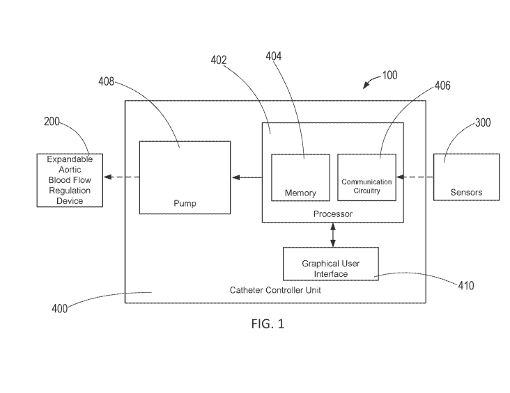
[0026] FIG. 1 is a schematic of an exemplary precision control system for
partial-aortic
occlusion constructed in accordance with the principles of the present
disclosure.
[0027] FIG. 2 illustrates an exemplary balloon catheter constructed in
accordance with
the principles of the present disclosure.
[0028] FIG. 3 illustrates the exemplary sensors of FIG. 1.
[0029] FIG. 4 is a conceptual illustration of an exemplary catheter
controller unit of FIG.
1.
[0030] FIG. 5 is a schematic of an exemplary external central processing
unit constructed
in accordance with the principles of the present disclosure.
[0031] FIG. 6 is a flow chart illustrating an exemplary method for
dynamically regulating
the degree of aortic blood flow regulation in accordance with the principles
of the present
disclosure.
[0032] FIGS. 7A-C are graphs illustrating change in proximal mean arterial
pressure,
distal mean arterial pressure, and aortic flow, respectively, of a study in
accordance with the
principles of the present disclosure.
[0033] FIGS. 8A-C illustrate the relationship between balloon volume and
various
hemodynamic parameters in a study in accordance with the principles of the
present
disclosure. FIG. 8D illustrates the relationship between aortic flow and goal
aortic flow in a
study.
[0034] FIG. 9A illustrates the relationship between balloon volume and
aortic flow
during EVAC, and FIG. 9B illustrates the relationship between balloon volume
and proximal
mean arterial pressure during EVAC.
9
CA 03060519 2019-10-18
WO 2018/195507 PCT/US2018/028694
[0035] FIG. 10 is a flow chart illustrating a study in accordance with the
principles of the
present disclosure.
[0036] FIGS. 11A-C are graphs comparing proximal mean arterial pressures,
distal mean
arterial pressures, and aortic blood flow, respectively, in a study during
REBOA and EVAC.
[0037] FIGS. 12A and 12B illustrate total resuscitation fluids and total
vasopressors,
respectively, required during REBOA and EVAC.
[0038] FIG. 13 illustrates the peak and final lactate levels during REBOA
and EVAC.
DETAILED DESCRIPTION
[0039] Partial-Resuscitative Endovascular Balloon Occlusion of the Aorta (P-
REBOA) is
a partial-aortic occlusion platform for decreasing distal ischemia and
reperfusion injury and
mitigating high proximal pressure by allowing titrated controlled low-volume
aortic flow
distal to the site of occlusion. P-REBOA may be achieved and maintained using
techniques
described in M. Austin Johnson, MD, PhD, et al., Partial Resuscitative Balloon
Occlusion of
the Aorta (P-REBOA): Clinical Technique and Rationale, 80 J Trauma Acute Care
Surg S133
(2016), the entire contents of which is incorporated by reference herein.
Another form of
partial-aortic occlusion that may be used during a variety of shock states to
improve the blood
pressure above the balloon is Endovascular Perfusion Augmentation for Critical
Care
(EPACC), which is achieved via a system comprising a series of endovascular
devices,
controller units for those devices, and algorithms capable of real-time
changes in the EPACC
devices in response to patient physiology. The concept of EPACC, in contrast
to techniques
such as REBOA, works via partial occlusion of the aorta similar to P-REBOA.
REBOA
maximizes proximal perfusion by completely occluding the aorta, at the expense
of
progressive ischemic injury to distal tissues. In contrast, EPACC only
partially occludes the
aorta, resulting in a more physiologic augmentation of proximal blood
pressure. By placing
the balloon at different levels within the aorta, the practitioner can select
which distal
capillary beds are exposed to decreased flow. Deployment of EPACC in the
descending
thoracic aorta results in mild reduction in blood flow to the mesentery,
kidneys, liver, and
extremities. In contrast, deployment at the aortic bifurcation only results in
potential
reduction in blood flow to the pelvis and limbs. Since aortic blood flow often
exceeds what
is physiologically required, minimal to moderate aortic blood flow restriction
only results in
CA 03060519 2019-10-18
WO 2018/195507 PCT/US2018/028694
minimal ischemia. This tradeoff between proximal blood pressure augmentation
and distal
ischemia is dependent upon the extent of shock as well as underlying patient's
physiology.
[0040] The ability of partial-REBOA, partial aortic occlusion, and/or EPACC
to be
automated to respond dynamically to any physiologic measure makes it a viable
technology
to maximize perfusion in multiple shock states. Endovascular Variable Aortic
Control
(EVAC) utilizes automated control of aortic occlusion to precisely and
dynamically regulate
distal aortic flow across the full spectrum from complete occlusion to full
unimpeded flow.
For trauma-specific applications, the EVAC technique can be used to restrict
distal aortic
flow down to a very low level, striking a delicate balance between ongoing
hemorrhage and
progressive distal ischemia, while simultaneously augmenting proximal
hemodynamics, e.g.,
blood flow, to the heart, lungs and brain, termed Regional Perfusion
Optimization (REPO)
(previously Permissive Regional Hypoperfusion). REPO is predicated on a method
that can
precisely and dynamically control the flow of blood to the abdominal aorta. To
be clinically
applicable, REPO must be accomplished with endovascular devices that can be
precisely
controlled. In another embodiment, these techniques may generate decision
support to
instruct a user and or devices to respond dynamically to any physiologic
measure, thereby
maximizing perfusion in multiple shock states.
[0041] REPO with EVAC has been shown to extend the duration of aortic
intervention to
90 minutes in a lethal liver injury swine model, with improved survival, end
organ function,
and lower resuscitation requirements compared to complete aortic occlusion,
e.g., REBOA.
These techniques are just as viable for the treatment of impending death from
exsanguination
while attempting to obtain surgical control of ongoing bleeding as it is for
septic shock to
decrease the amount of IV fluids and vasopressors required for treatment, or
neurogenic
shock in the setting of traumatic brain injury or intracerebral hemorrhage. As
will be
understood by a person having ordinary skill in the art, the exemplary
catheter controller unit
described herein may be used with any commercially available P-REBOA catheter
system or
with custom designed catheter systems.
[0042] Referring to FIG. 1, an exemplary precision control system for
partial-aortic
occlusion constructed in accordance with the principles of the present
disclosure is described.
In FIG. 1, the components of precision control system 100 are not depicted to
scale on either
a relative or absolute basis. System 100 comprises catheter controller unit
400 operatively
11
CA 03060519 2019-10-18
WO 2018/195507 PCT/US2018/028694
coupled to expandable blood flow regulation device 200 and sensors 300, and
optionally to
an external processing unit.
[0043] Catheter controller unit 400 may receive the data indicative of the
measured
physiological information from sensors 300, and determine whether the measured
physiological information is within a predetermined target physiological
range. For example,
catheter controller unit 400 may receive data indicative of measured blood
pressure distal to
the expandable blood flow regulation device and measured blood pressure
proximal to the
expandable blood flow regulation device, estimate aortic blood flow based on
the measured
blood pressures distal and proximal to the expandable blood flow regulation
device and/or
from waveforms corresponding to the measured distal and proximal blood
pressures, and
determine whether the estimated aortic blood flow is within a predetermined
target aortic
blood flow range.
[0044] Catheter controller unit 400 may be coupled to expandable blood flow
regulation
device 200 via a catheter sized and shaped for placement within aorta A of
patient P. For
example, catheter controller unit 400 may be coupled to a proximal end of the
catheter and
expandable blood flow regulation device 200 may be disposed at the distal end
of the
catheter. The catheter may be any catheter well-known in the art, having a
length sufficiently
long such that the catheter may be inserted into a patient via the femoral
artery or radial
artery, and extend through the patient's vasculature into the aorta.
[0045] Following placement of a compliant aortic occlusion balloon in the
aorta, e.g.,
.blood flow regulation device 200, catheter controller unit 400 would be
connected to this
catheter to allow inflation or deflation to regulate aortic flow using manual
control of
controller unit 400 or through decision support, whereby inflation or
deflation is
recommended to the provider. The expandable blood flow regulation device 200
may also be
any currently available balloon catheter that has not been designed for aortic
occlusion and
may undergo morphological changes over time.
[0046] Catheter controller unit 400 may also be coupled to expandable blood
flow
regulation device 200 such that catheter controller unit 400 automatically
adjusts expansion
and contraction of expandable blood flow regulation device 200 to adjust the
amount of blood
flow through the aorta if the measured physiological information falls outside
the target
physiological range as described in further detail below. In another
embodiment, catheter
12
CA 03060519 2019-10-18
WO 2018/195507
PCT/US2018/028694
controller unit 400 may be coupled to expandable blood flow regulation device
200 such that
catheter controller unit 400 adjusts expansion and contraction of expandable
blood flow
regulation device 200 to adjust the amount of blood flow through the aorta if
the measured
physiological information falls outside the target physiological range,
responsive to user input
received by catheter controller unit 400 via, e.g., switches and/or buttons
operatively coupled
to catheter controller unit 400. For example, catheter controller unit 400 may
generate
decision support if the measured physiological information falls outside the
predetermined
target physiological range, which guides a user to provide user input
sufficient to adjust the
amount of blood flow through the aorta to bring the patient physiology within
the target
physiological range. Catheter controller unit 400 may expand and contract,
e.g., inflate and
deflate, expandable blood flow regulation device 200 in small aliquots on the
order of e.g., 1
to 50 microliters. Catheter controller unit 400 may be battery powered or
plugged directly
into an electrical outlet.
[0047] In
one embodiment, system 100 may include an external central processing unit.
As described in further detail below, the external central processing unit may
be operatively
coupled to sensors 300 and catheter controller unit 400 such that the external
central
processing unit may receive the data indicative of the measured physiological
information
from sensors 300, determine whether the measured physiological information is
within a
predetermined target physiological range, calculate the amount of change of
size of
expandable blood flow regulation device 200 to bring the patient physiology
within the target
physiological range, and transmit information indicative of whether the
measured
physiological information falls outside the target physiological range to
catheter controller
unit 400 as described in further detail below. Accordingly, catheter
controller unit 400
automatically adjusts expansion and contraction of expandable blood flow
regulation device
200 to adjust the amount of blood flow through the aorta based on the
information received
from the external central processing unit. In another embodiment, catheter
controller unit
400 adjusts expansion and contraction of expandable blood flow regulation
device 200 to
adjust the amount of blood flow through the aorta if the measured
physiological information
falls outside the target physiological range, responsive to user input
received by catheter
controller unit 400, wherein the user input is entered by a user guided by
instructions
generated by catheter controller unit 400 based on the information received
from the external
central processing unit.
13
CA 03060519 2019-10-18
WO 2018/195507 PCT/US2018/028694
[0048] Referring now to FIG. 2, expandable blood flow regulation device 200
of FIG. 1
may be a balloon catheter. Accordingly, expandable blood flow regulation
device 200 may
include balloon 204 positioned at the distal end of the catheter. Balloon 204
is designed to
be inflated to a carefully titrated balloon volume to regulate blood flow in
the aorta. For
example, an incompressible fluid may be introduced into balloon 204 through a
lumen of the
catheter via exit ports 202 such that balloon 204 may maintain the carefully
titrated balloon
volume. Balloon 204 may be made of a suitable membrane that will prevent
diffusion of the
inflation fluid across the membrane and into the vasculature of the patient.
The membrane
may also be designed to inflate and deflate without undergoing morphological
changes over
time. However, as will be understood by a person having ordinary skill in the
art, expandable
blood flow regulation device may be any catheter system known in the art to
provide partial
REBOA, aortic occlusion balloon sysmte, or any vascular occlusion balloon
system.
[0049] As expandable blood flow regulation device 200 only partially
restricts blood flow
in the aorta, more physiologic augmentation of proximal blood pressure may
result, while
simultaneously optimizing blood flow to downstream organs and tissue beds.
Since aortic
blood flow is greater overall than is physiologically required in the majority
of cases for
patient's in shock, minimal-to-moderate occlusion results in only minimal
ischemia. This
tradeoff between proximal blood pressure augmentation and distal ischemia is
dependent
upon the extent of shock as well as the patient's underlying physiology. As
will be
understood by a person having ordinary skill in the art, catheter controller
unit 400 may be
operatively coupled to any P-REBOA balloon catheter system, REBOA catheter
system,
aortic occlusion balloon system, or other vascular occlusion balloon system,
e.g., IVC or
iliac.
[0050] Referring now to FIG. 3, sensors 300 may measure physiological
information
indicative of blood flow through the aorta to determine the patient's
underlying physiology.
For example, sensors 300 may measure physiological parameters including, but
not limited
to, blood pressure distal to the expandable blood flow regulation device,
blood pressure
proximal to the expandable blood flow regulation device, pressure within the
expandable
blood flow regulation device, heart rate, respiratory rate, aortic blood flow
proximal or distal
to the expandable blood flow regulation device, blood temperature, cardiac
output of the
patient, carotid blood flow, pulmonary pressures, peripheral vascular
resistance, or
intracranial pressure. Sensors 300 may include one or more sensors. For
example, as shown
14
CA 03060519 2019-10-18
WO 2018/195507 PCT/US2018/028694
in FIG. 3, sensors 300 comprise two sensors, distal sensor 302 positioned to
measure blood
pressure from a blood pressure line inserted into a distal artery and sensor
304 positioned to
measure blood pressure form a blood pressure line inserted into an artery
proximal to the
expandable blood flow regulation device. For example, distal sensor 302 may be
connected,
e.g., via luer lock connectors, to a flush port of an expandable blood flow
regulation device
introducer sheath or to an arterial line positioned in the contralateral
femoral artery, and
proximal sensor 304 may be connected to a proximal pressure port on the
expandable blood
flow regulation device or on a proximal arterial line via a radial artery.
Another sensor may
be connected to an inflation port of expandable blood flow regulation device
200 to measure
pressure within expandable blood flow regulation device 200 to protect against
over inflation
and facilitate detection of loss of expandable blood flow regulation device
pressure due either
to poor connection or expandable blood flow regulation device leak/rupture.
[0051] Sensors 300 may record data indicative of the measured physiological
information
either through analog or digital mechanisms. This data may then be used to
determine
whether more or less restriction of aortic blood flow is required to maximize
vital organ
perfusion via automated augmentation of blood pressure while simultaneously
aiming to
control hemorrhage, mitigate ischemia below the expandable blood flow
regulation device,
and mitigate high pressure above the expandable blood flow regulation device,
as described
in further detail below.
[0052] Patient physiology may also be monitored via real-time and
intermittent measures
of compounds with in the patient's blood, serum, urine, or saliva, e.g.,
levels of lactate, levels
of cortisol, levels of reactive oxygen species, the pH of the fluid, as well
as other commonly
used patient physiology markers.
[0053] Referring back to FIG. 1, catheter controller unit 400 includes
processor 402
having memory 404 and communication circuitry 406, and pump 408. Processor 402
may be
operatively coupled to sensors 300, graphical user interface 410, and pump
408, and pump
408 may be operatively coupled to expandable blood flow regulation device 200.
[0054] Processor 402 may receive data indicative of the measured
physiological
information from sensors 300 via communication circuitry 406, and record the
data in
memory 404. Processor 402 may further record waveforms corresponding to the
measured
physiological information from sensors 300 in memory 404. Memory 404, e.g.,
non-
CA 03060519 2019-10-18
WO 2018/195507
PCT/US2018/028694
transitory computer readable media, may store a target physiological parameter
and a
corresponding range associated with blood flow through the aorta, and
instructions that, when
executed by processor 402, cause processor 402 to compare the measured
physiological
information with the target physiological range to determine whether the
measured
physiological information is within the predetermined target physiological
range. Processor
402 may cause graphical user interface 410, e.g., touch enabled LCD display,
to display
information indicative of the comparison of the measured physiological
information with the
target physiological range. For example, processor 402 may receive data
indicative of
measured blood pressure distal to expandable blood flow regulation device 200
and measured
blood pressure proximal to expandable blood flow regulation device 200, from
which
processor 402 may estimate aortic blood flow. Accordingly, processor 402 may
cause
graphical user interface 410 to display, e.g., graphically, the estimated
aortic blood flow as
well as the target physiological parameter, e.g., target aortic blood flow,
and its desired
corresponding range. For example, graphical user interface 410 may display the
target aortic
blood flow in the centerline with the predetermined desired range above and
below the
centerline, e.g., in green, wherein the desired range is surrounded by a
predetermined
acceptable range, e.g., in yellow, above and below the desired range, and
wherein the
acceptable range is surrounded by a predetermined unacceptable range, e.g., in
red, above and
below the acceptable range. Graphical user interface 410 may display the
estimated aortic
blood flow in relation to the ranges, which will indicate whether the
estimated aortic blood
flow falls outside the desired and/or acceptable range. As such, processor 402
may calculate
an appropriate change in the amount of occlusion by expandable blood flow
regulation device
200 necessary to bring the patient physiology within the target physiological
range based on
the current measured patient physiology.
[0055]
Graphical user interface 410 may permit a user to select various menu
functions,
e.g., setting the target aortic flow, setting audible alarms to indicate when
blood flow is
deviating from the desired range, to allow for zeroing of sensors 300, to
allow for entering
patient information including, for example, height, weight, gender, name, and
date of birth.
Accordingly, memory 404 may store patient profiles corresponding to the
patient information
entered via graphical user interface 410. Information stored in memory 404 may
be
downloaded from catheter controller unit 400 via, e.g., a removable media
card.
16
CA 03060519 2019-10-18
WO 2018/195507 PCT/US2018/028694
[0056] Processor 402 comprises a series of sub-algorithms for controlling
each aspect of
appropriate balloon inflation, deflation, and rate of response to physiologic
changes when a
balloon catheter is used. These individual algorithms may also calculate:
initial calibration to
identify the physical measurements of the vessel, determination of complete
occlusion,
identification of a working range of the catheter, e.g., the range of
occlusion that results due
to changes in patient physiology, set point optimization, weaning off from
catheter-based
physiologic support, and balloon volume tuning.
[0057] For balloon catheter 200, the balloon calibration sequence occurs
upon initial
insertion of the catheter or upon initiation of EPACC. The calibration
sequence is also
activated any time large changes in the hemodynamics are detected that are not
induced by
EPACC. Upon initiation of the balloon calibration sequence, pump 408 of
catheter controller
unit 400 will iteratively introduce small aliquots of gas or fluid, e.g.,
carbon dioxide, saline,
or a mixture of contrast and saline, into the balloon. During sequential
boluses, proximal
physiology may be monitored until a change is observed, which denotes the low
set point of
the working range of balloon 204. Balloon 204 will continue to inflate until
the distal blood
pressure waveform is extinguished or until proximal physiologic changes are no
longer
observed, which denotes the upper working range of balloon catheter 200.
Alternatively, the
upper limit may be denoted by measuring the cessation of aortic flow. A mid-
point of the
working range may be set as an interval increase in balloon volume from the
low set point
and may be referenced for a rapid return to working range if needed during
EPACC.
[0058] After balloon calibration has occurred and initial balloon volume
set points have
been identified, processor 402 causes catheter controller unit 400 to adjust
the shape and size
of expandable blood flow regulation device 200 via pump 408 to augment
proximal blood
pressure responsive to patient physiology. As described above, processor 402
compares the
measured physiological information received from sensors 300 with the target
physiological
range stored in memory 404 to determine whether the measured physiological
information is
within the predetermined target physiological range. For example, if proximal
blood pressure
is set as the physiologic marker, when processor 402 determines that proximal
blood pressure
drops below the target blood pressure range, catheter controller unit 400
expands expandable
blood flow regulation device 200 via pump 408, e.g., inflate the balloon.
Similarly, when
processor 402 determines that proximal blood pressure exceeds the target blood
pressure
range, catheter controller unit 400 contracts expandable blood flow regulation
device 200 via
17
CA 03060519 2019-10-18
WO 2018/195507 PCT/US2018/028694
pump 408, e.g., deflate the balloon. The amount of change in balloon volume
that occurs in
response to blood pressure changes that are out of range is dependent upon how
far the
current measured blood pressure is from the target blood pressure. Therefore,
if the blood
pressure is only minimally out of the target range, a small change in size of
expandable blood
flow regulation device 200 is made. In contrast, when the blood pressure is
significantly out
of the target range, a larger change in size of expandable blood flow
regulation device 200 is
made. Processor 402 may record the amount of change in size of expandable
blood flow
regulation device 200 caused by pump 408 such that memory 404 stores a running
tally of,
e.g., balloon filling volume. Accordingly, a user may be provided with
constant real-time
information indicative of, e.g., how much fluid is within the balloon.
[0059] An example algorithm that may be used to provide EPACC includes:
uLBolus = (Po-Ps) J* V
where Po is current pressure, Ps is set point pressure, J is a constant, and V
is a constant
described below. One skilled in the art will understand that alternative
algorithms could be
used to adjust balloon volumes based upon current and goal physiology. For
example,
alternative algorithms may use less or more than two constants and/or
variables.
[0060] The balloon tuning algorithm allows for the magnitude of the change
of size of
expandable blood flow regulation device 200 in response to the difference
between the
measured physiological information and the target physiological parameter to
be dynamic,
controlled by, e.g., the constant V and J. Initially V and J may be set to a
default, but V or J
may change dynamically dependent upon the magnitude of physiologic changes
that occur
beyond the initial target set points. For example, if blood pressure is set as
the physiologic
marker and the initial blood pressure recorded by sensors 300 was below the
set point
pressure, but the resulting blood pressure recorded by sensors 300 after the
change in
expansion amount of expandable blood flow regulation device 200 by pump 408, V
and or J
would then be modified in order to correct for overshooting the goal set
point. If the
measured blood pressure is determined to be within the target blood pressure
and as a result,
the amount of expansion of expandable blood flow regulation device 200 drops
below the
low set point, expandable blood flow regulation device 200 will then wean off
to its baseline
zero set point. This may occur by deflating the balloon. For example, the
following code
illustrates dynamically scaling expansion of an expandable blood flow
regulation device.
18
CA 03060519 2019-10-18
WO 2018/195507 PCT/US2018/028694
void balloon titration07
void balloon_titration_correction0.;
void balloon_titrationO 7.1 this function establishes the bolus volume that-
is delivered to the ba7loon based on the devation of the
normalized_distai_pressure from the setpoint pressure_setpoint
if ielamsed timer is ater than the delay period timedelay)
Set ,=,,Iapsed timer to zero
Set the variable pressure _l = normalized distal pressure
Calculate the difference between the current normalized distal pressure
and the oressure set-point
pressure difference: = pressure? pressure_setpoint
CaIcuistP, the absolutP, value of this difference
pressure_difference=abstpressure_difference)
if (pressure difference is areater than ImmHd)
Then nem-form function balloon_titrationcorrection0;
Read Ailjensors(); //reads the oressure sensor values
Set the variable pressure ) = normalizeddistal_pressure
If (normalized distal pressure is less than 0.5 mMiiq above or below the
pTessure_setpoint)
Then set the delay period to minimum delay
timedelay = 2000 msec
Else If (normalized distal pressure is greater than 0.5 mmHg above or
below the pressure_setpoint4
ThPh
Inflate balloon by volume established from equation:
Bolus_VOlume = round(((normalized_distal pressure -
pressure setpoint)*scalina factor))
Set for delay period established from eaustion: timedelay =
2000 msec
timescale*pressure_difference*timer_variable_oomstant
Else If (normalized distal pressure: is Less than 0.5 mMFig above or
below the pressure_setboint)
7hpn
Deflate balloon by volume established from equation:
Bolus Vole = round( ((normalized distal pressure -
pressure_setpoint)*scalina factor))
Set for delay period established from equation: timedelay =
2000 msec -0-
timescale*pressure_difference*timer variable_constant
19
CA 03060519 2019-10-18
WO 2018/195507 PCT/US2018/028694
void balloon_titratcorrection0 //dynamically scales balloon titration
amount when the distance from the setpoint in dreater than immmlio,
If (pressure_p is greater than pressure_setpaint
If tpressur_A is less than pressurls_,setpointl
The holue volume has overshot the target pressure, therefore the
scaIind factor v will, be incremented to a small number ae
Set v = v - (pressure difference/o
// her o represents the volume correction constant
Else If (pressure_l iz greater than pressure_setpoint)
The bolus volume has undPrEhot the tardet nressure, therPfore the
scaIind factor v will be incremented to a small number as follows:
Set v = v 4 (prassure_differenoa)/(4*0
Else If ,prlassnre_C ia less than than pressure_setDoint
If pre/ is .eater than pressure_setpoint
The bolus volite has ovF,rehot the tat pressure, th[F.rfore thF,
scalina factor v will be incremented to a all nuMber as fOlIows:
Set v = v - Wressure_d.iffarence)/o
where o rerrPaents tevi iue correctin constant
Else If pressura_A is _less than pressurasetpoint)
The bolus voinie ndr9hot
the tarat pressure, therfnre th
scalino factor v will he incremented to a amaIl number as follows:
SetV = v tpressure_ifference)/(4*o)
[0061] As
described above, processor 402 may automatically expand and contract
expandable blood flow regulation device 200 via pump 408 in accordance with
the principles
of the present disclosure. For example, when expandable blood flow regulation
device 200
comprises a balloon catheter, pump 408 may be a syringe pump designed to
inject or remove
fluid from the balloon to inflate or deflate the balloon via the exit ports in
fluid
communication with the lumen of the catheter. The syringe pump may make small
titrated
changes in balloon volume, e.g., on the order of 1 to 50 microliters, in
response to patient
physiology via automation. For example, the syringe pump may include a
metallic threaded
CA 03060519 2019-10-18
WO 2018/195507 PCT/US2018/028694
rod to allow linear translation of the syringe plunger guide. The syringe pump
may be
actuated via a low power consumption stepper motor, e.g., NEMA 11 or 8, and
may contain a
reduction gear box. In one embodiment, the syringe pump may be controlled via
a linear
actuator.
[0062] After each change in balloon volume by pump 408 of catheter
controller unit 400,
processor 402 may wait for a predetermined period of time for the resulting
physiologic
response to be monitored before further adjusting the balloon volume.
[0063] In one embodiment, pump 408 may provide for manual inflation of the
balloon,
e.g., when automation is either unavailable or not feasible. For example, a
manual pump may
include a syringe pump that may inject fluid using the normal action of a
syringe, but may
also inject or remove fluid via screw actuation once threads on the plunger
and within the
barrel of the syringe have been activated. Injection via normal syringe
plunging, but fluid
removal only via screw actuation allows for rapid inflation of the balloon,
but carefully
titrated removal of fluid based upon the pitch of the thread on the plunger.
As will be
understood by one having ordinary skill in the art, the manual pump may
include, e.g.,
peristaltic pumps, rotatory pumps, etc.
[0064] In another embodiment, catheter controller unit 400 may generate
decision
support if the measured physiological information falls outside the
predetermined target
physiological range. The decision support, e.g., set of instructions, may be
communicated to
the user via, e.g., visually or audibly via graphical user interface 410, such
that the decision
support guides a user to provide user input via, e.g., graphical user
interface 410 or a plurality
of switches and/or buttons operatively coupled to processor 402. As will be
understood by
one of ordinary skill in the art, graphical user interface 410 may include a
plurality of
switches and/or buttons for receiving user input. The user input may be
sufficient to change
the amount of occlusion by expandable blood flow regulation device 200
necessary to bring
the patient physiology within the target physiological range based on the
current measured
patient physiology as calculated by processor 402. Accordingly, in response to
receiving the
user input, processor 402 causes pump 408, e.g., syringe pump, to inject or
remove fluid from
the balloon to inflate or deflate the balloon via the exit ports in fluid
communication with the
lumen of the catheter. The syringe pump may make small titrated changes in
balloon
volume, e.g., on the order of 1 to 1000 microliters, in response to the user
input.
21
CA 03060519 2019-10-18
WO 2018/195507 PCT/US2018/028694
[0065] FIG. 4 illustrates a conceptual embodiment of catheter controller
unit 400 having
graphical user interface 410 and wherein pump 408 is a syringe pump. As shown
in FIG. 4,
graphical user interface 410 may include display 412 for visually
communicating information
to a user, and plurality of buttons and switches 414 for allowing a user to
interact with
catheter controller unit 400, e.g., expand or contract expandable blood flow
regulation device
200 and/or navigate through the menu functions provided by graphical user
interface 410.
[0066] Referring to FIG. 5, an exemplary external central processing unit
constructed in
accordance with the principles of the present disclosure is described. As
shown in FIG. 5,
external central processing unit 500 comprises processor 502 having memory 504
and
communication circuitry 506. In FIG. 5, components of processor 502 are not
depicted to
scale on either a relative or absolute basis. Processor 502 may be constructed
similarly to
processor 402 of catheter controller unit 400 of FIG. 1, such that processor
502 may be
operatively coupled to sensors 300, receive data indicative of the measured
physiological
information from sensors 300, and compare the measured physiological
information with a
target physiological range stored in memory 504. When system 100 comprises
external
central processing unit 500, processor 502 of external central processing unit
500 determines
whether the measured physiological information is within the target
physiological range,
calculates information indicative of the appropriate change in the amount of
occlusion by
expandable blood flow regulation device 200 required to bring the patient's
physiology
within the target physiological range if the measured physiological
information falls outside
the target physiological range, and transmits the information to catheter
controller unit 400
via communication circuitry 506. For example, communication circuitry 506 of
external
central processing unit 500 may transmit the information to communication
circuitry 406 of
catheter controller unit 400 via at least one of WiFi, Bluetooth, Wixel-based
communication,
or cellular communication, or a wired connection, or other form of
communication.
[0067] Referring to FIG. 6, an exemplary method for dynamically regulating
the degree
of aortic blood flow regulation in accordance with the principles of the
present disclosure is
described. Method 300 may be used to perform partial aortic occlusion, e.g., P-
REBOA,
EVAC, or EPACC, on a patient, for example, in shock from sepsis or trauma. At
step 602, a
distal end of the catheter is introduced into the patient via the femoral
artery or the radial
artery such that expandable blood flow regulation device 200 disposed at the
distal end is
22
CA 03060519 2019-10-18
WO 2018/195507 PCT/US2018/028694
placed within the aorta. As described above, expandable blood flow regulation
device 200
may comprise balloon catheter 200.
[0068] At step 604, expandable blood flow regulation device 200 may be
expanded to
regulate blood flow through the aorta. For example, drive mechanism 408 of
catheter
controller unit 400 may cause balloon 204 of balloon catheter 200 to be
inflated such that it
regulates blood flow in the aorta.
[0069] At step 606, sensors 300 may measure physiological information
indicative of
blood flow through the aorta. For example, as described above, sensors 300 may
measure
information indicative of blood pressure distal to the expandable blood flow
regulation
device, blood pressure proximal to the expandable blood flow regulation
device, pressure
within the expandable blood flow regulation device, heart rate, respiratory
rate, aortic blood
flow proximal or distal to the expandable blood flow regulation device, blood
temperature,
cardiac output of the patient, carotid blood flow, pulmonary pressures,
peripheral vascular
resistance, or intracranial pressure. Sensors 300 may comprise one or more
sensors
positioned proximal and/or distal to expandable blood flow regulation device
200 to
effectively monitor patient physiology. For example, one sensor may be
positioned distal to
the expandable blood flow regulation device to measure blood pressure distal
to the
expandable blood flow regulation device, and another sensor may be positioned
proximal to
the expandable blood flow regulation device to measure blood pressure proximal
to the
expandable blood flow regulation device.
[0070] At step 608, processor 402 of catheter controller unit 400, or when
external central
processing unit 500 is utilized, processor 802, may compare the measured
physiological
information with a target physiological range. At step 608, processor 402 may
first estimate,
e.g., aortic blood flow, from the measured physiological information, e.g.,
blood pressure
distal and proximal to the expandable blood flow regulation device and
corresponding
waveforms, then compare the estimated aortic blood flow with a target
physiological range,
e.g., target aortic blood flow. At step 610, processor 402 determines whether
the measured
physiological information falls within the target physiological range. If it
is determined at
step 610 that the measured physiological information falls within the target
physiological
range, method 300 may maintain the current state of expansion of expandable
blood flow
regulation device 200 and return to step 606 to continue measuring
physiological information
of the patient. If it is determined at step 610 that the measured
physiological information
23
CA 03060519 2019-10-18
WO 2018/195507 PCT/US2018/028694
falls outside the target physiological range, e.g., exceeds or falls below the
target
physiological range, processor 402 of catheter controller until 400 may
determine the amount
of change in expansion of expandable blood flow regulation device 200
necessary to bring
patient physiology within the target physiological range.
[0071] At step 612, processor 402 causes drive mechanism 408 to adjust the
expansion or
contraction of the expandable blood flow regulation device, e.g., inflate or
deflate balloon, to
adjust the amount of blood flow through the aorta. In one embodiment, drive
mechanism 408
automatically adjusts the expansion or contraction of the expandable blood
flow regulation
device based on the amount of change in expansion of expandable blood flow
regulation
device 200 necessary to bring patient physiology within the target
physiological range
determined by processor 402. In another embodiment, at step 612, processor 402
may
generate decision support that may be used by a user to enter user input such
that processor
402 causes drive mechanism 408 to adjust the expansion or contraction of the
expandable
blood flow regulation device based on the user input. During step 612,
processor 402 may
generate an alert if it is determined at step 610 that the measured
physiological information
falls outside the target physiological range, and cause graphical user
interface to
communicate the alert to a user, e.g., visually or audibly.
[0072] When external central processing unit 500 is utilized, processor 502
transmits
informative indicative of the amount of change in expansion of expandable
blood flow
regulation device 200 necessary to bring patient physiology within the target
physiological
range, determined at step 610, to catheter controller unit 400 via
communication circuitry 506
and 406 before proceeding to step 612.
Study # 1 Comparing EVAC Syringe Pump with Manual Control Pump
[0073] The following experimental study involving in vivo animal testing of
a custom-
built hardware and software system to control aortic flow was approved by the
Institutional
Animal Care and Use Committee at David Grant Medical Center, Travis Air Force
Base,
California. Healthy adult, castrate male and non-pregnant female Yorkshire-
cross swine (Sus
scrofa) were acclimated for a minimum of seven days. At the time of
experimentation,
animals weighed between 60 and 95 kg.
[0074] The novel components of this platform include a precision automated
syringe
pump coupled with a custom microcontroller that integrates streaming
physiologic data from
24
CA 03060519 2019-10-18
WO 2018/195507
PCT/US2018/028694
the patient. In brief, the hardware architecture utilizes a commercially
available
microcontroller (available from Arduino, Somerville, MA) with wireless
functionality and a
multi-channel 16-bit analog-to-digital converter for acquisition of real-time
physiologic data
including aortic flow, proximal arterial pressure, and distal arterial
pressure. The custom
syringe pump utilized a NEMA 17 stepper motor that drives a standard lead
screw, a
commercially available stepper motor controller (BigEasyDriver, available from
Sparkfun,
Niwot, CO), custom 3D-printed components that hold the syringe and plunger,
and a wireless
microcontroller that performs bidirectional communication with the master
controller unit.
Custom software was developed to precisely regulate aortic flow using a closed
loop
feedback algorithm. A weight-based aortic flow rate of 4.3 mL/kg/min was
established,
which is approximately 10% of baseline distal aortic flow.
[0075]
Animals were premedicated with 6.6 mg/kg intramuscular tiletamine/zolazepam
(TELAZOL, available from Fort Dodge Animal Health, Fort Dodge, IA). Following
isoflurane induction and endotracheal intubation, general anesthesia was
maintained with 2%
isoflurane in 100% oxygen. To offset the vasodilatory effects of general
anesthesia, an
intravenous infusion of norepinephrine (0.01 mg/kg/min) was instituted upon
venous access
and titrated prior to experimentation to achieve a target mean arterial
pressure between 65
and 75 mm Hg. Animals were mechanically ventilated to maintain end-tidal CO2
at 40 5
mm Hg. Plasmalyte (available from Baxter, Deerfield, IL) maintenance
intravenous fluid
was administered at a rate of 10 mL/kg/h until the abdomen was closed, at
which point the
rate was decreased to 5 mL/kg/h for the remainder of the study to overcome
insensible losses.
Intravenous heparin was administered to achieve an activated clotting time
(ACT) of 100
seconds, similar to human baseline values. An underbody warmer was used to
maintain core
body temperature between 35 and 37 C.
[0076]
Following laparotomy, a splenectomy was performed to minimize hemodynamic
variation from autotransfusion. The supraceliac aorta was exposed by dividing
the left
diaphragm and dissected circumferentially for a length of 5-10 cm. A
perivascular aortic
flow probe (available from Transonic Systems Inc., Ithaca, NY) was placed with
ligation of
two adjacent intercostal arteries distally, thus preventing intervening flow
between the flow
probe and the endovascular occlusion balloon. The abdomen was closed with
cable ties.
External jugular veins were cannulated to facilitate medication and fluid
administration. The
right brachial artery was exposed and cannulated with a 7F sheath
(SuperSheath, available
CA 03060519 2019-10-18
WO 2018/195507 PCT/US2018/028694
from Boston Scientific, Marlborough, MA) for controlled hemorrhage. The left
axillary
artery was exposed and cannulated with a 9F sheath (SuperSheath, available
from Boston
Scientific, Marlborough, MA) for proximal arterial pressure monitoring. The
left femoral
artery was exposed and cannulated with a 12F sheath, available from Teleflex
Inc., Wayne,
PA), through which an 9F Coda LP balloon (available from Cook Medical,
Bloomington, IN)
advanced under fluoroscopic guidance to the level of the supraceliac aorta
(Zone 1), just
distal to the aortic flow probe. Distal pressure was also monitored via this
sheath.
[0077] Physiologic parameters and aortic flow measurements were collected
in real time
using a Biopac MP150 multichannel data acquisition system and the custom
Arduino-based
data acquisition system/controller (available from BioPac, Goleta, CA).
Parameters
measured included heart rate, blood pressure proximal and distal to the intra-
aortic balloons,
and aortic flow beyond the Zone I balloon.
[0078] Data analysis was performed and graphs constructed using Excel
(available from
Microsoft Corporation, Redmond, WA), and STATA version 14.0 (available from
Stata
Corporation, Bryan, TX). Continuous variables are graphically presented as
means and
standard error of the means. Categorical variables are presented as means with
standard
deviation and standard error of the means.
[0079] At the beginning of experimentation (TO), animals were subjected to
a 25% total
blood volume hemorrhage over 30 minutes. Following this 30-minute hemorrhage
interval,
the master controller initiated stepwise balloon inflation over approximately
3 minutes until
the target weight-based flow rate was achieved. The EVAC syringe pump
automatically
adjusted the balloon volume to actively maintain aortic flow at this level for
the duration of
the 45-minute EVAC interval. To ascertain the performance of the EVAC syringe
pump
during active resuscitation, whole blood transfusion was initiated at T65. The
EVAC syringe
pump then initiated a 5-minute balloon deflation and weaning sequence,
beginning at T75.
[0080] Five animals underwent instrumentation, hemorrhage, and a subsequent
45
minutes of Zone 1 EVAC. All animals survived the experimental phase. As shown
in FIGS.
7A-C, hemorrhage was associated with an anticipated decline in distal aortic
flow and in
mean arterial pressure as measured in both the proximal descending thoracic
aorta and the
distal abdominal aorta, e.g., proximal MAP and distal MAP. Upon initiation of
EVAC at
T30, there was an abrupt increase in proximal mean arterial pressure and a
concurrent
26
CA 03060519 2019-10-18
WO 2018/195507 PCT/US2018/028694
decrease in distal MAP. As shown in FIG. 7B, distal aortic pressure also
remained stable
throughout EVAC, at approximately 16 mmHg. Referring now to FIGS. 7C, 8C, and
8D, the
EVAC syringe pump was able to maintain stable aortic flow throughout the 45-
minute
intervention period with minimal deviation from the aortic flow goal.
[0081] As shown in FIG. 8A, upon initiation of blood transfusion at T65,
there was a
steep rise in proximal MAP. The EVAC syringe pump responded with compensatory
increase in balloon volume in order to maintain the specified aortic flow
rate. Both aortic
flow and distal aortic pressure remained stable and unchanged during active
volume
resuscitation as a result of these compensatory balloon adjustments.
[0082] The relationship of balloon volume and the various hemodynamic
parameters is
represented in FIGS. 9A and 9B. The EVAC syringe pump made small, yet
discernible
changes in balloon volume throughout the EVAC interval, while the largest
changes occurred
during the 10-minute period of blood transfusion. Actual aortic flow closely
approximated
the target aortic flow (mean flow, 4.5 vs 4.4 ml/kg/min). As shown in FIGS. 8D
and 9A, the
EVAC syringe pump maintained aortic flow within 11% -14% of baseline
throughout the
entirety of the intervention period and between 13%- 14% for greater than 90%
of the
intervention.
[0083] Stepwise balloon deflation resulted in a rapid, steep increase in
aortic flow around
the balloon. As shown in FIG. 9A, return to full baseline flow rates was
observed following
withdrawal of 2.5 mL from the balloon, with nearly twice baseline flow
observed upon full
balloon deflation (34 ml/kg/min and 67 ml/kg/min, respectively).
[0084] Throughout the 45-minute period of EVAC, the syringe pump made an
average of
537 balloon adjustments, with a mean balloon volume change of 6.4 uL per
adjustment. As
shown in Table 1 below, the largest average balloon volume change required in
order to
maintain flow within the specified range was approximately 100 uL.
Table 1
Standard
Average Deviation SEM
Total Volume Moved (pL) 3490.8 1575.1 643.0
Total # Movements 537 33 14
Largest Volume Change
(pL) 98.7 101.5 41.4
27
CA 03060519 2019-10-18
WO 2018/195507 PCT/US2018/028694
Average Volume Change
(pL) 6.4 2.5 1.0
[0085] FIGS. 9A and 9B demonstrate the EVAC syringe pump's control of
balloon
volume in response to aortic flow and proximal mean arterial pressure,
respectively, for a
single representative experiment. Flow remains essentially unchanged over time
as a result
of small, highly dynamic adjustments to balloon volume. Of note, the profile
of balloon
volume closely mirrors the trend in proximal mean arterial pressure throughout
the
intervention.
[0086] This study is the first-in-animal experience with EVAC using
commercially
available aortic occlusion catheters and an entirely automated flow regulating
device.
Precision aortic flow regulation was demonstrated to be feasible and
imminently achievable
with commercially available aortic occlusion catheters by adjusting occlusion
balloon
volumes with an algorithm-driven, autonomous syringe pump.
[0087] Early clinical experiences have demonstrated that P-REBOA is
extremely
challenging and results in labile hemodynamics due to the lack of fidelity
with manual
balloon titration. Additionally, large animal models of P-REBOA have
demonstrated a
tendency towards perpetuating ongoing hemorrhage. Continued bleeding is a
serious concern
in every clinical environment, but especially in scenarios where blood
products are in limited
supply or when a significant delay to surgical hemostasis is anticipated.
[0088] Thus, automation was to improve upon the P-REBOA concept. The use of
automation addresses several key limitations of the manual approach to aortic
flow
regulation. First, the EVAC syringe pump is capable of executing very small
changes in
balloon filling volume. The current hardware design of the syringe pump is
capable of
delivering or withdrawing 10 uL aliquots of fluid at a time. These changes are
too precise to
be performed manually with any manner of fidelity or consistency and demand
robotic
control. As demonstrated in prior experiments and again in the present
experiment, very
small adjustments in occlusion balloon volume translate into large variations
in aortic flow
rate. This hyperemic aortic flow was observed during the balloon deflation
phase of the
experiment and likely arises from the combination of the extreme proximal
aortic pressure in
combination with maximally vasodilated distal tissue beds. This hyperemic flow
is fairly
unpredictable in its onset and occurs at different balloon volumes depending
on the individual
animal. Therefore, precise control is necessary to prevent hemodynamic
collapse or
28
CA 03060519 2019-10-18
WO 2018/195507 PCT/US2018/028694
precipitate clot destabilization and renewed hemorrhage. With balloon volume
changes of as
little at lOuL producing demonstrable differences in aortic flow rates, it is
apparent why
previous attempts at manual flow titration have resulted in erratic
hemodynamics.
[0089] Additionally, this EVAC syringe pump is capable of dynamic changes
in response
to changing patient physiology with near continuous adjustments of balloon
volume. In the
present study, there were an average of nearly 600 balloon adjustments made
over a 45-
minute period to maintain the desired aortic flow rate. This frequency of
adjustments would
be difficult, if not impossible, to achieve with manual control of the balloon
volume.
Moreover, humans are highly inefficient at integrating multiple streams of
continuous data
and responding in a timely fashion with an appropriate, measured response.
Yet, computers
and robots excel at these integration and computation tasks. Inefficiencies in
care are
compounded by assigning a skilled medical provider the sole task of manually
titrating an
occlusion balloon. This potential misallocation of key medical expertise and
manpower may
prevent the adoption of partial aortic occlusion as a resuscitation adjunct in
austere
environments.
[0090] In comparison to previous extracorporeal flow regulation circuits,
the current
EVAC syringe pump provides an equivalent, if not superior, degree of aortic
flow control.
With earlier approaches, tight regulation of low volume distal aortic flow was
demonstrated
to effectively mitigate the ischemic burden of sustained aortic occlusion,
while
simultaneously minimizing hemorrhage. However, one key consideration of this
current
software design is that titration is predicated on aortic flow data input.
Experimentally, this
data was acquired through the use of a surgically implanted perivascular flow
probe, which is
clinically impractical. Unfortunately, there is no commercially available
method of obtaining
an accurate measure of aortic flow with a minimally invasive or endovascular
means to
enable careful titration of a balloon catheter. Despite this fact, this study
does demonstrate
that distal aortic pressure and aortic flow do correlate in this particular
model of hemorrhage
and ischemia. Therefore, it is conceivable that titrating the degree of
occlusion to a specific
distal aortic pressure would result in a stable downstream aortic flow.
Study #2 Comparing EVAC and REBOA
[0091] The following study comparing EVAC and REBOA was approved by The
Institutional Animal Care and Use Committee at David Grant Medical Center,
Travis Air
29
CA 03060519 2019-10-18
WO 2018/195507 PCT/US2018/028694
Force Base. Referring now to FIG. 10, in this study, healthy adult, castrate
male and non-
pregnant female Yorkshire-cross swine (Sus scrofa) weighing between 60 and 95
kg were
subjected to a 25% total blood volume hemorrhage over 30 minutes, followed by
block
randomization to a 45 minute intervention with either Zone 1 or EVAC (n=6 per
group) All
animals then underwent resuscitation with shed blood and received protocolized
critical care
phase for the remainder of a 360 minute time period, during which vasopressor
and isotonic
fluid administration were automatically regulated based upon predefined
physiologic
parameters.
[0092] Animals were premedicated with 6.6 mg/kg intramuscular
tiletamine/zolazepam
(TELAZOL, available from Fort Dodge Animal Health, Fort Dodge, IA). Following
isoflurane induction and endotracheal intubation, general anesthesia was
maintained with 2%
isoflurane in 100% oxygen which was titrated to 40% oxygen to maintain a pulse
oximetry
between 92-98%. To offset the vasodilatory effects of general anesthesia, an
intravenous
infusion of norepinephrine (0.01 mg/kg/min) was instituted upon venous access,
and titrated
prior to experimentation to achieve a target mean arterial pressure (MAP)
between 65 and 75
mm Hg. Animals were mechanically ventilated to maintain end-tidal CO2 at 40
5 mm Hg.
All animals received a bolus of 1 L Plasmalyte-A (available from Baxter,
Deerfield, IL)
upon venous access. Following the bolus, Plasmalyte-A maintenance intravenous
fluid was
administered at a rate of 10 mL/kg/h until the abdomen was closed, after which
it was
decreased to 5 mL/kg/h for the remainder of the study. Intravenous heparin was
administered
prior to experimentation to achieve an activated clotting time (ACT) of 100
seconds. An
underbody warmer was used to maintain core body temperature between 35 and 37
C.
[0093] Following laparotomy, splenectomy was performed to minimize
hemodynamic
variation from autotransfusion. The supraceliac aorta was exposed by dividing
the left
diaphragm followed by circumferential dissection of the aorta for a length of
5-10 cm. Two
adjacent intercostal arteries were ligated and a perivascular aortic flow
probe (available from
Transonic Systems Inc., Ithaca, NY) was placed proximal to the ligated vessels
preventing
intervening flow between the flow probe and the endovascular occlusion
balloon. The
abdomen was closed with cable ties, and vascular access was performed as
previously
described. A 9F Coda LP balloon (available from Cook Medial LLC, Bloomington,
IN) was
positioned just distal to the aortic flow probe. The inflation syringe was
connected to a
CA 03060519 2019-10-18
WO 2018/195507 PCT/US2018/028694
custom-developed EVAC automated syringe pump capable of both complete REBOA or
EVAC.
[0094] Following hemorrhage and subsequent randomization, animals in the
EVAC arm
had tightly controlled aortic flow below the balloon for 45 minutes, ranging
from 1.5 to 4.4
ml/kg/min, e.g., 100 ml/min to 300 ml/min for a 70 kg animal, achieved using a
wireless
automated syringe pump running custom closed loop feedback algorithms. In the
REBOA
arm, animals were subjected to 45 minutes of sustained complete aortic
occlusion, which was
maintained with the same automated syringe pump.
[0095] Beginning at 80 minutes through end of study, the administration of
intravenous
crystalloid fluid boluses and the titration of vasopressors was automated
using a
microcontroller interfaced with a standard peristaltic fluid pump and a custom
infusion
syringe pump, respectively. Fluid boluses were triggered based on continuous
central venous
pressure (CVP) and MAP values. Vasopressors were titrated up or down in
response to
hypotension (MAP <60) or hypertension (MAP > 70), respectively. Animals were
euthanized at T360, followed promptly by necropsy.
[0096] During the study, physiologic parameters and aortic flow
measurements were
collected in real time using a multichannel data acquisition system (Biopac
MP150, available
from BioPac, Goleta, CA). Parameters measured included heart rate, blood
pressure
proximal and distal to the intraaortic balloon catheters, central venous
pressure, core
temperature, and aortic flow. Arterial blood and urine were collected at
routine intervals
throughout the study for analysis. End-organ histology was performed and
analyzed by a
blinded veterinary pathologist. Data analysis was performed with STATA version
14.0
(available from Stata Corporation, Bryan, TX). Continuous variables are
presented as means
and standard errors of the mean if normally distributed and as medians with
interquartile
ranges if not distributed normally and analyzed using the appropriate test.
Dichotomous and
categorical variables were analyzed by Fisher's exact test and presented as
percentages.
Statistical significance was set at p <0.05.
[0097] As shown in Table 2 below, there were no differences in baseline
characteristic
across groups, including hemodynamics, laboratory parameters, and baseline
vasopressor
requirements.
31
CA 03060519 2019-10-18
WO 2018/195507 PCT/US2018/028694
Table 2
REBOA (n=6) EVAC (n=6) p-value
Sex 1.0
Male 4 (66.7) 4 (66.7)
Female 2 (33.3) 2 (33.3)
Weight 77.5 (8.0) 82.5 (4.4) 0.21
pH 7.4 (0.0) 7.4 (0.0) 0.59
P:F 373 (96) 338 (74) 0.50
Hgb 10.3 (0.8) 11.0 (0.09) 0.18
WBC 15.2 (3.0) 15.4 (1.9) 0.91
Plt 275 (45) 183 (35) 0.75
K+ 3.7 (0.2) 3.7 (0.2) 0.99
Lactate 2.4 (0.5) 2.9 (0.6) 0.20
Creatinine 1.3 (0.13) 1.5 (0.2) 0.08
Glucose 93 (8) 87 (19) 0.47
Proximal MAP 66 (7) 70 (8) 0.44
Aortic Flow 38.3 (4.9) 35.7 (3.8) 0.37
(m1/1(g)
[0098] Referring now to FIGS. 11A-C REBOA and EVAC animals had a similar
hypotensive response to hemorrhage (33 mmHg 95CI 29-36 vs 38 mmHg 95CI 32-44
respectively, p=0.08).
[0099] As shown in Table 3 below, during intervention, REBOA animals had
significantly higher proximal MAP as compared to EVAC (129 mmHg 95CI 105-151
vs 101
mmHg 95CI 83-119 respectively, p=0.04), however there was no difference in
peak MAP
across the two groups. There was no appreciable flow beyond the balloon in the
REBOA
arm during intervention, whereas EVAC animals had a mean flow of 5.2 mL/kg/min
95CI
4.86-5.62.
Table 3
REBOA (n=6) EVAC (n=6) p-value
Lowest pMAP - Hemorrhage Phase
mmHg (SEM) 33 (29-36) 38 (32-44) 0.06
Average pMAP - Intervention Phase
mmHg (SEM) 129(105-151) 101(83-119) 0.04
Maximum pMAP - Intervention Phase
mmHg (SEM) 161(141-182) 144 (125-162) 0.13
Average pMAP - Critical Care Phase
mmHg (SEM) 60 (57-63) 64 (62-67) 0.02
Average dMAP - Critical Care Phase
mmHg (SEM) 55 (51-59) 61(57-64) 0.02
32
CA 03060519 2019-10-18
WO 2018/195507
PCT/US2018/028694
Average Aortic Flow ¨ Critical Care Phase
ml/min (SEM) 3960 (3176-4743) 3028 (2458-3598)
0.03
Average Aortic Flow ¨ Critical Care Phase
ml/kg/min (SEM) 51(41-61) 36 (30-44) 0.01
Urine Output ¨ Total
ml/kg (SEM) 40 (32-48) 23 (20-26) 0.08
Serum Creatinine ¨ Final
1.68 (1.56-1.80) 1.66 (1.63-1.69)
pMAP = proximal mean arterial pressure;
[00100] During the critical care phase, EVAC animals maintained an average
proximal
MAP within the goal range and had a higher overall MAP throughout this time
period than
REBOA animals. EVAC animals also demonstrated aortic flow rates closer to
baseline
values during critical care as compared to REBOA animals (36 ml/kg/min 95CI 30-
44 vs 51
mL/kg/min 95CI 41-61, p=0.01).
[00101] Referring now to FIGS. 12A and 12B, notable differences were seen in
resuscitation requirements during the critical care phase of the experiment.
In response to
more frequent episodes of hypotension and low CVP, REBOA animals required more
than
twice the amount of crystalloid when compared to EVAC animal (7400 ml 95CI
6148-8642
vs 3450 ml 95CI 1215-5684, p<0.01). Additionally, vasopressor requirements
were
significantly higher in REBOA animals (50.5 ng/kg +/- 5.3 vs 21.5 ng/kg +/-
7.4, p=0.05).
[00102] One animal in the EVAC group experienced progressive hemodynamic
deterioration during the critical care phase and died 40 minutes prior to the
end of study.
Overall fluid and vasopressor requirements in this animal were greater than 2
standard
deviations greater than the mean fluid and vasopressor requirements for the
entire EVAC
cohort and 3 standard deviations compared to the 5 surviving EVAC animals.
This animal
was also the only animal in either group to have a baseline aortic flow below
inclusion
criteria prior to the initial pre-experiment fluid bolus but did meet
inclusion criteria for aortic
flow following fluid administration and was therefore included in the study.
[00103] Referring now to FIG. 13, both peak and final lactate levels were
significantly
lower in the EVAC group. There were no differences in hemoglobin values across
groups
following resuscitation, and there was no difference in final creatinine
levels across groups.
33
CA 03060519 2019-10-18
WO 2018/195507 PCT/US2018/028694
Histologic analysis did not reveal any significant differences between REBOA
and EVAC
animals.
[00104] In this large animal model of hemorrhage with a period of intervention
reflective
of current clinical REBOA use, EVAC resulted in less distal ischemia and
physiologic
derangement when compared to REBOA. This improvement is evidenced by lower
levels of
serum lactate and decreased resuscitation requirements during the critical
care phase.
Additionally, EVAC augments proximal pressure in a more physiologic manner
during
hemorrhagic shock, reducing the extreme blood pressures to the heart, lungs
and brain that
are produced by standard REBOA. These beneficial physiologic outcomes were
demonstrated over the clinically relevant occlusion period of 45 minutes.
Finally, this study
demonstrates that carefully titrated distal aortic flow is possible by
combining an automated
syringe pump with a standard, currently available aortic occlusion catheter.
[00105] In a previous proof-of-concept experiment, an extracorporeal circuit
was utilized
to precisely control distal aortic blood flow in a porcine liver injury model,
where all control
animals died within minutes. An intervention period of 90 minutes was used to
simulate the
reality of modern tactical evacuation on the battlefield, recognizing that
application of
complete REBOA is not feasible in scenarios where prolonged intervention
(greater than 60
minutes) is required. The degree of distal ischemia from complete aortic
occlusion, e.g.,
REBOA, resulted in dramatic increases in mortality and resuscitation
requirements compared
to animals provided carefully titrated distal aortic flow. Importantly, this
study demonstrated
that a mere 10% of baseline aortic flow delivered by the EVAC device was
sufficient to
offset the deleterious effects of sustained aortic occlusion, including distal
ischemia and the
supraphysiologic proximal aortic pressure and cardiac afterload induced by
sustained
complete aortic occlusion. Additionally, this prior study demonstrated that
allowing 10%
distal aortic flow in the face of a devastating, uncontrolled liver injury did
not create clot
disruption and fatal ongoing hemorrhage. This was a striking difference from
previous
attempts to achieve partial aortic flow using manual titration in an analogous
liver injury
model, where early demise was encountered due to uncontrolled downstream blood
flow and
subsequent ongoing hemorrhage.
[00106] These initial experiments suggested that titrated aortic flow may be a
viable
approach to extend the benefits of REBOA for prolonged periods of
intervention. However,
these preliminary studies did not address the more commonplace scenario of in-
hospital
34
CA 03060519 2019-10-18
WO 2018/195507 PCT/US2018/028694
application of REBOA, where maximum occlusion time is typically limited to
less than 60
minutes. For these shorter occlusion periods, no previous data exist, either
clinical or
translational, suggesting that a partial flow strategy would provide a
physiologic benefit over
complete aortic occlusion. This current study sought to address this key
concern regarding
the clinical applicability of EVAC in present-day scenarios. For intervention
periods as short
as 45 minutes, EVAC still dramatically reduced the physiologic impact of
sustained aortic
occlusion, resulting in a more modest physiologic proximal pressure
augmentation.
Following resuscitation, both fluid and vasopressor requirements were less
than half that
required following REBOA. Moreover, EVAC resulted in less hyperemic flow
throughout
the critical care phase, as evidence by lower aortic flow rates during the
critical care phase.
This finding likely reflects the reduced physiologic insult of this
intervention. In all, the
present study provides experimental support for an EVAC partial flow strategy
for
intervention periods reflective of typical in-hospital use.
[00107] Mounting clinical evidence suggests that applying REBOA at or after
the point of
cardiac arrest results in poor survival, on par with the dismal survival rates
for patients
undergoing resuscitative thoracotomy. Conversely, application of REBOA prior
to
hemodynamic collapse has been shown to result in improved survival. Therefore,
intervening
earlier in the course of resuscitative efforts should be a priority in the
overall management of
this patient population. Based on the improved ischemic profile and
physiologic response
delivered by EVAC, this strategy could theoretically be applied prior to the
threshold at
which one would utilize REBOA. Use in this fashion may not only lead to
improved
survival, but also favorably minimize morbidity by reducing the downstream
consequences of
large volume transfusions, vasopressors, and crystalloids that are well
described during
trauma resuscitation.
[00108] Through continued technological development, a viable strategy to
achieve
controlled, titrated distal aortic flow using conventional compliant balloon
catheters inflated
with an automated syringe pump was refined. This pump and controller
represents a major
advancement forward. Given the complex interplay of pressure, cardiac output,
vascular
tone, and other neuroendocrine factors, precision aortic flow regulation with
EVAC requires
automation using closed-loop feedback, where small changes in balloon volume
beyond the
capacity of manual control are executed in real time. The automated syringe
pump utilized in
this study is capable of making microliter-sized balloon volume adjustments in
a near
CA 03060519 2019-10-18
WO 2018/195507 PCT/US2018/028694
continuous fashion. The development of this experimental EVAC syringe pump
advances
the field closer to a clinically relevant endovascular device for the
management of non-
compressible truncal hemorrhage.
[00109] While the only death in this experiment was in the EVAC study arm, it
is
important to highlight that this animal was an outlier in the data, requiring
significantly
higher fluid and vasopresssor requirements by comparison. The present study
was a subset of
a broader study involving six randomization arms, evaluating several
derivative applications
of this technology. This was the only animal out of nearly sixty to not
survive the duration of
the study. Despite being an outlier, all physiologic and laboratory data from
this animal were
included in the analysis, which certainly decreases the overall differences
between groups.
Nonetheless, the present study demonstrated significant differences between
the groups
across multiple physiologic and biochemical endpoints, only strengthening the
perceived
benefit of EVAC compared to REBOA.
[00110] There are several limitations to the current study. First, this was
a non-survival
study with a total experimental time of only 6 hours. It is likely that
critical differences
between groups with respect to physiology or histology would manifest with
studies of longer
duration. Nonetheless, fidelity of the EVAC syringe pump to deliver stable
aortic flow,
particularly during active resuscitation with whole blood at the end of the
intervention period,
was on par with previous extracorporeal flow circuit models. The current
hardware
implementation to generate EVAC via endovascular means is experimental, with
aortic flow
being regulated based on direct aortic flow measurements via a perivascular
probe encircling
the supraceliac aorta. However, consistent with previous reports, there is a
strong correlation
between the distal aortic pressure and downstream aortic flow beyond the
balloon,
specifically at the low flow rates targeted in this study. This suggests that
distal pressure may
serve as a viable surrogate metric for aortic flow by which to regulate EVAC
clinically.
Notwithstanding these limitations, these results provide a significant
advancement in
technique and technology to mitigate the deleterious consequences of REBOA
while
maintaining the lifesaving advantages.
[00111] While various illustrative embodiments of the disclosure are described
above, it
will be apparent to one skilled in the art that various changes and
modifications may be made
therein without departing from the disclosure. The appended claims are
intended to cover all
such changes and modifications that fall within the true scope of the
disclosure.
36