Note: Descriptions are shown in the official language in which they were submitted.
CA 03060966 2019-10-18
WO 2018/195139
PCT/US2018/028064
PATIENT INTERFACE DEVICE FOR OPHTHALMIC SURGICAL LASER SYSTEM
CROSS-REFERENCES TO RELATED APPLICATIONS
This application claims priority to U.S. Provisional Patent Application No.
62/487,435,
filed on April 19, 2017, which is incorporated herein by reference in its
entirety.
BACKGROUND OF THE INVENTION
Field of the Invention
Embodiments of this invention generally relate to ophthalmic surgical laser
systems, and
particularly to a patient interface device used to stabilize the patient's eye
and to deliver the laser
beam to the eye during ophthalmic surgery, as well as related methods.
Description of Related Art
Significant developments in laser technology have led to its application in
the field of
ophthalmic surgery, and laser surgery has become the technique of choice for
ophthalmic
surgical applications. Ophthalmic surgery is a precision operation and
requires precise coupling
between the surgical tool (i.e., the laser beam) and the region to be
surgically altered (i.e., a
portion of the patient's eye). Movement of the eye with respect to the
intended focal point of the
laser beam can lead to non-optimal results and could even result in permanent
damage to tissue
within the eye. Given that eye movement is often the result of autonomic
reflex, techniques have
.. been developed in an attempt to stabilize the position of a patient's eye
with respect to an
incident laser beam.
Mechanical stabilization devices, referred to as patient interfaces (PI), have
been
developed for coupling the patient's eye to the laser system. A PI typically
has a component that
directly contacts the eye, and engages and stabilizes the eye; meanwhile, the
PI is attached to the
laser system, so that the laser beam can be aligned to the eye. Conventional
designs of PIs
typically have either a one-piece or a two-piece structure.
Using a two-piece structure, the user (surgeon) installs a lens cone having an
applanation
lens on the laser system, and installs a suction ring assembly on the
patient's eye using a suction
force, and then docks the two pieces together using the motorized gantry of
the laser system.
Two-piece structures allow the surgeon to manipulate the suction ring to fit
difficult eye
geometries such as small eye fissures, deep set eyes, etc., since the suction
ring assembly is a
1
CA 03060966 2019-10-18
WO 2018/195139
PCT/US2018/028064
stand-alone piece held in the surgeon's hand and thus free to move in any
direction. In particular,
the suction ring assembly can be angled and moved around to tuck under eyelids
and avoid the
patient's nose bridge and brow ridge prior to applying suction. As the PI is
properly docked, the
applanation lens is in contact with the eye and typically flattens the eye to
the shape of the
applanation lens during surgery.
In a one-piece structure, the lens cone having the applanation lens and the
suction ring
assembly are integrated as one piece. The PI is first attached to the laser
system gantry, and then
docked to the eye by moving the gantry in the X, Y and Z directions. The
inability to move the
suction ring in other degrees of freedom can make it more difficult to
correctly dock the PI to the
patient's eye. In some conventional system, this issue is mitigated somewhat
because the
diameter of the PI is small and the suction ring touches only the cornea so it
is not as difficult to
dock to the eye. Some other conventional systems address this issue by
utilizing an articulated
laser beam delivery head, to which the one-piece PI is attached. The beam
delivery head is
designed in such a way as to allow X, Y, Z and rotational degrees of freedom
that the surgeon
can use to aid docking.
Commonly-owned U.S. Pat. Appl. Pub. No. 2012/0016349 describes a PI having a
two-
piece structure. PCT Application Publication No. W02014120990A1 shows a
patient interface
in which the contact element that contacts the eye can accommodate a small
amount of
deformation. U.S. Pat. No. 9089401 describes a PI which includes, among other
things, a
connector that couples the PI to the laser optical system and is configured to
accommodate
adjustment of the contact element of the PI, where the connector may include a
flexible element,
an elastic element, a magnetic coupling element, a vacuum-suction element, a
gravitational
connector, a frictional connector or a viscous connector.
SUMMARY
Embodiments of the present invention are directed to a patient interface
device and
related method that substantially obviate one or more of the problems due to
limitations and
disadvantages of the related art.
An object of the present invention is to provide a patient interface that has
a simple
.. construction and is easy and convenient to use.
2
CA 03060966 2019-10-18
WO 2018/195139
PCT/US2018/028064
Additional features and advantages of the invention will be set forth in the
descriptions
that follow and in part will be apparent from the description, or may be
learned by practice of the
invention. The objectives and other advantages of the invention will be
realized and attained by
the structure particularly pointed out in the written description and claims
thereof as well as the
appended drawings.
To achieve these and/or other objects, as embodied and broadly described, an
embodiment of present invention provides a patient interface device for
coupling an eye of a
patient to an ophthalmic surgical laser system, which includes: a hollow shell
formed of a rigid
material; a flexible suction ring joined to a lower edge of the shell; an
applanation lens located
near the lower edge of the shell; and a flexible annular diaphragm, which
joins the applanation
lens to the shell at a location near the lower edge of the shell, wherein the
flexible annular
diaphragm allows the applanation lens to move relative to the shell.
Preferably, the flexible annular diaphragm allows the applanation lens to move
in a
longitudinal direction of the shell, to tilt, and to shift in a lateral
direction of the shell. Preferably,
the flexible annular diaphragm is formed of a thermoplastic elastomer having a
hardness of
Shore A durometer 20 to 65, and has a thickness of 0.010 to 0.160 inches.
In another aspect, an embodiment of present invention provides a method of
using the
above patient interface device to couple the eye of the patient to the
ophthalmic surgical laser
system, which includes: placing the patient interface device on the patient's
eye, wherein the
flexible suction ring contacts a surface of the patient's eye; applying a
vacuum force via the
flexible suction ring, whereby the patient interface device is secured to the
patient's eye; and
moving a laser delivery head of the ophthalmic laser system into the interior
space of the hollow
shell, wherein a bottom optical surface of the laser delivery head applies a
force on the
applanation lens to press it against a cornea of the patient's eye.
In another aspect, an embodiment of present invention provides a method of
coupling an
eye of a patient to an ophthalmic surgical laser system for laser eye surgery,
which includes:
providing a patient interface device, the patient interface device including a
hollow shell formed
of a rigid material and a flexible suction ring joined to a lower edge of the
shell, the suction ring
including a annular exterior portion and an annular interior portion, the
exterior portion and
.. interior portion being concentric with each other and defining an annular
channel therebetween;
placing the patient interface device on the patient's eye, wherein the
flexible suction ring
3
CA 03060966 2019-10-18
WO 2018/195139
PCT/US2018/028064
contacts a surface of the patient's eye; applying a vacuum force in the
annular channel of the
flexible suction ring, whereby the patient interface device is secured to the
patient's eye; placing
a liquid or a viscoelastic material over a surface of the eye inside the area
surrounded by the
suction ring; and moving a laser delivery head of the ophthalmic laser system
into the interior
space of the hollow shell, wherein a bottom optical surface of the laser
delivery head contacts the
liquid or the viscoelastic material.
In another aspect, an embodiment of present invention provides a method of
coupling an
eye of a patient to an ophthalmic surgical laser system for laser eye surgery,
which includes:
providing a patient interface device, the patient interface device including a
hollow shell formed
of a rigid material and a flexible suction ring joined to a lower edge of the
shell, the suction ring
including a annular exterior portion and an annular interior portion, the
exterior portion and
interior portion being concentric with each other and defining an annular
channel therebetween;
placing the patient interface device on the patient's eye, wherein the
flexible suction ring
contacts a surface of the patient's eye; applying a vacuum force in the
annular channel of the
flexible suction ring, whereby the patient interface device is secured to the
patient's eye; and
moving a laser delivery head of the ophthalmic laser system into the interior
space of the hollow
shell, wherein a bottom optical surface of the laser delivery head contacts
and applanates a
cornea of the patient's eye.
It is to be understood that both the foregoing general description and the
following
detailed description are exemplary and explanatory and are intended to provide
further
explanation of the invention as claimed.
BRIEF DESCRIPTION OF THE DRAWINGS
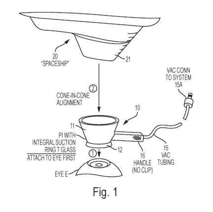
Figure 1 is a perspective view illustrating a patient interface device (PI)
and a part of the
laser delivery system according to an embodiment of the present invention.
Figure 2 is a cross-sectional view illustrating the PI coupled to a patient's
eye.
Figure 3 is a cross-sectional view illustrating the PI coupled to a patient's
eye with the
laser delivery head in place.
Figure 4 schematically illustrates a docking process using the PI of Figs. 1-3
according to
an embodiment of the present invention.
4
CA 03060966 2019-10-18
WO 2018/195139
PCT/US2018/028064
Figure 5 is a perspective cut-away view illustrating a patient interface
device and a part
of the laser delivery system according to an alternative embodiment of the
present invention.
DETAILED DESCRIPTION OF PREFERRED EMBODIMENTS
Embodiments of the invention provide an apparatus and related method for
interfacing an
ophthalmic surgical laser system with a patient's eye using a single-piece
patient interface device
(PI). The PI has advantages of both conventional one-piece and two-piece PIs.
The PI is a hand-
held piece, which allows the surgeon the full freedom of movement to achieve
docking on a
large variety of patient eye geometry. It enables the surgeon to first secure
the PI to the eye by
hand, and then couple the laser system to the PI by moving the gantry of the
laser system. The
integral applanation lens and suction ring also achieve the cost advantages of
a conventional one-
piece PI.
A PI according to embodiments of the present invention is described in more
detail with
reference to Figs. 1-3. The PI 10 includes a hard hollow shell 11, formed of a
rigid material and
preferably having an interior shape of an upside-down truncated cone, a soft
and flexible suction
ring 12 joined to the lower edge of the shell for coupling the PI to the
patient's eye by vacuum
pressure, and an applanation lens 13 located near the lower edge of the shell.
The applanation
lens 13 is mounted to the lower part of the shell 11 via a flexible annular
diaphragm 14, which
joins the applanation lens to the shell and allows the applanation lens to
"float" relative to the
shell 11, including to move in the longitudinal direction of the PI (the
vertical direction of Fig. 2),
to tilt, and to shift laterally (i.e. perpendicular to the longitudinal
direction). As will be described
later, this freedom to float facilitates the eye-docking procedure in which
the PI is coupled to the
patient's eye before it is docked with the head of the laser delivery system.
The hard shell 11 may be made of any suitable material, such as plastic,
metal, etc. Note
.. that the shell 11 does not need to have a solid side wall; it may have
openings on the side wall, or
it may be formed of a top ring and a bottom ring and a set of support struts
extending between
the top and bottom rings.
In the illustrated embodiment, the applanation lens 13 is flat on both of its
surfaces, but it
may also have a non-flat shape for either or both surfaces; for example, it
may have a concave
.. top surface and a flat bottom surface, or a flat top surface and a concave
bottom surface, etc.
When the applanation lens is concave on the bottom surface, it may be used to
shape the cornea
5
CA 03060966 2019-10-18
WO 2018/195139
PCT/US2018/028064
into a desired shape for the laser procedure. The applanation lens 13 is
preferably made of glass,
but it may also be made of other suitable materials such as transparent
plastics.
The diaphragm 14 is preferably formed of a thermoplastic elastomer (TPE), or
other
suitable elastic materials. A large variety of commercially available
materials may be used. The
mechanical properties of the diaphragm 14 is designed so that it holds the
applanation lens in
place when no force is applied to the applanation lens, but allows the
applanation lens to move
within a certain range, in particular to move in the longitudinal direction
and to tilt, when a force
is applied to it by the cornea and/or by the laser delivery head. The
stiffness of the diaphragm is
a function of its material hardness and its shape, including thickness. In
preferred embodiments,
the diaphragm is made of a material with a hardness of Shore A durometer 20 to
65, and has a
thickness of about 0.010 to 0.160 inches. The size of the diaphragm may be
determined by
practical considerations such as the desired size of the applanation lens 13
and the size of the
suction ring 12.
The diaphragm 14 may be formed integrally with the shell 11 using overmolding
techniques, or it may be formed separately by molding and then bonded to the
shell using a
suitable adhesive material. Note that the diaphragm 14 does not need to be a
solid ring; it may
have slits or cutouts, in the radial and/or circumferential directions. Such
slits and cutouts may
be designed to help achieve desired mechanical properties of the diaphragm.
The suction ring 12, which is designed to be affixed to a corneal portion C of
the eye E
by a suction force, includes annular, concentric exterior portion 12A and
interior portion 12B
defining an annular channel 12C between them. The lower portion of the
exterior portion 12A
and the lower portion of the interior portion 12B form flexible skirts, each
of which functions to
come into intimate contact with the anterior portion of the human eye E. The
flexible skirt
portions have a relatively thin cross-section and are deformable so as to
establish and maintain
conformal contact with the anterior corneal surface. The upper portions of the
exterior portion
12A and the interior portion 12B have a structure that can maintain their
shape against
deformations of the lower skirt portions in response to pressure against the
lower skirt portions
by the human eye E. An orifice 12D that opens to the annular channel 12C is
provided on the
exterior portion 12A or another portion of the suction ring to provide air
communication between
the annular channel and a vacuum tubing 15. The vacuum tubing 15 is joined to
and preferably
extends radially away from the suction ring; the other end 15A of the vacuum
tubing 15 is
6
CA 03060966 2019-10-18
WO 2018/195139
PCT/US2018/028064
adapted to be coupled to a vacuum source. When the suction ring 12 is placed
on the surface of
the patient's eye E, such that the lower portions of the exterior portion 12A
and interior portion
12B are in contact with the surface of the eye, the annular channel 12C is
sealed, and a vacuum
applied to the annular channel via the vacuum tubing 15 generates a vacuum
sealing force that
securely attaches the suction ring 12 to the eye. Various designs of suction
rings for patient
interfaces are known; for example, the above-mentioned U.S. Pat. Appl. Pub.
No. 2012/0016349
describes some exemplary suction ring structures (see, for example, Figs. 5, 6
and 11 of that
application). Any suitable suction ring structure may be used in embodiments
of the present
invention.
The PI 10 optionally includes a handle 16 joined to the side of the shell 11
to aid in
handling of the PI. A portion of the vacuum tubing may be attached to or
integrated within the
handle 16.
The gantry 20 of the laser system has a head portion 21, referred to as the
laser delivery
head, which has an exterior shape that matches the interior shape of the PI
shell 11, for example,
an upside-down truncated cone shape. When the gantry 20 is docked to the P110,
the laser
delivery head 21 is located inside the PI shell 11 with a fit that prevents it
from moving sideways
inside the PI shell, and a bottom end of the laser delivery head presses on
(either directly or via a
spacer structure described below) the applanation lens 13.
Note that when the exterior shape of the laser delivery head 21 is said to
match the
interior shape of the PI shell 11, it is meant that their shapes prevent the
laser delivery head from
moving sideways inside the PI shell when the laser delivery head is fully
placed inside the PI
shell. It does not mean that the exterior shape of the laser delivery head and
the interior shape of
the PI shell must be identical. Also, structural features may be provided on
the laser delivery
head 21 and the PI shell 11 to allow the laser delivery head to contact the PI
shell to define a
"fully inserted" position. For example, the gantry 20 may have a step feature
20A that contacts
the top edge 11A of the P111 when the laser delivery head is fully inserted
into the PI shell. Or,
the PI shell 11 may have a step feature 11B in its interior, and a part of the
laser delivery head 21
contacts this step feature when the laser delivery head is fully inserted into
the PI shell. Other
alternative structural features may be provided.
7
CA 03060966 2019-10-18
WO 2018/195139
PCT/US2018/028064
The bottom end of the laser delivery head 21 preferably has a flat optical
surface 22 from
which the laser light exits, although non-flat surfaces may also be used,
especially when the
applanation lens 13 has a non-flat top surface.
A spacer structure 23 is provided between the bottom surface 22 of the laser
delivery
head 21 and the top surface of the applanation lens 13 to form a thin air gap
between these two
surfaces when the laser system is docked to the eye. In a preferred
embodiment, the spacer 23
includes a plurality of small beads; in other embodiments, the spacer may be a
ring or other
shapes. The location of the spacer 23 is selected to avoid a central area of
the optical surface 22
where the laser beam passes through. The spacer 23 is formed of a hard
material that will have
negligible deformation when the bottom surface 22 of laser delivery head
presses down on the
applanation lens 13 via the spacer to applanate the cornea. The spacer 23 may
be affixed to the
bottom optical surface 22 by a suitable adhesive, or formed integrally on the
bottom optical
surface. Alternatively, the spacer 23 may be attached to or formed integrally
on the top surface
of the applanation lens 13.
When the laser system is docked to the eye, the applanation lens 13 is pressed
from
below by the patient's eye and from above by the laser delivery head 21 via
the spacer 23. In
such a state, the spacer 23 defines a precise, thin spacing (air gap) between
the applanation lens
13 and the bottom optical surface 22 of the laser delivery head. The thickness
of the air gap is
preferably 25 um to 200 um. By precisely controlling the thickness of the air
gap and the
thickness of the applanation lens 13, where the latter can typically be
controlled to within 5
or even smaller tolerance, the treatment depth, i.e. the depth of the laser
focus point from the
surface of the cornea, can be precisely controlled. This precision control of
the air gap is made
possible by the spacer 23 and the flexible diaphragm 14 which allows the
applanation lens 13 to
float so as to conform to the position of the optical surface 22. Note that
the flexible diaphragm
14 does not determine the alignment of the various parts in the docked
position; the laser
delivery head 21 and the spacer 23 do. The diaphragm is sufficiently soft and
flexible to allow
the applanation lens 13 to conform to the position requirement imposed by the
laser delivery
head 21 and the spacer 23.
The position of the applanation lens 13 in the "free" state, i.e., when no
external force is
applied on the applanation lens 13 by the eye or the laser delivery head 21,
is not critical. The
as-formed shape of the flexible diaphragm 14 may be such that in the free
state the applanation
8
CA 03060966 2019-10-18
WO 2018/195139
PCT/US2018/028064
lens 13 is held at a location near its position in the docked state, or above
(see the example
shown in Figs. 2 and 3) or below such a position.
In operation, the P110 is used to dock the patient's eye to the laser system
as follows.
Referring to Fig. 4, the user (surgeon) applies the PI to the patient's eye
(which generally faces
upward) by hand, manipulating it as needed for the patient's eye geometry
(step S41). The
flexible skirt portions of the suction ring 12 contact the eye (e.g. the
sclera). The end 15A of the
vacuum tubing is connected to a vacuum source, either before or after applying
the PI to the eye.
The user then turns on the vacuum source to apply a vacuum pressure to the
annular channel 12C
of the suction ring, so that the suction ring 12, and hence the P110, is
securely coupled to the eye
(step S42).
Preferably, the PI is designed such that in this state, i.e. after the PI is
secured to the eye
and before the laser delivery head is placed into the PI shell, the
applanation lens does not
applanate the cornea appreciably. For example, in the example shown in Figs. 2
and 3, the as-
formed shaped of the flexible diaphragm is such that it holds the applanation
lens at a position
above the cornea before the laser delivery head is coupled to the PI. In an
alternative
embodiment, the as-formed shape of the flexible diaphragm holds the
applanation lens at a
position such that, after the PI is secured to the eye and before the laser
delivery head is placed
into the PI shell, the applanation lens presses against the cornea and
applanates it to a certain
extent.
Then, the user moves the gantry of the laser system, which has at least three
degrees of
freedom of movement in the X, Y, and Z directions, to approximately align the
laser delivery
head 21 with the PI that has been attached to the patient's eye, and lowers
the gantry 20 so that
the laser delivery head 21 is placed inside the PI shell 11 (step S43).
Preferably, the gantry is
lowered until a part of the laser delivery head 21 contacts a part of the PI
shell 11 (e.g., upper
edge 11A, step feature 11B). As mentioned earlier, the laser delivery head 21
has a shape that
matches the interior shape of the shell 11, such that when the laser delivery
head 21 is fully
lowered into the PI shell 11, the laser delivery head 21 and the shell 11 have
a pre-defined spatial
relationship. As the gantry is moved down, the laser delivery head 21 applies
a proper amount of
force, via the spacer 23, to push the floating applanation lens 13 against the
cornea to applanate
the cornea. Once the laser delivery head and the PI are properly docked, the
system is ready to
9
CA 03060966 2019-10-18
WO 2018/195139
PCT/US2018/028064
proceed with the laser surgery (step S44). The vacuum is continuously applied
throughout the
laser surgery.
In summary, a feature of the above-described docking procedure according to
embodiments of the present invention is that a single-piece PI is used, and
the unattached PI is
secured to the patient's eye by hand first, before it is coupled to the laser
delivery head. This
provides the surgeon full freedom to manipulate the PI prior to docking it to
laser system. Such
a docking procedure using a single-piece PI is enabled by the fact that the
applanation lens can
float relative to the PI shell due to the flexible diaphragm and therefore
conform to the position
of the optical surface of the laser delivery head.
In alternative embodiments, modification may be made to the structure of the
single-
piece PI while still allowing the above described docking procedure. One
modification is to
eliminate the applanation lens (and hence the flexible diaphragm), but use a
volume of liquid or
transparent viscoelastic material as an interface between the eye surface and
the bottom optical
surface of the laser delivery head 21. In operation, the single-piece PI is
placed on the patient's
eye and secured by vacuum force; at this time, the surface of the eye is
exposed to the interior of
the PI due to the lack of the applanation lens, and the liquid or viscoelastic
material is applied
over the surface of the eye inside the area surrounded by the suction ring.
Then, the laser
delivery head 21 is lowered into the PI shell 11, to an appropriate vertical
position, so that the
space between the bottom optical surface 22 and the eye surface is filled with
the liquid or
material. In a further modification, where the applanation lens and the
diaphragm are eliminated,
no liquid or viscoelastic material is used, and the bottom optical surface 22
of the laser delivery
head directly applanates the cornea. Both of these modified procedure allows
for a single-piece
PI to be secured to the eye first and then be docked to the laser system.
Using these modified
procedures, however, because the bottom optical surface of the laser delivery
head contacts the
.. eye or the liquid or viscoelastic material, the surface will need to be
cleaned for each patient.
Fig. 5 is a perspective cut-away view illustrating a patient interface device
and a part of
the laser delivery system according to an alternative embodiment of the
present invention. In
this alternative embodiment, the flexible diaphragm 114 and the flexible
suction ring 112 are
formed integrally as one piece of the same material, and joined to the lower
edge of the rigid
shell 111. Other aspects of the PI of Fig. 5 and its use are the same as or
similar to that of the
embodiments of Figs. 1-4. Note that Fig. 5 does not show spacers between the
applanation lens
CA 03060966 2019-10-18
WO 2018/195139
PCT/US2018/028064
113 and the bottom surface 122 of the laser delivery head 21, but such spacers
may be provided
in this alternative embodiment as well. Moreover, Fig. 5 illustrates a
peripheral groove 113G on
the edge of the applanation lens 113 which allows the inner edge of the
flexible diaphragm 114
be partially inserted into the groove to hold the applanation lens 113. Such a
structure may also
be provided in the embodiment of Figs. 1-3.
In additional embodiments, the PI shell 11/111 is formed of a transparent
material such as
lass, polycarbonate, or acrylic, where the PI shell serves as a light guide to
transmit an
illumination light. The illumination light is provided from the top of the PI
shell 11/111 and
exits the PI shell at its bottom to illuminate the eye. In this regard, a part
of the bottom portion
of the PI shell 11/111 may be exposed, i.e., not covered by either the suction
ring 12/112 or the
flexible diaphragm 14/114, to allow the light to shine onto the eye. In
various ophthalmic
procedures, illumination of the eye is required to form an image of the eye to
aid in the
procedure, or for the purpose of other optical measurements. In this
embodiment, the
illumination light source is integrated with the PI. Further details of such a
PI integrating an
illumination light source are provided in commonly owned, co-pending U.S. Pat.
Appl. No.
15/479613, filed April 5, 2017, which claims priority from U.S. Prov. Appl.
No. 62/318693, filed
April 5, 2016, both of which are incorporated herein by reference in their
entireties.
The PI according to embodiments of the present invention can be used in
various
ophthalmic laser systems, including, without limitation, femtosecond lasers
for flap cutters and
laser cataract systems.
It will be apparent to those skilled in the art that various modification and
variations can
be made in the patient interface device and the laser delivery system as well
as related methods
of the present invention without departing from the spirit or scope of the
invention. Thus, it is
intended that the present invention cover modifications and variations that
come within the scope
of the appended claims and their equivalents.
11