Note: Descriptions are shown in the official language in which they were submitted.
CA 03061620 2019-10-25
WO 2018/200980
PCT/US2018/029836
METHODS FOR DETECTING ALLELES ASSOCIATED WITH
KERATOCONUS
FIELD OF THE APPLICATION
[0001] This application generally relates to methods for the isolation and
detection of
disease-associated genetic alleles. In particular, this application relates to
methods for the
detection of an alleles associated with keratoconus diagnosis and prognosis.
BACKGROUND
[0002] Keratoconus (KC) is the most common corneal ectatic disorder with
approximately 6
¨ 23.5% of subjects carrying a positive family history (Wheeler, J., Hauser,
M.A., Afshari,
N. A., Allingham, R.R., Liu, Y., Reproductive Sys Sexual Disord 2012; S:6).
The reported
prevalence of KC ranges from 8.8 to 54.4 per 100,000. This variation in
prevalence is partly
due to the different criteria used to diagnose the disease. (Wheeler, J.,
Hauser, M.A., Afshari,
N.A., Allingham, R.R., Liu, Y., Reproductive Sys Sexual Disord 2012; S:6; and
Nowak, D.,
Gajecka, M., Middle East Afr J Ophthalmol 2011; 18(1): 2-6). Many studies
exist within the
literature that attempt to define the genetic causes of KC. These studies have
uncovered
numerous possible genetic variants or SNPs that are believed to contribute to
the etiology of
the disease depending on the experimental parameters.
[0003] KC is a common corneal disorder where the central or paracentral cornea
undergoes
progressive thinning and steepening causing irregular astigmatism. The
hereditary pattern is
neither prominent nor predictable, but positive family histories have been
reported. The
incidence of KC is often reported to be 1 in 2000 people. KC can show the
following
pathologic findings, including, fragmentation of Bowman's layer, thinning of
stroma and
overlying epithelium, folds or breaks in Descemet's membrane, and variable
amounts of
diffuse corneal scarring.
[0004] Histopathology studies demonstrate breaks in or complete absence of
Bowman's
layer, collagen disorganization, scarring and thinning. The etiology of these
changes is not
known, though some suspect changes in enzymes that lead to breakdown of
collagen in the
cornea. While a genetic predisposition to KC is suggested, a specific gene has
not been
identified. The majority of KC cases are bilateral, but often asymmetric. The
less affected
eye may show a high amount of astigmatism or mild steepening. Onset is
typically in early
1
CA 03061620 2019-10-25
WO 2018/200980
PCT/US2018/029836
adolescence and progresses into the mid-20's and 30's. However, cases may
begin much
earlier or later in life. There is variable progression for each individual.
There is often a
history of frequent changes in eye glasses which do not adequately correct
vision. Another
common progression is from soft contact lenses, to toric or astigmatism
correcting contact
lens, to rigid gas permeable contact lens.
[0005] No preventive strategy has been proven effective to date. Some feel
that eye rubbing
or pressure (e.g., sleeping with the hand against the eye) can cause and/or
lead to progression
of KC, so subjects should be informed not to rub the eyes. In some subjects,
avoidance of
allergens may help decrease eye irritation and therefore decrease eye rubbing.
100061 At present, diagnosis can be made by slit-lamp examination and
observation of central
or inferior corneal thinning. Computerized videokeratography is also useful in
detecting early
KC and allows following its progression. Ultrasound pachymetry can also be
used to measure
the thinnest zone on the cornea. New algorithms using computerized
videokeratography have
been devised which now allow the detection of forme fruste, subclinical or
suspected
keratoconus. These devices may allow better screening of subjects for
prospective refractive
surgery, however there remains a need in the art for better prognostic and
diagnostic methods.
100071 The present disclosure meets this need and by providing methods for
prognosis and
diagnosis of KC by detection of mutated alleles associated with keratoconus.
SUMMARY
[0008] The present disclosure provides improved methods for the detection of
one or more
alleles associated with KC
[0009] In some embodiments, the disclosure provides methods for detecting
variants related
to KC in a subject, the method comprising detecting two or more genetic
variants (e.g., single
nucleotide polymorphisms (SNPs) and indels) in a sample from a subject,
wherein two or
more genetic variants are selected from the group consisting of genetic
variants listed in
Figure 1, and wherein the presence of two or more genetic variants is
indicative of KC in the
subject.
[0010] In some embodiments, the disclosure provides methods for diagnosing or
prognosing
KC in a subject, the method comprising detecting two or more genetic variants
(e.g., single
nucleotide polymorphisms (SNPs) and indels) in a sample from a subject,
wherein two or
2
CA 03061620 2019-10-25
WO 2018/200980
PCT/US2018/029836
more genetic variants are selected from the group consisting of genetic
variants listed in
Figure 1, and wherein the presence of two or more genetic variants is
indicative of a
diagnosis or prognosis of KC in the subject.
[0011] In some embodiments, the two or more genetic variants are selected from
the group
consisting of genetic variants listed in Figure 2. In additional embodiments,
the subject is
Afro-American. In further embodiments, the Afro-American is identified by
detecting two or
more genetic variants specific to the Afro-American.
[0012] In some embodiments, the two or more genetic variants are selected from
the group
consisting of genetic variants listed in Figure 3. In additional embodiments,
the subject is
Caucasian. In further embodiments, the Caucasian is identified by detecting
two or more
genetic variants specific to the Caucasian.
[0013] In some embodiments, the two or more genetic variants are selected from
the group
consisting of genetic variants listed in Figure 4. In additional embodiments,
the subject is
Hispanic. In further embodiments, the Hispanic is identified by detecting two
or more
genetic variants specific to the Hispanic.
[0014] In some embodiments, the two or more genetic variants are selected from
the group
consisting of genetic variants listed in Figure 5. In additional embodiments,
the subject is
East Asian or Korean. In further embodiments, the East Asian or Korean is
identified by
detecting two or more genetic variants specific to the East Asian or Korean.
[0015] In some embodiments, two or more genetic variants are selected from the
group
consisting of any combination of the mutations (e.g., genetic variants)
described herein (e.g.,
Figures 1-5).
[0016] In some embodiments, said genetic variant detection is by a sequencing
method.
[0017] In some embodiments, the disclosure provides methods for detecting
variants related
to or causing KC in a subject, the method comprising detecting two or more
genetic variants
(e.g., single nucleotide polymorphisms (SNPs) and indels) in a sample from a
subject,
wherein two or more genetic variants are selected from the group consisting of
genetic
variants listed in Figure 1, and wherein the presence of two or more genetic
variants is
indicative of KC in the subject.
3
CA 03061620 2019-10-25
WO 2018/200980
PCT/US2018/029836
[0018] In some embodiments, the disclosure provides methods for predicting
risk of
developing KC in a subject, the method comprising detecting two or more
genetic variants in
a sample from a subject, wherein the two or more genetic variants are selected
from the group
consisting of genetic variants listed in Figure 1, and wherein the presence of
two or more of
genetic variants is indicative of risk for the development of KC in the
subject.
[0019] In some embodiments, the two or more genetic variants are selected from
the group
consisting of genetic variants listed in Figure 2. In additional embodiments,
the subject is
Afro-American.
[0020] In some embodiments, the two or more genetic variants are selected from
the group
listed in Figure 3. In additional embodiments, the subject is Caucasian.
[0021] In some embodiments, the two or more genetic variants are selected from
the group
consisting of genetic variants listed Figure 4. In additional embodiments, the
subject is
Hispanic.
[0022] In some embodiments, the two or more genetic variants are selected from
the group
consisting of listed in Figure 5. In additional embodiments, the subject is
East Asian or
Korean.
[0023] In some embodiments, the two or more genetic variants are selected from
the group
consisting of any combination of the mutations (e.g., genetic variants)
described herein (e.g.,
Figures 1-5).
[0024] In some embodiments, said variant detection is by a sequencing method.
[0025] In some embodiments, the disclosure provides methods for developing a
treatment
regimen for the treatment of KC in a subject, the method comprising detecting
two or more
genetic variants in a sample from a subject, wherein the two or more genetic
variants are
selected from the group consisting of genetic variants listed in Figure 1, and
wherein the
presence of two or more genetic variants is indicative of the need for a KC
treatment regimen
in the subject.
[0026] In some embodiments, the two or more genetic variants are selected from
the group
consisting of genetic variants listed in Figure 2. In additional embodiments,
the subject is
Afro-American.
4
CA 03061620 2019-10-25
WO 2018/200980
PCT/US2018/029836
[0027] In some embodiments, the two or more genetic variants are selected from
the group
consisting of genetic variants listed in Figure 3. In additional embodiments,
the subject is
Caucasian.
[0028] In some embodiments, the two or more genetic variants are selected from
the group
consisting of genetic variants listed in Figure 4. In additional embodiments,
the subject is
Hispanic.
[0029] In some embodiments, the two or more genetic variants are selected from
the group
consisting of genetic variants listed in Figure 5. In additional embodiments,
the subject is
East Asian or Korean.
[0030] In some embodiments, the two or more genetic variants (e.g., SNPs) are
selected from
the group consisting of any combination of the mutations (e.g., genetic
variants) described
herein (e.g., Figures 1-5).
[0031] In some embodiments, said variant detection is by a sequencing method.
[0032] In some embodiments, the disclosure provides methods for treating
keratoconus in a
subject, the method comprising diagnosing or prognosing KC and treating KC in
the subject.
In further embodiments, the treatment may comprise wearing eye glasses or
contact lenses,
and/or performing collagen cross-linking or corneal transplant.
BRIEF DESCRIPTION OF THE DRAWINGS
[0033] Figure 1 depicts a table listing the frequency of each variant found
within the study
cohort ordered by chromosome, gene symbol, dbSNP id and ethnicity. The list is
divided
into shared variants between ethnic groups, Caucasian (C), East Asian (EA),
Hispanic (H),
African American (AA), and South Asian (SA), followed by variants that are
specific to each
group. A total of 1,117 nonsynonymous single nucleotide variants (SNVs) and
insertion/deletions (INDELs) within 259 genes spanning the entire exome are
listed. A
RefSeq (ncbi.nlm.nih.gov/) accession number along with the minor allele
frequency (MAF)
taken from the Exome Aggregation Consortium (ExAC, exac.broadinstitute.org/)
is provided.
N = total alleles for each group.
[0034] Figure 2 lists genetic variants specific to Afro-American subjects
having keratoconus.
[0035] Figure 3 lists genetic variants specific to Caucasian subjects having
keratoconus.
CA 03061620 2019-10-25
WO 2018/200980
PCT/US2018/029836
[0036] Figure 4 lists genetic variants specific to Hispanic subjects having
keratoconus.
[0037] Figure 5 lists genetic variants specific to East Asian subjects having
keratoconus.
[0038] Figure 6 lists additional genetic variants shared to all subjects
having keratoconus.
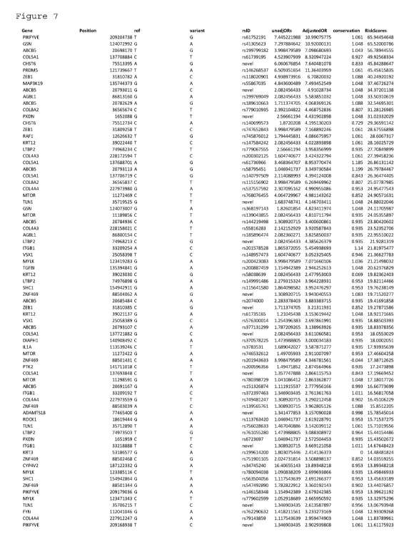
[0039] Figure 7 depicts a table that lists an odds ratio (OR) and risk score
assignment for
rare variants from cornea genes identified within the Caucasian group.
Variants were taken
from 48 genes related to corneal structure and function and were drawn from a
larger list of
variants in the Caucasian study cohort. Variants were further selected based
on their
presence in 1 or more case samples and in 0 ethnic-matched controls. Risk
scores were
derived from an algorithm incorporating adjusted ORs from conservation priors
in a Bayesian
model, and also in silico predictions from 7 bioinformatic tools indicated by
red and yellow
circles.
[0040] Abbreviations: A = Afro-American, C = Caucasian, H = Hispanic, and EA =
East
Asian.
DETAILED DESCRIPTION
[0041] The detection of disease-related variants is an increasingly important
tool for the
diagnosis and prognosis of various medical conditions. With regard to KC, the
present
disclosure provides methods for detection of mutant alleles and use of this
information in or
to diagnose a subject with KC as well as to predict the risk of an individual
in developing
KC.
[0042] The term "invention" or "present invention" as used herein is not meant
to be limiting
to any one specific embodiment of the invention but applies generally to any
and all
embodiments of the invention as described in the claims and specification.
[0043] As used herein, the singular forms "a", "an", and "the" include plural
references
unless the context clearly dictates otherwise. Thus, for example, references
to "the method"
includes one or more methods, and/or steps of the type described herein which
will become
apparent to those persons skilled in the art upon reading this disclosure. it
should be
understood that the use of -and/or" is defined inclusively such that the term
"a, b and/or c"
should be read to include the sets of "a," "b," "c," "a and b," "b and c," "c
and a," and "a, b
and c."
6
CA 03061620 2019-10-25
WO 2018/200980
PCT/US2018/029836
[0044] As used herein, the term "about" means modifying, for example, lengths
of nucleotide
sequences, degrees of errors, dimensions, the quantity of an ingredient in a
composition,
concentrations, volumes, process temperature, process time, yields, flow
rates, pressures, and
like values, and ranges thereof, refers to variation in the numerical quantity
that may occur,
for example, through typical measuring and handling procedures used for making
compounds, compositions, concentrates or use formulations; through inadvertent
error in
these procedures; through differences in the manufacture, source, or purity of
starting
materials or ingredients used to carry out the methods; and like
considerations. The term
"about" also encompasses amounts that differ due to aging of, for example, a
composition,
formulation, or cell culture with a particular initial concentration or
mixture, and amounts
that differ due to mixing or processing a composition or formulation with a
particular initial
concentration or mixture. Whether modified by the term "about" the claims
appended hereto
include equivalents to these quantities. The term "about" further may refer to
a range of
values that are similar to the stated reference value. In certain embodiments,
the term
"about" refers to a range of values that fall within 50, 25, 10, 9, 8,7, 6,
5,4, 3, 2, 1 percent or
less of the stated reference value.
[0045] As used herein, the term "polymorphism" and variants thereof refers to
the occurrence
of two or more alternative genomic sequences or alleles between or among
different genomes
or individuals. The terms "genetic mutation" or "genetic variation" and
variants thereof
include polymorphisms.
[0046] As used herein the term "single nucleotide polymorphism" ("SNP") and
variants
thereof refers to a site of one nucleotide that varies between alleles. A
single nucleotide
polymorphism (SNP) is a single base change or point mutation but variants also
include the
so-called "indel" mutations (insertions or deletions of 1 to several up to 75
nucleotides),
resulting in genetic variation between individuals. SNPs, which make up about
90% of all
human genetic variation, occur every 100 to 300 bases along the 3-billion-base
human
genome. However, SNPs can occur much more frequently in other organisms like
viruses.
SNPs can occur in coding or non-coding regions of the genome. A SNP in the
coding region
may or may not change the amino acid sequence of a protein product. A SNP in a
non-
coding region can alter promoters or processing sites and may affect gene
transcription and/or
processing. Knowledge of whether an individual has particular SNPs in a
genomic region of
interest may provide sufficient information to develop diagnostic, preventive
and therapeutic
applications for a variety of diseases.
7
CA 03061620 2019-10-25
WO 2018/200980
PCT/US2018/029836
[0047] The term "primer" and variants thereof refers to an oligonucleotide
that acts as a point
of initiation of DNA synthesis in a polymerase chain reaction (PCR). A primer
is usually
about 10 to about 35 nucleotides in length and hybridizes to a region
complementary to the
target sequence.
[0048] The term "probe" and variants thereof (e.g., detection probe) refers to
an
oligonucleotide that hybridizes to a target nucleic acid in a PCR reaction.
Target sequence
refers to a region of nucleic acid that is to be analyzed and comprises the
polymorphic site of
interest.
[0049] The hybridization occurs in such a manner that the probes within a
probe set may be
modified to form a new, larger molecular entity (e.g., a probe product). The
probes herein
may hybridize to the nucleic acid regions of interest under stringent
conditions. As used
herein the term "stringency" is used in reference to the conditions of
temperature, ionic
strength, and the presence of other compounds such as organic solvents, under
which nucleic
acid hybridizations are conducted. "Stringency" typically occurs in a range
from about Tm C
to about 20 C to 25 C below T. A stringent hybridization may be used to
isolate and
detect identical polynucleotide sequences or to isolate and detect similar or
related
polynucleotide sequences. Under "stringent conditions" the nucleotide
sequence, in its
entirety or portions thereof, will hybridize to its exact complement and
closely related
sequences. Low stringency conditions comprise conditions equivalent to binding
or
hybridization at 68 C. in a solution consisting of 5x SSPE (43.8 g/lNaC1, 6.9
g/1
NaH2PO4.H20 and 1.85 g/1 EDTA, pH adjusted to 7.4 with NaOH), 0.1% SDS, 5x
Denhardt's reagent (50x Denhardt's contains per 500 ml: 5 g Ficoll (Type 400),
5 g BSA)
and 100 pg/m1 denatured salmon sperm DNA followed by washing in a solution
comprising
2.0+SSPE, 0.1% SDS at room temperature when a probe of about 100 to about 1000
nucleotides in length is employed. It is well known in the art that numerous
equivalent
conditions may be employed to comprise low stringency conditions; factors such
as the
length and nature (DNA, RNA, base composition) of the probe and nature of the
target
(DNA, RNA, base composition, present in solution or immobilized, etc.) and the
concentration of the salts and other components (e.g., the presence or absence
of formamide,
dextran sulfate, polyethylene glycol), as well as components of the
hybridization solution
may be varied to generate conditions of low stringency hybridization different
from, but
equivalent to, the above listed conditions. In addition, conditions which
promote
hybridization under conditions of high stringency (e.g., increasing the
temperature of the
8
CA 03061620 2019-10-25
WO 2018/200980
PCT/US2018/029836
hybridization and/or wash steps, the use of formamide in the hybridization
solution, etc.) are
well known in the art. High stringency conditions, when used in reference to
nucleic acid
hybridization, comprise conditions equivalent to binding or hybridization at
68 C in a
solution consisting of 5+SSPE, 1% SDS, 5x Denhardt's reagent and 100 pg/ml
denatured
salmon sperm DNA followed by washing in a solution comprising 0.1+SSPE and
0.1% SDS
at 68 C when a probe of about 100 to about 1000 nucleotides in length is
employed.
[0050] Unless defined otherwise, all technical and scientific terms used
herein have the same
meaning as commonly understood by those of ordinary skill in the art to which
the invention
pertains. Although any methods and materials similar or equivalent to those
described herein
can be used in the practice or testing of the present invention, various
embodiments of
methods and materials are specifically described herein.
[0051] As explained above, KC is the most common corneal ectatic disorder with
approximately 6 ¨ 23.5% of patients carrying a positive family history.
(Wheeler, J., Hauser,
M.A., Afshari, N.A., Allingham, R.R., Liu, Y. Reproductive Sys Sexual Disord
2012; S:6.)
The reported prevalence of KC ranges from 8.8 to 54.4 per 100,000. This
variation in
prevalence is partly due to the different criteria used to diagnose the
disease. (Wheeler, J.,
Hauser, M.A., Afshari, N.A., Allingham, R.R., Liu, Y. Reproductive Sys Sexual
Disord
2012; S:6; and Nowak, D., Gajecka, M. Middle East Afr J Ophthalmol 2011;
18(1):2-6)
Many studies exist within the literature that attempt to define the genetic
causes of KC.
These studies have uncovered numerous possible genetic variants or SNPs that
are believed
to contribute to the etiology of the disease depending on the experimental
parameters.
[0052] In general, the work conducted thus far primarily makes use of micro-
satellite
genotyping and micro-chip technologies (SNP arrays) to interrogate regions of
interest within
the genome. In comparison, the study described herein utilized Next Gen
Sequencing (NGS)
technology to identify and to validate genetic variants that contribute to the
etiology of the
disease. The study involved a whole exome sequencing (WES) approach (ACE
PlatformTM;
Personalis Inc., Menlo Park, CA) in which the ¨22,000 genes that comprise the
human exome
were captured and sequenced; single point mutations or variants including
INDELS were
identified.
[0053] It is recognized that within the human genome there exist various loci
harboring gene
mutations that contribute to the phenotypical profile of KC. Among those loci
documented in
the literature are regions mapped to chromosomes 15q2.32 and 15q22.33-q24.2,
13q32,
9
CA 03061620 2019-10-25
WO 2018/200980
PCT/US2018/029836
16q22.3-q23.1, 3p14-q13, 5q14.3-q21.1, 5q21.2 and 5q32-q33, 1p36.23-36.21 and
8q13.1-
q21.11, 9q34, 14q11.2 and 14q24.3 (see, for example, Bisceglia L, De Bonis P,
Pizzicoli C et
al., Invest Ophthalmol Vis Sci. 2009; 50: 1081-1086; Hughes AE, Dash DP,
Jackson AJ,
Frazer DG, Silvestri G, Invest Ophthalmol Vis Sci 2003; 44:5063-5066; Gajecka
M,
Radhakrishna U, Winters D et al., Invest Ophthalmol Vis Sci 2009; 50:1531-
1539; Czugala,
M., Karolak, J.A., Nowak, D.A., et.al., European Journal of Human Genetics
2012; 20:389-
397; Tyynismaa H, Sistonen P, Tuupanen S et al.; Invest Ophthalmol Vis Sci
2002; 43: 3160-
3164; Brancati F, Valente EM, Sarkozy A et al., J Med Genet 2004; 41:188-192;
Tang YG,
Rabinowitz YS, Taylor KD et al., Genet Med 2005; 7: 397-405; Burdon KP, Coster
DJ,
Charlesworth JC et al.; Hum Genet 2008; 124:379-386; Li, X., Rabinowitz, Y.S.,
Tang, Y.G.,
Picornell, Y., Taylor,K.D., Hu, M., Yang, H.; Invest Ophthalmol Vis Sci 2006;
47:3791-
3795; and Liskova P, Hysi PG, WaseemN, Ebenezer ND, Bhattacharya SS, Tuft SJ,
Arch
Ophthalmol 2010; 128:1191-1195.)
[0054] As explained above, these studies mostly utilize micro-satellite
genotyping in
conjunction with array chip technologies to interrogate regions of interest
within the genome.
[0055] In addition to the above referenced studies, mutations in the visual
system homeobox
gene 1 (VSX1) have been identified through the targeted screening of this gene
in patients
diagnosed with KC. The research conducted on the VSX1 gene so far has not
clearly
identified a causative agent and in fact, much of the literature presents
conflicting results.
See, for example, Bisceglia, L., Ciaschetti, M., De Bonis, P., Campo, P.A.,
Pizzicoli, C.,
Scala, C., Grifa, M., Ciavarella, P., Delle Noci, N., Vaira, F. et al., Invest
Ophthalmol Vis Sci
2005; 46:39-45, Heon, E., Greenberg, A., Kopp, K.K., Rootman, D., Vincent,
A.L.,
Billingsley, G., Priston, M., Dorval, K.M., Chow, R.L., McInnes, R.R. et al.,
Hum Mol Genet
2002; 11(9):1029-1036, Tang, Y.G., Picornell, Y., Su, X., Li, X., Yang, H. and
Rabinowitz,
Y.S. Cornea 2008; 27:189-192; Aldave, A.J., Yellore, V.S., Salem, A.K., Yoo,
G.L., Rayner,
S.A., Yang, H., Tang, G.Y., Piconell, Y., Rabinowitz, Y.S., Invest Ophthalmol
Vis Sci 2006;
47(7):2820-2; Tanwar, M., Kumar, M., Nayak, B., Pathak, D., Sharma, N.,
Titiyal, J.S. and
Dada, R., Mo/ Vis 2010; 16: 2395-2401; Mok, L.W., Baek, S.J., Joo, C.K., JHum
Genet
2008; 53:842-849; Jeoung, J.W., Kim, M.K., Park, S.S., Kim, S.Y., Ko, H.S.,
Won Ryang
Wee, W.R., Jin Hak Lee, J.H., Cornea 2012; 31,7:746-750; Dehkordi, F.A.,
Rashki, A.,
Bagheri, N.,Chaleshtori, M.H., Memarzadeh, E.,Salehi, A., Ghatreh, H., Zandi,
F.,
Yazdanpanahi, N.,Tabatabaiefar, M.A., Chaleshtori, M.H. Method. Acta
Cytologica 2013; 57:
646-651; Saee-Rad, S., Hashemi, H., Miraftab,M., Noori-Daloii, M.R.,
Chaleshtori, M.H.,
CA 03061620 2019-10-25
WO 2018/200980
PCT/US2018/029836
Raoofian, R., Jafari, F., Greene, W., Fakhraie, G., Rezvan, F., Heidari, M.,
Mol Vis 2011;
17:3128-3136; Wang, Y., Jin, T., X. Zhang, X., Wei, W., Cui, Y., Geng, T.,
Liu, Q., Gao, J.,
Liu, M., Chen, C., Zhang, C., Zhu, X., Ophthalmic Genetics 2013; 34,3: 160-
166; Dash,
D.P., S George, S., O'Prey, D., Burns, D., Nabili, S., Donnelly, U., Hughes,
A.E., Silvestri,
G., Jackson, J., Frazer, D., Heon, E., Willoughby, C.E., Eye, 2010; 24,6: 1085-
1092.
[0056] While much investigative work has been carried out on the possible role
of the VSX1
gene in the etiology of KC, this is not the only gene that has been targeted
for analysis.
[0057] Most prominent among the genes that have been investigated within the
literature are
the various genes related to the structure of collagen. Collagens are the
major protein
components of the human cornea, and there exist several types of collagen
genes that code for
the various collagen proteins. Of interest here are COL4A3 and COL4A4 (S'tabuc-
S'ilih, M.,
Ravnik-Glavg, M., Glavg, D., Hawlina, M., Straigar M., Mol Vis 2009; 15:2848-
2860;
S'tabuc-S'ilih, M., Strthgar, M., Ravnik Glavg, M., Hawlina, Glavg, D.; Acta
Dermatoven
APA 2010; 19(2):3-10; Vitart, V., Bencic, G., Hayward, C., Herman, J.S.,
Huffman J.,
Campbell, S., Bucan, K., Navarro, P., Gunjaca, G., Mar, J., Zgaga, L., Kolcic,
I., Polasek,
0., Kirin, M., Hastie, N.D., Wilson, J.F., Rudan, I., Campbell, H., Vatavuk,
Z., Fleck, B.,
Wright, A., Hum Mol Genet 2010; 19(21): 4304-4311) mapped to 2q36.3 (Vitart,
V., Bencic,
G., Hayward, C., Herman, J.S., Huffman J., Campbell, S., Bucan, K., Navarro,
P., Gunjaca,
G., Marin, J., Zgaga, L., Kolcic, I., Polasek, 0., Kirin, M., Hastie, N.D.,
Wilson, J.F., Rudan,
I., Campbell, H., Vatavuk, Z., Fleck, B., Wright, A., Hum Mol Genet 2010;
19(21): 4304-
4311) along with COL4A1 and COL4A2 mapped to the 13q32 locus (Gajecka M,
Radhakrishna U, Winters D et al., Invest Ophthalmol Vis Sci 2009; 50:1531-
1539; Czugala,
M., Karolak, J.A., Nowak, D.A., et.al., European Journal of Human Genetics
2012; 20:389-
397; Karolak, J.A., Kulinska, K., Nowak, D.M., Pitarque, J.A., Molinari, A.,
Rydzanicz, M.,
Bejjani, B.A., Gajecka, M., Mol Vis 2011; 17:827-843). In reference to the
COL4A3 and
COL4A4 genes, S'tabuc-S'ilih et al. in a study published in 2009 identified
several SNPs that
carried significant p-values. In this study which included 104 unrelated
diagnosed patients
and 157 healthy blood donors, polymorphism M1327V located at allele 3979 in
the COL4A4
gene had a p-value <0.0001 with 134 point mutations out of 208 total alleles
for the cases and
132 out of 314 alleles for the controls (S'tabuc-S'ilih, M., Ravnik-Glavg, M.,
Glavg, D.,
Hawlina, M., Straigar M., Mol Vis 2009; 15:2848-2860). With that said, in a
subsequent
paper published in 2010, S'tabuc-S'ilih et al. excludes COL4A3 and COL4A4 from
playing a
11
CA 03061620 2019-10-25
WO 2018/200980
PCT/US2018/029836
significant role in KC pathogenesis (Stabuc-Silih, M., StraZ'igar, M., Ravnik
Glavg, M.,
Hawlina, Glavg, D., Acta Dermatoven APA 2010; 19(2):3-10).
[0058] Similarly, Karolak et al. documents findings relating to the COL4A1 and
the
COL4A2 genes within Ecuadorian families; 23 individuals from one family, 25
affected
individuals from other Ecuadorian families, and 64 Ecuadorian control subjects
were
included in this study (Karolak, J.A., Kulinska, K., Nowak, D.M., Pitarque,
J.A., Molinari,
A., Rydzanicz, M., Bejjani, B.A., Gajecka, M., Mo/ Vis 2011; 17:827-843). This
study
identifies several mutations within the COL4A1 and the COL4A2 genes that were
significant.
For instance, a polymorphism, Gln1334His found at the 4002 allele on COL4A1
gene was
observed more frequently in patients than in healthy individuals in the family
where twenty-
three individuals (p=0.056) were examined. However, there was no difference in
the c.
4002A>C allele distribution between the analyzed affected individuals from the
remaining
KC families and the Ecuadorian control subjects (p=0.17).
[0059] In conjunction with the work described above (Karolak, J.A., Kulinska,
K., Nowak,
D.M., Pitarque, J.A., Molinari, A., Rydzanicz, M., Bejjani, B.A., Gajecka, M.,
Mo/ Vis 2011;
17:827-843), Czugala et.al conducted a study on the same Ecuadorian family
group that
revealed eight candidate genes other than COL4A1 and COL4A2 (Czugala, M.,
Karolak,
J.A., Nowak, D.A., et.al., European Journal of Human Genetics 2012; 20: 389-
397). These
genes are MBNL1, IP05, FARP1, RNF113B, 5TK24, DOCK9, ZIC5 and ZIC2. Ninety-two
sequence variants were identified within these eight genes. At least four of
the ninety-two
variants referred to in this study show a statistical correlation to the KC
phenotype. These
genes and the SNPs associated with them are located at the 13q32 locus, but
another
important aspect of both this study and the work conducted with the COL4A1 and
COL4A2
genes is that the results are derived from the genetic analysis primarily of
one extended
family in Ecuador (Czugala, M., Karolak, J.A., Nowak, D.A., et.al., European
Journal of
Human Genetics 2012; 20:389-397).
[0060] The case studies referenced here were conducted to further elucidate
the role of
collagen genes and the role they play within the cornea and to investigate the
role of the
13q32 locus, a location on the genome that could be an important hotspot
within the human
genome (Gajecka M, Radhakrishna U, Winters D et al., Invest Ophthalmol Vis Sci
2009;
50:1531-1539; and Czugala, M., Karolak, J.A., Nowak, D.A., et.al., European
Journal of
Human Genetics 2012; 20:389-397). COL4A3 and COL4A4 genes, which are known to
be
12
CA 03061620 2019-10-25
WO 2018/200980
PCT/US2018/029836
deregulated in KC patients, are often subjected to chromosomal aberrations,
and could also
be responsible for a decrease in collagen types I and III, a feature often
detected in the disease
(Critchfield, J.W., Calandra, A.J., Nesburn, A.B., Kenney, M.C., Exp Eye Res
1988; 46: 953-
63; Kenney, M.C., Nesburn ,A.B, Burgeson, R.E., Butkowski, R.J., Ljubimov
A.V., Cornea
1997; 16:345-51; Meek, K.M., Tuft, S.J., Huang, Y., Gill P.S., Hayes, S.,
Newton, R.H.,
Bron, A.J., Invest Ophthalmol Vis Sci 2005; 46:1948-56; Bochert, A., Berlau,
J., Koczan, D.,
Seitz, B., Thiessen, H.J., Guthoff, R.F., Ophthalmologe 2003; 100:545-9;
Stachs, 0., Bocher,
A., Gerber, T., Koczan, D., Thiessen, H.J., Guthoff, R.F., Ophthalmologe 2004;
101:384-9;
Pettenati, M.J, Sweatt, A.J., Lantz, P., Stanton, C.A., Reynolds, J., Rao,
P.N., Davis, R.M.,
Hum Genet 1997; 101:26-9).
[0061] The search for a genetic link that defines the subset of KC, labeled as
familial KC
mostly results in the identification of different SNP candidates depending on
the family
pedigree. For example, the gene, VSX1 was thought to be a primary candidate
based on a
few isolated family studies (Bisceglia, L., Ciaschetti, M., De Bonis, P.,
Campo, P.A.,
Pizzicoli, C., Scala, C., Grifa, M., Ciavarella, P., Delle Noci, N., Vaira, F.
et al., Invest
Ophthalmol Vis Sci 2005; 46: 39-45; Heon, E., Greenberg, A., Kopp, K.K.,
Rootman, D.,
Vincent, A.L., Billingsley, G., Priston, M., Dorval, K.M., Chow, R.L.,
McInnes, R.R. et al.,
Hum Mol Genet 2002; 11(9):1029-1036); however, non-family based studies have
also been
conducted with this gene that involved unrelated individuals of different
ethnicities and
geographic locations. These studies attempt to identify specific SNPs within
the gene that
would better define the role of VSX1 (Aldave, A.J., Yellore, V.S., Salem,
A.K., Yoo, G.L.,
Rayner, S.A., Yang, H., Tang, G.Y., Piconell, Y., Rabinowitz, Y.S., Invest
Ophthalmol Vis
Sci 2006; 47,7:2820-2; Tanwar, M., Kumar, M., Nayak, B., Pathak, D., Sharma,
N., Titiyal,
J.S. and Dada, R., Mo/ Vis 2010; 16: 2395-2401; Mok, L.W., Baek, S.J., Joo,
C.K., J Hum
Genet 2008; 53: 842-849; Jeoung, J.W., Kim, M.K., Park, S.S., Kim, S.Y., Ko,
H.S., Won
Ryang Wee, W.R., Jin Hak Lee, J.H., Cornea 2012; 31,7: 746-750; Dehkordi,
F.A., Rashki,
A., Bagheri, N.,Chaleshtori, M.H., Memarzadeh, E.,Salehi, A., Ghatreh, H.,
Zandi, F.,
Yazdanpanahi, N.,Tabatabaiefar, M.A., Chaleshtori, M.H., Acta Cytologica 2013;
57: 646-
651, Wang, Y., Jin, T., X. Zhang, X., Wei, W., Cui, Y., Geng, T., Liu, Q.,
Gao, J., Liu, M.,
Chen, C., Zhang, C., Zhu, X., Ophthalmic Genetics 2013; 34,3: 160-166; Dash,
D.P., S
George, S., O'Prey, D., Burns, D., Nabili, S., Donnelly, U., Hughes, A.E.,
Silvestri, G.,
Jackson, J., Frazer, D., Heon, E., Willoughby, C.E., Eye, 2010; 24,6: 1085-
1092). In general,
publications resulting from these studies are inconclusive and in fact, the
pathogenic role of
13
CA 03061620 2019-10-25
WO 2018/200980
PCT/US2018/029836
certain non-synonymous candidate SNPs found within the VSX1 gene has been
refuted
(Tang, Y.G., Picornell, Y., Su, X., Li, X., Yang, H. and Rabinowitz, Y.S.,
Cornea 2008; 27:
189-192; Aldave, A.J., Yellore, VS., Salem, A.K., Yoo, G.L., Rayner, S.A.,
Yang, H., Tang,
G.Y., Piconell, Y., Rabinowitz, Y.S., Invest Ophthalmol Vis Sci 2006; 47(7):
2820-2;
Tanwar, M., Kumar, M., Nayak, B., Pathak, D., Sharma, N., Titiyal, J.S. and
Dada, R. VSX1
gene analysis in keratoconus. Mol Vis 2010; 16: 2395-2401).
[0062] KC with no family associations is the most common form of the disease
seen by
practicing clinicians (Rabinowitz, Y.S., Ophthalmol Clin N Am. 2003; 16(4):
607-620). With
that said, it is likely that familial aggregation has been underreported due
to undetected forms
of KC. Recent advances in diagnostic techniques such as videokeratography may
help better
understand whether other forms of the disease are, in actuality, inherited.
[0063] The work described above that involves the VSX1 gene (Bisceglia, L.,
Ciaschetti, M.,
De Bonis, P., Campo, P.A., Pizzicoli, C., Scala, C., Grifa, M., Ciavarella,
P., Delle Noci, N.,
Vaira, F. et al., Invest Ophthalmol Vis Sci 2005; 46:39-45; Heon, E.,
Greenberg, A., Kopp,
K.K., Rootman, D., Vincent, A.L., Billingsley, G., Priston, M., Dorval, K.M.,
Chow, R.L.,
McInnes, R.R. et al.; Hum Mol Genet 2002; 11(9):1029-1036; Tang, Y.G.,
Picornell, Y., Su,
X., Li, X., Yang, H. and Rabinowitz, Y.S. Cornea 2008; 27:189-192; Aldave,
A.J., Yellore,
V.S., Salem, A.K., Yoo, G.L., Rayner, S.A., Yang, H., Tang, G.Y., Piconell,
Y., Rabinowitz,
Y.S. Invest Ophthalmol Vis Sci 2006; 47(7): 2820-2; Tanwar, M., Kumar, M.,
Nayak, B.,
Pathak, D., Sharma, N., Titiyal, J.S. and Dada, R. Mo/ Vis 2010; 16: 2395-
2401; Mok, L.W.,
Baek, S.J., Joo, C.K. J Hum Genet 2008; 53: 842-849; Jeoung, J.W., Kim, M.K.,
Park, S.S.,
Kim, S.Y., Ko, H.S., Won Ryang Wee, W.R., Jin Hak Lee, J.H. VSX1 Gene and
Keratoconus: Genetic Analysis in Korean Patients Cornea 2012; 31(7): 746-750;
Dehkordi,
F.A., Rashki, A., Bagheri, N., Chaleshtori, M.H., Memarzadeh, E.,Salehi, A.,
Ghatreh, H.,
Zandi, F., Yazdanpanahi, N.,Tabatabaiefar, M.A., Chaleshtori, M.H., Acta
Cytologica 2013;
57: 646-651; Saee-Rad, S., Hashemi, H., Miraftab,M., Noori-Daloii, M.R.,
Chaleshtori,
M.H., Raoofian, R., Jafari, F., Greene, W., Fakhraie, G., Rezvan, F., Heidari,
M. Mo/ Vis
2011; 17: 3128-3136; Wang, Y., Jin, T., X. Zhang, X., Wei, W., Cui, Y., Geng,
T., Liu, Q.,
Gao, J., Liu, M., Chen, C., Zhang, C., Zhu, X., Common single nucleotide
polymorphisms
and keratoconus in the Han Chinese population. Ophthalmic Genetics 2013;
34(3):160-166)
and the various COL genes (S'tabuc-S'ilih, M., Ravnik-Glavg, M., Glavg, D.,
Hawlina, M.,
StraZ'igar M., Mo/ Vis 2009; 15:2848-2860; S' tabuc-S' ilih, M., StraZ'igar,
M., Ravnik Glavg,
M., Hawlina, Glavg, D. Acta Dermatoven APA 2010; 19(2):3-10; Karolak, J.A.,
Kulinska,
14
CA 03061620 2019-10-25
WO 2018/200980
PCT/US2018/029836
K., Nowak, D.M., Pitarque, J.A., Molinari, A., Rydzanicz, M., Bejjani, B.A.,
Gajecka, M.,
Mo/ Vis 2011; 17: 827-843; Critchfield, J.W., Calandra, A.J., Nesbum, A.B.,
Kenney, M.C.,
Exp Eye Res 1988; 46:953-63; Kenney, MC., Nesbum ,A.B, Burgeson, R.E.,
Butkowski,
R.J., Ljubimov A.V., Cornea 1997; 16: 345-51; Meek, K.M., Tuft, S.J., Huang,
Y., Gill P.S.,
Hayes, S., Newton, R.H., Bron, A.J., Invest Ophthalmol Vis Sci 2005; 46:1948-
56; Bochert,
A., Berlau, J., Koczan, D., Seitz, B., Thiessen, H.J., Guthoff, R.F.,
Ophthalmologe 2003;
100:545-9; Stachs, 0., Bocher, A., Gerber, T., Koczan, D., Thiessen, H.J.,
Guthoff, R.F.,
Ophthalmologe 2004; 101: 384-9; Pettenati, M.J, Sweatt, A.J., Lantz, P.,
Stanton, C.A.,
Reynolds, J., Rao, P.N., Davis, R.M., Hum Genet 1997; 101:26-9; Li, X.,
Bykhovskaya,Y.,
Caiado Canedo, A.L., Haritunians, T., Siscovick, D., Anthony J. Aldave, A.J.,
Szczotka-
Flynn, L., Iyengar, S.K., Rotter, J.I., Taylor, K.D., Yaron S. Rabinowitz,
Y.S., Invest
Ophthalmol Vis Sci 2013; 54: 2696-2704) are just a few examples where
mutations within
genes may be contributing to the phenotype of the disease. These studies
primarily focus on
the structure and function of one or two genes of interest and in doing so
overlook the
possibility of other gene mutations within the genome that may contribute to
the etiology of
the disease. Much of the literature stipulates that genetically, KC is a
complex disease
(Bisceglia L, De Bonis P, Pizzicoli C et al., Invest Ophthalmol Vis Sci 2009;
50:1081-1086;
Tang YG, Rabinowitz YS, Taylor KD et al.,Genet Med 2005; 7:397-405; Li, X.,
Rabinowitz,
Y.S., Tang, Y.G., Picomell, Y., Taylor,K.D., Hu, M., Yang, H., Invest
Ophthalmol Vis Sci
2006; 47:3791-3795; Liskova P, Hysi PG, Waseem N, Ebenezer ND, Arch Ophthalmol
2010;
128:1191-1195; Wheeler, J., Hauser, M.A., Afshari, N.A., Allingham, R.R., Liu,
Y.,
Reproductive Sys Sexual Disord 2012; S:6; Nowak, D., Gajecka, M., Middle East
Afr J
Ophthalmol 2011; 18(1):2-6; Burdon, K.P. and Vincent, A.L. Clin Exp Optom
2013; 96:
146-154), implicating multiple mutations within more than one gene. HGF and
LOX genes
harbor SNPs that have been identified as significant in patients diagnosed
with KC (Burdon,
K.P., Macgregor, S., Bykhovskaya, Y., Javadiyan, S., Li, X., Laurie, K.J.,
Muszynska, D.,
Lindsay, R., Lechner, J., Haritunians, T., Henders, A.K., Dash, D., Siscovick,
D., Anand, S.,
Aldave, A., Coster, D.J., Szczotka-Flynn, L., Mills, R.A., Iyengar, S.K.,
Taylor, K.D.,
Phillips, T., Grant W. Montgomery, G.W., Rotter, J.I., Hewitt, A.W., Sharma,
S.,
Rabinowitz, Y.S., Willoughby, C., Craig, J.E., Invest Ophthalmol Vis Sci 2011;
52(11):
8514-8519; Sahebjada, S., Schache, M., Richardson, A.J., Snibson, G., Daniell,
M., Baird,
P.N., PLoS ONE 2014; 9,1; Dudakova, L., Palos, M., Jirsova, K., Stranecky, V.,
Krepelova,
A., Hysi PG., Liskova, P., Eur J Hum Genet. 2015; Bykhovskaya, Y., Li, X.,
Epifantseva, I.,
Haritunians, T., Siscovick, D., Aldave, A., Szczotka-Flynn, L., Iyengar, S.K.,
Taylor, K.D.,
CA 03061620 2019-10-25
WO 2018/200980
PCT/US2018/029836
Rotter, J.I., Rabinowitz, Y.S., Invest Ophthalmol Vis Sci; 2012; 53(7): 4152-
4157; Hao XD1,
Chen P, Chen ZL, Li SX, Wang Y., Ophthalmic Genet. 2015; 36(2): 132-136).
[0064] The HGF gene is known to be expressed in the cornea by all three
cellular layers
(Wilson SE, Walker JW, Chwang EL, He YG., Invest Ophthalmol Vis Sci. 1993;
34,8: 2544-
2561). The protein is also produced in the lacrimal glands, and HGF expression
in corneal
keratinocytes is unregulated in response to corneal injury suggesting its
involvement in the
epithelial wound healing process (Burdon, K.P., Macgregor, S., Bykhovskaya,
Y.,
Javadiyan, S., Li, X., Laurie, K.J., Muszynska, D., Lindsay, R., Lechner, J.,
Haritunians, T.,
Henders, A.K., Dash, D., Siscovick, D., Anand, S., Aldave, A., Coster, D.J.,
Szczotka-Flynn,
L., Mills, R.A., Iyengar, S.K., Taylor, K.D., Phillips, T., Grant W.
Montgomery, G.W.,
Rotter, J.I., Hewitt, A.W., Sharma, S., Rabinowitz, Y.S., Willoughby, C.,
Craig, J.E., Invest
Ophthalmol Vis Sci 2011; 52(11): 8514-8519; Li Q, Weng J, Mohan RR, et al.,
Invest
Ophthalmol Vis Sci. 1996; 37(5): 727-739). Furthermore, certain SNPs
associated with the
HGF gene have been correlated to hypermetropia and myopia (Yanovitch, T., Li,
Y.J.,
Metlapally, R., Abbott, D., Tran Viet, K.N., Young, T.L.,Mo/ Vis 2009; 15:
1028-1035;
Veerappan, S., Pertile, K.K., Islam, A.F., Schache, M., Chen, C.Y., Mitchell,
P., Dirani, M.,
Baird, P.N., Ophthalmology 2010; 117(2): 239-245) along with primary angle
closure
glaucoma (PACG) (Awadalla, M.S., Thapa, S.S., Burdon, K.P., Hewitt, A.W.,
Craig, J.E.,
Mo/ Vis 2011; 17: 2248-2254).
[0065] A subset of the SNPs found to be associated with these various eye
conditions were
also found in the genomes of KC patients (Burdon, K.P., Macgregor, S.,
Bykhovskaya, Y.,
Javadiyan, S., Li, X., Laurie, K.J., Muszynska, D., Lindsay, R., Lechner, J.,
Haritunians, T.,
Henders, A.K., Dash, D., Siscovick, D., Anand, S., Aldave, A., Coster, D.J.,
Szczotka-Flynn,
L., Mills, R.A., Iyengar, S.K., Taylor, K.D., Phillips, T., Grant W.
Montgomery, G.W.,
Rotter, Hewitt, A.W., Sharma, S., Rabinowitz, Y.S., Willoughby, C., Craig,
J.E., Invest
Ophthalmol Vis Sci 2011; 52(11): 8514-8519).
[0066] Regarding the role of the HGF protein in the eye, Burdon et al. states,
"The refractive
power of the eye is determined at least in part by the shape of the cornea,
which is severely
altered in KC, thus suggesting overlap between the genetic determinants of
these complex
ophthalmic conditions" (Burdon, K.P., Macgregor, S., Bykhovskaya, Y.,
Javadiyan, S., Li,
X., Laurie, K.J., Muszynska, D., Lindsay, R., Lechner, J., Haritunians, T.,
Henders, A.K.,
Dash, D., Siscovick, D., Anand, S., Aldave, A., Coster, D.J., Szczotka-Flynn,
L., Mills, R.A.,
16
CA 03061620 2019-10-25
WO 2018/200980
PCT/US2018/029836
Iyengar, S.K., Taylor, K.D., Phillips, T., Grant W. Montgomery, G.W., Rotter,
J.I., Hewitt,
A.W., Sharma, S., Rabinowitz, Y.S., Willoughby, C., Craig, J.E., Invest
Ophthalmol Vis Sci
2011; 52(11): 8514-8519). There exist at least two other studies published
within the
literature that provide verification that the HGF gene is associated with KC
(Sahebjada, S.,
Schache, M., Richardson, A.J., Snibson, G., Daniell, M., Baird, P.N. PLoS ONE
2014, 9(1);
Dudakova, L., Palos, M., Jirsova, K., Stranecky, V., Krepelova, A., Hysi P.G.,
Liskova, P.,
Eur J Hum Genet. 2015).
[0067] LOX encodes an enzyme that initiates the crosslinking of collagens and
elastin in a
variety of tissues including the cornea (Hamalainen, E.R, Jones, T.A., Sheer,
D., Taskinen,
K., Pihlajanemi, T., Kivirikko, K.I. Genomics. 1991; 11:508-516). Li et al.
carried out a
genome-wide linkage scan that mapped several loci to KC including the 5q23.2
locus where
the LOX gene is located (Li, X., Rabinowitz, Y.S., Tang, Y.G., Picornell, Y.,
Taylor,K.D.,
Hu, M., Yang, H. Invest Ophthalmol Vis Sci 2006; 47:3791-3795). In addition
LOX
expression levels were found to be upregulated in a study that analyzed KC
epithelium on
microarrays (Nielsen, K., Birkenkamp-Demtroder, K., Ehlers, N., Orntoft, T.F.
Invest
Ophthalmol Vis Sci. 2003; 44: 2466-2476). Bykhovskaya et al. in a study that
involved two
independent panels of patients with KC and controls and KC families found at
least four
SNPs within this gene that are associated with KC (Bykhovskaya, Y., Li, X.,
Epifantseva, I.,
Haritunians, T., Siscovick, D., Aldave, A., Szczotka-Flynn, L., Iyengar, S.K.,
Taylor, K.D.,
Rotter, J.I., Rabinowitz, Y.S. Invest Ophthalmol Vis Sci; 2012; 53,7: 4152-
4157). This work
was duplicated in a group that found the rs2956540 SNP to be associated with
KC in a
population of European descent (Dudakova, L., Palos, M., Jirsova, K.,
Stranecky, V.,
Krepelova, A., Hysi PG., Liskova, P., Eur J Hum Genet. 2015) and again in a
study
conducted on a Han Chinese population (Hao XD1, Chen P, Chen ZL, Li SX, Wang
Y.,
Ophthalmic Genet. 2015; 36,2: 132-136).
[0068] Since riboflavin/ultraviolet-a¨induced corneal collagen cross-linking
(CXL) has
become a prevalent form of treatment for the KC patient (Ashwin, PT.,
McDonnell, P.J.
Collagen cross-linkage: a comprehensive review and directions for future
research. Br J
Ophthalmol. 2010; 94: 965-970), there is interest in a gene such as LOX which
encodes for a
molecular pathway that lead to collagen cross-linking in the cornea. It is
believed that
knowing the genotype of the LOX gene within the KC patient may have
implications and
provide insight into the outcome of CXL treatment (Bykhovskaya, Y., Li, X.,
Epifantseva, I.,
17
CA 03061620 2019-10-25
WO 2018/200980
PCT/US2018/029836
Haritunians, T., Siscovick, D., Aldave, A., Szczotka-Flynn, L., Iyengar, S.K.,
Taylor, K.D.,
Rotter, J.I., Rabinowitz, Y.S. Invest Ophthalmol Vis Sci; 2012; 53,7: 4152-
4157).
[0069] In one aspect, the disclosure provides methods for isolating genomic
samples to
identify and validate single nucleotide polymorphism detection. In some
embodiments, the
genomic samples may be selected from the group consisting of isolated cells,
whole blood,
serum, plasma, urine, saliva, sweat, fecal matter, and tears.
[0070] In some embodiments, the genomic sample is plasma or serum, and the
method
further comprises isolating the plasma or serum from a blood sample of the
subject.
[0071] In some embodiments, the method includes providing a sample of cells
from a
subject. In some embodiments, the cells are collected by contacting a cellular
surface of a
subject with a substrate capable of reversibly immobilizing the cells onto a
substrate.
[0072] The disclosed methods are applicable to a variety of cell types
obtained from a variety
of samples. In some embodiments, the cell type for use with the disclosed
methods include
but is not limited to epithelial cells, endothelial cells, connective tissue
cells, skeletal muscle
cells, endocrine cells, cardiac cells, urinary cells, melanocytes,
keratinocytes, blood cells,
white blood cells, buffy coat, hair cells (including, e.g., hair root cells)
and/or salival cells. In
some embodiments, the cells are epithelial cells. In some embodiments, the
cells are
subcapsular-perivascular (epithelial type 1); pale (epithelial type 2);
intermediate (epithelial
type 3); dark (epithelial type 4); undifferentiated (epithelial type 5); and
large-medullary
(epithelial type 6). In some embodiments, the cells are buccal epithelial
cells (e.g., epithelial
cells collected using a buccal swab). In some embodiments, the sample of cells
used in the
disclosed methods include any combination of the above identified cell types.
[0073] In some embodiments, the method includes providing a sample of cells
from a
subject. In some embodiments, the cells provided are buccal epithelial cells.
[0074] The cell sample is collected by any of a variety of methods which allow
for reversible
binding of the subjects cells to the substrate. In some embodiments, the
substrate is
employed in a physical interaction with the sample containing the subject's
cells in order to
reversibly bind the cells to the substrate. In some embodiments, the substrate
is employed in
a physical interaction with the body of the subject directly in order to
reversibly bind the cells
to the substrate. In some embodiments, the sample is a buccal cell sample and
the sample of
buccal cells is collected by contacting a buccal membrane of the subject
(e.g., the inside of
18
CA 03061620 2019-10-25
WO 2018/200980
PCT/US2018/029836
their cheek) with a substrate capable of reversibly immobilizing cells that
are dislodged from
the membrane. In such embodiments, the swab is rubbed against the inside of
the subject's
cheek with a force equivalent to brushing a person's teeth (e.g., a light
amount of force or
pressure). Any method which would allow the subject's cells to be reversibly
bound to the
substrate is contemplated for use with the disclosed methods.
[0075] In some embodiments, the sample is advantageously collected in a non-
invasive
manner. As such sample collection is accomplished anywhere and by almost
anyone. For
example, in some embodiments, the sample is collected at a physician's office,
at a subject's
home, or at a facility where a medical procedure is performed or to be
performed. In some
embodiments the subject, the subject's doctor, nurses or a physician's
assistant or other
clinical personnel collects the sample.
[0076] In some embodiments the substrate is made of any of a variety of
materials to which
cells are reversibly bound. Exemplary substrates include those made of rayon,
cotton, silica,
an elastomer, a shellac, amber, a natural or synthetic rubber, cellulose,
BAKELITE, NYLON,
a polystyrene, a polyethylene, a polypropylene, a polyacrylonitrile, or other
materials or
combinations thereof In some embodiments, the substrate is a swab having a
rayon tip or a
cotton tip.
[0077] In some embodiments, the substrate containing the sample is freeze-
thawed one or
more times (e.g., after being frozen, the substrate containing the sample is
thawed, used
according to the present methods and re-frozen) and or used in the present
methods.
[0078] In another aspect, a variety of lysis solutions have been described and
are known to
those of skill in the art. Any of these well-known lysis solutions can be
employed with the
present methods in order to isolate nucleic acids from a sample. Exemplary
lysis solutions
include those commercially available, such as those sold by INVITROGENO,
QIAGENO,
LIFE TECHNOLOGIES and other manufacturers, as well as those which can be
generated
by one of skill in a laboratory setting. Lysis buffers have also been well
described and a
variety of lysis buffers can find use with the disclosed methods, including
for example those
described in Molecular Cloning (three volume set, Cold Spring Harbor
Laboratory Press,
2012) and Current Protocols (Genetics and Genomics; Molecular Biology; 2003-
2013), both
of which are incorporated herein by reference for all purposes.
19
CA 03061620 2019-10-25
WO 2018/200980
PCT/US2018/029836
[0079] Cell lysis is a commonly practiced method for the recovery of nucleic
acids from
within cells. In many cases, the cells are contacted with a lysis solution,
commonly an
alkaline solution comprising a detergent, or a solution of a lysis enzyme.
Such lysis solutions
typically contain salts, detergents and buffering agents, as well as other
agents that one of
skill would understand to use. After full and/or partial lysis, the nucleic
acids are recovered
from the lysis solution.
[0080] In some embodiments, cells are resuspended in an aqueous buffer, with a
pH in the
range of from about pH 4 to about 10, about 5 to about 9, about 6 to about 8
or about 7 to
about 9.
[0081] In some embodiments, the buffer salt concentration is from about 10 mM
to about 200
mM, about 10 mM to about 100 mM or about 20 mM to about 80 mM.
[0082] In some embodiments, the buffer further comprises chelating agents such
as
ethylenediaminetetraacetic acid (EDTA) or ethylene glycol tetraacetic acid
(EGTA).
[0083] In some embodiments, the lysis solution further comprises other
compounds to assist
with nucleic acid release from cells such as polyols, including for example
but not limited to
sucrose, as well as sugar alcohols such as maltitol, sorbitol, xylitol,
erythritol, and/or isomalt.
In some embodiments, polyols are in the range of from about 2% to about 15%
w/w, or about
5% to about 15% w/w or about 5% to about 10% w/w.
[0084] In some embodiments, the lysis solutions further comprises surfactants,
such as for
example but not limited to Triton X-100, SDS, CTAB, X-114, CHAPS, DOC, and/or
NP-40.
In some embodiments such surfactants are in the range of from about 1% to
about 5% w/w,
about 1% to about 4% w/w, or about 1% to about 3% w/w.
[0085] In embodiments, the lysis solution further comprises chaotropes, such
as for example
but not limited to urea, sodium dodecyl sulfate and/or thiourea. In some
embodiments, the
chaotrope is used at a concentration in the range of from about 0.5 M to 8 M,
about 1 M to
about 6 M, about 2 M to about 6 M or about 1 M to 3 M.
[0086] In some embodiments, the lysis solution further comprises one or more
additional
lysis reagents and such lysis reagents are well known in the art. In some
embodiments, such
lysis reagents include cell wall lytic enzymes, such as for example but not
limited to
CA 03061620 2019-10-25
WO 2018/200980
PCT/US2018/029836
lysozyme. In some embodiments, lysis reagents comprise alkaline detergent
solutions, such
as 0.1 aqueous sodium hydroxide containing 0.5% sodium dodecyl sulphate.
[0087] In some embodiments, the lysis solution further comprises aqueous sugar
solutions,
such as sucrose solution and chelating agents such as EDTA, for example the
STET buffer.
In certain embodiments, the lysis reagent is prepared by mixing the cell
suspension with an
equal volume of lysis solution having twice the desired concentration (for
example 0.2
sodium hydroxide, 1.0% sodium dodecyl sulphate).
[0088] In some embodiments, after the desired extent of lysis has been
achieved, the mixture
comprising lysis solution and lysed cells is contacted with a neutralizing or
quenching
reagent to adjust the conditions such that the lysis reagent does not
adversely affect the
desired product. In some embodiments, the pH is adjusted to a pH of from about
5 to about
9, about 6 to about 8, about 5 to about 7, about 6 to about 7 or about 6.5 to
7.5 to minimize
and/or prevent degradation of the cell contents, including for example but not
limited to the
nucleic acids. In some embodiments, when the lysis reagent comprises an
alkaline solution,
the neutralizing reagent comprises an acidic buffer, for example an alkali
metal acetate/acetic
acid buffer. In some embodiments, lysis conditions, such as temperature and
composition of
the lysis reagent are chosen such that lysis is substantially completed while
minimizing
degradation of the desired product, including for example but not limited to
nucleic acids.
[0089] Any combination of the above can be employed by one of skill, as well
as combined
with other known and routine methods, and such combinations are contemplated
by the
present invention.
[0090] In another aspect, the nucleic acids, including for example but not
limited to genomic
DNA, are isolated from lysis buffer prior to performing subsequent analysis.
In some
embodiments, the nucleic acids are isolated from the lysis buffer prior to the
performance of
additional analyses, such as for example but not limited to real-time PCR
analyses. Any of a
variety of methods useful in the isolation of small quantities of nucleic
acids are used by
various embodiments of the disclosed methods. These include but are not
limited to
precipitation, gel filtration, density gradients and solid phase binding. Such
methods have
also been described in for example, Molecular Cloning (three volume set, Cold
Spring
Harbor Laboratory Press, 2012) and Current Protocols (Genetics and Genomics;
Molecular
Biology; 2003-2013), incorporated herein by reference for all purposes.
21
CA 03061620 2019-10-25
WO 2018/200980
PCT/US2018/029836
[0091] Nucleic Acid precipitation is a well know method for isolation that is
known by those
of skill in the art. A variety of solid phase binding methods are also known
in the art
including but not limited to solid phase binding methods that make use of
solid phases in the
form of beads (e.g., silica, magnetic), columns, membranes or any of a variety
other physical
forms known in the art. In some embodiments, solid phases used in the
disclosed methods
reversibly bind nucleic acids. Examples of such solid phases include so-called
"mixed-bed"
solid phases are mixtures of at least two different solid phases, each of
which has a capacity
to nucleic acids under different solution conditions, and the ability and/or
capacity to release
the nucleic acid under different conditions; such as those described in US
Patent Application
No. 2002/0001812, incorporated by reference herein in its entirety for all
purposes. Solid
phase affinity for nucleic acids according to the disclosed methods can be
through any one of
a number of means typically used to bind a solute to a substrate. Examples of
such means
include but are not limited to, ionic interactions (e.g., anion-exchange
chromatography) and
hydrophobic interactions (e.g., reversed-phase chromatography), pH
differentials and
changes, salt differentials and changes (e.g., concentration changes, use of
chaotropic
salts/agents). Exemplary pH based solid phases include but are not limited to
those used in
the INVITROGEN ChargeSwitch Normalized Buccal Kit magnetic beads, to which
bind
nucleic acids at low pH (<6.5) and releases nucleic acids at high pH (>8.5)
and mono-amino-
N-aminoethyl (MANAE) which binds nucleic acids at a pH of less than 7.5 and
release
nucleic acids at a pH of greater than 8. Exemplary ion exchange based
substrates include but
are not limited to DEA-SEPHAROSETM, Q-SEPHAROSETM, and DEAE-SEPHADEXTM
from PHARMACIA (Piscataway, N.J.), DOWEXO I from The Dow Chemical Company
(Midland, Mich.), AMBERLITEO from Rohm & Haas (Philadelphia, Pa.), DUOLITEO
from
Duolite International, In. (Cleveland, Ohio), DIALON TI and DIALON TII.
[0092] Any individual method is contemplated for use alone or in combination
with other
methods, and such useful combination are well known and appreciated by those
of skill in the
art.
[0093] In another aspect, the disclosed methods are used to isolate nucleic
acids, such as
genomic DNA (gDNA) for a variety of nucleic acid analyses, including genomic
analyses. In
some embodiments, such analysis includes detection of variety of genetic
mutations, which
include but are not limited to deletions, insertions, transitions and
transversions. In some
embodiments, the mutation is a single-nucleotide polymorphism (SNP).
22
CA 03061620 2019-10-25
WO 2018/200980
PCT/US2018/029836
[0094] A variety of methods for analyzing such isolated nucleic acids, for
example but not
limited to genomic DNA (gDNA) are known in the art and include nucleic acid
sequencing
methods (including Next Generation Sequencing methods), PCR methods (including
real-
time PCR analysis, microarray analysis, hybridization analysis) as well as any
other nucleic
acid sequence analysis methods that are known in the art, which include a
variety of other
methods where nucleic acid compositions are analyzed and which are known to
those of skill
in the art. See, for example, Molecular Cloning (three volume set, Cold Spring
Harbor
Laboratory Press, 2012) and Current Protocols (Genetics and Genomics;
Molecular Biology;
2003-2013).
[0095] In one aspect, the SNP described herein may be detected by sequencing.
For
example, High-throughput or Next Generation Sequencing (NGS) represents an
attractive
option for detecting mutations within a gene. Distinct from PCR, microarrays,
high-
resolution melting and mass spectrometry, which all indirectly infer sequence
content, NGS
directly ascertains the identity of each base and the order in which they fall
within a gene.
The newest platforms on the market have the capacity to cover an exonic region
10,000 times
over, meaning the content of each base position in the sequence is measured
thousands of
different times. This high level of coverage ensures that the consensus
sequence is extremely
accurate and enables the detection of rare variants within a heterogeneous
sample. For
example, in a sample extracted from formalin-fixed, paraffin-embedded (FFPE)
tissue, often
a mutation of interest is only present at a frequency of 1%. When this sample
is sequenced at
10,000X coverage, then even the rare allele, comprising only 1% of the sample,
is uniquely
measured 100 times over. Thus, NGS provides reliably accurate results with
very high
sensitivity, making it ideal for clinical diagnostic testing of FFPEs and
other mixed samples.
[0096] Examples of sequencing techniques, often referred to as Next Generation
Sequencing
(NGS) techniques include, but are not limited to Sequencing by Synthesis
(SBS), Massively
Parallel Signature Sequencing (MPSS), Polony sequencing, pyrosequencing,
Reversible dye-
terminator sequencing, SOLiD sequencing, Ion semiconductor sequencing, DNA
nanoball
sequencing, Helioscope single molecule sequencing, Single molecule real time
(SMRT)
sequencing, Single molecule real time (RNAP) sequencing, and Nanopore DNA
sequencing.
[0097] MPSS was a bead-based method that used a complex approach of adapter
ligation
followed by adapter decoding, reading the sequence in increments of four
nucleotides; this
method made it susceptible to sequence-specific bias or loss of specific
sequences.
23
CA 03061620 2019-10-25
WO 2018/200980
PCT/US2018/029836
[0098] Polony sequencing, combined an in vitro paired-tag library with
emulsion PCR, an
automated microscope, and ligation-based sequencing chemistry to sequence an
E. coli
genome at an accuracy of > 99.9999% and a cost approximately 1/10 that of
Sanger
sequencing.
[0099] A parallelized version of pyrosequencing, the method amplifies DNA
inside water
droplets in an oil solution (emulsion PCR), with each droplet containing a
single DNA
template attached to a single primer-coated bead that then forms a clonal
colony. The
sequencing machine contains many picolitre-volume wells each containing a
single bead and
sequencing enzymes. Pyrosequencing uses luciferase to generate light for
detection of the
individual nucleotides added to the nascent DNA, and the combined data are
used to generate
sequence read-outs. This technology provides intermediate read length and
price per base
compared to Sanger sequencing on one end and Solexa and SOLiD on the other.
[00100] SBS is a sequencing technology based on reversible dye-terminators.
DNA
molecules are first attached to primers on a flowcell and amplified so that
local clonal
colonies are formed. Four types of reversible terminator bases (RT-bases) are
added, and
non-incorporated nucleotides are washed away. Unlike pyrosequencing, the DNA
can only be
extended one nucleotide at a time. A camera takes images of the fluorescently
labeled
nucleotides, then the dye along with the terminal 3' blocker is chemically
removed from the
DNA, allowing the next cycle.
[00101] SOLiD technology employs sequencing by ligation. Here, a pool of
all
possible oligonucleotides of a fixed length are labeled according to the
sequenced position.
[00102] Oligonucleotides are annealed and ligated; the preferential
ligation by DNA
ligase for matching sequences results in a signal informative of the
nucleotide at that position.
Before sequencing, the DNA is amplified by emulsion PCR. The resulting bead,
each
containing only copies of the same DNA molecule, are deposited on a glass
slide. The result
is sequences of quantities and lengths comparable to Illumina sequencing.
[00103] Ion semiconductor sequencing is based on using standard sequencing
chemistry, but with a novel, semiconductor based detection system. This method
of
sequencing is based on the detection of hydrogen ions that are released during
the
polymerization of DNA, as opposed to the optical methods used in other
sequencing systems.
A micro well containing a template DNA strand to be sequenced is flooded with
a single type
24
CA 03061620 2019-10-25
WO 2018/200980
PCT/US2018/029836
of nucleotide. If the introduced nucleotide is complementary to the leading
template
nucleotide it is incorporated into the growing complementary strand. This
causes the release
of a hydrogen ion that triggers a hypersensitive ion sensor, which indicates
that a reaction has
occurred. If homopolymer repeats are present in the template sequence multiple
nucleotides
will be incorporated in a single cycle. This leads to a corresponding number
of released
hydrogens and a proportionally higher electronic signal.
[00104] DNA nanoball sequencing is a type of high throughput sequencing
technology
used to determine the entire genomic sequence of an organism. The method uses
rolling
circle replication to amplify small fragments of genomic DNA into DNA
nanoballs.
Unchained sequencing by ligation is then used to determine the nucleotide
sequence. This
method of DNA sequencing allows large numbers of DNA nanoballs to be sequenced
per
run.
[00105] Helicos Biosciences Corporation's single-molecule sequencing uses
DNA
fragments with added polyA tail adapters, which are attached to the flow cell
surface. The
next steps involve extension-based sequencing with cyclic washes of the flow
cell with
fluorescently labeled nucleotides (one nucleotide type at a time, as with the
Sanger method).
The reads are performed by the Helioscope sequencer.
[00106] Single molecule real time (SMRT) sequencing is based on the SBS
approach.
The DNA is synthesized in zero-mode wave-guides (ZMWs) - small well-like
containers with
the capturing tools located at the bottom of the well. The sequencing is
performed with use of
unmodified polymerase (attached to the ZMW bottom) and fluorescently labeled
nucleotides
flowing freely in the solution. The wells are constructed in a way that only
the fluorescence
occurring by the bottom of the well is detected. The fluorescent label is
detached from the
nucleotide at its incorporation into the DNA strand, leaving an unmodified DNA
strand.
[00107] Single molecule real time sequencing based on RNA polymerase
(RNAP),
which is attached to a polystyrene bead, with distal end of sequenced DNA is
attached to
another bead, with both beads being placed in optical traps. RNAP motion
during
transcription brings the beads in closer and their relative distance changes,
which can then be
recorded at a single nucleotide resolution. The sequence is deduced based on
the four
readouts with lowered concentrations of each of the four nucleotide types
(similarly to
Sangers method).
CA 03061620 2019-10-25
WO 2018/200980
PCT/US2018/029836
[00108] Nanopore sequencing is based on the readout of electrical signal
occurring at
nucleotides passing by alpha-hemolysin pores covalently bound with
cyclodextrin. The DNA
passing through the nanopore changes its ion current. This change is dependent
on the shape,
size and length of the DNA sequence. Each type of the nucleotide blocks the
ion flow
through the pore for a different period of time.
[00109] VisiGen Biotechnologies uses a specially engineered DNA polymerase.
This
polymerase acts as a sensor - having incorporated a donor fluorescent dye by
its active centre.
This donor dye acts by FRET (fluorescent resonant energy transfer), inducing
fluorescence of
differently labeled nucleotides. This approach allows reads performed at the
speed at which
polymerase incorporates nucleotides into the sequence (several hundred per
second). The
nucleotide fluorochrome is released after the incorporation into the DNA
strand.
[00110] Mass spectrometry may be used to determine mass differences between
DNA
fragments produced in chain-termination reactions.
[00111] SBS technology is capable of overcoming the limitations of existing
pyrosequencing based NGS platforms.
[00112] Such technologies rely on complex enzymatic cascades for read out,
are
unreliable for the accurate determination of the number of nucleotides in
homopolymeric
regions and require excessive amounts of time to run individual nucleotides
across growing
DNA strands. The SBS NGS platform uses a direct sequencing approach to produce
a
sequencing strategy with very a high precision, rapid pace and low cost.
[00113] One exemplary SBS sequencing is initialized by fragmenting of the
template
DNA into fragments, amplification, annealing of DNA sequencing primers, and,
for example,
finally affixing as a high-density array of spots onto a glass chip. The array
of DNA
fragments are sequenced by extending each fragment with modified nucleotides
containing
cleavable chemical moieties linked to fluorescent dyes capable of
discriminating all four
possible nucleotides. The array is scanned continuously by a high-resolution
electronic
camera (Measure) to determine the fluorescent intensity of each base (A, C, G
or T) that was
newly incorporated into the extended DNA fragment. After the incorporation of
each
modified base the array is exposed to cleavage chemistry to break off the
fluorescent dye and
end cap allowing additional bases to be added. The process is then repeated
until the fragment
is completely sequenced or maximal read length has been achieved.
26
CA 03061620 2019-10-25
WO 2018/200980
PCT/US2018/029836
[00114] In another aspect, real-time PCR is used in detecting gene
mutations,
including for example but not limited to SNPs. In some embodiments, detection
of SNPs in
specific gene candidates is performed using real-time PCR, based on the use of
intramolecular quenching of a fluorescent molecule by use of a tethered
quenching moiety.
Thus, according to exemplary embodiments, real-time PCR methods also include
the use of
molecular beacon technology. The molecular beacon technology utilizes hairpin-
shaped
molecules with an internally-quenched fluorophore whose fluorescence is
restored by binding
to a DNA target of interest (See, e.g., Kramer, R. etal. Nat. Biotechnol.
14:303-308, 1996).
In some embodiments, increased binding of the molecular beacon probe to the
accumulating
PCR product is used to specifically detect SNPs present in genomic DNA.
[00115] For the design of Real-Time PCR assays, several parts are
coordinated,
including the DNA fragment that is flanked by the two primers and subsequently
amplified,
often referred to as the amplicon, the two primers and the detection probe or
probes to be
used.
[00116] In some embodiments, a SNP site in a sample from the subject may be
amplified by the amplification methods described herein or any other
amplification methods
known in the art. The nucleic acids in a sample may or may not be amplified
prior to
contacting the SNP site with a probe described herein, using a universal
amplification method
(e.g., whole genome amplification and whole genome PCR).
[00117] Real-time PCR relies on the visual emission of fluorescent dyes
conjugated to
short polynucleotides (termed "detection probes") that associate with genomic
alleles in a
sequence-specific fashion or on fluorescent molecules that intercalate into
double stranded
DNA referred to as quantitative or qPCR. Real-time PCR probes differing by a
single
nucleotide can be differentiated in a real-time PCR assay by the conjugation
and detection of
probes that fluoresce at different wavelengths. Real-Time PCR finds use in
detection
applications (diagnostic applications), quantification applications and
genotyping
applications.
[00118] Several related methods for performing real-time PCR are disclosed
in the art,
including assays that rely on TAQMANO probes (U.S. Pat. Nos. 5,210,015 and
5,487,972,
and Lee etal., Nucleic Acids Res. 21:3761-6, 1993), molecular beacon probes
(U.S. Pat. Nos.
5,925,517 and 6,103,476, and Tyagi and Kramer, Nat. Biotechnol. 14:303-8,
1996), self-
probing amplicons (scorpions) (U.S. Pat. No. 6,326,145, and Whitcombe etal.,
Nat.
27
CA 03061620 2019-10-25
WO 2018/200980
PCT/US2018/029836
Biotechnol. 17:804-7, 1999), Amplisensor (Chen etal., App!. Environ.
Microbiol. 64:4210-6,
1998), Amplifluor (U.S. Pat. No. 6,117,635, and Nazarenko etal., Nucleic Acids
Res.
25:2516-21, 1997, displacement hybridization probes (Li etal., Nucleic Acids
Res. 30:E5,
2002), DzyNA-PCR (Todd etal., Clin. Chem. 46:625-30, 2000), fluorescent
restriction
enzyme detection (Cairns etal., Biochem. Biophys. Res. Commun. 318:684-90,
2004) and
adjacent hybridization probes (U.S. Pat. No. 6,174,670 and Wittwer etal.,
Biotechniques
22:130-1, 134-8, 1997).
[00119] One of the many suitable genotyping procedures is the TAQMANO
allelic
discrimination assay. In some instances of this assay, an oligonucleotide
probe labeled with a
fluorescent reporter dye at the 5' end of the probe and a quencher dye at the
3' end of the
probe is utilized. The proximity of the quencher to the intact probe maintains
a low
fluorescence for the reporter. During the PCR reaction, the 5' nuclease
activity of DNA
polymerase cleaves the probe, and separates the dye and quencher. This results
in an increase
in fluorescence of the reporter. Accumulation of PCR product is detected
directly by
monitoring the increase in fluorescence of the reporter dye. The 5' nuclease
activity of DNA
polymerase cleaves the probe between the reporter and the quencher only if the
probe
hybridizes to the target and is amplified during PCR. The probe is designed to
straddle a
target SNP position and hybridize to the nucleic acid molecule only if a
particular SNP allele
is present.
[00120] Real-time PCR methods include a variety of steps or cycles as part
of the
methods for amplification. These cycles include denaturing double-stranded
nucleic acids,
annealing a forward primer, a reverse primer and a detection probe to the
target genomic
DNA sequence and synthesizing (i.e., replicating) second-strand DNA from the
annealed
forward primer and the reverse primer. This three step process is referred to
herein as a
cycle.
[00121] In some embodiments, about 10, 15, 20, 25, 30, 35, 40, 45, 50, 55,
or 60
cycles are employed. In some embodiments, about 10 to about 60 cycles, about
20 to about
50 or about 30 to about 40 cycles are employed. In some embodiments, 40 cycles
are
employed.
[00122] In some embodiments, the denaturing double-stranded nucleic acids
step
occurs at a temperature of about 80 C to 100 C, about 85 C to about 99 C,
about 90 C to
about 95 C for about 1 second to about 5 seconds, about 2 seconds to about 5
seconds, or
28
CA 03061620 2019-10-25
WO 2018/200980
PCT/US2018/029836
about 3 seconds to about 4 seconds. In some embodiments, the denaturing double-
stranded
nucleic acids step occurs at a temperature of 95 C for about 3 seconds.
[00123] In some embodiments, the annealing a forward primer, a reverse
primer and a
detection probe to the target genomic DNA sequence step occurs at about 40 C
to about 80 C,
about 50 C to about 70 C, about 55 C to about 65 C for about 15 seconds to
about 45
seconds, about 20 seconds to about 40 seconds, about 25 seconds to about 35
seconds. In
some embodiments, the annealing a forward primer, a reverse primer and a
detection probe to
the target genomic DNA sequence step occurs at about 60 C for about 30
seconds.
[00124] In some embodiments, the synthesizing (i.e., replicating) second-
strand DNA
from the annealed forward primer and the reverse primer occurs at about 40 C
to about 80 C,
about 50 C to about 70 C, about 55 C to about 65 C for about 15 seconds to
about 45
seconds, about 20 seconds to about 40 seconds, about 25 seconds to about 35
seconds. In
some embodiments, the annealing a forward primer, a reverse primer and a
detection probe to
the target genomic DNA sequence step occurs at about 60 C for about 30
seconds.
[00125] In some embodiments, it was found that about 1 IA, about 2 [it,
about 3 [it,
about 4 [it or about 5 [it of a genomic DNA sample prepared according to the
present
methods described herein, are combined with only about 0.05 [it, about 0.10
[it about 0.15
IA, about 0.20 [it, about 0.25 pi or about 0.25 pi of a 30X, 35X, 40X, 45X,
50X or 100X
real-time PCR assay mix and distilled water to form the PCR master mix. In
some
embodiments, the PCR master mix has a final volume of about 5 [it, about 6
[it, about 7 [it,
about 8 [it, about 9 [it, about 0 [it, about 11 [it, about 12 [it, about 13
IA, about 14 [it,
about 15 [it, about 16 [it, about 17 [it, about 18 [it, about 19 [it or about
20 pi or more.
In some embodiments, it was found that 2 [it of a genomic DNA sample prepared
as
described above, are combined with only about 0.15 [it of a 40X real-time PCR
assay mix
and 2.85 [it of distilled water in order to form the PCR master mix.
[00126] While exemplary reactions are described herein, one of skill would
understand
how to modify the temperatures and times based on the probe design. Moreover,
the present
methods contemplate any combination of the above times and temperatures.
[00127] In some embodiments, primers are tested and designed in a
laboratory setting.
In some embodiments, primers are designed by computer based in silico methods.
Primer
sequences are based on the sequence of the amplicon or target nucleic acid
sequence that is to
29
CA 03061620 2019-10-25
WO 2018/200980
PCT/US2018/029836
be amplified. Shorter amplicons typically replicate more efficiently and lead
to more
efficient amplification as compared to longer amplicons.
[00128] In designing primers, one of skill would understand the need to
take into
account melting temperature (Tm; the temperature at which half of the primer-
target duplex is
dissociated and becomes single stranded and is an indication of duplex
stability; increased Tm
indicates increased stability) based on GC and AT content of the primers being
designed as
well as secondary structure considerations (increased GC content can lead to
increased
secondary structure). Tm's can be calculated using a variety of methods known
in the art and
those of skill would readily understand such various methods for calculating
Tm; such
methods include for example but are not limited to those available in online
tools such as the
Tm calculators available on the World Wide Web at
promega.com/techserv/tools/biomath/calc11.htm. Primer specificity is defined
by its
complete sequence in combination with the 3' end sequence, which is the
portion elongated
by Taq polymerase. In some embodiments, the 3' end should have at least 5 to 7
unique
nucleotides not found anywhere else in the target sequence, in order to help
reduce false-
priming and creation of incorrect amplification products. Forward and reverse
primers
typically bind with similar efficiency to the target. In some instances, tools
such as NCBI
BLAST (located on the World Wide Web at ncbi.nlm.nih.gov) are employed to
performed
alignments and assist in primer design.
[00129] Those of skill in the art would be well aware of the basics
regarding primer
design for a target nucleic acid sequence and a variety of reference manuals
and texts have
extensive teachings on such methods, including for example, Molecular Cloning
(three
volume set, Cold Spring Harbor Laboratory Press, 2012) and Current Protocols
(Genetics and
Genomics; Molecular Biology; 2003-2013) and Real-Time PCR in Microbiology:
From
Diagnostics to Characterization (Ian M. MacKay, Calster Academic Press; 2007);
PrimerAnalyser Java tool available on the World Wide Web at
primerdigital.com/tools/PrimerAnalyser.html and Kalendar R, etal. (Genomics,
98(2): 137-
144 (2011)), all of which are incorporated herein in their entireties for all
purposes.
[00130] An additional aspect of primer design is primer complexity or
linguistic
sequence complexity (see, Kalendar R, etal. (Genomics, 98(2): 137-144 (2011)).
Primers
with greater linguistic sequence complexity (e.g., nucleotide arrangement and
composition)
are typically more efficient. In some embodiments, the linguistic sequence
complexity
CA 03061620 2019-10-25
WO 2018/200980
PCT/US2018/029836
calculation method is used to search for conserved regions between compared
sequences for
the detection of low-complexity regions including simple sequence repeats,
imperfect direct
or inverted repeats, polypurine and polypyrimidine triple-stranded cDNA
structures, and
four-stranded structures (such as G-quadruplexes). In some embodiments,
linguistic
complexity (LC) measurements are performed using the alphabet-capacity L-gram
method
(see, A. Gabrielian, A. Bolshoy, Computer & Chemistry 23:263-274 (1999) and
Y.L. Orlov,
V.N. Potapov, Complexity: an internet resource for analysis of DNA sequence
complexity,
Nucleic Acids Res. 32: W628¨W633(2004)) along the whole sequence length and
calculated
as the sum of the observed range (xi) from 1 to L size words in the sequence
divided by the
sum of the expected (E) value for this sequence length. Some G-rich (and C-
rich) nucleic
acid sequences fold into four-stranded DNA structures that contain stacks of G-
quartets (see,
the World Wide Web at quadruplex.org). In some instances, these quadruplexes
are formed
by the intermolecular association of two or four DNA molecules, dimerization
of sequences
that contain two G-bases, or by the intermolecular folding of a single strand
containing four
blocks of guanines (see, P.S. Ho, PNAS, 91:9549-9553 (1994); I.A. Il'icheva,
V.L. Florent'ev,
Russian Journal of Molecular Biology 26:512-531(1992); D. Sen, W. Gilbert,
Methods
Enzymol. 211:191-199 (1992); P.A. Rachwal, K.R. Fox, Methods 43:291-301
(2007); S.
Burge, G.N. Parkinson, P. Hazel, A.K. Todd, K. Neidle, Nucleic Acids Res.
34:5402-5415
(2006); A. Guedin, J. Gros, P. Alberti, J. Mergny, Nucleic Acids Res. 38:7858-
7868 (2010);
0. Stegle, L. Payet, J.L. Mergny, D.J. MacKay, J.H. Leon, Bioinformatics
25:i374¨i382
(2009); in some instances, these are eliminated from primer design because of
their low
linguistic complexity, LC=32% for (TTAGGG)4.
[00131] These methods include various bioinformatics tools for pattern
analysis in
sequences having GC skew, (G¨C)/(G+C), AT skew, (A¨T)/(A+T), CG¨AT skew,
(S¨W)/(S+W), or purine¨pyrimidine (R¨Y)/(R+Y) skew regarding CG content and
melting
temperature and provide tools for determining linguistic sequence complexity
profiles. For
example the GC skew in a sliding window of n, where n is a positive integer,
bases is
calculated with a step of one base, according to the formula, (G¨C)/(G+C), in
which G is the
total number of guanines and C is the total number of cytosines for all
sequences in the
windows (Y. Benita, et al., Nucleic Acids Res. 31:e99 (2003)). Positive GC-
skew values
indicated an overabundance of G bases, whereas negative GC-skew values
represented an
overabundance of C bases. Similarly, other skews are calculated in the
sequence. Such
31
CA 03061620 2019-10-25
WO 2018/200980
PCT/US2018/029836
methods, as well as others, are employed to determine primer complexity in
some
embodiments.
[00132] According to non-limiting example embodiments, real-time PCR is
performed
using exonuclease primers (TAQMANO probes). In such embodiments, the primers
utilize
the 5' exonuclease activity of thermostable polymerases such as Taq to cleave
dual-labeled
probes present in the amplification reaction (See, e.g., Wittwer, C. et al.
Biotechniques
22:130-138, 1997). While complementary to the PCR product, the primer probes
used in this
assay are distinct from the PCR primer and are dually-labeled with both a
molecule capable
of fluorescence and a molecule capable of quenching fluorescence. When the
probes are
intact, intramolecular quenching of the fluorescent signal within the DNA
probe leads to little
signal. When the fluorescent molecule is liberated by the exonuclease activity
of Taq during
amplification, the quenching is greatly reduced leading to increased
fluorescent signal. Non-
limiting examples of fluorescent probes include the 6-carboxy-fluorescein
moiety and the
like. Exemplary quenchers include Black Hole Quencher 1 moiety and the like.
[00133] A variety of PCR primers can find use with the disclosed methods.
Exemplary
primers include but are not limited to those described herein.
[00134] A variety of detection probes can find use with the disclosed
methods and are
employed for genotyping and or for quantification. Detection probes commonly
employed
by those of skill in the art include but are not limited to hydrolysis probes
(also known as
TAQMANO probes, 5' nuclease probes or dual-labeled probes), hybridization
probes, and
Scorpion primers (which combine primer and detection probe in one molecule).
[00135] In some embodiments, detection probes contain various
modifications. In
some embodiments, detection probes include modified nucleic acid residues,
such as but not
limited to 21-0-methyl ribonucleotide modifications, phosphorothioate backbone
modifications, phosphorodithioate backbone modifications, phosphoramidate
backbone
modifications, methylphosphonate backbone modifications, 3' terminal phosphate
modifications and/or 3' alkyl substitutions.
[00136] In some embodiments, the detection probe has increased affinity for
a target
sequence due to modifications. Such detection probes include detection probes
with
increased length, as well as detection probes containing chemical
modifications. Such
modifications include but are not limited to 2'-fluoro (2'-deoxy-2'-fluoro-
nucleosides)
32
CA 03061620 2019-10-25
WO 2018/200980
PCT/US2018/029836
modifications, LNAs (locked nucleic acids), PNAs (peptide nucleic acids), ZNAs
(zip nucleic
acids), morpholinos, methylphosphonates, phosphoramidates, polycationic
conjugates and 2'-
pyrene modifications. In some embodiments, the detector probes contains one or
more
modifications including 2' fluoro modifications (aka, 2'-Deoxy-2'-fluoro-
nucleosides), LNAs
(locked nucleic acids), PNAs (peptide nucleic acids), ZNAs (zip nucleic
acids), morpholinos,
methylphosphonates, phosphoramidates, and/or polycationic conjugates.
[00137] In some embodiments, the detection probes contain detectable
moieties, such
as those described herein as well as any detectable moieties known to those of
skill in the art.
Such detectable moieties include for example but are not limited to
fluorescent labels and
chemiluminescent labels. Examples of such detectable moieties can also include
members of
FRET pairs. In some embodiments, the detection probe contains a detectable
entity.
[00138] Examples of fluorescent labels include but are not limited to AMCA,
DEAC
(7-Diethylaminocoumarin-3-carboxylic acid); 7-Hydroxy-4-methylcoumarin-3; 7-
Hydroxycoumarin-3; MCA (7-Methoxycoumarin-4-acetic acid); 7-Methoxycoumarin-3;
AMF (4'-(Aminomethyl)fluorescein); 5-DTAF (5-(4,6-
Dichlorotriazinyl)aminofluorescein);
6-DTAF (6-(4,6-Dichlorotriazinyl)aminofluorescein); 6-FAM (6-
Carboxyfluorescein; aka
FAM; including TAQMANO FAMTm); TAQMAN VICO; 5(6)-FAM cadaverine; 5-FAM
cadaverine; 5(6)-FAM ethylenediamme; 5-FAM ethylenediamme; 5-FITC (FITC Isomer
I;
fluorescein-5-isothiocyanate); 5-FITC cadaverin; Fluorescein-5-maleimide; 5-
IAF (5-
Iodoacetamidofluorescein); 6-JOE (6-Carboxy-4',5'-dichloro-2',7'-
dimethoxyfluorescein); 5-
CR110 (5-Carboxyrhodamine 110); 6-CR110 (6-Carboxyrhodamine 110); 5-CR6G (5-
Carboxyrhodamine 6G); 6-CR6G (6-Carboxyrhodamine 6G); 5(6)-Carboxyrhodamine 6G
cadaverine; 5(6)-Carboxyrhodamine 6G ethylenediamme; 5-ROX (5-Carboxy-X-
rhodamine);
6-ROX (6-Carboxy-X-rhodamine); 5-TAMRA (5-Carboxytetramethylrhodamine); 6-
TAMRA (6-Carboxytetramethylrhodamine); 5-TAMRA cadaverine; 6-TAMRA cadaverine;
5-TAMRA ethylenediamme; 6-TAMRA ethylenediamme; 5-TMR C6 maleimide; 6-TMR C6
maleimide; TR C2 maleimide; TR cadaverine; 5-TRITC; G isomer
(Tetramethylrhodamine-
5-isothiocyanate); 6-TRITC; R isomer (Tetramethylrhodamine-6-isothiocyanate);
Dansyl
cadaverine (5-Dimethylaminonaphthalene-1-(N-(5-aminopenty1))sulfonamide);
EDANS C2
maleimide; fluorescamine; NBD; and pyrromethene and derivatives thereof
[00139] Examples of chemiluminescent labels include but are not limited to
those
labels used with Southern Blot and Western Blot protocols (see, for e.g.,
Sambrook and
33
CA 03061620 2019-10-25
WO 2018/200980
PCT/US2018/029836
Russell, Molecular Cloning: A Laboratory Manual, (3rd ed.) (2001);
incorporated by
reference herein in its entirety). Examples include but are not limited to -
(2'-
spiroadamantane)-4-methoxy-4-(3"-phosphoryloxy)pheny1-1,2-dioxetane (AMPPD);
acridinium esters and adamantyl-stabilized 1 ,2-dioxetanes, and derivatives
thereof
[00140] The labeling of probes is known in the art. The labeled probes are
used to
hybridize within the amplified region during amplification. The probes are
modified so as to
avoid them from acting as primers for amplification. The detection probe is
labeled with two
fluorescent dyes, one capable of quenching the fluorescence of the other dye.
One dye is
attached to the 5' terminus of the probe and the other is attached to an
internal site, so that
quenching occurs when the probe is in a non-hybridized state.
[00141] Typically, real-time PCR probes consist of a pair of dyes (a
reporter dye and
an acceptor dye) that are involved in fluorescence resonance energy transfer
(FRET),
whereby the acceptor dye quenches the emission of the reporter dye. In
general, the
fluorescence-labeled probes increase the specificity of amplicon
quantification.
[00142] Real-time PCR that are used in some embodiments of the disclosed
methods
also include the use of one or more hybridization probes (i.e., detection
probes), as
determined by those skilled in the art, in view of this disclosure. By way of
non-limiting
example, such hybridization probes include but are not limited to one or more
of those
provided in the described methods. Exemplary probes, such as the HEX channel
and/or FAM
channel probes, are understood by one skilled in the art.
[00143] According to example embodiments, detection probes and primers are
conveniently selected e.g., using an in silico analysis using primer design
software and cross-
referencing against the available nucleotide database of genes and genomes
deposited at the
National Center for Biotechnology Information (NCBI). Some additional
guidelines may be
used for selection of primers and/or probes in some embodiments. For example,
in some
embodiments, the primers and probes are selected such that they are close
together, but not
overlapping. In some embodiments, the primers may have the same (or close TM)
(e.g.,
between about 58 C and about 60 C). In some embodiments, the TM of the probe
is
approximately 10 C higher than that selected for the TM of the primers. In
some
embodiments, the length of the probes and primers is selected to be between
about 17 and 39
base pairs, etc. These and other guidelines are used in some instances by
those skilled in the
art in selecting appropriate primers and/or probes.
34
CA 03061620 2019-10-25
WO 2018/200980
PCT/US2018/029836
[00144] In some embodiments, the SNP described herein may be detected by
melting
curve analysis using the detection probes above. For example, the melting
curves of short
oligonucleotide probes hybridized to a region containing the SNP of interest
may be
analyzed. Two probes are used in these reactions, each one being complimentary
to a
particular allele at the SNP in question. Perfectly matched probes are more
stable and have a
higher melting temperature compared to mismatched probes. Hence, SNP genotypes
are
inferred according to the characteristic melting curves produced by annealing
and melting
either matched or mismatched oligonucleotide probes.
[00145] In one aspect, the methods described herein may include detecting
the two or
more SNPs described herein by hybridizing at least one detection probe to a
nucleotide
molecule from a sample or its amplicons and detecting the at least one
detection probe.
[00146] In another aspect, diagnostic testing is employed to determine one
or more
genetic conditions by detection of any of a variety of mutations. In some
embodiments,
diagnostic testing is used to confirm a diagnosis when a particular condition
is suspected
based on for example physical manifestations, signs and/or symptoms as well as
family
history information. In some embodiments, the results of a diagnostic test
assist those of skill
in the medical arts in determining an appropriate treatment regimen for a
given subject and
allow for more personalized and more effective treatment regimens. In some
embodiments, a
treatment regimen include any of a variety of pharmaceutical treatments,
surgical treatments,
lifestyles changes or a combination thereof as determined by one of skill in
the art.
[00147] The nucleic acids obtained by the disclosed methods are useful in a
variety of
diagnostic tests, including tests for detecting mutations such as deletions,
insertions,
transversions and transitions. In some embodiments, such diagnostics are
useful for
identifying unaffected individuals who carry one copy of a gene for a disease
that requires
two copies for the disease to be expressed, identifying unaffected individuals
who carry one
copy of a gene for a disease in which the information could find use in
developing a
treatment regimen, preimplantation genetic diagnosis, prenatal diagnostic
testing, newborn
screening, genealogical DNA test (for genetic genealogy purposes),
presymptomatic testing
for predicting or diagnosing KC.
[00148] In some embodiments, newborns can be screened. In some embodiments,
newborn screening includes any genetic screening employed just after birth in
order to
identify genetic disorders. In some embodiments, newborn screening finds use
in the
CA 03061620 2019-10-25
WO 2018/200980
PCT/US2018/029836
identification of genetic disorders so that a treatment regimen is determined
early in life.
Such tests include but are not limited to testing infants for phenylketonuria
and congenital
hypothyroidism.
[00149] In some embodiments, carrier testing is employed to identify
people who
carry a single copy of a gene mutation. In some cases, when present in two
copies, the
mutation can cause a genetic disorder. In some cases, one copy is sufficient
to cause a
genetic disorder. In some cases, the presence of two copies is contra-
indicated for a
particular treatment regimen, such as the presence of the Avellino mutation
and pre-screening
prior to performing surgical procedures in order to ensure the appropriate
treatment regimen
is pursued for a given subject. In some embodiments, such information is also
useful for
individual contemplating procreation and assists individuals with making
informed decisions
as well as assisting those skilled in the medical arts in providing important
advice to
individual subjects as well as subjects' relatives.
[00150] In some embodiments, predictive and/or presymptomatic types of
testing are
used to detect gene mutations associated with a variety of disorders. In some
cases, these
tests are helpful to people who have a family member with a genetic disorder,
but who may
exhibit no features of the disorder at the time of testing. In some
embodiments, predictive
testing identifies mutations that increase a person's chances of developing
disorders with a
genetic basis, including for example but not limited to certain types of
cancer. In some
embodiments, presymptomatic testing is useful in determining whether a person
will develop
a genetic disorder, before any physical signs or symptoms appear. The results
of predictive
and presymptomatic testing provides information about a person's risk of
developing a
specific disorder and help with making decisions about an appropriate medical
treatment
regimen for a subject as well as for a subject's relatives. Predictive testing
is also employed,
in some embodiments, to detect mutations which are contra-indicated with
certain treatment
regimens, such as the presence of the Avellino mutation being contra-indicated
with
performing LASIK surgery and/or other refractive procedures, such as but not
limited to
Phototherapeutic keratectomy (PTK) and/or Photorefractive keratectomy (PRK).
For
example, subjects exhibiting the Avellino mutation should not undergo LASIK
surgery or
other refractive procedures. Similarly, in some cases, subjects with KC
mutation(s) should
not undergo LASIK surgery or other refractive procedures.
36
CA 03061620 2019-10-25
WO 2018/200980
PCT/US2018/029836
[00151] In some embodiments, diagnostic testing also includes
pharmacogenomics
which includes genetic testing that determines the influence of genetic
variation on drug
response. Information from such pharmacogenomic analyses finds use in
determining and
developing an appropriate treatment regimen. Those of skill in the medical
arts employ
information regarding the presence and/or absence of a genetic variation in
designing
appropriate treatment regimen.
[00152] In some embodiments, diseases whose genetic profiles are determined
using
the methods of the present disclosure include KC.
[00153] In some embodiments, the present methods find use in development of
personalized medicine treatment regimens by providing the genomic DNA which is
used in
determining the genetic profile for an individual. In some embodiments, such
genetic profile
information is employed by those skilled in the art in order determine and/or
develop a
treatment regimen. In some embodiments, the presence and/or absence of various
genetic
variations and mutations identified in nucleic acids isolated by the described
methods are
used by those of skill in the art as part of a personalized medicine treatment
regimen or plan.
For example, in some embodiments, information obtained using the disclosed
methods is
compared to databases or other established information in order to determine a
diagnosis for a
specified disease and or determine a treatment regimen. In some cases, the
information
regarding the presence or absence of a genetic mutation in a particular
subject is compared to
a database or other standard source of information in order to make a
determination regarding
a proposed treatment regimen. In some cases, the presence of a genetic
mutation indicates
pursuing a particular treatment regimen. In some cases the absence of a
genetic mutation
indicates not pursuing a particular treatment regimen.
[00154] In some embodiments, information regarding the presence and/or
absence of a
particular genetic mutation is used to determine the treatment efficacy of
treatment with the
therapeutic entity, as well as to tailor treatment regimens for treatment with
therapeutic
entity. In some embodiments, information regarding the presence and/or absence
of a genetic
mutation is employed to determine whether to pursue a treatment regimen. In
some
embodiments, information regarding the presence and/or absence of a genetic
mutation is
employed to determine whether to continue a treatment regimen. In some
embodiments, the
presence and/or absence of a genetic mutation is employed to determine whether
to
discontinue a treatment regimen. In other embodiments, the presence and/or
absence of a
37
CA 03061620 2019-10-25
WO 2018/200980
PCT/US2018/029836
genetic mutation is employed to determine whether to modify a treatment
regimen. In some
embodiments the presence and/or absence of a genetic mutation is used to
determine whether
to increase or decrease the dosage of a treatment that is being administered
as part of a
treatment regimen. In other embodiments, the presence and/or absence of a
genetic mutation
is used to determine whether to change the dosing frequency of a treatment
administered as
part of a treatment regimen. In some embodiments, the presence and/or absence
of a genetic
mutation is used to determine whether to change the number of dosages per day,
per week,
times per day of a treatment. In some embodiments the presence and/or absence
of a genetic
mutation is used to determine whether to change the dosage amount of a
treatment. In some
embodiments, the presence and/or absence of a genetic mutation is determined
prior to
initiating a treatment regimen and/or after a treatment regimen has begun. In
some
embodiments, the presence and/or absence of a genetic mutation is determined
and compared
to predetermined standard information regarding the presence or absence of a
genetic
mutation.
[00155] In some embodiments, a composite of the presence and/or absence of
more
than one genetic mutation is generated using the disclosed methods and such
composite
includes any collection of information regarding the presence and/or absence
of more than
one genetic mutation. In some embodiments, the presence or absence of 2 or
more, 3 or
more, 4 or more, 5 or more, 6 or more, 7 or more, 8 or more, 9 or more, 10 or
more, 20 or
more, 30 or more or 40 or more genetic mutations, for example, including those
in Figures 1-
5, is examined and used for generation of a composite. Exemplary information
in some
embodiments includes nucleic acid or protein information, or a combination of
information
regarding both nucleic acid and/or protein genetic mutations. Generally, the
composite
includes information regarding the presence and/or absence of a genetic
mutation. In some
embodiments, these composites are used for comparison with predetermined
standard
information in order to pursue, maintain or discontinue a treatment regimen.
[00156] In some embodiments, KC is predicted and/or detected for example
through
detection of the two or more genetic variants described herein, for example,
including at least
2, 3, 4, 5, 6, 7, 8, 9, 10, 11, 12, 13, 14, 15, 16, 17, 18, 19, 20, 21, 22,
23, 24, 25, 50, 100, 150,
200 or 250 variants selected from but not limited to those listed in Figure 1.
[00157] The present disclosure also provides methods to assist with
differential
diagnosis. In some embodiments, KC is distinguished from pellucid marginal
degeneration,
38
CA 03061620 2019-10-25
WO 2018/200980
PCT/US2018/029836
keratoglobus, contact lens induced corneal warpage, and/or corneal ectasia
post excimer laser
treatment through detection of the two or more genetic variants described
herein, for
example, including at least 2, 3, 4, 5, 6, 7, 8, 9, 10, 11, 12, 13, 14, 15,
16, 17, 18, 19, 20, 21,
22, 23, 24, 25, 50, 100, 150, 200 or 250 variants selected from but not
limited to those listed
in Figure 1.
[00158] In some embodiments, the two or more genetic variants are at least
2, 3, 4, 5,
6, 7, 8, 9, 10, 11, 12, 13, 14, 15, 16, 17, 18, 19, 20, 21, 22, 23, 24, 25,
50, 100, 150, 200 or
250 variants selected from the group listed in Figure 2, and the subject is
Afro-American.
[00159] In some embodiments, the two or more genetic variants are at least
2, 3, 4, 5,
6, 7, 8, 9, 10, 11, 12, 13, 14, 15, 16, 17, 18, 19, 20, 21, 22, 23, 24, 25,
50, 100, 150, 200 or
250 variants selected from the group listed in Figure 3, and the subject is
Caucasian.
[00160] In some embodiments, the two or more genetic variants are at least
2, 3, 4, 5,
6, 7, 8, 9, 10, 11, 12, 13, 14, 15, 16, 17, 18, 19, 20, 21, 22, 23, 24, 25,
50, 100, 150, 200 or
250 variants selected from the group listed in Figure 4, and the subject is
Hispanic.
[00161] In some embodiments, the two or more genetic variants are at least
2, 3, 4, 5,
6, 7, 8, 9, 10, 11, 12, 13, 14, 15, 16, 17, 18, 19, 20, 21, 22, 23, 24, 25,
50, 100, 150, 200 or
250 variants selected from the group listed in Figure 5 and the subject is
East Asian.
[00162] In some embodiments, the detection of two or more genetic variants
is
combined with a physical examination in order to diagnose KC or predict the
risk of
developing KC. Such a physical examination can include an eye examination as
well as
ancillary tests to assess corneal curvature, astigmatism and thickness. In
some embodiments,
the best potential vision of the subject is evaluated. Components of the eye
exam can include
but are not limited to medical history (including, for example, change in eye
glass
prescription, decreased vision, history of eye rubbing, medical problems,
allergies, and/or
sleep patterns); assessment of relevant aspects of the subject's mental and
physical status;
visual acuity with current correction (the power of the present correction
recorded) at
distance and when appropriate at near and far distances; measurement of best
corrected visual
acuity with spectacles and/or hard or gas permeable contact lenses (with
refraction when
indicated); measurement of pinhole visual acuity; external examination (lids,
lashes, lacrimal
apparatus, orbit); examination of ocular alignment and motility; assessment of
pupillary
function; measurement of intraocular pressure (TOP); slit-lamp biomicroscopy
of the anterior
39
CA 03061620 2019-10-25
WO 2018/200980
PCT/US2018/029836
segment; dilated examination (including for example, dilated examination of
the lens,
macula, peripheral retina, optic nerve, and vitreous); and
Keratometry/Computerized
Topography/Computerized Tomography/Ultrasound Pachymetry.
[00163] In some embodiments, the detection of two or more genetic variants
is in
combination with one or more indications or signs of KC development in order
to diagnose
KC or predict the risk of developing KC. In some embodiments, the sign is an
early signs of
KC. In some embodiments, an early sign of KC includes but is not limited to
asymmetric
refractive error with high or progressive astigmatism; keratometry showing
high astigmatism
and irregularity (axis that do not add to 180 degrees); scissoring of the red
reflex on
ophthalmoscopy or retinoscopy; inferior steepening, skewed axis, or elevated
keratometry
values on K reading and computerized corneal topography; corneal thinning,
especially in
inferior cornea (maximum corneal thinning corresponds to the site of maximum
steepening or
prominence); Rizzuti's sign or a conical reflection on nasal cornea when a
penlight is shone
from the temporal side; Fleischer ring, an iron deposit often present within
the epithelium
around the base of the cone. It is brown in color and best visualized with a
cobalt blue filter;
or Vogt's striae, fine, roughly vertically parallel striations in the stroma
(these generally
disappear with firm pressure applied over the eyeball and re-appear when
pressure is
discontinued). In some embodiments, the sign is a late sign of KC. In some
embodiments, a
late sign of KC includes but is not limited to Munson's sign (a protrusion of
the lower eyelid
in downgaze); superficial scarring; break's in Bowman's membrane; acute
hydrops (a
condition where a break in Descemet's membrane allows aqueous fluid into the
stoma
causing severe corneal thickening, decreased vision and pain); or stromal
scarring after
resolution of acute hydrops (which paradoxically may improve vision in some
cases by
changing corneal curvature and reducing the irregular astigmatism).
[00164] In some embodiments, the detection of two or more genetic variants
associated
with an increased risk of developing KC can be used to assist with determining
a treatment
regimen for an individual suspected to have KC or predicted to develop KC in
the future.
[00165] KC treatment regimens include a variety of treatment regimens
directed to
providing visual acuity and maintaining sight. Spectacles or soft toric
contact lenses in mild
cases can be used. Rigid gas permeable contact lenses are needed in the
majority of cases to
neutralize the irregular corneal astigmatism. The majority of subjects that
can wear hard or
gas-permeable contact lenses have a dramatic improvement in their vision.
Specialty contact
CA 03061620 2019-10-25
WO 2018/200980
PCT/US2018/029836
lenses have been developed to better fit the irregular and steep corneas found
in KC; these
include (but not limited to) RoseKTM, custom designed contact lenses (based on
topography
and/or wavefront measurements), semi-scleral contact lenses, piggy back lens
use (soft and
hard lens used at the same time), and scleral lenses. Subjects that become
contact lens
intolerant or do not have acceptable vision (e.g., from central scaring)
proceed to surgical
alternatives.
[00166] In some embodiments, the detection of two or more genetic variants
as
described herein can be used to begin an appropriate treatment early in an
individual
suspected to be a risk of developing KC. In some embodiments, treatments are
directed to
halting changes in the corneal shape. In some embodiments, the detection of
two or more
genetic variants that predict and increased risk of developing KC can allow
for earlier and/or
more frequent monitoring of the cornea in order to identify disease onset at
an early stage.
(i.e., identify early disease onset).
[00167] In some embodiments, treatment includes medical therapy for
subjects who
have an episode of corneal hydrops involves acute management of the pain and
swelling. Subjects are usually given a cycloplegic agent, sodium chloride
(Muro) 5%
ointment and may be offered a pressure patch. After the pressure patch is
removed subjects
may still need to continue sodium chloride drops or ointment for several weeks
to months
until the episode of hydrops has resolved. Subjects are advised to avoid
vigorous eye rubbing
or trauma.
[00168] In another aspect, the detection of two or more SNPs as described
herein can
be used to begin early or regular monitoring in an individual suspected to be
a risk of
developing KC. In some embodiments, subjects can be followed on a 6-month to
yearly
basis to monitor the progression of the corneal-thinning and steepening and
the resultant
visual changes and to re-evaluate contact lens fit and care. In some
embodiments, subjects
who have developed hydrops are seen more frequently until the symptoms
resolve.
[00169] In another aspect, the detection of two or more genetic variants as
described
herein can be used to diagnose KC in a subject. In some embodiments, after
diagnosis, a
treatment regimen includes surgical interventions. While initial treatment
regimens focus on
less invasive procedures, such as contact lens fitting if the subject does not
exhibit corneal
scarring. However, as subjects become intolerant or no longer benefit from
contact lenses,
surgery is the next option. Surgical options can include but are not limited
to INTACS (i.e.,
41
CA 03061620 2019-10-25
WO 2018/200980
PCT/US2018/029836
implants, also known as ICRS or corneal rings), Anterior lamellar
keratoplasty, or penetrating
keratoplasty. Treatment can also include non- FDA approved treatments, which
include but
are not limited to the use of UV/riboflavin collagen cross-linking of the
cornea to stiffen the
cornea and possibly prevent progressive changes in shape and this treatment
can be combined
with excimer laser treatment, conductive keratoplasty, and/or INTACS. In some
embodiments, surgeons can also use phakic intraocular lenses (IOLs) to address
high
myopia and some of the astigmatism.
[00170] In some embodiments, the surgical intervention includes
intracorneal ring
segments (INTACS; commercially available from Addition Technology), which have
also
been approved for the treatment of mild to moderate KC in subjects who are
contact lens
intolerant. In these cases, subjects must have a clear central cornea and a
corneal thickness of
> 450 microns where the segments are inserted, approximately at 7 mm optical
zone. An
advantage of INTACS is that they require no removal of corneal tissue, no
intraocular
incision, and leave the central cornea untouched. Most subjects will need
spectacles and/or
contact lenses post-operatively for best vision, but will have flatter corneas
and easier use of
lenses after the procedure. In some instances, INTACS can be removed and then
other
surgical options can be considered.
[00171] In some embodiments, the surgical intervention includes Anterior
lamellary
keratoplasty, which has resurfaced as an option for treating KC. It involves
replacement of
the central anterior cornea, leaving the subject's endothelium intact. The
advantages are that
the risk of endothelial graft rejection is eliminated, and there is less risk
of traumatic rupture
of the globe in the incision, since the endothelium and Descemet's and some
stroma are left
intact, and faster visual rehabilitation. There are several techniques
including, deep anterior
lamellar keratoplasty (DALK) and big bubble keratoplasty (BBK) to remove the
anterior
stroma, while leaving Descemet's layer and endothelium untouched. However, the
procedures can be technically challenging requiring conversation to a
penetrating
keratoplasty, and post-operatively there is the possibility of interface haze
leading to a
decrease in best corrected visual acuity (BCVA); it is not clear if
astigmatism is better treated
with anterior vs penetrating keratoplasty. Penetrating keratoplasty has a high
success rate and
is the standard surgical treatment with along track record of safety and
efficacy. Risks of this
procedure include infection and cornea rejection and risk of traumatic rupture
at wound
margin. Many subjects after penetrating keratoplasty (PK) may still need hard
or gas-
permeable contact lenses due to residual irregular astigmatism. Any type of
refractive
42
CA 03061620 2019-10-25
WO 2018/200980
PCT/US2018/029836
procedure is considered a contraindication in keratoconic subjects due to the
unpredictability
of the outcome and risk of leading to increased and unstable irregular
astigmatism.
[00172] Additionally, the treatment for keratoconus includes collagen cross-
linking
and corneal transplant. Collagen cross-linking is a new treatment that uses a
special laser and
eyedrops to promote "cross-linking" or strengthening of the collagen fibers
that make up the
cornea. This treatment may flatten or stiffen the cornea, preventing further
protrusion. When
good vision is no longer possible with other treatments, a corneal transplant
may be
recommended. In a corneal transplant, the diseased cornea is removed from your
eye and is
replaced it with a healthy donor cornea.
[00173] In one aspect, the disclosure provides methods for treating
keratoconus in a
subject, the method comprising diagnosing or prognosing KC and treating KC in
the subject.
In further embodiments, the treating may comprise wearing eye glasses or
contact lenses,
administering a cycloplegic agent, applying intracorneal ring segments,
performing anterior
lamellary keratoplasty, and/or performing collagen cross-linking or corneal
transplant.
[00174] In another aspect, the disclosure provides a diagnostic kit for
diagnosing,
prognosing and/or treating KC. Any or all of the reagents described above may
be packaged
into a diagnostic kit. Such kits include any and/or all of the primers,
probes, buffers and/or
other reagents described herein in any combination. In some embodiments, the
kit includes
reagents for detection of 1, 2, 3, 4, 5, 6, 7, 8, 9, 10, 11, 12, 13, 14, 15,
16, 17, 18, 19, 20, 21,
22, 23, 24 or 25 variants selected from but not limited to those listed in
Figure 1.
[00175] In some embodiments, the reagents in the kit are included as
lyophilized
powders. In some embodiments, the reagents in the kit are included as
lyophilized powders
with instructions for reconstitution. In some embodiments, the reagents in the
kit are
included as liquids. In some embodiments, the reagents are included in plastic
and/or glass
vials or other appropriate containers. In some embodiments the primers and
probes are all
contained in individual containers in the kit. In some embodiments, the
primers are packaged
together in one container, and the probes are packaged together in another
container. In some
embodiments, the primers and probes are packaged together in a single
container.
[00176] In some embodiments, the kit further includes control gDNA and/or
DNA
samples. In some embodiments, the control DNA sample is normal (e.g., from a
subject who
43
CA 03061620 2019-10-25
WO 2018/200980
PCT/US2018/029836
does not have KC). In some embodiments, the control DNA sample corresponds to
the
mutation being detected, including any of variants selected from the group
listed in Figure 1.
[00177] In some embodiments, the concentration of the control DNA sample is
5
ng/pt, 10 ng/pt, 20 ng/pt, 30 ng/pt, 40 ng/pt, 50 ng/pt, 60 ng/pt, 70 ng/pt,
80 ng/pt,
90 ng/pt, 100 ng/pt, 110 ng/pt, 120 ng/pt, 130 ng/pt, 140 ng/pt, 150 ng/pt,
160 ng/pt,
170 ng/pt, 180 ng/pt, 190 ng/pt or 200 ng/4. In some embodiments, the
concentration of
the control DNA sample is 50 ng/pt, 100 ng/pt, 150 ng/pt or 200 ng/4. In some
embodiments, the concentration of the control DNA sample is 100 ng/4. In some
embodiments, the control DNA samples have the same concentration. In some
embodiments,
the control DNA samples have different concentrations.
[00178] In some embodiments, the kit can further include buffers, for
example,
GTXpress TAQMANO reagent mixture, or any equivalent buffer. In some
embodiments, the
buffer incldues any buffer described herein.
[00179] In some embodiments, the kit can further include reagents for use
in cloning,
such as vectors (including, e.g., M13 vector).
[00180] In some embodiments, the kit further includes reagents for use in
purification
of DNA.
[00181] In some embodiments, the kit further includes instructions for
using the kit for
the detection of corneal dystrophy in a subject. In some embodiments, these
instructions
include various aspects of the protocols described herein.
The Pilot Study
[00182] A study cohort consisted of 219 cases and 60 controls. The
Caucasian group
consisted of 70 individual cases and 33 family cases for a total of 104 cases
plus 38 controls;
the East Asian cohort consisted of 70 individual cases and 5 family cases for
a total of 75
cases plus 20 controls; the Hispanic group consisted of 13 individual cases
and 5 family cases
for a total of 18 cases plus 1 control; the African-American group consisted
of 15 individual
cases and 3 family cases for a total of 18 cases; and the South Asian group
consisted of 3
individual cases and 2 family cases totaling 5 cases and 1 control (Figures 1-
6).
[00183] Samples and controls were collected from clinics in the USA,
Canada, Czech
Republic, Greece, Brazil, Northern Ireland, South Korea, and Mexico. Controls
were
44
CA 03061620 2019-10-25
WO 2018/200980
PCT/US2018/029836
collected from individuals with neither a personal nor family history of eye
diseases. Each
clinic utilized criteria to diagnose KC based on the CLEK study and on a
global consensus
study on KC and ectatic diseases. In summary, all subjects underwent a
comprehensive
ophthalmological examination, and a diagnosis of KC was based on corneal
topography with
Placido-disk based reflection, corneal tomography and clinical findings on
slit lamp
examination. Corneal topography and pachymetry, mean keratometric value (K),
steep K,
maximum K, thinnest corneal thickness and central corneal thickness were
measured utilizing
a Scheimpflug camera system such as an Oculyzer II (Alcon Surgical, Ft. Worth,
Tex., USA)
or a Pentacam HR (OCULUS Optikgerate GmbH, Wetzler, Germany). At some
locations a
Schwind Sirius (Schwind Eye-Tech Solutions, Kleinostheim, Germany) was also
utilized to
diagnose for high order aberrations (HOAs). If a family history was known, it
was disclosed
by the patient at the time of sample collection.
[00184] Sample collections were carried out with iSWAB collection kits
(Mawi DNA
Technologies, Hayward, CA, USA). In brief, collection kits contained 4 buccal
swabs and a
1 mL solution containing an undisclosed preservative in a specialized 1.5 mL
Eppendorf
tube. Patients were required to rub the inner cheek with each of the 4 swabs
collecting
enough epithelial tissue to ensure a DNA yield of between 0.5 and 3.0 pg of
genomic DNA.
Each of the swabs was placed into an Eppendorf tube that is designed to scrape
the collected
cells from each of the buccal swabs. The tubes containing the collected
epithelial cells were
stored at 4 C until ready for use.
[00185] QIAamp0 DNA blood mini kits from QIAGEN Inc. (Hilden, Germany) were
used to carry out genomic DNA extractions. The DNA extraction protocol
recommended for
whole blood was utilized for all samples, and DNA was eluted from spin columns
in 150 pl
elution buffer provided in the kit. A concentration of 3.4 ng/p1 was the
minimum acceptable
DNA concentration to yield at least 0.5 pg, the minimum needed for the WES
library
preparation.
[00186] The ACE PlatformTM (Personalis Inc., Menlo Park, CA) was utilized
for all
whole exome sequening (WES) runs, which were conducted by Personalis on an
Illumina
HiSeq 2000. The whole exome ACE PlatformTM provides augmented coverage to
regions
outside the exome including regulatory regions for over 8,000 genes. Resulting
sequence
data was processed by Personalis, and variant call format (VCF) files were
generated for all
CA 03061620 2019-10-25
WO 2018/200980
PCT/US2018/029836
cases and controls. Each VCF file consisted of approximately 150,000 variants
found within
the approximately 22,000 genes that make up the human exome.
[00187] VCFs were processed using BCFtools version 1.3.1 to left-align and
normalize
indels, split multi-allelic sites into multiple calls, and to check that
reference bases matched
the known reference (1000 Genomes Phase 1 and 3 GRCh37 reference). VCFtools
version
0.1.15 was then used to purge all reference base calls and variants called on
contigs outside of
chr1-22XY. BCFtools version 1.3.1 was then used to merge all samples into a
single variant
'database', from which samples across each ethnic group were extracted into
sub-groups.
Each sub-group was then converted to PLINK format and allele tallies for each
variant
counted for cases and controls using PLINK v1.90b3.38. PLINK results files
were then
modified using custom BASH scripts, with all variants then annotated using
ANNOVAR.
[00188] Variants were annotated with three different scoring systems in
order to
determine the level of conservation in the region surrounding each variant.
These were
GERP++ where scores range from -12.3 to 6.17, with 6.17 being the most
conserved, PhyloP
which calculates a score based on 40+ genome alignments including both
vertebrates and
mammals, and SiPhy which utilizes 29 genome alignments (mammals) and produces
a log
odds ratio, with the higher value indicating higher conservation. Additional
filtering was
based on a minor allele frequency (MAF) of < 0.05 or NA as documented in the
Exome
Aggregation Consortium (ExAC, http://exac.broadinstitute.org/), which contains
data from
60,706 unrelated individuals. ExAc sub-populations were matched to the sample
ethnic
groups as follows: ExAc AFR (African/African-Americans), African-Americans;
ExAc NFE
(non-Finnish Europeans), Caucasians;ExAc EAS (East Asians), East Asians; and
ExAc AMR
(Hispanic (ad-mixed Americans), Hispanics.
[00189] In order to select variants most likely to be damaging and thus
related to
disease, the following criteria were applied: variants classified as missense,
STOP gain/loss,
nonsense, or frameshift/non-frameshift InDels were focused. These variants
were further
filtered to those within genes related to the cornea or KC, key terms through
gene set
enrichment analysis using the Database for Annotation, Visualization and
Integrated
Discovery. The functional annotation chart tool was used with default
categories plus
'GAD Disease' and 'GAD Disease Class,' and a list of all enriched terms was
derived with
gene count 1 and EASE 1Ø Finally, pathology for each variant was gauged on
the in silico
predictions from 7 published methods: SIFT, PolyPhen 2 HDIV, PolyPhen 2 Hvar,
LRT,
46
CA 03061620 2019-10-25
WO 2018/200980
PCT/US2018/029836
MutationTaster, MutationAssessor, and FATHMM. Each tool aims to determine the
likely
impact on the transcribed amino acid sequence and translated protein due to a
missense
change in the exonic DNA sequence, with each taking into account different
metrics when
arriving at a prediction. A variant would be classified as 100% pathogenic if
it satisfied the
following predictions from each tool: SIFT, deleterious; PolyPhen 2 HDIV,
probably
damaging/possibly damaging; PolyPhen 2 HVar, probably damaging/possibly
damaging;
LRT, deleterious; MutationTaster, disease-causing-automatic, disease-causing;
MutationAssessor, high; FATHMM, deleterious. Variants classified as benign
and/or
common were only considered if relevant to the disease profile and present
within the case
samples at a higher MAF level than what is documented in ExAC. For this study,
a common
variant is defined as having an MAF within ExAC greater than 1% i.e., MAF
(ExAC All) >
0.01
[00190] For profiling the ethnicity of each sample, publicly-available 1000
Genomes
Phase 3 VCF data (The 1000 Genomes Project Consortium, 2015) (n=2,504),
available on a
chromosome by chromosome basis was used. Samples within the study cohort were
compared against these data. Individual 1000 Genomes chromosome VCFs were
normalized
as per the KC samples. All proceeding analyses were then conducted using PLINK
v1.90b3.38.
[00191] Normalized VCFs were converted to PLINK format and then only
matching
variants based on dbSNP rs numbers across 1000 Genomes and keratoconus samples
were
retained. Variants were pruned from each 1000 Genomes chromosome and the
keratoconus
dataset based on the following parameters: only retain variants with MAF > 0.2
and those
not under linkage disequilibrium based on: window size, 50; step size (variant
count), 5;
variance inflation factor (VIF) threshold, 1.5. Multi-allelic variants were
further removed. All
1000 Genomes chromosomes and the KC dataset were then merged into a single
project. A
principal components analysis (PCA) was then performed. The sample eigenvalues
were
plotted for the first 3 principal components using R version 3.2.5 (2016-04-
14). The 1000
Genomes were categorized into their respective super populations, i.e.,
African/African
American, Hispanic (ad-mixed American), East Asian, European, and South Asian.
[00192] To predict the ethnicity of KC samples where ethnicity is not
mentioned in the
clinical record provided by the clinic, a simple multinomial logistic
regression model was
built using the 1000 Genomes data filtered as above. In this model, 1000
Genomes super
47
CA 03061620 2019-10-25
WO 2018/200980
PCT/US2018/029836
populations were the outcome and the first 20 principal components were
predictors. The best
principal components predicting the super populations were selected using
forward-backward
stepwise regression analysis and the Bayesian information criterion (BIC).
This model
achieved an area under the curve (AUC) of 0.987 (95% CI: 0.982-0.992) through
receiver
operating characteristics (ROC) analysis. This model was then used to predict
the ethnicity of
all 1000 Genomes and KC samples and plotted their respective prediction
values. In all but
one case, the predicted ethnicity of the KC sample matched the assumed
ethnicity based on
origin of shipment of the sample. All modeling was performed using R.
[00193] A relative risk (RR) score was created for the purpose of assigning
a
quantitative value for disease prediction for a subset of variants found
within genes directly
related to corneal structure and function. (Figure 7) The following steps were
used in the
calculation of risk scores. A Bayesian logistic regression model was first
constructed with
case/control status as outcome and variants selected for downstream analysis
as predictors.
The PhyloP conservation scores were supplied on the log odds scale for each
variant used as
a predictor in the model, with the mean prior being the mean PhyloP score
across all variants
called in each respective ethnic sub-group being analyzed. This model
therefore produced an
odds ratio (OR) for each variant that took relative allele tallies across
cases and controls into
account, and also the conservation of the region in which the variant was
identified
(conservation ORs'), such that: greater conservation resulted in an increase
in the OR; lower
conservation resulted in a decrease. Risk scores were then directly calculated
from the
conservation ORs through multiplication by the number of in silico tools
predicting a
damaging outcome by the defined criteria previously mentioned. The risk scores
for indels
were left as their respective conservation ORs as the current in silico tools
cannot provide
predictions for these. Explained keratoconus variation is defined by
McFadden's R2.
[00194] The heterogeneity of the WES data and the establishment of ethnic
subgroups:
The study cohort consisted of 5 ethnic groups: Caucasians, East Asians,
Hispanics, African
Americans and South Asians. Given the ethnic diversity of this study, and in
light of the
known variations in the incidence and prevalence of KC across ethnic groups,
how ethnicity
might influence the genetic profile of the study group was determined. A PCA
bi-plot was
used to graph the entire KC cohort against 1000 Genomes Phase 3 VCF data; the
sample
cohort was segregated into sub-groups based on population variant patterns
that occur
naturally.
48
CA 03061620 2019-10-25
WO 2018/200980
PCT/US2018/029836
[00195] Genetic variants were identified over the entire exome with varying
frequencies within each of the ethnic groups. A total of 1,117 variants
located in 259 genes
known to be associated with both syndromic and nonsyndromic eye disease were
identified
within the study cohort (Figure 1). Variants are defined here as missense
single nucleotide
polymorphisms (SNPs) and coding insertions and deletions (indels) predicted to
alter protein
function. Variants classified as benign were included if present within case
samples at a
higher minor allele frequency (MAF) than what is documented in the Exome
Aggregation
Consortium (ExAC).
[00196] Genes or the loci where genes are located were identified as
relevant to the
disease. For example, the results support that the common variants within the
ZNF469 gene,
such as rs3812954, play a role in the etiology of KC. This variant was present
within all case
samples at a rate of 18.3% (Table 3), 25.5% among the Caucasian cohort and
18.1% within
the East Asian cohort. Many of these types of variants were shared between
ethnicities.
[00197] A common variant, rs6138482 within the VSX/ gene was present in the
study
cohort at a MAF of 20.8%. Found in three of the ethnic groups, Caucasian,
Hispanic and
African American, it was most prevalent in the Caucasian group at 33.3% or 34
out of 102
cases. When considering the presence of any variant within an individual
genome, the
genotype, whether the variant is found in a heterozygous or homozygous form
must also be
taken into account.
[00198] Rare variants and the provision of a risk scoring strategy to
predict pathology:
Most variants were found in a specific individual, i.e., private variants;
consequently,
traditional statistical methodologies typically applied to GWAS and common
variants failed
to provide significance to the heterogenetic model that the data presented us.
Given the
extent of the findings over such a broad range of genes, an in-depth analysis
was conducted
on genes related to the structure and the function of the cornea. Since KC is
a disease whose
phenotype affects the cornea, quantifying a risk factor for variants within
genes of the cornea
is used, in some embodiments, as a diagnostic measure in a clinical setting.
[00199] In order to assess significance to a group of variants, a method
was created
that assigned a risk factor, which functioned to predict the pathology of the
chosen variants.
For this analysis, a total of 199 variants within 48 genes (Figure 7) related
to corneal structure
and function are represented.
49
CA 03061620 2019-10-25
WO 2018/200980
PCT/US2018/029836
[00200] Figure 7 lists an OR adjusted to the conservation for the region on
the genome
for each variant. The sensitivity and AUC based on the ROC for this set of
variants informs
us that for the Caucasian group (103 case samples) the panel successfully
identified variants
95% of the time.
[00201] Genetic Testing: Furthermore, this work supports genetic testing
for pre-
symptomatic individuals who may be at risk due to family history or who are
candidates for
refractive surgery. Understanding the risk of developing KC before the
symptoms appear
will help to ensure proper diagnosis and treatment and would help to alleviate
the trauma of
physical discomfort and vision loss that this disease brings. A quantitative
risk score can be
used to assess the pathology of rare variants within genes related to the
structure and function
of the cornea. This model can be expanded to include other rare variants and
even common
variants. Variants were conservatively chosen to be used in demonstrating this
tool, as the
study cohort was limited in numbers, and it must be emphasized that the risk
scores are
relative to the sample set from which they are taken.
[00202] Table 1: Average number of rare variants from cornea genes present
in case
samples
Ethnicity Ave. Variant St. Dev.
Number
Caucasian 2.17 2.53
East Asian 3.43 2.63
Hispanic 4.78 3.86
African American 11.11 5.17
[00203] Rare variants related to corneal structure and function were drawn
from a
larger list of variants found within the study cohort. Variants were further
selected based on
their presence in 1 or more case samples and 0 in ethnic-matched controls. The
average
variant count ranged from 2 to 5 variants per case with the exception of the
African American
cohort (Table 1).
[00204] In some embodiments, a higher order risk plot based on 3 or more
variants
within the genome of the patient is used as a predictor for KC.