Note: Descriptions are shown in the official language in which they were submitted.
CA 03062082 2019-10-30
WO 2018/204679
PCT/US2018/030931
DIAGNOSTIC ADVANCED GLYCATION END-PRODUCT
ANTIBODIES
BACKGROUND
[01] Senescent cells are cells that are partially-functional or non-
functional and are
in a state of proliferative arrest. Senescence is a distinct state of a cell,
and is
associated with biomarkers, such as activation of the biomarker p161nk4a, and
expression of13-galactosidase. Senescence begins with damage or stress (such
as
overstimulation by growth factors) of cells.
[02] Advanced glycation end-products (AGEs; also referred to as AGE-
modified
proteins, or glycation end-products) arise from a non-enzymatic reaction of
sugars
with protein side-chains (Ando, K. et al., Membrane Proteins of Human
Erythrocytes
Are Modified by Advanced Glycation End Products during Aging in the
Circulation,
Biochem Biophys Res Commun., Vol. 258, 123, 125 (1999)). This process begins
with a reversible reaction between the reducing sugar and the amino group to
form a
Schiff base, which proceeds to form a covalently-bonded Amadori rearrangement
product. Once formed, the Amadori product undergoes further rearrangement: to
produce AGEs. Hyperglycemia and oxidative stress promote this post-
translational
modification of membrane proteins (Lindsey JB, et al., "Receptor For Advanced
Glycation End-Products (RAGE) and soluble RAGE (sRAGE): Cardiovascular
Implications," Diabetes Vascular Disease Research, Vol. 6(1), 7-14, (2009)).
AGEs
may also be formed from other processes. For example, the advanced glycation
end
product, Nc-(carboxymethyl)lysine, is a product of both lipid peroxidation and
glycoxidation reactions. AGEs have been associated with several pathological
conditions including inflammation, retinopathy, nephropathy, atherosclerosis,
stroke,
endothelial cell dysfunction, and neurodegenerative disorders (Bierhaus A,
"AGEs
and their interaction with AGE-receptors in vascular disease and diabetes
mellitus. I.
The AGE concept," Cardiovasc Res, Vol. 37(3), 586-600 (1998)).
[03] AGE-modified proteins are also a marker of senescent cells. This
association
between glycation end-product and senescence is well known in the art. See,
for
- 1 -
CA 03062082 2019-10-30
WO 2018/204679
PCT/US2018/030931
example, Gruber, L. (WO 2009/143411, 26 Nov. 2009), Ando, K. et al. (Membrane
Proteins of Human Erythrocytes Are Modified by Advanced Glycation End Products
during Aging in the Circulation, Biochem Biophys Res Commun., Vol. 258, 123,
125
(1999)), Ahmed, E.K. etal. ("Protein Modification and Replicative Senescence
of WI-
38 Human Embryonic Fibroblasts" Aging Cells, vol. 9, 252, 260 (2010)),
Vlassara, H.
et al. (Advanced Glycosylation Endproducts on Erythrocyte Cell Surface Induce
Receptor-Mediated Phagocytosis by Macrophages, J. Exp. Med., Vol. 166, 5:39,
545
(1987)) and Vlassara et al. ("High-affinity-receptor-mediated Uptake and
Degradation
of Glucose-modified Proteins: A Potential Mechanism for the Removal of
Senescent
Macromolecules" Proc. Natl. Acad. Sci. USA!, Vol. 82, 5588, 5591 (1985)).
Furthermore, Ahmed, E.K. etal. indicates that glycation end-products are "one
of the
major causes of spontaneous damage to cellular and extracellular proteins"
(Ahmed,
E.K. etal., see above, page 353). Accordingly, the accumulation of glycation
end-
products is associated with senescence and lack of function.
[04] The damage or stress that causes cellular senescence also negatively
impacts mitochondrial DNA in the cells to cause them to produce free radicals
which
react with sugars in the cell to form methyl glyoxal (MG). MG in turn reacts
with
proteins or lipids to generate advanced glycation end products. In the case of
the
protein component lysine, MG reacts to form carboxyethyllysine, which is an
AGE.
(Al-Abed, Y. etal., "NE-Carboxymethyllysine formation by direct addition of
glyoxal to
lysine during the Maillard reaction", Bioorganic & Medicinal Chemistry Letters
Vol. 5,
No. 18, pp. 2161-2162 (1995)).
[05] Damage or stress to mitochondrial DNA also sets off a DNA damage
response which induces the cell to produce cell cycle blocking proteins. These
blocking proteins prevent the cell from dividing. Continued damage or stress
causes
mTOR production, which in turn activates protein synthesis and inactivates
protein
breakdown. Further stimulation of the cells leads to programmed cell death
(apoptosis).
[06] p16 is a protein involved in regulation of the cell cycle, by
inhibiting the S
phase (synthesis phase). It can be activated during aging or in response to
various
- 2 -
CA 03062082 2019-10-30
WO 2018/204679
PCT/US2018/030931
stresses, such as DNA damage, oxidative stress or exposure to drugs. p16 is
typically considered a tumor suppressor protein, causing a cell to become
senescent
in response to DNA damage and irreversibly preventing the cell from entering a
hyperproliferative state. However, there has been some ambiguity in this
regard, as
some tumors show overexpression of p16, while other show downregulated
expression. Evidence suggests that overexpression of p16 is some tumors
results
from a defective retinoblastoma protein ("Rb"). p16 acts on Rb to inhibit the
S
phase, and Rb downregulates p16, creating negative feedback. Defective Rb
fails to
both inhibit the S phase and downregulate p16, thus resulting in
overexpression of
p16 in hyperproliferating cells (Romagosa, C. etal., p16Ink4a overexpression
in
cancer: a tumor suppressor gene associated with senescence and high-grade
tumors, Oncogene, Vol. 30, 2087-2097 (2011)).
[07] Senescent cells are associated with secretion of many factors
involved in
intercellular signaling, including pro-inflammatory factors; secretion of
these factors
has been termed the senescence-associated secretory phenotype, or SASP
(Freund, A. "Inflammatory networks during cellular senescence: causes and
consequences" Trends Mol Med. 2010 May;16(5):238-46). Autoimmune diseases,
such as Crohn's disease and rheumatoid arthritis, are associated with chronic
inflammation (Ferraccioli, G. et al. "Interleukin-13 and Interleukin-6 in
Arthritis Animal
Models: Roles in the Early Phase of Transition from Acute to Chronic
Inflammation
and Relevance for Human Rheumatoid Arthritis" Mol Med. 2010 Nov-Dec; 16(11-
12):
552-557). Chronic inflammation may be characterized by the presence of pro-
inflammatory factors at levels higher than baseline near the site of
pathology, but
lower than those found in acute inflammation. Examples of these
factorsrInclude
TNF, IL-la, IL-113, IL-5, IL-6, IL-8, IL-12, IL-23, CD2, CD3, CD20, CD22,
C:D52,
CD80, CD86, C5 complement protein, BAFF, APRIL, IgE, a4131 integrin and 04137
integrin. Senescent cells also upregulate genes with roles in inflammation
including
IL-113, IL-8, ICAM1, TNFAP3, ESM1 and CCL2 (Burton, D.G.A. et al., "Microarray
analysis of senescent vascular smooth muscle cells: a link to atherosclerosis
and
vascular calcification", Experimental Gerontology, Vol. 44, No. 10, pp. 659-
665
(October 2009)). Because senescent cells produce pro-inflammatory factors,
- 3 -
CA 03062082 2019-10-30
WO 2018/204679
PCT/US2018/030931
removal of these cells alone produces a profound reduction in inflammation as
well
as the amount and concentration of pro-inflammatory factors.
[08] Senescent cells secrete reactive oxygen species ("ROS") as part cif
the
SASP. ROS is believed to play an important role in maintaining senescence of
cells.
The secretion of ROS creates a bystander effect, where senescent cells induce
senescence in neighboring cells: ROS create the very cellular damage known to
activate p16 expression, leading to senescence (Nelson, G., A senescent cell
bystander effect: senescence-induced senescence, Aging Cell, Vo. 11, 345-349
(2012)). The p16/Rb pathway leads to the induction of ROS, which in turn
activates
the protein kinase C delta creating a positive feedback loop that further
enhance
ROS, helping maintain the irreversible cell cycle arrest; it has even been
suggested
that exposing cancer cells to ROS might be effective to treat cancer by
inducing cell
phase arrest in hyperproliferating cells (Rayess, H. et al., Cellular
senescence and
tumor suppressor gene p16, Int J Cancer, Vol. 130, 1715-1725 (2012)).
[09] The relative level of senescent cells has been specifically correlated
with
disease. Senescent cells have long been associated with cancer and metastatic
cancer, and a cell culture with 10% senescent fibroblasts demonstrated growth
stimulation (Krtolica, A. etal., "Senescent fibroblasts promote epithelial
cell growth
and tumorigenesis: a link between cancer and aging", Proceedings of the
National
Academy of Sciences, Vol. 98, No. 21, pp. 12072-12077 (2001)). Aerobic
fitness, a
measure of biological aging, has been associated with 37% less senescent CD4+
and CD8+ T-cells (Spielmann, G. etal., "Aerobic fitness is associated with
lower
proportions of senescent blood T-cells in man", Brain, Behavior and Immunity,
Vol.
25, No. 8, pp. 1521-1529 (2011)). Likewise, senescent CD4+ T-cells increased
192% in triathletes two weeks after a 6-month training period for an lronman
triathlon
(Cosgrove, C. et al., "The impact of 6-month training preparation for an
lronman
triathlon on the proportions of naïve, memory and senescent T cells in resting
blood",
European Journal of Applied Physiology, Vol. 112, No. 8, pp. 2989-2998
(201;2)).
[10] Increased levels of advanced glycation end-products, which are
expressed on
the surface of senescent cells, have been recognized as markers of various
- 4 -
CA 03062082 2019-10-30
WO 2018/204679
PCT/US2018/030931
diseases, disorders and pathological conditions. Increased levels of
carboxymethyllysine (CML) has been found in the serum and muscular tissue of
fibromyalgia patients (ROster, M. et al., "Detection of elevated Alc-
carboxymethyllysine levels in muscular tissue and in serum of patients with
fibromyalgia", Scandinavian Journal of Rheumatology, Vol. 34, No. 6, pp. 460-
463
(2005)). Accelerated skin aging, such as thinner or wrinkled skin, is readily
noticeable in diabetics (Schmid, D. etal., "Collage glycation and skin aging",
Cosmetics and Toiletries Manufacture Worldwide). Similarly, skin
autofluorescence
in diabetics is correlated with tissue levels of pentosidine and CML, and is
strongly
related to coronary heart disease and predicted mortality (Meerwaldt, R. et
a!,, "Skin
autofluorescence is a strong predictor of cardiac mortality in diabetes",
Diabetes
Care, Vol. 30, No. 1, pp. 107-112 (2007)). Thus, elevated AGE levels are an
accepted marker of diseases, disorders and pathological conditions associated
with
cellular senescence.
[11] The absolute value of AGEs in a sample has been specifically
correlated with
disease. Levels of plasma CML were 30 pg/mL higher in patients with prostate
cancer as compared to controls (Yang, S. etal., "Impact of oxidative stress
biomarkers and carboxymethyllysine (an advanced glycation end product) on
prostate cancer: a prospective study", Clinical Genitourinary Cancer, Vol. 13,
No. 5,
pp. e347-e351 (2015)).
[12] Similarly, the correlation between senescent cells and aging or age-
related
disorders, as described in WO 2009/143411, has resulted in age-related markers
becoming diagnostic targets. Telomeres have long been associated with
biological
aging, and short telomere length has been used as an indicator of early-onset
of
age-related diseases such as diabetes, cardiovascular disease and cancer
("Telomere Testing White Paper", Titanovo). Telomere length may be measured
using polymerase chain reaction (PCR) analysis on DNA samples. A significant
limitation of using telomere length to detect aging and age-related disorders
is that
not all senescence involves telomeres.
- 5 -
CA 03062082 2019-10-30
WO 2018/204679
PCT/US2018/030931
SUMMARY
[13] In a first aspect, the invention is a method of diagnosing a disease,
disorder or
pathological condition associated with cellular senescence in a patient
comprising
obtaining a sample from the patient; measuring the number of cells that
exhibit cell-
surface AGEs in the sample; and diagnosing the patient with a disease,
disorder or
pathological condition associated with cellular senescence when the number of
cells
that exhibit cell-surface AGEs in the sample is greater than the number of
cells that
exhibit cell-surface AGEs in a control.
[14] In a second aspect, the invention is a method of determining the
biological
age of a patient comprising obtaining a sample from a patient containing cells
and
non-cellular material; separating the cells from the non-cellular material;
measuring
the number of cells that exhibit cell-surface AGEs in the sample by contacting
the
cells with an anti-AGE antibody and detecting binding between cell-surface
AGEs
and the anti-AGE antibody; measuring the number of unbound AGEs in the sample
by contacting the non-cellular material with an anti-AGE antibody and
detecting
binding between unbound AGEs and the anti-AGE antibody; and comparing the
ratio
of cell-surface AGEs to unbound AGEs in the sample.
[15] In a third aspect, the invention is a method of diagnosing a disease,
disorder
or pathological condition associated with advanced biological aging due to
cellular
senescence in a patient comprising obtaining a sample from a patient
containing
cells and non-cellular material; separating the cells from the non-cellular
material;
measuring the number of cells that exhibit cell-surface AGEs in the sample by
contacting the cells with an anti-AGE antibody and detecting binding between
cell-
surface AGEs and the anti-AGE antibody; measuring the number of unbound AGEs
in the sample by contacting the non-cellular material with an anti-AGE
antibody and
detecting binding between unbound AGEs and the anti-AGE antibody; comparing
the
ratio of cell-surface AGEs to unbound AGEs in the sample to determine the
biological age of the patient; and diagnosing the patient with a disease,
disorder or
pathological condition associated with advanced biological aging due to
cellular
- 6 -
CA 03062082 2019-10-30
WO 2018/204679
PCT/US2018/030931
senescence when the biological age of the patient exceeds the chronological
age of
the patient.
[16] In a fourth aspect, the invention is a method of diagnosing a disease,
disorder
or pathological condition associated with advanced biological aging due to
cellular
senescence in a patient comprising obtaining a sample from the patient;
measuring
the number of cells that exhibit cell-surface AGEs in the sample; determining
the
biological age of the patient by comparing the number of cells that exhibit
cell-
surface AGEs in the sample to the number of cells that exhibit cell-surface
AGEs in
an age-matched control; and diagnosing the patient with a disease, disorder or
pathological condition associated with advanced biological aging due to
cellular
senescence when the biological age of the patient is greater than the
chronological
age of the patient.
[17] In a fifth aspect, the invention is a method of detecting AGE-modified
cells in a
subject in vivo comprising administering to the subject an anti-AGE antibody
that has
been labeled with a detectable label.
[18] In a sixth aspect, the invention is a kit for detecting cells
expressing cell
surface advanced glycation end-products comprising an anti-AGE antibody, a
control
sample, and, optionally, a reagent that binds to the anti-AGE antibody.
[19] In a seventh aspect, the invention is a method of treating a disease,
disorder
or pathological condition associated with cellular senescence in a patient
comprising
administering a therapeutically effective amount of a senescent cell removal
agent to
a patient in need thereof. The biological age of the patient exceeds the
chronological
age of the patient.
[20] DEFINITIONS
[21] The term "peptide" means a molecule composed of 2-50 amino acids.
[22] The term "protein" means a molecule composed of more than 50 amino
acids.
- 7 -
CA 03062082 2019-10-30
WO 2018/204679
PCT/US2018/030931
[23] The terms "advanced glycation end-product", "AGE", "AGE-modified
protein or
peptide" and "glycation end-product" refer to modified proteins or peptides
that are
formed as the result of the reaction of sugars with protein side chains that
further
rearrange and form irreversible cross-links. This process begins with a
reversible
reaction between a reducing sugar and an amino group to form a Schiff base,
which
proceeds to form a covalently-bonded Amadori rearrangement product. Once
formed, the Amadori product undergoes further rearrangement to produce AGEs.
AGE-modified proteins and antibodies to AGE-modified proteins are described in
U.S. 5,702,704 to Bucala ("Bucala") and U.S. 6,380,165 to Al-Abed etal. ("Al-
Abed").
Glycated proteins or peptides that have not undergone the necessary
rearrangement
to form AGEs, such as N-deoxyfructosyllysine found on glycated albumin, are
not
AGEs. AGEs may be identified by the presence of AGE modifications (also
referred
to as AGE epitopes or AGE moieties) such as 2-(2-furoyI)-4(5)-(2-furany1)-1H-
imidazole ("FFI"); 5-hydroxymethy1-1-alkylpyrrole-2-carbaldehyde
("Pyrraline"); 1-
alky1-2-formy1-3,4-diglycosyl pyrrole ("AFGP"), a non-fluorescent model AGE;
carboxymethyllysine; carboxyethyllysine; and pentosidine. ALI, another AGE, is
described in Al-Abed.
[24] An "anti-AGE antibody" or "AGE antibody" means an antibody, antibody
fragment or other protein or peptide that binds to an AGE-modified protein or
peptide, and preferably includes a constant region of an antibody. The AGE-
modified protein or peptide may be a protein or peptide normally found bound
on the
surface of a cell, preferably a mammalian cell, more preferably a human, cat,
dog,
horse, camelid (for example, camel or alpaca), cattle, sheep, or goat cell.
Alternatively, the AGE-modified protein or peptide may be a protein or peptide
that is
not bound to the surface of a cell (also referred to as free, unbound or
circulating
proteins or peptides). An "anti-AGE antibody" or "AGE antibody" does not
include an
antibody or other protein which binds with the same specificity and
selectivity to both
the AGE-modified protein or peptide, and the same non-AGE-modified protein or
peptide (that is, the presence of the AGE modification does not increase
binding).
An "anti-AGE antibody" or "AGE antibody" includes antibodies which are
conjugated,
- 8 -
CA 03062082 2019-10-30
WO 2018/204679
PCT/US2018/030931
for example to a toxin, drug, or other chemical or particle. Preferably, the
antibodies
are monoclonal antibodies, but polyclonal antibodies are also possible.
[25] The term "senescent cell" means a cell which is in a state of
proliferative
arrest and expresses one or more biomarkers of senescence, such as activation
of
p161nk4a or expression of senescence-associated p-galactosidase. Also included
are
cells which express one or more biomarkers of senescence, do not proliferate
in
vivo, but may proliferate in vitro under certain conditions, such as some
satellite cells
found in the muscles of ALS patients.
[26] The term "senolytic agent" means a small molecule with a molecular
weight of
less than 900 daltons that destroys senescent cells. The term "senolytic
agent" does
not include antibodies, antibody conjugates, proteins, peptides or biologic
therapies.
[27] The term "senescent cell removal agent" means a substance that
destroys
senescent cells. Senescent cell removal agents include therapeutic anti-AGE
antibodies such as those described in U.S. Pat. No. 9,161,810 and senolytic
agents.
[28] The term "variant" means a nucleotide, protein or amino acid sequence
different from the specifically identified sequences, wherein one or more
nucleotides,
proteins or amino acid residues is deleted, substituted or added. Variants may
be
naturally-occurring allelic variants, or non-naturally-occurring variants.
Variants of
the identified sequences may retain some or all of the functional
characteristics of
the identified sequences.
[29] The term "percent CYO sequence identity" is defined as the percentage
of
amino acid residues in a candidate sequence that are identical to the amino
acid
residues in a reference polypeptide sequence, after aligning the sequences and
introducing gaps, if necessary, to achieve the maximum percent sequence
identity,
and not considering any conservative substitutions as part of the sequence
identity.
Alignment for purposes of determining percent amino acid sequence identity can
be
achieved in various ways using publicly available computer software such as
I3LAST,
BLAST-2, ALIGN or Megalign (DNASTAR) software. Preferably, % sequence
identity values are generated using the sequence comparison computer program
- 9 -
CA 03062082 2019-10-30
WO 2018/204679
PCT/US2018/030931
ALIGN-2. The ALIGN-2 sequence comparison computer program is publicly
available from Genentech, Inc. (South San Francisco, CA), or may be compiled
from
the source code, which has been filed with user documentation in the U.S.
Copyright
Office and is registered under U.S. Copyright Registration No. TXU510087. The
ALIGN-2 program should be compiled for use on a UNIX operating system,
including
digital UNIX V4.0D. All sequence comparison parameters are set by the ALIGN-2
program and do not vary.
[30] In situations where ALIGN-2 is employed for amino acid sequence
comparisons, the % sequence identity of a given amino acid sequence A to,
with, or
against a given amino acid sequence B (which can alternatively be phrased as a
given amino acid sequence A that has or comprises a certain % amino acid
sequence identity to, with, or against a given amino acid sequence B) is
calculated
as follows: 100 times the fraction X/Y where X is the number of amino acid
residues
scored as identical matches by the sequence alignment program ALIGN-2 in that
program's alignment of A and B, and where Y is the total number of amino acid
residues in B. Where the length of amino acid sequence A is not equal to the
length
of amino acid sequence B, the `)/0 amino acid sequence identity of A to B will
not
equal the % amino acid sequence identity of B to A. Unless specifically stated
otherwise, all % amino acid sequence identity values used herein are obtained
using
the ALIGN-2 computer program.
BRIEF DESCRIPTION OF THE DRAWING
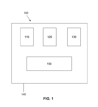
[31] FIG. 1 illustrates a kit for detecting cells expressing cell surface
advanced
glycation end-products.
[32] FIG. 2 is a graph of the response versus time in an antibody binding
experiment.
[33] FIG. 3A is a photograph of cells of an Alzheimer's disease sample
showing
carboxymethyllysine stained red (dark gray) and phosphorylated tau stained
green
(light gray).
- 10-
CA 03062082 2019-10-30
WO 2018/204679
PCT/US2018/030931
[34] FIG. 3B is a photograph of cells of an Alzheimer's disease sample
showing
carboxymethyllysine stained red (dark gray) and amyloid precursor protein
stained
green (light gray).
[35] FIG. 3C is a photograph of cells of a Parkinson's disease sample from
the
substantia nigra showing carboxymethyllysine stained red (dark gray) and alpha
synuclein stained green (light gray).
[36] FIG. 3D is a photograph of cells of a Parkinson's disease sample from
the
ventral tegmental area showing carboxymethyllysine stained red (dark gray) and
alpha synuclein stained green (light gray).
DETAILED DESCRIPTION
[37] The recognition of a quantifiable association between cellular
senescence and
various diseases, disorders and pathological conditions has resulted in
senescent
cells becoming a diagnostic target. For example, CD57 is a known marker of
senescent cells, including immune cells such as natural killer (NK) cells and
1-cells
(Kared, H. etal., "CD57 in human natural killer cells and T-lymphocytes",
Cancer
Immunology, Immunotherapy, Vol. 65, No. 4, pp. 441-452 (2016)). CD57 isolation
kits are commercially available, such as the CD8+CD57+ T Cell Isolation Kit
from
Miltenyi Biotec (Bergisch Gladbach, Germany), but these quantitative
measurement
tools are intended for research use only. The data sheet for the Miltenyi
Biotec T
Cell isolation kit explicitly states that the kit is not for diagnostic or
therapeutic use.
[38] The detection and quantification of advanced glycation end-products
has
been carried out with analytical techniques that are capable of detecting
proteins,
such as mass spectrometry and high-performance liquid chromatography. However,
these techniques are cumbersome and often rely on complex laboratory
equipment.
Wet lab techniques for measurement of advanced glycation end-products using
anti-
AGE antibodies, such as immunoassays, are significantly easier to use. Anti-
AGE
assays are commercially available, such as the Carboxymethyl Lysine (CML)
ELISA
Cat. No. KT-32428 from Kamiya Biomedical Company (Seattle, WA, USA). Much
like the senescent cell detection tools, these quantitative measurement tools
are
-11 -
CA 03062082 2019-10-30
WO 2018/204679
PCT/US2018/030931
intended for research use only. The data sheet for the Kamiya Biomedical
Company
CML ELISA (Cat. No. KT-32428) explicitly states that the product is not for
use in
diagnostic procedures. Accordingly, the use of anti-AGE antibodies for
diagnostic
purposes is neither routine nor conventional.
[39] The present invention uses antibodies that bind to advanced glycation
end-
product-modified proteins and peptides to diagnose and monitor senescence--
associated diseases, disorders or pathological conditions. AGE-modified
proteins
and peptides, especially AGE-modified proteins and peptides on the surface of
partially-functional and non-functional cells, are a unique target for
antibody-based
diagnostic methods including the enzyme-linked immunosorbent assay (E:LISA),
cell
sorting and cell counting. For example, detection of cell-bound
carboxymethyllysine
(CML), a well-known advanced glycation end-product, may be used to determine
the
total number, concentration or ratio of senescent cells in a sample. Patients
may be
identified as in need of treatment based on the number of cells that exhibit
cell-
surface AGEs in a sample as compared to the number of cells that exhibit cell-
surface AGEs in a control, or when the number of cells that exhibit cell-
surface AGEs
in the sample exceeds a clinical threshold. Alternatively, or in addition,
comparing
the ratio of cell-bound AGEs to free (unbound) AGEs may be used to normalize
the
number of senescent cells in the sample and monitor disease progression or
biological aging. A greater ratio of cell-bound AGEs indicates a greater
amount of
cellular senescence due to internal sources and cell dysfunction. Anti-AGE
antibody-based diagnostic methods offer the advantages of being minimally
invasive
and simple to carry out, which allows such tests to be carried out in a
doctor's office
or clinic.
[40] An anti-AGE antibody may be used to detect the presence of senescent
cells
in a sample since senescent cells express cell-surface advanced glycation end-
products. In one embodiment, a sample is provided. The sample may be obtained
from a human patient. Next, the presence of cell-surface AGEs in the sample is
determined or measured by contacting the sample with an anti-AGE antibody and
detecting binding between cell-surface AGEs and the anti-AGE antibody.
Optionally,
a control sample may be obtained from the patient, or from a healthy subject,
as a
- 12 -
CA 03062082 2019-10-30
WO 2018/204679
PCT/US2018/030931
baseline for comparison. A baseline for comparison may also be obtained as an
average number of cells exhibiting an AGE-modification from a group of healthy
controls. Preferably, the control samples are obtained from healthy subjects
that are
the same chronological age as the patient from whom the sample is obtained
(also
known as "age-matched" or "age-indexed" controls).
[41] The number of cells exhibiting cell-surface AGEs may be determined
using
qualitative or quantitative measurements. The measurement is intended to
provide
information that is useful for comparison to a healthy control. Examples cif
quantitative measurements include measuring the total number, average number,
concentration, ratio or percentage of cells exhibiting cell-surface AGEs in a
sample.
Examples of qualitative measurements include analyzing tissue samples with
immunohistochemical or immunocytochemical techniques. For example, the
location
of glycation within a sample may be indicative of a disease, disorder or
pathological
condition associated with cellular senescence.
[42] The measurement of senescent cells in the sample may be used to
diagnose
the patient with a disease, disorder or pathological condition associated with
cellular
senescence, or to identify the patient as in need of treatment. An elevated
level of
cellular dysfunction may be indicated when the total number, average number,
concentration, ratio or percentage of cells that exhibit cell-surface AGEs in
the
sample exceeds the total number, average number, concentration, ratio or
percentage of cells that exhibit cell-surface AGEs in a control. For example,
a
sample that contains greater than 5% senescent cells is indicative of an
elevated
level of cellular dysfunction. A patient may be diagnosed with a disease,
disorder or
pathological condition associated with cellular senescence or identified as in
need of
treatment prior to demonstrating symptoms or receiving a clinical diagnosis
from a
health care professional. Preferably, the patient already exhibits at least
one
symptom of the disease, disorder or pathological condition associated with
cellular
senescence prior to testing to aid in identification of the disease, disorder
or
pathological condition associated with cellular senescence. The measurement of
senescent cells in the sample may also be used in differential diagnosis to
- 13 -
CA 03062082 2019-10-30
WO 2018/204679
PCT/US2018/030931
distinguish diseases, disorders or pathological conditions with overlapping or
similar
symptoms.
[43] An elevated level of cellular dysfunction may also be indicated when
the
number of cells that exhibit cell-surface AGEs in the sample exceeds a
clinical
threshold. For example, a patient may be diagnosed with a disease, disorder or
pathological condition associated with cellular senescence or identified as in
need of
treatment if the sample contains 5% ¨ 50% senescent cells, including at least
6%,
7%, 8%, fso,
/0 10%, 11%, 12%, 13%, 14%, 15%, 16%, 17%, 18%, 19%, 20%, 21%,
22%, 23%, 24%, 25%, 30%, 35%, 40% and 45% senescent cells. The clinical
threshold may be based on the highest number or the average number of AGE-
modified cells found in a collection of samples obtained from healthy
patients.
[44] The measurement of senescent cells in the sample may also be used to
determine the biological age of the patient. Since senescent cells accumulate
with
age, a greater number of senescent cells in a sample as compared to an age-
matched control is indicative of an advanced biological age. A patient may be
identified as in need of treatment when the biological age of the patient is
greater
than the chronological age of the patient. For example, a patient may be
diagnosed
with advanced biological age when her biological age is 10% - 50% greater than
her
chronological age, including at least 15%, 20%, 25%, 30%, 35%, 40% and 45%
greater than her chronological age. Similarly, a patient may be diagnosed with
advanced biological age when her biological age is 5-50 years greater than her
chronological age, including at least 10 years, 15 years, 20 years, 25 years,
30
years, 35 years, 40 years and 45 years greater than her chronological age.
[45] An anti-AGE antibody may also be used to detect free (unbound) AGEs
and
AGE-modified proteins or peptides in the sample. The free AGEs may serve as a
measure of advanced glycation end-products that are not associated with
cellular
senescence. The number of cell-surface AGEs in the sample may be compared to
the number of free AGEs to normalize the number of senescent cells in the
sample.
For example, the ratio of cell-surface AGEs to free AGEs may be used to
determine
the percentage AGEs that have accumulated on the cell surface, with a high
ratio
- 14 -
CA 03062082 2019-10-30
WO 2018/204679
PCT/US2018/030931
indicating an increase in cellular senescence. The percentage of senescent
cells in
a sample may also be used as a measure of the biological age of the patient.
[46] The sample may be any substance obtained from the patient that
contains
cells which may be senescent. Examples of suitable samples include saliva, a
buccal swab, a blood sample, a urine sample, a skin sample and a biopsy. The
sample may optionally be physically processed, such as by centrifugation, or
chemically processed, such as by trypsinization. Sample processing may be used
to
isolate specific portions of the sample, such as separating a blood sample
into serum
and plasma.
[47] The cells within the sample being tested for the presence of cell-
surface
advanced glycation end-products may be any cells that are capable of
undergoing
cellular senescence. Examples of suitable cells to be tested include T-cells,
erythrocytes, fibroblasts and epithelial cells. T-cells are a preferred cell
for testing.
[48] The presence of AGE-modified peptides or proteins in the sample may be
determined by any antibody-based identification technique. Examples of
suitable
antibody identification techniques include immunoassays, such as enzyme-linked
immunosorbent assays (ELISAs), radioimmunoassays (RIAs) and real-time
immunoquantitative PCR (iqPCR), cell sorting, such as fluorescent activated
cell
sorting (FACS), flow cytometry and magnetic cell sorting, cell counting,
Western
blots, immunohistochemistry (IHC), immunocytochemistry (ICC),
immunoprecipitation and enzyme linked immunospot (ELISPOT). Preferably, the
antibody identification technique is an immunoassay.
[49] A preferred technique for detecting senescent cells in tissue samples
is
immunohistochemical (IHC) staining. Histological analysis of tissue samples is
a
well-established technique for identification of specific proteins in a tissue
sample.
For example, AGEs have been detected in tissue samples of atherosclerotic
lesions
and pancreatic cancer (Wendel, U. et al., "A novel monoclonal antibody
targeting
carboxymethyllysine, an advanced glycation end product in atherosclerosis arid
- 15-
CA 03062082 2019-10-30
WO 2018/204679
PCT/US2018/030931
pancreatic cancer", PLoS One, Vol. 13, No. 2, e0191872 (2018)).
Immunohistochemical staining allows for detecting specific sites of glycation.
[50] The diagnostic techniques may be carried out on-site where the sample
was
obtained. Alternatively, the sample may be sent to an off-site testing
facility, such as
a laboratory.
[51] The anti-AGE antibody may be any antibody that binds to an AGE-
modified
protein or peptide, including AGE-modified proteins or peptides that are
expressed
on the surface of senescent cells. Anti-AGE antibodies are known in the art
and are
commercially available. Examples include those described in U.S. 5,702,704
(Bucala) and U.S. 6,380,165 (Al-Abed etal.). The antibody may bind to one or
more
AGE-modified proteins or peptides having an AGE modification such as FFI,
pyrraline, AFGP, ALI, carboxymethyllysine (CML), carboxyethyllysine (CEL) and
pentosidine, and mixtures of such antibodies. The antibody may be monoclonal
or
polyclonal. Preferably, the antibody is a monoclonal antibody.
[52] Preferred anti-AGE antibodies include those which bind to proteins or
peptides that exhibit a carboxymethyllysine or carboxyethyllysine AGE
modification.
Carboxymethyllysine (also known as N(epsilon)-(carboxymethyl)lysine, N(6)-
carboxymethyllysine, or 2-Amino-6-(carboxymethylamino)hexanoic acid) and
carboxyethyllysine (also known as N-epsilon-(carboxyethyl)lysine) are found on
proteins or peptides and lipids as a result of oxidative stress and chemical
glycation.
CML- and CEL-modified proteins or peptides are recognized by the receptor RAGE
which is expressed on a variety of cells. CML and CEL have been well-studied
and
CML- and CEL-related products are commercially available. For example, Cell
Biolabs, Inc. sells CML-BSA antigens, CML polyclonal antibodies, CML
immunoblot
kits, and CML competitive ELISA kits (www.cellbiolabs.com/cml-assays) as well
as
CEL-BSA antigens and CEL competitive ELISA kits (www.cellbiolabs.com/cel-n-
epsilon-carboxyethyl-lysine-assays-and-reagents). A preferred commercially-
available anti-AGE antibody is the mouse anti-glycation end-product antibody
raised
against carboxymethyl lysine conjugated with keyhole limpet hemocyanin (Clone
- 16 -
CA 03062082 2019-10-30
WO 2018/204679
PCT/US2018/030931
318003) available from R&D Systems, Inc. (Minneapolis, MN; catalog no.
MAB3247).
[53] An anti-AGE antibody may have or may include a heavy chain having the
protein sequence of SEQ ID NO: 1 and a light chain having the protein sequence
of
SEQ ID NO: 3. The variable domains of the heavy chain and the light chain are
shown in SEQ ID NO: 2 and SEQ ID NO: 4, respectively. The DNA and protein
sequences of additional anti-AGE antibodies may be found in WO 2017/143073,
the
publication of International Patent Application No. PCT/U52017/18185, which is
herein incorporated by reference.
[54] The anti-AGE antibody may optionally be a bi-specific antibody, which
is an
antibody directed to two different epitopes. Such antibodies include a
variable region
(or complementary determining region) from one anti-AGE antibody, and a
variable
region (or complementary determining region) from a different antibody.
[55] Antibody fragments may be used in place of whole antibodies. For
example,
immunoglobulin G may be broken down into smaller fragments by digestion with
enzymes. Papain digestion cleaves the N-terminal side of inter-heavy chain
disulfide
bridges to produce Fab fragments. Fab fragments include the light chain and
one of
the two N-terminal domains of the heavy chain (also known as the Fd fragment).
Pepsin digestion cleaves the C-terminal side of the inter-heavy chain
disulfide
bridges to produce F(ab')2 fragments. F(ab')2 fragments include both light
chains
and the two N-terminal domains linked by disulfide bridges. Pepsin digestion
may
also form the Fv (fragment variable) and Fc (fragment crystallizable)
fragments. The
Fv fragment contains the two N-terminal variable domains. The Fc fragment
contains the domains which interact with immunoglobulin receptors on cells and
with
the initial elements of the complement cascade. Pepsin may also cleave
immunoglobulin G before the third constant domain of the heavy chain (CH3) to
produce a large fragment F(abc) and a small fragment pFc'. Antibody fragments
may alternatively be produced recombinantly.
- 17-
CA 03062082 2019-10-30
WO 2018/204679
PCT/US2018/030931
[56] Antibodies may be produced using well-known methods. For example,
polyclonal antibodies (pAbs) can be raised in a mammalian host by one or more
injections of an immunogen, and if desired, an adjuvant. Typically, the
immunogen
(and adjuvant) is injected in a mammal by a subcutaneous or intraperitoneal
injection. The immunogen may be an AGE-modified protein or peptide of a cell,
such as AGE-antithrombin III, AGE-calmodulin, AGE-insulin, AGE-ceruloplasmin,
AGE-collagen, AGE-cathepsin B, AGE-albumin such as AGE-bovine serum albumin
(AGE-BSA), AGE-human serum albumin and ovalbumin, AGE-crystallin, AGE-
plasminogen activator, AGE-endothelial plasma membrane protein, AGE-aldehyde
reductase, AGE-transferrin, AGE-fibrin, AGE-copper/zinc SOD, AGE-apo B, AGE-
fibronectin, AGE-pancreatic ribose, AGE-apo A-I and II, AGE-hemoglobin, AGE-
Na+/K+-ATPase, AGE-plasminogen, AGE-myelin, AGE-Iysozyme, AGE-
immunoglobulin, AGE-red cell Glu transport protein, AGE-p-N-acetyl hexominase,
AGE-apo E, AGE-red cell membrane protein, AGE-aldose reductase, AGE-ferritin,
AGE-red cell spectrin, AGE-alcohol dehydrogenase, AGE-haptoglobin, AGE-
tubulin,
AGE-thyroid hormone, AGE-fibrinogen, AGE-32-microglobulin, AGE-sorbitol
dehydrogenase, AGE-ai-antitrypsin, AGE-carbonate dehydratase, AGE-RNAse,
AGE-low density lipoprotein, AGE-hexokinase, AGE-apo C-I, AGE-RNAse, AGE-
hemoglobin such as AGE-human hemoglobin, AGE-low density lipoprotein (AGE-
LDL) and AGE-collagen IV. AGE-modified cells, such as AGE-modified
erythrocytes, whole, lysed, or partially digested, may also be used as AGE
antigens.
Examples of adjuvants include Freund's complete, monophosphoryl Lipid A
synthetic-trehalose dicorynomycolate, aluminum hydroxide (alum), heat shock
proteins HSP 70 or HSP96, squalene emulsion containing monophosphoryl lipid A,
a2-macroglobulin and surface active substances, including oil emulsions,
pleuronic
polyols, polyanions and dinitrophenol. To improve the immune response, an
immunogen may be conjugated to a polypeptide that is immunogenic in the host,
such as keyhole limpet hemocyanin (KLH), serum albumin, bovine thyroglobullin,
cholera toxin, labile enterotoxin, silica particles or soybean trypsin
inhibitor. A
preferred immunogen conjugate is AGE-KLH. Alternatively, pAbs may be made in
chickens, producing IgY molecules.
- 18 -
CA 03062082 2019-10-30
WO 2018/204679
PCT/US2018/030931
[57] Monoclonal antibodies (mAbs) may be made by immunizing a host or
lymphocytes from a host, harvesting the mAb-secreting (or potentially
secreting)
lymphocytes, fusing those lymphocytes to immortalized cells (for example,
myeloma
cells), and selecting those cells that secrete the desired mAb. Other
techniques may
be used, such as the EBV-hybridoma technique. If desired, the mAbs may be
purified from the culture medium or ascites fluid by conventional procedures,
such as
protein A-sepharose, hydroxyapatite chromatography, gel electrophoresis,
dialysis,
ammonium sulfate precipitation or affinity chromatography.
[58] The anti-AGE antibodies may be used to diagnose the onset or measure
the
progression of any disease, disorder or pathological condition that is
characterized
by cellular senescence. Examples of diseases, disorders and pathological
conditions that have been associated with cellular senescence include
Alzheimer's
disease, amyotrophic lateral sclerosis (ALS or Lou Gehrig's Disease), chronic
obstructive pulmonary disease (COPD), Huntington's chorea, idiopathic
pulmonary
fibrosis, muscular dystrophy (including Becker's, Duchenne, Limb-Girdle and
Yamamoto's muscular dystrophy), macular degeneration, cataracts, diabetic
retinopathy, Parkinson's disease, progeria (including Werner Syndrome and
Hutchinson Gilford progeria), vitiligo, cystic fibrosis, atopic dermatitis,
eczema,
arthritis (including osteoarthritis, rheumatoid arthritis and juvenile
rheumatoid
arthritis), atherosclerosis, cancer and metastatic cancer (including, for
example,
breast cancer, triple negative breast cancer, lung cancer, melanoma, colon
cancer,
renal cell carcinoma, prostate cancer, cancer of the cervix, bladder cancer,
rectal
cancer, esophageal cancer, liver cancer, mouth and throat cancer, multiple
myeloma, ovarian cancer, stomach cancer, pancreatic cancer and retinal
blastoma
cancers), cancer therapy-related disability or cancer therapy side effects,
hypertension, glaucoma, osteoporosis, sarcopenia, cachexia, stroke, myocardial
infarction, atrial fibrillation, transplantation rejection, diabetes mellitus
¨ Type I,
diabetes mellitus ¨ Type II, radiation exposure, HIV treatment side effects
chemical
weapons exposure, poisoning, inflammation, nephropathy, Lewy body dementia,
prion disease (including bovine spongiform encephalopathy, Creutzfeldt-Jakob
disease, scrapie, chronic wasting disease, kuru and fatal familial insomnia),
- 19-
CA 03062082 2019-10-30
WO 2018/204679
PCT/US2018/030931
lordokyphosis, auto-immune disorders, loss of adipose tissue, psoriasis,
Crohn's
disease, asthma, the physiological effects of aging (including "cosmetic"
effects,
such as wrinkling, age spots, hair loss, reduction in subcutaneous adipose
tissue
and thinning of the skin), idiopathic myopathy (including, for example,
idiopathic
inflammatory myopathy, idiopathic inflammatory myositis, polymyositis,
dermatomyositis, sporadic inclusion body myositis and juvenile myositis),
multiple
sclerosis, neuromyelitis optica (NMO, Devic's disease or Devic's syndrome),
epilepsy and adrenoleukodystrophy (ALD, X-linked adrenoleukodystrophy, X-ALD,
cerebral ALD or cALD).
[59] Any subject that could develop a disease, disorder or pathological
condition
associated with cellular senescence may be diagnosed by the methods herein
described. The subject may be a mammal. Humans are a preferred subject for
diagnosis. Other subjects that may be diagnosed include mice, rats, goats,
sheep,
cows, horses, camels and companion animals, such as dogs or cats.
[60] A patient who has been diagnosed with a disease, disorder or
pathological
condition associated with cellular senescence or identified as in need of
treatment
may be administered a senescent cell removal agent to target and destroy
senescent cells. Examples of senescent cell removal agents include therapeutic
anti-AGE antibodies, an anti-AGE antibody conjugated to a toxin, a senolytic
agent,
such as dasatinib and/or quercetin, and combinations thereof. Senescent cells
may
also be destroyed by the application of therapeutic ultrasound. Senescent cell
destruction techniques may be combined to achieve a desired therapeutic
outcome.
For example, a patient may be administered a combination of dasatinib and
quercetin as well as high intensity focused ultrasound to selectively destroy
senescent cells while sparing functional cells. A therapeutically effective
amount of
the senescent cell removal agent will vary depending on the specific senescent
cell
removal agent used. For example, an appropriate dosage level of an anti-AGE
antibody will generally be about 0.01 to 500 mg/kg patient body weight,
including
about 0.01 to 250 mg/kg, about 0.05 to 100 mg/kg, and about 0.1 to 50 mg/kg.
Similarly, an appropriate dosage level of the combination therapy dasatinib
and
quercetin will generally be about 5 mg/kg patent body weight dasatinib and
about 50
- 20 -
CA 03062082 2019-10-30
WO 2018/204679
PCT/US2018/030931
mg/kg patent body weight quercetin. Treatment efficacy may be monitored by
repeated measurements of the number of cells exhibiting cell-surface AG Es.
[61] Administration of a senescent cell removal agent has been demonstrated
as
effective in treating sarcopenia, atherosclerosis and metastatic cancer. Other
diseases, disorders and pathological conditions associated with cellular
senescence
that are particularly suitable for treatment by administration of a senescent
cell
removal agent include inflammation, autoimmune diseases, osteoarthritis.
Alzheimer's disease and Parkinson's disease.
[62] In addition to the in vitro diagnostic methods described above, anti-
AGE
antibodies may also be used in in vivo diagnostic methods. In vivo diagnostic
tests
provide non-invasive methods of detecting AGE-modified proteins and peptides.
In
vivo diagnostic tests are particularly useful for detecting senescent cells
that express
cell-surface advanced glycation end-products, such as metastatic cancer cells.
(See, for example, WO 2017/143073). Anti-AGE antibodies may be labeled with a
detectable label or tracer and then administered to a subject. The labeled
anti-AGE
antibodies specifically bind to AGE-modified proteins or peptides, which
allows the
AGE-modified proteins or peptides to be detected with any suitable apparatus
that is
capable of detecting the label. Examples of in vivo diagnostic methods include
positron emission tomography (PET) and immuno-PET, magnetic resonance
imaging (MRI), single photon emission computed tomography (SPECT), optical
imaging, ultrasound, radioimmunoscintigraphy and combinations thereof. Any
label
that is appropriate for a given diagnostic technique may be used, such as
radiolabels, fluorescent labels, positron emitters, dyes that emit in near
infrared
(NIR), nanoparticles such as gold and gadolinium, quantum dots,
superparamagnetic
iron oxide (SP10), carbon nanotubes or microbubbles that have been conjugated
to
the antibodies.
[63] FIG. 1 illustrates a kit 100 for detecting cells expressing cell
surface advanced
glycation end-products. The kit may include an anti-AGE antibody 110, a
control 120
and, optionally, a reagent 130 for detecting the anti-AGE antibody. The anti-
AGE
antibody, the control and the optional reagent may be supplied in any suitable
-21-
CA 03062082 2019-10-30
WO 2018/204679
PCT/US2018/030931
container, such as bottles, ampules, envelopes, test tubes, vials, flasks or
syringes.
The anti-AGE antibody and/or the reagent may optionally be labelled, such as
with a
fluorescent label, radiolabel or a gold particle. The control may be normal
serum
from an animal in which a secondary antibody was made, a solution containing a
known amount of an AGE-modified protein or peptide or fixed or preserved cells
that
exhibit and AGE modification. Examples of reagents for detecting the anti-AGE
antibody include secondary antibodies, such as an anti-human polyclonal
antibody
made in donkey and labelled with rhodamine. The kit may optionally be housed
in a
container 140. The kit may optionally include printed instructions 150.
Preferably,
the contents of the kit are sterile and ready for use.
[64] The kit may optionally include a container for housing the kit
ingredients. The
container may be formed of a rigid, durable material, such as plastic, or may
be
flexible, such as a bag or soft-sided box.
[65] The kit may optionally include instructions for use. The instructions
may be
provided as printed instructions or in electronic format, such as on a
universal serial
bus (USB) drive, on a secure digital (SD) card, or hosted over the internet
and
accessible through a quick response (QR) code.
[66] Kits may optionally contain additional diagnostic materials or
equipment such
as buffers, fixatives, blocking solutions, protease inhibitors, substrates for
analysis
such as microscope slides and/or cover slips, microtiter plates and cell
extraction
reagents such as detergents and detergent solutions.
[67] EXAMPLES
[68] Example 1: Collection of buccal epithelial cells
[69] A patient swishes a saline solution in her mouth for 30 seconds. She
then
spits the solution into a cup. 1.5 mL of the saline solution from the cup
is1:hen
transferred to a centrifuge tube using a micropipette. The centrifuge tube is
placed
into a balanced centrifuge and centrifuged at 10,000¨ 14,000 RPM for 2
minutes.
The centrifuging may be repeated until a pellet is visible in the bottom of
the
- 22 -
CA 03062082 2019-10-30
WO 2018/204679
PCT/US2018/030931
centrifuge tube. The supernatant is then discarded by decanting and/or
removing it
with a micropipette. The pellet contains isolated buccal epithelial cells
which may
then be tested for the presence of cell-surface AGEs to determine if any of
the
epithelial cells are senescent.
[70] Example 2: Diagnosis and treatment based on skin cells
[71] Epidermal cells are collected using a tape harvesting process.
Adhesive tape
(Adhesives Research, Glen Rock, PA) is fabricated into circular disks
approximately
17 mm in diameter. The tape is applied to a patient's skin, then removed to
harvest
epidermal cells from the stratum corneum. Tape harvesting is repeated 3
additional
times to obtain a total of 4 epidermal samples. The epidermal cells are then
tested
for the presence of cell-surface AGEs to determine if any of the epidermal
cells are
senescent.
[72] Dermal cells are collected using a shave biopsy. A scalpel blade is
used to
remove sufficient skin to pass through the epidermis and access the dermis to
obtain
a sample. Fibroblasts within the sample are then tested for the presence of
cell-
surface AGEs to determine if any of the fibroblasts are senescent. The
presence of
at least 5% senescent cells in the shave biopsy indicates skin damage and the
need
for treatment to reduce the number of senescent skin cells. The patient is
administered a senescent cell removal agent to target and remove senescent
skin
cells.
[73] Example 3: Direct binding ELISA using anti-AGE antibodies
[74] The binding of murine and chimeric anti-AGE antibodies was
investigated by
a direct binding ELISA. An anti-carboxymethyl lysine (CML) antibody (R&D
Systems, MAB3247) was used as a control. CML was conjugated to KLH (CML-
KLH) and both CML and CML-KLH were coated overnight onto an ELISA plate.
HRP-goat anti-mouse Fc was used to detect the control and murine anti-AGE
antibodies. HRP-goat anti-human Fc was used to detect the chimeric anti-AGE
antibody.
- 23 -
CA 03062082 2019-10-30
WO 2018/204679
PCT/US2018/030931
[75] The antigens were diluted to 1 pg/mL in lx phosphate buffer at pH 6.5.
A 96-
well microtiter ELISA plate was coated with 100 pL/well of the diluted antigen
and let
sit at 4 C overnight. The plate was blocked with lx PBS, 2.5% BSA and allowed
to
sit for 1-2 hours the next morning at room temperature. The antibody samples
were
prepared in serial dilutions with lx PBS, 1% BSA with the starting
concentration of
50 pg/mL. Secondary antibodies were diluted 1:5,000. 100 pL of the antibody
dilutions was applied to each well. The plate was incubated at room
temperature for
0.5-1 hour on a microplate shaker. The plate was washed 3 times with 1x PBS.
100
pL/well diluted HRP-conjugated goat anti-human Fc secondary antibody was
applied
to the wells. The plate was incubated for 1 hour on a microplate shaker. The
plate
was then washed 3 times with lx PBS. 100 pL HRP substrate TMB was added to
each well to develop the plate. After 3-5 minutes elapsed, the reaction was
terminated by adding 100 pL of 1N HCl. A second direct binding ELISA was
performed with only CML coating. The absorbance at 0D450 was read using a
microplate reader.
[76] The 0D450 absorbance raw data for the CML and CML-KLH ELISA is shown
in the plate map below. 48 of the 96 wells in the well plate were used. Blank
wells in
the plate map indicate unused wells.
- 24 -
CA 03062082 2019-10-30
WO 2018/204679
PCT/US2018/030931
[77] Plate map of CML and CML-KLH ELISA:
Conc.
(ug/mL) 1 2 3 4 5 6 7
50 0.462 0.092 0.42 1.199
0.142 -- 1.852
16.67 0.312 0.067 0.185 0.31
0.13 -- 0.383
5.56 0.165 0.063 0.123 0.19
0.115 0.425
1.85 0.092 0.063 0.088 0.146
0.099 0.414
0.62 0.083 0.072 0.066 0.108
0.085 0.248
0.21 0.075 0.066 0.09 0.096
0.096 -- 0.12
0.07 0.086 0.086 0.082 0.098
0.096 0.098
0 0.09 0.085 0.12 0.111 0.083
0.582
R&D Parental Chimeric R&D Parental Chimeric
Positive Anti- Anti- Positive Anti- Anti-
Control AGE AGE Control -- AGE -- AGE
CML-KLH Coat CML Coat
[78] The 0D450 absorbance raw data for the CML-only ELISA is shown in the
plate map below. 24 of the 96 wells in the well plate were used. Blank wells
in the
plate map indicate unused wells.
[79] Plate map of CML-only ELISA:
Conc.
(ug/mL) 1 2 3 4 5 6 7
50 1.913 0.165 0.992
16.66667 1.113 0.226 0.541
5.555556 0.549 0.166 0.356
1.851852 0.199 0.078 0.248
0.617284 0.128 0.103 0.159
0.205761 0.116 0.056 0.097
0.068587 0.073 0.055 0.071
0 0.053 0.057 0.06
R&D Parental Chimeric
Positive Anti- Anti-
Control AGE AGE
- 25 -
CA 03062082 2019-10-30
WO 2018/204679
PCT/US2018/030931
[80] The control and chimeric anti-AGE antibodies showed binding to both
CML
and CML-KLH. The murine (parental) anti-AGE antibody showed very weak to no
. binding to either CML or CML-KLH. Data from repeated ELISA confirms
binding of
the control and chimeric anti-AGE antibody to CML. All buffer control showed
negative signal.
[81] This data confirms the ability of the anti-AGE antibodies to bind AGEs
and
AGE-immunogen conjugates. Evidence of binding to the well-known AGE
carboxymethyllysine supports the suitability of anti-AGE antibodies in
diagnostic
applications.
[82] Example 4: Diagnosis and treatment based on blood sample
[83] A blood sample is drawn from a patient. The blood sample is
centrifuged to
isolate the serum. CD57+ T-cells are isolated using the Miltenyi Biotec
CD8+CD57+
T Cell Isolation Kit (Bergisch Gladbach, Germany). CD57+ T-cells are counted
using
a hemocytometer. The serum and the isolated CD57+ T-cells are then tested' for
binding to an anti-CML antibody. Serum CML less than or equal to 152 pg/mIL
combined with greater than or equal to 50% of isolated CD57+ T-cells binding
to a
labeled anti-CML antibody indicates that the patient is in need of treatment
with a
senescent cell removal agent. The patient is administered an anti-AGE antibody
that targets and removes senescent cells.
[84] Example 5: Diagnosis and treatment based on buccal swab
[85] A sample is obtained from a patient by a buccal swab. The buccal
epithelial
cells and the saliva from the swab are separated. The buccal cells are
trypsinized
and mixed with an anti-CML monoclonal antibody. The mixture is then passed
through a hemocytometer to count the senescent buccal cells. Free CML is
measured by an ELISA of the saliva. Saliva CML less than or equal to 3 pg/n1L
combined with greater than or equal to 50% of the buccal cells binding to the
anti-
CML antibody indicates the patient is in need of treatment with a senescent
cell
removal agent. The patient is administered an anti-AGE antibody conjugated to
a
toxin that targets and removes senescent cells.
- 26 -
CA 03062082 2019-10-30
WO 2018/204679
PCT/US2018/030931
[86] Example 6: Determination of biological age
[87] A sample is obtained from a 50-year-old patient by buccal swab. The
buccal
epithelial cells and the saliva from the swab are separated. The buccal cells
are
trypsinized and mixed with an anti-CML monoclonal antibody. The mixture is
then
passed through a hemocytometer to count the senescent buccal cells. Free CML
is
measured by an ELISA of the saliva. The ratio of buccal cells expressing cell-
surface CML to free CML in saliva is 5:1. This ratio is greater than would be
expected for a healthy 50-year-old, which demonstrates that the patient has a
biological age that is greater than her chronological age. The results
indicate that
the patient is experiencing the early onset of aging and aging-related
diseases,
disorders or pathological conditions due to cellular senescence. These results
also
indicate that the patient is in need of treatment with a senescent cell
removal agent.
[88] Example 7: Diagnosis and treatment of a disease, disorder or
pathological
condition order associated with advanced biological aging due to cellular
senescence
[89] A blood sample is obtained from a 45-year-old patient. The blood
sample is
centrifuged to isolate the serum. CD57+ T-cells are isolated using the
Miltenyi
Biotec CD8+CD57+ T Cell Isolation Kit (Bergisch Gladbach, Germany). CD57+ T-
cells are counted using a hemocytometer. The serum and the isolated CD57 T-
cells are then tested for binding to an anti-CML antibody. The ratio of cells
expressing cell-surface CML to free CML in serum is 10:1. This ratio indicates
the
patient has a biological age of 65. Since the biological age of the patient
exceeds
the chronological age of the patient, the patient is diagnosed with a disease,
disorder
or pathological condition associated with advanced biological aging due to
cellular
senescence. The patient is administered an anti-AGE antibody that targets and
removes senescent cells.
[90] Example 8:/n vivo study of the administration of anti-glycation end-
product
antibody
- 27 -
CA 03062082 2019-10-30
WO 2018/204679
PCT/US2018/030931
[91] To examine the effects of an anti-glycation end-product antibody,
the antibody
was administered to the aged CD1(ICR) mouse (Charles River Laboratories),
twice
daily by intravenous injection, once a week, for three weeks (Days 1, 8 and
15),
followed by a 10 week treatment-free period. The test antibody was a
commercially
available mouse anti-glycation end-product antibody raised against
carboxymethyl
lysine conjugated with keyhole limpet hemocyanin, the carboxymethyl lysine
IV1Ab
(Clone 318003) available from R&D Systems, Inc. (Minneapolis, MN; catalog no.
MAB3247). A control reference of physiological saline was used in the control
animals.
[92] Mice referred to as "young" were 8 weeks old, while mice referred
to as "old"
were 88 weeks ( 2 days) old. No adverse events were noted from the
administration
of the antibody. The different groups of animals used in the study are shown
in
Table 1.
[93] Table 1: The
different groups of animals used in the study
Number of Animals
Main Study Treatment-
Grou Test Dose Level Free
p No. Material _ Mice (pg/gm/BID/ week) Females Females
1 Saline young 0 20
2 Saline old 0 20 20
3 Antibody old 2.5 20 20
4 None old 0 20 _____ pre
Antibody old 5.0 20 20
- = Not Applicable, Pre = Subset of animals euthanized prior to treatment
start for collection
of adipose tissue.
[94] p16INK48 mRNA, a marker for senescent cells, was quantified in
adipose tissue
of the groups by Real Time-qPCR. The results are shown in Table 2. In the
table
AACt = ACt mean control Group (2) ¨ ACt mean experimental Group (1 or 3 or 5);
Fold Expression= 2 -AACt.
[95] Table 2: P161NK48 mRNA quantified in adipose tissue
Calculation (unadjusted Group 2 vs Group 1 Group 2 vs Group 3 Group 2 vs Group
5
- 28 -
CA 03062082 2019-10-30
WO 2018/204679
PCT/US2018/030931
to Group 4: 5.59) Group 2 Group Group
Group
Group 2
Group 5
1 3 2
Mean ACt 5.79 7.14 5.79 6.09 5.79 7.39
AACt -1.35 -0.30 -1.60
Fold Expression 2.55 1.23 3.03
[96] The table above indicates that untreated old mice (Control Group 2)
express
2.55-fold more p16Ink4a mRNA than the untreated young mice (Control Group 1),
as
expected. This was observed when comparing Group 2 untreated old mice
euthanized at end of recovery Day 85 to Group 1 untreated young mice
euthanized
at end of treatment Day 22. When results from Group 2 untreated old mice were
compared to results from Group 3 treated old mice euthanized Day 85, it was
observed that p1611k4a mRNA was 1.23-fold higher in Group 2 than in Group 3.
Therefore, the level of p161"k" mRNA expression was lower when the old mice
were
treated with 2.5 pg/gram/BID/week of antibody.
[97] When results from Group 2 (Control) untreated old mice were compared
to
results from Group 5 (5 pg/gram) treated old mice euthanized Day 22, it was
observed that p16Ink48 mRNA was 3.03-fold higher in Group 2 (controls) than in
Group 5 (5 pg/gram). This comparison indicated that the Group 5 animals had
lower
levels of p161nk4a mRNA expression when they were treated with 5.0
pg/gram/BID/week, providing p161nk4a mRNA expression levels comparable to that
of
the young untreated mice (i.e. Group 1). Unlike Group 3 (2.5 pg/gram) mice
that
were euthanized at end of recovery Day 85, Group 5 mice were euthanized al end
of
treatment Day 22.
[98] These results demonstrate that the administration of the anti-AGE
antibody
resulted in the killing of senescent cells.
[99] The mass of the gastrocnemius muscle was also measured, to determine
the
effect of antibody administration on sarcopenia. The results are provided in
Table 3.
The results indicate that administration of the antibody increased muscle mass
as
compared to controls, but only at the higher dosage of 5.0 pg/gm/BID/ week.
- 29 -
CA 03062082 2019-10-30
WO 2018/204679
PCT/US2018/030931
[100] Table 3: Effect of antibody administration on mass of the
gastrocnemius
muscle
Weight relative to body
Summary Absolute weight of mass of Gastrocnemius
Group Information Gastrocnemius Muscle Muscle
1 Mean 0.3291 1.1037
SD 0.0412 0.1473
20 20
2 Mean 0.3304 0.7671
SD 0.0371 0.1246
20 20
3 Mean 0.3410 0.7706
SD 0.0439 0.0971
19 19
Mean 0.4074 0.9480
SD 0.0508 0.2049
9 9
[101] These results demonstrate that administration of antibodies that
bind to AGEs
of a cell resulted in a reduction of cells expressing p16ink4a, a biomarker of
senescence. The data show that reducing senescent cells leads directly to an
increase in muscle mass in aged mice. These results indicate that the loss of
muscle mass, a classic sign of sarcopenia, can be treated by administration of
senescent cell removal agents, such as antibodies that bind to AGEs of a cell.
[102] This data confirms that anti-AGE antibodies are capable of
selectively binding
to cells expressing cell-surface AGE-modified proteins or AGE-modified
peptides.
Evidence of selective binding supports the suitability of anti-AGE antibodies
in
diagnostic applications. The data also demonstrates that anti-AGE antibodies
are
safe for in vivo use.
[103] Example 9: Affinity and kinetics of test antibody
- 30 -
CA 03062082 2019-10-30
WO 2018/204679
PCT/US2018/030931
[104] The affinity and kinetics of the test antibody used in Example 8 were
analyzed
using Na,Na-bis(carboxymethyl)-L-lysine trifluoroacetate salt (Sigma-Aldrich,
St.
Louis, MO) as a model substrate for an AGE-modified protein of a cell. Label-
free
interaction analysis was carried out on a BIACORETM T200 (GE Healthcare,
Pittsburgh, PA), using a Series S sensor chip CM5 (GE Healthcare, Pittsburgh,
PA),
with Fc1 set as blank, and Fc2 immobilized with the test antibody (molecular
weight
of 150,000 Da). The running buffer was a HBS-EP buffer (10 mM HEPES, 150 mM
NaCI, 3 mM EDTA and 0.05% P-20, pH of 7.4), at a temperature of 25 C.
Software
was BIACORETM T200 evaluation software, version 2Ø A double reference (Fc2-1
and only buffer injection), was used in the analysis, and the data was fitted
to a
Langmuir 1:1 binding model.
[105] Table 4: Experimental set-up of affinity and kinetics analysis
Association and dissociation
Flow path Fc1 and Fc2
Flow rate (pl/min.) 30
Association time (s) 300
Dissociation time (s) 300
Sample concentration (pM) 20 ¨ 5 ¨ 1.25 (x2) ¨ 0.3125 ¨ 0.078 - 0
[106] A graph of the response versus time is illustrated in FIG. 2. The
following
values were determined from the analysis: ka (1/MS) = 1.857 x 10³; kd (1/s) =
6.781 x
10-3; KD (M) = 3.651 x 10⁻⁶; Rmax (RU) = 19.52; and Chi2 = 0.114. Because the
Chi2
value of the fitting is less than 10% of Rmax, the fit is reliable.
[107] Example 10: In vivo study of the administration of a carboxymethyl
lysine
monoclonal antibody
- 31 -
CA 03062082 2019-10-30
WO 2018/204679
PCT/US2018/030931
[108] The effect of a carboxymethyl lysine antibody on tumor growth,
metastatic
potential and cachexia was investigated. In vivo studies were carried out in
mice
using a murine breast cancer tumor model. Female BALB/c mice (BALB/cAnNCrl,
Charles River) were eleven weeks old on Day 1 of the study.
[109] 4T1 murine breast tumor cells (ATCC CRL-2539) were cultured in RPMI
1640
medium containing 10% fetal bovine serum, 2 mM glutamine, 25 pg/mL gentamicin,
100 units/mL penicillin G Na and 100 pg/mL streptomycin sulfate. Tumor cells
were
maintained in tissue culture flasks in a humidified incubator at 37 C in an
atmosphere of 5% CO2 and 95% air.
[110] The cultured breast cancer cells were then implanted in the mice. 4T1
cells
were harvested during log phase growth and re-suspended in phosphate buffered
saline (PBS) at a concentration of 1 x 10⁶ cells/mL on the day of implant.
Tumors
were initiated by subcutaneously implanting 1 x 10⁵ 4T1 cells (0.1 mL
suspension)
into the right flank of each test animal. Tumors were monitored as their
volumes
approached a target range of 80-120 mm3. Tumor volume was determined using
the formula: tumor volume = (tumor width)2(tumor length)/2. Tumor weight was
approximated using the assumption that 1 mm3 of tumor volume has a weight of 1
mg. Thirteen days after implantation, designated as Day 1 of the study, mice
were
sorted into four groups (n=15/group) with individual tumor volumes ranging
from 108
to 126 mm3 and a group mean tumor volume of 112 mm3. The four treatment groups
are shown in Table 5 below:
[111] Table 5: Treatment groups
Group Description Agent Dosing
(plg/g)
1 Control phosphate buffered saline (PBS) N/A
2 Low-dose carboxymethyl lysine monoclonal 5
antibody
- 32 -
CA 03062082 2019-10-30
WO 2018/204679
PCT/US2018/030931
3 High-dose carboxymethyl lysine monoclonal 10
antibody
4 Observation None N/A
only
[112] An anti-carboxymethyl lysine monoclonal antibody was used as a
therapeutic
agent. 250 mg of carboxymethyl lysine monoclonal antibody was obtained from
R&D Systems (Minneapolis, MN). Dosing solutions of the carboxymethyl lysine
monoclonal antibody were prepared at 1 and 0.5 mg/mL in a vehicle (PBS) to
provide the active dosages of 10 and 5 pg/g, respectively, in a dosing volume
of 10
mL/kg. Dosing solutions were stored at 4 C protected from light.
[113] All treatments were administered intravenously (i.v.) twice daily for
21 days,
except on Day 1 of the study where the mice were administered one dose. On Day
19 of the study, i.v. dosing was changed to intraperitoneal (i.p.) dosing for
those
animals that could not be dosed i.v. due to tail vein degradation. The dosing
volume
was 0.200 mL per 20 grams of body weight (10 mL/kg), and was scaled to the
body
weight of each individual animal.
[114] The study continued for 23 days. Tumors were measured using calipers
twice
per week. Animals were weighed daily on Days 1-5, then twice per week until
the
completion of the study. Mice were also observed for any side effects.
Acceptable
toxicity was defined as a group mean body weight loss of less than 20% during
the
study and not more than 10% treatment-related deaths. Treatment efficacy was
determined using data from the final day of the study (Day 23).
[115] The ability of the anti-carboxymethyl lysine antibody to inhibit
tumor growth
was determined by comparing the median tumor volume (MTV) for Groups 1-3.
Tumor volume was measured as described above. Percent tumor growth inhibition
(%TGI) was defined as the difference between the MTV of the control group
(Group
1) and the MTV of the drug-treated group, expressed as a percentage of the MTV
of
- 33 -
CA 03062082 2019-10-30
WO 2018/204679
PCT/US2018/030931
the control group. %TGI may be calculated according to the formula: %TGI = (1-
MTVtreated/MTVcontroI) X 100.
[116] The ability of the anti-carboxymethyl lysine antibody to inhibit
cancer
metastasis was determined by comparing lung cancer foci for Groups 1-3.
Percent
inhibition (%Inhibition) was defined as the difference between the mean count
of
metastatic foci of the control group and the mean count of metastatic foci of
a drug-
treated group, expressed as a percentage of the mean count of metastatic foci
of the
control group. %Inhibition may be calculated according to the following
formula:
%Inhibition = (1-Mean Count of Focitreated/Mean Count of FOCicontrol) X 100.
[117] The ability of the anti-carboxymethyl lysine antibody to inhibit
cachexia was
determined by comparing the weights of the lungs and gastrocnemius muscles for
Groups 1-3. Tissue weights were also normalized to 100 g body weight.
[118] Treatment efficacy was also evaluated by the incidence and magnitude
of
regression responses observed during the study. Treatment may cause partial
regression (PR) or complete regression (CR) of the tumor in an animal. In a PR
response, the tumor volume was 50% or less of its Day 1 volume for three
consecutive measurements during the course of the study, and equal to or
greater
than 13.5 mm3 for one or more of these three measurements. In a CR response,
the
tumor volume was less than 13.5 mm3 for three consecutive measurements during
the course of the study.
[119] Statistical analysis was carried out using Prism (GraphPad) for
Windows 6.07.
Statistical analyses of the differences between Day 23 mean tumor volumes
(MTVs)
of two groups were accomplished using the Mann-Whitney U test. Comparisons of
metastatic foci were assessed by ANOVA-Dunnett. Normalized tissue weights were
compared by ANOVA. Two-tailed statistical analyses were conducted at
sigmficance
level P = 0.05. Results were classified as statistically significant or not
statistically
significant.
[120] The results of the study are shown below in Table 6:
- 34 -
CA 03062082 2019-10-30
WO 2018/204679
PCT/US2018/030931
[121] Table 6: Results
Group MTV %TGI Lung %Inhibition PR CR Gastroc. Lung weight/
(mm3)
foci weight/ normalized
normalized (mg)
(mg)
1 1800 N/A 70.4 N/A 0
0 353.4/19.68 2799.4/292.98
2 1568 13% 60.3 14% 0
0 330.4/21.62 2388.9/179.75
3 1688 6% 49.0 30% 0
0 398.6/24.91 2191.6/214.90
[122]
All treatment regimens were acceptably tolerated with no treatment-related
deaths. The only animal deaths were non-treatment-related deaths due to
metastasis. The (YoTGI trended towards significance (P > 0.05, Mann-Whitney)
for the 5 pg/g (Group 2) and 10 pg/g treatment group (Group 3). The
%Inhibition
trended towards significance (P > 0.05, ANOVA-Dunnett) for the 5 pg/g
treatment
group. The %Inhibition was statistically significant (P <0.01, ANOVA-Dunnett)
for the 10 pg/g treatment group. The ability of the carboxymethyl lysine
antibody
to treat cachexia trended towards significance (P > 0.05, ANOVA) based on a
comparison of the organ weights of the lung and gastrocnemius between
treatment groups and the control group. The results indicate that
administration
of an anti-carboxymethyl lysine monoclonal antibody is able to reduce cancer
metastases.
[123] This data confirms that anti-AGE antibodies are capable of
selectively binding
to cells expressing cell-surface AGE-modified proteins or AGE-modified
peptides.
Evidence of selective binding supports the suitability of anti-AGE antibodies
in
diagnostic applications. The data also demonstrates that anti-AGE antibodies
are
safe for in vivo use.
- 35 -
CA 03062082 2019-10-30
WO 2018/204679
PCT/US2018/030931
[124] Example 11: Anti-AGE antibodies bind to senescent chondrocytes in
vitro
[125] Senescent chondrocytes were obtained from osteoarthritic joints. Anti-
AGE
antibodies bound to the senescent chondrocytes in vitro. These results confirm
that
anti-AGE antibodies are capable of binding to senescent cells. The results
also
confirm the suitability of anti-AGE antibodies in diagnostic applications.
[126] Example 12: Immunohistochemical study
[127] Tissue samples were obtained from patients with Alzheimer's disease
and
Parkinson's disease. Two Alzheimer's disease samples were taken from the
hippocampus. One Parkinson's disease sample was taken from the substantia
nigra, and a second Parkinson's disease sample was taken from the ventral
tegmental area. All cells were stained for carboxymethyllysine (CML) using
anti-
AGE antibodies as described above. The Alzheimer's disease cells were stained
for
phosphorylated tau (phospho tau) or separately amyloid precursor protein. The
Parkinson's disease cells were stained for alpha synuclein. Nuclear staining
of the
cells was identified using DAPI counter stain. (Experiments were carried out
and
images were prepared by Dr. Diego Mastroeni of Arizona State University.)
[128] FIG. 3A is a photograph of cells of the Alzheimer's disease sample
showing
carboxymethyllysine stained red and phosphorylated tau stained green.
[129] FIG. 3B is a photograph of cells of the Alzheimer's disease sample
showing
carboxymethyllysine stained red and amyloid precursor protein stained green.
[130] FIG. 3C is a photograph of cells of the Parkinson's disease sample
from the
substantia nigra showing carboxymethyllysine stained red and alpha synuclein
stained green.
[131] FIG. 3D is a photograph of cells of the Parkinson's disease sample
from the
ventral tegmental area showing carboxymethyllysine stained red and alpha
synuclein
stained green.
- 36 -
CA 03062082 2019-10-30
WO 2018/204679
PCT/US2018/030931
[132] CML, a well-known AGE, did not co-localize with established
pathologies in
Alzheimer's disease and Parkinson's disease. Instead, the CML presented on
glial
cells. It was suspected that the CML immunoreactivity in the Alzheimer's
disease
samples was with microglia, and the CML immunoreactivity in the Parkinson's
disease samples was with astrocytes. The results demonstrate the presence of
senescent glial cells in Alzheimer's disease and Parkinson's disease. Removal
of
senescent glial cells using an anti-AGE antibody would be expected to result
in
regeneration of the glial cells by neural stem/progenitor cells. (See, for
example,
Leonard, B.W. etal., "Subventricular zone neural progenitors from rapid brain
autopsies of elderly subjects with and without neurodegenerative disease", The
Journal of Comparative Neurology, Vol. 515, pp. 269-294 (2009)).
[133] This data confirms the ability of the anti-AGE antibodies to bind to
AGEs
present on cells in tissue samples obtained from patients with various
neurodegenerative diseases. The evidence further confirms the suitability of
anti-
AGE antibodies in diagnostic applications.
- 37 -
CA 03062082 2019-10-30
WO 2018/204679
PCT/US2018/030931
[134] REFERENCES
[135] 1. Lowe, R. etal., "Buccals are likely to be a more informative
surrogate
tissue than blood for epigenome-wide association studies", Epigenetics, Vol.
8, No.
4, pp. 445-454 (2013).
[136] 2. Kared, H. etal., "0D57 in human natural killer cells and T-
lymphocytes", Cancer Immunology, Immunotherapy, Vol. 65, No. 4, pp. 441-452
(2016).
[137] 3. Yoon, M-S. etal., "Characterisation of advanced glycation
endproducts
in saliva from patients with diabetes mellitus", Biochemical and Biophysical
Research
Communications, Vol. 323, Issue 2, pp. 377-381 (2004).
[138] 4. Pearce, M.S. etal., "Childhood growth, IQ and education as
predictors
of white blood cell telomere length at age 49-51 years: the Newcastle Thousand
Families Study", PLoS ONE, Vol. 7, Issue 7, e40116 (2012).
[139] 5. "Telomere Testing White Paper", Titanovo, available online at
titanovo.com/telomere-length-testing/ (accessed on April 20, 2017).
[140] 6. "Carboxymethyl Lysine (CML) ELISA Cat. No. KT-32428", Kamiya
Biomedical Company, available online at www.kamiyabiomedical.com/pdf/KT-
32428.pdf (accessed on April 20, 2017).
[141] 7. "CD8+CD57+ T Cell Isolation Kit Data Sheet", Miltenyi Biotec,
available
online at
www.miltenyibiotec.com/--/media/Images/Products/Import/0001600/IM0001652.ashx
?force=1 (2008).
[142] 8. "CD8+CD57+ T Cell Isolation Kit, human", Miltenyi Biotec,
available
online at www.miltenyibiotec.com/en/products-and-services/macs-cell-
separation/cell-separation-reagents/t-cells/cd8-cd57-t-cell-isolation-kit-
human aspx
(accessed on April 19, 2017).
- 38 -
CA 03062082 2019-10-30
WO 2018/204679
PCT/US2018/030931
[143] 9. "Extraction of genomic DNA from buccal epithelial cells",
available
online at authors.fhcrc.org/591/1/Buccal%20Cell%2ODNA%201so1%20v2.pdf
(accessed on April 19, 2017).
[144] 10. Severin, F.F. etal., "Advanced glycation of cellular proteins
as a
possible basic component of the 'Master Biological Clock-, Biochemistry
(Moscow),
Vol. 78, No. 9, pp. 1331-1336 (2013).
[145] 11. Wong, R. et al., "Use of RT-PCR and DNA microarrays to
characterize
RNA recovered by non-invasive tape harvesting of normal and inflamed skin",
The
Journal of Investigative Dermatology, Vol. 123, pp. 159-167 (2004).
[146] 12. Yang, S. etal., "Impact of oxidative stress biomarkers and
carboxymethyllysine (an advanced glycation end product) on prostate cancer: a
prospective study", Clinical Genitourinary Cancer, Vol. 13, No. 5, pp. e347-
e351
(2015).
[147] 13. Meerwaldt, R. et al., "Skin autofluorescence is a strong
predictor of
cardiac mortality in diabetes", Diabetes Care, Vol. 30, No. 1, pp. 107-112
(2007).
[148] 14. Cosgrove, C. et al., "The impact of 6-month training
preparation for an
Ironman triathlon on the proportions of naïve, memory and senescent T cells in
resting blood", European Journal of Applied Physiology, Vol. 112, No. 8, pp.
2989-
2998 (2012).
[149] 15. Krtolica, A. etal., "Senescent fibroblasts promote epithelial
cell growth
and tumorigenesis: a link between cancer and aging", Proceedings of the
National
Academy of Sciences, Vol. 98, No. 21, pp. 12072-12077 (2001).
[150] 16. Schmid, D. etal., "Collage glycation and skin aging",
Cosmetics and
Toiletries Manufacture Worldwide, available online at
mibellebiochemistry.com/app/uploads/2015/03/GSP-T-for-Skin_Collagen-glycation-
and-skin-aging-CT-2002.pdf (accessed on April 27, 2017).
- 39 -
CA 03062082 2019-10-30
WO 2018/204679
PCT/US2018/030931
[151] 17. Spielmann, G. etal., "Aerobic fitness is associated with lower
proportions of senescent blood T-cells in man", Brain, Behavior and Immunity,
Vol.
25, No. 8, pp. 1521-1529 (2011).
[152] 18. Pauwels, E.K.J. etal., "Radiolabelled monoclonal antibodies: a
new
diagnostic tool in nuclear medicine", Radiotherapy and Oncology, Vol. 1, No.
4, pp.
333-338 (1984).
[153] 19. U.S. Pat. App. Pub. No. US 2016/0017033.
[154] 20. Khoja, L. et al., "A pilot study to explore circulating tumour
cells in
pancreatic cancer as a novel biomarker", British Journal of Cancer, Vol. 106,
No. 3,
pp. 508-516 (2012).
[155] 21. ROster, M. etal., "Detection of elevated NE-
carboxymethyllysine levels
in muscular tissue and in serum of patients with fibromyalgia", Scandinavian
Journal
of Rheumatology, Vol. 34, No. 6, pp. 460-463 (2005).
[156] 22. Al-Abed, Y. etal., "NE-Carboxymethyllysine formation by direct
addition
of glyoxal to lysine during the Maillard reaction", Bioorganic & Medicinal
Chemistry
Letters, Vol. 5, No. 18, pp. 2161-2162 (1995).
[157] 23. Freise, A.C. etal., "In vivo imaging with antibodies and
engineered
fragments", Molecular Immunology, Vol. 67, No. 2, pp. 142-152 (2015).
[158] 24. Wendel, U. etal., "A novel monoclonal antibody targeting
carboxymethyllysine, an advanced glycation end product in atherosclerosis and
pancreatic cancer", PLoS One, Vol. 13, No. 2, e0191872 (2018).
- 40 -