Note: Descriptions are shown in the official language in which they were submitted.
CA 03062861 2019-10-28
WO 2018/195430
PCT/US2018/028580
ANCHORING SYSTEM FOR A CATHETER DELIVERED DEVICE
CROSS-REFERENCE TO RELATED APPLICATIONS
[0001] This application claims priority to and benefit of U.S.
Provisional
Application No. 62/487,508 entitled "ANCHORING SYSTEM FOR A CATHETER
DELIVERED DEVICE," filed on April 20, 2017, which is hereby incorporated by
reference in its entirety. This application also claims priority to and
benefit of U.S.
Provisional Application No. 62/624,146 entitled "DEVICE AND METHOD FOR
DEPLOYING AND SECURING AN IMPLANT TO A VESSEL WALL," filed on
January 31, 2018, which is also related to U.S. Patent Application No.
14/428551
entitled "PRESSURE SENSOR, ANCHOR, DELIVERY SYSTEM AND METHOD"
filed on March 16, 2015 which claims priority to PCT Patent Application No.
PCT/U52013/059769 entitled "PRESSURE SENSOR, ANCHOR, DELIVERY
SYSTEM AND METHOD" filed on September 13, 2013 which claims priority to
Provisional Patent Application No. 61/701,058 entitled "PRESSURE SENSOR,
ANCHOR, DELIVERY SYSTEM AND METHOD," filed on September 14, 2012,
each of which are hereby incorporated by reference in their entirety.
FIELD OF THE DISCLOSURE
[0002] The present disclosure relates to various anchoring systems for a
catheter delivered device. In one instance the anchoring systems of the
present
disclosure are designed to be used in connection with an implant, such as a
pulmonary
artery implant device. In one embodiment, an anchoring system of the present
disclosure comprises two anchoring ends, a distal end anchoring structure and
a
proximal end anchoring structure, where at least one of the distal or proximal
anchoring structures has a clover-shaped structure formed by at least three
lobes. In
another embodiment, the distal end anchoring structure has an elongated and
angled
orientation relative the implant body. In another embodiment, both the distal
and
1
CA 03062861 2019-10-28
WO 2018/195430
PCT/US2018/028580
proximal anchoring structures have a clover-shaped structure formed by at
least three
lobes.
BACKGROUND
[0003] Recently, the long-sought goal of implantable biosensors has
begun to
see realization and, thus, clinical use. As this use for implantable
biosensors has
developed and grown, issues regarding intracorporeal fixation of the sensor
have
come to light. Particularly within blood vessels, the sensor is subjected to a
continuous, pulsatile flow. This is a difficult environment in which to secure
a sensor
or other apparatus reliably without unduly restricting blood flow and/or
impairing the
vessel wall. Further, some devices require accurate positioning within the
body in
order to achieve sufficient wireless communication with a device outside the
body.
One major vessel of interest in the realm of cardiology is the pulmonary
artery. The
pulmonary artery is a particularly challenging location in which to secure an
intracorporeal device because, in addition to the above considerations, the
vessel is
especially thin, compliant and prone to perforation.
[0004] Implantable wireless sensors are useful in assisting diagnosis
and
treatment of many diseases. Some of these sensors may be configured to
communicate
with wireless sensor readers. Examples of wireless sensor readers are
disclosed in
U.S. Patent No. 8,154,389, US Patent No. 8,493,187, and US 8,570,186 and each
are
incorporated by reference herein. In particular, there are many applications
where
measuring pressure from within a blood vessel deep in a patient's body is
desired. For
example, measuring the pressure in the heart's pulmonary artery is helpful in
optimizing treatment of heart failure and pulmonary hypertension. In this type
of
application, an implant may need to be positioned up to 20 cm beneath the
surface of
the skin. These devices may require a specific implant to provide optimal
functionality of the reader/sensor system. An optimal implant for such systems
may
be configured to transduce pressure into an electrical resonant frequency.
Examples of
these implants are described in U.S. Patent No. 9,867,552 entitled
"IMPLANTABLE
SENSOR ENCLOSURE WITH THIN SIDEWALLS," and U.S. Utility No.
14/777,654 entitled "PRESSURE SENSING IMPLANT," each of which are hereby
incorporated by reference herein in their entirety.
[0005] Design considerations for an ideal fixation device intended for
intravascular fixation are outlined as follows. The fixation device should be
passive
2
CA 03062861 2019-10-28
WO 2018/195430
PCT/US2018/028580
and maintain a separation distance between the sensor and the vessel wall.
Alternatively, the fixation device may be placed against a vessel wall in a
particular
geometric arrangement for sensing and communication. The implant should have
secure attachment against a smooth, slippery surface in the presence of
continuous
pulsatile flow. The implant should be able to adapt and conform to a compliant
surface which may be undergoing radial distention and contraction. The
deployed size
and radial strength of the device should be sufficient to prevent its
migration into
vessels that would be occluded by the dimensions of the sensor while creating
minimal stress concentrations where the fixation device contacts the vessel
wall.
Alternatively, intracorporeal devices should be designed sufficiently small in
size so
that when deployed in organs or regions with sufficiently redundant blood
flow, the
device can embolize on its own without harming the organ or the host. Finally,
the
fixation device should be sufficiently versatile as not to depend, within
physiologically relevant ranges, on the size of the vessel in order to
maintain its
position. The implant should be sufficiently versatile to accommodate a broad
range
of vessel sizes, curves, random sub-branches, and tortuosity. Otherwise,
unintended
proximal movement or dislodgement of the fixation device may pose serious
health
risks that may require surgical intervention.
[0006] The implant should meet these requirements without damaging or
puncturing delicate vessel walls, or without translating, rotating, or
becoming
dislodged and migrating to a different location in the vessel. Anchors for the
implant
must also be foldable in order to be placed within the vessel with a catheter
in a
minimally invasive procedure. This is a difficult environment in which to
secure an
implant or other apparatus reliably without unduly restricting blood flow
and/or
impairing the vessel wall.
[0007] There have been various attempts to create devices intended to
hold
intracorporeal devices fixedly within vessels. Known implants and anchoring
assemblies have not always been successful in balancing the tradeoff between
establishing a secure anchor against the vessel wall at an intended location
while
maintaining vessel safety and integrity. Several such attempts are described
in United
States Patent No. 8,021,307. The anchors disclosed therein use the super
elastic
properties of nitinol. They do not need to be expanded with a balloon or
utilize a
transition temperature above room temperature. As such, the anchors of USP
8,021,307 intend to position their implantable device centrally within the
vessel
3
CA 03062861 2019-10-28
WO 2018/195430
PCT/US2018/028580
lumen. However, given the design utilized in USP 8,021,307, the anchors
disclosed
therein rely on passive placement within a vessel and have a longitudinally
extending
configuration. These designs have a very limited intended vessel size range in
which
the device may be stable. Since the overall size of the anchors is also very
small, the
device is intended to be placed in a very distal and small section of the
pulmonary
artery¨ the location of which may vary greatly from patient to patient. At
this distal
location, the pulmonary artery is extremely delicate and wireless
communication must
be performed from the patients back. As such, the anchors of USP 8,021,307
utilize a
very low outward radial force as to not damage the distal pulmonary artery
vessel in
which they are indicated for. This lack of outward radial force results in a
poor
stability and thus an increased chance of device rotation and migration both
acutely
and chronically.
[0008] Further, it is a challenge for health clinicians to position the
implant in a
desired location within the vessel of a patient particularly when the location
is tied to
an allowable vessel size range. Many times it becomes necessary to utilize a
CT scan
or "quantitative angiography" to make precise measurements of vessel sizes and
configurations with the help of software. These methods require special
equipment,
added time, and operator skill which may often not be available.
[0009] Thus, acute placement and long term stability of an implantable
device
in a blood vessel is a challenging task. The environment is dynamic and
extremely
sensitive to disturbances. As such, there are many design considerations
associated
with fixating the sensor or implant within a blood vessel. One consideration
is for the
sensor and anchoring assembly to be apposed to a specific side of the vessel
wall for
the safety of the patient and the performance and functionality of the device.
In other
words, a given implantable device should land where it is intended to land
with
reduced subsequent rotation or migration. The device should remain stable when
exposed to pulsatile blood flow, the changing diameter of a compliant vessel,
changing pressures, and several other physiological factors. The device should
not
exert force that could damage or perforate the vessel wall and it also should
not
substantially disturb normal blood flow. Finally, the device should remain
stable over
a diverse range of patient vessel shapes and sizes without clinically
disrupting the
vasculature. Any variation of these design factors may interrupt electronic
communication with the implantable device, cause grave health consequences, or
otherwise fail.
4
CA 03062861 2019-10-28
WO 2018/195430
PCT/US2018/028580
[0010] Given the above, there is a need in the art for both an improved
implant
and anchoring system and method of utilizing the same to deliver an
implantable
device into a blood vessel such as a pulmonary blood vessel. The instant
disclosure
provides an anchor assembly design that is intended to address the above
identified
problems.
SUMMARY
[0011] The present disclosure relates to various anchoring assemblies
and
systems for a catheter delivered device. In one instance the anchoring systems
of the
present disclosure are designed to be used in connection with a pulmonary
artery
implant device. In one embodiment, an anchoring system of the present
disclosure
comprises two anchoring ends, a distal end anchoring structure and a proximal
end
anchoring structure, where at least one of the distal or proximal anchoring
structures
has a clover-shaped structure formed by at least three lobes. In another
embodiment,
the distal end anchoring structure has an elongated and angled orientation
relative the
implant body. In another embodiment, both the distal and proximal anchoring
structures have a clover-shaped structure formed by at least three lobes.
[0012] In one embodiment, the present disclosure relates to an anchoring
system for a biomedical sensor comprising: a biomedical sensor having a distal
end
and a proximal end; and an anchoring system comprising a distal anchor and a
proximal anchor, where the distal anchor is attached to the distal end of the
biomedical sensor and the proximal anchor is attached to the proximal end of
the
biomedical sensor, wherein at least one of the distal anchor or the proximal
anchor has
formed therein at least three lobe structures arranged in a manner where at
least two
smaller lobes are located on either side of a larger lobe so as to accomplish
secure
placement of the biomedical sensor upon implantation thereof by a catheter
device.
[0013] In one embodiment, provided is an anchoring assembly for a vascular
implant
comprising an implant including an oblong shaped housing that extends along a
housing axis. At least one anchor may be attached to said housing. Said at
least one
anchor may be formed from at least one flexible member configured to be placed
into
a retracted position for catheter delivery and placed in an expanded position
for
placement within a vessel. Said at least one anchor may be configured to
position said
housing against a vessel wall. The at least one anchor may be configured to
adapt to at
least one anatomical feature of a vessel to prevent movement of said housing.
The at
CA 03062861 2019-10-28
WO 2018/195430
PCT/US2018/028580
least one anchor may be a distal anchor attached to a distal end of said
implant or the
at least one anchor may be a proximal anchor attached to a proximal end of
said
housing. Further, the implant may include two anchors wherein one anchor is a
proximal anchor attached to a proximal end of said housing and the other
anchor is a
distal anchor attached to a distal end of said housing. The at least one
anchor may be a
wire and the wire may be made of at least one type of material selected from
the
following: nitinol, stainless steel, platinum, polished nitinol, low-inclusion
nitinol,
nitinol with a platinum core, and polymer.
[0014] The at least one anatomical feature may be a first vessel segment
oriented at
an angle with respect to an adjoining second vessel segment. The first vessel
segment
may be the right interlobar pulmonary artery and said second vessel segment
may be
the right posterior basal pulmonary artery. The housing may be configured to
be
located in said first vessel segment, and said at least one anchor may be
configured to
extend into said second vessel segment a distance sufficient to prevent
translational
movement of said implant in at least one direction by impeding movement of the
implant about said angle formed by said vessel segments. The housing of said
implant
may be configured to be located in said first vessel segment, and said at
least one
anchor is configured to extend into said second vessel segment a distance
sufficient to
prevent rotational movement of said implant by inhibiting movement of said
implant
about said housing axis. The housing may be configured to be positioned at a
location
near the surface of the skin and the housing may be configured to communicate
wirelessly with a device positioned outside said vessel containing said
implant.
[0015] The assembly may be configured to facilitate deployment of said
vascular
implant at a predetermined location wherein said predetermined location is
identifiable by proximity to at least one anatomical feature. Said at least
one
anatomical feature may be an intersection of the superior apical branch and
the
interlobar branch of the right pulmonary artery. The anchor configured to
extend into
said second vessel segment may be a distal anchor located on the distal
portion of said
housing. A proximal anchor may be configured to hold said housing against said
wall
of said vessel. Said anchor may include a base portion and an elongated
portion
wherein said elongated portion extends along an elongated axis, wherein said
elongated axis extends at a desired angular orientation relative to said
second vessel
segment. The anchor may include at least three lobe structures arranged in a
manner
where at least two smaller lobes are located on either side of a larger lobe.
Said
6
CA 03062861 2019-10-28
WO 2018/195430
PCT/US2018/028580
implant may be a sensor or may be an actuator. Said actuator may be selected
from
among the following: neurostimulation, cardiac pacing, electrical stimulation,
drug
elution.
[0016] In another
embodiment, the present disclosure relates to an anchoring
system for a biomedical sensor comprising: a biomedical sensor having a distal
end
and a proximal end; and an anchoring system comprising a distal anchor and a
proximal anchor, where the distal anchor is attached to the distal end of the
biomedical sensor and the proximal anchor is attached to the proximal end of
the
biomedical sensor, wherein both the distal anchor and the proximal anchor have
formed therein at least three lobe structures arranged in a manner where at
least two
smaller lobes are located on either side of a larger lobe so as to accomplish
secure
placement of the biomedical sensor upon implantation thereof by a catheter
device.
[0017] In another
embodiment, provided is a method for anchoring an implant
inside a blood vessel. The steps comprises: attaching at least one flexible
anchor to a
housing, the housing extends along a housing axis. Said anchor may be
collapsed to a
collapsed configuration and said housing may be attached to a catheter. The
catheter
may be inserted into a vasculature system and said housing may be translated
to a
deployment location. The housing may be released from the catheter and the at
least
one anchor may be caused to expand thereby disconnecting said housing from
said
catheter, wherein said anchor positions said housing against a wall of said
vessel,
further wherein said at least one anchor adapts to at least one anatomical
feature to
inhibit movement of said housing. The catheter may be removed. Said anchor may
be
an elongated and angled anchor. Said at least one anchor may include at least
three
lobe structures arranged in a manner where at least two smaller lobes are
located on
either side of a larger lobe. Said at least one anchor may be formed from a
nitinol
alloy. Said housing may include a sensor that is designed for use in a
pulmonary
artery and said sensor may be designed to be read wirelessly from the chest of
a
patient in which said sensor is implanted.
[0018] In another
embodiment, the present disclosure relates to a method for
inserting a biomedical sensor and anchoring system for securing same, the
method
comprising the steps of: (i) placing
a biomedical sensor-anchoring system
combination into an insertion catheter where the biomedical sensor-anchoring
system
combination comprises: a biomedical sensor having a distal end and a proximal
end;
and an anchoring system comprising a distal anchor and a proximal anchor,
where the
7
CA 03062861 2019-10-28
WO 2018/195430
PCT/US2018/028580
distal anchor is attached to the distal end of the biomedical sensor and the
proximal
anchor is attached to the proximal end of the biomedical sensor, wherein at
least one
of the distal anchor or the proximal anchor has formed therein at least three
lobe
structures arranged in a manner where at least two smaller lobes are located
on either
side of a larger lobe so as to accomplish secure placement of the biomedical
sensor
upon implantation thereof by an insertion catheter; (ii) inserting the
insertion catheter
with the biomedical sensor-anchoring system combination into a desired blood
vessel;
and (iii) implanting the biomedical sensor-anchoring system combination into a
desired blood vessel by releasing the biomedical sensor-anchoring system
combination from the insertion catheter such that anchoring system secures
placement
of the biomedical sensor in a desired location in the desired blood vessel.
BRIEF DESCRIPTION OF THE DRAWINGS
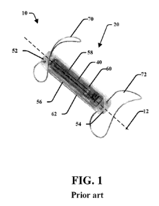
[0019] Figure 1 is a perspective view of a known implant;
[0020] Figure 2 is a photographic illustration of a sensor device or
implant and
an anchoring structure according to an embodiment of the present disclosure in
a state
ready for insertion into a patient and/or individual;
[0021] Figure 3A is a photographic illustration of an embodiment of a
sensor
device or implant and an anchoring structure attached thereto in a state where
the
anchoring structure is in its expanded state as would be the case once
placement
occurs in a desired blood vessel (e.g., a pulmonary blood vessel);
[0022] Figure 3B is a schematic illustration of an embodiment of a
sensor
device or implant and an anchoring structure attached thereto in a state where
the
anchoring structure is in an expanded state as would be the case once
placement
occurs in a desired blood vessel (e.g., a pulmonary blood vessel);
[0023] Figure 4 is a photographic illustration of a sensor device or
implant and
an anchoring structure attached thereto in a state where the anchoring
structure is in
an expanded state in a 14 mm blood vessel (e.g., a pulmonary blood vessel);
[0024] Figure 5A is an end view of an embodiment of a sensor device or
implant with an anchor assembly including a distal anchor having an elongated
and
angled orientation and a proximal anchor having three lobes in accordance with
the
present disclosure;
[0025] Figure 5B is a perspective view of the embodiment of the sensor
device or implant and anchor assembly of Figure 5A;
8
CA 03062861 2019-10-28
WO 2018/195430
PCT/US2018/028580
[0026] Figure 6A is a photographic illustration of the a sensor device
or
implant of Figures 5A and 5B positioned within a model of a pulmonary artery;
[0027] Figure 6B is a photographic illustration of the a sensor device
or
implant of Figures 5A and 5B positioned within a model of a pulmonary artery;
[0028] Figure 7A is a photographic illustration of the a sensor device
or
implant of Figures 5A and 5B positioned within a model of a pulmonary artery;
[0029] Figure 7B is a photographic illustration of the a sensor device
or
implant of Figures 5A and 5B positioned within a model of a pulmonary artery;
[0030] Figure 8 is a schematic illustration of an embodiment of the
present
disclosure where a sensor device or implant and an anchoring structure
attached
thereto are in a state where the anchoring structure is in an expanded state.
[0031] Figure 9 is a schematic illustration of various arteries of the
human
anatomy; and
[0032] Figure 10 is a schematic cross-sectional view of a sensor device
or
implant positioned within the body of a patient and in communication with a
reading
device.
DETAILED DESCRIPTION
[0033] The present disclosure relates to various anchoring systems for a
catheter delivered device. In one instance the anchoring systems of the
present
disclosure are designed to be used in connection with a pulmonary artery
implant
device. In one embodiment, an anchoring system of the present disclosure
comprises
two anchoring ends, a distal end anchoring structure and a proximal end
anchoring
structure, where at least one of the distal or proximal anchoring structures
has a
clover-shaped structure formed by at least three lobes. In another embodiment,
the
distal end anchoring structure has an elongated and angled orientation
relative the
implant body. In another embodiment, both the distal and proximal anchoring
structures have a clover-shaped structure formed by at least three lobes.
[0034] Figure 1 illustrates a prior art implant 10 that includes a
housing 20
that includes an oblong, narrow, rectangular shape that extends along a
housing axis
12, although the housing may have various shapes and geometry. The dimension
of
the housing 20 may be generally cuboid and may define a cavity therein. The
housing
side walls may be of specific dimensions and proportions to each other. For
example,
the housing may have four side walls 52, 54, 56, and 58, a top wall 60 and a
bottom
9
CA 03062861 2019-10-28
WO 2018/195430
PCT/US2018/028580
wall 62. The housing 20 may be made of a hermetic, strong, and biocompatible
material, such as ceramic. The examples illustrate a cuboid housing, but other
shapes
and configurations may be used, such as cylindrical housings, prism-shaped
housings,
octagonal or hexagonal cross-sectioned housings, or the like. A sensor 40 is
positioned along the top wall 60 and is attached to an antenna coil as well as
other
electronic components that may be positioned within the housing of the
implant. The
sensor 40 as well as the antenna coil and internal electronics may be
positioned along
a sensor axis 42 that extends generally normal relative to the implant 10
wherein the
sensor 40 along the top surface 60 may be exposed to blood flow and pressure
within
the vessel once positioned within a patient. A distal anchor 70 and a proximal
anchor
72 opposite the distal anchor may extend from the top surface of the implant
10. The
anchors may fixate the implant 10 in a desired position in the body of the
patient.
[0035] Figures 2-10 disclose various embodiments of an anchoring system
according to the present disclosure. Figure 2 depicts an embodiment of an
anchoring
assembly 100 of the present disclosure in a state ready for insertion into a
patient
and/or individual by, for example, a catheter in a minimally invasive
procedure.
[0036] As illustrated by the embodiments in Figures 3A-3B, the anchoring
assembly 100 comprises two anchoring ends, a distal anchoring structure 102
and a
proximal anchoring structure 104, where at least one of the distal or proximal
anchoring structures 102/104 has a clover-shaped structure formed by at least
smaller
two lobes 106 and 108 located on either side of a larger lobe 110. Located in
between
the distal anchoring structure 102 and the proximal anchoring structure 104 is
a
suitable implant sensor 112, such as the implant 10 illustrated by Figure 1.
Figure 4
depicts the present invention in a state where the anchoring structure is in
an
expanded state in a 14 mm blood vessel, e.g., a pulmonary blood vessel.
[0037] The distal anchoring structure 102 and the proximal anchoring
structure 104 may extend from a top surface 60 of the implant 10. Notably, the
top
surface 60 may include a sensor 40 as illustrated by Figure 1. Alternatively,
the
implant 10 may include an actuator such as one that may be selected from among
the
following: neurostimulation, cardiac pacing, electrical stimulation, drug
elution and
the embodiments of the implant and anchoring system is not limited as to the
type of
sensor that may be utilized. Furthermore, the anchoring structures may
comprise two
individual shape set nitinol wires. As illustrated by Figures 3A-3B, the two
wires
comprise a distal wire and a proximal wire, where one anchor wire 102 is
attached to
CA 03062861 2019-10-28
WO 2018/195430
PCT/US2018/028580
the distal portion of a top surface 60 (spade shape, see Figures 3A-3B) of the
implant
112 and the other anchor wire 104 is attached to the proximal portion of the
top
surface 60 (club shape, see Figures 3A-3B). Both anchors 102/104 can be
collapsed
down and attached to a delivery catheter via "release wires" or other
mechanism like a
shroud. The implant 112 and anchors 102/104 can be introduced into the human
vasculature in the collapsed position and expanded to place the implant within
a
desired location of a vessel.
[0038] Figures 5A-5B illustrate an embodiment of an anchor assembly 200
with a distal anchoring structure 202 and a proximal anchoring structure 204.
The
distal anchoring structure includes a wire shaped with an elongated and angled
orientation relative to the implant 212. The proximal anchoring structure 204
includes
a wire that is shaped as a clover-shaped structure formed by at least smaller
two lobes
206 and 208 located on either side of a larger lobe 210. Located in between
the distal
anchoring structure 202 and the proximal anchoring structure 204 is a suitable
implant
212 such as one illustrated by Figure 1.
[0039] The distal anchoring structure 202 and the proximal anchoring
structure 204 may extend from a top surface 60 of the implant 212. Notably,
the top
surface 60 may include a sensor 40 that is attached to an antenna coil within
the cavity
of the implant housing as illustrated by Figure 1. Furthermore, the anchoring
systems
of the present invention may comprise two individual shape set nitinol wires.
As
illustrated by Figures 5A-5B, the two wires comprise a distal wire and a
proximal
wire, where one anchor wire 202 is attached to the distal portion of a top
surface 60
(elongated and angled, see Figures 5A-5B) of the implant 212 and the other
anchor
wire 204 is attached to the proximal portion of the top surface 60 (club
shape, see
Figures 5A-5B). Both anchors 202/204 can be collapsed down and attached to a
delivery catheter via "release wires" or other mechanism. The implant 212 and
anchors 202/204 can be introduced into the human vasculature in the collapsed
position.
[0040] In the expanded position, the three-lobed proximal anchor 204 may
radially expand to abut the inner wall of the vessel. Lobes 206 and 208 may
expand
outwardly from the implant 212 while lobe 210 may extend upwardly from the
implant 212. These three lobes may radially abut against the inner wall of the
vessel
and may be arranged to abut against vessels of various sizes. The elongated
and
angled distal anchor 202 may include a slender configuration that may include
a base
11
CA 03062861 2019-10-28
WO 2018/195430
PCT/US2018/028580
portion 220 that may extend upwardly and slightly outwardly from the width of
the
implant 212 and an elongated portion 230 that may extend from the base portion
220
at an angle that includes a gradual taper until it ends at end portion 240.
The elongated
portion 230 may extend along elongated axis 232 wherein the elongated axis 232
may
be positioned angularly relative to the sensor axis 42 as identified in Figure
5A. The
elongated axis 232 may intersect the sensor axis 42 at angle A wherein angle A
may
be about 20 degrees to about 40 degrees, or more particularly may be about 30
degrees. The elongated portion 230 may be over twice the length of the base
portion
220. The slender elongated angle configuration may allow the distal anchor 202
to
extend within a branch vessel of the pulmonary artery ("PA") and may correctly
position the implant 212 to allow the sensor axis 42 to extend towards the
chest of a
patient. Further, the configuration of the anchors 202, 204 may be arranged to
allow
the catheter or other delivery device to deploy the implant 212 with enough
room to
allow the catheter to be removed without bumping or rubbing against the
implant 212
in which it may otherwise move or rotate the implant from its desired
position.
[0041] It has been found that the elongated and angled configuration of
the
distal anchoring structure 202 may provide various benefits which may allow
health
clinicians to deploy the implant at an exact location and orientation with a
reduced
risk of translation or rotation once deployed. In one embodiment, the implant
212 with
the distal anchor 202 may be placed in the right main trunk of the PA. As
such,
clinicians may be able to position the implant within the PA without having to
rely on
CT scans or quantitative angiography. Instead, in an embodiment, the clinician
may
reference the first apical branch of the right main trunk of the PA as an
anatomical
marker to identify where to position the implant 212 in which the elongated
distal
anchor 202 may be positioned. Figure 9 is a labeled sketch of the right
pulmonary
artery wherein the apical branch is positioned adjacent the superior trunk of
the right
PA. It should be noted that a wide variety of pulmonary artery anatomy exists
between patients. This includes differences in size and number of branches.
Despite
all this variation, the right PA main trunk has an anatomical feature that is
present in
nearly all patients in which the clinician may reference for implant
placement: a sharp
downturn from the right interlobar segment into the right posterior basal
segment, see
Figure 9.
[0042] The elongated and angled distal anchoring structure 202 may allow
the
implant 212 to self-correct its position within the vessel. As illustrated by
Figures 6A-
12
CA 03062861 2019-10-28
WO 2018/195430
PCT/US2018/028580
6B and 7A-7B, the distal anchor 202 may be positioned to extend deep into the
right
posterior basal segment branch of the PA while the proximal anchor 204 may be
positioned upstream in the interlobar PA segment. The distal anchor 202 may
contact
the vessel wall at or near the end portion 240 but it does not need to.
Further, it does
not need to contact along other radial positions along the base portion 220 or
near the
base portion 220 along the elongated portion 230, although in some patients it
may do
so. This may help prevent the implant from translating and rotating and allow
the
implant to be permanently located therein.
[0043] These anchors may allow for ease of implant placement as the
distal
anchor may be long enough so that when the catheter is removed, there is very
little
chance of migration into a side branch of the PA. The embodiment may also
provide
an anatomical landmark facilitating location of the target implant site, that
may be
easily identified by basic angiography and may allow a health clinician to
align the
implant such that is just distal from the superior trunk takeoff and proximal
to the
downturn of the PA. The disclosure may further prevent unwanted rotation due
to: the
spring force nature of the anchor, delivery system rubbing against the implant
during
removal, and patient coughing or other patient movement. The angle that the
posterior
basal makes with respect to the chest skin surface may ensure that the implant
assumes an angle towards the chest that is optimal for RF communication. The
angle
may ensure that the implant faces the chest surface when the distal anchor 202
is
placed into the posterior basal segment of the PA.
[0044] Further, if there is an unintentional deployment that is too
distally
positioned in the PA, the distal anchor 202 may still fit within the right
posterior basal
segment. If there is an unintentional deployment too proximally positioned,
the distal
anchor 202 may act to "pull" the implant 212 in the distal direction. In the
event that
the lobes of the proximal anchor 204 may migrate due to the spring force
action, the
downturn of the distal anchor 202 in the posterior basal segment may prevent
it from
translating as the elongated distal anchor will be generally prevented from
"turning
the corner" as the device moves proximally. Further, if there is migration of
the
implant 212 distally, the housing and proximal anchor 202 may form an angle
that
prohibits them from making the turn. As such, the implant 212 includes self-
adjusting
properties in this anatomical location within the pulmonary artery.
[0045] The two anchors may act to hold the bottom surface 62 of the
implant
212 against the vessel wall with the sensor 40 and top surface 60 away from
the vessel
13
CA 03062861 2019-10-28
WO 2018/195430
PCT/US2018/028580
wall. Because the posterior basal segment is relatively thin, the implant may
not sit
any other way. The proximal anchor may hold the implant body against the
vessel
wall by itself without help from the distal anchor 204. The distal anchor may
utilize
its length relative to the implant to prevent rotation, by staying in the
downturn.
Additionally, it may prevent unintended interactions with other branches of
the PA.
The distal anchor 202 may not include loops and may be too straight and long
to
migrate into side branches easily.
[0046] In another embodiment as is illustrated in Figure 8, an anchoring
system 300 comprises two anchoring ends, a distal end anchoring structure 302
and a
proximal end anchoring structure 304, where both the distal or proximal
anchoring
structures 302/304 have clover-shaped structures formed by at least two sets
of
smaller lobes 306 and 308 located on either side of a larger lobe 310. Located
in
between the distal end anchoring structure 302 and the proximal end anchoring
structure 304 is a suitable implant sensor 312.
[0047] The anchoring structures of Figures 3A-3B, 5A-5B, and 8 are
illustrated in the expanded position and it is understood that the anchors may
be
positioned in a collapsed or retracted position when attached to a catheter or
other
type of delivery device, such as that shown in Figure 2. Notably, other anchor
configurations and shapes may be implemented, including a different number of
anchors (other than two); different locations of anchor attachment to the
housing;
anchors which attach to the housing at one point, or more than two points;
anchors
that extend under the housing, around it, or laterally to the sides. The
anchors may be
formed as loops which anchor the implant to body structures or within a vessel
using
spring force. The anchors may be made of nitinol, stainless steel, polymer, or
any
material which is biocompatible and extrudable. The anchors may be made of a
combination of materials, such as nitinol with a platinum core. The anchors
may be
configured to fold down during the implantation procedure to allow easy
ingress to
the deployment location. The anchors may be configured to be tied down to a
delivery
system, such as a catheter, for minimally invasive ingress to the implant
deployment
site. The anchors may be designed to deploy from their tied-down configuration
to
their open configuration when an operator actuates a control on the proximal
end of
the delivery system. The control may include release wires that are pulled
from the
proximal end either directly or with help from a mechanical handle. The
anchors may
be coated with a material to increase lubricity.
14
CA 03062861 2019-10-28
WO 2018/195430
PCT/US2018/028580
[0048] The anchors may be positioned within the vessel at a desired
location
and caused to expand in the illustrated expanded positions as illustrated in
Figures 4,
6A-B, and 7A-B. In these embodiments, as illustrated by Figure 10, it may be
desirable to position the implant 112, 212 within a vessel 540 with the top
surface 60
and the sensor 40 aligned along the sensor axis 42 directed through the chest
510 of
the patient and wherein the top surface 60 may be spaced from an inner wall
542 of
the vessel 540. The configuration of the disclosed anchors may make this
position
possible as the aligned direction would allow the patient to utilize a reader
device 530
to be positioned on or near the chest in proximity to the implant while also
being
directionally aligned with the top surface 60 and sensor 40. Figure 10
schematically
illustrates a cross sectional view of an implant 112, 212 positioned within a
body 500
of a patient wherein the top surface 60 and sensor 40 thereon may be directed
towards
the center of a blood vessel 540. The implant 112, 212 may be located on the
side of
vessel 540 such that its distance from the wall of the chest 510, and hence
from
external reader 530, is minimized. The sensor 40 on the top surface 60 may be
aligned
along the sensor axis 42 that extends through the chest 510 and a user may
allow the
reader device 530 to be placed in alignment with the sensor axis 42 to
wirelessly
communicate with the implant through the chest 510 of the patient. The
configuration
of the anchor assemblies may allow for the implant to be placed in the desired
location, so that a patient having the implant may be able to hold reader
device 530
and take his own readings from the implant without the assistance of others.
The
above embodiment may also be applied to the implant of Figure 8.
[0049] During the deployment of the implant 100/200/300, the anchors
202,
204 may be deployed sequentially when the release wires are retracted. Once an
anchor is free and/or fully released, the anchor may utilize nitinol's super
elastic
property and instantly attempt to return to its initial shape set shape within
the vessel.
The distal anchor 202 may deploy first, pushing the distal end of the implant
straight
off the delivery catheter and onto the target position along the vessel wall.
Next the
proximal anchor 204 may deploy, pushing down the proximal end of the sensor
body
(the 'implant') 212 along the vessel wall target and engaging the two side
lobes.
Although stated in terms of implant 200, the above may be applied to any of
the
embodiments described herein, including implants 100 and 300.
[0050] Furthermore, the anchoring systems of the present invention
comprise
two individual shape set nitinol wires. As discussed above, the two wires
comprise a
CA 03062861 2019-10-28
WO 2018/195430
PCT/US2018/028580
distal wire and a proximal wire, where one anchor wire 102 is attached to the
distal
end (spade) of the implant 112 and the other anchor wire 104 is attached to
the
proximal end (club). Both anchors 102/104 can be collapsed down and attached
to a
delivery catheter via "release wires." The implant sensor 112 and anchors
102/104 can
be introduced into the human vasculature through a 14 Fr introducer. The
anchors
102/104 are deployed sequentially when the release wires are retracted. Once
an
anchor is free and/or fully released, the anchor utilizes nitinol's super
elastic property
and instantly attempts to return to the initial shape set shape within the
vessel. The
distal anchor deploys first, pushing the distal end of the implant straight
off the
delivery catheter and onto the target position along the vessel wall. Next the
proximal
anchor deploys pushing down the proximal end of the sensor body along the
vessel
wall target and engaging the two side lobes which provide the most radial
force and
the largest deterrent to proximal migration and rotation. Although stated in
terms of
implant 100, the above may be applied to any of the embodiments described
herein,
including implants 200 and 300.
[0051] In one embodiment, the overall implant and anchoring structures
are
sized such that the anchoring system allows the implant to be placed in a
proximal
segment of the pulmonary artery. The proximal placement allows communication
with device to occur from the chest instead of the back. The anchoring system
of the
present invention is designed to keep maximum vessel contact and remain stable
over
a large range of vessel sizes as compared to other devices known to those of
skill in
the art. The anchoring system of the present disclosure is designed to
withstand any
forces imposed by the retraction of or contact with the delivery catheter
which is a
well-documented procedure risk for devices designed with anchoring system
failing to
possess the various physical structures of the present disclosure. For
example, if the
insertion catheter snags the tip of the proximal anchor, the forces provided
by the
proximal anchor lobes increases to mitigate proximal movement.
[0052] As would be apparent to those of skill in the art, the use of the
labels
proximal and distal are for convenience sake and could be interchanged such
that in
the embodiment of Figures 3A-B, the distal end of the anchoring system 100
would
have the clover-shaped structure formed by at least three lobes 106, 108 and
110.
Such a change in orientation could be dictated by the environment and/or blood
vessel
in which the anchoring system and sensor device of the present disclosure are
to be
16
CA 03062861 2019-10-28
WO 2018/195430
PCT/US2018/028580
implanted in. The same may be applied to any of the embodiments described
herein,
including implants in Figures 5A-B and Figure 8.
[0053] Regarding the nitinol wires utilized in the embodiments of the
present
disclosure, such wires are well known in the art and as such a detailed
discussion
herein is omitted for the sake of brevity. However, as is known to those of
skill in the
art, nitinol is formed from at least one nitinol alloys, where such alloys
exhibit two
closely related and unique properties: shape memory effect (SME) and
superelasticity
(SE; also called pseudoelasticity, PE). Shape memory is the ability of nitinol
to
undergo deformation at one temperature and then recover its original, un-
deformed
shape upon heating above its "transformation temperature". Superelasticity
occurs at a
narrow temperature range just above its transformation temperature; in this
case, no
heating is necessary to cause the un-deformed shape to recover, and the
material
exhibits enormous elasticity, some 10 to 30 times that of ordinary metal.
Given
nitinol's biocompatibility it is well suited for use in biomedical devices
and/or
implants. Regarding the relationship between smaller lobes 106/108 and 206/208
and
larger lobe 110 and 210 of the multi-lobed anchoring structures of the present
disclosure, it should be noted that the larger lobe should have an overall
length of at
least 200 percent the length of the smaller lobes.
[0054] While in accordance with the patent statutes the best mode and
certain
embodiments of the disclosure have been set forth, the scope of the disclosure
is not
limited thereto, but rather by the scope of the attached. As such, other
variants within
the spirit and scope of this disclosure are possible and will present
themselves to those
skilled in the art.
17