Note: Descriptions are shown in the official language in which they were submitted.
CA 03063928 2019-11-15
WO 2018/213600
PCT/US2018/033217
- 1 -
SELF-RIGHTING SYSTEMS AND RELATED COMPONENTS AND METHODS
RELATED APPLICATIONS
This Application claims priority under 35 U.S.C. 119(e) to U.S. Provisional
.. Application Serial No. 62/507,647, entitled "SELF-RIGHTING ARTICLES" filed
on May
17, 2017, to U.S. Provisional Application Serial No. 62/507,653, entitled
"SELF-
ACTUATING ARTICLES" filed on May 17, 2017, and to U.S. Provisional Application
Serial No. 62/507,665, entitled "COMPONENTS WITH HIGH API LOADING" filed on
May 17, 2017, each of which is herein incorporated by reference in its
entirety.
FIELD
The present invention generally relates to self-righting systems and related
components such as self-righting articles, self-actuating articles including,
for example, self-
actuating needles and/or self-actuating biopsy punches, as well as components
with relatively
high loading of active pharmaceutical ingredients (API).
BACKGROUND
The GI tract offers an incredible opportunity for diagnosing and treating
patients. The
development of smart dosage systems and articles to enable this has witnessed
significant
.. growth over the preceding decade. One of the most significant challenges in
maximizing
delivery and interaction with the mucosa is ensuring juxtaposition between an
article and/or
dosing system and the GI mucosa. Prior attempts at doing this have included
the introduction
of mucoadhesives as well as texturing of one side of a 2 sided system. Orally
ingested drugs
generally diffuse through the GI tract tissue walls in order to enter the
blood stream. Typical
.. ingested pills or articles release their cargo into the GI tract randomly
and allow it move via
convection and diffusion to the tissue wall. However, many biologic drugs such
as insulin
cannot move through the liquid in the GI tract because they will be, for
example, degraded by
enzymes, even if housed in a solid formulation.
Additionally, many pharmaceutical drug formulations on the market require
administration via in injection, including numerous vaccines, RNA, and
peptides. Injections
traditionally involve the use of a liquid formulation passing through a hollow
needle and
CA 03063928 2019-11-15
WO 2018/213600
PCT/US2018/033217
- 2 -
entering into the body intravenously or intramuscularly. However, these liquid
formulations
can cause the active pharmaceutical ingredient (API) to become unstable and
thus may
require refrigeration and/or increase the bulk of the dose significantly
because of the required
dilution.
Accordingly, improved systems, articles and methods are needed.
SUMMARY
The present invention generally relates to self-righting articles, such as
self-righting
capsules.
In one aspect, self-righting articles are provided. In some embodiments, the
self-
righting article comprises a first portion, a second portion adjacent the
first portion having a
different average density than the first portion, and a hollow portion,
wherein the self-righting
article is configured and arranged to be encapsulated in a 000 capsule, or
smaller.
In some embodiments, although the self-righting article is configured for
potential
encapsulation in a 000 capsule, or smaller, the self-righting article does not
necessarily need
to be encapsulated in such capsule. In embodiments wherein the self-righting
article is to be
administered, such as by ingesting the self-righting article, the self-
righting article may thus
be administered without encapsulation.
In some embodiments, the self-righting article comprises a first portion, a
second
portion adjacent the first portion having a different average density than the
first portion, and
a tissue-interfacing component associated with the self-righting article,
wherein a ratio of an
average density of the first material to an average density of the second
material is greater
than or equal to 2.5:1. In some embodiments, the ratio of an average density
of the second
material to an average density of the first material is greater than or equal
to 2.5:1.
In some embodiments, the self-righting article is configured to anchor at a
location
internal to a subject and comprises at least a first portion having an average
density greater
than 1 g/cm3 wherein a longitudinal axis perpendicular to a tissue-engaging
surface of the
article is configured to maintain an orientation of 20 degrees or less from
vertical when acted
on by 0.09 *10^-4 Nm or less externally applied torque and at least one
anchoring mechanism
associated with the self-righting article.
CA 03063928 2019-11-15
WO 2018/213600
PCT/US2018/033217
- 3 -
In some embodiments, the self-righting article is configured for
administration to a
location internal to a subject and comprises at least a first portion having
an average density
greater than 1 g/cm3, the self-righting article has a self-righting time from
90 degrees in water
of less than or equal to 0.05 second, at least two tissue interfacing
components comprising a
tissue-contacting portion configured for contacting tissue, each tissue-
contacting portion
comprising an electrically-conductive portion configured for electrical
communication with
tissue and an insulative portion configured to not be in electrical
communication with tissue,
and a power source in electric communication with the at least two tissue
interfacing
components.
In another aspect, self-actuating articles are provided. In some embodiments,
the
article comprises an outer shell, a spring at least partially encapsulated
within the outer shell,
a support material associated with the spring such that the support material
maintains at least
a portion of the spring under at least 5% compressive strain under ambient
conditions and a
tissue interfacing component associated with the spring.
In some embodiments, the article is configured to anchor at a location
internal to a
subject and comprises an outer shell, a spring at least partially encapsulated
with the outer
shell, the spring maintained in an at least partially compressed state by a
support material
under at least 5% compressive strain, and at least one anchoring mechanism
operably linked
to the spring.
In some embodiments, the article is configured for administration to at a
location
internal to a subject and comprises an outer shell, a spring at least
partially encapsulated with
the outer shell, the spring maintained in an at least partially compressed
state by a support
material under at least 5% compressive strain, at least two tissue interfacing
components
comprising a tissue-contacting portion configured for contacting tissue, each
tissue-
contacting portion comprising an electrically-conductive portion configured
for electrical
communication with tissue and an insulative portion configured to not be in
electrical
communication with tissue, and a power source in electric communication with
the at least
two tissue interfacing components.
In another aspect, tissue-interfacing components are provided. In some
embodiments,
the component comprises a solid therapeutic agent and a support material,
wherein the solid
CA 03063928 2019-11-15
WO 2018/213600
PCT/US2018/033217
- 4 -
therapeutic agent is present in the tissue interfacing component in an amount
of greater than
or equal to 10 wt% as a function of the total weight of the tissue interfacing
component,
wherein the solid therapeutic agent and support material are distributed
substantially
homogeneously, and wherein the tissue interfacing component is configured to
penetrate
tissue.
In some embodiments, the component has a tip and comprises a solid therapeutic
agent and a support material associated with the solid therapeutic agent,
wherein at least a
portion of the solid therapeutic agent is associated with one or more tips of
the tissue
interfacing component, and wherein the solid therapeutic agent is present in
the tissue
interfacing component in an amount of greater than or equal to 10 wt% as a
function of the
total weight of the tissue interfacing component.
In another aspect, methods are provided. In some embodiments, the method
comprises administering, to a subject, a capsule comprising an outer shell and
a self-righting
article, the self-righting article comprising, a first portion, and a second
portion adjacent the
first portion and having an average density different than the first portion.
In some embodiments, the method comprises administering, to the subject, a
capsule
comprising an outer shell and a self-righting article, the self-righting
article comprising, a
first portion comprising a first material, a second portion adjacent the first
portion and
comprising a second material, different than the first material, and a needle
associated with an
active pharmaceutical agent, wherein a ratio of an average density of the
first material to an
average density of the second material is greater than or equal to 2.5:1,
orienting the self-
righting article at the location internal of a subject such that the needle
punctures a tissue
proximate the location internal of the subject, and releasing at least a
portion of the active
pharmaceutical agent into the tissue.
In some embodiments, the method comprises administering, to a subject, an
article,
the article comprising an outer shell, a spring at least partially
encapsulated with the outer
shell, a support material associated with the spring such that the support
material maintains at
least a portion of the spring under at least 5% compressive strain under
ambient conditions
and a tissue interfacing component associated with the spring.
CA 03063928 2019-11-15
WO 2018/213600
PCT/US2018/033217
- 5 -
In some embodiments, the method comprises administering, to a subject, an
article,
the article comprising an outer shell, a spring at least partially
encapsulated with the outer
shell, a support material associated with the spring such that the support
material maintains at
least a portion of the spring under at least 5% compressive strain under
ambient conditions;
and a tissue interfacing component associated with the spring, and degrading
at least a
portion of the support material such that the spring extends and/or the tissue
interfacing
component penetrates a tissue located internal to the subject.
In some embodiments, the method comprises administering, to the subject, the
article,
wherein the article comprises at least a first portion having an average
density greater than 1
g/cm3 and at least one anchoring mechanism, the article configured to be
retained at the
location under greater than or equal to 0.6 N of force and/or a change in
orientation of greater
than or equal to 30 degrees.
In some embodiments, the method comprises administering, to the subject, an
article
comprising at least one tissue interfacing component disposed within the
article, each tissue
interfacing component comprising a conductive material, releasing the at least
one interfacing
component from the article, inserting the at least one interfacing component
into a tissue at
the location internal to the subject, applying a current generated by a power
source in
electrical communication with the tissue interfacing components across the two
or more
tissue interfacing components, wherein the article comprises a spring
maintained in an at least
partially compressed state by a support material under at least 5% compressive
strain, each
tissue interfacing component operably linked to the spring.
In another aspect, methods of forming tissue interfacing components are
provided. In
some embodiments, the method comprises providing a solid therapeutic agent and
a support
material and compressing, using at least 1 MPa of pressure, and/or heating the
solid
therapeutic agent and a support material together to form the tissue
interfacing component,
wherein the tissue interfacing component is configured to penetrate tissue.
In another aspect, self-righting articles are provided. In some embodiments,
the
article comprises a first portion having a mass, a second portion having a
mass different than
the mass of the first portion; a self-actuating component comprising a spring
and a support
material adapted to maintain the spring in at least a partially compressed
state, wherein the
CA 03063928 2019-11-15
WO 2018/213600
PCT/US2018/033217
- 6 -
support material is configured for at least partial degradation in a
biological fluid; a tissue
interfacing component associated with an active pharmaceutical agent and
operably linked to
the self-actuating component; and a tissue engaging surface configured to
contact a surface of
a tissue internal to a subject; wherein the self-righting article is
configured as a monostatic
body due to the center of mass of the self-righting article and the shape of
the self-righting
article; wherein when the self-righting article is at least partially
supported by the tissue of
the subject, the self-righting article orients in a direction to allow the
tissue interfacing
component to release at least a portion of the active pharmaceutical agent
into the tissue.
In some embodiments the article is so configured that upon said at least
partial degradation of
the support material, the spring expands to release said portion of the active
pharmaceutical
agent into the tissue. In some embodiments, the expansion of the spring forces
the
pharmaceutical agent into the tissue.
In some embodiments, the first portion comprises a first material and the
second portion
comprises a second material, wherein the first material and the second
material are different.
In some embodiments, the first portion comprises a first material and the
second portion
comprises a second material, wherein the first material and the second
material are the same.
In some embodiments, the self-righting article has an average density greater
than 1 g/cm3.
In some embodiments, the first material and/or second material is selected
from the group
consisting of a polymer, a ceramic, a metal, a metal alloy, and combinations
thereof.
In some embodiments ,the metal is selected from the group consisting of
stainless steel, iron-
carbon alloys, Field's metal, wolfram, molybdemum, gold, zinc, iron, and
titanium. In some
embodiments, the ceramic is selected from the group consisting of
hydroxyapatite, aluminum
oxide, calcium oxide, tricalcium phosphate, zirconium oxide, silicates, and
silicon dioxide. In
some embodiments, the polymer is selected from the group consisting of
polycaprolactone,
polylactic acid, polyethylene glycol, polypropylene, polyethylene,
polycarbonate,
polystyrene, polyether ether ketone, and polyvinyl alcohol.
In some embodiments, the spring comprises a spring constant in the range of
100 N/m to
1500 N/m.
CA 03063928 2019-11-15
WO 2018/213600
PCT/US2018/033217
- 7 -
In some embodiments the support material is configured as a plug, wherein the
plug
is operably linked to the tissue interfacing component, and wherein the plug
is exposed to the
exterior of the self-righting article via a hole in the tissue engaging
surface.
In some embodiments the self-righting article is provided, wherein the spring
is positioned in
a space surrounded by the first portion, wherein the tissue interfacing
component is
configured as a projectile that extends substantially along the major axis of
the self-righting
article; wherein the tissue interfacing component is operably linked to the
spring at one end
and operably linked to the plug at the other end, and wherein the plug is
located in a space
surrounded by the second portion and configured such that the second portion
prevents the
spring in at least a partially compressed state from pushing the plug out of
the hole in the
tissue engaging surface via the tissue interfacing component.
In some embodiments, the support material is configured in the shape of a flat
structure with a major plane and operably linked to the spring, and wherein
the major plane
of the flat structure is perpendicular to the major axis of the spring. In
some embodiments the
support material comprises a first surface along the major plane and having a
first total
surface area, wherein the support material comprises a second surface parallel
to the first
surface along the major plane and having a second total surface area different
from the first
total surface area, wherein the first surface comprises one or more cavities,
and wherein the
first total surface area is greater than the second total surface area.
In some embodiments, the support material is configured within the self-
righting article such
that the biological fluid entering the self-righting article contacts the
first surface to initiate
the at least partial degradation of the support material; and wherein the one
or more cavities is
configured for controlled failure of the support material after the at least
partial degradation
of the support material.
In some embodiments, the spring is positioned in a space surrounded by the
first portion;
wherein the support material is positioned between the first portion and the
second portion;
wherein the support material comprises a hole through which the tissue
interfacing
component extends substantially along the major axis of the self-righting
article; wherein the
tissue interfacing component is configured in the shape of a projectile such
that one end of
CA 03063928 2019-11-15
WO 2018/213600
PCT/US2018/033217
- 8 -
the projectile is operably linked to the spring and the other end of the
projectile is located
proximate to a hole in the tissue engaging surface such that a distance exists
between the
projectile and the hole; and wherein the tissue engaging surface is on the
second portion.
In some embodiments, the one or more cavities surround the hole in the support
material.
In some embodiments, the support material is configured in the shape of a
disk.
In some embodiments, the support material is selected from the group
consisting of a sugar, a
derivative of a sugar, starch, calcium carbonate, zinc, sodium chloride,
polymers, and
combinations thereof.
In some embodiments, the tissue interfacing component comprises the active
pharmaceutical agent. In some embodiments, the active pharmaceutical agent is
present in the
tissue interacting component in an amount greater than or equal to 80 wt% of
the total weight
of the tissue interfacing component. In some embodiments, 100 wt% of the
tissue interacting
component is the active pharmaceutical agent.
In some embodiments, the self-righting article comprises one or more vents
configured such that the self-actuating component is in fluidic communication
with an
external environment. In some embodiments, the one or more vents are located
in the first
portion. In some embodiments, the one or more vents are covered by a coating.
In some embodiments, the biological fluid is gastric fluid.
Other advantages and novel features of the present invention will become
apparent
from the following detailed description of various non-limiting embodiments of
the invention
when considered in conjunction with the accompanying figures. In cases where
the present
specification and a document Incorporated by reference include conflicting
and/or
inconsistent disclosure, the present specification shall control.
BRIEF DESCRIPTION OF THE DRAWINGS
Non-limiting embodiments of the present invention will be described by way of
example with reference to the accompanying figures, which are schematic and
are not
intended to be drawn to scale. In the figures, each identical or nearly
identical component
illustrated is typically represented by a single numeral. For purposes of
clarity, not every
component is labeled in every figure, nor is every component of each
embodiment of the
CA 03063928 2019-11-15
WO 2018/213600
PCT/US2018/033217
- 9 -
invention shown where illustration is not necessary to allow those of ordinary
skill in the art
to understand the invention. In the figures:
FIG. 1 is a schematic diagram of a self-righting system, according to one set
of
embodiments;
FIG. 2 is a cross-sectional schematic diagram of an exemplary self-righting
system,
according to one set of embodiments;
FIG. 3 is a schematic illustration of administration of a self-righting
system, according
to one set of embodiments;
FIG. 4 is a schematic diagram of an exemplary self-righting article, according
to one
set of embodiments;
FIG. 5 is a cross-sectional schematic diagram of an exemplary self-righting
system,
according to one set of embodiments;
FIG. 6 is a cross-sectional schematic diagram of an exemplary self-actuating
component, according to one set of embodiments;
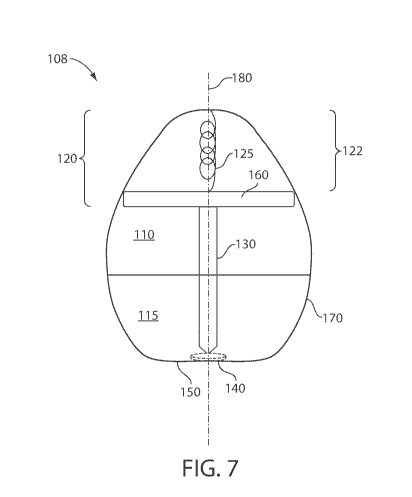
FIG. 7 is a cross-sectional schematic diagram of an exemplary self-righting
system,
according to one set of embodiments;
FIG. 8 is a schematic illustration of a support material, according to one set
of
embodiments;
FIG. 9 is a schematic illustration of a support material, according to one set
of
embodiments;
FIG. 10 is a schematic diagram of a self-righting system, according to one set
of
embodiments;
FIG. 11 is a cross-sectional schematic diagram of an exemplary self-righting
system,
according to one set of embodiments;
FIG. 12 is a plot of an exemplary self-righting shape graph, according to one
set of
embodiments;
FIG. 13 is a photograph of an exemplary self-righting article inside a 000
capsule,
according to one set of embodiments;
FIG. 14 is a plot of self-righting article speed of righting testing via
computer models
(predicted), according to one set of embodiments;
CA 03063928 2019-11-15
WO 2018/213600
PCT/US2018/033217
- 10 -
FIG. 15 is a plot of self-righting article speed of righting via high speed
camera
analysis (poly), according to one set of embodiments;
FIG. 16 is a plot of self-righting article speed of righting via high speed
camera
analysis (poly), according to one set of embodiments;
FIG. 17 is a photograph of an exemplary self-righting article, according to
one set of
embodiments;
FIG. 18 is a series of x-ray images of an exemplary self-righting article at
0, 45, and
90 degrees of orientation compared to a control (washer), according to one set
of
embodiments;
FIG.19 is an x-ray photograph of an exemplary series of self-righting articles
in the
GI of a pig, according to one set of embodiments
FIG. 20 is an endoscopy of an exemplary self-righting article in the GI of a
pig,
according to one set of embodiments;
FIG. 21 is a plot of the fraction of articles righted, according to one set of
embodiments;
FIG. 22 is a plot of maximum tilt versus shape, according to one set of
embodiments;
FIG. 23 is a photograph of a maximum tilt testing apparatus, according to one
set of
embodiments;
FIG. 24 is a photograph of an exemplary self-righting article comprising
air/water
vents, according to one set of embodiments;
FIG. 25 is a photograph of an exemplary self-righting article comprising a
magnetic
portion, attached to a magnetic object, according to one set of embodiments.
FIG. 26 is a schematic illustration of a self-actuating article, according to
one set of
embodiments;
FIG. 27 is a schematic of an exemplary self-actuating article, according to
one set of
embodiments, a photograph of the article in vivo, and a photograph of the
article as compared
to an uncompressed spring, according to one set of embodiments;
FIG. 28 is a plot of force versus displacement for various spring constants,
according
to one set of embodiments;
CA 03063928 2019-11-15
WO 2018/213600
PCT/US2018/033217
- 11 -
FIG. 29 is a plot of diameter versus time for sugar dissolution, according to
one set of
embodiments;
FIG. 30 is a plot of spring actuation time versus diameter, according to one
set of
embodiments;
FIG. 31 is a photograph and diagram of an exemplary tissue interfacing
component
(e.g., biopsy punch) associated with a spring, according to one set of
embodiments;
FIG. 32 is a histology of a needle inserted into tissue in vitro from a spring
associated
article, reaching the muscle layer of the stomach tissue, according to one set
of embodiments.
FIG. 33 is a schematic illustration of a tissue interfacing component,
according to one
set of embodiments;
FIG. 34 is photograph of an in plane needle made with 80% BSA and 20% PEG 200k
w/w exposed to 3 metric tons of pressure at 100 C for 2 min, according to one
set of
embodiments;
FIG. 35 is a photograph of an in plane needle made with 80% Human Insulin and
20% PEG 200k w/w exposed to 3 metric tons of pressure at 100 C for 2 min,
according to
one set of embodiments;
FIG. 36 is a photograph of an in plane needle, the made with 80% Human Insulin
and
20% PEG 200k w/w exposed to 2 metric tons of pressure, tips are created by dip
coating in
maltose, according to one set of embodiments;
FIG. 37 is a plot of insulin release versus time for components having a
relatively
high loading of API, according to one set of embodiments;
FIG. 38 is a plot of load versus extension (lateral load) for various
components having
a relatively high loading of API, according to one set of embodiments;
FIG.39 is a plot of load versus extension (axial load) for various components
having a
relatively high loading of API, according to one set of embodiments;
FIG. 40 is a plot of penetration force versus insertion depth for an exemplary
component having a relatively high loading of API loading as compared to a 32
gauge
stainless needle, according to one set of embodiments;
FIG. 41 is a photograph of a component having a relatively high API loading
(e.g.,
needle protrusion on a base plate) made with 83% Human Insulin, 5% HPMC, 2%
CA 03063928 2019-11-15
WO 2018/213600
PCT/US2018/033217
- 12 -
Magnesium Stearate and 10% PEG 35k w/w exposed to 3 metric tons of pressure at
100 C
for 2 min, according to one set of embodiments;
FIG. 42 is a schematic diagram of a method for fabricating a component having
a
relatively high API loading (e.g., needles) with a non API base plate, the
needle protrusion on
a base plate made with 85% Human Insulin, 5% HPMC and 10% PEG 35k w/w exposed
to 3
metric tons of pressure at 100 C for 2 min, according to one set of
embodiments;
FIG. 43 is a schematic diagram of a method for fabricating a component (e.g.,
a
needle tip) having a relatively high API loading with a non API base plate and
needle base. A
needle protrusion on a base plate made with 85% Human Insulin, 5% HPMC and 10%
PEG
.. 35k w/w exposed to 3 metric tons of pressure at 100 C for 2 min, according
to one set of
embodiments;
FIG. 44 is a plot of axial loading of a component having a relatively high API
loading
(e.g., a microneedle), according to one set of embodiments;
FIG. 45A is a photograph of an exemplary tissue-interfacing component
comprising
95 wt% API, according to one set of embodiments;
FIGs. 45B-45C are compression tests of the tissue-interfacing component in
FIG.
11A;
FIG. 45D is a plot of percent insulin recovery versus temperature for a tissue-
interfacing component, according to one set of embodiments;
FIG. 45E is a plot of percent insulin dimer formation versus temperature,
according to
one set of embodiments;
FIG. 46 is a schematic diagram of an exemplary method for fabricating a
component
having a plurality of microneedles and a relatively high API loading,
according to one set of
embodiments;
FIGs. 47A-47B are confocal microscopy images of exemplary components loading
with FITC-dextran, according to one set of embodiments;
FIG. 48 shows the dissolution of a tissue interfacing component comprising a
plurality of microneedles and a relatively high loading of API after
administration to various
tissues, according to one set of embodiments;
CA 03063928 2019-11-15
WO 2018/213600
PCT/US2018/033217
- 13 -
FIG. 49 shows the dissolution of a tissue interfacing component comprising a
plurality of microneedles and a relatively high loading of API after
administration to human
cheek tissue ex vivo, according to one set of embodiments;
FIG. 50 is a plot of blood concentration of insulin versus time after
application of a
tissue interfacing component comprising a plurality of microneedles and a
relatively high
loading of insulin to the small intestine of swine, according to one set of
embodiments;
FIG. 51 is a plot of blood concentration of insulin versus time after
application of a
tissue interfacing component comprising a plurality of microneedles and a
relatively high
loading of insulin to the palatal tissue of swine, according to one set of
embodiments;
FIG. 52 is a plot of blood concentration of human growth hormone versus time
after
application of a tissue interfacing component comprising a plurality of
microneedles and a
relatively high loading of human growth hormone to the lip of swine, according
to one set of
embodiments;
FIG. 53 is a plot of blood concentration of human growth hormone versus time
after
application of a tissue interfacing component comprising a plurality of
microneedles and a
relatively high loading of human growth hormone to the palatal tissue of
swine, according to
one set of embodiments;
FIG. 54 is a plot of blood concentration of human growth hormone versus time
after
application of a tissue interfacing component comprising a plurality of
microneedles and a
relatively high loading of human growth hormone to the lip of swine, according
to one set of
embodiments;
FIG. 55 is a plot of activity of adalimumab before and after exposure to
relative high
pressure and relative high temperature, according to one set of embodiments;
FIG. 56 is a schematic diagram of the self-righting system that is used for
tissue
localization and ejecting a hooked micropost (i.e. hook). An example of a
hooked 32-gauge
stainless steel needle is shown on the left, according to one set of
embodiments;
FIG. 57 is a plot of penetration force into swine gastric tissue using hooked
microposts, according to one set of embodiments;
FIG. 58 is a plot of hooking force based on penetration of swine stomach
tissue using
hooked microposts, according to one set of embodiments;
CA 03063928 2019-11-15
WO 2018/213600
PCT/US2018/033217
- 14 -
FIG. 59 is a photograph of a hooked micropost that has attached itself to the
muscle
fibers of swine stomach tissue;
FIG. 60 is a plot of hooking force based on penetration of human stomach
tissue using
hooked microposts, according to one set of embodiments;
FIG. 61 is a plot of hooking force based on penetration of swine small
intestinal tissue
using hooked microposts, according to one set of embodiments;
FIG. 62 is a plot of pullup height based on penetration of swine small
intestinal tissue
using hooked microposts, according to one set of embodiments;
FIG. 63 is a photograph of a hooked micropost that has attached itself to
swine small
intestinal tissue, according to one set of embodiments;
FIG. 64 is a schematic diagram of a model of horizontal tissue retention test.
A probe
presses down on a device anchored to the tissue via needles and records the
force required to
dislodge the device, according to one set of embodiments;
FIG. 65 is a plot of the force required to dislodge a self-righting system and
increases
linearly with the number of needles inserted into the swine gastric tissue,
according to one set
of embodiments;
FIG. 66 is a plot of the force required to dislodge a self-righting system
from swine
stomach tissue versus needle distance, according to one set of embodiments;
FIG. 67 is a schematic diagram demonstrating design of in-vitro experiment
where
self-orienting devices are anchored to swine stomach tissue while experiencing
pulsatile flow,
according to one set of embodiments;
FIG. 68 is a plot demonstrating that the three devices with hooked microposts
retained
their position for an entire week, as opposed to comparative systems that were
dislodged in
under two days, according to one set of embodiments;
FIG. 69 is a plot of anchoring force versus in-vivo and ex-vivo swine
stomachs. The
ex-vivo measurement reflects studies using three separate tissue samples from
different
stomachs, according to one set of embodiments;
FIG. 70A is a plot demonstrating in-vivo using a swine model that as an
anchored
self-orienting device encounters a force that is parallel to the stomach
tissue, it can retain its
position while being rotated up to 30 degrees and experiencing between 0.5N-
0.75N of force
CA 03063928 2019-11-15
WO 2018/213600
PCT/US2018/033217
- 15 -
(the peaks and valleys correspond to the animal's breathing), according to one
set of
embodiments;
FIG. 70B is a plot showing the relationship between the number of ancillary
bodies
attached to the self righting device and the drag torque exerted on the system
by the gastric
acid, according to one set of embodiments;
FIG. 70C is a plot comparing the size of the food boluses colliding with the
self
righting device and the torque exerted on it, according to one set of
embodiments;
FIG. 71 is a schematic diagram demonstrating how parylene-coated electrical
probes
may bypass the mucus and conduct electricity through the tissue (e.g., without
the coating,
the electricity would flow through the lower resistance mucus and not
stimulate the tissue),
according to one set of embodiments;
FIG. 72 is a schematic diagram demonstrating an electrical stimulation pill,
including
the self-orienting device containing two probes, as well as an electrical
power source and a
programmable microcontroller that are encapsulated in an insulating shell
(e.g. PDMS),
according to one set of embodiments;
FIG. 73 is a plot demonstrating that current does not significantly change as
the radius
increases of the tissue-stimulating, electrical probes when powered by two
silver oxide
batteries (1.55V, 6.8mm coin cell), according to one set of embodiments;
FIG. 74 is a plot demonstrating that current decreases as the distance
increases
between tissue-stimulating, electrical probes when powered by two silver oxide
batteries
(1.55V, 6.8mm coin cell), according to one set of embodiments;
FIGs. 75A-75B are plots showing electrical probes, powered by a voltage
generator,
provide pulsatile stimulation through the tissue, as measured by an
oscilloscope (FIG. 75A)
which can be compared to the background voltage measured within the tissue
(FIG. 75B),
according to one set of embodiments;
FIGs. 76A-76E are schematic illustrations of an exemplary assembly process for
the
system, according to one set of embodiments;
FIG. 77 is a schematic illustration of an exemplary system, according to one
set of
embodiments;
CA 03063928 2019-11-15
WO 2018/213600
PCT/US2018/033217
- 16 -
FIG. 78 is a plot of velocity versus distance between the tissue interfacing
component
and the tissue engaging surface (e.g., gap size), according to one set of
embodiments;
FIGs. 79A-79D shows mechanical API localization and injection for oral gastric
delivery. (FIG. 79A) The exemplary system localizes to the stomach lining and
utilizes a
unique shape to quickly orient its injection mechanism towards the tissue
wall. Within one
minute the device actuates and injects a drug payload into the mucosa and
submucosa. The
drug loaded micropost then slowly dissolves, and the rest of the device passes
out of the
body. (FIG. 79B) A fabricated exemplary device. (FIG. 79C) A comparison
between the
Leopard tortoise (Stigmochelys pardalis) and the computationally optimized
shape for self-
orientation and stability in the stomach. The optimized shape possess a more
narrow build to
allow for quicker orientation times while still maintaining the stability
desired for the
stomach environment. (FIG. 79D) The exemplary device utilizes a compressed
spring fixed
in caramelized sucrose to provide a force for micropost insertion, according
to one set of
embodiments;
FIGs. 80A-80E shows optimization and self-orientation in vivo of an exemplary
system. (FIG. 80A) High speed imaging at 1000 FPS reveals that the SOMA
device, made
from a mixture of PCL and stainless steel, self-orients from a 90 angle in 64
ms. (FIG. 80B)
Theoretical orientation times from a given initial angle of ellipsoids,
spheres, and exemplary
system shapes. All are made from the same mass of PCL and stainless steel.
(FIG. 80C)
Experimentally measured relative righting times of weighted shapes in
different fluids from a
90 starting angle when normalized to their righting times in water (n=6 Error
Bars = SEM).
(FIG. 80D) The experimentally determined maximum tilting angle of weighted 3D
shapes
when exposed to a rocking motion of 15 at 0.25 rad/s (n=3, Error Bars = SEM).
(E) Two
exemplary systems made from PCL and stainless steel orient in a porcine
stomach in vivo
after being dropped from a height of 5 cm, while three exemplary devices made
with only
PCL failed to orient appropriately, according to one set of embodiments;
FIGs. 81A-81Ishows micropost fabrication and insertion force characterization
for an
exemplary system. (FIG. 81A) (i) micropost five part stainless steel mold.
(ii) API mixture is
screen printed into tip section. (iii) Vibrations ensure powder fills the
cavity. (iv) Top section
is filled with biodegradable polymer. (v) Material is compressed at 550 MPa.
(FIG. 81B) An
CA 03063928 2019-11-15
WO 2018/213600
PCT/US2018/033217
- 17 -
insulin micropost. (FIG. 81C) MicroCT imaging shows (i) exemplary system
delivering a
barium sulfate micropost into (ii) porcine stomach tissue. Bottom is larger to
ensure
micropost stability during imaging. (FIG. 3D) In vivo insertion force profile
measured in
swine stomach using insulin microposts propelled at 0.2 mm/s (n=2 stomachs,
n=8 insertions,
Error Bars = SEM). (FIG. 81E) In vivo H&E stained histology results from Carr-
Locke
needle insertion into swine stomach tissue. (FIG. 81F) H&E and insulin stained
and (FIG.
81H) smooth muscle stained histology from insulin micropost injected into in
situ swine via a
5 N spring in exemplary system. (FIG. 81G) H&E stained and (FIG. 811) smooth
muscle
stained histology of a steel micropost inserted into ex vivo swine stomach
with a 9 N spring,
according to one set of embodiments;
FIGs. 82A-82D show in vivo API micropost delivery and device evaluation for an
exemplary system. Blood plasma levels for (FIG. 82A AND FIG. 82B) human
insulin and
(FIG. 82C AND FIG. 82D) glucose (B.G.) were recorded in swine after injecting
a micropost
containing human insulin manually subcutaneously (S.C.) or intragastrically
(I.G.) via an
exemplary system (n=5, Error bars = SEM). These swine are compared to swine
dosed with
exemplary systems designed to localize the micropost to the tissue wall but
not inject it (I.G.
no Inj). 280 15 1.tg of human insulin was submerged underneath the tissue
for each injection
trial. The manually placed microposts contain 20% PEO 200k in addition to
human insulin.
B.G. lowering was measured compared to the 15 minute time point, because
anaesthesia
caused the BG level to vary dramatically during that time. B.G. lowering was
seen during
both dosing methods. The I.G. data sets only includes swine with successful
fasting without
residual food or significant gastric fluid, according to one set of
embodiments;
FIG. 83 shows stainless steel toxicity examination for an exemplary system.
Histology from the digestive tract of one of six rats fed a single dose of
2000 mg/kg 316
stainless steel particles suspended in 1 mL canola oil via a 15G oral gavage
shows no
abnormalities when compared to a rat dosed only with 1 mL of canola oil,
according to one
set of embodiments;
FIG. 84 shows X-ray of SOMA shape in vivo for an exemplary system. Six SOMA
devices were fed to a pig along with one control device with the same SOMA
shape but a
homogeneous density. Due to the circular metal bottom of the SOMA, the devices
showed up
CA 03063928 2019-11-15
WO 2018/213600
PCT/US2018/033217
- 18 -
on an X-ray as a full circle when fully oriented and as a waning circle when
unoriented. The
control device was also marked with a thin metal washer. The pig was then
rotated axially up
to 180 as well as tilted in other directions up to 30 to simulated
ambulation and extensive
motion stress. The pig was then X-rayed. This process was repeated 10 times,
and yielded a
100% correction orientation rate for SOMA devices and a 50% orientation rate
for control
devices, according to one set of embodiments;
FIG. 85 shows gastro-retentive properties of an exemplary system. Six SOMA
devices are shown to pass through a swine's GI tract in 8 days. The SOMA
devices spend
days 1-7 in the stomach. The day 1 x-ray shows one SOMA device being delivered
through
the esophagus and 5 soma devices in the stomach. On day 2, all of the SOMA
devices are in
the stomach, and they remain there until day 7. On day 8, 4 SOMA devices are
shown to have
moved into the intestines. By day 9, there are no SOMA devices present in the
x-rays. This
indicates that the SOMAs have passed out of the swine. The pig showed no signs
obstruction
throughout the experiment, according to one set of embodiments;
FIG. 86 shows Raman spectroscopy analysis of compressed insulin for an
exemplary
system. Several microposts were fabricated of compressed insulin and PEO at
varying
pressures. These API mixtures were analyzed using Raman spectroscopy to
determine if any
protein folding changes occurred during exposure to high pressures. (A)
Standards of human
insulin and PEO 200k. Black circles represent peaks present in the insulin
reading that are not
present in the PEO reading. These peaks are analyzed in FIG. (C-E). (B) The
differences
between the two components allowed for an imaging software to generate a
visualization of
the mixture using built in pre-processing and chemometrics. In this picture,
the blue areas
contain greater amounts of PEO. The insulin Raman bands overlapped with the
PEO bands
over all but five bands: (C) The Amide I band occurring at 1660 cm-1; a Tyr
peak occurring
at 1613 cm-1; (D) a Phenylalanine (Phe) peak occurring at 1003 cm-1 ; (E) the
Phe peak
occurring at 622.5 cm-1; and the Tyr peak occurring at 644.3 cm-1. No band
shifts or width
increases were observed demonstrating that there were no protein folding
changes, according
to one set of embodiments;
FIG. 87 shows compressed insulin needle crush test for an exemplary system.
Cuboid
shaped pellets with the dimension of 3.3 x 0.55 x 0.55 mm3 were fabricated
from the
CA 03063928 2019-11-15
WO 2018/213600
PCT/US2018/033217
- 19 -
described insulin/PEO 200k mixture. These pellets, while undergoing a crush
test,
demonstrated a Young's modulus of 730 30 MPa. This is similar to the Young's
modulus
of PEO. The ultimate strength of the pellet is 36 2 N, according to one set
of embodiments;
FIG. 88 shows micropost dissolution profile for an exemplary system.
microposts
containing 80% Human Insulin and 20% PEO 200k by weight were dissolved in a
falcon tube
containing 2 mL of PBS at 37 C shaken on a lab shaker at 50 rpm. 200 pt was
sampled every
three minutes for the first 15 minutes and every 5 minutes thereafter, and the
removed liquid
was replaced with fresh PBS. Complete dissolution occurred within 1 h,
according to one set
of embodiments;
FIGs. 89A-89B shows micropost API stability studies for an exemplary system.
(FIG.
89A) Insulin purity and (FIG. 89B) high molecular weight protein (HMWP)
concentration
during 16 weeks of stability testing (n=3, Error Bars = SEM), according to one
set of
embodiments;
FIG. 90 shows a schematic and a photograph of needle insertion mechanism for
an
exemplary system. In vivo insertion data and ex vivo insertion data requiring
video was
acquired using the following device consisting of a linear glide, stepper
motor, 0.5 N or 10 N
load cell and video camera. The lower right picture shows the 10 N load cell
attached to the
device. All of the devices were controlled via a custom-made Lab View setup,
according to
one set of embodiments;
FIGs. 91A-91E shows characterization of sucrose actuation mechanism for an
exemplary system. Concentration gradient of sucrose modeled in COMSOL
Multiphysics as
sucrose cylinder dissolves in an infinite body of (A) water flowing at a
velocity of 0.02 m/s
and (B) water without convection. The black circle indicates the shrinking
boundary of the
sugar cylinder, and concentration is shown in units of mol/m3. (C) Rate of
dissolution of
sucrose cylinder over 4 trials; slope indicates mass transfer coefficient
between water and
sucrose. (D) The time measured from when a sucrose coated spring is submerged
in DI water
until it actuates. The bars represent the experimental actuation time (n=3,
Error bars = Std.
Dev.) and the line represents the time predicted by COMSOL. (E) High speed
image of
spring popping out of sucrose coating as DI water is dripped on it from above,
according to
one set of embodiments;
CA 03063928 2019-11-15
WO 2018/213600
PCT/US2018/033217
- 20 -
FIGs. 92A-92D shows zero order kinetic release of implantable insulin
microposts for
an exemplary system. (A) micropost shafts inserted into the subcutaneous
(S.C.) space
deliver insulin for 30 hours (n=6, Error Bars = SEM). (B) Sustained BG
lowering is seen
throughout the first 15 h. The swine were fed at hour 22, causing a B.G.
spike. These
implants do not have a sharp tip and are instead a 1.2 mm in diameter rod that
is 1 mm in
height. (C) micropost shafts inserted into the intragastric (I.G.) space via a
laparotomy and
open stomach surgery deliver insulin over 2 hours of sampling (n=5, Error Bars
= SEM). (D)
Dramatic B.G. lowering is observed, which may be due in part to the surgery,
according to
one set of embodiments;
FIGs. 93A-93D shows enzymatic activity assays of fabricated microposts for an
exemplary system. micropost tips created with (A) 80% lysosyme and 20% PEO
200k and
(B) 40% glucose-6-phosphate-dehydrogenase and 60% PEO 200k were dissolved, and
(C-D)
enzymatic activity assays were performed to ensure that the proteins remained
active after the
manufacturing process. The control represents uncompressed powder. Scale bar
is 1 mm.
(Error bar = SEM) , according to one set of embodiments;
FIG. 94 shows sugar coated spring fabrication work flow for an exemplary
system.
Sugar coated springs were fabricated in a short four step process. (I) A
compression spring
was placed in a silicone mold and (II) caramelized sucrose heated to 210 C
for 15 minutes in
an oven was poured into the mold. Isomalt was also used. A custom-made plunger
compressed the spring into the caramelized sucrose and the mold was left to
cool for several
minutes. (III) The plunger was then removed and (IV) the sucrose encapsulated
spring was
pulled out of the mold. The size of the hole in the mold determined the width
of the sugar
encapsulated spring, according to one set of embodiments;
FIG. 95 shows insulin quantification assay for an exemplary system. The ELISA
and
AlphaLisa experiments utilize a homogeneous bead assay that employs two
monoclonal
antibodies against human insulin. The assay is specific to human insulin over
swine insulin,
according to one set of embodiments; and
FIG. 96 shows computational results from self-orientating shape optimization
for an
exemplary system, according to one set of embodiments.
CA 03063928 2019-11-15
WO 2018/213600
PCT/US2018/033217
- 21 -
DETAILED DESCRIPTION
Overview
Self-righting articles, such as self-righting capsules for administration to a
subject, are
generally provided. In some embodiments, the self-righting article may be
configured such
that the article may orient itself relative to a surface (e.g., a surface of a
tissue of a subject).
The self-righting articles described herein may comprise one or more tissue
engaging
surfaces configured to engage (e.g., interface with, inject into, anchor) with
a surface (e.g., a
surface of a tissue of a subject). For example, the self-righting article may
be placed at any
orientation proximate a surface and the self-righting article will (re)-orient
itself such that the
tissue engaging surface is in contact (e.g., direct contact) with the surface.
In some
embodiments, the self-righting article may have a particular shape and/or
distribution of
density (or mass) which, for example, enables the self-righting behavior of
the article. In
some such embodiments, the capsule containing the self-righting article may be
administered
to a subject (e.g., for delivery of the self-righting article to a location
internal of the subject
such as the gastrointestinal tract). In some embodiments, the self-righting
may comprise a
tissue interfacing component and/or a pharmaceutical agent (e.g., for delivery
of the active
pharmaceutical agent to a location internal of the subject). In some cases,
upon contact of the
tissue with the tissue engaging surface of the article, the self-righting
article may be
configured to release one or more tissue interfacing components. In some
cases, the tissue
interfacing component is associated with a self-actuating component. For
example, the self-
righting article may comprise a self-actuating component configured, upon
exposure to a
fluid, to release the tissue interfacing component from the self-righting
article. In some
cases, the tissue interfacing component may comprise and/or be associated with
the
pharmaceutical agent (e.g., for delivery to a location internal to a subject).
The self-righting articles described herein may be useful, for example, as a
general
platform for delivery of a wide variety of pharmaceutical agents that
otherwise are generally
delivered via injection directly into tissue due to degradation in the GI
tract. In some cases,
the self-righting article may be configured to deliver pharmaceutical agents
at a desired
location and/or at a desired time and/or over a desired duration to a subject.
In some
embodiments, the self-righting articles described herein may be used to
deliver sensors and/or
CA 03063928 2019-11-15
WO 2018/213600
PCT/US2018/033217
- 22 -
take biopsies, for example, without the need for an endoscopy. In certain
embodiments, the
self-righting articles described herein may be used to anchor one or more
articles to a surface
of tissue e.g., in the GI tract. In some cases, the self-righting articles
described herein may be
used to provide electrical stimulation directly into tissue.
Advantageously, in some embodiments, the self-righting articles and/or self-
actuating
components described herein may be useful as a general platform for delivery
of a wide
variety of pharmaceutical agents (e.g., APIs) that are typically delivered via
injection directly
into tissue due to degradation in the GI tract. For example, the self-righting
article may be
capable of localizing itself to the tissue wall in a specified direction
(e.g., allowing loaded
drugs to avoid long passages through the GI tract fluid before diffusing into
the blood
stream). This article, in some cases, may serve as a platform to allow drugs
that are currently
degraded by the enzymes in the GI tract to be absorbed with higher
bioavailability.
Additionally, the article may enable mechanical and electrical mechanisms such
as needle
plungers, anchors, sensors, etc., to actuate directly at and/or into the
tissue wall. In this way,
in certain embodiments, the article may serve as a vehicle to deliver
electronics or other
articles into the GI tract.
In some embodiments, the tissue interfacing component (e.g., associated with a
self-
actuating component) may comprise a relatively high loading of active
pharmaceutical
ingredients (e.g., drugs). For example, in certain embodiments, the tissue
interfacing
component comprises a solid therapeutic agent (e.g., a solid API) and,
optionally, a support
material (e.g., a binder such as a polymer) such that the solid therapeutic
agent is present in
the component in a relatively high amount (e.g., greater than or equal to 80
wt%) versus the
total weight of the tissue interfacing component. Such tissue-interfacing
components may be
useful for delivery of API doses (e.g., to a subject). Advantageously, in some
embodiments,
the reduction of volume required to deliver the required API dose as compared
to a liquid
formulation permits the creation of solid needle delivery systems for a wide
variety of drugs
in a variety of places/tissues (e.g., tongue, GI mucosal tissue, skin) and/or
reduces and/or
eliminates the application of an external force in order to inject a drug
solution through the
small opening in the needle. In some cases, a physiologically relevant dose
may be present in
a single tissue interfacing component (e.g., having a relatively high API
loading).
CA 03063928 2019-11-15
WO 2018/213600
PCT/US2018/033217
-23 -
In an exemplary embodiment, the self-righting article may comprise a tissue
interfacing component and a self-actuating component (e.g., comprising a
spring and/or a
support material) associated with the tissue interfacing component.
As illustrated in FIG. 1, in some embodiments, system 100 (e.g., a self-
righting
article) comprises a tissue-engaging surface 150. While embodiments described
herein refer
to a single tissue interfacing surface, in some embodiments, two or more
tissue interfacing
surfaces may be present. In certain embodiments, the self-righting article may
be designed
and configured such that the tissue-engaging surface contacts a surface (e.g.,
a surface of a
tissue at a location internal to a subject such as a surface of a stomach of
the subject). In
some embodiments, system 100 will self-right (e.g., will orient without the
need or use of
external forces applied to the self-righting article) such that tissue-
engaging surface 150
contacts the surface. In certain embodiments, the self-righting article is
configured such that
an axis essentially perpendicular to the tissue-engaging surface
preferentially aligns parallel
to the direction of gravity. As described in more detail herein, the self-
righting article may be
configured such that the axis essentially perpendicular to the tissue-engaging
surface is able
to maintain an orientation of 20 degrees or less from vertical under
externally applied torque.
In some embodiments, the self-righting article is configured such that the
tissue interfacing
component has a longest longitudinal axis oriented within 15 degrees of
vertical upon self-
righting.
Without wishing to be bound by theory, the self-righting article may be
designed to
self-right as a result of a distribution of densities (and/or masses) within
the self-righting
article. For example, in some embodiments, system 100 (e.g., a self-righting
article)
comprises a first portion 110 and a second portion 115, the first portion and
the second
portion having different densities and/or different masses. Different
densities/masses of the
self-righting article are described in more detail herein. In certain
embodiments, the self-
righting article may have a particular shape which enables the self-righting
behavior. For
example, as illustrated in FIG. 1, system 100 comprises a monostatic shape
(e.g., a mono-
monostatic shape, a gomboc-type shape) as indicated by external surface 170 of
system 100.
The term "monostatic" as used herein is given its ordinary meaning in the art
and generally
refers to a three-dimensional shape which has a single stable resting position
(e.g., a point of
CA 03063928 2019-11-15
WO 2018/213600
PCT/US2018/033217
- 24 -
balance). The term "mono-monostatic" as used herein is given its ordinary
meaning in the art
and generally refers to a three-dimensional shape having a single stable
resting position and a
single unstable resting positon. By way of example, and without wishing to be
bound by
theory, a sphere with a center of mass shifted from the geometrical center is
general
considered a mono-monostatic shape. The term "gomboc" as used herein is given
its
ordinary meaning in the art and generally refers to a convex three-dimensional
shape which,
when placed on a flat surface, has a single stable point of equilibrium (or
orientation) and a
single unstable point of equilibrium (or orientation). For example, and
without wishing to be
bound by theory, a gomboc-type shape when placed on a surface at any
orientation other than
the single stable orientation of the shape, then the shape will tend to re-
orient to its single
stable orientation. Such shapes are described in more detail below.
FIG. 2 shows a cross-sectional illustration of exemplary system 102. In some
embodiments, system 102 comprises a self-actuating component 120. Self-
actuating
component 120 may be configured, e.g., upon exposure to a particular fluid, to
release tissue
interfacing component 130 associated with self-actuating component 120, from
system 102.
For example, in some cases, self-actuating component 120 comprises a spring
125 such that,
upon actuation of the self-actuating component, spring 125 expands pushing
tissue
interfacing component 130 out of system 102 through hole 140 (associated with
tissue
engaging surface 150). In some cases, spring 125 comprises a support material
160 which
maintains spring 125 under compression (e.g., under at least 5% compressive
strain). In
some cases, upon exposure of support material 160 and/or spring 125 to a
fluid, the spring
may be configured to release at least 10% (e.g., at least 20%, at least 30%,
at least 40%, at
least 50%, at least 60%, at least 70%, at least 80%, at least 90%, including
any percentage
therein) of a stored compressive energy of the spring (e.g., such that tissue
interfacing
component 130 is released). In some embodiments, the spring is associated with
the support
material (e.g., at least partially encapsulated by the support material, in
direct contact with the
support material).
In some embodiments, the hole (e.g., hole 140 of FIG. 2) may comprise a
fluidic gate
(e.g., a plug, a coating, a barrier). In some cases, the fluidic gate may
prevent a fluid (e.g., a
fluid external to the system) from entering the system at the hole until a
desired time. In
CA 03063928 2019-11-15
WO 2018/213600
PCT/US2018/033217
- 25 -
certain embodiments, the fluidic gate comprises a barrier material. Non-
limiting examples of
suitable barrier materials include foils of polycaprolactone, thermoplastic
elastomers,
cellulose, and silicone. The barrier material may comprise one or more
hydrophobic
materials. In certain embodiments the barrier material may comprise one or
more hydrophilic
materials (e.g., sugar, PEG). Possible fabrication methods for these coatings
include spray
coating, dip coating, wrapping, deposition or other manufacturing methods.
Those of
ordinary skill in the art would be capable of selecting suitable hydrophobic
and hydrophilic
materials as a barrier material based upon the teachings of this
specification.
In certain embodiments, tissue interfacing component 130 comprises an active
.. pharmaceutical agent. In some embodiments, the active pharmaceutical agent
may be present
in the tissue interfacing component at relatively high amounts (e.g., greater
than or equal to
10 wt%, greater than or equal to 80 wt%, or greater than or equal to 90 wt%
API versus the
total weight of the tissue interfacing component). The self-righting articles
described herein
may, in some cases, be administered to a subject e.g., such that the
pharmaceutical agent is
.. delivered to the subject. For example, in some cases, the article may be
administered to the
subject and a pharmaceutical agent is released from the article at a location
internal to the
subject. Administration of the articles and release of pharmaceutical agents
are described in
more detail herein.
In some embodiments, the system is administered to a subject (e.g., orally).
In certain
embodiments, the system may be administered orally, rectally, vaginally,
nasally, or
uretherally. In certain embodiments, upon reaching a location internal to the
subject (e.g., the
gastrointestinal tract), at least a portion of a support material degrades
such that a spring
extends and/or a tissue interfacing component interfaces (e.g., contacts,
penetrates) with a
tissue located internal to the subject. In some embodiments, the location
internally of the
.. subject is the colon, the duodenum, the ileum, the jejunum, the stomach, or
the esophagus. As
described above and herein, in some embodiments, an active pharmaceutical
ingredient may
be released during and/or after penetrate of the tissue located internal to
the subject.
By way of example, and without wishing to be limited by such an exemplary set
of
embodiments, the system may be administered to a subject orally where it, in
some cases,
travels to the stomach of the subject, sinks to the bottom of the subject's
stomach, and the
CA 03063928 2019-11-15
WO 2018/213600
PCT/US2018/033217
- 26 -
system self-rights such that a tissue-engaging surface of the system contacts
the stomach
tissue (e.g., the system is at least partly supported by the stomach tissue).
For example, as
illustrated schematically in FIG. 3, exemplary system 100 may be administered
to a subject
(e.g., orally) such that system 100 enters gastrointestinal system 198 of the
subject. System
100 may travel through gastrointestinal system 198 until reaching stomach 199
of the subject
(system 100a). In some embodiments, system 100 may sink to the bottom of
stomach 199
(system 100b) such that it contacts a surface of stomach 199. In certain
embodiments, system
100 self-rights (system 100c) such that tissue engaging surface 150 of system
100 contacts
the surface of stomach 199 and system 100 self-actuates such that tissue
interfacing
component 130 interfaces with a tissue at a location internal to a subject
(e.g., the surface of
stomach 199). While FIG. 3 illustrates interfacing of the tissue interfacing
component with
surface of the stomach 199, those of ordinary skill in the art would
understand, based upon
the teachings of this specification, that the tissue interfacing component may
contact one or
more layers underlying the surface of the stomach (or other location internal
to the subject)
including e.g., mucosal, sub-mucosal, and/or muscular tissue layer(s).
In some cases, as described herein, self-righting of system 100 may be driven
by
gravitational forces (e.g., acting on a center of mass of system 100). After a
desired period of
time, in some embodiments, system 100 disengages (e.g., tissue interfacing
component 130
dissolves and/or is released) and exits stomach 1999 (system 100d). The
description above is
not meant to be limiting and those of ordinary skill in the art would
understand that other
interactions between the system and the gastrointestinal system of a subject
are also possible,
as described herein. In some embodiments, system 100 is a monostatic body, as
described in
more detail below.
The following description provides various embodiments for the self-righting,
self-
actuating, and relatively high API loaded components of the systems described
herein.
Self-Righting
As described above, in some embodiments, the self-righting article may
comprise two
or more portions having different average densities such that, for example,
the self-righting
article may orient itself substantially perpendicular to the surface (e.g., a
surface substantially
orthogonal to the force of gravity, a surface of a tissue such as the wall of
the gastrointestinal
CA 03063928 2019-11-15
WO 2018/213600
PCT/US2018/033217
- 27 -
tract). In some cases, the self-righting article may have a particular shape
which, for example,
enables the self-righting behavior of the article. In some embodiments, the
self-righting
article may be disposed (e.g., encapsulated) in a capsule. In certain
embodiments, the self-
righting article is not provided in a capsule. In some embodiments, the
capsule containing the
self-righting article may be administered to a subject (e.g., for delivery of
the self-righting
article to a location internal of the subject such as the gastrointestinal
tract). In some
embodiments, the self-righting article and/or the capsule may comprise a
pharmaceutical
agent (e.g., for delivery of the active pharmaceutical agent to a location
internal of the
subject).
The self-righting articles described herein may be useful, for example, as a
general
platform for delivery of a wide variety of pharmaceutical ingredients that
otherwise are
generally delivered via injection directly into tissue due to degradation in
the GI tract. In
some embodiments, the self-righting articles described herein may be used to
deliver sensors
and/or take biopsies, for example, without the need for an endoscopy.
Advantageously, the self-righting article may be capable of localizing itself
to the
tissue wall in a specified direction (e.g., allowing loaded drugs to avoid
long passages
through the GI tract fluid before diffusing into the blood stream). As
described herein, this
article, in some cases, may serve as a platform to allow drugs that are
currently degraded by
the enzymes in the GI tract to be absorbed with higher bioavailability.
Additionally, the
article may enable mechanical and electrical mechanisms such as needle
plungers, anchors,
sensors, etc., to actuate directly at and/or into the tissue wall. In this
way, in certain
embodiments, the article may serve as a vehicle to deliver electronics or
other articles into the
GI tract.
In some embodiments, the self-righting article may have a particular cross-
sectional
shape. In certain embodiments, the shape may be any suitable cross-sectional
shape
including circular, oval, triangular, irregular, trapezoidal, square or
rectangular, or the like.
In certain embodiments, the self-righting article may be non-spherical. In
some
embodiments, the self-righting article may be a monostatic body and/or has
only one stable
point (e.g., the self-righting article may stably maintain a particular
orientation in only one
given orientation). In an exemplary embodiment, the self-righting article has
a gomboc shape
CA 03063928 2019-11-15
WO 2018/213600
PCT/US2018/033217
- 28 -
and/or comprises a gomboc shaped component. Self-righting articles having a
gomboc shape
may self-right to a particular orientation upon displacement from that
orientation, without
additional forces. In some cases, the self-righting article may self-right in
a fluid (e.g., a
liquid having a relatively low viscosity, a liquid having a relatively high
viscosity).
Advantageously, the shape is such that the self-righting article orients the
self-righting article
predictably and quickly while minimizing the motion caused from forces inside
of the GI
tract is described. In some cases, at least a surface of the self-righting
article comprises a flat
surface. For example, as illustrated in FIG. 1 and FIG. 2, in some
embodiments, tissue
engaging surface 150 may be flat.
Referring again to FIG. 1, in some embodiments, self-righting article
comprises a first
portion 110 and a second portion 115 adjacent first portion 110, having a
different average
density than the first portion and/or a different mass than the first portion.
For example, in
some embodiments, the self-righting article comprises a first portion and a
second portion
adjacent the first portion having a different average density in the first
portion. For example,
the first portion may have a first average density and a second portion may
have a second
average density, different than the first average density. In some
embodiments, a ratio of an
average density of the first portion to an average density of the second
portion may be greater
than 1:1, greater than equal to 2:1, greater than equal to 2.5:1, greater than
equal to 3:1,
greater than equal to 3.5:1, greater than equal to 4:1, greater than or equal
to 4.5:1, greater
than or equal to 5:1, greater than equal to 5.5:1, greater than equal to
5.5:1, greater than equal
to 6:1, greater than or equal to 6.5:1, greater than or equal to 7:1, greater
than equal to 8:1,
greater than or equal to 9:1, or greater than or equal to 10:1. In certain
embodiments, a ratio
of an average density of the first portion to an average density of the second
portion may be
less than or equal to 15:1, less than or equal to 10:1, less than or equal to
9:1, less than or
equal to 8:1, less than or equal to 7:1, less than or equal to 6.5:1, less
than or equal to 6:1, less
than or equal to 5.5:1, less than or equal to 5:1, less than or equal to
4.5:1, less than or equal
to 4:1, less than or equal to 3.5:1, less than or equal to 3:1, less than or
equal to 2.5:1, less
than or equal to 2:1, or less than or equal to 1.5:1. Combinations of the
above referenced
ranges are possible (e.g., greater than or equal to 1:1 and less than or equal
to 15:1). Other
ranges are also possible. Without wishing to be bound by theory, the self-
righting article
CA 03063928 2019-11-15
WO 2018/213600
PCT/US2018/033217
- 29 -
having a first portion and a second portion having different average densities
may result in
the self-righting article substantially maintaining a particular
orientation(s) relative to the
surface (e.g. a wall of the gastrointestinal track).
In some embodiments, a ratio of an average density of the second portion to an
average density of the first portion may be greater than 1:1, greater than
equal to 2:1, greater
than equal to 2.5:1, greater than equal to 3:1, greater than equal to 3.5:1,
greater than equal to
4:1, greater than or equal to 4.5:1, greater than or equal to 5:1, greater
than equal to 5.5:1,
greater than equal to 5.5:1, greater than equal to 6:1, greater than or equal
to 6.5:1, greater
than or equal to 7:1, greater than equal to 8:1, greater than or equal to 9:1,
or greater than or
equal to 10:1. In certain embodiments, a ratio of an average density of the
second portion to
an average density of the first portion may be less than or equal to 15:1,
less than or equal to
10:1, less than or equal to 9:1, less than or equal to 8:1, less than or equal
to 7:1, less than or
equal to 6.5:1, less than or equal to 6:1, less than or equal to 5.5:1, less
than or equal to 5:1,
less than or equal to 4.5:1, less than or equal to 4:1, less than or equal to
3.5:1, less than or
.. equal to 3:1, less than or equal to 2.5:1, less than or equal to 2:1, or
less than or equal to
1.5:1. Combinations of the above referenced ranges are possible (e.g., greater
than or equal to
1:1 and less than or equal to 15:1). Other ranges are also possible.
In certain embodiments, the self-righting article comprises a first portion
and a second
portion adjacent the first portion having a different mass than the first
portion. For example,
the first portion may have a first mass and a second portion may have a second
mass,
different than the first mass. In some embodiments, a ratio of a mass of the
first portion to a
mass of the second portion may be greater than 1:1, greater than equal to 2:1,
greater than
equal to 2.5:1, greater than equal to 3:1, greater than equal to 3.5:1,
greater than equal to 4:1,
greater than or equal to 4.5:1, greater than or equal to 5:1, greater than
equal to 5.5:1, greater
than equal to 5.5:1, greater than equal to 6:1, greater than or equal to
6.5:1, greater than or
equal to 7:1, greater than equal to 8:1, greater than or equal to 9:1, or
greater than or equal to
10:1. In certain embodiments, a ratio of a mass of the first portion to a mass
of the second
portion may be less than or equal to 15:1, less than or equal to 10:1, less
than or equal to 9:1,
less than or equal to 8:1, less than or equal to 7:1, less than or equal to
6.5:1, less than or
.. equal to 6:1, less than or equal to 5.5:1, less than or equal to 5:1, less
than or equal to 4.5:1,
CA 03063928 2019-11-15
WO 2018/213600
PCT/US2018/033217
- 30 -
less than or equal to 4:1, less than or equal to 3.5:1, less than or equal to
3:1, less than or
equal to 2.5:1, less than or equal to 2:1, or less than or equal to 1.5:1.
Combinations of the
above referenced ranges are possible (e.g., greater than or equal to 1:1 and
less than or equal
to 15:1). Other ranges are also possible. Without wishing to be bound by
theory, the self-
righting article having a first portion and a second portion having different
masses may result
in the self-righting article substantially maintaining a particular
orientation(s) relative to the
surface (e.g. a wall of the gastrointestinal track).
In some embodiments, a ratio of a mass of the second portion to a mass of the
first
portion may be greater than 1:1, greater than equal to 2:1, greater than equal
to 2.5:1, greater
than equal to 3:1, greater than equal to 3.5:1, greater than equal to 4:1,
greater than or equal
to 4.5:1, greater than or equal to 5:1, greater than equal to 5.5:1, greater
than equal to 5.5:1,
greater than equal to 6:1, greater than or equal to 6.5:1, greater than or
equal to 7:1, greater
than equal to 8:1, greater than or equal to 9:1, or greater than or equal to
10:1. In certain
embodiments, a ratio of a mass of the second portion to a mass of the first
portion may be less
than or equal to 15:1, less than or equal to 10:1, less than or equal to 9:1,
less than or equal to
8:1, less than or equal to 7:1, less than or equal to 6.5:1, less than or
equal to 6:1, less than or
equal to 5.5:1, less than or equal to 5:1, less than or equal to 4.5:1, less
than or equal to 4:1,
less than or equal to 3.5:1, less than or equal to 3:1, less than or equal to
2.5:1, less than or
equal to 2:1, or less than or equal to 1.5:1. Combinations of the above
referenced ranges are
possible (e.g., greater than or equal to 1:1 and less than or equal to 15:1).
Other ranges are
also possible.
As illustrated in FIG. 4, system 100 may comprise a first portion 110 and a
second
portion 120 adjacent first portion 110. As used herein, when a portion is
referred to as being
"adjacent" another portion, it can be directly adjacent to (e.g., in contact
with) the portion, or
one or more intervening components (e.g., a liquid, a hollow portion) also may
be present. A
portion that is "directly adjacent" another portion means that no intervening
component(s) is
present.
For example, referring again to FIG. 1, first portion 110 may occupy a first
volume of
the self-righting article having a first average density and/or mass and
second portion 115
may occupy a remaining volume of the self-righting article having a second
average density
CA 03063928 2019-11-15
WO 2018/213600
PCT/US2018/033217
-31 -
and/or mass. In certain embodiments, referring back to FIG. 4, first portion
110 may occupy
a first volume of the self-righting article, second portion 115 may occupy a
second volume of
the self-righting article, and a third portion 130 may be hollow and/or may
contain one or
more (additional) components.
In some embodiments, the first portion occupies greater than or equal to 1
vol%,
greater than or equal to 5 vol%, greater than or equal to 10 vol%, greater
than or equal to 20
vol%, greater than or equal to 25 vol%, greater than or equal to 30 vol%,
greater than or
equal to 40 vol%, greater than or equal to 45 vol%, greater than or equal to
50 vol%, greater
than or equal to 55 vol%, greater than or equal to 60 vol%, greater than or
equal to 65 vol%,
greater than or equal to 70 vol%, greater than or equal to 75 vol%, greater
than or equal to 80
vol%, greater than or equal to 90 vol%, or greater than or equal to 95 vol%,
versus the total
volume of the self-righting article. In certain embodiments, the first portion
occupies less
than or equal to 99 vol%, less than or equal to 95 vol%, less than or equal to
90 vol%, less
than or equal to 80 vol%, less than or equal to 75 vol%, less than or equal to
70 vol%, less
than or equal to 60 vol%, less than or equal to 55 vol%, less than or equal to
50 vol%, less
than or equal to 45 vol%, less than or equal to 40 vol%, less than or equal to
30 vol%, less
than or equal to 25 vol%, less than or equal to 20 vol%, less than or equal to
10 vol%, or less
than or equal to 5 vol%, versus the total volume of the self-righting article.
Combinations of
the above-referenced ranges are also possible (e.g., greater than or equal to
1 vol% and less
than or equal to 99 vol%, greater than or equal to 40 vol% and less than or
equal to 60 vol%O.
Other ranges are also possible.
In certain embodiments, the second portion occupies greater than or equal to 1
vol%,
greater than or equal to 5 vol%, greater than or equal to 10 vol%, greater
than or equal to 20
vol%, greater than or equal to 25 vol%, greater than or equal to 30 vol%,
greater than or
equal to 40 vol%, greater than or equal to 45 vol%, greater than or equal to
50 vol%, greater
than or equal to 55 vol%, greater than or equal to 60 vol%, greater than or
equal to 65 vol%,
greater than or equal to 70 vol%, greater than or equal to 75 vol%, greater
than or equal to 80
vol%, greater than or equal to 90 vol%, or greater than or equal to 95 vol%,
versus the total
volume of the self-righting article. In some embodiments, the second portion
occupies less
than or equal to 99 vol%, less than or equal to 95 vol%, less than or equal to
90 vol%, less
CA 03063928 2019-11-15
WO 2018/213600
PCT/US2018/033217
- 32 -
than or equal to 80 vol%, less than or equal to 75 vol%, less than or equal to
70 vol%, less
than or equal to 60 vol%, less than or equal to 55 vol%, less than or equal to
50 vol%, less
than or equal to 45 vol%, less than or equal to 40 vol%, less than or equal to
30 vol%, less
than or equal to 25 vol%, less than or equal to 20 vol%, less than or equal to
10 vol%, or less
than or equal to 5 vol%, versus the total volume of the self-righting article.
Combinations of
the above-referenced ranges are also possible (e.g., greater than or equal to
1 vol% and less
than or equal to 99 vol%, greater than or equal to 40 vol% and less than or
equal to 60 vol%O.
Other ranges are also possible.
In some embodiments, the third portion (e.g., the hollow portion) occupies
greater
than or equal to 1 vol%, greater than or equal to 5 vol%, greater than or
equal to 10 vol%,
greater than or equal to 20 vol%, greater than or equal to 25 vol%, greater
than or equal to 30
vol%, greater than or equal to 40 vol%, greater than or equal to 45 vol%,
greater than or
equal to 50 vol%, greater than or equal to 55 vol%, greater than or equal to
60 vol%, greater
than or equal to 65 vol%, greater than or equal to 70 vol%, greater than or
equal to 75 vol%,
greater than or equal to 80 vol%, greater than or equal to 90 vol%, or greater
than or equal to
95 vol%, versus the total volume of the self-righting article. In certain
embodiments, the
third portion occupies less than or equal to 99 vol%, less than or equal to 95
vol%, less than
or equal to 90 vol%, less than or equal to 80 vol%, less than or equal to 75
vol%, less than or
equal to 70 vol%, less than or equal to 60 vol%, less than or equal to 55
vol%, less than or
equal to 50 vol%, less than or equal to 45 vol%, less than or equal to 40
vol%, less than or
equal to 30 vol%, less than or equal to 25 vol%, less than or equal to 20
vol%, less than or
equal to 10 vol%, or less than or equal to 5 vol%, versus the total volume of
the self-righting
article. Combinations of the above-referenced ranges are also possible (e.g.,
greater than or
equal to 1 vol% and less than or equal to 99 vol%, greater than or equal to 40
vol% and less
than or equal to 60 vol%O. Other ranges are also possible.
In some embodiments, the self-righting article may comprise any suitable ratio
of a
first volume occupied by the first portion versus a second volume occupied by
the second
portion. In certain embodiments, the ratio of the first volume to the second
volume is greater
than or equal to 1:100, greater than or equal to 1:50, greater than or equal
to 1:25, greater
than or equal to 1:10, greater than or equal to 1:8, greater than or equal to
1:6, greater than or
CA 03063928 2019-11-15
WO 2018/213600
PCT/US2018/033217
- 33 -
equal to 1:4, greater than or equal to 1:3, greater than or equal to 1:2,
greater than or equal to
1:1.5, greater than or equal to 1:1.1, greater than or equal to 1:1, greater
than or equal to
1.1:1, greater than or equal to 1.5:1, greater than or equal to 2:1, greater
than or equal to 3:1,
greater than or equal to 4:1, greater than or equal to 6:1, greater than or
equal to 8:1, greater
than or equal to 10:1, greater than or equal to 25:1, or greater than or equal
to 50:1. In certain
embodiments, the ratio of the first volume to the second volume is less than
or equal to 100:1,
less than or equal to 50:1, less than or equal to 25:1, less than or equal to
10:1, less than or
equal to 8:1, less than or equal to 6:1, less than or equal to 4:1, less than
or equal to 2:1, less
than or equal to 1.5:1, less than or equal to 1.1:1, less than or equal to
1:1, less than or equal
to 1:1.1, less than or equal to 1:1.5, less than or equal to 1:2, less than or
equal to 1:4, less
than or equal to 1:6, less than or equal to 1:8, less than or equal to 1:10,
less than or equal to
1:25, or less than or equal to 1:50. Combinations of the above-referenced
ranges are also
possible (e.g., greater than or equal to 1:100 and less than or equal to
100:1, greater than or
equal to 1:10 and less than or equal to 10:1, greater than or equal to 1:2 and
less than or equal
to 2:1). Other ranges are also possible. Other volume ratios are also
possible. Without
wishing to be bound by theory, in some embodiments, the ratio of the first
volume occupied
by the first portion versus the second volume occupied by the second portion
may be selected
such that the center of mass of the self-righting article has one local
minimum.
In some embodiments, the self-righting article is configured to be
administered
directly to a subject (e.g., without encapsulation in a capsule). In certain
embodiments, the
self-righting article is configured and arranged to be encapsulated in a
capsule having a shell
(e.g., outer surface 170 of FIG. 4 comprises a shell). In some such
embodiments, referring
now to FIG. 4, the self-righting article may comprise a third portion 130
(e.g., a hollow
portion). In certain embodiments, a tissue interfacing component and/or an
active
pharmaceutical ingredient may be disposed within the hollow portion.
In some embodiments, the capsule is a 000 capsule or smaller (e.g., the
capsule has a
shape or size as described in the USP including, but not limited to, 000
capsule, 00 capsule, 0
capsule, 1 capsule, 2 capsule, 3 capsule, 4 capsule, or 5 capsule.) In certain
embodiments, the
capsule at least partially encapsulates the first portion and the second
portion of the self-
righting article. In some embodiments, multiple devices can be placed inside
of a capsule.
CA 03063928 2019-11-15
WO 2018/213600
PCT/US2018/033217
- 34 -
In some embodiments, although the self-righting article may be configured for
potential encapsulation in a 000 capsule, or smaller, the self-righting
article does not
necessarily need to be encapsulated in such capsule. In embodiments wherein
the self-
righting article is to be administered, such as by ingesting the self-righting
article, the self-
righting article may thus be administered without encapsulation.
In certain embodiments, the self-righting article may comprise a coating on at
least a
portion of an outer surface of the self-righting article. In certain
embodiments, the system
(e.g., the system comprising the self-righting article) comprises a coating
(e.g., a film
disposed on a least a surface of the system). In some embodiments, the coating
may be
applied as an aqueous or organic solvent-based polymer system, fats and/or
wax. In certain
embodiments, the coating comprises one or more of a polymer, a plasticizer, a
colorant, a
solvent, a fat, and a wax. Non-limiting examples of suitable fats and/or waxes
include
beeswax, carnauba wax, cetyl alcohol, and cetostearyl alcohol.
Non-limiting examples of suitable polymers for the coating include of
cellulosic (e.g.
hydroxypropylmethylcellulose, hydroxypropylcellulose, hydroxyethylcellulose,
hydroxyethylcellulose phthalate, ethylcellulose, cellulose acetate phthalate,
cellulose acetate
trimellitate), vinyl (e.g. poly(vinyl pyrrolidone), poly(vinyl alcohol),
poly(vinyl pyrrolidone)-
poly(vinyl acetate)copolymers, poly(vinyl alcohol)-poly(ethylene glycol) co-
polymers,
poly(vinyl acetate phthalate), glycols (e.g. poly(ethylene glycol)), acrylics
(e.g. amino alkyl
methacrylate copolymers), other carbohydrates (e.g. maltodextrin,
polydextrose), and
combinations thereof.
Non-limiting examples of suitable colorants include natural pigments (e.g.
riboflavin,
beta-carotene, carmine lake), inorganic pigments (e.g. titanium dioxide, iron
oxides), water-
soluble dyes (FD&C Yellow #5, FD&C blue #2), FD&C lakes (FD&C Yellow #5 Lake,
FD&C Blue #2 Lake), and D&C lakes (D&C Yellow #10 Lake, D&C Red #30 Lake).
Non-limiting examples of suitable plasticizers include polyhydric alcohols
(e.g.
propylene glycol, glycerol, polyethylene glycols), acetate esters (e.g.
triacetin, triethyl citrate,
acetyl triethyl citrate), phthalate esters (e.g. diethyl phthalate),
glycerides (e.g. acylated
monoglycerides) and oils (e.g. castor oils, mineral oils).
CA 03063928 2019-11-15
WO 2018/213600
PCT/US2018/033217
- 35 -
Polymers, plasticizers, colorants, solvents, fats, and/or waxes may be
combined in any
suitable amount to form the coating. The coating may be applied in any
suitable method
including, for example, dip coating and/or spray atomization. Other methods of
depositing
the coating are also possible.
In some embodiments, a tissue interfacing component is associated with the
self-
righting article. Non-limiting examples of tissue interfacing components
include needles
(e.g., stainless steel needles, needles comprising an API), biopsy punches,
microneedles (e.g.,
microneedles comprising an API), projectiles, or the like.
In certain embodiments, the tissue interfacing component comprises a jet
injection
component (e.g., for liquid jet injection using high velocity stream into a
tissue of a subject).
In an exemplary embodiment, the jet injection component comprises a chamber
comprising a
polymeric portion. In certain embodiments, the polymeric portion may comprise
an acid
(e.g., a weak acid) and/or a base. In some cases, a fluid (e.g., a gastric
fluid) may enter the
chamber such that it reacts with the acid and/or base to form a gas. In some
cases, the
chamber may comprise a coating (e.g., such that the fluid does not contact the
polymeric
portion under the coating dissolves). In another exemplary embodiments, the
jet injection
component comprises a plunger/piston (e.g., activated by a spring associated
with the
plunger/piston) such that a material is expelled rapidly from the system.
In some embodiments, the tissue-interfacing component comprises a spring-
actuated
component. Such tissue interfacing components are generally described in a co-
owned U.S.
Provisional Application Serial No. 62/507,653, entitled "SELF-ACTUATING
ARTICLES"
filed on May 17, 2017 which is incorporated herein by reference in its
entirety. For example,
a self-righting article comprising a tissue interfacing component (e.g., a
needle) may be
administered to a subject such that, he self-righting article orients at a
location internal of the
.. subject such that the tissue interfacing opponent punctures a tissue
proximate the location
internal of the subject. In some such amendments, and active pharmaceutical
ingredient
associated with the self-righting article may be released into and or
proximate the tissue. In
some embodiments, the tissue-interfacing component may penetrate the tissue.
In some
embodiments, the tissue is penetrated with a force of greater than or equal to
1 mN and less
than or equal to 20,000 mN (e.g., greater than or equal to 10 mN and less than
or equal to 20
CA 03063928 2019-11-15
WO 2018/213600
PCT/US2018/033217
- 36 -
mN, greater than or equal to 10 mN and less than or equal to 100 mN, greater
than or equal to
100 mN and less than or equal to 20,000 mN, greater than or equal to 5,000 mN
and less than
or equal to 20,000 mN).
In certain embodiments, the tissue interfacing component may be oriented
within the
self-righting article such that, upon administration to a subject, the tissue
interfacing
component is aligned substantially orthogonally (e.g., within 15 of
orthogonal) with a tissue
internal to the subject (e.g., GI mucosal tissue). In some embodiments, the
tissue interfacing
component may be disposed within a hollow portion of the self-righting device
such that the
tissue interfacing component releases from the self-righting device along a
longitudinal axis
of the hollow portion. For example, referring again to FIG. 2, self-righting
article may have a
longest longitudinal axis 180 aligned within 15 degrees of orthogonal of
tissue engaging
surface 150. In certain embodiments, longest longitudinal axis 180 is parallel
to a major axis
of tissue interfacing component 130. In some embodiments, tissue interfacing
component
130 is released (e.g., upon activation of self-actuating component 120 and/or
spring 125) such
that spring 125 expands along longitudinal axis 180 and/or tissue interfacing
component
travels parallel to the direction of longitudinal axis 180. In some such
embodiments, tissue
interfacing component may exit hole 140 and enter a tissue of the subject in a
direction
substantially parallel to longitudinal axis 180. In other embodiments,
however, the tissue
interfacing component is not aligned substantially orthogonally with a tissue
internal to a
subject.
In some embodiments, the self-righting article has a longest longitudinal axis
oriented
within less than or equal to 15 degrees, less than or equal to 10 degrees,
less than or equal to
5 degrees, less than or equal to 2 degrees, or less than or equal to 1 degree
of vertical upon
self-righting. In certain embodiments, the self-righting article has a longest
longitudinal axis
oriented within greater than or equal to 0.1 degrees, greater than or equal to
1 degree, greater
than or equal to 2 degrees, greater than or equal to 5 degrees, or greater
than or equal to 10
degrees. Combinations of the above-referenced ranges are also possible (e.g.,
greater than or
equal to 0.1 degrees and less than or equal to 15 degrees). Other ranges are
also possible.
In certain embodiments, the tissue-interfacing component has a longest
longitudinal
.. axis oriented within less than or equal to 15 degrees, less than or equal
to 10 degrees, less
CA 03063928 2019-11-15
WO 2018/213600
PCT/US2018/033217
- 37 -
than or equal to 5 degrees, less than or equal to 2 degrees, or less than or
equal to 1 degree of
vertical upon self-righting. In some embodiments, the tissue-interfacing
component has a
longest longitudinal axis oriented within greater than or equal to 0.1
degrees, greater than or
equal to 1 degree, greater than or equal to 2 degrees, greater than or equal
to 5 degrees, or
greater than or equal to 10 degrees. Combinations of the above-referenced
ranges are also
possible (e.g., greater than or equal to 0.1 degrees and less than or equal to
15 degrees).
Other ranges are also possible.
In some embodiments, the hollow portion may be cylindrical in shape. Other
shapes
are also possible.
In an exemplary embodiment, the tissue-interfacing component comprises a
plurality
of microneedles. In another exemplary embodiment, the tissue interfacing
component
comprises a single needle. In yet another exemplary embodiment, the tissue
interfacing
component comprises a biopsy component (e.g., a biopsy jaw). In some cases,
the tissue
interfacing component may comprise an anchoring mechanism (e.g., a hook, a
mucoadhesive). Tissue interfacing components are described in more detail,
below.
As described above, in some embodiments, the first portion comprises a first
material
having a first average density. In some embodiments, the first material and/or
the second
material may be selected to impart a particular mass and/or density to the
first portion and/or
the second portion.
In some embodiments the average density of the first portion is less than or
equal to 2
g/mL, less than or equal to 1.8 g/mL, less than equal to 1.6 g/mL, less than
or equal to 1.4
g/mL, less than or equal to 1.2 g/mL, less than or equal to 1 g/mL, less than
or equal to 0.8
g/mL, less than or equal to 0.6 g/mL, less than or equal to 0.4 g/mL, less
than or equal to 0.2
g/mL, less than or equal to 0.1 g/mL, less than or equal to 0.05 g/mL, or less
than or equal to
0.02 g/mL. In certain monuments, the first portion has an average density of
greater than or
equal to 0.01 g/mL, greater than or equal to 0.02 g/mL, greater than or equal
to 0.05 g/mL,
greater than or equal to 0.1 g/mL, greater than or equal to 0.2 g/mL, greater
than or equal to
0.4 g/mL, greater than or equal to 0.6 g/mL, greater than or equal to 0.8
g/mL, greater than or
equal to 1 g/mL, greater than or equal to 1.2 g/mL, greater than or equal to
1.4 g/mL, greater
than or equal to 1.6 g/mL, or greater than or equal to 1.8 g/mL. Combinations
of the above
CA 03063928 2019-11-15
WO 2018/213600
PCT/US2018/033217
- 38 -
referenced ranges are also possible (e.g., greater than or equal to 0.01 g/mL
and less than or
equal to 2 g/mL, greater than or equal to 0.6 g/mL and less than or equal to 2
g/mL). Other
ranges are also possible.
In certain embodiments, the second portion comprises a second material having
a
second average density (e.g., different than the first average density). In
some embodiments,
the average density of the second portion (e.g. and/or second material) is
less than or equal to
20 g/mL, less than or equal to 18 g/mL, less than or equal to 16 g/mL, less
than or equal to 14
g/mL, less than or equal to 12 g/mL, less than or equal to 10 g/mL, less than
or equal to 8
g/mL, less than or equal to 6 g/mL, less than or equal to 4 g/mL, or less than
or equal to 3
g/L. In certain embodiments, the average density of the second portion is
greater than or
equal to 2 g/mL, greater than or equal to 3 g/mL, greater than or equal to 4
g/mL, greater than
or equal to 6 g/mL, greater than or equal to 8 g/mL, greater than equal to 10
g/mL, greater
than equal to 12 g/mL, greater than or equal to 14 g/mL, greater than or equal
to 16 g/mL, or
greater than or equal to 18 g/mL. Combinations of the above referenced ranges
are also
possible (e.g., greater than or equal to 2 g/mL and less than or equal to 20
g/mL). Other
ranges are also possible. In some embodiments, the second portion may have an
average
density in one or more ranges described above in the context of the first
portion (e.g., greater
than or equal to 0.6 g/mL and less than or equal to 2 g/mL) and is different
than the average
density of the first portion.
The first portion and the second portion may be selected to have any suitable
mass. In
some embodiments, the first portion may have a total mass (e.g., including all
components
within the first portion) of greater than or equal to 20 mg, greater than or
equal to 50 mg,
greater than or equal to 75 mg, greater than or equal to 100 mg, greater than
or equal to 200
mg, greater than or equal to 300 mg, greater than or equal to 400 mg, greater
than or equal to
500 mg, greater than or equal to 750 mg, greater than or equal to 1 g, greater
than or equal to
1.5 g, greater than or equal to 2 g, greater than or equal to 3 g. greater
than or equal to 4 g,
greater than or equal to 5 g, greater than or equal to 7 g, greater than or
equal to 10 g, greater
than or equal to 15 g, including any mass in between 20 mg and 15 g. In
certain
embodiments, the first portion may have a total mass of less than or equal to
15 g, less than or
equal to 10 g, less than or equal to 7 g, less than or equal to 5 g, less than
or equal to 4 g, less
CA 03063928 2019-11-15
WO 2018/213600
PCT/US2018/033217
- 39 -
than or equal to 3 g, less than or equal to 2 g, less than or equal to 1.5 g,
less than or equal to
1 g, less than or equal to 750 mg, less than or equal to 500 mg, less than or
equal to 400 mg,
less than or equal to 300 mg, less than or equal to 200 mg, less than or equal
to 100 mg, less
than or equal to 75 mg, less than or equal to 50 mg, or less than or equal to
20 mg, including
any mass in between 15 g and 20 mg. Combinations of the above-referenced
ranges are also
possible (e.g., greater than or equal to 50 mg and less than or equal to 4 g,
greater than or
equal to 50 mg and less than or equal to 15 g). In some embodiments, the first
portion or
second portion has a mass in a range of greater than equal to 20 mg and less
than or equal to
g. In some embodiments, the first portion or second portion has a mass in a
range of
10 greater than equal to 20 mg and less than or equal to 1 g. In some
embodiments, the first
portion or second portion has a mass in a range of greater than equal to 300
mg and less than
or equal to 12 g. In some embodiments, the first portion or second portion has
a mass in a
range of greater than equal to 100 mg and less than or equal to 250 mg. In
some
embodiments, the first portion or second portion has a mass in a range of
greater than equal to
15 20 mg and less than or equal to 15 g. In some embodiments, the first
portion or second
portion has a mass in a range of greater than equal to 1.5 and less than or
equal to 6.5 g.
Other ranges are also possible.
In certain embodiments, the second portion may have a total mass (e.g.,
including all
components within the second portion) of greater than or equal to 50 mg,
greater than or
equal to 75 mg, greater than or equal to 100 mg, greater than or equal to 200
mg, greater than
or equal to 400 mg, greater than or equal to 500 mg, greater than or equal to
750 mg, greater
than or equal to 1 g, greater than or equal to 1.5 g, greater than or equal to
2 g, greater than or
equal to 3 g. greater than or equal to 4 g, greater than or equal to 5 g,
greater than or equal to
7 g, or greater than or equal to 10 g In certain embodiments, the second
portion may have a
total mass of less than or equal to 15 g, less than or equal to 10 g, less
than or equal to 7 g,
less than or equal to 5 g, less than or equal to 4 g, less than or equal to 3
g, less than or equal
to 2 g, less than or equal to 1.5 g, less than or equal to 1 g, less than or
equal to 750 mg, less
than or equal to 500 mg, less than or equal to 400 mg, less than or equal to
200 mg, less than
or equal to 100 mg, or less than or equal to 75 mg. Combinations of the above-
referenced
CA 03063928 2019-11-15
WO 2018/213600
PCT/US2018/033217
- 40 -
ranges are also possible (e.g., greater than or equal to 50 mg and less than
or equal to 4 g,
greater than or equal to 50 mg and less than or equal to 15 g). Other ranges
are also possible.
In some embodiments the first material and/or second material is selected from
the
group consisting of polymers, ceramics, metals, and combinations thereof
(e.g., metal filled
polymer). In some cases, the first material and/or the second material may be
biocompatible.
In some cases, the metal may be selected from the group consisting of
stainless steel, iron-
carbon alloys, Field's metal, wolfram, molybdemum, gold, zinc, iron, and
titanium.
In some embodiments, the ceramic may be selected from the group consisting of
hydroxyapatite, aluminum oxide, calcium oxide, tricalcium phosphate,
silicates, silicon
dioxide, and zirconium oxide.
In certain embodiments, the polymer may be selected from the group consisting
of
polycaprolactone, polylactic acid, polyethylene glycol, polypropylene,
polyethylene,
polycarbonate, polystyrene, and polyether ether ketone, and polyvinyl alcohol.
In an exemplary embodiment, the first material comprises a metal and the
second
material comprises a polymer.
The self-righting article generally has a geometric center (e.g., center of
the geometric
volume). In certain embodiments, the density, mass, and/or volume of the first
portion and/or
the second portion may be selected such that the self-righting article exhibit
self-righting
behavior. For example, in some embodiments, a center of mass of the self-
righting article
may be offset from the geometric center such that the article, suspended via
an axis passing
through the geometric center, with the center of mass offset laterally from
the geometric
center, is configured to maintain an orientation of 20 degrees or less from
vertical when acted
on by 0.09 *10^-4 Nm or less externally applied torque.
In some embodiments, the self-righting article maintains an orientation of 20
or less
from vertical when acted on by 0.09 * 10^-4 Nm or less of externally applied
torque. In
certain embodiments, the self-righting article maintains an orientation of 15
or less, 12 or
less, 10 or less, 8 or less, 6 or less, 4 or less, or 2 or less from
vertical when acted on by
0.09 * 10^-4 Nm or less of externally applied torque. In some embodiments, the
self-righting
article maintains an orientation of greater than or equal to 1 , greater than
or equal to 2 ,
greater than or equal to 4 , greater than or equal to 6 , greater than or
equal to 8 , greater than
CA 03063928 2019-11-15
WO 2018/213600
PCT/US2018/033217
- 41 -
or equal to 100, greater than or equal to 12 , or greater than or equal to 15
from vertical
when acted on by 0.09 * 10^-4 Nm or less of externally applied torque.
Combinations of the
above referenced ranges are also possible (e.g., 20 or less and greater than
or equal to 1 ).
Other ranges are also possible.
In some embodiments the self-righting article may be characterized as having a
particular self-righting time from 90 in a particular fluid. The self-
righting time may be
determined by placing the self-righting article in the particular fluid at 90
, and allowing the
self-righting article to return to a particular orientation otherwise
maintained by the self-
righting article in the absence of the fluid (e.g., an orientation
corresponding to a stable point
of equilibrium (or orientation) of the article).
In certain embodiments, the fluid is oil. In some such embodiments, the self-
righting
article has a self-righting time from 90 in oil of less than or equal to 0.15
seconds, less than
or equal to 0.1 seconds, less than or equal to 0.05 seconds, or less than or
equal to 0.02
seconds. In certain embodiments, the self-righting article has a self-righting
time from 90 in
oil of greater than or equal to 0.01 seconds, greater than or equal to 0.02
seconds, greater than
or equal to 0.05 seconds, greater than or equal to 0.1 seconds, or greater
than or equal to 0.12
seconds. Combinations of the above referenced ranges are also possible (e.g.,
less than or
equal to 0.15 seconds and greater than or equal to 0.01 seconds). Other ranges
are also
possible. Self-righting time in oil is determined with the system/article
fully submerged.
In some embodiments, the fluid is gastric fluid. In some such embodiments the
self-
righting article has a self-righting time from 90 in gastric fluid of less
than or equal to 0.06
seconds, less than or equal to 0.05 seconds, less than or equal to 0.04
seconds, less than or
equal to 0.03 seconds, or less than or equal to 0.02 seconds. In certain
embodiments, the self-
righting article has a self-righting time from 90 in gastric fluid of greater
than or equal to
0.005 seconds greater than or equal to 0.01 seconds, greater than or equal to
0.02 seconds,
greater than or equal to 0.03 seconds, greater than or equal to 0.04 seconds,
or greater than or
equal to 0.05 seconds. Combinations of the above referenced ranges are also
possible (e.g.,
less than or equal to 0.06 seconds and greater than or equal to 0.005
seconds). Other ranges
are also possible. Self-righting time in gastric fluid is determined with the
system/article fully
submerged.
CA 03063928 2019-11-15
WO 2018/213600
PCT/US2018/033217
- 42 -
In certain embodiments, the fluid is mucus. In some such embodiments the self-
righting article has a self-righting time from 90 in mucus of less than or
equal to 0.05
seconds, less than or equal to 0.04 seconds, less than or equal to 0.03
seconds, or less than or
equal to 0.02 seconds. In certain embodiments, the self-righting article has a
self-righting
time from 90 in mucus of greater than or equal to 0.005 seconds greater than
or equal to 0.01
seconds, greater than or equal to 0.02 seconds, greater than or equal to 0.03
seconds, greater
than or equal to 0.04 seconds, or greater than or equal to 0.045 seconds.
Combinations of the
above referenced ranges are also possible (e.g., less than or equal to 0.05
seconds and greater
than or equal to 0.005 seconds). Other ranges are also possible. Self-righting
time in mucus is
determined with the system/article fully submerged.
In some embodiments, the fluid is water. In some such embodiments the self-
righting
article has a self-righting time from 90 in water of less than or equal to
0.05 seconds, less
than or equal to 0.04 seconds, less than or equal to 0.03 seconds, or less
than or equal to 0.02
seconds. In certain embodiments, the self-righting article has a self-righting
time from 90 in
water of greater than or equal to 0.005 seconds greater than or equal to 0.01
seconds, greater
than or equal to 0.02 seconds, greater than or equal to 0.03 seconds, greater
than or equal to
0.04 seconds, or greater than or equal to 0.045 seconds. Combinations of the
above
referenced ranges are also possible (e.g., less than or equal to 0.05 seconds
and greater than
or equal to 0.005 seconds). Other ranges are also possible. Self-righting time
in water is
determined with the system/article fully submerged.
In some embodiments, the self-righting article comprises one or more vents
(e.g., to
permit the flow of air and/or fluid through the self-righting article). In
some embodiments,
the self-righting article comprises one or more (e.g., two or more, three or
more, four or
more) vents associated with at least a portion (e.g., the first portion, the
second portion) of the
self-righting article. In some such embodiments, the vent may permit a fluid
(e.g., gastric
fluid) to enter at least a portion of the self-righting article such that
e.g., the self-actuating
component and/or the spring are exposed to the fluid (e.g., such that the self-
actuating
component and/or the spring actuate). For example, referring again to FIG. 2,
system 102
comprises vents 190 associated with at least a portion of the self-righting
article (e.g., first
portion 110). In some cases, vent(s) 190 may be in fluidic communication with
self-actuating
CA 03063928 2019-11-15
WO 2018/213600
PCT/US2018/033217
-43 -
component 120, support material 160, and/or spring 125. While vents are
depicted herein as
being associated with the first portion of the self-righting article, in some
embodiments, one
of ordinary skill in the art based upon the teachings of this specification
would understand
that one or more vents may be associated with the second portion of the self-
righting article.
In some embodiments, one or more vents (e.g., vent 190 of FIG. 2) may comprise
a
fluidic gate (e.g., a plug, a coating, a barrier). In some cases, the fluidic
gate may prevent a
fluid (e.g., a fluid external to the system) from entering the system at the
vent until a desired
time. In certain embodiments, the fluidic gate comprises a barrier material.
Non-limiting
examples of suitable barrier materials include foils of polycaprolactone,
thermoplastic
elastomers, silicone, cellulosic (e.g. hydroxypropylmethylcellulose,
hydroxypropylcellulose,
hydroxyethylcellulose, hydroxyethylcellulose phthalate, ethylcellulose,
cellulose acetate
phthalate, cellulose acetate trimellitate), vinyl (e.g. poly(vinyl
pyrrolidone), poly(vinyl
alcohol), poly(vinyl pyrrolidone)-poly(vinyl acetate)copolymers, poly(vinyl
alcohol)-
poly(ethylene glycol) co-polymers, poly(vinyl acetate phthalate), glycols
(e.g. poly(ethylene
glycol)), acrylics (e.g. amino alkyl methacrylate copolymers), other
carbohydrates (e.g.
maltodextrin, polydextrose), and combinations thereof. . The barrier material
may comprise
one or more hydrophilic materials. The barrier material may comprise one or
more
hydrophobic materials. Those of ordinary skill in the art would be capable of
selecting
suitable hydrophilic or hydrophobic materials as a barrier material based upon
the teachings
of this specification. In certain embodiments, at least one of the one or more
vents (e.g., at
least one, at least two, all of the vents) does not comprise a fluidic gate
(e.g., the vent is
open).
In certain embodiments, the self-righting article does not comprise vents.
In some embodiments, the self-righting article may have a particular larges
cross-
sectional dimension. In some embodiments, the largest cross-sectional
dimension of the self-
righting article is less than or equal to 2.0 cm, less than or equal to 1.8
cm, less than or equal
to 1.6 cm, less than or equal to 1.4 cm, less than or equal to 1.2 cm, less
than or equal to 1.1
cm, less than or equal to 1 cm, less than equal to 0.8 cm, less than or equal
to 0.6 cm, less
than or equal to 0.4 cm, or less than or equal to 0.2 cm, including any
dimension less than 2.0
cm (e.g., 0.1 cm, 0.3 cm, 0.5 cm ... 1.7 cm, etc.). In certain embodiments,
the largest cross-
CA 03063928 2019-11-15
WO 2018/213600
PCT/US2018/033217
- 44 -
sectional dimension of the self-righting article is greater than or equal to
0.1 cm, greater than
or equal to 0.2 cm, greater than or equal to 0.4 cm, greater than or equal to
0.6 cm, greater
than or equal to 0.8 cm, greater than or equal to 1 cm, greater than or equal
to 1.2 cm, greater
than or equal to 1.4 cm, greater than or equal to 1.6 cm, greater than or
equal to 1.8 cm,
.. including any dimension greater than 0.1 cm and less than or equal to 2.0
cm (e.g., 0.3 cm,
0.5 cm...1.7 cm, 1.9 cm, etc.). Combinations of the above referenced ranges
are also possible
(e.g., less than or equal to 2 cm and greater than or equal to 0.1 cm, less
than or equal to 1.1
cm and greater than or equal to 0.1 cm). Other ranges are also possible.
In some embodiments, the self-righting article may be administered (e.g.,
orally) to a
subject. In some such embodiments, the self-righting article may comprise one
or more active
pharmaceutical ingredients. In certain embodiments, the active pharmaceutical
ingredient is
released at a location internal of the subject (e.g. within the G.I. tract).
In certain embodiments, one or more sensors may be associated with the self-
righting
article. For example, in some cases, one or more sensors may be used to
determine the
location of the self-righting article (e.g., a location internal to a subject)
and/or to trigger
actuation of one or more tissue interfacing components associated with the
self-righting
article. Non-limiting examples of suitable sensors include pH, gas, light,
GPS, Bluetooth,
orientation, proximity, thermal, fluid, and others.
In some cases, one or more of the first portion and/or second portion may be
magnetic.
In an exemplary embodiment, the self-righting article is ingestible. According
to
certain embodiments, the ingestible self-righting article comprises a first
portion having an
average density, a second portion having an average density different from the
average
density of the first portion, and a payload portion for carrying an agent for
release internally
of a subject that ingests the article. In certain embodiments, the self-
righting article
comprises at least a first portion having an average density greater than 1
g/cm3. According
to certain embodiments, the ratio of the average density of the first portion
to the average
density of the second portion is greater than or equal to 2.5:1. In certain
exemplary
embodiments, the self-righting article comprises a first portion comprising a
first material
having a first average density, and a second portion comprising a second
material having a
CA 03063928 2019-11-15
WO 2018/213600
PCT/US2018/033217
- 45 -
second average density different from the first average density. In certain
embodiments, the
self-righting article comprises a first material and a second material
different than the first
material, and an active pharmaceutical agent associated with the self-righting
article.
According to some embodiments, the ratio of an average density of the first
material to an
average density of the second material is greater than or equal to 2.5:1. In
some
embodiments, the self-righting article has a largest cross-sectional dimension
of less than or
equal to 1.1 cm.
In certain embodiments, the article has a geometric center, and a center of
mass offset
from the geometric center such that the article, suspended via an axis passing
through the
geometric center, with the center of mass offset laterally from the geometric
center,
experiences an externally applied torque of 0.09 *10^-4 Nm or less due to
gravity about the
axis. According to some embodiments, the self-righting article is configured
to be
encapsulated in a 000 or smaller capsule. In other embodiments, the self-
righting article is
not encapsulated. In certain embodiments, the self-righting article comprises
a tissue
interfacing component associated with the self-righting article. Some
exemplary
embodiments are related to an axis essentially perpendicular to the tissue-
engaging surface of
the self-righting article configured to maintain an orientation of 20 degrees
or less from
vertical when acted on by 0.09 *10^-4 Nm or less externally applied torque.
According to
some embodiments, the self-righting article has a most stable, lowest-
potential-energy
physical configuration, and a self-righting time, from 90 degrees offset in
any orientation
from the most stable configuration, in water of less than or equal to 0.05
seconds. According
to certain embodiments, the self-righting article has a rate of obstruction of
less than or equal
to 1% (e.g., less than or equal to 0.5%, less than or equal to 0.1%).
Certain exemplary embodiments are related to a method of delivering a
pharmaceutical agent to a location internal of a subject. According to some
embodiments, the
method comprises administering, to the subject, a capsule comprising an outer
shell and a
self-righting article, and orienting the self-righting article at the location
internal of a subject
such that the tissue interfacing component punctures a tissue proximate the
location internal
of the subject.
Tissue Anchoring
CA 03063928 2019-11-15
WO 2018/213600
PCT/US2018/033217
- 46 -
In some embodiments, the article (e.g., the self-righting article) may be
configured to
anchor to a location internal to a subject (e.g., a tissue at a location
internal to a subject). As
described above, in some embodiments, the self-righting article may comprise
one or more
tissue interfacing components comprising one or more anchoring mechanisms
(e.g., a hook, a
mucoadhesive). Hooks are described in more detail below. Mucoadhesives are
described in
more detail below. In an exemplary embodiment, the self-righting article may,
in some cases,
have a longitudinal axis perpendicular to a tissue-engaging surface of the
article configured to
maintain an orientation of 20 degrees or less from vertical when acted on by
0.09 *10^-4 Nm
or less externally applied torque and at least one anchoring mechanism
associated with the
self-righting article. In another exemplary embodiment, the article may
comprise a spring
associated with (e.g., at least partially encapsulated with, in direct contact
with) a support
material (e.g., such that the spring is maintained in an at least partially
compressed state by a
support material under at least 5% compressive strain) and at least one
anchoring mechanism
operably linked to the spring. Springs and support materials are described in
more detail,
below. Other embodiments are also possible comprising at least one anchoring
mechanism
associated with a self-righting article and/or a self-actuating component.
In some embodiments, the anchoring mechanism comprises a hook (e.g., a hooked
needle). For example, as illustrated in FIG. 5, system 104 comprises a first
portion 110 and a
second portion 115. In certain embodiments, a tissue-engaging surface 150 is
associated with
second portion 115. In some cases, system 104 may comprises a tissue
interfacing
component 130 comprising an anchoring mechanism 135. In some embodiments,
anchoring
mechanism 135 may be a hook. In certain embodiments, anchoring mechanism 135
may be
disposed internally within system 104 and released (e.g., via hole 140) under
a desired set of
conditions (e.g., at a particular location internal to a subject). In certain
embodiments, not
depicted in FIG. 5, hook 135 may disposed on an external surface of system
104.
Referring now to FIG. 6, in certain embodiments, system 106 comprises
anchoring
mechanism 135 associated with self-actuating component 120 (e.g., comprising
spring 125
and/or support material 160). In certain embodiments, upon exposure to a fluid
(e.g., gastric
fluid) and/or under a particular set of conditions (e.g., physiological
conditions of the
CA 03063928 2019-11-15
WO 2018/213600
PCT/US2018/033217
- 47 -
gastrointestinal tract such as in the stomach), the self-actuating component
actuates inserting
the anchoring mechanism into a tissue located internal to a subject.
In some embodiments, the anchoring mechanism (and/or the article comprising
the
anchoring mechanism) is configured to be retained at a location internal to a
subject. For
example, in some embodiments, the anchoring mechanism engages with a surface
(e.g., a
surface of a tissue) at the location internal to the subject such that it is
retained at that
location.
Advantageously, the systems comprising one or more anchoring mechanisms
described herein may be inserted into a surface of tissue at a location
internal to a subject, and
.. may maintain contact with the tissue under relatively high applied forces
and/or relatively
high change in orientation (e.g., by compressive forces exerted by the
gastrointestinal tract
and/or under high flow rates within the gastrointestinal tract). In some
embodiments, the
systems described herein do not substantially block orifices within the
gastrointestinal tract
(e.g., in the pylorus) e.g., restricting flow and enabling longer contact
times. In certain
embodiments, natural replenishment of the walls of the gastrointestinal tract
may permit
desirable detachment and/or expulsion of the systems described herein, without
the need for
surgical and/or endoscopic retrieval.
For example, in some embodiments, the anchoring mechanism may be inserted into
a
surface of a tissue at a location internal to a subject and maintains contact
with the tissue
.. (e.g., the system remains anchored) under a change of orientation of the
system of greater
than or equal to 1 degree, greater than or equal to 2 degrees, greater than or
equal to 5
degrees, greater than or equal to 10 degrees, greater than or equal to 15
degrees, greater than
or equal to 20 degrees, greater than or equal to 25 degrees, greater than or
equal to 30
degrees, greater than or equal to 45 degrees, greater than or equal to 60
degrees, greater than
or equal to 75 degrees, or greater than or equal to 85 degrees. In certain
embodiments, the
system may remain anchored under a change of orientation of the system of less
than or equal
to 90 degrees, less than or equal to 85 degrees, less than or equal to 75
degrees, less than or
equal to 60 degrees, less than or equal to 45 degrees, less than or equal to
30 degrees, less
than or equal to 25 degrees, less than or equal to 20 degrees, less than or
equal to 15 degrees,
.. less than or equal to 10 degrees, less than or equal to 5 degrees, or less
than or equal to 2
CA 03063928 2019-11-15
WO 2018/213600
PCT/US2018/033217
- 48 -
degrees. Combinations of the above-referenced ranges are also possible (e.g.,
greater than or
equal to 1 degree and less than or equal to 90 degrees, greater than or equal
to 1 degree and
less than or equal to 45 degrees, greater than or equal to 2 degrees and less
than or equal to 30
degrees). Other ranges are also possible.
In certain embodiments, the system (e.g., comprising the anchoring mechanism)
is
configured to be retained at the location internal to the subject under a
normal retention force
of greater than or equal to 0.002 N, greater than or equal to 0.004 N, greater
than or equal to
0.006 N, greater than or equal to 0.008 N, greater than or equal to 0.01 N,
greater than or
equal to 0.012 N, greater than or equal to 0.014 N, greater than or equal to
0.016 N, greater
than or equal to 0.018 N, greater than or equal to 0.02 N, greater than or
equal to 0.025 N,
greater than or equal to 0.03 N, greater than or equal to 0.04 N, greater than
or equal to 0.05
N, greater than or equal to 0.1 N, greater than or equal to 0.15 N, greater
than or equal to 0.2
N, greater than or equal to 0.25 N, greater than or equal to 0.3 N, greater
than or equal to 0.35
N, greater than or equal to 0.4 N, greater than or equal to 0.5 N, greater
than or equal to 0.6
N, greater than or equal to 0.7 N, greater than or equal to 0.8 N, or greater
than or equal to 0.9
N of normally applied force per anchoring mechanism. In some embodiments, the
system
has a normal retention force of less than or equal to 1 N, less than or equal
to 0.9 N, less than
or equal to 0.8 N, less than or equal to 0.7 N, less than or equal to 0.6 N,
less than or equal to
0.5 N, less than or equal to 0.4 N, less than or equal to 0.35 N, less than or
equal to 0.3 N,
less than or equal to 0.25 N, less than or equal to 0.2 N, less than or equal
to 0.15 N, less than
or equal to 0.1 N, less than or equal to 0.05 N, less than or equal to 0.04 N,
less than or equal
to 0.03 N, less than or equal to 0.025 N, less than or equal to 0.02 N, less
than or equal to
0.018 N, less than or equal to 0.016 N, less than or equal to 0.014 N, less
than or equal to
0.012 N, less than or equal to 0.01 N, less than or equal to 0.008 N, less
than or equal to
0.006, or less than or equal to 0.004 N of normally applied force per
anchoring mechanism.
Combinations of the above referenced ranges are also possible (e.g., greater
than or equal to
0.002 N and less than or equal to 1 N, greater than or equal to 0.02 N and
less than or equal to
0.08 N, greater than or equal to 0.1 N and less than or equal to 1 N). Other
ranges are also
possible. The normal retention force as described herein may be determined by
inserting the
anchoring mechanism of the system into a surface of tissue (e.g., ex vivo
swine stomach) to a
CA 03063928 2019-11-15
WO 2018/213600
PCT/US2018/033217
- 49 -
penetration depth of at least 0.9 mm and then pulling the system, in a
direction orthogonal to
the surface of the tissue until the system dislodges from the tissue. The
maximum force
before dislodging the system is the normal retention force.
In some embodiments, the system (e.g., comprising the anchoring mechanism) is
configured to be retained at the location internal to the subject under an
orthogonal retention
force of greater than or equal to 0.002 N, greater than or equal to 0.004 N,
greater than or
equal to 0.006 N, greater than or equal to 0.008 N, greater than or equal to
0.01 N, greater
than or equal to 0.012 N, greater than or equal to 0.014 N, greater than or
equal to 0.016 N,
greater than or equal to 0.018 N, greater than or equal to 0.02 N, greater
than or equal to
.. 0.025 N, greater than or equal to 0.03 N, greater than or equal to 0.04 N,
greater than or equal
to 0.05 N, greater than or equal to 0.1 N, greater than or equal to 0.15 N,
greater than or equal
to 0.2 N, greater than or equal to 0.25 N, greater than or equal to 0.3 N,
greater than or equal
to 0.35 N, greater than or equal to 0.4 N, greater than or equal to 0.5 N,
greater than or equal
to 0.6 N, greater than or equal to 0.7 N, greater than or equal to 0.8 N, or
greater than or equal
.. to 0.9 N of normally applied force per anchoring mechanism. In some
embodiments, the
system has an orthogonal retention force of less than or equal to 1 N, less
than or equal to 0.9
N, less than or equal to 0.8 N, less than or equal to 0.7 N, less than or
equal to 0.6 N, less than
or equal to 0.5 N, less than or equal to 0.4 N, less than or equal to 0.35 N,
less than or equal
to 0.3 N, less than or equal to 0.25 N, less than or equal to 0.2 N, less than
or equal to 0.15 N,
.. less than or equal to 0.1 N, less than or equal to 0.05 N, less than or
equal to 0.04 N, less than
or equal to 0.03 N, less than or equal to 0.025 N, less than or equal to 0.02
N, less than or
equal to 0.018 N, less than or equal to 0.016 N, less than or equal to 0.014
N, less than or
equal to 0.012 N, less than or equal to 0.01 N, less than or equal to 0.008 N,
less than or equal
to 0.006, or less than or equal to 0.004 N of normally applied force per
anchoring mechanism.
.. Combinations of the above referenced ranges are also possible (e.g.,
greater than or equal to
0.002 N and less than or equal to 1 N, greater than or equal to 0.02 N and
less than or equal to
0.08 N, greater than or equal to 0.1 N and less than or equal to 1 N). Other
ranges are also
possible. The orthogonal retention force as described herein may be determined
by inserting
the anchoring mechanism of the system into a surface of tissue (e.g., ex vivo
swine stomach)
.. to a penetration depth of at least 0.9 mm and then applying a force to the
system (see e.g.,
CA 03063928 2019-11-15
WO 2018/213600
PCT/US2018/033217
- 50 -
FIG. 64), in a direction parallel to the surface of the tissue, until the
system dislodges from
the tissue. The maximum force before dislodging the system is the orthogonal
retention
force.
In some embodiments, the system is configured to remain anchored to the
surface of
the tissue located internal to the subject under less than or equal to 30
degrees change in
orientation and less than or equal to 1 N of applied (e.g., normal,
orthogonal) force.
In some embodiments, the system comprises two or more anchoring mechanisms. In
some cases, the system may comprise a single self-righting article comprising
two or more
anchoring mechanisms. In certain embodiments, the system comprises two or more
self-
righting articles each comprising one or more anchoring mechanisms. In certain
embodiments, the force required to dislodge the anchoring mechanism (e.g., the
normal
retention force, the orthogonal retention force) may be increased by
increasing the number of
anchoring mechanisms associated with the system. Without wishing to be bound
by theory,
the spacing between anchoring mechanisms may be related to the retention force
(e.g., the
normal retention force, the orthogonal retention force) of the system.
In some embodiments, the system may have an average spacing between anchoring
mechanisms of greater than or equal to 0.1 mm, greater than or equal to 0.2
mm, greater than
or equal to 0.3 mm, greater than or equal to 0.4 mm, greater than or equal to
0.5 mm, greater
than or equal to 0.6 mm, greater than or equal to 0.7 mm, greater than or
equal to 0.8 mm,
.. greater than or equal to 0.9 mm, greater than or equal to 1 mm, greater
than or equal to 1.2
mm, greater than or equal to 1.4 mm, greater than or equal to 1.5 mm, greater
than or equal to
1.6 mm, greater than or equal to 1.8 mm, or greater than or equal to 2 mm. In
certain
embodiments, the system may have an average spacing between anchoring
mechanisms of
less than or equal to 2.5 mm, less than or equal to 2 mm, less than or equal
to 1.8 mm, less
than or equal to 1.6 mm, less than or equal to 1.4 mm, less than or equal to
1.2 mm, less than
or equal to 1 mm, less than or equal to 0.9 mm, less than or equal to 0.8 mm,
less than or
equal to 0.7 mm, less than or equal to 0.6 mm, less than or equal to 0.5 mm,
less than or equal
to 0.4 mm, less than or equal to 0.3 mm, or less than or equal to 0.2 mm.
Combinations of
the above-referenced ranges are also possible (e.g., greater than or equal to
0.1 mm and less
CA 03063928 2019-11-15
WO 2018/213600
PCT/US2018/033217
- 51 -
than or equal to 2.5 mm, greater than or equal to 1 mm and less than or equal
to 1.5 mm).
Other ranges are also possible.
The anchoring mechanism may have any suitable dimension and/or shape. For
example, in some embodiments, the largest dimension (e.g., the length) of the
tissue
interfacing component comprising the anchoring mechanism may be less than or
equal to 1
cm, less than or equal to 0.8 cm, less than or equal to 0.6 cm, less than or
equal to 0.5 cm,
less than or equal to 0.4 cm, less than or equal to 0.3 cm, less than or equal
to 0.25 cm, less
than or equal to 0.23 cm, or less than or equal to 0.2 cm. In certain
embodiments, the largest
dimension (e.g., the length) of the tissue interfacing component comprising
the anchoring
mechanism may be greater than or equal to 0.15 cm, greater than or equal to
0.2 cm, greater
than or equal to 0.23 cm, greater than or equal to 0.25 cm, greater than or
equal to 0.3 cm,
greater than or equal to 0.4 cm, greater than or equal to 0.5 cm, greater than
or equal to 0.6
cm, or greater than or equal to 0.8 cm. Combinations of the above-referenced
ranges are also
possible (e.g., greater than or equal to 0.2 cm and less than or equal to 1
cm, greater than or
equal to 0.15 cm and less than or equal to 1 cm). Other ranges are also
possible.
In some embodiments, the anchoring mechanism has a particular anchor length.
By
way of example, for an anchoring mechanism comprising a hook, the anchor
length
corresponds to the largest cross-sectional dimension of a bent length of the
hook (e.g., a
diameter of the hook, not including any unbent portion). In certain
embodiments, the anchor
length is greater than or equal to 10 microns, greater than or equal to 20
microns, greater than
or equal to 23 microns, greater than or equal to 25 microns, greater than or
equal to 30
microns, greater than or equal to 34 microns, greater than or equal to 35
microns, greater than
or equal to 40 microns, greater than or equal to 50 microns, greater than or
equal to 60
microns, greater than or equal to 70 microns, greater than or equal to 80
microns, greater than
or equal to 90 microns, greater than or equal to 100 microns, greater than or
equal to 120
microns, greater than or equal to 140 microns, greater than or equal to 160
microns, greater
than or equal to 180 microns, greater than or equal to 200 microns, or greater
than or equal to
225 microns. In certain embodiments, the anchor length is less than or equal
to 250 microns,
less than or equal to 225 microns, less than or equal to 200 microns, less
than or equal to 180
microns, less than or equal to 160 microns, less than or equal to 140 microns,
less than or
CA 03063928 2019-11-15
WO 2018/213600
PCT/US2018/033217
- 52 -
equal to 120 microns, less than or equal to 100 microns, less than or equal to
90 microns, less
than or equal to 80 microns, less than or equal to 70 microns, less than or
equal to 60
microns, less than or equal to 50 microns, less than or equal to 40 microns,
less than or equal
to 30 microns, or less than or equal to 20 microns. Combinations of the above-
referenced
ranges are also possible (e.g., greater than or equal to 10 microns and less
than or equal to
250 microns). Other ranges are also possible.
In some cases, the anchoring mechanism may be configured to have an optimal
penetration depth (e.g., the depth at which the anchoring mechanism is
disposed beneath the
surface of a tissue located internal to a subject). In some embodiments, the
anchoring
mechanism has a penetration depth of greater than or equal to 0.5 mm, greater
than or equal
to 0.6 mm, greater than or equal to 0.7 mm, greater than or equal to 0.8 mm,
greater than or
equal to 0.9 mm, greater than or equal to 1 mm, greater than or equal to 1.2
mm, greater than
or equal to 1.4 mm, greater than or equal to 1.5 mm, greater than or equal to
1.7 mm, greater
than or equal to 1.9 mm, greater than or equal to 2 mm, greater than or equal
to 2.2 mm,
greater than or equal to 2.4 mm, greater than or equal to 2.5 mm, greater than
or equal to 3
mm, greater than or equal to 3.5 mm, greater than or equal to 4 mm, greater
than or equal to
4.5 mm, or greater than or equal to 5 mm. In certain embodiments, the
anchoring mechanism
has a penetration depth of less than or equal to 6 mm, less than or equal to 5
mm, less than or
equal to 4.5 mm, less than or equal to 4 mm, less than or equal to 3.5 mm,
less than or equal
to 3 mm, less than or equal to 2.5 mm, less than or equal to 2.4 mm, less than
or equal to 2.2
mm, less than or equal to 2 mm, less than or equal to 1.9 mm, less than or
equal to 1.7 mm,
less than or equal to 1.5 mm, less than or equal to 1.4 mm, less than or equal
to 1.2 mm, less
than or equal to 1 mm, less than or equal to 0.9 mm, less than or equal to 0.8
mm, less than or
equal to 0.7 mm, or less than or equal to 0.6 mm. Combinations of the above-
referenced
ranges are also possible (e.g., greater than or equal to 0.5 mm and less than
or equal to 6 mm,
greater than or equal to 0.9 mm and less than or equal to 2.5 mm). Other
ranges are also
possible. Without wishing to be bound by theory, the displacement of the
tissue may be
greater than or equal to the penetration depth of the anchoring mechanism. By
way of
example only, and in a particular set of embodiments, the anchoring mechanism
may displace
tissue up to 14 mm to achieve a penetration depth of e.g., up to 4 mm.
CA 03063928 2019-11-15
WO 2018/213600
PCT/US2018/033217
- 53 -
Advantageously, the systems comprising an anchoring mechanism described herein
may be retained for a relatively long period of time under physiological
conditions and fluid
flows (e.g., exposed to a fluid flowing at approximately 0.1 m/s). For
example, in some
embodiments, the system comprising an anchoring mechanism is retained at a
surface of
tissue located internal to a subject for greater than or equal to 1 hour,
greater than or equal to
2 hours, greater than or equal to 4 hours, greater than or equal to 8 hours,
greater than or
equal to 12 hours, greater than or equal to 24 hours, greater than or equal to
2 days, greater
than or equal to 3 days, greater than or equal to 5 days, greater than or
equal to 7 days, or
greater than or equal to 10 days. In certain embodiments, the system is
retained for less than
or equal to 14 days, less than or equal to 10 days, less than or equal to 7
days, less than or
equal to 5 days, less than or equal to 3 days, less than or equal to 2 days,
less than or equal to
24 hours, less than or equal to 12 hours, less than or equal to 8 hours, less
than or equal to 4
hours, or less than or equal to 2 hours. Combinations of the above referenced
ranges are also
possible (e.g., greater than or equal to 1 hour and less than or equal to 14
days). Other ranges
are also possible. In some cases, the anchoring mechanism may be configured to
be retained
for relative very long periods of time under physiological conditions and
fluid flows. For
example, in certain embodiments, the anchoring mechanism may be retained at a
surface of
tissue location internal to a subject for greater than or equal to 1 month,
greater than or equal
to 2 months, greater than or equal to 3 months, greater than or equal to 6
months, or greater
than or equal to 1 year. In some embodiments, the anchoring mechanism may be
retained at
a surface of tissue location internal to a subject for less than or equal to 2
years, less than or
equal to 1 year, less than or equal to 6 months, less than or equal to 3
months, or less than or
equal to 2 months. Combinations of the above-referenced ranges are also
possible (e.g.,
greater than or equal to 1 hour and less than or equal to 2 years, greater
than or equal to 1
month and less than or equal to 2 years). Other ranges are also possible.
The anchoring mechanisms described herein may comprise any suitable material.
In
some embodiments, the anchoring mechanism material is relatively non-
degradable. In
certain embodiments, the anchoring mechanism may be configured to degrade
within a
certain period of time. In some embodiments, the anchoring mechanism is
configured to
degrade within one or more ranges of time described above in the context of
being retained.
CA 03063928 2019-11-15
WO 2018/213600
PCT/US2018/033217
- 54 -
For example, in some embodiments, the anchoring mechanism is configured to
degrade (e.g.,
such that the system is no longer retained at the location internal to the
subject) in greater
than or equal to 1 hour, greater than or equal to 2 hours, greater than or
equal to 4 hours,
greater than or equal to 8 hours, greater than or equal to 12 hours, greater
than or equal to 24
hours, greater than or equal to 2 days, greater than or equal to 3 days,
greater than or equal to
5 days, greater than or equal to 7 days, or greater than or equal to 10 days.
In certain
embodiments, the anchoring mechanism is configured to degrade in less than or
equal to 14
days, less than or equal to 10 days, less than or equal to 7 days, less than
or equal to 5 days,
less than or equal to 3 days, less than or equal to 2 days, less than or equal
to 24 hours, less
than or equal to 12 hours, less than or equal to 8 hours, less than or equal
to 4 hours, or less
than or equal to 2 hours. Combinations of the above referenced ranges are also
possible (e.g.,
greater than or equal to 1 hour and less than or equal to 14 days). Other
ranges are also
possible. In some cases, the anchoring mechanism may be configured to degrade
(e.g., such
that the system is no longer retained at the location internal to the subject)
in greater than or
equal to 1 month, greater than or equal to 2 months, greater than or equal to
3 months, greater
than or equal to 6 months, or greater than or equal to 1 year. In some
embodiments, the
anchoring mechanism may degrade in less than or equal to 2 years, less than or
equal to 1
year, less than or equal to 6 months, less than or equal to 3 months, or less
than or equal to 2
months. Combinations of the above-referenced ranges are also possible (e.g.,
greater than or
equal to 1 hour and less than or equal to 2 years, greater than or equal to 1
month and less
than or equal to 2 years). Other ranges are also possible.
In some cases, the anchoring mechanism may comprise a conductive material, as
described below.
Electrical Stimulation
In some embodiments, the systems, articles, and methods described herein may
be
useful for providing electrical stimulation at a location internal to a
subject. Advantageously,
the systems described herein may be administered orally (e.g., in a capsule)
to provide
temporary electrical stimulation to the gastrointestinal tract, as compared to
traditional
methods including e.g., endoscopic placement and/or electrical device
installation. In some
embodiments, the system comprises one or more anchoring mechanisms, wherein at
least one
CA 03063928 2019-11-15
WO 2018/213600
PCT/US2018/033217
- 55 -
anchoring mechanism comprises a conductive portion (e.g., for electrical
communication
with the tissue at the location internal to the subject). Such systems may be
useful for, for
example, iontophoresis (e.g., introducing an API into a tissue internal to a
subject during
application of a local electric current). In certain embodiments in which the
systems
described herein are configured for iontophoresis, the system may comprise a
first tissue
interfacing component (e.g., contained within a first self-righting article)
comprising a
conductive tip and a second tissue interfacing component (e.g., contained
within a second
self-righting article) configured to contact but not penetrate tissue (e.g., a
blunt cylinder). In
some embodiments, one or more electrodes may be in electrical communication
with the first
and/or second tissue interfacing components.
In some embodiments, the system (e.gõ a self-righting system) comprises two or
more
tissue interfacing components. In certain embodiments, each of the tissue
interfacing
components comprises a tissue-contacting portion configured to contact tissue.
In some
cases, the tissue-contacting portion may be electrically conductive. In
certain embodiments,
the tissue-contacting portion may be electrically insulative.
In some embodiments, the tissue-contacting portion comprises a first
electrically-
conductive portion and a second insulative portion. In some such embodiments,
the
electrically conductive portion may be configured for electrical communication
with tissue
and the insulative portion may be configured to not be in electrical
communication with
tissue.
Without wishing to be bound by theory, in some embodiments, the length of the
insulative portion may be configured to prevent electrical communication with
certain layers
of tissue (e.g., for muscle stimulation of the stomach the length may
correspond to the outer
muscular layer (e.g., 2-4 mm), for SI mucosa the length may be e.g., 0.1-1 mm.
In some
cases, the insulative portion may be configured such that gastrointestinal
fluid and/or a mucus
coating of the tissue does not contact the electrically conductive portion
(e.g., without
wishing to be bound by theory, the gastrointestinal fluid and mucus coating
are generally
electrically conductive, and thus may prevent, in some cases, electrical
stimulation from
reaching the underlying tissue). The tissue contacting portion may comprise
any suitable
ratio of the electrically conductive portion to the insulative portion. For
example, in some
CA 03063928 2019-11-15
WO 2018/213600
PCT/US2018/033217
- 56 -
embodiments, the electrically conductive portion is present in the tissue
contacting portion in
the amount greater than or equal to 0.1%, greater than or equal to 0.5%,
greater than or equal
to 1%, greater than or equal to 2 %, greater than or equal to 5%, greater than
or equal to10%,
greater than equal to 20%, greater than equal to 30%, greater than equal to
40%, greater than
equal to 50%, greater than equal to 60%, greater or equal to 70%, greater or
equal to 80%, or
greater or equal to 90%, of the total surface area of the tissue contacting
portion of the tissue
interfacing component. In certain embodiments, the electrically conductive
portion is present
in the tissue contacting portion in an amount less than or equal to 100%, less
than equal to
90%, less than or equal to 80%, less than or equal to 70%, less than or equal
to 60%, less than
or equal to 50%, less than or equal to 40%, less than or equal to 30%, less
than or equal to
20%, less than or equal to 10%, less than or equal to 5%, less than or equal
to 2%, less than
or equal to 1%, or less than or equal to 0.5% of the total surface area of the
tissue contacting
portion of the tissue interfacing component. Combinations of the above
referenced ranges are
also possible (e.g., greater than or equal to 0.1% and less than or equal to
100%, greater than
or equal to 10% and less than or equal to 100%, greater than or equal to 30%
and less than or
equal to 90%). Other ranges are also possible. In some embodiments, the tip of
the tissue
contacting portion is conductive and the remainder of the tissue contacting
portion is
insulative.
In certain embodiments, the insulative portion is present in the tissue
contacting
portion in the amount greater than or equal to 10%, greater than equal to 20%,
greater than
equal to 30%, greater than equal to 40%, greater than equal to 50%, greater
than equal to
60%, greater or equal to 70%, greater or equal to 80%, or greater or equal to
90%, of the total
surface area of the tissue contacting portion of the tissue interfacing
component. In certain
embodiments, the insulative portion is present in the tissue contacting
portion in an amount
less than or equal to 100%, less than equal to 90%, less than or equal to 80%,
less than or
equal to 70%, less than or equal to 60%, less than or equal to 50%, less than
or equal to 40%,
less than or equal to 30%, or less than or equal to 20% of the total surface
area of the tissue
contacting portion of the tissue interfacing component. Combinations of the
above referenced
ranges are also possible (e.g., greater than or equal to 10% less than or
equal to 100%, greater
than or equal to 30% and less than or equal to 90%). Other ranges are also
possible.
CA 03063928 2019-11-15
WO 2018/213600
PCT/US2018/033217
- 57 -
In some embodiments, the system comprises a self-righting article as described
herein
and at least one tissue interfacing component each comprising a tissue
contacting portion
configured for contacting tissue associated with each tissue interfacing
opponent. In certain
embodiments, the system comprises two or more self-righting articles described
herein, each
self-righting article comprising at least one tissue interfacing component,
each tissue
interfacing component comprising a tissue contacting portion configured for
contacting
tissue. For example, in an exemplary set of embodiments, a single self-
righting article may be
administered to a subject, the self-righting article comprising two or more
tissue interfacing
components, where a power source may be placed in electrical communication
with the two
or more tissue interfacing components, such that a current may be applied to
the tissue in
direct contact with a tissue contacting portion of the tissue interfacing
components. In another
exemplary set of embodiments, two (or more) self-righting articles may be
administered to
the subject, each self-righting article comprising at least one tissue
interfacing component,
where a power source may be placed electrical communication with the to self-
righting
articles, such an economy be applied to the tissue in direct contact with the
tissue contacting
portion of each tissue interfacing component from each self-righting article.
Other
combinations are also possible. One of ordinary skill in the art would
understand how to
select combinations of self-righting articles, tissue interfacing components,
and tissue
contacting portions based upon the teachings of this specification.
As described herein, in some embodiments, a system comprising a self-righting
article
and/or a self-actuating article may be administered to a subject, where the
system comprises
at least one tissue interfacing component disposed within the article (e.g.,
the self-writing
article and/or the self-actuating article). The system may be administered
such that, at least
one interfacing component is released from the article and/or inserted into
the tissue at a
location internal to the subject. In certain embodiments, a current may be
applied (e.g.,
generated by a power source knowledgeable communication with the tissue
interfacing
component) such that the current travels across two or more tissue interfacing
components. In
some such embodiments, the tissue interfacing components are not electrical
communication
with the tissue.
CA 03063928 2019-11-15
WO 2018/213600
PCT/US2018/033217
- 58 -
The electrically conductive portion may comprise any suitably electrically
conductive
material. Non-limiting examples of suitable electronic conductive materials
include
electrically conductive polymers, silver, copper, gold, stainless steel,
platinum, zinc, and
steel. Other conductive materials are also possible.
The insulative portion may comprise any suitably electrically insulating
material.
Non-limiting examples of suitable to insulative materials include polymers
such as parylene,
polycaprolactone, and polyethylene. Other insulative materials are also
possible.
The electrically conductive material and/or the insulative material may, in
some cases,
be provided as a coating on the tissue interfacing component. In certain
embodiments, the
tissue contacting portion may comprise a bulk material comprising the
electrically conductive
and/or the insulative material.
In some embodiments, the current applied (e.g., across the tissue contacting
portions,
for electrically stimulating the tissue) may be greater than or equal to 0.001
milliamps,
greater than or equal to 0.01 milliamps, greater than or equal to 0.1
milliamps, greater than or
.. equal to 0.5 milliamps, greater than or equal to 1 milliamp, greater than
or equal to 5
milliamps, greater than or equal to 10 milliamps, greater than or equal to 50
milliamps,
greater than or equal to 100 milliamps, or greater than or equal to 250
milliamps. In certain
embodiments, the current applied may be less than or equal to 500 milliamps,
less than or
equal to 250 milliamps, less than or equal to 100 milliamps, less than or
equal to 50
.. milliamps, less than or equal to 10 milliamps, less than or equal to 5
milliamps, less than or
equal to 1 milliamp, less than or equal to 0.5 milliamps, less than or equal
to 0.1 milliamps,
or less than or equal to 0.01 milliamps. Combinations of the above-referenced
ranges are
also possible (e.g., greater than or equal to 0.001 milliamps and less than or
equal to 500
milliamps, greater than or equal to 0.1 milliamps and less than or equal to 10
milliamps).
.. Other ranges are also possible. Current may be applied using any suitable
means including,
for example, an external power source (e.g., a battery).
In certain embodiments, the system is configured to be retained at the
location internal
to subject under greater than or equal to 0.1 N (e.g., greater than or equal
to 0.6 N) of force
and/or a change in orientation of greater than or equal to 30 degrees, as
described above.
Self-Actuating
CA 03063928 2019-11-15
WO 2018/213600
PCT/US2018/033217
- 59 -
Self-actuating articles including, for example, self-actuating tissue
interfacing
components such as self-actuating needles, self-actuating anchoring
mechanisms, and/or self-
actuating biopsy punches, are generally provided. Advantageously, in some
embodiments,
the self-actuating articles described herein may be useful as a general
platform for delivery of
a wide variety of pharmaceutical drugs that are typically delivered via
injection directly into
tissue due to degradation in the GI tract. The self-actuating articles
described herein may also
be used to deliver sensors, electrical stimulation, anchor systems described
herein to tissue,
and/or take biopsies without the need for an endoscopy. In some embodiments,
the article
comprises a spring (e.g., a coil spring, wave springs, Belleville washers, a
beam, a membrane,
a material having particular mechanical recovery characteristics). Those of
ordinary skill in
the art would understand that the term spring is not intended to be limited to
coil springs, but
generally encompass any reversibly compressive material and/or component
which, after
releasing an applied compressive force on the material/component, the
material/component
substantially returns to an uncompressed length of the material/component
under ambient
conditions (e.g., within 40%, within 50%, within 60%, within 70%, within 80%,
within 90%,
within 95%, or any percentage in between, of the length of the
material/component prior to
compression).
In certain embodiments, the term spring of the self-actuating article may be
provided
as, or further comprise, an expanding component. Those of ordinary skill in
the art would
understand the term extending component comprises reversibly and irreversibly
compressive
materials and are components which, upon stimulating and/or releasing a
restraint on the
expanding component, the expanding component extends in at least one direction
(e.g., along
its length). In some embodiments, the expanding component comprises a gaseous
composition(s) for expanding the gaseous volume expanding component (e.g., a
mixture of
baking soda and vinegar).
In some embodiments, the spring and/or expanding component may extend in at
least
one direction via thermal expansion, swelling (e.g., due to fluid absorption),
a gas driven
process, a pneumatic process, a hydraulic process, an electrical motor, a
magnetic
mechanism, a torsional spring mechanism, a chemical gas generator, and/or an
self-catalyzing
reaction. In an exemplary set of embodiments, the spring and/or expanding
component may
CA 03063928 2019-11-15
WO 2018/213600
PCT/US2018/033217
- 60 -
extend in at least one direction upon exposure of the spring and/or expanding
component to a
fluid (e.g., gastrointestinal fluid).
In some cases, the spring and/or the expanding component may be activated
(e.g.,
extended in at least one direction, returns to an uncompressed length of the
component) by
any suitable activation mechanism. Non-limiting examples of suitable
activation
mechanisms include release of a pressure difference, electrical timer, light
sensor, color
sensor, enzymatic sensor, capacitance, magnetism, activation by applied stress
(e.g., shape
memory materials), external activation (e.g., applied magnetic field, applied
light, reaction
with gastrointestinal fluid such as stomach acid), and combinations thereof.
In an exemplary
set of embodiments, the spring and/or expanding component are activated by
interaction (e.g.,
reaction) with a gastrointestinal fluid.
In some cases, the activation mechanism displaces the tissue interfacing
component
by a particular distance (e.g., less than or equal to 10 mm, less than or
equal to 8 mm, less
than or equal to 6 mm, less than or equal to 4 mm, less than or equal to 2 mm)
and/or with a
particular force (e.g., greater than or equal to 0.1 N, greater than or equal
to 0.3 N, greater
than or equal to 0.5 N, greater than or equal to 1 N, greater than or equal to
1.5 N).
As illustrated in FIG. 26, in some embodiments, article 100 comprises a spring
110
and a support material 120 associated with (e.g., operably linked with) spring
110. Support
material 120, in certain embodiments, maintains the spring under compressive
strain under a
first set of conditions (e.g., under ambient conditions (e.g., room
temperature, atmospheric
pressure and relative humidity)). In some embodiments, the support material at
least partially
releases (e.g., at least a portion of the support material degrades) the
spring from compressive
strain under a second set of conditions different than the first set of
conditions. For example,
in some embodiments, the second set of conditions comprises physiological
conditions (e.g.,
at or about 37 C, in physiologic fluids such as gastric fluid).
In some cases, spring 110 may be adjacent (e.g., directly adjacent) support
material
120. As used herein, when a component is referred to as being "adjacent"
another
component, it can be directly adjacent to (e.g., in contact with) the
component, or one or
more intervening components also may be present. A component that is "directly
adjacent"
another component means that no intervening component(s) is present. In some
cases, the
CA 03063928 2019-11-15
WO 2018/213600
PCT/US2018/033217
- 61 -
spring may be at least partially embedded within the support material. In
certain
embodiments, the spring is coated with the support material.
In certain embodiments, referring again to FIG. 26, article 100 comprises an
outer
shell 170 (e.g., such that spring 110 is at least partially encapsulated
within outer shell 170).
In some cases, the support material may be a coating. In some embodiments, the
support
material is a biodegradable coating. In certain embodiments, the coating may
have any
suitable thickness. For example, the thickness of the coating may be greater
than or equal to 3
mm, greater than or equal to 4 mm, or greater than or equal to 5 mm. In
certain
embodiments, the thickness of the coating may be less than or equal to 6 mm,
less than or
equal to 5 mm, or less than or equal to 4 mm. Combinations of the above-
referenced ranges
are also possible (e.g., greater than or equal to 3 mm and less than or equal
to 6 mm). In
certain embodiments, the biodegradable coating at least partially degrades
under
physiological conditions. In some cases, the support material may be a brittle
material. Non-
limiting examples of suitable support materials include sugars and/or polymers
(e.g.,
polyethylene glycol, polyvinylpyrrolidinone, polyvinylalcohol).
The support material may have any suitable cross-sectional dimension. In some
embodiments, the average cross-sectional dimension of the support material is
greater than or
equal to 0.1 mm, greater than or equal to 0.5 mm, greater than or equal to 1
mm, greater than
or equal to 2 mm, greater than or equal to 3 mm, greater than or equal to 4
mm, or greater
than or equal to 5 mm. In certain embodiments, the average cross-sectional
dimension of the
support material is less than or equal to 10 mm, less than or equal to 6 mm,
less than or equal
to 5 mm, less than or equal to 4 mm, less than or equal to 3 mm, less than or
equal to 2 mm,
less than or equal to 1 mm, or less than or equal to 0.5 mm. Combinations of
the above-
referenced ranges are also possible (e.g., greater than or equal to 0.1 mm and
less than or
equal to 10 mm). Other ranges are also possible.
In some embodiments, the support material, the spring, and/or the expanding
component comprise one or more materials configured to dissolve (e.g., in an
acidic
environment in a pH neutral environment, in water, in a basic environment),
melt at
physiological temperature (e.g., 37 C), change in stiffness (e.g., in response
to a change in
CA 03063928 2019-11-15
WO 2018/213600
PCT/US2018/033217
- 62 -
temperature, in response to fluid absorption), thermally expand, and/or change
in shape (e.g.,
in response to fluid absorption, by deflation, by leakage).
Support Material
In some embodiments, the support material is positioned at a distal end of a
spring
(e.g., at an opposing end from the end of the spring associated with the
tissue interfacing
component).
For example, as illustrated in FIG. 7, system 108 comprises spring 125
associated
with support material 160 which maintains spring 125 under compression (e.g.,
under at least
5% compressive strain). In some embodiments, support material 160 may be in
the form of a
disk positioned at a distal end of spring 125. In certain embodiments, spring
125 may be in
direct contact with support material 160 (e.g., a disk). In some cases, one or
more additional
layers and/or components may be positioned between spring 125 and disk 160. In
some
embodiments, spring 125 may be at least partially embedded in support material
160 (e.g.,
disk).
In certain embodiments, the support material comprises a plug configured to
maintain
the spring under compression e.g., until the support material dissolves. The
term "plug", as
used herein, is given its ordinary meaning in the art and general refers to a
component
configured to obstruct. In an exemplary set of embodiments, the article
comprises an outer
shell and a support material associated with at a least a portion of the outer
shell. In certain
embodiments, upon exposure of the outer shell and the support material to a
fluid (e.g.,
gastrointestinal fluid), the support material disassociates and a spring
directly adjacent the
support material releases at least a portion of its stored energy (e.g., such
that a tissue
interfacing component is released from the article).
In some cases, the support material may be in the form of a disk (e.g.,
comprising a
sugar). For example, the support material may be a disk having an axis
orthogonal to the
major plane, where the axis orthogonal to the major plane of the disk is
perpendicular to a
major axis of the spring, such that the support material maintains the spring
in a state of
compression. The disk may be disposed within the article such that e.g., a
fluid may interact
with the support material such that it may dissolve, releasing the spring.
Other configurations for the support material are also possible.
CA 03063928 2019-11-15
WO 2018/213600
PCT/US2018/033217
- 63 -
Advantageously, the configuration and/or material used for the support
material may
permit tuning of the dissolution of the support material. In some cases, the
dissolution of the
support material may be tuned such that the tissue interfacing component is
released from the
article at a desired location and/or at a desired time. In certain
embodiments, the geometry of
the support material (e.g., the shape, the ratio of a first surface area to a
second surface area)
may be design and configured such that the holding force/strength (e.g.,
against a spring) may
be tuned.
The support material (e.g., the disk) may comprise any suitable material. Non-
limiting examples of suitable materials include sugars and derivatives thereof
(e.g., sugar
alcohols such as isomalt, sugar mixtures such as toffee), starch, calcium
carbonate, zinc,
sodium chloride, and/or polymers (e.g., polyethylene glycol,
polyvinylpyrrolidinone,
polyvinylalcohol, polyethylene oxide, diethyl pyrocarbonate, hydrogels). Other
materials are
also possible. Without wishing to be bound by theory, the support material may
be selected to
be relatively brittle (e.g., such that the spring is released upon dissolution
of the support
material).
The support material may comprise any suitable shape. In some embodiments, the
support material has a cylindrical shape, an ellipsoidal shape, a spherical
shape, a section of a
sphere, a conical shape, a tapered shape (e.g., a tapered disk such as a
section of a cone),
triangular shape, rectangular shape, prismatic shape, star shape, and
combinations thereof. In
certain embodiments, the support material has a disk shape (e.g., a tapered
disk shape). Other
shapes are also possible.
In certain embodiments, the support material may be configured to have a
particular
architecture which provides desirable dissolution profiles. For example, in
some
embodiments, the support material may be configured to enhance dissolution
profiles, have
controlled failure modes (e.g., breakage into small pieces at relatively
predictable locations)
and/or provide structural integrity of the support material.
In an exemplary embodiment illustrated in FIG. 8, support material 300
comprises a
hole 310 and one or more cavities 320. Without wishing to be bound by theory,
the cavities
of the support material may be useful for controlling surface area (e.g.,
exposed to a fluid
prior to dissolution) and/or provide locations of controlled mechanical
failure after at least
CA 03063928 2019-11-15
WO 2018/213600
PCT/US2018/033217
- 64 -
partial dissolution of the support material (e.g., after exposure to the
fluid). The support
material may comprise any suitable number of holes (e.g., one or more, two or
more, three or
more, four or more, five or more holes) and/or any suitable numbers of
cavities (e.g., one or
more, two or more, three or more, four or more, five or more, six or more,
seven or move,
eight or more, nine or more, ten or more cavities) such that e.g., mechanical
failure of the
support material (e.g., upon exposure to a fluid such as gastrointestinal
fluid) may be
controlled (e.g., to occur within a certain amount of time, such as in less
than 10 minutes after
exposure to the fluid).
While FIG. 8 depicts a plurality of cavities, other structures are also
possible. In some
embodiments, the support material comprises one or more cavities, one or more
rings, and/or
one or more holes. The cavities, rings, and/or holes may have any suitable
shape.
In some embodiments, the support material may have a first surface having a
first
total surface area and a second surface, having a second total surface area
different than the
first total surface area. For example, as illustrated in FIG. 9, support
material 300 comprises
a first side 330 having a first surface, and second side 340 (e.g., opposite
first side 330),
having a second surface. In some embodiments, the first surface has a total
surface area
greater than or equal to a total surface area of the second surface.
FIG. 10 shows an exemplary system 302 comprising support material 300
comprising
a plurality of cavities 320 and associated with spring 325.
In some embodiments, the first surface has a first total surface area that is
greater than
or equal to 0.1, greater than or equal to 0.2, greater than or equal to 0.3,
greater than or equal
to 0.4, greater than or equal to 0.5, greater than or equal to 0.6, greater
than or equal to 0.7,
greater than or equal to 0.8, greater than or equal to 0.9, greater than or
equal to 1, greater
than or equal to 1.1, greater than or equal to 1.2, greater than or equal to
1.25, greater than or
equal to 1.3, greater than or equal to 1.35, greater than or equal to 1.4,
greater than or equal to
1.45, greater than or equal to 1.5, greater than or equal to 1.55, greater
than or equal to 1.6,
greater than or equal to 1.65, greater than or equal to 1.7, greater than or
equal to 1.75, greater
than or equal to 1.8, greater than or equal to 1.9, or greater than or equal
to 2 times a second
total surface area of the second surface. In certain embodiments, the first
total surface area is
less than or equal to 2.5, less than or equal to 2, less than or equal to 1.9,
less than or equal to
CA 03063928 2019-11-15
WO 2018/213600
PCT/US2018/033217
- 65 -
1.8, less than or equal to 1.75, less than or equal to 1.7, less than or equal
to 1.65, less than or
equal to 1.6, less than or equal to 1.55, less than or equal to 1.5, less than
or equal to 1.45,
less than or equal to 1.4, less than or equal to 1.35, less than or equal to
1.3, less than or equal
to 1.25, less than or equal to 1.2, less than or equal to 1.1, less than or
equal to 1, less than or
equal to 0.9, less than or equal to 0.8, less than or equal to 0.7, less than
or equal to 0.6, less
than or equal to 0.5, less than or equal to 0.4, less than or equal to 0.3, or
less than or equal to
0.2 times the second total surface area. Combinations of the above-referenced
ranges are also
possible (e.g., greater than or equal to 0.1 and less than or equal to 2.5,
greater than or equal
to 1 and less than or equal to 2.5, greater than or equal to 1.4 and less than
or equal to 1.6).
Other ranges are also possible. Those of ordinary skill in the art would
understand that total
surface area described herein generally refers to the geometric surface area
of an equivalent
smooth surface, irrespective of local micro- and nano-scale roughness.
Advantageously, such
ratios of total surface area of the first surface to the total surface area
second surface may
increase the strength of resistance to mechanical failure by forces exerted by
an adjacent
spring and/or controlling the dissolution/failure time of the support material
upon exposure to
a fluid such as gastrointestinal fluid (e.g., exposure of the surface having
the greater surface
area as compared to the other surface).
In some embodiments, the roughness (e.g., microscale roughness, nanoscale
roughness) and/or texture of one or more surfaces (e.g., the first surface,
the second surface)
of the support material may be increased or decreased (e.g., to alter
dissolution time of the
support material).
In some embodiments, the support material has desirable mechanical properties
(e.g.,
such that the spring recovers at least a portion of its uncompressed length
relatively quickly).
For example, in certain embodiments, the support material may have a critical
stress of
greater than or equal to 0.01 N, greater than or equal to 0.1 N, greater than
or equal to 0.5 N,
greater than or equal to 1 N, greater than or equal to 2 N, greater than or
equal to 3 N, greater
than or equal to 5 N, greater than or equal to 7 N, greater than or equal to
10 N, greater than
or equal to 15 N, greater than or equal to 20 N, greater than or equal to 25
N, greater than or
equal to 30 N, greater than or equal to 35 N, greater than or equal to 40 N,
greater than or
equal to 45 N, greater than or equal to 50 N, or greater than or equal to 60
N, including any
CA 03063928 2019-11-15
WO 2018/213600
PCT/US2018/033217
- 66 -
critical stress value in between. In certain embodiments, the support material
may have a
critical stress of less than or equal to 70 N, less than or equal to 60 N,
less than or equal to 50
N, less than or equal to 45 N, less than or equal to 40 N, less than or equal
to 35 N, less than
or equal to 30 N, less than or equal to 25 N, less than or equal to 20 N, less
than or equal to
.. 15 N, less than or equal to 10 N, less than or equal to 7 N, less than or
equal to 5 N, less than
or equal to 3 N, less than or equal to 2 N, less than or equal to 1 N, less
than or equal to 0.5
N, or less than or equal to 0.1 N .including any critical stress value in
between. Combinations
of the above-referenced ranges are also possible (e.g., greater than or equal
to 10 N and less
than or equal to 70 N, greater than or equal to 30 N and less than or equal to
45 N). Other
.. ranges are also possible. The critical stress is generally the maximum
force the support
material can hold (e.g., as applied by the adjacent spring) before cracking
and may be
determined by calculating the critical stress, where:
2 2YE
Cr = ¨
c ma,
where a, is the critical stress applied by the spring, y is the surface energy
of the
.. material, E is the Young's modulus of the material, and a is the surface
area perpendicular to
the applied stress.In some embodiments, the support material may have a
characteristic
dissolution time. In certain embodiments, the characteristic dissolution time
of the support
material is less than or equal to 10 minutes, less than or equal to 9 minutes,
less than or equal
to 8 minutes, less than or equal to 7 minutes, less than or equal to 6
minutes, less than or
.. equal to 5 minutes, less than or equal to 4 minutes, less than or equal to
3 minutes, or less
than or equal to 2 minutes. In some embodiments, the characteristic
dissolution time of the
support material is greater than or equal to 1 minute, greater than or equal
to 2 minutes,
greater than or equal to 3 minutes, greater than or equal to 4 minutes,
greater than or equal to
5 minutes, greater than or equal to 6 minutes, greater than or equal to 7
minutes, greater than
.. or equal to 8 minutes, or greater than or equal to 9 minutes. Combinations
of the above-
referenced ranges are also possible (e.g., greater than or equal to 1 minute
and less than or
equal to 10 minutes). Other ranges are also possible. The characteristic
dissolution time is
determined as the time in which a support material begins to propagate a crack
after exposure
to gastrointestinal fluid.
*Spring
CA 03063928 2019-11-15
WO 2018/213600
PCT/US2018/033217
- 67 -
In some embodiments, the support material maintains at least a portion of the
spring
under at least 5%, at least 10%, at least 15%, at least 20%, at least 25%, at
least 30%, at least
40%, at least 50%, at least 60%, at least 70%, or at least 80% compressive
strain under the
first set of conditions. In certain embodiments, the support material
maintains at least a
portion of the spring under less than or equal to 90%, less than or equal to
80%, less than or
equal to 70%, less than or equal to 60%, less than or equal to 50%, less than
or equal to 40%,
less than or equal to 30%, less than or equal to 25%, less than or equal to
20%, less than or
equal to 15%, or less than or equal to 10% compressive strain under the first
set of
conditions.
In certain embodiments, the spring recovers (e.g., within less than 10
minutes, less
than 5 minutes, less than 1 minute, less than 30 seconds, less than 10
seconds, less than 5
seconds, less than 1 second, less than 0.1 seconds, less than 0.01 seconds) to
a length of
greater than or equal to 10%, greater than or equal to 20%, greater than or
equal to 30%,
greater than or equal to 40%, greater than or equal to 50%, greater than or
equal to 60%,
greater than or equal to 70%, greater than or equal to 80%, greater than or
equal to 85%,
greater than or equal to 90%, greater than or equal to 95%, greater than or
equal to 98%, or
greater than or equal to 99% of the length of the spring (e.g., an
uncompressed spring length)
prior to applying and/or in the absence of the compressive strain (e.g., by
the support
material), including any percentage in between 10% and 99%. In some
embodiments, the
spring recovers to a length of less than or equal to 100%, less than or equal
to 99%, less than
or equal to 98%, less than or equal to 95%, less than or equal to 90%, less
than or equal to
85%, less than or equal to 80%, less than or equal to 75%, less than or equal
to 70%, less than
or equal to 60%, less than or equal to 50%, less than or equal to 40%, less
than or equal to
30%, or less than or equal to 20% of the length of the spring prior to
applying and/or in the
.. absence of the compressive strain, including any percentage in between 20%
and 100%.
Advantageously, the use of springs and support materials as described herein
may enable, for
example, the release of a tissue interfacing component (e.g., a needle)
associated with (e.g.,
operably linked with) the spring such that the tissue interfacing component
contacts and/or
penetrates tissue proximate the article. In an illustrative example, in some
embodiments, a
needle associated with the spring is administered to a subject such that, upon
degradation of
CA 03063928 2019-11-15
WO 2018/213600
PCT/US2018/033217
- 68 -
the support material, the spring recovers and the needle is pushed into tissue
proximate the
article such that the needle penetrates the tissue (e.g., a GI mucosal layer).
In some such
embodiments, an active pharmaceutical ingredient may be delivered into the
tissue by the
tissue interfacing components. For example, in some embodiments, the article
comprises an
active pharmaceutical ingredient such that, upon release of the spring at a
location internal of
a subject, the active pharmaceutical ingredient is released (e.g., into tissue
proximate the
location internal of the subject). In other embodiments, a biopsy may be
conducted (e.g., by
the tissue interfacing component such as a biopsy device) upon release of the
spring by the
support material. Referring again to FIG. 26, in some embodiments, article 100
comprises
tissue interfacing component 115 associated with spring 110. Tissue
interfacing components
(e.g., needles, hooks, high API loaded components) are described in more
detail, herein.
In certain embodiments, the tissue interfacing component comprises a needle, a
patch
or an array of needles (e.g., microneedles), a biopsy component, a hook, a
mucoadhesive
patch, or combinations thereof.
In some embodiments, the spring comprises an elastic material. In certain
embodiments, the spring comprises a material selected from the group
consisting of nitinol,
metals, polymers, and combinations thereof.
In certain embodiments, the spring may have a particular spring constant. For
example, in some embodiments, the spring constant of the spring may be greater
than or
equal to 100 N/m, greater than or equal to 150 N/m, greater than or equal to
200 N/m, greater
than or equal to 250 N/m, greater than or equal to 300 N/m, greater than or
equal to 350 N/m,
greater than or equal to 400 N/m, greater than or equal to 450 N/m, greater
than or equal to
500 N/m, greater than or equal to 600 N/m, greater than or equal to 700 N/m,
greater than or
equal to 800 N/m, greater than or equal to 900 N/m, greater than or equal to
1000 N/m,
greater than or equal to 1100 N/m, greater than or equal to 1200 N/m, greater
than or equal to
1300 N/m, or greater than or equal to 1400 N/m, less than or equal to 1500
N/m, less than or
equal to 1800 N/m, or greater than or equal to 2000 N/m, and including any
spring constant
in between these values. In certain embodiments, the spring constant of the
spring may be
less than or equal to 2200 N/m, less than or equal to 2000 N/m, less than or
equal to 1800
N/m, less than or equal to 1500 N/m, less than or equal to 1400 N/m, less than
or equal to
CA 03063928 2019-11-15
WO 2018/213600
PCT/US2018/033217
- 69 -
1300 N/m, less than or equal to 1200 N/m, less than or equal to 1100 N/m, less
than or equal
to 1000 N/m, less than or equal to 900 N/m, less than or equal to 800 N/m,
less than or equal
to 700 N/m, less than or equal to 600 N/m, less than or equal to 500 N/m, less
than or equal to
450 N/m, less than or equal to 400 N/m, less than or equal to 350 N/m, less
than or equal to
300 N/m, less than or equal to 250 N/m, less than or equal to 200 N/m, or less
than or equal
to 150 N/m, including any spring constant in between these values.
Combinations of the
above-referenced ranges are also possible (e.g., greater than or equal to 100
N/m and less
than or equal to 500 N/m, greater than or equal to 100 N/m and less than or
equal to 1500
N/m). Other ranges are also possible.
In some embodiments, the spring is compressed (e.g., by the support material)
by
greater than or equal to 1 mm, greater than or equal to 2 mm, greater than or
equal to 3 mm,
greater than or equal to 4 mm, greater than or equal to 5 mm, greater than or
equal to 6 mm,
greater than or equal to 7 mm, greater than or equal to 8 mm, greater than or
equal to 9 mm,
greater than or equal to 10 mm, greater than or equal to 12 mm, or greater
than or equal to 15
mm along a longitudinal axis of the spring as compared to the uncompressed
length of the
spring. In certain embodiments, the spring is compress by less than or equal
to 20 mm, less
than or equal to 15 mm, less than or equal to 12 mm, less than or equal to 10
mm, less than or
equal to 9 mm, less than or equal to 8 mm, less than or equal to 7 mm, less
than or equal to 6
mm, less than or equal to 5 mm, less than or equal to 4 mm, less than or equal
to 3 mm, or
less than or equal to 2 mm along a longitudinal axis of the spring as compared
to the
uncompressed length of the spring. Combinations of the above-referenced ranges
are also
possible (e.g., greater than or equal to 1 mm and less than or equal to 5 mm,
greater than or
equal to 5 mm and less than or equal to 10 mm). Other ranges are also
possible.
In certain embodiments, the spring is configured to release a desirable amount
of a
stored compressive energy of the spring (e.g., upon exposure of the support
material to a fluid
such as gastrointestinal fluid). For example, the spring and/or the support
material may be
exposed to a fluid and, upon at least partial dissolution of the support
material, the spring at
least partially releases stored compressive energy e.g., to displace the
tissue interfacing
component operably linked to the spring (e.g., to release it into a tissue
located internal to a
subject). For example, in some embodiments, the spring is configured to
release at least
CA 03063928 2019-11-15
WO 2018/213600
PCT/US2018/033217
- 70 -
10%, at least 20%, at least 30%, at least 40%, at least 50%, at least 60%, at
least 70%, or at
least 80% of the stored compressive energy of the spring, including any
percentage in
between these values. In certain embodiments, the spring is configured to
release at least
90% of the stored compressive energy of the spring, at least 92% of the stored
compressive
energy of the spring, at least 94% of the stored compressive energy of the
spring, at least 96%
of the stored compressive energy of the spring, at least 98% of the stored
compressive energy
of the spring, or at least 99% of the stored compressive energy of the spring
(e.g., upon
exposure of the support material to a fluid such as gastrointestinal fluid),
including any
percentage in between these values. In certain embodiments, the spring is
configured to
.. release less than or equal to 100% of the stored compressive energy of the
spring, less than
99% of the stored compressive energy of the spring, less than 98% of the
stored compressive
energy of the spring, less than 96% of the stored compressive energy of the
spring, less than
94% of the stored compressive energy of the spring, less than 92% of the
stored compressive
energy of the spring, or less than 91% of the stored compressive energy of the
spring. In
some embodiments, the spring is configured to release less than or equal to
90%, less than or
equal to 80%, less than or equal to 70%, less than or equal to 60%, less than
or equal to 50%,
less than or equal to 40%, less than or equal to 30%, or less than or equal to
20% of the stored
compressive energy of the spring (e.g., upon exposure of the support material
to a fluid such
as gastrointestinal fluid), including any percentage in between these values.
Combinations of
the above-referenced ranges are also possible (e.g., at least 92% and less
than 98% of the
stored compressive energy of the spring, at least 94% and less than 96% of the
stored
compressive energy of the spring, at least 10% and less than or equal to 99%).
Other ranges
are also possible.
In some embodiments, the spring is configured to release the stored
compressive
energy of the spring within any suitable time of exposing the support material
to a fluid
and/or mechanical failure (e.g., cracking, fracture) of the support material.
For example, in
some embodiments, the spring is configured to release the stored compressive
energy (e.g., at
least 10% of the stored compressive energy) of the spring within less than 5
ms, less than 4
ms, less than 3 ms, less than 2 ms, less than 1 ms, less than 0.5 ms, or less
than 0.2 ms
ofmechanical failure of the support material. In certain embodiments, the
spring is
CA 03063928 2019-11-15
WO 2018/213600
PCT/US2018/033217
-71 -
configured to release the stored compressive energy of the spring within in
greater than 0.1
ms, greater than 0.2 ms, greater than 0.5 ms, greater than 1 ms, greater than
2 ms, greater than
3 ms, or greater than 4 ms of mechanical failure of the support material.
Combinations of the
above-referenced ranges are also possible (e.g., within less than 5 ms and
greater than 1 ms,
within less than 2 ms and greater than 0.1 ms). Other ranges are also
possible.
In certain embodiments, the spring is configured to release the stored
compressive
energy of the spring (e.g., at least 10% of the stored compressive energy) as
described herein
within less than 10 min, less than 9 min, less than 7 min, less than 5 min,
less than 3 min, or
less than 1 min of exposing the support material to a fluid, including any
time in between
these values. In some embodiments, the spring is configured to release the
stored
compressive energy of the spring within greater than 30 seconds, greater than
1 min, greater
than 3 min, greater than 5 min, greater than 7 min, or greater than 9 min,
including any time
in between these values. Combinations of the above-referenced ranges (e.g.,
within less than
10 min and greater than 30 seconds, within less than 7 min and greater than 5
min). Other
.. ranges are also possible.
Any combination of the above-referenced ranges are also possible. For example,
in
certain embodiments, the spring is configured to release at least 10% (e.g.,
at least 90%) of
the stored compressive energy of the spring within 10 min of exposing the
support material to
a fluid. In certain embodiments, the spring is configured to release at least
10% (e.g., at least
90%) of a stored compressive energy of the spring within 30 seconds of
exposing the support
material to a fluid. In some embodiments, the spring is configured to release
less than or
equal to 100% of a stored compressive energy of the spring within 10 min of
exposing the
support material to a fluid. In certain embodiments, the spring is configured
to release less
than or equal to 100% of the stored compressive energy of the spring within 30
seconds of
exposing the support material to a fluid.
In certain embodiments, the spring is configured to release at least 10%
(e.g., at least
90%) of the stored compressive energy of the spring within 5 ms of mechanical
failure of the
support material. In certain embodiments, the spring is configured to release
at least 10%
(e.g., at least 90%) of a stored compressive energy of the spring within 0.1
ms of mechanical
failure of the support material. In some embodiments, the spring is configured
to release less
CA 03063928 2019-11-15
WO 2018/213600
PCT/US2018/033217
- 72 -
than or equal to 100% of a stored compressive energy of the spring within 5 ms
of
mechanical failure of the support material. In certain embodiments, the spring
is configured
to release less than or equal to 100% of the stored compressive energy of the
spring within
0.1 ms of mechanical failure of the support material.
The spring may have any suitable cross-sectional dimension. In some
embodiments,
the largest cross-sectional dimension of the (uncompressed) spring is greater
than or equal to
1 mm, greater than or equal to 2 mm, greater than or equal to 3 mm, greater
than or equal to 4
mm, or greater than or equal to 5 mm. In certain embodiments, the largest
cross-sectional
dimension of the (uncompressed) spring is less than or equal to 10 mm, less
than or equal to 6
mm, less than or equal to 5 mm, less than or equal to 4 mm, less than or equal
to 3 mm, or
less than or equal to 2 mm. Combinations of the above-referenced ranges are
also possible
(e.g., greater than or equal to 1 mm and less than or equal to 10 mm). Other
ranges are also
possible.
In some embodiments, the article is administered to a subject (e.g., orally).
In certain
embodiments, the article may be administered orally, rectally, vaginally,
nasally, or
uretherally. In certain embodiments, upon reaching a location internal to the
subject (e.g., the
gastrointestinal tract), at least a portion of the support material degrades
such that the spring
extends and/or the tissue interfacing component interfaces (e.g., contacts,
penetrates) with a
tissue located internal to the subject. In some embodiments, the location
internally of the
subject is the colon, the duodenum, the ileum, the jejunum, the stomach, or
the esophagus. In
certain embodiments, the location internally of the subject is in the buccal
space, in the
venous system (e.g., an artery), in the respiratory system (e.g., lung), in
the renal system, in
the urinary system, or in the gastrointestinal system. As described above and
herein, in some
embodiments, an active pharmaceutical ingredient is released during and/or
after penetrate of
the tissue located internal to the subject.
In some embodiments, the tissue interfacing component comprises a needle and
the
tissue is penetrated with a force of greater than or equal to 1 mN and less
than or equal to 100
mN (e.g., greater than or equal to 10 mN and less than or equal to 20 mN). In
certain
embodiments, the tissue interfacing component comprises a plurality of
microneedles and the
tissue is penetrated with a force of greater than or equal to 100 mN and less
than or equal to
CA 03063928 2019-11-15
WO 2018/213600
PCT/US2018/033217
-73 -
N (e.g., greater than or equal to 1 N and less than or equal to 2 N, greater
than or equal to
100 mN and less than or equal to 6 N).
In some cases, and as described herein, the article may be oriented such that
a
longitudinal axis of the tissue interfacing component is orthogonal (e.g.,
within less than or
5 equal to 10%, less than or equal to 5%, or less than or equal to 1% of 90
) to the tissue
located proximate the article. In some embodiments, the self-actuating
articles (e.g.,
comprising a tissue-interfacing component) described herein may be associated
with one or
more self-righting articles. Non-limiting examples of suitable self-righting
articles are
generally described in a co-owned U.S. Provisional Application Serial No.
62/507,647,
10 entitled "SELF-RIGHTING ARTICLES" filed on May 17, 2017, which is
incorporated
herein by reference in its entirety.
In an exemplary embodiment, the article comprises an outer shell, a spring at
least
partially encapsulated within the outer shell, a support material associated
with the spring
such that the support material maintains at least a portion of the spring
under at least 5%
compressive strain under ambient conditions, and a tissue interfacing
component operably
linked to the spring. In certain embodiments, the article comprises a tissue
interfacing
component and a spring associated with the tissue interfacing component, the
spring
maintained in an at least partially compressed state by a support material
under at least 5%
compressive strain. According to certain embodiments, the spring is configured
to release at
least 10% (e.g., at least 90%) of a stored compressive energy of the spring
within 0.1 ms of
mechanical failure of the support material. According to certain embodiments,
the article
compresses a pharmaceutical agent associated with the tissue interfacing
component. In
some embodiments, the article comprises a self-righting article associated
with the tissue
interfacing component.
Needle Distance and Velocity
In some embodiments, as illustrated in FIG. 11, a self-righting system such as
exemplary system 300 comprises a tissue interfacing component 330 proximate a
hole 340 in
tissue engaging surface 350. For example, referring again to FIG. 2, a self-
actuating
component 120 comprises a spring 125 such that, upon actuation of the self-
actuating
component, spring 125 expands pushing tissue interfacing component 130 out of
system 102
CA 03063928 2019-11-15
WO 2018/213600
PCT/US2018/033217
- 74 -
through hole 140 (associated with tissue engaging surface 150). Referring back
to FIG. 11, in
some embodiments, an end 335 of the tip of tissue interfacing component 330
may be
positioned such that it has a particular distance 310, d, from the tissue
engaging surface.
Without wishing to be bound by theory, the greater the distance, d, between
the end of the tip
of the tissue interfacing component and the tissue engaging surface, the
greater the velocity at
which the tissue interfacing component passes through the tissue engaging
surface (e.g., and
into a tissue of a subject located internal to a subject). For example, the
tissue interfacing
component may accelerate over the distance, d, travelled. Advantageously, the
distance, d,
may be selected such that, for example, the system remains in contact with the
tissue (e.g.,
does not bounce off of the tissue) upon activation of the self-actuating
component and/or
engagement of the tissue interfacing component with the surface of the tissue.
In some embodiments, the system is configured such that the tissue interfacing
component has a velocity at impact with the tissue of a subject (e.g., the
velocity of the tip of
the tissue interfacing component as it passes through the tissue engaging
surface) of greater
than or equal to 0.1 m/s, greater than or equal to 0.2 m/s, greater than or
equal to 0.5 m/s,
greater than or equal to 1 m/s, greater than or equal to 1.5 m/s, greater than
or equal to 2 m/s,
greater than or equal to 5 m/s, greater than or equal to 10 m/s, greater than
or equal to 12 m/s,
greater than or equal to 15 m/s, greater than or equal to 20 m/s, greater than
or equal to 25
m/s, greater than or equal to 50 m/s, greater than or equal to 60 m/s, greater
than or equal to
70 m/s, greater than or equal to 75 m/s, greater than or equal to 80 m/s,
greater than or equal
to 90 m/s, greater than or equal to 100 m/s, greater than or equal to 120 m/s,
or greater than or
equal to 150 m/s, including any velocity in between these values. In certain
embodiments,
the tissue interfacing component has a velocity at impact with the tissue of
the subject of less
than or equal to 200 m/s, less than or equal to 150 m/s, less than or equal to
120 m/s, less than
or equal to 100 m/s, less than or equal to 90 m/s, less than or equal to 80
m/s, less than or
equal to 75 m/s, less than or equal to 70 m/s, less than or equal to 60 m/s,
less than or equal to
50 m/s, less than or equal to 25 m/s, less than or equal to 20 m/s, less than
or equal to 15 m/s,
less than or equal to 12 m/s, less than or equal to 10 m/s, less than or equal
to 5 m/s, less than
or equal to 2 m/s, less than or equal to 1.5 m/s, less than or equal to 1 m/s,
less than or equal
to 0.5 m/s, or less than or equal to 0.2 m/s, including any velocity in
between these values.
CA 03063928 2019-11-15
WO 2018/213600
PCT/US2018/033217
-75 -
Combinations of the above-referenced ranges are also possible (e.g., greater
than or equal to
0.1 m/s and less than or equal to 80 m/s, greater than or equal to 0.1 m/s and
less than or
equal to 25 m/s, greater than or equal to 20 m/s and less than or equal to 80
m/s). Other
ranges are also possible.
In some embodiments, the tip of tissue interfacing component (e.g., prior to
engagement with the tissue of the subject) is positioned at a distance of
greater than or equal
to 0 mm, greater than or equal to 0.1 mm, greater than or equal to 0.2 mm,
greater than or
equal to 0.5 mm, greater than or equal to 1.0 mm, greater than or equal to 1.5
mm, greater
than or equal to 2.0 mm, or greater than or equal to 2.5 mm from the tissue
engaging surface,
including any distance in between these values. In certain embodiments, the
tip of the tissue
interfacing component is positioned at a distance of less than or equal to 3.0
mm, less than or
equal to 2.5 mm, less than or equal to 2.0 mm, less than or equal to 1.5 mm,
less than or equal
to 1.0 mm, less than or equal to0.5 mm, less than or equal to 0.2 mm, or less
than or equal to
0.1 mm from the tissue engaging surface, including any distance in between
these values.
Combinations of the above-referenced ranges are also possible (e.g., greater
than or equal to
0 mm and less than or equal to 3 mm, greater than or equal to 0.1 mm and less
than or equal
to 3.0 mm). Other ranges are also possible.
Plugs and Vents
In some embodiments, the system may comprise one or more vents (e.g., such
that at
least a portion of the system is in fluidic communication with the external
environment). For
example, referring again to FIG. 2, system 102 comprises at least one vent
190, such that e.g.,
self-actuating component 120 is in fluidic communication with an external
environment. In
certain embodiments, a fluid external to system 102 enters through vent(s) 190
and contacts
self-actuating component 102, support material 160, and/or spring 125 (e.g.,
such that the
spring extends).
In certain embodiments, at least a portion of the system may be fluidically
isolated
from the external environment. For example, referring again to FIG. 2, in some
cases, hole
140 and/or vent(s) 190 may comprise a fluidic gate, as described herein. The
fluidic gate, in
CA 03063928 2019-11-15
WO 2018/213600
PCT/US2018/033217
- 76 -
certain embodiments, prevents fluid from contacting one or more internal
components of the
system (e.g., the tissue interfacing component, the self-actuating component)
until a desired
time and/or location. In some embodiments, each fluidic gate may be the same
or different.
For example, the fluidic gate associated with hole 140 may dissolve under a
first set of
.. conditions and/or rate, and the fluidic gate associated with vent(s) 190
may dissolve under a
second set of conditions and/or rate, different than the first set of
conditions and/or rate. In
an exemplary embodiment, the fluidic gate associated with hole 140 may
comprise a
hydrophobic material and the fluidic gate associated with vent 190 may
comprise a
dissolvable material. Other combinations are also possible.
In some cases, the fluidic gate may be a plug. In some cases, the fluidic gate
may
prevent a fluid (e.g., a fluid external to the system) from entering the
system at the hole
and/or vent(s) until a desired time. In certain embodiments, the fluidic gate
comprises a
barrier material. Non-limiting examples of suitable barrier materials include
foils of
polycaprolactone, thermoplastic elastomers, cellulose, and silicone. The
barrier material may
.. comprise one or more hydrophobic materials. Those of ordinary skill in the
art would be
capable of selecting suitable hydrophobic materials as a barrier material
based upon the
teachings of this specification.
In some embodiments, the fluidic gate may comprise a dissolvable material
(e.g., the
fluidic gate dissolves such that a fluid enters the system at a desired time
and/or location
.. internal to a subject). Non-limiting examples of suitable dissolvable
materials include sugar
and polyvinyl alcohol.
In certain embodiments, the fluidic gate may comprise a substantially non-
dissolvable
material (e.g., the material does not dissolve under physiological conditions
in the
gastrointestinal environment e.g., in less than 7 days, in less than 3 days,
in less than 24
.. hours). In some such embodiments, the non-dissolvable material may have
suitable
mechanical properties such that the tissue interfacing component, upon release
from the
system, can penetrate through at least a portion of the non-dissolvable
material.
Referring now to FIG. 77, in some embodiments, the fluidic gate may be present
in
hole 140 such that the tissue interfacing component is not in fluidic
communication with the
.. external environment (e.g., until dissolution/removal of the plug).
CA 03063928 2019-11-15
WO 2018/213600
PCT/US2018/033217
- 77 -
Fluidicially Isolated Compartments
The systems described herein may, in some cases, comprise two or more
fluidically
isolated components. For example, in some embodiments, two or more portions of
the
.. system may not be in fluidic communication.
Referring again to FIG. 2, in some embodiments, first portion 110 and second
portion
115 are not in fluidic communication. In certain embodiments, self-actuating
component 120
may be fluidically isolated (e.g., not in fluidic communication) with tissue
interfacing
component 130. Advantageously, having two or more fluidically isolated
components may,
.. in some cases, permit the dissolution and/or actuation of one component
without dissolution
and/or activation of another component. By way of example, in an exemplary
embodiment,
tissue interfacing component 130 may be fluidically isolated from self-
actuating component
120 such that, upon exposure of self-actuating component 120 to a fluid, self-
actuating
component 120 actuates (e.g., spring 125 expands) without exposing tissue
interfacing
.. component 130 to the fluid. For example, tissue interfacing component may
comprise an API
that, upon exposure to the fluid, would at least partially dissolve.
Advantageously,
preventing exposure of the tissue interfacing component to the fluid (e.g.,
protection of the
tissue interfacing component) until a desired time (e.g., after release from
the system) may
prevent premature dissolution of the API prior to insertion into a tissue of a
subject and/or
.. may maintain the tissue interfacing component's mechanical integrity.
In some embodiments, the support material (e.g., the support material
associated with
the spring) is configured such that at least a portion of the self-actuating
component (e.g., a
first surface of the support material, a spring) is not in fluidic
communication with the tissue
interfacing component. That is to say, in some cases, the support material may
act as a
.. barrier (e.g., a fluidic barrier) between this self-actuating component and
the tissue
interfacing component. For example, referring again to FIG. 7, in some
embodiments, a
portion 122 of system 108 is not in fluidic communication with tissue
interfacing component
130. In some such embodiments, portion 122 may be exposed to a fluid such that
at least a
portion of support material 160 dissolves without tissue interfacing component
130 being
.. contacted by the fluid.
CA 03063928 2019-11-15
WO 2018/213600
PCT/US2018/033217
- 78 -
Assembly Process
In some embodiments, the system may be assembled such that a self-actuating
component (e.g., comprising a spring and a support material) and a tissue
interfacing
component are associated with one another. For example, as illustrated in
FIGs. 76A-76E a
bottom portion and top portion of the system may be fixed along a 1-D axis.
The bottom
portion, in some embodiments, is held in place at its center hole, and the top
is held in place
by a hole e.g., which is drilled directly in the center of the top. The
support material, in
certain embodiments, is placed on top of the bottom portion, and the tissue
interfacing
component, already inside of the holder, may be placed on top of the support
material. In
some cases, the spring may then be placed on top of the support material. An
alignment pin
may be used, in some cases, which may be placed through the top portion and
the spring. The
top portion and bottom portion may be, in some cases, then moved together
until they either
snap together, are press fit, or are threaded together. In some embodiments,
the alignment
pin may then be removed. Other methods of assembling the components are also
possible.
High API
In some embodiments, as described above and herein, the system comprises a
component (e.g., a tissue interfacing component) comprising a solid
therapeutic agent (e.g., a
solid API) and a second material (e.g., a support(ing) material for the solid
API such as a
binder and/or a polymer) such that the solid therapeutic agent is present in
the component in
an amount of greater than or equal to 10 wt% versus the total weight of the
tissue interfacing
component. Such tissue-interfacing components may be useful for delivery of
API doses
(e.g., to a subject). Advantageously, in some embodiments, the reduction of
volume required
to deliver the required API dose as compared to a liquid formulation permits
the creation of
solid needle delivery systems for a wide variety of drugs in a variety of
places/tissues (e.g.,
tongue, GI mucosal tissue, skin) and/or reduces and/or eliminates the
application of an
external force in order to inject a drug solution through the small opening in
the needle. In
some cases, a physiologically relevant dose may be present in a single tissue
interfacing
component (e.g., having a relatively high API loading).
CA 03063928 2019-11-15
WO 2018/213600
PCT/US2018/033217
- 79 -
In certain embodiments, the API is substantially solid (e.g., a powder, a
compressed
powder, a crystalline solid, an amorphous solid) i.e. a solid therapeutic
agent. In some
embodiments, the API may be in liquid form. In certain embodiments, the API
may be
In some embodiments, the tissue-interfacing component comprises a needle, a
biopsy
component, a projectile, a plurality of microneedles, a hook, a mucoadhesive
patch, or
combinations thereof. In certain embodiments, as described herein and above,
the tissue
interfacing component is configured to penetrate tissue (e.g., skin, tongue,
tissue of the GI
tract such as GI mucosal tissue). In some embodiments, the tissue in
penetrated with a force
of greater than or equal to 1 mN and less than or equal to 20 N (e.g., greater
than or equal to
10 mN and less than or equal to 20 mN, greater than or equal to 1 mN and less
than or equal
to 100 mN, greater than or equal to 20 mN and less than or equal to 1 N,
greater than or equal
to 1 N and less than or equal to 20 N, greater than or equal to 10 N and less
than or equal to
N).
Advantageously, a tissue-interfacing component comprising a needle and/or a
15 .. plurality of microneedles comprising a relative high API loading (e.g.,
greater than or equal
to 10 wt% versus the total weight of the component) may significantly reduce
the number of
needles and/or the overall size of the microneedle array required to deliver a
particular API
dose, as compared to traditional microneedles (e.g., generally comprising less
than 10 wt%
loading and/or requiring a plurality of microneedles on the order of thousands
to tens of
20 thousands of microneedles to deliver a similar dose).
In some embodiments, the tissue-interfacing component has a particular largest
dimension (e.g., length). In certain embodiments, the largest dimension of the
tissue
interfacing component is greater than or equal to 1 mm, greater than or equal
to 2 mm,
greater than or equal to 3 mm, greater than or equal to 5 mm, greater than or
equal to 7 mm,
greater than or equal to 10 mm, greater than or equal to 12 mm, greater than
or equal to 15
mm, greater than or equal to 20 mm, greater than or equal to 25 mm, greater
than or equal to
mm, or greater than or equal to 50 mm. In some embodiments, the largest
dimension of
the tissue interfacing component is less than or equal to 100 mm, less than or
equal to 50 mm,
less than or equal to 30 mm, less than or equal to 25 mm, less than or equal
to 20 mm, less
30 than or equal to 15 mm, less than or equal to 12 mm, less than or equal
to 10 mm, less than or
CA 03063928 2019-11-15
WO 2018/213600
PCT/US2018/033217
- 80 -
equal to7 mm, less than or equal to 5 mm, less than or equal to 3 mm, or less
than or equal to
2 mm. Combinations of the above-referenced ranges are also possible.
In certain embodiments, the tissue-interfacing component has an average cross-
sectional dimension (e.g., diameter) of greater than or equal to 0.25 mm,
greater than or equal
to 0.5 mm, greater than or equal to 0.6 mm, greater than or equal to 0.7 mm,
greater than or
equal to 0.8 mm, greater than or equal to 0.9 mm, greater than or equal to 1
mm, greater than
or equal to 1.1 mm, greater than or equal to 1.2 mm, greater than or equal to
1.3 mm, greater
than or equal to 1.4 mm, greater than or equal to 1.5 mm, greater than or
equal to 1.7 mm,
mm, greater than or equal to 1.9 mm, greater than or equal to 2.5 mm, greater
than or equal to
3.0 mm, greater than or equal to 4.0 mm, or greater than or equal to 5.0 mm.
In some
embodiments, the tissue-interfacing component has an average cross-sectional
dimension of
less than or equal to 6.0 mm, less than or equal to 5.0 mm, less than or equal
to 4.0 mm, less
than or equal to 3.0 mm, less than or equal to 2.5 mm, less than or equal to
1.9 mm, less than
or equal to 1.7 mm, less than or equal to 1.5 mm, less than or equal to 1.4
mm, less than or
equal to 1.3 mm, less than or equal to 1.2 mm, less than or equal to 1.1 mm,
less than or equal
to 1 mm, less than or equal to 0.9 mm, less than or equal to 0.8 mm, less than
or equal to 0.7
mm, or less than or equal to 0.6, or less than or equal to 0.5 mm.
Combinations of the above-
referenced ranges are also possible (e.g., greater than or equal to 0.5 mm and
less than or
equal to 2.0 mm). Other ranges are also possible.
In some embodiments, the tissue interfacing component may comprise a plurality
of
microneedles. In some such embodiments, the plurality of microneedles may have
a
particular base largest cross-sectional dimension (e.g., diameter of the
base), a particular
height, and/or a particular spacing.
In some embodiments, the average diameter of the base of the plurality of
microneedles is greater than or equal to 100 microns, greater than or equal to
150 microns,
greater than or equal to 200 microns, greater than or equal to 250 microns,
greater than or
equal to 300 microns, greater than or equal to 350 microns, greater than or
equal to 400
microns, or greater than or equal to 450 microns. In certain embodiments, the
average
diameter of the base of the plurality of microneedles is less than or equal to
500 microns, less
than or equal to 450 microns, less than or equal to 400 microns, less than or
equal to 350
CA 03063928 2019-11-15
WO 2018/213600
PCT/US2018/033217
- 81 -
microns, less than or equal to 300 microns, less than or equal to 250 microns,
less than or
equal to 200 microns, or less than or equal to 150 microns. Combinations of
the above-
referenced ranges are also possible (e.g., greater than or equal to 100
microns and less than or
equal to 500 microns). Other ranges are also possible.
In certain embodiments, the average height of the plurality of microneedles is
greater
than or equal to 0.1 mm, greater than or equal to 0.2 mm, greater than or
equal to 0.5 mm,
greater than or equal to 0.7 mm, greater than or equal to 1 mm, greater than
or equal to 1.2
mm, greater than or equal to 1.5 mm, or greater than or equal to 2 mm. In some
embodiments, the average height of the plurality of microneedles is less than
or equal to 2.5
mm, less than or equal to 2 mm, less than or equal to 1.5 mm, less than or
equal to 1.2 mm,
less than or equal to 1 mm, less than or equal to 0.7 mm, less than or equal
to 0.5 mm, or less
than or equal to 0.2 mm. Combinations of the above-referenced ranges are also
possible
(e.g., greater than or equal to 0.1 mm and less than or equal to 2.5 mm).
Other ranges are
also possible.
In some cases, the average spacing (e.g., spacing between adjacent
microneedles in
the plurality of microneedles) of the plurality of microneedles may be greater
than or equal to
100 microns, greater than or equal to 200 microns, greater than or equal to
300 microns,
greater than or equal to 400 microns, greater than or equal to 500 microns,
greater than or
equal to 600 microns, greater than or equal to 700 microns, greater than or
equal to 800
microns, greater than or equal to 900 microns, greater than or equal to 1000
microns, greater
than or equal to 1100 microns, greater than or equal to 1200 microns, greater
than or equal to
1300 microns, or greater than or equal to 1400 microns. In certain
embodiments, the average
spacing of the plurality of microneedles is less than or equal to 1500
microns, less than or
equal to 1400 microns, less than or equal to 1300 microns, less than or equal
to 1200 microns,
less than or equal to 1100 microns, less than or equal to 1000 microns, less
than or equal to
900 microns, less than or equal to 800 microns, less than or equal to 700
microns, less than or
equal to 600 microns, less than or equal to 500 microns, less than or equal to
400 microns,
less than or equal to 300 microns, or less than or equal to 200 microns.
Combinations of the
above-referenced ranges are also possible (e.g., greater than or equal to 100
microns and less
than or equal to 1500 microns). Other ranges are also possible.
CA 03063928 2019-11-15
WO 2018/213600
PCT/US2018/033217
- 82 -
Advantageously, in some embodiments, the tissue-interfacing component (e.g.,
needle), dissolves relatively quickly, reducing and/or eliminating the risk of
secondary
penetration by the component in undesired locations. In some embodiments, the
largest
cross-sectional dimension (e.g., length) of the component is designed to be
delivered to
whichever organ it is targeting to prevent pain and/or undesired perforation
of the GI tract.
In some embodiments, the tissue interfacing component comprises a base portion
and
a tip. For example, as illustrated in FIG. 33, tissue interfacing component
100 comprises
base portion 110 and tip 115. In some embodiments, the base portion and/or the
tip portion
comprises a mucoadhesive material. Non-limiting examples of suitable
mucoadhesive
materials include polymers such as poly(vinyl alcohol), hydroxylated
methacrylate, and
poly(methacrylic acid), polyacrylates (e.g., polyacrylic acid, thiolated
poly(acrylic acid),
Carbopol ), cyanoacrylates, sodium carboxymethylcellulose, hyaluronic acid,
hydroxypropylcellulose, polycarbophil, chitosan, mucin, alginate, xanthan gum,
gellan,
poloxamer, celluloseacetophthalate, methyl cellulose, hydroxy ethyl cellulose,
poly(amidoamine) dendrimers, poly(dimethyl siloxane), poly(vinyl pyrrolidone),
polycarbophil, combinations thereof, and copolymers thereof.
In some embodiments, the base portion and/or the tip comprises a solid
therapeutic
agent (e.g., API) and a second material (if present), such that the solid
therapeutic agent is
present in the tissue interfacing component in an amount of greater than or
equal to 10 wt%
versus the total weight of the tissue interfacing component. In certain
embodiments, the solid
therapeutic agent is present in the tissue interfacing component in an amount
of greater than
or equal to 10 wt%, greater than or equal to 20 wt%, greater than or equal to
30 wt%, greater
than or equal to 40 wt%, greater than or equal to 50 wt%, greater than or
equal to 60 wt%,
greater than or equal to 70 wt%, greater than or equal to 80 wt%, greater than
or equal to 90
wt%, greater than or equal to 95 wt%, greater than or equal to 98 wt%, or
greater than or
equal to 99.1 wt% versus the total weight of the tissue interfacing component.
In some
embodiments, the solid therapeutic agent is present in the tissue interfacing
component in an
amount of less than or equal to 100 wt%, less than or equal to 99 wt%, less
than or equal to
98 wt%, less than or equal to 95 wt%, less than or equal to 90 wt%, less than
or equal to 80
wt%, less than or equal to 70 wt%, less than or equal to 60 wt%, less than or
equal to 50 wt%,
CA 03063928 2019-11-15
WO 2018/213600
PCT/US2018/033217
- 83 -
less than or equal to 40 wt%, less than or equal to 30 wt%, or less than or
equal to 20 wt%
versus the total weight of the tissue interfacing component. Combinations of
the above-
referenced ranges are also possible (e.g., greater than or equal to 10 wt% and
less than or
equal to 100 wt%, greater than or equal to 80 wt% and less than or equal to
100 wt%). Other
ranges are also possible. In an exemplary set of embodiments, the solid
therapeutic agent is
present in the tissue interfacing component in an amount greater than or equal
to 80 wt% and
less than or equal to 100 wt% versus the total weight of the tissue
interfacing component.
In certain embodiments, the solid therapeutic agent is present in the base
portion in an
amount of greater than or equal to 0 wt%, greater than or equal to 5 wt%,
greater than or
equal to 10 wt%, greater than or equal to 20 wt%, greater than or equal to 30
wt%, greater
than or equal to 40 wt%, greater than or equal to 50 wt%, greater than or
equal to 60 wt%,
greater than or equal to 70 wt%, greater than or equal to 80 wt%, greater than
or equal to 90
wt%, greater than or equal to 95 wt%, greater than or equal to 98 wt%, or
greater than or
equal to 99 wt% versus the total weight of the base portion. In some
embodiments, the solid
therapeutic agent is present in the base portion in an amount of less than or
equal to 100 wt%,
less than or equal to 99 wt%, less than or equal to 98 wt%, less than or equal
to 95 wt%, less
than or equal to 90 wt%, less than or equal to 80 wt%, less than or equal to
70 wt%, less than
or equal to 60 wt%, less than or equal to 50 wt%, less than or equal to 40
wt%, less than or
equal to 30 wt%, less than or equal to 20 wt%, less than or equal to 10 wt%,
or less than or
equal to 5 wt% versus the total weight of the base portion. Combinations of
the above-
referenced ranges are also possible (e.g., greater than or equal to 10 wt% and
less than or
equal to 100 wt%, greater than or equal to 80 wt% and less than or equal to
100 wt%). Other
ranges are also possible. In an exemplary embodiment, the base portion
substantially
comprises only the solid therapeutic agent.
In certain embodiments, the solid therapeutic agent is present in the tip in
an amount
of greater than or equal to 0 wt%, greater than or equal to 5 wt%, greater
than or equal to 10
wt%, greater than or equal to 20 wt%, greater than or equal to 30 wt%, greater
than or equal
to 40 wt%, greater than or equal to 50 wt%, greater than or equal to 60 wt%,
greater than or
equal to 70 wt%, greater than or equal to 80 wt%, greater than or equal to 90
wt%, greater
than or equal to 95 wt%, greater than or equal to 98 wt%, or greater than or
equal to 99 wt%
CA 03063928 2019-11-15
WO 2018/213600
PCT/US2018/033217
- 84 -
versus the total weight of the tip. In some embodiments, the solid therapeutic
agent is present
in the tip in an amount of less than or equal to 100 wt%, less than or equal
to 99 wt%, less
than or equal to 98 wt%, less than or equal to95 wt%, less than or equal to 90
wt%, less than
or equal to 80 wt%, less than or equal to 70 wt%, less than or equal to 60
wt%, less than or
equal to 50 wt%, less than or equal to 40 wt%, less than or equal to 30 wt%,
less than or
equal to 20 wt%, less than or equal to 10 wt%, or less than or equal to 5 wt%
versus the total
weight of the tip. Combinations of the above-referenced ranges are also
possible (e.g.,
greater than or equal to 10 wt% and less than or equal to 100 wt%, greater
than or equal to 80
wt% and less than or equal to 100 wt%). Other ranges are also possible. In an
exemplary
embodiment, the tip substantially comprises only the solid therapeutic agent.
In another
exemplary embodiment, the tip substantially comprises no solid therapeutic
agent.
In certain embodiments, the tissue interfacing component comprises greater
than or
equal to 10 wt% (e.g., greater than or equal to 80 wt%) solid therapeutic
agent, regardless of
the makeup of the base portion and/or the tip, versus the total weight of the
tissue interfacing
component.
In some embodiments, the tissue interfacing component comprises greater than
or
equal to 0.1 mg, greater than or equal to 0.5 mg, greater than or equal to 0.8
mg, greater than
or equal to 1 mg, greater than or equal to 1.5 mg, greater than or equal to 2
mg, greater than
or equal to 2.5 mg, greater than or equal to 3 mg, greater than or equal to 4
mg, greater than
or equal to 5 mg, greater than or equal to 7 mg, greater than or equal to 9 mg
of therapeutic
agent (e.g., solid therapeutic agent). In certain embodiments, the tissue
interfacing
component comprises less than or equal to 10 mg, less than or equal to 9 mg,
less than or
equal to 7 mg, less than or equal to 5 mg, less than or equal to 4 mg, less
than or equal to 3
mg, less than or equal to 2.5 mg, less than or equal to 2 mg, less than or
equal to 1.5 mg, less
than or equal to 1 mg, less than or equal to 0.8 mg, less than or equal to 0.5
mg, or less than
or equal to 0.2 mg of therapeutic agent. Combinations of the above-referenced
ranges are
also possible (e.g., greater than or equal to 0.1 mg and less than or equal to
10 mg). Other
ranges are also possible.
In certain embodiments, at least a portion of the solid therapeutic agent
(e.g., API) is
associated with a base portion and/or one or more tips of the tissue
interfacing component.
CA 03063928 2019-11-15
WO 2018/213600
PCT/US2018/033217
- 85 -
For example, in some embodiments, the solid therapeutic agent and second
material (if
present) are distributed substantially homogeneously in the tissue interfacing
component
(e.g., in the base portion and/or in the tip). In some cases, the solid
therapeutic agent may be
a coating (e.g., disposed on at least a portion of the tip(s)) such that the
tissue interfacing
component comprises greater than or equal to 10 wt% solid therapeutic agent
versus the total
weight of the tissue interfacing component.
In some embodiments, the tissue interfacing component may comprise an
additional
coating. In some embodiments, the additional coating may comprise a material
configured to
e.g., slow the dissolution time relative to the dissolution of the tissue
interfacing component
without said additional coating. Non-limiting examples of suitable additional
coating
materials including Zn, Al, Mg, polymers (e.g., enteric polymers,
polycaprolactone, parylene,
hypromellose, polyethylene glycol), and combinations thereof. Other additional
coating
materials are also possible. In some embodiments, the additional coating may
be configured
such that the solid therapeutic agent is released over a particular amount of
time. For
example, in some embodiments, the additional coating is configured such that
the solid
therapeutic agent is released in less than or equal to 6 months, less than or
equal to 3 months,
less than or equal to 1 month, less than or equal to 2 weeks, less than or
equal to 1 week, less
than or equal to 4 days, less than or equal to 2 days, less than or equal to 1
day, less than or
equal to 12 hours, less than or equal to 6 hours, less than or equal to 3
hours, less than or
equal to 1 hour, less than or equal to 30 minutes, less than or equal to 15
minutes, less than or
equal to 10 minutes, less than or equal to 5 minutes, or less than or equal to
2 minutes (e.g.,
upon exposure of the additional coating to a fluid such as gastric fluid). In
certain
embodiments, the additional coating is configured such that the solid
therapeutic agent is
released in greater than or equal to 1 minute, greater than or equal to 2
minutes, greater than
or equal to 5 minutes, greater than or equal to 10 minutes, greater than or
equal to 15 minutes,
greater than or equal to 30 minutes, greater than or equal to 1 hour, greater
than or equal to 3
hours, greater than or equal to 6 hours, greater than or equal to 12 hours,
greater than or equal
to 1 day, greater than or equal to 2 days, greater than or equal to 4 days,
greater than or equal
to 1 week, greater than or equal to 2 weeks, greater than or equal to 1 month,
or greater than
or equal to 3 months. Combinations of the above-referenced ranges are also
possible (e.g.,
CA 03063928 2019-11-15
WO 2018/213600
PCT/US2018/033217
- 86 -
greater than or equal to 1 minute and less than or equal to 1 day, greater
than or equal to 1
day and less than or equal to 2 weeks, greater than or equal to 1 week and
less than or equal
to 6 months). Other ranges are also possible.
In certain embodiments, the tissue interfacing component comprises a plurality
of
microneedles comprising the solid therapeutic agent and the second material
(if present).
In some embodiments, at least a portion of the solid therapeutic agent is
present on at
least a surface of the tip. In certain embodiments, at least a portion of the
second material is
present on at least a surface of the tip.
The tissue-interfacing components described herein may be formed using any
suitable
method. In some embodiments, the tissue-interfacing component is formed by
providing the
solid therapeutic agent and the second material (if present) and centrifuging
and/or
compressing, using at least 1 MPa of pressure, the solid therapeutic agent and
a second
material together to form the tissue interfacing component. In some
embodiments, the
second material (if present) and the solid therapeutic agent is heated to form
the tissue
interfacing component.
In some embodiments, the tissue-interfacing component is formed using at least
1
MPa of pressure, at least 2 MPa of pressure, at least 3 MPa of pressure, at
least 5 MPa of
pressure, at least 7 MPa of pressure, at least 10 MPa of pressure, at least 12
MPa of pressure,
at least 15 MPa of pressure, at least 20 MPa of pressure, at least 25 MPa of
pressure, at least
30 MPa of pressure, at least 40 MPa of pressure, at least 50 MPa of pressure,
at least 75 MPa
of pressure, at least 150 MPa of pressure, at least 300 MPa of pressure, at
least 600 MPa of
pressure, at least 900 MPa of pressure, at least 1 GPa of pressure, or at
least 1.2 GPa of
pressure. In some embodiments, the tissue-interfacing component is formed
using less than
or equal to 1.4 GPa of pressure, less than or equal to 1.2 GPa of pressure,
less than or equal to
1 GPa of pressure, less than or equal to 900 MPa of pressure, less than or
equal to 600 MPa
of pressure, less than or equal to 300 MPa of pressure, less than or equal to
150 MPa of
pressure, less than or equal to 100 MPa of pressure, less than or equal to 75
MPa of pressure,
less than or equal to 50 MPa of pressure, less than or equal to 40 MPa of
pressure, less than
or equal to 30 MPa of pressure, less than or equal to 25 MPa of pressure, less
than or equal to
20 MPa of pressure, less than or equal to 15 MPa of pressure, less than or
equal to 12 MPa of
CA 03063928 2019-11-15
WO 2018/213600
PCT/US2018/033217
- 87 -
pressure, less than or equal to 10 MPa of pressure, less than or equal to 7
MPa of pressure,
less than or equal to 5 MPa pressure, less than or equal to 3 MPa of pressure,
or less than or
equal to 2 MPa of pressure. Combinations of the above-referenced ranges are
also possible
(e.g., at least 1 MPa of pressure and less than or equal to 100 MPa of
pressure, at least 20
MPa of pressure and less than or equal to 100 MPa of pressure, at least 100
MPa and less
than or equal to 1.4 GPa of pressure). Other ranges are also possible.
In certain embodiments, the tissue interfacing component may be formed at a
particular temperature. For example, the tissue interfacing component, in some
embodiments, is formed at a temperature of greater than or equal to 50 C,
greater than or
equal to 60 C, greater than or equal to 70 C, greater than or equal to 80
C, greater than or
equal to 90 C, greater than or equal to 100 C, or greater than or equal to
120 C. In some
embodiments, the tissue interfacing component is formed at a temperature of
less than or
equal to 150 C, less than or equal to 130 C, less than or equal to 120 C,
less than or equal to
110 C, less than or equal to 100 C, less than or equal to 90 C, less than or
equal to 80 C,
less than or equal to 70 C, or less than or equal to 60 C. Combinations of
the above
referenced ranges are also possible (e.g., greater than or equal to 50 C and
less than or equal
to 130 C). Other temperatures and ranges are also possible.
Advantageously, the tissue interfacing component may have desirable mechanical
properties (e.g., Young's elastic modulus) e.g., such that the tissue
interfacing component
may suitably puncture tissue of the gastrointestinal tract. In some
embodiments, the Young's
elastic modulus of the tissue interfacing component is greater than or equal
to 100 MPa (e.g.,
greater than or equal to 125 MPa, greater than or equal to 150 MPa, greater
than or equal to
175 MPa, greater than or equal to 200 MPa, greater than or equal to 250 MPa,
greater than or
equal to 300 MPa, or greater than or equal to 350 MPa). In certain
embodiments, the tissue
interfacing component has a Young's elastic modulus of less than or equal to
400 MPa, less
than or equal to 350 MPa, less than or equal to 300 MPa, less than or equal to
250 MPa, less
than or equal to 200 MPa, less than or equal to 175 MPa, less than or equal to
150 MPa, or
less than or equal to 125 MPa. Combinations of the above-referenced ranges are
also
possible (e.g., greater than or equal to 100 MPa and less than or equal to 250
MPa, greater
than or equal to 100 MPa and less than or equal to 400 MPa). Other ranges are
also possible.
CA 03063928 2019-11-15
WO 2018/213600
PCT/US2018/033217
- 88 -
In some cases, the tissue interfacing component may be configured to penetrate
a
particular depth into human gastrointestinal mucosal tissue at a particular
force. For
example, the tissue interfacing component may be configured to penetrate
greater than or
equal to 1 mm (e.g., greater than or equal to 2 mm, greater than or equal to 3
mm, or greater
than or equal to 4 mm) with a force of less than or equal to 20 N (e.g., less
than or equal to
less than or equal to 10 N, less than or equal to 5 N, less than or equal to 1
N, less than or
equal to 500 mN, less than or equal to 100 mN, less than or equal to 50 mN,
less than or
equal to 20 mN, less than or equal to 15 mN, less than or equal to 10 mN, less
than or equal
to 5 mN).
As described above and herein, the tissue interfacing component may be
configured to
have a particular velocity at penetration into e.g., human gastrointestinal
mucosal tissue.
In some embodiments, the second material comprises a polymerizable monomer
and/or a polymer. In certain embodiments, the second material is
biodegradable. Non-
limiting examples of suitable materials for the second material include
polyethylene glycol,
polyvinylpyrrolidone, polylactic acid, polysaccharaides (e.g., maltose,
lactose, starch,
cellulose), acacia, methyl cellulose, gelatin, tragacanth, clays, HPMC,
stearic acid, sodium
stearate, magnesium stearate, talc, polyethylene glycol, mineral oil,
preservatives (e.g.,
phenol, paraben, cetrimide), antioxidants (e.g., gallic acid, tocopherol),
derivatives thereof,
and combinations thereof.
In some embodiments, the tissue interfacing component comprises a coating
having a
yield strength of greater than or equal to 50 MPa (e.g., greater than or equal
to 60 MPa,
greater than or equal to 70 MPa, or greater than or equal to 80 MPa).
In some embodiments, the coating may be comprised of a thin film metal, a
ceramic
or a Diamond Like Coating (DLC). In some embodiments, the tissue interfacing
component
does not comprise a coating.
In some embodiments, the coating may be comprised of a corrodible material
(e.g.
iron, zinc, aluminum or alloys) such that when the coating comes in contact
with the
physiological environment it will disintegrate and present the therapeutic
agent. In certain
embodiments, the coating may comprise a polymer such as parylene, as described
herein.
CA 03063928 2019-11-15
WO 2018/213600
PCT/US2018/033217
- 89 -
In some cases, the tissue interfacing component may be configured to deliver a
particular amount of active pharmaceutical agent per square centimeter of
tissue of a subject.
For example, in some embodiments, the tissue interfacing component is
configured to deliver
greater than or equal to 0.01 Ilg, greater than or equal to 0.05 Ilg, greater
than or equal to 0.1
Ilg, greater than or equal to 0.2 Ilg, greater than or equal to 0.5 Ilg,
greater than or equal to 0.7
Ilg, greater than or equal to 1 Ilg, greater than or equal to 2 Ilg, greater
than or equal to 5 Ilg,
or greater than or equal to 10 jig of pharmaceutical agent per square
centimeter of tissue of
the subject proximate the penetration location of the tissue interfacing
component. In certain
embodiments, the tissue interfacing component is configured to deliver less
than or equal to
20 Ilg, less than or equal to 5 Ilg, less than or equal to 2 Ilg, less than or
equal to 1 Ilg, less
than or equal to 0.7 Ilg, less than or equal to 0.5 Ilg, less than or equal to
0.2 Ilg, less than or
equal to 0.1 Ilg, or less than or equal to 0.05 jig of pharmaceutical agent
per square centimeter
of tissue. Combinations of the above-referenced ranges are also possible
(e.g., greater than or
equal to 1 jig and less than or equal to 20 m). In some embodiments, the
tissue interfacing
component is configured to deliver greater than or equal to 1 jig of
pharmaceutical agent per
square centimeter of tissue of the subject over any suitable time period
(e.g., in greater than
or equal to 0.1 seconds, in greater than or equal to 0.5 seconds, in greater
than or equal to 1
second, in greater than or equal to 5 seconds, in greater than or equal to 30
seconds, greater
than or equal to 1 minute, greater than or equal to 5 minutes, 10 minutes,
greater than or
equal to 30 minutes, greater than or equal to 1 hour, greater than or equal to
4 hours, greater
than or equal to 24 hours, greater than or equal to 48 hours, greater than or
equal to 72 hours,
greater than or equal to 96 hours, greater than or equal to 120 hours, greater
than or equal to
144 hours, greater than or equal to 168 hours).
In certain embodiments, the tissue interfacing component comprises a binder
(e.g., in
some cases, the second material is a binder). Non-limiting examples of
suitable binders
include sugar such as sorbitol and sucrose, gelatin, polymers such as
polyvinyl alcohol
(PVA), polyethylene glycol (PEG), polycaprolactone (PCL), and
polyvinylpyrrolidone
(PVP), and polymers comprising ethanol or other Class 3 organic solvents
(e.g., acetic acid,
heptane, acetone, formic acid, isobutyl acetate, etc.).
CA 03063928 2019-11-15
WO 2018/213600
PCT/US2018/033217
- 90 -
In an exemplary embodiment, the article comprises greater than or equal to 80
wt%
solid active pharmaceutical agent versus the total article weight. In certain
embodiments, the
article comprises greater than or equal to 1 mg of active pharmaceutical
agent. According to
some embodiments, the pharmaceutical agent is selected from the group
consisting of
bacteriophage, DNA, mRNA, insulin, human growth hormone, monoclonal
antibodies,
adalimumab, epinephrine, and ondansetron. In certain exemplary embodiments,
the active
pharmaceutical agent is cast into a mold to form the article. In some
embodiments, the mold
is centrifuged. According to certain embodiments, the article further
comprises a binder. In
certain embodiments, the binder comprises sugar such as sorbitol or sucrose,
gelatin, polymer
such as PVA, PEG, PCL, PVA, or PVP, and/or ethanol. According to certain
embodiments,
the article has a Young's elastic modulus of greater than or equal to 100 MPa.
In some
embodiments, the article is configured to penetrate at least 1 mm into human
gastrointestinal
mucosal tissue with a force of less than or equal to 20 mN. According to
certain
embodiments, the article is configured to deliver at least 1 mg of
pharmaceutical agent per
square centimeter of a tissue of a subject, and/or the article comprises
greater than or equal to
1 mg of active pharmaceutical agent per square centimeter.
Certain exemplary embodiments are related to a method of forming the article,
wherein the method comprises introducing, into a mold, a composition
comprising greater
than 80 wt% solid pharmaceutical agent versus the total weight of the
composition, applying
greater than or equal to 1 MPa of pressure to the composition, and heating the
composition to
a temperature of at least 70 C for at least 1 minute. As used herein, the
term "active
pharmaceutical ingredient" (also referred to as a "drug" or "therapeutic
agent") refers to an
agent that is administered to a subject to treat a disease, disorder, or other
clinically
recognized condition, or for prophylactic purposes, and has a clinically
significant effect on
the body of the subject to treat and/or prevent the disease, disorder, or
condition.
Agents
According to some embodiments, the composition and methods described herein
are
compatible with one or more therapeutic, diagnostic, and/or enhancement
agents, such as
drugs, nutrients, microorganisms, in vivo sensors, and tracers. In some
embodiments, the
active substance, is a therapeutic, nutraceutical, prophylactic or diagnostic
agent. While
CA 03063928 2019-11-15
WO 2018/213600
PCT/US2018/033217
- 91 -
much of the specification describes the use of therapeutic agents, other
agents listed herein
are also possible.
Agents can include, but are not limited to, any synthetic or naturally-
occurring
biologically active compound or composition of matter which, when administered
to a
subject (e.g., a human or nonhuman animal), induces a desired pharmacologic,
immunogenic,
and/or physiologic effect by local and/or systemic action. For example, useful
or potentially
useful within the context of certain embodiments are compounds or chemicals
traditionally
regarded as drugs, vaccines, and biopharmaceuticals, Certain such agents may
include
molecules such as proteins, peptides, hormones, nucleic acids, gene
constructs, etc., for use in
therapeutic, diagnostic, and/or enhancement areas, including, but not limited
to medical or
veterinary treatment, prevention, diagnosis, and/or mitigation of disease or
illness (e.g., HMG
co-A reductase inhibitors (statins) like rosuvastatin, nonsteroidal anti-
inflammatory drugs like
meloxicam, selective serotonin reuptake inhibitors like escitalopram, blood
thinning agents
like clopidogrel, steroids like prednisone, antipsychotics like aripiprazole
and risperidone,
analgesics like buprenorphine, antagonists like naloxone, montelukast, and
memantine,
cardiac glycosides like digoxin, alpha blockers like tamsulosin, cholesterol
absorption
inhibitors like ezetimibe, metabolites like colchicine, antihistamines like
loratadine and
cetirizine, opioids like loperamide, proton-pump inhibitors like omeprazole,
anti(retro)viral
agents like entecavir, dolutegravir, rilpivirine, and cabotegravir,
antibiotics like doxycycline,
ciprofloxacin, and azithromycin, anti-malarial agents, and
synthroid/levothyroxine);
substance abuse treatment (e.g., methadone and varenicline); family planning
(e.g., hormonal
contraception); performance enhancement (e.g., stimulants like caffeine); and
nutrition and
supplements (e.g., protein, folic acid, calcium, iodine, iron, zinc, thiamine,
niacin, vitamin C,
vitamin D, and other vitamin or mineral supplements).
In certain embodiments, the active substance is one or more specific
therapeutic
agents. As used herein, the term "therapeutic agent" or also referred to as a
"drug" refers to
an agent that is administered to a subject to treat a disease, disorder, or
other clinically
recognized condition, or for prophylactic purposes, and has a clinically
significant effect on
the body of the subject to treat and/or prevent the disease, disorder, or
condition. Listings of
examples of known therapeutic agents can be found, for example, in the United
States
CA 03063928 2019-11-15
WO 2018/213600
PCT/US2018/033217
- 92 -
Pharmacopeia (USP), Goodman and Gilman's The Pharmacological Basis of
Therapeutics,
10th Ed., McGraw Hill, 2001; Katzung, B. (ed.) Basic and Clinical
Pharmacology, McGraw-
Hill/Appleton & Lange; 8th edition (September 21, 2000); Physician's Desk
Reference
(Thomson Publishing), and/or The Merck Manual of Diagnosis and Therapy, 17th
.. ed. (1999), or the 18th ed (2006) following its publication, Mark H. Beers
and Robert
Berkow (eds.), Merck Publishing Group, or, in the case of animals, The Merck
Veterinary
Manual, 9th ed., Kahn, C.A. (ed.), Merck Publishing Group, 2005; and "Approved
Drug
Products with Therapeutic Equivalence and Evaluations," published by the
United States
Food and Drug Administration (F.D.A.) (the "Orange Book"). Examples of drugs
approved
for human use are listed by the FDA under 21 C.F.R. 330.5, 331 through 361,
and 440
through 460, incorporated herein by reference; drugs for veterinary use are
listed by the FDA
under 21 C.F.R. 500 through 589, incorporated herein by reference. In
certain
embodiments, the therapeutic agent is a small molecule. Exemplary classes of
therapeutic
agents include, but are not limited to, analgesics, anti-analgesics, anti-
inflammatory drugs,
antipyretics, antidepressants, antiepileptics, antipsychotic agents,
neuroprotective agents,
anti-proliferatives, such as anti-cancer agents, antihistamines, antimigraine
drugs, hormones,
prostaglandins, antimicrobials (including antibiotics, antifungals,
antivirals, antiparasitics),
antimuscarinics, anxioltyics, bacteriostatics, immunosuppres sant agents,
sedatives, hypnotics,
antipsychotics, bronchodilators, anti-asthma drugs, cardiovascular drugs,
anesthetics, anti-
coagulants, inhibitors of an enzyme, steroidal agents, steroidal or
non¨steroidal anti¨
inflammatory agents, corticosteroids, dopaminergics, electrolytes, gastro-
intestinal drugs,
muscle relaxants, nutritional agents, vitamins, parasympathomimetics,
stimulants, anorectics
and anti-narcoleptics. Nutraceuticals can also be incorporated into the drug
delivery
device. These may be vitamins, supplements such as calcium or biotin, or
natural ingredients
such as plant extracts or phytohormones.
In some embodiments, the therapeutic agent is one or more antimalarial
drugs. Exemplary antimalarial drugs include quinine, lumefantrine,
chloroquine,
amodiaquine, pyrimethamine, proguanil, chlorproguanil-dapsone, sulfonamides
such as
sulfadoxine and sulfamethoxypyridazine, mefloquine, atovaquone, primaquine,
halofantrine,
.. doxycycline, clindamycin, artemisinin and artemisinin derivatives. In some
embodiments,
CA 03063928 2019-11-15
WO 2018/213600
PCT/US2018/033217
- 93 -
the antimalarial drug is artemisinin or a derivative thereof. Exemplary
artemisinin
derivatives include artemether, dihydroartemisinin, arteether and artesunate.
In certain
embodiments, the artemisinin derivative is artesunate.
In another embodiment, the therapeutic agent is an immunosuppressive
agent. Exemplary immunosuppressive agents include glucocorticoids, cytostatics
(such as
alkylating agents, antimetabolites, and cytotoxic antibodies), antibodies
(such as those
directed against T-cell recepotors or 11-2 receptors), drugs acting on
immunophilins (such as
cyclosporine, tacrolimus, and sirolimus) and other drugs (such as interferons,
opioids, TNF
binding proteins, mycophenolate, and other small molecules such as
fingolimod).
In certain embodiments, the therapeutic agent is a hormone or derivative
thereof.
Non-limiting examples of hormones include insulin, growth hormone (e.g., human
growth
hormone), vasopres sin, melatonin, thyroxine, thyrotropin-releasing hormone,
glycoprotein
hormones (e.g., luteinzing hormone, follicle-stimulating hormone, thyroid-
stimulating
hormone), eicosanoids, estrogen, progestin, testosterone, estradiol, cortisol,
adrenaline, and
other steroids.
In some embodiments, the therapeutic agent is a small molecule drug having
molecular weight less than about 2500 Daltons, less than about 2000 Daltons,
less than about
1500 Daltons, less than about 1000 Daltons, less than about 750 Daltons, less
than about 500
Daltons, less or than about 400 Daltons. In some cases, the therapeutic agent
is a small
.. molecule drug having molecular weight between 200 Daltons and 400 Daltons,
between 400
Daltons and 1000 Daltons, or between 500 Daltons and 2500 Daltons.
In some embodiments, the therapeutic agent is selected from the group
consisting of
active pharmaceutical agents such as insulin, nucleic acids, peptides,
bacteriophage, DNA,
mRNA, human growth hormone, monoclonal antibodies, adalimumab, epinephrine,
GLP-1
.. Receptor agoinists, semaglutide, liraglutide, dulaglitide, exenatide,
factor VIII, small
molecule drugs, progrstin, vaccines, subunit vaccines, recombinant vaccines,
polysaccharide
vaccines, and conjugate vaccines, toxoid vaccines, influenza vaccine, shingles
vaccine,
prevnar pneumonia vaccine, mmr vaccine, tetanus vaccine, hepatitis vaccine,
HIV vaccine
Ad4-env Clade C, HIV vaccine Ad4-mGag, dna vaccines, ma vaccines, etanercept,
infliximab, filgastrim, glatiramer acetate, rituximab, bevacizumab, any
molecule
CA 03063928 2019-11-15
WO 2018/213600
PCT/US2018/033217
- 94 -
encapsulated in a nanoparticle, epinephrine, lysozyme, glucose-6-phosphate
dehydrogenase,
other enzymes, certolizumab pegol, ustekinumab, ixekizumab, golimumab,
brodalumab,
gusellu,ab, secikinumab, omalizumab, tnf-alpha inhibitors, interleukin
inhibitors,
vedolizumab, octreotide, teriperatide, crispr cas9, insulin glargine, insulin
detemir, insulin
lispro, insulin aspart, human insulin, antisense oligonucleotides, and
ondansetron.
In an exemplary embodiment, the therapeutic agent is insulin.
In some embodiments, the tissue-interfacing component described herein
comprises
two or more types of therapeutic agents.
In certain embodiments, the therapeutic agent is present in the tissue
interfacing
component at a concentration such that, upon release from the tissue
interfacing component,
the therapeutic agent elicits a therapeutic response.
In some cases, the therapeutic agent may be present at a concentration below a
minimal concentration generally associated with an active therapeutic agent
(e.g., at a
microdose concentration). For example, in some embodiments, the tissue
interfacing
component comprises a first therapeutic agent (e.g., a steroid) at a
relatively low dose (e.g.,
without wishing to be bound by theory, low doses of therapeutic agents such as
steroids may
mediate a subject's foreign body response(s) (e.g., in response to contact by
a tissue
interfacing components) at a location internal to a subject). In some
embodiments, the
concentration of the therapeutic agent is a microdose less than or equal to
100m and/or 30
nMol. In other embodiments, however, the therapeutic agent is not provided in
a microdose
and is present in one or more amounts listed above.
In some embodiments, the tissue-interfacing component comprises a self-
actuating
component. Such self-actuating tissue interfacing components are generally
described in a
co-owned U.S. Provisional Application Serial No. 62/507,653, entitled "SELF-
ACTUATING
ARTICLES" filed on May 17, 2017 which is incorporated herein by reference in
its entirety.
In some embodiments, the tissue-interfacing component is administered to a
subject
(e.g., orally). In certain embodiments, the article may be administered
orally, rectally,
vaginally, nasally, or uretherally. In certain embodiments, the tissue-
interfacing component
(e.g., and/or the API contained therein) is administered by contacting the
skin of a subject
with the component. In an exemplary embodiment, the tissue-interfacing
component (e.g.,
CA 03063928 2019-11-15
WO 2018/213600
PCT/US2018/033217
- 95 -
and/or the API contained therein) is administered by contacting the buccal
tissue (e.g., lip,
palatal area, cheek, sublingual, tongue) of a subject with the component. In
yet another
exemplary embodiment, the tissue-interfacing component is administered orally
and, upon
reaching a location internal the subject (e.g., the GI tract such as the
colon, the duodenum, the
ileum, the jejunum, the stomach, the buccal space, the esophagus, etc.), the
tissue-interfacing
component interfaces (e.g., contacts) with the tissue of the subject at the
location internal the
subject and at least partially penetrates the tissue. In certain embodiments,
at least a portion
of the tissue-interfacing component penetrates the tissue of the subject and
at least a portion
of the support material and/or the active pharmaceutical agent dissolves into
the tissue of the
subject.
Advantageously, administration of a tissue-interfacing component having a
relatively
high loading of API to the GI tract may permit more effective delivery of the
API as
compared to traditional methods. For example, without wishing to be bound by
theory,
delivering a drug via an injection to the GI tract has been shown to have a
higher
.. bioavailability compared to other methods.
In some embodiments, the system comprises a self-righting article (e.g.,
configured to
localize to a location internal to a subject at a particular orientation), a
self-actuating
component (e.g., configured to activate under a particular set of conditions
e.g., upon
exposure to a fluid such as gastrointestinal fluid), a tissue-interfacing
component associated
with the self-actuating component, and an API associated with the tissue-
interfacing
component. In certain embodiments, the system comprises a self-righting
article, a self-
actuating component, and a tissue interfacing component associated with the
self-actuating
component. In some embodiments, the system comprises a self-actuating
component and a
tissue interfacing component associated with the self-actuating component. In
certain
embodiments, the system comprises a self-righting article and an API
associated with the
self-righting article. In some embodiments, the system comprises a tissue
interfacing
component and an API associated with the tissue interfacing component. In some
embodiments, the system comprises a self-actuating component, a tissue
interfacing
component associated with the self-actuating component, and an API associated
with the
CA 03063928 2019-11-15
WO 2018/213600
PCT/US2018/033217
- 96 -
tissue interfacing component. Self-righting articles, self-actuating
components, tissue
interfacing components, and APIs and related configurations are described
above and herein.
Definitions
A "subject" refers to any animal such as a mammal (e.g., a human). Non-
limiting
examples of subjects include a human, a non-human primate, a cow, a horse, a
pig, a sheep, a
goat, a dog, a cat or a rodent such as a mouse, a rat, a hamster, a bird, a
fish, or a guinea pig.
Generally, the invention is directed toward use with humans. In some
embodiments, a
subject may demonstrate health benefits, e.g., upon administration of the self-
righting article.
As used herein, a "fluid" is given its ordinary meaning, i.e., a liquid or a
gas. A fluid
cannot maintain a defined shape and will flow during an observable time frame
to fill the
container in which it is put. Thus, the fluid may have any suitable viscosity
that permits flow.
If two or more fluids are present, each fluid may be independently selected
among essentially
any fluids (liquids, gases, and the like) by those of ordinary skill in the
art.
EXAMPLES
The following examples are intended to illustrate certain embodiments
described
herein, including certain aspects of the present invention, but do not
exemplify the full scope
of the invention.
Example 1 - Self-Righting Article
A self-righting article consisting of a specific shape and/or density
distribution,
optionally, with the capacity for encapsulation in standard `000,"00,' or
potentially smaller
or larger capsules are provided. For example, the distribution of density
and/or shape may be
such that:
1. The design has only one stable point and one unstable point so that it
will
always right itself to a single configuration and orientation;
2. The design of the article has a relatively lowrighting time to its
stable
configuration from every possible orientation;
CA 03063928 2019-11-15
WO 2018/213600
PCT/US2018/033217
- 97 -
3. The design minimizes the destabilizing effects felt from forces in the
GI tract
such as fluid flow and muscle contractions; and/or
4. The design allows for the loading of articles of various shapes and
weights
into the system via hollow crevices created in specific locations on the
article.
In some cases, the article shape originates from a smooth curve that is drawn
within
the two right quadrants of a Cartesian plane and rotated about the y axis. The
shape has
several noticeable characteristics. It possesses a flat bottom perpendicular
to the y axis
moving into a high curvature corner and then slowly lowers its curvature as
the curve
continues. The flat bottom section of the curve may help to satisfy the third
specification for
the article. Because the bottom is flat and is surrounded by steep corners, a
larger force is
required to push the article onto its side. This is similar to the way that an
ellipsoid will
wobble when pushed but a cube will not.
The rest of the curve may be is optimized in a way to satisfy the first and
second
specifications using the equations below. The righting times of the article
are calculated from
the angular kinematic equation:
AO = cot + yat2 where w is the angular velocity, t is time and a is angular
2
acceleration. The angular acceleration is calculated from the torques
generated by the
gravitational and buoyant forces acting on the article. a=r1 I where T is
torque and I is
moment of inertia. Torque is determined from the cross product between the
force and
distance vectors: r = IId F 11 = d * F * sin(0) where d is a distance vector
from the center of
mass (for gravity) or center of volume (for buoyancy) to the edge point of the
curve touching
the resting surface, F is the force vector in the direction of the force
generated, and 0 is the
angle between those two vectors.
The article can be made, in some cases, of two different materials: one with a
high
.. density and another with a low density. The ratio of the densities is
defined so that the center
of mass of the shape is located at the origin of the coordinate system. The
lower half of the
plane consists of the high density material while the upper portion of the
plane consists of the
low density material. In order to keep the material densities realizable from
currently
available materials, certain holes and modifications can be made to the
original shape which
are explained in the examples. These holes and modification are also utilized
to house articles
CA 03063928 2019-11-15
WO 2018/213600
PCT/US2018/033217
- 98 -
within the system, which are then taken into account when determining the
densities of the
other materials.
Once a 3D shape has been designed, it is possible to test the righting times
from a
given orientation by using the equations above. The weight and volume of the
article
determine the acting forces that determine the torque and are set by the
densities of the
materials as well as the generated curve. The distance and angle measurements
used to
determine the torque are determined solely by the generated curve. A curve is
generated by
drawing a smooth curve through a set of points in radial coordinates with the
angle
coordinate set. The code then varies the distance coordinates of the points
until the minimum
set of righting times is reached.
Example 2
A solid shape that is created by rotating a smooth curve defined by the around
the y
axis (Example: FIG. 12). The shape is made out of a biocompatible polymer (ex.
PCL, PLA,
PEG) in all areas with positive y values and a biocompatible ceramic (ex.
Hydroxyapatite) or
metal (ex. Stainless steel, field's metal) in all areas with negative y
values. The ratio of the
densities of the two materials should be between 6:1 and 16:1. The article can
be scaled to
any length, but the points in the FIG. 12 describe an object that can fit
within a capsule (FIG.
13) such as a 000 capsule.
This shape has been tested against an ellipsoid and a sphere with the same
volumes
and similar dimension for its righting ability. The articles were tested under
a high speed
camera at 1000 FPS in several different liquids, including water, oil and
gastric fluid, as well
as on different surfaces, including plastic and porcine stomach tissue. The
results (FIGs. 14-
17) showed that the article had faster righting times overall, as well as
faster righting times at
angles close to the stable orientation. Since the article is most likely to
start close to its stable
orientation, this makes the article better than the other shapes.
The articles were also tested for their ability to stay righted by being
placed on a
tilting mixer. The mixer was set to tilt 15 degrees in each direction at 50
rpm. The article
never left its stable orientation, while the sphere tilted 18 degrees from its
optimal orientation
and the ellipsoid tilted 31 degrees from its optimal orientation (FIGs. 18-
21).
CA 03063928 2019-11-15
WO 2018/213600
PCT/US2018/033217
- 99 -
The article was also placed into a suspended full pig stomach in vitro using a
plastic
tube as an artificial esophagus and compared how many times it landed in the
correct
orientation when compared to a sphere made out of only PCL. Out of 60 trials
for each of the
articles done in water filled, oil filled or empty stomachs, it was found that
the article having
a shape as in FIG. 12 landed in the correct orientation every time while the
sphere landed in
the correct orientation only 25% of the time.
Additionally, a similar experiment was performed in vivo. 6 self-righting
articles and
6 articles that did not self-right but were the same shape were fed to a
sedated pig via a
gastric tube. The pigs were then shaken vigorously to simulate walking. After
shaking the
pigs, they were placed under x-ray and counted the number of articles that
remained in the
correct orientation. These articles were identified by placing a piece of
metal inside of them
(FIGs. 22-23). The self-righting articles already had a half sphere of metal
on their lower half,
which displayed as a full circle under x-ray when self-righted and as a waning
moon when
not self-righted. A circular washer was placed in the control articles and
showed as a full
circle when self-righted or as a warped oval when not righted. 65/66 self-
righting trials
showed the correct orientation after shaking, while only 7/31 control articles
showed the
correct orientation.
Example 3
An object with similar shape to that described in Example 2, but with holes,
vents and
slits built into the article. Such holes and slits can be used to allow fluid
to enter the system or
could be used to store articles within the system (FIG. 24). These slits can
also be used to
hollow out the article to keep the density ratios to reasonable values that
can be realized using
available materials. For example, by hollowing out the top section of an
article, a higher
density material can be used to fill in the remaining top areas; higher
density materials are
allowed, because the only constraints on the article are the outer shape and
the center of mass.
When making holes, the article should try to remain axisymmetric, or as close
to
axisymmetric as possible.
Such examples of these holes and slits include but are not limited to the
following:
CA 03063928 2019-11-15
WO 2018/213600
PCT/US2018/033217
- 100 -
1. A cylinder with a radius less than the radius of the article that is
centered at the
y axis.
2. A conic section that is centered about the y axis which allows the
radius to
change as the radius of the system changes.
3. A vertical straight cut with a given width from the top or bottom of the
system.
4. Any other sort of cut to the article which maintains the
overall integrity of the
system.
Example 4
An object with similar shape to that described in Examples 2 and 3, but with a
drug
delivery article built into the system. This article could be a drug loaded
solid or hollow
needle. It could be a hollow needle connected to a reservoir, or it could be a
series of needles
that are loaded or coated with a drug. Other drug delivery articles such as
patches are possible
as well.
In the example of needles, the needles could either be housed inside or
outside of the
system. When housed outside the system, they could be connected via an
adhesive or
embedded within the mold of the article. When housed inside the system, it
could be housed
within a hollowed out hole in the article.
The needle puncture could be passively actuated from the gravitational force
of the
article. In this implementation, the weight of the article could push the
needles into the tissue.
Example 5
An object with similar shape to that described in Examples 2-4 but with a
piece of
electronics built into the system.
By adding a piece of electronics to the article in combination with the
anchor, the
article could be used as a gastro retentive mechanism for electronics. The
sensor could have
access to the tissue wall or the inside of the GI tract due to the
directionality of the article. For
example, a pH sensor attached to the bottom of the article would be able to
read the pH of the
stomach wall area or the inside stomach area depending on its placement on the
system.
CA 03063928 2019-11-15
WO 2018/213600
PCT/US2018/033217
- 101 -
Example 6
An object with similar shape to that described in Examples 2-4 but with the
ability to
attach other articles to the system remotely (FIG. 25).
By adding an attractive and/or adhesive force to the walls of the system, a
patient
could be able to swallow other capsules filled with new articles or with drugs
and have them
aggregate together at the system. Such forces could be generated by a magnet,
an adhesive, a
vacuum or any number of other mechanisms.
For example, a magnet could be attached to the wall of the system as well as
the wall
of an electronic sensor. The patient could first swallow the self-righting
system and have it
anchor to the tissue wall as described in example 4. Then the patient could
take a separate
capsule containing an electronic sensor. The magnetic force generated between
the two
articles from the placed magnets would allow the two systems to attach.
Because the self-
righting system is anchored to the tissue wall, the electronic sensor will be
able to remain in
the stomach as well, even though it does not have any gastro retentive
properties. This system
could allow for any sort of article to become gastro retentive.
Example 7¨ Self-Actuating Article
The device could be actuated actively. This could include mechanisms such as
shape
memory nitinol, expanding elastomers, or compressed springs. The compressed
spring could
be immobilized in a solid biodegradable and biocompatible polymer or a sugar
(ex. Sucrose,
maltose), a mechanism which has been shown to work in vivo (FIG. 27). These
mechanisms
could then be housed within the hollowed out sections of the article or
outside the article.
Ways of anchoring the device to the system article but are not limited to
magnets, tying
knots, and applying adhesives.
Delving further into the spring example, it may be desirable that the needle
enter into
the sub-mucosal layer of the GI tract in order to deliver drug, e.g., the
needle should penetrate
at least 1 mm into the tissue. If the needle penetrates more than 5 mm into
the tissue, then the
patient will risk perforation. For this reason, the spring may be compressed
between 1-5 mm.
Also, while the amount of force required to penetrate the GI tissue is
generally low, on the
order of 1-10 mN, it may take about 100 mN of force to enter into the muscular
layer of the
CA 03063928 2019-11-15
WO 2018/213600
PCT/US2018/033217
- 102 -
stomach in between the mucosal and sub-mucosal layer. In some cases, the
spring will
contain enough force when compressed that it will push on the tissue with a
force of 100 mN
plus a safety factor of 3x-10x. This means that the spring could, in some
cases, have a spring
constant of around 100-250 N/m (FIG. 28).
Additionally, the compressed spring may be encased in a material that can hold
such a
force. The material may also be brittle, such that e.g., the spring to break
out of the material
all at once. A brittle material such as (crystallized) sugar will generally
crack quickly and
completely once it experiences a given stress. Caramelized sucrose generally
fractures under
0.1 Mpa of stress. If the compressed spring exerts 1 N of force on the sucrose
coating it, then
.. the sucrose coating may be at least 3.56 mm in diameter to contain the
spring. Any more
caramelized sucrose added to the coating acts could be used as a timing
mechanism for the
device (e.g., without wishing to be bound by theory ¨ the thickness of the
coating may be at
least proportional to the time required to degrade the coating).
Using modeling software that runs a diffusion mass transfer problem with an
interface
balance, it was determined that the actuation could be delayed between 1-4
minutes once the
sucrose coated spring was dissolved in water by coating the spring with
between 4-6 mm of
sucrose. This was confirmed by experiment (FIGs. 29-30). A delay of at least
20 seconds was
shown to be sufficient such that the actuation occurs in the stomach instead
of in the mouth or
esophagus.
In order to make sure that liquid reaches the sucrose to start this
dissolution process,
vents may be added to the top and bottom of the device to allow for fluid
flow. These vents
allow e.g., a way for the air trapped inside to escape. They may also be
hydroscopic to allow
for water to easily pass though.
In some cases, an anchoring device will allow the system to attach itself via
physical
or chemical means to the tissue wall of the GI tract. Such a device could
include a barbed or
hooked needle, a mucoadhesive patch, a trapping and closing mechanism (FIG.
31), vacuum
suction, or any number of other mechanisms. The anchoring device could be
located on the
bottom of the device to ensure that it is facing the tissue wall.
If the anchoring device uses hooks, such as the hooked needle, then it could
reach the
muscular layer of the tissue in between the mucosal and submucosal layers.
FIG. 32 shows a
CA 03063928 2019-11-15
WO 2018/213600
PCT/US2018/033217
- 103 -
histology slide of a piece of stomach tissue penetrated by the device
penetrating to the
muscular layer of interest. This penetrate was created by using a sugar coated
spring like the
ones described above that was compressed 6 mm and had a spring constant of 210
N/m.
Example 8¨ High API Loading
A solid dissolving needle (e.g., the tissue interfacing component) containing
a high
concentration of API (e.g., solid therapeutic agent) and a binder (e.g.,
support material) was
formed. This API can consist of anything from a small molecule to a peptide
drug to a
vaccine. The fabrication of the needle used one or both of the following to
create: heat and
pressure. Pressure can be applied via a pill press, a hydraulic press,
centrifugation, or any
other way to provide a large amount of force. Forces applied are between 1-3
metric tons
over 100 cm2 but they can be higher without damage to the API and they can be
lower if
enough heat is applied. Heat is provided either convectively by a heat gun,
oven or similar
device or conductively to the melting temperature of the binder used. In the
examples below,
.. PEG was used due to its relatively low melting point and relatively high
level of plasticity.
Heat and pressure can be used consecutively or concurrently to force the
mixture of
powdered API and binder into an in plane or an out of plane mold described in
the examples
below.
A dissolvable tissue-interfacing component that contained a binder and a solid
API
.. loaded at double digit percentages is described. This tissue-interfacing
component (e.g.,
needle) can be applied to the skin, the GI tract, or any other area of the
body. In some cases,
the needle uses a powdered form of the API. These needles were created by
applying
pressure and/or adding heat to a powdered mixture, which is a different method
from
traditional dissolving needles which are pulled or solvent casted, although
such a method may
be used. Such a needle can be added to an actuator in order to be given enough
force to enter
the body.
The GI tract offers an incredible opportunity for such a needle formulation.
Because
the walls of certain areas of the GI tract are generally thick and have an
enormous surface
area, these needles could be lengthened and expanded to hold an even larger
amount of drug
when compared to a microneedle. For example, a formulation using an 80%
loading of
CA 03063928 2019-11-15
WO 2018/213600
PCT/US2018/033217
- 104 -
insulin by weight allows one milligram of API delivery in a needle with a
diameter of less
than 600 [tm and a length of 3.3 mm. Such a needle could be delivered to the
stomach
without the risk of perforation. In addition, less than one hundred conical
needles with a
length of a mm and a base diameter of 450 [tm could deliver the same dosage of
API to the
slightly thinner small intestine without the risk of perforation.
Example 9
An In-Plane mold was used to create a needle with a projected two dimensional
design. The needle can be up to 2 mm in diameter or greater, although a larger
needle will
hinder penetration. The needle can be up to a centimeter in length as well. It
can be blunt or
have a tip angle. It is possible to create an in plane mold using a laser with
a small focal
diameter, and the tip radius is only limited by this measurement. Larger
molecular weight
proteins or proteins that are less likely to aggregate such as BSA may use a
greater amount of
binder. However, needles with a tip radius of 40 micrometers using 100%
insulin can also be
created. The amount of binder used may help, in some cases, to control the
dose of the API
given as well as the integrity of the needle. When a 20-30 w/w percentage of
binder was
added to the mixture, then no issues with binding were observed. Needles with
the following
dimension (510 um x 510 um x 3.3 mm) in an 80% API / 20% PEG 200k formulation
for
both insulin and BSA (FIGs. 34-35).
A needle can also be made with 2 parts, one containing API and the other
containing
no API. This allows the creation of a needle where only the tip contains drug.
Previous
literature has shown that when needles penetrate they create a crater in the
penetrated tissue
hindering the needle from entering fully. Loading drug at the tip helps to
make sure that the
entirety of the API dose is delivered. This type of needle can be created by
creating a
partition above the needle mold and loading only binder on one side and API +
Binder on the
other side. Because both the formulations contain the same binder, the two
sides will fuse to
create one needle either under pressure or heat (FIG. 36).
The high loaded insulin needles were shown to dissolve quickly in PBS at 37
C,
within 20 minutes (FIG. 37). The dissolution profiles of the three needles
also show the
uniformity in drug loading in each of the needles. Additionally, these needles
have been
CA 03063928 2019-11-15
WO 2018/213600 PCT/US2018/033217
- 105 -
tested for their strength using an Instron machine to conduct a crush test.
The needles
perform with a profile similar to a ductile material. This makes sense since a
large
percentage of the needle is made of PEG (FIGs. 38-39). Finally, penetration
force for these
needles were tested in a human stomach. It was found that the needles fully
penetrated with
18 mN of force (FIG. 40).
Example 10
An Out-of-Plane mold can create needles with a three dimension shape. This
mold is
created by first using a 3D printer to fabricate a solid positive mold. Such a
printer can create
a tip radius of around 1 micron. This positive mold is then coated with a
thin, 10 um layer of
chromium and another 200 um layer of copper using an evaporator to create a
metallic shell
with small grain sizes to keep retain the tip sharpness found in the printed
prototypes. Next,
to generate a negative mold, several millimeters of nickel are electroplated
on top of the
copper layer. The resulting nickel mold is then separated from the positive
mold, planarized
and smoothed down to allow for an even distribution of force.
Needles were created by compressing a powder into the molds in one of the
following
methods:
1. The powder is filled on top of the mold and compressed creating a needle
and
a base made entirely of one formulation (FIG. 41).
2. The powder is filled on top of the mold and compressed creating a needle
and
a base made entirely of one formulation. The base plate is then separated
leaving the needles inside of the mold. The mold is then repressed using a
formulation without API. The entire pressed device is removed leaving
needles with an API formulation connected to a base plate with no API (FIG.
42).
3. API formulation is loosely packed into the holes of the mold. Then a
formulation without API is placed on top of the API formulation. The entire
device is pressed at once, leaving an API formulation in the needle tips and a
formulation with no API in the needle base and in the base plate (FIG. 43).
CA 03063928 2019-11-15
WO 2018/213600
PCT/US2018/033217
- 106 -
These needles have a strong integrity, as shown through axial load tests on an
Instron
machine. The needles from method 3 began with a tip radius under 10 um and
after 0.06 N of
force top the tip had a tip radius of 34 um (FIG. 44).
Example 11
This example demonstrates the formation of a tissue-interfacing component
comprising 95 wt% insulin (e.g., the API) and 5 wt% Hydroxypropyl
Methylcellulose
(HPMC) (e.g., the binder material). The insulin and HPMC were pressed together
using a
pressure of > 1 MPa, as described herein. A photograph of the component is
shown in FIG.
45A. The component was shown to withstand a force of > 62.7 N (FIG. 45B - FIG.
45C)
before cracking.
A tissue-interfacing component comprising 100 wt% insulin was also formed.
Another tissue-interfacing component was produced using insulin as the API
extruded
with PCL. The percentage of insulin recovered was quantified and is shown in
FIG. 45D.
Insulin dimer formation was also tested, demonstrating that the insulin was
stable to
temperatures of up to 120 C ¨ 150 C (FIG. 45E).
Example 12
The following example demonstrates the formation of tissue-interfacing
components
comprising a plurality of microneedles having a high loading of API.
Briefly, as illustrated in FIG. 46, the API was cast in the molds, being
pressed into the
microneedle cavities. The molds were then centrifuged to force the API into
the tip of the
microneedle cavities. In some cases, a binder was added into the mold. The
molds were
again centrifuged to force the binder into the microneedle cavities. The
microneedles were
left to dry for 1-3 days. The microneedles were removed from the mold and were
ready to
use. In some cases, the microneedles comprised at least 1 mg of API.
To visualize the distribution of API in the microneedles, FITC-dextran having
a
molecular weight of 3-5 kDa (e.g., similar to that of insulin) and a molecular
weight of 20-22
kDa (e.g., similar to that of some human growth hormone) was used in the
methods outlined
above in place of the API, and then imaged using confocal microscopy. FIGs.
47A-47B
CA 03063928 2019-11-15
WO 2018/213600
PCT/US2018/033217
- 107 -
show the distribution of the FITC-dextran in the microneedles. In some cases,
the FITC-
dextran was most visibly concentrated in the upper third to upper two-thirds
of the
microneedles (e.g., at the tip).
Microneedles were also prepared with insulin as the API, as described above.
All the
microneedle patches were imaged prior to the application to the buccal space
of swine.
Microneedle patches were inserted for different times 5, 15 and 30 seconds
into the different
areas of the buccal space (tongue, sublingual, cheek, lip and palate) of a
swine, in vivo (under
anaesthesia). Microneedle patches were tested as control (labelled Control
(30s) in FIG. 48
and these were just placed on top of the surface of the tissue (e.g., so that
any potential
degradation will be related to the moisture of the surface of placement
instead of degradation
occurring inside the tissue). All microneedle patches were imaged again post
application.
FIG. 48 shows the dissolution of the microneedles on the tongue, sublingual,
cheek,
lip and palatal tissue in swine over 30 seconds. The experiment demonstrates
that, in some
cases, the microneedles can dissolve and deliver the API to the tissue in less
than 30 seconds
and, in some cases, less than 15 seconds or less than 5 seconds.
Microneedles were again prepared with insulin as the API, as described in
Example 5.
Here, microneedle patches were inserted in ex vivo human tissue (e.g., human
cheek) for
different times 5, 15 and 30 seconds. FIG. 49 shows the dissolution of the
microneedles over
time.
Example 13
The following example demonstrates the in vivo dissolution of microneedles
loaded
with API at a location internal to a subject.
Microneedles were prepared with insulin as the API, as described in Example
12.
Microneedle patches were inserted into the different areas of the buccal space
(tongue,
sublingual, cheek, lip and palate) and small intestine (SI) in swine, in vivo
(under
anaesthesia). Blood samples were collected at set times (0, 2.5, 5, 7.5, 10,
15, 20, 25, 30, 35,
40, 45, 50, 55, 60, 70, 80, 90, 100, 110, 120, 135, 150, 165, 180, 210 and 240
min) from
where insulin concentration was quantified. FIGs. 50-51 show the plot of blood
concentration of insulin after microneedle application to the small intestine
(FIG. 50) and
CA 03063928 2019-11-15
WO 2018/213600
PCT/US2018/033217
- 108 -
palatal tissues (FIG. 51) for various loading of API (1.4 mg, 1.6 mg, 2.01 mg,
2.42 mg, and
3.56 mg).
Microneedles were also prepared with human growth hormone (hGH) as the API, as
described in Example 12. Microneedle patches were inserted into the different
areas of the
buccal space (tongue, sublingual, cheek, lip and palate) and small intestine
(SI) in swine, in
vivo (under anaesthesia). Blood samples were collected at set times (0, 2.5,
5, 7.5, 10, 15, 20,
25, 30, 35, 40, 45, 50, 55, 60, 70, 80, 90, 100, 110, 120, 135, 150, 165, 180,
210 and 240 min)
from where hGH concentration was quantified. FIGs. 52-53 show the plot of
blood
concentration of hGH after microneedle application to the lip (FIG. 52) and
palatal (FIG. 53)
for various loading of API (1.75 mg, 2.35 mg, 2.13 mg).
Microneedles were also prepared with hGH using sorbitol (e.g., a sugar) as a
binder.
FIG. 54 shows a plot of blood concentration of hGH after microneedle
application to the lip
of swine in vivo.
Example 14
The following example demonstrates the formation of tissue interfacing
components
comprising high loading of monoclonal antibodies.
A dose of adalimumab was freeze dried and subjected to relatively high
pressure (up
to 3 mT) and/or relatively high heat (up to 70 C). PEG 200K was used as a
binder. An
ELISA assay was performed to confirm antibody activity. FIG. 55 shows a plot
of the activity
of lyophilized adalimumab after exposed to high pressure and high heat.
Prophetic Exemplary Embodiments
1. An article with the capacity for encapsulation that possesses
the ability to
quickly orient itself towards the tissue wall of the GI tract.
a. Wherein the shape of the article may be described by the curve in FIG.
12 rotated about the y axis.
b. Wherein the article is made of a biodegradable and biocompatible
polymer (ex PCL) or metal (ex Stainless Steel), or combination thereof.
CA 03063928 2019-11-15
WO 2018/213600
PCT/US2018/033217
- 109 -
c. Wherein there are 2 distinct sections of the article
defined by the x axis
in 12 made out of materials with different densities with a density ratio of 6-
16:1.
2. The article according to embodiment 1 wherein the article can be
hollowed out
in a manner to retain self-righting capabilities with holes or vents such as
cylinders, conic
sections, rectangular sections, or other geometric shapes.
3. The article according to embodiment 1 wherein the article can hold a
drug
delivery system made out of a needle (hollow or sold) or patch and an
actuation mechanism.
a. Wherein the actuation mechanism can be shape memory nitinol.
b. Wherein the actuation mechanism can be a compressed spring.
c. Wherein the actuation mechanism can be gravity.
d. Wherein the actuation mechanism can be expanding
materials.
e. Wherein the needle can be attached to a drug reservoir.
f. Wherein the needle can be made of the drug formulation.
g. Wherein the needle can house the drug formulation.
4. The article according to embodiment 3b wherein the spring has a spring
constant between 100-250 N/m, is compressed 1-5 mm, and is coated in 3.6-6 mm
of
caramelized sucrose.
5. The article according to embodiment 1 wherein the article can be
connected to
an anchoring system to maintain gastric retention.
a. Wherein the anchoring mechanism is a hooked needle.
b. Wherein the anchoring mechanism is a bear trap
mechanism.
c. Wherein the anchoring mechanism is a mucoadhesive patch.
d. Wherein the anchoring mechanism is vacuum suction.
6. The article according to embodiment 1 wherein the article can attach to
other
ingested capsules via a magnet, a chemical adhesive, a vacuum force, or
another attractive
force.
7. The article according to embodiment 1 wherein the article can be
connected to
an electronic system such as a sensor.
a. Wherein the electronic system is housed within the
article
CA 03063928 2019-11-15
WO 2018/213600
PCT/US2018/033217
- 110 -
b. Wherein the electronic system is taken in another
capsule and then
attaches to the self-righting system.
8. A device having an actuation mechanism.
a. Wherein the actuation mechanism can be shape memory nitinol.
b. Wherein the actuation mechanism can be a compressed spring.
c. Wherein the actuation mechanism can be gravity.
d. Wherein the actuation mechanism can be expanding materials.
e. Wherein the needle can be attached to a drug reservoir.
f. Wherein the needle can be made of the drug formulation.
g. Wherein the needle can house the drug formulation.
9. The device according to embodiment 8b wherein the spring has a
spring
constant between 100-250 N/m, is compressed 1-5 mm, and is coated in 3.6-6 mm
of
caramelized sucrose.
10. The device according to embodiment 8 wherein the device can be
connected to
an anchoring system to maintain gastric retention.
a. Wherein the anchoring mechanism is a hooked needle.
b. Wherein the anchoring mechanism is a bear trap mechanism.
c. Wherein the anchoring mechanism is a mucoadhesive patch.
d. Wherein the anchoring mechanism is vacuum suction.
11. A pressed and/or heated formulation of powdered API and binder with an
API
loading of >10% w/w that is molded into a penetrable object.
a. A penetrating object that is a microneedle, with a height of 0.3-1.5 mm and
a
base diameter of 200 um ¨ 700 um.
b. A penetrating object that is shaped to a traditional needle, with a
diameter of
up to 1.5 mm and a length of up to 10 cm.
c. A penetrating object that is shaped like a projectile with a
diameter of up to 2
mm in any direction.
12. A penetrating shape where the API with binder is concentrated in the
top
portion of the object, and the bottom portion of the object is only binder.
CA 03063928 2019-11-15
WO 2018/213600
PCT/US2018/033217
- 1 1 1 -
13. A penetrating shape made from pressing powder that possesses the
structural
integrity to penetrate through GI tissue.
14. A penetrating shape made from pressing powder that possesses the
structural
integrity to penetrate through skin.
15. A penetrating shape where a tip is created out of another brittle material
such as a
sugar.
16. A penetrating shape where a tip is created by cutting and milling the
existing tip
of the shape.
17. A penetrating shape created by pressing the API and binder into an in
plane mold.
18. A penetrating shape created by pressing the API and binder into an out of
plane
mold.
19. A penetrating shape created by pressing the API and binder inside of a
pill press.
20. A pressed and/or heated formulation of powdered API and binder where the
binder is a PEG with a molecular weight between 5 thousand and 1 million
21. A pressed and/or heated formulation of powdered API and binder where the
API
is Insulin or another peptide.
22. A pressed and/or heated formulation of powdered API and binder where the
API
is a nucleic acid.
23. A pressed and/or heated formulation of powdered API, a binder and an
antiadherent where the antiadherent is chosen from waxes, oils and stearates,
for
example magnesium stearate, sodium stearyl fumarate and alike.
Example 15¨ Anchoring Mechanism
The following example demonstrates the formation and use of anchoring
mechanisms
associated with the systems described herein.
This addendum to the disclosure discusses ways that hooked needles can be used
to
anchor a device onto the tissue wall of the GI tract. Needles can be propelled
into the GI tract
from a self orienting device via a loaded spring mechanism (FIG. 56). There
are optimal
ways to place the needles so that the device can retain with greater strength
in the stomach
including: penetration depth (FIG. 57) and hook size (FIG. 58). For example, a
32 gauge
CA 03063928 2019-11-15
WO 2018/213600
PCT/US2018/033217
- 112 -
needle generally displaces the tissue at least 1.9 mm in order to actually
penetrate the
stomach lining. This means that the device, in some cases, expels the needle
this distance to
create a hooking effect. If the device expels the needle even further, then it
will continue to
penetrate the tissue further and it will maintain its hooking hold on the
tissue. The hook size
refers to the length of the bend at the very tip of the needle. While needles
are usually
sharpened to a fine point, the needles were purposefully bend this point to
create a hook at the
end. As this hook becomes larger, the penetration force for the needle
increases. A30 um
hook showed a length that balances the penetration force with the amount of
tissue hooked
into. As seen in FIG. 59)., the hook grabs onto the stomach tissue and
provides a vertical
.. retention force for the device. This retention force specifically helps the
device resist
expulsion due to peristaltic movements. The same experiments were performed on
a human
stomach as well with a 30 um hooked needle, and the devices were shown to hook
onto the
tissue (FIG. 60). Human stomachs required slightly greater insertion depths
compared to pig
stomachs. Hooking was also shown to occur in swine small intestine as well
(FIGs. 61-63).
While the hooks on the tips of the needles provide a method to anchor the
device to
the tissue and provide a vertical retention force, the major forces in the
stomach act
perpendicular to the stomach lining and come from fluid flow. To test this,
the system was
inserted into a piece of tissue and pressed down with a probe at a constant
force to determine
the horizontal retention force of the device (FIG. 64). By inserting more
needles into the
tissue, the relative horizontal retention force increased linearly with each
additional needle
(FIG. 65). As the needles are further apart from each other, they also provide
greater retention
force (FIG. 66). The needle anchoring device had the ability to withstand
forces from fluid
flow as well as probes. FIGs. 67-68. shows an in vitro setup that models the
fluid flow in the
stomach. Devices were attached to a piece of tissue suspended perpendicular to
the ground
and exposed to a pulsatile flow of 0.1 m/s for a week. Each device only had
one needle
anchoring it to the tissue. Devices with straight needles held onto the tissue
for one day, while
devices with hooked needles held onto the tissue for an entire week. The
horizontal tissue test
was also performed in live pig models as well (FIG. 69, FIG. 70). These
experiments,
performed in two different animals, demonstrated that the devices retained
with an equal
CA 03063928 2019-11-15
WO 2018/213600
PCT/US2018/033217
- 113 -
amount of force in vivo and ex vivo. On average, the devices possessed a
retention for of
between 0.6-0.8N and could be rotated 30 degrees before they dislodged from
the tissue.
Because the GI tract contains a thick layer of highly conductive mucus on top
of its
tissue, the shaft of the needles were coated with a 5 um layer of parylene for
insulation. Only
the base and the tip of the needle were conductive, allowing for the
electricity to flow through
the tissue rather than the mucus (FIG. 71). The full system consists of a
power source, a self
actuating device with needle probes, and a microcontroller to regulate the
pulses providing
the stimulation (FIG. 72). The electrical components must be insulated to
prevent a short
circuit. All of these components fit easily inside of a 000 capsule. FIG. 73
and FIG. 74 show
the effects of changing the probe size as well as changing the probe distance
with a fixed
power source. The distance between the probes greatly affects the resistance
of the completed
circuit and therefore changes the amount of current that passes through the
system when the
voltage is fixed. Changing the probe size surprisingly did not affect the
current very much,
and this is likely due to the fact that the major factor in the circuit is the
tissue and not the
probes. FIG. 75A demonstrates the voltage measurements from circuit created by
the final
device implanted into the tissue wall. The background noise, shown in FIG. 75B
is negligible
compared to the power produced by the circuit. Using a microcontroller, pulses
of electricity
were programed into the circuit. This circuit was created by using the
paralyene coated
needles and attaching them to a self actuating system connected to a constant
voltage source
and inserting these probes into the tissue wall. While the self righting/self
actuating system
contains a metal bottom, this bottom was coated in parylene to insulate it.
These graphs
demonstrate that the device can indeed deliver a programed electrical current
into the tissue
wall of the GI tract.
Hooked needles possess a few possible safety concerns. First, they must not
perforate
the tissue. The stomach tissue is about 5 mm thick and the small intestine is
about 1-1.5 mm
thick. Both of these tissues are malleable, and needles can displace them a
greater distance
than their depth before they are perforated. For a small intestine, a needle
can displace tissue
5.9 mm +- 1.1 mm in a sample size of tissues from 3 different pigs for a total
of n=15. The
lowest value recorded was 4.5 mm. For the stomach it is difficult to displace
the tissue an
entire centimeter, but if it is done slowly, then it still will not perforate
the tissue. For safety's
CA 03063928 2019-11-15
WO 2018/213600
PCT/US2018/033217
- 114 -
sake, it is ideal to keep the needles the thickness of the tissue, especially
if the needles are
penetrating quickly.
Needles may also be non-degradable or degrade very slowly in order to provide
Gastro-retentive capabilities. This provides the possibility for needles to be
left in the tissue
for extended periods of time. However, the tissues in the GI tract renew very
quickly, so the
needles will be forced out of the tissue in time. As long as the needles
remained attached to
the device, it will be possible to retrieve them using a retrieval protocol as
well. For example,
a device could be removed via an endoscopy, or it could attach to another
swallowed device
such as an adhesive hydrogel using host/guest interactions.
Finally, if needles are separated from the device or when the device detaches
from the
tissue, then the device must pass safely through the GI tract. It has been
noted in literature
that sharp objects one dimensional objects less than 1 cm in length do not
pose a risk for
perforation. Generally, if the needles are smaller than 1 cm in length then
there is little risk
for perforation. However, the ideal length for safety and perforation may
depend, in some
cases, on the type of tissue, type of subject (e.g., animal, human), and
location of the tissue
and may, in some cases, be greater than 1 cm.
Prophetic Example
1. A device that uses hooks to latch onto the tissue walls of the
GI tract
2. Hooks used are between 10-250 um long with the optimal being around 30
um
3. Hooks are penetrated into the tissue between 1-3 mm
4. Hooks are spaced at least 1.5mm apart
5. Hooks are non-degradable
6. Needles containing hooks are less than lcm in length
7. More than 1 hook can be used per device.
8. Hooks provide vertical retention forces
9. Inserted objects provide horizontal retention forces
10. Metal needles can be used for electrical stimulation
11. Circuit can be made from 1 device with 2 needle probes or two devices
each
with one needle probe.
CA 03063928 2019-11-15
WO 2018/213600
PCT/US2018/033217
- 115 -
12. The whole device setup can fit inside of a 000 capsule and be ingested.
13. The retention of the device is temporary as the stomach lining sloughs
off.
In humans, as well as in several animals like pigs, the stomach lies at the
end of the
esophagus, a long fibromuscular tube that connects to the mouth where food
enters the GI
tract. The stomach, which is the primary location for food digestion in the
human body, is a
significant space that offers high residence times of 1-4 hours. To digest the
food, the
stomach contains gastric acid that creates a low pH environment, as well as
many enzymes,
such as pepsin, that break it down into amino acids. Through muscular
movements, the
stomach exerts translational forces on its contents of roughly 0.2 N, which
facilitates solution
movement. Once food has sufficiently degraded, it passes through the pyloric
sphincter into
the duodenum to reach the small intestine. To protect itself from the harsh
environment
within, the interior surface of the stomach has a mucous coating that is 40 to
4501.tm thick.
Under the mucosa lies the muscularis mucosa, a thin layer composed of smooth
muscle
.. fibers. The muscularis mucosa separates the mucosa from the submucosa,
which covers the
stomach's primary muscle fibers used for contraction.
In order for the needle to penetrate the stomach lining, a system was designed
to
ensure its placement. Using the theory of a Gomboc, a self-righting shape was
previously
designed so that the device can invert itself in the gastric acid with the
needle facing down.
The device itself was made of two different pieces, the heavier bottom piece
is made of
stainless steel, while the top piece is made out of polycaprolactone (PCL). In
the center of the
device sits the needle, which is attached to a sugarcoated, condensed spring.
Once the sugar
dissolves, the spring serves as an auto-injector that ejects the needle from
the interior of the
device so that it can insert itself into the muscle lining, as shown in FIG.
56. To increase the
retention capacity of the needle, a force of 1N was exerted on the needle
using an Instron
machine to bend its tip, as shown in FIG. 56. This hook at the end of the
needle was created
to help the needle latch onto the muscle fibers in the distal stomach near the
antrum, as shown
in FIG. 59.
To determine the maximum force necessary to dislodge the needle from the
stomach
lining, an ex-vivo model was created using swine tissue since the pig's
gastrointestinal tract
CA 03063928 2019-11-15
WO 2018/213600
PCT/US2018/033217
- 116 -
has been shown to be a good model of its human counterpart. In order to
confirm this, a
preliminary ex-vivo experiment was performed. To do so, a 10 cm by 10 cm
section of tissue
was dissected from the stomach of a Yorkshire pig. The swine tissue was then
fixed between
two acrylic plates, with the interior surface of the stomach facing up under
the plate
containing an approximate 3 cm diameter hole in its center. These plates were
then placed on
the Instron machine comprised of a moving arm containing a force sensor that
is accurate up
to 0.1 mN. On this arm of the Instron, a stainless steel hooked needle adhered
to a screw was
secured in place. To determine the force necessary to penetrate the tissue,
the Instron arm was
lowered at a constant rate of 0.1 mm/sec until it reached 5 mm of depth while
the device
recorded the hooking force required to be exerted to reach that layer. This
experiment was
then repeated using tissue from a human cadaver stomach. As shown in FIG. 57
and FIG. 60,
the human stomach exhibited very similar properties and produced almost
equivalent results
compared to the porcine trials.
With pig tissue shown to be a strong model for its human counterpart, a
similar
experiment was conducted to determine the ideal depth of penetration to
maximize the
retention force. To determine the ideal depth of penetration to maximize the
retention force,
the Instron arm was lowered at a constant rate of 0.1 mm/sec until it reached
1 mm, 3 mm, or
5 mm of penetration into the tissue. In this experiment, the Instron recorded
the hooking force
required to be exerted in order to reach that layer of penetration, as shown
in FIG. 61.
In order to verify these measurements, as well as determine which layer of
tissue
maximized the hooking force, the needles were dyed with a surgical dye before
use. Once the
experiments were completed, the tissue was fixed to paraffin. The needle
puncture site was
found by sectioning the tissue by making parallel lateral cuts every 10
microns. Once it was
located, the site was analyzed under an inverted microscope to determine the
penetration
depth. These histology findings also confirmed that the needle had latched
onto the muscle
fibers in the mucosal musculae layer under the mucous in the stomach lining.
Lastly, to determine the force required to dislodge a needle anchored in the
stomach
lining, a similar experiment was conducted. A stainless steel hooked needle
that was adhered
to a screw was attached to the moving arm of the Instron. This arm was then
lowered at a
constant rate of 0.1 mm/sec until the needle penetrated 2.5 mm into fixed,
fresh porcine
CA 03063928 2019-11-15
WO 2018/213600
PCT/US2018/033217
- 117 -
tissue. Once this distance was reached, the arm was raised at a constant rate
of 0.1 mm/sec
until the needle detached from the tissue. Through this experiment, the
Instron recorded the
penetration depth and the force exerted to remove the needle from the tissue.
This experiment
was repeated several times, and the average forces required to penetrate the
tissue and
dislodge the needle were found to be on average 3.86 mN and 10 mN
respectively.
With the determined force required to dislodge an anchored needle in the
stomach
lining, as well as confirmation that porcine tissue exhibits similar
properties to that of the
human stomach, a computational model was created to determine the ability of a
self righting
device with a hooked needle to retain its position in the human stomach. In
addition, this
.. model determined whether a self righting device with a variable number of
ancillary bodies,
which could be designed for various applications, would be capable of gastric
retention.
According to literature, the characteristic fluid flow in the stomach has been
found to
be 2-3 mm/sec while its Reynolds number has been determined to be on the order
of 0.1 to
30. This Reynolds number signifies that the flow within the stomach is laminar
and is
dominated by viscous forces. Stokes' law, a derivation of the Navier-Stokes
equation
modeled for small spherical objects in viscous fluids, can then be used to
determine the drag
force exerted on the device. This expression is shown in Equation 1, where F
is the drag
force, r is the radius of the device, v is the velocity of the liquid, and is
the dynamic
viscosity of the liquid.
F= 6Tr*r*v*
Equation 1
In order to use this equation, the dynamic viscosity of stomach acid must be
found.
According to literature, the dynamic viscosity of gastric acid can vary
tremendously based on
the rheological properties of gastric digesta. If a 10% glucose solution meal
is ingested, the
gastric contents can be modeled as a Newtonian fluid with a viscosity of 10-3
Pa.s and
density of 1 kg/L. However, some foods have been shown to have viscosities as
large as 10
Pa.s. The introduction of even 1% of a more viscous food has been shown to
increase the
viscosity of gastric acid. As a result, an average dynamic viscosity has been
difficult to
establish. However, for the purposes of this first-order simulation, an
assumption was made
CA 03063928 2019-11-15
WO 2018/213600
PCT/US2018/033217
- 118 -
that the food digested was glucose-based and therefore the dynamic viscosity
is
approximately 10-3 Pa.s.
Using the radius of the self righting device that is attached to the needle of
4 mm, the
Stokes' equation presented in Equation 1 can be used to determine the drag
force exerted on
the device. This drag force is established to be 2.26 * 10-7 N, as shown using
Equation 2.
F = 6Tr * 0.004 m * 0.003 m/s * 0.001 Pa.s = 2.26 * 10' N
Equation 2
As previously noted, since this force is significantly below the necessary
force, as
determined through the ex-vivo experiments using the Instron, to dislodge the
device, it
allows for the ability to attach separate, ancillary bodies to the self-
righting device using
surgical non-absorbable suture that could be used for a range of applications
that shall be
discussed in Chapter 4. Utilizing Equation 1 to calculate the drag force on
these devices,
which would likely have a maximum radius of 4.5 mm to fit comfortably in a 00
capsule,
each device's drag force can be found to be 2.54 * 10' N.
When determining the conditions required to dislodge the needle from the
stomach
lining, it is also important to consider torque. Utilizing the forces found
for the self-righting
device and the ancillary bodies, the torque can be calculated using Equation
3, where r is
torque, r is the moment arm, and F is force.
T =rxF
Equation 3
Using this equation, where the moment arm is the needle's length from the
tissue to
the bottom of the device (1.25 mm), a plot can then be created. As shown in
FIG. 70B, a
graph was created to compare the number of ancillary bodies attached to the
self-righting
device to the torque exerted by the drag force (The dotted red line denotes
the maximum
torque that can be exerted on the system before the needle is dislodged. This
value was
determined from the force required to dislodge the needle in the ex-vivo
experiments on the
Instron, while using the needle's length from the tissue to the bottom of the
device of 1.25
mm as the moment arm). However, as shown in this plot, even a device with 9
ancillary
CA 03063928 2019-11-15
WO 2018/213600
PCT/US2018/033217
- 119 -
bodies would only experience torques several orders of magnitude smaller than
what is
needed to dislodge it.
From FIG. 70B, it can be determined that the drag torque remains several
orders of
magnitude smaller than the torque necessary to dislodge the device from the
stomach lining.
However, as noted earlier, since the dynamic viscosity does not account for
food effects, a
second model must be created. In the process of chewing, food is mashed into
small spherical
food boluses that then travel down the esophagus to the stomach. Once these
boluses reach
the stomach, they mix with gastric acid to form chyme. Using sieving and laser
diffraction
measurements, studies have shown that across individuals these chewed
particles can vary in
size based on the texture of the foods ingested. For example, raw vegetables
create boluses
that are on average larger than 2 mm, whereas more than half of nut particles
are less than 1
mm in diameter 26.
Because of this large variation in the size of food boluses, a model was
created to
determine whether they could exert a large enough torque on impact with the
self-righting
device to dislodge it. It should be noted that this simulation was created
with the assumption
that no ancillary bodies were attached to the device, however, the needle
would have to
overcome the torque from the food boluses in addition to its own drag force.
To do so, the
kg
food density was assumed to be 1000 ¨m3 and that the boluses would compress on
average
50% in a collision with the self-righting device while moving with the gastric
acid at 3 mm/s.
The lengths of the food boluses considered ranged from 0.1 mm to 100 mm to
cover all
possible diameters. However, as FIG. 70C shows, even though the torque exerted
by the food
boluses can increase by an order of magnitude depending on their textures, the
torques
exerted on the self-righting device would still be far smaller than what would
be required to
dislodge it (The dotted red line denotes the maximum torque that can be
exerted on the
system before the needle is dislodged. This value was determined from the
force required to
dislodge the needle in the ex-vivo experiments on the Instron, while using the
needle's length
from the tissue to the bottom of the device of 1.25 mm as the moment arm).
With the preliminary measurements for the penetration depth and dislodgement
forces
determined, in addition to verification using computational simulations that
the device could
resist the forces present in the stomach, experiments were designed to test
its retentive
CA 03063928 2019-11-15
WO 2018/213600
PCT/US2018/033217
- 120 -
abilities. This chapter will discuss the in-vitro, as well as in-vivo, trials
that were necessary to
adequately simulate gastric conditions to determine whether the device can
resist
dislodgement.
In order to test the micropost's ability to retain its position in the stomach
lining
despites the effects of drag forces from the gastric flow, an in-vitro
experiment was designed.
To do so, Tygon PVC tubing were connected together to create a closed circuit
attached to a
water pump. A 10 cm by 10 cm section of tissue was dissected from the stomach
of a
Yorkshire pig and was fixed to the interior of tubing perpendicular to the
ground. 3 self
righting devices with hooked needles were then placed on top of this tissue.
In addition, 3 self
righting devices with unhooked needles, 3 self righting devices with no
needle, and 3
spherical objects the same size as the self righting devices were also placed
on the tissue as
controls. Water was then introduced to the system and the pump was turned on
to pump fluid
at 0.1 m/s. FIG. 67 illustrates how this experiment was conducted.
The system was run for one week to determine the hooked needle's ability to
withstand fluid flow compared to its counterparts. As shown in FIG. 68, while
all the controls
were dislodged by Day 2, the self-righting device with the hooked needle was
able to retain
its position for the entire week to verify the computational simulation's
results.
With the positive results from the synthetic stomach experiment that confirmed
the
predictions from the computational simulations, a multi-day in-vivo trial was
designed for a
swine model. Using an overtube, 4 self-righting devices with hooked needles
were placed in a
straight line on the right side of the stomach. Another 4 self-righting
devices with regular
needles were placed in a similar arrangement on the left side of the stomach
so that they
could be differentiated. On Days #2 and #3, an endoscope was used to monitor
whether any
of the self-righting devices had moved. However, when the experiment was
conducted, all of
the devices, whether hooked or unhooked, did not retain their position in the
stomach.
There are several potential explanations for why the devices were dislodged in
the pig
stomach. In order to determine whether the device is not as resilient as the
computational
models predicted, further experiments must be conducted in-vitro to
characterize its retentive
ability. The protocols of several of these experiments will be described in
Chapter 4.
However, the dislodgement may also be due to the differences between the human
stomach
CA 03063928 2019-11-15
WO 2018/213600
PCT/US2018/033217
- 121 -
and the porcine model, such as motility. Unlike humans, which digest food
between 1-4
hours in the stomach, pigs can take over 6 hours to pass their meal into the
small intestine 27.
In addition, based on observation, pig boluses are much larger than their
human counterparts,
which would increase the force exerted on the device in a collision. Lastly,
pigs eat large
portions several times a day to keep their stomachs full, whereas humans exert
more
moderation in limiting their food intake.
Through ex-vivo experiments on an Instron machine, the force required to
penetrate
the stomach lining, the depth necessary to ensure maximum retention, and the
force required
to remove a hooked needle were determined. A couple of computational models
were created
to utilize this data to verify the self righting device with a hooked needle
would be able to
retain its position despite the gastric conditions and associated effects it
would be subject to.
To simulate the drag forces, an in-vitro experiment was conducted to ensure
the self-righting
device would not be dislodged when exposed to fluid flows. With the positive
results from
this experiment, an in-vivo trial was conducted using a swine model, however,
none of the
hooked needles managed to retain their position over the multi-day
investigation.
The long-term retention of microposts in the gastric lining could create a
number of
applications. As previously noted, it would allow for the prolonged delivery
of medications
that must traditionally be administered daily, such as insulin. It would also
offer a viable
method for biologic drugs, which traditionally must be injected due to
enzymatic degradation
in the gastric environment, to be delivered orally.
Such microposts could serve as an anchor in the stomach for other devices that
traditionally cannot maintain high residence times in the GI tract. These
devices could be
attached to the self-righting device using non-absorbable suture and sit in
the stomach as
ancillary bodies. One potential application could be for Bluetooth low energy
for medical
monitoring. This technology has created a growing field that promises to help
doctors and
health workers monitor their patient's condition at home. A small Bluetooth
monitor that
could fit into a 00 capsule could thus be combined with a long-term needle
retentive device to
monitor different properties in the stomach, such as pH or temperature
changes. Lastly,
gastric electrical stimulation has been shown promise in dealing with several
clinical
problems, such as gastroparesis and obesity. If the ancillary bodies attached
to the self-
CA 03063928 2019-11-15
WO 2018/213600
PCT/US2018/033217
- 122 -
righting device were batteries in a multi-needle system was created, an
electrical circuit could
be created with the stomach lining that could facilitate this stimulation.
FIG. 56: Diagram of the self-righting system that is used for tissue
localization and
ejecting a hooked micropost. An example of a hooked 32-gauge stainless steel
needle is
shown on the left
FIG. 57: Penetration of swine gastric tissue using hooked microposts show that
1.9
mm of depth requires the largest penetration force for both 23 mm and 30 mm
hooks
FIG. 58: Penetration of swine stomach tissue using hooked microposts show that
the
forces required to dislodge the self-righting system were maximized using 1.9
mm and 2.4
mm hooks when the hooks were 30 mm long
FIG. 59: Hooked micropost that has attached itself to the muscle fibers of
swine
stomach tissue
FIG. 60: Penetration of human stomach tissue using hooked microposts show that
the
forces required to dislodge the self-righting system from the body and antrum
tissue were
maximized when the penetration depth was 5 mm
FIG. 61: Penetration of swine small intestinal tissue using hooked microposts
show
the forces required to dislodge the self-righting system plateaued after 1.5
mm of penetration
FIG. 62: Penetration of swine small intestinal tissue using hooked microposts
show
that the height to which the tissue could be elevated plateaued following 1.5
mm of
penetration
FIG. 63: Hooked micropost that has attached itself to swine small intestinal
tissue
FIG. 64: Model of horizontal tissue retention test. A probe presses down on a
device
anchored to the tissue via needles and records the force required to dislodge
the device.
FIG. 65: The force required to dislodge a self-righting system is shown to
increase
linearly with the number of needles inserted into the swine gastric tissue
FIG. 66: The force required to dislodge a self-righting system from swine
stomach
tissue is shown to statistically significantly increase when its three needles
are spaced farther
apart
FIGs. 67-68: Diagram demonstrating design of in-vitro experiment where self-
orienting devices are anchored to swine stomach tissue while experiencing
pulsatile flow
CA 03063928 2019-11-15
WO 2018/213600
PCT/US2018/033217
- 123 -
(FIG. 67). Graph demonstrating that the three devices with hooked microposts
retained their
position for an entire week, as opposed to other systems that were dislodged
in under two
days (FIG. 68).
FIG. 69: Graph demonstrating that there is not a statistically significant
difference
between the anchoring forces of the self-orienting device to in-vivo and ex-
vivo swine
stomachs. The ex-vivo measurement reflects studies using three separate tissue
samples from
different stomachs.
FIG. 70: Graph demonstrating in-vivo using a swine model that as an anchored
self-
orienting device encounters a force that is parallel to the stomach tissue, it
can retain its
position while being rotated up to 30 degrees and experiencing between 0.5N-
0.75N of
force. The peaks and valleys are a result of the animal's breathing.
FIG. 71: Diagram demonstrating how parylene-coated electrical probes bypass
the
mucus and conduct electricity through the tissue. Without the coating, the
electricity would
flow through the lower resistance mucus and not stimulate the tissue.
FIG. 72: Diagram demonstrating electrical stimulation pill, including the self-
orienting device containing two probes, as well as an electrical power source
and a
programmable microcontroller that are encapsulated in an insulating shell
(e.g. PDMS). This
system is connected in a proper electrical circuit using insulated wires. This
circuit is
completed through the tissue. This entire system can be packaged in a 000
capsule.
FIG. 73: Graph demonstrating that current does not significantly change as the
radius
increases of the tissue-stimulating, electrical probes when powered by two
silver oxide
batteries (1.55V, 6.8mm coin cell).
FIG. 74: Graph demonstrating that current decreases as the distance increases
between
tissue-stimulating, electrical probes when powered by two silver oxide
batteries (1.55V,
6.8mm coin cell).
FIGs. 75A and 75B: Electrical probes, powered by a voltage generator, provide
pulsatile stimulation through the tissue, as measured by an oscilloscope (FIG.
75A). This can
be compared to the background voltage measured within the tissue (FIG. 75B).
.. Example 16¨ Exemplary System (SOMA)
CA 03063928 2019-11-15
WO 2018/213600
PCT/US2018/033217
- 124 -
The following example demonstrates the fabrication and design of an exemplary
self-
righting system, as described herein.
The exemplary system (SOMA)'s self-orienting capability helps ensure that the
device is positioned correctly to insert microposts into the tissue wall, and
it addresses the
safety and efficacy concerns associated with insertion by delivering
microposts with force
enough to only reach the submucosa, in some embodiments. The stomach's natural
biology
provides a wide safety margin during the insertion event; it was shown that a
micropost
would useuse, in some cases, more than 4 additional Newtons of force to
penetrate through
the next layer of tissue, the muscularis externa. The SOMA was made from
materials tested
in both rats and swine for biocompatibility, and its small form factor
generally prevents
obstruction in the lower GI tract. The SOMA is smaller in volume than the FDA
approved
daily dosed OROS system (0 9 mm x 15 mm), a non-degradable drug delivery
system which
provides obstruction rates on the order of 1 in 29 million. When tested in
vivo, the SOMA
showed no signs of obstruction, did not perforate the tissue, and delivered
similar amounts of
API over 2 hours as compared to a subcutaneously placed micropost. The unique
shape of the
SOMA provides an optimized setup for gastric micropost delivery.
A mono-monostatic body optimized for rapid self-orientation with the capacity
to
resist external forces (e.g. fluid flow, peristaltic motion, exercise) upon
reaching a stable
point (FIGs. 79A-79D) was designed. For example, the upper section of a
tortoise shell,
known as the carapace, has a high curvature to aid in self-orientation, while
the lower section,
known as the plastron, possesses a lower curvature to increase stability. The
tortoise's soft
tissue occupies the lower area of the shell, shifting the center of mass
towards the plastron
and further stabilizing the preferred orientation. Since self-orienting
devices generally rely on
low centers of mass compared to their centers of volume, a combination of poly-
caprolactone
(PCL) and 316L stainless steel to produce the density gradient was used.
Similarly dense
materials such as polypropylene and field's metal function were used
interchangeably during
the in vitro prototyping process. Because stainless steel is not typically
used in oral devices,
its oral toxicity was evaluated in rats during both acute and sub-chronic
studies. No
inflammation or toxicity signs (FIG. 83) were observed, which in line with
other studies on
stainless steel in the GI space, including ones on dental braces.
CA 03063928 2019-11-15
WO 2018/213600
PCT/US2018/033217
- 125 -
Utilizing MATLAB's finincon function, an axisymmetric shape was designed
described by a planar curve C in polar coordinates (r,O) that minimized the
average time
required for the object to orient towards the GI tract tissue wall from 36
different angles
while maximizing the torque required to tilt the device from its preferred
orientation.
Theoretical orientation times were computed using Newtonian angular kinematic
equations,
as described below .. As initial guesses for the shape, geometric models of
tortoise shells
were employed, which combined hyperbolas to represent the carapace and low
curvature arcs
to represent the plastron. Mimicking the tortoise's mass distribution, the
upper portion of the
device was hollowed out in the model and used to house the actuation mechanism
and API
microposts. Additionally the device was scaled to possess a relatively smaller
volume.
AA fabricated version of the optimized shape was compared to a homo-dense
sphere
and ellipsoid. Self-orientation and destabilization testing were conducted in
vitro with high-
speed photography to validate computer modeling (FIG. 80A). The optimal shape
oriented
quickest in 69% of all possible orientations and oriented more quickly on
average than the
other shapes (FIG. 80B). The device reached its preferred orientation in less
than 100 ms
from over 85% of all starting angles in an idealized environment. When placed
in liquids
found in the GI tract, such as oil, gastric fluid, mucous and water, the
optimized device
showed less deceleration due to viscous effects when compared to an ellipsoid
(FIG. 80C).
The device also showed a strong resilience after orienting to its preferred
state when
compared to the other shapes, as it did not tilt more than a single degree
when exposed to
mixing on a tilt shaker at 50 rpm with excursions of +/-15 degrees (FIG. 80D).
After identifiying a final shape, it was tested 300 times in an ex vivo
experimental
setup of a swine stomach as well as 60 times in vivo in fasted animals for
self-orienting and
persistence of mucosal engagement. In vivo simulated ambulation and extensive
motion stress
testing via 180 degree rotations and 30 degree tilts of the animal model were
conducted. To
measure proper device orientation, endoscopy was performed on (FIG. 80E) and x-
rays taken
of (FIG. 84) the swine following agitation of the abdomen. Optimized devices
oriented 100%
in each trial, while a control device of the same shape made solely of PCL
oriented 50% of
the time. No evidence of GI obstruction or other adverse clinical effects were
found when 6
SOMA prototypes were dosed to swine at once (FIG. 85). By using a device with
the
CA 03063928 2019-11-15
WO 2018/213600
PCT/US2018/033217
- 126 -
capability of quick and consistent self-orientation in vivo, the drug delivery
actuation event
occured generally in the direction of the tissue.
Having created a localization system, fabricated compressed API microposts
were
fabricated. Compared to liquid or solvent casted formulations, a compressed
solid
formulation delivered up to 100 times more API per unit volume. By compressing
a mixture
of 80% human insulin and 20% 200k molecular weight poly(ethylene) oxide (PEO)
under
pressures of 550 MPa, 0.5 mg of insulin was loaded into a sharp, conical
structure measuring
1.7 mm in height and 1.2 mm in diameter and attached it to a shaft made of
degradable
biocompatible polymers such as PEO and hydroxypropyl methylcellulose (FIGs.
81A-81B).
Mechanical and chemical characterization studies ensured the stability of the
microposts. Raman spectroscopy of the compressed micropost revealed uniform
API
distribution throughout the micropost tip and validated the protein structure
of the API after
high pressure exposure (FIG. 86, Table 1). Compression tests measured a
Young's modulus
of 730 +/- 30 MPa, like that of PEO, and an ultimate strength of 20.0 +/- 0.7
MPa, ensuring
micropost integrity in the presence of external force (FIG. 87). Dissolution
profiles in vitro
demonstrated complete dissolution within 60 minutes (FIG. 88). Stability
studies conducted
at 40 C showed that the solid insulin and PEO microposts remained stable in a
desiccated
environment for 16 weeks, maintaining greater than 80% purity and less than 5%
high
molecular weight protein (HMWP) formation (FIGs. 89A-89B). This compares to 4
weeks of
stability for a liquid formulation. Using the same compression concept,
microposts were
fabricated out of 100% insulin, with both the tip and shaft composed entirely
of insulin due to
the lack of binder. The 100% insulin microposts were utilized in the SOMA to
increase the
insertion payload.
Table 1.
Sample Amide Tyr Phe Tyr Phe
Pas,/Width Pas/Width 'Pas/Width Pas/Width Pas/Width
Standard 1660/56.1 1613/18.5 1003,4,17:7 644.3/1M0
6223/77
110 h4Pa 1660.6/54.8 1614.3/16.5 1004.9/7.5 E44.3/10.0
623.1/8.1
550 MPa 1658.7/59.0 1616.3/17.4 1003,0/7.0 644.3/10.1
621.2/6.8
1000 Mpa 1658.7/57.1 1618.2/18.2 1004.9/7.8 6443/8.7
621.2/74
CA 03063928 2019-11-15
WO 2018/213600
PCT/US2018/033217
- 127 -
Insertion profiles of insulin microposts into swine gastric tissue in vivo
were assessed.
The tips were inserted at a rate of 0.2 mm/s using a custom controllable stage
(FIG. 90), and
generally used generally used a force on the order of 1 N to displace the
tissue more than 7
mm (FIG. 81D). Using this measurement as a boundary condition, a time delayed
actuation
mechanism was implemented into the SOMA with forces capable of inserting drug
loaded
microposts into stomach tissue without causing perforation. AA spring was used
as a source
of power because, for example, of its low space requirement and its ability to
release energy
along one axis near instantaneously. The SOMAs were loaded with stainless
steel springs
providing 1.7-5 N of force (k = 0.1-0.5 N/mm) at full compression. The springs
accelerated
the microposts for 1 mm and then insert them 5 mm into the tissue. After
actuation they
remained inside the device. Histology results from the SOMA insertion events
were directly
compared to ones from an in vivo porcine stomach manually inserted with a dyed
Carr-Locke
needle (FIG. 81E). Micro computed tomography (CT) imaging established that a
spring can
propel a barium sulfate loaded micropost from a SOMA into ex vivo swine tissue
up to e.g., 2
mm (FIG. 81C). Histology from in situ experiments demonstrated that insulin
microposts
inserted into the submucosa of swine stomach tissue after being ejected from a
SOMA with a
5 N spring (FIGs. 81F and 81H), reaching the same depth as the Carr-Locke
needle. In order
to ensure a safety margin on insertion force, stainless steel microposts were
ejected using 9 N
steel springs (k = 1.13 N/mm) into ex vivo swine tissue. Even with the
additional force and
momentum, the stainless steel micropost did not perforate the tissue (FIG.
81G, and FIG.
811).
In order to time the actuation event to occur in the stomach rather than the
mouth or
esophagus, crystalized sugar and sugar-like materials such as sucrose and
isomalt were
identified as usefuluseful spring encapsulation materials. The brittle nature
of the substance
allows e.g., for the spring to release completely in a period of 1 ms after
the diameter of the
coating dissolves to a critical size. Through simulations in COMSOL and in
vitro
experiments, the ability to tune and release the spring was demonstrated over
the time span of
4 minutes with a standard deviation of 11.4 s (FIGs. 91A-91E). The entire
spring actuation
system easily fitfit into the hollow portion of the SOMA, while holes placed
above the spring
allow gastrointestinal fluid to permeate and reach the encapsulation material.
CA 03063928 2019-11-15
WO 2018/213600
PCT/US2018/033217
- 128 -
InsulinInsulin loaded microposts were administered to swine and blood glucose
and
insulin levels were measured over the course of 2 h. Delivered
intragastrically via the SOMA
and subcutaneously via a manual injection, the microposts inserted into the
tissue released at
a near zero order kinetic rate (FIGs. 82A-82D) (n=5). AA laparotomy and open
stomach
surgery was also performed to manually place microposts intragastrically, and
this delivery
method yielded comparable pharmacokinetic uptake to the SOMA (FIGs. 92A-92D).
Human
insulin levels in the swine plasma stayed within a range of 10-70 pM
throughout the sampling
period. The manually inserted microposts, fabricated from PEO 200k and human
insulin, as
well as the SOMA delivered microposts, produced from 100% human insulin,
submerged 280
201.tg of API below the tissue as estimated from weight measurements and
histology. All
methods of micropost insertion yielded a blood glucose lowering effect, and
the microposts
inserted intragastrically provided a more pronounced drop compared to
subcutaneously dosed
microposts. This data was compared to a study which utilized SOMAs designed to
localize
the microposts to the stomach wall without inserting them into the tissue
(n=5). The swine
that received the non-inserting SOMAs saw no insulin uptake or blood glucose
lowering
effects. The near zero order kinetic release rate of the inserted microposts
presents the
possibility of using them as an implantable drug reservoir, and the ability
for these
formulations to release API over longer periods of time was rested (FIGs. 92A-
92D). The
microposts continued to release API in the subcutaneous space over the course
of at least 30 h
when inserting 1 mg or greater of API (n=6). This could generally enable a
reduction in the
frequency of dosing.
micropostmicropost
The SOMA generally provides a way to deliver APIs such as insulin orally, and
it also
shows potential to be used with other APIs.. Because somesome methods of
micropost
fabrication useuse high amounts of pressure, delivered molecules should remain
active under
such a stress. Activity assays on microposts fabricated with lysozyme and
glucose-6-
phosphate dehydrogenase demonstrate that multiple APIs maintain their activity
after the
manufacturing process (FIGs. 93A-93D). Additionally, the deliverable dose is
constrained by
the volume of the micropost which enters into the gastric mucosa. While
increasing the depth
of penetration and the width of the micropost will allow for a first and
second order increase
CA 03063928 2019-11-15
WO 2018/213600
PCT/US2018/033217
- 129 -
in dose capacity respectively, this may compromise the gastric mucosal barrier
and increase
the risk of perforation. The SOMA represents a platform with the potential to
deliver a broad
range of biologic drugs including but not limited to: other protein and
nucleic acid based. The
drug delivery efficacy achieved with this novel technology suggests that this
method could
supplant traditional subcutaneous injections for insulin and justifies further
evaluation for
other biomacromolecules.
Materials and Methods
Dulbecco's Phosphate-Buffered Saline (PBS) was purchased from Gibco by Life
Technologies (Woburn, USA). Human insulin was obtained from Novo Nordisk
(Maalov,
Denmark). 200,000 molecular weight PEO, 45,000 molecular weight
Polycaprolactone
(PCL), and sucrose was purchased from Sigma Aldrich (Saint Louis, USA). 301
steel springs
were custom fabricated by Madsens Fjedrefabrik (Brondby, Denmark). The three
custom
fabricated springs possessed the specifications show in Table 2. The 1.7 N
spring was
purchased from Lee Spring Company (Brooklyn, USA) and is serial #C1008B05S316.
Isomalt was purchased from CK Products (Fort Wayne, USA).
Table 2
Specification Spring I Spring 2 Spring 3
Diameter (mm) 2.2 2.2 23
Free Length (mm) 133 10.9 103
Compressed Length( mm) L60 1.75 2.55
k (N/mm) 019 035 1.1
Coils 8 7 7
Wire diameter (mm) 0.20 0.25 030
Compressed Force (N) 2.2 5 9
Device fabrication:
CA 03063928 2019-11-15
WO 2018/213600
PCT/US2018/033217
- 130 -
A two part negative mold was designed in Solidworks (Dassault Systemes, Velizy-
Villacoublay, France) and printed on a Form 2 3D printer (Formlabs,
Somerville, USA) for
the Ellipsoid, Sphere and SOMA top portions. Each device was designed to have
a weight of
0.77 g with 88% of the weight comprised of stainless steel and the resulting
weight
comprised of PCL. The PCL top portions were cast into the negative mold in a
melted state to
form the top section of the device, and the bottom part was created from 316L
stainless steel
using a milling machine.
The springs were then fixed to the top section of the device using melted PCL,
and the
drug loaded micropost was attached to the spring again using PCL. Finally, the
devices were
attached together using PCL.
Before creating the stainless-steel parts, prototype models were made with
Field's
metal purchased from Alfa Aesar (Haverville, USA). The low melting point of
this metal
alloy allows for easy device fabrication, and its 7.88 g/cm3 density is
similar to that of
stainless steel (7.7g/cm3). These prototypes were used to assess the device in
vitro and ex
.. vivo. Stainless steel and PCL devices were used in all in vivo experiments,
and were also used
in experiments measuring the SOMA' s orientation ability in air and water,
inside of an
excised stomach, and in the presence of motion.
Sugar spring encapsulation:
Sucrose was heated to 210 C for 15 minutes in a mold made from SYLGARD 184
Elastomer Kit (Dow Chemical, Midland, USA) with holes of three different
diameters (4 mm,
5 mm, and 6 mm) (FIG. 94). A spring was placed inside the mold filled with
molten sucrose,
and caramelized for an additional 5 minutes in the oven. The mold was removed
from the
oven, and a tailor-made plunger was used to compress the spring into the
sucrose, and the
sucrose spring was left to cool before being removed from the mold. Isomalt
springs were
fabricated using the same method, but the material was not caramelized.
Insulin micropost fabrication
Insulin microposts were fabricated as described in herein and in FIG. 81A.
Self-orienting experiments in various fluids
CA 03063928 2019-11-15
WO 2018/213600
PCT/US2018/033217
- 131 -
To calculate the righting speeds of the devices, a Vision Research Phantom
v7.1
monochrome high-speed-video camera was used (Vision Research, Homewood, USA)
recording at 1000 fps. SOMAs made from PCL and Field's metal as well as PCL
and 316L
stainless steel were released from a 90 angle while submerged inside of a 2 x
5 x 10 cm3
.. clear plastic vessel in one of the following fluids: canola oil (Crisco,
Orrville, USA); gastric
fluid obtained from a Yorkshire swine and filtered using a 10 [tm syringe
filter; reconstituted
mucin from porcine stomach at 10 mg/mL in 1 M NaOH (Sigma-Aldrich, St. Louis,
USA);
and tap water (Cambridge, USA). A line was drawn on the axial plane of the
device in order
to determine the angle in a given frame, and orientation speeds were
determined using
sequential image analysis in Image J (Open Source). A device was considered
oriented when
the line drawn was perpendicular to the bottom of the vessel.
Self-Orienting Experiments in Excised Swine Stomach
Swine tissue for ex vivo evaluation was acquired from the Blood Farm
Slaughterhouse
(West Groton, USA). Swine were euthanized, and fresh tissue was procured and
stored on
ice. Tissue was tested within 6 hours of euthanasia. To determine the
orienting efficiency of
devices in a stomach, an intact Yorkshire swine stomach was positioned to hang
so that the
esophageal sphincter and the pyloric sphincter were elevated above the body of
the stomach.
A 12.7 cm long and 1.9 cm diameter Tygon tube was then inserted into and
clamped against
the esophageal sphincter of the stomach to mimic the esophagus. The stomach
was then filled
with water, and devices were dropped through the tube and into the stomach.
Through a
window cut on the uppermost section of the stomach (lesser curvature), devices
were
assessed to determine whether or not the desired side of the device was in
contact with the
tissue wall. This experiment was performed with SOMA shapes made with just PCL
as well
as SOMA shapes made with Field's metal and PCL, as well as 316L stainless
steel and PCL.
Additionally the ellipsoid and the sphere devices were tested as well.
Resistance to outside motion testing
Resistance to outside motion was tested in vitro by submerging devices in
water
inside of a 500 mL Erlenmeyer flask and recording them while on a tilting
shaker using a 15
tilt at 50 rpm. Footage was assessed using Image J on a frame by frame basis
and the tilting
CA 03063928 2019-11-15
WO 2018/213600
PCT/US2018/033217
- 132 -
angle was calculated by determining the maximum angle between the axial plane
of the
device and the plane of the shaker table over one tilt period.
In vivo simulated walking test
All animal experiments were approved by and performed in accordance with the
Committee on Animal Care at MIT. Female Yorkshire swine were obtained from
Tufts
University (Medford, USA) for in vivo experiments. Two devices were fed to a
swine using
an overtube. One device was a SOMA, while another device was of the same
shapes as an
SOMA but made entirely out of PCL containing a steel washer for X-ray
visualization
purposes. The swine was moved rostro-caudally and laterally as well as rolled
from left
.. lateral side to right lateral side two times. Next the swine was placed
back on the table and
rolled 180 degrees. Finally, an X-ray was taken to visualize the orientation
of the devices.
These X-rays were compared to in vitro X-rays where the devices were placed at
known
angles. Since the stomach of a swine contains different curvatures, a device
was considered
oriented if it was within 30 degrees of the perpendicular plane of the X-ray
(FIG. 84).
Needle penetration force testing in vivo
AA specialized stage was constructed to test force insertion profiles in vivo
(FIG. 90).
This device consisted of a linear that moved downwards towards a piece of
tissue at a
controlled speed of 0.2 mm/s. A force gauge and a camera was placed on the
moving stage.
As the needle penetrated the tissue, the force and movie measurements along
with the video
feed were recorded in Lab VIEW. Yorkshire swine were sedated as described in
the "In vivo
Insulin Delivery Evaluation" methods section. A laparotomy procedure was
performed to
access the gastric surface mucosa. Gastric tissue was reflected to reveal a
working area of at
least 7.5 x 7.5 cm2. The custom apparatus was then positioned above the tissue
and used to
insert the microposts at 0.2 mm/s. Intraoperative measurements were affected
by breathing
and determined that the displacement caused by breathing accounted for an
extra 3 mm of
insertion. This was measured using a ruler and confirmed by comparing the
forces on the
needles during inhalation and exhalation during the entire insertion process.
It was seen that
the forces read during exhaled state equaled the forces felt during the
inhaled state 3 mm
CA 03063928 2019-11-15
WO 2018/213600
PCT/US2018/033217
- 133 -
earlier. In vivo force measurements were read by a 10 N force gauge (Shimpo,
Cedarhurst
USA) with an accuracy of 0.03 N and a resolution of 0.01 N.
Insulin micropost in vitro dissolution
Three 50 ml-Falcon tubes were filled with 2 mL of PBS and incubated at 37 0.1
C.
At the beginning of the test, one insulin micropost tip was submerged in each
of the Falcon
tubes. A rack containing the tubes was placed in an Innova 44 Shaker Series
incubator (New
Brunswick Scientific, Edison, USA) set to 37 0.1 C and 50 rpm.
The tubes were sampled every three minutes until 15 minutes elapsed and then
every
5 minutes until 60 minutes elapsed. At each of these times, the test tube rack
was removed
from the incubator and 200 [IL of solution was pipetted into an HPLC vial.
Then, 200 [IL of
PBS at 37 0.1 C was pipetted back into the tubes. The test tube rack was
reinserted into the
incubator. A blank reference sample was also collected from a vial of pure PBS
incubated at
37 0.1 C.
The HPLC vials were tested in an HPLC machine (Agilent, Santa Clara, USA) to
determine the amount of dissolved insulin at a given time using a method
retrieved from the
following paper with a modification to the run time. Briefly, a 7.8 x 300 mm2
Insulin HMWP
column was utilized and (Waters Corp, Milford, USA) set to room temperature.
ElutionElution was performed with a flow rate of 0.5 mL/min for 26 minutes
using a mobile
phase made from 15% acetic acid (v/v), 20% acetonitrile (v/v), and 0.65 g/L L-
arginine all
purchased from (Sigma-Aldrich).
Insulin stability testing
Insulin micropost tips were placed inside of a desiccated pill container and
left inside
of a climate controlled room set to 40 C and 75% relative humidity. An
identical batch of
micropost tips was placed inside of a climate controlled chamber at 5 C and
15% relative
humidity. Additionally, a liquid formulation of pure insulin dissolved in PBS
at a
concentration of 4 mg/mL was placed inside of the two climate chambers as
well. The
samples were left for 0, 2, 4, and 16 weeks. Once removed, dissolution tests
were performed
on the microposts in addition to a high molecular weight protein (HMWP)
analysis, activity
CA 03063928 2019-11-15
WO 2018/213600
PCT/US2018/033217
- 134 -
testing, and a Raman spectroscopy analysis. The Raman analysis is described in
a later
section entitled "Raman Spectroscopy", while the HMWP analysis was performed
using the
HPLC method described in the "in vitro dissolution" section, and the activity
testing was
performed using a receptor binding assay. In a few words, a scintillation
proximity assay
(SPA) was performed on the human insulin from the micropost, and the binding
receptor
affinities were verified by competition of the human insulin from the
micropost and
[125I]TyrA14-labeled insulin in the SPA. The affinities were analyzed using a
four-
parameter logistic model and the results compared to untreated human insulin.
Raman spectroscopy
A DXRxi EM-CCD Raman Imaging microscope (ThermoFisher Scientific, Waltham,
USA), was used to image the insulin and PEO compressed mixtures. Samples were
exposed
to a laser wavelength of 780 nm at a power of 24 mW and a frequency of 200 Hz.
The laser
beam was focused through a 20x NA 0.40 objective and the scattering collected
through
same. Rayleigh and anti-Stokes scattering were blocked by an edge filter prior
to entrance to
a spectrograph configured with a 400 line/mm grating. Areas of 200x200 t.m2
were scanned
with a scanning step size of 5 p.m in each dimension. 300 scans of each
section were taken. In
order to smooth the data, a principal component analysis was performed to
eliminate
spectrums with high noise, and a root mean squared analysis was performed to
further filter
the data. MATLAB's peak finding tools were used to determine the peak location
and width
of the peaks of interest. Only insulin peaks which did not overlap with the
PEO peaks were
analyzed, and the results are detailed in FIG. 86.
Enzyme activity assays
Micropost tips were fabricated as described above, however, instead of using
insulin
as an active ingredient, lysozyme from chicken egg was used (Sigma Aldrich)
and glucose-6-
phosphate dehydrogenase expressed in E. coli (G6PD) as the API (Sigma
Aldrich). To
perform the activity assay on G6PD, an activity assay kit (Sigma Aldrich) was
used which
measures the amount of oxidized glucose-6-phosphate. 3 micropost tips were
fabricated using
40% G6PD and 60% PEO 200k and dissolved them all together to perform the assay
and then
CA 03063928 2019-11-15
WO 2018/213600
PCT/US2018/033217
- 135 -
compared to G6PD that was not compressed into a micropost tip.
DuplicateDuplicate assays
were performed on the dissolved solution.
To measure the activity of lysozyme, the assay provided by Sigma Aldrich was
used
which measures the amount of lysed Micrococcus lysodeikticus cells. Briefly, a
200 unit/mL
Lysozyme solution in 50 mM Potassium Phosphate Buffer was added to a 0.015%
[w/v]
Micrococcus lysodeikticus cell suspension in the same buffer. The decrease was
recorded in
A450 over 5 minutes. Nine micropost tips were fabricated from 80% lysozyme and
20% PEO
200k and dissolved sets of three micropost tips together. Triplicate assays
were performed on
each dissolved solution for a total of nine tests and the results were
compared to the results of
a solution made with lysozyme that was not compressed into a micropost tip.
In vivo insulin delivery evaluation
To assess the insulin micropost formulation, the API formulation was
administered to
a large animal model (female Yorkshire swine, 35 kg to 65 kg) via three
separate methods:
intragastric injection (I.G.) via the SOMA device; manual I.G.; and
subcutaneous injection
(S.C.). A swine model was chosen due to the anatomical similarities of the GI
tract to humans
as well as its wide use in GI tract, device evaluation. No adverse effects
were observed during
the experiments. To deliver the SOMA devices, the swine were placed on a
liquid diet 24
hours before the procedure and fasted the swine overnight. Swine were sedated
with
intramuscular injection of Telazol (tiletamine/zolazepam) (5 mg/kg), xylazine
(2 mg/kg), and
atropine (0.05 mg/kg) and if needed supplemental isoflurane (1 to 3% in
oxygen) via a face
mask. An orogastric tube or overtube was placed with guidance of a gastric
endoscopic and
remained in the esophagus to ease the passage of the device. SOMA devices were
passed
through the overtube and placed into the stomach. Although swine were fasted,
some swine
still possessed food in their stomach during the SOMA delivery. Blood samples
collected
from SOMA devices which landed on food or did not inject their drug payload
after actuation
were discarded from the sample. Blood samples were obtained via a central
venous line at
designated time points, including but not limited to every 10 minutes for the
first two hours,
every 30 minutes for hours 2-4, and at 6, 12, and 24 hours. Blood samples were
immediately
tested for glucose levels using a OneTouch Ultra glucose monitor by LifeScan
Inc. (Milpitas,
CA 03063928 2019-11-15
WO 2018/213600
PCT/US2018/033217
- 136 -
USA). Additional blood was collected into Ethylenediaminetetraacetic K3 tubes
(Sarstedt,
Numbrecht, Germany) and spun down at 2000 Relative Centrifugal Force for 15
minutes.
Collected plasma was shipped on dry ice for analysis . Briefly, the homogenous
bead assay
employed two monoclonal antibodies against human insulin, creating an acceptor-
bead,
insulin, and donor-bead layering. This generated a signal which was
proportional to the
concentration of insulin. This test is specific for human insulin and does not
detect other
endogenous insulins (FIG. 95).
Insulin microposts were delivered subcutaneously by creating a guide hole 3 mm
deep
in the swine's skin using an 18G needle and placing the micropost into the
guide hole. The
microposts were delivered via an intragastric injection during a laparotomy
procedure in
which a 3 cm incision was used to access the gastric mucosa, and a micropost
was manually
inserted into the gastric surface epithelium. Blood samples and sedation were
performed in
the same manner as described above.
The amount of insulin inserted into the tissue via the SOMA device was
estimated
using histology results from in situ experiments (FIG. 81F). Because the SOMA
microposts
shafts and tips were made from 100% human insulin, not all of the API was
considered as
payload. The micropost insertion depth was evaluated and used to calculate the
volume of the
micropost which was submerged in the tissue. This volume was then multiplied
by the
density of the micropost to estimate the amount of API delivered. The amount
of human
insulin delivered by the manually placed microposts, made from 80% human
insulin and 20%
PEO 200k, were assumed to be 100% of the incorporated API because the entire
microposts
were inserted into the tissue.
In vivo retention and safety evaluation
Six SOMAs with 32G stainless steel needles permanently fixed protruding 3 mm
out
of the bottom of the device were placed in the stomach of a swine using an
overtube. While
these devices were still inside of the stomach, translational swine movements
were simulated
(to mimic the outside forces as described in the "Simulated Walking Test"
methods section)
the device might experience while inside of the body. An endoscopy was then
performed to
check for any bleeding caused by the needles. Daily radiographs were
subsequently
CA 03063928 2019-11-15
WO 2018/213600
PCT/US2018/033217
- 137 -
performed to determine residency time of the devices. X-rays were taken until
all devices
passed. Additionally, during retention of the devices the animals were
evaluated clinically for
normal feeding and stooling patterns.
Rat toxicity test
Acute Toxicity Study: Three rats (Charles River Labs, Sprague Dawley 400-450 g
in
weight) were dosed once with 2000 mg/kg of stainless steel particles (McMaster
Carr
Elmhurst, USA) measuring between 100 and 3001.tm in diameter, in 1 mL of
soybean soil
(Crisco Orrville, USA). These rats were compared to a control group of three
rats which were
only dosed with 1 mL of soybean oil. After 14 days, both groups were
euthanized via an
overdose of inhaled carbon dioxide and a necropsy was performed and samples of
heart, lung,
stomach, small intestine, colon, liver, kidney, spleen, pancreas and bladder
were fixed in
formalin, stained using H&E and analyzed by a pathologist to determine if any
abnormalities
were noted.
Sub chronic Study: Six rats (Charles River Labs, Sprague Dawley 330-450 g in
weight) were dosed, via oral gavage, with 80 mg/kg of stainless steel
particles, measuring
between 100 and 3001.tm in diameter, in 1 mL of soybean oil five days per week
for four
weeks. These rats were compared to a control group of six rats which were only
dosed with 1
mL of soybean oil for the same frequency and duration. Whole blood samples
were taken at
days 1, 15, and 26 and tested for traces of chromium and nickel. Urine samples
were taken at
day 15 to test for traces of chromium and nickel as well. Radiographs of the
GI tract were
taken using a Faxitron Multifocus (Faxitron, Tucson, USA) at day 8 to confirm
passage of the
stainless steel. At the end of the study, on day 26, all 12 rats were
euthanized via an overdose
of inhaled carbon dioxide and a necropsy was performed. Samples of heart,
lung, stomach,
small intestine, colon, liver, kidney, spleen, pancreas and bladder were fixed
in formalin,
stained using H&E and analyzed by a pathologist to determine if any
abnormalities were
noted.
Computational optimization:
The optimized shape was created by performing a two dimensional curve
optimization over a 180 degree plane in quadrants I and IV and revolving the
curve about the
CA 03063928 2019-11-15
WO 2018/213600
PCT/US2018/033217
- 138 -
Y axis. FIG. 96 illustrates the optimized curve as well as the vectors and
methods described
in this section. The optimization function varied the radius of 25 different
points spaced apart
at equal angles along a curve drawn in polar coordinates. When reconverted
into Cartesian
coordinates, the space inside the revolved curve and below the X-Z plane was
set to contain
high density material (7.7 g/cm3) while space above the X-Z plane and inside
the revolved
curve was set to contain low density material (1.1 g/cm3). To simulate a
hollow top section, a
4 mm in radius cylinder centered about the Y axis, beginning at the X-Z plane
and ending at
the curve boundary was removed from the top portion of the shape. The mass of
the spring
and the micropost were incorporated into the model. In order to define a scale
for the shape
the center of mass was constrained to the origin and the highest possible
point to the
coordinate [0,1]. The final shape was scaled to fit the size constraints.
These constraints
matched the requirements of an axisymmetric mono-monostatic shape, so no
possible
solutions were lost.
The optimization itself utilized Newton's kinematic equations to find a given
shape's
self-orientation time, t:
AO = cot + -1 at2 Equation (1)
2
a = T/1 Equation (2)
co = coo + at Equation (3)
1 = f r2 dM Equation (4)
r = d * F * sin(0) Equation (5)
where angular acceleration a, and angular velocity w, are determined based on
the device's
moment of inertia I, and torque -t-. The gravitational force F, acted as the
external force in the
model and was used to calculate the simulated torque applied to the lever arm
d, defined as
the distance between the device's center of mass and point of contact with the
tissue wall.
The angular acceleration of the device at a given orientation, defined by
equation 2,
determines the orientation speed and varies with torque and moment of inertia.
The moment
of inertia was calculated along with the total weight of the device by
breaking the 3D space
CA 03063928 2019-11-15
WO 2018/213600
PCT/US2018/033217
- 139 -
up into a 50x50x50 array of equally sized blocks, assigning a density to each
block, and
performing a summation described in equation 4.
Calculating the torque on the device, required determining both the direction
and
magnitude of the force and distance vectors as per equation 5. The force
vector was the
gravitational force on the object starting from the center of mass and
pointing in a direction
perpendicular to the surface of contact. The distance vector was calculated as
the distance
between the center of mass and the pivot point of the device on the surface of
contact. When
determining the pivot point, the greater curvature of the device was taken
into account, as
areas with concave curvature do not touch the surface.
Sucrose encapsulation dissolution modeling
The radius at which the sucrose encapsulation would propagate a crack was
calculated
using Griffith's criterion: az. = 24 where a, is the critical stress applied
by the spring, y is
ira
the surface energy of the material, E is the Young's modulus of the material,
and a is the
surface area perpendicular to the applied stress. Because all variables in the
equation remain
constant aside from the surface area, the dissolution rate defines the time
until the cracking
event and spring release. The COMSOL models and experimental testing are based
on a
spring that provides 1N of force. The physical spring was created by cutting a
purchased
spring into the appropriate size.
COMSOL Multiphysics (Stockholm, Sweden) was used to mathematically model the
.. dissolution of a sucrose cylinder in both still water and water that flowed
at 0.02 m/s, similar
to that of the human stomach (37). Fick's law was used to estimate the rate of
the diffusion
process at the shrinking boundary between the sucrose and the water. Diffusion
coefficient of
5.2*10A-lo
m2/s, an equilibrium concentration for sucrose in water of 6720 mol/m3, and
mass
transfer coefficient of 7.8* i0' m/s (found experimentally) were used as
parameters. The
.. COMSOL model was run at starting sucrose cylinder diameters of 6 mm, 5 mm,
and 4 mm,
and the time it took for the cylinder to dissolve to a diameter of 1.7 mm was
used to predict
the actuation timing if a spring had been present in the cylinder.
CA 03063928 2019-11-15
WO 2018/213600
PCT/US2018/033217
- 140 -
To calculate the mass transfer coefficient of sucrose in water, sucrose was
caramelized at 215 C for 15 minutes in a PDMS mold with a 6 mm in diameter
hole to create
a cylindrical shape. The caramelized sucrose cylinder was placed in a 500 mL
beaker of
water at room temperature, and the diameter of the sucrose was measured every
minute. The
rate of dissolution was modeled and the slope of the linear fit was determined
to be the mass
transfer coefficient.
In order to test the dissolution of the sucrose coating on springs, sucrose
encapsulated
springs were placed in 500 mL beaker of water at room temperature, and the
timing of the
spring actuation was recorded for 4 mm, 5 mm, and 6 mm diameter sucrose
spring, with three
trials each.
Example 17¨ Distance between Tip and Tissue
The following example demonstrates the relationship between velocity and gap
size
(i.e. the distance between the tip of the tissue interfacing component and the
tissue engaging
surface), as described herein.
Table 3 shows the relationship between starting distance from and the velocity
of the
tip in m/s, for various springs with different spring constants (e.g., 500
N/m, 1000 N/m, and
1500 N/m). FIG. 78 shows a plot of velocity versus gap (in mm) for a
particular force (6 N).
Table 3.
Velocity of TIC at
impact with tissue
(m/s)
Starting Distance 500 N/m Spring 1000 N/m Spring 1500 N/m Spring
from tissue engaging Constant Constant Constant
surface (m)
0 0 0 0
0.0001 0.707106781 1 1.224744871
0.0002 1.414213562 2 2.449489743
0.0003 2.121320344 3 3.674234614
0.001 7.071067812 10 12.24744871
0.0015 10.60660172 15 18.37117307
0.002 14.14213562 20 24.49489743
0.01 70.71067812 100 122.4744871
CA 03063928 2019-11-15
WO 2018/213600
PCT/US2018/033217
- 141 -
mass of TIC (kg) 0.00001
Example 18¨ Assembly Process
FIGs. 76A-76E show an exemplary process for assembling a self-righting system,
as
described herein.
Example 19 - Coating
This example demonstrates the use of various coatings on the systems,
described
herein.
An Instron was used to compress at 0.1 mm/s for various coatings (PDMS Dip
Coating, PDMS Film Coating, PCL Dip Coating, and PCL 3x Dip Coating). The
results are
summarized in Table 4.
Table 4.
PDMS Dip PDMS Film PCL 1 Dip PCL 3 dip
Coating (N) Coating (N) Coating (N) coating (N)
Average 2.87 0.04 0.09 0.18
Standard Error 0.75 0.01 0.01 0.03
EXEMPLARY EMBODIMENTS
1. An ingestible self-righting article, comprising:
a first portion having an average density;
a second portion having an average density different from the average density
of the
first portion; and
a payload portion for carrying an agent for release internally of a subject
that ingests
CA 03063928 2019-11-15
WO 2018/213600
PCT/US2018/033217
- 142 -
the article,
wherein the self-righting article is configured to be encapsulated in a
capsule.
2. An ingestible self-righting article, comprising a payload portion
for carrying an agent
for release internally of a subject that ingests the article, wherein the
article has a geometric
center, and a center of mass offset from the geometric center such that the
article, suspended
via an axis passing through the geometric center, with the center of mass
offset laterally from
the geometric center, experiences an externally applied torque of 0.09 *10^-4
Nm or less due
to gravity about the axis,
wherein the self-righting article is configured to be encapsulated in a
capsule.
3 A self-righting article, comprising:
a first portion having an average density;
a second portion having an average density different than the average density
of the
first portion; and
a tissue-interfacing component associated with the self-righting article,
wherein a ratio of the average density of the first portion to the average
density of the
second portion is greater than or equal to 2.5:1.
4. A self-righting article as in any preceding embodiment, wherein the
self-righting
article is a gomboc shape.
5. A self-righting article as in any preceding embodiment, wherein the self-
righting
article maintains an orientation of 20 degrees or less from vertical when
acted on by 0.09
*10^-4 Nm or less externally applied torque
6. A self-righting article as in any preceding embodiment, wherein the
first portion has
an average density of less than or equal to 2 g/mL and greater than or equal
to 0.6 g/mL.
7. A self-righting article as in any preceding embodiment, wherein the
second portion
has an average density of less than 20 g/mL and greater than or equal to 3
g/mL.
8. A self-righting article as in any preceding embodiment, wherein the
first portion
comprises a first material and the second portion comprises a second material.
CA 03063928 2019-11-15
WO 2018/213600
PCT/US2018/033217
- 143 -
9. A self-righting article as in any preceding embodiment, wherein the
first material
and/or second material is selected from the group consisting of polymers,
ceramics, and
metals.
10. A self-righting article as in any preceding embodiment, wherein the
first material
and/or second material is biocompatible.
11. A self-righting article as in any preceding embodiment, wherein the
first material
and/or the second material are biodegradable.
12. A self-righting article as in any preceding embodiment, wherein the
first material is a
metal, ceramic, or combinations thereof.
13. A self-righting article as in any preceding embodiment, wherein the
metal is selected
from the group consisting of stainless steel, iron-carbon alloys, Field's
metal, wolfram,
molybdemum, gold, zinc, iron, and titanium.
14. A self-righting article as in any preceding embodiment, wherein the
ceramic is
selected from the group consisting of hydroxyapatite, aluminum oxide, calcium
oxide, and
tricalcium phosphate, zirconium oxide, silicates, silicon dioxide.
15. A self-righting article as in any preceding embodiment, wherein the
second material is
a polymer.
16. A self-righting article as in any preceding embodiment, wherein the
polymer is
selected from the group consisting of polycaprolactone, polylactic acid,
polyethylene glycol,
polypropylene, polyethylene, polycarbonate, polystyrene, and polyether ether
ketone, and
polyvinyl alcohol.
17. A self-righting article as in any preceding embodiment, wherein the
first material is
different from the second material.
18. A self-righting article as in any preceding embodiment, wherein an
active
pharmaceutical ingredient is disposed in a hollow portion.
19. A self-righting article as in any preceding embodiment, wherein the
self-righting
article has a self-righting time from 90 degrees in oil of less than or equal
to 0.15 seconds, a
self-righting time from 90 degrees in gastric fluid of less than or equal to
0.06 seconds, a self-
righting time from 90 degrees in mucus of less than or equal to 0.05 seconds,
and/or a self-
righting time from 90 degrees in water of less than or equal to 0.05 seconds.
CA 03063928 2019-11-15
WO 2018/213600
PCT/US2018/033217
- 144 -
20. A self-righting article as in any preceding embodiment, wherein the
self-righting
article comprises one or more vents.
21. A self-righting article as in any preceding embodiment, wherein the
self-righting
article comprises one or more fluidic gates associated with the one or more
vents.
22. A self-righting article as in any preceding embodiment, wherein the
self-righting
article has a largest cross-sectional dimension of less than or equal to 1.1
cm.
23. A capsule comprising an outer shell and a self-righting article as in
any preceding
embodiment.
24. A capsule as in embodiment 23, comprising a spring-actuated component.
25. A method of orienting a capsule in a subject, comprising:
administering, to the subject, a capsule comprising an outer shell and a self-
righting
article, the self-righting article comprising:
a first portion having an average density;
a second portion having an average density different from the average density
of the first portion; and
a tissue interfacing component associated with the self-righting article.
26. A method as in embodiment 25, wherein the self-righting article
comprises an active
pharmaceutical agent.
27. A method as in embodiment 26, wherein at least a portion of the active
pharmaceutical agent is released to a location internal of the subject.
28. A method as in embodiment 25, comprising administering, to the subject,
a sensor
such that the sensor is associated with the self-righting article.
29. A method of delivering a pharmaceutical agent to a location internal of
a subject,
comprising:
administering, to the subject, a capsule comprising an outer shell and a self-
righting
article, the self-righting article comprising:
a first portion comprising a first material having a first average density;
a second portion comprising a second material, having a second average
density, different from the first average density; and
CA 03063928 2019-11-15
WO 2018/213600
PCT/US2018/033217
- 145 -
a tissue interfacing component disposed within the self-righting article and
associated with an active pharmaceutical agent,
wherein a ratio of the average density of the first material to the average
density of the second material is greater than or equal to 2.5:1,
wherein the self-righting article is oriented at the location internal of a
subject such
that the tissue interfacing component punctures a tissue proximate the
location internal of the
subject; and
wherein at least a portion of the active pharmaceutical agent is released into
the tissue.
30. A self-righting article, comprising:
a first material and a second material, different than the first material; and
an active pharmaceutical agent associated with the self-righting article,
wherein an axis essentially perpendicular to a tissue-engaging surface of the
self-
righting article is configured to maintain an orientation of 20 degrees or
less from vertical
when acted on by 0.09 *10^-4 Nm or less externally applied torque, and
wherein a ratio of an average density of the first material to an average
density of the
second material is greater than or equal to 2.5:1.
31. A self-righting article, comprising:
at least a first portion having an average density greater than 1 g/cm3,
wherein the self-righting article has a largest cross-sectional dimension of
less than or
equal to 1.1 cm, and
wherein an axis perpendicular to a tissue-engaging surface of the self-
righting article
is configured to maintain an orientation of 20 degrees or less from vertical
when acted on by
0.09 *10^-4 Nm or less externally applied torque.
32. A self-righting article, comprising:
a first portion comprising a first material having a first average density;
a second portion comprising a second material, having a second average
density,
different from the first average density; and
wherein the self-righting article has a most stable, lowest-potential-energy
physical
configuration, and a self-righting time, from 90 degrees offset in any
orientation from the
most stable configuration, in water of less than or equal to 0.05 seconds, and
CA 03063928 2019-11-15
WO 2018/213600
PCT/US2018/033217
- 146 -
wherein a ratio of average density of the first material to an average density
of the
second material is greater than or equal to 2.5:1.
33. A self-righting article, comprising:
at least a first portion having an average density greater than 1 g/cm3,
wherein the self-righting article has a largest cross-sectional dimension of
less than or
equal to 1.1 cm, and
wherein the self-righting article has a self-righting time from 90 degrees in
water of
less than or equal to 0.05 seconds.
34. A self-righting article, comprising:
at least a first portion having an average density greater than 1 g/cm3,
wherein the self-righting article has a self-righting time from 90 degrees in
water of
less than or equal to 0.05 seconds,
wherein a longitudinal axis perpendicular to a tissue-engaging surface of the
self-
righting article is configured to maintain an orientation of 20 degrees or
less from vertical
when acted on by 0.09 *10^-4 Nm or less externally applied torque, and/or
wherein the self-righting article has a rate of obstruction of less than or
equal to 1%.
34. An article, comprising:
an outer shell;
a spring at least partially encapsulated within the outer shell;
a support material associated with the spring such that the support material
maintains
at least a portion of the spring under at least 5% compressive strain under
ambient conditions;
and
a tissue interfacing component operably linked to the spring.
35. An article as in any preceding embodiment, wherein the support material
at least
partially releases the spring under physiological conditions.
36. An article as in any preceding embodiment, wherein the tissue
interfacing component
comprises a needle, a biopsy component, a hook, a mucoadhesive patch, or
combinations
thereof.
37. An article as in any preceding embodiment, wherein the article
comprises an active
pharmaceutical agent.
CA 03063928 2019-11-15
WO 2018/213600
PCT/US2018/033217
- 147 -
38. An article as in any preceding embodiment, wherein the article is
configured such that
at least a portion of the active pharmaceutical agent is released from the
article upon at least
partial degradation of the support material.
39. An article as in any preceding embodiment, wherein the support material
is
configured to maintain the spring under compression such that, upon at least
partial
degradation of the support material, the spring decompresses.
40. An article as in any preceding embodiment, wherein the support material
comprises a
brittle material.
41. An article as in embodiment 40, wherein the brittle material comprises
sugar and/or a
polymer.
42. An article as in any preceding embodiment, wherein the support material
is a coating
having greater than or equal to 3 mm and less than or equal to 6 mm in
thickness.
43. An article as in any preceding embodiment, wherein the spring comprises
a material
selected from the group consisting of nitinol, metals, and polymers.
44. An article as in any preceding embodiment, wherein the spring has a
spring constant
of greater than or equal to 100 N/m and less than or equal to 20000 N/m.
45. An article as in any preceding embodiment, wherein the spring is
compressed by
greater than or equal to 1 mm and less than or equal to 5 mm from the
uncompressed length
of the spring.
46. An article as in any preceding embodiment, wherein the outer shell is a
capsule.
47. An article as in any preceding embodiment, wherein the article is
associated with a
self-righting system.
48. An article as in any preceding embodiment, herein the spring has a mean
cross-
sectional dimension of greater than or equal to 1 mm and less than or equal to
10 mm.
49. A method, comprising:
administering, to a subject, an article, the article comprising:
an outer shell;
a spring at least partially encapsulated with the outer shell;
a support material associated with the spring such that the support material
maintains at least
a portion of the spring under at least 10% compressive strain under ambient
conditions; and
CA 03063928 2019-11-15
WO 2018/213600
PCT/US2018/033217
- 148 -
a tissue interfacing component associated with the spring.
50. A method for puncturing a tissue located internally of a subject,
comprising:
administering, to a subject, an article, the article comprising:
an outer shell;
a spring at least partially encapsulated by the outer shell;
a support material associated with the spring such that the support material
maintains at least
a portion of the spring under at least 10% compressive strain under ambient
conditions; and
a tissue interfacing component associated with the spring; wherein at least a
portion of the
support material is degraded such that the spring extends and/or the tissue
interfacing
component penetrates a tissue located internal to the subject.
51. A method as in embodiment 50, wherein an active pharmaceutical agent is
released
during and/or after penetration of the tissue located internal to the subject.
52. A method as in embodiment 51, wherein the self-righting article is
oriented such that
a longitudinal axis of the tissue interfacing component is orthogonal to the
tissue located
proximate the self-righting article.
53. An article, comprising:
a tissue interfacing component and a spring associated with the tissue
interfacing
component, the spring maintained in an at least partially compressed state by
a support
material under at least 5% compressive strain,
wherein the spring is configured to release at least 10% of a stored
compressive
energy of the spring within 10 minutes of exposing the support material to a
fluid.
54. An article as in embodiment 53, comprising a pharmaceutical agent
associated with
the tissue interfacing component.
55. An article as in embodiment 53 or 54, comprising a self-righting
article associated
with the tissue interfacing component.
56. A tissue interfacing component, comprising:
a solid therapeutic agent and a support material,
wherein the solid therapeutic agent is present in the tissue interfacing
component in an
amount of greater than or equal to 10 wt% as a function of the total weight of
the tissue
interfacing component,
CA 03063928 2019-11-15
WO 2018/213600
PCT/US2018/033217
- 149 -
wherein the solid therapeutic agent and support material are distributed
substantially
homogeneously, and
wherein the tissue interfacing component is configured to penetrate tissue.
57. A tissue interfacing component as in embodiment 56, comprising a
plurality of
microneedles comprising the solid therapeutic agent and the support material.
58. A tissue interfacing component as in embodiment 56, comprising a
support material
associated with the tissue interfacing component.
59. A tissue interfacing component having a tip, and comprising:
a solid therapeutic agent and a support material associated with the solid
therapeutic agent,
wherein at least a portion of the solid therapeutic agent is associated with
one or more tips of
the tissue interfacing component, and
wherein the solid therapeutic agent is present in the tissue interfacing
component in an
amount of greater than or equal to 10 wt% as a function of the total weight of
the tissue
interfacing component.
60. A tissue interfacing component as in embodiment 59, comprising a
plurality of
microneedles comprising the solid therapeutic agent and the support material.
61. A tissue interfacing component as in embodiment 59 or 60, wherein at
least a portion
of the solid therapeutic agent is present on at least a surface of the tip.
62. A tissue interfacing component as in any one of embodiments 59-61,
wherein at least
a portion of the tip comprises the solid therapeutic agent.
63. A tissue interfacing component as in embodiment 62, wherein the tip
comprises
greater than or equal to 70 wt% solid therapeutic agent versus the total
weight of the tip.
64. A tissue interfacing component as in embodiment 59 or 60, wherein at
least a portion
of the support material is present on at least a surface of the tip.
65. A method of forming a tissue interfacing component, comprising:
providing a solid therapeutic agent and a support material; and
compressing, using at least 1 MPa of pressure, and/or heating the solid
therapeutic
agent and a support material together to form the tissue interfacing
component,
wherein the tissue interfacing component is configured to penetrate tissue.
CA 03063928 2019-11-15
WO 2018/213600
PCT/US2018/033217
- 150 -
66. A method as in embodiment 65, wherein compressing comprises
centrifugation of the
solid therapeutic agent and the support material.
67. A method as in embodiment 65, wherein compressing comprises using at
least 20
MPa of pressure.
68. A tissue interfacing component or method as in any preceding
embodiment, wherein
the support material is biodegradable.
69. A tissue interfacing component or method as in any preceding
embodiment, wherein
the support material comprises a polymer.
70. A tissue interfacing component or method as in embodiment 69, wherein
the polymer
is selected from the group consisting of polyethylene glycol and HPMC.
71. A tissue interfacing component or method as in any preceding
embodiment, wherein
the solid therapeutic agent is selected from the group consisting of active
pharmaceutical
ingredients, insulin, nucleic acids, peptides, and antibodies.
72. A tissue interfacing component or method as in any preceding
embodiment, wherein
the tissue interfacing component comprises a coating.
73. A tissue interfacing component or method as in any preceding
embodiment, wherein
the coating has a yield strength of greater than or equal to 50 MPa.
74. An article, comprising:
greater than or equal to 80 wt% solid active pharmaceutical agent versus the
total
article weight,
wherein the article has a Young's elastic modulus of greater than or equal to
100 MPa, and
wherein the article is configured to penetrate at least 1 mm into human
gastrointestinal mucosal tissue with a force of less than or equal to 20 mN.
75. A method of forming an article, comprising:
introducing, into a mold, a composition comprising greater than or equal to 80
wt%
solid active pharmaceutical agent versus the total weight of the composition;
applying greater than or equal to 1 MPa of pressure to the composition; and
heating the composition to a temperature of at least 70 C for at least 1 min,
wherein the article is configured to penetrate at least 1 mm into human
gastrointestinal mucosal tissue with a force of less than or equal to 20 mN.
CA 03063928 2019-11-15
WO 2018/213600
PCT/US2018/033217
- 151 -
76. An article, comprising:
greater than or equal to 80 wt% solid active pharmaceutical agent versus the
total
article weight,
wherein the article is configured to deliver at least 1 mg of active
pharmaceutical
agent per square centimeter of a tissue of a subject, and/or
wherein the article comprises greater than or equal to 1 mg of active
pharmaceutical agent per
square centimeter.
77. An article or method as in any preceding embodiment, wherein the active
pharmaceutical agent is cast into a mold to form the article.
78. An article or method as in any preceding embodiment, wherein the mold
is
centrifuged.
79. An article or method as in any preceding embodiment, further comprising
a binder.
80. An article or method as in embodiment 79, wherein the binder comprises
sugar such
as sorbitol or sucrose, gelatin, polymer such as PVA, PEG, PCL, PVA or PVP,
and/or
ethanol.
81. An article or method as in any preceding embodiment, wherein the
article comprises
greater than or equal to 1 mg of active pharmaceutical agent.
82. An article or method as in any preceding embodiment, wherein the active
pharmaceutical agent is selected from the group consisting of bacteriophage,
DNA, insulin,
human growth hormone, monoclonal antibodies, adalimumab, epinephrine, and
ondansetron.
83. A self-righting article configured to anchor at a location internal to
a subject,
comprising:
at least a first portion having an average density greater than 1 g/cm3
wherein a
longitudinal axis perpendicular to a tissue-engaging surface of the article is
configured to
maintain an orientation of 20 degrees or less from vertical when acted on by
0.09 *10^-4 Nm
or less externally applied torque; and
at least one anchoring mechanism associated with the self-righting article.
84. An article configured to anchor at a location internal to a subject,
comprising:
an outer shell;
CA 03063928 2019-11-15
WO 2018/213600
PCT/US2018/033217
- 152 -
a spring at least partially encapsulated by the outer shell, the spring
maintained in an
at least partially compressed state by a support material under at least 5%
compressive strain,
at least one anchoring mechanism operably linked to the spring.
85. A method for anchoring an article to a location internal to a subject,
comprising:
administering, to the subject, the article, wherein the article comprises at
least a first
portion having an average density greater than 1 g/cm3 and at least one
anchoring
mechanism, the article configured to be retained at the location under greater
than or equal to
0.6 N of force and/or a change in orientation of greater than or equal to 30
degrees.
86. A method or article as in any preceding embodiment, wherein each
anchoring
mechanism comprises a hook
87. An article or method as in any preceding embodiment, wherein each
anchoring
mechanism is a hooked needle
88. An article or method as in any preceding embodiment, wherein each
anchoring
mechanism is configured to penetrate a tissue at the location internal to the
subject at a depth
of greater than or equal to 1 mm and less than or equal to 3 mm.
89. An article or method as in any preceding embodiment, wherein the hooks
comprise a
non-degradable material under physiological conditions.
90. An article or method as in any preceding embodiment, wherein the
anchoring
mechanism has a length of greater than or equal to 10 microns and less than or
equal to 250
microns
91. An article or method as in any preceding embodiment, wherein each
anchoring
mechanism has a hooking force of greater than or equal to 0.002 N and less
than or equal to 1
N
92. An article or method as in any preceding embodiment, wherein the
article is
configured to be retained at the location under greater than or equal to 0.6 N
of transversely
applied force.
93. An article or method as in any preceding embodiment, wherein the
article is
configured to be retained at the location after a change in orientation of
greater than or equal
to 30 degrees
CA 03063928 2019-11-15
WO 2018/213600
PCT/US2018/033217
- 153 -
94. An article or method as in any preceding embodiment, wherein the
article comprises
two or more anchoring mechanisms spaced at least 1 mm apart.
95. A self-righting article configured for administration to a location
internal to a subject,
comprising:
at least a first portion having an average density greater than 1 g/cm3, the
self-righting
article has a self-righting time from 90 degrees in water of less than or
equal to 0.05 second;
at least two tissue interfacing component comprising a tissue-contacting
portion
configured for contacting tissue, each tissue-contacting portion comprising an
electrically-
conductive portion configured for electrical communication with tissue and an
insulative
portion configured to not be in electrical communication with tissue; and
a power source in electric communication with the at least two tissue
interfacing
components.
96. An article configured for administration to at a location internal to a
subject,
comprising:
an outer shell;
a spring at least partially encapsulated by the outer shell, the spring
maintained in an
at least partially compressed state by a support material under at least 5%
compressive strain,
at least two tissue interfacing components comprising a tissue-contacting
portion
configured for contacting tissue, each tissue-contacting portion comprising an
electrically-
conductive portion configured for electrical communication with tissue and an
insulative
portion configured to not be in electrical communication with tissue; and
a power source in electric communication with the at least two tissue
interfacing
components.
97. A method for providing electrical stimulation to a location internal to
a subject,
comprising:
administering, to the subject, an article comprising at least one tissue
interfacing
component disposed within the article, each tissue interfacing component
comprising a
conductive material;
releasing the at least one interfacing component from the article;
CA 03063928 2019-11-15
WO 2018/213600
PCT/US2018/033217
- 154 -
inserting the at least one interfacing component into a tissue at the location
internal to
the subject;
applying a current generated by a power source in electrical communication
with the
tissue interfacing components across the two or more tissue interfacing
components,
wherein the article comprises a spring maintained in an at least partially
compressed
state by a support material under at least 5% compressive strain, each tissue
interfacing
component operably linked to the spring.
98. A method as in embodiment 97, comprising administering two or more
articles to the
subject and applying the current across the two articles.
99. An article or method as in any preceding embodiment, wherein the
article is
configured to be retained at the location internal to subject under greater
than or equal to 0.6
N of force and/or a change in orientation of greater than or equal to 30
degrees.
100. An article or method as in any preceding embodiment, wherein the support
material is
a disk positioned on a distal end of the spring.
101. A self-righting article, comprising:
a tissue interfacing component and a spring associated with the tissue
interfacing
component, the spring maintained by a support material under at least 5%
compressive strain,
wherein the self-righting article has a largest cross-sectional dimension of
less than or
equal to 1.1 cm, and
wherein an axis essentially perpendicular to a tissue-engaging surface of the
self-
righting article is configured to maintain an orientation of 20 degrees or
less from vertical
when acted on by 0.09 *10^-4 Nm or less externally applied torque, and/or
wherein the self-righting article has a self-righting time from 90 degrees in
water of
less than or equal to 0.05 seconds.
102. A self-righting article as in embodiment 101, wherein the spring is
configured to
release at least 10% of a stored compressive energy of the spring within 10
min of exposing
the support material to a fluid.
103. A self-righting article as in embodiment 101 or 102, wherein the self-
righting article
has a self-righting time from 90 degrees in water of less than or equal to
0.05 seconds.
104. An article for delivering a pharmaceutical agent to a subject,
comprising:
CA 03063928 2019-11-15
WO 2018/213600
PCT/US2018/033217
- 155 -
a tissue interfacing component; and
a spring associated with the tissue interfacing component and maintained by a
support
material under at least 5% compressive strain,
wherein the tissue interfacing component comprises a solid pharmaceutical
agent in
an amount of greater than or equal to 110 wt% versus the total tissue
interfacing component
weight.
105. An article as in embodiment 104, wherein the spring is configured to
release at least
90% of a stored compressive energy of the spring within 10 min of exposing the
support
material to a fluid.
106. An article as in embodiment 104 or 105, wherein the tissue interfacing
component is a
needle.
107. An article as in any one of embodiments 104-106, wherein the tissue
interfacing
component has a Young's elastic modulus of greater than or equal to 100 MPa.
108. An article for delivering a pharmaceutical agent to a subject,
comprising:
a tissue interfacing component; and
a spring associated with the tissue interfacing component,
wherein the needle comprises a solid pharmaceutical agent in an amount of
greater
than or equal to 80 wt% versus the total needle weight, and
wherein the spring is configured to release at least 10% of a stored
compressive
energy of the spring within 10 min of exposing the support material to a
fluid.
109. An article as in embodiment 108, wherein the spring is maintained by a
support
material under at least 5% compressive strain.
110. A self-righting article, comprising one or more tissue interfacing
components
associated with the self-righting article,
wherein the self-righting article has a self-righting time from 90 degrees in
water of
less than or equal to 0.05 seconds, and
wherein the self-righting article is configured such that at least one tissue
interfacing
component has a longest longitudinal axis oriented within 15 degrees of
vertical upon self-
righting.
CA 03063928 2019-11-15
WO 2018/213600
PCT/US2018/033217
- 156 -
111. A self-righting article, comprising a tissue interfacing component
associated with the
self-righting article,
wherein the tissue interfacing component comprises a solid pharmaceutical
agent in
an amount of greater than or equal to 10 wt% versus the total tissue
interfacing component
weight, and
wherein an axis essentially perpendicular to a tissue-engaging surface of the
self-
righting article is configured to maintain an orientation of 20 degrees or
less from vertical
when acted on by 0.09 *10^-4 Nm or less externally applied torque.
112. A method of delivering a pharmaceutical agent to a subject, comprising:
administering, to the subject, an article comprising a tissue interfacing
component
associated with the pharmaceutical agent; and
releasing, at the location internal to the subject, at least a portion of the
pharmaceutical agent from the article.
wherein, upon reaching a location internal to the subject, the article:
has a longitudinal axis of the article is configured to orient to about 90
degrees with
respect to vertical; and/or
has a longitudinal axis that maintains an orientation of 20 degrees or less
from vertical
when acted on by 0.09 *10^-4 Nm or less externally applied torque; and/or
can penetrate mucosal tissue with certain amount of force; and/or
has a self-righting time from 90 degrees in water of less than or equal to
0.05 seconds;
and/or
has an average density greater than 1 g/cm3; and/or
comprises a solid pharmaceutical agent in an amount of greater than or equal
to 10
wt% versus the total tissue interfacing component weight; and/or
comprises a spring configured for instantaneous release.
113. A method of collecting a sample from a subject, comprising:
administering, to the subject, an article comprising a spring, a support
material, and a
biopsy mechanism; and
collecting the sample, via the biopsy mechanism, at a location internal to the
subject,
CA 03063928 2019-11-15
WO 2018/213600
PCT/US2018/033217
- 157 -
wherein, upon reaching the location internal to the subject an axis
essentially
perpendicular to a tissue-engaging surface of the self-righting article is
configured to
maintain an orientation of 20 degrees or less from vertical when acted on by
0.09 *10^-4 Nm
or less externally applied torque and the spring is configured to release at
least 10% of a
stored compressive energy of the spring within 0.1 ms of mechanical failure of
the support
material.
114. A method as in embodiment 113, comprising exposing the tissue interfacing
component to a fluid of the subject such that at least a portion of the tissue
interfacing
component actuates.
115. A self-righting article, comprising
a self-actuating component comprising a spring and a support material adapted
to
maintain the spring in at least a partially compressed state and structured
for at least partial
degradation when exposed to a biological fluid; and
a tissue interfacing component associated with an active pharmaceutical agent;
wherein the self-righting article is configured as a mono static body due to
the center
of mass of the self-righting article and the shape of the self-righting
article.
116. A self-righting article as in embodiment 115, wherein when the self-
righting article is
at least partially supported by the tissue of the subject, the self-righting
article orients in a
direction to allow the tissue interfacing component to release at least a
portion of the active
pharmaceutical agent into the tissue.
117. A self-righting article, comprising:
a first portion having a mass;
a second portion having a mass different from the mass of the first portion;
a self-actuating component;
a tissue interfacing component associated with an active pharmaceutical agent
and
operably linked to the self-actuating component; and
a tissue engaging surface configured to contact a surface of a tissue internal
to a
subject;
wherein the self-righting article is configured as a monostatic body due to
the center
of mass of the self-righting article and the shape of the self-righting
article;
CA 03063928 2019-11-15
WO 2018/213600
PCT/US2018/033217
- 158 -
wherein when the self-righting article is at least partially supported by the
tissue of the
subject, the self-righting article orients in a direction to allow the tissue
interfacing
component to release at least a portion of the active pharmaceutical agent
into the tissue.
118. A self-righting article as in embodiment 117, wherein the first portion
comprises a
first material and the second portion comprises a second material, wherein the
first material
and the second material are the same; or wherein the first portion comprises a
first material
and the second portion comprises a second material, wherein the first material
and the second
material are different.
119. A self-righting article, comprising:
a first portion comprising a first material and having a mass;
a second portion comprising a second material and having a mass different from
the
mass of the first portion;
a self-actuating component;
a tissue interfacing component associated with an active pharmaceutical agent
and
operably linked to the self-actuating component; and
a tissue engaging surface configured to contact a surface of a tissue located
internal to
a subject;
wherein the self-righting article has an average density greater than 1 g/cm3;
wherein the self-righting article is configured as a monostatic body due to
the
center of mass of the self-righting article and the shape of the self-righting
article; and
wherein when the self-righting article is at least partially supported by the
tissue of the subject, the self-righting article orients in a direction to
allow the tissue
interfacing component to release at least a portion of the active
pharmaceutical agent
into the tissue.
120. A self-righting article as in any one of embodiments 115-119, wherein the
first
material and/or second material is selected from the group consisting of a
polymer, a ceramic,
a metal, a metal alloy, and combinations thereof.
121. A self-righting article as in embodiment 120, wherein the metal is
selected from the
group consisting of stainless steel, iron-carbon alloys, Field's metal,
wolfram, molybdemum,
gold, zinc, iron, and titanium.
CA 03063928 2019-11-15
WO 2018/213600
PCT/US2018/033217
- 159 -
122. A self-righting article as in embodiment 120, wherein the ceramic is
selected from the
group consisting of hydroxyapatite, aluminum oxide, calcium oxide, tricalcium
phosphate,
zirconium oxide, silicates, and silicon dioxide.
123. A self-righting article as in embodiment 120, wherein the polymer is
selected from the
group consisting of polycaprolactone, polylactic acid, polyethylene glycol,
polypropylene,
polyethylene, polycarbonate, polystyrene, and polyether ether ketone, and
polyvinyl alcohol.
124. A self-righting article as in any one of embodiments 117-123, wherein the
first
material is a metal and the second material is a polymer.
125. A self-righting article as in any one of embodiments 117-123, wherein the
first
material is a polymer and the second material is a metal.
126. A self-righting article as in any one of embodiments 117-125, wherein the
self-
actuating component comprises a spring and a support material adapted to
maintain the
spring in at least a partially compressed state, wherein the support material
is configured for
at least partial degradation in a biological fluid.
127. A self-righting article as in embodiment 126, wherein the spring
comprises a spring
constant in the range of 100 N/m to 1500 N/m.
128. A self-righting article as in any one of embodiments 126 and 127,
wherein the support material is configured as a plug,
wherein the plug is operably linked to the tissue interfacing component, and
wherein the plug is exposed to the exterior of the self-righting article via a
hole in the
tissue engaging surface.
129. A self-righting article as in embodiment 128,
wherein the spring is positioned in a space surrounded by the first portion,
wherein the tissue interfacing component is configured as a projectile that
extends
substantially along the major axis of the self-righting article;
wherein the tissue interfacing component is operably linked to the spring at
one end
and operably linked to the plug at the other end, and
wherein the plug is located in a space surrounded by the second portion and
configured such that the second portion prevents the spring in at least a
partially compressed
CA 03063928 2019-11-15
WO 2018/213600
PCT/US2018/033217
- 160 -
state from pushing the plug out of the hole in the tissue engaging surface via
the tissue
interfacing component.
130. A self-righting article as in any one of embodiments 126 and 127,
wherein the support material is configured in the shape of a flat structure
with a major
plane and operably linked to the spring, and
wherein the major plane of the flat structure is perpendicular to the major
axis of the
spring.
131. A self-righting article as in embodiment 130,
wherein the support material comprises a first surface along the major plane
and
having a first total surface area
wherein the support material comprises a second surface parallel to the first
surface
along the major plane and having a second total surface area different from
the first total
surface area,
wherein the first surface comprises one or more cavities, and
wherein the first total surface area is greater than the second total surface
area.
132. A self-righting article as in embodiment 131,
wherein the support material is configured within the self-righting article
such that the
biological fluid entering the self-righting article contacts the first surface
to initiate the at
least partial degradation of the support material; and
wherein the one or more cavities is configured for controlled failure of the
support
material after the at least partial degradation of the support material.
133. A self-righting article as in embodiment 132,
wherein the spring is positioned in a space surrounded by the first portion;
wherein the support material is positioned between the first portion and the
second
portion;
wherein the support material comprises a hole through which the tissue
interfacing
component extends substantially along the major axis of the self-righting
article;
wherein the tissue interfacing component is configured in the shape of a
projectile
such that one end of the projectile is operably linked to the spring and the
other end of the
CA 03063928 2019-11-15
WO 2018/213600
PCT/US2018/033217
- 161 -
projectile is located proximate to a hole in the tissue engaging surface such
that a distance
exists between the projectile and the hole; and
wherein the tissue engaging surface is on the second portion.
134. A self-righting article as in any one of embodiments 132-133, wherein the
one or
more cavities surround the hole in the support material.
135. A self-righting article as in any one of embodiments 126-127 and 130-134,
wherein
the support material is configured in the shape of a disk.
136. A self-righting article as in any of embodiments 115-116 and 126-135,
wherein the
support material is selected from the group consisting of a sugar, a
derivative of a sugar,
starch, calcium carbonate, zinc, sodium chloride, polymers, and combinations
thereof.
137. A self-righting article as in any one of embodiments 115-136, wherein the
tissue
interfacing component comprises the active pharmaceutical agent.
138. A self-righting article as in embodiment 137, wherein the active
pharmaceutical agent
is present in the tissue interacting component in an amount greater than or
equal to 80 wt% of
the total weight of the tissue interfacing component.
139. A self-righting article as in embodiment 138, wherein 100 wt% of the
tissue
interacting component is the active pharmaceutical agent.
140. A self-righting article as in any one of embodiments 115-139, wherein the
self-
righting article comprises one or more vents configured such that the self-
actuating
component is in fluidic communication with an external environment.
141. A self-righting article as in embodiment 140, wherein the one or more
vents are
located in the first portion.
142. A self-righting article as in embodiment 141, wherein the one or more
vents are
covered by a coating.
143. A self-righting article as in any one of embodiments 115-116 and 126-142,
wherein
the biological fluid is gastric fluid.
144. A self-righting article , comprising:
a first portion comprising a polymer and having a mass;
CA 03063928 2019-11-15
WO 2018/213600
PCT/US2018/033217
- 162 -
a second portion comprising a metal and having a mass that is different from
the mass
of the first portion, wherein the second portion comprises a tissue engaging
surface
configured to contact a surface of a tissue internal to a subject;
a spring positioned in a space surrounded by the first portion;
a support material adapted to maintain the spring in at least a partially
compressed
state and structured for at least partial degradation in gastric fluid,
wherein the support
material is configured in the shape of a flat surface, and wherein the support
material is
positioned between the first portion and the second portion;
a tissue interfacing component comprising an active pharmaceutical agent and
operably linked to the spring, wherein the tissue interfacing component is
configured in the
shape of a projectile and extends through a hole in the support material along
the major axis
of the self-righting article, wherein one end of the projectile is operably
linked to the spring
and the other end of the projectile is located proximate to a hole in the
tissue engaging
surface such that a distance exists between the projectile and the hole; and
one or more vents configured in the first portion such that the support
material
is in fluidic communication with an external environment;
wherein the self-righting article is configured as a monostatic body due to
the
center of mass of the self-righting article and the shape of the self-righting
article;
wherein when the self-righting article is at least partially supported by the
tissue of the subject, the self-righting article orients in a direction to
allow the tissue
interfacing component to release at least a portion of the active
pharmaceutical agent
into the tissue;
wherein the support material comprises a first surface along the major plane
of
the flat surface that has one or more cavities;
wherein the support material comprises a second surface parallel to the first
surface along the major plane;
wherein the total surface area of the first surface is greater than the total
surface area of the second surface;
CA 03063928 2019-11-15
WO 2018/213600
PCT/US2018/033217
- 163 -
wherein the support material is configured within the self-righting article
such
that gastric juice entering the self-righting article contacts the first
surface to initiate
the at least partial degradation of the support material;
wherein the one or more cavities is configured for controlled failure of the
support material after the at least partial degradation of the support
material to allow
at least a portion of the stored energy of the spring to be released and
resulting in the
projectile being pushed out the hole in the tissue engaging surface; and
wherein the active pharmaceutical agent is present in the tissue interacting
component in an amount greater than or equal to 80 wt% of the total weight of
the
tissue interfacing component.
145. A self-righting article as in embodiment 144, wherein the spring
comprises a spring
constant in the range of 100 N/m to 1500 N/m.
146. A self-righting article as in any one of embodiments 115-145, wherein the
shape of
the self-righting article is a gomboc shape.
While several embodiments of the present invention have been described and
illustrated herein, those of ordinary skill in the art will readily envision a
variety of other
means and/or structures for performing the functions and/or obtaining the
results and/or one
or more of the advantages described herein, and each of such variations and/or
modifications
is deemed to be within the scope of the present invention. More generally,
those skilled in
the art will readily appreciate that all parameters, dimensions, materials,
and configurations
described herein are meant to be exemplary and that the actual parameters,
dimensions,
materials, and/or configurations will depend upon the specific application or
applications for
which the teachings of the present invention is/are used. Those skilled in the
art will
recognize, or be able to ascertain using no more than routine experimentation,
many
equivalents to the specific embodiments of the invention described herein. It
is, therefore, to
be understood that the foregoing embodiments are presented by way of example
only and
that, within the scope of the appended claims and equivalents thereto, the
invention may be
practiced otherwise than as specifically described and claimed. The present
invention is
directed to each individual feature, system, article, material, kit, and/or
method described
CA 03063928 2019-11-15
WO 2018/213600
PCT/US2018/033217
- 164 -
herein. In addition, any combination of two or more such features, systems,
articles,
materials, kits, and/or methods, if such features, systems, articles,
materials, kits, and/or
methods are not mutually inconsistent, is included within the scope of the
present invention.
The indefinite articles "a" and "an," as used herein in the specification and
in the
claims, unless clearly indicated to the contrary, should be understood to mean
"at least one."
The phrase "and/or," as used herein in the specification and in the claims,
should be
understood to mean "either or both" of the elements so conjoined, i.e.,
elements that are
conjunctively present in some cases and disjunctively present in other cases.
Other elements
may optionally be present other than the elements specifically identified by
the "and/or"
clause, whether related or unrelated to those elements specifically identified
unless clearly
indicated to the contrary. Thus, as a non-limiting example, a reference to "A
and/or B," when
used in conjunction with open-ended language such as "comprising" can refer,
in one
embodiment, to A without B (optionally including elements other than B); in
another
embodiment, to B without A (optionally including elements other than A); in
yet another
embodiment, to both A and B (optionally including other elements); etc.
As used herein in the specification and in the claims, "or" should be
understood to
have the same meaning as "and/or" as defined above. For example, when
separating items in
a list, "or" or "and/or" shall be interpreted as being inclusive, i.e., the
inclusion of at least
one, but also including more than one, of a number or list of elements, and,
optionally,
additional unlisted items. Only terms clearly indicated to the contrary, such
as "only one of'
or "exactly one of," or, when used in the claims, "consisting of," will refer
to the inclusion of
exactly one element of a number or list of elements. In general, the term "or"
as used herein
shall only be interpreted as indicating exclusive alternatives (i.e. "one or
the other but not
both") when preceded by terms of exclusivity, such as "either," "one of,"
"only one of," or
"exactly one of." "Consisting essentially of," when used in the claims, shall
have its ordinary
meaning as used in the field of patent law.
As used herein in the specification and in the claims, the phrase "at least
one," in
reference to a list of one or more elements, should be understood to mean at
least one element
selected from any one or more of the elements in the list of elements, but not
necessarily
including at least one of each and every element specifically listed within
the list of elements
CA 03063928 2019-11-15
WO 2018/213600
PCT/US2018/033217
- 165 -
and not excluding any combinations of elements in the list of elements. This
definition also
allows that elements may optionally be present other than the elements
specifically identified
within the list of elements to which the phrase "at least one" refers, whether
related or
unrelated to those elements specifically identified. Thus, as a non-limiting
example, "at least
one of A and B" (or, equivalently, "at least one of A or B," or, equivalently
"at least one of A
and/or B") can refer, in one embodiment, to at least one, optionally including
more than one,
A, with no B present (and optionally including elements other than B); in
another
embodiment, to at least one, optionally including more than one, B, with no A
present (and
optionally including elements other than A); in yet another embodiment, to at
least one,
optionally including more than one, A, and at least one, optionally including
more than one,
B (and optionally including other elements); etc.
In the claims, as well as in the specification above, all transitional phrases
such as
"comprising," "including," "carrying," "having," "containing," "involving,"
"holding," and
the like are to be understood to be open-ended, i.e., to mean including but
not limited to.
Only the transitional phrases "consisting of' and "consisting essentially of'
shall be closed or
semi-closed transitional phrases, respectively, as set forth in the United
States Patent Office
Manual of Patent Examining Procedures, Section 2111.03.
Any terms as used herein related to shape, orientation, alignment, and/or
geometric
relationship of or between, for example, one or more articles, structures,
forces, fields, flows,
directions/trajectories, and/or subcomponents thereof and/or combinations
thereof and/or any
other tangible or intangible elements not listed above amenable to
characterization by such
terms, unless otherwise defined or indicated, shall be understood to not
require absolute
conformance to a mathematical definition of such term, but, rather, shall be
understood to
indicate conformance to the mathematical definition of such term to the extent
possible for
the subject matter so characterized as would be understood by one skilled in
the art most
closely related to such subject matter. Examples of such terms related to
shape, orientation,
and/or geometric relationship include, but are not limited to terms
descriptive of: shape - such
as, round, square, gomboc, circular/circle, rectangular/rectangle,
triangular/triangle,
cylindrical/cylinder, elliptical/ellipse, (n)polygonal/(n)polygon, etc.;
angular orientation -
such as perpendicular, orthogonal, parallel, vertical, horizontal, collinear,
etc.; contour and/or
CA 03063928 2019-11-15
WO 2018/213600
PCT/US2018/033217
- 166 -
trajectory ¨ such as, plane/planar, coplanar, hemispherical, semi-
hemispherical, line/linear,
hyperbolic, parabolic, flat, curved, straight, arcuate, sinusoidal,
tangent/tangential, etc.;
direction ¨ such as, north, south, east, west, etc.; surface and/or bulk
material properties
and/or spatial/temporal resolution and/or distribution ¨ such as, smooth,
reflective,
transparent, clear, opaque, rigid, impermeable, uniform(ly), inert, non-
wettable, insoluble,
steady, invariant, constant, homogeneous, etc.; as well as many others that
would be apparent
to those skilled in the relevant arts. As one example, a fabricated article
that would described
herein as being "square' would not require such article to have faces or sides
that are
perfectly planar or linear and that intersect at angles of exactly 90 degrees
(indeed, such an
article can only exist as a mathematical abstraction), but rather, the shape
of such article
should be interpreted as approximating a" square," as defined mathematically,
to an extent
typically achievable and achieved for the recited fabrication technique as
would be
understood by those skilled in the art or as specifically described. As
another example, two
or more fabricated articles that would described herein as being " aligned"
would not require
such articles to have faces or sides that are perfectly aligned (indeed, such
an article can only
exist as a mathematical abstraction), but rather, the arrangement of such
articles should be
interpreted as approximating "aligned," as defined mathematically, to an
extent typically
achievable and achieved for the recited fabrication technique as would be
understood by
those skilled in the art or as specifically described.