Note: Descriptions are shown in the official language in which they were submitted.
CA 03063983 2019-11-18
WO 2018/215936
PCT/IB2018/053623
ANTIBODY-CYTOKINE ENGRAFTED PROTEINS AND METHODS OF USE
IN THE TREATMENT OF CANCER
CROSS-REFERENCE TO RELATED APPLICATIONS
[001] This application claims the benefit of U.S. Provisional Application
No.
62/510,533 filed May 24, 2017, the content of which is hereby incorporated by
reference in
its entirety.
FIELD
[002] The present invention relates to antibody-cytokine engrafted proteins
that bind
the interleukin-2 (IL2) low affinity receptor, and methods of cancer
treatment.
SEQUENCE LISTING
[003] The instant application contains a Sequence Listing which has been
submitted
electronically in ASCII format and is hereby incorporated by reference in its
entirety. Said
ASCII copy, created on April 5, 2018, is named PAT057462-WO-PCT_SL.txt and is
69,489 bytes in size.
BACKGROUND
[004] IL2 was first cloned in 1983 (Taniguchi et al., Nature 1983, 302:305-
310,
Devos et al., Nucleic Acid Res. 1983, 11(13):4307-4323, Maeda et al., Biochem.
Biophys.
Res. Comm. 1983, 115:1040-1047). The IL2 protein has a length of 153 amino
acids with a
signal peptide from amino acids 1-20 and folds into a structure of 4 anti-
parallel, amphipathic
alpha-helices (Smith K.A., Science 1988, 240:1169-1176).
[005] IL2 mediates its biological effect by signalling through a high
affinity or low
affinity receptor (Kreig et al., PNAS 2010, 107(26)11906-11911). The high
affinity receptor
is trimeric, consisting of IL2-Ra (CD25) IL2-R13 (CD122) and IL2-Ry (CD132).
The low
affinity receptor is dimeric, consisting only of the IL2-RI3(CD122) and IL2-
Ry(CD132)
chains. The low affinity receptor binds IL2, but with 10-100 times less
affinity than the
trimeric, high affinity receptor, indicating that IL2-Ra (CD25) is important
for increase in
affinity, but is not a signalling component (Kreig et al., supra). The
expression of the IL2
receptors is also distinct. The high affinity IL2 receptor is expressed on
activated T cells and
CA 03063983 2019-11-18
WO 2018/215936
PCT/IB2018/053623
2
CD4+/Foxp3+ T regulatory cells (Treg). In contrast, the low affinity IL2
receptor is found on
CD8+ T effector cells and natural killer cells (NK).
[006] Recombinant IL2 (rhIL2) was initially approved for clinical use in
1992
(Coventry et al., Cancer Mgt Res. 2012 4:215-221). Proleukin (Aldesleukin) is
a modified
IL2 that is aglycosylated, lacks an N-terminal alanine and has a serine
substituted for cysteine
at amino acid 125. Proleukin was initially indicated as a therapy for
malignant melanoma
and renal cell carcinoma, but has been used for other cancer types such as
colorectal, breast,
lung and mesothelioma (Coventry, supra). A study spanning 259 renal cell
carcinoma
patients from 1986 to 2006, found that 23 patients has a complete response and
30 had a
partial response (Mapper et al., Cancer 2008 113(2):293-301). This accounted
for an overall
objective response rate of 20%, with complete tumor regression in 7% of the
patients with
renal cell cancer (Klapper et al., supra).
[007] However, IL2 treatment of cancer was not without adverse effects. The
259
patient study noted capillary/vascular leakage, vasodilation and oliguria.
There were also
Grade 3 and Grade 4 infections, both of catheters and general infection,
attributed to
neutrophil dysfunction (Klapper et al., supra). Proleukin literature notes
that Proleukin
has been associated with exacerbation of autoimmune diseases and inflammatory
disorders
such as Crohn's Disease, scleroderma, thyroiditis, inflammatory arthritis,
diabetes mellitus,
oculo-bulbar myasthenia gravis, crescentic IgA glomerulonephritis,
cholecystitis, cerebral
vasculitis, Stevens-Johnson syndrome and bullous pemphigoid.
[008] The discovery that Treg cells constitutively expressed the high
affinity IL2
receptor and were dependent on IL2 for survival and function indicated why
this side effect
was seen (D'Cruz et al., Nat. Immuno. 2005, 6:1152-1159). This illustrates the
need for IL2
therapeutics with improved pharmacokinetics and with selectivity for
activation of CD8+ T
cells cells via the low affinity receptor without activation of Treg cells via
the high affinity
receptor, as this allows for the treatment of cancer without the unwanted side
effects seen
with Proleukin .
DESCRIPTION
[009] The present disclosure provides for IL2 engrafted into the CDR
sequences of
an antibody having preferred therapeutic profiles over molecules known and
used in the
clinic. In particular, the provided antibody cytokine engrafted protein
compositions increase
or maintain CD8+ T effector cells while reducing the activity of Treg cells.
Additionally,
CA 03063983 2019-11-18
WO 2018/215936
PCT/IB2018/053623
3
provided compositions convey improved half-life, stability and produceability
over
recombinant human IL2 formulations such as Proleukin . The present disclosure
thus
provides antibody cytokine engrafted proteins that bind to and promote
preferred signalling
through the IL2 low affinity receptor, with reduced binding to the IL2 high
affinity receptor.
Provided are antibody-cytokine engrafted proteins comprising (i) an
immunoglobulin heavy
chain sequence comprising a heavy chain variable region (VH) and (ii) an
immunoglobulin
light chain sequence comprising a light chain variable region (VL), and
wherein an IL2
molecule is engrafted into a complementarity determining region (CDR) of the
VH or the VL
of the antibody.
[0010] Embodiments of the present disclosure provide antibody cytokine
engrafted
proteins comprising:
(a) a heavy chain variable region (VH), comprising Complementarity
Determining Regions (CDR) HCDR1, HCDR2, HCDR3; and
(b) a light chain variable region (VL), comprising LCDR1, LCDR2, LCDR3;
and
(c) an Interleukin 2 (IL2) molecule engrafted into a CDR of the VH or the VL.
[0011] The antibody cytokine engrafted protein, comprising an IL2 molecule
engrafted into a heavy chain CDR.
[0012] The antibody cytokine engrafted protein, wherein the IL2 molecule
is
engrafted into a region selected from complementarity determining region 1
(HCDR1),
complementarity determining region 2 (HCDR2) or complementarity determining
region 3
(HCDR3).
[0013] The antibody cytokine engrafted protein, comprising an IL2 molecule
engrafted into HCDR1.
[0014] The antibody cytokine engrafted protein, comprising an IL2 molecule
engrafted into a light chain CDR.
[0015] The antibody cytokine engrafted protein, wherein the IL2 molecule
is
engrafted into a region selected from complementarity determining region 1
(LCDR1),
complementarity determining region 2 (LCDR2) or complementarity determining
region 3
(LCDR3).
[0016] The antibody cytokine engrafted protein, comprising an IL2 molecule
containing a mutation that reduces the affinity of the IL2 molecule to the
high affinity IL2
receptor.
CA 03063983 2019-11-18
WO 2018/215936
PCT/IB2018/053623
4
[0017] The antibody cytokine engrafted protein, where the antibody
cytokine
engrafted protein stimulates CD8 T cell effector proliferation greater than
recombinant IL2 or
Proleukin .
[0018] The antibody cytokine engrafted protein, where the antibody
cytokine
engrafted protein stimulates Treg cell proliferation less than recombinant IL2
or Proleukin .
[0019] The antibody cytokine engrafted protein, wherein the antibody
cytokine
engrafted protein stimulates NK cell proliferation greater than recombinant
IL2 or
Proleukin .
[0020] The antibody cytokine engrafted protein, where the antibody
cytokine
engrafted protein has a longer half-life than recombinant IL2 or Proleukin .
[0021] The antibody cytokine engrafted protein, wherein the IL2 molecule
consists of
SEQ ID NO:4.
[0022] The antibody cytokine engrafted protein, wherein the IL2 molecule
consists of
SEQ ID NO:6.
[0023] The antibody cytokine engrafted protein, comprising an IgG class
antibody
heavy chain.
[0024] The antibody cytokine engrafted protein, wherein the IgG is
selected from
IgGl, IgG2, or IgG4.
[0025] The antibody cytokine engrafted protein, wherein the binding
specificity of the
CDRs to a target is reduced by 10%, 20%, 30%, 40%, 50%, 60%, 70%, 80%, 90%,
95%,
98%, 99%, or 100%, by the engrafted IL2 molecule.
[0026] The antibody cytokine engrafted protein, wherein the binding
specificity of the
CDRs to a target is retained by 10%, 20%, 30%, 40%, 50%, 60%, 70%, 80%, 90%,
95%,
98%, 99%, or 100%, in the presence of the engrafted IL2 molecule.
[0027] The antibody cytokine engrafted protein, wherein the binding
specificity of the
CDRs is distinct from the binding specificity of the IL2 molecule.
[0028] The antibody cytokine engrafted protein, wherein the binding
specificity of the
CDRs is to a non-human target.
[0029] The antibody cytokine engrafted protein, wherein the non-human
antigen is a
virus.
[0030] The antibody cytokine engrafted protein, wherein the virus is
respiratory
syncytial virus (RSV).
CA 03063983 2019-11-18
WO 2018/215936
PCT/IB2018/053623
[0031] The antibody cytokine engrafted protein, wherein the RSV is
selected from
RSV subgroup A and RSV subgroup B.
[0032] The antibody cytokine engrafted protein, wherein the antibody
scaffold
portion of the antibody cytokine engrafted protein is humanized or human.
[0033] Embodiments of the present disclosure provide antibody cytokine
engrafted
proteins comprising:(i) a heavy chain variable region that comprises (a) a
HCDR1 of SEQ ID
NO: 13, (b) a HCDR2 of SEQ ID NO:14, (c) a HCDR3 of SEQ ID NO:15 and a light
chain
variable region that comprises: (d) a LCDR1 of SEQ ID NO:29, (e) a LCDR2 of
SEQ ID
NO:30, and (f) a LCDR3 of SEQ ID NO:31; or (ii) a heavy chain variable region
that
comprises (a) a HCDR1 of SEQ ID NO:45, (b) a HCDR2 of SEQ ID NO:46, (c) a
HCDR3 of
SEQ ID NO:47; and a light chain variable region that comprises: (d) a LCDR1 of
SEQ ID
NO:61, (e) a LCDR2 of SEQ ID NO:62, and (f) a LCDR3 of SEQ ID NO:63.
[0034] Embodiments of the present disclosure provide antibody cytokine
engrafted
proteins comprising: (i) a heavy chain variable region (VH) that comprises SEQ
ID NO:19,
and a light chain variable region (VL) that comprises SEQ ID NO: 35; or (ii) a
heavy chain
variable region (VH) that comprises SEQ ID NO: 51, and a light chain variable
region (VL)
that comprises SEQ ID NO: 67.
[0035] The antibody cytokine engrafted protein, wherein the antibody
comprises a
modified Fc region corresponding with reduced effector function.
[0036] The antibody cytokine engrafted protein, wherein the modified Fe
region
comprises a mutation selected from one or more of D265A, P329A, P329G, N297A,
L234A,
and L235A.
[0037] The antibody cytokine engrafted protein, wherein the modified Fe
region
comprises a combination of mutations selected from one or more of D265A/P329A,
D265A/N297A, L234/L235A, P329A/L234A/L235A, and P329G/L234A/L235A.
[0038] Embodiments of the present disclosure provide antibody cytokine
engrafted
proteins comprising a HCDR1 of SEQ ID NO: 13, a HCDR2 of SEQ ID NO:14, a HCDR3
of
SEQ ID NO:15, a LCDR1 of SEQ ID NO:29, a LCDR2 of SEQ ID NO:30, a LCDR3 of SEQ
ID NO:31, a modified Fc region containing the mutation D265A/P329A, wherein
the
antibody cytokine engrafted protein stimulates less activation of Treg cells
when compared to
recombinant IL2 or Proleukin .
[0039] Embodiments of the present disclosure provide antibody cytokine
engrafted
proteins comprising a HCDR1 of SEQ ID NO: 45, a HCDR2 of SEQ ID NO:46, a HCDR3
of
CA 03063983 2019-11-18
WO 2018/215936
PCT/IB2018/053623
6
SEQ ID NO:47, a LCDR1 of SEQ ID NO:61, a LCDR2 of SEQ ID NO:62, a LCDR3 of SEQ
ID NO:63, a modified Fc region containing the mutation D265A/P329A, wherein
the
antibody cytokine engrafted protein stimulates less activation of Treg cells
when compared to
recombinant IL2 or Proleukin .
[0040] Embodiments of the present disclosure provide isolated nucleic
acids encoding
an antibody cytokine engrafted protein comprising: (i) a heavy chain of SEQ ID
NO:22
and/or a light chain of SEQ ID NO:38; or (ii) a heavy chain of SEQ ID NO:54
and/or a light
chain of SEQ ID NO:70.
[0041] Embodiments of the present disclosure provide recombinant host
cells suitable
for the production of an antibody cytokine engrafted protein, comprising the
nucleic acids
disclosed herein encoding the heavy and light chain polypeptides of the
protein, and
optionally, a secretion signal.
[0042] The recombinant host cell, which is a mammalian cell line.
[0043] The recombinant host cell, wherein the mammalian cell line is a CHO
cell
line.
[0044] Embodiments of the present disclosure provide pharmaceutical
compositions
comprising the antibody cytokine engrafted protein disclosed herein and one or
more
pharmaceutically acceptable carrier.
[0045] Embodiments of the present disclosure provide methods of treating
cancer in
an individual in need thereof, comprising administering to the individual a
therapeutically
effective amount of the antibody cytokine engrafted protein or the
pharmaceutical
composition disclosed herein.
[0046] The method of treating cancer, wherein the cancer is selected from
the group
consisting of: melanoma, lung cancer, colorectal cancer, prostate cancer,
breast cancer and
lymphoma.
[0047] The method of treating cancer, wherein the antibody cytokine
engrafted
protein or the pharmaceutical composition is administered in combination with
another
therapeutic agent.
[0048] The method of treating cancer, wherein the therapeutic agent is
another
antibody cytokine engrafted protein.
[0049] The method of treating cancer, wherein the therapeutic agent is an
immune
checkpoint inhibitor.
CA 03063983 2019-11-18
WO 2018/215936
PCT/IB2018/053623
7
[0050] The method of treating cancer, wherein the immune checkpoint is
selected
from the group consisting of: PD-1, PD-L1, PD-L2, TIM3, CTLA-4, LAG-3, CEACAM-
1,
CEACAM-5, VISTA, BTLA, TIGIT, LAIR1, CD160, 2B4 and TGFR.
[0051] Embodiments of the present disclosure provide methods of expanding
CD8 T
effector cells in a patient in need thereof, comprising administering the
antibody cytokine
engrafted protein or the pharmaceutical composition disclosed herein to the
patient.
[0052] The method of expanding CD8 T effector cells, wherein CD8 T
effector cells
are expanded and Treg cells are not expanded.
[0053] The method of expanding CD8 T effector cells, wherein CD8 T
effectors are
expanded and NK cells are not expanded.
[0054] The method of expanding CD8 T effector cells, further comprising
administration of an immune checkpoint inhibitor.
[0055] The method of expanding CD8 T effector cells, wherein the immune
checkpoint is selected from the group consisting of: PD-1, PD-L1, PD-L2, TIM3,
CTLA-4,
LAG-3, CEACAM-1, CEACAM-5, VISTA, BTLA, TIGIT, LAIR1, CD160, 2B4 and TGFR.
[0056] Embodiments of the present disclosure provide uses of an antibody
cytokine
engrafted protein in the treatment of cancer comprising: (i) a heavy chain
variable region that
comprises (a) a HCDR1 of SEQ ID NO: 13, (b) a HCDR2 of SEQ ID NO:14, (c) a
HCDR3
of SEQ ID NO:15 and a light chain variable region that comprises: (d) a LCDR1
of SEQ ID
NO:29, (e) a LCDR2 of SEQ ID NO:30, and (f) a LCDR3 of SEQ ID NO:31; and (ii)
a heavy
chain variable region that comprises (a) a HCDR1 of SEQ ID NO:45, (b) a HCDR2
of SEQ
ID NO:46, (c) a HCDR3 of SEQ ID NO:47; and a light chain variable region that
comprises:
(d) a LCDR1 of SEQ ID NO:61, (e) a LCDR2 of SEQ ID NO:62, and (f) a LCDR3 of
SEQ
ID NO:63, in the treatment of cancer.
[0057] The use of the antibody cytokine engrafted protein in the treatment
of cancer
wherein the antibody cytokine engrafted protein is administered in combination
with another
therapeutic agent.
[0058] The use of the antibody cytokine engrafted protein wherein the
therapeutic
agent is an antagonist of an immune checkpoint inhibitor.
[0059] The use wherein the antagonist of the immune checkpoint inhibitor
is selected
from the group consisting of: PD-1, PD-L1, PD-L2, TIM3, CTLA-4, LAG-3, CEACAM-
1,
CEACAM-5, VISTA, BTLA, TIGIT, LAIR1, CD160, 2B4 and TGFR.
CA 03063983 2019-11-18
WO 2018/215936
PCT/IB2018/053623
8
[0060] In certain embodiments, the antibody cytokine engrafted protein
comprises an
IgG class antibody Fc region. In particular embodiments, the immunoglobulin is
selected
from IgGl, IgG2, or IgG4 subclass Fc region. The antibody, antibody fragment,
or antigen
binding molecule optionally contains at least one modification that modulates
(i.e., increases
or decreases) binding of the antibody or antibody fragment to an Fc receptor.
The
immunoglobulin heavy chain may optionally comprise a modification conferring
modified
effector function. In particular embodiments the immunoglobulin heavy chain
may comprise
a mutation conferring reduced effector function selected from any of D265A,
P329A, P329G,
N297A, D265A/P329A, D265A/N297A, L234/L235A, P329A/L234A/L235A, and
P329G/L234A/L235A.
[0061] In some embodiments, the antibody cytokine engrafted protein also
comprises
variations in the IL2 portion of the molecule. The variations can be single
amino acid
changes, single amino acid deletions, multiple amino acid changes and multiple
amino acid
deletions. These changes in the IL2 cytokine portion of the molecule can
decrease the
affinity of the antibody cytokine engrafted protein for the high-affinity IL2
receptor.
[0062] Furthermore, the disclosure provides polynucleotides encoding at
least a heavy
chain and/or a light chain protein of an antibody cytokine engrafted protein
as described
herein. In another related aspect, host cells are provided that are suitable
for the production
of an antibody cytokine engrafted protein as described herein. In particular
embodiments,
host cells comprise nucleic acids encoding a light chain and/or heavy chain
polypeptide of the
antibody cytokine engrafted protein. In still another aspect, methods for
producing antibody
cytokine engrafted proteins are provided, comprising culturing provided host
cells as
described herein under conditions suitable for expression, formation, and
secretion of the
antibody cytokine engrafted protein and recovering the antibody cytokine
engrafted protein
from the culture. In a further aspect, the disclosure further provides kits
comprising an
antibody cytokine engrafted protein, as described herein.
[0063] In another related aspect, the disclosure further provides
compositions
comprising an antibody cytokine engrafted protein, as described herein, and a
pharmaceutically acceptable carrier. In some embodiments, the disclosure
provides
pharmaceutical compositions comprising an antibody cytokine engrafted protein
for
administering to an individual.
[0064] In another aspect, methods of treating cancer in an individual in
need thereof,
comprising administering to the individual a therapeutically effective amount
of an antibody
CA 03063983 2019-11-18
WO 2018/215936
PCT/IB2018/053623
9
cytokine engrafted protein, as described herein. In a further aspect, an
antibody cytokine
engrafted protein for use in treatment or prophylaxis of cancer in an
individual is provided.
[0065] In some embodiments, the patient has a cell proliferation disorder
or cancer,
for example, melanoma, lung cancer, colorectal cancer, prostate cancer, breast
cancer and
lymphoma.
DEFINITIONS
[0066] An "antibody" refers to a molecule of the immunoglobulin family
comprising
a tetrameric structural unit. Each tetramer is composed of two identical pairs
of polypeptide
chains, each pair having one "light" chain (about 25 kD) and one "heavy" chain
(about 50-70
kD), connected through a disulfide bond. Recognized immunoglobulin genes
include the lc,
a, y, 6, e, and constant region genes, as well as the myriad immunoglobulin
variable
region genes. Light chains are classified as either lc or Heavy chains are
classified as y,
a, 6, or e, which in turn define the immunoglobulin classes, IgG, IgM, IgA,
IgD, and IgE,
respectively. Antibodies can be of any isotype/class (e.g., IgG, IgM, IgA,
IgD, and IgE), or
any subclass (e.g., IgGl, IgG2, IgG3, IgG4, IgAl, IgA2).
[0067] Both the light and heavy chains are divided into regions of
structural and
functional homology. The terms "constant" and "variable" are used structurally
and
functionally. The N-terminus of each chain defines a variable (V) region or
domain of about
100 to 110 or more amino acids primarily responsible for antigen recognition.
The terms
variable light chain (VL) and variable heavy chain (VH) refer to these regions
of light and
heavy chains respectively. The pairing of a VH and VL together forms a single
antigen-
binding site. In addition to V regions, both heavy chains and light chains
contain a constant
(C) region or domain. A secreted form of a immunoglobulin C region is made up
of three C
domains, CH1, CH2, CH3, optionally CH4 (CO, and a hinge region. A membrane-
bound
form of an immunoglobulin C region also has membrane and intracellular
domains. Each
light chain has a VL at the N-terminus followed by a constant domain (C) at
its other end.
The constant domains of the light chain (CL) and the heavy chain (CH1, CH2 or
CH3) confer
important biological properties such as secretion, transplacental mobility, Fc
receptor
binding, complement binding, and the like. By convention, the numbering of the
constant
region domains increases as they become more distal from the antigen binding
site or amino-
terminus of the antibody. The N-terminus is a variable region and at the C-
terminus is a
constant region; the CH3 and CL domains actually comprise the carboxy-terminal
domains of
CA 03063983 2019-11-18
WO 2018/215936
PCT/IB2018/053623
the heavy and light chain, respectively.The VL is aligned with the VH and the
CL is aligned
with the first constant domain of the heavy chain. As used herein, an
"antibody"
encompasses conventional antibody structures and variations of antibodies.
Thus, within the
scope of this concept are antibody cytokine engrafted proteins, full length
antibodies,
chimeric antibodies, humanized antibodies, human antibodies, and antibody
fragments
thereof.
[0068] Antibodies exist as intact immunoglobulin chains or as a number of
well-
characterized antibody fragments produced by digestion with various
peptidases. The term
"antibody fragment," as used herein, refers to one or more portions of an
antibody that retains
six CDRs. Thus, for example, pepsin digests an antibody below the disulfide
linkages in the
hinge region to produce F(ab)'2, a dimer of Fab' which itself is a light chain
joined to VH-CH1
by a disulfide bond. The F(ab)'2 may be reduced under mild conditions to break
the disulfide
linkage in the hinge region, thereby converting the F(ab)'2 dimer into an Fab'
monomer. The
Fab' monomer is essentially a Fab with a portion of the hinge region (Paul,
Fundamental
Immunology 3d ed. (1993)). While various antibody fragments are defined in
terms of the
digestion of an intact antibody, one of skill will appreciate that such
fragments may be
synthesized de novo either chemically or by using recombinant DNA methodology.
As used
herein, an "antibody fragment" refers to one or more portions of an antibody,
either produced
by the modification of whole antibodies, or those synthesized de novo using
recombinant
DNA methodologies, that retain binding specificity and functional activity.
Examples of
antibody fragments include Fv fragments, single chain antibodies (ScFv), Fab,
Fab', Fd (Vh
and CH1 domains), dAb (Vh and an isolated CDR); and multimeric versions of
these
fragments (e.g., F(ab')2,) with the same binding specificity. Antibody
cytokine engrafted
proteins can also comprise antibody fragments necessary to achieve the desired
binding
specificity and activity.
[0069] A "Fab" domain as used in the context comprises a heavy chain
variable
domain, a constant region CH1 domain, a light chain variable domain, and a
light chain
constant region CL domain. The interaction of the domains is stabilized by a
disulfide bond
between the CH1 and CL domains. In some embodiments, the heavy chain domains
of the
Fab are in the order, from N-terminus to C-terminus, VH-CH and the light chain
domains of a
Fab are in the order, from N-terminus to C-terminus, VL-CL. In some
embodiments, the
heavy chain domains of the Fab are in the order, from N-terminus to C-
terminus, CH-VH and
the light chain domains of the Fab are in the order CL-VL. Although the Fab
fragment was
CA 03063983 2019-11-18
WO 2018/215936
PCT/IB2018/053623
11
historically identified by papain digestion of an intact immunoglobulin, in
the context of this
disclosure, a "Fab" is typically produced recombinantly by any method. Each
Fab fragment
is monovalent with respect to antigen binding, i.e., it has a single antigen-
binding site.
[0070] "Complementarity-determining domains" or "complementary-determining
regions" ("CDRs") interchangeably refer to the hypervariable regions of VL and
VH. CDRs
are the target protein-binding site of antibody chains that harbor specificity
for such target
protein. There are three CDRs (CDR1-3, numbered sequentially from the N-
terminus) in
each human VL or VH, constituting about 15-20% of the variable domains. CDRs
are
structurally complementary to the epitope of the target protein and are thus
directly
responsible for the binding specificity. The remaining stretches of the VL or
VH, the so-called
framework regions (FR), exhibit less variation in amino acid sequence (Kuby,
Immunology,
4th ed., Chapter 4. W.H. Freeman & Co., New York, 2000).
[0071] Positions of CDRs and framework regions can be determined using
various
well known definitions in the art, e.g., Kabat, Chothia, and AbM (see, e.g.,
Kabat et al. 1991
Sequences of Proteins of Immunological Interest, Fifth Edition, U.S.
Department of Health
and Human Services, NIH Publication No. 91-3242, Johnson et al., Nucleic Acids
Res.,
29:205-206 (2001); Chothia and Lesk, J. Mol. Biol., 196:901-917 (1987);
Chothia et al.,
Nature, 342:877-883 (1989); Chothia et al., J. Mol. Biol., 227:799-817 (1992);
Al-Lazikani et
al., J.Mol.Biol., 273:927-748 (1997)). Definitions of antigen combining sites
are also
described in the following: Ruiz et al., Nucleic Acids Res., 28:219-221(2000);
and Lefranc,
M.P., Nucleic Acids Res., 29:207-209 (2001); (ImMunoGenTics (IMGT) numbering)
Lefranc, M.-P., The Immunologist, 7,132-136 (1999); Lefranc, M.-P. et al.,
Dev. Comp.
Immunol., 27,55-77 (2003); MacCallum et al., J. Mol. Biol., 262:732-745
(1996); and
Martin et al., Proc. Natl. Acad. Sci. USA, 86:9268-9272 (1989); Martin et al.,
Methods
Enzymol., 203:121-153 (1991); and Rees et al., In Sternberg M.J.E. (ed.),
Protein Structure
Prediction, Oxford University Press, Oxford, 141-172 (1996).
[0072] Under Kabat, CDR amino acid residues in the VH are numbered 31-35
(HCDR1), 50-65 (HCDR2), and 95-102 (HCDR3); and the CDR amino acid residues in
the
VL are numbered 24-34 (LCDR1), 50-56 (LCDR2), and 89-97 (LCDR3). Under
Chothia,
CDR amino acids in the VH are numbered 26-32 (HCDR1), 52-56 (HCDR2), and 95-
102
(HCDR3); and the amino acid residues in VL are numbered 26-32 (LCDR1), 50-52
(LCDR2),
and 91-96 (LCDR3). By combining the CDR definitions of both Kabat and Chothia,
the
CDRs consist of amino acid residues 26-35 (HCDR1), 50-65 (HCDR2), and 95-102
CA 03063983 2019-11-18
WO 2018/215936
PCT/IB2018/053623
12
(HCDR3) in human VH and amino acid residues 24-34 (LCDR1), 50-56 (LCDR2), and
89-
97 (LCDR3) in human VL.
[0073] An "antibody variable light chain" or an "antibody variable heavy
chain" as
used herein refers to a polypeptide comprising the VL or VH, respectively. The
endogenous
VL is encoded by the gene segments V (variable) and J (junctional), and the
endogenous VH
by V, D (diversity), and J. Each of VL or VH includes the CDRs as well as the
framework
regions (FR). The term "variable region" or "V-region" interchangeably refer
to a heavy or
light chain comprising FR1-CDR1-FR2-CDR2-FR3-CDR3-FR4. A V-region can be
naturally occurring, recombinant or synthetic. In this application, antibody
light chains
and/or antibody heavy chains may, from time to time, be collectively referred
to as "antibody
chains." As provided and further described herein, an "antibody variable light
chain" or an
"antibody variable heavy chain" and/or a "variable region" and/or an "antibody
chain"
optionally comprises a cytokine polypeptide sequence incorporated into a CDR.
[0074] The C-terminal portion of an immunoglobulin heavy chain herein,
comprising,
e.g., CH2 and CH3 domains, is the "Fc" domain. An "Fe region" as used herein
refers to the
constant region of an antibody excluding the first constant region (CH1)
immunoglobulin
domain. Fc refers to the last two constant region immunoglobulin domains of
IgA, IgD, and
IgG, and the last three constant region immunoglobulin domains of IgE and IgM,
and the
flexible hinge N-terminal to these domains. For IgA and IgM Fc may include the
J chain.
For IgG, Fc comprises immunoglobulin domains Cy2 and Cy3 and the hinge between
Cyl
and Cy. It is understood in the art that boundaries of the Fc region may vary,
however, the
human IgG heavy chain Fc region is usually defined to comprise residues C226
or P230 to its
carboxyl-terminus, using the numbering is according to the EU index as in
Kabat et al. (1991,
NIH Publication 91-3242, National Technical Information Service, Springfield,
Va.). "Fc
region" may refer to this region in isolation or this region in the context of
an antibody or
antibody fragment. "Fc region" includes naturally occurring allelic variants
of the Fc region,
e.g., in the CH2 and CH3 region, including, e.g., modifications that modulate
effector
function. Fc regions also include variants that don't result in alterations to
biological
function. For example, one or more amino acids are deleted from the N-terminus
or C-
terminus of the Fc region of an immunoglobulin without substantial loss of
biological
function. For example, in certain embodiments a C-terminal lysine is modified
replaced or
removed. In particular embodiments one or more C-terminal residues in the Fc
region is
altered or removed. In certain embodiments one or more C-terminal residues in
the Fc (e.g.,
CA 03063983 2019-11-18
WO 2018/215936
PCT/IB2018/053623
13
a terminal lysine) is deleted. In certain other embodiments one or more C-
terminal residues
in the Fc is substituted with an alternate amino acid (e.g., a terminal lysine
is replaced). Such
variants are selected according to general rules known in the art so as to
have minimal effect
on activity (see, e.g., Bowie, et al., Science 247:306-1310, 1990). The Fc
domain is the
portion of the immunoglobulin (Ig) recognized by cell receptors, such as the
FcR, and to
which the complement-activating protein, Cl q, binds. The lower hinge region,
which is
encoded in the 5' portion of the CH2 exon, provides flexibility within the
antibody for
binding to FcR receptors.
[0075] A "chimeric antibody" is an antibody molecule in which (a) the
constant
region, or a portion thereof, is altered, replaced or exchanged so that the
antigen binding site
(variable region) is linked to a constant region of a different or altered
class, effector function
and/or species, or an entirely different molecule which confers new properties
to the chimeric
antibody, e.g., an enzyme, toxin, hormone, growth factor, and drug; or (b) the
variable region,
or a portion thereof, is altered, replaced or exchanged with a variable region
having a
different or altered antigen specificity.
[0076] A "humanized" antibody is an antibody that retains the reactivity
(e.g., binding
specificity, activity) of a non-human antibody while being less immunogenic in
humans.
This can be achieved, for instance, by retaining non-human CDR regions and
replacing
remaining parts of an antibody with human counterparts. See, e.g., Morrison et
al., Proc.
Natl. Acad. Sci. USA, 81:6851-6855 (1984); Morrison and 0i, Adv. Immunol.,
44:65-92
(1988); Verhoeyen et al., Science, 239:1534-1536 (1988); Padlan, Molec.
Immun., 28:489-
498 (1991); Padlan, Molec. Immun., 31(3):169-217 (1994).
[0077] A "human antibody" includes antibodies having variable regions in
which
both the framework and CDR regions are derived from sequences of human origin.
Furthermore, if an antibody contains a constant region, the constant region
also is derived
from such human sequences, e.g., human germline sequences, or mutated versions
of human
germline sequences or antibody containing consensus framework sequences
derived from
human framework sequences analysis, for example, as described in Knappik et
al., J. Mol.
Biol. 296:57-86, 2000). Human antibodies may include amino acid residues not
encoded by
human sequences (e.g., mutations introduced by random or site-specific
mutagenesis in vitro
or by somatic mutation in vivo, or a conservative substitution to promote
stability or
manufacturing).
CA 03063983 2019-11-18
WO 2018/215936
PCT/IB2018/053623
14
[0078] The term "corresponding human germline sequence" refers to a
nucleic acid
sequence encoding a human variable region amino acid sequence or subsequence
that shares
the highest determined amino acid sequence identity with a reference variable
region amino
acid sequence or subsequence in comparison to all other all other known
variable region
amino acid sequences encoded by human germline immunoglobulin variable region
sequences. A corresponding human germline sequence can also refer to the human
variable
region amino acid sequence or subsequence with the highest amino acid sequence
identity
with a reference variable region amino acid sequence or subsequence in
comparison to all
other evaluated variable region amino acid sequences. A corresponding human
germline
sequence can be framework regions only, complementary determining regions
only,
framework and complementary determining regions, a variable segment (as
defined above),
or other combinations of sequences or sub-sequences that comprise a variable
region.
Sequence identity can be determined using the methods described herein, for
example,
aligning two sequences using BLAST, ALIGN, or another alignment algorithm
known in the
art. The corresponding human germline nucleic acid or amino acid sequence can
have at
least about 90%, 91%, 92%, 93%, 94%, 95%, 96%, 97%, 98%, 99%, or 100% sequence
identity with the reference variable region nucleic acid or amino acid
sequence.
[0079] The term "valency" as used herein refers to the number of potential
target
binding sites in a polypeptide. Each target binding site specifically binds
one target molecule
or a specific site on a target molecule. When a polypeptide comprises more
than one target
binding site, each target binding site may specifically bind the same or
different molecules
(e.g., may bind to different molecules, e.g., different antigens, or different
epitopes on the
same molecule). A conventional antibody, for example, has two binding sites
and is bivalent;
"trivalent" and "tetravalent" refer to the presence of three binding sites and
four binding sites,
respectively, in an antibody molecule. The antibody cytokine engrafted
proteins can be
monovalent (i.e., bind one target molecule), bivalent, or multivalent (i.e.,
bind more than one
target molecule).
[0080] The phrase "specifically binds" or "binding specificity" when used
in the
context of describing the interaction between a target (e.g., a protein) and
an antibody
cytokine engrafted protein, refers to a binding reaction that is determinative
of the presence
of the target in a heterogeneous population of proteins and other biologics,
e.g., in a
biological sample, e.g., a blood, serum, plasma or tissue sample. Thus, under
certain
designated conditions, an antibody cytokine engrafted protein with a
particular binding
CA 03063983 2019-11-18
WO 2018/215936
PCT/IB2018/053623
specificity binds to a particular target at least two times the background and
do not
substantially bind in a significant amount to other targets present in the
sample. In one
embodiment, under designated conditions, an antibody cytokine engrafted
protein with a
particular binding specificity bind to a particular antigen at least ten (10)
times the
background and do not substantially bind in a significant amount to other
targets present in
the sample. Specific binding to an antibody cytokine engrafted protein under
such conditions
can require an antibody cytokine engrafted protein to have been selected for
its specificity for
a particular target protein. As used herein, specific binding includes
antibody cytokine
engrafted proteins that selectively bind to human IL2 low affinity receptor
and do not include
antibody cytokine engrafted proteins that cross-react with, e.g., other
cytokine receptor
superfamily members. In some embodiments, antibody cytokine engrafted proteins
are
selected that selectively bind to human IL2 low affinity receptor and cross-
react with non-
human primate IL2R (e.g., cynomolgus IL2R). In some embodiments, antibody
engrafted
proteins are selected that selectively bind to human IL2 low affinity receptor
and react with
an additional target. A variety of formats may be used to select antibody
cytokine engrafted
proteins that are specifically reactive with a particular target protein. For
example, solid-
phase ELISA immunoassays are routinely used to select antibodies specifically
immunoreactive with a protein (see, e.g., Harlow & Lane, Using Antibodies, A
Laboratory
Manual (1998), for a description of immunoassay formats and conditions that
can be used to
determine specific immunoreactivity). Typically a specific or selective
binding reaction will
produce a signal at least twice over the background signal and more typically
at least than 10
to 100 times over the background.
[0081] The term "equilibrium dissociation constant (KD, M)" refers to the
dissociation rate constant (ka, time-1) divided by the association rate
constant (ka, time-1, M-1).
Equilibrium dissociation constants can be measured using any known method in
the art. The
antibody cytokine engrafted proteins generally will have an equilibrium
dissociation constant
of less than about 10-7 or 10-8 M, for example, less than about 10-9 M or 10-1
M, in some
embodiments, less than about 10-11 M 10-12 M or 10-13 M.
[0082] As used herein, the term "epitope" or "binding region" refers to a
domain in
the antigen protein that is responsible for the specific binding between the
antibody CDRs
and the antigen protein.
[0083] As used herein, the term "receptor-cytokine binding region" refers
to a domain
in the engrafted cytokine portion of the antibody cytokine engrafted protein
that is
CA 03063983 2019-11-18
WO 2018/215936
PCT/IB2018/053623
16
responsible for the specific binding between the engrafted cytokine and its
receptor (e.g. the
IL2 low affinity receptor). There is at least one such receptor-cytokine
binding region
present in each antibody cytokine engrafted protein, and each of the binding
regions may be
identical or different from the others.
[0084] The term "agonist" interchangeably refers to an antibody capable of
activating
a receptor to induce a full or partial receptor-mediated response. For
example, an agonist of
the IL2 low affinity receptor binds to the IL2 low affinity receptor and
induces IL2-mediated
intracellular signaling, cell activation and/or proliferation of CD8+ T
effector cells and NK
cells. The antibody cytokine engrafted protein agonist stimulates signaling
through the IL
low affinity receptor similarly in some respects to the native IL2 ligand. The
binding of IL2
to IL2 low affinity receptor induces Jakl and Jak2 activation which results in
STAT5
phosphorylation. In some embodiments, an antibody cytokine engrafted protein
agonist can
be identified by its ability to bind IL2 low affinity receptor and induce
STAT5
phosphorylation, and/or proliferation of CD8+ T effector cells or NK cells.
[0085] The term "IL2" or "interleukin 2" or "interleukin-2" or "IL-2",
interchangeably, refer to an alpha helical cytokine family member wherein the
native protein
functions in the regulation and maintenance of inflammatory processes. A
property of IL2 is
that the N and C-termini are close to each other in space, which make the IL2
cytokine
protein suitable for antibody grafting. IL2 comprising residues 21-153 of full
length native
human is utilized in the context of the agonist antibody cytokine engrafted
proteins. The
human IL2 as disclosed herein has over its full length at least about 90%,
91%, 92%, 93%,
94%, 95%, 96%, 97%, 98%, 99%, or 100% sequence identity with the amino acid
SEQ ID
NO:2, and retains preferential agonist activity of the antibody cytokine
engrafted proteins as
described herein and has been published as GenBank Accession No: NP_000577.
SEQ ID
NO:1 is the human IL2 cDNA sequence. The human IL2 nucleic acid encoding for
the IL2
protein as disclosed herein has over its full length at least about 90%, 91%,
92%, 93%, 94%,
95%, 96%, 97%, 98%, 99%, or 100% sequence identity with the nucleic acid
sequence of
SEQ ID NO:1, and was published under GenBank Accession No: NM_000586.
[0086] The term "antibody cytokine engrafted protein" or "antibody
cytokine graft"
or "engrafted" means that at least one cytokine is incorporated directly
within a CDR of the
antibody, interrupting the sequence of the CDR. The cytokine can be
incorporated within
HCDR1, HCDR2, HCDR3, LCDR1, LCDR2 or LCDR3. The cytokine can be incorporated
within HCDR1, HCDR2, HCDR3, LCDR1, LCDR2 or LCDR3 and incorporated toward the
CA 03063983 2019-11-18
WO 2018/215936
PCT/IB2018/053623
17
N-terminal sequence of the CDR or toward the C-terminal sequence of the CDR.
The
cytokine incorporated within a CDR can disrupt the specific binding of the
antibody portion
to the original target protein or the antibody cytokine engrafted protein can
retain its specific
binding to its target protein. Exemplary cytokines include, but are not
limited to; IL-1 a, IL-
113, IL-2, IL-3, IL-4, IL-5, IL-6, IL-7, IL-8, IL-12, IFN-a, IFN-I3, IFN-y, GM-
CSF, MIP-1 a,
MIP-113, TGF-I3, TNF-a, and TNF-I3. It is also possible to engraft a cytokine
into a specific
CDR of one "arm" of the antibody and to engraft another, different cytokine
into a CDR of
the other "arm" of the antibody. For example, engrafting IL2 into the HCDR1 of
one "arm"
of the antibody and engrafting IL-7 into the LCDR1 of the other "arm" of the
antibody
cytokine engrafted protein, can create a dual function antibody cytokine
engrafted protein.
[0087] The term "isolated," when applied to a nucleic acid or protein,
denotes that the
nucleic acid or protein is essentially free of other cellular components with
which it is
associated in the natural state. It is preferably in a homogeneous state. It
can be in either a
dry or aqueous solution. Purity and homogeneity are typically determined using
analytical
chemistry techniques such as polyacrylamide gel electrophoresis or high
performance liquid
chromatography. A protein that is the predominant species present in a
preparation is
substantially purified. In particular, an isolated gene is separated from open
reading frames
that flank the gene and encode a protein other than the gene of interest. The
term "purified"
denotes that a nucleic acid or protein gives rise to essentially one band in
an electrophoretic
gel. Particularly, it means that the nucleic acid or protein is at least 85%
pure, more
preferably at least 95% pure, and most preferably at least 99% pure.
[0088] The term "nucleic acid" or "polynucleotide" refers to
deoxyribonucleic acids
(DNA) or ribonucleic acids (RNA) and polymers thereof in either single- or
double-stranded
form. Unless specifically limited, the term encompasses nucleic acids
containing known
analogues of natural nucleotides that have similar binding properties as the
reference nucleic
acid and are metabolized in a manner similar to naturally occurring
nucleotides. Unless
otherwise indicated, a particular nucleic acid sequence also implicitly
encompasses
conservatively modified variants thereof (e.g., degenerate codon
substitutions), alleles,
orthologs, SNPs, and complementary sequences as well as the sequence
explicitly indicated.
Specifically, degenerate codon substitutions may be achieved by generating
sequences in
which the third position of one or more selected (or all) codons is
substituted with mixed-
base and/or deoxyinosine residues (Batzer et al., Nucleic Acid Res. 19:5081
(1991); Ohtsuka
CA 03063983 2019-11-18
WO 2018/215936
PCT/IB2018/053623
18
et al., J. Biol. Chem. 260:2605-2608 (1985); and Rossolini et al., Mol. Cell.
Probes 8:91-98
(1994)).
[0089] The terms "polypeptide," "peptide," and "protein" are used
interchangeably
herein to refer to a polymer of amino acid residues. The terms apply to amino
acid polymers
in which one or more amino acid residue is an artificial chemical mimetic of a
corresponding
naturally occurring amino acid, as well as to naturally occurring amino acid
polymers and
non-naturally occurring amino acid polymer.
[0090] The term "amino acid" refers to naturally occurring and synthetic
amino acids,
as well as amino acid analogs and amino acid mimetics that function in a
manner similar to
the naturally occurring amino acids. Naturally occurring amino acids are those
encoded by
the genetic code, as well as those amino acids that are later modified, e.g.,
hydroxyproline, y-
carboxyglutamate, and 0-phosphoserine. Amino acid analogs refer to compounds
that have
the same basic chemical structure as a naturally occurring amino acid, i.e.,
an a-carbon that is
bound to a hydrogen, a carboxyl group, an amino group, and an R group, e.g.,
homoserine,
norleucine, methionine sulfoxide, methionine methyl sulfonium. Such analogs
have modified
R groups (e.g., norleucine) or modified peptide backbones, but retain the same
basic chemical
structure as a naturally occurring amino acid. Amino acid mimetics refers to
chemical
compounds that have a structure that is different from the general chemical
structure of an
amino acid, but that functions in a manner similar to a naturally occurring
amino acid.
[0091] "Conservatively modified variants" applies to both amino acid and
nucleic
acid sequences. With respect to particular nucleic acid sequences,
conservatively modified
variants refers to those nucleic acids which encode identical or essentially
identical amino
acid sequences, or where the nucleic acid does not encode an amino acid
sequence, to
essentially identical sequences. Because of the degeneracy of the genetic
code, a large
number of functionally identical nucleic acids encode any given protein. For
instance, the
codons GCA, GCC, GCG, and GCU all encode the amino acid alanine. Thus, at
every
position where an alanine is specified by a codon, the codon can be altered to
any of the
corresponding codons described without altering the encoded polypeptide. Such
nucleic acid
variations are "silent variations," which are one species of conservatively
modified
variations. Every nucleic acid sequence herein which encodes a polypeptide
also describes
every possible silent variation of the nucleic acid. One of skill will
recognize that each codon
in a nucleic acid (except AUG, which is ordinarily the only codon for
methionine, and TGG,
which is ordinarily the only codon for tryptophan) can be modified to yield a
functionally
CA 03063983 2019-11-18
WO 2018/215936
PCT/IB2018/053623
19
identical molecule. Accordingly, each silent variation of a nucleic acid that
encodes a
polypeptide is implicit in each described sequence.
[0092] As to amino acid sequences, one of skill will recognize that
individual
substitutions, deletions or additions to a nucleic acid, peptide, polypeptide,
or protein
sequence which alters, adds or deletes a single amino acid or a small
percentage of amino
acids in the encoded sequence is a "conservatively modified variant" where the
alteration
results in the substitution of an amino acid with a chemically similar amino
acid.
Conservative substitution tables providing functionally similar amino acids
are well known in
the art. Such conservatively modified variants are in addition to and do not
exclude
polymorphic variants, interspecies homologs, and alleles. The following eight
groups each
contain amino acids that are conservative substitutions for one another: 1)
Alanine (A),
Glycine (G); 2) Aspartic acid (D), Glutamic acid (E); 3) Asparagine (N),
Glutamine (Q); 4)
Arginine (R), Lysine (K); 5) Isoleucine (I), Leucine (L), Methionine (M),
Valine (V); 6)
Phenylalanine (F), Tyrosine (Y), Tryptophan (W); 7) Serine (S), Threonine (T);
and 8)
Cysteine (C), Methionine (M) (see, e.g., Creighton, Proteins (1984)).
[0093] "Percentage of sequence identity" is determined by comparing two
optimally
aligned sequences over a comparison window, wherein the portion of the
polynucleotide
sequence in the comparison window may comprise additions or deletions (i.e.,
gaps) as
compared to the reference sequence (e.g., a polypeptide), which does not
comprise additions
or deletions, for optimal alignment of the two sequences. The percentage is
calculated by
determining the number of positions at which the identical nucleic acid base
or amino acid
residue occurs in both sequences to yield the number of matched positions,
dividing the
number of matched positions by the total number of positions in the window of
comparison
and multiplying the result by 100 to yield the percentage of sequence
identity.
[0094] The terms "identical" or percent "identity," in the context of two
or more
nucleic acids or polypeptide sequences, refer to two or more sequences or
subsequences that
are the same sequences. Two sequences are "substantially identical" if two
sequences have a
specified percentage of amino acid residues or nucleotides that are the same
(i.e., at least
85%, 90%, 91%, 92%, 93%, 94%, 95%, 96%, 97%, 98%, 99% or 100% sequence
identity
over a specified region, or, when not specified, over the entire sequence of a
reference
sequence), when compared and aligned for maximum correspondence over a
comparison
window, or designated region as measured using one of the following sequence
comparison
algorithms or by manual alignment and visual inspection. The disclosure
provides
CA 03063983 2019-11-18
WO 2018/215936
PCT/IB2018/053623
polypeptides or polynucleotides that are substantially identical to the
polypeptides or
polynucleotides, respectively, exemplified herein (e.g., the variable regions
exemplified in
any one of SEQ ID NO:19, SEQ ID NO:35, SEQ ID NO:51, or SEQ ID NO:67. The
identity
exists over a region that is at least about 15, 25 or 50 nucleotides in
length, or more
preferably over a region that is 100 to 500 or 1000 or more nucleotides in
length, or over the
full length of the reference sequence. With respect to amino acid sequences,
identity or
substantial identity can exist over a region that is at least 5, 10, 15 or 20
amino acids in
length, optionally at least about 25, 30, 35, 40, 50, 75 or 100 amino acids in
length, optionally
at least about 150, 200 or 250 amino acids in length, or over the full length
of the reference
sequence. With respect to shorter amino acid sequences, e.g., amino acid
sequences of 20 or
fewer amino acids, substantial identity exists when one or two amino acid
residues are
conservatively substituted, according to the conservative substitutions
defined herein.
[0095] For sequence comparison, typically one sequence acts as a reference
sequence,
to which test sequences are compared. When using a sequence comparison
algorithm, test
and reference sequences are entered into a computer, subsequence coordinates
are designated,
if necessary, and sequence algorithm program parameters are designated.
Default program
parameters can be used, or alternative parameters can be designated. The
sequence
comparison algorithm then calculates the percent sequence identities for the
test sequences
relative to the reference sequence, based on the program parameters.
[0096] A "comparison window," as used herein, includes reference to a
segment of
any one of the number of contiguous positions selected from the group
consisting of from 20
to 600, usually about 50 to about 200, more usually about 100 to about 150 in
which a
sequence may be compared to a reference sequence of the same number of
contiguous
positions after the two sequences are optimally aligned. Methods of alignment
of sequences
for comparison are well known in the art. Optimal alignment of sequences for
comparison
can be conducted, e.g., by the local homology algorithm of Smith and Waterman
(1970) Adv.
Appl. Math. 2:482c, by the homology alignment algorithm of Needleman and
Wunsch (1970)
J. Mol. Biol. 48:443, by the search for similarity method of Pearson and
Lipman (1988) Proc.
Nat'l. Acad. Sci. USA 85:2444, by computerized implementations of these
algorithms (GAP,
BESTFIT, FASTA, and TFASTA in the Wisconsin Genetics Software Package,
Genetics
Computer Group, 575 Science Dr., Madison, WI), or by manual alignment and
visual
inspection (see, e.g., Ausubel et al., Current Protocols in Molecular Biology
(1995
supplement)).
CA 03063983 2019-11-18
WO 2018/215936
PCT/IB2018/053623
21
[0097] Two examples of algorithms that are suitable for determining
percent
sequence identity and sequence similarity are the BLAST and BLAST 2.0
algorithms, which
are described in Altschul et al. (1977) Nuc. Acids Res. 25:3389-3402, and
Altschul et al.
(1990) J. MoL Biol. 215:403-410, respectively. Software for performing BLAST
analyses is
publicly available through the National Center for Biotechnology Information.
This
algorithm involves first identifying high scoring sequence pairs (HSPs) by
identifying short
words of length W in the query sequence, which either match or satisfy some
positive-valued
threshold score T when aligned with a word of the same length in a database
sequence. T is
referred to as the neighborhood word score threshold (Altschul et al., supra).
These initial
neighborhood word hits act as seeds for initiating searches to find longer
HSPs containing
them. The word hits are extended in both directions along each sequence for as
far as the
cumulative alignment score can be increased. Cumulative scores are calculated
using, for
nucleotide sequences, the parameters M (reward score for a pair of matching
residues; always
> 0) and N (penalty score for mismatching residues; always < 0). For amino
acid sequences,
a scoring matrix is used to calculate the cumulative score. Extension of the
word hits in each
direction are halted when: the cumulative alignment score falls off by the
quantity X from its
maximum achieved value; the cumulative score goes to zero or below, due to the
accumulation of one or more negative-scoring residue alignments; or the end of
either
sequence is reached. The BLAST algorithm parameters W, T, and X determine the
sensitivity and speed of the alignment. The BLASTN program (for nucleotide
sequences)
uses as defaults a wordlength (W) of 11, an expectation (E) or 10, M=5, N=-4
and a
comparison of both strands. For amino acid sequences, the BLASTP program uses
as
defaults a wordlength of 3, and expectation (E) of 10, and the BLOSUM62
scoring matrix
(see Henikoff and Henikoff (1989) Proc. NatL Acad. Sci. USA 89:10915)
alignments (B) of
50, expectation (E) of 10, M=5, N=-4, and a comparison of both strands.
[0098] The BLAST algorithm also performs a statistical analysis of the
similarity
between two sequences (see, e.g., Karlin and Altschul (1993) Proc. Natl. Acad.
Sci. USA
90:5873-5787). One measure of similarity provided by the BLAST algorithm is
the smallest
sum probability (P(N)), which provides an indication of the probability by
which a match
between two nucleotide or amino acid sequences would occur by chance. For
example, a
nucleic acid is considered similar to a reference sequence if the smallest sum
probability in a
comparison of the test nucleic acid to the reference nucleic acid is less than
about 0.2, more
preferably less than about 0.01, and most preferably less than about 0.001.
CA 03063983 2019-11-18
WO 2018/215936
PCT/IB2018/053623
22
[0099] An indication that two nucleic acid sequences or polypeptides are
substantially
identical is that the polypeptide encoded by the first nucleic acid is
immunologically cross
reactive with the antibodies raised against the polypeptide encoded by the
second nucleic
acid, as described below. Thus, a polypeptide is typically substantially
identical to a second
polypeptide, for example, where the two peptides differ only by conservative
substitutions.
Another indication that two nucleic acid sequences are substantially identical
is that the two
molecules or their complements hybridize to each other under stringent
conditions, as
described below. Yet another indication that two nucleic acid sequences are
substantially
identical is that the same primers can be used to amplify the sequence.
[00100] The term "link," when used in the context of describing how the
binding
regions are connected within an antibody cytokine engrafted protein of this
invention,
encompasses all possible means for physically joining the regions. The
multitude of binding
regions are frequently joined by chemical bonds such as a covalent bond (e.g.,
a peptide bond
or a disulfide bond) or a non-covalent bond, which can be either a direct bond
(i.e., without a
linker between two binding regions) or indirect bond (i.e., with the aid of at
least one linker
molecule between two or more binding regions).
[00101] The terms "subject," "patient," and "individual" interchangeably
refer to a
mammal, for example, a human or a non-human primate mammal. The mammal can
also be
a laboratory mammal, e.g., mouse, rat, rabbit, hamster. In some embodiments,
the mammal
can be an agricultural mammal (e.g., equine, ovine, bovine, porcine, camelid)
or domestic
mammal (e.g., canine, feline).
[00102] As used herein, the terms "treat," "treating," or "treatment" of
any disease or
disorder refer in one embodiment, to ameliorating the disease or disorder
(i.e., slowing or
arresting or reducing the development of the disease or at least one of the
clinical symptoms
thereof). In another embodiment, "treat," "treating," or "treatment" refers to
alleviating or
ameliorating at least one physical parameter including those which may not be
discernible by
the patient. In yet another embodiment, "treat," "treating," or "treatment"
refers to
modulating the disease or disorder, either physically, (e.g., stabilization of
a discernible
symptom), physiologically, (e.g., stabilization of a physical parameter), or
both. In yet
another embodiment, "treat," "treating," or "treatment" refers to preventing
or delaying the
onset or development or progression of a disease or disorder.
[00103] The term "therapeutically acceptable amount" or "therapeutically
effective
dose" interchangeably refer to an amount sufficient to effect the desired
result (i.e., a
CA 03063983 2019-11-18
WO 2018/215936
PCT/IB2018/053623
23
reduction in inflammation, inhibition of pain, prevention of inflammation,
inhibition or
prevention of inflammatory response). In some embodiments, a therapeutically
acceptable
amount does not induce or cause undesirable side effects. A therapeutically
acceptable
amount can be determined by first administering a low dose, and then
incrementally
increasing that dose until the desired effect is achieved. A "prophylactically
effective
dosage," and a "therapeutically effective dosage," of an IL2 antibody cytokine
engrafted
protein can prevent the onset of, or result in a decrease in severity of,
respectively, disease
symptoms, including symptoms associated with cancer and cancer treatment.
[00104] The term "co-administer" refers to the simultaneous presence of two
(or more)
active agents in an individual. Active agents that are co-administered can be
concurrently or
sequentially delivered.
[00105] As used herein, the phrase "consisting essentially of' refers to
the genera or
species of active pharmaceutical agents included in a method or composition,
as well as any
inactive carrier or excipients for the intended purpose of the methods or
compositions. In
some embodiments, the phrase "consisting essentially of' expressly excludes
the inclusion of
one or more additional active agents other than an IL2 antibody cytokine
engrafted protein.
In some embodiments, the phrase "consisting essentially of' expressly excludes
the inclusion
of more additional active agents other than an IL2 antibody cytokine engrafted
protein and a
second co-administered agent.
[00106] The terms "a," "an," and "the" include plural referents, unless the
context
clearly indicates otherwise.
BRIEF DESCRIPTION OF THE DRAWINGS
[00107] Figure 1 is a table summarizing exemplary the IL2 antibody cytokine
engrafted proteins and their activities on CD8 T effector cells.
[00108] Figure 2 shows that IgG.IL2R67A.H1 has a greater half-life than
that of
Proleukin . IgG.IL2R67A.H1 has a half-life of 12-14 hours as shown in the
graph, while
Proleukin has a T1/2 of less than 4 hours and cannot be shown on the graph.
[00109] Figures 3A-3C demonstrate that IgG.IL2R67A.H1 expands CD8+ T
effector
cells more effectively and with less toxicity than Proleukin or an IL2-Fc
fusion molecule in
C57BL/6 mice at a 100 g equivalent dose, at day 4, day 8 and day 11 time
points.
CA 03063983 2019-11-18
WO 2018/215936
PCT/IB2018/053623
24
[00110] Figures 3D-3F demonstrate that IgG.IL2R67A.H1 expands CD8+ T
effector
cells more effectively and with less toxicity than Proleukin or an IL2-Fc
fusion molecule in
C57BL/6 mice at a 500iug equivalent dose at day 4, day 8 and day 11 time
points.
[00111] Figure 4A shows that IgG.IL2R67A.H1 selectively expands CD8 T
effectors
and is better tolerated than Proleukin in NOD mice.
[00112] Figure 4B shows a table depicting the increased activity of
IgG.IL2R67A.H1
and IgG.IL2F71A.H1 on CD8 T effectors in NOD mice.
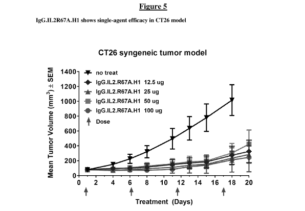
[00113] Figure 5 shows a graph of single agent efficacy of IgG.IL2R67A.H1
in a
CT26 tumor model.
[00114] Figure 6 presents the data of IgG.IL2R67A.H1 either as a single
agent or in
combination with an antibody in a B16 melanoma mouse model. The graph shows
that
IgG.IL2R67A.H1 in combination with TA99, an anti-TRP1 antibody, is more
efficacious
than TA99 alone, an IL2-Fc fusion molecule alone, TA99 plus an IL2-Fc fusion.
Synergy
was seen with TA99 and IgG.IL2R67A.H1 at the 100 and 500 tig doses.
[00115] Figure 7 shows a graph with values monitoring pSTAT5 activity in a
panel of
human cells comparing IgG.IL2R67A.H1 and IgG.IL2F71A.H1 with Proleukin .
[00116] Figure 8 shows a graph of ELISA data showing that when IL2 is
engrafted
into CDRH1 of an anti-RSV antibody (IgG.IL2R67A.H1), RSV binding is
maintained.
However, binding to RSV is reduced when IL2 is engrafted into CDRL3 or CDRH3.
When
IL2 is engrafted into a different antibody backbone (Xolair), there is no
binding to RSV.
Antibody Cytokine engrafted proteins targeting the IL2 low Affinity Receptor
[00117] Provided herein are protein constructs comprising an IL2 molecule
engrafted
to into the complementarity determining region (CDR) of an antibody. The
antibody cytokine
engrafted proteins of the present disclosure show suitable properties to be
used in human
patients, for example, they retain immunostimulatory activity similar to that
of native or
recombinant human IL2. However, the negative effects are diminished. For
example, there
is less stimulation of Treg cells. Other activities and characteristics are
also demonstrated
throughout the specification. Thus, provided are antibody cytokine engrafted
proteins having
an improved therapeutic profile over previously known IL2 and modified IL2
therapeutic
agents such as Proleukin , and methods of use of the provided antibody
cytokine engrafted
proteins in cancer treatment.
CA 03063983 2019-11-18
WO 2018/215936
PCT/IB2018/053623
[00118] Accordingly, the present disclosure provides antibody cytokine
engrafted
proteins that are agonists of the IL2 low affinity receptor, with selective
activity profiles.
Provided antibody cytokine engrafted proteins comprise an immunoglobulin heavy
chain
sequence and an immunoglobulin light chain sequence. Each immunoglobulin heavy
chain
sequence comprises a heavy chain variable region (VH) and a heavy chain
constant region
(CH), wherein the heavy chain constant region consists of CH1, CH2, and CH3
constant
regions. Each immunoglobulin light chain sequence comprises a light chain
variable region
(VL) and a light chain constant region (CL). In each antibody cytokine
engrafted protein an
IL2 molecule is incorporated into a complementarity determining region (CDR)
of the VH or
VL.
[00119] In some embodiments, the antibody cytokine engrafted protein
comprises an
IL2 molecule incorporated into a heavy chain CDR. In certain embodiments the
IL2
molecule is incorporated into heavy chain complementarity determining region 1
(HCDR1).
In certain embodiments the IL2 molecule is incorporated into heavy chain
complementarity
determining region 2 (HCDR2). In certain embodiments the IL2 molecule is
incorporated
into heavy chain complementarity determining region 3 (HCDR3).
[00120] In some embodiments, the antibody cytokine engrafted protein
comprises an
IL2 molecule incorporated into a light chain CDR. In certain embodiments the
IL2 molecule
is incorporated into light chain complementarity determining region 1 (LCDR1).
In certain
embodiments the IL2 molecule is incorporated into light chain complementarity
determining
region 2 (LCDR2). In certain embodiments the IL2 molecule is incorporated into
light chain
complementarity determining region 3 (LCDR3).
[00121] In some embodiments, the antibody cytokine engrafted comprises an
IL2
sequence incorporated into a CDR, whereby the IL2 sequence is inserted into
the CDR
sequence. The insertion can be at or near the N-terminal region of the CDR, in
the middle
region of the CDR or at or near the C-terminal region of the CDR. In other
embodiments, the
antibody cytokine engrafted comprises an IL2 molecule incorporated into a CDR,
whereby
the IL2 sequence does not frameshift the CDR sequence. In other embodiments,
the antibody
cytokine engrafted comprises an IL2 molecule incorporated into a CDR, whereby
the IL2
sequence replaces all or part of a CDR sequence. A replacement can be the N-
terminal
region of the CDR, in the middle region of the CDR or at or near the C-
terminal region the
CDR. A replacement can be as few as one or two amino acids of a CDR sequence,
or the
entire CDR sequence.
CA 03063983 2019-11-18
WO 2018/215936
PCT/IB2018/053623
26
[00122] In some embodiments an IL2 molecule is engrafted directly into a
CDR
without a peptide linker, with no additional amino acids between the CDR
sequence and the
IL2 sequence.
[00123] In some embodiments antibody cytokine engrafted proteins comprise
immunoglobulin heavy chains of an IgG class antibody heavy chain. In certain
embodiments
an IgG heavy chain is any one of an IgGl, an IgG2 or an IgG4 subclass.
[00124] In some embodiments antibody cytokine engrafted proteins comprise
heavy
and light chain immunoglobulin sequences selected from a known, clinically
utilized
immunoglobulin sequence. In certain embodiments antibody cytokine engrafted
proteins
comprise heavy and light chain immunoglobulin sequences which are humanized
sequences.
In other certain embodiments antibody cytokine engrafted proteins comprise
heavy and light
chain immunoglobulin sequences which are human sequences.
[00125] In some embodiments antibody cytokine engrafted proteins comprise
heavy
and light chain immunoglobulin sequences selected from germline immunoglobulin
sequences.
[00126] In some embodiments antibody cytokine engrafted proteins comprise
heavy
and light chain immunoglobulin sequences having binding specificity of the
immunoglobulin
variable domains to a target distinct from the binding specificity of the IL2
molecule. In
some embodiments the binding specificity of the immunoglobulin variable domain
to its
target is retained by 10%, 20%, 30%, 40%, 50%, 60%, 70%, 80%, 90%, 95%, 98%,
99%, or
100%, in the presence of the engrafted cytokine. In certain embodiments the
retained binding
specificity is to a non-human target. In certain embodiments the retained
binding specificity it
to a virus, for example, RSV. In other embodiments the binding specificity is
to a human
target having therapeutic utility in conjunction with an IL2 therapy. In
certain embodiments,
targeting the binding specificity of the immunoglobulin conveys additional
therapeutic
benefit to the IL2 component. In certain embodiments the binding specificity
of the
immunoglobulin to its target conveys synergistic activity with IL2.
[00127] In still other embodiments, the binding specificity of the
immunoglobulin is
reduced by 10%, 20%, 30%, 40%, 50%, 60%, 70%, 80%, 90%, 95%, 98%, 99%, or
100%, by
the engrafting of the IL2 molecule.
[00128] Provided antibody cytokine engrafted proteins comprise an IL2
molecule
engrafted into a complementarity determining region (CDR) of the VH or VL. In
some
embodiments, the IL2 sequence has at least 85%, 89%, 90%, 91%, 92%, 93%,
94%,95%,
CA 03063983 2019-11-18
WO 2018/215936
PCT/IB2018/053623
27
96%, 97%, 98%, 99% or 100% sequence identity to the amino acid sequence of SEQ
ID
NO:4. In some embodiments, the IL2 molecule comprises the sequence of SEQ ID
NO:4. In
some embodiments, the IL2 molecule consists of the sequence of SEQ ID NO:4.
[00129] Provided antibody cytokine engrafted proteins comprise an IL2
molecule
engrafted into a complementarity determining region (CDR) of the VH or VL. In
some
embodiments, the IL2 sequence has at least 85%, 89%, 90%, 91%, 92%, 93%,
94%,95%,
96%, 97%, 98%, 99% or 100% sequence identity to the amino acid sequence of SEQ
ID
NO:6. In some embodiments, the IL2 molecule comprises the sequence of SEQ ID
NO:6. In
some embodiments, the IL2 molecule consists of the sequence of SEQ ID NO:6.
[00130] In some embodiments, the antibody cytokine engrafted protein
confers pro
immunomodulatory properties superior to human IL2, recombinant human IL2,
Proleukin
or IL2 fused to an Fc. The antibody cytokine engrafted protein confers
increased activity on
CD8 T effector cells while providing reduced Treg activity as compared to
human IL2,
recombinant human IL2, Proleukin or IL2 fused to an Fc.
[00131] In some embodiments, the antibody cytokine engrafted proteins
comprise a
modified immunoglobulin IgG having a modified Fc conferring modified effector
function.
In certain embodiments the modified Fc region comprises a mutation selected
from one or
more of D265A, P329A, P329G, N297A, L234A, and L235A. In particular
embodiments the
immunoglobulin heavy chain may comprise a mutation or combination of mutations
conferring reduced effector function selected from any of D265A, P329A, P329G,
N297A,
D265A/P329A, D265A/N297A, L234/L235A, P329A/L234A/L235A, and
P329G/L234A/L235A.
[00132] In some embodiments, the antibody cytokine engrafted proteins
comprise (i) a
heavy chain variable region having at least 85%, 89%, 90%, 91%, 92%, 93%, 94%,
95%,
96%, 97%, 98%, 99%, or 100% amino acid sequence identity to a heavy chain
variable
region of SEQ ID NO:19 and (ii) a light chain variable region having at least
85%, 89%,
90%, 91%, 92%, 93%, 94%, 95%, 96%, 97%, 98%, 99%, or 100% amino acid sequence
identity to a light chain variable region of SEQ ID NO:35. The immunoglobulin
chain is an
IgG class selected from IgGl, IgG2, or IgG4. In certain embodiments the
immunoglobulin
optionally comprises a mutation or combination of mutations conferring reduced
effector
function selected from any of D265A, P329A, P329G, N297A, D265A/P329A,
D265A/N297A, L234/L235A, P329A/L234A/L235A, and P329G/L234A/L235A.
CA 03063983 2019-11-18
WO 2018/215936
PCT/IB2018/053623
28
[00133] In some embodiments, the antibody cytokine engrafted proteins
comprise (i) a
heavy chain variable region having at least 85%, 89%, 90%, 91%, 92%, 93%, 94%,
95%,
96%, 97%, 98%, 99%, or 100% amino acid sequence identity to a heavy chain
variable
region of SEQ ID NO:51 and (ii) a light chain variable region having at least
85%, 89%,
90%, 91%, 92%, 93%, 94%, 95%, 96%, 97%, 98%, 99%, or 100% amino acid sequence
identity to a light chain variable region of SEQ ID NO:67. The immunoglobulin
chain is an
IgG class selected from IgGl, IgG2, or IgG4. In certain embodiments the
immunoglobulin
optionally comprises a mutation or combination of mutations conferring reduced
effector
function selected from any of D265A, P329A, P329G, N297A, D265A/P329A,
D265A/N297A, L234/L235A, P329A/L234A/L235A, and P329G/L234A/L235A.
Engineered and Modified Antibody Cytokine Engrafted Proteins
[00134] In certain aspects, antibody cytokine engrafted constructs are
generated by
engrafting an IL2 sequence into a CDR region of an immunoglobulin scaffold.
Both heavy
and light chain immunoglobulin chains are produced to generate final antibody
engrafted
proteins. Antibody cytokine engrafted proteins confer preferred therapeutic
activity on CD8
T effector cells, and the antibody cytokine engrafted proteins have reduced
Treg activity as
compared with native or recombinant human IL2 (rhIL2 or ProleukinC) or IL2
fused to an
Fc.
[00135] To engineer antibody cytokine engrafted proteins, IL2 sequences
containing
specific muteins (SEQ ID NO:4 or SEQ ID NO:6), are inserted into a CDR loop of
an
immunoglobulin chain scaffold protein. Engrafted constructs can be prepared
using any of a
variety of known immunoglobulin sequences which have been utilized in clinical
settings,
known immunoglobulin sequences which are in current discovery and/or clinical
development, human germline antibody sequences, as well as sequences of novel
antibody
immunoglobulin chains. Constructs are produced using standard molecular
biology
methodology utilizing recombinant DNA encoding relevant sequences. Sequences
of IL2 in
an exemplary scaffold, referred to as GFTX3b, are depicted in TABLE 2.
Insertion points
were selected to be the mid-point of the loop based on available structural or
homology
model data, however, insertion points can be adjusted toward the N or C-
terminal end of the
CDR loop.
[00136] Thus the present disclosure provides antibodies or fragments
thereof that
specifically bind to the low affinity IL2 receptor comprising an IL2 protein
recombinantly
inserted into a heterologous antibody protein or polypeptide to generate
engrafted proteins.
CA 03063983 2019-11-18
WO 2018/215936
PCT/IB2018/053623
29
In particular, the disclosure provides engrafted proteins comprising an
antibody or antigen-
binding fragment of an antibody described herein or any other relevant
scaffold antibody
polypeptide (e.g., a full antibody immunoglobulin protein, a Fab fragment, Fc
fragment, Fv
fragment, F(ab)2 fragment, a VH domain, a VH CDR, a VL domain, a VL CDR, etc.)
and a
heterologous cytokine protein, polypeptide, or peptide, e.g., IL2. Methods for
fusing or
conjugating proteins, polypeptides, or peptides to an antibody or an antibody
fragment are
known in the art. See, e.g., U.S. Patent Nos. 5,336,603, 5,622,929, 5,359,046,
5,349,053,
5,447,851, and 5,112,946; European Patent Nos. EP 307,434 and EP 367,166;
International
Publication Nos. WO 96/04388 and WO 91/06570; Ashkenazi et al., 1991, Proc.
Natl. Acad.
Sci. USA 88: 10535-10539; Zheng et al., 1995, J. Immunol. 154:5590-5600; and
Vil et al.,
1992, Proc. Natl. Acad. Sci. USA 89:11337- 11341. Additionally, antibody
cytokine
engrafted proteins may be generated through the techniques of gene-shuffling,
motif-
shuffling, exon-shuffling, and/or codon-shuffling (collectively referred to as
"DNA
shuffling"). DNA shuffling may be employed to prepare engrafted protein
constructs and/or
to alter the activities of antibodies or fragments thereof (e.g., antibodies
or fragments thereof
with higher affinities and lower dissociation rates). See, generally, U.S.
Patent Nos.
5,605,793, 5,811,238, 5,830,721, 5,834,252, and 5,837,458; Patten et al.,
1997, Curr. Opinion
Biotechnol. 8:724-33; Harayama, 1998, Trends Biotechnol. 16(2):76-82; Hansson,
et al.,
1999, J. Mol. Biol. 287:265-76; and Lorenzo and Blasco, 1998, Biotechniques
24(2):308-
313. Antibodies or fragments thereof, or the encoded antibodies or fragments
thereof, may
be altered by being subjected to random mutagenesis by error-prone PCR, random
nucleotide
insertion or other methods prior to recombination. A polynucleotide encoding
an antibody or
fragment thereof that specifically binds to an antigen protein of interest may
be recombined
with one or more components, motifs, sections, parts, domains, fragments, etc.
of one or
more heterologous cytokine molecules, e.g., IL2, for preparation of antibody
cytokine
engrafted proteins as provided herein.
[00137] An antibody Fab contains six CDR loops, 3 in the light chain
(CDRL1,
CDRL2, CDRL3) and 3 in the heavy chain (CDRH1, CDRH2, CDRH3) which can serve
as
potential insertion sites for a cytokine protein. Structural and functional
considerations are
taken into account in order to determine which CDR loop(s) to insert the
cytokine. As a CDR
loop size and conformation vary greatly across different antibodies, the
optimal CDR for
insertion can be determined empirically for each particular antibody/protein
combination.
CA 03063983 2019-11-18
WO 2018/215936
PCT/IB2018/053623
Additionally, since a cytokine protein will be inserted into a CDR loop, this
can put
additional constraints on the structure of the cytokine protein as discussed
in Example 1.
[00138] CDRs of immunoglobulin chains are determined by well-known
numbering
systems known in the art, including those described herein. For example, CDRs
have been
identified and defined by (1) using the numbering system described in Kabat et
al. (1991),
"Sequences of Proteins of Immunological Interest," 5th Ed. Public Health
Service, National
Institutes of Health, Bethesda, MD ("Kabat" numbering scheme), NIH publication
No. 91-
3242; and (2) Chothia, see Al-Lazikani et al., (1997) "Standard conformations
for the
canonical structures of immunoglobulins," J.Mol.Biol. 273:927-948. For
identified CDR
amino acid sequences less than 20 amino acids in length, one or two
conservative amino acid
residue substitutions can be tolerated while still retaining the desired
specific binding and/or
agonist activity.
[00139] An antibody cytokine engrafted protein further can be prepared
using an
antibody having one or more of the CDRs and/or VH and/or VL sequences shown
herein
(e.g., TABLE 2) as starting material to engineer a modified antibody cytokine
engrafted
protein, which may have altered properties from the starting antibody
engrafted protein.
Alternatively any known antibody sequences may be utilized as a scaffold to
engineer
modified antibody cytokine engrafted protein. For example, any known,
clinically utilized
antibody may be utilized as a starting materials scaffold for preparation of
antibody engrafted
protein. Known antibodies and corresponding immunoglobulin sequences include,
e.g.,
palivizumab, alirocumab, mepolizumab, necitumumab, nivolumab, dinutuximab,
secukinumab, evolocumab, blinatumomab, pembrolizumab, ramucirumab vedolizumab,
siltuximab, obinutuzumab, trastuzumab, raxibacumab, pertuzumab, belimumab,
ipilimumab,
denosumab, tocilizumab, ofatumumab, canakinumab, golimumab, ustekinumab,
certolizumab, catumaxomab, eculizumab, ranibizumab, panitumumab, natalizumab,
bevacizumab, cetuximab, efalizumab, omalizumab, tositumomab, ibritumomab
tiuxetan,
adalimumab, alemtuzumab, gemtuzumab, infliximab, basiliximab, daclizumab,
rituximab,
abciximab, muromonab, or modifications thereof. Known antibodies and
immunoglobulin
sequences also include germline antibody sequences. Framework sequences can be
obtained
from public DNA databases or published references that include germline
antibody gene
sequences. For example, germline DNA sequences for human heavy and light chain
variable
region genes can be found in the "VBase" human germline sequence database, as
well as in
Kabat, E. A., et al., 1991 Sequences of Proteins of Immunological Interest,
Fifth Edition,
CA 03063983 2019-11-18
WO 2018/215936
PCT/IB2018/053623
31
U.S. Department of Health and Human Services, NIH Publication No. 91-3242;
Tomlinson, I.
M., et al., 1992 J. fol. Biol. 227:776-798; and Cox, J. P. L. et al., 1994
Eur. J Immunol.
24:827-836. In still other examples, antibody and corresponding immunoglobulin
sequences
from other known entities which can be in early discovery and/or drug
development can be
similarly adapted as starting material to engineer a modified antibody
cytokine engrafted
protein.
[00140] A wide variety of antibody/immunoglobulin frameworks or scaffolds
can be
employed so long as the resulting polypeptide includes at least one binding
region which
accommodates incorporation of a cytokine (e.g., IL2). Such frameworks or
scaffolds include
the 5 main idiotypes of human immunoglobulins, or fragments thereof, and
include
immunoglobulins of other animal species, preferably having humanized and/or
human
aspects. Novel antibodies, frameworks, scaffolds and fragments continue to be
discovered
and developed by those skilled in the art.
[00141] Antibodies can be generated using methods that are known in the
art. For
preparation of monoclonal antibodies, any technique known in the art can be
used (see, e.g.,
Kohler & Milstein, Nature 256:495-497 (1975); Kozbor et al., Immunology Today
4:72
(1983); Cole et al., Monoclonal Antibodies and Cancer Therapy, pp. 77-96. Alan
R. Liss, Inc.
1985). Techniques for the production of single chain antibodies (U.S. Patent
No. 4,946,778)
can be adapted to produce antibodies for use in antibody cytokine engrafted
proteins. Also,
transgenic mice, or other organisms such as other mammals, can be used to
express and
identify primatized or humanized or human antibodies. Alternatively, phage
display
technology can be used to identify antibodies and heteromeric Fab fragments
that specifically
bind to selected antigens for use in antibody cytokine engrafted proteins
(see, e.g.,
McCafferty et al., supra; Marks et al., Biotechnology, 10:779-783, (1992)).
[00142] Methods for primatizing or humanizing non-human antibodies are well
known
in the art. Generally, a primatized or humanized antibody has one or more
amino acid
residues introduced into it from a source which is non-primate or non-human.
Such non-
primate or non-human amino acid residues are often referred to as import
residues, which are
typically taken from an import variable domain. Humanization can be
essentially performed
following the method of Winter and co-workers (see, e.g., Jones et al., Nature
321:522-525
(1986); Riechmann et al., Nature 332:323-327 (1988); Verhoeyen et al., Science
239:1534-
1536 (1988) and Presta, Curr. Op. StrucL Biol. 2:593-596 (1992)), by
substituting rodent
CDRs or CDR sequences for the corresponding sequences of a human antibody.
CA 03063983 2019-11-18
WO 2018/215936
PCT/IB2018/053623
32
Accordingly, such humanized antibodies are chimeric antibodies (U.S. Patent
No. 4,816,567),
wherein substantially less than an intact human variable domain has been
substituted by the
corresponding sequence from a non-human species. In practice, primatized or
humanized
antibodies are typically primate or human antibodies in which some
complementary
determining region ("CDR") residues and possibly some framework ("FR")
residues are
substituted by residues from analogous sites in an originating species (e.g.,
rodent antibodies)
to confer binding specificity.
[00143] Alternatively or additionally, an in vivo method for replacing a
nonhuman
antibody variable region with a human variable region in an antibody while
maintaining the
same or providing better binding characteristics relative to that of the
nonhuman antibody
may be utilized to convert non-human antibodies into engineered human
antibodies. See,
e.g., U.S. Patent Publication No. 20050008625, U.S. Patent Publication No.
2005/0255552.
Alternatively, human V segment libraries can be generated by sequential
cassette replacement
in which only part of the reference antibody V segment is initially replaced
by a library of
human sequences; and identified human "cassettes" supporting binding in the
context of
residual reference antibody amino acid sequences are then recombined in a
second library
screen to generate completely human V segments (see, U.S. Patent Publication
No.
2006/0134098).
[00144] Various antibodies or antigen-binding fragments for use in
preparation of
antibody cytokine engrafted proteins can be produced by enzymatic or chemical
modification
of the intact antibodies, or synthesized de novo using recombinant DNA
methodologies (e.g.,
single chain Fv), or identified using phage display libraries (see, e.g.,
McCafferty et al.,
Nature 348:552-554, 1990). For example, minibodies can be generated using
methods
described in the art, e.g., Vaughan and Sollazzo, Comb. Chem. High Throughput
Screen
4:417-30 2001. Bispecific antibodies can be produced by a variety of methods
including
engrafted of hybridomas or linking of Fab' fragments. See, e.g., Songsivilai &
Lachmann,
Clin. Exp. Immunol. 79:315-321 (1990); Kostelny et al., J. Immunol. 148, 1547-
1553 (1992).
Single chain antibodies can be identified using phage display libraries or
ribosome display
libraries, gene shuffled libraries. Such libraries can be constructed from
synthetic, semi-
synthetic or native and immunocompetent sources. Selected immunoglobulin
sequences may
thus be utilized in preparation of antibody cytokine engrafted protein
constructs as provided
herein.
CA 03063983 2019-11-18
WO 2018/215936
PCT/IB2018/053623
33
[00145] Antibodies, antigen-binding molecules or antibody cytokine
engrafted
molecules of use in the present disclosure further include bispecific
antibodies. A bispecific
or bifunctional antibody is an artificial hybrid antibody having two different
heavy/light chain
pairs and two different binding sites. Other antigen-binding fragments or
antibody portions
include bivalent scFv (diabody), bispecific scFv antibodies where the antibody
molecule
recognizes two different epitopes, single binding domains (dAbs), and
minibodies. Selected
immunoglobulin sequences may thus be utilized in preparation of antibody
cytokine
engrafted protein constructs as provided herein.
[00146] Antigen-binding fragments of antibodies e.g., a Fab fragment, scFv,
can be
used as building blocks to construct antibody cytokine engrafted proteins, and
may optionally
include multivalent formats. In some embodiments, such multivalent molecules
comprise a
constant region of an antibody (e.g., Fc).
[00147] Antibody cytokine engrafted proteins can be engineered by modifying
one or
more residues within one or both variable regions (i.e., VH and/or VL) of an
antibody, for
example, within one or more CDR regions, and such adapted VH and/or VL region
sequences
are utilized for engrafting a cytokine or for preparation of cytokine
engrafting. Antibodies
interact with target antigens predominantly through amino acid residues that
are located in
the six heavy and light chain complementarity determining regions (CDRs). For
this reason,
the amino acid sequences within CDRs are more diverse between individual
antibodies than
sequences outside of CDRs. CDR sequences are responsible for most antibody-
antigen
interactions, it is possible to express recombinant antibodies that mimic the
properties of a
specific antibody by constructing expression vectors that include CDR
sequences from a
specific antibody grafted onto framework sequences from a different antibody
with different
properties (see, e.g., Riechmann, L. et al., 1998 Nature 332:323-327; Jones,
P. et al., 1986
Nature 321:522-525; Queen, C. et al., 1989 Proc. Natl. Acad., U.S.A. 86:10029-
10033; U.S.
Patent No. 5,225,539 to Winter, and U.S. Patent Nos. 5,530,101; 5,585,089;
5,693,762 and
6,180,370 to Queen et al.). In certain instances it is beneficial to mutate
residues within the
framework regions to maintain or enhance the antigen binding ability of the
antibody (see
e.g., U.S. Patent Nos. 5,530,101; 5,585,089; 5,693,762 and 6,180,370 to Queen
et al).
[00148] In some aspects mutation of amino acid residues within the VH
and/or VL
CDR1, CDR2, and/or CDR3 regions to thereby improve one or more binding
properties (e.g.,
affinity) of the antibody of interest, known as "affinity maturation," may be
beneficial, e.g.,
to optimize antigen binding of an antibody in conjunction with the context of
the cytokine
CA 03063983 2019-11-18
WO 2018/215936
PCT/IB2018/053623
34
engrafted protein. Site-directed mutagenesis or PCR-mediated mutagenesis can
be performed
to introduce the mutation(s) and the effect on antibody binding, or other
functional property
of interest, can be evaluated in in vitro or in vivo assays as described
herein and/or alternative
or additional assays known in the art. Conservative modifications can be
introduced. The
mutations may be amino acid substitutions, additions or deletions. Moreover,
typically no
more than one, two, three, four or five residues within a CDR region are
altered.
[00149] Engineered antibodies or antibody fragments include those in which
modifications have been made to framework residues within VH and/or VL, e.g.
to improve
the properties of the antibody. In some embodiments such framework
modifications are made
to decrease immunogenicity of the antibody. For example, one approach is to
change one or
more framework residues to the corresponding germline sequence. More
specifically, an
antibody that has undergone somatic mutation may contain framework residues
that differ
from germline sequence from which the antibody is derived. Such residues can
be identified
by comparing the antibody framework sequences to the germline sequences from
which the
antibody is derived. To return the framework region sequences to their
germline
configuration, the somatic mutations can be "backmutated" to the germline
sequence by, for
example, site-directed mutagenesis. Additional framework modification involves
mutating
one or more residues within the framework region, or even within one or more
CDR regions,
to remove T cell epitopes to thereby reduce the potential immunogenicity of
the antibody.
This approach is also referred to as "deimmunization" and is described in
further detail in
U.S. Patent Publication No. 20030153043 by Can et al.
[00150] Constant regions of the antibodies or antibody fragments utilized
for
preparation of the antibody cytokine engrafted protein can be any type or
subtype, as
appropriate, and can be selected to be from the species of the subject to be
treated by the
present methods (e.g., human, non-human primate or other mammal, for example,
agricultural mammal (e.g., equine, ovine, bovine, porcine, camelid), domestic
mammal (e.g.,
canine, feline) or rodent (e.g., rat, mouse, hamster, rabbit). In some
embodiments antibodies
utilized in antibody cytokine engrafted proteins are engineered to generate
humanized or
Humaneered@ antibodies. In some embodiments antibodies utilized in antibody
cytokine
engrafted proteins are human antibodies. In some embodiments, antibody
constant region
isotype is IgG, for example, IgGl, IgG2, IgG3, IgG4. In certain embodiments
the constant
region isotype is IgGi. In some embodiments, antibody cytokine engrafted
proteins comprise
CA 03063983 2019-11-18
WO 2018/215936
PCT/IB2018/053623
an IgG. In some embodiments, antibody cytokine engrafted proteins comprise an
IgG1 Fc.
In some embodiments, antibody cytokine engrafted proteins comprise an IgG2 Fc.
[00151] In addition or alternative to modifications made within framework
or CDR
regions, antibodies or antibody fragments utilized in preparation of antibody
cytokine
engrafted proteins may be engineered to include modifications within an Fc
region, typically
to alter one or more functional properties of the antibody, such as, e.g.,
serum half-life,
complement fixation, Fc receptor binding, and/or antigen-dependent cellular
cytotoxicity.
Furthermore, an antibody, antibody fragment thereof, or antibody cytokine
engrafted protein
can be chemically modified (e.g., one or more chemical moieties can be
attached to the
antibody) or be modified to alter its glycosylation, again to alter one or
more functional
properties of the antibody cytokine engrafted protein.
[00152] In one embodiment, a hinge region of CH1 is modified such that the
number
of cysteine residues in the hinge region is altered, e.g., increased or
decreased. For example,
by the approach is described further in U.S. Patent No. 5,677,425 by Bodmer et
al. wherein
the number of cysteine residues in the hinge region of CH1 is altered to, for
example,
facilitate assembly of the light and heavy chains or to increase or decrease
the stability of the
antibody cytokine engrafted protein. In another embodiment, an Fe hinge region
of an
antibody is mutated to alter the biological half-life of the antibody cytokine
engrafted protein.
More specifically, one or more amino acid mutations are introduced into the
CH2-CH3
domain interface region of the Fc-hinge fragment such that the antibody
cytokine engrafted
protein has impaired Staphylococcyl protein A (SpA) binding relative to native
Fc-hinge
domain SpA binding. This approach is described in further detail in U.S.
Patent No.
6,165,745 by Ward et al.
[00153] The present disclosure provides for antibody cytokine engrafted
proteins that
specifically bind to the IL2 low affinity receptor which have an extended half-
life in vivo. In
another embodiment, an antibody cytokine engrafted protein is modified to
increase its
biological half-life. Various approaches are possible. Antibody cytokine
engrafted proteins
having an increased half-life in vivo can also be generated introducing one or
more amino
acid modifications (i.e., substitutions, insertions or deletions) into an IgG
constant domain, or
FcRn binding fragment thereof (preferably a Fc or hinge Fc domain fragment).
For example,
one or more of the following mutations can be introduced: T252L, T2545, T256F,
as
described in U.S. Patent No. 6,277,375 to Ward. See, e.g., International
Publication No. WO
98/23289; International Publication No. WO 97/34631; and U.S. Patent No.
6,277,375.
CA 03063983 2019-11-18
WO 2018/215936
PCT/IB2018/053623
36
Alternatively, to increase the biological half-life, the antibody cytokine
engrafted protein is
altered within the CH1 or CL region to contain a salvage receptor binding
epitope taken from
two loops of a CH2 domain of an Fc region of an IgG, as described in U.S.
Patent Nos.
5,869,046 and 6,121,022 by Presta et al. In yet other embodiments, the Fc
region is altered
by replacing at least one amino acid residue with a different amino acid
residue to alter the
effector functions of the antibody cytokine engrafted protein. For example,
one or more
amino acids can be replaced with a different amino acid residue such that the
antibody
cytokine engrafted protein has an altered affinity for an effector ligand but
retains antigen-
binding ability of the parent antibody. The effector ligand to which affinity
is altered can be,
for example, an Fc receptor (FcR) or the Cl component of complement. This
approach is
described in further detail in U.S. Patent Nos. 5,624,821 and 5,648,260, both
by Winter et al.
[00154] In another embodiment, one or more amino acids selected from amino
acid
residues can be replaced with a different amino acid residue such that the
antibody cytokine
engrafted protein has altered Clq binding and/or reduced or abolished
complement dependent
cytotoxicity (CDC). This approach is described in further detail in U.S.
Patent Nos. 6,194,551
by Idusogie et al.
[00155] Antibody cytokine engrafted proteins containing such mutations
mediate
reduced or no antibody-dependent cellular cytotoxicity (ADCC) or complement-
dependent
cytotoxicity (CDC). In some embodiments, amino acid residues L234 and L235 of
the IgG1
constant region are substituted to Ala234 and Ala235. In some embodiments,
amino acid
residue N267 of the IgG1 constant region is substituted to Ala267.
[00156] In another embodiment, one or more amino acid residues are altered
to thereby
alter the ability of the antibody cytokine engrafted protein to fix
complement. This approach
is described further in PCT Publication WO 94/29351 by Bodmer et al.
[00157] In yet another embodiment, an Fc region is modified to increase the
ability of
the antibody to mediate antibody dependent cellular cytotoxicity (ADCC) and/or
to increase
the affinity of the antibody cytokine engrafted protein for an Fey receptor by
modifying one
or more amino acids. This approach is described further in PCT Publication WO
00/42072
by Presta. Moreover, binding sites on human IgG1 for FeyR1, FeyRII, FeyRIII
and FcRn
have been mapped and variants with improved binding have been described (see
Shields,
R.L. et al., 2001 J. Biol. Chen. 276:6591-6604).
[00158] In still another embodiment, glycosylation of an antibody cytokine
engrafted
protein is modified. For example, an aglycoslated antibody cytokine engrafted
protein can be
CA 03063983 2019-11-18
WO 2018/215936
PCT/IB2018/053623
37
made (i.e., the antibody cytokine engrafted protein lacks glycosylation).
Glycosylation can be
altered to, for example, increase the affinity of the antibody for "antigen."
Such carbohydrate
modifications can be accomplished by, for example, altering one or more sites
of
glycosylation within the antibody sequence. For example, one or more amino
acid
substitutions can be made that result in elimination of one or more variable
region framework
glycosylation sites to thereby eliminate glycosylation at that site. Such
aglycosylation can
increase the affinity of the antibody for antigen. Such an approach is
described in further
detail in U.S. Patent Nos. 5,714,350 and 6,350,861 by Co et al.
[00159] Additionally or alternatively, an antibody cytokine engrafted
protein can be
made that has an altered type of glycosylation, such as a hypofucosylated
antibody cytokine
engrafted protein having reduced amounts of fucosyl residues or an antibody
having
increased bisecting GlcNac structures. Such altered glycosylation patterns
have been
demonstrated to increase the antibody dependent cellular cytotoxicity (ADCC)
ability of
antibodies. Such carbohydrate modifications can be accomplished by, for
example,
expressing the antibody cytokine engrafted protein in a host cell with altered
glycosylation
machinery. Cells with altered glycosylation machinery have been described in
the art and can
be used as host cells in which to express recombinant antibody cytokine
engrafted proteins to
thereby produce an antibody cytokine engrafted protein with altered
glycosylation. For
example, EP 1,176,195 by Hang et al. describes a cell line with a functionally
disrupted
FUT8 gene, which encodes a fucosyl transferase, such that antibody cytokine
engrafted
proteins expressed in such a cell line exhibit hypofucosylation. PCT
Publication WO
03/035835 by Presta describes a variant CHO cell line, Lec13 cells, with
reduced ability to
attach fucose to Asn(297)-linked carbohydrates, also resulting in
hypofucosylation of
antibody cytokine engrafted proteins expressed in that host cell (see also
Shields, R.L. et al.,
2002 J. Biol. Chem. 277:26733-26740). PCT Publication WO 99/54342 by Umana et
al.
describes cell lines engineered to express glycoprotein-modifying glycosyl
transferases (e.g.,
beta(1,4)-N acetylglucosaminyltransferase III (GnTIII)) such that antibody
cytokine
engrafted proteins expressed in the engineered cell lines exhibit increased
bisecting GlcNac
structures which results in increased ADCC activity of the antibodies (see
also Umana et al.,
1999 Nat. Biotech. 17:176-180).
[00160] In some embodiments, one or more domains, or regions, of an
antibody
cytokine engrafted protein are connected via a linker, for example, a peptide
linker, such as
those that are well known in the art (see e.g., Holliger, P., et al. (1993)
Proc. Natl. Acad. Sci.
CA 03063983 2019-11-18
WO 2018/215936
PCT/IB2018/053623
38
USA 90:6444-6448; Poljak, RJ., et al. (1994) Structure 2:1121-1123). A peptide
linker may
vary in length, e.g., a linker can be 1-100 amino acids in length, typically a
linker is from five
to 50 amino acids in length, e.g., 5, 6, 7, 8, 9, 10, 11, 12, 13, 14, 15, 16,
17, 18, 19, 20, 21, 22,
23, 24, 25, 26, 27, 28, 29, 30, 31, 32, 33, 34, 35, 36, 37, 38, 39, 40, 41,
42, 43, 44, 45, 46, 47,
48, 49, or 50 amino acids in length.
[00161] In some embodiments IL2 is engrafted into the CDR sequence
optionally with
one or more peptide linker sequences. In certain embodiments one or more
peptide linkers is
independently selected from a (Glyn-Ser)m sequence (SEQ ID NO: 71), a (Glyn-
Ala)m
sequence (SEQ ID NO: 72), or any combination of a (Glyn-Ser)m/(Glyn-Ala)m
sequence
(SEQ ID NOS: 71-72), wherein each n is independently an integer from 1 to 5
and each m is
independently an integer from 0 to 10. Examples of linkers include, but are
not limited to,
glycine-based linkers or gly/ser linkers G/S such as (GmS)n wherein n is a
positive integer
equal to 1, 2, 3, 4, 5, 6, 7, 8, 9, or 10 and m is an integer equal to 0, 1,
2, 3, 4, 5, 6, 7, 8, 9, or
(SEQ ID NO: 73). In certain embodiments one or more linkers include G45 (SEQ
ID
NO: 74) repeats, e.g., the Gly-Ser linker (G45). wherein n is a positive
integer equal to or
greater than 1 (SEQ ID NO: 74). For example, n=1, n=2, n=3. n=4, n=5 and n=6,
n=7, n=8,
n=9 and n=10. In some embodiments, Ser can be replaced with Ala e.g., linkers
G/A such as
(GmA)n wherein n is a positive integer equal to 1, 2, 3, 4, 5, 6, 7, 8, 9, or
10 and m is an
integer equal to 0, 1, 2, 3, 4, 5, 6, 7, 8, 9, or 10 (SEQ ID NO: 75). In
certain embodiments
one or more linkers include G4A (SEQ ID NO: 76) repeats, (G4A)11 wherein n is
a positive
integer equal to or greater than 1 (SEQ ID NO: 76). For example, n=1, n=2,
n=3. n=4, n=5
and n=6, n=7, n=8, n=9 and n=10. In some embodiments, the linker includes
multiple repeats
of linkers. In other embodiments, a linker includes combinations and multiples
of G45 (SEQ
ID NO: 74) and G4A (SEQ ID NO: 76).
[00162] Other examples of linkers include those based on flexible linker
sequences
that occur naturally in antibodies to minimize immunogenicity arising from
linkers and
junctions. For example, there is a natural flexible linkage between the
variable domain and a
CH1 constant domain in antibody molecular structure. This natural linkage
comprises
approximately 10-12 amino acid residues, contributed by 4-6 residues from C-
terminus of V
domain and 4-6 residues from the N-terminus of the CH1 domain. Antibody
cytokine
engrafted proteins can, e.g., employ linkers incorporating terminal 5-6 amino
acid residues,
or 11-12 amino acid residues, of CH1 as a linker. The N-terminal residues of
the CH1
domain, particularly the first 5-6 amino acid residues, adopt a loop
conformation without
CA 03063983 2019-11-18
WO 2018/215936
PCT/IB2018/053623
39
strong secondary structure, and, therefore, can act as a flexible linker. The
N-terminal
residues of the CH1 domain are a natural extension of the variable domains, as
they are part
of the Ig sequences, and, therefore, minimize to a large extent any
immunogenicity
potentially arising from the linkers and junctions. In some embodiments a
linker sequence
includes a modified peptide sequence based on a hinge sequence.
[00163] Moreover, the antibody cytokine engrafted proteins can include
marker
sequences, such as a peptide to facilitate purification of antibody cytokine
engrafted proteins.
In preferred embodiments, a marker amino acid sequence is a hexa-histidine
peptide (SEQ
ID NO: 78), such as the tag provided in a pQE vector (QIAGEN, Inc., 9259 Eton
Avenue,
Chatsworth, CA, 91311), among others, many of which are commercially
available. As
described in Gentz et al., 1989, Proc. Natl. Acad. Sci. USA 86:821-824, for
instance, hexa-
histidine (SEQ ID NO: 78) provides for convenient purification of the
engrafted protein.
Other peptide tags useful for purification include, but are not limited to,
the hemagglutinin
("HA") tag, which corresponds to an epitope derived from the influenza
hemagglutinin
protein (Wilson et al., 1984, Cell 37:767), and the "flag" tag.
[00164] Antibodies may also be attached to solid supports, which are
particularly
useful for immunoassays or purification of the target antigen. Such solid
supports include, but
are not limited to, glass, cellulose, polyacrylamide, nylon, polystyrene,
polyvinyl chloride or
polypropylene.
Assays for Antibody Cytokine Engrafted Protein activity
[00165] Assays for identifying antibody cytokine engrafted proteins are
known in the
art and described herein. Agonist antibody cytokine engrafted proteins bind to
the IL2 low
affinity receptor and promote, induce, stimulate intracellular signalling
resulting in CD8 T
effector cell proliferation as well as other immunostimulatory effects.
[00166] Binding of the antibody cytokine engrafted proteins to the IL2 low
affinity
receptor can be determined using any method known in the art. For example,
binding to the
IL2 low affinity receptor can be determined using known techniques, including
without
limitation ELISA, Western blots, surface plasmon resonance (SPR) (e.g.,
BIAcore), and flow
cytometry.
[00167] Intracellular signalling through the IL2 low affinity receptor can
be measured
using any method known in the art. For example, activation of the IL2 low
affinity receptor
by IL2 promotes STAT5 activation and signalling. Methods for measuring STAT5
activation
CA 03063983 2019-11-18
WO 2018/215936
PCT/IB2018/053623
are standard in the art (e.g., phosphorylation status of STAT5 protein,
reporter gene assays,
downstream signalling assays, etc.). Activation through the IL2 low affinity
receptor
expands CD8 T effector cells, so the absolute numbers of CD8 T effector cells
can be assayed
for or the ratio of CD8 T effector cells to Tregs can be assayed for. Methods
for measuring
proliferation of cells are standard in the art (e.g., 3H-thymidine
incorporation assays, CFSE
labelling). Methods for measuring cytokine production are well known in the
art (e.g.,
ELISA assays, ELISpot assays). In performing in vitro assays, test cells or
culture
supernatant from test cells contacted with antibody cytokine engrafted
proteins can be
compared to control cells or culture supernatants from control cells that have
not been
contacted with an antibody cytokine engrafted protein and/or those that have
been contacted
with recombinant human IL2 (e.g. Proleukin ) or an IL2-Fc fusion molecule.
[00168] The activity of the antibody cytokine engrafted proteins can also
be measured
ex vivo and/or in vivo. In some aspects, methods for measuring STAT5
activation across
various cell types ex vivo from animals treated with antibody cytokine
engrafted proteins as
compared to untreated control animals and/or animals similarly treated with
Proleukin may
be used to show differential activity of the agonist antibody engrafted
proteins across cell
types. Preferred agonist antibody cytokine engrafted proteins have the ability
to activate and
expand CD8 T effector cells. For example, in vivo activation and expansion of
CD 8 T
effector cells can be measured using any method known in the art, e.g., by
flow cytometry.
Preferred agonist antibody cytokine engrafted proteins can be therapeutically
useful in
preventing, reducing, alleviating or the treatment of cancer, for example:
melanoma, lung
cancer, colorectal cancer, prostate cancer, breast cancer and lymphoma. The
efficacy of the
antibody cytokine engrafted proteins can be determined by administering a
therapeutically
effective amount of the antibody cytokine engrafted protein to a subject and
comparing the
subject before and after administration of the antibody cytokine engrafted
protein. Efficacy
of the antibody cytokine engrafted proteins can also be determined by
administering a
therapeutically effective amount of an antibody cytokine engrafted protein to
a test subject
and comparing the test subject to a control subject who has not been
administered the
antibody and/or comparison to a subject similarly treated with Proleukin .
Polynucleotides Encoding Antibody Cytokine Engrafted Proteins
[00169] In another aspect, isolated nucleic acids encoding heavy and light
chain
proteins of the antibody cytokine engrafted proteins are provided. Antibody
cytokine
CA 03063983 2019-11-18
WO 2018/215936
PCT/IB2018/053623
41
engrafted proteins can be produced by any means known in the art, including
but not limited
to, recombinant expression, chemical synthesis, and enzymatic digestion of
antibody
tetramers. Recombinant expression can be from any appropriate host cells known
in the art,
for example, mammalian host cells, bacterial host cells, yeast host cells,
insect host cells, etc.
[00170] Provided herein are polynucleotides that encode the variable
regions
exemplified in any one of SEQ ID NO:20, SEQ ID NO:36, SEQ ID NO:52 and SEQ ID
NO:68.
[00171] The disclosure thus provides polynucleotides encoding the light
and/or heavy
chain polypeptides of the antibody cytokine engrafted proteins described
herein, e. g. ,
polynucleotides encoding light or heavy chain variable regions or segments
comprising the
complementary determining regions as described herein. In some embodiments,
the
polynucleotide encoding the heavy chain variable regions comprises a sequence
having at
least 85%, 89%, 90%, 91%, 92%, 93%, 94%, 95%, 96%, 97%, 98%, 99%, or 100%
nucleic
acid sequence identity with a polynucleotide selected from the group
consisting of SEQ ID
NO:20, and SEQ ID NO:52. In some embodiments, the polynucleotide encoding the
light
chain variable regions comprises a sequence having at least 85%, 89%, 90%,
91%, 92%,
93%, 94%, 95%, 96%, 97%, 98%, 99%, or 100% nucleic acid sequence identity with
a
polynucleotide selected from the group consisting of SEQ ID NO:36, and SEQ ID
NO:68.
[00172] In some embodiments, the polynucleotide encoding the heavy chain
has at
least 85%, 89%, 90%, 91%, 92%, 93%, 94%, 95%, 96%, 97%, 98%, 99%, or 100%
nucleic
acid sequence identity with a polynucleotide of SEQ ID NO:22. In some
embodiments, the
polynucleotide encoding the light chain has at least 85%, 89%, 90%, 91%, 92%,
93%, 94%,
95%, 96%, 97%, 98%, 99%, or 100% nucleic acid sequence identity with a
polynucleotide of
SEQ ID NO:38.
[00173] In some embodiments, the polynucleotide encoding the heavy chain
has at
least 85%, 89%, 90%, 91%, 92%, 93%, 94%, 95%, 96%, 97%, 98%, 99%, or 100%
nucleic
acid sequence identity with a polynucleotide of SEQ ID NO:54. In some
embodiments, the
polynucleotide encoding the light chain has at least 85%, 89%, 90%, 91%, 92%,
93%, 94%,
95%, 96%, 97%, 98%, 99%, or 100% nucleic acid sequence identity with a
polynucleotide
selected of SEQ ID NO:70.
[00174] Polynucleotides can encode only the variable region sequence of an
antibody
cytokine engrafted protein. They can also encode both a variable region and a
constant
region of the antibody cytokine engrafted protein. Some of the polynucleotide
sequences
CA 03063983 2019-11-18
WO 2018/215936
PCT/IB2018/053623
42
encode a polypeptide that comprises variable regions of both the heavy chain
and the light
chain of one of the antibody cytokine engrafted proteins. Some other
polynucleotides encode
two polypeptide segments that respectively are substantially identical to the
variable regions
of the heavy chain and the light chain of one of the antibody cytokine
engrafted proteins.
[00175] In certain embodiments polynucleotides or nucleic acids comprise
DNA. In
other embodiments polynucleotides or nucleic acids comprise RNA, which may be
single
stranded or double stranded.
[00176] In some embodiments a recombinant host cell comprising the nucleic
acids
encoding one or more immunoglobulin protein chain of an antibody cytokine
engrafted
protein, and optionally, secretion signals is provided. In certain embodiments
a recombinant
host cell comprises a vector encoding one immunoglobulin protein chain and
secretion
signals. In other certain embodiments a recombinant host cell comprises one or
more vectors
encoding two immunoglobulin protein chains of the antibody cytokine engrafted
protein and
secretion signals. In some embodiments a recombinant host cell comprises a
single vector
encoding two immunoglobulin protein chains of the antibody cytokine engrafted
protein and
secretion signals. In some embodiments a recombinant host cell comprises two
vectors, one
encoding a heavy chain immunoglobulin protein chain, and another encoding a
light chain
immunoglobulin protein chain of the antibody cytokine engrafted protein, with
each
including secretion signals. A recombinant host cell may be a prokaryotic or
eukaryotic cell.
In some embodiments the host cell is a eukaryotic cell line. In some
embodiments, the host
cell is a mammalian cell line. In some embodiments, the host cell line is a
CHO cell line for
antibody production.
[00177] Additionally provided are methods for producing the antibody
cytokine
engrafted proteins. In some embodiments the method comprises the steps of (i)
culturing a
host cell comprising one or more vectors encoding immunoglobulin protein
chains of an
antibody cytokine engrafted protein under conditions suitable for expression,
formation, and
secretion of the antibody cytokine engrafted protein and (ii) recovering the
antibody cytokine
engrafted protein.
[00178] The polynucleotide sequences can be produced by de novo solid-phase
DNA
synthesis or by PCR mutagenesis of an existing sequence (e.g., sequences as
described
herein) encoding a polypeptide chain of an antibody cytokine engrafted
protein. Direct
chemical synthesis of nucleic acids can be accomplished by methods known in
the art, such
as the phosphotriester method of Narang et al., Meth. Enzymol. 68:90, 1979;
the
CA 03063983 2019-11-18
WO 2018/215936
PCT/IB2018/053623
43
phosphodiester method of Brown et al., Meth. Enzymol. 68:109, 1979; the
diethylphosphoramidite method of Beaucage et al., Tetra. Lett., 22:1859, 1981;
and the solid
support method of U.S. Patent No. 4,458,066. Introducing mutations to a
polynucleotide
sequence by PCR can be performed as described in, e.g., PCR Technology:
Principles and
Applications for DNA Amplification, H.A. Erlich (Ed.), Freeman Press, NY, NY,
1992; PCR
Protocols: A Guide to Methods and Applications, Innis et al. (Ed.), Academic
Press, San
Diego, CA, 1990; Mattila et al., Nucleic Acids Res. 19:967, 1991; and Eckert
et al., PCR
Methods and Applications 1:17, 1991.
[00179] Also provided in the disclosure are expression vectors and host
cells for
producing the antibody cytokine engrafted proteins described above. Various
expression
vectors can be employed to express polynucleotides encoding the immunoglobulin
polypeptide chains, or fragments, of the antibody cytokine engrafted proteins.
Both viral-
based and nonviral expression vectors can be used to produce the
immunoglobulin proteins in
a mammalian host cell. Nonviral vectors and systems include plasmids, episomal
vectors,
typically with an expression cassette for expressing a protein or RNA, and
human artificial
chromosomes (see, e.g., Harrington et al., Nat. Genet. 15:345, 1997). For
example, nonviral
vectors useful for expression of the antibody cytokine engrafted protein
polynucleotides and
polypeptides in mammalian (e.g., human) cells include pThioHis A, B & C,
pcDNA3.1/His,
pEBVHis A, B & C (Invitrogen, San Diego, CA), MPSV vectors, and numerous other
vectors
known in the art for expressing other proteins. Useful viral vectors include
vectors based on
retroviruses, adenoviruses, adeno-associated viruses, herpes viruses, vectors
based on 5V40,
papilloma virus, HBP Epstein Barr virus, vaccinia virus vectors and Semliki
Forest virus
(SFV). See, Brent et al., supra; Smith, Annu. Rev. Microbiol. 49:807, 1995;
and Rosenfeld et
al., Cell 68:143, 1992.
[00180] The choice of expression vector depends on the intended host cells
in which
the vector is to be expressed. Typically, the expression vectors contain a
promoter and other
regulatory sequences (e.g., enhancers) that are operably linked to the
polynucleotides
encoding an immunoglobulin protein of the antibody cytokine engrafted protein.
In some
embodiments, an inducible promoter is employed to prevent expression of
inserted sequences
except under inducing conditions. Inducible promoters include, e.g.,
arabinose, lacZ,
metallothionein promoter or a heat shock promoter. Cultures of transformed
organisms can
be expanded under noninducing conditions without biasing the population for
coding
sequences whose expression products are better tolerated by the host cells. In
addition to
CA 03063983 2019-11-18
WO 2018/215936
PCT/IB2018/053623
44
promoters, other regulatory elements may also be required or desired for
efficient expression
of an immunoglobulin chain or fragment of the antibody cytokine engrafted
proteins. These
elements typically include an ATG initiation codon and adjacent ribosome
binding site or
other sequences. In addition, the efficiency of expression may be enhanced by
the inclusion
of enhancers appropriate to the cell system in use (see, e.g., Scharf et al.,
Results Probl. Cell
Differ. 20:125, 1994; and Bittner et al., Meth. Enzymol., 153:516, 1987). For
example, the
SV40 enhancer or CMV enhancer may be used to increase expression in mammalian
host
cells.
[00181] Expression vectors can also provide a secretion signal sequence
position to
form an antibody cytokine engrafted protein that exported out of the cell and
into the culture
medium. In certain aspects, the inserted immunoglobulin sequences of the
antibody cytokine
engrafted proteins are linked to a signal sequences before inclusion in the
vector. Vectors to
be used to receive sequences encoding immunoglobulin light and heavy chain
variable
domains sometimes also encode constant regions or parts thereof. Such vectors
allow
expression of the variable regions as engrafted proteins with the constant
regions thereby
leading to production of intact antibody cytokine engrafted proteins or
fragments thereof.
Typically, such constant regions are human.
[00182] Host cells for harboring and expressing the antibody cytokine
engrafted
protein chains can be either prokaryotic or eukaryotic. E. coli is one
prokaryotic host useful
for cloning and expressing the polynucleotides of the present disclosure.
Other microbial
hosts suitable for use include bacilli, such as Bacillus subtilis, and other
enterobacteriaceae,
such as Salmonella, Serratia, and various Pseudomonas species. In these
prokaryotic hosts,
one can also make expression vectors, which typically contain expression
control sequences
compatible with the host cell (e.g., an origin of replication). In addition,
any number of a
variety of well-known promoters will be present, such as the lactose promoter
system, a
tryptophan (trp) promoter system, a beta-lactamase promoter system, or a
promoter system
from phage lambda. The promoters typically control expression, optionally with
an operator
sequence, and have ribosome binding site sequences and the like, for
initiating and
completing transcription and translation. Other microbes, such as yeast, can
also be
employed to express antibody cytokine engrafted protein polypeptides. Insect
cells in
combination with baculovirus vectors can also be used.
[00183] In some preferred embodiments, mammalian host cells are used to
express and
produce the antibody cytokine engrafted protein polypeptides. For example,
they can be
CA 03063983 2019-11-18
WO 2018/215936
PCT/IB2018/053623
either a mammalian cell line containing an exogenous expression vector. These
include any
normal mortal or normal or abnormal immortal animal or human cell. For
example, a
number of suitable host cell lines capable of secreting intact immunoglobulins
have been
developed, including the CHO cell lines, various Cos cell lines, HeLa cells,
myeloma cell
lines, transformed B-cells and hybridomas. The use of mammalian tissue cell
culture to
express polypeptides is discussed generally in, e.g., Winnacker, From Genes to
Clones, VCH
Publishers, N.Y., N.Y., 1987. Expression vectors for mammalian host cells can
include
expression control sequences, such as an origin of replication, a promoter,
and an enhancer
(see, e.g., Queen et al., Immunol. Rev. 89:49-68, 1986), and necessary
processing
information sites, such as ribosome binding sites, RNA splice sites,
polyadenylation sites, and
transcriptional terminator sequences. These expression vectors usually contain
promoters
derived from mammalian genes or from mammalian viruses. Suitable promoters may
be
constitutive, cell type-specific, stage-specific, and/or modulatable or
regulatable. Useful
promoters include, but are not limited to, the metallothionein promoter, the
constitutive
adenovirus major late promoter, the dexamethasone-inducible MMTV promoter, the
5V40
promoter, the MRP polIII promoter, the constitutive MPSV promoter, the
tetracycline-
inducible CMV promoter (such as the human immediate-early CMV promoter), the
constitutive CMV promoter, and promoter-enhancer combinations known in the
art.
[00184] Methods for introducing expression vectors containing the
polynucleotide
sequences of interest vary depending on the type of cellular host. For
example, calcium
chloride transfection is commonly utilized for prokaryotic cells, whereas
calcium phosphate
treatment or electroporation may be used for other cellular hosts (see
generally Sambrook et
al., supra). Other methods include, e.g., electroporation, calcium phosphate
treatment,
liposome-mediated transformation, injection and microinjection, ballistic
methods,
virosomes, immunoliposomes, polycation:nucleic acid conjugates, naked DNA,
artificial
virions, engrafted to the herpes virus structural protein VP22 (Elliot and
O'Hare, Cell 88:223,
1997), agent-enhanced uptake of DNA, and ex vivo transduction. For long-term,
high-yield
production of recombinant proteins, stable expression will often be desired.
For example,
cell lines which stably express antibody cytokine engrafted protein
immunoglobulin chains
can be prepared using expression vectors which contain viral origins of
replication or
endogenous expression elements and a selectable marker gene. Following
introduction of the
vector, cells may be allowed to grow for 1-2 days in an enriched media before
they are
switched to selective media. The purpose of the selectable marker is to confer
resistance to
CA 03063983 2019-11-18
WO 2018/215936
PCT/IB2018/053623
46
selection, and its presence allows growth of cells which successfully express
the introduced
sequences in selective media. Resistant, stably transfected cells can be
proliferated using
tissue culture techniques appropriate to the cell type.
Compositions Comprising Antibody Cytokine Engrafted Proteins
[00185] Provided are pharmaceutical compositions comprising an antibody
cytokine
engrafted protein formulated together with a pharmaceutically acceptable
carrier. Optionally,
pharmaceutical compositions additionally contain other therapeutic agents that
are suitable
for treating or preventing a given disorder. Pharmaceutically acceptable
carriers enhance or
stabilize the composition, or facilitate preparation of the composition.
Pharmaceutically
acceptable carriers include solvents, dispersion media, coatings,
antibacterial and antifungal
agents, isotonic and absorption delaying agents, and the like that are
physiologically
compatible.
[00186] A pharmaceutical composition of the present disclosure can be
administered
by a variety of methods known in the art. Route and/or mode of administration
vary
depending upon the desired results. It is preferred that administration be by
parenteral
administration (e.g., selected from any of intravenous, intramuscular,
intraperitoneal,
intrathecal, intraarterial, or subcutaneous), or administered proximal to the
site of the target.
A pharmaceutically acceptable carrier is suitable for administration by any
one or more of
intravenous, intramuscular, intraperitoneal, intrathecal, intraarterial,
subcutaneous, intranasal,
inhalational, spinal or epidermal administration (e.g., by injection).
Depending on the route
of administration, active compound, e.g., antibody cytokine engrafted protein,
may be coated
in a material to protect the compound from the action of acids and other
natural conditions
that may inactivate the compound. In some embodiments the pharmaceutical
composition is
formulated for intravenous administration. In some embodiments the
pharmaceutical
composition is formulation for subcutaneous administration.
[00187] An antibody cytokine engrafted protein, alone or in combination
with other
suitable components, can be made into aerosol formulations (i.e., they can be
"nebulized") to
be administered via inhalation. Aerosol formulations can be placed into
pressurized
acceptable propellants, such as dichlorodifluoromethane, propane, nitrogen,
and the like.
[00188] In some embodiments, a pharmaceutical composition is sterile and
fluid.
Proper fluidity can be maintained, for example, by use of coating such as
lecithin, by
maintenance of required particle size in the case of dispersion and by use of
surfactants. In
CA 03063983 2019-11-18
WO 2018/215936
PCT/IB2018/053623
47
many cases, it is preferable to include isotonic agents, for example, sugars,
polyalcohols such
as mannitol or sorbitol, and sodium chloride in the composition. Long-term
absorption of the
injectable compositions can be brought about by including in the composition
an agent which
delays absorption, for example, aluminum monostearate or gelatin. In certain
embodiments
compositions can be prepared for storage in a lyophilized form using
appropriate excipients
(e.g., sucrose).
[00189] Pharmaceutical compositions can be prepared in accordance with
methods
well known and routinely practiced in the art. Pharmaceutically acceptable
carriers are
determined in part by the particular composition being administered, as well
as by the
particular method used to administer the composition. Accordingly, there is a
wide variety of
suitable formulations of pharmaceutical compositions. Applicable methods for
formulating
an antibody cytokine engrafted protein and determining appropriate dosing and
scheduling
can be found, for example, in Remington: The Science and Practice of Pharmacy,
21st Ed.,
University of the Sciences in Philadelphia, Eds., Lippincott Williams &
Wilkins (2005); and
in Martindale: The Complete Drug Reference, Sweetman, 2005, London:
Pharmaceutical
Press., and in Martindale, Martindale: The Extra Pharmacopoeia, 31st Edition.,
1996, Amer
Pharmaceutical Assn, and Sustained and Controlled Release Drug Delivery
Systems, J.R.
Robinson, ed., Marcel Dekker, Inc., New York, 1978. Pharmaceutical
compositions are
preferably manufactured under GMP conditions. Typically, a therapeutically
effective dose
or efficacious dose of an antibody cytokine engrafted protein is employed in
the
pharmaceutical compositions. An antibody cytokine engrafted protein is
formulated into
pharmaceutically acceptable dosage form by conventional methods known to those
of skill in
the art. Dosage regimens are adjusted to provide the desired response (e.g., a
therapeutic
response). In determining a therapeutically or prophylactically effective
dose, a low dose can
be administered and then incrementally increased until a desired response is
achieved with
minimal or no undesired side effects. For example, a single bolus may be
administered,
several divided doses may be administered over time or the dose may be
proportionally
reduced or increased as indicated by the exigencies of the therapeutic
situation. It is
especially advantageous to formulate parenteral compositions in dosage unit
form for ease of
administration and uniformity of dosage. Dosage unit form as used herein
refers to
physically discrete units suited as unitary dosages for the subjects to be
treated; each unit
contains a predetermined quantity of active compound calculated to produce the
desired
therapeutic effect in association with the required pharmaceutical carrier.
CA 03063983 2019-11-18
WO 2018/215936
PCT/IB2018/053623
48
[00190] Actual dosage levels of active ingredients in the pharmaceutical
compositions
can be varied so as to obtain an amount of the active ingredient which is
effective to achieve
the desired therapeutic response for a particular patient, composition, and
mode of
administration, without being toxic to the patient. The selected dosage level
depends upon a
variety of pharmacokinetic factors including the activity of the particular
compositions
employed, or the ester, salt or amide thereof, the route of administration,
the time of
administration, the rate of excretion of the particular compound being
employed, the duration
of the treatment, other drugs, compounds and/or materials used in combination
with the
particular compositions employed, the age, sex, weight, condition, general
health and prior
medical history of the patient being treated, and like factors.
Articles of Manufacture/Kits
[00191] In some aspects an antibody cytokine engrafted protein is provided
in an
article of manufacture (i.e., a kit). A provided antibody cytokine engrafted
protein is
generally in a vial or a container. Thus, an article of manufacture comprises
a container and a
label or package insert, on or associated with the container. Suitable
containers include, for
example, a bottle, vial, syringe, solution bag, etc. As appropriate, the
antibody cytokine
engrafted protein can be in liquid or dried (e.g., lyophilized) form. The
container holds a
composition which, by itself or combined with another composition, is
effective for preparing
a composition for treating, preventing and/or ameliorating cancer. The label
or package
insert indicates the composition is used for treating, preventing and/or
ameliorating cancer.
Articles of manufacture (kits) comprising an antibody cytokine engrafted
protein, as
described herein, optionally contain one or more additional agent. In some
embodiments, an
article of manufacture (kit) contains antibody cytokine engrafted protein and
a
pharmaceutically acceptable diluent. In some embodiments an antibody cytokine
engrafted
protein is provided in an article of manufacture (kit) with one or more
additional active agent
in the same formulation (e.g., as mixtures). In some embodiments an antibody
cytokine
engrafted protein is provided in an article of manufacture (kit) with a second
or third agent in
separate formulations (e.g., in separate containers). In certain embodiments
an article of
manufacture (kit) contains aliquots of the antibody cytokine engrafted protein
wherein the
aliquot provides for one or more doses. In some embodiments aliquots for
multiple
administrations are provided, wherein doses are uniform or varied. In
particular
embodiments varied dosing regimens are escalating or decreasing, as
appropriate. In some
CA 03063983 2019-11-18
WO 2018/215936
PCT/IB2018/053623
49
embodiments dosages of an antibody cytokine engrafted protein and a second
agent are
independently uniform or independently varying. In certain embodiments, an
article of
manufacture (kit) comprises an additional agent such as an anti-cancer agent
or immune
checkpoint molecule. Selection of one or more additional agent will depend on
the dosage,
delivery, and disease condition to be treated.
Methods of Treatment and Use of Compositions for Treatment of Cancer
Conditions Subject to Treatment or Prevention
[00192] Antibody cytokine engrafted proteins find use in treatment,
amelioration or
prophylaxis of cancer. In one aspect, the disclosure provides methods of
treatment of cancer
in an individual in need thereof, comprising administering to the individual a
therapeutically
effective amount of an antibody cytokine engrafted protein, as described
herein. In some
embodiment an antibody cytokine engrafted protein is provided for use as a
therapeutic agent
in the treatment or prophylaxis of cancer in an individual. In a further
aspect, the disclosure
provides a composition comprising such an antibody cytokine engrafted protein
for use in
treating or ameliorating cancer in an individual in need thereof.
[00193] Conditions subject to treatment include various cancer indications.
For
therapeutic purposes, an individual was diagnosed with cancer. For
preventative or
prophylactic purposes, an individual may be in remission from cancer or may
anticipate
future onset. In some embodiments, the patient has cancer, is suspected of
having cancer, or
is in remission from cancer. Cancers subject to treatment with an antibody
cytokine
engrafted protein usually derive benefit from activation of IL2 low affinity
receptor
signalling, as described herein. Cancer indications subject to treatment
include without
limitation: melanoma, lung cancer, colorectal cancer, prostate cancer, breast
cancer and
lymphoma
Administration of Antibody Cytokine Engrafted Proteins
[00194] A physician or veterinarian can start doses of an antibody cytokine
engrafted
protein employed in the pharmaceutical composition at levels lower than that
required to
achieve the desired therapeutic effect and gradually increase the dosage until
the desired
effect is achieved. In general, effective doses of the compositions vary
depending upon many
different factors, including the specific disease or condition to be treated,
means of
administration, target site, physiological state of the patient, whether a
patient is human or an
CA 03063983 2019-11-18
WO 2018/215936
PCT/IB2018/053623
animal, other medications administered, and whether treatment is prophylactic
or therapeutic.
Treatment dosages typically require titration to optimize safety and efficacy.
For
administration with an antibody cytokine engrafted protein, dosage ranges from
about 0.0001
to 100 mg/kg, and more usually 0.01 to 5 mg/kg, of the host body weight. For
example
dosages can be 1 mg/kg body weight or 10 mg/kg body weight or within the range
of 1-10
mg/kg. Dosing can be daily, weekly, bi-weekly, monthly, or more or less often,
as needed or
desired. An exemplary treatment regime entails administration once weekly,
once per every
two weeks or once a month or once every 3 to 6 months.
[00195] The antibody cytokine engrafted protein can be administered in
single or
divided doses. An antibody cytokine engrafted protein is usually administered
on multiple
occasions. Intervals between single dosages can be weekly, bi-weekly, monthly
or yearly, as
needed or desired. Intervals can also be irregular as indicated by measuring
blood levels of
antibody cytokine engrafted protein in the patient. In some methods, dosage is
adjusted to
achieve a plasma antibody cytokine engrafted protein concentration of 1-1000
tig/m1 and in
some methods 25-300 tig/ml. Alternatively, antibody cytokine engrafted protein
can be
administered as a sustained release formulation, in which case less frequent
administration is
required. Dosage and frequency vary depending on the half-life of the antibody
cytokine
engrafted protein in the patient. In general, antibody engrafted proteins show
longer half-life
than that of native IL2 or recombinant cytokines such as Proleukin . Dosage
and frequency
of administration can vary depending on whether treatment is prophylactic or
therapeutic. In
general for prophylactic applications, a relatively low dosage is administered
at relatively
infrequent intervals over a long period of time. Some patients continue to
receive treatment
for the duration of their lives. In general for therapeutic applications, a
relatively high dosage
in relatively short intervals is sometimes required until progression of the
disease is reduced
or terminated, and preferably until the patient shows partial or complete
amelioration of
symptoms of disease. Thereafter, a patient may be administered a prophylactic
regime.
Co-Administration with a Second Agent
[00196] The term "combination therapy" refers to the administration of two
or more
therapeutic agents to treat a therapeutic condition or disorder described in
the present
disclosure. Such administration encompasses co-administration of these
therapeutic agents in
a substantially simultaneous manner, such as in a single capsule having a
fixed ratio of active
ingredients. Alternatively, such administration encompasses co-administration
in multiple, or
in separate containers (e.g., capsules, powders, and liquids) for each active
ingredient.
CA 03063983 2019-11-18
WO 2018/215936
PCT/IB2018/053623
51
Powders and/or liquids may be reconstituted or diluted to a desired dose prior
to
administration. In addition, such administration also encompasses use of each
type of
therapeutic agent in a sequential manner, either at approximately the same
time or at different
times. In either case, the treatment regimen will provide beneficial effects
of the drug
combination in treating the conditions or disorders described herein.
[00197] The combination therapy can provide "synergy" and prove
"synergistic", i.e.,
the effect achieved when the active ingredients used together is greater than
the sum of the
effects that results from using the compounds separately. A synergistic effect
can be attained
when the active ingredients are: (1) co-formulated and administered or
delivered
simultaneously in a combined, unit dosage formulation; (2) delivered by
alternation or in
parallel as separate formulations; or (3) by some other regimen. When
delivered in
alternation therapy, a synergistic effect can be attained when the compounds
are administered
or delivered sequentially, e.g., by different injections in separate syringes.
In general, during
alternation therapy, an effective dosage of each active ingredient is
administered sequentially,
i.e., serially, whereas in combination therapy, effective dosages of two or
more active
ingredients are administered together.
[00198] In one aspect, the present disclosure provides a method of treating
cancer by
administering to a subject in need thereof an antibody cytokine engrafted
protein in
combination with one or more tyrosine kinase inhibitors, including but not
limited to, EGFR
inhibitors, Her2 inhibitors, Her3 inhibitors, IGFR inhibitors, and Met
inhibitors.
[00199] For example, tyrosine kinase inhibitors include but are not limited
to, Erlotinib
hydrochloride (Tarceva0); Linifanib (N-[4-(3-amino-1H-indazol-4-yl)phenyl]-N'-
(2-fluoro-
5-methylphenyl)urea, also known as ABT 869, available from Genentech);
Sunitinib malate
(Sutent ); Bosutinib (4-[(2,4-dichloro-5-methoxyphenyl)amino]-6-methoxy-7-[3-
(4-
methylpiperazin-1-yl)propoxy]quinoline-3-carbonitrile, also known as SKI-606,
and
described in US Patent No. 6,780,996); Dasatinib (Sprycel ); Pazopanib
(Votrient );
Sorafenib (Nexavar ); Zactima (ZD6474); nilotinib (Tasigna ); Regorafenib
(Stivarga0)
and Imatinib or Imatinib mesylate (Gilvec and Gleevec ).
[00200] Epidermal growth factor receptor (EGFR) inhibitors include but are
not
limited to, Erlotinib hydrochloride (Tarceva,0), Gefitnib (Iressa ); N444(3-
Chloro-4-
fluorophenyl)amino]-7-[[(3"S")-tetrahydro-3-furanyl]oxy]-6-quinazoliny1]-
4(dimethylamino)-2-butenamide, Tovok ); Vandetanib (Caprelsa ); Lapatinib
(Tykerb );
(3R,4R)-4-Amino-14(44(3-methoxyphenyl)amino)pyrrolo[2,1-f][1,2,4]triazin-5-
CA 03063983 2019-11-18
WO 2018/215936
PCT/IB2018/053623
52
yl)methyl)piperidin-3-ol (BMS690514); Canertinib dihydrochloride (CI-1033); 6-
114-11(4-
Ethyl-l-piperazinyl)methyl] phenyl] -N-[(1R)-1-phenylethyl] - 7H-Pyrrolo[2,3-
d] pyrimidin-4-
amine (AEE788, CAS 497839-62-0); Mubritinib (TAK165); Pelitinib (EKB569);
Afatinib
(BIB W2992); Neratinib (HKI-272); N-[4-[[1-[(3-Fluorophenyl)methy1]-1H-indazol-
5-
yflamino]-5-methylpyrrolo[2,1-f][1,2,4]triazin-6-y1]-carbamic acid, (3S)-3-
morpholinylmethyl ester (BMS599626); N-(3,4-Dichloro-2-fluoropheny1)-6-methoxy-
7-
[[(3aa,513,6aa)-octahydro-2-methylcyclopenta[c]pyrrol-5-yl]methoxy]- 4-
quinazolinamine
(XL647, CAS 781613-23-8); and 4-[4-[[(1R)-1-Phenylethyl]amino]-7H-pyrrolo[2,3-
d]pyrimidin-6-y1]-phenol (PM166, CAS 187724-61-4).
[00201] EGFR antibodies include but are not limited to, Cetuximab
(Erbitux@);
Panitumumab (Vectibix@); Matuzumab (EMD-72000); Nimotuzumab (hR3);
Zalutumumab; TheraCIM h-R3; MDX0447 (CAS 339151-96-1); and ch806 (mAb-806,
CAS 946414-09-1).
[00202] Human Epidermal Growth Factor Receptor 2 (HER2 receptor) (also
known as
Neu, ErbB-2, CD340, or p185) inhibitors include but are not limited to,
Trastuzumab
(Herceptin@); Pertuzumab (Omnitarg@); Neratinib (HKI-272, (2E)-N-[4-[[3-chloro-
4-
[(pyridin-2-yl)methoxy]phenyl]amino]-3-cyano-7-ethoxyquinolin-6-y1]-4-
(dimethylamino)but-2-enamide, and described PCT Publication No. WO 05/028443);
Lapatinib or Lapatinib ditosylate (Tykerb@); (3R,4R)-4-amino-14(44(3-
methoxyphenyeamino)pyrrolo[2,1-f][1,2,4]triazin-5-yl)methyl)piperidin-3-ol
(BMS690514);
(2E)-N-[4-[(3-Chloro-4-fluorophenyl)amino]-7-[[(3S)-tetrahydro-3-furanyl]oxy]-
6-
quinazoliny1]-4-(dimethylamino)-2-butenamide (BIBW-2992, CAS 850140-72-6); N-
[44[1-
[(3-Fluorophenyl)methy1]-1H-indazol-5-yflamino]-5-methylpyrrolo[2,1-
f][1,2,4]triazin-6-y1]-
carbamic acid, (3S)-3-morpholinylmethyl ester (BMS 599626, CAS 714971-09-2);
Canertinib dihydrochloride (PD183805 or CI-1033); and N-(3,4-Dichloro-2-
fluoropheny1)-6-
methoxy-7-[[(3aa,513,6aa)-octahydro-2-methylcyclopenta[c]pyrrol-5-yl]methoxy]-
4-
quinazolinamine (XL647, CAS 781613-23-8).
[00203] HER3 inhibitors include but are not limited to, LJM716, MM-121, AMG-
888,
RG7116, REGN-1400, AV-203, MP-RM-1, MM-111, and MEHD-7945A.
[00204] MET inhibitors include but are not limited to, Cabozantinib (XL184,
CAS
849217-68-1); Foretinib (GSK1363089, formerly XL880, CAS 849217-64-7);
Tivantinib
(ARQ197, CAS 1000873-98-2); 1-(2-Hydroxy-2-methylpropy1)-N-(5-(7-
methoxyquinolin-4-
yloxy)pyridin-2-y1)-5-methy1-3-oxo-2-pheny1-2,3-dihydro-1H-pyrazole-4-
carboxamide
CA 03063983 2019-11-18
WO 2018/215936
PCT/IB2018/053623
53
(AMG 458); Cryzotinib (Xalkori , PF-02341066); (3Z)-5-(2,3-Dihydro-1H-indo1-1-
ylsulfony1)-3-(13,5-dimethyl-4-[(4-methylpiperazin-1-y1)carbonyl]-1H-pyrrol-2-
yllmethylene)-1,3-dihydro-2H-indol-2-one (SU11271); (3Z)-N-(3-Chloropheny1)-3-
(13,5-
dimethyl-4-11(4-methylpiperazin-1-y1)carbonyl]-1H-pyrrol-2-yll methylene)-N-
methy1-2-
oxoindoline-5-sulfonamide (SU11274); (3Z)-N-(3-Chloropheny1)-3- [3,5-dimethy1-
4-(3-
morpholin-4-ylpropy1)-1H-pyrrol-2-yl]methylene I-N-methy1-2-oxoindoline-5-
sulfonamide
(SU11606); 6- [Difluoro[6-(1-methy1-1H-pyrazol-4-y1)-1,2,4-triaz010[4,3-
b]pyridazin-3-
yl]methy1]-quinoline (JNJ38877605, CAS 943540-75-8); 2-[4-[1-(Quinolin-6-
ylmethyl)-1H-
[1,2,3]triazolo[4,5-b]pyrazin-6-y1]-1H-pyrazol-1-yl]ethanol (PF04217903, CAS
956905-27-
4); N-((2R)-1,4-Dioxan-2-ylmethyl)-N-methyl-N'-[3-(1-methy1-1H-pyrazol-4-y1)-5-
oxo-5H-
benzo[4,5]cyclohepta[1,2-b]pyridin-7-yl]sulfamide (MK2461, CAS 917879-39-1);
64[6-(1-
Methy1-1H-pyrazol-4-y1)-1,2,4-triazolo[4,3-b]pyridazin-3-yl]thio]-quinoline
(SGX523, CAS
1022150-57-7); and (3Z)-5-[[(2,6-Dichlorophenyl)methyl]sulfony1]-3-[[3,5-
dimethy1-4-
[[(2R)-2-(1-pyrrolidinylmethyl)-1-pyrrolidinyl]carbonyl]-1H-pyrrol-2-
yl]methylene]-1,3-
dihydro-2H-indo1-2-one (PHA665752, CAS 477575-56-7).
[00205] IGF1R inhibitors include but are not limited to, BMS-754807, XL-
228, OSI-
906, GSK0904529A, A-928605, AXL1717, KW-2450, MK0646, AMG479, IMCA12,
MEDI-573, and BI836845. See e.g., Yee, JNCI, 104; 975 (2012) for review.
[00206] In another aspect, the present disclosure provides a method of
treating cancer
by administering to a subject in need thereof an antibody cytokine engrafted
protein in
combination with one or more FGF downstream signaling pathway inhibitors,
including but
not limited to, MEK inhibitors, Braf inhibitors, PI3K/Akt inhibitors, SHP2
inhibitors, and
also mTor inhibitors.
[00207] For example, mitogen-activated protein kinase (MEK) inhibitors
include but
are not limited to, XL-518 (also known as GDC-0973, Cas No. 1029872-29-4,
available
from ACC Corp.); 2-[(2-Chloro-4-iodophenyl)amino]-N-(cyclopropylmethoxy)-3,4-
difluoro-
benzamide (also known as CI-1040 or PD184352 and described in PCT Publication
No.
W02000035436); N-R2R)-2,3-Dihydroxypropoxy]-3,4-difluoro-2-[(2-fluoro-4-
iodophenyl)amino]- benzamide (also known as PD0325901 and described in PCT
Publication
No. W02002006213); 2,3-Bis[amino[(2-aminophenyl)thio]methylene]-
butanedinitrile (also
known as U0126 and described in US Patent No. 2,779,780); N43,4-Difluoro-2-[(2-
fluoro-
4-iodophenyl)amino]-6-methoxypheny1]-1-[(2R)-2,3-dihydroxypropyl]-
cyclopropanesulfonamide (also known as RDEA119 or BAY869766 and described in
PCT
CA 03063983 2019-11-18
WO 2018/215936
PCT/IB2018/053623
54
Publication No. W02007014011); (3S,4R,5Z,8S,9S,11E)-14-(Ethylamino)-8,9,16-
trihydroxy-3,4-dimethy1-3,4,9, 19-tetrahydro-1H-2-benzoxacyclotetradecine-
1,7(8H)-dione]
(also known as E6201 and described in PCT Publication No. W02003076424); 2'-
Amino-3'-
methoxyflavone (also known as PD98059 available from Biaffin GmbH & Co., KG,
Germany); Vemurafenib (PLX-4032, CAS 918504-65-1); (R)-3-(2,3-Dihydroxypropy1)-
6-
fluoro-5-(2-fluoro-4-iodophenylamino)-8-methylpyrido[2,3-d]pyrimidine-
4,7(3H,8H)-dione
(TAK-733, CAS 1035555-63-5);
Pimasertib (AS-703026, CAS 1204531-26-9); and Trametinib dimethyl sulfoxide
(GSK-
1120212, CAS 1204531-25-80).
[00208] Phosphoinositide 3-kinase (P13 K) inhibitors include but are not
limited to, 4-
[2-(1H-Indazol-4-y1)-6-[[4-(methylsulfonyl)piperazin-1-yl]methyl]thieno[3,2-
d]pyrimidin-4-
yl]morpholine (also known as GDC 0941 and described in PCT Publication Nos. WO
09/036082 and WO 09/055730); 2-Methy1-2-[4-[3-methy1-2-oxo-8-(quinolin-3-y1)-
2,3-
dihydroimidazo[4,5-c]quinolin-1-yl]phenyl]propionitrile (also known as BEZ 235
or NVP-
BEZ 235, and described in PCT Publication No. WO 06/122806); 4-
(trifluoromethyl)-5-(2,6-
dimorpholinopyrimidin-4-yl)pyridin-2-amine (also known as BKM120 or NVP-
BKM120,
and described in PCT Publication No. W02007/084786); Tozasertib (VX680 or MK-
0457,
CAS 639089-54-6); (5Z)-5-[[4-(4-Pyridiny1)-6-quinolinyl]methylene]-2,4-
thiazolidinedione
(GSK1059615, CAS 958852-01-2); (1E,4S,4aR,5R,6aS,9aR)-5-(Acetyloxy)-1-[(di-2-
propenylamino)methylene]-4,4a,5,6,6a,8,9,9a-octahydro-11-hydroxy-4-
(methoxymethyl)-
4a,6a-dimethyl-cyclopenta[5,6]naphtho[1,2-c]pyran-2,7,10(1H)-trione (PX866,
CAS 502632-
66-8); and 8-Phenyl-2-(morpholin-4-y1)-chromen-4-one (LY294002, CAS 154447-36-
6).
[00209] mTor inhibitors include but are not limited to, Temsirolimus
(Torisel );
Ridaforolimus (formally known as deferolimus, (1R,2R,4S)-4-[(2R)-2
[(1R,9S,12S,15R,16E,18R,19R,21R, 23S,24E,26E,28Z,30S,32S,35R)-1,18-dihydroxy-
19,30-
dimethoxy-15,17,21,23, 29,35-hexamethy1-2,3,10,14,20-pentaoxo-11,36-dioxa-4-
azatricyclo[30.3.1.04,9] hexatriaconta-16,24,26,28-tetraen-12-yl]propy1]-2-
methoxycyclohexyl dimethylphosphinate, also known as AP23573 and MK8669, and
described in PCT Publication No. WO 03/064383); Everolimus (Afinitor or
RAD001);
Rapamycin (AY22989, Sirolimus ); Simapimod (CAS 164301-51-3); (5-12,4-BisR3S)-
3-
methylmorpholin-4-yl]pyrido[2,3-d]pyrimidin-7-y1I -2-methoxyphenyl)methanol
(AZD8055);
2-Amino-8-[trans-4-(2-hydroxyethoxy)cyclohexyl]-6-(6-methoxy-3-pyridiny1)-4-
methyl-
pyrido[2,3-d]pyrimidin-7(8H)-one (PF04691502, CAS 1013101-36-4); and N2-[1,4-
dioxo-4-
CA 03063983 2019-11-18
WO 2018/215936
PCT/IB2018/053623
[[4-(4-oxo-8-pheny1-4H-1-benzopyran-2-yl)morpholinium-4-yl]methoxy]buty1]-L-
arginylglycyl-L-a-aspartylL-serine-('L-arginylglycyl-L-a-aspartylL-serine-"
disclosed as
SEQ ID NO: 77), inner salt (SF1126, CAS 936487-67-1).
[00210] In yet another aspect, the present disclosure provides a method of
treating
cancer by administering to a subject in need thereof an antibody cytokine
engrafted protein in
combination with one or more pro-apoptotics, including but not limited to, TAP
inhibitors,
Bc12 inhibitors, MC11 inhibitors, Trail agents, Chk inhibitors.
[00211] For examples, TAP inhibitors include but are not limited to, NVP-
LCL161,
GDC-0917, AEG-35156, AT406, and TL32711. Other examples of TAP inhibitors
include
but are not limited to those disclosed in W004/005284, WO 04/007529,
W005/097791, WO
05/069894, WO 05/069888, WO 05/094818, U52006/0014700, U52006/0025347, WO
06/069063, WO 06/010118, WO 06/017295, and W008/134679.
[00212] BCL-2 inhibitors include but are not limited to, 4444[2-(4-
Chloropheny1)-
5,5-dimethyl-1-cyclohexen-1-yl]methy1]-1-piperazinyl]-N-[[4-[[(1R)-3-(4-
morpholinyl)-1-
[(phenylthio)methyl]propyl]amino]-3-
[(trifluoromethyl)sulfonyl]phenyl]sulfonyl]benzamide
(also known as ABT-263 and described in PCT Publication No. WO 09/155386);
Tetrocarcin A; Antimycin; Gossypol ((-)BL-193); Obatoclax; Ethy1-2-amino-6-
cyclopenty1-4-(1-cyano-2-ethoxy-2-oxoethyl)-4Hchromone-3-carboxylate (HA14 -
1);
Oblimersen (G3139, Genasense ); Bak BH3 peptide; (-)-Gossypol acetic acid (AT-
101);
4-114- [(4'-Chloro[1,1'-bipheny1]-2-yl)methyl] -1-piperazinyl] -N-[[4- [[(1R)-
3-(dimethylamino)-
1-[(phenylthio)methyl]propyl]amino]-3-nitrophenyl]sulfony1]-benzamide (ABT-
737, CAS
852808-04-9); and Navitoclax (ABT-263, CAS 923564-51-6).
[00213] Proapoptotic receptor agonists (PARAs) including DR4 (TRAILR1) and
DRS
(TRAILR2), including but are not limited to, Dulanermin (AMG-951,
RhApo2L/TRAIL);
Mapatumumab (HRS-ETR1, CAS 658052-09-6); Lexatumumab (HGS-ETR2, CAS 845816-
02-6); Apomab (Apomab ); Conatumumab (AMG655, CAS 896731-82-1); and
Tigatuzumab (C51008, CAS 946415-34-5, available from Daiichi Sankyo).
[00214] Checkpoint Kinase (CHK) inhibitors include but are not limited to,
7-
Hydroxystaurosporine (UCN-01); 6-Bromo-3-(1-methy1-1H-pyrazol-4-y1)-5-(3R)-3-
piperidinyl-pyrazolo[1,5-a]pyrimidin-7-amine (5CH900776, CAS 891494-63-6); 543-
Fluoropheny1)-3-ureidothiophene-2-carboxylic acid N-[(S)-piperidin-3-yl]amide
(AZD7762,
CAS 860352-01-8); 4-R(35)-1-Azabicyclo[2.2.2]oct-3-yl)amino]-3-(1H-
benzimidazol-2-y1)-
6-chloroquinolin-2(1H)-one (CHIR 124, CAS 405168-58-3); 7-Aminodactinomycin (7-
CA 03063983 2019-11-18
WO 2018/215936
PCT/IB2018/053623
56
AAD), Isogranulatimide, debromohymenialdisine; N-[5-Bromo-4-methy1-2-[(2S)-2-
morpholinylmethoxy]-pheny1]-N'-(5-methyl-2-pyrazinyl)urea (LY2603618, CAS
911222-45-
2); Sulforaphane (CAS 4478-93-7, 4-Methylsulfinylbutyl isothiocyanate);
9,10,11,12-
Tetrahydro- 9,12-epoxy-1H-diindolo[1,2,3-fg:3',2',1'-kl]pyrr010[3,4-
i][1,6]benzodiazocine-
1,3(2H)-dione (SB-218078, CAS 135897-06-2); and TAT-S216A (Sha et al., Mol.
Cancer.
Ther 2007; 6(1):147-153), and CBP501.
[00215] In one aspect, the present disclosure provides a method of treating
cancer by
administering to a subject in need thereof an antibody cytokine engrafted
protein in
combination with one or more FGFR inhibitors. For example, FGFR inhibitors
include but
are not limited to, Brivanib alaninate (BMS-582664, (S)-((R)-1-(4-(4-Fluoro-2-
methy1-1H-
indo1-5-yloxy)-5-methylpyrrolo[2,141[1,2,4]triazin-6-yloxy)propan-2-y1)2-
aminopropanoate); Vargatef (BIBF1120, CAS 928326-83-4); Dovitinib dilactic
acid
(TKI258, CAS 852433-84-2); 3-(2,6-Dichloro-3,5-dimethoxy-pheny1)-1-16-[4-(4-
ethyl-
piperazin-1-y1)-phenylamino]-pyrimidin-4-y11-1-methyl-urea (BGJ398, CAS 872511-
34-7);
Danusertib (PHA-739358); and (PD173074, CAS 219580-11-7). In a specific
aspect, the
present disclosure provides a method of treating cancer by administering to a
subject in need
thereof an antibody drug conjugate in combination with an FGFR2 inhibitor,
such as 3-(2,6-
dichloro-3,5-dimethoxypheny1)-1-(6((4-(4-ethylpiperazin-1-
y1)phenyl)amino)pyrimidin-4-
y1)-1-methylurea (also known as BGJ-398); or 4-amino-5-fluoro-3-(5-(4-
methylpiperazin1-
y1)-1H-benzo[d]imidazole-2-yl)quinolin-2(1H)-one (also known as dovitinib or
TKI-258).
AZD4547 (Gavine et al., 2012, Cancer Research 72, 2045-56, N4542-(3,5-
Dimethoxyphenyl)ethy1]-2H-pyrazol-3-y1]-4-(3R,5S)-diemthylpiperazin-1-
y1)benzamide),
Ponatinib (AP24534; Gozgit et al., 2012, Mol Cancer Ther., 11; 690-99; 342-
(imidazo[1,2-
b]pyridazin-3-yeethyny1]-4-methyl-N- { 4- [(4-methylpiperazin-1- yl)methyl] -3-
(trifluoromethyl)phenyll benzamide, CAS 943319-70-8).
[00216] The antibody cytokine engrafted proteins can also be administered
in
combination with an immune checkpoint inhibitor. In one embodiment, the
antibody
cytokine engrafted proteins can be administered in combination with an
inhibitor of an
immune checkpoint molecule chosen from one or more of PD-1, PD-L1, PD-L2,
TIM3,
CTLA-4, LAG-3, CEACAM-1, CEACAM-5, VISTA, BTLA, TIGIT, LAIR1, CD160, 2B4
or TGFR In one embodiment, the immune checkpoint inhibitor is an anti-PD-1
antibody,
wherein the anti-PD-1 antibody is chosen from Nivolumab, Pembrolizumab or
Pidilizumab.
In some embodiments, the anti-PD-1 antibody molecule is Nivolumab. Alternative
names for
CA 03063983 2019-11-18
WO 2018/215936
PCT/IB2018/053623
57
Nivolumab include MDX- 1106, MDX-1106-04, ONO-4538, or BMS-936558. In some
embodiments, the anti-PD- 1 antibody is Nivolumab (CAS Registry Number: 946414-
94-4).
Nivolumab is a fully human IgG4 monoclonal antibody which specifically blocks
PD1.
Nivolumab (clone 5C4) and other human monoclonal antibodies that specifically
bind to PD1
are disclosed in US 8,008,449 and W02006/121168.
[00217] In some embodiments, the anti-PD-1 antibody is Pembrolizumab.
Pembrolizumab (also referred to as Lambrolizumab, MK-3475, MK03475, SCH-900475
or
KEYTRUDA ; Merck) is a humanized IgG4 monoclonal antibody that binds to PD-1.
Pembrolizumab and other humanized anti-PD-1 antibodies are disclosed in Hamid,
0. et al.
(2013) New England Journal of Medicine 369 (2): 134-44, US 8,354,509 and
W02009/114335.
[00218] In some embodiments, the anti-PD-1 antibody is Pidilizumab.
Pidilizumab
(CT-011; Cure Tech) is a humanized IgGlk monoclonal antibody that binds to
PD1.
Pidilizumab and other humanized anti-PD-1 monoclonal antibodies are disclosed
in
W02009/101611.
[00219] Other anti-PD1 antibodies include AMP 514 (Amplimmune) and, e.g.,
anti-
PD1 antibodies disclosed in US 8,609,089, US 2010/028330, and/or US
2012/0114649 and
US2016/0108123.
In some embodiments, the antibody cytokine engrafted proteins can be
administered with the
anti-Tim3 antibody disclosed in US2015/0218274. In other embodiments, the
antibody
cytokine engrafted proteins can be administered with the anti-PD-Li antibody
disclosed in
US2016/0108123, Durvalumab (MEDI4736), Atezolizumab (MPDL3280A) or
Avelumab .
EXAMPLES
Example 1: Creation of IL2 antibody cytokine engrafted proteins
[00220] Antibody cytokine engrafted proteins were generated by engineering
an IL2
sequence into CDR regions of various immunoglobulin scaffolds, then both heavy
and light
chain immunoglobulin chains were used to generate final antibody cytokine
proteins.
Antibody cytokine engrafted proteins confer preferred therapeutic properties
of IL2;
however, antibody cytokine engrafted proteins have reduced undesired effects,
such as
increased Treg cell activity, as compared with rhIL2.
CA 03063983 2019-11-18
WO 2018/215936
PCT/IB2018/053623
58
[00221] To create antibody cytokine engrafted proteins, IL2 sequences
containing
muteins (SEQ ID NO:4 or 6) were inserted into CDR loops of an immunoglobulin
chain
scaffold. Antibody cytokine engrafted proteins were prepared using a variety
of known
immunoglobulin sequences which have been utilized in clinical settings as well
as germline
antibody sequences. Sequences of IL2 in an exemplary scaffold, referred to as
GFTX3b, are
depicted in TABLE 2. Insertion points were selected to be the mid-point of the
loop based on
available structural or homology model data. Antibody cytokine engrafted
proteins were
produced using standard molecular biology methodology utilizing recombinant
DNA
encoding the relevant sequences.
[00222] The selection of which CDR is chosen for cytokine engraftment was
chosen
on the parameters of: the required biology, biophysical properties and a
favorable
development profile. Modeling software was only partially useful in predicting
which CDR
and which location within the CDR will provide the desired parameters, so
therefore all six
possible antibody cytokine grafts were made and then evaluated in biological
assays. If the
required biological activity is achieved, then the biophysical properties such
as structural
resolution as to how the antibody cytokine engrafted molecule interacts with
the respective
cytokine receptor are resolved.
[00223] For the IL2 antibody cytokine engrafted molecules, the structure of
the
antibody candidate considered for cytokine engrafting was initially solved.
From this
structure, it was noted that the paratope was at the extreme N-terminus of the
antibody "arm"
and that a cytokine engrafted into this location would present the cytokine to
its respective
receptor. Because of the grafting technology, each antibody IL2 engrafted
protein is
constrained by a CDR loop of different length, sequence and structural
environments. As
such, IL2 was engrafted into all six CDRs, corresponding to LCDR-1, LCDR-2,
LCDR-3 and
HCDR-1, HCDR-2 and HCDR-3. From the table in Figure 1, it is apparent that the
antibody
cytokine engrafted proteins differ in their activities, including that IL2
engrafted into the light
chain of CDR2 (IgG.IL2.L2) did not express. It was also observed that IL2
antibody cytokine
grafts with altered Fc function (e.g. Fc silent) had a better profile.
[00224] HCDR-1 was chosen because it had the best combination of properties
(biophysical and biological) and the IL2 point mutations that were included
enhanced the
desired biological properties. For the selection of the insertion point, the
structural center of
the CDR loop was chosen as this would provide the most space on either side
(of linear size
3.8A x the number of residues) and without being bound by any one theory, this
provided a
CA 03063983 2019-11-18
WO 2018/215936
PCT/IB2018/053623
59
stable molecule by allowing the IL2 to more readily fold independently. As the
structure of
the grafting scaffold GFTX3b was already known, so the structural center of
each CDR was
also known. This coincided with the center of the CDR loop sequence as defined
using the
Chothia numbering format. As mentioned previously, the insertion point of the
IL-2 grafts
were shifted away from the center and toward either the N or C terminal
portion of the CDR
loop. However, shifting IL2 within the CDR loop did not make a significant
difference in
biological activity.
[00225] In summary, the insertion point in each CDR was chosen on a
structural basis,
with the hypothesis that grafting into the CDR would provide some level of
steric hindrance
to individual subunits of the IL2 receptor. The final selection of which CDR
graft was best
for a particular cytokine was based on desired biology and biophysical
properties. The nature
of the cytokine receptor, the cytokine/receptor interactions and the mechanism
of signaling
also played a role and this was done by comparing each individual antibody
cytokine
molecule for their respective properties.
TABLE 1
SEQ ID IL2 wild type
AGTTCCCTATCACTCTCTTTAATCACTACTCACAGTAA
NO:1 DNA
CCTCAACTCCTGCCACAATGTACAGGATGCAACTCCTG
TCTTGCATTGCACTAAGTCTTGCACTTGTCACAAACAG
TGCACCTACTTCAAGTTCTACAAAGAAAACACAGCTAC
AACTGGAGCATTTACTGCTGGATTTACAGATGATTTTG
AATGGAATTAATAATTACAAGAATCCCAAACTCACCAG
GATGCTCACATTTAAGTTTTACATGCCCAAGAAGGCCA
CAGAACTGAAACATCTTCAGTGTCTAGAAGAAGAACTC
AAACCTCTGGAGGAAGTGCTAAATTTAGCTCAAAGCAA
AAACTTTCACTTAAGACCCAGGGACTTAATCAGCAATA
TCAACGTAATAGTTCTGGAACTAAAGGGATCTGAAACA
ACATTCATGTGTGAATATGCTGATGAGACAGCAACCAT
TGTAGAATTTCTGAACAGATGGATTACCTTTTGTCAAA
GCATCATCTCAACACTGACTTGATAATTAAGTGCTTCC
CACTTAAAACATATCAGGCCTTCTATTTATTTAAATAT
TTAAATTTTATATTTATTGTTGAATGTATGGTTTGCTA
CCTATTGTAACTATTATTCTTAATCTTAAAACTATAAA
TATGGATCTTTTATGATTCTTTTTGTAAGCCCTAGGGG
CTCTAAAATGGTTTCACTTATTTATCCCAAAATATTTA
TTATTATGTTGAATGTTAAATATAGTATCTATGTAGAT
TGGTTAGTAAAACTATTTAATAAATTTGATAAATATAA
AAAAAAAAAAAAAAAAAAAAAAAA
SEQ ID IL2 Protein
MYRMQLLSCIALSLALVTNSAPTSSSTKKTQLQLEHLL
NO:2
LDLQMILNGINNYKNPKLTRMLTFKFYMPKKATELKHL
QCLEEELKPLEEVLNLAQSKNFHLRPRDLISNINVIVL
ELKGSETTFMCEYADETATIVEFLNRWITFCQSIISTL
SEQ ID IL2 mutein DNA
CAAGTCACACTGCGTGAAAGCGGCCCTGCCCTGGTCAA
NO:3
GCCCACCCAGACCCTGACCCTGACCTGCACCTTCTCCG
CA 03063983 2019-11-18
WO 2018/215936
PCT/IB2018/053623
GCTTCAGCCTGGCCCCTACCTCCTCCAGCACCAAGAAA
ACCCAGCTGCAGCTCGAACATCTGCTGCTGGACCTGCA
GATGATCCTGAACGGCATCAACAACTACAAGAACCCCA
AGCTGACCCGGATGCTGACCGCCAAGTTCTACATGCCC
AAGAAGGCCACCGAGCTGAAACATCTGCAGTGCCTGGA
AGAGGAACTGAAGCCCCTGGAAGAAGTGCTGAACCTGG
CCCAGTCCAAGAACTTCCACCTGAGGCCTCGGGACCTG
ATCTCCAACATCAACGTGATCGTGCTGGAACTGAAGGG
CTCCGAGACAACCTTCATGTGCGAGTACGCCGACGAGA
CAGCCACCATCGTGGAATTTCTGAACCGGTGGATCACC
TTCTGCCAGTCCATCATCTCCACCCTGACCTCCACCTC
CGGCATGTCCGTGGGCTGGATCCGGCAGCCTCCTGGCA
AGGCCCTGGAGTGGCTGGCCGACATTTGGTGGGACGAC
AAGAAGGACTACAACCCCAGCCTGAAGTCCCGGCTGAC
CATCTCCAAGGACACCTCCAAGAACCAAGTGGTGCTGA
AAGTGACCAACATGGACCCCGCCGACACCGCCACCTAC
TACTGCGCCCGGTCCATGATCACCAACTGGTACTTCGA
CGTGTGGGGCGCTGGCACCACCGTGACCGTGTCCTCT
SEQ ID IL2 mutein APTSSSTKKTQLQLEHLLLDLQMILNGINNYKNPKLTA
NO:4 protein, the MLTFKFYMPKKATELKHLQCLEEELKPLEEVLNLAQSK
mutein amino NFHLRPRDLISNINVIVLELKGSETTFMCEYADETATI
acid is bolded VEFLNRWITFCQSIISTLT
and underlined
SEQ ID IL2 mutein DNA GCCCCTACCTCCTCCAGCACCAAGAAAACCCAGCTGCA
NO:5 GCTCGAACATCTGCTGCTGGACCTGCAGATGATCCTGA
ACGGCATCAACAACTACAAGAACCCCAAGCTGACCCGG
ATGCTGACCGCCAAGTTCTACATGCCCAAGAAGGCCAC
CGAGCTGAAACATCTGCAGTGCCTGGAAGAGGAACTGA
AGCCCCTGGAAGAAGTGCTGAACCTGGCCCAGTCCAAG
AACTTCCACCTGAGGCCTCGGGACCTGATCTCCAACAT
CAACGTGATCGTGCTGGAACTGAAGGGCTCCGAGACAA
CCTTCATGTGCGAGTACGCCGACGAGACAGCCACCATC
GTGGAATTTCTGAACCGGTGGATCACCTTCTGCCAGTC
CATCATCTCCACCCTGACC
SEQ ID IL2 mutein APTSSSTKKTQLQLEHLLLDLQMILNGINNYKNPKLTR
NO:6 protein, the MLTAKFYMPKKATELKHLQCLEEELKPLEEVLNLAQSK
mutein amino NFHLRPRDLISNINVIVLELKGSETTFMCEYADETATI
acid is bolded VEFLNRWITFCQSIISTLT
and underlined
TABLE 2
IgG.IL2R67A.H1
SEQ ID NO:7 HCDR1 GFSLAPTSSSTKKTQLQLEHLLLDLQMILNGINNYKNP
(Combined) KLTAMLTFKFYMPKKATELKHLQCLEEELKPLEEVLNL
AQSKNFHLRPRDLISNINVIVLELKGSETTFMCEYADE
TATIVEFLNRWITFCQSIISTLTSTSGMSVG
SEQ ID NO:8 HCDR2 DIWWDDKKDYNPSLKS
(Combined)
SEQ ID NO:9 HCDR3 SMITNWYFDV
(Combined)
SEQ ID NO:10 (Kabat) HCDR1 APTSSSTKKTQLQLEHLLLDLQMILNGINNYKNPKLTA
MLTFKFYMPKKATELKHLQCLEEELKPLEEVINLAQSK_
CA 03063983 2019-11-18
WO 2018/215936
PCT/IB2018/053623
61
NFHLRPRDLISNINVIVIELKGSETTFMCEYADETATI
VEFLNRWITFCQSIISTLTSTSGMSVG
SEQ ID NO:11 (Kabat) HCDR2 DIWWDDKKDYNPSLKS
SEQ ID NO:12 (Kabat) HCDR3 SMITNWYFDV
SEQ ID HCDR1 GFSLAPTSSSTKKTQLQLEHLLLDLQMILNGINNYKNP
NO:13(Chothia) KLTAMLTFKFYMPKKATELKHLQCLEEELKPLEEVLNL
AQSKNFHLRPRDLISNINVIVLELKGSETTFMCEYADE
TATIVEFLNRWITFCQSIISTLTSTSGM
SEQ ID NO:14 HCDR2 WWDDK
(Chothia)
SEQ ID NO:15 HCDR3 SMITNWYFDV
(Chothia)
SEQ ID NO:16 (IMGT) HCDR1 GFSLAPTSSSTKKTQLQLEHLLLDLQMILNGINNYKNP
KLTAMLTFKFYMPKKATELKHLQCLEEELKPLEEVLNL
AQSKNFHLRPRDLISNINVIVLELKGSETTFMCEYADE
TATIVEFLNRWITFCQSIISTLTSTSGMS
SEQ ID NO:17 (IMGT) HCDR2 IWWDDKK
SEQ ID NO:18 (IMGT) HCDR3 ARSMITNWYFDV
SEQ ID NO:19 VH QVTLRESGPALVKPTQTLTLTCTFSGFSLAPTSSSTKK
TQLQLEHLLLDLQMILNGINNYKNPKLTAMLTFKFYMP
KKATELKHLQCLEEELKPLEEVINLAQSKNFHLRPRDL
ISNINVIVIELKGSETTFMCEYADETATIVEFLNRWIT
FCQSIISTLTSTSGMSVGWIRQPPGKALEWLADIWWDD
KKDYNPSLKSRLTISKDTSKNQVVLKVTNMDPADTATY
YCARSMITNWYFDVWGAGTTVTVSS
SEQ ID NO:20 DNA VH CAAGTCACACTGCGTGAAAGCGGCCCTGCCCTGGTCAA
GCCCACCCAGACCCTGACCCTGACCTGCACCTTCTCCG
GCTTCAGCCTGGCCCCTACCTCCTCCAGCACCAAGAAA
ACCCAGCTGCAGCTCGAACATCTGCTGCTGGACCTGCA
GATGATCCTGAACGGCATCAACAACTACAAGAACCCCA
AGCTGACCGCCATGCTGACCTTCAAGTTCTACATGCCC
AAGAAGGCCACCGAGCTGAAACATCTGCAGTGCCTGGA
AGAGGAACTGAAGCCCCTGGAAGAAGTGCTGAACCTGG
CCCAGTCCAAGAACTTCCACCTGAGGCCTCGGGACCTG
ATCTCCAACATCAACGTGATCGTGCTGGAACTGAAGGG
CTCCGAGACAACCTTCATGTGCGAGTACGCCGACGAGA
CAGCCACCATCGTGGAATTTCTGAACCGGTGGATCACC
TTCTGCCAGTCCATCATCTCCACCCTGACCTCCACCTC
CGGCATGTCCGTGGGCTGGATCCGGCAGCCTCCTGGCA
AGGCCCTGGAGTGGCTGGCCGACATTTGGTGGGACGAC
AAGAAGGACTACAACCCCAGCCTGAAGTCCCGGCTGAC
CATCTCCAAGGACACCTCCAAGAACCAAGTGGTGCTGA
AAGTGACCAACATGGACCCCGCCGACACCGCCACCTAC
TACTGCGCCCGGTCCATGATCACCAACTGGTACTTCGA
CGTGTGGGGCGCTGGCACCACCGTGACCGTGTCCTCT
SEQ ID NO:21 Heavy QVTLRESGPALVKPTQTLTLTCTFSGFSLAPTSSSTKK
Chain TQLQLEHLLLDLQMILNGINNYKNPKLTAMLTFKFYMP
KKATELKHLQCLEEELKPLEEVINLAQSKNFHLRPRDL
ISNINVIVIELKGSETTFMCEYADETATIVEFLNRWIT
FCQSIISTLTSTSGMSVGWIRQPPGKALEWLADIWWDD
KKDYNPSLKSRLTISKDTSKNQVVLKVTNMDPADTATY
YCARSMITNWYFDVWGAGTTVTVSSASTKGPSVFPLAP
SSKSTSGGTAALGCLVKDYFPEPVTVSWNSGALTSGVH
TFPAVLQSSGLYSLSSVVTVPSSSLGTQTYICNVNHKP
CA 03063983 2019-11-18
WO 2018/215936
PCT/IB2018/053623
62
SNTKVDKRVEPKSCDKTHTCPPCPAPELLGGPSVFLFP
PKPKDTLMISRTPEVTCVVVAVSHEDPEVKFNWYVDGV
EVHNAKTKPREEQYNSTYRVVSVLTVLHQDWLNGKEYK
CKVSNKALAAPIEKTISKAKGQPREPQVYTLPPSREEM
TKNQVSLTCLVKGFYPSDIAVEWESNGQPENNYKTTPP
VLDSDGSFFLYSKLTVDKSRWQQGNVFSCSVMHEALHN
HYTQKSLSLSPGK
SEQ ID NO:22 DNA
CAAGTCACACTGCGTGAAAGCGGCCCTGCCCTGGTCAA
Heavy
GCCCACCCAGACCCTGACCCTGACCTGCACCTTCTCCG
Chain
GCTTCAGCCTGGCCCCTACCTCCTCCAGCACCAAGAAA
ACCCAGCTGCAGCTCGAACATCTGCTGCTGGACCTGCA
GATGATCCTGAACGGCATCAACAACTACAAGAACCCCA
AGCTGACCGCCATGCTGACCTTCAAGTTCTACATGCCC
AAGAAGGCCACCGAGCTGAAACATCTGCAGTGCCTGGA
AGAGGAACTGAAGCCCCTGGAAGAAGTGCTGAACCTGG
CCCAGTCCAAGAACTTCCACCTGAGGCCTCGGGACCTG
ATCTCCAACATCAACGTGATCGTGCTGGAACTGAAGGG
CTCCGAGACAACCTTCATGTGCGAGTACGCCGACGAGA
CAGCCACCATCGTGGAATTTCTGAACCGGTGGATCACC
TTCTGCCAGTCCATCATCTCCACCCTGACCTCCACCTC
CGGCATGTCCGTGGGCTGGATCCGGCAGCCTCCTGGCA
AGGCCCTGGAGTGGCTGGCCGACATTTGGTGGGACGAC
AAGAAGGACTACAACCCCAGCCTGAAGTCCCGGCTGAC
CATCTCCAAGGACACCTCCAAGAACCAAGTGGTGCTGA
AAGTGACCAACATGGACCCCGCCGACACCGCCACCTAC
TACTGCGCCCGGTCCATGATCACCAACTGGTACTTCGA
CGTGTGGGGCGCTGGCACCACCGTGACCGTGTCCTCTG
CTAGCACCAAGGGCCCCTCCGTGTTCCCTCTGGCCCCT
TCCAGCAAGTCTACCTCCGGCGGCACAGCTGCTCTGGG
CTGCCTGGTCAAGGACTACTTCCCTGAGCCTGTGACAG
TGTCCTGGAACTCTGGCGCCCTGACCTCTGGCGTGCAC
ACCTTCCCTGCCGTGCTGCAGTCCTCCGGCCTGTACTC
CCTGTCCTCCGTGGTCACAGTGCCTTCAAGCAGCCTGG
GCACCCAGACCTATATCTGCAACGTGAACCACAAGCCT
TCCAACACCAAGGTGGACAAGCGGGTGGAGCCTAAGTC
CTGCGACAAGACCCACACCTGTCCTCCCTGCCCTGCTC
CTGAACTGCTGGGCGGCCCTTCTGTGTTCCTGTTCCCT
CCAAAGCCCAAGGACACCCTGATGATCTCCCGGACCCC
TGAAGTGACCTGCGTGGTGGTGGCCGTGTCCCACGAGG
ATCCTGAAGTGAAGTTCAATTGGTACGTGGACGGCGTG
GAGGTGCACAACGCCAAGACCAAGCCTCGGGAGGAACA
GTACAACTCCACCTACCGGGTGGTGTCCGTGCTGACCG
TGCTGCACCAGGACTGGCTGAACGGCAAAGAGTACAAG
TGCAAAGTCTCCAACAAGGCCCTGGCCGCCCCTATCGA
AAAGACAATCTCCAAGGCCAAGGGCCAGCCTAGGGAAC
CCCAGGTGTACACCCTGCCACCCAGCCGGGAGGAAATG
ACCAAGAACCAGGTGTCCCTGACCTGTCTGGTCAAGGG
CTTCTACCCTTCCGATATCGCCGTGGAGTGGGAGTCTA
ACGGCCAGCCTGAGAACAACTACAAGACCACCCCTCCT
GTGCTGGACTCCGACGGCTCCTTCTTCCTGTACTCCAA
ACTGACCGTGGACAAGTCCCGGTGGCAGCAGGGCAACG
TGTTCTCCTGCTCCGTGATGCACGAGGCCCTGCACAAC
CACTACACCCAGAAGTCCCTGTCCCTGTCTCCCGGCAA
CA 03063983 2019-11-18
WO 2018/215936
PCT/IB2018/053623
63
SEQ ID NO:23 LCDR1 KAQLSVGYMH
(Combined)
SEQ ID NO:24 LCDR2 DTSKLAS
(Combined)
SEQ ID NO:25 LCDR3 FQGSGYPFT
(Combined)
SEQ ID NO:26 (Kabat) LCDR1 KAQLSVGYMH
SEQ ID NO:27 (Kabat) LCDR2 DTSKLAS
SEQ ID NO:28 (Kabat) LCDR3 FQGSGYPFT
SEQ ID NO:29 LCDR1 QLSVGY
(Chothia)
SEQ ID NO:30 LCDR2 DTS
(Chothia)
SEQ ID NO:31 LCDR3 GSGYPF
(Chothia)
SEQ ID NO:32 (IMGT) LCDR1 LSVGY
SEQ ID NO:33 (IMGT) LCDR2 DTS
SEQ ID NO:34 (IMGT) LCDR3 FQGSGYPFT
SEQ ID NO:35 VL DIQMTQSPSTLSASVGDRVTITCKAQLSVGYMHWYQQK
PGKAPKLLIYDTSKLASGVPSRFSGSGSGTEFTLTISS
LQPDDFATYYCFQGSGYPFTEGGGTKLEIK
SEQ ID NO:36 DNA VL GACATCCAGATGACCCAGAGCCCCTCCACCCTGTCCGC
CTCCGTGGGCGACAGAGTGACCATCACTTGCAAGGCCC
AGCTGTCCGTGGGCTACATGCACTGGTATCAGCAGAAG
CCCGGCAAGGCCCCTAAGCTGCTGATCTACGACACCTC
CAAGCTGGCCTCCGGCGTGCCCTCCAGATTCTCCGGCT
CTGGCTCCGGCACCGAGTTCACCCTGACCATCTCCAGC
CTGCAGCCCGACGACTTCGCCACCTACTACTGTTTTCA
AGGCTCCGGCTACCCCTTCACCTTCGGCGGAGGCACCA
AGCTGGAAATCAAG
SEQ ID NO:37 Light DIQMTQSPSTLSASVGDRVTITCKAQLSVGYMHWYQQK
Chain PGKAPKLLIYDTSKLASGVPSRFSGSGSGTEFTLTISS
LQPDDFATYYCFQGSGYPFTEGGGTKLEIKRTVAAPSV
FIFPPSDEQLKSGTASVVCLLNNFYPREAKVQWKVDNA
LQSGNSQESVTEQDSKDSTYSLSSTLTLSKADYEKHKV
YACEVTHQGLSSPVTKSFNRGEC
SEQ ID NO:38 DNA GACATCCAGATGACCCAGAGCCCCTCCACCCTGTCCGC
Light CTCCGTGGGCGACAGAGTGACCATCACTTGCAAGGCCC
Chain AGCTGTCCGTGGGCTACATGCACTGGTATCAGCAGAAG
CCCGGCAAGGCCCCTAAGCTGCTGATCTACGACACCTC
CAAGCTGGCCTCCGGCGTGCCCTCCAGATTCTCCGGCT
CTGGCTCCGGCACCGAGTTCACCCTGACCATCTCCAGC
CTGCAGCCCGACGACTTCGCCACCTACTACTGTTTTCA
AGGCTCCGGCTACCCCTTCACCTTCGGCGGAGGCACCA
AGCTGGAAATCAAGCGTACGGTGGCCGCTCCCAGCGTG
TTCATCTTCCCCCCCAGCGACGAGCAGCTGAAGAGCGG
CACCGCCAGCGTGGTGTGCCTGCTGAACAACTTCTACC
CCCGGGAGGCCAAGGTGCAGTGGAAGGTGGACAACGCC
CTGCAGAGCGGCAACAGCCAGGAGAGCGTCACCGAGCA
GGACAGCAAGGACTCCACCTACAGCCTGAGCAGCACCC
TGACCCTGAGCAAGGCCGACTACGAGAAGCATAAGGTG
TACGCCTGCGAGGTGACCCACCAGGGCCTGTCCAGCCC
CGTGACCAAGAGCTTCAACAGGGGCGAGTGC
CA 03063983 2019-11-18
WO 2018/215936
PCT/IB2018/053623
64
IgG.IL2F71A.H1
SEQ ID NO:39 HCDR1 GFSLAPTSSSTKKTQLQLEHLLLDLQMILNGINNYKNP
(Combined) KLTRMLTAKFYMPKKATELKHLQCLEEELKPLEEVLNL
AQSKNFHLRPRDLISNINVIVLELKGSETTFMCEYADE
TATIVEFLNRWITFCQSIISTLTSTSGMSVG
SEQ ID NO:40 HCDR2 DIWWDDKKDYNPSLKS
(Combined)
SEQ ID NO:41 HCDR3 SMITNWYFDV
(Combined)
SEQ ID NO:42 (Kabat) HCDR1 APTSSSTKKTQLQLEHLLLDLQMILNGINNYKNPKLTR
MLTAKFYMPKKATELKHLQCLEEELKPLEEVINLAQSK
NFHLRPRDLISNINVIVIELKGSETTFMCEYADETATI
VEFLNRWITFCQSIISTLTSTSGMSVG
SEQ ID NO:43 (Kabat) HCDR2 DIWWDDKKDYNPSLKS
SEQ ID NO:44 (Kabat) HCDR3 SMITNWYFDV
SEQ ID NO:45 HCDR1 GFSLAPTSSSTKKTQLQLEHLLLDLQMILNGINNYKNP
(Chothia) KLTRMLTAKFYMPKKATELKHLQCLEEELKPLEEVLNL
AQSKNFHLRPRDLISNINVIVLELKGSETTFMCEYADE
TATIVEFLNRWITFCQSIISTLTSTSGM
SEQ ID NO:46 HCDR2 WWDDK
(Chothia)
SEQ ID NO:47 HCDR3 SMITNWYFDV
(Chothia)
SEQ ID NO:48 (IMGT) HCDR1 GFSLAPTSSSTKKTQLQLEHLLLDLQMILNGINNYKNP
KLTRMLTAKFYMPKKATELKHLQCLEEELKPLEEVLNL
AQSKNFHLRPRDLISNINVIVLELKGSETTFMCEYADE
TATIVEFLNRWITFCQSIISTLTSTSGMS
SEQ ID NO:49 (IMGT) HCDR2 IWWDDKK
SEQ ID NO:50 (IMGT) HCDR3 ARSMITNWYFDV
SEQ ID NO:51 VH QVTLRESGPALVKPTQTLTLTCTFSGFSLAPTSSSTKK
TQLQLEHLLLDLQMILNGINNYKNPKLTRMLTAKFYMP
KKATELKHLQCLEEELKPLEEVINLAQSKNFHLRPRDL
ISNINVIVIELKGSETTFMCEYADETATIVEFLNRWIT
FCQSIISTLTSTSGMSVGWIRQPPGKALEWLADIWWDD
KKDYNPSLKSRLTISKDTSKNQVVLKVTNMDPADTATY
YCARSMITNWYFDVWGAGTTVTVSS
SEQ ID NO:52 DNA VH CAAGTCACACTGCGTGAAAGCGGCCCTGCCCTGGTCAA
GCCCACCCAGACCCTGACCCTGACCTGCACCTTCTCCG
GCTTCAGCCTGGCCOCTACCTCCTCCAGCACCAAGAAA
ACCCAGCTGCAGCTCGAACATCTGCTGCTGGACCTGCA
GATGATCCTGAACGGCATCAACAACTACAAGAACCCCA
AGCTGACCCGGATGCTGACCGCCAAGTTCTACATGCCC
AAGAAGGCCACCGAGCTGAAACATCTGCAGTGCCTGGA
AGAGGAACTGAAGCCCCTGGAAGAAGTGCTGAACCTGG
CCCAGTCCAAGAACTTCCACCTGAGGCCTCGGGACCTG
ATCTCCAACATCAACGTGATCGTGCTGGAACTGAAGGG
CTCCGAGACAACCTTCATGTGCGAGTACGCCGACGAGA
CAGCCACCATCGTGGAATTTCTGAACCGGTGGATflAC,
T7CTGflflAGICCATCAT:T-rACCCTGACCICCAflCTC
CGGCATGTCCGTGGGCTGGATCCGGCAGCCTCCTGGCA
AGGCCCTGGAGTGGCTGGCCGACATTTGGTGGGACGAC
AAGAAGGACTACAACCCCAGCCTGAAGTCCCGGCTGAC
CATCTCCAAGGACACCTCCAAGAACCAAGTGGTGCTGA
CA 03063983 2019-11-18
WO 2018/215936
PCT/IB2018/053623
AAGTGACCAACATGGACCCCGCCGACACCGCCACCTAC
TACTGCGCCCGGTCCATGATCACCAACTGGTACTTCGA
CGTGTGGGGCGCTGGCACCACCGTGACCGTGTCCTCT
SEQ ID NO:53 Heavy
QVTLRESGPALVKPTQTLTLTCTFSGFSLAPTSSSTKK
Chain TQLQLEHLLLDLQMILNGINNYKNPKLTRMLTAKFYMP
KKATELKHLQCLEEELKPLEEVINLAQSKNFHLRPRDL
ISNINVIVIELKGSETTFMCEYADETATIVEFLNRWIT
FCQSIISTLTSTSGMSVGWIRQPPGKALEWLADIWWDD
KKDYNPSLKSRLTISKDTSKNQVVLKVTNMDPADTATY
YCARSMITNWYFDVWGAGTTVTVSSASTKGPSVFPLAP
SSKSTSGGTAALGCLVKDYFPEPVTVSWNSGALTSGVH
TFPAVLQSSGLYSLSSVVTVPSSSLGTQTYICNVNHKP
SNTKVDKRVEPKSCDKTHTCPPCPAPELLGGPSVFLFP
PKPKDTLMISRTPEVTCVVVAVSHEDPEVKFNWYVDGV
EVHNAKTKPREEQYNSTYRVVSVLTVLHQDWLNGKEYK
CKVSNKALAAPIEKTISKAKGQPREPQVYTLPPSREEM
TKNQVSLTCLVKGFYPSDIAVEWESNGQPENNYKTTPP
VLDSDGSFFLYSKLTVDKSRWQQGNVFSCSVMHEALHN
HYTQKSLSLSPGK
SEQ ID NO:54 DNA
CAAGTCACACTGCGTGAAAGCGGCCCTGCCCTGGTCAA
Heavy
GCCCACCCAGACCCTGACCCTGACCTGCACCTTCTCCG
Chain
GCTTCAGCCTGGCCCCTACCTCCTCCAGCACCAAGAAA
ACCCAGCTGCAGCTCGAACATCTGCTGCTGGACCTGCA
GATGATCCTGAACGGCATCAACAACTACAAGAACCCCA
AGCTGACCCGGATGCTGACCGCCAAGTTCTACATGCCC
AAGAAGGCCACCGAGCTGAAACATCTGCAGTGCCTGGA
AGAGGAACTGAAGCCCCTGGAAGAAGTGCTGAACCTGG
CCCAGTCCAAGAACTTCCACCTGAGGCCTCGGGACCTG
ATCTCCAACATCAACGTGATCGTGCTGGAACTGAAGGG
CTCCGAGACAACCTTCATGTGCGAGTACGCCGACGAGA
CAGCCACCATCGTGGAATTTCTGAACCGGTGGATCACC
TTCTGCCAGTCCATCATCTCCACCCTGACCTCCACCTC
CGGCATGTCCGTGGGCTGGATCCGGCAGCCTCCTGGCA
AGGCCCTGGAGTGGCTGGCCGACATTTGGTGGGACGAC
AAGAAGGACTACAACCCCAGCCTGAAGTCCCGGCTGAC
CATCTCCAAGGACACCTCCAAGAACCAAGTGGTGCTGA
AAGTGACCAACATGGACCCCGCCGACACCGCCACCTAC
TACTGCGCCCGGTCCATGATCACCAACTGGTACTTCGA
CGTGTGGGGCGCTGGCACCACCGTGACCGTGTCCTCTG
CTAGCACCAAGGGCCCCTCCGTGTTCCCTCTGGCCCCT
TCCAGCAAGTCTACCTCCGGCGGCACAGCTGCTCTGGG
CTGCCTGGTCAAGGACTACTTCCCTGAGCCTGTGACAG
TGTCCTGGAACTCTGGCGCCCTGACCTCTGGCGTGCAC
ACCTTCCCTGCCGTGCTGCAGTCCTCCGGCCTGTACTC
CCTGTCCTCCGTGGTCACAGTGCCTTCAAGCAGCCTGG
GCACCCAGACCTATATCTGCAACGTGAACCACAAGCCT
TCCAACACCAAGGTGGACAAGCGGGTGGAGCCTAAGTC
CTGCGACAAGACCCACACCTGTCCTCCCTGCCCTGCTC
CTGAACTGCTGGGCGGCCCTTCTGTGTTCCTGTTCCCT
CCAAAGCCCAAGGACACCCTGATGATCTCCCGGACCCC
TGAAGTGACCTGCGTGGTGGTGGCCGTGTCCCACGAGG
ATCCTGAAGTGAAGTTCAATTGGTACGTGGACGGCGTG
GAGGTGCACAACGCCAAGACCAAGCCTCGGGAGGAACA
GTACAACTCCACCTACCGGGTGGTGTCCGTGCTGACCG
TGCTGCACCAGGACTGGCTGAACGGCAAAGAGTACAAG
CA 03063983 2019-11-18
WO 2018/215936
PCT/IB2018/053623
66
TGCAAAGTCTCCAACAAGGCCCTGGCCGCCCCTATCGA
AAAGACAATCTCCAAGGCCAAGGGCCAGCCTAGGGAAC
CCCAGGTGTACACCCTGCCACCCAGCCGGGAGGAAATG
ACCAAGAACCAGGTGTCCCTGACCTGTCTGGTCAAGGG
CTTCTACCCTTCCGATATCGCCGTGGAGTGGGAGTCTA
ACGGCCAGCCTGAGAACAACTACAAGACCACCCCTCCT
GTGCTGGACTCCGACGGCTCCTTCTTCCTGTACTCCAA
ACTGACCGTGGACAAGTCCCGGTGGCAGCAGGGCAACG
TGTTCTCCTGCTCCGTGATGCACGAGGCCCTGCACAAC
CACTACACCCAGAAGTCCCTGTCCCTGTCTCCCGGCAA
SEQ ID NO:55 LCDR1 KAQLSVGYMH
(Combined)
SEQ ID NO:56 LCDR2 DTSKLAS
(Combined)
SEQ ID NO:57 LCDR3 FQGSGYPFT
(Combined)
SEQ ID NO:58 (Kabat) LCDR1 KAQLSVGYMH
SEQ ID NO:59 (Kabat) LCDR2 DTSKLAS
SEQ ID NO:60 (Kabat) LCDR3 FQGSGYPFT
SEQ ID NO:61 LCDR1 QLSVGY
(Chothia)
SEQ ID NO:62 LCDR2 DTS
(Chothia)
SEQ ID NO:63 LCDR3 GSGYPF
(Chothia)
SEQ ID NO:64 (IMGT) LCDR1 LSVGY
SEQ ID NO:65 (IMGT) LCDR2 DTS
SEQ ID NO:66 (IMGT) LCDR3 FQGSGYPFT
SEQ ID NO:67 VL DIQMTQSPSTLSASVGDRVTITCKAQLSVGYMHWYQQK
PGKAPKLLIYDTSKLASGVPSRFSGSGSGTEFTLTISS
LQPDDFATYYCFQGSGYPFTFGGGTKLEIK
SEQ ID NO:68 DNA VL GACATCCAGATGACCCAGAGCCCCTCCACCCTGTCCGC
CTCCGTGGGCGACAGAGTGACCATCACTTGCAAGGCCC
AGCTGTCCGTGGGCTACATGCACTGGTATCAGCAGAAG
CCCGGCAAGGCCCCTAAGCTGCTGATCTACGACACCTC
CAAGCTGGCCTCCGGCGTGCCCTCCAGATTCTCCGGCT
CTGGCTCCGGCACCGAGTTCACCCTGACCATCTCCAGC
CTGCAGCCCGACGACTTCGCCACCTACTACTGTTTTCA
AGGCTCCGGCTACCCCTTCACCTTCGGCGGAGGCACCA
AGCTGGAAATCAAG
SEQ ID NO:69 Light DIQMTQSPSTLSASVGDRVTITCKAQLSVGYMHWYQQK
Chain PGKAPKLLIYDTSKLASGVPSRFSGSGSGTEFTLTISS
LQPDDFATYYCFQGSGYPFTFGGGTKLEIKRTVAAPSV
FIFPPSDEQLKSGTASVVCLLNNFYPREAKVQWKVDNA
LQSGNSQESVTEQDSKDSTYSLSSTLTLSKADYEKHKV
YACEVTHQGLSSPVTKSFNRGEC
SEQ ID NO:70 DNA GACATCCAGATGACCCAGAGCCCCTCCACCCTGTCCGC
Light CTCCGTGGGCGACAGAGTGACCATCACTTGCAAGGCCC
Chain AGCTGTCCGTGGGCTACATGCACTGGTATCAGCAGAAG
CCCGGCAAGGCCCCTAAGCTGCTGATCTACGACACCTC
CAAGCTGGCCTCCGGCGTGCCCTCCAGATTCTCCGGCT
CTGGCTCCGGCACCGAGTTCACCCTGACCATCTCCAGC
CA 03063983 2019-11-18
WO 2018/215936
PCT/IB2018/053623
67
CTGCAGCCCGACGACTTCGCCACCTACTACTGT TT TCA
AGGCTCCGGCTACCCCTTCACCTTCGGCGGAGGCACCA
AGCTGGAAATCAAGCGTACGGTGGCCGCTCCCAGCGTG
TTCATCTTCCCCCCCAGCGACGAGCAGCTGAAGAGCGG
CACCGCCAGCGTGGTGTGCCTGCTGAACAACTTCTACC
CCCGGGAGGCCAAGGTGCAGTGGAAGGTGGACAACGCC
CTGCAGAGCGGCAACAGCCAGGAGAGCGTCACCGAGCA
GGACAGCAAGGACTCCACCTACAGCCTGAGCAGCACCC
TGACCCTGAGCAAGGCCGACTACGAGAAGCATAAGGTG
TACGCCTGCGAGGTGACCCACCAGGGCCTGTCCAGCCC
CGTGACCAAGAGCTTCAACAGGGGCGAGTGC
Example 2: IgG.IL2R67A.H1 Has Extended Half-Life Compared to Proleukin (IL-2)
[00226] Naïve CD-1 mice were dosed I.P. and blood collected from all
animals at pre-
dose, 1 hour, 3, 7, 24, 31, 48, 55 and 72 hours post-dose. Blood samples were
centrifuged,
and plasma samples obtained. Resulting plasma samples were transferred into a
single
polypropylene tube and frozen at -80 C. All samples were analyzed, and
concentrations of
IgG.IL2R67A.H1 in plasma measured using immuno-assays. Pharmacokinetic
parameters
such as half-life were calculated. Each sample was run in duplicate, with each
of the
duplicated analyses requiring 5iut of sample that had been diluted 1:20.
Capture: goat anti-
human IL-2 biotinylated antibody (R&D Systems BAF202) Detect: Alexa 647 anti-
human
IL-2, Clone MQ1-17H12 (Biolegend #500315) All immunoassay were conducted using
a
Gyrolab Bioaffy200 with Gyros CD-200s. As shown in the graph in Figure 2, the
half-life
of IgG.IL2R67A.H1 is approximately 12 hours and then diminishing over the next
48 hours.
The Proleukin half-life could not be shown on this graph as its half-life is
approximately 4
hours.
Example 3: IgG.IL2R67A.H1 selectively expands CD8 T effectors and is better
tolerated
than IL-2 Fc or Proleukin in normal B6 mice
[00227] IgG.IL2R67A.H1 augments CD8 T effectors over Tregs without causing
the
adverse events seen with Proleukin administration. After dosing mice on day
1, CD8 T
effector expansion was monitored at day 4, day 8 and day 11. At each
timepoint, the CD8 T
effector cell population was greatly expanded, without Treg expansion. This
was in contrast
to Proleukin and an IL-2Fc fusion, in which mortality and morbidity were
observed at
equimolar doses of IL-2.
[00228] B6 female mice were administered Proleukin (5x weekly), IL-2 Fc
and
IgG.IL2R67A.H1 (1x/week) at equimolar concentrations. Eight days after first
treatment,
spleens were processed to obtain a single cell suspension and washed in RPMI
(10% FBS).
CA 03063983 2019-11-18
WO 2018/215936
PCT/IB2018/053623
68
Red blood cells were lysed with Red Blood Cell Lysis Buffer (Sigma #R7757) and
cells
counted for cell number and viability. FACS staining was performed under
standard
protocols using FACS buffer (1xPBS + 0.5% BSA + 0.05% sodium azide). Cells
were
stained with surface antibodies: Rat anti-mouse CD3-efluor 450 (Ebioscience
#48-0032), Rat
anti-mouse CD4-Pacific Blue (BD Pharmingen #558107), Rat anti-mouse CD8-PerCp
(BD
Pharmingen #553036), Rat anti-mouse CD44 FITC (Pharmingen #553133), Rat anti-
mouse
CD25-APC (Ebioscience #17-0251), Rat anti-mouse Nk1.1 (Ebioscience #95-5941)
and
subsequently fixed/permeabilized and stained for FoxP3 according to the anti-
Mouse/Rat
FoxP3 Staining Set PE (Ebioscience #72-5775). Cells were analyzed on the
Becton-
Dickinson LSR Fortessa or Becton-Dickinson FACS LSR II, and data analyzed
with
FlowJo software.
[00229] Figures 3A-3C shows the preferential expansion of CD8 T effector
cells in B6
female mice after administration of Proleukin (5x weekly), IL2-Fc and
IgG.IL2R67A.H1
(1x/week) at Proleukin equimolar concentrations (IgG.IL2R67A.H1 and IL2-Fc
100 tig
lnmol IL2 equivalent). The data in the graphs demonstrate that CD8 T effector
cells
proliferate without similar proliferation of Tregs. Contrast this data to
Proleukin which
expanded both CD8 T effectors and Tregs. Note that IgG.IL2R67A.H1 was superior
in both
absolute numbers of CD8 T effector cell expansion and in the ratio CD8 T
effector
cells:Tregs to an IL2-Fc fusion construct, demonstrating that there is a
structural and
functional basis for the IgG.IL2R67A.H1 antibody cytokine engrafted protein.
Figures 3D-
3F shows that the beneficial effect of IgG.IL2R67A.H1 is more apparent at
higher doses.
When 500 tig (5nm01 IL2 equivalent) of IgG.IL2R67A.H1 was administered to B6
mice, the
preferential expansion of CD8 T effector cells was seen relative to Treg cells
similar to the
lower dose. However, in the IL2-Fc treatment group, mice were found dead after
only a
single dose at the higher level (data not shown). This indicates that
IgG.IL2R67A.H1 has a
larger therapeutic index that IL2-Fc fusion constructs, and can be safely
administered in a
wider dosage range.
Example 4: IgG.IL2R67A.H1 selectively expands CD8 T effector cells, and is
better
tolerated than Proleukin in NOD Mice
[00230] The non-obese diabetic (NOD) mouse develops type 1 diabetes
spontaneously
and is often used as an animal model for human type 1 diabetes. Using the same
protocol for
the B6 mice described in Example 3, IgG.IL2R67A.H1, IL2-Fc and Proleukin were
CA 03063983 2019-11-18
WO 2018/215936
PCT/IB2018/053623
69
administered to NOD mice at Proleukin@ equimolar equivalents. Again,
administration of
IgG.IL2R67A.H1 at this dose preferentially expanded CD8 T effector cells over
Tregs as
shown in the graph in Figure 4A. In addition, administration of IgG.IL2R67A.H1
showed no
adverse events in NOD mice, while the Proleukin@ treated group had 5 moribund
mice and 2
deaths. Figure 4B is a graph reporting the dosages, fold cellular changes and
cell type from
the NOD mouse model.
Example 5: IgG.IL2R67A.H1 shows single-agent efficacy in a CT26 colon tumor
mouse
model
[00231] After studying the safety of IgG.IL2R67A.H1, its single-agent
efficacy was
tested in a CT26 mouse model. Murine CT26 cells are rapidly growing grade IV
colon
carcinoma cells, used in over 500 published studies and is one of the commonly
used cell
lines and models in drug development. CT26 (ATCC CRL-2638) cells were grown in
sterile
conditions in a 37 C incubator with 5% CO2. The cells were cultured in RPMI
1640 media
supplemented with 10% 1-BS. Cells were passed every 3-4 days. For the day of
injection,
cells were harvested (Passage 11) and re-suspended in HBSS at a concentration
of 2.5x
106/ml. Cells were Radil tested on for mycoplasma and murine viruses. Balbc
mice were
used. For each mouse, 0.25 x 10⁶ cells were implanted with subcutaneously
injection into
right flank using a 28g needle (100 .1 injection volume). After implantation,
animals were
calipered and weighed 3 times per week once tumors were palpable. Caliper
measurements
were calculated using (LxWxW)/2. Mice were fed with normal diet and housed in
SPF
animal facility in accordance with the Guide for Care and Use of Laboratory
Animals and
regulations of the Institutional Animal Care and Use Committee.
[00232] When tumors reached about 100 mm3, mice were administered by
intraperitoneal route 12.5-100 tig of IgG.IL2R67A.H1. Tumors were measured
twice a week.
Average tumor volumes were plotted using Prism 5 (GraphPad@) software. An
endpoint for
efficacy studies was achieved when tumor size reached a volume of 1000 mm3.
Following
injection, mice were also closely monitored for signs of clinical
deterioration. If for any
reason mice showed any signs of morbidity, including respiratory distress,
hunched posture,
decreased activity, hind leg paralysis, tachypnea as a sign for pleural
effusions, weight loss
approaching 20% or 15% plus other signs, or if their ability to carry on
normal activities
(feeding, mobility), was impaired, mice were euthanized.
CA 03063983 2019-11-18
WO 2018/215936
PCT/IB2018/053623
[00233] IgG.IL2R67A.H1 was efficacious in the CT26 mouse model at doses
ranging
from 12.5 tig to 100 pig, with 4 administrations of IgG.IL2R67A.H1 over 17
days in a 20 day
study. The tumor volume curves shown in Figure 5 are indicative of the
efficacy of
IgG.IL2R67A.H1 in this study, as tumor volumes were kept under 200mm for 15
days and
then under 400mm for the remaining 5 days.
Example 6: IgG.IL2R67A.H1 and additional cancer therapeutics show efficacy in
a B16
mouse model
[00234] To assess the efficacy of IgG.IL2R67A.H1 in combination with other
cancer
therapeutics, a B 16F10 melanoma mouse model was used. B 16F10 cells (ATCC CRL-
6475)
were grown in sterile conditions in a 37 C incubator with 5% CO2 for two
weeks. B 16F10
cells were cultured in DMEM+10%FBS. Cells were harvested and re-suspended in
FBS-free
medium DMEM at a concentration of 1 x 10⁶/100 pl. Bl6F10 cells were Radil
tested for
mycoplasma and murine viruses. Cells were implanted into the right flank of B6
mice using
a 28 gauge needle (100 .1 injection volume). After implant, mice were
calipered and weighed
2 times per week once tumors were palpable. Caliper measurements were
calculated using (L
x W x W)/2.
[00235] In this study, IgG.IL2R67A.H1 was used as a single agent or in
combination
with the TA99 antibody, which binds Trpl, an antigen that is expressed on B
16F10 cells. An
IL2-Fc fusion was administered as a single agent or in combination with the
TA99 antibody.
As a control, the TA99 antibody was administered as a single agent.
[00236] Surprisingly, IgG.IL2R67A.H1 when administered as a single agent at
a 500
tig dose was the most efficacious treatment in this model (Figure 6). The next
best treatment
was the combination of IgG.IL2R67A.H1 (100 pig) and TA99. This combination was
more
efficacious than IgG.IL2F71A.H1 as a single agent at 100 pig, TA99 in
combination with
IgG.IL2F71A.H1 at 500 tig and IL2-Fc as a single agent or as an IL2-Fc/TA99
combination.
When TA99 was administered a single agent, it had no effect, and the mean
tumor volume
was similar to untreated control. This data demonstrates that IgG.IL2R67A.H1
is efficacious
as a single agent in melanoma mouse tumor model, but it is also efficacious
when paired with
another anti-cancer agent.
Example 7: Activity of IgG.IL2R67A.H1 and IgG.IL2F71A.H1 in Human Cells
CA 03063983 2019-11-18
WO 2018/215936
PCT/IB2018/053623
71
[00237] In order to test the activity of IgG.IL2R67A.H1 on human CD8 T
effectors,
human peripheral blood mononuclear cells (PBMC) were assayed for pSTAT5
activity.
PBMC cells were rested in serum-free test media, and plated. IgG.IL2R67A.H1,
IgG.IL2F71A.H1 or Proleukin was added to the PBMCs, and incubated for 20
minutes at
37 C. After 20 min, cells were fixed with 1.6% formaldehyde, washed and
stained with
surface markers. After 30 minutes at room temperature, samples were washed and
re-
suspended cell pellets were permeabilized with -20 C methanol, washed and
stained for
pSTAT5 and DNA intercalators. Cells were run on Cytof and data analyzed with
FlowJo
software to quantify the level of pSTAT5 activity. The table in Figure 7
demonstrates the
preferential activation IgG.IL2R67A.H1 has for CD8 T effector cells and
minimizes the
activation of Treg cells.
Example 8: Binding of antibody cytokine engrafted proteins
[00238] IL2 sequences containing a mutein (SEQ ID NO:4) were inserted into
CDR
loops of an immunoglobulin chain scaffold. Antibody cytokine engrafted
proteins were
prepared using a variety of known immunoglobulin sequences which have been
utilized in
clinical settings as well as germline antibody sequences. One of the
antibodies used has RSV
as its antigen. To determine if engrafting IL2 into the CDRs of this antibody
reduced or
abrogated binding to RSV, an ELISA assay was run on RSV proteins either in PBS
or a
carbonate buffer. As shown in Figure 8, this appears to be influenced by which
CDR was
chosen for IL2 engrafting. For example, IgG.IL2R67A.H1 has RSV binding similar
to the
un-grafted (un-modified) original antibody. In contrast, engrafting IL2 into
the light chain of
CDR3 (CDR-L3) or into CDR-H3 reduces binding. As expected, IL2 engrafted into
an
irrelevant antibody (Xolair) produces no binding. This demonstrates that
antibody cytokine
engrafted proteins can retain binding to the original target of the antibody
scaffold, or this
binding can be reduced.
[00239] It is understood that the examples and embodiments described herein
are for
illustrative purposes and that various modifications or changes in light
thereof will be
suggested to persons skilled in the art and are to be included within the
spirit and purview of
this application and scope of the appended claims. All publications, sequence
accession
numbers, patents, and patent applications cited herein are hereby incorporated
by reference in
their entirety for all purposes.