Note: Descriptions are shown in the official language in which they were submitted.
CA 03064375 2019-11-20
WO 2018/217203 PCT/US2017/034364
USE OF THE IL-15/IL-15RA COMPLEX IN THE GENERATION OF ANTIGEN-
SPECIFIC T CELLS FOR ADOPTIVE IMMUNOTHERAPY
REFERENCE TO SEQUENCE LISTING SUBMITTED ELECTRONICALLY
[0001] This application incorporates by reference a Sequence Listing
submitted with this
application as a text file entitled "14259-032-228 Sequence Listing" created
on May 17, 2017
and having a size of 14.2 kilobytes.
1. FIELD
[0002] Provided herein are methods of generating antigen-specific T cells
for therapeutic
administration to a human patient having or suspected of having a pathogen or
cancer, utilizing
soluble Interleukin 15 (IL-15)/Interleukin 15 Receptor Subunit Alpha (IL-15Ra)
complexes ex
vivo, in cell culture during ex vivo sensitizing of T cells to the antigen or
during ex vivo culturing
of antigen-specific T cells. Also disclosed are antigen-specific T cells
generated by such
methods, and methods of treating a human patient using such antigen-specific T
cells. Cell
culture systems comprising human T cells, antigen-presenting cells, and
soluble IL-15/IL-15Ra
complexes are also provided.
2. BACKGROUND
[0003] The clinical success of adoptive immunotherapy has been hampered due
to the
limited persistence of infused self tumor antigen-specific (Huang et at.,
2005, J Immunother
28:258-267) or virus antigen-specific T cells (Walter et at., 1995, N Engl J
Med 333:1038-1044)
leading to recurrence of cancer or infection. Central memory T cells (Tcm
cells) expressing high
levels of L-selectin (CD62L), CCR7 and CD44 can home to and persist within
lymphoid tissues,
and therefore represent a desirable T cell population for adoptive
immunotherapy that have the
potential to provide durable protection from disease by virtue of their
prolonged in vivo survival
(Wherry et at., 2003, Nat Immunol 4:225-234). In both animal models and
humans, adoptively
transferred Tcm phenotype T cells directed against viral antigens such as CMV
have
demonstrated prolonged in vivo persistence and durable protection from
infection (Quinn et at.,
2015, J Immunol 194:1726-1736; Berger et at., 2008, J Clin Invest 118:294-305;
Stemberger et
at., 2014, Blood 124:628-637). Common gamma chain cytokines, in particular IL-
7 and IL-15,
1
CA 03064375 2019-11-20
WO 2018/217203 PCT/US2017/034364
can potentiate memory T cell survival and proliferation respectively (Schluns
and Lefrancois,
2003, Nat Rev Immunol 3:269-279). Accordingly, cytokine cocktails
incorporating IL-7 and/or
IL-15 have been evaluated for their effect on supporting the in vitro
expansion of memory
phenotype antigen-specific T cells for adoptive immunotherapy applications
(Gerdemann et at.,
2012, Mol Ther 20:1622-1632; Wolfl et at., 2011, Cancer Immunol Immunother
60:173-186).
[0004] Interleukin-15 has been shown to be critical for the homeostatic
proliferation of CD8+
memory T cells (Zhang et at., 1998, Immunity 8:591-599; Sprent and Surh, 2001,
Curr Opin
Immunol 13:248-254) and it also functionally stimulates both memory T and NK
cells (Kennedy
et at., 2000, J Exp Med 191:771-780; Schluns et at., 2002, J Immunol 168:4827-
4831). In
animal models, IL-15 treatment delivered by NK cells (Imamura et al., 2014,
Blood 124:1081-
1088), intravenously (Roychowdhury et al., 2004, Cancer Res 64:8062-8067;
Perna et al., 2013,
Clin Cancer Res 19:106-117), or via transduced tumor cells (Liu et at., 2013,
Proc Natl Acad Sci
U S A 110:8158-8163), induced significant tumor regressions shown to be
mediated by host
derived or adoptively transferred CD8+ T cells and NK cells. Recent in vitro
and animal model
studies indicate that IL-15 is most potent in stimulating CD8+ memory T cell
and NK cell
proliferation when it is exclusively bound with IL-15Ra forming an IL-15Ra/IL-
15 complex
(Dubois et al., 2002, Immunity 17:537-547; Chertova et al., 2013, J Biol Chem
288:18093-
18103) (see also Kokaji et al., 2008, J Immunol 180:4391-4401). Such IL-
15Ra/IL-15
complexes when infused into tumor bearing animals have been shown to induce
significant
tumor regressions that are mediated by the sustained proliferation of memory
CD8+ T cells (Sato
et at., 2007, Proc Natl Acad Sci U S A 104:588-593; Stoklasek et at., 2006, J
Immunol
177:6072-6080; Xu et at., 2013, Cancer Res 73:3075-3086; Epardaud et at.,
2008, Cancer Res
68:2972-2983).
[0005] It is now recognized that both secreted and cell surface expressed
forms of IL-15
exist in complex with IL-15Ra (Bergamaschi et al., 2008, J Biol Chem 283:4189-
4199). These
IL-15Ra/IL-15 complexes can function in both cis and trans configurations and
stimulate
responding T and NK cells (Rowley et al., 2009, Eur J Immunol 39:491-506;
Burkett et al.,
2004, J Exp Med 200:825-834). However, it remains unclear if the secreted IL-
15Ra/IL-15
differs from membrane bound IL-15Ra/IL-1.5 in its functional effects on
lymphocyte responses
when exposed to antigen (Mortier et at., 2008, J Exp Med 205:1213-1225).
[0006] Citation of a reference herein shall not be construed as an
admission that such is prior
2
CA 03064375 2019-11-20
WO 2018/217203 PCT/US2017/034364
art to the present disclosure.
3. SUMMARY OF THE INVENTION
[0007] In one aspect, provided herein are methods of generating a
population of cells
comprising antigen-specific T cells for therapeutic administration to a human
patient having or
suspected of having a pathogen or cancer, comprising ex vivo sensitizing human
T cells to one or
more antigens of the pathogen or cancer, said ex vivo sensitizing comprising
co-culturing, over a
period of time in culture, a population of human blood cells comprising the
human T cells with
antigen presenting cells presenting the one or more antigens, in the presence
of soluble IL-15/IL-
15Ra complexes while in the absence of cells recombinantly expressing soluble
IL-15/IL-15Ra
complexes.
[0008] In various embodiments, the ex vivo sensitizing further comprises
adding soluble IL-
15/IL-15Ra complexes to the culture. In a preferred embodiment, the adding
soluble IL-15/IL-
15Ra complexes is such that the concentration of IL-15 in culture supernatant
is 102 to 103 pg/ml
upon said adding. In a preferred embodiment, adding soluble IL-15/IL-15Ra
complexes to the
culture is done at the initiation of the co-culturing and every 7 to 10 days
thereafter during the
co-culturing.
[0009] In specific embodiments, the ex vivo sensitizing further comprises
adding antigen
presenting cells presenting the one or more antigens to the culture at the
initiation of said co-
culturing and every 7 to 10 days thereafter during the co-culturing. In a
specific embodiment,
adding soluble IL-15/IL-15Ra complexes to the culture is done at the time of
adding antigen
presenting cells to the culture.
[0010] In specific embodiments, the aforementioned period of time in
culture (termed herein
"the Sensitization Culture Time," i.e., the culture time period over which co-
culturing occurs) is
at least 21 days. In a specific embodiment, the Sensitization Culture Time is
in the range of 21-
28 days. In a preferred embodiment, the Sensitization Culture Time is 28 days.
[0011] In specific embodiments, the antigen presenting cells used in the ex
vivo sensitizing
step are dendritic cells, cytokine-activated monocytes, peripheral blood
mononuclear cells
(PBMCs), Epstein-Barr virus-transformed B-lymphoblastoid cell line cells (EBV-
BLCL cells),
or artificial antigen presenting cells (AAPCs). In a specific embodiment, the
antigen presenting
cells are AAPCs.
3
CA 03064375 2019-11-20
WO 2018/217203 PCT/US2017/034364
[0012] In some embodiments, the antigen presenting cells are loaded with
one or more
immunogenic peptides or proteins derived from the one or more antigens. In
other embodiments,
the antigen presenting cells are genetically engineered to recombinantly
express one or more
immunogenic peptides or proteins derived from the one or more antigens.
[0013] In some embodiments, the one or more immunogenic peptides or
proteins are a pool
of overlapping peptides derived from the one or more antigens. In specific
embodiments, the
pool of overlapping peptides is a pool of overlapping pentadecapeptides. In
other embodiments,
the one or more immunogenic peptides or proteins are one or more proteins
derived from the one
or more antigens.
[0014] In another aspect, provided herein are methods of generating a
population of cells
comprising antigen-specific T cells for therapeutic administration to a human
patient having or
suspected of having a pathogen or cancer, comprising ex vivo culturing a
population of human
blood cells comprising human antigen-specific T cells over a period of time in
culture in the
presence of soluble IL-15/IL-15Ra complexes while in the absence of cells
recombinantly
expressing soluble IL-15/IL-15Ra complexes, wherein the human antigen-specific
T cells are
specific to one or more antigens of the pathogen or cancer.
[0015] In various embodiments, the method of generating a population of
cells comprising
antigen-specific T cells further comprises adding soluble IL-15/IL-15Ra
complexes to the
culture. In a preferred embodiment, the adding soluble IL-15/IL-15Ra complexes
is such that
the concentration of IL-15 in culture supernatant is 102 to 103 pg/ml upon
said adding. In a
preferred embodiment, adding soluble IL-15/IL-15Ra complexes to the culture is
done at the
initiation of the ex vivo culturing and every 7 to 10 days thereafter during
the ex vivo culturing.
[0016] In specific embodiments, the aforementioned period of time in
culture (termed herein
"the Sensitization Culture Time," i.e., the culture time period over which ex
vivo culturing
occurs) is at least 21 days. In a specific embodiment, the Sensitization
Culture Time is in the
range of 21-28 days. In a preferred embodiment, the Sensitization Culture Time
is 28 days.
[0017] In some embodiments, the human antigen-specific T cells
recombinantly express one
or more chimeric antigen receptors (CARs) recognizing the one or more
antigens.
[0018] In some embodiments, the human antigen-specific T cells
recombinantly express one
or more T cell receptors (TCRs) recognizing the one or more antigens.
[0019] In preferred embodiments of the preceding aspects and embodiments,
the population
4
CA 03064375 2019-11-20
WO 2018/217203 PCT/US2017/034364
of human blood cells is derived from a human donor that is seropositive for
the one or more
antigens.
[0020] In certain embodiments, the method of generating a population of
cells comprising
antigen-specific T cells described herein further comprises a step of deriving
the population of
human blood cells from a human peripheral blood mononuclear cell (PBMC)
sample.
[0021] In a specific embodiment, the population of human blood cells used
in accordance
with the methods of generating a population of cells comprising antigen-
specific T cells
described herein contains, at initiation of culture, at least 90% Tcm cells.
In a specific
embodiment, the population of human blood cells used in accordance with the
methods of
generating a population of cells comprising antigen-specific T cells described
herein contains, at
initiation of culture, at least 95% Tcm cells. In a specific embodiment, the
population of human
blood cells used in accordance with the methods of generating a population of
cells comprising
antigen-specific T cells described herein contains, at initiation of culture,
at least 99% Tcm cells.
In a specific embodiment, the population of human blood cells used in
accordance with the
methods of generating a population of cells comprising antigen-specific T
cells described herein
contains, at initiation of culture, 100% Tcm cells. In specific embodiments
when the method of
generating a population of cells comprising antigen-specific T cells described
herein further
comprises a step of deriving the population of human blood cells from a human
PBMC sample,
the deriving step comprises enriching for Tcm cells from the human PBMC
sample. In a
particular embodiment, the enriching step comprises sorting Tcm cells from the
human PBMC
sample by fluorescence-activated cell sorting (FACS).
[0022] In specific embodiments, the population of cells comprising antigen-
specific T cells
described herein lacks substantial cytotoxicity in vitro toward antigen
presenting cells that do not
present the one or more antigens.
[0023] In some embodiments, the one or more antigens is one or more
antigens of a
pathogen. The pathogen can be a virus, bacterium, fungus, helminth or protist.
[0024] In specific embodiments, the pathogen is a virus. In a specific
embodiment, the virus
is cytomegalovirus (CMV). In another specific embodiment, the virus is Epstein-
Barr virus
(EBV). In another specific embodiment, the virus is BK virus (BKV), John
Cunningham virus
(JCV), herpesvirus (such as human herpesvirus-6 or human herpesvirus-8), human
papillomavirus (HPV), hepatitis B virus (HBV), hepatitis C virus (HCV), herpes
simplex virus
CA 03064375 2019-11-20
WO 2018/217203 PCT/US2017/034364
(HSV), varicella zoster virus (VZV), Merkel cell polyomavirus (MCV),
adenovirus (ADV),
human immunodeficiency virus (HIV), influenza virus, ebola virus, poxvirus,
rhabdovirus, or
paramyxovirus.
[0025] In other embodiments, the one or more antigens is one or more
antigens of a cancer.
[0026] The cancer can be a blood cancer. The cancer can also be a solid
tumor cancer,
including, but is not limited to, a sarcoma, a carcinoma, a lymphoma, a germ
cell tumor, or a
blastoma. The solid tumor cancer that can be, such as, but is not limited to:
a cancer of the
breast, lung, ovary, stomach, pancreas, larynx, esophagus, testes, liver,
parotid, biliary tract,
colon, rectum, cervix, uterus, endometrium, kidney, bladder, prostate,
thyroid, brain, or skin.
[0027] In certain embodiments, the one or more antigens is Wilms Tumor 1
(WT1). In a
specific aspect of the certain embodiments, the cancer is multiple myeloma or
plasma cell
leukemia.
[0028] In another aspect, provided herein are cell culture systems
comprising: (a) a
population of human blood cells comprising human T cells; (b) antigen
presenting cells
presenting one or more antigens of a human pathogen or human cancer; and (c)
soluble IL-15/IL-
15Ra complexes; said cell culture system lacking cells recombinantly
expressing soluble IL-
15/IL-15Ra complexes.
[0029] In another aspect, provided herein are cell culture systems
comprising: (a) a
population of human blood cells comprising human antigen-specific T cells; (b)
antigen
presenting cells presenting one or more antigens of a human pathogen or human
cancer; and (c)
soluble IL-15/IL-15Ra complexes; said cell culture system lacking cells
recombinantly
expressing soluble IL-15/IL-15Ra complexes.
4. BRIEF DESCRIPTION OF FIGURES
[0030] FIG 1. Expression of Transduced IL-15Ra and IL-15 Genes. A2-AAPC
transduced to express IL-15Ra alone and A2-AAPC or Baf-3 cells transduced to
co-express IL-
15Ra and IL-15, were evaluated for the protein level expression of the
transduced genes by
FACS. As shown (L ¨R) high expression of the transduced genes was observed in
all cell lines.
[0031] FIG 2. Soluble IL-15 Augments Expansion of CMV-CTLs In Vitro and
Prevents
T Cell Apoptosis. T cells from parallel co-cultures of A2-AAPC supplemented
with either sIL-
2, sIL-15 or sIL-7 +sIL-4 were incubated with anti-CD3 FITC, anti-CD8 PerCP
(BD Bioscience,
6
CA 03064375 2019-11-20
WO 2018/217203 PCT/US2017/034364
San Jose, CA) and APC conjugated WIC-peptide tetrameric complex (20 minutes at
4 C). Data
were acquired by FACS (LSR-II flow cytometer, BD Biosciences, San Jose, CA,
USA) and
analyzed using flowjo software (Tree Star Inc, Ashland, OR). CD3+, CD8+ gated
T cells were
analyzed for percentage of CD8+ Tee T cells binding the A2-NLV tetramer in
each culture. (A)
CD8+ Tee T cells at day 7 (upper panel), 21 and 28 (lower panel). (B) The
total yield of Tee T
cells was calculated from the percentages of CD8 Tee T cells within the total
CD3+ T cells.
The number of Tee T cells present at 7, 14, 21 and 28 days is plotted. (C) The
total yield of Tee
T cells at day 28 is plotted for each donor in each cytokine condition to
determine differences in
total yields of Tee T cells between sIL-2 and sIL-15 (p < 0.01). (D, E) The
proportion of
apoptotic T cells within A2-AAPC sensitized T cells supplemented with either
sIL-2, sIL-15,
sIL-2 + sIL-15 or sIL-7 + sIL-4 were analyzed using FACS after labeling with
7AAD. Analysis
was performed 3 days after each A2-AAPC re-stimulation, and 2 days after
cytokine
supplementation to avoid including cell death resulting from depletion of
alloreactive cells after
re-stimulation or from activation-induced cell death (AICD). (D) CD8+ 7AAD T
cells are
shown in a representative donor, and (E) among all donors tested.
[0032] FIG 3. AAPC Genetically Modified to Co-express IL-15Ra and IL-15
Secrete IL-
15 and are Potent Stimulators of Antigen-Specific T Cell Expansion. (A) Baf 3
cells not
expressing IL-15Ra (top panel) were sorted and then transduced with the IL-15
gene alone. IL-
15 expressing Baf-3 cells were cloned by limiting dilution, and individual
clones were then
analyzed for intracellular expression of IL-15 protein by FACS after 2, 5 and
7 passages (lower
panel). The IL-15 expression within Baf-3 cells expressing IL-15 alone was
compared to Baf-3
cells co-expressing IL-15Ra and IL-15. (B) The cell culture supernatants from
A2-AAPC IL-15Ra
and A2-AAPC co-incubated with sIL-15 (10 ¨ 50 ng/ml) were analyzed for IL-15
in an ELISA
assay 10-30 mins after IL-15 supplementation. Parallel analysis was performed
for A2-AAPC
15Rall5arld Baf-315Rail5containing 106 cells/ml. (C) Parallel in vitro T cell
cultures stimulated with
A2-AAPC and A2-AAPC IL-15Ra supplemented with either soluble IL-2 or IL-15
were
established. Total yield of Tee T cells (analyzed by FACS) at 7, 14, 21 and 28
days (left) is
shown (Error bars = SEM). The scatter graph (right) shows the overall yields
of Tee T cells at
28 days after culture initiation for each of the 6 donors tested. The
horizontal line = median.
sIL-15 supplemented T cells stimulated with A2-AAPC or A2-AAPC IL-15Ra
generated similar
yields of Tee T cells (p = 0.7), while sIL-2 supplemented CTLs elicited
significantly lower
7
CA 03064375 2019-11-20
WO 2018/217203 PCT/US2017/034364
yields of Tee T cells (p< 0.01).
[0033] FIG 4. IL-15 Detected in the Supernatants of A2-AAPC 15Ra/15, Baf 3
15Ra/15 and
A2-AAPCIL-15 Ru is Predominantly Bound to IL-15Ra. Concentrated supernatant
samples
were analyzed by Western blot under non-reducing non-heat denaturing (no
dithiothreitol (DTT),
100 minutes at room temperature); reducing, heat denaturing conditions (50mM
DTT, 10
minutes at 95 C or 98 C); non-reducing, heat-denaturing conditions (no DTT, 10
minutes at
95 C). (A) Representative Western blots of Baf-315Rca5supernatants are shown.
Baf-3 15Ra/15
cells were first incubated in serum-free RPMI for 48 hours, then 20 1 of
concentrated
supernatant was subjected to 12.5% SDS-PAGE under: (I), Non-reducing, non-heat
denaturing
conditions; (II), reducing, heat-denaturing; (III), non-reducing, heat
denaturing conditions.
15Ra/15complex and IL-15Ra were detected using antibody against IL-15Ra (left
panels) and
against IL-15 (right panels). (B) Baf-315Rai15ce115 were incubated in serum-
free RPMI for 24
hours, filtered and concentrated. Serum free (RPMI 1640, Life Technologies,
Grand Island, NY,
USA) cell supernatants were concentrated 14 to 20-fold using 3 kDa filtration
units (Millipore
Corporation, Billerica, MA). One ml fractions of the supernatants were
obtained a classic FPLC
system. Recombinant human soluble IL-15 (10 ng/ml) (R & D Systems) in RPMI was
prepared
in parallel. Conditioned media (Baf-3 15Rail5supernatants and sIL-15 lOng/m1)
was run through
the FPLC system using BSA (MW 66 kDa) and Lysozyme (MW 14 kDa) as MW markers.
IL-
15 was detected in each fraction by ELISA. FPLC fractions (volumes 8 -30 ml;
ranging from
retention volumes below BSA and above Lysozyme) were analyzed for IL-15. As
shown, all IL-
15 activity in Baf-315Rail5supernatants was detected in fractions containing
molecules greater
than 66 kDa MW (BSA). Medium containing recombinant human sIL-15 was detected
in
fractions comparable to MW of lysozyme (14 kDa). (C) Concentrated supernatants
from A2-
AAPC 15Rail5arld A2-AAPC IL-15Ra or sIL-15 (long/ml) loaded A2-AAPC were run
in parallel
through the FPLC system using BSA and lysozyme as MW markers, and fractions
analyzed for
IL-15 by ELISA. In both A2-AAPC 15Rail5arld sIL-15 loaded A2-AAPC IL-15Ra, IL-
15 was
exclusively detected in the high MW fractions > 66 kDa (BSA). In contrast, IL-
15 detected in
sIL-15 loaded A2-AAPC was exclusively in the low MW fractions ¨ 16kDa, similar
to the peak
for recombinant human IL-15.
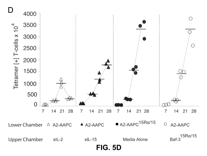
[0034] FIG 5. AAPC Co-expressing IL-15Ra and IL-15 Support Continuous
Enrichment of Antigen-Specific CD8+ T cells During Prolonged In Vitro
Expansion. T cells
8
CA 03064375 2019-11-20
WO 2018/217203
PCT/US2017/034364
from HLA A 02:01+ and CMV seropositive donors were sensitized in parallel
using (A) A2-
AAPC 15Rail5ar A2-AAPC + Baf-315Rail5with no exogenously supplemented
cytokines. Tet+ T
cells were quantitated by FACS analysis at 7, 21 and 28 days after incubation
with anti-CD3,
anti-CD8 and A2-NLV tetrameric complexes at 4 C for 20 mins (B) The mean total
yield of Tet+
T cells calculated after FACS analysis is plotted for each time point (error
bars = SEM). For
cultures sensitized with either A2-AAPC 15Ra/150r A2-AAPC + Baf-3 15Ra/IL-15,
the yield of Tet+ T
cells was 5-6 x 10⁷ compared to 1.8 ¨2.3 x 10⁷ for T cells sensitized with A2-
AAPC or A2-
AAPC IL-15Ra and supplemented with soluble IL-15 (p < 0.01). (C) T cells
stimulated for 14 days
with A2-AAPC 15Ra/15, A2-AAPC + Baf-3 15Ra/IL-15,
or sIL-2, sIL-15 or sIL-7+ sIL-4 loaded A2-
AAPC were labeled with CFSE, and then further stimulated for 5 days in the
same condition: i.e
with A2-AAPC 15Ra/15,
or A2-AAPC Baf_315Ra/IL-15
or sIL-2, sIL-15 or sIL-7+ sIL-4 loaded A2-
AAPC. sIL-2 loaded A2-AAPC T cells stimulated with CD3/CD28 beads (1:1) were
used as a
positive control. T cells in each condition were then stained with CD3 FITC,
CD8 PE, and A2-
NLV APC tetrameric complexes and analyzed by FACS. CFSE dilution was analyzed
within
A2-NLV Tet+ T cells as well as TetNeg CD8+ T cells to compare the
proliferative potential of
antigen-specific and non-specific CD8+ T cells in each condition. (D) T cells
from 3 HLA A2+
donors were co-cultured in 6 transwell plates containing a 3[tm permeable
membrane with (i)
A2-AAPC supplemented with either sIL-2 or sIL-15 or Baf3 15Ra/15 or A2-AAPC
15RW15 separated
from T cell co-cultures by the permeable membrane, (ii) A2-AAPC 15Ra/15 co-
cultured with T
cells in direct contact. The proportion of antigen-specific T cells in each
culture condition were
quantitated at 7, 14, 21 and 28 days by tetramer analysis and the total yield
of tetramer+ T cells,
calculated based on the proportion within the total CD3 + T cells is shown.
[0035] FIG
6. 15Ra/15 Stimulation Endorses the Expansion of Central Memory Phenotype
Antigen-Specific T cells. T cell memory phenotype was evaluated after 7, 14,
21 and 28 days in
culture for each culture condition using CCR7 and CD62L as markers of central
memory
phenotype (Tcm). T cells sensitized for 21-28 days under the different culture
conditions were
labeled with immunofluorescent antibodies: anti CD3 PE, anti-CD8 perCP, anti-
CD62L FITC
and anti CCR-7 PE-Cy7 and APC labelled A2-NLV tetrameric complexes for 20 mins
at 4 C
and analyzed by FACS. CD8+ Tet+ T cells were gated to determine the proportion
of antigen-
specific T cells expressing CD62L and CCR7. T cells labelled with HLA B 07:02 -
TPR
tetramers and unstained tubes served as controls for CD62L and CCR7. The total
yield of
9
CA 03064375 2019-11-20
WO 2018/217203 PCT/US2017/034364
CD62L+/ CCR7 + Tet+ T cells was calculated based on the proportion of each
population within
CD3+ T cells. (A) CD62L+/ CCR7 + Tet+ T cells at 7, 14, 21 and 28 days is
shown for each donor
in each culture condition (error bars = SEM). (B) A representative example
demonstrating the
proportion of CD62L+ /CD45RA- Tet+ T cells detected at 21 days (left panel)
and 28 days (right
panel) of culture initiation for each culture condition is shown.
[0036] FIG 7. 15Ra/15 Complexes Support the Generation of High Avidity
Antigen-Specific T
Cells. The proportion of CD8+ IFN y+ T cells responding to the CMVpp65 epitope
NLVPMVATV presented by HLA A 02:01, were quantitated on day 21 for each
parallel culture
condition (i) A2-AAPC + sIL-2 (20U/m1) or sIL-15 (long/ml) or sIL-2 + sIL-15,
or sIL-7
(long/ml) + sIL-4 (1666 U/ml); (ii) A2-AAPC IL-15Rct sIL-2 or sIL-15; and
(iii) A2-AAPC
15Ra/15 or A2-AAPC + Baf-3 IL-15Ra/IL-15,
with no exogenous cytokines. Aliquots of autologous
PBMC were loaded (37 C x 3 hrs) with serial dilutions of NLV peptide (10nM,
lOpM, 0.1pM),
and co-incubated with T cells at a responder: target ratio of 5:1 x 12 hours
in the presence of
brefeldin A (BFA). T cells were labelled with immunofluorescent antibodies
against CD3, CD4,
CD8, fixed and then permeabilized (fix and perm kit, invitrogen) and then
incubated with anti-
human IFNy FITC. Data were acquired on a BD LSRII flow cytometer and analyzed
using
flowjo software. (A) One representative example demonstrating the proportion
of IFNy + CD8+ T
cells in response to lOnM peptide loaded targets within CD3+ T cells is shown
(B) The total
yield of IFNy+ CD8+ T cells generated in response to lOnM peptide was
calculated from the
percentage of IFNy + CD8+ T cells and plotted for each donor in each culture
condition. (C) T
cells from 3 separate HLA A2 + donors that were sensitized in 6 well transwell
plates according
to cytokine conditions providing sIL-2, sIL-15, or 15Ra/15 complexes via the
permeable
transmembrane. Antigen-specific T cells generating functional cytokines in
response to lOnM
NLV peptide were evaluated on day 21 to quantitate the proportion of NLV
specific CD8+ IFNy+
T cells. (D) After 21 days of stimulation, the proportion of IFNy + CD8+ T
cells elicited upon
secondary stimulation with autologous targets loaded with serial dilutions of
NLV peptide is
shown for each donor in each culture condition (error bars = SEM), and (E) In
one representative
donor, IFNy + CD8+ T cells elicited in response targets loaded with serial
peptide dilutions is
shown. The proportion IFNy + CD8+ T cells in 15Ra/15 stimulated T cells was
significantly
greater than sIL-2 or sIL-15 cultures at all peptide dilutions (p= 0.001).
There was a significant
reduction in the proportion of IFNy + CD8+ T cells at lOpM versus 0.1 pM
peptide concentrations
CA 03064375 2019-11-20
WO 2018/217203 PCT/US2017/034364
for sIL-15 cultures (p <0.05).
[0037] FIG 8. 15Ra/15 Stimulated Antigen-Specific T Cells Efficiently Lyse
Targets at
Lower E:T Ratios. T cell cytotoxic capacity was measured in a standard 51Cr
release assay,
performed at 21- 28 days after culture initiation using peptide loaded
autologous BLCL as
targets. BLCL not loaded with peptide were used as control. (A) A fixed E:T
ratio of 10 T cells
to 1 target cell was used and the cytotoxic activity of T cells sensitized in
all culture conditions
was tested against targets loaded with serial dilutions of the NLV peptide
(10nM, 1nM, 01M,
lOpM, and 0.1pM at 37 C x 3 hours in serum free medium). (B) The cytotoxic
activity of T
cells was evaluated at decreasing E:T ratios against targets loaded with a
fixed concentration
(10nM) of peptide. (C) T cells in all culture conditions were evaluated for
expression of
intracellular granzyme B upon secondary re-stimulation with NLV peptide loaded
autologous
PBMC 21-28 days after culture initiation. T cells co-incubated with peptide
loaded autologous
PBMC were labelled with fluorescently labelled anti-CD3, anti-CD8, anti-CD4,
followed by
incubation with anti-human granzyme B after cell permeabilization and analyzed
by FACS. The
proportion of granzyme B positive T cells CD8+ T cells was evaluated. T cells
sensitized in the
presence of IL-15Ra/IL-15complexes generated significantly higher proportions
of granzyme B+
T cells compared to sensitization in the presence of soluble IL-2 (p= 0.05).
5. DETAILED DESCRIPTION
[0038] The present invention provides methods of generating antigen-
specific T cells for
therapeutic administration to a human patient having or suspected of having a
pathogen or
cancer, utilizing soluble Interleukin 15 (IL-15)/ Interleukin 15 Receptor
Subunit Alpha (IL-
15Ra) complexes ex vivo, in cell culture during ex vivo sensitizing of T cells
to the antigen or
during ex vivo culturing of antigen-specific T cells. Also disclosed are
antigen-specific T cells
generated by such methods, and methods of treating a human patient using such
antigen-specific
T cells. Cell culture systems comprising human T cells, antigen-presenting
cells, and soluble IL-
15/IL-15Ra complexes are also provided. According to the present invention,
soluble IL-15/IL-
15Ra complexes augment the expansion of antigen-specific T cells in vitro.
5.1. Methods of Generating Antigen-Specific T Cells for Adoptive Immunotherapy
5.1.1. Methods Using Ex Vivo Sensitization of Human T Cells
11
CA 03064375 2019-11-20
WO 2018/217203 PCT/US2017/034364
[0039] In one aspect, provided herein are methods of generating a
population of cells
comprising antigen-specific T cells for therapeutic administration to a human
patient having or
suspected of having a pathogen or cancer, comprising ex vivo sensitizing human
T cells to one or
more antigens of the pathogen or cancer, said ex vivo sensitizing comprising
co-culturing, over a
period of time in culture, a population of human blood cells comprising the
human T cells with
antigen presenting cells presenting the one or more antigens, in the presence
of soluble IL-15/IL-
15Ra complexes while in the absence of cells recombinantly expressing soluble
IL-15/IL-15Ra
complexes. In a preferred embodiment, the ex vivo sensitizing results in
expansion of antigen-
specific T cells that are specific for the one or more antigens. In a specific
embodiment, the
human T cells that are ex vivo sensitized are not genetically engineered to be
specific for the one
or more antigens (e.g., by expression of a chimeric antigen receptor (CAR) or
T cell receptor
(TCR) specific to the one or more antigens).
[0040] In various embodiments, the ex vivo sensitizing further comprises
adding soluble IL-
15/IL-15Ra complexes to the culture. In specific embodiments, the adding
soluble IL-15/IL-
15Ra complexes is such that the concentration of IL-15 in culture supernatant
is 10 to 104 pg/ml
upon said adding. In a preferred embodiment, the adding soluble IL-15/IL-15Ra
complexes is
such that the concentration of IL-15 in culture supernatant is 102 to 103
pg/ml upon said adding.
In a specific embodiment, the adding soluble IL-15/IL-15Ra complexes is such
that the
concentration of IL-15 in culture supernatant is 10 to 102 pg/ml upon said
adding. In another
specific embodiment, the adding soluble IL-15/IL-15Ra complexes is such that
the concentration
of IL-15 in culture supernatant is 103 to 104 pg/ml upon said adding. In
another specific
embodiment, the adding soluble IL-15/IL-15Ra complexes is such that the
concentration of IL-
15 in culture supernatant is about 102 pg/ml (i.e., 102 20% pg/ml) upon said
adding. In another
specific embodiment, the adding soluble IL-15/IL-15Ra complexes is such that
the concentration
of IL-15 in culture supernatant is about 103 pg/ml (i.e., 103 20% pg/ml) upon
said adding. In
specific embodiments, the adding soluble IL-15/IL-15Ra complexes is such that
the
concentration of IL-15 in culture supernatant is at least 10 pg/ml upon said
adding. In specific
embodiments, the adding soluble IL-15/IL-15Ra complexes is such that the
concentration of IL-
15 in culture supernatant is at least 102 pg/ml upon said adding. In specific
embodiments, the
adding soluble IL-15/IL-15Ra complexes is such that the concentration of IL-15
in culture
supernatant is at least 103 pg/ml upon said adding.
12
CA 03064375 2019-11-20
WO 2018/217203 PCT/US2017/034364
[0041] In specific embodiments, adding soluble IL-15/IL-15Ra complexes to
the culture is
done at the initiation of the co-culturing and every 1 to 14 days thereafter
during the co-
culturing. In specific embodiments, adding soluble IL-15/IL-15Ra complexes to
the culture is
done at the initiation of the co-culturing and every 3 to 12 days thereafter
during the co-
culturing. In specific embodiments, adding soluble IL-15/IL-15Ra complexes to
the culture is
done at the initiation of the co-culturing and every 5 to 10 days thereafter
during the co-
culturing. In preferred embodiments, adding soluble IL-15/IL-15Ra complexes to
the culture is
done at the initiation of the co-culturing and every 7 to 10 days thereafter
during the co-
culturing. In a specific embodiment, adding soluble IL-15/IL-15Ra complexes to
the culture is
done at the initiation of the co-culturing and about every 5 days thereafter
during the co-
culturing. In another specific embodiment, adding soluble IL-15/IL-15Ra
complexes to the
culture is done at the initiation of the co-culturing and about every 6 days
thereafter during the
co-culturing. In another specific embodiment, adding soluble IL-15/IL-15Ra
complexes to the
culture is done at the initiation of the co-culturing and about every 7 days
thereafter during the
co-culturing. In another specific embodiment, adding soluble IL-15/IL-15Ra
complexes to the
culture is done at the initiation of the co-culturing and about every 8 days
thereafter during the
co-culturing. In another specific embodiment, adding soluble IL-15/IL-15Ra
complexes to the
culture is done at the initiation of the co-culturing and about every 9 days
thereafter during the
co-culturing. In another specific embodiment, adding soluble IL-15/IL-15Ra
complexes to the
culture is done at the initiation of the co-culturing and about every 10 days
thereafter during the
co-culturing.
[0042] The soluble IL-15/IL-15Ra complexes can be any heterodimer complexes
of (1) an
IL-15 subunit that is a full-length wild-type human IL-15, or a fragment,
variant, mutant, or
derivative thereof that retains the ability to bind to IL-15Ra, and (2) an IL-
15Ra subunit that is a
fragment, variant, mutant, or derivative of wild-type human IL-15Ra that
retains the ability to
bind to IL-15 but lacks the ability to be anchored to the cell membrane by
itself, wherein the IL-
15 subunit and the IL-15Ra subunit are in a 1:1 molar ratio. Non-limiting
exemplary soluble IL-
15/IL-15Ra complexes that can be used according to the invention described
herein are
described in Section 6; Tamzalit et al., 2014, Proc Natl Acad Sci USA 111:8565-
8570;
Chertova et al., 2013, J Biol Chem 288:18093-18103; and Xu et al., 2013,
Cancer Res 73:3075-
3086. In certain embodiments, the IL-15 subunit is able to bind to the
Interleukin 15 Receptor
13
CA 03064375 2019-11-20
WO 2018/217203 PCT/US2017/034364
Subunit Beta (IL-15R13)/Interleukin 15 Receptor Subunit Gamma (IL-15Ry)
dimeric receptor. In
a specific embodiment, the IL-15 subunit is a full-length wild-type human IL-
15, such as a
protein having the amino acid sequence of
MRISKPHLRSISIQCYLCLLLNSHFLTEAGIHVFILGCFSAGLPKTEANWVNVISDLKKIED
LIQSMHIDATLYTESDVHPSCKVTAMKCFLLELQVISLESGDASIHDTVENLIILANNSLSS
NGNVTESGCKECEELEEKNIKEFLQSFVHIVQMFINTS (SEQ ID NO:1) (National Center
for Biotechnology Information (NCBI) Reference Sequence: NP 000576.1)
preferably from
which the signal peptide has been cleaved, or
MVLGTIDLCSCFSAGLPKTEANWVNVISDLKKIEDLIQSMHIDATLYTESDVHPSCKVTA
MKCFLLELQVISLESGDASIHDTVENLIILANNSLSSNGNVTESGCKECEELEEKNIKEFLQ
SFVHIVQMFINTS (SEQ ID NO:2) (NCBI Reference Sequence: NP 751915.1) preferably
from
which the signal peptide has been cleaved, or, preferably, the mature form
having the amino
acid sequence of
NWVNVISDLKKIEDLIQSMHIDATLYTESDVHPSCKVTAMKCFLLELQVISLESGDASIH
DTVENLIILANNSLSSNGNVTESGCKECEELEEKNIKEFLQSFVHIVQMFINTS (SEQ ID
NO:3) (Grabstein et al., 1994, Science 264:965-968). In another specific
embodiment, the IL-15
subunit is a mutant human IL-15 as described in Xu et at., 2013, Cancer Res
73:3075-3086. In
another specific embodiment, the IL-15 subunit is a fusion protein, for
example, wherein the IL-
15 sequence is linked to the Fc portion of a human immunoglobulin, such as the
Fc portion of
human IgG (e.g., IgG1). In a specific embodiment, the IL-15Ra subunit is a
cleaved form of IL-
15Ra that is secreted by a cell in which it is expressed, such as the
naturally produced cleaved
form of IL-15Ra described in Chertova et al., 2013, J Biol Chem 288:18093-
18103 or a secreted
form of any of the other IL-15Ra isoforms. In a specific embodiment, the IL-
15Ra isoform has
the amino acid sequence of one of the following sequences from which the
signal peptide has
been cleaved:
MAPRRARGCRTLGLPALLLLLLLRPPATRGITCPPPMSVEHADIWVKSYSLYSRERYICN
SGFKRKAGTSSLTECVLNKATNVAHWTTPSLKCIRDPALVHQRPAPPSTVTTAGVTPQP
ESLSPSGKEPAASSPSSNNTAATTAAIVPGSQLMPSKSPSTGTTEISSHESSHGTPSQTTAK
NWELTASASHQPPGVYPQGHSDTTVAISTSTVLLCGLSAVSLLACYLKSRQTPPLASVE
MEAMEALPVTWGTSSRDEDLENCSHHL (SEQ ID NO:4) (NCBI Reference Sequence:
NP 002180.1),
14
CA 03064375 2019-11-20
WO 2018/217203 PCT/US2017/034364
MAPRRARGCRTLGLPALLLLLLLRPPATRGITCPPPMSVEHADIWVKSYSLYSRERYICN
SGFKRKAGTSSLTECVLNKATNVAHWTTPSLKCIKPAASSPSSNNTAATTAAIVPGSQL
MPSKSPSTGTTEISSHESSHGTPSQTTAKNWELTASASHQPPGVYPQGHSDTTVAISTSTV
LLCGLSAVSLLACYLKSRQTPPLASVEMEAMEALPVTWGTSSRDEDLENCSHHL (SEQ
ID NO:5) (NCBI Reference Sequence: NP 751950.2),
MSVEHADIWVKSYSLYSRERYICNSGFKRKAGTSSLTECVLNKATNVAHWTTPSLKCIR
DPALVHQRPAPPSTVTTAGVTPQPESLSPSGKEPAASSPSSNNTAATTAAIVPGSQLMPSK
SPSTGTTEISSHESSHGTPSQTTAKNWELTASASHQPPGVYPQGHSDTTVAISTSTVLLCG
LSAVSLLACYLKSRQTPPLASVEMEAMEALPVTWGTSSRDEDLENCSHHL (SEQ ID
NO:6) (NCBI Reference Sequence: NP 001230468.1), and
MRLAGRQVPEQRSPPPPGLGSARPGSPAVSCGAAAMAPRRARGCRTLGLPALLLLLLLR
PPATRDARDRLAVLAGRSRISESFNHEVQTHEACVRLRTMENCPQCHEIHRTSRQQAGIT
CPPPMSVEHADIWVKSYSLYSRERYICNSGFKRKAGTSSLTECVLNKATNVAHWTTPSL
KCIRDPALVHQRPAPPSTVTTAGVTPQPESLSPSGKEPAASSPSSNNTAATTAAIVPGSQL
MPSKSPSTGTTEISSHESSHGTPSQTTAKNWELTASASHQPPGVYPQGHSDTTVAISTSTV
LLCGLSAVSLLACYLKSRQTPPLASVEMEAMEALPVTWGTSSRDEDLENCSHHL (SEQ
ID NO:7) (NCBI Reference Sequence: NP 001243694.1). In another specific
embodiment, the
IL-15Ra subunit is a fusion protein, for example, wherein the IL-15Ra sequence
is linked to the
Fc portion of a human immunoglobulin, such as the Fc portion of human IgG
(e.g., IgG1). In a
specific embodiment, the IL-15 subunit is a fusion protein, for example,
wherein the IL-15
sequence is linked to the Fc portion of a human immunoglobulin, such as the Fc
portion of
human IgG (e.g., IgG1), and the IL-15Ra subunit is a fusion protein, for
example, wherein the
IL-15Ra sequence is linked to the Fc portion of a human immunoglobulin, such
as the Fc portion
of human IgG (e.g., IgG1).
[0043] In a specific embodiment, the soluble IL-15/IL-15Ra complexes are
produced by
cells transduced with vector(s) to co-express within the same cell the IL-15
subunit and the IL-
15Ra subunit. In a preferred embodiment, the IL-15 subunit and the IL-15Ra
subunit are
expressed from two different vectors within the same cell. Preferably, the
soluble IL-15/IL-
15Ra complexes are secreted by a cell that recombinantly expresses the IL-15
subunit and the
IL-15Ra subunit, and can be recovered from cell culture supernatant.
[0044] In a specific embodiment, the soluble IL-15/IL-15Ra complexes are
produced by
CA 03064375 2019-11-20
WO 2018/217203 PCT/US2017/034364
adding purified IL-15 subunit to a culture of cells transduced with a vector
comprising a
nucleotide sequence encoding the IL-15Ra subunit, which cells secrete the
encoded IL-15Ra
subunit, and recovering the soluble IL-15/IL-15Ra complexes from the cell
culture supernatant.
The IL-15 subunit can be from a previously purified, cryopreserved preparation
or
acommercially available product or recovered from cell culture supernatant of
cells transduced
with a vector comprising a nucleotide sequence encoding the IL-15 subunit,
which cells secrete
the encoded IL-15 subunit. Since IL-15 is typically transiently expressed when
recombinantly
expressed in cells in the absence of recombinant co-expression in the same
cell of IL-15Ra, in
order to avoid the need for repeated transduction of cells for purposes of
producing IL-15 alone,
obtaining an IL-15 subunit from a cryopreserved preparation or commercially
available source is
preferred for use in the adding step.
[0045] In specific embodiments, the soluble IL-15/IL-15Ra complexes are
produced by
complexing the IL-15 subunit and the IL-15Ra subunit ex vivo. In a specific
embodiment, the
IL-15 subunit is a commercially available product or is recovered from cell
culture supernatant
of cells transduced with a vector comprising a nucleotide sequence encoding
the IL-15 subunit,
which cells secrete the encoded IL-15 subunit. The IL-15Ra subunit can be a
commercially
available product or recovered from cell culture supernantant of cells
transduced with a vector
comprising a nucleotide sequence encoding the IL-15Ra subunit, which cells
secrete the encoded
IL-15Ra subunit. The IL-15 subunit and the IL-15Ra subunit can be complexed ex
vivo by any
method known in the art for complexing proteins ex vivo, such as by combining
purified IL-15
subunit proteins and purified IL-15Ra subunit proteins ex vivo in a buffered
solution (for
example, in the presence of bovine serum albumin in phosphate-buffered saline
(PBS)) at 37 C
for a period of time (for example, as described in Epardaud et al., 2008,
Cancer Res 68:2972-
2983).
[0046] Any mammalian cell line cells that can be passaged in vitro can be
used for producing
the soluble IL-15/IL-15Ra complexes, the IL15 subunit and/or the IL-15Ra
subunit, such as
Ba/F3 cells (i.e., Baf-3 cells), K562 cells, or murine fibroblast NIH 3T3
based artificial antigen
presenting cells (AAPCs) (Hasan et al., 2009, J Immunol 183: 2837-2850). The
cells can be
human, murine, hamster, or other mammalian cells. The soluble IL-15/IL-15Ra
complexes, the
IL15 subunit and/or the IL-15Ra subunit can be purified from the supernatants
from the culture
of transduced cells by any method known in the art for purifying proteins,
such as by fast protein
16
CA 03064375 2019-11-20
WO 2018/217203 PCT/US2017/034364
liquid chromatography (FPLC). Non-limiting exemplary methods of producing the
soluble IL-
15/IL-15Ra complexes are described in Section 6; Tamzalit et al., 2014, Proc
Natl Acad Sci U S
A 111:8565-8570; Chertova etal., 2013, J Biol Chem 288:18093-18103; and Xu
etal., 2013,
Cancer Res 73:3075-3086.
[0047] In some embodiments, the preparation of soluble IL-15/IL-15Ra
complexes produced
as described herein is free of cells recombinantly expressing the soluble IL-
15/IL-15Ra
complexes. In other embodiments, the preparation of soluble IL-15/IL-15Ra
complexes
produced as described herein contains cells recombinantly expressing the
soluble IL-15/IL-15Ra
complexes. When the preparation of soluble IL-15/IL-15Ra complexes contains
cells
recombinantly expressing the soluble IL-15/IL-15Ra complexes, the method of
generating a
population of cells comprising antigen-specific T cells further comprises,
before the step of
adding soluble IL-15/IL-15Ra complexes to the culture, a step of removing the
cells
recombinantly expressing the soluble IL-15/IL-15Ra complexes from the
preparation or a step of
purifying the soluble IL-15/IL-15Ra complexes from the preparation so as to
separate the
complexes from the cells recombinantly expressing the soluble IL-15/IL-15Ra
complexes. The
removing or purifying can be performed by any method known in the art for
removing cells from
a mixture of cells and proteins or purifying proteins from a mixture of cells
and proteins, such as
by centrifugation or by use of a filter.
[0048] In certain embodiments, the soluble IL-15/IL-15Ra complexes are
thawed from a
cryopreserved stock before being added to the culture. In a specific
embodiment, the method of
generating a population of cells comprising antigen-specific T cells further
comprises thawing
the soluble IL-15/IL-15Ra complexes from a cryopreserved stock before adding
them to the
culture. In a further specific embodiment, the method of generating a
population of cells
comprising antigen-specific T cells further comprises cryopreserving soluble
IL-15/IL-15Ra
complexes and thawing the soluble IL-15/IL-15Ra complexes before adding them
to the culture.
In a particular embodiment, cryopreserving soluble IL-15/IL-15Ra complexes
comprises
combining soluble IL-15/IL-15Ra complexes with a cryopreservative, such as
dimethyl
sulfoxide (DMSO), glycerol, polyvinylpyrrolidine, polyethylene glycol, albumin
(such as bovine
serum albumin), dextran, sucrose, ethylene glycol, i-erythritol, D-ribitol, D-
mannitol, D-sorbitol,
i-inositol, D-lactose, choline chloride, amino acids, methanol, acetamide,
glycerol monoacetate,
inorganic salts, or any cryopreservative known in the art for use in
cryopreserving proteins. The
17
CA 03064375 2019-11-20
WO 2018/217203 PCT/US2017/034364
concentration of the soluble IL-15/IL-15Ra complexes in the cryopreserved
stock can be any
concentration suitable for long-term storage of the complexes, such as 10
pg/m1 to 10 mg/ml (for
example, 10 pg/m1 to 0.1 mg/ml, 0.1 mg/ml to 1 mg/ml, or 1 mg/ml to 10 mg/ml).
In a specific
embodiment, the concentration of the soluble IL-15/IL-15Ra complexes in the
cryopreserved
stock is about 0.1 mg/ml (i.e., 0.1 20% mg/ml). The cryopreserved soluble IL-
15/IL-15Ra
complexes can be stored in liquid nitrogen or dry ice for long-term storage,
or a fridge (0-8 C)
for short-term storage (such as up to a week or 1-3 days).
[0049] In various embodiments, the ex vivo sensitizing further comprises
adding antigen
presenting cells presenting the one or more antigens to the culture. The
antigen presenting cells
are typically irradiated cells to prevent multiplication of these cells after
being added to the
culture. In specific embodiments, the ex vivo sensitizing further comprises
adding antigen
presenting cells presenting the one or more antigens to the culture at the
initiation of said co-
culturing and every 1 to 14 days thereafter during the co-culturing. In
specific embodiments, the
ex vivo sensitizing further comprises adding antigen presenting cells
presenting the one or more
antigens to the culture at the initiation of said co-culturing and every 3 to
12 days thereafter
during the co-culturing. In specific embodiments, the ex vivo sensitizing
further comprises
adding antigen presenting cells presenting the one or more antigens to the
culture at the initiation
of said co-culturing and every 5 to 10 days thereafter during the co-
culturing. In preferred
embodiments, the ex vivo sensitizing further comprises adding antigen
presenting cells
presenting the one or more antigens to the culture at the initiation of said
co-culturing and every
7 to 10 days thereafter during the co-culturing. In a specific embodiment, the
ex vivo sensitizing
further comprises adding antigen presenting cells presenting the one or more
antigens to the
culture at the initiation of said co-culturing and about every 5 days
thereafter during the co-
culturing. In another specific embodiment, the ex vivo sensitizing further
comprises adding
antigen presenting cells presenting the one or more antigens to the culture at
the initiation of said
co-culturing and about every 6 days thereafter during the co-culturing. In
another specific
embodiment, the ex vivo sensitizing further comprises adding antigen
presenting cells presenting
the one or more antigens to the culture at the initiation of said co-culturing
and about every 7
days thereafter during the co-culturing. In another specific embodiment, the
ex vivo sensitizing
further comprises adding antigen presenting cells presenting the one or more
antigens to the
culture at the initiation of said co-culturing and about every 8 days
thereafter during the co-
18
CA 03064375 2019-11-20
WO 2018/217203 PCT/US2017/034364
culturing. In another specific embodiment, the ex vivo sensitizing further
comprises adding
antigen presenting cells presenting the one or more antigens to the culture at
the initiation of said
co-culturing and about every 9 days thereafter during the co-culturing. In
another specific
embodiment, the ex vivo sensitizing further comprises adding antigen
presenting cells presenting
the one or more antigens to the culture at the initiation of said co-culturing
and about every 10
days thereafter during the co-culturing. In specific embodiments, adding
soluble IL-15/IL-15Ra
complexes to the culture is done at the time of adding antigen presenting
cells to the culture
(such as, on the same day or preferably in the same hour).
[0050] In specific embodiments, the aforementioned period of time in
culture (termed herein
"the Sensitization Culture Time;" i.e., the culture time period over which co-
culturing occurs) is
at least 14 days (preferably, at least 21 days). In a specific embodiment, the
Sensitization
Culture Time is in the range of 21-28 days. In another specific embodiment,
the Sensitization
Culture Time is 21 days. In another specific embodiment, the Sensitization
Culture Time is 22
days. In another specific embodiment, the Sensitization Culture Time is 23
days. In another
specific embodiment, the Sensitization Culture Time is 24 days. In another
specific
embodiment, the Sensitization Culture Time is 25 days. In another specific
embodiment, the
Sensitization Culture Time is 26 days. In another specific embodiment, the
Sensitization Culture
Time is 27 days. In a preferred embodiment, the Sensitization Culture Time is
28 days. In
specific embodiments, the Sensitization Culture Time is at least 28 days.
[0051] The ex vivo sensitizing step can be performed by any method known in
the art to
stimulate T cells to be antigen-specific ex vivo, such as a method as
described in Section 6;
Koehne etal., 2000, Blood 96:109-117; Trivedi etal., 2005, Blood 105:2793-
2801; Hague etal.,
2007, Blood 110:1123-1131; Hasan et al., 2009, J Immunol 183: 2837-2850;
Feuchtinger et at.,
2010, Blood 116:4360-4367; Doubrovina etal., 2012, Blood 120:1633-1646; Leen
etal., 2013,
Blood 121:5113-5123; Papadopoulou etal., 2014, Sci Transl Med 6:242ra83;
Sukdolak etal.,
2013, Biol Blood Marrow Transplant 19:1480-1492; Koehne etal., 2015, Biol
Blood Marrow
Transplant 21: 1663-1678; International Patent Application Publication No. WO
2016/073550;
or International Patent Application Publication No. WO 2017/044678.
[0052] The antigen presenting cells used in the ex vivo sensitizing step
can be any antigen
presenting cells suitable for presenting the one or more antigens, including
professional antigen
presenting cells and non-professional antigen presenting cells. In specific
embodiments, the
19
CA 03064375 2019-11-20
WO 2018/217203 PCT/US2017/034364
antigen presenting cells used in the ex vivo sensitizing step are dendritic
cells, cytokine-activated
monocytes, peripheral blood mononuclear cells (PBMCs), Epstein-Barr virus-
transformed B-
lymphoblastoid cell line cells (EBV-BLCL cells), or artificial antigen
presenting cells (AAPCs).
In a specific embodiment, the antigen presenting cells are dendritic cells. In
another specific
embodiment, the antigen presenting cells are PBMCs. In another specific
embodiment, the
antigen presenting cells are EBV-BLCL cells. In another specific embodiment,
the antigen
presenting cells are AAPCs. In some embodiments, the antigen presenting cells
are derived from
the donor of the population of human blood cells. In other embodiments, the
antigen presenting
cells are allogeneic to the donor of the population of human blood cells. The
antigen presenting
cells can be obtained by any method known in the art, such as the method(s)
described in Section
6; Koehne et al., 2000, Blood 96:109-117; Koehne et al., 2002, Blood 99:1730-
1740; Trivedi et
at., 2005, Blood 105:2793-2801; O'Reilly et al., 2007, Immunol Res 38:237-250;
Hasan et al.,
2009, J Immunol 183: 2837-2850; Barker et at., 2010, Blood 116:5045-5049; 0'
Reilly et at.,
2011, Best Practice & Research Clinical Haematology 24:381-391; Doubrovina et
at., 2012,
Blood 120:1633-1646; Koehne et at., 2015, Biol Blood Marrow Transplant 21:
1663-1678;
International Patent Application Publication No. WO 2016/073550; or
International Patent
Application Publication No. WO 2017/044678.
[0053] In some embodiments, the antigen presenting cells are loaded with
one or more
immunogenic peptides or proteins derived from the one or more antigens. Non-
limiting
exemplary methods for loading antigen presenting cells with peptide(s) derived
from antigen(s)
can be found in Section 6; Trivedi et at., 2005, Blood 105:2793-2801; Barker
et at., 2010, Blood
116:5045-5049; Doubrovina et al., 2012, Blood 120:1633-1646; Hasan et al.,
2009, J Immunol
183: 2837-2850; Koehne et al., 2015, Biol Blood Marrow Transplant 21: 1663-
1678;
International Patent Application Publication No. WO 2016/073550; and
International Patent
Application Publication No. WO 2017/044678. In other embodiments, the antigen
presenting
cells are genetically engineered to recombinantly express one or more
immunogenic peptides or
proteins derived from the one or more antigens. Any appropriate method known
in the art for
introducing nucleic acid vehicles into cells to express proteins, such as
transduction or
transformation, can be used to genetically engineer the antigen presenting
calls to recombinantly
express the one or more immunogenic peptides or proteins derived from the one
or more
antigens.
CA 03064375 2019-11-20
WO 2018/217203 PCT/US2017/034364
[0054] In some embodiments, the one or more immunogenic peptides or
proteins are a pool
of overlapping peptides derived from the one or more antigens. In specific
embodiments, the
pool of overlapping peptides is a pool of overlapping pentadecapeptides. In
other embodiments,
the one or more immunogenic peptides or proteins are one or more proteins
derived from the one
or more antigens.
[0055] In specific embodiments, the method of generating a population of
cells comprising
antigen-specific T cells further comprises, after the step of ex vivo
sensitizing, a step of
cryopreserving the ex vivo sensitized (and preferably expanded) human T cells,
or a fraction
thereof. In a specific embodiment, the method of generating a population of
cells comprising
antigen-specific T cells further comprises, after the step of cryopreserving,
steps of thawing and
optionally expanding in culture the ex vivo sensitized (and preferably
expanded) and
cryopreserved human T cells or a faction thereof The cryopreserving and
thawing steps can be
performed by known methods in the art for cryopreserving T cells and thawing T
cells,
respectively.
[0056] The term "about" shall be construed so as to allow normal variation,
such as, for
example, a variation within 20%.
5.1.2. Methods Using Human Antigen-Specific T Cells
[0057] In another aspect, provided herein are methods of generating a
population of cells
comprising antigen-specific T cells for therapeutic administration to a human
patient having or
suspected of having a pathogen or cancer, comprising ex vivo culturing a
population of human
blood cells comprising human antigen-specific T cells over a period of time in
culture in the
presence of soluble IL-15/IL-15Ra complexes while in the absence of cells
recombinantly
expressing soluble IL-15/IL-15Ra complexes, wherein the human antigen-specific
T cells are
specific to one or more antigens of the pathogen or cancer. In a preferred
embodiment, the ex
vivo culturing results in expansion of the human antigen-specific T cells.
[0058] In some embodiments, the human antigen-specific T cells
recombinantly express one
or more chimeric antigen receptors (CARs) recognizing the one or more
antigens. In a specific
embodiment, the method of generating a population of cells comprising antigen-
specific T cells
further comprises, prior to the ex vivo culturing, transducing a population of
T cells with one or
21
CA 03064375 2019-11-20
WO 2018/217203 PCT/US2017/034364
more nucleic acids encoding the one or more CARs recognizing the one or more
antigens;
thereby producing the human antigen-specific T cells.
[0059] CARs are engineered receptors that provide both antigen binding and
immune cell
activation functions (Sadelain et al., 2013, Cancer Discovery 3:388-398). They
usually comprise
an antigen-binding domain (e.g., derived from a monoclonal antibody or the
extracellular domain
of a receptor), a transmembrane domain, an intracellular domain, and
optionally a co-stimulatory
domain. CARs can be used to graft the specificity of an antigen-binding domain
onto an
immune cell such as a T cell.
[0060] The population of T cells transduced with one or more nucleic acids
encoding the one
or more CARs can be generated by any method known in the art, for example, as
described in
Stauss et at., 2015, Curr Opin Pharmacol 24:113-118; Sharpe and Mount, 2015,
Dis Model Mech
8:337-350; or Park et al., 2011, Trends Biotechnol 29:550-557.
[0061] The nucleic acid encoding a CAR can be DNA, RNA, or a nucleic acid
analog. In
specific embodiments, such a nucleic acid may be part of a vector. In a
specific embodiment, the
vector is an expression vector that is capable of directing the expression of
a nucleic acid
encoding a polypeptide of the CAR described herein in T cells. Non-limiting
examples of
expression vectors suitable for directing the expression of a nucleic acid
encoding a polypeptide
of the CAR described herein include, but are not limited to, plasmids and
viral vectors, such as
synthetic vectors, lentiviral vectors, replication-defective retroviral
vectors, or autonomously
replicating plasmids. In a specific embodiment, an expression vector used for
directing the
expression of a nucleic acid encoding a polypeptide of the CAR described
herein includes one or
more regulatory sequences operably linked to the nucleic acid to be expressed.
"Operably
linked" is intended to mean that a nucleic acid of interest is linked to the
regulatory sequence(s)
in a manner which allows for expression of the nucleic acid in T cells.
Regulatory sequences
include promoters, enhancers and other expression control elements (e.g.,
polyadenylation
signals).
[0062] A nucleic acid encoding a polypeptide of the CAR described herein,
for example, an
expression vector, can be transduced into host cells via conventional
transformation or
transfection (such as, transfection by a virus, e.g., a retrovirus or
lentivirus) techniques. Such
techniques include, but are not limited to, calcium phosphate or calcium
chloride co-
precipitation, DEAE-dextran-mediated transfection, lipofection, and
electroporation. Cells
22
CA 03064375 2019-11-20
WO 2018/217203 PCT/US2017/034364
containing a nucleic acid encoding a polynucleotide of the CAR described
herein may be
selected using one or more selectable markers known in the art.
[0063] In some embodiments, the human antigen-specific T cells
recombinantly express one
or more T cell receptors (TCRs) recognizing the one or more antigens. In a
specific
embodiment, the method of generating a population of cells comprising antigen-
specific T cells
further comprises, prior to the ex vivo culturing, transducing a population of
T cells with one or
more nucleic acids encoding the one or more TCRs recognizing the one or more
antigens;
thereby producing the human antigen-specific T cells.
[0064] TCR is a cell surface molecule on T cells that is responsible for
recognizing antigen
peptide-bound major histocompatibility complex (MEW) molecules.
[0065] The population of T cells transduced with one or more nucleic acids
encoding the one
or more TCRs can be generated by any method known in the art, for example, as
described in
Stauss et at., 2015, Curr Opin Pharmacol 24:113-118; Sharpe and Mount, 2015,
Dis Model Mech
8:337-350; Kunert et al., 2013, Front Immunol 4: 363; Stone et al., 2012,
Methods Enzymol
503:189-222; or Park et al., 2011, Trends Biotechnol 29:550-557.
[0066] The nucleic acid encoding a TCR can be DNA, RNA, or a nucleic acid
analog. In
specific embodiments, such a nucleic acid may be part of a vector. In a
specific embodiment, the
vector is an expression vector that is capable of directing the expression of
a nucleic acid
encoding a polypeptide of the TCR described herein in T cells. Non-limiting
examples of
expression vectors suitable for directing the expression of a nucleic acid
encoding a polypeptide
of the TCR described herein include, but are not limited to, plasmids and
viral vectors, such as
synthetic vectors, lentiviral vectors, replication-defective retroviral
vectors, or autonomously
replicating plasmids. In a specific embodiment, an expression vector used for
directing the
expression of a nucleic acid encoding a polypeptide of the TCR described
herein includes one or
more regulatory sequences operably linked to the nucleic acid to be expressed.
"Operably
linked" is intended to mean that a nucleic acid of interest is linked to the
regulatory sequence(s)
in a manner which allows for expression of the nucleic acid in T cells.
Regulatory sequences
include promoters, enhancers and other expression control elements (e.g.,
polyadenylation
signals).
[0067] A nucleic acid encoding a polypeptide of the TCR described herein,
for example, an
expression vector, can be transduced into host cells via conventional
transformation or
23
CA 03064375 2019-11-20
WO 2018/217203 PCT/US2017/034364
transfection (such as, transfection by a virus, e.g., a retrovirus or
lentivirus) techniques. Such
techniques include, but are not limited to, calcium phosphate or calcium
chloride co-
precipitation, DEAE-dextran-mediated transfection, lipofection, and
electroporation. Cells
containing a nucleic acid encoding a polynucleotide of the TCR described
herein may be selected
using one or more selectable markers known in the art.
[0068] In some embodiments, the human antigen-specific T cells are antigen-
specific T cells
generated by ex vivo sensitization, performed by any method known in the art
to stimulate T cells
to be antigen-specific ex vivo, such as a method as described in Section 6;
Koehne et at., 2000,
Blood 96:109-117; Trivedi et al., 2005, Blood 105:2793-2801; Hague et al.,
2007, Blood
110:1123-1131; Hasan et al., 2009, J Immunol 183: 2837-2850; Feuchtinger et
al., 2010, Blood
116:4360-4367; Doubrovina et al., 2012, Blood 120:1633-1646; Leen et al.,
2013, Blood
121:5113-5123; Papadopoulou et al., 2014, Sci Transl Med 6:242ra83; Sukdolak
et al., 2013,
Biol Blood Marrow Transplant 19:1480-1492; Koehne et al., 2015, Biol Blood
Marrow
Transplant 21: 1663-1678; International Patent Application Publication No. WO
2016/073550;
or International Patent Application Publication No. WO 2017/044678.
[0069] In other embodiments, the human antigen-specific T cells are antigen-
specific T cells
purified from cells (such as peripheral blood mononuclear cells (PBMCs))
derived from a blood
sample that is seropositive for the one or more antigens (for example, by
sorting (such as
fluorescence activated cell sorting) T cells that recognize the one or more
antigens from the
blood sample cells).
[0070] In various embodiments, the method of generating a population of
cells comprising
antigen-specific T cells further comprises adding soluble IL-15/IL-15Ra
complexes to the
culture. In specific embodiments, the adding soluble IL-15/IL-15Ra complexes
is such that the
concentration of IL-15 in culture supernatant is 10 to 104 pg/ml upon said
adding. In a preferred
embodiment, the adding soluble IL-15/IL-15Ra complexes is such that the
concentration of IL-
15 in culture supernatant is 102 to 103 pg/ml upon said adding. In a specific
embodiment, the
adding soluble IL-15/IL-15Ra complexes is such that the concentration of IL-15
in culture
supernatant is 10 to 102 pg/ml upon said adding. In another specific
embodiment, the adding
soluble IL-15/IL-15Ra complexes is such that the concentration of IL-15 in
culture supernatant
is 103 to 104 pg/ml upon said adding. In another specific embodiment, the
adding soluble IL-
15/IL-15Ra complexes is such that the concentration of IL-15 in culture
supernatant is about 102
24
CA 03064375 2019-11-20
WO 2018/217203 PCT/US2017/034364
pg/ml (i.e.,102 20% pg/ml) upon said adding. In another specific embodiment,
the adding
soluble IL-15/IL-15Ra complexes is such that the concentration of IL-15 in
culture supernatant
is about 103 pg/ml (i.e., 103 20% pg/ml) upon said adding. In specific
embodiments, the
adding soluble IL-15/IL-15Ra complexes is such that the concentration of IL-15
in culture
supernatant is at least 10 pg/ml upon said adding. In specific embodiments,
the adding soluble
IL-15/IL-15Ra complexes is such that the concentration of IL-15 in culture
supernatant is at least
102 pg/ml upon said adding. In specific embodiments, the adding soluble IL-
15/IL-15Ra
complexes is such that the concentration of IL-15 in culture supernatant is at
least 103 pg/ml
upon said adding.
[0071] In specific embodiments, adding soluble IL-15/IL-15Ra complexes to
the culture is
done at the initiation of the ex vivo culturing and every 1 to 14 days
thereafter during the ex vivo
culturing. In specific embodiments, adding soluble IL-15/IL-15Ra complexes to
the culture is
done at the initiation of the ex vivo culturing and every 3 to 12 days
thereafter during the ex vivo
culturing. In specific embodiments, adding soluble IL-15/IL-15Ra complexes to
the culture is
done at the initiation of the ex vivo culturing and every 5 to 10 days
thereafter during the ex vivo
culturing. In preferred embodiments, adding soluble IL-15/IL-15Ra complexes to
the culture is
done at the initiation of the ex vivo culturing and every 7 to 10 days
thereafter during the ex vivo
culturing. In a specific embodiment, adding soluble IL-15/IL-15Ra complexes to
the culture is
done at the initiation of the ex vivo culturing and about every 5 days
thereafter during the ex vivo
culturing. In another specific embodiment, adding soluble IL-15/IL-15Ra
complexes to the
culture is done at the initiation of the ex vivo culturing and about every 6
days thereafter during
the ex vivo culturing. In another specific embodiment, adding soluble IL-15/IL-
15Ra complexes
to the culture is done at the initiation of the ex vivo culturing and about
every 7 days thereafter
during the ex vivo culturing. In another specific embodiment, adding soluble
IL-15/IL-15Ra
complexes to the culture is done at the initiation of the ex vivo culturing
and about every 8 days
thereafter during the ex vivo culturing. In another specific embodiment,
adding soluble IL-15/IL-
15Ra complexes to the culture is done at the initiation of the ex vivo
culturing and about every 9
days thereafter during the ex vivo culturing. In another specific embodiment,
adding soluble IL-
15/IL-15Ra complexes to the culture is done at the initiation of the ex vivo
culturing and about
every 10 days thereafter during the ex vivo culturing.
[0072] The soluble IL-15/IL-15Ra complexes can be any heterodimer complexes
of (1) an
CA 03064375 2019-11-20
WO 2018/217203 PCT/US2017/034364
IL-15 subunit that is a full-length wild-type human IL-15, or a fragment,
variant, mutant, or
derivative thereof that retains the ability to bind to IL-15Ra, and (2) an IL-
15Ra subunit that is a
fragment, variant, mutant, or derivative of wild-type human IL-15Ra that
retains the ability to
bind to IL-15 but lacks the ability to be anchored to the cell membrane by
itself, wherein the IL-
15 subunit and the IL-15Ra subunit are in a 1:1 molar ratio. Non-limiting
exemplary soluble IL-
15/IL-15Ra complexes that can be used according to the invention described
herein are
described in Section 6; Tamzalit et al., 2014, Proc Natl Acad Sci USA 111:8565-
8570;
Chertova et al., 2013, J Biol Chem 288:18093-18103; and Xu et al., 2013,
Cancer Res 73:3075-
3086. In certain embodiments, the IL-15 subunit is able to bind to the
Interleukin 15 Receptor
Subunit Beta (IL-15Rf3)/Interleukin 15 Receptor Subunit Gamma (IL-15Ry)
dimeric receptor. In
a specific embodiment, the IL-15 subunit is a full-length wild-type human IL-
15, such as a
protein having the amino acid sequence of
MRISKPHLRSISIQCYLCLLLNSHFLTEAGIHVFILGCFSAGLPKTEANWVNVISDLKKIED
LIQSMHIDATLYTESDVHPSCKVTAMKCFLLELQVISLESGDASIHDTVENLIILANNSLSS
NGNVTESGCKECEELEEKNIKEFLQSFVHIVQMFINTS (SEQ ID NO:1) (NCBI Reference
Sequence: NP 000576.1) preferably from which the signal peptide has been
cleaved, or
MVLGTIDLCSCFSAGLPKTEANWVNVISDLKKIEDLIQSMHIDATLYTESDVHPSCKVTA
MKCFLLELQVISLESGDASIHDTVENLIILANNSLS SNGNVTESGCKECEELEEKNIKEFLQ
SFVHIVQMFINTS (SEQ ID NO:2) (NCBI Reference Sequence: NP 751915.1) preferably
from
which the signal peptide has been cleaved, or, preferably, the mature form
having the amino
acid sequence of
NWVNVISDLKKIEDLIQSMHIDATLYTESDVHPSCKVTAMKCFLLELQVISLESGDASIH
DTVENLIILANNSLS SNGNVTESGCKECEELEEKNIKEFLQSFVHIVQMFINTS (SEQ ID
NO:3) (Grabstein et al., 1994, Science 264:965-968). In another specific
embodiment, the IL-15
subunit is a mutant human IL-15 as described in Xu et at., 2013, Cancer Res
73:3075-3086. In
another specific embodiment, the IL-15 subunit is a fusion protein, for
example, wherein the IL-
15 sequence is linked to the Fc portion of a human immunoglobulin, such as the
Fc portion of
human IgG (e.g., IgG1). In a specific embodiment, the IL-15Ra subunit is a
cleaved form of IL-
15Ra that is secreted by a cell in which it is expressed, such as the
naturally produced cleaved
form of IL-15Ra described in Chertova et al., 2013, J Biol Chem 288:18093-
18103 or a secreted
form of any of the other IL-15Ra isoforms. In a specific embodiment, the IL-
15Ra isoform has
26
CA 03064375 2019-11-20
WO 2018/217203 PCT/US2017/034364
the amino acid sequence of one of the following sequences from which the
signal peptide has
been cleaved:
MAPRRARGCRTLGLPALLLLLLLRPPATRGITCPPPMSVEHADIWVKSYSLYSRERYICN
SGFKRKAGTSSLTECVLNKATNVAHWTTPSLKCIRDPALVHQRPAPPSTVTTAGVTPQP
ESLSPSGKEPAASSPSSNNTAATTAAIVPGSQLMPSKSPSTGTTEISSHESSHGTPSQTTAK
NWELTASASHQPPGVYPQGHSDTTVAISTSTVLLCGLSAVSLLACYLKSRQTPPLASVE
MEAMEALPVTWGTSSRDEDLENCSHHL (SEQ ID NO:4) (NCBI Reference Sequence:
NP 002180.1),
MAPRRARGCRTLGLPALLLLLLLRPPATRGITCPPPMSVEHADIWVKSYSLYSRERYICN
SGFKRKAGTSSLTECVLNKATNVAHWTTPSLKCIKPAASSPSSNNTAATTAAIVPGSQL
MPSKSPSTGTTEISSRESSHGTPSQTTAKNWELTASASHQPPGVYPQGHSDTTVAISTSTV
LLCGLSAVSLLACYLKSRQTPPLASVEMEAMEALPVTWGTSSRDEDLENCSHHL (SEQ
ID NO:5) (NCBI Reference Sequence: NP 751950.2),
MSVEHADIWVKSYSLYSRERYICNSGFKRKAGTSSLTECVLNKATNVAHWTTPSLKCIR
DPALVHQRPAPPSTVTTAGVTPQPESLSPSGKEPAASSPSSNNTAATTAAIVPGSQLMPSK
SPSTGTTEISSHESSHGTPSQTTAKNWELTASASHQPPGVYPQGHSDTTVAISTSTVLLCG
LSAVSLLACYLKSRQTPPLASVEMEAMEALPVTWGTSSRDEDLENCSHHL (SEQ ID
NO:6) (NCBI Reference Sequence: NP 001230468.1), and
MRLAGRQVPEQRSPPPPGLGSARPGSPAVSCGAAAMAPRRARGCRTLGLPALLLLLLLR
PPATRDARDRLAVLAGRSRISESFNHEVQTHEACVRLRTMENCPQCHEIHRTSRQQAGIT
CPPPMSVEHADIWVKSYSLYSRERYICNSGFKRKAGTSSLTECVLNKATNVAHWTTPSL
KCIRDPALVHQRPAPPSTVTTAGVTPQPESLSPSGKEPAASSPSSNNTAATTAAIVPGSQL
MPSKSPSTGTTEISSRESSHGTPSQTTAKNWELTASASHQPPGVYPQGHSDTTVAISTSTV
LLCGLSAVSLLACYLKSRQTPPLASVEMEAMEALPVTWGTSSRDEDLENCSHHL (SEQ
ID NO:7) (NCBI Reference Sequence: NP 001243694.1). In another specific
embodiment, the
IL-15Ra subunit is a fusion protein, for example, wherein the IL-15Ra sequence
is linked to the
Fc portion of a human immunoglobulin, such as the Fc portion of human IgG
(e.g., IgG1). In a
specific embodiment, the IL-15 subunit is a fusion protein, for example,
wherein the IL-15
sequence is linked to the Fc portion of a human immunoglobulin, such as the Fc
portion of
human IgG (e.g., IgG1), and the IL-15Ra subunit is a fusion protein, for
example, wherein the
IL-15Ra sequence is linked to the Fc portion of a human immunoglobulin, such
as the Fc portion
27
CA 03064375 2019-11-20
WO 2018/217203 PCT/US2017/034364
of human IgG (e.g., IgG1).
[0073] In a specific embodiment, the soluble IL-15/IL-15Ra complexes are
produced by
cells transduced with vector(s) to co-express within the same cell the IL-15
subunit and the IL-
15Ra subunit. In a preferred embodiment, the IL-15 subunit and the IL-15Ra
subunit are
expressed from two different vectors within the same cell. Preferably, the
soluble IL-15/IL-
15Ra complexes are secreted by a cell that recombinantly expresses the IL-15
subunit and the
IL-15Ra subunit, and can be recovered from cell culture supernatant.
[0074] In a specific embodiment, the soluble IL-15/IL-15Ra complexes are
produced by
adding purified IL-15 subunit to a culture of cells transduced with a vector
comprising a
nucleotide sequence encoding the IL-15Ra subunit, which cells secrete the
encoded IL-15Ra
subunit, and recovering the soluble IL-15/IL-15Ra complexes from the cell
culture supernatant.
The IL-15 subunit can be from a previously purified, cryopreserved preparation
or a
commercially available product or recovered from cell culture supernatant of
cells transduced
with a vector comprising a nucleotide sequence encoding the IL-15 subunit,
which cells secrete
the encoded IL-15 subunit. Since IL-15 is typically transiently expressed when
recombinantly
expressed in cells in the absence of recombinant co-expression in the same
cell of IL-15Ra, in
order to avoid the need for repeated transduction of cells for purposes of
producing IL-15 alone,
obtaining an IL-15 subunit from a cryopreserved preparation or commercially
available source is
preferred for use in the adding step.
[0075] In specific embodiments, the soluble IL-15/IL-15Ra complexes are
produced by
complexing the IL-15 subunit and the IL-15Ra subunit ex vivo. In a specific
embodiment, the
IL-15 subunit is a commercially available product or is recovered from cell
culture supernatant
of cells transduced with a vector comprising a nucleotide sequence encoding
the IL-15 subunit,
which cells secrete the encoded IL-15 subunit. The IL-15Ra subunit can be a
commercially
available product or recovered from cell culture supernatant of cells
transduced with a vector
comprising a nucleotide sequence encoding the IL-15Ra subunit, which cells
secrete the encoded
IL-15Ra subunit. The IL-15 subunit and the IL-15Ra subunit can be complexed ex
vivo by any
method known in the art for complexing proteins ex vivo, such as by combining
purified IL-15
subunit proteins and purified IL-15Ra subunit proteins ex vivo in a buffered
solution (for
example, in the presence of bovine serum albumin in phosphate-buffered saline
(PBS)) at 37 C
for a period of time (for example, as described in Epardaud et al., 2008,
Cancer Res 68:2972-
28
CA 03064375 2019-11-20
WO 2018/217203 PCT/US2017/034364
2983).
[0076] Any mammalian cell line cells that can be passaged in vitro can be
used for producing
the soluble IL-15/IL-15Ra complexes, the IL15 subunit and/or the IL-15Ra
subunit, such as
Ba/F3 cells (i.e., Baf-3 cells), K562 cells, or murine fibroblast NIH 3T3
based artificial antigen
presenting cells (AAPCs) (Hasan etal., 2009, J Immunol 183: 2837-2850). The
cells can be
human, murine, hamster, or other mammalian cells. The soluble IL-15/IL-15Ra
complexes, the
IL15 subunit and/or the IL-15Ra subunit can be purified from the supernatants
from the culture
of transduced cells by any method known in the art for purifying proteins,
such as by fast protein
liquid chromatography (FPLC). Non-limiting exemplary methods of producing the
soluble IL-
15/IL-15Ra complexes are described in Section 6; Tamzalit et at., 2014, Proc
Natl Acad Sci U S
A 111:8565-8570; Chertova etal., 2013, J Biol Chem 288:18093-18103; and Xu
etal., 2013,
Cancer Res 73:3075-3086.
[0077] In some embodiments, the preparation of soluble IL-15/IL-15Ra
complexes produced
as described herein is free of cells recombinantly expressing the soluble IL-
15/IL-15Ra
complexes. In other embodiments, the preparation of soluble IL-15/IL-15Ra
complexes
produced as described herein contains cells recombinantly expressing the
soluble IL-15/IL-15Ra
complexes. When the preparation of soluble IL-15/IL-15Ra complexes contains
cells
recombinantly expressing the soluble IL-15/IL-15Ra complexes, the method of
generating a
population of cells comprising antigen-specific T cells further comprises,
before the step of
adding soluble IL-15/IL-15Ra complexes to the culture, a step of removing the
cells
recombinantly expressing the soluble IL-15/IL-15Ra complexes from the
preparation or a step of
purifying the soluble IL-15/IL-15Ra complexes from the preparation so as to
separate the
complexes from the cells recombinantly expressing the soluble IL-15/IL-15Ra
complexes. The
removing or purifying can be performed by any method known in the art for
removing cells from
a mixture of cells and proteins or purifying proteins from a mixture of cells
and proteins, such as
by centrifugation or by use of a filter.
[0078] In certain embodiments, the soluble IL-15/IL-15Ra complexes are
thawed from a
cryopreserved stock before being added to the culture. In a specific
embodiment, the method of
generating a population of cells comprising antigen-specific T cells further
comprises thawing
the soluble IL-15/IL-15Ra complexes from a cryopreserved stock before adding
them to the
culture. In a further specific embodiment, the method of generating a
population of cells
29
CA 03064375 2019-11-20
WO 2018/217203 PCT/US2017/034364
comprising antigen-specific T cells further comprises cryopreserving soluble
IL-15/IL-15Ra
complexes and thawing the soluble IL-15/IL-15Ra complexes before adding them
to the culture.
In a particular embodiment, cryopreserving soluble IL-15/IL-15Ra complexes
comprises
combining soluble IL-15/IL-15Ra complexes with a cryopreservative, such as
dimethyl
sulfoxide (DMSO), glycerol, polyvinylpyrrolidine, polyethylene glycol, albumin
(such as bovine
serum albumin), dextran, sucrose, ethylene glycol, i-erythritol, D-ribitol, D-
mannitol, D-sorbitol,
i-inositol, D-lactose, choline chloride, amino acids, methanol, acetamide,
glycerol monoacetate,
inorganic salts, or any cryopreservative known in the art for use in
cryopreserving proteins. The
concentration of the soluble IL-15/IL-15Ra complexes in the cryopreserved
stock can be any
concentration suitable for long-term storage of the complexes, such as 10
pg/m1 to 10 mg/ml (for
example, 10 pg/m1 to 0.1 mg/ml, 0.1 mg/ml to 1 mg/ml, or 1 mg/ml to 10 mg/ml).
In a specific
embodiment, the concentration of the soluble IL-15/IL-15Ra complexes in the
cryopreserved
stock is about 0.1 mg/ml (i.e., 0.1 20% mg/ml). The cryopreserved soluble IL-
15/IL-15Ra
complexes can be stored in liquid nitrogen or dry ice for long-term storage,
or a fridge (0-8 C)
for short-term storage (such as up to a week or 1-3 days).
[0079] In specific embodiments, the aforementioned period of time in
culture (termed herein
"the Sensitization Culture Time," i.e., the culture time period over which ex
vivo culturing
occurs) is at least 14 days (preferably, at least 21 days). In a specific
embodiment, the
Sensitization Culture Time is in the range of 21-28 days. In another specific
embodiment, the
Sensitization Culture Time is 21 days. In another specific embodiment, the
Sensitization Culture
Time is 22 days. In another specific embodiment, the Sensitization Culture
Time is 23 days. In
another specific embodiment, the Sensitization Culture Time is 24 days. In
another specific
embodiment, the Sensitization Culture Time is 25 days. In another specific
embodiment, the
Sensitization Culture Time is 26 days. In another specific embodiment, the
Sensitization Culture
Time is 27 days. In a preferred embodiment, the Sensitization Culture Time is
28 days. In
specific embodiments, the Sensitization Culture Time is at least 28 days.
[0080] In specific embodiments, the method of generating a population of
cells comprising
antigen-specific T cells further comprises, after the step of ex vivo
culturing, a step of
cryopreserving the cultured (and preferably expanded) human antigen-specific T
cells, or a
fraction thereof. In a specific embodiment, the method of generating a
population of cells
comprising antigen-specific T cells further comprises, after the step of
cryopreserving, steps of
CA 03064375 2019-11-20
WO 2018/217203 PCT/US2017/034364
thawing and optionally expanding in culture the cryopreserved human antigen-
specific T cells or
a faction thereof. The cryopreserving and thawing steps can be performed by
known methods in
the art for cryopreserving T cells and thawing T cells, respectively.
[0081] As noted above, the term "about" shall be construed so as to allow
normal variation,
such as, for example, a variation within 20%.
5.1.3. The Population of Human Blood Cells
[0082] In certain embodiments, the method of generating a population of
cells comprising
antigen-specific T cells described herein further comprises a step of deriving
the population of
human blood cells from a human blood cell sample. The human blood cell sample
can be any
cell sample that contains T cells, such as, but is not limited to, a
hematopoietic cell sample, a
fractionated or unfractionated whole blood sample, a fractionated or
unfractionated apheresis
collection (e.g., a leukapheresis collection, such as leukopak), PBMCs, or a T
cell population
(e.g., T cells enriched from PBMCs). In a preferred embodiment, the human
blood cell sample is
a human PBMC sample. PBMCs can be isolated from the blood sample by any method
known
in the art to isolate PBMCs from a blood sample, such as by Ficoll-Hypaque
centrifugation as
described in Koehne et at., 2000, Blood 96:109-117 or Trivedi et at., 2005,
Blood 105:2793-
2801. In another specific embodiment, the human blood cell sample is a
population enriched in
T cells from PBMCs. T cells can be enriched for from the PBMCs by any method
known in the
art to enrich for T cells from a blood sample or PBMCs. Non-limiting exemplary
methods for
enriching for T cells from PBMCs can be found in Koehne et at., 2000, Blood
96:109-117;
Trivedi et al., 2005, Blood 105:2793-2801; Hasan et al., 2009, J Immunol 183:
2837-2850; and
Koehne et at., 2015, Biol Blood Marrow Transplant 21: 1663-1678. For example,
T cells can be
enriched for from PBMCs by sorting the PBMCs using an anti-CD3 antibody and/or
depleting
from the PBMCs adherent monocytes and natural killer cells.
[0083] In preferred embodiments, the population of human blood cells is
derived from a
human donor that is seropositive for the one or more antigens. In certain
embodiments, the
population of human blood cells is derived from a human donor that is
seronegative for the one
or more antigens.
[0084] In some embodiments, the population of human blood cells is derived
autologously
from the human patient. In other embodiments, the population of human blood
cells is derived
31
CA 03064375 2019-11-20
WO 2018/217203 PCT/US2017/034364
from a human donor that is allogeneic to the human patient. In a specific
embodiment, the
human patient has been the recipient of a transplant from a transplant donor,
and the human
donor is a third-party donor that is different from the transplant donor. In
another specific
embodiment, the human patient has been the recipient of a transplant from a
transplant donor,
and the human donor is the transplant donor. The transplant can be a
hematopoietic stem cell
transplantation (HSCT) (such as a peripheral blood stem cell transplantation,
a bone marrow
transplantation, or a cord blood transplantation) or a solid organ transplant
(such as a kidney
transplant, a liver transplant, a heart transplant, an intestinal transplant,
a pancreas transplant, a
lung transplant, or a small bowel transplant).
[0085] The human donor from whom the population of human blood cells is
derived can be
an adult (at least age 16), an adolescent (age 12-15), a child (under age 12),
a fetus, or a neonate.
In a specific embodiment, the human donor from whom the population of human
blood cells is
derived is an adult. In a specific embodiment, the population of human blood
cells is derived
from human (umbilical) cord blood.
[0086] In specific embodiments, the population of human blood cells used in
accordance
with the methods of generating a population of cells comprising antigen-
specific T cells
described herein comprises CD4+ T cells. In specific embodiments, the
population of human
blood cells used in accordance with the methods of generating a population of
cells comprising
antigen-specific T cells described herein comprises CD8+ T cells. In a
preferred embodiment,
the population of human blood cells used in accordance with the methods of
generating a
population of cells comprising antigen-specific T cells described herein
comprises both CD4+
and CD8+ T cells.
[0087] The population of human blood cells used in accordance with the
methods of
generating a population of cells comprising antigen-specific T cells described
herein preferably
contains at least 50% T cells. In a specific embodiment, the population of
human blood cells
contains at least 60% T cells. In another specific embodiment, the population
of human blood
cells contains at least 70% T cells. In a specific embodiment, the population
of human blood
cells contains at least 80% T cells. In a specific embodiment, the population
of human blood
cells contains at least 90% T cells. In a specific embodiment, the population
of human blood
cells contains at least 95% T cells. In a specific embodiment, the population
of human blood
cells contains at least 99% T cells. In a specific embodiment, the population
of human blood
32
CA 03064375 2019-11-20
WO 2018/217203 PCT/US2017/034364
cells contains 100% T cells.
[0088] In certain embodiments, the population of human blood cells used in
accordance with
the methods of generating a population of cells comprising antigen-specific T
cells described
herein contains, at initiation of culture, at least 50% memory T cells. In a
specific embodiment,
the population of human blood cells used in accordance with the methods of
generating a
population of cells comprising antigen-specific T cells described herein
contains, at initiation of
culture, at least 60% memory T cells. In another specific embodiment, the
population of human
blood cells used in accordance with the methods of generating a population of
cells comprising
antigen-specific T cells described herein contains, at initiation of culture,
at least 70% memory T
cells. In another specific embodiment, the population of human blood cells
used in accordance
with the methods of generating a population of cells comprising antigen-
specific T cells
described herein contains, at initiation of culture, at least 80% memory T
cells. In another
specific embodiment, the population of human blood cells used in accordance
with the methods
of generating a population of cells comprising antigen-specific T cells
described herein contains,
at initiation of culture, at least 90% memory T cells. In another specific
embodiment, the
population of human blood cells used in accordance with the methods of
generating a population
of cells comprising antigen-specific T cells described herein contains, at
initiation of culture, at
least 95% memory T cells. In another specific embodiment, the population of
human blood cells
used in accordance with the methods of generating a population of cells
comprising antigen-
specific T cells described herein contains, at initiation of culture, at least
99% memory T cells.
In another specific embodiment, the population of human blood cells used in
accordance with the
methods of generating a population of cells comprising antigen-specific T
cells described herein
contains, at initiation of culture, 100% memory T cells. In specific
embodiments when the
method of generating a population of cells comprising antigen-specific T cells
described herein
further comprises a step of deriving the population of human blood cells from
a human blood cell
sample (preferably, a human PBMC sample), the deriving step comprises
enriching for memory
T cells from the human blood cell sample (such as by affinity selection for
cells that express cell
surface markers of memory T cells (e.g., using antibodies to the cell surface
markers)). In a
particular embodiment, the enriching step comprises sorting memory T cells
from the human
blood cell sample (preferably, a human PBMC sample) by fluorescence-activated
cell sorting
(FACS). In another particular embodiment, the enriching step comprises sorting
memory T cells
33
CA 03064375 2019-11-20
WO 2018/217203 PCT/US2017/034364
from the human blood cell sample (preferably, a human PBMC sample) by magnetic
separation.
In specific embodiments when the method of generating a population of cells
comprising
antigen-specific T cells described herein further comprises a step of deriving
the population of
human blood cells from a human blood cell sample (preferably, a human PBMC
sample), the
deriving step comprises in vitro reprogramming cells in the human blood cell
sample to turn
them into memory T cells. The memory T cells described herein can be central
memory T cells
(Tcm cells), stem cell-like memory T cells (Tscm cells), effector memory T
cells (TEm cells), or a
combination thereof.
[0089] In preferred embodiments, the population of human blood cells used
in accordance
with the methods of generating a population of cells comprising antigen-
specific T cells
described herein contains, at initiation of culture, at least 50% Tcm cells.
In a specific
embodiment, the population of human blood cells used in accordance with the
methods of
generating a population of cells comprising antigen-specific T cells described
herein contains, at
initiation of culture, at least 60% Tcm cells. In a specific embodiment, the
population of human
blood cells used in accordance with the methods of generating a population of
cells comprising
antigen-specific T cells described herein contains, at initiation of culture,
at least 70% Tcm cells.
In a specific embodiment, the population of human blood cells used in
accordance with the
methods of generating a population of cells comprising antigen-specific T
cells described herein
contains, at initiation of culture, at least 80% Tcm cells. In a specific
embodiment, the
population of human blood cells used in accordance with the methods of
generating a population
of cells comprising antigen-specific T cells described herein contains, at
initiation of culture, at
least 90% Tcm cells. In a specific embodiment, the population of human blood
cells used in
accordance with the methods of generating a population of cells comprising
antigen-specific T
cells described herein contains, at initiation of culture, at least 95% Tcm
cells. In a specific
embodiment, the population of human blood cells used in accordance with the
methods of
generating a population of cells comprising antigen-specific T cells described
herein contains, at
initiation of culture, at least 99% Tcm cells. In a specific embodiment, the
population of human
blood cells used in accordance with the methods of generating a population of
cells comprising
antigen-specific T cells described herein contains, at initiation of culture,
100% Tcm cells. In
specific embodiments when the method of generating a population of cells
comprising antigen-
specific T cells described herein further comprises a step of deriving the
population of human
34
CA 03064375 2019-11-20
WO 2018/217203
PCT/US2017/034364
blood cells from a human blood cell sample (preferably, a human PBMC sample),
the deriving
step comprises enriching for Tcm cells from the human blood cell sample (such
as by affinity
selection for cells that express cell surface markers of Tcm cells (e.g.,
using antibodies to the cell
surface markers)). In a particular embodiment, the enriching step comprises
sorting Tcm cells
from the human blood cell sample (preferably, a human PBMC sample) by
fluorescence-
activated cell sorting (FACS). In another particular embodiment, the enriching
step comprises
sorting Tcm cells from the human blood cell sample (preferably, a human PBMC
sample) by
magnetic separation. In specific embodiments when the method of generating a
population of
cells comprising antigen-specific T cells described herein further comprises a
step of deriving the
population of human blood cells from a human blood cell sample (preferably, a
human PBMC
sample), the deriving step comprises in vitro reprogramming cells in the human
blood cell
sample to turn them into Tcm cells.
[0090] In
specific embodiments, the population of human blood cells used in accordance
with the methods of generating a population of cells comprising antigen-
specific T cells
described herein contains, at initiation of culture, at least 50% Tscm cells.
In a specific
embodiment, the population of human blood cells used in accordance with the
methods of
generating a population of cells comprising antigen-specific T cells described
herein contains, at
initiation of culture, at least 60% Tscm cells. In a specific embodiment, the
population of human
blood cells used in accordance with the methods of generating a population of
cells comprising
antigen-specific T cells described herein contains, at initiation of culture,
at least 70% Tscm cells.
In a specific embodiment, the population of human blood cells used in
accordance with the
methods of generating a population of cells comprising antigen-specific T
cells described herein
contains, at initiation of culture, at least 80% Tscm cells. In a specific
embodiment, the
population of human blood cells used in accordance with the methods of
generating a population
of cells comprising antigen-specific T cells described herein contains, at
initiation of culture, at
least 90% Tscm cells. In a specific embodiment, the population of human blood
cells used in
accordance with the methods of generating a population of cells comprising
antigen-specific T
cells described herein contains, at initiation of culture, at least 95% Tscm
cells. In a specific
embodiment, the population of human blood cells used in accordance with the
methods of
generating a population of cells comprising antigen-specific T cells described
herein contains, at
initiation of culture, at least 99% Tscm cells. In a specific embodiment, the
population of human
CA 03064375 2019-11-20
WO 2018/217203
PCT/US2017/034364
blood cells used in accordance with the methods of generating a population of
cells comprising
antigen-specific T cells described herein contains, at initiation of culture,
100% Tscm cells. In
specific embodiments when the method of generating a population of cells
comprising antigen-
specific T cells described herein further comprises a step of deriving the
population of human
blood cells from a human blood cell sample (preferably, a human PBMC sample),
the deriving
step comprises enriching for Tscm cells from the human blood cell sample (such
as by affinity
selection for cells that express cell surface markers of Tscm cells (e.g.,
using antibodies to the
cell surface markers)). In a particular embodiment, the enriching step
comprises sorting Tscm
cells from the human blood cell sample (preferably, a human PBMC sample) by
fluorescence-
activated cell sorting (FACS). In another particular embodiment, the enriching
step comprises
sorting Tscm cells from the human blood cell sample (preferably, a human PBMC
sample) by
magnetic separation. In specific embodiments when the method of generating a
population of
cells comprising antigen-specific T cells described herein further comprises a
step of deriving the
population of human blood cells from a human blood cell sample (preferably, a
human PBMC
sample), the deriving step comprises in vitro reprogramming cells in the human
blood cell
sample to turn them into Tscm cells.
[0091] In
specific embodiments, the population of human blood cells used in accordance
with the methods of generating a population of cells comprising antigen-
specific T cells
described herein contains, at initiation of culture, at least 50% TEm cells.
In a specific
embodiment, the population of human blood cells used in accordance with the
methods of
generating a population of cells comprising antigen-specific T cells described
herein contains, at
initiation of culture, at least 60% TEM cells. In a specific embodiment, the
population of human
blood cells used in accordance with the methods of generating a population of
cells comprising
antigen-specific T cells described herein contains, at initiation of culture,
at least 70% TEm cells.
In a specific embodiment, the population of human blood cells used in
accordance with the
methods of generating a population of cells comprising antigen-specific T
cells described herein
contains, at initiation of culture, at least 80% TEm cells. In a specific
embodiment, the population
of human blood cells used in accordance with the methods of generating a
population of cells
comprising antigen-specific T cells described herein contains, at initiation
of culture, at least
90% TEm cells. In a specific embodiment, the population of human blood cells
used in
accordance with the methods of generating a population of cells comprising
antigen-specific T
36
CA 03064375 2019-11-20
WO 2018/217203 PCT/US2017/034364
cells described herein contains, at initiation of culture, at least 95% TEM
cells. In a specific
embodiment, the population of human blood cells used in accordance with the
methods of
generating a population of cells comprising antigen-specific T cells described
herein contains, at
initiation of culture, at least 99% TEM cells. In a specific embodiment, the
population of human
blood cells used in accordance with the methods of generating a population of
cells comprising
antigen-specific T cells described herein contains, at initiation of culture,
100% TEM cells. In
specific embodiments when the method of generating a population of cells
comprising antigen-
specific T cells described herein further comprises a step of deriving the
population of human
blood cells from a human blood cell sample (preferably, a human PBMC sample),
the deriving
step comprises enriching for TEM cells from the human blood cell sample (such
as by affinity
selection for cells that express cell surface markers of TEM cells (e.g.,
using antibodies to the cell
surface markers)). In a particular embodiment, the enriching step comprises
sorting TEM cells
from the human blood cell sample (preferably, a human PBMC sample) by
fluorescence-
activated cell sorting (FACS). In another particular embodiment, the enriching
step comprises
sorting TEM cells from the human blood cell sample (preferably, a human PBMC
sample) by
magnetic separation. In specific embodiments when the method of generating a
population of
cells comprising antigen-specific T cells described herein further comprises a
step of deriving the
population of human blood cells from a human blood cell sample (preferably, a
human PBMC
sample), the deriving step comprises in vitro reprogramming cells in the human
blood cell
sample to turn them into TEM cells.
5.2. Methods of Treating Patients Using the Generated Antigen-Specific T Cells
[0092] In another aspect, provided herein are methods of treating a human
patient having a
pathogen or cancer, comprising: (i) generating a population of cells
comprising antigen-specific
T cells according to a method described in Section 5.1; and (ii) administering
the population of
cells comprising antigen-specific T cells to the human patient.
[0093] In another aspect, provided herein are methods of treating a human
patient having a
pathogen or cancer, comprising administering a population of cells comprising
antigen-specific T
cells to the human patient, wherein the population of cells comprising antigen-
specific T cells is
the product of a method comprising generating the population of cells
comprising antigen-
specific T cells according to a method described in Section 5.1.
37
CA 03064375 2019-11-20
WO 2018/217203 PCT/US2017/034364
[0094] In specific embodiments, the administering of the population of
cells comprising
antigen-specific T cells does not result in any graft-versus-host disease
(GvHD) in the human
patient.
5.2.1. Administration and Dosage
[0095] The route of administration of the population of cells comprising
antigen-specific T
cells and the amount to be administered to the human patient can be determined
based on the
nature of the disease, condition of the human patient and the knowledge of the
physician.
Generally, the administration of the population of cells is intravenous. In
certain embodiments,
the method of treating comprises infusing to the human patient the population
of cells
comprising antigen-specific T cells. In specific embodiments, the infusing is
by bolus
intravenous infusion.
[0096] In some embodiments, the method of treating comprises administering
to the human
patient the population of cells comprising antigen-specific T cells, at a dose
that is less than or
equal to about 1 x 10⁵ cells of the population of cells comprising antigen-
specific T cells per kg
of the human patient. In a specific embodiment, the method of treating
comprises administering
to the human patient the population of cells comprising antigen-specific T
cells, at a dose that is
less than or equal to about 5 x 10⁴ cells of the population of cells
comprising antigen-specific T
cells per kg of the human patient. In another specific embodiment, the method
of treating
comprises administering to the human patient the population of cells
comprising antigen-specific
T cells, at a dose that is less than or equal to about 1 x 10⁴ cells of the
population of cells
comprising antigen-specific T cells per kg of the human patient. In another
specific
embodiment, the method of treating comprises administering to the human
patient the population
of cells comprising antigen-specific T cells, at a dose that is less than or
equal to about 5 x 10³
cells of the population of cells comprising antigen-specific T cells per kg of
the human patient.
In another specific embodiment, the method of treating comprises administering
to the human
patient the population of cells comprising antigen-specific T cells, at a dose
that is less than or
equal to about 1 x 10³ cells of the population of cells comprising antigen-
specific T cells per kg
of the human patient. In another specific embodiment, the method of treating
comprises
administering to the human patient the population of cells comprising antigen-
specific T cells, at
a dose that is less than or equal to about 5 x 10² cells of the population of
cells comprising
38
CA 03064375 2019-11-20
WO 2018/217203 PCT/US2017/034364
antigen-specific T cells per kg of the human patient (such dose being used
preferably when the
antigen-specific T cells are specific to one or more antigens of a virus, such
as one or more
antigens of cytomegalovirus (CMV)). In another specific embodiment, the method
of treating
comprises administering to the human patient the population of cells
comprising antigen-specific
T cells, at a dose that is less than or equal to about 1 x 10² cells of the
population of cells
comprising antigen-specific T cells per kg of the human patient (such dose
being used preferably
when the antigen-specific T cells are specific to one or more antigens of a
virus, such as one or
more antigens of CMV). In another specific embodiment, the method of treating
comprises
administering to the human patient the population of cells comprising antigen-
specific T cells, at
a dose of about 1 x 10² to 5 x 10² cells of the population of cells comprising
antigen-specific T
cells per kg of the human patient (such dose being used preferably when the
antigen-specific T
cells are specific to one or more antigens of a virus, such as one or more
antigens of CMV). In
another specific embodiment, the method of treating comprises administering to
the human
patient the population of cells comprising antigen-specific T cells, at a dose
of about 5 x 10² to 1
x 103 cells of the population of cells comprising antigen-specific T cells per
kg of the human
patient (such dose being used preferably when the antigen-specific T cells are
specific to one or
more antigens of a virus, such as one or more antigens of CMV). In another
specific
embodiment, the method of treating comprises administering to the human
patient the population
of cells comprising antigen-specific T cells, at a dose of about 1 x 10³ to 5
x 103 cells of the
population of cells comprising antigen-specific T cells per kg of the human
patient. In another
specific embodiment, the method of treating comprises administering to the
human patient the
population of cells comprising antigen-specific T cells, at a dose of about 5
x 103 to 1 x 10⁴ cells
of the population of cells comprising antigen-specific T cells per kg of the
human patient. In
another specific embodiment, the method of treating comprises administering to
the human
patient the population of cells comprising antigen-specific T cells, at a dose
of about 1 x 10⁴ to 5
x 104 cells of the population of cells comprising antigen-specific T cells per
kg of the human
patient. In another specific embodiment, the method of treating comprises
administering to the
human patient the population of cells comprising antigen-specific T cells, at
a dose of about 5 x
104 to 1 x 10⁵ cells of the population of cells comprising antigen-specific T
cells per kg of the
human patient.
[0097] In other embodiments, the method of treating comprises administering
to the human
39
CA 03064375 2019-11-20
WO 2018/217203 PCT/US2017/034364
patient the population of cells comprising antigen-specific T cells, at a dose
that is at least 1 x
105 cells of the population of cells comprising antigen-specific T cells per
kg of the human
patient. In a specific embodiment, the method of treating comprises
administering to the human
patient the population of cells comprising antigen-specific T cells, at a dose
that is about 5 x 10⁵
cells of the population of cells comprising antigen-specific T cells per kg of
the human patient.
In another specific embodiment, the method of treating comprises administering
to the human
patient the population of cells comprising antigen-specific T cells, at a dose
that is about 1 x 10⁶
cells of the population of cells comprising antigen-specific T cells per kg of
the human patient.
In another specific embodiment, the method of treating comprises administering
to the human
patient the population of cells comprising antigen-specific T cells, at a dose
that is about 2 x 10⁶
cells of the population of cells comprising antigen-specific T cells per kg of
the human patient.
In another specific embodiment, the method of treating comprises administering
to the human
patient the population of cells comprising antigen-specific T cells, at a dose
that is about 3 x 10⁶
cells of the population of cells comprising antigen-specific T cells per kg of
the human patient.
In another specific embodiment, the method of treating comprises administering
to the human
patient the population of cells comprising antigen-specific T cells, at a dose
that is about 4 x 10⁶
cells of the population of cells comprising antigen-specific T cells per kg of
the human patient.
In another specific embodiment, the method of treating comprises administering
to the human
patient the population of cells comprising antigen-specific T cells, at a dose
that is about 5 x 10⁶
cells of the population of cells comprising antigen-specific T cells per kg of
the human patient.
In another specific embodiment, the method of treating comprises administering
to the human
patient the population of cells comprising antigen-specific T cells, at a dose
that is about 6 x 10⁶
cells of the population of cells comprising antigen-specific T cells per kg of
the human patient.
In another specific embodiment, the method of treating comprises administering
to the human
patient the population of cells comprising antigen-specific T cells, at a dose
that is about 1 x 10⁷
cells of the population of cells comprising antigen-specific T cells per kg of
the human patient.
In another specific embodiment, the method of treating comprises administering
to the human
patient the population of cells comprising antigen-specific T cells, at a dose
that is about 1 x 10⁵
to 5 x 10⁵ cells of the population of cells comprising antigen-specific T
cells per kg of the human
patient. In another specific embodiment, the method of treating comprises
administering to the
human patient the population of cells comprising antigen-specific T cells, at
a dose that is about
CA 03064375 2019-11-20
WO 2018/217203 PCT/US2017/034364
x 105 to 1 x 10⁶ cells of the population of cells comprising antigen-specific
T cells per kg of the
human patient. In another specific embodiment, the method of treating
comprises administering
to the human patient the population of cells comprising antigen-specific T
cells, at a dose that is
about 1 x 10⁶ to 2 x 10⁶ cells of the population of cells comprising antigen-
specific T cells per kg
of the human patient. In another specific embodiment, the method of treating
comprises
administering to the human patient the population of cells comprising antigen-
specific T cells, at
a dose that is about 2 x 10⁶ to 5 x 10⁶ cells of the population of cells
comprising antigen-specific
T cells per kg of the human patient. In another specific embodiment, the
method of treating
comprises administering to the human patient the population of cells
comprising antigen-specific
T cells, at a dose that is about 5 x 10⁶ to 1 x 10⁷ cells of the population of
cells comprising
antigen-specific T cells per kg of the human patient.
[0098] In certain embodiments, the method of treating comprises
administering to the human
patient the population of cells comprising antigen-specific T cells at the
dose described above
weekly. In certain embodiments, the method of treating comprises administering
to the human
patient the population of cells comprising antigen-specific T cells at the
dose described above
twice weekly. In certain embodiments, the method of treating comprises
administering to the
human patient the population of cells comprising antigen-specific T cells at
the dose described
above biweekly. In certain embodiments, the method of treating comprises
administering to the
human patient the population of cells comprising antigen-specific T cells at
the dose described
above every three weeks.
[0099] In certain embodiments, the method of treating comprises
administering to the human
patient at least 2 doses of the population of cells comprising antigen-
specific T cells. In specific
embodiments, the method of treating comprises administering to the human
patient 2, 3, 4, 5, or
6 doses of the population of cells comprising antigen-specific T cells. In a
specific embodiment,
the method of treating comprises administering to the human patient 2 doses of
the population of
cells comprising antigen-specific T cells. In another specific embodiment, the
method of treating
comprises administering to the human patient 3 doses of the population of
cells comprising
antigen-specific T cells. In another specific embodiment, the method of
treating comprises
administering to the human patient 4 doses of the population of cells
comprising antigen-specific
T cells.
[00100] In specific embodiments, the method of treating comprises
administering to the
41
CA 03064375 2019-11-20
WO 2018/217203 PCT/US2017/034364
human patient at least two cycles (e.g., 2, 3, 4, 5, or 6 cycles) of one dose
per week of the
population of cells comprising antigen-specific T cells for at least two
consecutive weeks (e.g., 2,
3, 4, 5, or 6 consecutive weeks), each cycle separated by a washout period
during which no dose
of the population of cells comprising antigen-specific T cells is
administered. In a specific
embodiment, the at least two consecutive weeks are 2 consecutive weeks. In a
preferred
embodiment, the at least two consecutive weeks are 3 consecutive weeks. In
another specific
embodiment, the at least two consecutive weeks are 4 consecutive weeks. In
another specific
embodiment, the method of treating comprises administering to the human
patient two, three,
four, five, or six cycles of one dose per week of the population of cells
comprising antigen-
specific T cells for three consecutive weeks, each cycle separated by a
washout period during
which no dose of the population of cells comprising antigen-specific T cells
is administered. In
another specific embodiment, the method of treating comprises administering to
the human
patient a first cycle of one dose per week of the population of cells
comprising antigen-specific T
cells for 3 consecutive weeks followed by a washout period during which no
dose of the
population of cells comprising antigen-specific T cells is administered,
followed by a second
cycle of said one dose per week of the population of cells comprising antigen-
specific T cells for
3 consecutive weeks. In specific embodiments, the washout period is at least
about 1 week (e.g.,
about 1-6 weeks). In specific embodiments, the washout period is about 1, 2,
3, or 4 weeks. In a
specific embodiment, the washout period is about 2 weeks. In a preferred
embodiment, the
washout period is about 3 weeks. In another specific embodiment, the washout
period is about 4
weeks. Preferably, an additional cycle is administered only when the previous
cycle has not
exhibited toxicity (for example, no grade 3-5 serious adverse events, graded
according to NCI
CTCAE 4.0).
[00101] In specific embodiments, the method of treating comprises
administering to the
human patient continuously the population of cells comprising antigen-specific
T cells at a dose
described herein weekly (i.e., there is no week during which the population of
cells comprising
antigen-specific T cells is not administered, and thus there is no washout
period).
[00102] In certain embodiments, a first dosage regimen described herein is
carried out for a
first period of time, followed by a second and different dosage regimen
described herein that is
carried out for a second period of time, wherein the first period of time and
the second period of
time are optionally separated by a washout period. In specific embodiments,
the washout period
42
CA 03064375 2019-11-20
WO 2018/217203 PCT/US2017/034364
is at least about 1 week (e.g., about 1-6 weeks). In specific embodiments, the
washout period is
about 1, 2, 3, or 4 weeks. In a specific embodiment, the washout period is
about 2 weeks. In a
preferred embodiment, the washout period is about 3 weeks. In another specific
embodiment,
the washout period is about 4 weeks. Preferably, the second dosage regimen is
carried out only
when the first dosage regimen has not exhibited toxicity (for example, no
grade 3-5 serious
adverse events, graded according to NCI CTCAE 4.0).
[00103] As noted above, the term "about" shall be construed so as to allow
normal variation,
such as, for example, a variation within 20%.
5.2.2. Serial Treatment with Different Cell Populations
[00104] In certain embodiments, the method of treating a human patient having
a pathogen or
cancer as described above further comprises, after administering to the human
patient a first
population of cells comprising antigen-specific T cells that is generated by a
method described in
Section 5.1, administering to the human patient a second population of cells
comprising antigen-
specific T cells that is generated by a method described in Section 5.1,
wherein the antigen-
specific T cells in the second population of cells comprising antigen-specific
T cells are
restricted by a different HLA allele (different from the HLA allele by which
antigen-specific
cells contained in the first population of cells comprising antigen-specific T
cells are restricted)
shared with the diseased cells in the human patient. In a specific embodiment,
the method of
treating a human patient having a pathogen or cancer comprises administering a
first cycle of one
dose per week of the first population of cells comprising antigen-specific T
cells, for at least two
consecutive weeks (e.g., 2, 3, 4, 5, or 6 consecutive weeks), optionally
followed by a washout
period during which no dose of any population of cells comprising antigen-
specific T cells is
administered, and followed by a second cycle of one dose per week of the
second population of
cells comprising antigen-specific T cells for at least two consecutive weeks
(e.g., 2, 3, 4, 5, or 6
consecutive weeks). In specific embodiments, the washout period is at least
about 1 week (e.g.,
about 1-6 weeks). In specific embodiments, the washout period is about 1, 2,
3, or 4 weeks. In a
specific embodiment, the washout period is about 2 weeks. In a preferred
embodiment, the
washout period is about 3 weeks. In certain embodiments, the human patient has
no response, an
incomplete response, or a suboptimal response (i.e., the human patient may
still have a
substantial benefit from continuing treatment, but has reduced chances of
optimal long-term
43
CA 03064375 2019-11-20
WO 2018/217203 PCT/US2017/034364
outcomes) after administering the first population of cells comprising antigen-
specific T cells
and prior to administering the second population of cells comprising antigen-
specific T cells.
[00105] The first and second populations of cells comprising antigen-specific
T cells can each
be administered by any route and any dosage regimen as described in Section
5.2.1, supra.
[00106] In specific embodiments, two populations of cells comprising antigen-
specific T cells
that are each restricted (i.e., antigen-specific T cells in the two
populations of cells are each
restricted) by a different HLA allele shared with the diseased cells in the
human patient and that
are each generated by a method described in Section 5.1 are administered
serially. In specific
embodiments, three populations of cells comprising antigen-specific T cells
that are each
restricted (i.e., antigen-specific T cells in the three populations of cells
are each restricted) by a
different HLA allele shared with the diseased cells in the human patient and
that are each
generated by a method described in Section 5.1 are administered serially. In
specific
embodiments, four populations of cells comprising antigen-specific T cells
that are each
restricted (i.e., antigen-specific T cells in the four populations of cells
are each restricted) by a
different HLA allele shared with the diseased cells in the human patient and
that are each
generated by a method described in Section 5.1 are administered serially. In
specific
embodiments, more than four populations of cells comprising antigen-specific T
cells that are
each restricted (i.e., antigen-specific T cells in the more than four
populations of cells are each
restricted) by a different HLA allele shared with the diseased cells in the
human patient and that
are each generated by a method described in Section 5.1 are administered
serially.
5.2.3. Additional Therapies
[00107] In specific embodiments, the method of treating a human patient having
a pathogen or
cancer further comprises concurrently treating the human patient with a second
therapy for the
pathogen or cancer, which second therapy is not treatment with a population of
cells comprising
antigen-specific T cells according to the invention, for example, at about the
same time, the same
day, or same week, or same treatment period (treatment cycle) during which the
population of
cells comprising antigen-specific T cells is administered, or on similar
dosing schedules, or on
different but overlapping dosing schedules. In specific embodiments, no second
therapy for the
pathogen or cancer is concurrently administered to the human patient over a
period of time over
which the population of cells is repeatedly administered to the human patient.
In specific
44
CA 03064375 2019-11-20
WO 2018/217203 PCT/US2017/034364
embodiments, the method of treating a human patient having a pathogen or
cancer further
comprises, before the administering step, a step of treating the human patient
with a second
therapy for the pathogen or cancer, which is not treatment with a population
of cells comprising
antigen-specific T cells according to the invention.
5.3. Population of Cells Comprising Antigen-Specific T Cells
[00108] In another aspect, provided herein are isolated populations of cells
comprising
antigen-specific T cells for therapeutic administration to a human patient
having or suspected of
having a pathogen or cancer, wherein the isolated population of cells
comprising antigen-specific
T cells is the product of a method comprising generating the population of
cells comprising
antigen-specific T cells according to a method described in Section 5.1,
supra. In specific
embodiments, provided herein is an isolated population of cells comprising
antigen-specific T
cells for therapeutic administration to a human patient having or suspected of
having a pathogen
or cancer, wherein the isolated population of cells comprising antigen-
specific T cells is the
product of a method comprising generating the population of cells comprising
antigen-specific T
cells according to a method described in Section 5.1, supra, and wherein the
population of cells
comprising antigen-specific T cells is cryopreserved.
[00109] In specific embodiments, the isolated population of cells comprising
antigen-specific
T cells comprises CD8+ T cells. In specific embodiments, the isolated
population of cells
comprising antigen-specific T cells comprises CD4+ T cells. In specific
embodiments, the
isolated population of cells comprising antigen-specific T cells comprises
both CD8+ and CD4+
T cells.
[00110] Also provided herein is a cell bank comprising a plurality of isolated
populations of
cells comprising antigen-specific T cells described herein. Preferably,
information as to antigen
reactivity (for example, cytotoxicity), alloreactivity, HLA restriction,
and/or assignment, as
described in Section 5.4, is ascertained for each of the plurality of isolated
populations of cells
comprising antigen-specific T cells contained in the cell bank, and linked to
the identifier of the
corresponding population of cells comprising antigen-specific T cells, so as
to facilitate the
selection of a suitable population of cells comprising antigen-specific T
cells from the plurality
for therapeutic administration to a human patient.
CA 03064375 2019-11-20
WO 2018/217203 PCT/US2017/034364
5.4. Characteristics of the Generated Antigen-Specific T Cells
[00111] To be suitable for therapeutic administration to a human patient in
adoptive
immunotherapy, the population of cells comprising antigen-specific T cells
that is generated by a
method described in Section 5.1, supra, preferably (1) exhibits substantial
antigen reactivity (for
example, cytotoxicity) toward fully or partially HLA-matched (relative to the
human donor of
the population of human blood cells used to generate the population of cells)
antigen presenting
cells that present the one or more antigens (e.g., antigen presenting cells
that are loaded with or
genetically engineered to express one or more peptides or proteins derived
from the one or more
antigens of the pathogen or cancer); and (2) lacks substantial alloreactivity.
When a particular
human patient exists or when a potential particular human patient is
envisioned, the population
of cells comprising antigen-specific T cells that is generated by a method
described in Section
5.1, supra, also preferably is restricted (i.e., the antigen-specific T cells
contained in the
population of cells comprising antigen-specific T cells are restricted) by an
HLA allele shared
with the diseased cells in the human patient, and/or shares (i.e., the antigen-
specific T cells
contained in the population of cells comprising antigen-specific T cells
share) at least 2 HLA
alleles (e.g., at least 2 out of 8 HLA alleles or at least 2 out of 10 HLA
alleles) with the diseased
cells in the human patient. Thus, preferably, antigen reactivity (for example,
cytotoxicity),
alloreactivity, information as to which HLA allele(s) the population of cells
comprising antigen-
specific T cells is restricted (i.e., to which HLA allele(s) the antigen-
specific T cells contained in
the population of cells comprising antigen-specific T cells are restricted),
and/or the HLA
assignment of the population of cells comprising antigen-specific T cells
(i.e., the HLA
assignment of the antigen-specific T cells contained in the population of
cells comprising
antigen-specific T cells) are measured by a method known in the art before
administration to a
human patient (for example, such a method as described in Koehne etal., 2000,
Blood 96:109-
117; Trivedi etal., 2005, Blood 105:2793-2801; Hague etal., 2007, Blood
110:1123-1131;
Hasan etal., 2009, J Immunol 183: 2837-2850; Feuchtinger et al., 2010, Blood
116:4360-4367;
Doubrovina et al., 2012, Blood 120:1633-1646; Leen et al., 2013, Blood
121:5113-5123;
Papadopoulou etal., 2014, Sci Transl Med 6:242ra83; Sukdolak etal., 2013, Biol
Blood Marrow
Transplant 19:1480-1492; Koehne etal., 2015, Biol Blood Marrow Transplant 21:
1663-1678;
Weren etal., J Immunol Methods, 289:17-26; Shafer-Weaver etal., 2003, J Transl
Med 1:14;
Nagorsen and Marincola, ed., 2005, Analyzing T Cell Responses: How to Analyze
Cellular
46
CA 03064375 2019-11-20
WO 2018/217203 PCT/US2017/034364
Immune Responses against Tumor Associated Antigens, Springer Netherlands;
International
Patent Application Publication No. WO 2016/073550; or International Patent
Application
Publication No. WO 2017/044678).
5.4.1. Cytotoxicity and Other Measures of Antigen Reactivity
[00112] The antigen reactivity (for example, cytotoxicity) of a population of
cells comprising
antigen-specific T cells described herein toward fully or partially HLA-
matched (relative to the
human donor of the population of human blood cells used to generate the
population of cells)
antigen presenting cells can be determined by any assay known in the art to
measure T cell
mediated antigen reactivity (for example, cytotoxicity), such as, but is not
limited to, a method
described in Nagorsen and Marincola, ed., 2005, Analyzing T Cell Responses:
How to Analyze
Cellular Immune Responses against Tumor Associated Antigens, Springer
Netherlands. The
assay can be performed using the population of cells comprising antigen-
specific T cells directly,
an aliquot thereof, or a precursor cell population that indicates the antigen
reactivity (for
example, cytotoxicity) of the population of cells comprising antigen-specific
T cells. In a
specific embodiment, the antigen reactivity (for example, cytotoxicity) is
determined by a
standard 51Cr release assay, an IFN-y¨production assay, a limiting dilution
assay to measure CTL
precursors (CTLps), a perforin release assay, a granzyme B release assay, or a
CD107
mobilization assay, as described in Trivedi et at., 2005, Blood 105:2793-2801;
Hasan et at.,
2009, J Immunol 183: 2837-2850; Doubrovina et al., 2012, Blood 119:2644-2656;
Koehne et al.,
2000, Blood 96:109-117; Weren et al., J Immunol Methods, 289:17-26; Shafer-
Weaver et al.,
2003, J Transl Med 1:14; or Nagorsen and Marincola, ed., 2005, Analyzing T
Cell Responses:
How to Analyze Cellular Immune Responses against Tumor Associated Antigens,
Springer
Netherlands.
[00113] In certain embodiments, the population of cells comprising antigen-
specific T cells
generated by a method described in Section 5.1 exhibits substantial antigen
reactivity (for
example, cytotoxicity) in vitro toward (e.g., exhibits substantial lysis of)
fully or partially HLA
matched antigen presenting cells that present the one or more antigens (e.g.,
antigen presenting
cells that are loaded with or genetically engineered to express one or more
peptides or proteins
derived from the antigen of the pathogen or cancer). Preferably, the fully or
partially HLA-
matched antigen presenting cells are fully HLA-matched antigen presenting
cells (e.g., antigen
47
CA 03064375 2019-11-20
WO 2018/217203 PCT/US2017/034364
presenting cells derived from the human donor of the population of human blood
cells used to
generate the population of cells). In specific embodiments, the population of
cells comprising
antigen-specific T cells generated by a method described in Section 5.1
exhibits lysis of greater
than or equal to 20%, 25%, 30%, 35%, or 40% of the fully or partially HLA-
matched antigen
presenting cells that present the one or more antigens (e.g., antigen
presenting cells that are
loaded with or genetically engineered to express one or more peptides or
proteins derived from
the antigen of the pathogen or cancer). In a specific embodiment, the
population of cells
comprising antigen-specific T cells generated by a method described in Section
5.1 exhibits lysis
of greater than or equal to 20% of the fully or partially HLA-matched antigen
presenting cells
that present the one or more antigens (e.g., antigen presenting cells that are
loaded with or
genetically engineered to express one or more peptides or proteins derived
from the antigen of
the pathogen or cancer).
[00114] Antigen presenting cells that can be used in the antigen reactivity
(for example,
cytotoxicity) assay include, but are not limited to, dendritic cells,
phytohemagglutinin (PHA)-
lymphoblasts, macrophages, B-cells that generate antibodies, EBV-BLCL cells,
and artificial
antigen presenting cells (AAPCs).
[00115] In specific embodiments, the fully or partially HLA-matched antigen
presenting cells
used in the antigen reactivity (for example, cytotoxicity) assay are loaded
with a pool of peptides
derived from the antigen of the pathogen or cancer. The pool of peptides, can
be, for example, a
pool of overlapping peptides (e.g., pentadecapeptides) spanning the sequence
of the antigen of
the pathogen or cancer.
5.4.2. Alloreactivity
[00116] Alloreactivity of a population of cells comprising antigen-specific
T cells described
herein can be measured using an antigen reactivity (for example, cytotoxicity)
assay known in
the art to measure T cell mediated antigen reactivity (for example,
cytotoxicity), such as, but is
limited to, a standard 51Cr release assay, an IFN-y¨production assay, a
limiting dilution assay to
measure CTL precursors (CTLps), a perforin release assay, a granzyme B release
assay, a
CD107 mobilization assay, or any other antigen reactivity assay as described
in Section 5.4.1,
but with antigen presenting cells that do not present the one or more antigens
(e.g., antigen
presenting cells that are not loaded with or genetically engineered to express
one or more
48
CA 03064375 2019-11-20
WO 2018/217203 PCT/US2017/034364
peptides or proteins derived from the antigen of the pathogen or cancer),
and/or HLA-
mismatched (relative to the human donor of the population of human blood cells
used to generate
the population of cells) antigen presenting cells. The assay can be performed
using the
population of cells comprising antigen-specific T cells directly, an aliquot
thereof, or a precursor
cell population that indicates the alloreactivity of the population of cells
comprising antigen-
specific T cells. A population of cells comprising antigen-specific T cells
that lacks substantial
alloreactivity results generally in the absence of graft-versus-host disease
(GvHD) when
administered to a human patient.
[00117] In certain embodiments, the population of cells comprising antigen-
specific T cells
generated by a method described in Section 5.1 lacks substantial antigen
reactivity (for example,
cytotoxicity) in vitro toward antigen presenting cells that do not present the
one or more antigens
(e.g., antigen presenting cells that are not loaded with or genetically
engineered to express one or
more peptides or proteins derived from the antigen of the pathogen or cancer).
In preferred
embodiments, such antigen-presenting cells are fully or partially HLA-matched
antigen
presenting cells (relative to the human donor of the population of human blood
cells used to
generate the population of cells) (e.g., antigen presenting cells derived from
the human donor of
the population of human blood cells used to generate the population of cells).
In specific
embodiments, the population of cells comprising antigen-specific T cells
generated by a method
described in Section 5.1 lyses less than or equal to 15%, 10%, 5%, 2%, or 1%
of antigen
presenting cells that do not present the one or more antigens (e.g., antigen
presenting cells that
are not loaded with or genetically engineered to express one or more peptides
or proteins derived
from the antigen of the pathogen or cancer). In a specific embodiment, the
population of cells
comprising antigen-specific T cells generated by a method described in Section
5.1 lyses less
than or equal to 10% of antigen presenting cells that do not present the one
or more antigens
(e.g., antigen presenting cells that are not loaded with or genetically
engineered to express one or
more peptides or proteins derived from the antigen of the pathogen or cancer).
In another
specific embodiment, the population of cells comprising antigen-specific T
cells generated by a
method described in Section 5.1 lyses less than or equal to 5% of antigen
presenting cells that do
not present the one or more antigens (e.g., antigen presenting cells that are
not loaded with or
genetically engineered to express one or more peptides or proteins derived
from the antigen of
the pathogen or cancer).
49
CA 03064375 2019-11-20
WO 2018/217203 PCT/US2017/034364
[00118] In certain embodiments, the population of cells comprising antigen-
specific T cells
generated by a method described in Section 5.1 lacks substantial antigen
reactivity (for example,
cytotoxicity) in vitro toward HLA-mismatched (relative to the human donor of
the population of
human blood cells used to generate the population of cells) antigen presenting
cells. In some
embodiments, such antigen-presenting cells present the one or more antigens
(e.g., are loaded
with or genetically engineered to express one or more peptides or proteins
derived from the
antigen of the pathogen or cancer). In other embodiments, such antigen-
presenting cells do not
present the one or more antigens (e.g., are not loaded with or genetically
engineered to express
one or more peptides or proteins derived from the antigen of the pathogen or
cancer). In specific
embodiments, the population of cells comprising antigen-specific T cells
generated by a method
described in Section 5.1 lyses less than or equal to 15%, 10%, 5%, 2%, or 1%
of HLA-
mismatched (relative to the human donor of the population of human blood cells
used to generate
the population of cells) antigen presenting cells. In a specific embodiment,
the population of
cells comprising antigen-specific T cells generated by a method described in
Section 5.1 lyses
less than or equal to 10% of HLA-mismatched (relative to the human donor of
the population of
human blood cells used to generate the population of cells) antigen presenting
cells. In another
specific embodiment, the population of cells comprising antigen-specific T
cells generated by a
method described in Section 5.1 lyses less than or equal to 5% of HLA-
mismatched (relative to
the human donor of the population of human blood cells used to generate the
population of cells)
antigen presenting cells.
[00119] In certain embodiments, the population of cells comprising antigen-
specific T cells
generated by a method described in Section 5.1 lacks substantial antigen
reactivity (for example,
cytotoxicity) in vitro toward antigen presenting cells that do not present the
one or more antigen
presenting cells (e.g., antigen presenting cells that are not loaded with or
genetically engineered
to express one or more peptides or proteins derived from the antigen of the
pathogen or cancer),
as described above, and lacks substantial antigen reactivity (for example,
cytotoxicity) in vitro
toward HLA-mismatched antigen presenting cells as described above.
[00120] Antigen presenting cells that can be used in the alloreactivity assay
include, but are
not limited to, dendritic cells, phytohemagglutinin (PHA)-lymphoblasts,
macrophages, B-cells
that generate antibodies, EBV-BLCL cells, and artificial antigen presenting
cells (AAPCs).
5.4.3. HLA Type
CA 03064375 2019-11-20
WO 2018/217203 PCT/US2017/034364
[00121] The HLA assignment (i.e., the HLA loci type) of a population of cells
comprising
antigen-specific T cells generated by a method described in Section 5.1,
supra, (i.e., the HLA
assignment of the antigen-specific T cells contained in the population of
cells comprising
antigen-specific T cells) and/or the HLA assignment of the diseased cells in
the human patient to
be treated or envisioned to be treated can be ascertained (i.e., typed) by any
method known in the
art for typing HLA alleles. The assignment can be performed using the
population of cells
comprising antigen-specific T cells directly, an aliquot thereof, or a
precursor cell population that
indicates the HLA assignment of the population of cells comprising antigen-
specific T cells.
Non-limiting exemplary methods for ascertaining the HLA assignment can be
found in ASHI
Laboratory Manual, Edition 4.2 (2003), American Society for Histocompatibility
and
Immunogenetics; ASHI Laboratory Manual, Supplements 1 (2006) and 2 (2007),
American
Society for Histocompatibility and Immunogenetics; Hurley, "DNA-based typing
of HLA for
transplantation." in Leffell et al., eds., 1997, Handbook of Human Immunology,
Boca Raton:
CRC Press; Dunn, 2011, Int J Immunogenet 38:463-473; Erlich, 2012, Tissue
Antigens, 80:1-11;
Bontadini, 2012, Methods, 56:471-476; and Lange et al., 2014, BMC Genomics 15:
63. In
specific embodiments, at least 4 HLA loci (preferably HLA-A, HLA-B, HLA-C, and
HLA-DR)
are typed. In a specific embodiment, 4 HLA loci (preferably HLA-A, HLA-B, HLA-
C, and
HLA-DR) are typed. In another specific embodiment, 6 HLA loci are typed. In
another specific
embodiment, 8 HLA loci are typed. In another specific embodiment, 10 HLA loci
are typed.
[00122] In general, high-resolution typing is preferable for HLA typing. The
high-resolution
typing can be performed by any method known in the art, for example, as
described in ASHI
Laboratory Manual, Edition 4.2 (2003), American Society for Histocompatibility
and
Immunogenetics; ASHI Laboratory Manual, Supplements 1 (2006) and 2 (2007),
American
Society for Histocompatibility and Immunogenetics; Flomenberg et at., Blood,
104:1923-1930;
Kogler et at., 2005, Bone Marrow Transplant, 36:1033-1041; Lee et at., 2007,
Blood 110:4576-
4583; Erlich, 2012, Tissue Antigens, 80:1-11; Lank et al., 2012, BMC Genomics
13:378; or
Gabriel et al., 2014, Tissue Antigens, 83:65-75.
[00123] In specific embodiments, the HLA assignment of the diseased cells in
the human
patient to be treated or envisioned to be treated is ascertained by typing the
origin of the diseased
cells (e.g., the human patient or a transplant donor for the human patient, as
the case may be).
The origin of the diseased cells can be determined by any method known in the
art, for example,
51
CA 03064375 2019-11-20
WO 2018/217203 PCT/US2017/034364
by analyzing variable tandem repeats (VTRs) (which is a method that uses
unique DNA
signature of small DNA sequences of different people to distinguish between
the recipient and
the donor of a transplant), or by looking for the presence or absence of
chromosome Y if the
donor and the recipient of a transplant are of different sexes (which is done
by cytogenetics or by
FISH (fluorescence in situ hybridization)).
[00124] The HLA allele by which the population of cells comprising antigen-
specific T cells
generated by a method described in Section 5.1, supra, is restricted (i.e.,
the HLA allele by
which the antigen-specific T cells contained in the population of cells
comprising antigen-
specific T cells are restricted) can be determined by any method known in the
art, for example,
as described in Trivedi et al., 2005, Blood 105:2793-2801; Barker et al.,
2010, Blood 116:5045-
5049; Hasan et al., 2009, J Immunol, 183:2837-2850; Doubrovina et al., 2012,
Blood 120:1633-
1646; International Patent Application Publication No. WO 2016/073550; or
International Patent
Application Publication No. WO 2017/044678. The determination can be performed
using the
population of cells comprising antigen-specific T cells directly, an aliquot
thereof, or a precursor
cell population that indicates the HLA allele by which the population of cells
comprising
antigen-specific T cells is restricted (i.e., the HLA allele by which the
antigen-specific T cells
contained in the population of cells comprising antigen-specific T cells are
restricted).
[00125] In some embodiments, the antigen-specific T cells contained in the
population of cells
comprising antigen-specific T cells are restricted by an HLA allele shared
with the diseased cells
in the human patient to be treated or envisioned to be treated. In other
embodiments, the
antigen-specific T cells contained in the population of cells comprising
antigen-specific T cells
share at least 2 HLA alleles (for example, at least 2 out of 10 HLA alleles,
or at least 2 out of 8
HLA alleles (such as two HLA-A alleles, two HLA-B alleles, two HLA-C alleles,
and two HLA-
DR alleles)) with the diseased cells in the human patient to be treated or
envisioned to be treated.
In other embodiments, the antigen-specific T cells contained in the population
of cells
comprising antigen-specific T cells are restricted by an HLA allele shared
with diseased cells in
the human patient to be treated, and share at least 2 HLA alleles (for
example, at least 2 out of 10
HLA alleles, or at least 2 out of 8 HLA alleles (such as two HLA-A alleles,
two HLA-B alleles,
two HLA-C alleles, and two HLA-DR alleles)) with the diseased cells in the
human patient to be
treated or envisioned to be treated.
5.5. Composition and Kits
52
CA 03064375 2019-11-20
WO 2018/217203 PCT/US2017/034364
[00126] In another aspect, provided herein are pharmaceutical compositions
comprising a
therapeutically effective amount of an isolated population of cells comprising
antigen-specific T
cells described herein, and a pharmaceutically acceptable carrier. In a
preferred embodiment, the
pharmaceutical composition is in a cryopreserved form.
[00127] The pharmaceutical acceptable carrier can be any physiologically-
acceptable solution
suitable for the storage and/or therapeutic administration of T cells, for
example, a saline
solution, a buffered saline solution, or a bio-compatible solution comprising
one or more
cryopreservatives (e.g., phosphate-buffered saline containing 7% DMSO, 5 %
dextrose and 1%
dextran; hypothermosol containing 5% DMSO and 5% human serum albumin; normal
saline
containing 10% DMSO and 16% human serum albumin; or normal saline containing
10%
DMSO and 15% human serum albumin).
[00128] The population of cells comprising antigen-specific T cells can be
stored in the
pharmaceutical composition at any concentration desirable for its long-term
storage and
convenience of storage and handling. In a specific embodiment, the population
of cells
comprising antigen-specific T cells is stored in the pharmaceutical
composition at a
concentration of about 5 x 10⁶ cells/ml. In another specific embodiment, the
population of cells
comprising antigen-specific T cells is stored in the pharmaceutical
composition at a
concentration of about 10 x 106 cells/ml. In another specific embodiment, the
population of cells
comprising antigen-specific T cells is stored in the pharmaceutical
composition at a
concentration of about 20 x 106 cells/ml. In another specific embodiment, the
population of cells
comprising antigen-specific T cells is stored in the pharmaceutical
composition at a
concentration of about 50 x 106 cells/ml. In another specific embodiment, the
population of cells
comprising antigen-specific T cells is stored in the pharmaceutical
composition at a
concentration of about 100 x 106 cells/ml. In another specific embodiment, the
population of
cells comprising antigen-specific T cells is stored in the pharmaceutical
composition at a
concentration of about 200 x 106 cells/ml. In another specific embodiment, the
population of
cells comprising antigen-specific T cells is stored in the pharmaceutical
composition at a
concentration of about 500 x 106 cells/ml. In another specific embodiment, the
population of
cells comprising antigen-specific T cells is stored in the pharmaceutical
composition at a
concentration of about 1 to 10 x 106 cells/ml. In another specific embodiment,
the population of
cells comprising antigen-specific T cells is stored in the pharmaceutical
composition at a
53
CA 03064375 2019-11-20
WO 2018/217203 PCT/US2017/034364
concentration of about 10 to 100 x 106 cells/ml. In another specific
embodiment, the population
of cells comprising antigen-specific T cells is stored in the pharmaceutical
composition at a
concentration of about 100 to 1000 x 106 cells/ml.
[00129] Also provided herein are kits comprising in one or more containers the
pharmaceutical composition described herein. In specific embodiments, the kits
further
comprise a second pharmaceutical composition comprising a second compound or
biological
product for treating the pathogen infection or cancer.
[00130] Optionally associated with such one or more containers can be a notice
in the form
prescribed by a governmental agency regulating the manufacture, use or sale of
pharmaceuticals
or biological products, which notice reflects approval by the agency of
manufacture, use or sale
for human administration.
[00131] The pharmaceutical compositions and kits encompassed herein can be
used in
accordance with the methods of treating a human patient as provided in this
disclosure.
[00132] As stated above, the term "about" shall be construed so as to allow
normal variation,
such as, for example, a variation within 20%.
5.6. Cell Culture Systems
[00133] In another aspect, the present invention provides a cell culture
system (including, but
not limited to, a cell culture flask, cell culture dish, or cell culture
plate) that comprises: (a) a
population of human blood cells comprising human T cells; (b) antigen
presenting cells
presenting one or more antigens of a human pathogen or human cancer; and (c)
soluble IL-15/IL-
15Ra complexes; said cell culture system lacking cells recombinantly
expressing soluble IL-
15/IL-15Ra complexes. In a preferred embodiment, the cell culture system
further comprises
cell culture medium contacting the population of human blood cells, antigen
presenting cells, and
soluble IL-15/IL-15Ra complexes. In a specific embodiment, the human T cells,
antigen-
presenting cells, and soluble IL-15/IL-15Ra complexes are present in amounts
conducive to
sensitization of the human T cells to the one or more antigens. In a specific
embodiment, the cell
culture system is subjected to, or is present in, culture conditions conducive
to T cell survival,
expansion, and sensitization. Such cell culture conditions are well known in
the art.
[00134] In another aspect, the present invention provides a cell culture
system (including, but
not limited to, a cell culture flask, cell culture dish, or cell culture
plate) thatcomprises: (a) a
54
CA 03064375 2019-11-20
WO 2018/217203 PCT/US2017/034364
population of human blood cells comprising human antigen-specific T cells; (b)
antigen
presenting cells presenting one or more antigens of a human pathogen or human
cancer; and (c)
soluble IL-15/IL-15Ra complexes; said cell culture system lacking cells
recombinantly
expressing soluble IL-15/IL-15Ra complexes. In a preferred embodiment, the
cell culture
system further comprises cell culture medium contacting the population of
human blood cells,
antigen presenting cells, and soluble IL-15/IL-15Ra complexes. In a specific
embodiment, the
cell culture system is subjected to, or is present in, culture conditions
conducive to T cell survival
and expansion. Such cell culture conditions are well known in the art.
[00135] The population of human blood cells, human T cells, human antigen-
specific T cells,
antigen-presenting cells, soluble IL-15/IL-15Ra complexes, etc., can be as
described in this
disclosure.
5.7. Antigen Specificity and Patients
[00136] The antigen of a pathogen or cancer described herein is an antigen of
a human
pathogen or human cancer. It can be a peptide or protein whose expression is
higher in diseased
cells (for example, cells infected by the pathogen, or cancerous cells)
relative to non-diseased
cells (for example, cells not infected by the pathogen, or non-cancerous
cells), or a peptide or
protein that is uniquely expressed in diseased cells (for example, cells
infected by the pathogen,
or cancerous cells) relative to non-diseased cells (for example, cells not
infected by the pathogen,
or non-cancerous cells).
[00137] In some embodiments, the one or more antigens is one or more antigens
of a
pathogen. In specific embodiments, the human patient to be treated or
envisioned to be treated
has the pathogen. The pathogen can be a virus, bacterium, fungus, helminth or
protist.
[00138] In specific embodiments, the pathogen is a virus. In a specific
embodiment, the virus
is cytomegalovirus (CMV). In an aspect of the specific embodiment, the one or
more antigens of
CMV is CMV pp65 and/or CMV IEl. In another aspect of the specific embodiment,
the one or
more antigens of CMV is CMV pp65. In another specific embodiment, the virus is
Epstein-Barr
virus (EBV). In an aspect of the specific embodiment, the one or more antigens
of EBV is
EBNA1, EBNA2, EBNA3A, EBNA3B, EBNA3C, LNIP1, and/or LMP2. In another aspect of
the specific embodiment, the one or more antigens of EBV is EBNA1, LNIP1,
and/or Ll\fP2. In
another specific embodiment, the virus is BK virus (BKV), John Cunningham
virus (JCV),
CA 03064375 2019-11-20
WO 2018/217203 PCT/US2017/034364
herpesvirus (such as human herpesvirus-6 or human herpesvirus-8), human
papillomavirus
(HPV), hepatitis B virus (HBV), hepatitis C virus (HCV), herpes simplex virus
(HSV), varicella
zoster virus (VZV), Merkel cell polyomavirus (MCV), adenovirus (ADV), human
immunodeficiency virus (HIV), influenza virus, ebola virus, poxvirus,
rhabdovirus, or
paramyxovirus.
[00139] In specific embodiments, the pathogen is a bacterium, such as a
mycobacterium or
Chlamydia trachomatis. In specific embodiments, the pathogen is a fungus, such
as
Cryptococcus neoformans, Pneumocystis jiroveci, a Candida, or an invasive
fungus. In specific
embodiments, the pathogen is a helminth. In specific embodiments, the pathogen
is a protist,
such as Toxoplasma gondii. In specific embodiments, the pathogen is a
protozoon.
[00140] In specific embodiments, the human patient has an infection with the
pathogen. In a
specific embodiment, the pathogen is CMV and the human patient has a CMV
infection (e.g.,
CMV viremia, CMV retinitis, CMV pneumonia, CMV hepatitis, CMV colitis, CMV
encephalitis, CMV meningoencephalitis, CMV-positive meningioma, or CMV-
positive
glioblastoma multiforme). In another specific embodiment, the pathogen is EBV
and the human
patient has an EBV-positive lymphoproliferative disorder (EBV-LPD) (for
example, an EBV-
positive post-transplant lymphoproliferative disorder) resulting from EBV
infection, such as B-
cell hyperplasia, lymphoma (such as, B-cell lymphoma, non-Hodgkin lymphoma
(e.g., diffuse
large B-cell lymphoma, for example in the elderly), T cell lymphoma, EBV-
positive Hodgkin's
lymphoma, Burkitt lymphoma), polymorphic or monomorphic EBV-LPD, autoimmune
lymphoproliferative syndrome, or mixed PTLD (post-transplant
lymphoproliferative disorder).
In another specific embodiment, the pathogen is EBV and the human patient has
an EBV-
positive nasopharyngeal carcinoma. In another specific embodiment, the
pathogen is EBV and
the human patient has an EBV-positive gastric cancer. In another specific
embodiment, the
pathogen is EBV and the human patient has an EBV-positive leiomyosarcoma. In
another
specific embodiment, the pathogen is EBV and the human patient has an EBV-
positive NK/T
lymphoma. In another specific embodiment, the pathogen is EBV and the human
patient has an
EBV viremia.
[00141] In other embodiments, the one or more antigens is one or more antigens
of a cancer.
In specific embodiments, the human patient to be treated or envisioned to be
treated has the
cancer.
56
CA 03064375 2019-11-20
WO 2018/217203 PCT/US2017/034364
[00142] The cancer can be a blood cancer, such as, but is not limited to:
acute lymphoblastic
leukemia, chronic lymphocytic leukemia, acute myelogenous leukemia, chronic
myelogenous
leukemia, hairy cell leukemia, T cell prolymphocytic leukemia, Large granular
lymphocytic
leukemia, adult T cell leukemia, plasma cell leukemia, Hodgkin lymphoma, Non-
Hodgkin
lymphoma, or multiple myeloma. In a specific embodiment, the cancer is
multiple myeloma or
plasma cell leukemia.
[00143] The cancer can also be a solid tumor cancer, including, but is not
limited to, a
sarcoma, a carcinoma, a lymphoma, a germ cell tumor, or a blastoma. The solid
tumor cancer
that can be, such as, but is not limited to: a cancer of the breast, lung,
ovary, stomach, pancreas,
larynx, esophagus, testes, liver, parotid, biliary tract, colon, rectum,
cervix, uterus, endometrium,
kidney, bladder, prostate, thyroid, brain, or skin.
[00144] In certain embodiments, the one or more antigens is Wilms Tumor 1
(WT1). In a
specific aspect of the certain embodiments, the cancer is multiple myeloma or
plasma cell
leukemia.
[00145] In a specific embodiment, the human patient is an adult (at least age
16). In another
specific embodiment, the human patient is an adolescent (age 12-15). In
another specific
embodiment, the patient is a child (under age 12).
[00146] In a specific embodiment, the human patient has failed a previous
therapy for the
pathogen or cancer, which previous therapy is not treatment with a population
of cells
comprising antigen-specific T cells according to the invention, due to
resistance to or intolerance
of the previous therapy. A disease is considered resistant to a therapy, if it
has no response, or
has an incomplete response (a response that is less than a complete
remission), or progresses, or
relapses after the therapy. The previous therapy could be an antiviral agent
known in the art (e.g.,
an antiviral drug or antibody), or an anti-cancer therapy known in the art
(e.g., a chemotherapy or
a radiotherapy), as the case may be.
6. EXAMPLE
[00147] Certain embodiments provided herein are illustrated by the following
non-limiting
example, which is described in Hasan et al., 2016, Clinical and Experimental
Immunology
186:249-265 (first published in a manuscript form online on May 26, 2016 and
later published in
a print form online on August 31, 2016 and in paper form in November 2016) and
which
57
CA 03064375 2019-11-20
WO 2018/217203
PCT/US2017/034364
demonstrates that soluble IL-15/IL15Ra complexes augment the expansion of
antigen-specific T
cells in vitro.
6.1. Summary
[00148] As described herein, the soluble and membrane bound forms of 15Ra/15
were
examined in a series of in vitro experiments to determine the most
functionally active form of
15Ra/15 that supports expansion of human antigen-specific T cells. A cell
based AAPC system
expressing human 15Ra/15, which permitted a controlled evaluation of soluble
and membrane
bound 15Ra/15 in comparison to sIL-15, was developed and employed. Genetically
modified
NIH 3T3 based HLA A2+ AAPC (A2-AAPC) cell lines (Hasan et al., 2009, J Immunol
183:2837-2850) as well as a third party murine pro-B cell line Baf-3
(Rodriguez-Tarduchy et al.,
1990, EMBO J 9:2997-3002) were transduced to co- express either human IL-15Ra
alone or IL-
15Ra in complex with IL-15 (A2-AAPC 15Ra, A2-AAPC 15Rail5arld Baf-31-5Ra/15).
[00149]
These studies established that co-expression of IL-15Ra and IL-15 is essential
for
stable expression of 15Ra/15. Using cell lines transduced to co-express IL-
15Ra and IL-15, the
differential effects of soluble versus membrane bound 15Ra/15 were examined in
comparison to
sIL-15 in stimulating the in vitro expansion of memory phenotype epitope-
specific T cells in
response to a viral antigen such as CMVpp65. This study demonstrated that both
soluble and
secreted 15Ra/15 complexes can sustain the expansion of antigen-specific
central memory T
cells (Tcm cells), more efficiently than soluble cytokine supplementation with
IL-15 or IL-7.
These data underscore the advantage of 15Ra/15 in stimulating the expansion of
highly
functional antigen-specific Tcm cells for adoptive immunotherapy applications.
Such complexes
could be harnessed for appropriate immunotherapy applications in conjunction
with cell, vaccine
or other immunomodulating agents.
6.2. Materials and Methods
6.2.1. Donors
[00150] Blood was collected from six HLA A 02:01 positive healthy, CMV
seropositive,
volunteer donors consenting to approved protocols by the Institutional Review
Board at
Memorial Sloan-Kettering Cancer Center (MSKCC) after high resolution HLA
typing (HLA
Laboratory ¨ MSKCC).
58
CA 03064375 2019-11-20
WO 2018/217203 PCT/US2017/034364
6.2.2. Generation of AAPC and Baf-3 Cells Co-expressing IL-15Ra and IL-15
[00151] Cloned plasmids encoding IL-15 and IL-15Ra genes and containing the
CD8 leader
sequence, were inserted into SFG retroviral vectors at HindIII and BamHI sites
and sequentially
transduced into A2-AAPC (Latouche and Sadelain, 2000, Nat Biotechnol 18:405-
409). The
Kozak sequence (GCCGCCACC) inserted prior to the AUG initiator codon ensured
enhanced
expression of the transduced gene (Kozak, 1987, J Mol Biol 196:947-950). IL-
15Ra transduced
cells (A2-AAPC 15Ra) were isolated by FACS and stored (anti- IL-15Ra FITC -BD
Biosciences).
Some aliquots of A2-AAPC 15Ra cells were then transduced with IL-15, and cells
expressing both
IL-15Ra and IL-15 were cloned out by serial dilution. High expressing clones
were further
isolated by FACS (anti-IL-15 PE and anti- IL-15Ra FITC, BD Biosciences),
expanded in
Dulbecco's Modified Eagle Medium (DMEM; Invitrogen Inc., Carlsbad, CA) + 10%
heat
inactivated defined calf serum (DC S; Hyclone, Logan, UT) and stored in
aliquots for T cell
sensitization (A2-AAPC15Ra/15). Similarly, the mouse pro-B cell line Baf-3
(Rodriguez-Tarduchy
et at., 1990, EMBO J 9:2997-3002), passaged in RPMI 1640 with 10% fetal calf
serum (FCS)
(Life Technologies, Grand Island, NY, USA), was sequentially transduced with
retroviral vectors
containing the plasmid DNA for IL-15Ra and IL-15 genes (Baf-315Ra/15), and
irradiated aliquots
were used in T cell cultures (FIG. 1).
6.2.3. Generation of CMV-CTLs
[00152] T cells were enriched from Ficoll Hypaque separated PBMC (Accurate
Chemical &
Scientific Corporation, Westbury, NY, USA) using immunomagnetic beads (Pan T-
Cell Isolation
Kit II, Miltenyi Biotec Inc, Auburn, CA USA) (Hasan et al., 2009, J Immunol
183:2837-2850).
CMV-CTLs were then generated as previously described (Hasan et at., 2009, J
Immunol
183:2837-2850) using A2-AAPC at a stimulator to effector ratio of 1:10 in AIM-
V medium in 8
different conditions: (1) A2-AAPC + sIL-2, (2) A2-AAPC + sIL-15, (3) A2-AAPC +
sIL-2 +
sIL-15, (4) A2-AAPC + sIL-7 + sIL-4, (5) A2-AAPC 15Ra sIL-2, (6) A2-AAPC
15Ra sIL-15,
(7) A2-AAPC 15Ra/15,
and (8) A2-AAPC + Baf-'5115.
T cells were re-stimulated every 10 days.
T cells were supplemented with IL-2 (20U/m1) and or IL-15 (long/m1) or IL7 (10
ng/ml) + IL4
(1,666 U/ml) (R&D Systems, Inc., Minneapolis, MN) based on the assigned
groups. Cytokines
were first supplemented on day 8 and then three times per week. Group (8)
received 1 x 10⁶
irradiated Baf-3 15Ra/15 cells at each re-stimulation, and group (7) was
restimulated with A2-
59
CA 03064375 2019-11-20
WO 2018/217203 PCT/US2017/034364
AApc 15Ra/15 every
days without additional soluble cytokine supplementation.
6.2.4. Transwell T Cell Cultures
[00153] Parallel T cell co-cultures were set up from 3 HLA-A0201+ donors with
irradiated
A2-AAPCs in trans-well tissue culture plates consisting of two chambers in
each well separated
by a 3 p.m permeable membrane (Corning Costar #3414). The permeable membrane
in each
well allowed the passage of soluble cytokines as well as secreted soluble
15Ra/15, while
separating the T cell co-cultures from cell surface expressed 15Ra/15. In
parallel co-cultures, T
cells stimulated with A2-AAPCs were supplemented with (1) irradiated Baf-
3'5''5 cells (106/
ml), (2) irradiated A2-AAPC 15Ra/15 (106/ ml), (3) sIL-15 (10 ng/ml), or (4)
sIL-2 (20 units/nil).
Soluble cytokines were added at day 8 and then thrice a week, and irradiated
Baf-315Ra/15 or
A2AAPC15Ra/15were replenished every 10 days.
6.2.5. Epstein-Barr virus (EBV)-B Lymphoblastoid Cell Lines (BLCLs)
[00154] Autologous EBV-BLCLs were generated for each donor as previously
described
(Koehne et al., 2002, Blood 99:1730-1740). The cells were maintained in RPMI
1640 + 10%
FCS (Life Technologies, Grand Island, NY, USA).
6.2.6. CMV pp65 Peptides
[00155] The HLA A 02:01 presented nonamer NLVPMVATV (NLV) within CMVpp65 was
synthesized by the microchemistry and proteomics core facility at MSKCC,
stored in small
aliquots (2.4 g/10 1) and used to assess the responses in functional T cell
assays.
6.2.7. Isolation and Quantitation of IL-15, IL-15Ra and 15Ra/15 Complexes
[00156] IL-15 in all samples was quantitated by Human IL-15 Quantikine ELISA
Kit (R&D
Systems, Inc., Minneapolis, MN). Concentrated (3 kDa filtration units,
Millipore Corp.,
Billerica, MA) serum free cell supernatants (RPMI 1640) were fractionated into
1 ml fractions
running over a Superdex 200 10/30 column at 0.5 ml/min in 20 mM TRIS, 50 mM
NaCl, pH 8.0
buffer using a classic FPLC system (GE Healthcare Bio-Sciences Corp.,
Piscataway, NJ). BSA
(66.4 kDa) and lysozyme (4.3 kDa) (1mg/m1 , Sigma-Aldrich, USA), served as MW
markers
(confirmed by Bradford protein assay and gel electrophoresis with Coomassie
staining). FPLC
fractions were analyzed for IL-15. Baf-3'515 supernatants were subjected
to12.5% SDS-PAGE
CA 03064375 2019-11-20
WO 2018/217203 PCT/US2017/034364
to distinguish free IL-15 from 15Ra/15. Heat denatured, reduced and non-
reduced supernatants
were then analyzed by Western blot using anti-human IL-15 Ra and IL-15
antibodies (R&D
Systems, Inc., Minneapolis, MN).
6.2.8. Phenotypical Analysis of CMV-CTLs
[00157] Quantitation of Tetramer+ CD8+ CMV-CTLs
[00158] HLA A 02:01- NLV MHC-peptide tetramers (MSKCC tetramer core) were used
to
quantitate CMVpp65 NLV responsive T cells at days 0, 7, 14, 21 and 28 in
culture as described
previously (Hasan et al., 2009, J Immunol 183:2837-2850). HLA A 24:02-
QYDPVAALF and
HLA B 07:02 TPRVTGGGAM peptide-MHC tetramers (MSKCC tetramer core) were used
as
controls.
[00159] Memory Phenotype of Tetramer+ T cells
[00160] T cells were incubated with anti-CD8 PerCP, APC labelled Tetrameric
MHC-peptide
complexes, anti-CD62L FITC, anti-CD45RA PE and anti-CCR7 Pe-Cy7. CD8+ and Tet+
T cells
were analyzed to determine the proportion of CD45RA-CD62L+ or CCR7+ (central
memory T
cells (Tcm)) or CD45RA-CD62L- or CCRT (effector memory T cells (TEm)). All
antibodies for
FACS analysis were purchased from BD biosciences.
6.2.9. Cell Proliferation and Apoptosis
[00161] Carboxyfluorescein diacetate succinimidyl ester (CFSE) Dilution Assay
[00162] Day 14 stimulated T cells were resuspended in PBS/0.1% BSA at 107
cells/ml, and
incubated with a 5 mM DMSO stock solution of CFSE (Invitrogen Grand Island,
NY) to achieve
a final concentration of 1011M CFSE for 10 min at 37 C. Labelled T cells were
washed with 5
volume ice-cold RPMI 1640/10% FBS, incubated on ice x 5 min for quenching,
then washed x 3
in T cell medium (AIM V + 5% DC S). Aliquots of 1-2 x 10⁶/m1 CFSE labelled T
cells were then
co-cultured with irradiated A2 AAPC in separate 6 well plates supplemented
with the same
cytokines as prior stimulation: sIL-2 (20U/m1), sIL-15 (long/m1), sIL-7
(long/m1) + sIL-4
(1666U/m1), 1 x 10⁶ irradiated Baf-3 15Ra/15,
or with irradiated A2-AAPC 15Ra/15. Primary T cells
stimulated with CD3-CD28 beads at a 1:1 ratio + 50U/m1 sIL-2 served as
positive control. CFSE
labelled T cells were then stained with CD3, CD8 and A2-NLV tetramer and
analyzed by FACS
at 2 and 7 days in culture after CFSE labeling.
61
CA 03064375 2019-11-20
WO 2018/217203 PCT/US2017/034364
6.2.10. Apoptosis Assay
[00163] Non-viable T cells in the different culture conditions were assessed
by FACS using
the dead cell stain 7AAD. Epitope specific A2-NLV tetramer+ T cells labelled
with 7AAD were
quantitated.
6.2.11. Functional Analysis of CMV-CTLs
[00164] Thl Cytokine Generation
[00165] T cell responses to the nonamer peptide NLVPMVATV (NLV) were evaluated
by
quantitating IFNy+ CD8+ T cells upon secondary stimulation with peptide loaded
autologous
APCs (PBMC or BLCL) as previously described (Koehne et al. , 2002, Blood
99:1730-1740;
Waldrop et al., 1997, J Clin Invest 99:1739-1750). Autologous APCs loaded with
serial
dilutions of NLV peptide (10nM to 0.1 pM) were also used to elicit
differential T cell responses.
[00166] Intracellular Granzyme B
[00167] NLV peptide loaded autologous BLCL were co-incubated with CMV-CTLs for
4-6
hours at a 5:1 responder to stimulator ratio in the presence of brefeldin A.
Fluorescent antibody
labeled T cells (anti- CD3, CD4, CD8 ¨ BD biosciences) were fixed, then
permeabilized (BD
biosciences fix and perm kit) and labeled with anti- human granzyme B antibody
(GB11,
eBiosciences, CA, USA) and analyzed by FACS.
[00168] In Vitro Cytotoxicity
[00169] T cell cytotoxic activity was evaluated in a standard in vitro 51Cr
release assay
(Koehne et al., 2002, Blood 99:1730-1740). T cell targets included: autologous
EBV-BLCLs (1)
loaded with titrated concentrations of the NLV peptide (2.411g ¨ 2.4ng/106EBV-
BLCLs), (2)
loaded with 2.4 g/106EBV-BLCLs at progressively diminishing E:T ratios, (3)
NLV peptide
loaded HLA mismatched EBV-BLCL and (4) BLCL lines without peptide. (3) and (4)
served as
controls.
6.2.12. Statistics
[00170] The Wilcoxon rank sum test was used to compare groups.
62
CA 03064375 2019-11-20
WO 2018/217203 PCT/US2017/034364
6.3. Results
6.3.1. Soluble IL-15 Augments Expansion of CMV-CTLs In Vitro and Prevents T
Cell Apoptosis
[00171] The goal of this study was to develop strategies for robust in vitro
expansion of
antigen-specific T cells. Initially, the effects of the pro-survival cytokine
IL-15 were compared
in comparison to IL-2 on the enrichment and overall expansion of CMVpp65
specific T cells in
an AAPC model system. This panel of HLA class-I expressing AAPCs is
specifically designed
for the expansion of CD8+ CMV-CTLs responding to HLA class-I presented
epitopes (Hasan et
at., 2009, J Immunol 183:2837-2850). To generate CMV-CTLs, T cells from 6
healthy CMV
seropositive HLA A02:01+ donors were stimulated using A2-AAPC and supplemented
with
either sIL-2 (20u/m1) or sIL-15 (long/ml). With this approach, CTLs
supplemented with sIL-15
demonstrated a steady enrichment through 28 days of epitope specific T cells
responding to the
HLA A02:01 presented NLV epitope in MHC-peptide tetramer binding assays.
Strikingly, sIL-
15 supplementation maintained a high proportion of Tet+ T cells even beyond 21
days of
continuous antigenic stimulation (FIG. 2A shows one representative example).
In comparison,
the enrichment of Tet+ T cells in sIL-2 supplemented CMV-CTLs peaked at 21
days, after which
Tet+ T cells underwent an attrition in both proportion and numbers between 21
and28 days (FIG.
2B.). As a result, sIL-15 generated a significantly higher overall yield of
Tet+ T cells with a
median of 1.8 x 10⁷ compared to 3.4 x 10⁶ Tet+ T cells in sIL-2 CTLs (p<0.01)
(FIG. 2B, C),
providing a median fold expansion of 900 vs. 375 (Table 1). This also
correlated with
proportionately lower numbers of 7AAD+ apoptotic T cells observed in sIL-15
CTLs compared
to sIL-2 CTLs (3% - 5% and 24% - 32% respectively) (p< 0.001) (FIG. 2D).
Simultaneously,
combinations of y chain cytokines were examined for their effect on overall
yields of Tet+ T
cells. When sIL-15 was supplemented together with sIL-2, an augmented yield of
Tet+ T cell
was achieved at 28 days in comparison to sIL-2 CTLs, but the yield remained
below that
obtained with sIL-15 alone (median = 1 x10⁷ and 1.8 x 10⁷, respectively, or
550 vs. 900-fold
expansion with sIL-15 alone) (p< 0.01) (FIG. 2B, 2C). Then, sIL-7 + sIL-4 was
also examined
in 3 separate T cell donors based on previously reported T cell expansion in
short term in vitro
cultures (Gerdemann et at., 2012, Mol Ther 20:1622-1632). As shown in FIG.2A,
although this
combination led to an excellent overall T cell expansion, CTLs expanded in the
presence of sIL-
7 and sIL-4 contained a sizable proportion of CD4+ T cells (38% - 51%).
Importantly,
63
CA 03064375 2019-11-20
WO 2018/217203 PCT/US2017/034364
enrichment of Tee T cells was achieved in these cultures within the first 15-
21 days that then
reached a plateau between 21 and28 days. This resulted in an overall higher
yield of Tee T cells
with sIL-7 + sIL-4 than in sIL-2 supplemented CTLs, but also remained lower
than in sIL-15-
only CTLs in the AAPC system which fosters expansion of CD8+ T cells (FIG. 2B,
C). The
proportion of apoptotic T cells in sIL-7 + sIL-4 cultures was low as with sIL-
15 supplemented
CTLs (FIG. 2D, E). Overall, in this in vitro system, supplementation with sIL-
15 demonstrated
the most robust CTL expansion.
[00172] Table 1. Summary of in vitro analysis of T cells cultured under
different cytokine
conditions.
Culture Condition Fold Expansion Fold Expansion IFNy[+]CD8[1061 % In Vifro
Cytotoxicity
Tet[+] CD8[+] Tet[+] CD62L[+] NLV
CD8[+] nM 0.1pM E:T=1:1 E:T=1:10
A2-AAPC+ IL-2 200-600 0 1-2 0 12-21 0
A2-AAPC+ IL-15 300-1300 3-5 2-4 <1-2 15-23 0
A2-AAPC+ IL-2 + IL-15 250-750 0 1-3 <1 11-19 0
A2-AAPC+ IL-7 + IL-4 330 - 675 7-11 1-4 1-2 17 -21 3
A2-AAPCIL-15Ra + IL-2 25-100 0 <1-2 0 8-14 0
A2-AAPCIL-15Ra + IL-15 100-300 7-10 1-4 1-2 13-24 -- 3-
5
A2-AAPCIL-15RailL-15 1200-2300 600-1000 10-16 7-12 52-73 12-20
A2-AAPC Baf_31L-15Reum-15 1100-1600 550-700 10-14 7-10 40-
60 16-25
A2-AAPC = A2-artificial antigen-presenting cells; IFN = interferon; IL =
interleukin.
6.3.2. Generation of an AAPC System Providing IL-15Ra/IL-15 complex for
Robust Expansion of Antigen Specific T cells Requires both IL-15 and IL-15Ra
genes
[00173] The study presented in this example sought to develop an off-the-shelf
APC system
providing both IL-15 and IL-15Ra for in vitro expansion of antigen-specific T
cells as a strategy
to provide potentially superior and more physiological T cell stimulation. The
requisite in vitro
conditions for the formation and cell surface expression of 15Ra/15 were
initially examined.
A2AAPC as well as Baf-3 cells were transduced with the IL-15 gene alone and
the expression
and secretion of IL-15 were evaluated. In several independent experiments, IL-
15 transduced
cells lost expression after a few in vitro passages, and minimal amounts of IL-
15 (64 -145 pg/ml)
were detected in the supernatants of these cells by ELISA (FIG. 3A). This
suggested that the IL-
15 gene is unstable when transduced alone, and requires IL-15Ra to form a
stable complex.
Thereafter, A2 AAPC transduced with IL-15Ra alone (A2-AAPC 15Ra) were
generated, which
demonstrated stable expression of IL-15Ra. These cells were then loaded with
saturating doses
64
CA 03064375 2019-11-20
WO 2018/217203 PCT/US2017/034364
of sIL-15 (10-50 ng/ml) to evaluate the expression of 15Ra/15 and secretion of
IL-15.
Surprisingly, sIL-15 loaded A2-AAPC 15Ra cells also demonstrated a markedly
lower level of
immunologically detectable IL-15 (94 ¨270 pg/ml IL-15) in comparison to A2-
AAPC
supernatants supplemented with the same concentrations of sIL-15 (6000 to
10,000 pg/ml) (FIG.
3B), and did not express 15Ra/15 on the cell surface. In T cell co-cultures,
sIL-15 loaded A2-
AAPC 15Ra elicited a lower yield of epitope specific Tee T cell numbers
compared to sIL-15
supplemented A2-AAPC (FIG. 3C). To elucidate reasons for lower IL-15
concentrations
detected in A2-AAPC 15Ra cells, time sequence studies quantitating cell
surface expressed IL-15
were performed and it was observed that all detectable IL-15 was
intracellular, suggesting that
A2-AAPC 15Ra cells rapidly bound and internalized the supplemented sIL-15 from
the cell
medium, without recycling for surface presentation (data not shown). The
inferior T cell
expansion in A2-AAPC 15Ra co-cultures was therefore ascribed to the non-
availability of IL-15
due to intracellular sequestration within these AAPCs. Although in other
systems, IL-15Ra
expressing cells loaded with sIL-15 have demonstrated surface expression of
15Ra/15 complexes
(Tamzalit et at., 2014, Proc Natl Acad Sci U S A 111:8565-8570), these data
suggested that, in
this system, both IL-15 and IL-15Ra genes would be required within the same
cell for secretion
of IL-15 and stable expression of 15Ra/15 complexes. Accordingly, AAPCs
transduced to
express both IL-15 and IL-15Ra genes (A2-AAPC 15Ra/15) were generated. These
cells
demonstrated high expression levels of 15Ra/ 15 complex on the cell surface
(FIG. 1) and also
secreted detectable quantities of IL-15 by ELISA (3000-6000 pg/ml of IL-15)
(FIG. 3A).
6.3.3. IL-15 Detected in the Supernatants of A2-AAPC 15Ra/15, Baf 3 15Ra/15
and A2-
AAPC15 Ru Is Predominantly Bound to IL-15Ra
[00174] It was examined in the study of this example whether IL-15
preferentially exists as a
complex bound to IL-15Ra in the case of genetically modified cells expressing
both human IL-
15 and IL-15Ra genes (A2-AAPC 15Ra/15 and Baf-31-5Ra/1-5), and these
genetically modified cells
were compared with sIL-15 loaded A2-AAPC 15Ra. In western blot analysis,
performed on
concentrated cell supernatants that had retained all detectable IL-15 (see
methods in Section 6.2),
both IL-15 and IL-15Ra proteins were detected as a high molecular weight (HMW)
band under
non-reducing conditions in Baf-31-5 /15, A2-AAPC 15Ra/15 and A2-AAPC 15Ra
cultures (FIG.
4A). Upon fractionation of the concentrated supernatants and FPLC analysis, it
was confirmed
CA 03064375 2019-11-20
WO 2018/217203 PCT/US2017/034364
that the immunologically detectable IL-15 was exclusively present in the HMW
fractions (FIG.
4B). Nevertheless, in sIL-15 supplemented supernatants of A2-AAPC, IL-15 was
only detected
in LMW fractions (FIG. 4C). Based on these data, it was inferred that IL-15
existed as a
complex with IL-15Ra in both Baf-315Rail5and A2-AAPC 15Ra/15(FIG. 4B, C).
6.3.4. AAPC Co-expressing IL-15Rcc and IL-15 Support Continuous Enrichment of
Antigen-specific CD8+ T cells During Prolonged In Vitro Expansion
[00175] Next, the enrichment of antigen-specific T cells when stimulated in
the presence of
15Ra/15 complexes was compared versus the enrichment of antigen-specific T
cells when
stimulated in the presence of sIL-15 or sIL-2. CMV-CTLs from 6 seropositive
donors were
expanded in vitro in parallel co-cultures with A2 AAPC15Ra/15with A2 AAPC
supplemented with
sIL-2 or sIL-15. As shown in a representative example in FIG. 5A and 2A, in
the first 7 days
after culture initiation, a lower proportion of Tee T cells was observed
within A2-AAPC 15Ra/15
stimulated T cells (5.8%) compared to sIL-15 or sIL-2 supplemented A2-AAPC T
cell cultures
(21% and 16% respectively-FIG. 2A.). However, after the initial week, A2-AAPC
15Ra/15
sensitized T cells demonstrated robust enrichment of NLV epitope specific Tee
T cells from
5.8% to 92% at 28 days, thus achieving the highest enrichment within all
conditions. This
enhanced enrichment of Tee CMV-CTLs with A2-AAPC 15Ra/15 was confirmed in
triplicate
analyses of CTLs from each donor (p< 0.01). In T cell proliferation assays
measuring CFSE
dilution, Tee T cells within T cells stimulated with A2-AAPC 15Ra/15 or with
A2-AAPC + Baf-
315Ra/15 demonstrated a higher proliferative rate compared to sIL-15, sIL-7 or
sIL-2 supplemented
T cells. A higher proliferation of TetNeg T cells was also observed within A2-
AAPC + Baf-
315Ra/15 and IL-7+ IL-4 stimulated T cells. However, for A2-AAPC Baf_315Ra/15,
the
proliferation of Tee T cells remained higher than the TetNeg T cells (FIG.
5C). The delayed
enrichment of Tee T cells with A2-AAPC15Ra/15 could therefore be attributed to
early non-
specific expansion of T cells mediated by the 15Ra/15 complexes. Expansion of
non-specific T
cells within sIL-7+ sIL-4 stimulated T cells would also explain the lower
enrichment of Tee T
cells compared to IL-15 stimulated T cells.
6.3.5. Soluble and Membrane Bound 15Ra/15Complexes Are Equally Efficient in
Stimulating High Proportions of Antigen-Specific T Cell Expansion
[00176] Thus far, this study demonstrated secretion of significant
quantities of IL-15,
66
CA 03064375 2019-11-20
WO 2018/217203 PCT/US2017/034364
predominantly existing as a stable 15Ra
15Ra/15/15 complex, in cell supernatants of A2-AAPC ana
Baf_315Rc115
.
It was next examined whether 15Ra/15 complexes presented on neighboring non-
APC cells or soluble/ secreted complexes could mediate the same effects as APC
expressed
15Ra/15. Parallel T cell co-cultures with A2-AAPC were established in
transwell culture plates
where the supplemented cytokines were separated from the T cell co-cultures by
a 3am
permeable membrane that would permit the diffusion of soluble cytokines (sIL-
15, sIL-2) and
secreted 15Ra/15 complexes from Baf-315R115 or A2-AAPC 15Ra/15, but would not
enable cellular
contact with the membrane bound 15Ra/15 complexes. Within T cells stimulated
by A2-AAPCs
in the presence of soluble 15Ra/15 permeating through the transwell membrane,
a significantly
higher enrichment of Tee T cells compared to sIL-2 or sIL-15 supplemented T
cells (p<0.01)
was observed. These yields were similar to the overall yields of Tee T cells
obtained with
CMV-CTLs generated by direct co-culture with A2-AAPC15Ra /15 (FIG. 5D).
6.3.6. 15Ra/15 Stimulation Supports the Expansion of Central Memory Phenotype
Antigen Specific T-Cells
[00177] The above data clearly demonstrated that 15Ra/15 supported superior
enrichment of
antigen-specific T cells. For adoptive immunotherapy applications, it was then
asked if 15Ra/15
could potentiate the enrichment of antigen-specific T cells bearing a central
memory phenotype
that would have longer in vivo persistence after infusion. The expression of
CD62L and CCR7
was examined within A2-NLV Tee T cells expanded in vitro under different
cytokine
conditions. As with sIL-2, within the first 14 days, all sIL-15 CMV-CTLs also
had minimally
detectable proportions of CD62L+ and CCR7+ T cells (FIG. 6A), but because of
the overall T cell
stimulatory effects of sIL-15, these residual CD62L+/CCR7+ Tee T cells (Tcm)
expanded 3-5
fold between 14-21 days in culture (Table 1), at which time no Tee Tcm cells
could be detected
in sIL-2 CTLs. In contrast, A2-AAPC 15Ra/15 stimulated CMV-CTLs demonstrated a
sustained
expansion of Tee Tcm through 28 days (FIG. 6A) resulting in a 600-1000 fold
expansion (Table
1), and a total yield at 21 days of 2-3 x 10⁶ and approximately 5 x 106 by 28
days. These yields
of Tee Tcm were significantly higher than in sIL-15 CTLs, which generated only
0.5 -1 x 10⁶ at
21 days and 1.5 x 10⁶ Tee Tcm at 28 days (p <0.01). In a representative
example shown (FIG.
6B), 15Ra/15 stimulated T cells (A2-AAPC15 Ra /15 or A2-AAPC Baf-315 Ra /15)
maintained a
sizable proportion of Tee CD62L+ T cells even at later time points between 21
and 28 days after
initial stimulation; ranging from 16% to 36%, suggesting a role for 15Ra/15
complexes in
67
CA 03064375 2019-11-20
WO 2018/217203
PCT/US2017/034364
sustaining Tcm expansion during continuous antigenic stimulation. Of note,
expansion of
CD62L+ and CCR7+ Tee Tcm cells was also observed with sIL-7 + sIL-4 stimulated
CTLs,
which was intermediate between sIL-15 and 15Ra/15 stimulated T cells (FIG.
6A). The Tee T
cells expanded in the presence of sIL-7+ sIL-4 demonstrated a higher
proportion of CD62L+ at
day 21 that was comparable to 15Ra/15 and much higher than sIL-15 and sIL-2
stimulated T
cells. However, by day 28, the highest proportion of CD62L+ Tee Tcm cells were
elicited within
15Ra/15 stimulated T cells as shown in a representative example (FIG. 6B).
6.3.7. 15Ra /15 Complexes Support the Generation of High Avidity Antigen-
Specific T cells
[00178] Next, the effect of 15 Ra/15 complexes on the functional capacity of
CMV-CTLs
was evaluated in comparison to sIL-15. T cell cytokine secretion was initially
examined 21 days
after stimulation in response to secondary stimulation with lOnM NLV loaded
autologous APCs.
As shown in one representative donor (FIG. 7A), 15 Ra/15 stimulated T cells
(A2-AAPC 15 R
a
/15
and Baf-3 15Ra/15) elicited a markedly higher proportion of IFNy+CD8+ T cells
(43.4% and 32.4%)
compared to sIL-15 stimulated T cells with either A2-AAPC or A2-AAPC 15Ra
(19.2 % and
22.6%). sIL-2 supplemented CMV-CTLs elicited lower proportions of NLV
responsive
IFNy+CD8+ T cells with either A2-AAPC or A2-AAPC 15Ra stimulation (9.7% and
3.7%), which
could be augmented with additional sIL-15, but the yields were still lower
than those achieved
within sIL-15 alone supplemented T cells (15.5% vs 19.2%) (FIG. 7A). Overall,
15 Ra/15
stimulated T cells (A2-AAPC 15Ra/15 and Baf-3 15Ra/15 ) produced the highest
yield of NLV-
responsive IFNy+CD8+ T cell numbers, generating a median of 1 x 10⁷ and 8.3 x
106 epitope
specific T cells respectively, compared to a median of 1-3 x 10⁶IFNy+CD8+ T
cells in other
conditions (p<0.001) (FIG. 7B and Table 1). T cells stimulated with soluble,
secreted
15Ra/15complexes delivered via a permeable membrane also demonstrated
similarly high
proportions of IFNy +CD8+ T cells in response to NLV peptide (FIG. 7C) as
those observed in T
cells stimulated by direct co-culture with A2-AAPC 15Rall5alld Baf-3'5115.
[00179] To
further delineate the most functionally avid T cells, T cell cytokine
secretion in
response to titrated doses of the NLV peptide (10nM, lOpM, 0.1pM) was
examined. In these
studies, significant differences in T cell responses could only be discerned
at peptide
concentrations of < 1013M (0.1pM) within T cells expanded under different
cytokine conditions
68
CA 03064375 2019-11-20
WO 2018/217203 PCT/US2017/034364
(FIG. 7D). At higher peptide concentrations, there were minimal differences in
T cell responses
in any of the cytokine conditions. In a representative example (FIG.7E), at
1013M peptide, no
responses were elicited in sIL-2 supplemented T cells, while 15Ra/15stimulated
T cells (A2-
AAPC 15 Ra /15 or A2-AAPC + Baf-315Ra/15) elicited robust IFNg+ CD8+ T cell
responses. At 10-
13M peptide, diminishing responses were elicited in T cells supplemented with
sIL-15 as well as
sIL-7+ sIL-4, with 10% and 14% IFNy+CD8+ T cells compared to 23% and 32%
enumerated in
response to 1012M peptide in sIL-15 CTLs (p<0.05) (FIG. 7D, E).
6.3.8. 15Ra/15 Stimulated Antigen Specific T cells Efficiently Lyse Targets at
Lower E:T Ratios
[00180] Then the T cell cytotoxic activity of CMV-CTLs was evaluated as
another
differentiating parameter of functional activity. Lysis of autologous targets
loaded with titrated
doses of the NLV peptide, and at graded E:T ratios was examined. At
concentrations > 0.1nM,
all IL-15 supplemented CTLs equally lysed the peptide loaded autologous
targets without
exhibiting any explicit cytotoxicity hierarchy. T cells supplemented with sIL-
7+ sIL-4 also
demonstrated similar cytotoxic activity at graded peptide concentrations. In
comparison, sIL-2
supplemented CTLs exhibited inferior cytotoxicity at all peptide
concentrations (p < 0.05) (FIG.
8A). Peptide concentrations lower than 0.1 nM did not elicit CTL toxicity in
any condition.
[00181] Then the cytotoxic activity of CMV-CTLs was evaluated at graded E:T
ratios. This
permitted the recognition of differential cytotoxic activity for 15Ra/15
stimulated CMV-CTLs
compared to other conditions. At E:T ratios lower than 10:1, only 15Ra/15
stimulated CMV-
CTLs demonstrated sufficient cytotoxic activity, which was markedly diminished
at this E:T
ratio in all other cytokine conditions (p < 0.01) (FIG. 8B). A higher
proportion of granzyme B
generating CD8+ T cells was also observed within 15Ra/15 stimulated CMV-CTLs
in
comparison to sIL-2, sIL-15 or sIL-7 + sIL-4 supplemented T cells (p < 0.05)
(FIG. 8C). Taken
together, this analysis permitted a functional distinction between CTLs
stimulated in different
cytokine conditions, and 15Ra/15 stimulation emerged as a means to generate
high avidity CD8+
antigen-specific T cells for adoptive immunotherapy applications.
6.4. Discussion
[00182] Adoptive therapy with antigen-specific transplant donor derived T
cells is established
as a viable and effective approach for the treatment of life threatening viral
infections
69
CA 03064375 2019-11-20
WO 2018/217203 PCT/US2017/034364
complicating allogeneic hematopoietic cell or organ transplants (Doubrovina et
at., 2012, Blood
119:2644-2656; Heslop et al., 1996, Nat Med 2:551-555; Koehne et al., 2015,
Biol Blood
Marrow Transplant 21:1663-1678; Feuchtinger et at., 2010, Blood 116:4360-
4367). Induction of
cancer remission has also been achieved in a proportion of chemotherapy
refractory patients after
infusion of in vitro expanded autologous tumor infiltrating lymphocytes
(Rosenberg et at., 1988,
N Engl J Med 319:1676-1680) as well as tumor antigen-specific T cells
(Rosenberg and Dudley,
2004, Proc Natl Acad Sci U S A 101 Suppl 2:14639-14645; Hunder et at., 2008, N
Engl J Med
358:2698-2703; Morgan et al., 2006, Science 314:126-129). However, sustained
responses have
only been achieved in patients with detectable in vivo expansion of the
adoptively transferred T
cells (Klebanoff et al., 2005, Proc Natl Acad Sci US A 102:9571-9576). Since
then, studies in
animal tumor models have shown that infusion of highly differentiated tumor
antigen-specific T
cells are less effective in eradicating tumors compared to naive and early
effector T cells
(Gattinoni et al., 2005, J Clin Invest 115:1616-1626). These observations have
placed a major
emphasis on the development of methodologies that not only enhance the yields
of antigen-
specific T cells for adoptive therapy, but also the selective expansion of
less differentiated long-
lived memory T cells capable of inducing durable responses (Ahmed and Gray,
1996, Science
272:54-60). Techniques for augmenting the efficacy of adoptively transferred T
cells are equally
desirable to attain higher rates of remission.
[00183] IL-15 has been shown to play a central role in the stimulation and
maintenance of
antigen-specific CD8+ memory T cells when presented in complex with its high
affinity receptor
IL-15Ra to responding T cells (Zhang et at., 1998, Immunity 8:591-599; Burkett
et at., 2004, J
Exp Med 200:825-834; Waldmann et at., 2001, Immunity 14:105-110; Surh and
Sprent, 2008,
Immunity 29:848-862; Schluns et al., 2004, Blood 103:988-994). IL-15 signaling
through the
PI3K/AKT pathway has been shown to even revive the exhausted proliferative
function of
effector memory phenotype T cells specific for infectious agents or tumors in
a TCR independent
manner (Kim et al., 2007, J Immunol 179:6734-6740). In a study evaluating
acute graft rejection
in renal transplant recipients, IL-15 was shown to induce proliferation of
CD8+ memory T cells
that was independent of B7-CD28 co-stimulation (Traitanon et at., 2014, Am J
Transplant
14:1277-1289). These data suggest that in tumors that poorly express HLA or
tumor antigens, or
lack expression of co-stimulatory molecules, IL-15 would be able to endow the
host T cells or
adoptively transferred T cells with the necessary signals to proliferate and
lyse tumor cell targets.
CA 03064375 2019-11-20
WO 2018/217203 PCT/US2017/034364
Since the expression of IL-15Ra is not optimal in vivo, IL-15 monotherapy
would not be as
effective without IL-15Ra.
[00184] The study presented in this example defined the conditions required
for the stable
expression and generation of 15Ra/15 in vitro, and then compared the effects
of soluble IL-15
and other gamma chain cytokines with 15Ra/15 in their capacity to stimulate
antigen-specific T
cell expansion. This study also generated a novel cell based APC system that
can present and
secrete stable 15Ra/15 using genetically modified cells either transduced with
IL-15Ra alone or
with both IL-15 and IL-15Ra genes (A2-AAPC15 Ra /15 and Baf-3 15 Rail). This
study established
that both IL-15 and IL-15Ra genes are required to be expressed in the same
cell to form stable
15Ra/15 complexes, and that the IL-15 gene was not expressed when transduced
without IL-
15Ra. Such an obligate requirement for binding with the alpha chain receptor
for stabilization
and effect has not been described for other gamma chain cytokines including IL-
7 or IL-2. The
effects of 15Ra/15 complexes generated by these cells on the in vitro
enrichment, memory
phenotype and functional capacity of antigen-specific T cells were examined.
The data
demonstrated that 15Ra/15 complexes can not only augment the yields of antigen-
specific T
cells, but also specifically enrich Tcm phenotype cells that have the
potential to induce durable
remissions after adoptive transfer. Importantly, cells generating 15Ra/15
complexes supported
the steady expansion of antigen-specific CD62L+ Tee Tcm cells, which was not
observed in sIL-
15 supplemented cultures. Furthermore, in cultures where 15Ra/15 expressing
cells were
separated from T cells by semipermeable membranes, the secreted and soluble
15Ra/15,
potentially presenting IL-15 in a cis configuration, permeated the membrane
and efficiently
stimulated responding CD8+ T cells without cell to cell contact. In fact
15Ra/15 complexes can
not only signal to responding neighboring lymphocytes when bound to cell
membranes, but also
as soluble complexes they can get internalized into responding lymphocytes and
lead to
sustained stimulation (Bergamaschi et at., 2008, J Biol Chem 283:4189-4199;
Tamzalit et at.,
2014, Proc Natl Acad Sci U S A 111:8565-8570; Mortier et at., 2009, Immunity
31:811-822) .
However, in the study by Tamzalit et at. (Tamzalit et at., 2014, Proc Natl
Acad Sci U S A
111:8565-8570), cell to cell contact was shown to be essential for the
internalization of 15Ra/15
released upon cleavage from cell surface to effectively stimulate responding
CD8 and NK cells.
That study also demonstrated stable surface expression of 15Ra/15 using IL-
15Ra expressing
cells loaded with sIL-15, which was not observed in the study presented in
this example using
71
CA 03064375 2019-11-20
WO 2018/217203 PCT/US2017/034364
sIL-15 loaded A2-AAPC15 Rct. In the system presented in this example, one
cannot exclude the
possibility that the soluble 15Ra/15complexes could have bound to AAPCs not
expressing either
IL-15Ra or IL-15, or to activated T cells themselves, and thereby cross-
stimulating adjacent T
cells through direct cell contact (Schluns et al., 2004, Blood 103:988-994).
[00185] Importantly, the data herein illustrate that 15Ra/15 not only
stimulates the expansion
of Tcm phenotype CTLs, but that the T cells generated exhibit high functional
activity as
evidenced by high IFNy and granzyme B secretion, and response to minute
concentrations of
NLV (Oh et al., 2003, J Immunol 170:2523-2530) (FIG. 6 and Table 1). Such high-
affinity
p1\41-1C/TCR interactions can override the requirement for CD8 engagement for
cytotoxic activity
(Kerry et at., 2003, J Immunol 171:4493-4503), suggesting that, by promoting
the expansion of
high avidity T cells, 15Ra/15 could be an invaluable reagent for the expansion
of antigen-
specific T cells responding to less immunogenic antigens such as self tumor
antigens. Indeed,
15Ra/15 complexes expressed on langerhans cells have been shown to overcome
tolerance and
stimulate the expansion of WT-1 specific T cells when electroporated with WT-1
mRNA
(Romano et at., 2012, Blood 119:5182-5190). Such 15Ra/15 complexes could also
be
tremendously valuable for the expansion of T cells responding to subdominant
epitopes that
presumably have lower TCR avidities. Furthermore, infusion of these complexes
may also
enhance the function of tumor resident, low avidity T cells. In a recent study
using the
transgenic adenocarcinoma of the mouse prostate (TRAMP)-C2 murine tumor model,
treatment
with agonistic anti-CD40 in combination with sIL-15 resulted in tumor
regressions in 70-100%
of treated animals in comparison to 0-30% treated with antibody alone (Zhang
et at., 2012, J
Immunol 188:6156-6164). In that study, treatment with anti-CD40 augmented the
IL-15 Ra
expression on host DC resulting in the formation of 15Ra/15 complexes upon
exposure to sIL-
15, which then supported the expansion and cytotoxic activity of host tumor
specific CD8+ T
cells and enhanced anti-tumor activity. Synergistic anti-tumor activity has
also been
demonstrated using combination of IL-15 and immune checkpoint inhibitors (Yu
et al., 2012,
Proc Natl Acad Sci U S A 109:6187-6192; Yu et at., 2010, Clin Cancer Res
16:6019-6028).
[00186] In conclusion, the study presented in this example demonstrates that
15Ra/15complexes are required for optimal IL-15 activity. The study further
demonstrates that
these 15Ra/15complexes represent a potent biological reagent for in vitro
expansion of highly
functional long-lived antigen-specific Tcm suitable for adoptive immunotherapy
and may also
72
CA 03064375 2019-11-20
WO 2018/217203
PCT/US2017/034364
prove useful as therapeutic agents for augmentation of antitumor activity when
used in
conjunction with other immunotherapies.
7. Incorporation by reference
[00187] All references cited herein are incorporated herein by reference in
their entirety and
for all purposes to the same extent as if each individual publication or
patent or patent
application was specifically and individually indicated to be incorporated by
reference in its
entirety for all purposes.
[00188] Many modifications and variations of this invention can be made
without departing
from its spirit and scope, as will be apparent to those skilled in the art.
The specific
embodiments described herein are offered by way of example only, and the
invention is to be
limited only by the terms of the appended claims, along with the full scope of
equivalents to
which such claims are entitled.
73