Note: Descriptions are shown in the official language in which they were submitted.
CA 03064556 2019-11-21
WO 2018/217659 PCT/US2018/033728
METHODS OF USE OF SOLUBLE CD24 FOR TREATING IMMUNE RELATED
ADVERSE EVENTS IN CANCER THERAPIES
FIELD OF THE INVENTION
[0001] The present invention relates to the use of a CD24 protein for treating
immune-related
adverse events (irAEs) associated with cancer immunotherapy.
BACKGROUND OF THE INVENTION
[0002] The immune system has the ability to recognize and eliminate cancers in
experimental
model systems and in patients. As a result, cancer immunotherapies are
emerging as one of the
most promising areas of cancer therapy. Active cancer immunotherapies involve
agents that
amplify natural immune responses (including antibodies against PD-1, PD-L1 or
CTLA-4); small
molecules that modulate tumor microenvironment; or, adoptive cell transfer
(ACT) using ex vivo
stimulated tumor infiltrating lymphocytes (TILs), activated natural killer
(NK) cells, or
genetically-engineered T cells (chimeric antigen receptors [CARs] and T cell
receptor [TCR]
modified T cells). Alternatively, other cancer immunotherapies that target the
tumor directly can
indirectly cause activation of the immune system (including cancer-targeting
antibodies such as
anti-Her-2 antibodies in cases of solid cancer, anti-CD20 antibodies in cases
of B-cell
malignancies, or anti-GM2 antibodies in cases of neuroblastoma). Antibodies
against PD-1, PD-
L1 and CTLA-4, as well as tumor targeting antibodies, have demonstrated
substantial benefits
for treating solid tumors and hematologic tumors. Adoptive cellular
immunotherapies such as
CAR T cells have demonstrated impressive effects against hematologic tumors
such as leukemia,
but only limited effects against solid tumors to date. Whereas CAR-T
immunotherapies have
been targeted more towards hematologic malignancies, T cell immunotherapies
using gene-
modified TCRs have been targeted more towards solid tumors.
[0003] Tumors evade immune elimination by generating highly tolerogenic and
immunosuppressive tumor microenvironments (TMEs). Therefore, an important goal
of
immuno-oncology is to understand the varied tolerance mechanisms employed by
tumors in
order to eliminate these mechanisms to allow tumor infiltration, activation,
and destruction of
tumor cells by the immune system. However, despite their therapeutic promise,
systemic
-1-
CA 03064556 2019-11-21
WO 2018/217659 PCT/US2018/033728
delivery of immunotherapies can also lead to breaches in self-tolerance. This
in turn results in
mild to severe inflammatory reactions across a variety of organ systems, and
in some instances,
life-threatening autoimmunity, generally referred to as immune-related adverse
events (irAEs).
As immunotherapy approaches are expanded to more cancer indications and in
ever-more-
effective combinations, controlling non-specific irAEs will be a critical goal
for these next-
generation cancer immunotherapies.
[0004] Treatment with antibodies against PD-1, PD-L1 and CTLA-4 has been shown
to be a
powerful tool for enhancing anti-tumor immunity in preclinical models.
Monotherapy with an
antibody against CTLA4 promoted rejection of transplantable tumors of various
origins. Based
on promising preclinical tumor model studies, the clinical potential of
antibodies against CTLA4
has been explored in different human malignancies. Although anti-CTLA4
(Ipilimumab,
marketed as Yervoy, disclosed in U.S. Pat. No. 6,984,720) has demonstrated
efficacy in treating
melanoma, treatment and targeting of CTLA4 is associated with autoimmune-like
toxicities.
Anti-PD-1 and anti-PD-L1 antibodies show significantly higher clinical
response and
significantly lower irAE. However, severe autoimmune adverse events still
occur in
approximately 15-20% of cancer patients. In addition, anti-CTLA4 mAbs such as
Ipilimumab
and Tremelimumab are used in combination therapy with anti-PD-1/PD-L1
antibodies with
superior therapeutic effect. But the improved therapeutic effect is associated
even higher rates of
grade 3 and grade 4 organ toxicity. Instances of irAEs have been reported
across multiple body
sites, including gastrointestinal tract, skin, kidney, pancreas, liver, and
central and peripheral
nervous systems. The incidence and severity of irAEs correlates with overall
patient response
and survival to checkpoint blockade, suggesting that both anti-tumor and
autoimmune responses
are sensitive to anti-checkpoint immunotherapy. Such irAEs may be broadly
categorized as
"autoimmune toxicity," or so-called "on target, off-tumor toxicity," which
results from antigen-
specific attack on host tissues when the targeted tumor associated antigen is
expressed on
nonmalignant tissue. Autoimmune toxicity has resulted in fatal toxicities
after infusion of
genetically engineered T cells targeting MAGE-A3.
[0005] Genetically-engineered T cell therapies, such as CAR-T, are also
associated with irAEs
that limit their use, although here the adverse events are more commonly
cytokine-associated
toxicities. CAR-T cells lead to T-cell expansion in vivo, which can lead to
the release of toxic
levels of cytokines, a systemic inflammatory response referred to variously as
cytokine storm or
-2-
CA 03064556 2019-11-21
WO 2018/217659 PCT/US2018/033728
cytokine release syndrome (CRS). Infusion reactions are also common with
antibody and Fc-
fusion protein therapeutics, and are associated with symptoms ranging from
mild nausea and
fever, to life threatening multiple organ failure. Aggressive supportive care
is necessary for all
patients experiencing CAR T-cell toxicities, with early intervention for
hypotension and
treatment of concurrent infections being essential. However, pharmacologic
management is
complicated by the risk of immunosuppressive therapy abrogating the anti-
malignancy activity
of the CAR T cells and is the major reason why prophylactic immunosuppression
is not used.
Interleukin-6 receptor blockade with tocilizumab remains the mainstay
pharmacologic therapy
for CRS, though indications for administration vary among treating centers.
[0006] CRS has also occurred with other forms of cancer therapy associated
with rapid lysis of
malignant cells, resulting in acute anaphylaxis or a phenomenon called tumor
lysis syndrome
(TLS). Since cell lysis can cause the release of intracellular components
called danger (damage)-
associated molecular patterns (DAMPs), it can cause inflammation and set the
stages for
autoimmune disease if not properly controlled. However, traditional immune
suppressants may
be problematic for adverse events in cancer therapy as all forms of cancer
therapies are believed
to require anti-cancer immune responses either directly or indirectly.
[0007] Accrodingly, there is a large unmet medical need for treating irAEs
while preserving
cancer immunity.
SUMMARY OF THE INVENTION
[0008] Provided herein is a method of treating, mitigating, minimizing, or
preventing immune-
related adverse events (irAEs) associated with a cancer immunotherapy by
administering a CD24
protein to a subject in need thereof. The irAE may be diarrhea or another
gastrointestinal
disorder, pure red cell aplasia, microcytic anemia, lupus, autoimmune
nephritism, autoimmune
hepatitis, pneumonitis, myocarditis, pericarditis, endocrinopathy, Addison's
disease,
hypogonadism, Sjogren's syndrome, or type I diabetes. The CD24 protein may
comprise a
mature human CD24 or a variant thereof. The sequence of the mature human CD24
may
comprise an amino acid sequence set forth in SEQ ID NO: 1 or 2. The CD24
protein may
comprise any or all of the extracellular domain of human CD24. The sequence of
the CD24
protein may comprise the signal sequence having an amino acid sequence set
forth in SEQ ID
NO: 4 to allow secretion from a cell expressing the protein. The signal
peptide sequence may be
-3-
CA 03064556 2019-11-21
WO 2018/217659 PCT/US2018/033728
one that is found on other transmembrane or secreted proteins, or one modified
from the existing
signal peptides known in the art. The CD24 protein may be soluble and/or may
be glycosylated.
The CD24 protein may be produced using an eukaryotic protein expression
system, which may
comprise a vector contained in a Chinese Hamster Ovary cell line or a
replication-defective
retroviral vector. The replication defective retroviral vector may be stably
integrated into the
genome of a eukaryotic cell.
[0009] The CD24 protein may comprise a protein tag, which may be fused at the
N- or C-
terminus of the CD24 protein. The protein may comprise a portion of a
mammalian
immunoglobulin (Ig), which may be the Fc region of a human Ig protein. The
human Ig protein
may comprise the hinge region and CH2 and CH3 domains of the human Ig protein,
and the
human Ig protein may be IgG 1, IgG2, IgG3, IgG4, or IgA. The Fc region may
also comprise the
hinge region and CH2, CH3, and CH4 domains of IgM. The CD24 protein may
comprise an
amino acid sequence set forth in SEQ ID NO: 5, 6, 8, 9, 11, or 12.
[0010] The cancer immunotherapy may be an anti-CTLA4 antibody, which may be
Ipilimumab.
The anti-CTLA4 antibody may be administered in combination with another
therapy. The cancer
therapy may also be an anti-PD-1 antibody, which may be administered in
combination with
another therapy. The cancer therapy may also be an anti-PD-L1 antibody, which
may be
administered in combination with another therapy. The cancer therapy may also
be a chimeric
antigen T cell, a T cell receptor modified T cell, or an activated natural
killer cell. The cancer
therapy may also be irradiation therapy, chemotherapy, or a cancer therapy
that involves a cancer
cell-targeting antibody.
[0011] Also described herein is a method of treating, reducing, or preventing
graft versus host
disease (GvHD) in a subject that may receive, have received or be receiving
activated Natural
Killer (aNK) cells following allogeneic hematopoietic stem cell
transplantation (HSCT) by
administering the CD24 protein to a subject in need thereof.
[0012] Further described herein is a method of prophylaxis or treatment of
irAEs associated with
massive tumor lysis during a cancer therapy by administering the CD24 to a
subject in need
thereof. The cancer therapy may be a radiation therapy, chemotherapy, or an
anti-cancer
antibody that causes direct killing of cancer cells.
-4-
CA 03064556 2019-11-21
WO 2018/217659 PCT/US2018/033728
BRIEF DESCRIPTION OF THE DRAWINGS
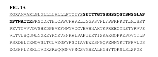
[0013] FIG. 1A shows the amino acid composition of the full length CD24 fusion
protein,
CD24Fc (also referred to herein as CD24Ig) (SEQ ID NO: 5). The underlined 26
amino acids are
the signal peptide of CD24 (SEQ ID NO: 4), which are cleaved off during
secretion from a cell
expressing the protein and thus missing from the processed version of the
protein (SEQ ID
NO: 6). The bold portion of the sequence is the extracellular domain of the
mature CD24 protein
used in the fusion protein (SEQ ID NO: 2). The last amino acid (A or V) that
is ordinarily
present in the mature CD24 protein has been deleted from the construct to
avoid
immunogenicity. The non-underlined, non-bold letters are the sequence of IgG1
Fc, including
the hinge region and CH1 and CH2 domains (SEQ ID NO: 7). FIG. 1B shows the
sequence of
CD24vFc (SEQ ID NO: 8), in which the mature human CD24 protein (bold) is the
valine
polymorphic variant of SEQ ID NO: 1. FIG. 1C shows the sequence of CD24AFc
(SEQ ID
NO: 9), in which the mature human CD24 protein (bold) is the alanine
polymorphic variant of
SEQ ID NO: 1. The various parts of the fusion protein in FIGS. 1B and 1C are
marked as in
FIG. 1A and the variant valine/alanine amino acid is double underlined.
[0014] FIG. 2 shows amino acid sequence variations between mature CD24
proteins from mouse
(SEQ ID NO: 3) and human (SEQ ID NO: 2). The potential 0-glycosylation sites
are bolded, and
the N-glycosylation sites are underlined.
[0015] FIG. 3. WinNonlin compartmental modeling analysis of pharmacokenitics
of CD24IgG1
(CD24Fc). The opened circles represent the average of 3 mice, and the line is
the predicted
pharmacokinetic curve. FIG. 3A. i.v. injection of 1 mg CD24IgG1. FIG. 3B. s.c.
injection of 1
mg CD24IgG1 (CD24Fc). FIG. 3C. Comparison of the total amounts of antibody in
the blood as
measured by areas under curve (AUC), half-life and maximal blood
concentration. Note that
overall, the AUC and Cmax of the s.c. injection is about 80% of i.v.
injection, although the
difference is not statistically significant.
[0016] FIG. 4. CD24-Siglec G (10) interaction discriminates between PAMP and
DAMP. FIG.
4A. Host response to PAMP was unaffected by CD24-Siglec G(10) interaction.
FIG. 4B. CD24-
Siglec G (10) interaction represses host response to DAMP, possibly through
the Siglec G/10-
associated SHP-1.
-5-
CA 03064556 2019-11-21
WO 2018/217659 PCT/US2018/033728
[0017] FIG. 5. CD24 Fc binds to Siglec 10 and HMGB1 and activates Siglec G,
the mouse
homologue of human Siglec 10. FIG. 5A. Affinity measurement of the CD24-Fc-
Siglec 10
interaction. FIG. 5B. CD24-Fc specifically interacts with HMGB-1 in a cation-
dependent
manner. CD24-Fc was incubated with HMGB1 in 0.1 mM of CaCl2 and MgCl2, in the
presence
or absence of the cation chelator EDTA. CD24Fc is pulled down with protein G-
beads, and the
amounts of HMGB1, CD24Fc or control Fc is determined by Western blot. FIG. 5C.
CD24-Fc
activates mouse Siglec G by inducing Tyrosine phosphorylation (middle panel)
and association
with SHP-1 (upper panel). The amounts of Siglec G are shown in the lower
panel. CD24-/- spleen
cells were stimulated with 1 mg/m1 of CD24-Fc, control Fc or vehicle (PBS)
control for 30
minutes. Siglec G was then immunoprecipitated and probed with anti-phospho-
tyrosine or anti-
SHP-1.
[0018] FIG. 6. CD24Fc inhibits production of TNF-a and IFN-y by anti-CD3
activated human T
cells. The human PBML were stimulated with anti-CD3 for 4 days in the presence
or absence of
CD24Fc and the amounts of IFN-y and TNF-a released in the supernatant of cell
culture were
measured by ELISA. Data shown are means of triplicate. Error bar, SEM.
[0019] FIG. 7. CD24 inhibits inflammatory cytokine production by human
macrophages. FIG.
7A. ShRNA silencing of CD24 leads to spontaneous production of TNF-a, IL-1(3,
and IL-6.
THP1 cells were transduced with lentiviral vectors encoding either scrambled
or two
independent CD24 shRNA molecules. The transduced cells were differentiated
into macrophages
by culturing for 4 days with PMA (15 ng/ml). After washing away PMA and non-
adherent cells,
the cells were cultured for another 24 hours for measurement of inflammatory
cytokines, by
cytokine beads array. FIG. 7B. As in FIG. 7A, except that the given
concentration of CD24Fc or
control IgG Fc was added to macrophages in the last 24 hours. Data shown in
FIG. 4A are means
and S.D. from three independent experiments, while those in FIG. 4B are
representative of at
least 3 independent experiments.
[0020] FIG. 8. Kaplan-Meier survival analysis of therapeutic efficacy of
CD24Fc and CsA,
summary data from two independent experiments.
[0021] FIG. 9 shows a plot of mean plasma CD24Fc concentration ( SD) by
treatment for a PK
Evaluable Population in human subjects. PK = pharmacokinetic; SD = standard
deviation.
-6-
CA 03064556 2019-11-21
WO 2018/217659
PCT/US2018/033728
[0022] FIG. 10 shows a dose proportionality plot of CD24Fc Cina, versus dose
for a PK
Evaluable Population.
[0023] FIG. 11 shows a dose proportionality plot of CD24Fc AUCo-42d versus
dose for a PK
Evaluable Population.
[0024] FIG. 12 shows a dose proportionality plot of CD24Fc AUCo-inf versus
dose for a
PK Evaluable Population.
DETAILED DESCRIPTION
[0025] The inventors have discovered that, surprisingly, a soluble form of
CD24 is highly
effective for treating immune-related adverse events (irAEs). The effect may
be mediated
through DAMPs. Pattern recognition is involved in inflammatory response
triggered by both
pathogen-associated and tissue damage-associated molecular patterns,
respectively called
PAMPs and DAMPs. The inventors have realized that recent studies have
demonstrated that an
exacerbated host response to DAMPs may play a part in the pathogenesis of
inflammatory and
autoimmune disease. DAMPs were found to promote the production of inflammatory
cytokines
and autoimmune diseases and in animal models, and inhibitors of DAMPs such as
HMGB1 and
HSP90 were consequently found to ameliorate rheumatoid arthritis (RA) (4-6).
TLRs, RAGE-R,
DNGR (encoded by Clec9A), and Mincle have been shown to be receptors
responsible for
mediating inflammation initiated by a variety of DAMPs (2, 7-14).
[0026] The inventors' recent work demonstrated that CD24-Siglec G interactions
discriminate
innate immunity to DAMPs from PAMPs (15, 16). Siglec proteins are membrane-
associated
immunoglobulin (Ig) superfamily members that recognize a variety of sialic
acid-containing
structures. Most Siglecs have an intra-cellular immune-tyrosine inhibitory
motif (ITIM) that
associates with SHP-1, -2 and Cbl-b to control key regulators of inflammatory
responses. The
inventors have reported CD24 as the first natural ligand for a Siglec, Siglec
G in mouse and
Siglec 10 in human (15). Siglec G interacts with sialylated CD24 to suppress
the TLR-mediated
host response to DAMPs, such as HMGB1, via a SHP-1/2 signaling mechanism (15).
[0027] Human CD24 is a small GPI-anchored molecule encoded by an open-reading
frame of
240 base pairs in the CD24 gene (28). Of the 80 amino acids, the first 26
constitute the signal
peptide, while the last 23 serve as a signal for cleavage to allow for the
attachment of the GPI
-7-
CA 03064556 2019-11-21
WO 2018/217659 PCT/US2018/033728
tail. As a result, the mature human CD24 molecule has only 31 amino acids. One
of the 31 amino
acids is polymorphic among the human population. A C to T transition at
nucleotide 170 of the
open-reading frame results in the substitution of alanine (a) with valine (v).
Since this residue is
in the immediate N-terminal to the cleavage site, and since the replacement is
non-conservative,
these two alleles may be expressed at different efficiencies on the cell
surface. Indeed,
transfection studies with cDNA demonstrated that the CD24" allele is more
efficiently expressed
on the cell surface (28). Consistent with this, CD24"/" PBL expressed higher
levels of CD24,
especially on T cells.
[0028] The inventors have demonstrated that CD24 negatively regulates host
response to cellular
DAMPs that are released as a result of tissue or organ damage, and at least
two overlapping
mechanisms may explain this activity. First, CD24 binds to several DAMPs,
including HSP70,
HSP90, HMGB1 and nucleolin and represses host response to these DAMPs. To do
this, it is
presumed that CD24 may trap the inflammatory stimuli to prevent interaction
with their
receptors, TLR or RAGE. Second, using an acetaminophen-induced mouse model of
liver
necrosis and ensuring inflammation, the inventors demonstrated that through
interaction with its
receptor, Siglec G, CD24 provides a powerful negative regulation for host
response to tissue
injuries. To achieve this activity, CD24 may bind and stimulate signaling by
Siglec G wherein
Siglec G-associated SHP1 triggers the negative regulation. Both mechanisms may
act in concert
as mice with targeted mutation of either gene mounted much stronger
inflammatory response. In
fact, DC cultured from bone marrow from either CD24-/- or Siglec G-/- mice
produced higher
levels of inflammatory cytokines when stimulated with either HMGB1, HSP70, or
HSP90. To
the inventors' knowledge, CD24 is the only inhibitory DAMP receptor capable of
shutting down
inflammation triggered by DAMPs and no drug is currently available that
specifically targets
host inflammatory response to tissue injuries. Furthermore, the inventors have
demonstrated the
ability of exogenous soluble CD24 protein to alleviate DAMP-mediated
autoimmune disease
using mouse models of RA, MS and GvHD.
1. Definitions.
[0029] The terminology used herein is for the purpose of describing particular
embodiments only
and is not intended to be limiting. As used in the specification and the
appended claims, the
singular forms "a," "an" and "the" include plural referents unless the context
clearly dictates
otherwise.
-8-
CA 03064556 2019-11-21
WO 2018/217659 PCT/US2018/033728
[0030] For recitation of numeric ranges herein, each intervening number there
between with the
same degree of precision is explicitly contemplated. For example, for the
range of 6-9, the
numbers 7 and 8 are contemplated in addition to 6 and 9, and for the range 6.0-
7.0, the numbers
6.0, 6.1, 6.2, 6.3, 6.4, 6.5, 6.6, 6.7, 6.8, 6.9, and 7.0 are explicitly
contemplated.
[0031] A "peptide" or "polypeptide" is a linked sequence of amino acids and
may be natural,
synthetic, or a modification or combination of natural and synthetic.
[0032] "Substantially identical" may mean that a first and second amino acid
sequence are at
least 60%, 65%, 70%, 75%, 80%, 85%, 90%, 95%, 96%, 97%, 98%,or 99% over a
region of 1,
2, 3, 4, 5, 6, 7, 8, 9, 10, 11, 12, 13, 14, 15, 16, 17, 18, 19, 20, 21, 22,
23, 24, 25, 26, 27, 28, 29,
30, 31, 32, 33, 34, 35, 36, 37, 38, 39, 40, 41, 42, 43, 44, 45, 46, 47, 48,
49, 50, 55, 60, 65, 70, 75,
80, 85, 90, 95, 100, 110, 120, 130, 140, 150, 160, 170, 180, 190, 200, 210,
220, 230, 240, 250,
260, 270, 280, 290, or 300 amino acids.
[0033] "Treatment" or "treating," when referring to protection of an animal
from a disease,
means preventing, suppressing, repressing, or completely eliminating the
disease. Preventing the
disease involves administering a composition of the present invention to an
animal prior to onset
of the disease. Suppressing the disease involves administering a composition
of the present
invention to an animal after induction of the disease but before its clinical
appearance.
Repressing the disease involves administering a composition of the present
invention to an
animal after clinical appearance of the disease.
[0034] A "variant" may mean a peptide or polypeptide that differs in amino
acid sequence by the
insertion, deletion, or conservative substitution of amino acids, but retain
at least one biological
activity. Representative examples of "biological activity" include the ability
to bind to a toll-like
receptor and to be bound by a specific antibody. Variant may also mean a
protein with an amino
acid sequence that is substantially identical to a referenced protein with an
amino acid sequence
that retains at least one biological activity. A conservative substitution of
an amino acid, i.e.,
replacing an amino acid with a different amino acid of similar properties
(e.g., hydrophilicity,
degree and distribution of charged regions) is recognized in the art as
typically involving a minor
change. These minor changes can be identified, in part, by considering the
hydropathic index of
amino acids, as understood in the art. Kyte et al., J. Mol. Biol. 157:105-132
(1982). The
hydropathic index of an amino acid is based on a consideration of its
hydrophobicity and charge.
-9-
CA 03064556 2019-11-21
WO 2018/217659 PCT/US2018/033728
It is known in the art that amino acids of similar hydropathic indexes can be
substituted and still
retain protein function. In one aspect, amino acids having hydropathic indexes
of 2 are
substituted. The hydrophilicity of amino acids can also be used to reveal
substitutions that would
result in proteins retaining biological function. A consideration of the
hydrophilicity of amino
acids in the context of a peptide permits calculation of the greatest local
average hydrophilicity
of that peptide, a useful measure that has been reported to correlate well
with antigenicity and
immunogenicity. U.S. Patent No. 4,554,101, incorporated fully herein by
reference. Substitution
of amino acids having similar hydrophilicity values can result in peptides
retaining biological
activity, for example immunogenicity, as is understood in the art.
Substitutions may be
performed with amino acids having hydrophilicity values within 2 of each
other. Both the
hyrophobicity index and the hydrophilicity value of amino acids are influenced
by the particular
side chain of that amino acid. Consistent with that observation, amino acid
substitutions that are
compatible with biological function are understood to depend on the relative
similarity of the
amino acids, and particularly the side chains of those amino acids, as
revealed by the
hydrophobicity, hydrophilicity, charge, size, and other properties.
2. CD24
[0035] Provided herein is a CD24 protein, which may comprise a mature CD24 or
a variant
thereof. Mature CD24 corresponds to the extracellular domain (ECD) of CD24.
The mature
CD24 may be from a human or another mammal. As described above, mature human
CD24
protein is 31 amino acids long and has a variable alanine (A) or valine (V)
residue at its C-
terminal end:
[0036] SETTTGTSSNSSQSTSNSGLAPNPTNATTK(V/A) (SEQ ID NO: 1)
[0037] The C-terminal valine or alanine may be immunogenic and may be omitted
from the
CD24 protein, which may reduce its immunogenicity. Therefore, the CD24 protein
may
comprise the amino acid sequence of human CD24 lacking the C-terminal amino
acid:
[0038] SETTTGTSSNSSQSTSNSGLAPNPTNATTK (SEQ ID NO: 2)
[0039] Despite considerable sequence variations in the amino acid sequence of
the mature CD24
proteins from mouse and human, they are functionally equivalent, as human
CD24Fc has been
shown to be active in the mouse. The amino acid sequence of the human CD24 ECD
shows some
sequence conservation with the mouse protein (39% identity; Genbank accession
number
-10-
CA 03064556 2019-11-21
WO 2018/217659 PCT/US2018/033728
NP_033976). However, it is not that surprising that the percent identity is
not higher as the CD24
ECD is only 27-31 amino acids in length, depending on the species, and binding
to some of its
receptor(s), such as Siglec 10/G, is mediated by its sialic acid and/or
galactose sugars of the
glycoprotein. The amino acid sequence identity between the extracellular
domains of the human
Siglec-10 (GenBank accession number AF310233) and its murine homolog Siglec-G
(GenBank
accession number NP_766488) receptor proteins is 63% (FIG. 2). As a result of
sequence
conservation between mouse and human CD24 primarily in the C-terminus and in
the abundance
of glycosylation sites, significant variations in the mature CD24 proteins may
be tolerated in
using the CD24 protein, especially if those variations do not affect the
conserved residues in the
C-terminus or do not affect the glycosylation sites from either mouse or human
CD24. Thus, the
CD24 protein may comprise the amino acid sequence of mature murine CD24:
[0040] NQTSVAPFPGNQNISASPNPTNATTRG (SEQ ID NO: 3).
[0041] The amino acid sequence of the human CD24 ECD shows more sequence
conservation
with the cynomolgus monkey protein (52% identity; UniProt accession number
UniProtKB -
I7GKK1) than with mouse. Again, this is not surprising given that the percent
identity is not
higher as the ECD is only 29-31 amino acids in length in these species, and
the role of sugar
residues in binding to its receptor(s). The amino acid sequence of cynomolgous
Siglec-10
receptor has not been determined but the amino acid sequence identity between
the human and
rhesus monkey Siglec-10 (GenBank accession number XP_001116352) proteins is
89%.
Therefore, the CD24 protein may also comprise the amino acid sequence of
mature cynomolgous
(or rhesus) monkey CD24:
[0042] TVTTSAPLSSNSPQNTSTTPNPANTTTKA (SEQ ID NO: 10)
[0043] The CD24 protein may be soluble. The CD24 protein may further comprise
an N-
terminal signal peptide, to allow secretion from a cell expressing the
protein. The signal peptide
sequence may comprise the amino acid sequence MGRAMVARLGLGLLLLALLLPTQIYS
(SEQ ID NO: 4). Alternatively, the signal sequence may be any of those that
are found on other
transmembrane or secreted proteins, or those modified from the existing signal
peptides known
in the art.
-11-
CA 03064556 2019-11-21
WO 2018/217659 PCT/US2018/033728
a. Fusion
[0044] The CD24 protein may be fused at its N- or C-terminal end to a protein
tag, which may
comprise a portion of a mammalian Ig protein, which may be human or mouse or
from another
species. The portion may comprise an Fc region of the Ig protein. The Fc
region may comprise at
least one of the hinge region, CH2, CH3, and CH4 domains of the Ig protein.
The Ig protein may
be human IgGl, IgG2, IgG3, IgG4, or IgA, and the Fc region may comprise the
hinge region,
and CH2 and CH3 domains of the Ig. The Fc region may comprise the human
immunoglobulin
G1 (IgG1) isotype (SEQ ID NO: 7). The Ig protein may also be IgM, and the Fc
region may
comprise the hinge region and CH2, CH3, and CH4 domains of IgM. The protein
tag may be an
affinity tag that aids in the purification of the protein, and/or a solubility-
enhancing tag that
enhances the solubility and recovery of functional proteins. The protein tag
may also increase the
valency of the CD24 protein. The protein tag may also comprise GST, His, FLAG,
Myc, MBP,
NusA, thioredoxin (TRX), small ubiquitin-like modifier (SUMO), ubiquitin (Ub),
albumin, or a
Camelid Ig. Methods for making fusion proteins and purifying fusion proteins
are well known in
the art.
[0045] Based on preclinical research, for the construction of the fusion
protein CD24Fc
identified in the examples, the truncated form of native CD24 molecule of 30
amino acids, which
lacks the final polymorphic amino acid before the GPI signal cleavage site
(that is, a mature
CD24 protein having SEQ ID NO: 2), has been used. The mature human CD24
sequence is fused
to a human IgG1 Fc domain (SEQ ID NO: 7). The full length CD24Fc fusion
protein is provided
in SEQ ID NO: 5 (FIG. 1), and the processed version of CD24Fc fusion protein
that is secreted
from the cell (i.e. lacking the signal sequence which is cleaved off) is
provided in SEQ ID NO: 6.
Processed polymorphic variants of mature CD24 (that is, mature CD24 protein
having SEQ ID
NO: 1) fused to IgG1 Fc may comprise SEQ ID NO: 11 or 12.
b. Production
[0046] The CD24 protein may be heavily glycosylated, and may be involved in
functions of
CD24 such as costimulation of immune cells and interaction with a damage-
associated molecular
pattern molecule (DAMP). The CD24 protein may be prepared using a eukaryotic
expression
system. The expression system may entail expression from a vector in mammalian
cells, such as
Chinese Hamster Ovary (CHO) cells. The system may also be a viral vector, such
as a
replication-defective retroviral vector that may be used to infect eukaryotic
cells. The CD24
-12-
CA 03064556 2019-11-21
WO 2018/217659 PCT/US2018/033728
protein may also be produced from a stable cell line that expresses the CD24
protein from a
vector or a portion of a vector that has been integrated into the cellular
genome. The stable cell
line may express the CD24 protein from an integrated replication-defective
retroviral vector. The
expression system may be GPExTM.
c. Pharmaceutical composition
[0047] The CD24 protein may be contained in a pharmaceutical composition,
which may
comprise a pharmaceutically acceptable amount of the CD24 protein. The
pharmaceutical
composition may comprise a pharmaceutically acceptable carrier. The
pharmaceutical
composition may comprise a solvent, which may keep the CD24 protein stable
over an extended
period. The solvent may be PBS, which may keep the CD24 protein stable for at
least 66 months
at -20 C (-15--25 C). The solvent may be capable of accommodating the CD24
protein in
combination with another drug.
[0048] The pharmaceutical composition may be formulated for parenteral
administration
including, but not limited to, by injection or continuous infusion.
Formulations for injection may
be in the form of suspensions, solutions, or emulsions in oily or aqueous
vehicles, and may
contain formulation agents including, but not limited to, suspending,
stabilizing, and dispersing
agents. The composition may also be provided in a powder form for
reconstitution with a
suitable vehicle including, but not limited to, sterile, pyrogen-free water.
[0049] The pharmaceutical composition may also be formulated as a depot
preparation, which
may be administered by implantation or by intramuscular injection. The
composition may be
formulated with suitable polymeric or hydrophobic materials (as an emulsion in
an acceptable
oil, for example), ion exchange resins, or as sparingly soluble derivatives
(as a sparingly soluble
salt, for example). A formulation for subcutaneous injection may be
particularly relevant for an
indication like lupus and its associated manifestations and complications.
d. Dosage
[0050] The dose of the CD24 protein may ultimately be determined through a
clinical trial to
determine a dose with acceptable toxicity and clinical efficacy. The initial
clinical dose may be
estimated through pharmacokinetics and toxicity studies in rodents and non-
human primates. The
dose of the CD24 protein may be 0.01 mg/kg to 1000mg/kg, and may be 1 to 500
mg/kg,
depending on the desired effect and the route of administration. The CD24
protein may be
-13-
CA 03064556 2019-11-21
WO 2018/217659 PCT/US2018/033728
administered by intravenous infusion or subcutaneous, intramural (that is,
within the wall of a
cavity or organ), or intraperitoneal injection, and the dose may be 10-1000
mg, 10-500 mg, 10-
240 mg, 10-120 mg, or 10, 30, 60, 120, or 240 mg, where the subject is a
human.
3. Methods of treatment
a. Immune-Related Adverse Events
[0051] Provided herein is a method of mitigating, reducing, minimizing, or
treating irAEs by
administering the CD24 protein to a subject in need thereof. The irAEs may be
associated with a
cancer therapy, and the subject may be a cancer patient. The cancer therapy
may be a cancer
immunotherapy. The CD24 protein may be administered to a subject with or at
risk of
developing irAEs associated with the cancer therapy. The CD24 protein may be
used
prophylactically to prevent irAEs before the cancer therapy is initiated or
before the clinical
signs of irAEs emerge. The CD24 protein may also be administered
therapeutically to treat irAEs
after the cancer therapy is initiated and the clinical symptoms are diagnosed.
The irAE may be
diarrhea or another gastrointestinal disorder, pure red cell aplasia,
microcytic anemia, lupus,
autoimmune nephritism, autoimmune hepatitis, pneumonitis, myocarditis,
pericarditis,
endocrinopathy, Addison's disease, hypogonadism, Sjogren's syndrome, or type I
diabetes.
[0052] The cancer therapy may be active immunotherapy. Examples of active
immunotherapy
include anti-CTLA4, anti-PD-1, anti-PD-L1, anti-TNF, an antibody against
another TNF-
receptor family member, anti-LAG3, anti-TIM3, and a small or large molecule
inhibitor that
modulates the tumor microenvironment. In particular, the cancer therapy may be
anti-CTLA4
immunotherapy, and the CD24 protein may be administered to a subject in
combination with, or
on a background of, anti-CTLA4 immunotherapy. Examples of anti-CTLA4
antibodies include
Ipilimumab (Yervoy) and Tremilimumab.
[0053] In another embodiment, the cancer therapy may be anti-PD-1/PD-L1
immunotherapy,
which may be an anti-PD-1 antibody or an anti-PD-L1 antibody. Examples of anti-
PD-1
antibodies include nivolumab, (Opdivo - Bristol Myers Squibb) and
Pembrolizumab (Keytruda,
MK-3475, Merck). Examples of anti-PD-L1 antibodies include Atezolizumab
(Tecentriq,
Roche), Avelumab (Merck KGaA and Pfizer) and durvalumab (Imfinzi, Astra-
Zeneca).
[0054] In yet another embodiment, the cancer therapy may be combination
therapy comprising
anti-CTLA-4 and anti-PD-1 monoclonal antibodies (mAbs). Combination therapy
with anti-
-14-
CA 03064556 2019-11-21
WO 2018/217659 PCT/US2018/033728
CTLA-4 and anti-PD-1 has emerged as the most potent and durable cancer
immunotherapy.
However, the autoimmune adverse effect associated with the combination therapy
is quite
severe, with greater than 50% of melanoma patients developing grade 3 and 4
organ toxicity.
Therefore, a major challenge in cancer immunotherapy is how to reduce adverse
effects of the
combination therapy without affecting therapeutic efficacy. Of the two
components of the
combination therapy, anti-CTLA-4 mAb exhibits considerably more immunotherapy-
related
adverse effects (irAE).
[0055] The cancer therapy may be adoptive cell transfer (ACT) using ex vivo
stimulated tumor
infiltrating lymphocytes (TILs) or genetically-engineered T cells (chimeric
antigen receptors
[CARs] or T cell receptor [TCR] modified T cells). In particular, by causing
rapid death of
normal and cancer cells, cancer therapies such as CAR-T cells can cause the
release of damage
associated molecular patterns (DAMPs) and, consequently, cytokine release
syndrome that can
also lead to widespread organ dysfunction. Therefore, the CD24 protein may be
used to reduce
or neutralize the effects of the DAMPs and mitigate, minimize or treat the
resulting cytokine
storm. CAR-T cell can damage normal cells if they target a tumor-associated
antigen that is also
expressed on non-tumor cells and tissues. CAR-T therapies may be used in the
treatment of
hematologic tumors such as acute lymphoblastic leukemia (ALL), B-cell Acute
Lymphoblastic
Leukemia, adult myeloid leukemia, (AML), diffuse large B-cell lymphoma
(DLBCL), non-
Hodgkin Lymphoma (NHL), Chronic Lymphocytic Leukemia (CLL), primary
mediastinal B-cell
lymphoma (PMBCL), mantle cell lymphoma (MCL), and multiple myeloma (MM).
Examples of
CAR-T therapies include those targeting the B cell surface antigens CD19 (such
as JCAR017
and JCAR014 [Juno Therapeutics]), CTL019 (tisagenlecleucel-T [Novartis] and
KTE-C19
[axicabtagene ciloleucel, Kite Pharma]), and CD22 (JCAR014 [Juno
Therapeutics]). Other
examples of CAR-T therapies include those targeting Li-CAM (JCAR023 [Juno
Therapeutics]),
ROR-1 (JCAR024 [Juno Therapeutics]) and MUC16 (JCAR020 [Juno Therapeutics]).
Examples
of targets for TCR modified T cells include those targeting MAGE-A3, such as
KITE-718 (Kite
Pharma), Wilms tumor antigen 1 (WT-1), such as JTCR016 (Juno Therapeutics),
and NY-
ESO-1.
[0056] The cancer therapy may be one that involves rapid killing of cancer
cells, such as
irradiation and chemotherapy. The resulting tumor lysis can lead to the
release of DAMPs that
-15-
CA 03064556 2019-11-21
WO 2018/217659 PCT/US2018/033728
initiate an inflammatory cascade. Such indications may be particularly
amenable to prophylactic
treatment with the CD24 protein.
b. Graft Versus Host Disease
[0057] Also provided herein is a method of reducing or treating graft versus
host disease
(GvHD) in a subject that may have received or be receiving activated Natural
Killer (aNK) cells
following allogeneic hematopoietic stem cell transplantation (HSCT) by
administering the CD24
protein to a subject in need thereof. NK cells can enhance engraftment and
mediate graft-versus-
leukemia following allogeneic HSCT, but the potency of graft-versus-leukemia
mediated by
naturally reconstituting NK cells following HSCT is limited. Preclinical
studies demonstrate that
activation of NK cells upregulates activating receptor expression and augments
killing capacity
(Shah et al 2015). This was then tested in a clinical trial studying the
adoptive transfer of donor-
derived activated NK cells (aNK-DLI) following HLA-matched, T-cell¨depleted
nonmyeloablative peripheral blood stem cell transplantation in children and
young adults with
ultra-high-risk solid tumors. aNK-DLI demonstrated potent killing capacity and
displayed high
levels of activating receptor expression. However, 5 of 9 transplant
recipients experienced acute
graft-versus-host disease (GVHD) following aNK-DLI, with grade 4 GVHD observed
in 3
subjects. GVHD was more common in matched unrelated donor vs matched sibling
donor
recipients and was associated with higher donor CD3 chimerism. Given that the
T-cell dose was
below the threshold required for GVHD in this setting, it was concluded that
aNK-DLI
contributed to the acute GVHD observed, likely by augmenting underlying T-cell
alloreactivity.
c. Administration
[0058] The route of administration of the pharmaceutical composition may be
parenteral.
Parenteral administration includes, but is not limited to, intravenous,
intraarterial, intraperitoneal,
subcutaneous, intramuscular, intrathecal, intraarticular, and direct
injection. The pharmaceutical
composition may be administered to a human patient, cat, dog, or large animal.
The composition
may be administered 1, 2, 3, 4, 5, 6, 7, 8, 9, 10, 11, or 12 times per day.
d. Combination treatment
[0059] The CD24 protein may be used in combination with another agent to
further reduce,
mitigate or treat cytokine release syndrome (CRS). Cytokine release syndrome
is associated with
elevated circulating levels of several cytokines including interleukin (IL)-6
and IFN-y.
-16-
CA 03064556 2019-11-21
WO 2018/217659 PCT/US2018/033728
Accordingly, the other agent may be tocilizumab (Actemra), an anti-IL-6
receptor antibody, or
another cytokine targeting agent, with or without corticosteroids, which may
be used for
immunosuppression and may be used to reverse the syndrome, particularly in
patients receiving
CAR-T. The other agent used for CRS management may be siltuximab (anti-IL-6,
Sylvant),
etanercept (TNFa inhibitor, Enbrel), infliximab (anti-TNFa, Remicade), or
anakinra (interleukin
1 receptor antagonist, Kineret). The CD24 protein may be used to target and
mitigate the effect
of DAMPs that are released from the damaged tissue and which initiate the
inflammatory
cascade, while the combination therapy may target the effector cytokine
molecule, thereby
providing a complementary two-pronged approach to mitigate, reduce or treat
CRS.
[0060] The CD24 protein may be administered simultaneously or metronomically
with other
treatments. The term "simultaneous" or "simultaneously" as used herein, means
that the CD24
protein and other treatment be administered within 48 hours, preferably 24
hours, more
preferably 12 hours, yet more preferably 6 hours, and most preferably 3 hours
or less, of each
other. The term "metronomically" as used herein means the administration of
the agent at times
different from the other treatment and at a certain frequency relative to
repeat administration.
[0061] The CD24 protein may be administered at any point prior to another
treatment including
about 120 hr, 118 hr, 116 hr, 114 hr, 112 hr, 110 hr, 108 hr, 106 hr, 104 hr,
102 hr, 100 hr, 98 hr,
96 hr, 94 hr, 92 hr, 90 hr, 88 hr, 86 hr, 84 hr, 82 hr, 80 hr, 78 hr, 76 hr,
74 hr, 72 hr, 70 hr, 68 hr,
66 hr, 64 hr, 62 hr, 60 hr, 58 hr, 56 hr, 54 hr, 52 hr, 50hr, 48 hr, 46 hr, 44
hr, 42 hr, 40 hr, 38 hr,
36 hr, 34 hr, 32 hr, 30 hr, 28 hr, 26 hr, 24 hr, 22 hr, 20 hr, 18 hr, 16 hr,
14 hr, 12 hr, 10 hr, 8 hr, 6
hr, 4 hr, 3 hr, 2 hr, 1 hr, 55 mins., 50 mins., 45 mins., 40 mins., 35 mins.,
30 mins., 25 mins., 20
mins., 15 mins, 10 mins, 9 mins, 8 mins, 7 mins., 6 mins., 5 mins., 4 mins., 3
mins, 2 mins, and 1
mins. The CD24 protein may be administered at any point prior to a second
treatment of the
CD24 protein including about 120 hr, 118 hr, 116 hr, 114 hr, 112 hr, 110 hr,
108 hr, 106 hr, 104
hr, 102 hr, 100 hr, 98 hr, 96 hr, 94 hr, 92 hr, 90 hr, 88 hr, 86 hr, 84 hr, 82
hr, 80 hr, 78 hr, 76 hr,
74 hr, 72 hr, 70 hr, 68 hr, 66 hr, 64 hr, 62 hr, 60 hr, 58 hr, 56 hr, 54 hr,
52 hr, 50hr, 48 hr, 46 hr,
44 hr, 42 hr, 40 hr, 38 hr, 36 hr, 34 hr, 32 hr, 30 hr, 28 hr, 26 hr, 24 hr,
22 hr, 20 hr, 18 hr, 16 hr,
14 hr, 12 hr, 10 hr, 8 hr, 6 hr, 4 hr, 3 hr, 2 hr, 1 hr, 55 mins., 50 mins.,
45 mins., 40 mins., 35
mins., 30 mins., 25 mins., 20 mins., 15 mins., 10 mins., 9 mins., 8 mins., 7
mins., 6 mins., 5
mins., 4 mins., 3 mins, 2 mins, and 1 mins.
-17-
CA 03064556 2019-11-21
WO 2018/217659 PCT/US2018/033728
[0062] The CD24 protein may be administered at any point after another
treatment including
about lmin, 2 mins., 3 mins., 4 mins., 5 mins., 6 mins., 7 mins., 8 mins., 9
mins., 10 mins., 15
mins., 20 mins., 25 mins., 30 mins., 35 mins., 40 mins., 45 mins., 50 mins.,
55 mins., 1 hr, 2 hr, 3
hr, 4 hr, 6 hr, 8 hr, 10 hr, 12 hr, 14 hr, 16 hr, 18 hr, 20 hr, 22 hr, 24 hr,
26 hr, 28 hr, 30 hr, 32 hr,
34 hr, 36 hr, 38 hr, 40 hr, 42 hr, 44 hr, 46 hr, 48 hr, 50 hr, 52 hr, 54 hr,
56 hr, 58 hr, 60 hr, 62 hr,
64 hr, 66 hr, 68 hr, 70 hr, 72 hr, 74 hr, 76 hr, 78 hr, 80 hr, 82 hr, 84 hr,
86 hr, 88 hr, 90 hr, 92 hr,
94 hr, 96 hr, 98 hr, 100 hr, 102 hr, 104 hr, 106 hr, 108 hr, 110 hr, 112 hr,
114 hr, 116 hr, 118 hr,
and 120 hr. The CD24 protein may be administered at any point prior after a
previous CD24
treatment including about 120 hr, 118 hr, 116 hr, 114 hr, 112 hr, 110 hr, 108
hr, 106 hr, 104 hr,
102 hr, 100 hr, 98 hr, 96 hr, 94 hr, 92 hr, 90 hr, 88 hr, 86 hr, 84 hr, 82 hr,
80 hr, 78 hr, 76 hr, 74
hr, 72 hr, 70 hr, 68 hr, 66 hr, 64 hr, 62 hr, 60 hr, 58 hr, 56 hr, 54 hr, 52
hr, 50hr, 48 hr, 46 hr, 44
hr, 42 hr, 40 hr, 38 hr, 36 hr, 34 hr, 32 hr, 30 hr, 28 hr, 26 hr, 24 hr, 22
hr, 20 hr, 18 hr, 16 hr, 14
hr, 12 hr, 10 hr, 8 hr, 6 hr, 4 hr, 3 hr, 2 hr, 1 hr, 55 mins., 50 mins., 45
mins., 40 mins., 35 mins.,
30 mins., 25 mins., 20 mins., 15 mins., 10 mins., 9 mins., 8 mins., 7 mins., 6
mins., 5 mins., 4
mins., 3 mins, 2 mins, and 1 mins.
-18-
CA 03064556 2019-11-21
WO 2018/217659 PCT/US2018/033728
Example 1
CD24 pharmacokinetics in mice
[0064] 1 mg of CD24Fc (CD24Fc) was injected into naïve C57BL/6 mice and
collected blood
samples at different timepoints (5 min, 1 hr, 4 hrs, 24 hrs, 48 hrs, 7 days,
14 days and 21 days)
with 3 mice in each timepoint. The sera were diluted 1:100 and the levels of
CD24Fc was
detected using a sandwich ELISA using purified anti-human CD24 (3.3 ng/m1) as
the capturing
antibody and peroxidase conjugated goat anti-human IgG Fc (5 ng/m1) as the
detecting
antibodies. As shown in FIG. 3A. The decay curve of CD24Fc revealed a typical
biphase decay
of the protein. The first biodistribution phase had a half-life of 12.4 hours.
The second phase
follows a model of first-order elimination from the central compartment. The
half-life for the
second phase was 9.54 days, which is similar to that of antibodies in vivo.
These data suggest
that the fusion protein is very stable in the blood stream. In another study
in which the fusion
protein was injected subcutaneously, an almost identical half-life of 9.52
days was observed
(FIG. 3B). More importantly, while it took approximately 48 hours for the
CD24Fc to reach peak
levels in the blood, the total amount of the fusion protein in the blood, as
measured by AUC, was
substantially the same by either route of injection. Thus, from a therapeutic
point of view,
different route of injection should not affect the therapeutic effect of the
drug. This observation
greatly simplified the experimental design for primate toxicity and clinical
trials.
Example 2
CD24-Siglec 10 interaction in host response to tissue injuries
[0065] Nearly two decades ago, Matzinger proposed what was popularly called
danger theory. In
essence, she argued that the immune system is turned on when it senses the
dangers in the host.
Although the nature of danger was not well defined at the time, it has been
determined that
necrosis is associated with the release of intracellular components such as
HMGB1 and Heat-
shock proteins, which were called DAMP, for danger-associated molecular
patterns. DAMP
were found to promote production of inflammatory cytokines and autoimmune
diseases. In
animal models, inhibitors of HMGB1 and HSP90 were found to ameliorate RA. The
involvement of DAMP raised the prospect that negative regulation for host
response to DAMP
can be explored for RA therapy.
-19-
CA 03064556 2019-11-21
WO 2018/217659 PCT/US2018/033728
[0066] Using acetaminophen-induced liver necrosis and ensuring inflammation,
it was observed
that through interaction Siglec G, CD24 provides a powerful negative
regulation for host
response to tissue injuries. CD24 is a GPI anchored molecules that is broadly
expressed in
hematopoietic cells and other tissue stem cells. Genetic analysis of a variety
of autoimmune
disease in human, including multiple sclerosis, systemic lupus erythromatosus,
RA, and giant
cell arthritis, showed significant association between CD24 polymorphism and
risk of
autoimmune diseases. Siglec G is a member of I-lectin family, defined by their
ability to
recognize sialic acid containing structure. Siglec G recognized sialic acid
containing structure on
CD24 and negatively regulates production of inflammatory cytokines by
dendritic cells. In terms
of its ability to interact with CD24, human Siglec 10 and mouse Siglec G are
functionally
equivalent. However, it is unclear if there is a one-to-one correlation
between mouse and human
homologues. Although the mechanism remains to be full elucidated, it is
plausible that SiglecG-
associated SHP1 may be involved in the negative regulation. These data,
reported in Science
recently, leads to a new model in which CD24-Siglec G/10 interaction may play
a critical in
discrimination pathogen-associated molecular pattern (PAMP) from DAMP (FIG.
4).
[0067] At least two overlapping mechanisms may explain the function of CD24.
First, by
binding to a variety of DAMP, CD24 may trap the inflammatory stimuli to
prevent their
interaction with TLR or RAGE. This notion is supported by observations that
CD24 is associated
with several DAMP molecules, including HSP70, 90, HMGB1 and nucleolin. Second,
perhaps
after associated with DAMP, CD24 may stimulate signaling by Siglec G. Both
mechanisms may
act in concert as mice with targeted mutation of either gene mounted much
stronger
inflammatory response. In fact, DC cultured from bone marrow from either CD24-
/- or Siglec G-
/- mice produced much higher inflammatory cytokines when stimulated with
either HMGB1,
HSP70, or HSP90. In contrast, no effect were found in their response to PAMP,
such as LPS and
PolyI:C. These data not only provided a mechanism for the innate immune system
to distinguish
pathogen from tissue injury, but also suggest that CD24 and Siglec G as
potential therapeutic
targets for diseases associated with tissue injuries.
-20-
CA 03064556 2019-11-21
WO 2018/217659 PCT/US2018/033728
Example 3
CD24 and the prevention of GvHD
[0068] CD24Fc interacts with HMGB1, Siglec 10 and induces association between
Siglec G and
SHP-1.
[0069] To measure the interaction between CD24Fc and Siglec 10, we immobilized
CD24Fc
onto a CHIP and used Biacore to measure the binding of different
concentrations of Siglec-10Fc.
As shown in FIG. 5A, CD24Fc binds with Siglec 10 with a Kd of 1.6x10⁻⁷M. This
is 100-fold
higher affinity than the control Fc. The interaction between CD24Fc and HMGB1
was confirmed
by pull down experiments using CD24Fc-bound protein G beads followed by
Western blot with
either anti-IgG or anti-HMGB1. These data demonstrate that CD24Fc, but not Fc,
binds to
HMGB1 and that this binding is cation-dependent (FIG. 5B). To determine
whether CD24Fc is
an agonist of Siglec G, the mouse counterpart of human Siglec 10, we
stimulated CD24-/- spleen
cells with CD24Fc, control Fc or vehicle (PBS) control for 30 minutes. Siglec
G was then
immunoprecipitated and probed with anti-phospho-tyrosine or anti-SHP-1. As
shown in FIG. 5C,
CD24Fc induced substantial phosphorylation of Siglec G and association of SHP-
1, a well-
known inhibitor for both adaptive and innate immunity.
[0070] In vitro efficacy studies of CD24Fc.
[0071] To study the impact of CD24Fc on the production of inflammatory
cytokines by human T
cells, the mature T cells in human PBML were activated by anti-CD3 antibody
(OKT3), a
commonly used agonist of the T cell receptor in the presence of different
concentrations of
CD24Fc or human IgG1 Fc. Four days later, the supernatants were collected and
the production
of IFN-y and TNF-cc were measured by Enzyme-linked immunosorbent assay (ELISA)
to
confirm activation. The results in FIG. 6 demonstrated that CD24Fc from two
different
manufacturing lots significantly reduced IFN-y and TNF-cc production from the
activated human
PBML compared with control IgG Fc control. In addition, when CD24Fc was added,
cytokine
production was inhibited in a dose-dependent manner. Therefore, CD24Fc can
inhibit anti-CD3
induced human PBML activation in vitro. This study not only indicated the
mechanism of action
of CD24Fc might be through the inhibition of T cell activation, but also
established a reliable
bioassay for drug potency and stability testing.
-21-
CA 03064556 2019-11-21
WO 2018/217659 PCT/US2018/033728
[0072] To determine whether CD24Fc regulates production of inflammatory
cytokines in a
human cell line, we first silenced CD24 in the human acute monocytic leukemia
THP1 cell line
using RNAi, and then induced differentiation into macrophages by treating them
with PMA. As
shown in FIG. 7A, CD24 silencing substantially increased the production of
TNFa, IL-10 and
IL-6. These data demonstrate an essential role for endogenous human CD24 in
limiting the
production of inflammatory cytokines. Importantly, CD24Fc restored inhibition
of TNFa in the
CD24-silenced cell line (FIG. 7B), as well as IL-1 13 and IL-6. These data not
only demonstrate
the relevance of CD24 in inflammatory response of human cells, but also
provides a simple assay
to assess biological activity of CD24Fc.
[0073] Taken together, these data demonstrate that CD24Fc is capable of
inhibiting cytokine
production triggered by adaptive and innate stimuli. However, since the drug
is much more
effective in reducing cytokine production by innate effectors, we consider
that the primary
mechanism for its prophylactic function is to prevent inflammation triggered
by tissue injuries at
the early phase of transplantation.
[0074] Comparison of therapeutic effect between CsA and CD24Fc in the
humanized mouse
GvHD model.
[0075] GvHD is known as a major complication in allogeneic BM transplantation.
However,
GvHD induction in all humanized animal models relies on transplantation of a
large amount of
human PBMCs. Some of these humanized animal models could not achieve the
systemic GvHD
seen in humans. Therefore, a humanized systemic GvHD animal model was
developed using one
half-million human BM cells in newborn NOD/SCID IL2ry-null (NSG) mice. The
results show
that mice developed xenogeneic GvHD with 100% penetrance and all mice
displayed high
human chimerism as early as 14 days after transplantation which significantly
increased to nearly
two fold 7 days later (data not shown). The total mortality rate is 100%
within 1-2 months of
transplantation depending on the donor used (data not shown). Moreover, the
human T cells
infiltrate multiple target organs, including lung, liver, skin and intestine.
To the knowledge of the
inventors, this is the best model for pathogenesis of human acute GVHD, but
therapeutically
more challenging due to severity and rapid onset of the disease.
[0076] The donors used for two experiments caused unusually rapid and severe
GVHD. To
compare the therapeutic efficacies of cyclosporine A (CsA) and CD24Fc, the NSG
mice were
-22-
CA 03064556 2019-11-21
WO 2018/217659 PCT/US2018/033728
treated at one week after transplantation with either daily maximal tolerable
doses of CsA
(between 0.3 and 1 mg/kg for up to 4 weeks, depending on the age of mice) or 2-
4 weekly doses
of CD24Fc (5 mg/kg). As shown in FIG. 8, starting at the second week, rapid
onset of GVHD-
related death was observed in the vehicle group. While CD24Fc significantly
extended the
survival of the recipient mice (P=0.015), CsA failed to significantly extend
the survival
(P=0.097). These data demonstrate that CD24Fc has superior therapeutic
efficacy as compared to
CsA.
Example 4
CD24 pharmacokinetics in humans
[0077] This example shows an analysis of the pharmacokinetics of a CD24
protein in humans.
This was derived from a Phase I, randomized, double-blind, placebo-controlled,
single ascending
dose study to assess the safety, tolerability, and PK of CD24Fc in healthy
male and female adult
subjects. A total of 40 subjects in 5 cohorts of 8 subjects each were enrolled
in this study. Six of
the 8 subjects in each cohort received study drug and 2 subjects received
placebo (0.9% sodium
chloride, saline). The first cohort was dosed with 10 mg. Succeeding cohorts
received 30 mg, 60
mg, 120 mg, and 240 mg of CD24Fc or matching placebo and were dosed at least 3
weeks apart
to allow for review of safety and tolerability data for each prior cohort.
Administration of the
next higher dose to a new cohort of subjects was permitted only if adequate
safety and
tolerability had been demonstrated.
[0078] In each cohort, the initial 2 subjects were 1 study drug recipient and
1 placebo recipient
on Day 1. The 3rd to 5th and 6th to 8th subjects were dosed after Day 7 (a
minimum of 24 hours
apart between the subgroups). Each subject was dosed at least 1 hour apart in
the same subgroup.
If necessary, dosing of the rest of subjects was delayed pending review of any
significant safety
issues that may have arisen during the post-dose period involving the first or
second subgroups
in that cohort. The subsequent cohort was dosed at least 3 weeks after the
prior cohort.
[0079] Screening Period:
[0080] The Screening Visit (Visit 1) occured up to 21 days prior to the
beginning of the active
treatment period. After providing informed consent, subjects underwent
screening procedures for
eligibility.
-23-
CA 03064556 2019-11-21
WO 2018/217659 PCT/US2018/033728
[0081] Treatment Period:
[0082] Subjects were admitted to the Clinical Pharmacology Unit (CPU) on Day -
1 (Visit 2), and
the randomized treatment period began on Day 1 following a 10-hour minimum
overnight fast.
Subjects were randomly assigned to treatment with CD24Fc or placebo as a
single dose. Subjects
remained confined until the morning of Day 4.
[0083] Follow-up:
[0084] All subjects returned to the CPU on Day 7, Day 14, Day 21, Day 28, and
Day 42 ( 1 day)
for follow-up visits (Visit 3, Visit 4, Visit 5, Visit 6, and Visit 7). Visit
7 was the final visit for all
subjects.
[0085] Duration of Treatment: The total study duration for each subject was up
to 63 days.
Single-dose administration occurred on Day 1.
[0086] Number of Subjects:
[0087] Planned: 40 subjects
[0088] Screened: 224 subjects
[0089] Randomized: 40 subjects
[0090] Completed: 39 subjects
[0091] Discontinued: 1 subject
[0092] Diagnosis and Main Criteria for Inclusion: The population for this
study was healthy
males and females between the ages of 18 and 55 years, inclusive, with a body
mass index
between 18 kg/m2 and 30 kg/m2, inclusive.
[0093] Investigational Product and Comparator Information:
[0094] CD24Fc: single dose of 10 mg, 30 mg, 60 mg, 120 mg, or 240 mg
administered via IV
infusion; lot number: 09MM-036. CD24Fc was a fully humanized fusion protein
consisting of
the mature sequence of human CD24 and the fragment crystallizable region of
human
immunoglobulin G1 (IgGlFc). CD24Fc was supplied as a sterile, clear,
colorless, preservative-
free, aqueous solution for IV administration. CD24Fc was formulated as single
dose injection
solution, at a concentration of 10 mg/mL and a pH of 7.2. Each CD24Fc vial
contained 160 mg
-24-
CA 03064556 2019-11-21
WO 2018/217659 PCT/US2018/033728
of CD24Fc, 5.3 mg of sodium chloride, 32.6 mg of sodium phosphate dibasic
heptahydrate, and
140 mg of sodium phosphate monobasic monohydrate in 16 mL 0.2 mL of CD24Fc.
CD24Fc
was supplied in clear borosilicate glass vials with chlorobutyl rubber
stoppers and aluminum
flip-off seals.
[0095] Matching placebo (0.9% sodium chloride, saline) administered via IV
infusion; lot
numbers: P296855, P311852, P300715, P315952.
[0096] The intent-to-treat (ITT) Population consisted of all subjects who
received at least 1 dose
of the study drug. The ITT Population was the primary analysis population for
subject
information and safety evaluation.
[0097] Clinical laboratory evaluations (chemistry, hematology, and urinalysis)
were summarized
by treatment and visit. Change from baseline was also summarized. Vital signs
(blood pressure,
heart rate, respiratory rate, and temperature) were summarized by treatment
and time point.
Change from baseline was also summarized. All physical examination data were
listed.
Electrocardiogram parameters and the change from baseline were summarized.
Overall
interpretations were listed.
[0098] Plasma CD24Fc Concentration
[0099] As shown in FIG. 9, the mean plasma concentration of CD24Fc increased
proportionally
to the dose of CD24Fc administered. For all dose groups except 120 mg, the
maximum mean
plasma concentration of CD24Fc was reached at 1 hour post-dose. The maximum
mean plasma
concentration of CD24Fc for the 120 mg group was reached at 2 hours post-dose.
By Day 42
(984 hours), the mean plasma concentration of CD24Fc for all groups had
decreased to between
2% and 4% of the maximum mean plasma concentration.
[0100] Table 1 summarizes the plasma CD24Fc PK parameters by treatment for the
PK
Evaluable Population.
Table 1 Summary of Plasma CD24Fc Pharmacokinetic Parameters by Treatment ¨ PK
Evaluable Population
CD24Fc CD24Fc CD24Fc CD24Fc CD24Fc
mg 30 mg 60 mg 120 mg 240 mg
Parameter
Statistic (N=6) (N=6) (N=6) (N=6) (N=6)
C. (ng/mL)
-25-
CA 03064556 2019-11-21
WO 2018/217659
PCT/US2018/033728
CD24Fc CD24Fc CD24Fc CD24Fc CD24Fc
mg 30 mg 60 mg 120 mg 240 mg
Parameter
Statistic (N=6) (N=6) (N=6) (N=6) (N=6)
n 6 6 6 6 6
2495 9735 30 083 52 435 95 865
Mean (SD) (576) (1715) (7179) (9910) (10 734)
CV% 23.1 17.6 23.9 18.9 11.2
Median 2371 9218 29 026 50 401 93 206
1,967, 8,583, 22,557, 40,434, 81,296,
Min, Max 3,390 13,086 42,628 65,704 110,110
Geometric mean 2,442 9,625 29,424 51,666 95,365
Geometric CV% 22.8 16.1 23.0 19.0 11.2
AUCo-42d (ng*hr/mL)
n 6 6 6 6 6
423,061 1,282,430 3,226,255 6,541,501 12,704,705
Mean (SD) (99,615) (88,798) (702,862) (2,190,944) (1,918,596)
CV% 23.5 6.9 21.8 33.5 15.1
Median 434,043 1,302,719 3,124,933 5,785,142 12,563,426
291,020, 1,175,733, 2,487,550, 4,485,193,
10,466,635,
Min, Max 528,079 1,403,024 4,139,748 9,415,266 15,693,606
Geometric mean 412,795 1,279,851 3,163,252 6,249,552 12,586,731
Geometric CV% 25.0 7.0 22.0 33.8 15.0
AUCo-int(ng*hr/mL)
n 6 6 6 6 6
462,260 1,434,464 3,497,196 7,198,196
13,861,796
Mean (SD) (116,040) (131,316) (705,653) (2,458,320)
(1,962,780)
CV% 25.1 9.2 20.2 34.2 14.2
Median 470,426 1,422,205 3,519,732 6,463,665 13,713,034
310,956, 1,281,715, 2,703,655, 4,910,640,
11,822,988,
Min, Max 596,599 1,650,503 4,309,023 10,479,940 17,175,236
Geometric mean 449,583 1,429,578 3,437,036 6,862,129 13,750,972
Geometric CV% 26.7 9.0 20.7 34.6 13.8
Tmax (hr)
n 6 6 6 6 6
Mean (SD) 1.15 (0.42) 1.17 (0.41) 1.01 (0.01) 1.34 (0.51)
1.33 (0.52)
CV% 36.1 35.0 1.2 38.0 38.7
Median 1.00 1.00 1.00 1.03 1.00
-26-
CA 03064556 2019-11-21
WO 2018/217659
PCT/US2018/033728
CD24Fc CD24Fc CD24Fc CD24Fc CD24Fc
mg 30 mg 60 mg 120 mg 240 mg
Parameter
Statistic (N=6) (N=6) (N=6) (N=6) (N=6)
Min, Max 0.92, 2.00 1.00, 2.00 1.00, 1.03 1.00, 2.00
1.00, 2.00
0/2 (hr)
n 6 6 6 6 6
280.83 327.10 279.82 286.45 285.33
Mean (SD) (22.37) (41.32) (65.59) (23.38) (24.33)
CV% 8.0 12.6 23.4 8.2 8.5
Median 279.61 317.23 264.69 290.76 287.74
Min, Max 258.87, 321.26 289.82, 394.24 210.18, 362.46 243.89, 309.26
249.24, 322.26
AUCextr (%)
n 6 6 6 6 6
Mean (SD) 7.61 (2.14) 10.44 (2.94) 7.88 (4.26) 8.92 (1.94)
8.46 (1.99)
CV% 28.1 28.2 54.0 21.8 23.5
Median 7.16 10.01 6.35 9.27 8.45
Min, Max 5.46, 11.47 7.10, 15.05 3.92, 14.48 5.49, 10.99
5.56, 11.50
CL (L/hr)
n 6 6 6 6 6
0.0229 0.0211 0.0178 0.0183 0.0176
Mean (SD) (0.0061) (0.0019) (0.0036) (0.0058) (0.0023)
CV% 26.7 8.8 20.5 31.7 13.3
Median 0.0216 0.0211 0.0173 0.0191 0.0175
Min, Max 0.0168, 0.0322 0.0182, 0.0234 0.0139, 0.0222 0.0115, 0.0244
0.0140, 0.0203
Vd (L)
n 6 6 6 6 6
9.153 9.867 7.289 7.491 7.276
Mean (SD) (1.943) (0.804) (2.592) (2.202) (1.426)
CV% 21.2 8.1 35.6 29.4 19.6
Median 8.507 10.007 7.486 7.691 7.151
Min, Max 7.326, 12.010 8.771, 10.958 4.222, 11.139
4.933, 9.974 5.814, 9.438
AUC0_42d = area under the concentration-time curve from time 0 to 42 days;
AUC01 = area under the concentration-time
curve extrapolated from time 0 to infinity; AUC,õ, = percentage of AUC01 that
was due to extrapolation from the time of
the last measurable concentration, per subject, to infinity; CL = total body
clearance; Cmaõ = maximum observed plasma drug
concentration; CV% = coefficient of variation; Min = minimum; Max = maximum;
SD = standard deviation; t1/2 = terminal
elimination half-life; Tmaõ = time of maximum observed plasma drug
concentration; Vd = volume of distribution.
-27-
CA 03064556 2019-11-21
WO 2018/217659
PCT/US2018/033728
[0101] Plasma CD24Fc Dose Proportionality Analysis
[0102] FIG. 10 shows a dose proportionality plot of CD24Fc Cina, versus dose
for the PK
Evaluable Population. FIG. 11 shows a dose proportionality plot of CD24Fc AUCo-
42d versus
dose for the PK Evaluable Population. FIG. 12 shows a dose proportionality
plot of CD24Fc
AUC0f versus dose for the PK Evaluable Population. Table 2 shows a power
analysis of dose
proportionality.
-28-
Table 2 Power Analysis of Dose Proportionality: Plasma CD24Fc Pharmacokinetic
Parameters ¨ PK Evaluable Population
CD24Fc CD24Fc CD24Fc CD24Fc CD24Fc
Dose Proportionality
0
mg 30 mg 60 mg 120 mg 240 mg
t..)
Parameter
Slope Standard o
1¨,
Statistic (N=6) (N=6) (N=6) (N=6) (N=6)
Estimate Error 90% CI oe
1¨,
C., (ng/mL)
1.172 0.040 (1.105, 1.240) --.1
cA
un
Geometric mean 2,441.8 9,624.9 29,424.4 51,666.4 95,364.9
Geometric CV% 22.8 16.1 23.0 19.0 11.2
AUCo-42d (ng*hr/mL)
1.088 0.036 (1.027, 1.148)
Geometric mean 412,794.8 1,279,850.8 3,163,251.7
6,249,551.9 12,586,731.3
Geometric CV% 25.0 7.0 22.0 33.8 15.0
AUCo-inf (ng*hr/mL)
1.087 0.036 (1.026,1.148)
P
Geometric mean 449,583.5 1,429,577.5 3,437,035.6
6,862,128.7 13,750,972.4
.2
Geometric CV% 26.7 9.0 20.7 34.6 13.8
Lt
u,
t:) Geometric CV% = 100*sqrt(exp(SD2)-1), where SD was the standard
deviation of the log-transformed data. The power model was fitted by
restricted maximum likelihood, N,
v:) regressing the log-transformed PK parameter on log transformed dose.
Both the intercept and slope were fitted as fixed effects. Dose
proportionality was not rejected if the 1-9
90% CI lies within (0.8, 1.25).
1
'-'
AUC0_42d = area under the concentration-time curve from time 0 to 42 days;
AUC0f = area under the concentration-time curve extrapolated from time 0 to
infinity;
1-
CI = confidence interval; Cma.õ = maximum observed plasma drug concentration;
CV% = coefficient of variation; PK = pharmacokinetic; SD = standard deviation.
IV
n
,-i
cp
t..,
oe
-a-,
--.1
t..,
oe
CA 03064556 2019-11-21
WO 2018/217659 PCT/US2018/033728
[0103] The Cmax slope estimate was 1.172 with a 90% CI of 1.105 to 1.240. The
AUCo-42d slope
estimate was 1.088 with a 90% CI of 1.027 to 1.148. The AUCo-inf slope
estimate was 1.087 with
a90% CI of 1.026 to 1.1.
[0104] Pharmacokinetic Conclusions
[0105] The Cma, and AUCs of plasma CD24Fc increased proportionally to the
doses
administered in mouse, monkey and human. The plasma CD24Fc reached Tma,
between 1.01 and
1.34 hours. The t1/2 of plasma CD24Fc ranged between 280.83 and 327.10 hours.
-30-
CA 03064556 2019-11-21
WO 2018/217659
PCT/US2018/033728
[0106] References
1. Munoz LE, Janko C, Schulze C, Schorn C, Sarter K, Schett G, Herrmann M.
Autoimmunity
and chronic inflammation - two clearance-related steps in the etiopathogenesis
of SLE.
Autoimmun Rev. 2010;10(1):38-42. Epub 2010/09/08. doi:
10.1016/j.autrev.2010.08.015.
PubMed PMID: 20817127.
2. Urbonaviciute V, Furnrohr BG, Meister S, Munoz L, Heyder P, De Marchis F,
Bianchi ME,
Kirschning C, Wagner H, Manfredi AA, Kalden JR, Schett G, Rovere-Querini P,
Herrmann
M, Voll RE. Induction of inflammatory and immune responses by HMGB1-nucleosome
complexes: implications for the pathogenesis of SLE. J Exp Med.
2008;205(13):3007-18.
PubMed PMID: 19064698.
3. Wen Z, Xu L, Chen X, Xu W, Yin Z, Gao X, Xiong S. Autoantibody induction by
DNA-
containing immune complexes requires HMGB1 with the TLR2/microRNA-155 pathway.
Journal of Immunology. 2013;190(11):5411-22. Epub 2013/04/26. doi:
10.4049/jimmuno1.1203301. PubMed PMID: 23616573.
4. Andersson U, Harris HE. The role of HMGB1 in the pathogenesis of rheumatic
disease.
Biochim Biophys Acta.1799(1-2):141-8. PubMed PMID: 20123076.
5. Ostberg T, Kawane K, Nagata S, Yang H, Chavan S, Klevenvall L, Bianchi M,
Harris HE,
Andersson U, Palmblad K. Protective targeting of HMGB1 in a spontaneous
arthritis model.
Arthritis Rheum. 2010;62:2963-72. PubMed PMID: 20533288.
6. Rice JW, Veal JM, Fadden RP, Barabasz AF, Partridge JM, Barta TE, Dubois
LG, Huang
KH, Mabbett SR, Silinski MA, Steed PM, Hall SE. Small molecule inhibitors of
Hsp90
potently affect inflammatory disease pathways and exhibit activity in models
of rheumatoid
arthritis. Arthritis Rheum. 2008;58(12):3765-75. PubMed PMID: 19035474.
7. Ahrens S, Zelenay S, Sancho D, Hanc P, Kjaer S, Feest C, Fletcher G, Durkin
C, Postigo A,
Skehel M, Batista F, Thompson B, Way M, Reis e Sousa C, Schulz 0. F-actin is
an
evolutionarily conserved damage-associated molecular pattern recognized by
DNGR-1, a
receptor for dead cells. Immunity. 2012;36(4):635-45. Epub 2012/04/10. doi:
S1074-
7613(12)00126-4 [pii]
10.1016/j.immuni.2012.03.008. PubMed PMID: 22483800.
-31-
CA 03064556 2019-11-21
WO 2018/217659 PCT/US2018/033728
8. Yamasaki S, Ishikawa E, Sakuma M, Hara H, Ogata K, Saito T. Mincle is an
ITAM-coupled
activating receptor that senses damaged cells. Nat Immunol. 2008;9(10):1179-
88. Epub
2008/09/09. doi: ni.1651 [pii]
10.1038/ni.1651. PubMed PMID: 18776906.
9. Cavassani KA, Ishii M, Wen H, Schaller MA, Lincoln PM, Lukacs NW, Hogaboam
CM,
Kunkel SL. TLR3 is an endogenous sensor of tissue necrosis during acute
inflammatory
events. Journal of Experimental Medicine. 2008;205(11):2609-21. PubMed PMID:
18838547.
10. Ivanov S, Dragoi AM, Wang X, Dallacosta C, Louten J, Musco G, Sitia G, Yap
GS, Wan Y,
Biron CA, Bianchi ME, Wang H, Chu WM. A novel role for HMGB1 in TLR9-mediated
inflammatory responses to CpG-DNA. Blood. 2007;110(6):1970-81. Epub
2007/06/06. doi:
blood-2006-09-044776 [pii]
10.1182/blood-2006-09-044776. PubMed PMID: 17548579; PMCID: 1976374.
11. Sims GP, Rowe DC, Rietdijk ST, Herbst R, Coyle AJ. HMGB1 and RAGE in
inflammation
and cancer. Annu Rev Immuno1.28:367-88. PubMed PMID: 20192808.
12. van Beijnum JR, Buurman WA, Griffioen AW. Convergence and amplification of
toll-like
receptor (TLR) and receptor for advanced glycation end products (RAGE)
signaling
pathways via high mobility group B1 (HMGB1). Angiogenesis. 2008;11(1):91-9.
PubMed
PMID: 18264787.
13. Warger T, Hilf N, Rechtsteiner G, Haselmayer P, Carrick DM, Jonuleit H,
von Landenberg
P, Rammensee HG, Nicchitta CV, Radsak MP, Schild H. Interaction of TLR2 and
TLR4
ligands with the N-terminal domain of Gp96 amplifies innate and adaptive
immune
responses. The Journal of biological chemistry. 2006;281(32):22545-53. PubMed
PMID:
16754684.
14. Zhang Q, Raoof M, Chen Y, Sumi Y, Sursal T, Junger W, Brohi K, Itagaki K,
Hauser CJ.
Circulating mitochondrial DAMPs cause inflammatory responses to injury.
Nature.
2010;464(7285):104-7. Epub 2010/03/06. doi: nature08780 [pii]
10.1038/nature08780. PubMed PMID: 20203610; PMCID: 2843437.
15. Chen GY, Tang J, Zheng P, Liu Y. CD24 and Siglec-10 selectively repress
tissue damage-
induced immune responses. Science. 2009;323(5922):1722-5. doi:
10.1126/science.1168988.
PubMed PMID: 19264983; PMCID: PMC2765686.
-32-
CA 03064556 2019-11-21
WO 2018/217659 PCT/US2018/033728
16. Chen GY, Chen X, King S, Cavassani KA, Cheng J, Zheng X, Cao H, Yu H, Qu
J, Fang D,
Wu W, Bai XF, Liu JQ, Woodiga SA, Chen C, Sun L, Hogaboam CM, Kunkel SL, Zheng
P,
Liu Y. Amelioration of sepsis by inhibiting sialidase-mediated disruption of
the CD24-
SiglecG interaction. Nat Biotechnol. 2011;29(5):428-35. doi: 10.1038/nbt.1846.
PubMed
PMID: 21478876; PMCID: PMC4090080.
17. Chen W, Han C, Xie B, Hu X, Yu Q, Shi L, Wang Q, Li D, Wang J, Zheng P,
Liu Y, Cao X.
Induction of Siglec-G by RNA viruses inhibits the innate immune response by
promoting
RIG-I degradation. Cell. 2013;152(3):467-78. Epub 2013/02/05. doi:
10.1016/j.ce11.2013.01.011. PubMed PMID: 23374343.
18. Goris A, Maranian M, Walton A, Yeo TW, Ban M, Gray J, Dubois B, Compston
A, Sawcer
S. CD24 Ala/Val polymorphism and multiple sclerosis. Journal of
neuroimmunology. 2006.
PubMed PMID: 16631259.
19. Otaegui D, Saenz A, Camano P, Blazquez L, Goicoechea M, Ruiz-Martinez J,
Olaskoaga J,
Emparanza JA, Lopez de Munain A. CD24 V/V is an allele associated with the
risk of
developing multiple sclerosis in the Spanish population. Multiple sclerosis
(Houndmills,
Basingstoke, England). 2006;12(4):511-4. Epub 2006/08/12. PubMed PMID:
16900767.
20. Wang L, Lin S, Rammohan K, Liu Z, Liu J, Liu R-H, Guinther N, Zhou Q, Wang
T, Zheng
X, Birmingham DJ, Rovin BH, Herbert LA, Wu Y, Lynn DJ, Cooke G, Yu CY, Zheng
P, Liu
Y. A di-nucleotide deletion in CD24 confers protection against autoimmune
diseases. Plos
Genetics. 2007;3:e49.
21. Zhou Q, Rammohan K, Lin S, Robinson N, Li 0, Liu X, Bai XF, Yin L,
Scarberry B, Du P,
You M, Guan K, Zheng P, Liu Y. CD24 is a genetic modifier for risk and
progression of
multiple sclerosis. Proc Natl Acad Sci USA. 2003;100(25):15041-6. doi:
10.1073/pnas.2533866100. PubMed PMID: 14657362; PMCID: PMC299898.
22. Sanchez E, Abelson AK, Sabio JM, Gonzalez-Gay MA, Ortego-Centeno N,
Jimenez-Alonso
J, de Ramon E, Sanchez-Roman J, Lopez-Nevot MA, Gunnarsson I, Svenungsson E,
Sturfelt
G, Truedsson L, Jonsen A, Gonzalez-Escribano MF, Witte T, Alarcon-Riquelme ME,
Martin
J. Association of a CD24 gene polymorphism with susceptibility to systemic
lupus
erythematosus. Arthritis Rheum. 2007;56(9):3080-6. Epub 2007/09/01. doi:
10.1002/art.22871. PubMed PMID: 17763438.
-33-
CA 03064556 2019-11-21
WO 2018/217659 PCT/US2018/033728
23. Sanchez E, Fernandez-Gutierrez B, Gonzalez-Gay MA, Balsa A, Garcia A,
Rodriguez L,
Pascual-Salcedo D, Gonzalez-Escribano MF, Martin J. Investigating the role of
CD24 gene
polymorphisms in rheumatoid arthritis. Annals of the rheumatic diseases.
2008;67(8):1197-8.
Epub 2008/07/16. doi: 10.1136/ard.2007.084475. PubMed PMID: 18621973.
24. Rueda B, Miranda-Filloy JA, Martin J, Gonzalez-Gay MA. Association of CD24
gene
polymorphisms with susceptibility to biopsy-proven giant cell arteritis. The
Journal of
rheumatology. 2008;35(5):850-4. Epub 2008/04/03. PubMed PMID: 18381780.
25. Lee YH, Bae SC. Association between functional CD24 polymorphisms and
susceptibility to
autoimmune diseases: A meta-analysis. Cell Mol Biol (Noisy-le-grand).
2015;61(8):97-104.
Epub 2016/01/01. PubMed PMID: 26718436.
26. Bokers S, Urbat A, Daniel C, Amann K, Smith KG, Espeli M, Nitschke L.
Siglec-G
deficiency leads to more severe collagen-induced arthritis and earlier onset
of lupus-like
symptoms in MRL/lpr mice. Journal of immunology (Baltimore, Md : 1950).
2014;192(7):2994-3002. Epub 2014/03/07. doi: 10.4049/jimmuno1.1303367. PubMed
PMID:
24600033.
27. Wigren M, Nilsson J, Kaplan MJ. Pathogenic immunity in systemic lupus
erythematosus and
atherosclerosis: common mechanisms and possible targets for intervention.
Journal of
internal medicine. 2015;278(5):494-506. Epub 2015/02/28. doi:
10.1111/joim.12357.
PubMed PMID: 25720452; PMCID: PMC4550575.
28. Kay R, Rosten PM, Humphries RK. CD24, a signal transducer modulating B
cell activation
responses, is a very short peptide with a glycosyl phosphatidylinositol
membrane anchor.
Journal of immunology (Baltimore, Md : 1950). 1991;147(4):1412-6. Epub
1991/08/15.
PubMed PMID: 1831224.
29. Perry D, Sang A, Yin Y, Zheng YY, Morel L. Murine models of systemic lupus
erythematosus. Journal of biomedicine & biotechnology. 2011;2011:271694. Epub
2011/03/16. doi: 10.1155/2011/271694. PubMed PMID: 21403825; PMCID:
PMC3042628.
30. Ge Y, Jiang C, Sung SS, Bagavant H, Dai C, Wang H, Kannapell CC, Cathro
HP, Gaskin F,
Fu SM. Cgnzl allele confers kidney resistance to damage preventing progression
of immune
complex-mediated acute lupus glomerulonephritis. J Exp Med. 2013;210(11):2387-
401.
Epub 2013/10/09. doi: 10.1084/jem.20130731. PubMed PMID: 24101379; PMCID:
PMC3804943.
-34-