Note: Descriptions are shown in the official language in which they were submitted.
CA 03064638 2019-11-21
WO 2018/237155
PCT/US2018/038771
PERIPHERAL VASCULAR FILTRATION SYSTEMS AND METHODS
CROSS-REFERENCE TO RELATED APPLICATIONS
[0001] This
application claims priority to U.S. Provisional Application No.
62/524,217 filed June 23, 2017, the entire contents of which is hereby
incorporated by
reference.
BACKGROUND
1. Technical Field
[0002] The
field generally relates to a vascular filter, and more particularly to devices
and methods for filtering bodily fluids in the peripheral vasculature.
2. Discussion of Related Art
[0003]
Filtering devices have been used for years to capture blood clots in the vena
cava and prevent them from migrating through the heart and into the lungs. A
thrombus
(blood clot) may break away from the vessel wall, and, depending on the size
of the
thrombus, may result in pulmonary embolism if it travels from the peripheral
vasculature
through the heart and into the lungs. Accordingly, a filter can be placed in
the inferior vena
cava, for example, to capture the thrombus before it moves into the heart.
[0004] Existing
filtering devices are designed for use the in the vena cava, but are too
large to be used in the peripheral vasculature, such as below the knee, for
example. Further,
many of the filtering systems use a guide wire to deploy and remove the
filter. The filter is
rigidly fixed to the guide wire, such that any movement of the guide wire
results in
movement of the filter. This can cause the filter to be inadvertently
dislodged from its
intended position. Finally, the devices are designed to trap clots once the
filter has been
deployed in the vasculature, but do not have a mechanism for maintaining the
clots inside the
1
CA 03064638 2019-11-21
WO 2018/237155
PCT/US2018/038771
filter during removal of the filter. Thus, captured clots can be re-introduced
into the blood
stream.
[0005] There
remains an unmet need for effective and reliable filtration options for
the peripheral vasculature.
SUMMARY
[0006] A
peripheral vascular filter includes a filter body forming a cavity therein,
the
filter body having a proximal end and a distal end in a length-wise direction
of the peripheral
vascular filter, the filter body having an opening in the proximal end
thereof; a spring system
arranged proximal to the filter body and in mechanical connection with the
filter body and
with a filter wire, the spring system being stretchable along the length-wise
direction; a
plurality of retractor wires, each retractor wire having a distal end
connected to the filter
body, and a proximal end connected to spring system. In a deployed
configuration, the spring
system absorbs forces applied to the filter wire proximal to the filter body
to prevent the
peripheral vascular filter from becoming dislodged from a position in a
peripheral
vasculature.
[0007]
According to one aspect, the filter body comprises a stent forming the opening
in a proximal end thereof, and a cone-shaped filter connected to the stent to
close a distal
opening of the stent. According to one aspect, the spring system comprises a
helical spring
disposed between the retractor wires and the filter wire.
[0008]
According to one aspect, the spring system comprises a flexible loop disposed
at the proximal end of each of the plurality of retractor wires. According to
one aspect, the
flexible loop is configured to lengthen or contract to absorb forces applied
to the filter wire
proximal to the filter body to prevent the peripheral vascular filter from
becoming dislodged.
2
CA 03064638 2019-11-21
WO 2018/237155
PCT/US2018/038771
[0009]
According to one aspect, the filter body further includes a support ring at a
proximal end of the filter body. According to one aspect, the filter body
further includes a
support ring at a distal end of the stent.
[0010]
According to one aspect, the stent is a self-expanding stent. According to one
aspect, in the deployed configuration, the filter body exerts an expansion
force on a tissue
lumen in which the filter body is disposed, creating a friction force that
resists displacement
of the filter body in the tissue lumen.
[0011]
According to one aspect, the filter body comprises a cylindrical primary
filter
and a cone-shaped secondary filter attached to the primary filter. According
to one aspect, the
secondary filter is partially disposed inside a lumen formed by the primary
filter. According
to one aspect, the proximal end of the secondary filter is connected to an
inner surface of
primary filter.
[0012]
According to one aspect, the peripheral vascular filter has a maximum
diameter between about 2 mm and about 26 mm. According to one aspect, the
peripheral
vascular filter has a maximum diameter between about 2 mm and about 4 mm.
According to
one aspect, the filter body comprises a porous material having pores between
about 5 p.m and
about 80 [tm. According to one aspect, the pores of the filter body are larger
at a proximal
end of the filter body than at a distal end of the filter body.
[0013]
According to one aspect, the peripheral vascular filter is adapted for use in
a
peripheral vasculature. According to one aspect, the plurality of retractor
wires comprises
three retractor wires. According to one aspect, the spring system has a
maximum width that is
less than 0.4 inches.
[0014] A method
for filtering fluid in a peripheral vasculature includes deploying a
filter in the peripheral vasculature, the filtering having a proximal opening
through which
fluid enters, and a spring system for absorbing forces that would cause the
filter to become
3
CA 03064638 2019-11-21
WO 2018/237155
PCT/US2018/038771
dislodged from a deployed position in the peripheral vasculature; capturing
large particles
suspended in the fluid in the filter; prior to retrieval, collapsing the
proximal opening of the
filter, thereby trapping the large particles within the filter; and removing
the filter from the
peripheral vasculature while the trapped large particles remain in the filter.
[0015] A
peripheral vascular filter according to another aspect includes a filter body
forming a cavity therein, the filter body having an opening in a proximal end
thereof; a
catheter adapted to form a helix concentric to the filter body, a distal end
of the catheter being
fixed to the filter body and a proximal end of the catheter extending proximal
to the filter
body; a plurality of expandable filter walls connected to the filter body
adjacent to the
opening; and a plurality of retractor wires, each retractor wire having a
distal end connected
to one of the plurality of expandable filter walls, and a proximal end
connected to the catheter
proximal to the filter body, wherein, in a deployed configuration, the
plurality of expandable
filter walls are compressed, and the opening in the proximal end of the filter
body is
unobstructed, and in a retrieval configuration, the expandable filter walls
are expanded to
obstruct the opening in the proximal end of the filter body.
[0016]
According to one aspect, the filter body comprises a stent forming the opening
in a proximal end thereof, and a cone-shaped filter connected to the stent to
close a distal
opening of the stent. According to one aspect, the expandable filter walls are
expanded by a
force applied to a proximal end of the catheter.
[0017]
According to one aspect, the filter further includes a guide wire disposed
inside the catheter, wherein the guide wire comprises a spring wire system,
the spring wire
system adapted to absorb forces exerted on the guide wire to prevent the
peripheral vascular
filter from becoming dislodged.
4
CA 03064638 2019-11-21
WO 2018/237155
PCT/US2018/038771
[0018]
According to one aspect, the filter body comprises a porous material having
pores between about 10 p.m and about 80 [tm. According to one aspect, the
pores of the filter
body are larger at a proximal end of the filter body than at a distal end of
the filter body.
[0019] A
peripheral vascular filter according to another aspect includes a catheter
having a proximal end and a distal end, the distal end having a helical
configuration; a self-
expanding stent in mechanical connection with the catheter, the self-expanding
stent forming
a lumen, the catheter forming a helix along a surface of the self-expanding
stent; a net
forming a cone, the net having a proximal end in mechanical connection with
the self-
expanding stent, the net adapted to capture particles flowing through the
lumen of the stent; a
connector ring disposed around the catheter proximal to the mechanical
connection with the
stent; a plurality of retractor wires, each retractor wire connecting one of
the plurality of
expandable filter walls to the catheter at a position proximal to the stent,
wherein, in a
retrieval configuration, the support wires deploy the expandable filter walls
to obstruct a
proximal opening of the lumen formed by the self-expanding stent.
[0020]
According to one aspect, the filter further includes a wire disposed in the
lumen of the catheter, the wire having a spring portion in mechanical
connection with the
distal end of the catheter, the spring portion configured to absorb forces
applied to the wire to
prevent dislodgement of the peripheral vascular filter.
[0021]
According to one aspect, a method for filtering fluid in a peripheral
vasculature includes deploying a filter in the peripheral vasculature, the
filtering having a
proximal opening through which fluid enters; capturing large particles
suspended in the fluid
in the filter; prior to retrieval, obstructing the proximal opening of the
filter, thereby trapping
the large particles within the filter; and removing the filter from the
peripheral vasculature
while the trapped large particles remain in the filter.
CA 03064638 2019-11-21
WO 2018/237155
PCT/US2018/038771
BRIEF DESCRIPTION OF THE DRAWINGS
[0022] Further objectives and advantages will become apparent from a
consideration
of the description, drawings, and examples.
[0023] Figure 1 shows a peripheral vascular filter in a deployed
configuration.
[0024] Figure 2 shows the proximal end of the self-expanding stent.
[0025] Figure 3 shows the distal end of the self-expanding stent and the
cone-shaped
net.
[0026] Figure 4 shows the peripheral vascular filter disposed within a
delivery
catheter.
[0027] Figure 5A shows a spring wire system in a first configuration.
[0028] Figure 5B shows the spring wire system of Figure 5A in a second
configuration in which the spring is stretched.
[0029] Figure 6A shows a helical portion of a catheter.
[0030] Figure 6B shows a catheter with a double spring wire system.
[0031] Figure 7 shows a deployed peripheral vascular filter prior to
retrieval.
[0032] Figure 8A shows a proximal end of a stent in a deployed
configuration.
[0033] Figure 8B shows an expandable filter wall in a compact
configuration.
[0034] Figure 8C shows a deployed expandable filter wall.
[0035] Figure 8D shows all four expandable filter walls in an expanded
configuration.
[0036] Figure 9 shows the mechanism for engaging a retrieval shaft with a
support
ring.
[0037] Figure 10A shows retrieval of the peripheral vascular filter wherein
the
retrieval shaft has engaged the support ring, and the expandable filter walls
have been
deployed.
6
CA 03064638 2019-11-21
WO 2018/237155
PCT/US2018/038771
[0038] Figure 10B shows a proximal portion of the filter having entered the
retrieval
catheter.
[0039] Figure 10C shows the entire filter having entered the retrieval
catheter.
[0040] Figure 11 shows a peripheral vascular filter that has just been
deployed.
[0041] Figure 12 shows a peripheral vascular filter in a deployed
configuration once it
has trapped particles.
[0042] Figures 13 shows a peripheral vascular filter in preparation for
retrieval in a
first configuration.
[0043] Figures 14 shows a peripheral vascular filter in preparation for
retrieval in a
second configuration.
[0044] Figure 15 shows another configuration of a peripheral vascular
filter in a
deployed state.
[0045] Figure 16 shows details of a spring system according to some
aspects.
[0046] Figures 17A shows additional aspects of the spring system.
[0047] Figure 17B shows tension being applied to the spring system.
[0048] Figure 17C shows the spring system absorbing a downward force.
[0049] Figures 18A shows aspects of deployment of the filter.
[0050] Figures 18B shows additional aspects of deployment of the filter.
[0051] Figures 18C shows additional aspects of deployment of the filter.
[0052] Figures 18D shows additional aspects of deployment of the filter.
[0053] Figure 19A shows aspects of retrieval of the filter.
[0054] Figure 19B shows additional aspects of retrieval of the filter.
[0055] Figure 19C shows additional aspects of retrieval of the filter.
[0056] Figure 20 shows the spring system entering the retrieval catheter
during
retrieval.
7
CA 03064638 2019-11-21
WO 2018/237155
PCT/US2018/038771
[0057] Figure 21A shows a peripheral vascular filter according to some
aspects.
[0058] Figure 21B shows the spring system of Figure 21A.
[0059] Figure 21C shows the spring system in two configurations.
[0060] Figure 22A shows additional aspects of the retrieval of the filter.
[0061] Figure 22B shows additional aspects of the retrieval of the filter.
[0062] Figure 22C shows additional aspects of the retrieval of the filter.
[0063] Figure 22D shows additional aspects of the retrieval of the filter.
[0064] Figure 22E shows additional aspects of the retrieval of the filter.
[0065] Figure 22F shows additional aspects of the retrieval of the filter.
[0066] Figure 23A shows a stage of retrieval of the filter into the
retrieval catheter.
[0067] Figure 23B shows an additional stage of retrieval of the filter into
the retrieval
catheter.
[0068] Figure 23C shows an additional stage of retrieval of the filter into
the retrieval
catheter.
[0069] Figure 23D shows an additional stage of retrieval of the filter into
the retrieval
catheter.
[0070] Figure 23E shows a final stage of retrieval of the filter into the
retrieval
catheter.
DETAILED DESCRIPTION
[0071] Some embodiments of the current invention are discussed in detail
below. In
describing embodiments, specific terminology is employed for the sake of
clarity. However,
the invention is not intended to be limited to the specific terminology so
selected. A person
skilled in the relevant art will recognize that other equivalent components
can be employed
and other methods developed without departing from the broad concepts of the
current
8
CA 03064638 2019-11-21
WO 2018/237155
PCT/US2018/038771
invention. All references cited anywhere in this specification, including the
Background and
Detailed Description sections, are incorporated by reference as if each had
been individually
incorporated.
[0072] The
devices and methods contemplated are configured to reliably and
effectively trap and remove blood clots in the vasculature, especially in the
peripheral
vasculature. The devices and methods in accordance with the principles of the
invention are
configured and adapted to be temporarily disposed in a vessel during
interventional
treatments, to prevent blood clots that become dislodged during the
interventional treatments
from traveling through the vasculature to the heart and lungs.
[0073] The
device in one configuration has a filter body forming a cavity therein. The
filter body has an opening in its proximal end. The terms "proximal" and
"distal" are defined
herein according to the direction of the fluid flowing through the cavity in
which the filter is
disposed. Proximal is intended to mean upstream, while distal is intended to
mean
downstream. Accordingly, fluid flowing through the cavity flows into the
proximal opening
of the filter body, and moves toward the distal end of the filter body. The
direction of the
fluid flowing through the cavity is parallel to the length-wise direction of
the filter. Further,
the aspects of the filter described with respect to one embodiment are not
intended to be
limited to that embodiment. Instead, those aspects may also be applied to
other embodiments
of the filter.
[0074] The
filter body comprises a porous material. Particles that are larger than the
pores of the filter body become trapped inside the filter body, while smaller
particles exit the
sides and distal end of the filter body through the pores. Fluid can therefore
continue to flow
through the filter, but larger particles such as blood clots in the fluid will
be prevented from
traveling downstream of the filter.
9
CA 03064638 2019-11-21
WO 2018/237155
PCT/US2018/038771
[0075] In one
aspect, the filter includes a spring system. The spring system provides a
connection between the filter and the guide wire that the operator uses to
manipulate the filter
from outside the patient's body. The operator deploys the filter in the
patient's vasculature at
a target position. While the filter is deployed, the filter remains tethered
to the guide wire. If
the guide wire is rigidly fixed to the filter, any inadvertent movement of the
guide wire by the
operator or by the patient can push or pull the filter away from the target
position in the
vasculature. This can not only change the location of the filter, but can also
cause particles
trapped in the filter to be re-released into the bloodstream.
[0076] The
spring system can address this problem by absorbing forces applied to the
guide wire. When forces are inadvertently applied to the guide wire, the
spring system can
expand, contract, or deform in a way that allows the system to absorb with
forces, without
transferring them to the filter. In one aspect, the filter body is self-
expanding, such that it
applies an outwardly radiating force on the wall of the vessel in which the
filter is disposed.
The outwardly radiating force creates a frictional force that resists motion
of the filter with
respect to the vessel wall. Thus, when forces are inadvertently applied to the
guide wire, the
spring system absorbs the forces without translating them to the filter, and
the filter maintains
its position in the peripheral vasculature due to the friction forces between
the filter body and
the vessel wall.
[0077] In one
aspect, the spring system is a system that connects the filter body to the
guide wire. The spring system may instead be incorporated into the guide wire,
such that the
guide wire can be used for a variety of different configurations of filters.
Alternatively or
additionally, the spring system may be incorporated into retractor wires that
connect to the
proximal end of the filter body. During retrieval of the filter, the spring
system can be
neutralized or disabled so that the operator can remove the filter from the
patient's body.
CA 03064638 2019-11-21
WO 2018/237155
PCT/US2018/038771
[0078] The
filter in one configuration includes a plurality of expandable filter walls
connected to the filter body adjacent to the opening in the proximal end of
the filter body.
When the filter is in the deployed configuration, the expandable filter walls
are compressed
and do not obstruct fluid and particles from entering and exiting the opening
in the proximal
end of the filter body. Prior to removal of the filter from the vessel, the
expandable filter
walls are expanded, obstructing the proximal opening of the filter body. The
expandable filter
walls prevent the large particles that have become trapped inside the filter
from exiting the
filter body during retrieval of the filter.
[0079] Figure 1
shows a peripheral vascular filter 100 in a deployed configuration.
The peripheral vascular filter 100 comprises a self-expanding stent 102 in
mechanical
connection with a catheter 104. The self-expanding stent forms a lumen that
extends from a
proximal end 106 to a distal end 108 of the stent 102.
[0080] The
catheter 104 forms a helix 110 that is in mechanical connection with the
stent 102. In one configuration, the catheter 104 is connected to an inner
surface of the stent
102, and winds around the inner surface to form the helix 110. In another
configuration, the
catheter 104 is connected to an outer surface of the stent 102, and winds
around the outer
surface of the stent 102 to form the helix 110. The helix 110 may be
continuously connected
to the stent 102 along the length of the helix 110, or may be attached to the
stent 102 at a
plurality of discrete points. The helix 110 may extend from the proximal end
106 of the stent
102 to the distal end 108 of the stent 102, or may terminate prior to reaching
the distal end
108 of the stent 102. A guide wire 122 is disposed inside the catheter 104.
[0081] A cone-
shaped net 112 is in mechanical connection with the stent 102. The
cone-shaped net 112 has an open proximal end 114 that is attached to the stent
102, and a
closed distal end 116. The cone-shaped net 112 tapers from the open proximal
end 114 to the
11
CA 03064638 2019-11-21
WO 2018/237155
PCT/US2018/038771
closed distal end 116. The cone-shaped net 112 effectively traps large
particles that enter the
stent lumen through the proximal end 106 of the stent 102, and prevents them
from escaping.
[0082] The
stent 102 comprises a permeable mesh material. The permeable mesh
material allows small particles to flow through the walls of the stent 102,
but prevents large
particles from flowing through the walls of the stent 102.
[0083] The
peripheral vascular filter 100 includes a plurality of retractor wires 118
connected to the catheter 104 at a location 120 proximal to the stent 102.
Each retractor wire
118 connects to an expandable filter wall. The expandable filter walls are in
a collapsed
configuration in Figure 1, and are therefore not shown. The expandable filter
walls are shown
in an expanded configuration in Figure 8D.
[0084] Figure 2
shows the proximal end of the self-expanding stent in more detail.
The self-expanding stent 200 has a mesh surface 202 that acts as a filter.
Large particles that
enter the lumen of the stent 200 from the proximal end 204 of the stent 200
cannot pass
through the mesh surface 202, and therefore become trapped inside the stent
200. Smaller
particles may be able to pass through the filter, so that the filter does not
completely obstruct
the flow of fluid through the vascular lumen. The mesh surface 202 may have
uniform
openings along its surface. In one aspect, the size of the openings in the
mesh surface
decreases from the proximal end 204 towards the distal end of the stent 200.
For example, the
holes may have an average diameter of about 60-80 microns at the proximal end
204, and
may decrease in size to an average diameter of about 10 microns at the distal
end of the stent
200. A retractor wire 206 extends proximal to the proximal end 204 of the
stent 200.
Additional retractor wire (not shown) may also extend proximal to the proximal
end 204 of
the stent 200.
[0085] Figure 3
shows the distal end 300 of the self-expanding stent 302 and the
cone-shaped net 304 in more detail. The cone-shaped net 304 may be
mechanically connected
12
CA 03064638 2019-11-21
WO 2018/237155
PCT/US2018/038771
to stent 302 at the proximal end 306 of the cone-shaped net 304. The
mechanical connection
may be proximal to the distal end 300 of the stent 302 as shown in Figure 3,
or may be at the
distal end 300 of the stent 302. The openings in the cone-shaped net 304 in
one configuration
are smaller than the openings in the stent 302. The proximal end 306 of the
cone-shaped net
304 may be mechanically connected to the inner surface of the stent 302, as
shown in Figure
3. The connection between the stent 302 and the cone-shaped net 304 may be
sufficiently
continuous so that no hole between the stent 302 and the net 304 is larger
than a hole of the
mesh-like surface of the stent 302. In one aspect, the connection between the
stent 302 and
the cone-shaped net 304 is sufficiently continuous so that no hole between the
stent 302 and
the net 304 is larger than a hole of the cone-shaped net 304.
[0086] As shown
in Figure 3, the cone-shaped net 304 may have an approximately
conical configuration, but may not form an exact cone. As shown in Figure 3,
the distal tip
308 of the cone-shaped net may be rounded. The cone-shaped net 304 can have a
general
configuration with a radius that is larger at the mechanical connection
between the stent 302
and the cone-shaped net 304 than at the distal tip 308 of the cone-shaped net
304.
[0087] Figure 4
shows the peripheral vascular filter 400 disposed within a delivery
catheter 402. The peripheral vascular filter 400 can be delivered to a site
for deployment
using a rapid exchange delivery system, as shown in Figure 4. The tapered end
406 of the
delivery catheter 402 flares out when the filter 400 exits the delivery
catheter 402.
[0088] As shown
in Figure 4, the self-expanding stent 408 can have a collapsed or
folded configuration inside the delivery catheter 402, thereby reducing the
size of the filter
400 for delivery. The filter 400 can be deployed via a pin and pull technique.
The delivery
catheter 402 with the filter 400 disposed therein can be guided through the
vasculature using
a guide wire 410. Once the delivery catheter 402 and filter 400 reach a
desired position for
deployment, the filter 400 can be held in place using the catheter 412, while
the delivery
13
CA 03064638 2019-11-21
WO 2018/237155
PCT/US2018/038771
catheter 402 is retracted, exposing the filter 400. In some configurations,
the catheter 412 has
an outer diameter of 0.14, 0.18, or 0.35.
[0089] The
peripheral vascular filter 400 can have a spring wire system that absorbs
random forces applied to the catheter 412. For example, if the operator
inadvertently bumps
the catheter, or if the patient moves the portion of their body in which the
filter is disposed, a
force may be applied to the catheter. Figure 4 shows a spring wire system 414
in mechanical
connection with the catheter 412 proximal to the connector ring 416. Once the
filter is
deployed, the system is designed such that the self-expanding stent 408
expands to exert a
friction force on the vessel wall, and therefore the filter 400 remains
stationary until it is
deliberately removed by the physician. However, if the stent 408 is rigidly
connected to the
catheter 412, extraneous forces on the catheter 412 could cause the filter 400
to become
dislodged from its intended position. Accordingly, a spring wire system 414
can be disposed
between the catheter 412 and the stent 408 to absorb these extraneous forces.
Although the
spring wire system shown in some configurations to include a coil spring, the
invention is not
limited to a coil spring. Any device that is biocompatible and that can absorb
forces applied
to the catheter or guide wire without transmitting them to the filter body can
be used. The
device in one aspect may have a structure that deforms when forces are applied
so that its
length changes, allowing it to absorb forces without transmitting them to the
filter body. The
device may be a spring, or may have spring-like properties.
[0090] Figures
5A and 5B show the spring wire system 500 in more detail. The spring
wire system functions as a safety tension element to prevent forces from
unintentionally
being applied to the deployed filter. The spring wire system has a spring 502
that connects a
first portion 504 of the catheter to a second portion 506 of the catheter. The
spring wire
system 500 includes a solid core wire 508 as a backup support system for
retrieving the filter.
14
CA 03064638 2019-11-21
WO 2018/237155
PCT/US2018/038771
[0091] Figure
5B shows the spring wire system 500 of Figure 5A in a second
configuration in which the spring 510 is stretched.
[0092] Figure
6A shows the helical portion 600 of the catheter 602. The helical
portion 600 can be mechanically connected to a surface of the catheter, and
can be concentric
to the catheter, as shown in Figure 1. According to one aspect, the helix 600
can
accommodate up to 100 mm of 0.09 wire. The catheter 602 forming the helix can
have an
inner diameter of 0.14, though the invention is not limited to this size. The
catheter 602 may
have a larger or smaller inner diameter.
[0093] A guide
wire 604 may be disposed inside the catheter 602. The guide wire 604
may have a spring portion 606 at its distal tip. The spring portion 606 may be
integral to the
wire 604, or may be welded to the wire 604. The spring portion 606 may connect
the
proximal end of the wire 604 to the distal tip 608 of the catheter 602. The
guide wire 604
with the spring portion 606 has a similar function as the spring wire system
500 in Figure 5,
and may form part of that system. If the filter were rigidly connected to the
guide wire 604,
any force on the guide wire 604 would be directly translated to the filter.
For example, a
slight motion of the patient or the physician could inadvertently displace the
filter. The wire
604 with the spring portion 606 attached to the distal tip 608 of the catheter
602 allows for
forces to be exerted on the wire 604 without the position of the filter being
affected. For
example, if the wire 604 is moved distally, the spring portion 606 will
compress, absorbing
the force on the wire 604. If the wire 604 is moved proximally, the wire will
expand, again
preventing the force from resulting in motion of the filter.
[0094] Figure
6B shows the catheter 610 with a double spring wire system. The
spring wire system includes a first spring 612 disposed within the catheter
610 between a
proximal portion 614 of the catheter 610 and the helical portion 616. The
first spring 612 is
CA 03064638 2019-11-21
WO 2018/237155
PCT/US2018/038771
connected to a guide wire 620. A second spring 618 connects the guide wire 620
to the distal
end 622 of the catheter 610.
[0095] The
filter of the present invention can be temporarily positioned in the
peripheral vasculature of a patient, and then easily removed once it is no
longer needed.
During its time within the vasculature, the filter may have collected
particles of various sizes,
most of which are larger than the holes in walls of the self-expanding stent.
An important
feature of the filter is successful removal without introducing the collected
particles back into
the bloodstream. Accordingly, the filter in one aspect has a plurality of
expandable filter
walls, each connected to a retractor wire.
[0096] Figure 7
shows a filter 700 prior to retrieval. Fluid enters the filter through the
proximal opening 702. In the configuration of Figure 7, the filter has four
quadrants. Three of
the quadrants have a retractor wire 704, while the catheter 706 acts as a
fourth and primary
retractor wire. The three retractor wires 704 attach to the catheter 706 at a
position 708
proximal to the stent 710 at one end and to an expandable filter wall at the
other end. The
catheter 706 is also attached to an expandable filter wall. The expandable
filter walls are not
shown in Figure 7, because they are inactive while the filter is deployed and
open.
[0097] Figures
8A-8D show the process of deploying the filter walls using the
retractor wires. Figure 8A shows the proximal end 800 of the stent 802 in a
deployed
configuration. Particles enter the filter through the opening in the proximal
end 800 of the
stent 802. The expandable filter walls are compressed, and do not obstruct
particles from
entering or exiting the proximal end 800 of the stent 802.
[0098] In order
to remove the filter, a retrieval shaft is moved distally toward the
filter until it engages the proximal end 804 of the catheter 806. Once the
proximal end 804 of
the catheter 806 has been engaged, the retrieval shaft is pulled proximally.
The catheter 806
is pulled proximally by the retrieval shaft. This motion exerts tension on the
catheter 806 and
16
CA 03064638 2019-11-21
WO 2018/237155
PCT/US2018/038771
retractor wires 808. The retractor wires 808 and catheter 806 in turn deploy
the expandable
filter walls. Figure 8B illustrates an expandable filter wall 810 in a compact
configuration.
The expandable filter wall 810 is connected to a retractor wire 812. Figure 8C
illustrates the
expandable filter wall 814 in an expanded configuration. The expandable filter
wall 814 is
connected to a proximal surface 816 of the stent at one end, and to a
retractor wire 818 at the
opposite end.
[0099] Figure
8D shows all four expandable filter walls in the expanded
configuration. In this configuration, the proximal opening 820 of the stent is
completely
obstructed by the expandable filter walls 822, so that particles trapped in
the filter cannot
escape during retrieval of the filter. The expandable filter walls 822 occlude
the filter inflow
to trap all materials inside the filter. In the configuration of Figures 8A
and 8D, the filter has
four quadrants that are covered by four expandable filter walls when the
expandable filter
walls are deployed. However, the invention may include more or fewer filter
walls as long as
the proximal opening of the filter is completely covered by the filter walls
when they have
been deployed. In one aspect, the filter walls comprise a mesh material that
has openings that
are less than 20 p.m.
[00100] Figure 9
shows a mechanism for engaging a retrieval shaft 900 with the
proximal end 902 of the catheter 904. The retrieval shaft 900 if guided down
the guide wire
906 until it reaches the proximal end 902 of the catheter 904. The retrieval
shaft 900 and
catheter 904 are configured to engage one other with a locking mechanism. A
locking
mechanism according to one aspect is shown in Figure 9, but the invention is
not limited to
this configuration. The locking mechanism can be any mechanism for engaging
the catheter
with the retrieval shaft such that a retraction force on the retrieval shaft
results in a retraction
force on the catheter. In the example in Figure 9, the proximal end 902 of the
catheter 904 has
indentations 908 that correspond to indentations 910 in the retrieval shaft.
The distal end 912
17
CA 03064638 2019-11-21
WO 2018/237155
PCT/US2018/038771
of the retrieval shaft 900 enters the proximal end 902 of the catheter 904.
The locking
mechanisms 908, 910 engage, and the retrieval shaft 900 is secured to the
catheter 904. Once
the retrieval shaft 900 has engaged the catheter 904, the retrieval shaft 900
can be moved
proximally. This action deploys the expandable filter walls.
[00101] Figures
10A-10C illustrate retrieval of the peripheral vascular filter. In Figure
10A, the retrieval shaft 1000 has engaged the support ring 1002, and the
expandable filter
walls 1004 have been deployed. The retrieval shaft 1000 is withdrawn into a
retrieval
catheter 1006. In Figure 10B, the proximal portion 1008 of the filter has
entered the retrieval
catheter 1010. In Figure 10C, the entire filter 1012 has entered the retrieval
catheter 1014.
Particles trapped by the filter prior to retrieval remain inside the filter,
due to the expandable
filter walls covering the proximal opening of the filter. The filter obtains
an ovale
configuration during retrieval with complete coverage of any material inside
the filter.
[00102] Figures
11 and 12 show a peripheral vascular filter deployed in a vascular
lumen. The filter in Figure 11 has just been deployed. The proximal end of the
filter is open
and unobstructed, but particles have not yet been trapped by the filter.
Figure 12 shows the
deployed filter once it has trapped particles. The particles enter the filter
through the proximal
opening of the stent, and become trapped within the stent and the cone-shaped
filter. The
outer surface of the stent contacts and exerts and radial force on the walls
of the vessel in
which the filter has been deployed. This creates a frictional force that keeps
the filter in place
after deployment.
[00103] Figures
13 and 14 show the filter in preparation for retrieval. The expandable
tent walls have been deployed, trapping particles inside the filter. The stent
in Figure 13 has
parallel walls, while the stent in Figure 14 has curved walls. The stent in
Figure 14 has a
configuration in which the diameter of the filter is greater at the proximal
and distal ends of
the stent than the diameter of the filter between the proximal and distal
ends.
18
CA 03064638 2019-11-21
WO 2018/237155
PCT/US2018/038771
[00104] In one
aspect, the stent has a diameter between about 2.0 mm and about 26.0
mm. In one aspect, the stent has a diameter between about 2 mm and about 4 mm;
between
about 4 mm and about 7 mm; between about 7 mm and about 12 mm; between about
12 mm
and about 18 mm; between about 18 mm and about 22 mm; or between about 22 mm
and
about 26 mm. The diameter of the stent may be equal to the diameter of the
filter. In one
aspect the filter has a length between about 20 mm and about 40 mm. In one
aspect the filter
has a length between about 20 mm and about 30 mm. In one aspect the filter has
a length of
about 40 mm; in another aspect the filter has a length of about 20 mm.
[00105] Figure
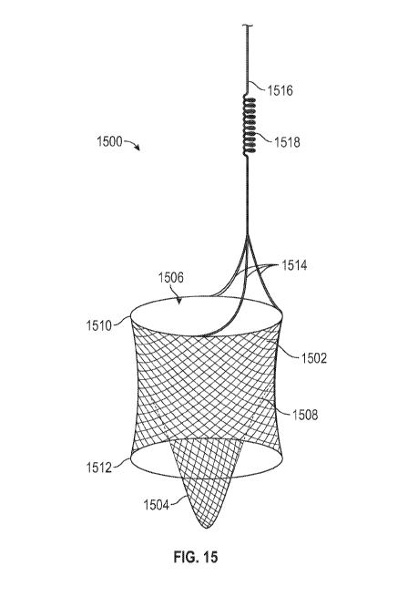
15 shows another configuration of a peripheral vascular filter 1500 in a
deployed state. The peripheral vascular filter 1500 includes a primary filter
1502, and a
secondary filter 1504. The primary filter 1502 and secondary filter 1504 form
the filter body.
The primary filter 1502 forms a lumen therein. While the filter is deployed,
the proximal end
of the lumen is open to allow an inflow 1506 of fluid into the lumen.
[00106] The wall
of the primary filter 1502 can include a plurality of struts, with holes
formed therebetween. The struts may form a mesh. The holes 1508 between the
struts may be
smaller than 5 p.m. The filter wall may oppose the vessel wall. The filter
wall may abutt the
vessel wall, creating a friction force that maintains the position of the
filter.
[00107] The
outflow from the filter is via the tapered secondary filter 1504. The
secondary filter allows for more storage and also traps particles deep within
the filter to
prevent particles from escaping while the filter is deployed, or during
removal. The primary
filter 1502 may include stabilizing rings 1510, 1512 at opposite ends thereof
The stabilizing
rings may aid in maintaining the position of the filter in the vessel, for
example, by creating a
friction force against the wall of the vessel.
[00108] The
filter may include a plurality of retractor wires 1514 that connect the
proximal end of the primary filter 1502 to a spring system 1518 and filter
spring wire 1516.
19
CA 03064638 2019-11-21
WO 2018/237155
PCT/US2018/038771
[00109] Figure
16 shows details of a spring system according to some aspects. The
spring system includes a number of retractor wires 1600 connected to the
proximal end of the
primary filter 1602. The secondary filter 1604 is disposed inside the primary
filter 1602, and
the proximal end of the secondary filter 1604 is connected to the inner
surface of the primary
filter 1602. Each retractor wire 1600 may have a loop 1606 at its proximal
end. The loop
1606 may be made of a flexible wire that allows the opposing sides of the loop
spread apart
from or draw near to each other when tension is applied to the retractor wire
1600. When a
distally-directed force is applied to the guide wire 1608, the sides of the
loop 1606 can spread
apart, absorbing the force so that it is not transmitted to the filter body.
When a proximally-
directed force is applied to the guide wire 1608, the sides of the loop 1606
can draw near to
each other, absorbing the force so that it is not transmitted to the filter
body.
[00110] Figures
17A-17C show additional aspects of the spring system. As shown in
Figure 17A, the loops can absorb upward forces 1700 and downward forces 1702.
As shown
in Figure 17B, when tension is applied, the width of the loop contracts, and
the length
increases. The width of the loop can continue to decrease until the loop has
the shape of a
single wire. The loop can thus absorb upward and downward forces without
changing the
position of the filter.
[00111] As shown
in Figure 17C, the loop 1704 can be formed from a single wire or
from two wires, and can be welded to the end of the retractor wire 1706.
Alternatively, the
loop 1704 may be integrally formed with the retractor wire 1706. When a
downward force
1708 is applied to the spring system, the loop 1704 expands. When all three
loops swell, they
act as a brake for the guide wire. This can alert the operator to the fact
that a force is being
applied to the guide wire that could potentially dislodge the filter. The
operator can pull the
guide wire away from the filter to restore the loop 1704 to its natural shape,
i.e., its shape
when no forces are exerted on it.
CA 03064638 2019-11-21
WO 2018/237155
PCT/US2018/038771
[00112] Figures
18A-18D show aspects of deployment of the filter. To deploy the
filter, the operator pins the filter wire 1802 and pulls the filter delivery
catheter 1801
proximally, as shown in Figure 18A. The operator continues pulling the filter
delivery
catheter until the distal end 1804 of the filter 1806 is deployed, as shown in
Figure 18B. At
this point the operator can still retrieve the filter if the location of the
filter 1806 is not the
target location.
[00113] Figure
18C shows the filter 1808 almost completely deployed. At this point it
may not be possible to retrieve the filter 1808 with the delivery catheter
1810, and a retrieval
catheter may be required to remove the filter. Figure 18D shows the filter
1812 in a deployed
state. At this point the delivery catheter 1814 is retracted.
[00114] Figure
19A-19C show aspects of retrieval of the filter. Figure 19A shows the
primary filter 1900 collapsing. The primary filter 1900 is pulled away from
the vessel wall
1902 as it collapses and enters the retrieval catheter. The secondary filter
1904 is attached to
the primary filter 1900, and is pulled proximally as the primary filter 1900
is pulled
proximally. The loops 1906 at the proximal ends of the retractor wires 1908
act as a single,
reinforced wire ready to pull the filter into the retrieval catheter. Figure
20 shows the loops
2000 coming together as they enter the retrieval catheter 2002, which makes
them stronger.
[00115] Figure
19B shows the filter as the loops are being pulled into the retrieval
catheter 1910. The retractor wires 1912 are now straight and close to each
other, and their
alignment with the catheter and with each other creates a significant strong
stable pulling
force that collapses the filter opening 1914. This is the first step in
pulling the self-expanding
portion of the filter into the retrieval catheter. The collapse of the opening
of the filter closes
the mouth of the primary filter and traps everything inside of it. The distal
end 1916 of the
primary filter also collapses, further ensuring secure trapping of the
material inside the filter.
The secondary filter 1918 with material trapped therein may extend distal to
the distal end
21
CA 03064638 2019-11-21
WO 2018/237155
PCT/US2018/038771
1916 of the primary filter. Finally, as shown in Figure 19C, the filter is
removed with the
guide wire 1920 in place.
[00116] Figure
21A shows a peripheral vascular filter according to some aspects. The
filter includes a primary filter 2100. The filter further includes a plurality
of retractor wires
2102. The filter may include three retractor wires 2102, as shown in Figure
21A, or it may
include more or fewer retractor wires. The filter includes a spring system
2104. The spring
system 2104 can include a coil spring that can expand and contract. The length
of the spring
when maximally stretched may be equal to 150%-200% the length of the spring
when no
stretching forces are applied. The spring system is positioned to prevent
pushing and pulling
filter when forces are applied to the filter wire 2106. The spring system plus
the self-
expanding filter body create an added anchoring force that provides stability
for the filter.
When forces are applied to the filter wire 2106, the spring system can expand
or contract to
absorb the tension, and prevent the forces from dislodging the filter from its
position in the
tissue cavity.
[00117] Figure
21B shows the spring system of Figure 21A. According to one aspect,
the retractor wires 2108 each have a diameter of about .11 inches, the three
grouped retractor
wires 2110 have a diameter of about 0.33 inches, and the spring 2112 has a
diameter of about
0.35 inches. In one aspect, the spring system has a maximum diameter that is
less than 0.4
inches. In one aspect, the spring system has maximum diameter that is within
10% of the
combined diameter of the retractor wires. These dimensions are exemplary and
non-limiting,
and other dimensions may be used. Figure 21C shows the spring system in a
first
configuration 2114 when the spring system is pre-loaded and not stretch, and
in a second
configuration 2216 when the spring system is pre-loaded and stretched.
[00118] Figures
22A-22F show additional aspects of the retrieval of the filter. First, an
operator advances the retrieval catheter 2200 over the filter spring wire
2202, as shown in
22
CA 03064638 2019-11-21
WO 2018/237155
PCT/US2018/038771
Figure 22A. Then, the operator initiates pulling of the filter wire 2202 (also
referred to as a
"filter spring wire" or a "guide wire") into the retrieval catheter 2200 while
slowly advancing
the retrieval catheter 2200. This is shown in Figure 22B. As shown in Figure
22C, the filter
spring wire 2202 is pulled into the retrieval catheter 2200 through a narrow
lumen 2204 until
the entire spring 2206 is beyond the retrieval catheter tip. Figure 22D shows
the spring 2206
in the retrieval catheter 2200.
[00119] As shown
in Figure 22E, once the spring 2206 has gone through the narrow
catheter lumen 2204, the spring 2206 is allowed to return to a non-stretched
spring
configuration. The narrow catheter lumen 2204 acts as a stopper for the spring
2206 from
falling out of the retrieval catheter. Then, as shown in Figure 22F, the
operator can advance a
filter spring wire condenser 2208 inside the retrieval catheter 2200 over the
filter spring wire
2202. The filter spring wire condenser 2208 prevents the spring 2206 from
stretching during
the additional pull back on the filter spring wire 2202 and this strengthens
the filter spring
wire 2202 for the final stages of retrieval.
[00120] Figure
23A-23E show the final stages of retrieval. Figure 23A shows the
initiation of a pulling force on the filter 2300. The force is generated by
the filter spring wire
condenser which is housed inside the retrieval catheter. The filter spring
wire condenser may
also be referred to as a spring wire retrieval catheter. As shown in Figure
23B, the filter 2300
is pulled by the spring wire retrieval catheter, causing the filter's proximal
portion 2302 to
collapse and enter the retrieval catheter 2304. Figure 23C shows two forces
resulting from
two action performed at the same time. The upwards arrow indicate the pulling
force from the
spring wire retrieval catheter. The downward arrow indicates the downward
pushing force
from the retrieval catheter.
[00121] Figure
23D shows the filter 2300 mostly disposed inside the retrieval catheter
2304. The filter 2300 shows important configuration changes including closure
of the orifice
23
CA 03064638 2019-11-21
WO 2018/237155
PCT/US2018/038771
at the proximal end 2306 of the filter 2300. Figure 23E shows the filter 2300
retrieved into
the retrieval catheter 2304, which complete the filter retrieval process.
[00122] The
embodiments illustrated and discussed in this specification are intended
only to teach those skilled in the art how to make and use the invention. In
describing
embodiments of the invention, specific terminology is employed for the sake of
clarity.
However, the invention is not intended to be limited to the specific
terminology so selected.
The above-described embodiments of the invention may be modified or varied,
without
departing from the invention, as appreciated by those skilled in the art in
light of the above
teachings. Moreover, features described in connection with one embodiment of
the invention
may be used in conjunction with other embodiments, even if not explicitly
stated above. It is
therefore to be understood that, within the scope of the claims and their
equivalents, the
invention may be practiced otherwise than as specifically described.
24