Note: Descriptions are shown in the official language in which they were submitted.
CA 03064897 2019-11-25
WO 2018/218151 PCT/US2018/034655
NOVEL ONCOLYTIC VIRUSES FOR SENSITIZING TUMOR CELLS TO KILLING
BY NATURAL KILLER CELLS
I. BACKGROUND
1. Oncolytic viruses (0Vs) hold high promise as a cancer treatment. OVs
selectively
spread in cancer cells and cause a massive cytopathic effect. These virally
infected, dying cancer
cells further recruit immune cells such as NK cells or cytotoxic T cells to
"clean up" infected
cancer cells that escaped the viral killing. However, cancer patients
frequently have
compromised immune systems that fail at doing the job of killing and/or
removing the infected
target cancer cells. Accordingly, what are needed are new oncolytic viruses
and methods of
using said cells that can offer improved outcomes.
SUMMARY
2. Disclosed are methods and compositions related to engineered or modified
oncolytic
viruses.
3. In one aspect, disclosed herein are engineered oncolytic viruses wherein
the oncolytic
virus expresses one or more exogenous membrane bound immune cell targeting
ligands
comprising an uncleaved signal anchor.
4. Also disclosed herein are fusion proteins comprising an uncleaved signal
anchor
domain comprising: a cytoplasmic tail region, a transmembrane region and an
extracellular stalk
region; and an immune cell targeting ligand comprising an N-terminus fused to
a C-terminus of
the extracellular stalk region.
5. In one aspect, disclosed herein are oncolytic viruses and/or fusion
peptides,
polypeptides, or proteins of any preceding aspect; wherein the one or more
exogenous
membrane bound immune cell targeting ligands comprises an engineered
immunoglobulin Fc
domain, a protein agonist of the NK cell receptor NKG2D (such as, for example
RAET1,
RAET1E, RAET1G, RAET1H, RAET1L, RAET1N, MICA, MICB), a protein epitope that is
reactive to anti-CD19 (such as CD19), and/or a protein epitope that is
reactive to anti-CD20
(such as CD20).
6. Also disclosed are oncolytic viruses and/or fusion peptides, polypeptides,
or proteins
of any preceding aspect; wherein the exogenous membrane bound immune cell
targeting ligand
is an immunoglobulin Fc domain and the immunoglobulin Fc domain (such as, an
IgGl, IgG2,
IgG3, or IgG4 Fc domain) is modified to have an inverted orientation with the
amino terminal
end facing intracellularly (i.e., the Fc is expressed on the extracellular
side of the cell surface
with its N-terminal side being attached to a membrane anchor peptide near the
surface of cell
¨ 1 ¨
CA 03064897 2019-11-25
WO 2018/218151
PCT/US2018/034655
membrane rather than the N-terminal side being at maximal distance from the
cell surface). In
one aspect, disclosed herein are oncolytic viruses and/or fusion peptides,
polypeptides, or
proteins of any preceding aspect; wherein the N-terminus of the Fc domain is
fused to the C-
terminus of the extracellular stalk region of the uncleaved signal anchor.
7. In one aspect, disclosed herein are engineered oncolytic viruses, wherein
the
engineered oncolytic virus is a fusogenic oncolytic virus. In some aspect, the
fusogenic
oncolytic virus can be modified or engineered parainfluenza virus type 5. Also
disclosed are
fusogenic oncolytic viruses of any preceding aspect, wherein the fusogenic
oncolytic virus
comprises a gene which codes for a peptide that allows a hyperfusogenic
property that allows
tumor cells to fuse. In one aspect, the oncolytic virus is modified or
engineered to comprise the
fusion peptide, polypeptide, or protein of any preceding aspect.
8. Also disclosed are oncolytic viruses of any preceding aspect, wherein
the oncolytic
virus is engineered to express one or more of IL-2, IL-12, IL-18, IL-21 or IL-
15.
9. In one aspect, disclosed herein are methods of treating cancer,
comprising
administering to a subject an engineered oncolytic virus and/or fusion
peptides, polypeptides, or
proteins of any preceding aspect.
10. Also disclosed are method of treating cancer of any preceding aspect,
wherein the
method further comprises adoptively transferring antibodies or immune cells
(for example, NK
cells, genetically modified NK cell, and/or CAR T cells).
11. In one aspect, disclosed herein are methods of treating cancer of any
preceding
aspect, wherein the NK cells are stimulated and expanded with one or more NK
cell stimulating
agents, such as, for example, a cytokine, growth factor, synthetic ligand, NK
cell stimulating
particle, NK cell stimulating exosome, or NK cell stimulating feeder cell.
III. BRIEF DESCRIPTION OF THE DRAWINGS
12. The accompanying drawings, which are incorporated in and constitute a part
of this
specification, illustrate several embodiments and together with the
description illustrate the
disclosed compositions and methods.
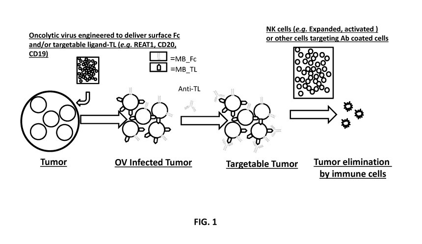
13. Figure 1 shows a schematic of tumors lacking targetable antigen are
treated and
infected with tumor targeting oncolytic virus engineered to deliver membrane
bound Fc region
of antibody (MB Fc) or a membrane bound-targetable ligand (MB TL); (e.g.
RAET1,
RAET1E, RAET1G, RAET1H, RAET1L, RAET1N, MICA, MICB, CD19, and/or CD20). If a
MB TL that is not a NK cell receptor agonist is used, tumors can be treated
with therapeutic
antibody against TL (e.g. anti ¨CD20- rituximab, ofatumumab, obtinutuzumab,
veltuzumab, or
¨ 2 ¨
CA 03064897 2019-11-25
WO 2018/218151 PCT/US2018/034655
ocrelizumab or anti-CD19 MDX1342, MEDI-551, AFM11, XmAb 5871, MOR-208, SGN-
19A,
SAR3419, Blinatumomab, or taplitumomab). Tumors marked with Fc or anti-TL
antibody can
then treated with adoptively transferred cells capable of antibody dependent
cell cytotoxicity
(ADCC) such as for example CD16+ NK cells.
14. Figure 2A and 2B show the construction of a membrane bound immune cell
targeting
ligand comprising an uncleaved signal anchor. Figure 2A shows the structure of
Type I and
Type II integral membrane proteins and the signal anchors for each. Figure 2B
shows the
structure of the uncleaved signal anchor used in the membrane bound immune
cell targeting
ligand.
15. Figures 3A shows a schematic of the genes in an engineered oncolytic virus
including insertion points for a membrane bound immune cell targeting ligand
and site of any
fusogenic mutations.
16. Figure 3B shows a micrograph of Vero cells following infection with the
oncolytic
virus.
17. Figure 3C shows that PM21 activated NK cells recognize and kill tumor
cells more
effectively when treated the engineered oncolytic viruses as compared to mock
treated tumor
targets.
18. Figure 3D shows alternative constructions of membrane bound immune cell
targeting
ligands comprising an Fc domain comprising a neuraminidase signal anchor and
increasing
neuraminidase stalk lengths.
19. Figure 4 shows an example of a membrane bound immune cell targeting ligand
sequence. Here neuraminidase signal anchor is fused to an IgG Fc domain by an
RS linker (i.e.,
a restriction site linker).
20. Figure 5 shows the flow cytometry analysis for ability of the NA-Fc fused
construct
to correctly express the membrane bound Fc targeting ligand on the surface of
infected cells
when transfected by plasmid carrying the NA-Fc construct.
21. Figure 6 shows PN/F virus sensitize A549 cells to NK cell killing. A549
lung
cancer cell line was mock infected or infected with PN/F virus. Following
infection NK cells
were added to the cells at indicated ratios and incubated for 4 hours. Cell
death was measured
using Cytotox Glow Assay. NK only and target (mock or PN/F infected) only
wells were
included as controls.
22. Figure 7 shows A549 lung cancer cells transfected with NAl-Fc construct
under
Zeocin selection express Fc on the cells surface. A549 lung cancer cell line
was transfected with
construct encoding expression of NAl-Fc and cultured in presence of Zeocine.
Cell were stained
¨ 3 ¨
CA 03064897 2019-11-25
WO 2018/218151 PCT/US2018/034655
with anti-humanFc-APC antibody and analysed by flow cytometry. Parental cells
were used as a
control.
23. Figure 8 shows the expression of Fc on tumors as well as infection with
PN/F
increase killing of A549 cells by NK cells. A549 cells or A549 cells stably
expressing Fc on the
surface (A549-Fc) were infected with mock or PN/F and incubated with NK cells
at 1:1 or 1:3
target to NK cell ratio. Cell death was determined by flow cytometry measuring
live cell events
in the target gate with reference to controls containing respective target
only cells.
24. Figure 9 shows that SKOV-3 ovarian cancer cells transfected with NAl-Fc
¨NA4-Fc
constructs express Fc on the cells surface after FACS sorting. SKOV-3 ovarian
cancer cell line
was transfected with constructs encoding expression of NAl-Fc, NA2-Fc, NA3-Fc
or NA4-Fc.
After few days cells were stained with anti-human Fc-APC antibody and sorted
by FACS to
enrich for Fc-expressing cell population cytometry. Sorted cells have stable
but variable level of
expression of Fc for all constructs tested. Parental cells were used as a
control.
25. Figure 10 shows that the increased length of the NA stalk improves NK cell
killing
via recognition of the surface expressed Fc domain. SKOV-3 cells with or
without stable
expression of (NA1-NA4)-Fc were mixed with NK cells at a 3:1 ratio of NK:
Targets. Cell death
was determined by flow cytometry measuring live cell events in the target gate
with reference to
controls containing respective target only cells. Killing correlates with the
length of the NA stalk
rather then the density of the Fc on the cells surface of SKOV-3 (Figure 9).
26. Figure 11 shows that the expression of Fc on tumors as well as infection
with PN/F
increase killing of SKOV-3 cells by NK cells. SKOV-3 cells or SKOV-3 cells
stably expressing
NAl-Fc on the surface (SKOV-3-Fc) were infected with mock or PN/F and
incubated with NK
cells at 1:1 or 1:3 target to NK cell ratio. Cell death was determined by flow
cytometry
measuring live cell events in the target gate with reference to controls
containing respective
.. target only cells. Both, expression of Fc on the surface as well as
infection with PN/F leads to
increased killing of SKOV-3 cells by NK cells and this effect is additive.
IV. DETAILED DESCRIPTION
27. Before the present compounds, compositions, articles, devices, and/or
methods are
disclosed and described, it is to be understood that they are not limited to
specific synthetic
methods or specific recombinant biotechnology methods unless otherwise
specified, or to
particular reagents unless otherwise specified, as such may, of course, vary.
It is also to be
understood that the terminology used herein is for the purpose of describing
particular
embodiments only and is not intended to be limiting.
- 4 -
CA 03064897 2019-11-25
WO 2018/218151 PCT/US2018/034655
A. Definitions
28. As used in the specification and the appended claims, the singular forms
"a," "an"
and "the" include plural referents unless the context clearly dictates
otherwise. Thus, for
example, reference to "a pharmaceutical carrier" includes mixtures of two or
more such carriers,
and the like.
29. Ranges can be expressed herein as from "about" one particular value,
and/or to
"about" another particular value. When such a range is expressed, another
embodiment includes
from the one particular value and/or to the other particular value. Similarly,
when values are
expressed as approximations, by use of the antecedent "about," it will be
understood that the
particular value forms another embodiment. It will be further understood that
the endpoints of
each of the ranges are significant both in relation to the other endpoint, and
independently of the
other endpoint. It is also understood that there are a number of values
disclosed herein, and that
each value is also herein disclosed as "about" that particular value in
addition to the value itself
For example, if the value "10" is disclosed, then "about 10" is also
disclosed. It is also
understood that when a value is disclosed that "less than or equal to" the
value, "greater than or
equal to the value" and possible ranges between values are also disclosed, as
appropriately
understood by the skilled artisan. For example, if the value "10" is disclosed
the "less than or
equal to 10"as well as "greater than or equal to 10" is also disclosed. It is
also understood that
the throughout the application, data is provided in a number of different
formats, and that this
data, represents endpoints and starting points, and ranges for any combination
of the data points.
For example, if a particular data point "10" and a particular data point 15
are disclosed, it is
understood that greater than, greater than or equal to, less than, less than
or equal to, and equal to
10 and 15 are considered disclosed as well as between 10 and 15. It is also
understood that each
unit between two particular units are also disclosed. For example, if 10 and
15 are disclosed,
then 11, 12, 13, and 14 are also disclosed.
30. In this specification and in the claims which follow, reference will be
made to a
number of terms which shall be defined to have the following meanings:
31. "Optional" or "optionally" means that the subsequently described event or
circumstance may or may not occur, and that the description includes instances
where said event
or circumstance occurs and instances where it does not.
32. As used herein, "N-terminal side" or "amino terminal end" refers to
directionality of
a peptide, polypeptide, or protein and may not mean the N-terminus. In some
aspects, where a
chimeric or fusion peptide, polypeptide, or protein is discussed, the N-
terminal side may refer
only to a component of the chimeric or fusion peptide, polypeptide, or protein
and not the entire
¨ 5 ¨
CA 03064897 2019-11-25
WO 2018/218151 PCT/US2018/034655
structure. For example, where a Fc domain comprising an uncleaved signal
anchor is discussed,
and the Fc domain is described as having an inverted orientation with the
amino terminal end or
N-terminal side facing intracellularly, contemplated herein are chimeric or
fusion peptide,
polypeptide, or protein wherein the signal anchor is at the N-terminus of the
chimeric or fusion
construct and actually spans the cellular membrane. Thus, in such a chimera,
the anchor is
closer to the amino terminus than the Fc domain, but the directionality of the
Fc domain has the
N-terminal side facing the cell which is inverted relative to the orientation
of the Fc domain in a
typical B cell which would typically have the carboxy end spanning the
cellular membrane and
amino terminal end extending to the extracellular matrix.
33. Throughout this application, various publications are referenced. The
disclosures of
these publications in their entireties are hereby incorporated by reference
into this application in
order to more fully describe the state of the art to which this pertains. The
references disclosed
are also individually and specifically incorporated by reference herein for
the material contained
in them that is discussed in the sentence in which the reference is relied
upon.
B. Compositions
34. Disclosed are the components to be used to prepare the disclosed
compositions as
well as the compositions themselves to be used within the methods disclosed
herein. These and
other materials are disclosed herein, and it is understood that when
combinations, subsets,
interactions, groups, etc. of these materials are disclosed that while
specific reference of each
various individual and collective combinations and permutation of these
compounds may not be
explicitly disclosed, each is specifically contemplated and described herein.
For example, if a
particular oncolytic virus or fusion protein is disclosed and discussed and a
number of
modifications that can be made to a number of molecules including the
oncolytic virus and/or
fusion protein are discussed, specifically contemplated is each and every
combination and
.. permutation of oncolytic virus and/or fusion protein and the modifications
that are possible
unless specifically indicated to the contrary. Thus, if a class of molecules
A, B, and C are
disclosed as well as a class of molecules D, E, and F and an example of a
combination molecule,
A-D is disclosed, then even if each is not individually recited each is
individually and
collectively contemplated meaning combinations, A-E, A-F, B-D, B-E, B-F, C-D,
C-E, and C-F
are considered disclosed. Likewise, any subset or combination of these is also
disclosed. Thus,
for example, the sub-group of A-E, B-F, and C-E would be considered disclosed.
This concept
applies to all aspects of this application including, but not limited to,
steps in methods of making
and using the disclosed compositions. Thus, if there are a variety of
additional steps that can be
¨ 6 ¨
CA 03064897 2019-11-25
WO 2018/218151 PCT/US2018/034655
performed it is understood that each of these additional steps can be
performed with any specific
embodiment or combination of embodiments of the disclosed methods.
35. Oncolytic viruses (0Vs) which preferentially infect and kill cancer cells
hold high
promise as a cancer treatment. OVs selectively spread in cancer cells and
cause a massive
cytopathic effect. These virally infected, dying cancer cells further recruit
immune cells such as
NK cells or cytotoxic T cells to "clean up" infected cancer cells that escaped
the viral killing.
Since, in cancer patients, the immune system is frequently compromised and
fails at doing the
job, combination with adoptive immune cell transfer can offer improved
outcomes.
36. Immune cells such as NK cells, directly target the destruction of infected
cells. NK
cells, for example, efficiently destroy tumor cells, stressed cells, and
virally infected cells by a
variety of different methods. The first is by directly engaging target cells,
permeating their
membranes, and then injecting a protein that cleaves and activates several
apoptotic proteins,
thereby initiating programmed cell death (apoptosis) of the targeted cell. The
surface of an NK
cell also contains protein ligands that can bind and activate receptors, such
as the receptor for
tumor-necrosis factor (TNF)-related apoptosis-inducing ligand (TRAIL), on
target cells that turn
on internal signals for apoptotic programmed cell death. When stimulated, NK
cells can also
secrete cytokines such as INFy and TNFa that not only inhibit viruses and
tumors, but also
signal invasion to other immune cells.
37. Through the use of recombinant nucleic acid modification, it is understood
and
herein contemplated that oncolytic viruses and/or fusion peptides,
polypeptides, and proteins can
be engineered to or otherwise modified so that expression of the fusion
peptides, polypeptides or
proteins in a cancer cell improves the NK cell recruitment to target cancer
cells. As used
interchangeably herein, the terms "fusion peptide(s)", "fusion
polypeptide(s)", and "fusion
proteins" refer to any peptide, polypeptide, or protein that has been
engineered to comprise
domains from two or more unrelated peptides, polypeptides, or proteins. In
some aspects, the
fusion peptide, polypeptides, or proteins comprise all or a portion of each of
the component two
or more peptide, polypeptide, or proteins that are joined to form the fusion.
38. Thus, one aspect of the invention pertains to engineered fusion proteins,
i.e.,
exogenous membrane bound targeting ligands expressed by the engineered
oncolytic viruses, as
disclosed herein. As used herein, the term "fusion protein" is synonymous with
"chimeric
protein," and refers to a first, uncleaved signal anchor polypeptide
comprising a cytoplasmic tail
region, a transmembrane region and an extracellular stalk region as explained
in further detail
below, the first polypeptide operatively linked to an immune cell targeting
ligand polypeptide.
The term "operatively linked" refers to the fusion of the two polypeptides,
i.e., fusion in-frame
- 7 -
CA 03064897 2019-11-25
WO 2018/218151 PCT/US2018/034655
of each region to the other. Fusion may be accomplished with or without the
use of a short
polypeptide linker consisting of 2, 3, 4, 5, 6, 7, 8, 9, 20, 11, 12, 13, 14,
15, 16, 17, 18, 19 or 20
or more amino acids. For example, the targeting ligand polypeptide may be
fused at its N-
terminus to the C-terminus of the first polypeptide.
39. In one aspect, the fusion peptides, polypeptides, or proteins are
exogenous membrane
bound targeting ligands as disclosed herein. The fusion peptides, polypeptides
or proteins thus
can comprise an uncleaved signal anchor domain comprising: a cytoplasmic tail
region, a
transmembrane region and an extracellular stalk region; and an immune cell
targeting ligand
wherein the N-terminus of the immune cell targeting ligand is fused to a C-
terminus of the
extracellular stalk region. (See, e.g., Fig. 2B). In other words, when the
fusion protein is
expressed in a cell, the immune cell targeting ligand is bound to the cell
membrane in an
inverted orientation with respect to the cell, as compared to the naturally
occurring orientation of
the immune cell targeting ligand.
40. In one aspect, the uncleaved signal anchor domain is derived from a Type
II integral
membrane protein which is schematically depicted in the lower panel of Figure
2A. A Type II
integral membrane protein generally comprises an N-terminus inside the cell,
i.e., a cytoplasmic
tail region, a transmembrane region, an extracellular stalk region and a
globular head region with
the C-terminus. As disclosed herein, the uncleaved signal anchor domain
comprises the
cytoplasmic tail region, the transmembrane region, and the extracellular stalk
region, but lacks
the globular head region. The uncleaved signal anchor domain can comprise for
example the
relevant portions of a Type II integral membrane protein such as
neuraminidase, parainfluenza
virus hemagglutinin-neuraminidase, transferrin receptor, MHC class II
invariant chain, P
glycoprotein, asialoglycoprotein receptor, or a neutral endopeptidase. In an
exemplary aspect,
the uncleaved signal anchor domain comprises a neuraminidase signal anchor
domain, as shown
in Figure 2B.
41. The immune cell targeting ligand is for example a ligand capable of
binding, for
example selectively binding an immune cell, and comprising an amino acid
modification
wherein the N-terminus of the ligand fuses or is fused to (via a peptide
linker) to the C-terminus
of the extracellular stalk domain of the uncleaved signal anchor domain.
Ligands can be
selected from known ligands that are capable of binding an immune cell such an
NK cell, a B
cell, a T cell and/or a CAR-T cell. Such ligands include, for example, an
immunoglobulin Fc
domain such as IgG1 (as shown in Fig, 2B), or alternatively IgG2, IgG3, or
IgG4. Amino acid
modifications to the Fc domain that are suitable for achieving the inverted
orientation described
herein include: 256A/K290A/5298A/E333A/K334A or L235V/F243L/R292P/Y300L/P396L.
¨8--
CA 03064897 2019-11-25
WO 2018/218151 PCT/US2018/034655
Alternatively, the targeting ligand is selected from an NK2GD ligand such as,
for example,
RAET1, RAET1E, RAET1G, RAET1H, RAET1L, RAET1N, MICA, and MICB; or an anti-
ligand domain such as CD19 or CD20.
42. By way of non-limiting example, fusion proteins as disclosed herein
encompass
polypeptides comprising amino acid sequences sufficiently identical to or
derived from the
amino acid sequence of the SEQ ID NO: 1. Fusion proteins as disclosed herein
encompass
polypeptides having fewer or more amino acids than the full length sequence of
SEQ ID NO:1,
and exhibit the same membrane anchoring function with a targeting ligand as
demonstrated by
the fusion protein having the sequence of SEQ ID NO: 1. Examples of useful
fusion proteins
according to the present disclosure include a protein which comprises an amino
acid sequence
that has at least about 45%, preferably 55%, 65%, 75%, 85%, 95%, or 99%
sequence identity
with the amino acid sequence of SEQ ID NO: 1, and retains the functional
activity of the fusion
protein of SEQ ID NO: 1. More specifically, a fusion protein according to the
present disclosure
can comprise an amino acid sequence having at least about 50, 51, 52, 53, 54,
55, 56, 57, 58, 59,
60, 61, 62, 63, 64, 65, 66, 67, 68, 69, 70, 71, 72, 73, 74, 75, 76, 77, 78,
79, 80, 81, 82, 83, 84, 85,
86, 87, 88, 89, 90, 91, 92, 93, 94, 95, 96, 97, 98, 99% sequence identity to
the amino acid
sequence of SEQ ID NO: 1.
43. The percent identity of two amino acid sequences or of two nucleic acid
sequences
can be determined by aligning the two sequences end to end to optimize the
number of amino
acid or nucleotide matches between the two sequences, wherein for example gaps
can be
introduced in the sequence of a first amino acid or nucleic acid sequence to
obtain the optimal
alignment with a second amino or nucleic acid sequence. The amino acid
residues or nucleotides
at corresponding amino acid positions or nucleotide positions are then
compared. When a
position in the first sequence is occupied by the same amino acid residue or
nucleotide as the
corresponding position in the second sequence, then the molecules are
identical at that position.
The percent sequence identity between the two sequences is a function of the
number of
identical positions shared by the sequences (i.e., % sequence identity is the
number of identical
positions/total number of positions x100).
44. The determination of percent sequence identity between two sequences may
be
accomplished using a mathematical algorithm. A non-limiting example of a
mathematical
algorithm as known in the art and utilized for the comparison of two sequences
is the algorithm
of Karlin and Altschul (1990) Proc. Nat'l Acad. Sci. USA 87:2264-2268,
modified as in Karlin
and Altschul (1993) Proc. Nat'l Acad. Sci. USA 90:5873-5877. Such an algorithm
is
incorporated into the NBLAST and XBLAST programs of Altschul, et al. (1990) J.
Mol. Biol.
- 9 -
CA 03064897 2019-11-25
WO 2018/218151 PCT/US2018/034655
215:403-410. BLAST nucleotide searches can be performed with the NBLAST
program,
score=100, wordlength=12 to obtain nucleotide sequences similar or homologous
to Adhesin
nucleic acid molecules of the invention. To obtain gapped alignments for
comparison purposes,
Gapped BLAST can be utilized as described in Altschul et al. (1997) Nucleic
Acids Res.
25:3389-3402. When utilizing BLAST and Gapped BLAST programs, the default
parameters of
the respective programs (e.g., XBLAST and NBLAST) can be used.
45. The fusion proteins and polynucleotides encoding them can be produced by
standard
recombinant DNA techniques as known in the art. For example, DNA fragments
coding for the
different polypeptide sequences are ligated together in-frame applying
conventional techniques.
Suitable techniques include by employing blunt-ended or stagger-ended termini
for ligation,
restriction enzyme digestion to provide for appropriate termini, filling-in of
cohesive ends as
appropriate, alkaline phosphatase treatment to avoid undesirable joining, and
enzymatic ligation.
Alternatively, a fusion gene may be synthesized by conventional techniques
including automated
DNA synthesizers. Alternatively, PCR amplification of gene fragments may be
carried out
using anchor primers that give rise to complementary overhangs between two
consecutive gene
fragments, which can subsequently be annealed and reamplified to generate a
chimeric gene
sequence. (See, e.g., Current Protocols in Molecular Biology, Ausubel et al.
eds., John Wiley &
Sons: 1992).
46. A fusion gene encoding a fusion protein as disclosed herein can be created
by
removing the stop codon from a cDNA sequence encoding the first polypeptide,
then adding a
cDNA encoding the second polypeptide protein in frame through ligation or
overlap extension
PCR. Optionally, a short sequence of amino acids (for example, a sequence of
about 2 to about
20 amino acids) can be engineered in as a linker between the first polypeptide
and the second
polypeptide. The resulting fusion gene which comprises a polynucleotide
sequence encoding a
fusion protein can then be introduced to the genome of a host virus, including
for example an
engineered oncolytic virus as disclosed herein. When the host virus contacts a
host cell and
delivers its modified genetic package to the cytoplasm of the cell, the fusion
gene will then be
expressed by the host cell as a single fusion protein.
47. As noted above, the disclosed oncolytic viruses can be modified or
engineered to
maximize the number of immune cells (for example NK cells, T cells, CART
cells, Innate
lymphoid cells, Macrophages, and B cells (including plasma cells)) at the
target cancer site and
thus increase the immune cell activity (for example, NK cell activity, T cell
activity, CAR T cell
activity, and/or B cell activity (including plasma cell and antibody activity)
in eliminating cancer
beyond that which an unmodified oncolytic virus would do. As used herein,
"oncolytic viruses"
¨ 10 ¨
CA 03064897 2019-11-25
WO 2018/218151 PCT/US2018/034655
refers to a virus that is tropic for and kills cancer cells. Oncolytic viruses
can be engineered to
selectively attack cancer cells. Accordingly, in one aspect, disclosed herein
are engineered
oncolytic viruses wherein the oncolytic viruses express one or more membrane
bound immune
cell targeting ligands comprising an uncleaved signal anchor. In some aspect,
the engineered
oncolytic viruses expresses one or more of the fusion peptides, polypeptides,
or proteins
disclosed herein.
48. In one aspect, the disclosed oncolytic viruses and/or fusion peptides,
polypeptides, or
proteins are modified to express or comprise one or more exogenous membrane
bound immune
cell targeting ligands (such as, for example, NK cell targeting ligands) for
increasing the affinity
towards NK cells. As used herein, exogenous membrane bound immune cell
targeting ligands
refers to any exogenous peptide, polypeptide, or protein that can serve as a
target for immune
cell activity including, but not limited to NK cell activity, B cell activity,
T cell activity, and
CAR T cell activity. Thus, in aspect, the oncolytic virus can comprise one or
more peptides,
polypeptides, or proteins comprising exogenous membrane bound immune cell
targeting ligands
including fusion proteins that comprise an exogenous membrane bound immune
cell targeting
ligand. The membrane bound immune cell targeting ligands of the disclosed
oncolytic viruses
and/or fusion peptides, polypeptides, or proteins can be bound by NK cells, B-
cells, T-cells, or
CAR T -cells. In one aspect, immune cell targeting ligands are membrane bound
via
modification to include a signaling anchor. Immune cell targeting ligands can,
for example,
comprise immunoglobulin Fc domains which are ligands for CD16 on NK cells,
ligands for
NKG2D receptors on NK cells, or targets for antibodies or CAR T cells. In one
aspect, it is
understood and herein contemplated that the exogenous membrane bound immune
cell targeting
ligands can be either bound directly by NK cell receptors such as, for
example, Fc domains (for
example IgGl, IgG2, IgG3, and/or IgG4), NK2GD ligands (for example, RAET1,
RAET1E,
RAET1G, RAET1H, RAET1L, RAET1N, MICA, and/or MICB), or can be bound indirectly
by
NK cells via the use of an anti-ligand antibody (for example CD19 or CD20
which can be bound
by anti-CD19 or anti-CD20 antibodies) or can be directly targeted by anti-
ligand CAR T cells
(such as, for example, anti-CD19 CART cells). Accordingly, in one aspect,
disclosed herein are
fusion proteins comprising immune cell targeting ligands and oncolytic viruses
comprising one
or more immune cell targeting ligands, wherein the immune cell targeting
ligand is an Fc
domain selected from the group consisting of IgGl, IgG2, IgG3, and/or IgG4.
49. The Fc domain is the ligand to which CD16 (FcyRIII) which is found on the
surface
of NK cells binds. CD16 is one of the primary receptors on NK cells and when
CD16 binds to
the Fc portion of an antibody (for example, an IgGl, IgG2, IgG3, and/or IgG4
Fc domain), this
¨ 11 ¨
CA 03064897 2019-11-25
WO 2018/218151 PCT/US2018/034655
activates the NK cells antibody-dependent cell mediated cytotoxity (ADCC).
However, the Fc
portion of the antibody is typically only available when secreted. When the
membrane bound
antibody receptor found on B cells is present, the Fc portion is typically
oriented to the cytosol
of the cell. Accordingly, in the modified oncolytic viruses disclosed herein,
the Fc domain is
modified to have an inverted orientation with the amino terminal end faced
intracellularly when
expressed on membranes of infected tumor targets thus mimicking the
orientation of an
extracellular antibody bound to the surface of a cell. In one aspect,
disclosed herein are
modified or engineered oncolytic viruses expressing one or more exogenous
membrane bound
immune cell targeting ligands comprising an uncleaved signal anchor; wherein
the one or more
exogenous membrane bound immune cell targeting ligand is an immunoglobulin Fc
domain (for
example, an IgGl, IgG2, IgG3, and/or IgG4 Fc domain) modified to have an
inverted orientation
with the amino terminal end faced intracellularly (i.e., the Fc is expressed
on the extracellular
side of the cell surface with its N-terminal side being attached to a membrane
anchor peptide
near the surface of cell membrane rather than the N-terminal side being at
maximal distance
.. from the cell surface).
50. It is understood and herein contemplated that the Fc domain can be
presented as a
monomeric, dimeric, or multimeric construct. In one aspect, the Fc domain can
be further
modified to enhance antibody mediated killing, NK cell recognition, and
control expansion of
activating Fey receptors. For example, the Fc domain can be modified to
increase affinity for
.. CD16. Thus, for example, the Fc domain may comprise one or more mutations
such as, for
example, T256A, K290A, S298A, E333A, K334A, L235V, F243L, R292P, Y300L, and/or
P396L. Similarly, the Fc domain can be further modified to increase
selectivity of binding to the
activating (Ma) vs, inhibitory Fc(IIb) receptor. Thus, for example, the Fc
domain may comprise
one or more mutations such as, for example, 5239D, 1332E, A330L, F243L, R292P,
V3051,
.. and/or P396L.
51. NKG2D is activating receptor on NK cells that triggers actin
reorganization (cell
polarization) and degranulation in target cells. NKG2D recognizes induced-self
proteins which
are typically completely absent or present only at low levels on surface of
normal cells, but are
overexpressed by infected, transformed, senescent and stressed cells. The
ligands for NKG2D
are from MHC class I polypeptide-related sequence (MIC) and retenoic acid
early transcript 1
(RAET1)/ULBP families which appear on the surface of stressed, malignant
transformed, and
infected cells. MIC is a surface glycoprotein. The MIC family of proteins
(MICA and MICB)
are structurally similar to MHC, but do not associate with f32-microglobulin
or peptides like
MHC. MIC family proteins are comprised of an extracellular domain (an a1a2a3
domain), a
¨ 12 ¨
CA 03064897 2019-11-25
WO 2018/218151 PCT/US2018/034655
transmembrane domain, and a C-terminal cytoplasmic tail. The RAET1 family are
surface
glycoproteins comprising an extracellular domain (an ala2 domain), a
transmembrane domain,
and a C-terminal cytoplasmic tail. The RAET1 family serve as stressed induced
ligands for
NKG2D and are related to MHC class 1 molecules. In one aspect, disclosed
herein are
engineered oncolytic viruses and/or fusion peptides, polypeptides, or proteins
comprising one or
more exogenous membrane bound immune cell targeting ligand comprising an
uncleaved signal
anchor; wherein the one or more exogenous membrane bound immune cell targeting
ligand is an
NKG2D ligand (for example, RAET1, RAET1E, RAET1G, RAET1H, RAET1L, RAET1N,
MICA, and/or MICB).
52. The exogenous membrane bound immune cell targeting ligands, i.e., the
fusion
proteins that are encoded by the engineered oncolytic viruses as disclosed
herein are modified to
present on the surface of the infected cancer cell. In one aspect, this
membrane bound
presentation can be achieved through the use of an uncleaved signal anchor.
Signal anchors can
comprise any signaling sequence that retains the encoded peptide, polypeptide,
or protein on a
cell surface membrane. For example, the signal anchor can be the transmembrane
domain of
neuraminidase, the signal-anchor from parainfluenza virus hemagglutinin-
neuraminidase, the
signal-anchor from the transferrin receptor, the signal-anchor from the MHC
class II invariant
chain, the signal-anchor from P glycoprotein, the signal-anchor from
asialoglycoprotein
receptor, or the signal-anchor from a neutral endopeptidase. Alternatively,
the exogenous
membrane bound immune cell targeting ligands can be modified to encode amino
acid
substitutions comprising additional positively charged amino acids on the
amino terminal end.
In one aspect, the exogenous membrane bound immune cell targeting ligand can
be a fusion
protein wherein the signal anchor is joined or fused to the targeting ligand
through use of a
linker such as a RS linker. Accordingly, in one aspect, are oncolytic viruses
and/or fusion
peptides, polypeptides, or proteins comprising one or more exogenous membrane
bound
immune cell targeting ligands, wherein the membrane bound immune cell
targeting ligands
comprises an uncleaved signal anchor. In one aspect, the immune cell targeting
ligand
comprises an immunoglobulin Fc domain comprising an amino acid modification
wherein the
N-terminus of the Fc domain fuses to the C-terminus of the extracellular stalk
domain of the
signal anchor domain. In one aspect, disclosed herein are engineered oncolytic
viruses and/or
fusion peptides, polypeptides, or proteins wherein the oncolytic virus and/or
fusion peptides,
polypeptides, or proteins that comprise one or more exogenous membrane bound
immune cell
targeting ligands and an uncleaved signal anchor, wherein the uncleaved signal
anchor is
neuraminidase, parainfluenza virus hemagglutinin-neuraminidase, transferrin
receptor, MHC
¨ 13 ¨
CA 03064897 2019-11-25
WO 2018/218151 PCT/US2018/034655
class II invariant chain, P glycoprotein, asialoglyoprotein receiptor, or a
neutral endopeptidease.
For example, an engineered oncolytic virus can comprise a nucleotide sequence
encoding a
fusion protein comprising an immunoglobulin Fc domain (for example, an IgGl,
IgG2, IgG3,
and/or IgG4 Fc domain) and a neuraminidase signal anchor domain, wherein the
Fc domain is
modified to have an inverted orientation with the amino terminal end faced
intracellularly as
compared to the naturally occurring orientation of the Fc domain with respect
to a cell. In other
words, in the fusion peptides, polypeptides and proteins described herein in
which the immune
cell targeting ligand comprises an immunoglobulin Fc domain, the Fc domain is
expressed on
the extracellular side of the cell surface with its N-terminal side being
attached to a membrane
anchor peptide near the surface of cell membrane rather than the N-terminal
side being at
maximal distance from the cell surface. Alternatively, a fusion protein as
described herein and
encoded in an engineered oncolytic virus can comprise a NKG2D ligand (for
example, RAET1,
RAET1E, RAET1G, RAET1H, RAET1L, RAET1N, MICA, and/or MICB) and a neuraminidase
signal anchor domain; or CD20 and a neuraminidase signal anchor domain; and/or
CD19 and a
.. neuraminidase signal anchor domain.
53. One embodiment of a fusion peptide, polypeptide, or protein comprising a
membrane
bound immune cell targeting ligand and an uncleaved signal anchor is set forth
in SEQ ID NO: 1
MNPNQKITTIGSICLVVGLISLILQIGNIISIWISHSIQTGSQNHTGICNRSDKTHTCPPCPAP
ELLGGPSVFLFPPKPKDTLMISRTPEVTCVVVDVSHEDPEVKFNWYVDGVEVHNAKTK
PREEQYNSTYRVVSVLTVLHQDWLNGKEYKCKVSNKALPAPIEKTISKAKGQPREPQV
YTLPPSREEMTKNQVSLTCLVKGFYPSDIAVEWESNGQPENNYKTTPPVLDSDGSFFLY
SKLTVDKSRWQQGNVFSCSVMHEALHNHYTQKSLSLSPGK and shown in Figure 4.
54. As discussed herein there are numerous variants of the fusion peptides,
polypeptides,
and/or proteins; exogenous membrane bound immune cell targeting ligands,
and/or signal
anchor domains that are known and herein contemplated. Protein variants and
derivatives are
well understood to those of skill in the art and in can involve amino acid
sequence
modifications. For example, amino acid sequence modifications typically fall
into one or more
of three classes: substitutional, insertional or deletional variants.
Insertions include amino
and/or carboxyl terminal fusions as well as intrasequence insertions of single
or multiple amino
acid residues. Insertions ordinarily will be smaller insertions than those of
amino or carboxyl
terminal fusions, for example, on the order of one to four residues.
Immunogenic fusion protein
derivatives, such as those described in the examples, are made by fusing a
polypeptide
sufficiently large to confer immunogenicity to the target sequence by cross-
linking in vitro or by
recombinant cell culture transformed with DNA encoding the fusion. Deletions
are
¨ 14 ¨
CA 03064897 2019-11-25
WO 2018/218151
PCT/US2018/034655
characterized by the removal of one or more amino acid residues from the
protein sequence.
Typically, no more than about from 2 to 6 residues are deleted at any one site
within the protein
molecule. These variants ordinarily are prepared by site specific mutagenesis
of nucleotides in
the DNA encoding the protein, thereby producing DNA encoding the variant, and
thereafter
expressing the DNA in recombinant cell culture. Techniques for making
substitution mutations
at predetermined sites in DNA having a known sequence are well known, for
example M13
primer mutagenesis and PCR mutagenesis. Amino acid substitutions are typically
of single
residues, but can occur at a number of different locations at once; insertions
usually will be on
the order of about from 1 to 10 amino acid residues; and deletions will range
about from 1 to 30
residues. Deletions or insertions preferably are made in adjacent pairs, i.e.
a deletion of 2
residues or insertion of 2 residues. Substitutions, deletions, insertions or
any combination
thereof may be combined to arrive at a final construct. The mutations must not
place the
sequence out of reading frame and preferably will not create complementary
regions that could
produce secondary mRNA structure. Substitutional variants are those in which
at least one
residue has been removed and a different residue inserted in its place. Such
substitutions
generally are made in accordance with the following Tables 1 and 2 and are
referred to as
conservative substitutions.
TABLE 1:Amino Acid Abbreviations
Amino Acid Abbreviations
Alanine Ala A
allosoleucine AIle
Arginine Arg
asparagine Asn
aspartic acid Asp
Cy steine Cy s
glutamic acid Glu
Glutamine Gln
Glycine Gly
Histidine His
Isolelucine Ile
Leucine Leu
Lysine Lys
phenylalanine Phe
proline Pro
pyroglutamic acid pGlu
Serine Ser
Threonine Thr
Tyrosine Tyr
Tryptophan Trp
Valine Val V
TABLE 2:Amino Acid Substitutions
Original Residue Exemplary Conservative Substitutions,
others are known in the art.
Ala Ser
¨ 15 ¨
CA 03064897 2019-11-25
WO 2018/218151
PCT/US2018/034655
Arg Lys; Gin
Asn Gin; His
Asp Glu
Cy s Ser
Gin Asn, Lys
Glu Asp
Gly Pro
His Asn;Gln
Ile Leu; Val
Leu Ile; Val
Lys Arg; Gin
Met Leu; Ile
Phe Met; Leu; Tyr
Ser Thr
Thr Ser
Tip Tyr
Tyr Trp; Phe
Val Ile; Leu
55. Substantial changes in function or immunological identity are made by
selecting
substitutions that are less conservative than those in Table 2, i.e.,
selecting residues that differ
more significantly in their effect on maintaining (a) the structure of the
polypeptide backbone in
the area of the substitution, for example as a sheet or helical conformation,
(b) the charge or
hydrophobicity of the molecule at the target site or (c) the bulk of the side
chain. The
substitutions which in general are expected to produce the greatest changes in
the protein
properties will be those in which (a) a hydrophilic residue, e.g. seryl or
threonyl, is substituted
for (or by) a hydrophobic residue, e.g. leucyl, isoleucyl, phenylalanyl, valyl
or alanyl; (b) a
cysteine or proline is substituted for (or by) any other residue; (c) a
residue having an
electropositive side chain, e.g., lysyl, arginyl, or histidyl, is substituted
for (or by) an
electronegative residue, e.g., glutamyl or aspartyl; or (d) a residue having a
bulky side chain,
e.g., phenylalanine, is substituted for (or by) one not having a side chain,
e.g., glycine, in this
case, (e) by increasing the number of sites for sulfation and/or
glycosylation.
56. For example, the replacement of one amino acid residue with another that
is
biologically and/or chemically similar is known to those skilled in the art as
a conservative
substitution. For example, a conservative substitution would be replacing one
hydrophobic
residue for another, or one polar residue for another. The substitutions
include combinations
such as, for example, Gly, Ala; Val, Ile, Leu; Asp, Glu; Asn, Gln; Ser, Thr;
Lys, Arg; and Phe,
Tyr. Such conservatively substituted variations of each explicitly disclosed
sequence are
included within the mosaic polypeptides provided herein.
57. Substitutional or deletional mutagenesis can be employed to insert sites
for N-
glycosylation (Asn-X-Thr/Ser) or 0-glycosylation (Ser or Thr). Deletions of
cysteine or other
labile residues also may be desirable. Deletions or substitutions of potential
proteolysis sites,
¨ 16 ¨
CA 03064897 2019-11-25
WO 2018/218151 PCT/US2018/034655
e.g. Arg, is accomplished for example by deleting one of the basic residues or
substituting one
by glutaminyl or histidyl residues.
58. Certain post-translational derivatizations are the result of the action of
recombinant
host cells on the expressed polypeptide. Glutaminyl and asparaginyl residues
are frequently
post-translationally deamidated to the corresponding glutamyl and asparyl
residues.
Alternatively, these residues are deamidated under mildly acidic conditions.
Other post-
translational modifications include hydroxylation of proline and lysine,
phosphorylation of
hydroxyl groups of seryl or threonyl residues, methylation of the o-amino
groups of lysine,
arginine, and histidine side chains (T.E. Creighton, Proteins: Structure and
Molecular
Properties, W. H. Freeman & Co., San Francisco pp 79-86 [19831), acetylation
of the N-terminal
amine and, in some instances, amidation of the C-terminal carboxyl.
59. It is understood that one way to define the variants and derivatives of
the disclosed
proteins herein is through defining the variants and derivatives in terms of
homology/identity to
specific known sequences. For example, SEQ ID NO: 1 sets forth a particular
sequence of a
fusion protein. Specifically disclosed are variants of these and other
proteins herein disclosed
which have at least, 70% or 75% or 80% or 85% or 90% or 95%, 96%, 97%, 98%, or
99%
sequence identity to the stated sequence. Those of skill in the art readily
understand how to
determine the sequence identity of two proteins. For example, the sequence
identity can be
calculated after aligning the two sequences so that the homology is at its
highest level.
60. As this specification discusses various proteins and protein sequences it
is understood
that the nucleic acids that can encode those protein sequences, i.e.,
polynucleotides, are also
disclosed. This would include all degenerate sequences related to a specific
protein sequence,
i.e., all nucleic acids having a sequence that encodes one particular protein
sequence as well as
all nucleic acids, including degenerate nucleic acids, encoding the disclosed
variants and
derivatives of the protein sequences. Thus, while each particular nucleic acid
sequence may not
be written out herein, it is understood that each and every sequence is in
fact disclosed and
described herein through the disclosed protein sequence. Accordingly, it is
understood and
herein contemplated that the person of skill in the art having possession of
the amino acid
sequence of the disclosed fusion peptides, polypeptides, or proteins, can
envision and construct
polynucleotides encoding said fusion peptides, polypeptides, and proteins. In
one aspect,
disclosed herein are polynucleotide sequence encoding the fusion proteins
disclosed herein (for
example, the fusion protein set forth in SEQ ID NO: 1).
61. In one aspect, it is contemplated herein that any NK cell activity induced
by the
engineered oncolytic cells and/or fusion peptides, polypeptides, or proteins
disclosed herein can
- 17 -
CA 03064897 2019-11-25
WO 2018/218151 PCT/US2018/034655
be increased by activating the NK cells through the contact of the NK cells
with activating
cytokines such as IL-2, IL-12, IL-18, IL-21 or IL-15. In one aspect, it is
recognized that the
activating cytokines can be expressed by the oncolytic viruses. Accordingly,
in one aspect are
engineered oncolytic viruses wherein the oncolytic virus expresses one or more
exogenous
.. membrane bound immune cell targeting ligands comprising an uncleaved signal
anchor domain,
wherein the oncolytic virus is further engineered to express one or more of IL-
2, IL-12, 11-18,
IL-21 or IL-15.
62. The oncolytic viruses disclosed herein can be constructed from any viral
backbone.
In one aspect, the virus is a modified or engineered Adenovirus, Adeno-
associated virus,
Herpesvirus (for example, Herpes Simplex virus- 1, Herpes Simplex virus-2,
Varicella-Zoster
virus, Epstein-Barr virus, Cytomegalovirus, and/or Human Herpes virus-6),
Poxvirus (for
example, Variola virus, Vaccinia virus, Molluscum contagiosum virus, and/or
Orf virus),
Reovirus (for example, rotavirus), Picornavirus (for example, Enterovirus,
Senecavirus,
Poliovirus, Coxsackie virus, Rhinovirus, Hepatitis A virus, and/or foot-and-
mouth disease
virus), Togavirus (for example, Alphavirus, Semliki Forest virus, Eastern
Equine Encephalitis
virus, Sindbis virus, and/or Rubella virus), Coronavirus, Flavivirus (for
example, Hepatitis C
virus, Japanese Encephalitis virus, St. Louis Encephalitis virus, Murray
Valley fever virus,
Yellow Fever virus, West Nile virus, Zika virus, and/or Dengue virus),
Filovirus (for example,
Ebola virus and/or Marburg virus), Arenavirus (for example, Lassa fever virus,
Lymphocytic
choriomeningitis virus, Pichine virus, Junin virus, and/or Machupo virus),
Bunyavirus (for
example, Hantaan virus, and/or Rift Valley fever virus), Paramyxovirus (for
example, human
parainfluenza virus, mumps virus, simian virus 5, and/or measles virus),
Rhabdovirus (for
example, Vesicular stomatitis virus and/or rabies virus), Pneumovirus (for
example, Respiratory
syncytial virus,), Orthomyxovirus (for example, Influenza virus A, Influenza
virus B, and/or
Influenza C virus), Delta virus (for example Hepatitis D virus), Retrovirus
(for example, Simian
Immunodeficiency virus, Human Immunodeficiency virus type-1, and Human
Immunodeficiency virus type-2, Rous sarcoma virus, Human T-cell Leukemia virus
type-1
and/or Simian foamy virus), Hepadnavirus (for example Hepatitis B virus),
Orthohepevirus (for
example Hepatitis E virus), Human Papilomavirus, or Polyomavirus. For example,
the oncolytic
virus can be the HSV-1 oncolytic viruses H5V1716 or Talimogene laherparepvec,
the modified
adenovirus oncolytic virus H101, the poliovirus oncolytic virus PVSRIPO, the
Reovirus
oncolytic vbiurs reosylin, the seneca valley virus SVV-001, the coxsackie
virus oncolytic virus
Coxsackievirus A21, the enterovirus oncolytic virus Riga virus, or the
vaccinia virus oncolytic
viruses GL-ONC1 or JX-594. In one aspect, disclosed herein are modified or
engineered
¨ 18 ¨
CA 03064897 2019-11-25
WO 2018/218151 PCT/US2018/034655
oncolytic viruses wherein the oncolytic virus expresses an exogenous membrane
bound immune
cell targeting ligand comprising an uncleaved signal anchor domain; wherein
the modified or
engineered oncolytic virus is a parainfluenza virus, such as, for example a
modified or
engineered parainfluenza virus type 5 for example, a CPI parainfluenza, wild-
type parainfluenza,
or a CPI-WT parainfluenza chimeric virus encoding PN from CPI and the
remainder of the viral
backbone being WT parainfluenza).
63. In one aspect, it is recognized that facilitating the membrane fusion of
the virus to a
target cell such as a cancer cell can increase the rate and efficiency of
delivery of genetic
material from the oncolytic virus to the target cell. One method that fusion
of the oncolytic virus
to the target cell can be facilitated is through the use of fusogenic
peptides, polypeptide, and
proteins. Fusogenic peptides, polypeptides, and proteins, can include, but are
not limited to viral
fusogenic peptides, polypeptides, and proteins such as, for example, influenza
hemagglutinin
peptide (HA), Dengue fusogenic peptide, HIV envelope (Env), paramyxovirus (for
example,
parainfluenza virus and SV5) fusion protein (F) and paramyxovirus hemaglutinin-
neuraminidase
(HN). Accordingly, in one aspect, disclosed herein are oncolytic viruses
comprising one or
more exogenous membrane bound immune cell targeting ligand and an uncleaved
signal anchor
domain wherein the engineered oncolytic virus is a fusogenic oncolytic virus.
In one aspect, the
fusion peptide, polypeptide, or protein can be endogenous to the oncolytic
virus or the virus can
be engineered to express an exogenous fusion peptide, polypeptide, or protein.
In other words,
the oncolytic virus can either be native or engineered/modified to be
fusogenic. For example,
the backbone oncolytic virus can be a Reovirus, Poliovirus, or Adenovirus,
which can be
modified/engineered to comprise a fusogenic peptide, polypeptide, or protein
and thus be
fusogenic. Accordingly, in one aspect, disclosed herein are modified or
engineered oncolytic
viruses wherein the oncolytic virus expresses an exogenous membrane bound
immune cell
targeting ligand comprising an uncleaved signal anchor domain; wherein the
modified or
engineered oncolytic virus is a parainfluenza virus, such as, for example a
modified or
engineered parainfluenza virus type 5; and wherein the oncolytic virus
expresses paramyxovirus
F and/or HN. In one aspect, natively fusogenic oncolytic viruses can also be
engineered to
comprise further fusion peptides, polypeptides, or proteins. Such engineered
fusogenic
oncolytic viruses are hyperfusogenic. Thus, in one aspect, disclosed herein
are fusogenic
oncolytic viruses comprising a gene which codes for a peptide that allows a
hyperfusogenic
property that allows tumor cells to fuse.
64. As noted above, the disclosed fusion peptides, polypeptides, or proteins
and/or
modified oncolytic viruses are designed to maximize the number of immune cells
(for example
- 19 -
CA 03064897 2019-11-25
WO 2018/218151 PCT/US2018/034655
NK cells, T cells, CAR T cells, Innate lymphoid cells, Macrophages, and B
cells (including
plasma cells)) at the target cancer site and thus increase the immune cell
activity (for example,
NK cell activity, T cell activity, CAR T cell activity, and/or B cell activity
(including plasma
cell and antibody activity). Accordingly, in one aspect, disclosed herein are
methods of
.. targeting an immune cell to a cancer cell for cancer immunotherapy, the
method comprising
modifying an oncolytic virus by inserting the fusion peptides, polypeptides,
or proteins;
exogenous membrane bound immune cell targeting ligands, and/or signal anchor
domains
disclosed herein into the oncolytic viral genome and contacting the cell with
the modified
oncolytic virus.
1. Pharmaceutical carriers/Delivery of pharmaceutical products
65. As described above, the compositions can also be administered in vivo in a
pharmaceutically acceptable carrier. By "pharmaceutically acceptable" is meant
a material that
is not biologically or otherwise undesirable, i.e., the material may be
administered to a subject,
along with the nucleic acid or vector, without causing any undesirable
biological effects or
.. interacting in a deleterious manner with any of the other components of the
pharmaceutical
composition in which it is contained. The carrier would naturally be selected
to minimize any
degradation of the active ingredient and to minimize any adverse side effects
in the subject, as
would be well known to one of skill in the art.
66. The compositions may be administered orally, parenterally (e.g.,
intravenously), by
intramuscular injection, by intraperitoneal injection, transdermally,
extracorporeally, topically or
the like, including topical intranasal administration or administration by
inhalant. As used
herein, "topical intranasal administration" means delivery of the compositions
into the nose and
nasal passages through one or both of the nares and can comprise delivery by a
spraying
mechanism or droplet mechanism, or through aerosolization of the nucleic acid
or vector.
.. Administration of the compositions by inhalant can be through the nose or
mouth via delivery by
a spraying or droplet mechanism. Delivery can also be directly to any area of
the respiratory
system (e.g., lungs) via intubation. The exact amount of the compositions
required will vary
from subject to subject, depending on the species, age, weight and general
condition of the
subject, the severity of the allergic disorder being treated, the particular
nucleic acid or vector
.. used, its mode of administration and the like. Thus, it is not possible to
specify an exact amount
for every composition. However, an appropriate amount can be determined by one
of ordinary
skill in the art using only routine experimentation given the teachings
herein.
67. Parenteral administration of the composition, if used, is generally
characterized by
injection. Injectables can be prepared in conventional forms, either as liquid
solutions or
¨ 20 ¨
CA 03064897 2019-11-25
WO 2018/218151 PCT/US2018/034655
suspensions, solid forms suitable for solution of suspension in liquid prior
to injection, or as
emulsions. A more recently revised approach for parenteral administration
involves use of a
slow release or sustained release system such that a constant dosage is
maintained. See, e.g.,
U.S. Patent No. 3,610,795, which is incorporated by reference herein.
68. The materials may be in solution, suspension (for example, incorporated
into
microparticles, liposomes, or cells). These may be targeted to a particular
cell type via
antibodies, receptors, or receptor ligands. The following references are
examples of the use of
this technology to target specific proteins to tumor tissue (S enter, et al.,
Bioconjugate Chem.,
2:447-451, (1991); Bagshawe, K.D., Br. I Cancer, 60:275-281, (1989); Bagshawe,
et al., Br.
Cancer, 58:700-703, (1988); Senter, et al., Bioconjugate Chem., 4:3-9, (1993);
Battelli, et al.,
Cancer Immunol. Immunother ., 35:421-425, (1992); Pietersz and McKenzie,
Immunolog.
Reviews, 129:57-80, (1992); and Roffler, et al., Biochem. Pharmacol, 42:2062-
2065, (1991)).
Vehicles such as "stealth" and other antibody conjugated liposomes (including
lipid mediated
drug targeting to colonic carcinoma), receptor mediated targeting of DNA
through cell specific
ligands, lymphocyte directed tumor targeting, and highly specific therapeutic
retroviral targeting
of murine glioma cells in vivo. The following references are examples of the
use of this
technology to target specific proteins to tumor tissue (Hughes et al., Cancer
Research, 49:6214-
6220, (1989); and Litzinger and Huang, Biochimica et Biophysica Acta, 1104:179-
187, (1992)).
In general, receptors are involved in pathways of endocytosis, either
constitutive or ligand
induced. These receptors cluster in clathrin-coated pits, enter the cell via
clathrin-coated
vesicles, pass through an acidified endosome in which the receptors are
sorted, and then either
recycle to the cell surface, become stored intracellularly, or are degraded in
lysosomes. The
internalization pathways serve a variety of functions, such as nutrient
uptake, removal of
activated proteins, clearance of macromolecules, opportunistic entry of
viruses and toxins,
dissociation and degradation of ligand, and receptor-level regulation. Many
receptors follow
more than one intracellular pathway, depending on the cell type, receptor
concentration, type of
ligand, ligand valency, and ligand concentration. Molecular and cellular
mechanisms of
receptor-mediated endocytosis has been reviewed (Brown and Greene, DNA and
Cell Biology
10:6, 399-409 (1991)).
a) Pharmaceutically Acceptable Carriers
69. The compositions, including antibodies, can be used therapeutically in
combination
with a pharmaceutically acceptable carrier. Thus, in one aspect, disclosed
herein are
pharmaceutical compositions comprising one or more engineered oncolytic
viruses and a
pharmaceutically acceptable carrier; wherein the oncolytic virus expresses an
exogenous
¨ 21 ¨
CA 03064897 2019-11-25
WO 2018/218151 PCT/US2018/034655
membrane bound immune cell targeting ligand selected from for example, an
immunoglobulin
Fc domain modified to have an inverted orientation with the amino terminal end
faced
intracellularly (for example, an IgGl, IgG2, IgG3, and/or IgG4 Fc domain); a
NKG2D ligand
(for example, RAET1, RAET1E, RAET1G, RAET1H, RAET1L, RAET1N, MICA, and/or
MICB); and/or CD19) comprising an uncleaved signal anchor domain (for example,
neuraminidase transmembrane segment).
70. Suitable carriers and their formulations are described in Remington: The
Science and
Practice of Pharmacy (19th ed.) ed. A.R. Gennaro, Mack Publishing Company,
Easton, PA
1995. Typically, an appropriate amount of a pharmaceutically-acceptable salt
is used in the
formulation to render the formulation isotonic. Examples of the
pharmaceutically-acceptable
carrier include, but are not limited to, saline, Ringer's solution and
dextrose solution. The pH of
the solution is preferably from about 5 to about 8, and more preferably from
about 7 to about
7.5. Further carriers include sustained release preparations such as
semipermeable matrices of
solid hydrophobic polymers containing the antibody, which matrices are in the
form of shaped
articles, e.g., films, liposomes or microparticles. It will be apparent to
those persons skilled in
the art that certain carriers may be more preferable depending upon, for
instance, the type of
oncolytic viral vector (i.e., the viral backbone of the oncolytic virus), the
route of administration
and concentration of composition being administered.
71. Pharmaceutical carriers are known to those skilled in the art. These most
typically
would be standard carriers for administration of drugs to humans, including
solutions such as
sterile water, saline, and buffered solutions at physiological pH. The
compositions can be
administered intramuscularly or subcutaneously. Other compounds will be
administered
according to standard procedures used by those skilled in the art.
72. Pharmaceutical compositions may include carriers, thickeners, diluents,
buffers,
preservatives, surface active agents and the like in addition to the molecule
of choice.
Pharmaceutical compositions may also include one or more active ingredients
such as antimicrobial
agents, antiinflammatory agents, anesthetics, and the like.
73. The pharmaceutical composition may be administered in a number of ways
depending
on whether local or systemic treatment is desired, and on the area to be
treated. Administration
may be topically (including ophthalmically, vaginally, rectally,
intranasally), orally, by inhalation,
or parenterally, for example by intravenous drip, subcutaneous,
intraperitoneal or intramuscular
injection. The disclosed antibodies can be administered intravenously,
intraperitoneally,
intramuscularly, subcutaneously, intracavity, or transdermally.
¨ 22 ¨
CA 03064897 2019-11-25
WO 2018/218151 PCT/US2018/034655
74. Preparations for parenteral administration include sterile aqueous or non-
aqueous
solutions, suspensions, and emulsions. Examples of non-aqueous solvents are
propylene glycol,
polyethylene glycol, vegetable oils such as olive oil, and injectable organic
esters such as ethyl
oleate. Aqueous carriers include water, alcoholic/aqueous solutions, emulsions
or suspensions,
including saline and buffered media. Parenteral vehicles include sodium
chloride solution,
Ringer's dextrose, dextrose and sodium chloride, lactated Ringer's, or fixed
oils. Intravenous
vehicles include fluid and nutrient replenishers, electrolyte replenishers
(such as those based on
Ringer's dextrose), and the like. Preservatives and other additives may also
be present such as,
for example, antimicrobials, anti-oxidants, chelating agents, and inert gases
and the like.
75. Formulations for topical administration may include ointments, lotions,
creams, gels,
drops, suppositories, sprays, liquids and powders. Conventional pharmaceutical
carriers, aqueous,
powder or oily bases, thickeners and the like may be necessary or desirable.
76. Compositions for oral administration include powders or granules,
suspensions or
solutions in water or non-aqueous media, capsules, sachets, or tablets.
Thickeners, flavorings,
diluents, emulsifiers, dispersing aids or binders may be desirable..
77. Some of the compositions may potentially be administered as a
pharmaceutically
acceptable acid- or base- addition salt, formed by reaction with inorganic
acids such as
hydrochloric acid, hydrobromic acid, perchloric acid, nitric acid, thiocyanic
acid, sulfuric acid,
and phosphoric acid, and organic acids such as formic acid, acetic acid,
propionic acid, glycolic
acid, lactic acid, pyruvic acid, oxalic acid, malonic acid, succinic acid,
maleic acid, and fumaric
acid, or by reaction with an inorganic base such as sodium hydroxide, ammonium
hydroxide,
potassium hydroxide, and organic bases such as mono-, di-, trialkyl and aryl
amines and
substituted ethanolamines.
b) Therapeutic Uses
78. Effective dosages and schedules for administering the compositions may be
determined empirically, and making such determinations is within the skill in
the art. In one
aspect, the oncolytic virus disclosed herein (or a composition comprising said
virus) can be
administered prior to the administration of any adoptively transferred NK
cells. For example, the
oncolytic virus can be administered 1, 2, 3, 4, 5, 6,7, 8, 9, 10,11, 12,
18hours, 1, 2, 3, 4, 5, 6 ,7,
8, 9, 10, 11, 12, 13, 14, 15, 20, 25, or 30 days prior to adoptive transfer of
NK cells allowing the
host immune system to respond to the oncolytic virus disclosed herein prior to
NK cells being
administered. In another aspect, the oncolytic virus and adoptively
transferred NK cells can be
administered concurrently to the same or different site, or simultaneously. In
another aspect, the
NK cells can be administered 1, 2, 3, 4, 5, 6,7, 8, 9, 10, 11, 12, 18 hours,
1, 2, 3, 4,5,6, 7,8, 9, 10,
- 23 -
CA 03064897 2019-11-25
WO 2018/218151 PCT/US2018/034655
11, 12, 13, 14, 21, 28, or 30 days prior to administration of the oncolytic
virus disclosed herein
or any compositions comprising said virus. When administered before or after
the oncolytic
virus, the NK cells can be administered to the same or a different site.
79. The dosage ranges for the administration of the compositions are those
large enough
to produce the desired effect in which the symptoms of the disorder are
affected. The dosage
should not be so large as to cause adverse side effects, such as unwanted
cross-reactions,
anaphylactic reactions, and the like. Generally, the dosage will vary with the
age, condition, sex
and extent of the disease in the patient, route of administration, or whether
other drugs are
included in the regimen, and can be determined by one of skill in the art. The
dosage can be
adjusted by the individual physician in the event of any counterindications.
Dosage can vary,
and can be administered in one or more dose administrations daily, for one or
several days.
Guidance can be found in the literature for appropriate dosages for given
classes of
pharmaceutical products. For example, guidance in selecting appropriate doses
for antibodies
can be found in the literature on therapeutic uses of antibodies, e.g.,
Handbook of Monoclonal
Antibodies, Ferrone et al., eds., Noges Publications, Park Ridge, N.J., (1985)
ch. 22 and pp. 303-
357; Smith et al., Antibodies in Human Diagnosis and Therapy, Haber et al.,
eds., Raven Press,
New York (1977) pp. 365-389. A typical daily dosage of the antibody used alone
can range
from about 1 [tg/kg to up to 100 mg/kg of body weight or more per day,
depending on the
factors mentioned above.
C. Methods of treating cancer
80. Oncolytic viruses have been shown in the art to be effective therapeutics
for the
treatment of cancer. The viruses lyse infected cancer cells at egress and the
infection of cancer
cells also stimulates the host immune response to kill the infected cells. It
is understood and
herein contemplated that the disclosed engineered viruses and/or fusion
peptides, polypeptides,
or proteins are similarly useful in the treatment of cancer and improve upon
the efficacy of such
oncolytic viruses to recruit NK cells to infected cancer cells. Thus, in one
aspect, the disclosed
oncolytic viruses expressing one or more peptides, polypeptides, or proteins
comprising a
membrane bound immune cell targeting ligand (for example, an immunoglobulin Fc
domain (for
example, an IgGl, IgG2, IgG3, and/or IgG4 Fc domain comprising an inverted
orientation with
the amino terminal end faced intracellularly); a NKG2D ligand (for example,
RAET1, RAET1E,
RAET1G, RAET1H, RAET1L, RAET1N, MICA, and/or MICB); and/or a CD19) and an
uncleaved signal anchor domain and/or fusion peptides, polypeptides, or
proteins comprising a
membrane bound immune cell targeting ligand and an uncleaved signal anchor
domain can be
used to treat cancer. In one aspect, the engineered oncolytic virus can be
modified to comprise
¨ 24 ¨
CA 03064897 2019-11-25
WO 2018/218151
PCT/US2018/034655
the fusion peptide, polypeptide, or protein. Where the one or more exogenous
membrane bound
immune cell targeting ligands is an immunoglobulin Fc domain, it is understood
that the Fc
domain can be modified to be expressed on the extracellular side of the cell
surface with its N-
terminal side being attached to a membrane anchor peptide near the surface of
cell membrane.
81. A non-limiting list of different types of cancers that can be treated by
administering
to a subject one of the oncolytic viruses disclosed herein is as follows:
lymphomas (Hodgkins
and non-Hodgkins), leukemias, carcinomas, carcinomas of solid tissues,
squamous cell
carcinomas, adenocarcinomas, sarcomas, gliomas, high grade gliomas, blastomas,
neuroblastomas, plasmacytomas, histiocytomas, melanomas, adenomas, hypoxic
tumors,
myelomas, AIDS-related lymphomas or sarcomas, metastatic cancers, or cancers
in general. A
representative but non-limiting list of cancers that the disclosed oncolytic
viruses and
compositions comprising the same can be used to treat is the following:
lymphoma, B cell
lymphoma, T cell lymphoma, mycosis fungoides, Hodgkin's Disease, myeloid
leukemia,
bladder cancer, brain cancer, nervous system cancer, head and neck cancer,
squamous cell
carcinoma of head and neck, kidney cancer, lung cancers such as small cell
lung cancer and non-
small cell lung cancer, neuroblastoma/glioblastoma, merkel cell carcinoma,
ovarian cancer,
pancreatic cancer, prostate cancer, skin cancer, liver cancer, melanoma,
squamous cell
carcinomas of the mouth, throat, larynx, and lung, colon cancer, cervical
cancer, cervical
carcinoma, breast cancer, and epithelial cancer, renal cancer, genitourinary
cancer, pulmonary
cancer, esophageal carcinoma, head and neck carcinoma, large bowel cancer,
hematopoietic
cancers; testicular cancer; colon and rectal cancers, prostatic cancer, or
pancreatic cancer.
82. Accordingly, in one aspect, disclosed herein are methods of treating a
cancer
comprising administering to a subject a composition comprising one or more
engineered
oncolytic viruses and/or fusion peptides, polypeptides, or proteins (including
oncolytic viruses
expressing the disclosed fusion peptides, polypeptides, or proteins), wherein
the one or more
oncolytic virus expresses one or more fusion peptides, polypeptides, or
proteins comprising an
exogenous a membrane bound immune cell targeting ligand. In one aspect, a
fusion peptide,
polypeptide or protein as disclosed herein, which comprises an exogenous a
membrane bound
immune cell targeting ligand, includes an uncleaved signal anchor domain
comprising: a
cytoplasmic tail region, a transmembrane region and an extracellular stalk
region; and an
immune cell targeting ligand comprising an N-terminus fused to a C-terminus of
the
extracellular stalk region. Thus the fusion peptide, polypeptide or protein
provides a membrane
bound immune cell targeting ligand is (such as, for example, immunoglobulin Fc
domain (for
example, an IgGl, IgG2, IgG3, and/or IgG4 Fc domain) modified to have an
inverted orientation
- 25 -
CA 03064897 2019-11-25
WO 2018/218151 PCT/US2018/034655
with respect to a cell, with the amino terminal end faced intracellularly
rather than
extracellularly, as compared to the naturally occurring orientation of the
ligand, a NKG2D
ligand (for example, RAET1, RAET1E, RAET1G, RAET1H, RAET1L, RAET1N, MICA,
and/or MICB); and/or another targetable ligand (for example, CD19 or CD20)).
83. It is understood and herein contemplated that the methods of treatment
employ
oncolytic viruses which have been modified and/or fusion peptides,
polypeptides, or proteins
that have been synthesized to increase NK cell activity against target cancer
cells. Thus, the
therapeutic activity of the oncolytic viruses and/or fusion peptides,
polypeptides, or proteins
disclosed herein can be augmented through the adoptive transfer of immune
cells (such as, for
example, Natural Killer (NK) cells, including, but not limited to genetically
modified NK cells)
or any combination thereof into the subject during oncolytic viral therapy
with any of the
oncolytic viruses disclosed herein. Accordingly, in one aspect, disclosed
herein are methods of
treating cancer further comprising adoptively transferring immune cells, such
as, for example
NK cells (including, for example, genetically modified NK cells) and/or CD19
targeting anti-
CD19 CAR T cells to the subject. In one aspect, the NK cells can be modified
to express CD19
targeting anti-CD19 chimeric antigen receptors.
84. In one aspect, it is understood and herein contemplated that some
targeting ligands
used in the disclosed oncolytic viruses are not a direct ligand for a receptor
on an NK cell. In
one aspect, disclosed herein are methods of treating cancer comprising
administering to the
subject an oncolytic virus comprising one or more membrane bound immune cell
targeting
ligands comprising an uncleaved signal anchor domain, said method further
comprising
administering to the subject one or more antibodies that recognize the
targeting ligand (for
example, anti-CD19 antibodies (for example, MDX1342, MEDI-551, AFM11, XmAb
5871,
MOR-208, SGN-19A, SAR3419, Blinatumomab, or taplitumomab) or anti-CD-20
antibodies
(for example, ritthximab, ofatumumab, obtinutuzumab, veltuzumab, or
ocrelizumab). It is
understood that the disclosed methods of treating cancer comprising
administering to the subject
an oncolytic virus comprising one or more membrane bound immune cell targeting
ligands
comprising an uncleaved signal anchor domain and an antibody that recognizes a
target lingand,
said method can further comprise the administration of any of the immune cells
disclosed above.
Additionally, the disclosed methods can further comprise the administration of
any anti-cancer
therapeutic known to those of skill in the art.
85. In the disclosed cancer treatment methods, it can be desirable to achieve
a degree of
NK cell activation and/or expansion that reaches an effective therapeutic
dose. NK cells
proliferate in an in vitro culture exponentially and preferentially within a
mixture of peripheral
¨ 26 ¨
CA 03064897 2019-11-25
WO 2018/218151 PCT/US2018/034655
blood mononuclear cells (PBMC) when stimulated cytokines (such as IL-15 or IL-
21) and
ligands for activating receptors (such as 4-1BBL) expressed on the surface of
stimulator cells.
Stimulation with membrane bound 1L-21 was found to stimulate continuous
propagation of NK
cells over countless generations allowing for continuous expansion of NK cells
provided that the
culture is periodically replenished with fresh stimulatory cells. While these
methods allow for
efficient in vitro NK cell expansion, the need for live feeder cells makes the
methodology
difficult to transfer to clinical settings that do not have large GMP facility
and capability. Also,
NK cells that are infused into the patient may stop dividing due to the lack
of continued
stimulation by the feeders. Through the use of plasma membrane (PM) particles,
exosomes
(EX), or feeder cells comprising one or more activating agents, stimulatory
peptides, cytokines,
and/or adhesion molecules to contact and activate and/or expand NK cells these
hurdles are
overcome. Examples of NK cell activating agents and stimulatory peptides
include, but are not
limited to, 41BBL, IL-2, IL-12, IL-21, IL-18, MICA, LFA-1, 2B4, BCM/SLAMF2,
CCR7
and/or other homing receptors. Examples of cytokines include, but are not
limited to, IL- 2, IL-
12, IL-21, and IL-18. Examples of adhesion molecules include, but are not
limited to LFA-1,
MICA, BCM/SLAMF2. For example, feeder cells or a plasma membrane particle (PM
particle)
or exosomes (EX) are prepared from feeder cells expressing membrane bound IL-
21 (FC21
feeder cells, PM21 particles, and EX21 exosomes, respectively). The membrane
bound IL21
expressing FC21 cells, PM21 particles, and EX21 exosomes can further comprise
additional one
or more activating agents, stimulatory peptides, cytokines, and/or adhesion
molecules including,
but not limited to 41BBL, IL-2, IL-12, IL-18, MICA, LFA-1, 2B4, BCM/SLAMF2,
CCR7 (for
example, PM21 particle, EX21 exosome, or FC21 feeder cell expressing 41BBL and
membrane
bound interleukin 21). Accordingly, in one aspect, disclosed herein are
methods of treating a
cancer comprising administering to a subject a composition comprising one or
more engineered
oncolytic viruses wherein the one or more oncolytic viruses express one or
more exogenous
membrane bound immune cell targeting ligand (for example, an immunoglobulin Fc
domain (for
example, an IgGl, IgG2, IgG3, and/or IgG4 Fc domain) modified to have an
inverted orientation
with respect to a cell, with the amino terminal end faced intracellularly
rather than
extracellularly, as compared to the naturally occurring orientation of the
ligand, a NKG2D
.. ligand (for example, RAET1, RAET1E, RAET1G, RAET1H, RAET1L, RAET1N, MICA,
and/or MICB); and/or CD19) comprising an uncleaved signal anchor domain;
further
comprising adoptively transferring to the subject immune cells, such as, for
example NK cells
(such as, for example, genetically modified NK cells) or CD19 targeting anti-
CD19 CART cells
to the subject, wherein the immune cells are NK cells, the NK cells are
stimulated and expanded
¨ 27 ¨
CA 03064897 2019-11-25
WO 2018/218151 PCT/US2018/034655
with one or more NK cell stimulating agents such as a cytokine (such as, for
example, IL-12; IL-
15; IL-18; and any combination thereof including IL-12 and IL-15; IL-12 and IL-
18; IL-15 and
IL-18; and IL-12, IL-15, and IL18), growth factor, synthetic ligand, NK cell
stimulating
particles, NK cell stimulating exosomes, and/or NK cell stimulating feeder
cells including NK
cell stimulating particles, exosomes, and/or feeder cells comprising IL-21, 4-
1BBL, IL-21 and 4-
1BBL; or any combination of cytokines or NK cell stimulating particles,
exosomes, or feeder
cells thereof
86. In one aspect, the plasma membrane particle or exosome can be purified
from NK
cell feeder cells. NK cell feeder cells for use in the claimed invention and
for use in making the
.. plasma membrane particles and exosomes disclosed herein can be either
irradiated autologous or
allogeneic peripheral blood mononuclear cells (PBMCs) or nonirradiated
autologous or
allogeneic PBMCs, RPMI8866, HFWT, K562, K562 cells transfected with membrane
bound IL-
and 41BBL, K562 cells transfected with membrane bound IL-21 and 41BBL, or EBV-
LCL.
In some aspects, the NK cell feeder cells can be K562 cells transfected with
membrane bound
15 IL-21 and 41BBL or K562 cells transfected with membrane bound IL-15 and
41BBL.
D. Examples
87. The following examples are put forth so as to provide those of ordinary
skill in the art
with a complete disclosure and description of how the compounds, compositions,
articles,
devices and/or methods claimed herein are made and evaluated, and are intended
to be purely
.. exemplary and are not intended to limit the disclosure. Efforts have been
made to ensure
accuracy with respect to numbers (e.g., amounts, temperature, etc.), but some
errors and
deviations should be accounted for. Unless indicated otherwise, parts are
parts by weight,
temperature is in C or is at ambient temperature, and pressure is at or near
atmospheric.
88. Herein, the oncolytic virus is further improved for enhanced immune
stimulation and
.. used to deliver immune targetable ligand, specifically an exogenous
membrane bound immune
cell targeting ligand such as, for example, a membrane bound Fc domain (MB Fc)
of an
antibody, for enhanced efficacy of adoptively transferred NK cells (FIGURE 1).
The Fc domain
of an antibody is recognized by the CD16 (FcyIII receptor) on NK cells which
then elicits
antibody-dependent cell cytotoxicity (ADCC). NK cells killing of target cells
via ADCC is less
susceptible to immune suppression mechanisms deployed by tumors, thus marking
the tumor
surface with antibody derived Fc's results in more effective killing via ADCC
for efficient tumor
elimination. To construct membrane bound immune cell targeting ligand, the
uncleaved signal
anchor from a Type II integral membrane protein can be fused to a targeting
ligand. Effectively,
¨ 28 ¨
CA 03064897 2019-11-25
WO 2018/218151 PCT/US2018/034655
the globular head typically present on a Type II integral membrane protein is
replaced with the
targeting ligand (Figure 2).
89. Artificial Fc-containing proteins can be delivered to tumor using tumor-
targeting
oncolytic virus to "mark" cancer cells for ADCC in the absence of targetable
antigen or
therapeutic antibody. The chimeric protein can mimic a surface-bound antibody:
type II
membrane orientation fused to a with a C-terminally extracellularly exposed Fc
domain that is in
inverted orientation relative to the naturally occurring orientation of the
ligand, such that the
amino terminal end of the Fc domain faces the cell membrane (i.e,
intracellularly) in
monomeric, dimeric or multimeric form. Alternatively, other targetable ligands
(e.g. CD19,
RAET1, RAET1E, RAET1G, RAET1H, RAET1L, RAET1N, MICA, MICB, and/or CD20) can
be delivered via oncolytic virus directly the surface membrane where the
targeting ligand (such
as RAET1, RAET1E, RAET1G, RAET1H, RAET1L, RAET1N, MICA, and/or MICB) can be
recognized directly by the NKG2D receptor on NK cells or along with
therapeutic antibody
against the targetable ligand (e.g., CD20 or CD19) capable of engaging ADCC
(e.g. anti-CD20-
Rittman, ofatumumab, obtinutuzumab, veltuzumab, Ocrelizumab etc. or anti-CD19
MDX1342,
MEDI-551, AFM11, XmAb 5871, MOR-208, SGN-19A, SAR3419, Blinatumomab, or
taplitumomab etc.) and/or along with CART cells that target the targeting
ligand (e.g., anti-
CD19 CART cells). An example of a suitable enabling oncolytic virus for
delivery of the
membrane bound (MB Fc) or membrane bound (MB TL) is PN/F mutant of
Parainfluenza
virus 5. This engineered PN/F mutant has mutations in: 1) the PN gene, which
encodes
proteins involved in both polymerase functions (P) and inhibition of immune
responses (V)
(FIGURE 3A), and 2) the viral F gene, which encodes the fusion protein
involved in virus entry
and generation of syncytia (FIGURE 3B). The PN mutations restricted the virus
for growth in
tumor cells, and the virus induces cell death and antiviral responses (14-16).
The F mutation
results in a virus that generates massive cell-cell fusion (syncytia), a
desirable property that
spreads the virus through a tumor and kills through necrosis. This is evident
in the micrograph in
FIG. 3B, which shows massive syncytia in Vero cells following PN/F infection.
PN/F infected
cancer cells are also recognized and killed more efficiently by human PM21-NK
cells (FIGURE
3C). In this experiment, human ovarian SKOV3-Luc cells were mock infected or
infected at an
MOI=10 with the PN/F virus and 20 h later incubated at various ratios with
human NK cells,
produced by a PM21-particle approach. The percentage of viable cells was
determined after 24 h
at a time when PN/F cytopathic effect in the absence of NK cells was not
evident (cell viability
>90%). As shown in FIG. 3C, PM21-NK cells were much more effective at killing
PV/F
infected cells as compared to mock infected, where at least 5 times less NK
cells were needed to
¨ 29 ¨
CA 03064897 2019-11-25
WO 2018/218151 PCT/US2018/034655
kill 50% of target cells. Thus, PN/F is a suitable enabling virus to use for
the intended use with
adoptive NK cell treatment. As a delivery vector this virus also has a number
of strong enabling
properties, including: (1) small genome with no known packaging constraints to
add multiple
foreign genes; (2) efficient infection of non-dividing cells enabling
production at high titers
(>101 pfu/ml); (3) cytoplasmic replication without integration into host DNA
and with no
observed recombination; (4) infects humans but infection is not associated
with disease or
pathogenic characteristics. To get the Fc domain on the plasma membrane,
membrane targeting
domain from the well characterized influenza virus neuraminidase protein (NA)
can be used
which consists of the N-terminal cytoplasmic tail, an uncleaved signal-anchor
which serves as a
transmembrane domain, and a stalk region which extends from the plasma
membrane. As shown
in FIGURE 3D construct composes of NA-Fc chimeras where the Fc domain is
linked to
increasing lengths of the NA stalk region. The NA-Fc can be inserted into
recombinant PN/F
virus to generate a novel oncolytic virus (FIG. 3A) which is specific for
tumor versus normal
cells (due to PN mutations) and can enhance ADCC by NK cells (A). An example
of the amino
acid sequence of one of the NA-Fc constructs is shown on FIGURE 4 its ability
to present Fc on
the surface of transfected tumor cells on FIGURE 5. In this experiment A549
lung cancer cells
were transfected with mock control, empty vector or vector encoding the NA1 Fc
and then
stained for surface expression the next day with anti-human Fc antibody (APC
anti-human IgG
Fc HP6017-Biolegend) to detect presence of Fc on the cell surface. Only cells
transected with
NA1 Fc had high mean fluorescence intensity in the APC channel reflective of
anti-Fc antibody
bound to the surface of Fc expressing cells. Thus, NA is suitable membrane
anchor to express Fc
in the proper orientation. Other examples of suitable transmembrane domains
with an NH2 ¨
terminal cytoplasmic domain and COOH-terminal ectodomain (Ncyt topology)
include but are
not limited to the transferrin receptor, asiagoglycoprotein, the family of
Golgi-resident
glycosyltransferases and the paramyxovirus HN protein.
90. Two NK cell resistant cell lines-A549 non-small lung carcinoma and SKOV-3
ovarian cancer cell line were used to collect poof-of-concept studies to show
that oncolytic virus
can sensitize tumor target to NK cell killing (Figures 6, 8, 11) and thus can
be also used as a
delivery vehicle to deliver "universal targetable ligands" to tumor cells.
Many viruses including
PN/F are known to be able to deliver DNA to the infected cells and lead to
expression of virally
encoded proteins in the host cell. Fc domain of antibodies that engages CD16
receptor on NK
cells is one example of above mentioned "universal targetable ligand". The
attachment of the Fc
domain to the neuramidase (NA) stalk allows expression of the Fc on the cell
surface (Figures 7
and 9). As expected, stable expression of NAl-Fc on either SKOV-3 cells or
A549 cells led to
¨ 30 ¨
CA 03064897 2019-11-25
WO 2018/218151 PCT/US2018/034655
increased killing by NK cells and this effect was additive with PN/F infection
(Figures 3 and 6).
Furthermore by increasing the length of the neuramidase stalk domain the
killing via the Fc
recognition was further enhanced (Figure 10). Thus, combine of the use of OVs
to enhance
killing as well as to deliver "universal ligands' can sensitize even the most
NK cell resistant
cancer cell lines into NK cell sensitive.
- 31 -