Note: Descriptions are shown in the official language in which they were submitted.
CA 03066424 2019-12-05
WO 2018/226708 PCT/US2018/036078
SINGLE CELL WHOLE GENOME LIBRARIES FOR METHYLATION SEQUENCING
CROSS-REFERENCE TO RELATED APPLICATIONS
This application claims the benefit of U.S. Provisional Application Serial No.
62/516,324, filed June 7, 2017, which is incorporated by reference herein.
FIELD
Embodiments of the present disclosure relate to sequencing nucleic acids. In
particular,
embodiments of the methods and compositions provided herein relate to
producing single-cell
bisulfite sequencing libraries and obtaining sequence data therefrom.
BACKGROUND
High cell count single-cell sequencing has shown its efficacy in separation of
populations
within complex tissues via transcriptomes, chromatin-accessibility, and
mutational differences.
Further, single-cell resolution has allowed for cell differentiation
trajectories to be assessed at
genomic-specific patterns, such as methylation of DNA. DNA methylation is a
covalent addition
to cytosine; a mark with cell type-specificity that is the subject of active
modification in
developing tissues. DNA methylation can be probed at base pair resolution
using the
deaminating chemistry of sodium bisulfite treatment.
Recent work has optimized bisulfite sequencing so far as to require single-
cell inputs in
either single cell reduced representation bisulfite sequencing (scRRBS) or
single cell whole
genome bisulfite sequencing (scWGBS). However, these methods lack scalability,
relying on
single-cell deconvolution via parallel and isolated library generation in
which single cell
reactions are performed in isolation. An entirely new set of reagents is
required for each cell
sequencing, resulting in linear scaling of costs for each additional cell. Due
to the challenges of
bisulfite conversion of DNA, no droplet- or chip-based microfluidics systems
have been
deployed for single cell bisulfite sequencing, nor do any theoretically-viable
strategies exist
using alternative platforms.
1
CA 03066424 2019-12-05
WO 2018/226708 PCT/US2018/036078
SUMMARY OF THE APPLICATION
Provided herein are compositions and scaleable high-cell count, single-cell
methylome
profiling assays. Single-cell whole genome sequencing (scWGBS) is improved by
the single-
cell combinatorial indexing strategies provided herein, such that cells can be
processed in bulk,
and single-cell output demultiplexed in sit/co. In some embodiments, the
methods provided
herein make use of transposase-based adaptor incorporation which results in
increased efficiency
and much higher alignment rates over exiting methods. The use of transposase
to append one of
the two sequencing adaptors enables much more efficient library construction
with fewer noise
reads, thus resulting in an alignment rate of ¨60% (similar rates as bulk cell
strategies) when
compared to 10-30% using single-cell-single-well methods. This results in more
useable
sequence reads and a dramatic cost reduction for the sequencing portion of the
assay. The use of
single-cell combinatorial indexing strategies to produce single-cell bisulfite
sequencing libraries
is demonstrated on a mix of human and mouse cells with a minimal collision
rate. Also
demonstrated is the successful deconvolution of a mix of three human cell
types and achieve a
cell type assignment using publicly available data.
Definitions
As used herein, the terms "organism," "subject," are used interchangeably and
refer to
animals and plants. An example of an animal is a mammal, such as a human.
As used herein, the term "cell type" is intended to identify cells based on
morphology,
phenotype, developmental origin or other known or recognizable distinguishing
cellular
characteristic. A variety of different cell types can be obtained from a
single organism (or from
the same species of organism). Exemplary cell types include, but are not
limited to urinary
bladder, pancreatic epithelial, pancreatic alpha, pancreatic beta, pancreatic
endothelial, bone
marrow lymphoblast, bone marrow B lymphoblast, bone marrow macrophage, bone
marrow
erythroblast, bone marrow dendritic, bone marrow adipocyte, bone marrow
osteocyte, bone
marrow chondrocyte, promyeloblast, bone marrow megakaryoblast, bladder, brain
B
lymphocyte, brain glial, neuron, brain astrocyte, neuroectoderm, brain
macrophage, brain
microglia, brain epithelial, cortical neuron, brain fibroblast, breast
epithelial, colon epithelial,
2
CA 03066424 2019-12-05
WO 2018/226708 PCT/US2018/036078
colon B lymphocyte, mammary epithelial, mammary myoepithelial, mammary
fibroblast, colon
enterocyte, cervix epithelial, ovary epithelial, ovary fibroblast, breast duct
epithelial, tongue
epithelial, tonsil dendritic, tonsil B lymphocyte, peripheral blood
lymphoblast, peripheral blood
T lymphoblast, peripheral blood cutaneous T lymphocyte, peripheral blood
natural killer,
peripheral blood B lymphoblast, peripheral blood monocyte, peripheral blood
myeloblast,
peripheral blood monoblast, peripheral blood promyeloblast, peripheral blood
macrophage,
peripheral blood basophil, liver endothelial, liver mast, liver epithelial,
liver B lymphocyte,
spleen endothelial, spleen epithelial, spleen B lymphocyte, liver hepatocyte,
liver Alexander,
liver fibroblast, lung epithelial, bronchus epithelial, lung fibroblast, lung
B lymphocyte, lung
Schwann, lung squamous, lung macrophage, lung osteoblast, neuroendocrine, lung
alveolar,
stomach epithelial, and stomach fibroblast.
As used herein, the term "tissue" is intended to mean a collection or
aggregation of cells
that act together to perform one or more specific functions in an organism.
The cells can
optionally be morphologically similar. Exemplary tissues include, but are not
limited to, eye,
muscle, skin, tendon, vein, artery, blood, heart, spleen, lymph node, bone,
bone marrow, lung,
bronchi, trachea, gut, small intestine, large intestine, colon, rectum,
salivary gland, tongue, gall
bladder, appendix, liver, pancreas, brain, stomach, skin, kidney, ureter,
bladder, urethra, gonad,
testicle, ovary, uterus, fallopian tube, thymus, pituitary, thyroid, adrenal,
or parathyroid. Tissue
can be derived from any of a variety of organs of a human or other organism. A
tissue can be a
healthy tissue or an unhealthy tissue. Examples of unhealthy tissues include,
but are not limited
to, a variety of malignancies with aberrant methylation, for example,
malignancies in lung,
breast, colorectum, prostate, nasopharynx, stomach, testes, skin, nervous
system, bone, ovary,
liver, hematologic tissues, pancreas, uterus, kidney, lymphoid tissues, etc.
The malignancies may
be of a variety of histological subtypes, for example, carcinomas,
adenocarcinomas, sarcomas,
fibroadenocarcinoma, neuroendocrine, or undifferentiated.
As used herein, the term "compartment" is intended to mean an area or volume
that
separates or isolates something from other things. Exemplary compartments
include, but are not
limited to, vials, tubes, wells, droplets, boluses, beads, vessels, surface
features, or areas or
volumes separated by physical forces such as fluid flow, magnetism, electrical
current or the
like. In one embodiment, a compartment is a well of a multi-well plate, such
as a 96- or 384-
well plate.
3
CA 03066424 2019-12-05
WO 2018/226708 PCT/US2018/036078
As used herein, a "transposome complex" refers to an integration enzyme and a
nucleic
acid including an integration recognition site. A "transposome complex" is a
functional complex
formed by a transposase and a transposase recognition site that is capable of
catalyzing a
transposition reaction (see, for instance, Gunderson et at., WO 2016/130704).
Examples of
integration enzymes include, but are not limited to, such as an integrase or a
transposase.
Examples of integration recognition sites include, but are not limited to, a
transposase
recognition site.
As used herein, the term "nucleic acid" is intended to be consistent with its
use in the art
and includes naturally occur ring nucleic acids or functional analogs thereof
Particularly useful
functional analogs are capable of hybridizing to a nucleic acid in a sequence
specific fashion or
capable of being used as a template for replication of a particular nucleotide
sequence. Naturally
occurring nucleic acids generally have a backbone containing phosphodiester
bonds. An analog
structure can have an alternate backbone linkage including any of a variety of
those known in the
art. Naturally occurring nucleic acids generally have a deoxyribose sugar
(e.g. found in
deoxyribonucleic acid (DNA)) or a ribose sugar (e.g. found in ribonucleic acid
(RNA)). A
nucleic acid can contain any of a variety of analogs of these sugar moieties
that are known in the
art. A nucleic acid can include native or non-native bases. In this regard, a
native
deoxyribonucleic acid can have one or more bases selected from the group
consisting of adenine,
thymine, cytosine or guanine and a ribonucleic acid can have one or more bases
selected from
the group consisting of uracil, adenine, cytosine or guanine. Useful non-
native bases that can be
included in a nucleic acid are known in the art. Examples of non-native bases
include a locked
nucleic acid (LNA) and a bridged nucleic acid (BNA). LNA and BNA bases can be
incorporated
into a DNA oligonucleotide and increase oligonucleotide hybridization strength
and specificity.
LNA and BNA bases and the uses of such bases are known to the person skilled
in the art and are
routine.
As used herein, the term "target," when used in reference to a nucleic acid,
is intended as
a semantic identifier for the nucleic acid in the context of a method or
composition set forth
herein and does not necessarily limit the structure or function of the nucleic
acid beyond what is
otherwise explicitly indicated. A target nucleic acid may be essentially any
nucleic acid of
known or unknown sequence. It may be, for example, a fragment of genomic DNA
or cDNA.
Sequencing may result in determination of the sequence of the whole, or a part
of the target
4
CA 03066424 2019-12-05
WO 2018/226708 PCT/US2018/036078
molecule. The targets can be derived from a primary nucleic acid sample, such
as a nucleus. In
one embodiment, the targets can be processed into templates suitable for
amplification by the
placement of universal sequences at the ends of each target fragment. The
targets can also be
obtained from a primary RNA sample by reverse transcription into cDNA.
As used herein, the term "universal," when used to describe a nucleotide
sequence, refers
to a region of sequence that is common to two or more nucleic acid molecules
where the
molecules also have regions of sequence that differ from each other. A
universal sequence that is
present in different members of a collection of molecules can allow capture of
multiple different
nucleic acids using a population of universal capture nucleic acids, e.g.,
capture oligonucleotides,
that are complementary to a portion of the universal sequence, e.g., a
universal capture sequence.
Non-limiting examples of universal capture sequences include sequences that
are identical to or
complementary to P5 and P7 primers. Similarly, a universal sequence present in
different
members of a collection of molecules can allow the replication or
amplification of multiple
different nucleic acids using a population of universal primers that are
complementary to a
portion of the universal sequence, e.g., a universal anchor sequence. A
capture oligonucleotide or
a universal primer therefore includes a sequence that can hybridize
specifically to a universal
sequence.
The terms "P5" and "P7" may be used when referring to amplification primers,
e.g., a
capture oligonucleotide. The terms "P5' " (P5 prime) and "P7' " (P7 prime)
refer to the
complement of P5 and P7, respectively. It will be understood that any suitable
amplification
primers can be used in the methods presented herein, and that the use of P5
and P7 are
exemplary embodiments only. Uses of amplification primers such as P5 and P7 on
flowcells are
known in the art, as exemplified by the disclosures of WO 2007/010251, WO
2006/064199, WO
2005/065814, WO 2015/106941, WO 1998/044151, and WO 2000/018957. For example,
any
suitable forward amplification primer, whether immobilized or in solution, can
be useful in the
methods presented herein for hybridization to a complementary sequence and
amplification of a
sequence. Similarly, any suitable reverse amplification primer, whether
immobilized or in
solution, can be useful in the methods presented herein for hybridization to a
complementary
sequence and amplification of a sequence. One of skill in the art will
understand how to design
and use primer sequences that are suitable for capture and/or amplification of
nucleic acids as
presented herein.
CA 03066424 2019-12-05
WO 2018/226708 PCT/US2018/036078
As used herein, the term "primer" and its derivatives refer generally to any
nucleic acid
that can hybridize to a target sequence of interest. Typically, the primer
functions as a substrate
onto which nucleotides can be polymerized by a polymerase; in some
embodiments, however,
the primer can become incorporated into the synthesized nucleic acid strand
and provide a site to
which another primer can hybridize to prime synthesis of a new strand that is
complementary to
the synthesized nucleic acid molecule. The primer can include any combination
of nucleotides or
analogs thereof. In some embodiments, the primer is a single-stranded
oligonucleotide or
polynucleotide. The terms "polynucleotide" and "oligonucleotide" are used
interchangeably
herein to refer to a polymeric form of nucleotides of any length, and may
include
ribonucleotides, deoxyribonucleotides, analogs thereof, or mixtures thereof.
The terms should be
understood to include, as equivalents, analogs of either DNA or RNA made from
nucleotide
analogs and to be applicable to single stranded (such as sense or antisense)
and double stranded
polynucleotides. The term as used herein also encompasses cDNA, that is
complementary or
copy DNA produced from an RNA template, for example by the action of reverse
transcriptase.
This term refers only to the primary structure of the molecule. Thus, the term
includes triple-,
double- and single-stranded deoxyribonucleic acid ("DNA"), as well as triple-,
double- and
single-stranded ribonucleic acid ("RNA").
As used herein, the term "adapter" and its derivatives, e.g., universal
adapter, refers
generally to any linear oligonucleotide which can be ligated to a nucleic acid
molecule of the
disclosure. In some embodiments, the adapter is substantially non-
complementary to the 3' end
or the 5' end of any target sequence present in the sample. In some
embodiments, suitable
adapter lengths are in the range of about 10-100 nucleotides, about 12-60
nucleotides and about
15-50 nucleotides in length. Generally, the adapter can include any
combination of nucleotides
and/or nucleic acids. In some aspects, the adapter can include one or more
cleavable groups at
one or more locations. In another aspect, the adapter can include a sequence
that is substantially
identical, or substantially complementary, to at least a portion of a primer,
for example a
universal primer. In some embodiments, the adapter can include a barcode or
tag to assist with
downstream error correction, identification or sequencing. The terms "adaptor"
and "adapter"
are used interchangeably.
6
CA 03066424 2019-12-05
WO 2018/226708 PCT/US2018/036078
As used herein, the term "each," when used in reference to a collection of
items, is
intended to identify an individual item in the collection but does not
necessarily refer to every
item in the collection unless the context clearly dictates otherwise.
As used herein, the term "transport" refers to movement of a molecule through
a fluid.
The term can include passive transport such as movement of molecules along
their concentration
gradient (e.g. passive diffusion). The term can also include active transport
whereby molecules
can move along their concentration gradient or against their concentration
gradient. Thus,
transport can include applying energy to move one or more molecule in a
desired direction or to
a desired location such as an amplification site.
As used herein, "amplify", "amplifying" or "amplification reaction" and their
derivatives,
refer generally to any action or process whereby at least a portion of a
nucleic acid molecule is
replicated or copied into at least one additional nucleic acid molecule. The
additional nucleic
acid molecule optionally includes sequence that is substantially identical or
substantially
complementary to at least some portion of the template nucleic acid molecule.
The template
nucleic acid molecule can be single-stranded or double-stranded and the
additional nucleic acid
molecule can independently be single-stranded or double-stranded.
Amplification optionally
includes linear or exponential replication of a nucleic acid molecule. In some
embodiments, such
amplification can be performed using isothermal conditions; in other
embodiments, such
amplification can include thermocycling. In some embodiments, the
amplification is a multiplex
amplification that includes the simultaneous amplification of a plurality of
target sequences in a
single amplification reaction. In some embodiments, "amplification" includes
amplification of at
least some portion of DNA and RNA based nucleic acids alone, or in
combination. The
amplification reaction can include any of the amplification processes known to
one of ordinary
skill in the art. In some embodiments, the amplification reaction includes
polymerase chain
reaction (PCR).
As used herein, "amplification conditions" and its derivatives, generally
refers to
conditions suitable for amplifying one or more nucleic acid sequences. Such
amplification can be
linear or exponential. In some embodiments, the amplification conditions can
include isothermal
conditions or alternatively can include thermocycling conditions, or a
combination of isothermal
and thermocycling conditions. In some embodiments, the conditions suitable for
amplifying one
or more nucleic acid sequences include polymerase chain reaction (PCR)
conditions. Typically,
7
CA 03066424 2019-12-05
WO 2018/226708 PCT/US2018/036078
the amplification conditions refer to a reaction mixture that is sufficient to
amplify nucleic acids
such as one or more target sequences, or to amplify an amplified target
sequence ligated to one
or more adapters, e.g., an adapter-ligated amplified target sequence.
Generally, the amplification
conditions include a catalyst for amplification or for nucleic acid synthesis,
for example a
polymerase; a primer that possesses some degree of complementarity to the
nucleic acid to be
amplified; and nucleotides, such as deoxyribonucleotide triphosphates (dNTPs)
to promote
extension of the primer once hybridized to the nucleic acid. The amplification
conditions can
require hybridization or annealing of a primer to a nucleic acid, extension of
the primer and a
denaturing step in which the extended primer is separated from the nucleic
acid sequence
undergoing amplification. Typically, but not necessarily, amplification
conditions can include
thermocycling; in some embodiments, amplification conditions include a
plurality of cycles
where the steps of annealing, extending and separating are repeated.
Typically, the amplification
conditions include cations such as Mg' or Mn' and can also include various
modifiers of ionic
strength.
As used herein, "re-amplification" and their derivatives refer generally to
any process
whereby at least a portion of an amplified nucleic acid molecule is further
amplified via any
suitable amplification process (referred to in some embodiments as a
"secondary" amplification),
thereby producing a reamplified nucleic acid molecule. The secondary
amplification need not be
identical to the original amplification process whereby the amplified nucleic
acid molecule was
produced; nor need the reamplified nucleic acid molecule be completely
identical or completely
complementary to the amplified nucleic acid molecule; all that is required is
that the reamplified
nucleic acid molecule include at least a portion of the amplified nucleic acid
molecule or its
complement. For example, the re-amplification can involve the use of different
amplification
conditions and/or different primers, including different target-specific
primers than the primary
amplification.
As used herein, the term "polymerase chain reaction" ("PCR") refers to the
method of
Mullis U.S. Pat. Nos. 4,683,195 and 4,683,202, which describe a method for
increasing the
concentration of a segment of a polynucleotide of interest in a mixture of
genomic DNA without
cloning or purification. This process for amplifying the polynucleotide of
interest consists of
introducing a large excess of two oligonucleotide primers to the DNA mixture
containing the
desired polynucleotide of interest, followed by a series of thermal cycling in
the presence of a
8
CA 03066424 2019-12-05
WO 2018/226708 PCT/US2018/036078
DNA polymerase. The two primers are complementary to their respective strands
of the double
stranded polynucleotide of interest. The mixture is denatured at a higher
temperature first and the
primers are then annealed to complementary sequences within the polynucleotide
of interest
molecule. Following annealing, the primers are extended with a polymerase to
form a new pair
of complementary strands. The steps of denaturation, primer annealing and
polymerase extension
can be repeated many times (referred to as thermocycling) to obtain a high
concentration of an
amplified segment of the desired polynucleotide of interest. The length of the
amplified segment
of the desired polynucleotide of interest (amplicon) is determined by the
relative positions of the
primers with respect to each other, and therefore, this length is a
controllable parameter. By
virtue of repeating the process, the method is referred to as the "polymerase
chain reaction"
(hereinafter "PCR"). Because the desired amplified segments of the
polynucleotide of interest
become the predominant nucleic acid sequences (in terms of concentration) in
the mixture, they
are said to be "PCR amplified". In a modification to the method discussed
above, the target
nucleic acid molecules can be PCR amplified using a plurality of different
primer pairs, in some
cases, one or more primer pairs per target nucleic acid molecule of interest,
thereby forming a
multiplex PCR reaction.
As defined herein "multiplex amplification" refers to selective and non-random
amplification of two or more target sequences within a sample using at least
one target-specific
primer. In some embodiments, multiplex amplification is performed such that
some or all of the
target sequences are amplified within a single reaction vessel. The "plexy" or
"plex" of a given
multiplex amplification refers generally to the number of different target-
specific sequences that
are amplified during that single multiplex amplification. In some embodiments,
the plexy can be
about 12-plex, 24-plex, 48-plex, 96-plex, 192-plex, 384-plex, 768-plex, 1536-
plex, 3072-plex,
6144-plex or higher. It is also possible to detect the amplified target
sequences by several
different methodologies (e.g., gel electrophoresis followed by densitometry,
quantitation with a
bioanalyzer or quantitative PCR, hybridization with a labeled probe;
incorporation of
biotinylated primers followed by avidin-enzyme conjugate detection;
incorporation of 32P-
labeled deoxynucleotide triphosphates into the amplified target sequence).
As used herein, "amplified target sequences" and its derivatives, refers
generally to a
nucleic acid sequence produced by the amplifying the target sequences using
target-specific
primers and the methods provided herein. The amplified target sequences may be
either of the
9
CA 03066424 2019-12-05
WO 2018/226708 PCT/US2018/036078
same sense (i.e. the positive strand) or antisense (i.e., the negative strand)
with respect to the
target sequences.
As used herein, the terms "ligating", "ligation" and their derivatives refer
generally to the
process for covalently linking two or more molecules together, for example
covalently linking
two or more nucleic acid molecules to each other. In some embodiments,
ligation includes
joining nicks between adjacent nucleotides of nucleic acids. In some
embodiments, ligation
includes forming a covalent bond between an end of a first and an end of a
second nucleic acid
molecule. In some embodiments, the ligation can include forming a covalent
bond between a 5'
phosphate group of one nucleic acid and a 3' hydroxyl group of a second
nucleic acid thereby
forming a ligated nucleic acid molecule. Generally for the purposes of this
disclosure, an
amplified target sequence can be ligated to an adapter to generate an adapter-
ligated amplified
target sequence.
As used herein, "ligase" and its derivatives, refers generally to any agent
capable of
catalyzing the ligation of two substrate molecules. In some embodiments, the
ligase includes an
enzyme capable of catalyzing the joining of nicks between adjacent nucleotides
of a nucleic acid.
In some embodiments, the ligase includes an enzyme capable of catalyzing the
formation of a
covalent bond between a 5' phosphate of one nucleic acid molecule to a 3'
hydroxyl of another
nucleic acid molecule thereby forming a ligated nucleic acid molecule.
Suitable ligases may
include, but not limited to, T4 DNA ligase, T4 RNA ligase, and E. coli DNA
ligase.
As used herein, "ligation conditions" and its derivatives, generally refers to
conditions
suitable for ligating two molecules to each other. In some embodiments, the
ligation conditions
are suitable for sealing nicks or gaps between nucleic acids. As used herein,
the term nick or gap
is consistent with the use of the term in the art. Typically, a nick or gap
can be ligated in the
presence of an enzyme, such as ligase at an appropriate temperature and pH. In
some
embodiments, T4 DNA ligase can join a nick between nucleic acids at a
temperature of about 70-
72 C.
The term "flowcell" as used herein refers to a chamber comprising a solid
surface across
which one or more fluid reagents can be flowed. Examples of flowcells and
related fluidic
systems and detection platforms that can be readily used in the methods of the
present disclosure
are described, for example, in Bentley et al., Nature 456:53-59 (2008), WO
04/018497; US
CA 03066424 2019-12-05
WO 2018/226708 PCT/US2018/036078
7,057,026; WO 91/06678; WO 07/123744; US 7,329,492; US 7,211,414; US
7,315,019; US
7,405,281, and US 2008/0108082, each of which is incorporated herein by
reference.
As used herein, the term "amplicon," when used in reference to a nucleic acid,
means the
product of copying the nucleic acid, wherein the product has a nucleotide
sequence that is the
same as or complementary to at least a portion of the nucleotide sequence of
the nucleic acid. An
amplicon can be produced by any of a variety of amplification methods that use
the nucleic acid,
or an amplicon thereof, as a template including, for example, polymerase
extension, polymerase
chain reaction (PCR), rolling circle amplification (RCA), ligation extension,
or ligation chain
reaction. An amplicon can be a nucleic acid molecule having a single copy of a
particular
nucleotide sequence (e.g. a PCR product) or multiple copies of the nucleotide
sequence (e.g. a
concatameric product of RCA). A first amplicon of a target nucleic acid is
typically a
complementary copy. Subsequent amplicons are copies that are created, after
generation of the
first amplicon, from the target nucleic acid or from the first amplicon. A
subsequent amplicon
can have a sequence that is substantially complementary to the target nucleic
acid or
substantially identical to the target nucleic acid.
As used herein, the term "amplification site" refers to a site in or on an
array where one
or more amplicons can be generated. An amplification site can be further
configured to contain,
hold or attach at least one amplicon that is generated at the site.
As used herein, the term "array" refers to a population of sites that can be
differentiated
from each other according to relative location. Different molecules that are
at different sites of an
array can be differentiated from each other according to the locations of the
sites in the array. An
individual site of an array can include one or more molecules of a particular
type. For example, a
site can include a single target nucleic acid molecule having a particular
sequence or a site can
include several nucleic acid molecules having the same sequence (and/or
complementary
sequence, thereof). The sites of an array can be different features located on
the same substrate.
Exemplary features include without limitation, wells in a substrate, beads (or
other particles) in
or on a substrate, projections from a substrate, ridges on a substrate or
channels in a substrate.
The sites of an array can be separate substrates each bearing a different
molecule. Different
molecules attached to separate substrates can be identified according to the
locations of the
substrates on a surface to which the substrates are associated or according to
the locations of the
11
CA 03066424 2019-12-05
WO 2018/226708 PCT/US2018/036078
substrates in a liquid or gel. Exemplary arrays in which separate substrates
are located on a
surface include, without limitation, those having beads in wells.
As used herein, the term "capacity," when used in reference to a site and
nucleic acid
material, means the maximum amount of nucleic acid material that can occupy
the site. For
example, the term can refer to the total number of nucleic acid molecules that
can occupy the site
in a particular condition. Other measures can be used as well including, for
example, the total
mass of nucleic acid material or the total number of copies of a particular
nucleotide sequence
that can occupy the site in a particular condition. Typically, the capacity of
a site for a target
nucleic acid will be substantially equivalent to the capacity of the site for
amplicons of the target
nucleic acid.
As used herein, the term "capture agent" refers to a material, chemical,
molecule or
moiety thereof that is capable of attaching, retaining or binding to a target
molecule (e.g. a target
nucleic acid). Exemplary capture agents include, without limitation, a capture
nucleic acid (also
referred to herein as a capture oligonucleotide) that is complementary to at
least a portion of a
target nucleic acid, a member of a receptor-ligand binding pair (e.g. avidin,
streptavidin, biotin,
lectin, carbohydrate, nucleic acid binding protein, epitope, antibody, etc.)
capable of binding to a
target nucleic acid (or linking moiety attached thereto), or a chemical
reagent capable of forming
a covalent bond with a target nucleic acid (or linking moiety attached
thereto).
As used herein, the term "clonal population" refers to a population of nucleic
acids that is
homogeneous with respect to a particular nucleotide sequence. The homogenous
sequence is
typically at least 10 nucleotides long, but can be even longer including for
example, at least 50,
100, 250, 500 or 1000 nucleotides long. A clonal population can be derived
from a single target
nucleic acid or template nucleic acid. Typically, all of the nucleic acids in
a clonal population
will have the same nucleotide sequence. It will be understood that a small
number of mutations
(e.g. due to amplification artifacts) can occur in a clonal population without
departing from
clonality.
As used herein, "providing" in the context of a composition, an article, a
nucleic acid, or
a nucleus means making the composition, article, nucleic acid, or nucleus,
purchasing the
composition, article, nucleic acid, or nucleus, or otherwise obtaining the
compound,
composition, article, or nucleus.
12
CA 03066424 2019-12-05
WO 2018/226708 PCT/US2018/036078
The term "and/or" means one or all of the listed elements or a combination of
any two or
more of the listed elements.
The words "preferred" and "preferably" refer to embodiments of the invention
that may
afford certain benefits, under certain circumstances. However, other
embodiments may also be
preferred, under the same or other circumstances. Furthermore, the recitation
of one or more
preferred embodiments does not imply that other embodiments are not useful,
and is not intended
to exclude other embodiments from the scope of the invention.
The terms "comprises" and variations thereof do not have a limiting meaning
where these
terms appear in the description and claims.
It is understood that wherever embodiments are described herein with the
language
"include," "includes," or "including," and the like, otherwise analogous
embodiments described
in terms of "consisting of' and/or "consisting essentially of' are also
provided.
Unless otherwise specified, "a," "an," "the," and "at least one" are used
interchangeably
and mean one or more than one.
Also herein, the recitations of numerical ranges by endpoints include all
numbers
subsumed within that range (e.g., 1 to 5 includes 1, 1.5, 2, 2.75, 3, 3.80, 4,
5, etc.).
For any method disclosed herein that includes discrete steps, the steps may be
conducted
in any feasible order. And, as appropriate, any combination of two or more
steps may be
conducted simultaneously.
Reference throughout this specification to "one embodiment," "an embodiment,"
"certain
embodiments," or "some embodiments," etc., means that a particular feature,
configuration,
composition, or characteristic described in connection with the embodiment is
included in at
least one embodiment of the disclosure. Thus, the appearances of such phrases
in various places
throughout this specification are not necessarily referring to the same
embodiment of the
disclosure. Furthermore, the particular features, configurations,
compositions, or characteristics
may be combined in any suitable manner in one or more embodiments.
BRIEF DESCRIPTION OF THE FIGURES
The following detailed description of illustrative embodiments of the present
disclosure
may be best understood when read in conjunction with the following drawings.
13
CA 03066424 2019-12-05
WO 2018/226708 PCT/US2018/036078
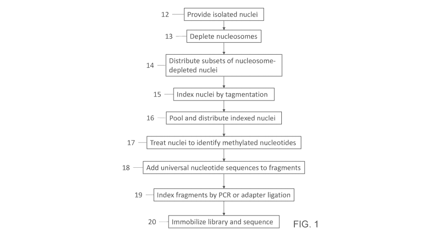
FIG. 1 shows a general block diagram of a general illustrative method for
single-cell
combinatorial indexing according to the present disclosure.
FIG. 2 shows a schematic drawing of one embodiment of the method for single-
cell
combinatorial indexing generally illustrated in FIG. 1.
FIG. 3 shows a schematic drawing of an illustrative embodiment of a fragment-
adapter molecule
after linear amplification.
FIG. 4 shows a schematic drawing of an illustrative embodiment of a fragment-
adapter molecule
after addition of universal adapters.
The schematic drawings are not necessarily to scale. Like numbers used in the
figures
refer to like components, steps and the like. However, it will be understood
that the use of a
number to refer to a component in a given figure is not intended to limit the
component in
another figure labeled with the same number. In addition, the use of different
numbers to refer to
components is not intended to indicate that the different numbered components
cannot be the
same or similar to other numbered components.
DETAILED DESCRIPTION OF ILLUSTRATIVE EMBODIMENTS
The method provided herein includes providing isolated nuclei from a plurality
of cells
(FIG. 1, block 12). The cells can be from any organism(s), and from any cell
type or any tissue
of the organism(s). The method can further include dissociating cells (FIG. 2,
block i), and/or
isolating the nuclei (FIG. 2, block ii). Methods for isolating nuclei from
cells are known to the
person skilled in the art and are routine. The number of nuclei can be at
least 2. The upper limit
is dependent on the practical limitations of equipment (e.g. multi-well
plates) used in other steps
of the method as described herein. For instance, in one embodiment the number
of nuclei can be
no greater than 1,000,000,000, no greater than 100,000,000, no greater than
10,000,000, no
greater than 1,000,000, no greater than 10,000, or no greater than 1,000. The
skilled person will
14
CA 03066424 2019-12-05
WO 2018/226708 PCT/US2018/036078
recognize that the nuclei acid molecules in each nucleus represent the entire
genetic complement
of an organism, and are genomic DNA molecules which include both intron and
exon sequences,
as well as non-coding regulatory sequences such as promoter and enhancer
sequences.
In one embodiment, the nuclei include nucleosomes bound to genomic DNA. Such
nuclei can be useful in methods that do not determine the DNA sequence of the
whole genome of
a cell, such as sciATAC-seq. In another embodiment, the isolated nuclei are
subjected to
conditions that deplete the nuclei of nucleosomes, generating nucleosome-
depleted nuclei (FIG.
1, block 13, and FIG. 2, block ii). Such nuclei can be useful in methods aimed
at determining the
whole genomic DNA sequence of a cell. In one embodiment, the conditions used
for
nucleosome-depletion maintain the integrity of the isolated nuclei. Methods
for generating
nucleosome depleted nuclei are known to the skilled person (see, for instance,
Vitak et al., 2017,
Nature Methods, 14(3):302-308). In one embodiment, the conditions are a
chemical treatment
that includes a treatment with a chaotropic agent capable of disrupting
nucleic acid-protein
interactions. An example of a useful chaotropic agent includes, but is not
limited to, lithium
diiodosalicylate. In another embodiment, the conditions are a chemical
treatment that includes a
treatment with a detergent capable of disrupting nucleic acid-protein
interactions. An example of
a useful detergent includes, but is not limited to,sodium dodecyl sulfate
(SDS). In some
embodiments, when a detergent such as SDS is used, the cells from which the
nuclei are isolated
are treated with a cross-linking agent prior to the isolating. A useful
example of a cross-linking
agent includes, but is not limited to, formaldehyde.
The method provided herein includes distributing subsets of the nuclei, such
as
nucleosome-depleted nuclei, into a first plurality of compartments (FIG. 1,
block 14, and FIG. 2,
left schematic). The number of nuclei present in a subset, and therefor in
each compartment, can
be at least 1. In one embodiment, the number of nuclei present in a subset is
no greater than
2,000. Methods for distributing nuclei into subsets are known to the person
skilled in the art and
are routine. Examples include, but are not limited to, fluorescence-activated
nuclei sorting
(FANS).
Each compartment includes a transposome complex. The transposome complex, a
transposase bound to a transposase recognition site, can insert the
transposase recognition site
into a target nucleic acid within a nucleus in a process sometimes termed
"tagmentation." In
some such insertion events, one strand of the transposase recognition site may
be transferred into
CA 03066424 2019-12-05
WO 2018/226708 PCT/US2018/036078
the target nucleic acid. Such a strand is referred to as a "transferred
strand." In one
embodiment, a transposome complex includes a dimeric transposase having two
subunits, and
two non-contiguous transposon sequences. In another embodiment, a transposase
includes a
dimeric transposase having two subunits, and a contiguous transposon sequence.
Some embodiments can include the use of a hyperactive Tn5 transposase and a
Tn5-type
transposase recognition site (Goryshin and Reznikoff, 1 Biol. Chem., 273:7367
(1998)), or MuA
transposase and a Mu transposase recognition site comprising R1 and R2 end
sequences
(Mizuuchi, K., Cell, 35: 785, 1983; Savilahti, H, et al., EMBO
14: 4893, 1995). Tn5 Mosaic
End (ME) sequences can also be used as optimized by a skilled artisan.
More examples of transposition systems that can be used with certain
embodiments of the
compositions and methods provided herein include Staphylococcus aureus Tn552
(Colegio et al.,
I Bacteriol., 183: 2384-8, 2001; Kirby C et al., Mol. Microbiol., 43: 173-86,
2002), Tyl (Devine
& Boeke, Nucleic Acids Res., 22: 3765-72, 1994 and International Publication
WO 95/23875),
Transposon Tn7 (Craig, N L, Science. 271: 1512, 1996; Craig, N L, Review in:
Curr Top
Microbiol Immunol., 204:27-48, 1996), Tn/O and IS10 (Kleckner N, et al., Curr
Top Microbiol
Immunol., 204:49-82, 1996), Mariner transposase (Lampe D J, et al., EMBO 1,
15: 5470-9,
1996), Tcl (Plasterk R H, Curr. Topics Microbiol. Immunol., 204: 125-43,
1996), P Element
(Gloor, GB, Methods Mol. Biol., 260: 97-114, 2004), Tn3 (Ichikawa & Ohtsubo, J
Biol. Chem.
265:18829-32, 1990), bacterial insertion sequences (Ohtsubo & Sekine, Curr.
Top. Microbiol.
Immunol. 204: 1-26, 1996), retroviruses (Brown, et al., Proc Natl Acad Sci
USA, 86:2525-9,
1989), and retrotransposon of yeast (Boeke & Corces, Annu Rev Microbiol.
43:403-34, 1989).
More examples include IS5, Tn10, Tn903, IS911, and engineered versions of
transposase family
enzymes (Zhang et al., (2009) PLoS Genet. 5:e1000689. Epub 2009 Oct 16; Wilson
C. et al
(2007)1 Microbiol. Methods 71:332-5).
Other examples of integrases that may be used with the methods and
compositions
provided herein include retroviral integrases and integrase recognition
sequences for such
retroviral integrases, such as integrases from HIV-1, HIV-2, Sly, PFV-1, RSV.
Transposon sequences useful with the methods and compositions described herein
are
provided in U.S. Patent Application Pub. No. 2012/0208705, U.S. Patent
Application Pub. No.
2012/0208724 and Int. Patent Application Pub. No. WO 2012/061832. In some
embodiments, a
16
CA 03066424 2019-12-05
WO 2018/226708 PCT/US2018/036078
transposon sequence includes a first transposase recognition site, a second
transposase
recognition site, and an index present between the two transposase recognition
sites.
Some transposome complexes useful herein include a transposase having two
transposon
sequences. In some such embodiments, the two transposon sequences are not
linked to one
another, in other words, the transposon sequences are non-contiguous with one
another.
Examples of such transposomes are known in the art (see, for instance, U.S.
Patent Application
Pub. No. 2010/0120098).
In some embodiments, a transposome complex includes a transposon sequence
nucleic
acid that binds two transposase subunits to form a "looped complex" or a
"looped transposome."
In one example, a transposome includes a dimeric transposase and a transposon
sequence.
Looped complexes can ensure that transposons are inserted into target DNA
while maintaining
ordering information of the original target DNA and without fragmenting the
target DNA. As
will be appreciated, looped structures may insert desired nucleic acid
sequences, such as indexes,
into a target nucleic acid, while maintaining physical connectivity of the
target nucleic acid. In
some embodiments, the transposon sequence of a looped transposome complex can
include a
fragmentation site such that the transposon sequence can be fragmented to
create a transposome
complex comprising two transposon sequences. Such transposome complexes are
useful to
ensuring that neighboring target DNA fragments, in which the transposons
insert, receive code
combinations that can be unambiguously assembled at a later stage of the
assay.
A transposome complex also includes at least one index sequence, also referred
to as a
transposase index. The index sequence is present as part of the transposon
sequence. In one
embodiment, the index sequence can be present on a transferred strand, the
strand of the
transposase recognition site that is transferred into the target nucleic acid.
An index sequence,
also referred to as a tag or barcode, is useful as a marker characteristic of
the compartment in
which a particular target nucleic acid was present. The index sequence of a
transposome
complex is different for each compartment. Accordingly, in this embodiment, an
index is a
nucleic acid sequence tag which is attached to each of the target nucleic
acids present in a
particular compartment, the presence of which is indicative of, or is used to
identify, the
compartment in which a population of nuclei were present at this stage of the
method.
An index sequence can be up to 20 nucleotides in length, e.g., 1, 2, 3, 4, 5,
6, 7, 8, 9, 10,
11, 12, 13, 14, 15, 16, 17, 18, 19, 20. A four nucleotide tag gives a
possibility of multiplexing
17
CA 03066424 2019-12-05
WO 2018/226708 PCT/US2018/036078
256 samples on the same array, a six base tag enables 4096 samples to be
processed on the same
array.
In one embodiment, the transferred strand can also include a universal
sequence, a first
sequencing primer sequence, or a combination thereof. Universal sequences and
sequencing
primer sequences are described herein. Thus, in some embodiments where the
transferred strand
is transferred to target nucleic acids, the target nucleic acids include a
transposase index, and also
include a universal sequence, a first sequencing primer sequence, or a
combination thereof.
In one embodiment, the cytosine nucleotides of a transferred strand are
methylated. In
another embodiment, the nucleotides of a transferred strand do not contain
cytosine. Such a
transferred strand, and any sequence present on the transferred strand
including a transposase
index sequence, universal sequence, and/or first sequencing primer sequence,
can be referred to
as cytosine-depleted. The use of cytosine-depleted nucleotide sequences in a
transposome
complex does not have a significant impact on transposase efficiency.
The method also includes generating indexed nuclei (FIG. 1, block 15, and FIG.
2, block
iii). In one embodiment, generating indexed nuclei includes fragmenting
nucleic acids present in
the subsets of nucleosome-depleted nuclei (e.g., the nuclei acids present in
each compartment)
into a plurality of nucleic acid fragments. In one embodiment, fragmenting
nucleic acids is
accomplished by using a fragmentation site present in the nucleic acids.
Typically,
fragmentation sites are introduced into target nucleic acids by using a
transposome complex. For
instance, a looped transposome complex can include a fragmentation site. A
fragmentation site
can be used to cleave the physical, but not the informational association
between index
sequences that have been inserted into a target nucleic acid. Cleavage may be
by biochemical,
chemical or other means. In some embodiments, a fragmentation site can include
a nucleotide or
nucleotide sequence that may be fragmented by various means. Examples of
fragmentation sites
include, but are not limited to, a restriction endonuclease site, at least one
ribonucleotide
cleavable with an RNAse, nucleotide analogues cleavable in the presence of
certain chemical
agent, a diol linkage cleavable by treatment with periodate, a disulfide group
cleavable with a
chemical reducing agent, a cleavable moiety that may be subject to
photochemical cleavage, and
a peptide cleavable by a peptidase enzyme or other suitable means (see, for
instance, U.S. Patent
Application Pub. No. 2012/0208705, U.S. Patent Application Pub. No.
2012/0208724 and WO
2012/061832. The result of the fragmenting is a population of indexed nuclei,
each nucleus
18
CA 03066424 2019-12-05
WO 2018/226708 PCT/US2018/036078
containing nucleic acid fragments, where the nucleic acid fragments include on
at least one
strand the index sequence indicative of the particular compartment.
The indexed nuclei from multiple compartments can be combined (FIG. 1, block
16, and
FIG. 2, schematic on left). For instance, the indexed nuclei from 2 to 96
compartments (when a
96-well plate is used), or from 2 to 384 compartments (when a 384-well plate
is used) are
combined. Subsets of these combined indexed nuclei, referred to herein as
pooled indexed
nuclei, are then distributed into a second plurality of compartments. The
number of nuclei
present in a subset, and therefor in each compartment, is based in part on the
desire to reduce
index collisions, which is the presence of two nuclei having the same
transposase index ending
up in the same compartment in this step of the method. The number of nuclei
present in a subset
in this embodiment can be from 2 to 30, such as 2, 3, 4, 5, 6, 7, 8, 9, 10,
11, 12, 13, 14, 15, 16,
17, 18, 19, 20, 21, 22, 23, 24, 25, 26, 27, 28, 29, or 30. In one embodiment,
the number of nuclei
present in a subset is from 20 to 24, such as 22. Methods for distributing
nuclei into subsets are
known to the person skilled in the art and are routine. Examples include, but
are not limited to,
fluorescence-activated nuclei sorting (FANS).
The distributed indexed nuclei are treated to identify methylated nucleotides
(FIG. 1,
block 17, and FIG. 2, block iv). Methylation of sites, such as CpG
dinucleotide sequences, can
be measured using any of a variety of techniques used in the art for the
analysis of such sites.
One useful method is the identification of methylated CpG dinucleotide
sequences. The
identification of methylated CpG dinucleotide sequences is determined using
cytosine
conversion based technologies, which rely on methylation status-dependent
chemical
modification of CpG sequences within isolated genomic DNA, or fragments
thereof, followed by
DNA sequence analysis. Chemical reagents that are able to distinguish between
methylated and
non-methylated CpG dinucleotide sequences include hydrazine, which cleaves the
nucleic acid,
and bisulfite. Bisulfite treatment followed by alkaline hydrolysis
specifically converts non-
methylated cytosine to uracil, leaving 5-methylcytosine unmodified as
described by Olek A.,
1996, Nucleic Acids Res. 24:5064-6 or Frommer et al., 1992, Proc. Natl. Acad.
Sci. USA
89:1827-1831. The bisulfite-treated DNA can subsequently be analyzed by
molecular
techniques, such as PCR amplification, sequencing, and detection including
oligonucleotide
hybridization (e.g. using nucleic acid microarrays). In one embodiment, the
indexed nuclei in
each compartment are exposed to conditions for bisulfite treatment. Bisulfite
treatment of
19
CA 03066424 2019-12-05
WO 2018/226708 PCT/US2018/036078
nucleic acids is known to the person skilled in the art and is routine. In one
embodiment, the
bisulfite treatment converts unmethylated cytosine residues of CpG
dinucleotides to uracil
residues and leaves 5-methylcytosine residues unaltered. Bisulfite treatment
results in bisulfite-
treated nucleic acid fragments.
After generation of the bisulfite-treated nucleic acid fragments, the
fragments are
modified to include additional nucleotides at one or both ends (FIG. 1, block
18, and FIG. 2,
blocks v and vi). In one embodiment, the modification includes subjecting the
bisulfite-treated
nucleic acid fragments to linear amplification using a plurality of primers.
Each primer includes
at least two regions; a universal nucleotide sequence at the 5' end and a
random nucleotide
sequence at the 3' end. The universal nucleotide sequence is identical in each
primer, and in one
embodiment it includes a second sequencing primer sequence (also referred to
as a Read 2
Primer in FIG. 2 (block vii). The region of random nucleotide sequence is used
so that at least
one primer should be present that is complementary to every sequence in the
bisulfite-treated
nucleic acid fragments. The number of random nucleotides that can be used to
increase the
probability of complete coverage to a desired level can be determined using
routine methods, and
can be from 6 to 12 random nucleotides, such as 9 random nucleotides. In one
embodiment, the
number of cycles is limited to no greater than 10 cycles, such as 9 cycles, 8
cycles, 7 cycles, 6
cycles, 5 cycles, 4 cycles, 3 cycles, 2 cycles, or 1 cycle. The result of
linear amplification is
amplified fragment-adapter molecules. An example of a fragment-adapter
molecule is shown in
FIG. 3. The fragment-adapter molecule 30 includes nucleotides originating from
the transferred
strand of the transposome complex 31 and 32, which includes a transposase
index and a
universal sequence that can be used for amplification and/or sequencing. The
fragment-adapter
molecule also includes the nucleotides originating from the genomic DNA of a
nucleus 33, the
region of random nucleotide sequence 34, and the universal nucleotide sequence
35.
Linear amplification is followed by an exponential amplification reaction,
such as a PCR,
to further modify the ends of the fragment-adapter molecule prior to
immobilizing and
sequencing. This step results in indexing of the fragment-adapter molecules by
PCR (FIG. 1,
block 19). The universal sequences 31, 32 and/or 35 present at ends of the
fragment-adapter
molecule can be used for the binding of universal anchor sequences which can
serve as primers
and be extended in an amplification reaction. Typically, two different primers
are used. One
primer hybridizes with universal sequences at the 3' end of one strand of the
fragment-adapter
CA 03066424 2019-12-05
WO 2018/226708 PCT/US2018/036078
molecule, and a second primer hybridizes with universal sequences at the 3'
end of the other
strand of the fragment-adapter molecule. Thus, the anchor sequence of each
primer can be
different. Suitable primers can each include additional universal sequences,
such as a universal
capture sequence, and another index sequence. Because each primer can include
an index, this
step results in the addition of one or two index sequences, e.g., a second and
an optional third
index. Fragment-adaptor molecules having the second and the optional third
indexes are referred
to as dual-index fragment-adapter molecules. The second and third indexes can
be the reverse
complements of each other, or the second and third indexes can have sequences
that are not the
reverse complements of each other. This second index sequence and optional
third index is
unique for each compartment in which the distributed indexed nuclei were
placed before
treatment with sodium bisulfite. The result of this PCR amplification is a
plurality or library of
fragment-adapter molecules having a structure similar or identical to the
fragment-adapter
molecule shown in FIG. 2, block vii.
In another embodiment, the modification includes subjecting the bisulfite-
treated nucleic
acid fragments to conditions that result in the ligation of additional
sequences to both ends of the
fragments. In one embodiment, blunt-ended ligation can be used. In another
embodiment, the
fragments are prepared with single overhanging nucleotides by, for example,
activity of certain
types of DNA polymerase such as Taq polymerase or Klenow exo minus polymerase
which has
a non-template-dependent terminal transferase activity that adds a single
deoxynucleotide, for
example, deoxyadenosine (A) to the 3' ends of the bisulfite-treated nucleic
acid fragments. Such
enzymes can be used to add a single nucleotide 'A' to the blunt ended 3'
terminus of each strand
of the fragments. Thus, an 'A' could be added to the 3' terminus of each
strand of the double-
stranded target fragments by reaction with Taq or Klenow exo minus polymerase,
while the
additional sequences to be added to each end of the fragment can include a
compatible 'T'
overhang present on the 3' terminus of each region of double stranded nucleic
acid to be added.
This end modification also prevents self-ligation of the nucleic acids such
that there is a bias
towards formation of the bisulfite-treated nucleic acid fragments flanked by
the sequences that
are added in this embodiment.
Fragmentation of nucleic acid molecules by the methods described herein
results in
fragments with a heterogeneous mix of blunt and 3'- and 5'-overhanging ends.
It is therefore
desirable to repair the fragment ends using methods or kits (such as the
Lucigen DNA terminator
21
CA 03066424 2019-12-05
WO 2018/226708 PCT/US2018/036078
End Repair Kit) known in the art to generate ends that are optimal for
insertion, for example, into
blunt sites of cloning vectors. In a particular embodiment, the fragment ends
of the population of
nucleic acids are blunt ended. More particularly, the fragment ends are blunt
ended and
phosphorylated. The phosphate moiety can be introduced via enzymatic
treatment, for example,
using polynucleotide kinase.
In one embodiment, the bisulfite-treated nucleic acid fragments are treated by
first
ligating identical universal adapters (also referred to as 'mismatched
adaptors,' the general
features of which are described in Gormley et al., US 7,741,463, and Bignell
et al., US
8,053,192,) to the 5' and 3' ends of the bisulfite-treated nucleic acid
fragments to form fragment-
adapter molecules. In one embodiment, the universal adaptor includes all
sequences necessary
for sequencing, including immobilizing the fragment-adapter molecules on an
array. Because
the nucleic acids to be sequenced are from single cells, further amplification
of the fragment-
adapter molecules is helpful to achieve a sufficient number of fragment-
adapter molecules for
sequencing.
In another embodiment, when the universal adapter does not include all
sequences
necessary for sequencing, then a PCR step can be used to further modify the
universal adapter
present in each fragment-adapter molecule prior to immobilizing and
sequencing. For instance,
an initial primer extension reaction is carried out using a universal anchor
sequence
complementary to a universal sequence present in the fragment-adapter
molecule, in which
extension products complementary to both strands of each individual fragment-
adapter molecule
are formed. Typically, the PCR adds additional universal sequences, such as a
universal capture
sequence, and another index sequence. Because each primer can include an
index, this step
results in the addition of one or two index sequences, e.g., a second and an
optional third index,
and indexing of the fragment-adapter molecules by adapter ligation (FIG. 1,
block 19). The
resulting fragment-adaptor molecules are referred to as dual-index fragment-
adapter molecules.
After the universal adapters are added, either by a single step method of
ligating a
universal adaptor including all sequences necessary for sequencing, or by a
two-step method of
ligating a universal adapter and then PCR amplification to further modify the
universal adapter,
the final fragment-adapter molecule will include a universal capture sequence,
a second index
sequence, and an optional third index sequence. These indexes are analogous to
the second and
third indexes described in the production of dual-index fragment-adapters by
linear
22
CA 03066424 2019-12-05
WO 2018/226708 PCT/US2018/036078
amplification. The second and third indexes can be the reverse complements of
each other, or
the second and third indexes can have sequences that are not the reverse
complements of each
other. These second and optional third index sequences are unique for each
compartment in
which the distributed indexed nuclei were placed before treatment with sodium
bisulfite. The
result of adding universal adapters to each end is a plurality or library of
fragment-adaptor
molecules having a structure similar or identical to the fragment-adaptor
molecule 40 shown in
FIG. 4. The fragment-adapter molecule 40 includes a capture sequence 41 and
48, also referred
to as a 3' flowcell adapter (e.g., P5) and 5' flowcell adapter (e.g., P7'),
respectively, and an index
42 and 47, such as i5 and i7. The fragment-adapter molecule 40 also includes
nucleotides
originating from the transferred strand of the transposome complex 43, which
includes a
transposase index 44 and a universal sequence 45 that can be used for
amplification and/or
sequencing. The fragment-adapter molecule also includes the nucleotides
originating from the
genomic DNA of a nucleus 46.
The resulting dual-index fragment-adapter molecules collectively provide a
library of
nucleic acids that can be immobilized and then sequenced. The term library
refers to the
collection of fragments from single cells containing known universal sequences
at their 3' and 5'
ends.
After the bisulfite-treated nucleic acid fragments are modified to include
additional
nucleotides, the dual-index fragment-adapter molecules can be subjected to
conditions that select
for a predetermined size range, such as from 150 to 400 nucleotides in length,
such as from 150
to 300 nucleotides. The resulting dual-index fragment-adapter molecules are
pooled, and
optionally can be subjected to a clean-up process to enhance the purity to the
DNA molecules by
removing at least a portion of unincorporated universal adapters or primers.
Any suitable clean-
up process may be used, such as electrophoresis, size exclusion
chromatography, or the like. In
some embodiments, solid phase reversible immobilization paramagnetic beads may
be employed
to separate the desired DNA molecules from unattached universal adapters or
primers, and to
select nucleic acids based on size. Solid phase reversible immobilization
paramagnetic beads are
commercially available from Beckman Coulter (Agencourt AMPure XP),
Thermofisher
(MagJet), Omega Biotek (Mag-Bind), Promega Beads (Promega), and Kapa
Biosystems (Kapa
Pure Beads).
23
CA 03066424 2019-12-05
WO 2018/226708 PCT/US2018/036078
The plurality of fragment-adapter molecules can be prepared for sequencing.
After the
fragment-adapter molecules are pooled they are immobilized and amplified prior
to sequencing
(FIG. 1, block 20). Methods for attaching fragment-adapter molecules from one
or more sources
to a substrate are known in the art. Likewise, methods for amplifying
immobilized fragment-
adapter molecules include, but are not limited to, bridge amplification and
kinetic exclusion.
Methods for immobilizing and amplifying prior to sequencing are described in,
for instance,
Bignell et al. (US 8,053,192), Gunderson et al. (W02016/130704), Shen et al.
(US 8,895,249),
and Pipenburg et al. (US 9,309,502).
A pooled sample can be immobilized in preparation for sequencing. Sequencing
can be
performed as an array of single molecules, or can be amplified prior to
sequencing. The
amplification can be carried out using one or more immobilized primers. The
immobilized
primer(s) can be a lawn on a planar surface, or on a pool of beads. The pool
of beads can be
isolated into an emulsion with a single bead in each "compartment" of the
emulsion. At a
concentration of only one template per "compartment," only a single template
is amplified on
each bead.
The term "solid-phase amplification" as used herein refers to any nucleic acid
amplification reaction carried out on or in association with a solid support
such that all or a
portion of the amplified products are immobilized on the solid support as they
are formed. In
particular, the term encompasses solid-phase polymerase chain reaction (solid-
phase PCR) and
solid phase isothermal amplification which are reactions analogous to standard
solution phase
amplification, except that one or both of the forward and reverse
amplification primers is/are
immobilized on the solid support. Solid phase PCR covers systems such as
emulsions, wherein
one primer is anchored to a bead and the other is in free solution, and colony
formation in solid
phase gel matrices wherein one primer is anchored to the surface, and one is
in free solution.
In some embodiments, the solid support comprises a patterned surface. A
"patterned
surface" refers to an arrangement of different regions in or on an exposed
layer of a solid
support. For example, one or more of the regions can be features where one or
more
amplification primers are present. The features can be separated by
interstitial regions where
amplification primers are not present. In some embodiments, the pattern can be
an x-y format of
features that are in rows and columns. In some embodiments, the pattern can be
a repeating
arrangement of features and/or interstitial regions. In some embodiments, the
pattern can be a
24
CA 03066424 2019-12-05
WO 2018/226708 PCT/US2018/036078
random arrangement of features and/or interstitial regions. Exemplary
patterned surfaces that
can be used in the methods and compositions set forth herein are described in
US Pat. Nos.
8,778,848, 8,778,849 and 9,079,148, and US Pub. No. 2014/0243224.
In some embodiments, the solid support includes an array of wells or
depressions in a
surface. This may be fabricated as is generally known in the art using a
variety of techniques,
including, but not limited to, photolithography, stamping techniques, molding
techniques and
microetching techniques. As will be appreciated by those in the art, the
technique used will
depend on the composition and shape of the array substrate.
The features in a patterned surface can be wells in an array of wells (e.g.
microwells or
nanowells) on glass, silicon, plastic or other suitable solid supports with
patterned, covalently-
linked gel such as poly(N-(5-azidoacetamidylpentyl)acrylamide-co-acrylamide)
(PAZAM, see,
for example, US Pub. No. 2013/184796, WO 2016/066586, and WO 2015/002813). The
process
creates gel pads used for sequencing that can be stable over sequencing runs
with a large number
of cycles. The covalent linking of the polymer to the wells is helpful for
maintaining the gel in
the structured features throughout the lifetime of the structured substrate
during a variety of uses.
However, in many embodiments the gel need not be covalently linked to the
wells. For example,
in some conditions silane free acrylamide (SFA, see, for example, US Pat. No.
8,563,477, which
is incorporated herein by reference in its entirety) which is not covalently
attached to any part of
the structured substrate, can be used as the gel material.
In particular embodiments, a structured substrate can be made by patterning a
solid
support material with wells (e.g. microwells or nanowells), coating the
patterned support with a
gel material (e.g. PAZAM, SFA or chemically modified variants thereof, such as
the azidolyzed
version of SFA (azido-SFA)) and polishing the gel coated support, for example
via chemical or
mechanical polishing, thereby retaining gel in the wells but removing or
inactivating
substantially all of the gel from the interstitial regions on the surface of
the structured substrate
between the wells. Primer nucleic acids can be attached to gel material. A
solution of fragment-
adapter molecules can then be contacted with the polished substrate such that
individual
fragment-adapter molecules will seed individual wells via interactions with
primers attached to
the gel material; however, the target nucleic acids will not occupy the
interstitial regions due to
absence or inactivity of the gel material. Amplification of the fragment-
adapter molecules will
be confined to the wells since absence or inactivity of gel in the
interstitial regions prevents
CA 03066424 2019-12-05
WO 2018/226708 PCT/US2018/036078
outward migration of the growing nucleic acid colony. The process can be
conveniently
manufactured, being scalable and utilizing conventional micro- or
nanofabrication methods.
Although the disclosure encompasses "solid-phase" amplification methods in
which only
one amplification primer is immobilized (the other primer usually being
present in free solution),
it is preferred for the solid support to be provided with both the forward and
the reverse primers
immobilized. In practice, there will be a 'plurality' of identical forward
primers and/or a
'plurality' of identical reverse primers immobilized on the solid support,
since the amplification
process requires an excess of primers to sustain amplification. References
herein to forward and
reverse primers are to be interpreted accordingly as encompassing a
'plurality' of such primers
unless the context indicates otherwise.
As will be appreciated by the skilled reader, any given amplification reaction
requires at
least one type of forward primer and at least one type of reverse primer
specific for the template
to be amplified. However, in certain embodiments the forward and reverse
primers may include
template-specific portions of identical sequence, and may have entirely
identical nucleotide
sequence and structure (including any non-nucleotide modifications). In other
words, it is
possible to carry out solid-phase amplification using only one type of primer,
and such single-
primer methods are encompassed within the scope of the invention. Other
embodiments may use
forward and reverse primers which contain identical template-specific
sequences but which
differ in some other structural features. For example, one type of primer may
contain a non-
nucleotide modification which is not present in the other.
In all embodiments of the disclosure, primers for solid-phase amplification
are preferably
immobilized by single point covalent attachment to the solid support at or
near the 5' end of the
primer, leaving the template-specific portion of the primer free to anneal to
its cognate template
and the 3' hydroxyl group free for primer extension. Any suitable covalent
attachment means
known in the art may be used for this purpose. The chosen attachment chemistry
will depend on
the nature of the solid support, and any derivatization or functionalization
applied to it. The
primer itself may include a moiety, which may be a non-nucleotide chemical
modification, to
facilitate attachment. In a particular embodiment, the primer may include a
sulphur-containing
nucleophile, such as phosphorothioate or thiophosphate, at the 5' end. In the
case of solid-
supported polyacrylamide hydrogels, this nucleophile will bind to a
bromoacetamide group
present in the hydrogel. A more particular means of attaching primers and
templates to a solid
26
CA 03066424 2019-12-05
WO 2018/226708 PCT/US2018/036078
support is via 5' phosphorothioate attachment to a hydrogel comprised of
polymerized
acrylamide and N-(5-bromoacetamidylpentyl) acrylamide (BRAPA), as described
fully in WO
05/065814.
Certain embodiments of the disclosure may make use of solid supports comprised
of an
inert substrate or matrix (e.g. glass slides, polymer beads, etc.) which has
been "functionalized",
for example by application of a layer or coating of an intermediate material
comprising reactive
groups which permit covalent attachment to biomolecules, such as
polynucleotides. Examples of
such supports include, but are not limited to, polyacrylamide hydrogels
supported on an inert
substrate such as glass. In such embodiments, the biomolecules (e.g.
polynucleotides) may be
directly covalently attached to the intermediate material (e.g. the hydrogel),
but the intermediate
material may itself be non-covalently attached to the substrate or matrix
(e.g. the glass substrate).
The term "covalent attachment to a solid support" is to be interpreted
accordingly as
encompassing this type of arrangement.
The pooled samples may be amplified on beads wherein each bead contains a
forward
and reverse amplification primer. In a particular embodiment, the library of
fragment-adapter
molecules is used to prepare clustered arrays of nucleic acid colonies,
analogous to those
described in U.S. Pub. No. 2005/0100900, U.S. Pat. No. 7,115,400, WO 00/18957
and WO
98/44151 by solid-phase amplification and more particularly solid phase
isothermal
amplification. The terms cluster' and colony' are used interchangeably herein
to refer to a
discrete site on a solid support including a plurality of identical
immobilized nucleic acid strands
and a plurality of identical immobilized complementary nucleic acid strands.
The term "clustered
array" refers to an array formed from such clusters or colonies. In this
context the term "array" is
not to be understood as requiring an ordered arrangement of clusters.
The term "solid phase" or "surface" is used to mean either a planar array
wherein primers
are attached to a flat surface, for example, glass, silica or plastic
microscope slides or similar
flow cell devices; beads, wherein either one or two primers are attached to
the beads and the
beads are amplified; or an array of beads on a surface after the beads have
been amplified.
Clustered arrays can be prepared using either a process of thermocycling, as
described in
WO 98/44151, or a process whereby the temperature is maintained as a constant,
and the cycles
of extension and denaturing are performed using changes of reagents. Such
isothermal
amplification methods are described in patent application numbers WO 02/46456
and U.S. Pub.
27
CA 03066424 2019-12-05
WO 2018/226708 PCT/US2018/036078
No. 2008/0009420. Due to the lower temperatures useful in the isothermal
process, this is
particularly preferred.
It will be appreciated that any of the amplification methodologies described
herein or
generally known in the art may be utilized with universal or target-specific
primers to amplify
immobilized DNA fragments. Suitable methods for amplification include, but are
not limited to,
the polymerase chain reaction (PCR), strand displacement amplification (SDA),
transcription
mediated amplification (TMA) and nucleic acid sequence based amplification
(NASBA), as
described in U.S. Pat. No. 8,003,354, which is incorporated herein by
reference in its entirety.
The above amplification methods may be employed to amplify one or more nucleic
acids of
interest. For example, PCR, including multiplex PCR, SDA, TMA, NASBA and the
like may be
utilized to amplify immobilized DNA fragments. In some embodiments, primers
directed
specifically to the polynucleotide of interest are included in the
amplification reaction.
Other suitable methods for amplification of polynucleotides may include
oligonucleotide
extension and ligation, rolling circle amplification (RCA) (Lizardi et al.,
Nat. Genet. 19:225-232
(1998)) and oligonucleotide ligation assay (OLA) (See generally U.S. Pat. Nos.
7,582,420,
5,185,243, 5,679,524 and 5,573,907; EP 0 320 308 Bl; EP 0 336 731 Bl; EP 0 439
182 Bl; WO
90/01069; WO 89/12696; and WO 89/09835) technologies. It will be appreciated
that these
amplification methodologies may be designed to amplify immobilized DNA
fragments. For
example, in some embodiments, the amplification method may include ligation
probe
amplification or oligonucleotide ligation assay (OLA) reactions that contain
primers directed
specifically to the nucleic acid of interest. In some embodiments, the
amplification method may
include a primer extension-ligation reaction that contains primers directed
specifically to the
nucleic acid of interest. As a non-limiting example of primer extension and
ligation primers that
may be specifically designed to amplify a nucleic acid of interest, the
amplification may include
primers used for the GoldenGate assay (I1lumina, Inc., San Diego, CA) as
exemplified by U.S.
Pat. No. 7,582,420 and 7,611,869.
Exemplary isothermal amplification methods that may be used in a method of the
present
disclosure include, but are not limited to, Multiple Displacement
Amplification (MDA) as
exemplified by, for example Dean et al., Proc. Natl. Acad. Sci. USA 99:5261-66
(2002) or
isothermal strand displacement nucleic acid amplification exemplified by, for
example U.S. Pat.
No. 6,214,587. Other non-PCR-based methods that may be used in the present
disclosure
28
CA 03066424 2019-12-05
WO 2018/226708 PCT/US2018/036078
include, for example, strand displacement amplification (SDA) which is
described in, for
example Walker et al., Molecular Methods for Virus Detection, Academic Press,
Inc., 1995; U.S.
Pat. Nos. 5,455,166, and 5,130,238, and Walker et al., Nucl. Acids Res.
20:1691-96 (1992) or
hyper-branched strand displacement amplification which is described in, for
example Lage et al.,
Genome Res. 13:294-307 (2003). Isothermal amplification methods may be used
with the strand-
displacing Phi 29 polymerase or Bst DNA polymerase large fragment, 5'->3' exo-
for random
primer amplification of genomic DNA. The use of these polymerases takes
advantage of their
high processivity and strand displacing activity. High processivity allows the
polymerases to
produce fragments that are 10-20 kb in length. As set forth above, smaller
fragments may be
produced under isothermal conditions using polymerases having low processivity
and strand-
displacing activity such as Klenow polymerase. Additional description of
amplification
reactions, conditions and components are set forth in detail in the disclosure
of U.S. Patent No.
7,670,810.
Another polynucleotide amplification method that is useful in the present
disclosure is
Tagged PCR which uses a population of two-domain primers having a constant 5'
region
followed by a random 3' region as described, for example, in Grothues et al.
Nucleic Acids Res.
21(5):1321-2 (1993). The first rounds of amplification are carried out to
allow a multitude of
initiations on heat denatured DNA based on individual hybridization from the
randomly-
synthesized 3' region. Due to the nature of the 3' region, the sites of
initiation are contemplated to
be random throughout the genome. Thereafter, the unbound primers may be
removed and further
replication may take place using primers complementary to the constant 5'
region.
In some embodiments, isothermal amplification can be performed using kinetic
exclusion
amplification (KEA), also referred to as exclusion amplification (ExAmp). A
nucleic acid
library of the present disclosure can be made using a method that includes a
step of reacting an
amplification reagent to produce a plurality of amplification sites that each
includes a
substantially clonal population of amplicons from an individual target nucleic
acid that has
seeded the site. In some embodiments, the amplification reaction proceeds
until a sufficient
number of amplicons are generated to fill the capacity of the respective
amplification site. Filling
an already seeded site to capacity in this way inhibits target nucleic acids
from landing and
amplifying at the site thereby producing a clonal population of amplicons at
the site. In some
embodiments, apparent clonality can be achieved even if an amplification site
is not filled to
29
CA 03066424 2019-12-05
WO 2018/226708 PCT/US2018/036078
capacity prior to a second target nucleic acid arriving at the site. Under
some conditions,
amplification of a first target nucleic acid can proceed to a point that a
sufficient number of
copies are made to effectively outcompete or overwhelm production of copies
from a second
target nucleic acid that is transported to the site. For example, in an
embodiment that uses a
bridge amplification process on a circular feature that is smaller than 500 nm
in diameter, it has
been determined that after 14 cycles of exponential amplification for a first
target nucleic acid,
contamination from a second target nucleic acid at the same site will produce
an insufficient
number of contaminating amplicons to adversely impact sequencing-by-synthesis
analysis on an
Illumina sequencing platform.
In some embodiments, amplification sites in an array can be, but need not be,
entirely
clonal. Rather, for some applications, an individual amplification site can be
predominantly
populated with amplicons from a first fragment-adapter molecule and can also
have a low level
of contaminating amplicons from a second target nucleic acid. An array can
have one or more
amplification sites that have a low level of contaminating amplicons so long
as the level of
contamination does not have an unacceptable impact on a subsequent use of the
array. For
example, when the array is to be used in a detection application, an
acceptable level of
contamination would be a level that does not impact signal to noise or
resolution of the detection
technique in an unacceptable way. Accordingly, apparent clonality will
generally be relevant to a
particular use or application of an array made by the methods set forth
herein. Exemplary levels
of contamination that can be acceptable at an individual amplification site
for particular
applications include, but are not limited to, at most 0.1%, 0.5%, 1%, 5%, 10%
or 25%
contaminating amplicons. An array can include one or more amplification sites
having these
exemplary levels of contaminating amplicons. For example, up to 5%, 10%, 25%,
50%, 75%, or
even 100% of the amplification sites in an array can have some contaminating
amplicons. It will
be understood that in an array or other collection of sites, at least 50%,
75%, 80%, 85%, 90%,
95% or 99% or more of the sites can be clonal or apparently clonal.
In some embodiments, kinetic exclusion can occur when a process occurs at a
sufficiently
rapid rate to effectively exclude another event or process from occurring.
Take for example the
making of a nucleic acid array where sites of the array are randomly seeded
with fragment-
adapter molecules from a solution and copies of the fragment-adapter molecules
are generated in
an amplification process to fill each of the seeded sites to capacity. In
accordance with the
CA 03066424 2019-12-05
WO 2018/226708 PCT/US2018/036078
kinetic exclusion methods of the present disclosure, the seeding and
amplification processes can
proceed simultaneously under conditions where the amplification rate exceeds
the seeding rate.
As such, the relatively rapid rate at which copies are made at a site that has
been seeded by a first
target nucleic acid will effectively exclude a second nucleic acid from
seeding the site for
amplification. Kinetic exclusion amplification methods can be performed as
described in detail
in the disclosure of US Application Pub. No. 2013/0338042.
Kinetic exclusion can exploit a relatively slow rate for initiating
amplification (e.g. a
slow rate of making a first copy of a fragment-adapter molecule) vs. a
relatively rapid rate for
making subsequent copies of the fragment-adapter molecule (or of the first
copy of the fragment-
adapter molecule). In the example of the previous paragraph, kinetic exclusion
occurs due to the
relatively slow rate of fragment-adapter molecule seeding (e.g. relatively
slow diffusion or
transport) vs. the relatively rapid rate at which amplification occurs to fill
the site with copies of
the fragment-adapter seed. In another exemplary embodiment, kinetic exclusion
can occur due
to a delay in the formation of a first copy of a fragment-adapter molecule
that has seeded a site
(e.g. delayed or slow activation) vs. the relatively rapid rate at which
subsequent copies are made
to fill the site. In this example, an individual site may have been seeded
with several different
fragment-adapter molecules (e.g. several fragment-adapter molecules can be
present at each site
prior to amplification). However, first copy formation for any given fragment-
adapter molecule
can be activated randomly such that the average rate of first copy formation
is relatively slow
compared to the rate at which subsequent copies are generated. In this case,
although an
individual site may have been seeded with several different fragment-adapter
molecules, kinetic
exclusion will allow only one of those fragment-adapter molecules to be
amplified. More
specifically, once a first fragment-adapter molecule has been activated for
amplification, the site
will rapidly fill to capacity with its copies, thereby preventing copies of a
second fragment-
adapter molecule from being made at the site.
An amplification reagent can include further components that facilitate
amplicon
formation and in some cases increase the rate of amplicon formation. An
example is a
recombinase. Recombinase can facilitate amplicon formation by allowing
repeated
invasion/extension. More specifically, recombinase can facilitate invasion of
a fragment-adapter
molecule by the polymerase and extension of a primer by the polymerase using
the fragment-
adapter molecule as a template for amplicon formation. This process can be
repeated as a chain
31
CA 03066424 2019-12-05
WO 2018/226708 PCT/US2018/036078
reaction where amplicons produced from each round of invasion/extension serve
as templates in
a subsequent round. The process can occur more rapidly than standard PCR since
a denaturation
cycle (e.g. via heating or chemical denaturation) is not required. As such,
recombinase-
facilitated amplification can be carried out isothermally. It is generally
desirable to include ATP,
or other nucleotides (or in some cases non-hydrolyzable analogs thereof) in a
recombinase-
facilitated amplification reagent to facilitate amplification. A mixture of
recombinase and single
stranded binding (SSB) protein is particularly useful as SSB can further
facilitate amplification.
Exemplary formulations for recombinase-facilitated amplification include those
sold
commercially as TwistAmp kits by TwistDx (Cambridge, UK). Useful components of
recombinase-facilitated amplification reagent and reaction conditions are set
forth in US
5,223,414 and US 7,399,590.
Another example of a component that can be included in an amplification
reagent to
facilitate amplicon formation and in some cases to increase the rate of
amplicon formation is a
helicase. Helicase can facilitate amplicon formation by allowing a chain
reaction of amplicon
formation. The process can occur more rapidly than standard PCR since a
denaturation cycle
(e.g. via heating or chemical denaturation) is not required. As such, helicase-
facilitated
amplification can be carried out isothermally. A mixture of helicase and
single stranded binding
(SSB) protein is particularly useful as SSB can further facilitate
amplification. Exemplary
formulations for helicase-facilitated amplification include those sold
commercially as IsoAmp
kits from Biohelix (Beverly, MA). Further, examples of useful formulations
that include a
helicase protein are described in US 7,399,590 and US 7,829,284, each of which
is incorporated
herein by reference.
Yet another example of a component that can be included in an amplification
reagent to
facilitate amplicon formation and in some cases increase the rate of amplicon
formation is an
origin binding protein.
Following attachment of fragment-adapter molecules to a surface, the sequence
of the
immobilized and amplified fragment-adapter molecules is determined. Sequencing
can be
carried out using any suitable sequencing technique, and methods for
determining the sequence
of immobilized and amplified fragment-adapter molecules, including strand re-
synthesis, are
known in the art and are described in, for instance, Bignell et al. (US
8,053,192), Gunderson et
al. (W02016/130704), Shen et al. (US 8,895,249), and Pipenburg et al. (US
9,309,502).
32
CA 03066424 2019-12-05
WO 2018/226708 PCT/US2018/036078
The methods described herein can be used in conjunction with a variety of
nucleic acid
sequencing techniques. Particularly applicable techniques are those wherein
nucleic acids are
attached at fixed locations in an array such that their relative positions do
not change and
wherein the array is repeatedly imaged. Embodiments in which images are
obtained in different
color channels, for example, coinciding with different labels used to
distinguish one nucleotide
base type from another are particularly applicable. In some embodiments, the
process to
determine the nucleotide sequence of a fragment-adapter molecule can be an
automated process.
Preferred embodiments include sequencing-by-synthesis ("SBS") techniques.
SBS techniques generally involve the enzymatic extension of a nascent nucleic
acid
strand through the iterative addition of nucleotides against a template
strand. In traditional
methods of SBS, a single nucleotide monomer may be provided to a target
nucleotide in the
presence of a polymerase in each delivery. However, in the methods described
herein, more than
one type of nucleotide monomer can be provided to a target nucleic acid in the
presence of a
polymerase in a delivery.
In one embodiment, a nucleotide monomer includes locked nucleic acids (LNAs)
or
bridged nucleic acids (BNAs). When the fragment-adapter molecules are produced
using one or
more cytosine-depleted nucleotide sequences, such as what results when
cytosine-depleted
nucleotide sequences are present in a transferred strand from a transposome
complex, the melting
temperature of a nucleotide monomer that hybridizes to a cytosine-depleted
region is altered.
The use of LNAs or BNAs in a nucleotide monomer increases hybridization
strength between a
nucleotide monomer and a sequencing primer sequence present on an immobilized
fragment-
adapter molecule.
SBS can utilize nucleotide monomers that have a terminator moiety or those
that lack any
terminator moieties. Methods utilizing nucleotide monomers lacking terminators
include, for
example, pyrosequencing and sequencing using y-phosphate-labeled nucleotides,
as set forth in
further detail below. In methods using nucleotide monomers lacking
terminators, the number of
nucleotides added in each cycle is generally variable and dependent upon the
template sequence
and the mode of nucleotide delivery. For SBS techniques that utilize
nucleotide monomers
having a terminator moiety, the terminator can be effectively irreversible
under the sequencing
conditions used as is the case for traditional Sanger sequencing which
utilizes
33
CA 03066424 2019-12-05
WO 2018/226708 PCT/US2018/036078
dideoxynucleotides, or the terminator can be reversible as is the case for
sequencing methods
developed by Solexa (now Illumina, Inc.).
SBS techniques can utilize nucleotide monomers that have a label moiety or
those that
lack a label moiety. Accordingly, incorporation events can be detected based
on a characteristic
of the label, such as fluorescence of the label; a characteristic of the
nucleotide monomer such as
molecular weight or charge; a byproduct of incorporation of the nucleotide,
such as release of
pyrophosphate; or the like. In embodiments where two or more different
nucleotides are present
in a sequencing reagent, the different nucleotides can be distinguishable from
each other, or
alternatively the two or more different labels can be the indistinguishable
under the detection
techniques being used. For example, the different nucleotides present in a
sequencing reagent
can have different labels and they can be distinguished using appropriate
optics as exemplified
by the sequencing methods developed by Solexa (now Illumina, Inc.).
Preferred embodiments include pyrosequencing techniques. Pyrosequencing
detects the
release of inorganic pyrophosphate (PPi) as particular nucleotides are
incorporated into the
nascent strand (Ronaghi, M., Karamohamed, S., Pettersson, B., Uhlen, M. and
Nyren, P. (1996)
"Real-time DNA sequencing using detection of pyrophosphate release."
Analytical Biochemistry
242(1), 84-9; Ronaghi, M. (2001) "Pyrosequencing sheds light on DNA
sequencing." Genome
Res. 11(1), 3-11; Ronaghi, M., Uhlen, M. and Nyren, P. (1998) "A sequencing
method based on
real-time pyrophosphate." Science 281(5375), 363; U.S. Pat. Nos. 6,210,891;
6,258,568 and
6,274,320). In pyrosequencing, released PPi can be detected by being
immediately converted to
adenosine triphosphate (ATP) by ATP sulfurase, and the level of ATP generated
is detected via
luciferase-produced photons. The nucleic acids to be sequenced can be attached
to features in an
array and the array can be imaged to capture the chemiluminescent signals that
are produced due
to incorporation of a nucleotides at the features of the array. An image can
be obtained after the
array is treated with a particular nucleotide type (e.g. A, T, C or G). Images
obtained after
addition of each nucleotide type will differ with regard to which features in
the array are
detected. These differences in the image reflect the different sequence
content of the features on
the array. However, the relative locations of each feature will remain
unchanged in the images.
The images can be stored, processed and analyzed using the methods set forth
herein. For
example, images obtained after treatment of the array with each different
nucleotide type can be
34
CA 03066424 2019-12-05
WO 2018/226708 PCT/US2018/036078
handled in the same way as exemplified herein for images obtained from
different detection
channels for reversible terminator-based sequencing methods.
In another exemplary type of SBS, cycle sequencing is accomplished by stepwise
addition of reversible terminator nucleotides containing, for example, a
cleavable or
photobleachable dye label as described, for example, in WO 04/018497 and U.S.
Pat. No.
7,057,026, the disclosures of which are incorporated herein by reference. This
approach is being
commercialized by Solexa (now Illumina Inc.), and is also described in WO
91/06678 and WO
07/123,744. The availability of fluorescently-labeled terminators in which
both the termination
can be reversed and the fluorescent label cleaved facilitates efficient cyclic
reversible termination
(CRT) sequencing. Polymerases can also be co-engineered to efficiently
incorporate and extend
from these modified nucleotides.
Preferably in reversible terminator-based sequencing embodiments, the labels
do not
substantially inhibit extension under SBS reaction conditions. However, the
detection labels can
be removable, for example, by cleavage or degradation. Images can be captured
following
incorporation of labels into arrayed nucleic acid features. In particular
embodiments, each cycle
involves simultaneous delivery of four different nucleotide types to the array
and each nucleotide
type has a spectrally distinct label. Four images can then be obtained, each
using a detection
channel that is selective for one of the four different labels. Alternatively,
different nucleotide
types can be added sequentially and an image of the array can be obtained
between each addition
step. In such embodiments each image will show nucleic acid features that have
incorporated
nucleotides of a particular type. Different features will be present or absent
in the different
images due the different sequence content of each feature. However, the
relative position of the
features will remain unchanged in the images. Images obtained from such
reversible terminator-
SBS methods can be stored, processed and analyzed as set forth herein.
Following the image
capture step, labels can be removed and reversible terminator moieties can be
removed for
subsequent cycles of nucleotide addition and detection. Removal of the labels
after they have
been detected in a particular cycle and prior to a subsequent cycle can
provide the advantage of
reducing background signal and crosstalk between cycles. Examples of useful
labels and removal
methods are set forth below.
In particular embodiments some or all of the nucleotide monomers can include
reversible
terminators. In such embodiments, reversible terminators/cleavable
fluorophores can include
CA 03066424 2019-12-05
WO 2018/226708 PCT/US2018/036078
fluorophores linked to the ribose moiety via a 3' ester linkage (Metzker,
Genome Res. 15:1767-
1776 (2005)). Other approaches have separated the terminator chemistry from
the cleavage of the
fluorescence label (Ruparel et al., Proc Natl Acad Sci USA 102: 5932-7
(2005)). Ruparel et al.
described the development of reversible terminators that used a small 3' allyl
group to block
extension, but could easily be deblocked by a short treatment with a palladium
catalyst. The
fluorophore was attached to the base via a photocleavable linker that could
easily be cleaved by a
30 second exposure to long wavelength UV light. Thus, either disulfide
reduction or
photocleavage can be used as a cleavable linker. Another approach to
reversible termination is
the use of natural termination that ensues after placement of a bulky dye on a
dNTP. The
presence of a charged bulky dye on the dNTP can act as an effective terminator
through steric
and/or electrostatic hindrance. The presence of one incorporation event
prevents further
incorporations unless the dye is removed. Cleavage of the dye removes the
fluorophore and
effectively reverses the termination. Examples of modified nucleotides are
also described in U.S.
Pat. Nos. 7,427,673, and 7,057,026, the disclosures of which are incorporated
herein by
reference in their entireties.
Additional exemplary SBS systems and methods which can be utilized with the
methods
and systems described herein are described in U.S. Pub. Nos. 2007/0166705,
2006/0188901,
2006/0240439, 2006/0281109, 2012/0270305, and 2013/0260372, U.S. Pat. No.
7,057,026, PCT
Publication No. WO 05/065814, U.S. Patent Application Publication No.
2005/0100900, and
PCT Publication Nos. WO 06/064199 and WO 07/010,251.
Some embodiments can utilize detection of four different nucleotides using
fewer than
four different labels. For example, SBS can be performed utilizing methods and
systems
described in the incorporated materials of U.S. Pub. No. 2013/0079232. As a
first example, a
pair of nucleotide types can be detected at the same wavelength, but
distinguished based on a
difference in intensity for one member of the pair compared to the other, or
based on a change to
one member of the pair (e.g. via chemical modification, photochemical
modification or physical
modification) that causes apparent signal to appear or disappear compared to
the signal detected
for the other member of the pair. As a second example, three of four different
nucleotide types
can be detected under particular conditions while a fourth nucleotide type
lacks a label that is
detectable under those conditions, or is minimally detected under those
conditions (e.g., minimal
detection due to background fluorescence, etc.). Incorporation of the first
three nucleotide types
36
CA 03066424 2019-12-05
WO 2018/226708 PCT/US2018/036078
into a nucleic acid can be determined based on presence of their respective
signals and
incorporation of the fourth nucleotide type into the nucleic acid can be
determined based on
absence or minimal detection of any signal. As a third example, one nucleotide
type can include
label(s) that are detected in two different channels, whereas other nucleotide
types are detected in
no more than one of the channels. The aforementioned three exemplary
configurations are not
considered mutually exclusive and can be used in various combinations. An
exemplary
embodiment that combines all three examples, is a fluorescent-based SBS method
that uses a
first nucleotide type that is detected in a first channel (e.g. dATP having a
label that is detected in
the first channel when excited by a first excitation wavelength), a second
nucleotide type that is
detected in a second channel (e.g. dCTP having a label that is detected in the
second channel
when excited by a second excitation wavelength), a third nucleotide type that
is detected in both
the first and the second channel (e.g. dTTP having at least one label that is
detected in both
channels when excited by the first and/or second excitation wavelength) and a
fourth nucleotide
type that lacks a label that is not, or minimally, detected in either channel
(e.g. dGTP having no
label).
Further, as described in the incorporated materials of U.S. Pub. No.
2013/0079232,
sequencing data can be obtained using a single channel. In such so-called one-
dye sequencing
approaches, the first nucleotide type is labeled but the label is removed
after the first image is
generated, and the second nucleotide type is labeled only after a first image
is generated. The
third nucleotide type retains its label in both the first and second images,
and the fourth
nucleotide type remains unlabeled in both images.
Some embodiments can utilize sequencing by ligation techniques. Such
techniques utilize
DNA ligase to incorporate oligonucleotides and identify the incorporation of
such
oligonucleotides. The oligonucleotides typically have different labels that
are correlated with the
identity of a particular nucleotide in a sequence to which the
oligonucleotides hybridize. As with
other SBS methods, images can be obtained following treatment of an array of
nucleic acid
features with the labeled sequencing reagents. Each image will show nucleic
acid features that
have incorporated labels of a particular type. Different features will be
present or absent in the
different images due the different sequence content of each feature, but the
relative position of
the features will remain unchanged in the images. Images obtained from
ligation-based
sequencing methods can be stored, processed and analyzed as set forth herein.
Exemplary SBS
37
CA 03066424 2019-12-05
WO 2018/226708 PCT/US2018/036078
systems and methods which can be utilized with the methods and systems
described herein are
described in U.S. Pat. Nos. 6,969,488, 6,172,218, and 6,306,597.
Some embodiments can utilize nanopore sequencing (Deamer, D. W. & Akeson, M.
"Nanopores and nucleic acids: prospects for ultrarapid sequencing." Trends
Biotechnol. 18, 147-
151 (2000); Deamer, D. and D. Branton, "Characterization of nucleic acids by
nanopore
analysis", Acc. Chem. Res. 35:817-825 (2002); Li, J., M. Gershow, D. Stein, E.
Brandin, and J.
A. Golovchenko, "DNA molecules and configurations in a solid-state nanopore
microscope" Nat.
Mater. 2:611-615 (2003)). In such embodiments, the fragment-adapter molecule
passes through a
nanopore. The nanopore can be a synthetic pore or biological membrane protein,
such as a-
hemolysin. As the fragment-adapter molecule passes through the nanopore, each
base-pair can be
identified by measuring fluctuations in the electrical conductance of the
pore. (U.S. Pat. No.
7,001,792; Soni, G. V. & Meller, "A. Progress toward ultrafast DNA sequencing
using solid-
state nanopores." Clin. Chem. 53, 1996-2001 (2007); Healy, K. "Nanopore-based
single-
molecule DNA analysis." Nanomed. 2, 459-481 (2007); Cockroft, S. L., Chu, J.,
Amorin, M. &
Ghadiri, M. R. "A single-molecule nanopore device detects DNA polymerase
activity with
single-nucleotide resolution." J. Am. Chem. Soc. 130, 818-820 (2008), the
disclosures of which
are incorporated herein by reference in their entireties). Data obtained from
nanopore sequencing
can be stored, processed and analyzed as set forth herein. In particular, the
data can be treated as
an image in accordance with the exemplary treatment of optical images and
other images that is
set forth herein.
Some embodiments can utilize methods involving the real-time monitoring of DNA
polymerase activity. Nucleotide incorporations can be detected through
fluorescence resonance
energy transfer (FRET) interactions between a fluorophore-bearing polymerase
and y-phosphate-
labeled nucleotides as described, for example, in U.S. Pat. Nos. 7,329,492 and
7,211,414, both of
which are incorporated herein by reference, or nucleotide incorporations can
be detected with
zero-mode waveguides as described, for example, in U.S. Pat. No. 7,315,019,
and using
fluorescent nucleotide analogs and engineered polymerases as described, for
example, in U.S.
Pat. No. 7,405,281 and U.S. Pub. No. 2008/0108082. The illumination can be
restricted to a
zeptoliter-scale volume around a surface-tethered polymerase such that
incorporation of
fluorescently labeled nucleotides can be observed with low background (Levene,
M. J. et al.
"Zero-mode waveguides for single-molecule analysis at high concentrations."
Science 299, 682-
38
CA 03066424 2019-12-05
WO 2018/226708 PCT/US2018/036078
686 (2003); Lundquist, P. M. etal. "Parallel confocal detection of single
molecules in real time."
Opt. Lett. 33, 1026-1028 (2008); Korlach, J. et al. "Selective aluminum
passivation for targeted
immobilization of single DNA polymerase molecules in zero-mode waveguide nano
structures."
Proc. Natl. Acad. Sci. USA 105, 1176-1181 (2008)). Images obtained from such
methods can be
stored, processed and analyzed as set forth herein.
Some SBS embodiments include detection of a proton released upon incorporation
of a
nucleotide into an extension product. For example, sequencing based on
detection of released
protons can use an electrical detector and associated techniques that are
commercially available
from Ion Torrent (Guilford, CT, a Life Technologies subsidiary) or sequencing
methods and
systems described in U.S. Pub. Nos. 2009/0026082; 2009/0127589; 2010/0137143;
and
2010/0282617. Methods set forth herein for amplifying target nucleic acids
using kinetic
exclusion can be readily applied to substrates used for detecting protons.
More specifically,
methods set forth herein can be used to produce clonal populations of
amplicons that are used to
detect protons.
The above SBS methods can be advantageously carried out in multiplex formats
such that
multiple different fragment-adapter molecules are manipulated simultaneously.
In particular
embodiments, different fragment-adapter molecules can be treated in a common
reaction vessel
or on a surface of a particular substrate. This allows convenient delivery of
sequencing reagents,
removal of unreacted reagents and detection of incorporation events in a
multiplex manner. In
embodiments using surface-bound target nucleic acids, the fragment-adapter
molecules can be in
an array format. In an array format, the fragment-adapter molecules can be
typically bound to a
surface in a spatially distinguishable manner. The fragment-adapter molecules
can be bound by
direct covalent attachment, attachment to a bead or other particle or binding
to a polymerase or
other molecule that is attached to the surface. The array can include a single
copy of a fragment-
adapter molecule at each site (also referred to as a feature) or multiple
copies having the same
sequence can be present at each site or feature. Multiple copies can be
produced by amplification
methods such as, bridge amplification or emulsion PCR as described in further
detail below.
The methods set forth herein can use arrays having features at any of a
variety of
densities including, for example, at least about 10 features/cm2, 100
features/ cm2, 500 features/
cm2, 1,000 features/ cm2, 5,000 features/ cm2, 10,000 features/ cm2, 50,000
features/ cm2,
100,000 features/ cm2, 1,000,000 features/ cm2, 5,000,000 features/ cm2, or
higher.
39
CA 03066424 2019-12-05
WO 2018/226708 PCT/US2018/036078
An advantage of the methods set forth herein is that they provide for rapid
and efficient
detection of a plurality of cm2, in parallel. Accordingly the present
disclosure provides
integrated systems capable of preparing and detecting nucleic acids using
techniques known in
the art such as those exemplified above. Thus, an integrated system of the
present disclosure can
include fluidic components capable of delivering amplification reagents and/or
sequencing
reagents to one or more immobilized DNA fragments, the system including
components such as
pumps, valves, reservoirs, fluidic lines and the like. A flow cell can be
configured and/or used in
an integrated system for detection of target nucleic acids. Exemplary flow
cells are described,
for example, in U.S. Pub. No. 2010/0111768 and US Ser. No. 13/273,666. As
exemplified for
flow cells, one or more of the fluidic components of an integrated system can
be used for an
amplification method and for a detection method. Taking a nucleic acid
sequencing embodiment
as an example, one or more of the fluidic components of an integrated system
can be used for an
amplification method set forth herein and for the delivery of sequencing
reagents in a sequencing
method such as those exemplified above. Alternatively, an integrated system
can include
separate fluidic systems to carry out amplification methods and to carry out
detection methods.
Examples of integrated sequencing systems that are capable of creating
amplified nucleic acids
and also determining the sequence of the nucleic acids include, without
limitation, the MiSeqTM
platform (Illumina, Inc., San Diego, CA) and devices described in US Ser. No.
13/273,666,
which is incorporated herein by reference.
During the practice of the methods described herein various compositions can
result. For
example, a dual-index fragment-adapter molecule, including a dual-index
fragment-adapter
molecule having a structure shown in FIG. 2 block vii or FIG. 4, and
compositions including a
dual-index fragment-adapter molecule, can result. A sequencing library of dual-
index fragment-
adapter molecules, including dual-index fragment-adapter molecules having a
structure shown in
FIG. 2 block vii or FIG. 4, and compositions including a sequencing library
can result. Such a
sequencing library can be bound to an array.
The present invention is illustrated by the following examples. It is to be
understood that
the particular examples, materials, amounts, and procedures are to be
interpreted broadly in
accordance with the scope and spirit of the invention as set forth herein.
CA 03066424 2019-12-05
WO 2018/226708
PCT/US2018/036078
EXAMPLES
Reagents Used in the Examples
= Phosphate Buffer Saline (PBS, Thermo Fisher, Cat. 10010023)
= 0.25% Trypsin (Thermo Fisher, Cat. 15050057)
= Tris (Fisher, Cat. T1503)
= HC1 (Fisher, Cat. A144)
= NaCl (Fisher, Cat. M-11624)
= MgCl2 (Sigma, Cat. M8226)
= Igepal CA-630 (Sigma, 18896)
= Protease Inhibitors (Roche, Cat. 11873580001)
= PCR-Clean ddH20
= Lithium 3,5-diiodosalicylic acid (Sigma, Cat. D3635) ¨ LAND method only
= Formaldehyde (Sigma, Cat. F8775) ¨ xSDS method only
= Glycine (Sigma, Cat. G8898) ¨ xSDS method only
= NEBuffer 2.1 (NEB, Cat. B7202) ¨ xSDS method only
= SDS (Sigma, Cat. L3771) ¨ xSDS method only
= TritonTm X-100 (Sigma, Cat. 9002-93-1) ¨ xSDS method only
= DAPI (Thermo Fisher, Cat. D1306)
= TD buffer from Nextera kit (I1lumina, Cat. FC-121-1031)
= 96 Indexed Cytosine-Depleted Transposomes (assembled using published
methods,
sequences shown in Table 1)
= 9-Nucleotide Random Primer (Table 2)
= 10 mM dNTP Mix (NEB, Cat. N0447)
= Klenow (3'->5' Exo-) Polymerase (Enzymatics, Cat. P7010-LC-L)
= 200 Proof Ethanol
= Indexed i5 and i7 PCR primers (Table 3)
= Kapa HiFiTM HotStart ReadyMix
= SYBR Green (FMC BioProducts, Cat. 50513)
= QIAquick PCR purification kit (Qiagen, Cat. 28104)
= dsDNA High Sensitivity Qubit (Thermo Fisher, Cat. Q32851)
41
CA 03066424 2019-12-05
WO 2018/226708
PCT/US2018/036078
= High Sensitivity Bioanalyzer kit (Agilent, Cat. 5067-4626)
= NextSeq sequencing kit (High or Mid 150-cycle)
= Unmethylated Lambda DNA (Promega, Cat. D1521)
= HiSeq 2500 Sequencing Kit (Illumina)
= HiSeq X Sequencing Kit (Illumina)
= EZ-96 DNA Methylation MagPrep Kit (Zymo Research, Cat D5040)
= Custom LNA Sequencing primers (Table 4)
= Polyethylene glycol (PEG)
= SPRI Beads
Equipment Used in the Examples
= 35pM Cell Strainer (BD Biosciences, Cat. 352235)
= 96-well plate compatible magnetic rack
= Sony 5H800 cell sorter (Sony Biotechnology, Cat. 5H800) or other FACS
instrument
capable of DAPI based single nuclei sorting
= CFX Connect RT Thermal Cycler (Bio-Rad, Cat. 1855200) or other real time
thermocycler
= Thermomixer
= Qubit 2.0 Fluorometer (Thermo Fisher, Cat. Q32866)
= 2100 Bioanalyzer (Agilent, Cat. G2939A)
= NextSeq 500 (Illumina, Cat. SY-415-1001-1)
= HiSeq 2500 (Illumina)
= HiSeq X (Illumina)
42
Oligonucleotides Used in the Examples
0
w
Table 1: sciMET Transposase-loaded Oligos (5'-3')
=
cio
i-J
t..)
Reverse Compliment: ( 5pho s ) C T GT C T C T TATACACATCT
--4
o
cio
Name i5 bsPCR index
i5 Tn5
sciMET Tn5 1 GG T G TAG T G GG T T TGG GT TAAGAGGAA
TGGTAGAGAGGGTG AGATGTGTATAAGAGACAG
c/
g sciMET Tn5 2 GG T G TAG T G GG T T TGG AG TAGGAAGAT
TGGTAGAGAGGGTG AGATGTGTATAAGAGACAG
sciMET Tn5 3 GGTGTAGTGGGT T TGG GAAT TAGG T GT
TGGTAGAGAGGGTG AGAT GT GTATAAGAGACAG
H
P-3 sciMET Tn5 4 GGTGTAGTGGGT T TGG GGAGAT TAATG TGGTAGAGAGGGTG
AGAT GT GTATAAGAGACAG
H sciMET Tn5 5 GGTGTAGTGGGT T TGG TAT T GT GGAAT TGGTAGAGAGGGTG
AGAT GT GTATAAGAGACAG
til sciMET Tn5 6 GG T G TAG T GGGT T TGG ATATAGAT GAT
TGGTAGAGAGGGTG AGATGTGTATAAGAGACAG P
sciMET Tn5 7 GG T G TAG T GGGT T TGG GTAAGAGGAAT
TGGTAGAGAGGGTG AGATGTGTATAAGAGACAG
M 4.
c,.) sciMET Tn5 8 GGTGTAGTGGGT T TGG GAGAGT TAT TG
TGGTAGAGAGGGTG AGAT GT GTATAAGAGACAG .."
H
sciMET Tn5 9 GGTGTAGTGGGT T TGG AGT TAGT G T GA
TGGTAGAGAGGGTG AGAT GT GTATAAGAGACAG .
,
,
P sciMET Tn5 10 GG T G TAG T GGG T T TGG GATATAGAAT T TGGTAGAGAGGGTG
AGATGTGTATAAGAGACAG
u,
sciMET Tn5 11 GGT G TAG T GGGT T TGG AAGGAAG T GAA
TGGTAGAGAGGGTG AGAT GT GTATAAGAGACAG
t=.)
C' sciMET Tn5 12 GG T G TAG T GGGT T TGG AATAAGGAAGG TGGTAGAGAGGGTG
AGATGTGTATAAGAGACAG
sciMET Tn5 13 GGTGTAGTGGGT T TGG G TAT GGATATA
TGGTAGAGAGGGTG AGAT GT GTATAAGAGACAG
sciMET Tn5 14 GG T G TAG T GGGT T TGG T TAGATAAT GA TGGTAGAGAGGGTG
AGATGTGTATAAGAGACAG
sciMET Tn5 15 GGTGTAGTGGGT T TGG GGTGTTGTAAT TGGTAGAGAGGGTG
AGAT GT GTATAAGAGACAG
sciMET Tn5 16 GG T G TAG T GGGT T TGG GAAGTGGAGAG TGGTAGAGAGGGTG
AGATGTGTATAAGAGACAG od
n
sciMET Tn5 17 GGTGTAGTGGGT T TGG T T GAGT GG TAG
TGGTAGAGAGGGTG AGAT GT GTATAAGAGACAG 1-3
sciMET Tn5 18 GG T G TAG T GGGT T TGG GATAAT G G T GA TGGTAGAGAGGGTG
AGATGTGTATAAGAGACAG cp
n.)
o
sciMET Tn5 19 GGTGTAGTGGGT T TGG G T GT TAATGGA TGGTAGAGAGGGTG AGAT GT
GTATAAGAGACAG
cio
'a
sciMET Tn5 20 GGTGTAGTGGGT T TGG TAGGAAT GG T G
TGGTAGAGAGGGTG AGAT GT GTATAAGAGACAG c,.)
o
o
sciMET Tn5 21 GGTGTAGTGGGT T TGG AT GTAT GGATA
TGGTAGAGAGGGTG AGAT GT GTATAAGAGACAG --4
cio
sciMET Tn5 22 GGTGTAGTGGGTTTGG T GAT T GT TGGT TGGTAGAGAGGGTG AGAT GT
GTATAAGAGACAG
sciMET Tn5 23 GGTGTAGTGGGTTTGG AAGAGAAT TAT TGGTAGAGAGGGTG AGATGTGTATAAGAGACAG
0
sciMET Tn5 24 GGTGTAGTGGGTTTGG AATGGT TGGTA TGGTAGAGAGGGTG AGAT GT
GTATAAGAGACAG w
o
sciMET Tn5 25 GGTGTAGTGGGTTTGG GGTTAAT T GAG TGGTAGAGAGGGTG
AGAT GT GTATAAGAGACAG oe
i-J
w
sciMET Tn5 26 GGTGTAGTGGGTTTGG GTATAATAGT T TGGTAGAGAGGGTG AGATGTGTATAAGAGACAG
c7,
--4
o
sciMET Tn5 27 GGTGTAGTGGGTTTGG T TAGTTGAATT TGGTAGAGAGGGTG AGAT GT
GTATAAGAGACAG oe
sciMET Tn5 28 GGTGTAGTGGGTTTGG T TGGTGAAGGT TGGTAGAGAGGGTG AGAT GT
GTATAAGAGACAG
c/ sciMET Tn5 29 GGTGTAGTGGGTTTGG T TAATAT T GAA TGGTAGAGAGGGTG
AGATGTGTATAAGAGACAG
g sciMET Tn5 30 GGTGTAGTGGGTTTGG GT TAGAAT TGG TGGTAGAGAGGGTG
AGAT GT GTATAAGAGACAG
H sciMET Tn5 31 GGTGTAGTGGGTTTGG GT TAT TAAT TA
TGGTAGAGAGGGTG AGAT GT GTATAAGAGACAG
H
sciMET Tn5 32 GGTGTAGTGGGTTTGG GAT T GGTAAGA TGGTAGAGAGGGTG AGAT GT
GTATAAGAGACAG
H
til sciMET Tn5 33 GGTGTAGTGGGTTTGG TGAAGTAT T GT TGGTAGAGAGGGTG
AGAT GT GTATAAGAGACAG P
c/ sciMET Tn5 34 GGTGTAGTGGGTTTGG GAT GGAT TAT G TGGTAGAGAGGGTG
AGAT GT GTATAAGAGACAG 2
sciMET Tn5 35 GGTGTAGTGGGTTTGG AT TAGTATAT T TGGTAGAGAGGGTG
AGAT GT GTATAAGAGACAG .
M 4,.
H sciMET Tn5 36 GGTGTAGTGGGTTTGG GTAGGT GT GGT TGGTAGAGAGGGTG
AGAT GT GTATAAGAGACAG
,
sciMET Tn5 37 GGTGTAGTGGGTTTGG AGTTGAATGTA TGGTAGAGAGGGTG AGAT GT
GTATAAGAGACAG .
,
P sciMET Tn5 38 GGTGTAGTGGGTTTGG AT T GT GAGATA TGGTAGAGAGGGTG
AGAT GT GTATAAGAGACAG ,
,
u,
t\J sciMET Tn5 39 GGTGTAGTGGGTTTGG T T GT GGT GAGT
TGGTAGAGAGGGTG AGAT GT GTATAAGAGACAG
ca
sciMET Tn5 40 GGTGTAGTGGGTTTGG T TAAGT TGGTT TGGTAGAGAGGGTG AGAT GT
GTATAAGAGACAG
sciMET Tn5 41 GGTGTAGTGGGTTTGG TATAATAATAT TGGTAGAGAGGGTG
AGATGTGTATAAGAGACAG
sciMET Tn5 42 GGTGTAGTGGGTTTGG AAGG TAT GAG T TGGTAGAGAGGGTG
AGATGTGTATAAGAGACAG
sciMET Tn5 43 GGTGTAGTGGGTTTGG AGGAT TATAAG TGGTAGAGAGGGTG AGATGTGTATAAGAGACAG
od
sciMET Tn5 44 GGTGTAGTGGGTTTGG AGAGTTAGGTT TGGTAGAGAGGGTG AGAT GT
GTATAAGAGACAG n
,-i
sciMET Tn5 45 GGTGTAGTGGGTTTGG AT GGATAGTAT TGGTAGAGAGGGTG AGAT GT
GTATAAGAGACAG
cp
sciMET Tn5 46 GGTGTAGTGGGTTTGG ATAT TAT GT TG TGGTAGAGAGGGTG AGAT GT
GTATAAGAGACAG w
o
sciMET Tn5 47 GGTGTAGTGGGTTTGG GGTGGAGATAG TGGTAGAGAGGGTG AGAT GT
GTATAAGAGACAG oe
'a
sciMET Tn5 48 GGTGTAGTGGGTTTGG TGGIGGTAGTG TGGTAGAGAGGGTG
AGAT GT GTATAAGAGACAG c7,
o
--4
sciMET Tn5 49 GGTGTAGTGGGTTTGG AGGTGAGAAGT TGGTAGAGAGGGTG AGAT GT
GTATAAGAGACAG oe
sciMET Tn5 50 GGTGTAGT GGGT T TGG TAGGAGGT T GT T GGTAGAGAGGGT G AGAT GT
GTATAAGAGACAG
sciMET Tn5 51 GGTGTAGT GGGT T TGG T GTATAGG TAT T
GGTAGAGAGGGTG AGAT GT GTATAAGAGACAG
0
sciMET Tn5 52 GGTGTAGT GGGT T TGG T GT TAT GTAGA T GGTAGAGAGGGTG AGAT GT
GTATAAGAGACAG w
o
sciMET Tn5 53 GGTGTAGT GGGT T TGG T GGAAGG TAT G T
GGTAGAGAGGGT G AGAT GT GTATAAGAGACAG oe
i-J
w
sciMET Tn5 54 GG T G TAG T GGGT T TGG AAT G TAAG GAG T GGTAGAGAGGGTG
AGATGTGTATAAGAGACAG c7,
--4
o
sciMET Tn5 55 GGTGTAGT GGGT T TGG GT TAT GT TAAG T
GGTAGAGAGGGTG AGAT GT GTATAAGAGACAG oe
sciMET Tn5 56 GGTGTAGT GGGT T TGG T GT TATAGGTG T GGTAGAGAGGGT G AGAT GT
GTATAAGAGACAG
c/ sciMET Tn5 57 GG T G TAG T GGGT T TGG AAGGAGAAT TG T
GGTAGAGAGGGT G AGATGTGTATAAGAGACAG
g sciMET Tn5 58 GG T G TAG T GGGT T T GG AGAGGT GGAAG T
GGTAGAGAGGGT G AGAT GT GTATAAGAGACAG
P-3 sciMET Tn5 59 GGTGTAGT GGGT T TGG GAT TAGGT G TA T
GGTAGAGAGGGTG AGAT GT GTATAAGAGACAG
H
sciMET Tn5 60 GG T G TAG T GGGT T TGG AT TATATAAGA T GGTAGAGAGGGT G
AGATGTGTATAAGAGACAG
H
til sciMET Tn5 61 GGTGTAGT GGGT T TGG GAGAATAT GGT T
GGTAGAGAGGGTG AGAT GT GTATAAGAGACAG P
c/ sciMET Tn5 62 GG T G TAG T GGGT T TGG GGAT TGAGAGG T GG TAGAGAG
GG T G AGATGTGTATAAGAGACAG 2
sciMET Tn5 63 GGTGTAGT GGGT T TGG AT TAT GG T GGT T
GGTAGAGAGGGTG AGAT GT GTATAAGAGACAG .
M 4,.
H sciMET Tn5 64 GG T G TAG T GGGT T TGG GAAGGAAGT TA T
GGTAGAGAGGGT G AGATGTGTATAAGAGACAG
,
sciMET Tn5 65 GG T G TAG T GGGT T TGG GAATAT GTAAG T GGTAGAGAGGGT G
AGATGTGTATAAGAGACAG .
,
P sciMET Tn5 66 GG T G TAG T GGGT T TGG TAG T TAATAT T T
GGTAGAGAGGGT G AGATGTGTATAAGAGACAG ,
,
u,
N sciMET Tn5 67 GG T G TAG T GGGT T TGG TGAATGAATAG T GG TAGAGAG
GG T G AGATGTGTATAAGAGACAG
ca
sciMET Tn5 68 GGTGTAGT GGGT T TGG AGGATGGAT TA T GGTAGAGAGGGTG AGAT GT
GTATAAGAGACAG
sciMET Tn5 69 GG T G TAG T GGGT T TGG AAGTGTATAGA T GGTAGAGAGGGT G
AGATGTGTATAAGAGACAG
sciMET Tn5 70 GGTGTAGT GGGT T TGG GAGGT T GAAGA T GGTAGAGAGGGT G AGAT GT
GTATAAGAGACAG
sciMET Tn5 71 GGTGTAGT GGGT T TGG T GT GTAATAGG T
GGTAGAGAGGGT G AGAT GT GTATAAGAGACAG
od
sciMET Tn5 72 GG T G TAG T GGGT T TGG T T GAT TAGAGA T GGTAGAGAGGGT G
AGATGTGTATAAGAGACAG n
1-i
sciMET Tn5 73 GGTGTAGT GGGT T TGG TAT GT GT GT GG T
GGTAGAGAGGGTG AGAT GT GTATAAGAGACAG
cp
sciMET Tn5 74 GG T G TAG T GGGT T TGG GAGATGAGAAT T GGTAGAGAGGGT G
AGATGTGTATAAGAGACAG w
o
scilVIET Tn5 75 GGTGTAGT GGGT T TGG TGGTGAAGT GA T
GGTAGAGAGGGTG AGAT GT GTATAAGAGACAG oe
'a
sciMET Tn5 76 GGTGTAGT GGGT T TGG GTGGTAGGATG T GGTAGAGAGGGT G AGAT GT
GTATAAGAGACAG c7,
o
--4
sciMET Tn5 77 GGTGTAGT GGGT T TGG TGTAGGT GATA T GGTAGAGAGGGTG AGAT GT
GTATAAGAGACAG oe
sciMET Tn5 78 GGTGTAGT GGGT T TGG GTAAGGT GT GA IGGTAGAGAGGGIG AGAT GT
GTATAAGAGACAG
scilV1ET Tn5 79 GG T G TAG T GGGT T TGG AGAAGAGAGTG T GGTAGAGAGGGT G
AGATGTGTATAAGAGACAG
0
sciMET Tn5 80 GGTGTAGT GGGT T TGG GGAT GT T G TAT T GGTAGAGAGGGTG AGAT GT
GTATAAGAGACAG w
o
sciMET Tn5 81 GG T G TAG T GGGT T TGG AAGT TATATAA T GG
TAGAGAG GG T G AGATGTGTATAAGAGACAG oe
i-J
w
sciMET Tn5 82 GGTGTAGT GGGT T TGG TGGAAT TAAGT T GGTAGAGAGGGTG AGAT GT
GTATAAGAGACAG o
--4
o
sciMET Tn5 83 GG T G TAG T GGGT T TGG TAAT GAGAG GA T
GGTAGAGAGGGT G AGATGTGTATAAGAGACAG oe
sciMET Tn5 84 GGTGTAGT GGGT T TGG ATAAT T GAT GG T GGTAGAGAGGGT G AGAT GT
GTATAAGAGACAG
c/ sciMET Tn5 85 GG T G TAG T GGGT T TGG T G T GAAGAG TA T
GGTAGAGAGGGT G AGATGTGTATAAGAGACAG
gsciMET Tn5 86 GG T G TAG T GGGT T T GG GAT GAATAT GT T GGTAGAGAGGGTG
AGATGTGTATAAGAGACAG
P-3 scilV1ET Tn5 87 GG T G TAG T GGGT T TGG TGAGGATAGAT T
GGTAGAGAGGGTG AGATGTGTATAAGAGACAG
H
sciMET Tn5 88 GG T G TAG T GGGT T TGG AT TAAT TAGAG T GGTAGAGAGGGT G
AGATGTGTATAAGAGACAG
H
til sciMET Tn5 89 GG T G TAG T GGGT T TGG GGAGAGAT G GA T
GGTAGAGAGGGT G AGATGTGTATAAGAGACAG P
c/ sciMET Tn5 90 GG T G TAG T GGGT T TGG TAAT T GAG GAA T GG
TAGAGAG GG T G AGATGTGTATAAGAGACAG 2
sciMET Tn5 91 GG T G TAG T GGGT T TGG T TGGAAT TAAT T
GGTAGAGAGGGT G AGATGTGTATAAGAGACAG .
M 4,.
H sciMET Tn5 92 GGTGTAGT GGGT T TGG AAT GT TAT T GT T
GGTAGAGAGGGTG AGAT GT GTATAAGAGACAG
,
sciMET Tn5 93 GGTGTAGT GGGT T TGG GTAGT TAT TAG T
GGTAGAGAGGGTG AGAT GT GTATAAGAGACAG .
,
P sciMET Tn5 94 GG T G TAG T GGGT T TGG TATAT T G T GAG T
GGTAGAGAGGGT G AGATGTGTATAAGAGACAG ,
,
u,
t\J sciMET Tn5 95 GGTGTAGT GGGT T TGG GTGTAGGATAG T
GGTAGAGAGGGT G AGAT GT GTATAAGAGACAG
ca
sciMET Tn5 96 GG T G TAG T GGGT T TGG AGAGAAGT T GG T GGTAGAGAGGGTG
AGATGTGTATAAGAGACAG
od
n
,-i
cp
t..)
=
oe
'a
c7,
=
-4
oe
Table 2: sciMET 9-nulceotide Random Primer (5'-3')
0
Name Sequence
t..)
o
sciMET N9 IPE2 GGAGTTCAGACGTGTGCTCTTCCGATCTNNNNNNNNN
co
i-J
w
--4
o
co
Table 3: sciMET PCR primers (5'-3')
c/
gName Sequence
H sciMET i7 1
CAAGCAGAAGACGGCATACGAGATcaagatgccgGTGACTGGAGTTCAGACGTGTGCTCTTCCGATCT
H
sciMET i7 2
CAAGCAGAAGACGGCATACGAGATaacgtctagtGTGACTGGAGTTCAGACGTGTGCTCTTCCGATCT
H
tri sciMET i7 3
CAAGCAGAAGACGGCATACGAGATaggtatactcGTGACTGGAGTTCAGACGTGTGCTCTTCCGATCT
P
sciMET i7 4
CAAGCAGAAGACGGCATACGAGATttcataggacGTGACTGGAGTTCAGACGTGTGCTCTTCCGATCT
sciMET i7 5
CAAGCAGAAGACGGCATACGAGATggaggcctccGTGACTGGAGTTCAGACGTGTGCTCTTCCGATCT
.
M 4,.
--4
H sciMET i7 6
CAAGCAGAAGACGGCATACGAGATttcaatataaGTGACTGGAGTTCAGACGTGTGCTCTTCCGATCT
" ,
sciMET i7 7
CAAGCAGAAGACGGCATACGAGATacgtcatataGTGACTGGAGTTCAGACGTGTGCTCTTCCGATCT
,
P sciMET i7 8
CAAGCAGAAGACGGCATACGAGATttgaccaggaGTGACTGGAGTTCAGACGTGTGCTCTTCCGATCT
,
u,
t=.) sciMET i7 9
CAAGCAGAAGACGGCATACGAGATcggttgcgcgGTGACTGGAGTTCAGACGTGTGCTCTTCCGATCT
ca
sciMET i7 10
CAAGCAGAAGACGGCATACGAGATcaaggaggtcGTGACTGGAGTTCAGACGTGTGCTCTTCCGATCT
sciMET i7 11
CAAGCAGAAGACGGCATACGAGATttacgatgaaGTGACTGGAGTTCAGACGTGTGCTCTTCCGATCT
sciMET i7 12
CAAGCAGAAGACGGCATACGAGATttgctggcatGTGACTGGAGTTCAGACGTGTGCTCTTCCGATCT
sciMET i7 13
CAAGCAGAAGACGGCATACGAGATaatactcttcGTGACTGGAGTTCAGACGTGTGCTCTTCCGATCT
od
sciMET i7 14
CAAGCAGAAGACGGCATACGAGATccaactaaccGTGACTGGAGTTCAGACGTGTGCTCTTCCGATCT
n
,-i
sciMET i7 15
CAAGCAGAAGACGGCATACGAGATtatcctcaatGTGACTGGAGTTCAGACGTGTGCTCTTCCGATCT
cp
t..)
sciMET i7 16
CAAGCAGAAGACGGCATACGAGATgccgtcgcgtGTGACTGGAGTTCAGACGTGTGCTCTTCCGATCT
=
co
sciMET i7 17
CAAGCAGAAGACGGCATACGAGATccgctgcttcGTGACTGGAGTTCAGACGTGTGCTCTTCCGATCT
'a
sciMET i7 18
CAAGCAGAAGACGGCATACGAGATtgaccgaatcGTGACTGGAGTTCAGACGTGTGCTCTTCCGATCT
o
--4
co
sciMET i7 19
CAAGCAGAAGACGGCATACGAGATgtctccagagGTGACTGGAGTTCAGACGTGTGCTCTTCCGATCT
sciMET i7 20
CAAGCAGAAGACGGCATACGAGATaatgctagtcGTGACTGGAGTTCAGACGTGTGCTCTTCCGATCT
sciMET i7 21
CAAGCAGAAGACGGCATACGAGATgacgacctgcGTGACTGGAGTTCAGACGTGTGCTCTTCCGATCT
0
sciMET i7 22
CAAGCAGAAGACGGCATACGAGATagagccagccGTGACTGGAGTTCAGACGTGTGCTCTTCCGATCT
w
o
sciMET i7 23
CAAGCAGAAGACGGCATACGAGATccaggccgcaGTGACTGGAGTTCAGACGTGTGCTCTTCCGATCT
oe
i-J
t..)
sciMET i7 24
CAAGCAGAAGACGGCATACGAGATcaggtatggaGTGACTGGAGTTCAGACGTGTGCTCTTCCGATCT
o
--4
o
sciMET i7 1
AATGATACGGCGACCACCGAGATCTACACgtatcatcgaGGIGTAGIGGGITTGG
cee
sciMET i7 2
AATGATACGGCGACCACCGAGATCTACACccgcgattatGGTGTAGTGGGTTTGG
c/ sciMET i7 3
AATGATACGGCGACCACCGAGATCTACACattcaggtacGGTGTAGTGGGTTTGG
g sciMET i7 4
AATGATACGGCGACCACCGAGATCTACACatggaattggGGTGTAGTGGGTTTGG
P-3 sciMET i7 5
AATGATACGGCGACCACCGAGATCTACACgacgaagcgtGGTGTAGTGGGTTTGG
H
sciMET i7 6
AATGATACGGCGACCACCGAGATCTACACcttgcagtagGGTGTAGTGGGTTTGG
H
til sciMET i7 7
AATGATACGGCGACCACCGAGATCTACACcttggtaatgGGTGTAGTGGGTTTGG
P
c/ sciMET i7 8
AATGATACGGCGACCACCGAGATCTACACcaagtcgaccGGTGTAGTGGGTTTGG
.
M 4,.
oe
,,
H
.
,
,
P
,
,
0
L.J
eõ
.0
n
,-i
cp
t..)
=
oe
'a
c7,
=
-4
oe
CA 03066424 2019-12-05
WO 2018/226708
PCT/US2018/036078
Table 4: sciMET Sequencing Primers (LNA, 5'-3')
Name Sequence
sciMET Readl TGGTAGAGAGGGTG AGATGTGTATAAGAGATAG
sciMET Iindexl CTATCTCTTATACACATCT CACCCTCTCTACCA
EXAMPLE 1
Preparation of Unmeth Tlated Control Lambda DNA
One hundred nanograms of unmethylated Lambda DNA, 5 uL of 2X TD Buffer, 5 uL
NIB buffer (10mM Tris-HC1 pH7.4, 10MM NaCl, 3mM MgCl2, 0.1% Igepale, lx
protease
inhibitors), and 4 [IL 500 nM of uniquely indexed cytosine-depleted
transposome were
combined. The mixture was incubated for 20 minutes at 55 C, and then purified
using
QIAquickt PCR.; Purification column and eluted in 30 [iL of EB.
The concentration of DNA was quantified with a dsDNA High Sensitivity Qubit
2.0
Fluorometer using 2 uL of the mixture. The concentration was diluted to 17.95
pg/uL, which
simulates the genomic mass of roughly 5 human cells.
EXAMPLE 2
Preparation of 18% PEG SPRI Bead Mixture
Sera-Mag beads (1 nil) were aliquoted to a low-bind 1.5 mL tube, and then
placed on a
magnetic stand until supernatant is cleared. The beads were washed with a
solution of 500 uL
10mM Tris-HC1, pH 8.0, and the solution removed after the supernatant cleared,
and this wash
step was repeated for a total of four washes. The beads were resuspended in
the following
mixture: 18% PEG 8000 (by mass), 1M NaCl, 10mM Tris-HC1, pH 8.0, 1mM EDTA,
0.05%
Tween-20; incubated at room temperature with mild agitation for at least an
hour, and then 18%
48a
SUBSTITUTE SHEET (RULE 26)
CA 03066424 2019-12-05
WO 2018/226708
PCT/US2018/036078
PEG SPRI beads were stored at 4 C. The beads were allowed to reach room
temperature before
use.
EXAMPLE 3
Preparation of Nuclei Using Lithium 3,5-diiodosalicylic acid (LAND) or SDS
(xSDS)
A. LAND Method of Nuclei Preparation & Nucleosome Depletion
If the cells were in a suspension cell culture, the culture was gently
triturated to break up
cell clumps, the cells were pelleted by spinning at 500xg for 5 minutes at 4
C, and washed with
500 RI, ice cold PBS.
If the cells were in an adherent cell culture, media was aspirated and the
cells washed
with 10 mL of PBS at 37 C, and then enough 0.25% Trypsin at 37 C was added to
cover the
monolayer. After incubating at 37 C for 5 minutes or until 90% of cells were
no longer adhering
to the surface, 37 C media was added at 1:1 ratio to quench Trypsin. The cells
were pelleted by
spinning at 500xg for 5 minutes at 4 C, and then washed with 500 t.tL ice cold
PBS.
The cells from either suspension cell culture or adherent cell culture were
pelleted by
spinning at 500xg for 5 minutes, and then resuspended in 200 L 12.5 mM LIS in
NIB buffer
(2.5 pL 1M LIS + 197.51AL NIB buffer). After incubating on ice for 5 minutes,
800 IlL MB
buffer was added. The cells were gently passed through a 35 M cell strainer,
and 5 ML DAP! (5
mg/mL) was added.
B. xSDS Method of Nuclei Preparation & Nucleosome Depletion
If the cells were in a suspension cell culture, the medium was gently
triturated to break up
cell clumps. To 10 mL of cells in media 4061AL of 37% formaldehyde were added
and incubated
at room temp for 10 minutes with gentle shaking. Eight hundred microliters of
2.5 M Glycine
were added to the cells and incubated on ice for 5 minutes, and then
centrifuged at 550xg for 8
minutes at 4 C. After washing with 10 mL of ice cold PBS, the cells were
resuspended in 5 mL
of ice cold NIB (10mM TrisHC1 pH7.4, 10mM NaCl, 3mM MgCl2, 0.1% Igepal , lx
protease
inhibitors), and incubated on ice for 20 minutes with gentle mixing.
49
CA 03066424 2019-12-05
WO 2018/226708
PCT/US2018/036078
If the cells were in an adherent cell culture, media was aspirated and the
cells washed
with 10 mL of PBS at 37 C, and then enough 0.25% Trypsin at 37 C was added to
cover the
monolayer. After incubating at 37 C for 5 minutes or until 90% of cells were
no longer adhering
to the surface, 37 C media was added at 1:1 ratio to quench Tiypsin, and the
volume brought to
10m1 with media. The cells were resuspended in 10 mL media, and 406 1, of 37%
formaldehyde added, and incubated at room temp for 10 minutes with gentle
shaking. Eight
hundred microliters of 2.5 M Glycine were added to the cells and incubated on
ice for 5 minutes.
The cells were centrifuged at 550xg for 8 minutes at 4 and washed with 10 mL
of ice cold PBS.
After resuspending the cells in 5 mL of ice cold NIB, they were incubated on
ice for 20 minutes
with gentle mixing.
The cells or nuclei from either suspension cell culture or adherent cell
culture were
pelleted by spinning at 500xg for 5 minutes and washed with 900 L of lx
NEBuffer 2.1. After
spinning at 500 x g for 5 minutes, the pellet was resuspended in 800 iiL lx
NEBuffer 2.1 with 12
!IL of 20% SDS and incubated at 42 C with vigorous shaking for 30 minutes, and
then 200 !IL of
10% TritonTm X-100 was added and incubated at 42 C with vigorous shaking for
30 minutes.
The cells were gently passed through a 35pM cell strainer, and 5 pL DAPI (5
mg/mL) was
added.
EXAMPLE 4
Nuclei Sorting and Tagmentation
A tagmentation plate was prepared with 10 pi, lx TD buffer (for 1 plate:
5001.IL NIB
buffer + 5004 TD buffer), and 2500 single nuclei were sorted into each well of
the
tagmentation plate. At this step the number of nuclei per well can be varied
slightly as long as
the number of nuclei per well is consistent for the whole plate. It is also
possible to multiplex
different samples into different wells of the plate as the transposase index
will be preserved. The
cells were gated according to Figure 2. After spinning down the plate at 500 x
g for 5 min, 4
500 niVI of uniquely indexed cytosine-depleted transposome were added to each
well. After
sealing, the plate was incubated at 55 C for 15 minutes with gentle shaking.
The plate was then
placed on ice. All the wells were pooled, and then passed through a 3511M cell
strainer. Five
microliters DAPI (5 mg/mL) were added.
CA 03066424 2019-12-05
WO 2018/226708
PCT/US2018/036078
EXAMPLE 5
Second Sort of Nuclei
A master mix was prepared for each well with 5uL Zymo Digestion Reagent (2.5
uL M-
Digestion Buffer, 2.25 uL H20, and 0.25 uL Proteinase K). Either 10 or 22
single nuclei were
sorted into each well using the most stringent sort settings. Ten single
nuclei were sorted into
wells to be used for unmethylated control spike-ins, and 22 cells were sorted
into the other wells.
The plate is then spun down at 600 x g for 5 min at 4 C.
EXAMPLE 6
Digestion and Bi sulfite Conversion
Approximately ¨35 pg (2uL) of Unmethylated Control Lambda DNA Pre-treated with
a
C-depleted transposome were used to spike the wells with 10 single nuclei. The
plate was
incubated for 20 minutes at 50 C to digest nuclei, and 32.5 uL freshly
prepared Zymo CT
Conversion Reagent was added following the manufacturer's protocol. The wells
were mixed by
triturating, and the plate was spun down at 600 x g for 2 min at 4 C. The
plate was placed on a
thermocycler for the following steps before continuing: 98 C for 8 minutes, 64
C for 3.5 hours,
then hold at 4 C for less than 20 hours. Zymo MagBinding Beads (5uL) were
added to each
well, and 150 uL of M-Binding Buffer were added to each well. After mixing the
wells by
triturating, the plate was incubated at room temperature for 5 minutes. The
plate was placed on a
96-well compatible magnetic rack until supernatant was clear.
The supernatant was removed and the wells were washed with fresh 80% Ethanol
(by
volume) by i) removing the plate from the magnetic rack, ii) adding 100 uL of
80% Ethanol to
each well, running over bead pellet, and iii) placing the plate back on the
magnetic rack and then
removing the supernatant once clear.
Desulphonation was accomplished by adding 50 uL M-Desulphonation Buffer to
each
well, resuspending the beads fully by trituration, incubating at room
temperature for 15 minutes,
and placing the plate on the magnetic rack and then removing the supernatant
once clear.
51
CA 03066424 2019-12-05
WO 2018/226708
PCT/US2018/036078
The supernatant was removed and the wells are washed with fresh 80% Ethanol
(by
volume) by i) removing the plate from the magnetic rack, ii) adding 100 uL of
80% Ethanol to
each well, running over bead pellet, and iii) placing the plate back on the
magnetic rack and then
removing the supernatant once clear.
The bead pellets were allowed to dry for ¨10 minutes until pellets began to
visibly crack.
Elution was accomplished by adding 25 uL of Zymo M-Elution Buffer to each
well,
triturating to fully dissociate pellet, and heating the plate at 55 C for 4
minutes.
EXAMPLE 7
Linear Amplification
The full elution was moved to a plate prepared with the following reaction mix
per well:
16 lila PCR-clean 1-120, 5 III, 10X NEBuffer 2.1,2 u1_, 10 rnM dNTP Mix, and 2
uL 10 uM 9-
Nucleotide Random Primer.
Linear amplification was performed as follows: ij render DNA single-stranded
by
incubating at 95 C for 45 seconds, then flash cool on ice and hold on ice, ii,
add 10U Klenow
(3'->5' exo-) polymerase to each well once fully cooled, and iii) incubate
plate at 4 C for 5
minutes, then ramp temperature up at a rate of +1 C/15 sec to 37 C, then hold
at 37 C for 90
minutes.
Steps i-iii were repeated three more times for a total of four rounds of
linear
amplification. For each amplification, the following mixture was added to the
reaction in each
well: 1 uL 10 uM 9-Nucleotide Random Primer, 1 uL 10 mM dNTP Mix, and 1.25 uL
4X
NEBuffer 2.1. Four rounds of linear amplification typically significantly
increases the read
alignment rate and library complexity compared to fewer rounds.
The wells were cleaned up using the prepared 18% PEG SPRI Bead Mixture at 1.1X
(concentration by volume compared to well reaction volume) as follows. The
plate was
incubated for 5 minutes at room temperature, placed on the magnetic rack, and
removed
supernatant once clear. The bead pellets were washed with 50 uL 80% Ethanol.
Any liquid
52
CA 03066424 2019-12-05
WO 2018/226708
PCT/US2018/036078
remaining was removed and the bead pellet allowed to dry until beginning to
crack. DNA was
eluted in 21 uL 10 mM Tris-Cl (pH 8.5).
EXAMPLE 8
Indexing PCR Reaction
The full elution was moved to a plate prepared with the following reaction mix
per well:
2 uL of 10 uM i7 index PCR primer, 2 uL of 10 0,1 i5 index PCR primer, 25 uL
of 2X KAPA
HiFiTM HotStart ReadyMix, and 0.5 uL 100X SYBRO Green I. PCR amplification was
performed on a real-time thermocycler with the following cycles: 95 C fur 2
minutes, (94 C for
80 seconds, 65 C for 30 seconds, 72 C for 30 seconds), and the reaction was
stopped once a
majority of wells showed an inflection of measured SYBR Green fluorescence.
Inflection
plateaus were observed between 16-21 PCR cycles for library preparations.
EXAMPLE 9
Library Clean Up and Quantification
Libraries were cleaned per-well using the 18% PEG SPRI Bead Mixture at 0.8X
(concentration by volume compared to well reaction volume) as follows. The
plate was
incubated for 5 minutes at room temperature, placed on the magnetic rack, and
supernatant was
removed once clear. The bead pellets were washed with 50 uL 80% ethanol. Any
liquid
remaining was removed and the bead pellet allowed to dry until beginning to
crack. DNA was
eluted in 25 uL 10 mM 'Iris-Cl (pH 8.5).
Libraries were pooled using 5 uL of each well, and 2 uL was used to quantify
the
concentration of DNA with dsDNA High Sensitivity Qubit 2.0 Fluorometer,
following
manufacturer's protocol. 'fhe Qubit readout was used to dilute library to ¨4
ng/uL, and 1 uL
was run on a High Sensitivity Bioanalyser 2100, following manufacturer's
protocol. The library
was then quantified for the 200bp ¨ 1 kbp range to dilute the pool to 1 nM for
Illumina
Sequencing.
53
CA 03066424 2019-12-05
WO 2018/226708
PCT/US2018/036078
EXAMPLE 10
Sequencing
A NextSeel 500 was set up for a run as per manufacturer's instructions for a
1 nikl
sample except for the following changes. The library pool was loaded at a
concentration of 0.9
pIVI and a total volume of 1. .5 mt. and deposited into cartridge position 10;
custom primers were
setup by diluting 9 t.it of 100 UNI stock sequencing primer 1 into a total of
1.5 mL of HTI buffer
into cartridge position 7, and 18 u11_, of each custom index sequencing primer
at 1001.LM stock
concentrations to a total of 3 ML of HT! buffer into cartridge position 9; the
NextSeq 500 was
operated in standalone mode; the SCIseq custom chemistry recipe (Amini et al.,
2014, Nat.
Genet 46, 1343-1349) was selected; dual index was selected; the appropriate
number of read
cycles was entered (150 recommended); 10 cycles for index 1 and 20 cycles for
index 2; the
custom checkbox for all reads and indices was selected.
The complete disclosure of all patents, patent applications, and publications,
and
electronically available material (including, for instance, nucleotide
sequence submissions in,
e.g., GenBank and RefSeq, and amino acid sequence submissions in, e.g.,
SwissProt, PIR, PRF,
PDB, and translations from annotated coding regions in GenBank and RefSeq)
cited herein are
incorporated by reference in their entirety. Supplementary materials
referenced in publications
(such as supplementary tables, supplementary figures, supplementary materials
and methods,
and/or supplementary experimental data) are likewise incorporated by reference
in their entirety.
In the event that any inconsistency exists between the disclosure of the
present application and
the disclosure(s) of any document incorporated herein by reference, the
disclosure of the present
application shall govern. The foregoing detailed description and examples have
been given for
clarity of understanding only. No unnecessary limitations are to be understood
therefrom. The
invention is not limited to the exact details shown and described, for
variations obvious to one
skilled in the art will be included within the invention defined by the
claims.
Unless otherwise indicated, all numbers expressing quantities of components,
molecular
weights, and so forth used in the specification and claims are to be
understood as being modified
54
CA 03066424 2019-12-05
WO 2018/226708
PCT/US2018/036078
in all instances by the term "about." Accordingly, unless otherwise indicated
to the contrary, the
numerical parameters set forth in the specification and claims are
approximations that may vary
depending upon the desired properties sought to be obtained by the present
invention. At the
very least, and not as an attempt to limit the doctrine of equivalents to the
scope of the claims,
each numerical parameter should at least be construed in light of the number
of reported
significant digits and by applying ordinary rounding techniques.
Notwithstanding that the numerical ranges and parameters setting forth the
broad scope
of the invention are approximations, the numerical values set forth in the
specific examples are
reported as precisely as possible. All numerical values, however, inherently
contain a range
necessarily resulting from the standard deviation found in their respective
testing measurements.
All headings are for the convenience of the reader and should not be used to
limit the
meaning of the text that follows the heading, unless so specified.
55