Note: Descriptions are shown in the official language in which they were submitted.
CA 03066429 2019-12-06
WO 2019/036758 PCT/AU2018/050890
1
A SYSTEM AND METHOD OF MONITORING TISSUE SAMPLES TO BE
PROCESSED BY A TISSUE PROCESSOR
[0001] This application claims priority from United States Provisional
Patent
Application No. 62/548,651 filed on 22 August 2017, the contents of which are
to be
taken as incorporated herein by this reference.
Technical Field
[0002] The present invention relates to a system and method of monitoring
tissue
samples to be processed by a tissue processor for a histopathology workflow.
The
present invention is of particular but not exclusive application in monitoring
and
recording properties of tissue samples being processed in association with an
electronic sample identifier in a sample record, outputting the sample record
for use
by laboratory instruments further processing the tissue samples in the
histopathology
workflow.
Background of Invention
[0003] Biological tissue samples, in particular histological tissue
samples, are
often required in the fields of human and veterinary medicine, in particular
as
microscopic prepared specimens for the assessment of cells and their
environment.
For microscopic inspection, thin sections of the tissue sample must be
prepared for
assessment under the microscope, in incident or transmitted light, by an
expert.
[0004] The production of thin sections, for example using a microtome,
requires
that the tissue sample have a certain strength so that thin, transparent
sections
having a thickness on the order of micrometres can be produced using a knife.
For
this purpose, the tissue sample must first pass through a treatment process in
which it
is fixed, dehydrated, cleared, and then infiltrated with a carrier material,
preferably
melted paraffin. These processes are often performed successively in a single
unit
called a "tissue processor"; this tissue processor includes for this purpose a
closable
process chamber called a "retort" that receives the various reagents, in
particular
process media, for carrying out the process steps at a suitable temperature
and
pressure.
CA 03066429 2019-12-06
WO 2019/036758 PCT/AU2018/050890
2
[0005] These processes for processing the tissue samples in the tissue
processor
are generally provided as a tissue processor workflow. The tissue processor
workflow defines the processes to be applied by selected laboratory stations
in the
tissue processor, such as the retort. Also, where the tissue sample is to be
analysed
for histopathological or histological assessment, the tissue processor
workflow forms
part of a histopathology workflow.
[0006] For example, immunohistochemical staining and in situ nucleic acid
analysis are existing tools used in histological diagnosis and the study of
tissue
morphology that are part of a typical histopathology workflow.
Immunohistochemical
staining relies on the specific binding affinity of antibodies with epitopes
in tissue
samples, and the increasing availability of antibodies which bind specifically
with
unique epitopes present only in certain types of diseased cellular tissue.
Immunohistochemical staining involves a series of treatment steps conducted on
a
tissue sample (typically a section) mounted on a glass slide to highlight, by
selective
staining, certain morphological indicators of disease states.
[0007] Typical treatment steps for immunohistochemical staining include
pretreatment of the tissue sample using the tissue processor to reduce non-
specific
binding, antibody treatment and incubation, enzyme labelled secondary antibody
treatment and incubation, substrate reaction with the enzyme to produce a
fluorophore or chromophore highlighting areas of the tissue sample having
epitopes
binding with the antibody, counterstaining, and the like. Between each
treatment
step, the tissue sample must be rinsed to remove unreacted residual reagent
from the
prior step. Most treatment steps involve a period of incubation typically
conducted at
ambient temperature of around 25 C up to around 40 C, while cell conditioning
steps
are typically conducted at somewhat higher temperatures, e.g. 90 C to 100 C.
In-
situ DNA analysis relies upon the specific binding affinity of probes (DNA
binding
proteins) with unique nucleotide sequences in cell or tissue samples and
similarly
involves a series of process steps, with a variety of reagents and process
temperature
requirements. Some specific reactions involve temperatures up to 120 C to 130
C.
Accordingly, the pre-treatment of tissue samples in a tissue processor
introduces a
large amount of variability which will likely affect the further processing of
the tissue
samples in the other laboratory instruments in the remainder of the
histopathology
workflow.
CA 03066429 2019-12-06
WO 2019/036758 PCT/AU2018/050890
3
[0008] Attempts have been made to automate some of the laboratory
instruments
used to process tissue samples in a histopathological workflow. In an existing
application, for instance, an automated tissue sample staining apparatus is
employed
to treat tissue samples disposed on slides for immunologic applications. In
this
example, an automated staining apparatus implements a number of laboratory
modules in a single instrument to treat tissue samples using reagents and to
then
stain the samples disposed on slides. In the example, the treatment of samples
using
such an apparatus includes configuring one or more robots to dunk the slides
in a
dewaxing solution and then dispensing reagents to the samples on the slides in
a
predetermined sequence according to a staining protocol. The automated tissue
sample staining apparatus, however, has limited or no knowledge of the prior
tissue
sample processing steps which can, in some instances, negatively affect the
quality of
stain.
[0009] Further, while most modern laboratory instruments are automated in
some
way and computer control is becoming common place, each instrument may have a
unique operating system and use different data schema and control signals to
communicate.
Summary of Invention
[0010] Accordingly, one aspect of the present invention provides a system
for
monitoring tissue samples to be processed by a tissue processor for a
histopathology
workflow, the system including: a scanner associated with the tissue processor
arranged to scan an electronic sample identifier of at least one tissue sample
to be
processed by the tissue processor; an input module arranged to receive tissue
processor workflow data indicative of a tissue processor workflow for the at
least one
tissue sample to be processed by selected ones of a plurality of processing
stations in
the tissue processor used for processing the at least one tissue sample; a
monitoring
module arranged to monitor properties of the at least one tissue sample
processed at
each of the selected ones of the processing stations and to record the
properties of
the at least one tissue sample in association with the electronic sample
identifier in a
sample record for the tissue processor workflow; and an output module arranged
to
output the sample record to one or more laboratory instruments for further
processing
the at least one tissue sample in a histopathology workflow.
CA 03066429 2019-12-06
WO 2019/036758 PCT/AU2018/050890
4
[0011] In this aspect, the system includes the modules arranged to be used
with
respect to a tissue processor for a histopathology workflow for monitoring the
tissue
samples processed by the tissue processor. One or more of the modules could be
arranged, for instance, in a client-server arrangement. For example, the input
module, monitoring module and output module are provided by a server in data
communication with one or more tissue processors, and the scanner may be co-
located with each tissue processor.
[0012] Another aspect of the present invention provides a tissue processor
for
processing tissue samples for a histopathology workflow, the tissue processor
including: a scanner arranged to scan an electronic sample identifier of at
least one
tissue sample to be processed by the tissue processor; a plurality of
processing
stations arranged to process the at least one tissue sample; an input module
arranged to receive tissue processor workflow data indicative of a tissue
processor
workflow for the at least one tissue sample to be processed by selected ones
of the
plurality of processing stations; a monitoring module arranged to monitor
properties of
the at least one tissue sample processed at each of the selected ones of the
processing stations and to record the properties of the at least one tissue
sample in
association with the electronic sample identifier in a sample record for the
tissue
processor workflow; and an output module arranged to output the sample record
to
one or more laboratory instruments for further processing the at least one
tissue
sample in a histopathology workflow.
[0013] In this aspect, the tissue processor incorporates the modules used
to
monitor tissue samples being processed for a histopathology workflow.
[0014] The plurality of processing stations arranged to process the at
least one
tissue sample may include a retort for processing tissue samples with
different
reagents. It will be appreciated by those persons skilled in the art that the
retort can
be used to implement a number of processing steps that could otherwise be
implemented in a number of processing stations, such as fixing with formalin,
dehydrating with alcohol, clearing with Xylene, and adding Paraffin to the
samples.
[0015] In some embodiments, the sample record further includes, in
association
with the electronic sample identifier, the tissue processor workflow data
indicative of
CA 03066429 2019-12-06
WO 2019/036758 PCT/AU2018/050890
the tissue processor workflow for the at least one tissue sample. Also, in
some
embodiments the sample record further includes, in association with the
electronic
sample identifier, expected properties of the at least one tissue sample based
on the
tissue processor workflow for the at least one tissue sample.
[0016] In some embodiments, the system further includes one or more sensors
associated with each of the plurality of processing stations in the tissue
processor
arranged to sense said properties of the at least one tissue sample being
processed.
In some embodiments, the tissue processor further includes one or more sensors
associated with each of the plurality of processing stations in the tissue
processor
arranged to sense said properties of the at least one tissue sample being
processed.
Preferably, the properties of the at least one tissue sample being processed
include:
fixation period, tissue thickness, tissue type, protocol step, protocol
duration, reagent
concentration, reagent lot number, retort temperature, cycle times, and
reagent
carryover. Some properties may also be derived from sensed properties.
Concentration of reagent that will process tissue is derived from a
calculation of
Carryover. The Carryover is defined as the amount of reagent brought forward
from
the previous steps in the process and from the number of cassettes, baskets,
tissues
and biopsy restraints in the current processing run. This is calculated from
both
instrument inputs and user inputs.
[0017] In some embodiments, the at least one tissue sample to be processed
by
the tissue processor is provided in a cassette, and the electronic sample
identifier
includes a cassette identifier. Further, the cassette may be provided in a
basket, and
the electronic sample identifier may further include a basket identifier.
Still further, the
electronic sample identifier (e.g. the cassette or basket identifier) may
include a
barcode tag. For example, the at least one sample may be associated with the
basket identifier in these embodiments at grossing of the at least one sample.
Preferably, the barcode tag is a 2-Dimensional datamatrix numerical barcode
with
human readable text. Alternatively, the basket identifier is a Radio Frequency
Identification Technology (RFID) tag.
[0018] It will be appreciated by those persons skilled in the art that
other electronic
readable identifiers could be used, such as, but are not limited to: printed
text, a bar
code (1, 2 or 3 dimensional), data glyphs, Optical Character Recognition (OCR)
code,
CA 03066429 2019-12-06
WO 2019/036758 PCT/AU2018/050890
6
integrated circuit disposed on the identifier or identifier support, Radio
Frequency
Identification Technology (RFID) tags and e-ink. The electronic readable
identifier
may also be communicatively coupled (through a communications infrastructure
such
as the internet, Wi-Fi or other communication network, Bluetooth, RFID,
cellular and
others) with the tissue processor or other monitoring device.
[0019] In some embodiments, the system can log the software and/or sensor
data
used to automate tissue processing and use it to provide information to
histopathology lab users.
[0020] In an example, a tissue sample is grossed by being cut up and placed
into
a cassette and the cassette is then placed in a basket. A user of the tissue
processor
can then associate the basket with a tissue processor workflow for the tissue
sample
when loading it into the basket and then into the tissue processor. The tissue
processor receives the tissue processor workflow data and scans the electronic
sample identifier in the form of a basket barcode tag using the scanner to
identify the
sample being processed according to the tissue processor workflow. As above,
the
properties of the tissue sample being processed are then recorded in
association with
the electronic sample identifier in a sample record for outputting to one or
more
further laboratory instruments for further processing the tissue sample in a
histopathology workflow.
[0021] The tissue processing protocol and reagents form part of the
validation
record for advanced staining optimisation in a laboratory. In an example, the
advanced staining process is altered based on the sample record to re-validate
staining techniques based on processing outputs, such as the macroscopic
and/or
microscopic appearance or some other property of the tissue (e.g. tissue
quality).
[0022] In some embodiments, the tissue processor further includes a display
so
that information indicative of the sample record is displayed on the display
to a user of
the tissue processor. Alternatively, the system may also further include a
display
associated with the server so that information indicative of the sample record
is
displayed to a user monitoring tissue samples being processed by one or more
tissue
processors. In these embodiments, the user can visually determine whether the
tissue processing met the requisite standards of processing.
CA 03066429 2019-12-06
WO 2019/036758 PCT/AU2018/050890
7
[0023] Preferably, the sample record is provided as a collated report that
can be
filtered by a user and viewed by the user on the tissue processor. Also, in
some
embodiments, the sample record is outputted via USB in a CSV format for use by
the
other laboratory instruments in the histopathology workflow. Further, the
sample
record may be outputted using a common architecture for the laboratory
instruments,
such as Laboratory Information System (LIS). It will be appreciated by those
persons
skilled in the art that other options may be provided to package the sample
record and
to allow users to export it. The sample record can be provided as raw data,
tabulated
data, collated as reports, have provision for filtering (e.g. filtering by
date, by user, by
event etc.), and can be in multiple formats (e.g. html, csv files for use in
excel, pdf
etc.). Users can access the sample record via multiple channels; for example,
it can
be viewed on an instrument display, manually exported by the user (e.g. via
USB or
other connection type) or automatically accessed (e.g. via an Ethernet for use
in a
LIS). It will also be appreciated that the typical data entry, data logging
and sample
tracking (e.g. barcoded samples at grossing), tissue processing instruments,
of the
type described above, do not have this provision. As a result, processes and
controls
are manual, making it difficult to track patient samples through the tissue
processing
process and making any troubleshooting (e.g. in the event of suboptimal tissue
processing) difficult and impractical.
[0024] Another aspect of the present invention provides a method of
monitoring
tissue samples to be processed by a tissue processor for a histopathology
workflow,
the method including: receiving from a scanner associated with the tissue
processor,
an electronic sample identifier of at least one tissue sample to be processed
by the
tissue processor; receiving tissue processor workflow data indicative of a
tissue
processor workflow for the at least one tissue sample to be processed by
selected
ones of a plurality of processing stations in the tissue processor used for
processing
the at least one tissue sample; monitoring properties of the at least one
tissue sample
processed at each of the selected ones of the processing stations; recording
the
properties of the at least one tissue sample in association with the
electronic sample
identifier in a sample record for the tissue processor workflow; and
outputting the
sample record to one or more laboratory instruments for further processing the
at
least one tissue sample in a histopathology workflow.
CA 03066429 2019-12-06
WO 2019/036758 PCT/AU2018/050890
8
Brief Description of Drawings
[0025] The invention will now be described in greater detail with reference
to the
accompanying drawings in which like features are represented by like numerals.
It is
to be understood that the embodiments shown are examples only and are not to
be
taken as limiting the scope of the invention as defined in the claims appended
hereto.
The embodiments are described with reference to the accompanying drawings, in
which:
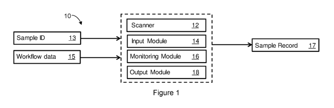
[0026] Figure 1 is a schematic view of a system for monitoring tissue
samples to
be processed by a tissue processor for a histopathology workflow according to
an
embodiment of the present invention;
[0027] Figure 2 is another schematic view of a system for monitoring tissue
samples to be processed by a tissue processor for a histopathology workflow
according to an embodiment of the present invention;
[0028] Figure 3 is a schematic view of a system for treating tissue samples
according to an embodiment of the present invention;
[0029] Figure 4 is a representation of a tissue processor for a
histopathology
workflow according to an embodiment of the present invention;
[0030] Figure 5 is a flow chart of a sample being processed by laboratory
instruments in a system for treating tissue samples according to an embodiment
of
the present invention; and
[0031] Figure 6 is a flow chart of a method of monitoring tissue samples to
be
processed by a tissue processor for a histopathology workflow according to an
embodiment of the present invention.
Detailed Description
[0032] Embodiments of the invention are discussed herein by reference to
the
drawings which are not to scale and are intended merely to assist with
explanation of
the invention.
CA 03066429 2019-12-06
WO 2019/036758 PCT/AU2018/050890
9
[0033] An embodiment of a system 10 for monitoring tissue samples to be
processed by a tissue processor for a histopathology workflow is shown in
Figure 1.
The system 10 includes a scanner 12 associated with a tissue processor that is
arranged to scan an electronic sample identifier 13 of at least one tissue
sample to be
processed by the tissue processor. As discussed, the system 10 could be
implemented in a client-server arrangement, with a number of modules for
monitoring
the tissue samples being processed by the tissue processor being implemented
by
the server. In this way, the system 10 can be used to monitor tissue samples
processed by many tissue processors and can be applied to existing tissue
processors in data communication with the server over a network.
[0034] These modules implemented by the server include: an input module 14
arranged to receive tissue processor workflow data 15 indicative of a tissue
processor
workflow for the at least one tissue sample to be processed by selected ones
of a
plurality of processing stations in the tissue processor used for processing
the at least
one tissue sample. That is, the tissue processor includes a number of
processing
stations discussed in more detail below that are arranged to process the
tissue
sample according to the tissue processor workflow.
[0035] The modules also include a monitoring module 16 arranged to monitor
properties of the at least one tissue sample processed at each of the selected
ones of
the processing stations according to the tissue processor workflow, and to
record the
properties of the at least one tissue sample in association with the
electronic sample
identifier 13 in a sample record 17 for the tissue processor workflow.
Further, an
output module 18 is arranged to output the sample record 17 to one or more
laboratory instruments for further processing the at least one tissue sample
in a
histopathology workflow.
[0036] Figure 5 shows an example of a histopathology workflow. Here it can
be
seen that the steps of grossing tissue samples, processing the tissue samples
using a
tissue processor, and embedding the tissue samples occur just before the
advanced
staining step. Accordingly, it will be appreciated that the quality of the
tissue
processing steps affect the advanced staining and thus diagnosis of the tissue
samples in a histopathological workflow.
CA 03066429 2019-12-06
WO 2019/036758 PCT/AU2018/050890
[0037] Figure 2 shows an alternative embodiment of a tissue processor 20
incorporating the above mentioned modules to monitor tissue samples being
processed by the tissue processor 20 for a histopathology workflow. That is,
the
tissue processor 20 includes a scanner 12, co-located with the tissue
processor 20,
arranged to scan an electronic sample identifier 13 of at least one tissue
sample to be
processed by the tissue processor 20.
[0038] For example, the electronic sample identifier is a barcode tag. In
one
embodiment, the barcode tag is applied to a basket containing tissue samples
that
were cut up and placed into a cassette which was placed into a basket for
batch
processing of the samples in the cassette. In another embodiment, the
identifier is
applied to a cassette. Further, the cassette can also have a cassette
identifier in
addition to the basket identifier to further identify the samples being
processed by the
tissue processor 20.
[0039] The tissue processor 20 includes a plurality of processing stations
22 that
are arranged to process the at least one tissue sample according to the tissue
processor workflow. One of the processing stations 22 is a retort for
processing
tissue samples with different reagents. The tissue processor workflow includes
details of which ones of these stations 22 are to be used to process the
samples and
in which order. These stations 22 will be described in more detail below.
[0040] The tissue processor 20 also includes a CPU 11 (or other
microprocessor)
configured to implement the above mentioned modules to monitor the tissue
samples
being processed by the tissue processor 20. The CPU 11 is configured to
perform
these modules by executing program code stored on a memory 24 for each of the
modules. It will be appreciated by those persons skilled in the art that the
client-
server arrangement described above also uses program code to implement the
modules and this code may be stored in a memory in data communication with a
server processor.
[0041] Specifically, the modules implemented by the CPU 11 of the tissue
processor 20 shown in Figure 2 include: an input module 14 arranged to receive
tissue processor workflow data 15 indicative of a tissue processor workflow
for the
samples being processed by selected ones of the plurality of processing
stations 22.
CA 03066429 2019-12-06
WO 2019/036758 PCT/AU2018/050890
11
While the samples are being processed by the tissue processor 20, a monitoring
module 16 is arranged to monitor properties of the tissue samples processed at
each
of the processing stations 22 and to record these properties in association
with the
electronic sample identifier 13 in a sample record 17 for the tissue processor
workflow. Further, the tissue processor 20 includes an output module 18
arranged to
output the sample record 17 to one or more laboratory instruments for further
processing the tissue sample in a histopathology workflow.
[0042] An example of another laboratory instrument for further processing a
tissue
sample in a histopathological workflow includes an automated tissue staining
apparatus. Figure 3 shows a representation of the tissue processor 20 in data
communication with instrument A 26 (e.g. automated tissue staining apparatus),
having instrument ID 28, and instrument B 30, having instrument ID 32, over a
network 34, the components forming a system 36 for treating tissue samples
according to an embodiment of the invention. The sample record 17 is
communicated
over the network 34 in a format that is understood by the laboratory
instruments A 26
and B 30 for further processing the tissue sample in a histopathology
workflow. The
sample record 17 includes, in association with the electronic sample
identifier, the
tissue processor workflow data indicative of the tissue processor workflow for
the at
least one tissue sample. The sample record 17 also includes expected
properties of
the at least one tissue sample based on the tissue processor workflow for the
at least
one tissue sample. These properties are used by say the automated tissue
staining
apparatus (e.g. instrument A 26) to modify its workflow to ensure a higher
quality
stain.
[0043] Furthermore, the sample record 17 is used as a troubleshooting tool
to
determine if there are any reasons, due to say processing or reagent issues,
for
errors in the tissue processing occurring. It is also used as a Quality
Control (QC)
record that may be required to be shown on audit.
[0044] Figure 4 shows an embodiment of the above described tissue processor
20
and its processing stations 22. The tissue processor 20 includes two
processing
stations 22 as retorts 21A and 21B for processing tissue samples with
different
reagents simultaneously. In the retorts 21A and 21B, tissue samples pass
through
multiple process steps. It will be appreciated by those persons skilled in the
art that
CA 03066429 2019-12-06
WO 2019/036758 PCT/AU2018/050890
12
when the retorts 21A and 21B are performing these different steps, the retorts
21A
and 21B themselves form different processing stations 22.
[0045] One such process is a fixing process, in which formalin is typically
used.
This process preferably occurs first in the tissue processing workflow. A
dehydration
process is then accomplished, using alcohol solutions of various degrees of
purity. In
a subsequent clearing process, alcohol residues are removed from the tissue
samples and the tissue samples are prepared for the uptake of carrier
material.
Xylene or a similar medium is often used in this clearing process. Paraffin or
wax of
various compositions is preferably suitable as a carrier material. Individual
or multiple
process steps can be subdivided into process sub-steps in which tissue samples
are
exposed to the aforesaid reagents having different degrees of purity.
[0046] Once these process steps have been executed in a tissue processor
workflow, a process of cleaning the retorts 21A and 21B is carried out using
the
aforesaid, or further reagents; for example by performing the aforesaid
process steps
in reverse order without tissue samples in retorts 21A and 21B. The tissue
processor
20 includes a cabinet 23 having two drawers for containers 25 containing the
reagents that are necessary for various processes, including the fixing
process, the
dehydration process, and/or the cleaning process.
[0047] A work area is provided on a desktop of the tissue processor 20, as
well as
a display 27. It will be appreciated that the CPU 11 and memory 24 are
provided by
the tissue processor 20 to control the treatment processes for the tissue
samples
according to the tissue processor workflow and to monitor the tissue samples
being
processed. The display 27 is configured by the CPU 11 to display information
indicative of the sample record 17 to a user of the tissue processor 20. For
example
the sample record 17 is shown as a collated report on the display 27 that can
be
filtered by the user.
[0048] The retorts 21A and 21B are embodied in the tissue processor 20 as a
sealable chamber having an opening for receiving the tissue samples in a
basket and
is shown in a closed position. Inside one of the retorts 21A, various reagents
(e.g.
paraffin, which is important for the infiltration process) can act on the
tissue samples
by pressure, vacuum, and or temperature. The interior of the retort 21A is
connected
CA 03066429 2019-12-06
WO 2019/036758 PCT/AU2018/050890
13
via a valve arrangement to lines from the reagent containers 25 via
electrically
controllable valves. For example, one line is connected via a valve to the
contents of
the retort 21A so that, under the control of the valve, liquid paraffin is
delivered from a
corresponding reagent container 25. Further lines connect to further reagent
containers 25 for reagents required for the fixing process, the dehydration
process,
and/or the clearing process, etc. In addition, another line is connected to a
distributor
that distributes liquid paraffin under the control of valves. The paraffin can
be
contained in a supply station for paraffin or one of the reagent containers
25. In a
further embodiment, the distributor is connected to lines that connect it to
containers
25 containing liquid paraffin with an increasing degree of purity.
[0049] In the embodiment, the lines are also heated, as is the distributor
and,
depending on the reagent used, the valve arrangements as well, in order to
ensure
that the paraffin is always kept in a liquid state, e.g. at 65 C, and does
not solidify
during operation. The same is also true of retorts 21A and 21B and its parts,
and of
the supply station and some of the containers.
[0050] Sensors 29 are arranged on the tissue processor 20 to sense
properties of
the tissue sample being processed (see, for example, Figure 2). These sensors
29
are associated with each of the plurality of processing stations 22, such as
the retorts
21A and 21B, in the tissue processor 20 and are arranged to sense the
properties of
the tissue sample as it processed.
[0051] For example, some of the sensors 29 are located between reagent
containers 25 and the retorts 21A and 21B, and between the distributor and its
valves.
Another sensor is provided for acquisition of a measured value that is
representative
of a characteristic property of the paraffin; in particular of a degree of
purity of the
paraffin that is currently flowing through the line. It is thus possible, as
the paraffin is
being pumped to the retorts 21A and 21B and back to the containers 25 to
ascertain
the different degrees of purity of the paraffin currently being used, before
and after
treatment of the tissue samples. In this example, the sample record includes
the
tissue processor workflow data indicative of the step of processing using
paraffin and
details of the purity of the paraffin that was used in this processing step.
This
information in the sample record 17 could be used to audit, troubleshoot,
check
CA 03066429 2019-12-06
WO 2019/036758 PCT/AU2018/050890
14
instrument usage and maintenance, check reagent usage and re-order reagents
for
inventory management and reagent usage optimization.
[0052] Examples of sensors 29 include an optical sensor configured to sense
turbidity or coloration of the paraffin ¨ the paraffin can be treated with a
colouring
agent in order to ascertain its degree of purity. Also, using this type of
sensor, it is
possible to ascertain a density or a conductivity of the paraffin, as a
function of which
the degree of purity can then be ascertained.
[0053] The next steps in the fixing process involve pumping successive
process
media from other reagent containers 25 via connectors to the retorts 21A and
21B by,
for example, applying pressure to these reagent containers 25. These reagent
containers 25 contain the corresponding process media at different degrees of
purity.
Other ones of the sensors 29 of the tissue processor 20 thus include a density
sensor
and a pressure sensor to sense the density of the process medium that is
currently
flowing to the retorts 21A and 21B. The degree of purity of the process medium
can
be determined as a function of its density. The density sensor and the
pressure
sensor are thus used for acquiring a measured value that is representative of
the
degree of purity of the process medium. The density sensor is suitable in
particular
for ascertaining the degree of purity of alcohol or xylene used in this
processing step.
[0054] Also, the process media that are stocked in the reagent containers
25
encompass, for example, fixing reagents, in particular alkaline fixing
reagents, for
example formalin; dehydration reagents, in particular alcohols, in particular
ethanol;
intermedia, for example isopropanol or aromatic compounds, in particular
xylene;
and/or cleaning reagents, in particular distilled water. In addition, the
fixing reagents,
dehydration reagents, and/or intermedia can also be used for cleaning and, in
this
context, can also be referred to as cleaning reagents. One or more other
sensors 29
can also be provided for sensing characteristic properties of all the process
media
used. These characteristic properties can be measured using the following, but
not
limited to, sensors: a photosensor, a conductivity sensor, and a pH sensor.
[0055] Referring now to Figure 6, there is shown a summary of a method 40
of
monitoring tissue samples to be processed by a tissue processor for a
histopathology
workflow, the method including: receiving 42 from a scanner associated with
the
CA 03066429 2019-12-06
WO 2019/036758 PCT/AU2018/050890
tissue processor, an electronic sample identifier of at least one tissue
sample to be
processed by the tissue processor; receiving 44 tissue processor workflow data
indicative of a tissue processor workflow for the at least one tissue sample
to be
processed by selected ones of a plurality of processing stations in the tissue
processor used for processing the at least one tissue sample; monitoring 46
properties of the at least one tissue sample processed at each of the selected
ones of
the processing stations; recording 48 the properties of the at least one
tissue sample
in association with the electronic sample identifier in a sample record for
the tissue
processor workflow; and outputting 50 the sample record to one or more
laboratory
instruments for further processing the at least one tissue sample in a
histopathology
workflow.
[0056] Further aspects of the method will be apparent from the above
description
of the system 10 and the tissue processor 20. Persons skilled in the art will
also
appreciate that the method could be embodied in program code. The program code
could be supplied in a number of ways, for example on a memory of the tissue
processor 20, or on a tangible computer readable medium, or communicated as a
data signal or file for the tissue processor 20.
[0057] It is also to be understood that various alterations, additions
and/or
modifications may be made to the parts previously described without departing
from
the ambit of the present invention, and that, in the light of the above
teachings, the
present invention may be implemented in software, firmware and/or hardware in
a
variety of manners as would be understood by the skilled person.
[0058] It is also to be understood that the following claims are provided
by way of
example only, and are not intended to limit the scope of what may be claimed
in any
future application. Features may be added to or omitted from the claims at a
later
date so as to further define or re-define the invention or inventions.
[0059] The discussion of documents, acts, materials, devices, articles and
the like
is included in this specification solely for the purpose of providing a
context for the
present invention. It is not suggested or represented that any or all of these
matters
formed part of the prior art base or were common general knowledge in the
field
CA 03066429 2019-12-06
WO 2019/036758 PCT/AU2018/050890
16
relevant to the present invention as it existed before the priority date of
each claim of
this application.
[0060] Where any or all of the terms "comprise", "comprises", "comprised"
or
"comprising" are used in this specification (including the claims) they are to
be
interpreted as specifying the presence of the stated features, integers, steps
or
components, but not precluding the presence of one or more other features,
integers,
steps or components.