Note: Descriptions are shown in the official language in which they were submitted.
CA 03066710 2019-12-06
WO 2018/231726 PCT/US2018/036960
PATCH GRAFT COMPOSITIONS FOR CELL ENGRAFTMENT
CROSS REFERENCE TO RELATED APPLICATIONS
This application claims priority under 35 U.S.C. 119(e) to U.S. Application
No.
62/518,380, filed June 12, 2017, and to U.S. Application No. 62/664,694, filed
April 30,
2018, the contents of which are hereby incorporated by reference in their
entirety.
FIELD OF THE INVENTION
The present invention is directed generally to the field of transplantation of
cells or tissue
engrafting. More specifically, from solid organs or tissues into solid organs
or tissues,
especially to internal organs. The invention concerns compositions and methods
providing
strategies for the rapid transplantation, engraftment and integration of cells
into solid
organs and tissues to treat diseases or conditions of solid organs or tissues,
or to establish
model systems of a disease. Representative examples of this potential are cell
therapies for
treatment of hepatic or pancreatic diseases.
BACKGROUND OF THE INVENTION
.. There has long been a need for grafting strategies for cells from solid
organs, strategies
distinct from those used for transplantation of hemopoietic cells or for
mesenchymal
stem/progenitors. Turner, R., et al. Transplantation 90, 807-810 (2010);
Gattinoni, L. et al.
Nature Medicine 23, 18-27 (2017); Trounson A. et al. Cell Stem Cell 17, 11-22
(2015); Sun
B.K. et al. Science 346, 941-945 (2014); Lainas, P. et al. J Hepatol 49, 354-
362 (2008).
Transplantation of hematopoietic cells and of mesenchymal cells is done
routinely by
delivery of cells via a vascular channel and is dependent on activation of
adhesion
molecules in transplanted cells when in relevant target sites because of micro-
environmental
signaling, a process referred to as "homing." Methods used for skin (with
similar ones for
ocular targets) employ grafting methods with cells applied directly to target
sites. Sun B.K.
et al. Science 346, 941-945 (2014). Many grafting methods for skin are
utilizable for cells
from solid internal organs but require substantial modifications to
accommodate the
microenvironment of these internal organs. Grafts must contend with mechanical
forces
exerted by interactions of tissues and organs on each other; examples include
the effects of
lungs during breathing, or the compression of the liver against the diaphragm,
or the
transient effects of mechanical forces exerted by the intestinal tract on
neighboring tissues
1
CA 03066710 2019-12-06
WO 2018/231726 PCT/US2018/036960
during processing of foods. Grafts, especially those for internal organs, are
challenging to
design because of concerns with respect to size, shape, and complexity in the
structure of
organs in addition to the dynamic mechanical forces evident.
For decades, cell therapies for cells from solid organs other than skin were
attempted using
transplantation via a vascular route or by direct injection into the tissue.
Most transplanted
cells, when delivered by either of these strategies, either die or are
transported to ectopic
sites, where they can live for months and create tissue in inappropriate
sites, resulting
potentially in adverse effects clinically. Turner, R., et al. Transplantation
90, 807-810
(2010); Lanzoni, G. et al. Stem Cells 31, 2047-2060 (2013). Engraftment in
liver can be
improved by coating the cells with hyaluronans and delivering them vascularly
to the liver;
the increased efficiency of engraftment is due to the liver's natural process
of clearance of
hyaluronans. Nevi et al. Stem Cell Research & Therapy 8, 68 , 2017. However,
this
improvement is still less efficient than that with grafting strategies and,
importantly, still
allows for delivery of cells to ectopic sites.
There remains a need for improved methods of cell engraftment into solid
organs. This
disclosure fulfills this need and provides related advantages.
SUMMARY OF THE INVENTION
There has long been a need for grafting strategies for cells from solid organs
(Turner, R., et
al. Transplantation 90, 807-810 (2010)õ strategies distinct from those used
for
transplantation of hemopoietic cells, mesenchymal stem cells or for skin.
Transplantation of
hemopoietic cells and mesenchymal cells is done routinely via a vascular
channel and is
dependent on activation of adhesion molecules in relevant target sites because
of micro-
environmental signaling, a process referred to as "homing". Methods used for
skin employ
grafting methods with cells applied directly to target sites.
Transplantation of cells from solid organs other than skin have long used
vascular delivery.
This is not logical, since adhesion molecules on these cells are always
activated and result
in rapid (seconds) cell aggregation that can generate life-threatening emboli.
Even if emboli
are managed successfully to minimize health risks, the efficiency of cell
engraftment is low,
only ¨20% for adult cells and even lower (<5%) for stem/progenitors. Most
transplanted
cells either die or are transported to ectopic sites, where they can live for
months, creating
2
CA 03066710 2019-12-06
WO 2018/231726 PCT/US2018/036960
tissue in inappropriate sites resulting in possible adverse effects
clinically. The small
percentage of cells that engraft into target sites integrate slowly, requiring
weeks to months
to become a significant portion of the tissue. There is improvement in
engraftment in liver if
cells are coated with hyaluronans and delivered vascularly due to the tissue's
(e.g. liver's)
clearance of hyaluronans. (Nevi et al. Stem Cell Research & Therapy 8, 68 ,
2017).
Applicants propose a radically different approach, one found even more
successful than
coating cells with hyaluronans: placing grafts directly onto the surface of
the target site and
using grafting biomaterials and the unique phenotypic traits of certain cells
when they are in
conditions of the graft biomaterials to enhance transplantation. This
parallels some aspects
of strategies of cell therapies for skin but requires substantial,
modifications for internal
organs given mechanical effects, abrasion or compression of organs near to
each other, and
given the unique fluid microenvironments around specific organs and the size,
structure,
and complexity of organs.
Described herein are novel patch graft compositions and methods for
transplantation of cells
into tissue and solid organs. In some embodiments, the methods and grafts are
adapted for
internal organs, with design features dependent on the level of maturity of
the cells,
especially whether cells are stem cells or mature cells. In some embodiments,
the donor
cells (optionally autologous or allogenic) for the patch grafts are disclosed
herein
incorporated into the graft biomaterials in optionally as a mixture of cells
or the form of
organoids, aggregates of epithelial stem cells and their native, lineage-stage
appropriate
mesenchymal cell partners ¨ e.g. mesenchymal stem/progenitor cells such as
early lineage
stage mesenchymal cells (ELSMCs). In some embodiments, the donor cells are
adult cells
incorporated into the graft materials as cell suspensions of adult epithelia
and patneredwith
mesenchymal stem/progenitor cells, optionally ELSMCs, at ratios designed to
optimize
their expression of membrane-associated and/or secreted matrix metallo-
proteinases
(MMPs). In some embodiments, other variables of importance are the grafting
biomaterials
and the backing material, both required to be neutral in effects on the
differentiation of the
donor cells.
Aspects of the disclosure relate to a patch graft for sustaining and
maintaining a single cell
population or a mixed population of cells, comprising: (a) a single cell type
or a mixed
population having two or more cell types, at least one of which is at an early
lineage stage
3
CA 03066710 2019-12-06
WO 2018/231726 PCT/US2018/036960
that is capable of expressing membrane- associated and/or secreted matrix
metalloproteinases (MMPs), or which has MMPs included from another source
(e.g.,
purified or recombinant MMPs), said cell population or mixed population
supported in a
medium present in a hydrogel matrix having a viscoelasticity sufficient to
allow for
migration of said mixed population, optionally, within or away from said
hydrogel and/or
within or away from the patch graft; (b) a backing comprising a biocompatible,
biodegradable material having a viscoelasticity sufficient to inhibit a
migration of said
mixed population in a direction of said backing; and, optionally, (c) a
hydrogel overlaid on a
serosal (i.e. outside) surface of said backing, which is opposite to that in
contact with said
mixed population and, in embodiments where the patch graft is tethered to a
target site, is
opposite the side in contact with the target site (e.g. organ or tissue). In
some embodiments,
this layer prevents or inhibits adhesions by or from other tissues or organs.
In some
embodiments, the patch graft is configured to sustain and maintain said mixed
population
while inhibiting said at least one early lineage stage cell type from
differentiating or further
maturing to a later lineage stage that is no longer capable of expressing
membrane-
associated and/or secreted MMPs. The patch graft may be a single layer plus a
backing or
multiple layers.
In some embodiments, said backing is porous or non-porous. In some
embodiments, the
backing comprises a porous mesh, scaffold, or membrane. In some embodiments,
the
backing comprises silk; a synthetic textile; or a natural material such as
aminion, placenta,
or omentum; or a combination thereof In some embodiments, said backing
comprises a
porous mesh infused with a hydrogel. In further embodiments, such an infusion
prevents
cell migration away from the target organ or tissue. In some embodiments, said
backing
comprises a solid material.
In some embodiments, one or more of said hydrogels comprise hyaluronans.
In some embodiments, said medium comprises Kubota's medium or another medium
supportive of stem cells and able to maintain stemness.
In some embodiments, said mixed population comprises mesenchymal cells and
epithelial
cells. In some embodiments, said epithelial cells may be ectodermal,
endodermal, or
mesodermal. In some embodiments, said mesenchymal cells comprise early lineage
stage
mesenchymal cells (ELSMCs). In some embodiments, said ELSMCs comprise one or
more
4
CA 03066710 2019-12-06
WO 2018/231726 PCT/US2018/036960
of angioblasts, precursors to endothelia, precursors to stellate cells, and
mesenchymal stem
cells (MSCs). In some embodiments, said epithelial cells comprise epithelial
stem cells. In
some embodiments, said epithelial cells comprise biliary tree stem cells
(BTSCs). In some
embodiments, said epithelial cells comprise committed and/or mature epithelial
cells. In
some embodiments, said committed and/or mature epithelial cells comprise
mature
parenchymal cells. In some embodiments, said mature parenchymal cells comprise
one or
more of hepatocytes, cholangiocytes, and islet cells. In some embodiments,
said
mesenchymal cells and epithelial cells both comprise stem cells.
In some embodiment said mixed population comprises autologous and/or
allogeneic cells.
In some embodiments, one or more cell types are genetically modified.
Further aspects related to methods employing the disclosed patch graft
ompositions.
Accordingly, provided herein are methods of engrafting cells into a target
tissue comprising,
consisting of, or consisting essentially of contacting the target tissue with
a patch graft
disclosed herein above.
In some embodiments of the methods, the target tissue is selected from the
group consisting
of liver, pancreas, biliary tree, thyroid, thymus, gastrointestine, lung,
prostate, breast, brain,
bladder, spinal cord, skin and underlying dermal tissues, uterine, kidney,
muscle, blood
vessel, heart, cartilage, tendons, and bone tissue. In some embodiments of the
methods, the
target tissue is liver tissue. In some embodiments of the methods, the target
tissue is
pancreatic tissue. In some embodiments of the methods, the target tissue is
biliary tree
tissue. In some embodiments of the methods, the target tissue is
gastrointestinal tissue. In
some embodiments, the tissue is diseased, damaged, or has a disorder. In some
embodiments of the methods, the target tissue is kidney tissue.
In some embodiments of the methods, the target tissue is an organ. In some
embodiments of
the methods, the organ is an organ of the musculoskeletal system, the
digestive system, the
respiratory system, the urinary system, the female reproductive system, the
male
reproductive system, the endocrine system, the circulatory system, the
lymphatic system,
the nervous system, or the integumentary system. In some embodiments of the
methods, the
organ is selected from the group consisting of liver, pancreas, biliary tree,
thyroid, thymus,
stomach, intestines, lung, prostate, breast, brain, bladder, spinal cord, skin
and underlying
5
CA 03066710 2019-12-06
WO 2018/231726 PCT/US2018/036960
dermal tissues, uterus, kidney, muscle, blood vessel, heart, cartilage,
tendon, and bone. In
some embodiments, the organ is diseased, damaged, or has a disorder.
Also provided herein are methods of treating a subject with a liver disease or
disorder, the
methods comprising, consisting of, or consisting essentially contacting the
subject's liver a
patch graft disclosed herein above. In some embodiments of the methods, the
liver disease
or disorder is liver fibrosis, liver cirrhosis, hemochromatosis, liver cancer,
biliary atresia,
nonalcoholic fatty liver disease, hepatitis, viral hepatitis, autoimmune
hepatitis, fascioliasis,
alcoholic liver disease, alpha 1-antitrypsin deficiency, glycogen storage
disease type II,
transthyretin-related hereditary amyloidoisis, Gilbert's syndrome, primary
biliary cirrhosis,
primary sclerosing cholangitis, Budd-Chiari syndrome, liver trauma, or Wilson
disease.
In other aspects, provided herein are methods of treating a subject with a
disease or disorder
of the pancreas, the methods comprising, consisting of, or consisting
essentially of
contacting the subject's pancreas with a patch graft disclosed herein above.
In some
embodiments of the methods, the disease or disorder of the pancreas is
diabetes mellitus,
.. exocrine pancreatic insufficiency, pancreatitis, pancreatic cancer,
sphincter of Oddi
dysfunction, cystic fibrosis, pancreas divisum, annular pancreas, pancreatic
trauma, or
hemosuccus pancreaticus.
In other aspects, provided herein are methods of treating a subject with a
gastrointestinal
disease or disorder, the method comprising, consisting of, or consisting
essentially of
contacting one or more of the subject's intestines with a patch graft
disclosed herein above.
In some embodiments, the gastrointestinal disease or disorder is
gastroenteritis,
gastrointestinal cancer, ileitis, inflammatory bowel disease, Crohn's disease,
ulcerative
colitis, irritable bowel syndrome, peptic ulcer disease, celiac disease,
fibrosis,
angiodysplasia, Hirschsprung's disease, pseudomembranous colitis, or
gastrointestinal
.. trauma.
In some aspects, provided herein are methods of treating a subject with a
kidney disease or
disorder, the methods comprising, consisting of, or consisting essentially of
contacting one
or more of the subject's kidneys with a patch graft disclosed herein above. In
some
embodiments of the methods, the kidney disease or disorder is nephritis,
nephrosis,
nephritic syndrome, nephrotic syndrome, chronic kidney disease, acute kidney
injury,
kidney trauma, cystic kidney disease, polycystic kidney disease,
glomerulonephritis, IgA
6
CA 03066710 2019-12-06
WO 2018/231726 PCT/US2018/036960
nephropathy, lupus nephritis, kidney cancer, Alport syndrome, amyloidosis,
Goodpasture
syndrome, or Wegener's granulomatosis.
BRIEF DESCRIPTION OF THE FIGURES
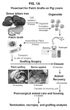
FIGS. 1A-1D provides information about porcine donor cells for the patch
grafts. FIG. 1A
is a schematic of the process and estimates of the time required for preparing
organoids,
assembling patch grafts and doing the surgeries. In FIG. 1B, donor cells for
the stem cell
patch grafts were isolated from cell suspensions of biliary tree tissue from
transgenic pigs;
the cells were prepared as organoids in serum-free Kubota's Medium and on low
attachment
culture dishes. Organoids of biliary tree stem cells (BTSCs) and of their
early lineage stage
mesenchymal cell (ELSMCs) partners, angioblasts and precursors to endothelia
and to
stellate cells. They are shown in a phase micrograph versus one demonstrating
expression of
the transgene, green fluorescent protein (GFP). ). All of the cells of the
aggregate are
green, since the transgene is in both the epithelial cells and the mesenchymal
cells. The
transgene was coupled to the histone (H-2B) locus. Histology of the stem cell
organoids
that were paraffin embedded, sectioned and stained with hematoxylin/eosin. (d)
Magnified
image of an organoid of BTSC and ELSMCs. FIG. 1C shows immunohistochemistry
(IHC) demonstrating expression of stem cell, hepatic and pancreatic markers
indicating that
these cells are precursors to both liver and to pancreas. The IHC assays
indicate outer
layers with intermediate stage stem cell markers such as EpCAM and interior
cells
expressing very primitive genes such as pluripotency genes and endodermal
transcription
factors (e.g. SOX17, SOX9, PDX1). FIG. 1D is a representative qRT-PCR assays
assessing expression of various genes in the organoids and indicating that
cells are stem
cells or early progenitors. The controls were mature hepatocytes from piglet
livers.
FIGS. 2A-2F provides information about the major components of patch grafts.
FIG. 2A is
a schematic of a patch graft affixed to the liver of a pig, and on the right,
the composition of
the grafts. Early lineage stage cells, both the epithelia and the mesenchymal
cells, are
sources for production of matrix metallo-proteinases (MMPs), key regulators of
engraftment. The matrix components of the graft biomaterials into which donor
cells are
placed are soft (-100 Pa), without (or with minimal) sulfation, such as
hyaluronan
hydrogels. The structure of the graft consist of layers of biomaterials and
cells tethered to
the target site. The medium components are devoid of serum, growth factors and
cytokines
influential to differentiation of the donor cells and should be ones tailored
for survival and
7
CA 03066710 2019-12-06
WO 2018/231726 PCT/US2018/036960
expansion of early lineage stage cells such as stem/progenitors. The backing
has sufficient
tensile strength to be used in surgical procedures but be neutral in its
effects on the
differentiation of the donor cells (e.g. ones with type I collagen should be
avoided). The
backing is impregnated or coated with a more rigid 10X hydrogel (¨ 700 Pa) to
serve as a
barrier to orient the migration of donor cells towards the target tissue and
to minimize
adhesions. After attachment to the target site, a 2X HA hydrogel, one that is
sufficiently
fluid to be coated or painted onto the serosal surface, is added and used to
further minimize
adhesions. FIG. 2B depicts the graft affixed to the liver or the pancreas of a
host. FIG. 2C
is a schematic of the graft demonstrating the layers constituting the graft
composition. FIG.
2D depicts the results of assays empirically assessing the rheological or
viscoelastic
properties (shear and compressive mechanical forces) of the specific hydrogel
layers. FIG.
2E provides a formulation of the viscoelastic properties of the 3 layers of
hydrogels. FIG.
2F is a close up image of a patch graft sutured to the surface of the liver of
a pig.
FIGS. 3A-3D depicts the result of immunohistochemistry (IHC) and histology of
the liver
patch grafts. FIG. 3A shows the results of Trichrome staining of the patch
graft at one
week. Trichrome identifies collagens (blue), cytoplasm (red) and nuclei
(black), and it was
used to identify Glisson's capsule (normally adjacent to the surface of liver
lobules) and
adhesions (on the serosal surface of the grafts). There is a high level of
blue staining in the
layers at the serosal surface and implicate adhesions to the graft. Also, the
graft has
separated from the host tissue at the interface between the backing and the
host; this was
found frequently due to the wealth of MMPs produced at this interface. The
remodeling
regions provide evidence of the loss of classic lobule structure of the liver;
they result in a
region in which the donor cells are migrating into the tissue and, in
parallel, altering the host
tissue structure. In low magnification images (a), Trichrome staining of
grafts placed on to
the liver validated that extensive remodeling of the Glisson capsule was
occurring and
resulted often in a separation between the graft and the host liver. In higher
magnification
images (b) the remodeling region is remarkably broad and consisting of areas
(c) near to the
graft where liver lobule structure is missing altogether and (d) regions
within the remaining
liver lobules that are undergoing breakdown in the remodeling process. FIG. 3B
shows the
results of Trichrome staining of the patch graft at three weeks. Hyaluronans
in the graft
have been resorbed leaving only the backing (a). With resorption of HA, the
Glisson
capsule reappears (b) and the liver lobules near to the graft have stabilized
again into their
typical histological patterns, such as lobule and acini for liver. The arrow
in (b) indicates the
8
CA 03066710 2019-12-06
WO 2018/231726 PCT/US2018/036960
reappearance of collagens in the reformation of the Glisson capsule. FIG. 3C
and FIG. 3D
shows the results of hematoxylin/eosin staining of a section from the grafts
at one week post
grafting (C) and two weeks post grafting (D). The figures at the top are 40X.
At sites within
the figure (a,b,c) are enlargements that are magnified at 100X; the
rectangular image below
each of these is magnified at 200 X. Shown are 3 sites of the graft: (a) a
site within the
backing and associated graft biomaterials; (b) a site at the interface between
graft and host
tissue; and (c) a site within the liver lobules. The hematoxylin/eosin
staining yields images
that contribute to an appreciation of the engraftment and migration process
that incorporates
features of inflammatory processes.
.. FIGS. 4A-4C shows engraftment, migration and rapid maturation to adult
fates within a
week. FIG. 4A is a low magnification image of the patch graft on the surface
of a pig liver
after one week. The dashed line indicates the interface of the graft and host
liver. Donor
GFP+ cells (with pink nuclei; white arrows indicate areas with large numbers
of the donor
GFP+ stem cells) were visualized by labeling with an antibody to GFP and
secondarily with
one coupled to Novo Red, a red fluoroprobe. Nuclei were stained blue with 4,6-
Diamidino-
2-phenylindole (DAPI) enabling recognition of host cells having only blue
nuclei and donor
ones having pink nuclei (merge of DAPI and the Novo Red). Host tissue (a)
extends into
the hyaluronans (HA, the black background) of the graft; tissue by the backing
contains
occasional organoids (inset) but with most donor cells dispersed into single
cells; large
numbers of dispersed donor GFP+ stem cells (b) are seen throughout the host
tissue. There
is no evidence for the Glisson capsule in this area that constitutes the
region of remodeling.
FIG. 4B demonstrates that engraftment and migration of donor cells was rapid;
within a
week, all donor cells were within the host liver; there were donor cells both
near the graft
site and also on the opposite side of the liver lobe (estimate of the distance
is at least1.5 cm
from the graft). Ongoing studies are analyzing regions of the piglet livers at
greater
distances (i.e. other lobes of the liver) to define more precisely how far the
migration can go
by the donor cells within a defined period of time. Shown are donor cells
(pink nuclei) near
lobules of host mature hepatocytes (forest green color from auto-fluorescence
of
lipofuscins) on the distant side of the liver lobe from that of the graft
site. FIG. 4C shows
that maturation of donor cells to adult fates occurred in parallel with HAs
being resorbed.
Enlargement of a region containing donor GFP+ cells (single cells with pink
nuclei) near to
host hepatocytes (a), forest green in color (autofluorescence of lipofuscins),
and readily
distinguished from mature donor ¨derived (b) hepatocytes that are lavender in
color (merge
9
CA 03066710 2019-12-06
WO 2018/231726 PCT/US2018/036960
of the pink--GFP, blue¨DAPI, and the green--lipofuscins) , that is they were
lineage
restricted from donor GFP+ stem cells. With other IHC assays (data not shown),
the bright,
spring green color of cells amidst the plates of both host and donor
hepatocytes proved to be
endothelia and stellate cells. .
FIGS. 5A-5C compares engraftment and maturation of cells in the liver patch
grafts after
one week and two weeks post-transplantation. FIG. 5A is an examination of
porcine liver
at 1 week after patch grafting. Sirius red stain, an azo dye staining
collagens was used and
immunohistochemistry for pan-cytokeratin (pCK) and Sox9; and
immunofluorescence (IF)
stains were performed on serial 3-1.1.m sections. At the patch graft site,
grafted donor cells
merged with liver lobules. In the upper panels (original magnification=5X),
patch grafts are
composed of mesenchymal and epithelial pCK + cells (arrows). In middle panels,
a higher
magnification is provided (20x). Epithelial cells show an immunophenotype that
is typical
of biliary tree stem cells (BTSCs) expressing biliary cytokeratins (pCK) and
the endodermal
stem cell marker 5ox9. BTSCs within the patch graft are arranged in cell
strings
reassembling bile ductules (arrows) and are in direct continuity with
hepatocyte plates of the
adjacent liver lobule (arrowheads). Host hepatocytes in lobules are pCK and
5ox9 negative.
In lower panels (Original magnification=20X), the immunofluorescence for GFP
allows one
to identify individual grafted cells and their progeny. Hepatocytes in lobules
adjacent to the
patch graft were GFP positive indicating that these were donor cells derived
that had
merged with host liver parenchyma. At the interface between patch graft and
liver lobules,
pCK/GFP + ductules (that is donor derived cholangiocytes) were in direct
continuity with
GFP/pCK - cells (donor-derived hepatocytes) within the lobules (arrowheads)
suggesting a
maturation of grafting cells towards an hepatocyte fate. FIG. 5B is an
examination of
porcine livers 2 weeks after patch grafting. IF stains reveal that GFP + cells
are present
within lobules distant to the graft site. They are dispersed uniformly and so
are in a mix of
host cells (ones with blue nuclei from DAPI) and of donor cells (pink/purple
nuclei from
merge of the blue from DAPI and the red of the GFP label). They co-express
mature
hepatocyte markers such as Hepatocyte Nuclear Factor (HNF) 4a (a mix of green
and
pink/purple nuclei) and albumin (green cytoplasm and with pink/purple nuclei).
Separate or
merged channels were included. Nuclei were displayed in blue (DAPI). Original
Magnification: 40x. FIG. 5C is an evaluation of porcine livers a week after
patch grafting
and demonstrating the broad region of remodeling that occurs at the interface
between the
CA 03066710 2019-12-06
WO 2018/231726 PCT/US2018/036960
patch graft and the host tissue. The section in the low magnification image
and in the
enlarged image of 1 is hematoxylin/eosin (lightly stained); that in 2 is
stained with Vector-
SG providing a blue/gray color; that in 3 is stained for alpha-fetoprotein
with
hematoxylin/eosin background. Specific sites within 5C are numbered and
correlate with
.. enlargements that indicate the changes occurring within the lobules. The
host liver lobules
and acini are breaking down due to the wealth of MMPs flooding into the area
along with
the donor cells. The donor cells are observed at the boundary regions of the
lobules, sites
also demonstrating liver-specific markers such as HNF4-a and a-fetoprotein,
meaning that
the cells are maturing to a liver fate. These traits were not expressed by the
BTSCs and so
these are indications that the donor cells are undergoing maturation to an
hepatic fate.
FIGS. 6A-6D provides information about patch grafts of stem cell organoids
tethered to
pancreas. FIG. 6A is a low magnification (panoramic scan) image of GFP+ donor
cells
that have engrafted into much of the pancreas and into the submucosa of the
duodenum (a
region containing Brunner's Glands). Immunofluorescent staining of pig
pancreas, liver,
and duodenum in the site of the patch graft. GFP (green), Insulin (red), DAPI
(blue). Donor-
derived GFP+ cells occur in the proximity of the site where the patch graft
was positioned,
and appear integrated in the pancreas parenchyma. The silk mesh of the SERI
surgical
scaffold is observed interposed among pancreas, liver, and duodenum. FIG. 6B
shows that
donor cells mature to functional islets. At higher magnification, donor-
derived
GFP+/Insulin+ beta cells (yellow-from merge of the GFP and of the red from
staining for
insulin) are observed intermingled with host GFP-/Insulin+ (red) beta cells in
the pancreas
parenchyma. Surrounding the islet cells are a large number of GFP+ cells
displaying a
morphology consistent with that of pancreatic exocrine cells, including acinar
and ductal
cells. Supporting this interpretation are the findings in C and D that,
indeed, these cells are
producing amylase, a classic acinar marker. FIG. 6C and FIG. 6D show evidence
of
functional acinar cells derived from donor stem cells. Immunofluorescent
staining of a serial
section from the same tissue block in the site of the patch graft and with
focus on the region
of engrafted GFP+ donor cells. Amylase (green), Insulin (red), Glucagon (white
- not
visible in the panoramic scan in C, but visible at the higher magnification in
D), DAPI
(blue). Amylase+ acinar cells are the vast majority of the exocrine tissue of
the pancreas.
By comparing the staining presented in the serial sections at low and high
magnifications, it
is deduced that most of the donor-derived GFP+ cells in the pancreas have
acquired an
amylase+ acinar fate.
11
CA 03066710 2019-12-06
WO 2018/231726 PCT/US2018/036960
FIGS. 7A-71I offers a characterization of matrix-metallo-proteinases (MMPs).
MMPs are
comprised of a large gene family of calcium-dependent, zinc-containing enzymes
that
dissolve extracellular matrix components. There are at least 24 isoforms known
in pigs of
which a subset are secreted factors (e.g. MMP1, MMP2, MMP7, MMP9) and a subset
are
.. membrane-associated (e.g. MMP14, MMP15). MMP1 was identified by IHC,
especially in
the areas of remodeling, but not by RNA-seq, since there has not yet been an
annotated
form of porcine MMP1 available for RNA seq analyses. FIG. 7A, FIG. 7B, FIG.
7C, and
FIG. 7D show isoforms of secreted and membrane-associated categories were
expressed by
both stem/progenitors and mature cells. Quantitation of the expression levels
indicated that
.. the membrane-associated forms were similar for both stem/progenitors and
mature cells
(note the comparisons in FIG. 7D). By contrast, secreted forms were expressed
at very
high levels in stem/progenitors and at low or negligible levels in mature cell
types. The cell
populations of adult cells analyzed were isolated from suspensions of piglet
livers and
biliary tree tissue and comprised of CD45+ cells (hemopoietic cells), CD146+
cells (stellate
.. cells), CD31+ cells (endothelia), EpCAM+/CD45- cells (adult diploid
hepatocytes and
cholangiocytes. These EpCAM+/CD45- cells are the mature parenchymal cells
found in
piglet livers. The BTSCs were isolated from the biliary tree by the protocols
given in the
examples. FIG. 7E shows representative MMP expression in regions of remodeling
with a
BTSC/ELSMCs graft. In a section adjacent to the patch graft of BTSCs/ELSMCs
were
.. stained with Trichrome indicating the region (bracket) of remodeling. The
region appears
as linear stripes of red and blue being cells and matrix components undergoing
dissolution
by the "sea" of MMPs. The stripes end at the edges of lobules that are still
mostly intact but
beginning to "fray" at their boundaries from the effects of the MMPs derived
from the
invading cells. FIG. 7F shows representative images of IHC assays for MMP1
(Novo-
.. red+). Methyl green is the background stain. The liver's lobular/acinar
structure has
dissolved into the undulating swirls of cells and marked by the strong
expression of MMP1,
a secreted isoform of MMPs. FIG. 7G shows a section stained for MMP2 (Novo-
red+).
Hematoxylin is the background stain. The liver's lobular/acinar structure has
disappeared
and has been replaced by a mix of cells with strong staining for MMP2 (rust
brown color).
FIG. 711 shows the remodeling process ongoing within the liver lobules. The
liver lobules
have become strips of cells interspersed by invading cells; MMP2+ expression
(rust
colored) is very high and contributing to the loss of lobular/acinar
structures. With
clearance of hyaluronans (by 2-3 weeks), the lobular structures reappeared.
12
CA 03066710 2019-12-06
WO 2018/231726 PCT/US2018/036960
FIG. 8 is a schematic demonstration of the engraftment and integration
phenomena in liver
and on pancreas.
FIGS. 9A-9E provides information about patch grafts of mature (adult)
hepatocytes
partnered with mature mesenchymal cells (MMCs), such as endothelia or stellate
cells.
These patch grafts were unable to engraft. Engraftment was achievable if the
hepatocytes
were partnered with early lineage stage mesenchymal cells (ELSMCs), here being
porcine
mesenchymal stem cells (MSCs). If presented with ELSMCs, then engraftment
occurred
but with restriction to regions near to the graft. FIG. 9A shows Trichrome
staining of
normal pig liver. Bar is 200 p.m for low magnification image (a) and 50 p.m
for the higher
magnification image (b). Note the collagens in the Glisson capsule and the
boundaries
between hepatic acini. FIG. 9B shows Trichrome staining of patch graft of
normal, adult
hepatocytes partnered with mature mesenchymal cells (MMCs), endothelia and
stellate
cells, did not engraft. In the low magnification image (a) note that the
Glisson capsule is
intact, and cells remain atop the capsule. (b) at the higher magnification,
there is evidence
of some remodeling (plasticity phenomena) of cells in the lobule next to the
graft (the
mottled red color within the hepatocytes). This plasticity is assumed due to
the membrane-
associated MMPs known to be present on both stem cells and adult cells. FIG.
9C shows
IHC assays on patch graft of normal, adult hepatocytes partnered with mature
mesenchymal
cells (MMCs). The section was stained with antibody to RBMY-1 and with
hematoxylin as
the counterstain. The Glisson capsule is intact and so are the boundary zones
between
lobules. At the higher magnification (b), it is evident that engraftment has
not occurred. (d)
negative control (staining without primary antibody) to indicate non-specific
staining. FIG.
9D shows Trichrome staining of patch graft of normal, adult hepatocytes
partnered with
ELSMCs that here were porcine mesenchymal stem cells (MSCs) played the role of
a
cellular source of MMPs. The graft is separating at the interface between the
graft and the
host tissue. The bracket indicates the region of remodeling. Note that the
liver lobules have
lost the matrix that normally constitutes boundary zones between them and
appear frayed at
the edges. In the higher magnification (a) are seen donor cells (pale red
compared with the
dark red ones in the centers of the lobules) throughout the image; in (b) is
an enlargement of
a region showing that the Glisson capsule is considerably thinner under the
patch (compare
with region to the left of the box) and in (c). Extensive remodeling was
evident in the cells
adjacent to the graft (c). FIG. 9E shows a patch graft of hepatocytes
partnered with
ELSMCs (porcine MSCs) after one week. The section (a) was stained with
antibody to
13
CA 03066710 2019-12-06
WO 2018/231726 PCT/US2018/036960
RBMY-1 (brown) and with methyl green as the counter stain. The donor cells
engrafted
(regions of rust red color) and matured into adult parenchymal cells in the
acini near to the
graft. The section (b) shows an enlargement of the image near to the remains
of the thinned
Glisson capsule showed that donor cells (dark brown nuclei) were interspersed
uniformly
with host cells (nuclei were methyl green color). The section (c) is the
negative control for
(b). The section (d) was stained with antibody to GFP (coupled with Novus red
and
yielding a rust brown color) and with methyl green as the counter stain. Most
of the cells
have engrafted and formed a band of dark red, donor (mature) hepatocytes
within the host
liver acini. The Glisson capsule remained but was diminished in thickness.
Migration much
beyond the region of the liver near to the graft was not observed within the
three-week time-
frame of the experiments.
FIG. 10 is a schematic comparing engraftment of stem cells versus adult cells.
FIG. 11 shows evidence that the engraftment process involves migration of
cells to
considerable distances within the host tissue. Here is demonstrated that for
grafts of
.. BTSCs/ELSMCs organoids at one week post-transplantation. The schematic of
the liver
divided into 8 different zones is used to indicate the regions evaluated for
the presence of
donor cells. Sections are prepared from the regions 1-8 and then stained to
enable
identification of donor cells. In the table are summarized the findings
showing the distances
between the graft and each region and the proportion of GFP+ cells found. The
images to
the left of the table are scans of a representative section from each zone.
The dark brown
staining is strongest in 6 near to the graft and is fainter with increasing
distance from the
graft, the palest being zone 1.
FIGS. 12A-12E provides evidence for migration of donor cells throughout the
host liver.
GFP+ cells stained with Novo-red (rust brown color); host cells are stained
with methyl
green. FIG. 12A is a low magnification image of interface of graft and the
host liver. The
separation of the graft from the host liver was often seen (note this also in
FIG. 3) and was
shown correlated with exceptionally high levels of secreted MMPs. Enlargement
of the
regions (a) and (b) are given below. Note the areas in the low magnification
image and in
the enlargement in (b) in which staining is mottled and with areas showing a
washed out
appearance and that proved due to hyaluronan levels in the tissue. FIG. 12B
depicts the
intermediate zones to which the cells migrated. Donor cells are throughout the
tissue, both
14
CA 03066710 2019-12-06
WO 2018/231726 PCT/US2018/036960
in bile ducts and in the parenchyma of the acini. FIG. 12C shows the distance
zones to
which the cells migrated. Note that only the bile ducts are stained. FIG. 12D
provides
enlargements showing donor cells in bile ducts. FIG. 12E and FIG. 12F provide
enlargements within the parenchyma to show that the donor cells have GFP
labeling in the
.. nuclei.
FIG. 13 shows the adverse conditions obtained for patch grafts with certain
backings (see
also Tables 1 and 2). These included necrosis, adhesions, and sites of
cholestasis found to
occur when grafts were placed too close to some ducts such that the swelling
caused
occlusion of the ducts.
FIG. 14 shows a chart of both lineage stages for epithelial cells (FIG. 14A)
and
mesenchymal cells (FIG. 14B) and the corresponding biomarker profiles.
FIG. 15 shows organoids of H2B-GFP+ BTSCs/ELSMCs patch grafted onto the
Kidney.
Evaluation was done at 1-week-post-grafting. Panel A shows Trichrome staining
of grafted
kidney. The kidney was prepared in cross-section to expose the deeper layer
that with the
graft as a "V" shape. The lower half "V" with bright blue staining is the
graft side on the
kidney; the upper "V" in the figure is a deeper layer to the grafted layer.
Panel B shows
H&E staining for the same section of the grafted kidney. Panel C is the higher
magnification of the patch grafted kidney. The capsule of the kidney under the
graft was
loosened (from dissolution by MMPs) in a fashion similar to that in the liver.
Panel D
shows IHC staining of GFP+ cells (dark red) that have engrafted into the
kidney at a layer
under the patch. Panel E shows engraftment of the GFP+ cells (dark red) at
deeper layers of
the kidney. Necropsy reports indicated that there was no necrosis found in the
grafted
kidney or elsewhere in the animals that were subjected to patch grafts.
BRIEF DESCRIPTION OF THE TABLES
TABLE 1 provides a summary of surgical or other approaches for patch grafting.
TABLE 2 provides a comparison of backings tested for the exemplary patch
grafts.
TABLE 3 provides a summary of the antibodies used for IHC and IF in the
examples.
TABLE 4 provides a summary of the primers used for qRT-PCR assays.
CA 03066710 2019-12-06
WO 2018/231726 PCT/US2018/036960
DETAILED DESCRIPTION
Embodiments according to the present disclosure will be described more fully
hereinafter.
Aspects of the disclosure may, however, be embodied in different forms and
should not be
construed as limited to the embodiments set forth herein. Rather, these
embodiments are
provided so that this disclosure will be thorough and complete, and will fully
convey the
scope of the invention to those skilled in the art. The terminology used in
the description
herein is for the purpose of describing particular embodiments only and is not
intended to be
limiting of the invention. All publications, patent applications, patents and
other references
mentioned herein are incorporated by reference in their entirety.
Unless otherwise defined, all terms (including technical and scientific terms)
used herein
have the same meaning as commonly understood by one of ordinary skill in the
art to which
this invention belongs. It will be further understood that terms, such as
those defined in
commonly used dictionaries, should be interpreted as having a meaning that is
consistent
with their meaning in the context of the present application and relevant art
and should not
be interpreted in an idealized or overly formal sense unless expressly so
defined herein.
While not explicitly defined below, such terms should be interpreted according
to their
common meaning.
The practice of the present technology will employ, unless otherwise
indicated,
conventional techniques of tissue culture, immunology, molecular biology,
microbiology,
cell biology, and recombinant DNA, which are within the skill of the art. See,
e.g.,
Sambrook and Russell eds. (2012) Molecular Cloning: A Laboratory Manual, 4rd
edition;
the series Ausubel et al. eds. (2012) Current Protocols in Molecular Biology;
the series
Methods in Enzymology (Academic Press, Inc., N.Y.); MacPherson et al. (1991)
PCR 1: A
Practical Approach (IRL Press at Oxford University Press); MacPherson et al.
(1995) PCR
2: A Practical Approach; Harlow and Lane eds. (2014) Antibodies, A Laboratory
Manual,
2d edition; Freshney (2011) Culture of Animal Cells: A Manual of Basic
Technique, 6th
edition; Gait ed. (1984) Oligonucleotide Synthesis;U U.S. Patent No.
4,683,195; Hames and
Higgins eds. (1985) Nucleic Acid Hybridization; Anderson (1999) Nucleic Acid
Hybridization; Hames and Higgins eds. (1984) Transcription and Translation;
Immobilized
Cells and Enzymes (IRL Press (1986)); Perbal (1984) A Practical Guide to
Molecular
Cloning; Miller and Cabs eds. (1987) Gene Transfer Vectors for Mammalian Cells
(Cold
16
CA 03066710 2019-12-06
WO 2018/231726 PCT/US2018/036960
Spring Harbor Laboratory); Makrides ed. (2003) Gene Transfer and Expression in
Mammalian Cells; Mayer and Walker eds. (1987) Immunochemical Methods in Cell
and
Molecular Biology (Academic Press, London); and Herzenberg et al. eds (1996)
Weir's
Handbook of Experimental Immunology.
Unless the context indicates otherwise, it is specifically intended that the
various features of
the invention described herein can be used in any combination. Moreover, the
disclosure
also contemplates that in some embodiments, any feature or combination of
features set
forth herein can be excluded or omitted. To illustrate, if the specification
states that a
complex comprises components A, B and C, it is specifically intended that any
of A, B or
C, or a combination thereof, can be omitted and disclaimed singularly or in
any
combination.
All numerical designations, e.g., pH, temperature, time, concentration, and
molecular
weight, including ranges, are approximations which are varied ( + ) or ( - )
by increments of
1.0 or 0.1, as appropriate, or alternatively by a variation of +/- 15%, or
alternatively 10%, or
alternatively 5%, or alternatively 2%. It is to be understood, although not
always explicitly
stated, that all numerical designations are preceded by the term "about." It
also is to be
understood, although not always explicitly stated, that the reagents described
herein are
merely exemplary and that equivalents of such are known in the art.
Definitions
As used in the description of the invention and the appended claims, the
singular forms "a,"
"an" and "the" are intended to include the plural forms as well, unless the
context clearly
indicates otherwise.
The term "about," as used herein when referring to a measurable value such as
an amount or
concentration (e.g., the percentage of collagen in the total proteins in the
biomatrix scaffold)
and the like, is meant to encompass variations of 20%, 10%, 5%, 1 %, 0.5%, or
even 0.1 %
of the specified amount.
The terms or "acceptable," "effective," or "sufficient" when used to describe
the selection
of any components, ranges, dose forms, etc. disclosed herein intend that said
component,
range, dose form, etc. is suitable for the disclosed purpose.
17
CA 03066710 2019-12-06
WO 2018/231726 PCT/US2018/036960
Also as used herein, "and/or" refers to and encompasses any and all possible
combinations
of one or more of the associated listed items, as well as the lack of
combinations when
interpreted in the alternative ("or").
As used herein, the term "comprising" is intended to mean that the
compositions and
methods include the recited elements, but do not exclude others. As used
herein, the
transitional phrase "consisting essentially of' (and grammatical variants) is
to be interpreted
as encompassing the recited materials or steps "and those that do not
materially affect the
basic and novel characteristic(s)" of the recited embodiment. See, In re Herz,
537 F.2d 549,
551-52, 190 U.S.P.Q. 461, 463 (CCPA 1976) (emphasis in the original); see also
MPEP
2111.03. Thus, the term "consisting essentially of' as used herein should not
be interpreted
as equivalent to "comprising." "Consisting of' shall mean excluding more than
trace
elements of other ingredients and substantial method steps for administering
the
compositions disclosed herein. Aspects defined by each of these transition
terms are within
the scope of the present disclosure.
As used herein, the term "patch graft" refers to a composition of cells
embedded or
comprised in an appropriate biomaterial that allows for transplanting donor
cells (allogeneic
or autologous) to the host. In some embodiments, the term refers to a
composition of cells
embedded or comprised in an appropriate biomaterial that allows for
transplanting donor
cells to the host. Biomaterials are ones that can be prepared under defined
conditions (e.g., a
basal medium optionally supplemented and/or a medium of nutritional factors,
vitamins,
amino acids, carbohydrates, minerals, insulin, transferrin/Fe, and/or lipids
(purified free
fatty acids complexed with purified albumin plus a lipoprotein carrier
molecule such as high
density lipoprotein)) and comprised, optionally solidified, into a soft gel
(under 200 Pa,
optionally approximately 100 Pa), and covered with a backing that has
sufficient tensile
strength to enable surgical attachment or otherwise tethered to a tissue or
organ of the host
and yet be of a chemistry with minimal effects on the differentiation of the
donor cells. To
be avoided are supplements with factors that might drive differentiation of
the cells,
especially the early lineage stage mesenchymal cells (ELSMCs); these include
serum,
growth factors and cytokines affecting ELSMCs, and mature matrix components
(e.g. type I
collagen).
18
CA 03066710 2019-12-06
WO 2018/231726 PCT/US2018/036960
The term "backing," as used herein, refers to a material that serves as a
backing or barrier
on the surface of the patch graft capable of tethering the graft to a target
site and/or
facilitating migration of the cells therein to the target site and/or
preventing or inhibiting
migration of the cells toward the backing. The backing is or comprises a
"biodegradable,
biocompatible material," "biocompatible, biodegradable material," or any
variation thereof
referring to a material which (i) is biocompatible with the subject into which
it is being
transplanted, (ii) exhibits mechanical resilience to withstand the compressive
and shear
forces that occur on organs and tissues (especially internal ones), which in
turn enables this
material to function as a surgical tissue, and (iii) has a neutral or minimal
effect on the
differentiation status of cells that come in contact with the material. In
some embodiments,
the backing of the patch graft comprises such a material. In such embodiments,
the
mechanical resilience of (ii) should be such that the backing can be tethered
the graft to the
target site. In further such embodiments, backing directs cell migration
toward the target
site ¨ e.g. by affecting the differentiation of those cells migrating in
directions away from
the target site or by physically blocking said migration. In this regard,
suitable materials
include but are not limited to Seri-silk, optionally contour Seri-Silk, or
derivatives thereof,
aminions or extracts thereof (for example, of the side facing the fetus and/or
a patch or
textile comprised of PGA and/or PLLA. Non-limiting examples of suitable
patches of
synthetic materials include a woven patch comprised of 91% PGA-co-9% PLLA, a
knit
patch comprised of 91% PGA-co-9% PLLA, or a non-woven patch comprised of 100%
PGA. More generally, suitable backings may include forms of Bombyx moth silk
such as
SeriR Surgical Silk Scaffolds (Sofregen, New York, NY), other derivatives of
Bombyx
moth silk, and synthetic textiles, such as forms of Polyglycolic acid-co-poly-
L-lactic acid
(PGA/PLLA).
In some embodiments, the backing is also bioresorbable. As used herein,
"bioresorbable"
refers to a material that can be broken down by the body of a host or
recipient of the graft
and does not require mechanical removal. In some embodiments, the
bioresorbable backing
is bioresorbable within a span of about 2 to about 10 weeks, about 2 to about
20 weeks,
about 2 to about 52 weeks, about 4 to about 16 weeks, about 4 to about 12
weeks, or about 4
to about 8 weeks. In some embodiments, the bioresorbable backing is
bioresorbable within
a span of about 4 to about 8 weeks; about 4 to about 12 weeks, about 4 to
about 16 weeks,
about 4 to about 20 weeks, and about 4 to about 52 weeks.
19
CA 03066710 2019-12-06
WO 2018/231726 PCT/US2018/036960
As used herein, the biomaterials of the graft, and independent of the backing,
include ones
that can form hydrogels. The term "gel" refers to a solid jelly-like material
that can have
properties ranging from soft and weak to hard and tough. Gels are defined as a
substantially
dilute cross-linked system, which exhibits no flow when in the steady-state.
By weight,
gels are mostly liquid, yet they behave like solids due to a three-dimensional
cross-linked
network within the liquid. It is the crosslinking within the fluid that gives
a gel its structure
(hardness, stiffness, mechanical, or viscoelasticity properties) and
contributes to its
adhesivity. In this way gels are a dispersion of molecules of a liquid within
a solid in which
the solid is the continuous phase and the liquid is the discontinuous phase. A
"hydrogel,"
also referred to herein as a "hydrogel matrix," is a non-limiting example of a
gel comprised
of a macromolecular polymer gel constructed of a network of polymer chains.
Hydrogels
are synthesized from hydrophilic monomers or hydrophilic dimers (e.g. in the
case of
hyaluronan) by either chain or step growth, along with network formation. A
net-like
structure along with void imperfections enhance the hydrogel's ability to
absorb large
amounts of water via hydrogen bonding. As a result, hydrogels develop
characteristic firm
yet elastic mechanical properties. They are able to undergo spontaneous
formation of new
bonds when old bonds are broken within a material. The structure of the
hydrogels along
with electrostatic attraction forces drive new bond formation through non-
covalent
hydrogen bonding.
The biomaterials used for the grafts have mechanical properties, stiffness,
that can be more
rigorously defined as the viscoelasticity of the biomaterials. See
https://en.wikipedia.org/wiki/Viscoelasticity. The graft biomaterials
conducive to
engraftment must be very soft (for example, about 100 Pa), conditions
permissive for the
donor cells to remain immature (Lozoya et al. Biomaterials 2011; 32 (30): 7389-
7402.) and
.. so be able to produce membrane-associated and/or secreted forms of MMPs.
As used herein, the term "viscoelasticity" refers to the property of materials
that exhibit
both viscous and elastic characteristics when undergoing deformation. Viscous
materials,
like honey, resist shear flow and strain linearly with time when a stress is
applied. Elastic
materials strain when stretched and quickly return to their original state
once the stress is
removed. Viscoelastic materials have elements of both of these properties and,
as such,
exhibit time-dependent strain. Whereas elasticity is usually the result of
bond stretching
along crystallographic planes in an ordered solid, viscosity is the result of
the diffusion of
CA 03066710 2019-12-06
WO 2018/231726 PCT/US2018/036960
atoms or molecules inside an amorphous material. Though there are many
instruments that
test the mechanical and viscoelastic response of materials, broadband
viscoelastic
spectroscopy (BVS) and resonant ultrasound spectroscopy (RUS) are more
commonly used
to test viscoelastic behavior because they can be used above and below ambient
temperatures and are more specific to testing viscoelasticity. These two
instruments employ
a damping mechanism at various frequencies and time ranges with no appeal to
time¨
temperature superposition. Using BVS and RUS to study the mechanical
properties of
materials is important to understanding how a material exhibiting
viscoelasticity will
perform
As used herein, the term "hyaluronan," or "hyaluronic acid," refers to a
polymer of
disaccharide units comprised of glucosamine and glucuronic acid [1-3] linked
by (31-4, 131-3
bonds and salts thereof. Thus, the term hyaluronan refers to both natural and
synthetic
forms of hyaluronans. The naturally occurring hyaluronan (HA), water-soluble
polysaccharide comprising disaccharide units of D-glucuronic acid (GlcUA) and
N-acetyl-
D-glucosamine (G1cNAc), which are alternately linked, forming a linear
polymer. High
molecular weight HA may comprise 100 to 10,000 disaccharide units. HAs often
occur
naturally as the sodium salt, sodium hyaluronate. HA; sodium hyaluronate, and
preparations
of either HA or sodium hyaluronate are often referred to as "hyaluronan." Non-
limiting
examples of acceptable hyaluronate salts, include potassium hyaluronate,
magnesium
hyaluronate, and calcium hyaluronate.
Other glycosaminoglycans (GAGs) can also be used in the hydrogel. These
include forms
of chondroitin sulfate (CSs) and dermatan sulfates (DSs), polymers of
glucuronic acid and
galactosamine, and heparan sulfates (HSs) and heparins (HPs), polymers of
glucuronic acid
and glucosamine. The extent and pattern of sulfation of these GAGs are
critical, since the
sulfation patterns dictate the formation of complexes with multiple families
of proteins (e.g.
coagulation proteins, growth factors, cytokines, neutrophilic enzymes). See,
e.g., Powell
AK, Yates EA, Fernig DG, Turnbull JE. Interactions of heparin/heparan sulfate
with
proteins: appraisal of structural factors and experimental approaches.
Glycobiology. 2004
Apr;14(4):17R-30R] Those appropriate for patch grafts that optimize
engraftment comprise
hyaluronans, non-sulfated GAGs, and ones with minimal sulfation such as forms
of
chondroitin sulfates found in stem cell niches, as shown in Karumbaiah L, et
al. Chondroitin
Sulfate Glycosaminoglycan Hydrogels Create Endogenous Niches for Neural Stem
Cells.
21
CA 03066710 2019-12-06
WO 2018/231726 PCT/US2018/036960
Bioconjug Chem. 2015 Dec 16;26(12):2336-49 and Hayes AJ, et al. Chondroitin
sulfate sulfation motifs as putative biomarkers for isolation of articular
cartilage progenitor
cells. J Histochem Cytochem. 2008 Feb;56(2):125-38 (incorporated herein by
reference).
As used herein, the term "cell" refers to one or more cells in the graft. The
cells of the
present disclosure are eukaryotic. In some embodiments, this cell is of animal
origin,
optionally from a human organ, and can be a stem cell, a mature somatic cell,
progenitor
cell, or intermediates in the lineage stages from the stem cells to the mature
cells. The term
"population of cells" or "cells" refers to a group of one or more cells of the
same or
different cell type with the same or different origin; this term is used
interchangeably herein
with the term "donor cells," which intend cells that may be autologous or
allogeneic. In
some embodiments, this population of cells may be derived from a cell line,
from freshly
isolated cells, or in some embodiments, this population of cells may be
derived from a
portion of an organ or tissue, optionally from a donor or a recipient.
The term "stem cell" refers to cell populations that can self-replicate
(produce daughter cells
identical to the parent cell) and that are multipotent, i.e. can give rise to
more than one type
of adult cell. The term "progenitor cell" or "precursor" as used herein, is
broadly defined to
encompass progeny of stem cells and their descendants. Progenitors are cell
populations
that can be multipotent, bipotent, or unipotent but have minimal (if any)
ability to self-
replicate. Committed progenitors are ones that are unipotent and can
differentiate into a
particular lineage leading to only one mature cell type. Non-limiting examples
of stem cells
include but are not limited to embryonic stem (ES) cells, induced pluripotent
stem (iPS)
cells, germ layer stem cells, determined stem cells, (ectodermal, mesodermal
or
endodermal), perinatal stem cells, amniotic fluid-derived stem cells,
mesenchymal stem
cells (MSCs), angioblasts, and those derived from umbilical cord, Wharton's
jelly, and/or
placenta. Intermediates between stem cells and committed progenitors include
cell
populations such as hepatoblasts and pancreatic ductal progenitors and other
forms of
transit amplifying cells that may be multipotent but have extensive
proliferative potential
but more limited (if any) self-replicative ability.
The term "mesenchymal cells" refers to cells derived from the mesenchyme,
including but
.. not limited to angioblasts, precursors to endothelia, precursors to
stellate cells, endothelia,
stellate cells, stromal cells, various subpopulations of mature and progenitor
cells, and
22
CA 03066710 2019-12-06
WO 2018/231726
PCT/US2018/036960
mesenchymal stem cells (MSCs) which are multipotent stromal cells and various
subpopulations of mature and progenitor mesenchymal cells. The MSCs are cell
populations prepared by culture selection processes from tissues (Cathery et
al. Stem Cells
2018; PMID:29732653; Graceb et al. Biochimie 2018: PMID 29698670; Caplan AT.
Stem
Cells Int. 2015;PMID: 26273305. There are at least two major categories of
mature
mesenchymal eclls: (a) Mature mesenchymal cells (stellate/stromal cells) that
produce and
are surrounded by forms of extracellular matrix that comprise fibrillar
collagens (e.g. type I,
III, V) and associated matrix components (fibronectins, chondroitin sulfate
proteoglycans,
dermatan sulfate proteoglycans) and bound signals (e.g. growth factors,
cytokines) that
form a complex and bound signals (e.g. growth factors/cytokines) that form a
complex
associated with cells that are typically linear (string-like) cell
populations. Nonlimiting
examples of such cells include stellate cells, tendon, stroma, and
myofibroblasts. (b) Mature
mesenchymal cells such as endothelia that produce and are surrounded by forms
of
extracellular matrix that comprise network collagens (e.g. type IV, type VI,
VIII, X) and
associated matrix molecules (laminins, heparan sulfate proteoglycans, heparin
proteoglycans) and bound signals (e.g. growth factors, cytokines) that
together are
associated with cells having more squamous or cuboidal or cobblestone
morphologies.
Nonlimiting examples of such cells include endothelia and myoepithelial.
The precursors to these mesenchymal cell types include but are not limited to
angioblasts
which are multipotent and that can differentiate into lineages of endothelia
(the late stages
of which are fenestrated endothelia) or stellate cells (the late stages of
which are
myofibroblasts (stroma). The precursors also include mesenchymal stem cells
(MSCs)
which are multipotent cells and can differentiate into fibroblasts (stroma),
osteoblasts (bone
cells), chondrocytes (cartilage cells), myocytes (muscle cells) and adipocytes
(fat cells)).
The MSCs may optionally be prepared by culture selection methods (Cathery et
al. Stem
Cells 2018; PMID:29732653; Graceb et al. Biochimie 2018: PMID 29698670; Caplan
AT.
Stem Cells Int. 2015;PMID: 26273305.
The term "epithelial cell expansion" is correlated with the diameter of a
colony of epithelial
cells that typically form colonies with cuboidal or cobblestone morphologies
and with
estimates of growth being the composite of the diameters of the cells of the
colony. By
contrast, estimates of growth of mesenchymal cell colonies are correlated with
the density
23
CA 03066710 2019-12-06
WO 2018/231726 PCT/US2018/036960
of the colony, since the mesenchymal cells are more migratory and motile, and
the colony
density is a reflection of the net sum of cells that remain within the colony
boundaries.
The term "epithelial cells" refers to cells derived from the epithelium,
specialized cells that
provide diverse functions for the tissue and/or the systemic needs of a host.
They are
recognized by their ability to migrate as precursors or immature cells; with
maturation, they
become stationary and form layers of squamous or cobblestone-like or columnar
polarized
cells with apical, basal and lateral sides, and that are bound to each other
by an assortment
of junctions (connexins, tight junctions, adherens). Their expansion potential
is indicated
by the diameter of a colony (not by its density). The mature epithelial cells
provide diverse
functions such as secretion of specialized products or contributions to
metabolism
(hepatocytes, cholangiocytes), detoxification (hepatocytes), production of
enzymes (acinar
cells), production of endocrine factors (e.g. islets or other endocrine
cells)), electrical
activity (neuronal cells), and absorption (intestinal cells).
The term "biliary tree stem cells" (BTSCs) refers to epithelial stem cells
found throughout
the biliary tree and located within peribiliary glands (PBGs), Brunner's
Glands, both
extramural and intramural, as well as within the crypts of gallbladder villi.
They have the
ability to transition into committed hepatic and/or pancreatic progenitor
cells The hepatic
descendants enter into the liver sinusoids via canals of Hering; the
pancreatic progenitors
are found within pancreatic duct glands (PDGs), regions of the biliary tree
located within
the pancreas.
Thus far, at least 7 subpopulations of stem cell populations have been
identified with
overlapping traits and ranging from extremely primitive BTSCs to stem cell
populations
definable as hepatic or pancreatic stem cells. Description of what is known
for these is
given below. The most primitive ones are found in both the extramural
peribiliary glands ¨
ones tethered to the surface of the bile ducts ¨ and; the intramural
peribiliary glands ¨ones
found within the bile duct walls. The intramural peribiliary glands (PBGs)
near to the
fibromuscular layer in the centers of the bile duct walls can also be
considered crypts (with
parallels to intestinal crypts), niches in which are found the most primitive
stem cell
populations. The largest numbers of the PBGs within the biliary tree network
are found
.. within the hepato-pancreatic common duct and within the large intrahepatic
bile ducts. No
PBGs occur in the gallbladder, and instead the stem cell niches within the
gallbladder are
24
CA 03066710 2019-12-06
WO 2018/231726 PCT/US2018/036960
the bottoms of the gall bladder villi that contain intermediate to late stage
stem cell
populations that are precursors to hepatic stem cells. The BTSCs are
precursors to both
liver and to pancreas. They give rise to hepatic stem cells, precursors to
liver, and to
pancreatic stem cells, precursors to pancreas, and these are found throughout
the biliary tree
but in numbers influenced by whether near to the liver versus the pancreas.
Thus, small
numbers of pancreatic stem cells and large numbers of hepatic stem cells are
located in the
PBGs of the large intrahepatic bile ducts, whereas small numbers of hepatic
stem cells and
large numbers of pancreatic stem cells are located in the PBGs of the hepato-
pancreatic
common duct.
Summaries of genetic signatures are presented in the Figures. In general, all
of the BTSCs
subpopulations express generic biomarkers that include endodermal
transcription factors
for both liver and pancreas (e.g. 50X9, 50X17, PDX1), pluripotency genes (e.g.
OCT4,
50X2, NANOG, SALL4, KLF4/KLF5, BMI-1); one or more of the hyaluronan receptor
isoforms (standard and/or variant isoforms) of CD44; CXCR4; and cytokeratins 8
and 18.
Stem cell subpopulations within the biliary tree and PBGs include (1)
Brunner's Glands
stem cells in the submucosa of the duodenum and that express CK7, TRA-160 and
181 and
with traits distinguishable from stem cells in the intestine; (2) early stage
intramural Biliary
Tree Stem Cell (BTSCs) that express sodium iodide symporter (NIS) and CXCR4,
OCT4,
50X2, NANOG, but do not express LGR5 or EpCAM; (3) intermediate stage
intramural
BTSCs that express less of NIS but gain expression of LGR5 but not EpCAM; (4)
late stage
intramural BTSCs (the only BTSCs found in the gallbladder) and also found in
high
numbers in the large intrahepatic bile ducts and in the hepato-pancreatic
common duct.
They express both LGR5 and EpCAM. These are precursors to hepatic stem cells
(in the
liver and expressing 50X17 but not PDX1) and to the pancreatic stem cells (in
the hepato-
pancreatic common duct and expressing PDX1 but not 50X17); (5) hepatic stem
cells may
be found in the canals of Hering, in PBGs of the large intrahepatic bile
ductules, in PBGs in
the extrahepatic biliary tree; and in the PBGs of the hepato-pancreatic common
duct, but the
highest numbers are those at intrahepatic sites. The hepatic stem cells retain
the ability to
self-replicate and to be multipotent. The biomarkers for these cells include
50X9, 50X17,
.. HNF-4 alpha, ITGB1 (CD29), ONECUT 2, SALL4, LGR5, CD44, epithelial cell
adhesion
molecule (EpCAM) found in the cytoplasm and at the plasma membrane, neural
cell
adhesion molecule (NCAM), CD133 (prominin), negligible levels (or none) of
albumin, a
complete absence of alpha-fetoprotein (AFP), an absence of P450 A7, and an
absence of
CA 03066710 2019-12-06
WO 2018/231726 PCT/US2018/036960
secretin receptor (SR). Hepatic stem cells and hepatoblasts express
cytokeratins 8, 18 and
19; (6) pancreatic stem cells are found in small numbers throughout the
biliary tree (even in
the PBGs in the large intrahepatic bile ducts) but are found in high numbers
in PBGs of the
hepato-pancreatic common duct. They have the pluripotency genes and expression
for the
other genes noted for all of the stem cell populations, but they differ in no
longer having
SOX17; the subpopulations that will lineage restrict to islets express NGN3.
They express
EpCAM throughout the cells and at the plasma membrane and express low (or no)
insulin.
Maturation of them is correlated with increasing insulin expression as well as
with
expression of other islet hormones (e.g. glucagon). Those maturing into acinar
populations
will express MUC6 and amylase.
It is noted that hepatic and pancreatic stem cells may also be found in their
respective
source organs when they are early in development (e.g. as ESCs or otherwise),
and that any
of those cells disclosed herein may be alternatively generated through
induction (i.e. as
iPSCs).
As used herein, the term "supportive" is used to describe cells which are able
to assist in the
propagation of cells from another lineage stage or provide assistance to
neighboring cells
through the production of "paracrine signals", factors active in their effects
on neighboring
cells in terms of survival, expansion, migration, differentiation, and
maturation. For
example, supportive mesenchymal cells may be defined by their ability to
influence
epithelial cells, optionally through the secretion of matrix metallo-
proteinases (MNIPs)
and/or one or more paracrine signals or growth factors. Many of these are
summarized in
recent reviews. (Cathery et al. Stem Cells 2018; PMID:29732653; Graceb et al.
Biochimie
2018: PMID 29698670; Caplan AT. Stem Cells Int. 2015;PMID: 26273305.
The term "lineage stage partners" refers herein to mesenchymal cells and/or
epithelial cells
that are lineage stage appropriate to support engraftment of the cells. For
the hepatic or
biliary tree stem cells, these are comprised of angioblasts (CD117+, CD133+,
VEGFr+,
CD31-negative) and their immediate descendants, precursors to endothelia
(CD133+,
VEGFr+, CD31+, Van Wildebrand Factor (vWF+)) and precursors to stellate cells
(CD146+, ICAM-1+, alpha-smooth muscle actin+ (ASMA), vitamin A-negative). They
can
.. be mimicked, in part and/or to some extent, by use of mesenchymal stem
cells (MSCs),
such as but not limited to ones derived from bone marrow or fat tissue. Not to
be bound by
26
CA 03066710 2019-12-06
WO 2018/231726 PCT/US2018/036960
theory, it is believed that such cells should be used immediately after
isolation from tissue
or after minimal passaging ideally under serum-free conditions. These cells
are collectively
referred to herein as early lineage stage mesenchymal cells (ELSMCs).
Intermediates in the lineage network are referred to as "transit amplifying
cells," which are
.. cells that can be bipotent (or multipotent), have considerable
proliferative potential but
demonstrate little (if any) true self-replication, have low to moderate (or
even no)
pluripotency gene expression, and express traits indicating commitment to an
hepatic (e.g.
albumin, alpha-fetoprotein) or a pancreatic (e.g. insulin, MUC6, amylase)
fate. These
include hepatoblasts (the network giving rise to liver) and pancreatic ductal
progenitors (the
.. network giving rise to pancreas).
As used herein, the term "pancreatic ductal progenitors" refers to bipotent
cells found
within pancreatic ductal glands (PDGs) within the pancreas and giving rise to
acinar cells
and islets. In our studies, we find that they express SOX9, PDX1, PTFla,
HNF1f3 , EpCAM,
LGR5, ICAM-1, CD44, and subpopulations express NGN3 or MUC6 or amylase. They
have been extensively characterized by others. See, e.g., Rezanej ad H, Ouziel-
Yahalom L,
Keyzer CA, Sullivan BA, Hollister-Lock J, Li WC, Guo L, Deng S, Lei J,
Markmann
J, Bonner-Weir S. Heterogeneity of 50X9 and HNF1f3 is dynamic. Stem Cell
Reports.
2018 Mar 13;10(3):725-738.
As used herein, the term "hepatoblasts" refers to bipotent hepatic cells that
can give rise to
.. hepatocytic and cholangiocytic lineages and are found in or adjacent to
canals of Hering or
in PBGs within the large intrahepatic bile ducts. They have an extraordinary
ability to
proliferate (that is expand) but with less ability (if any) to self-replicate
relative to that
observed in hepatic stem cells or BTSCs. These cells are characterized by a
biomarker
profile that overlaps with, but is distinct from, hepatic stem cells or
biliary tree stem cells.
They express 50X9, low (or even negligible) levels of 50X17, high levels of
LGR5,
HNF4-alpha, and EpCAM, found primarily at the plasma membrane, and expressing
P450A7, cytokeratin 7, secretin receptor, consistent expression of albumin in
all
hepatoblasts, high levels of alpha-fetoprotein (AFP), intercellular adhesion
molecule
(ICAM-1) but no expression of NCAM, and negligible or no expression of
pluripotency
.. genes (e.g. SALL4, KL4/KLF5, OCT4, 50X2, NANOG).) and no expression of
mature
hepatic parenchymal markers (e.g. P450s such as P4503A).
27
CA 03066710 2019-12-06
WO 2018/231726 PCT/US2018/036960
As used herein the term "committed progenitor" refers to a unipotent
progenitor cell that
gives rise to a single cell type, e.g. a committed hepatocytic progenitor
cell. In some
embodiments, they do not express pluripotency genes. The committed hepatocytic
progenitors are recognized by expression of albumin, AFP, glycogen, ICAM-1,
various
enzymes involved with glycogen synthesis, and the gap junction gene, connexin
28. These
give rise to hepatocytes. A committed biliary (or cholangiocytic) progenitor
gives rise to
cholangiocytes and is recognized by expression of EpCAM, cytokeratins 7 and
19,
aquaporins, CFTR (Cystic Fibrosis Transmembrane Conductance Regulator), and
membrane pumps associated with production of bile. In some embodiments, a
committed
islet progenitor expresses insulin, glucagon, and other islet hormones albeit
at low levels;
with maturation the expression levels of the islet hormones increase but with
particular cells
expressing preferentially certain hormones.
As used herein, the term "aggregates" refers to a plurality of cells that are
amassed together.
The aggregates may vary in both size and shape or may be substantially uniform
in size
and/or shape. The cell aggregates used herein can be of various shapes, such
as, for
example, a sphere, a cylinder (preferably with equal height and diameter), or
rod-like
among others. Although other shaped aggregates may be used, in one embodiment
of the
disclosure, it is generally preferable that the cell aggregates be spherical
or cylindrical. The
term "non-aggregated" refers to individual, or single-celled, stem and/or
progenitor cells or
mature cells. In some embodiments, the compositions provided herein can
comprise
substantially aggregated cells, substantially non-aggregated cells, or a
mixture thereof.
The term "organoid" refers herein to a particular cellular aggregate of donor
epithelial cells
with mesenchymal cells that is self-assembled by simple panning methods
described herein.
In some embodiments, the mesenchymal cells are supportive mesenchymal cells.
In some
embodiments, the organoids are formed after culturing on low attachment dishes
and under
serum-free, defined conditions tailored to the lineage stage(s) of the
aggregated cells in
suspension. Others prepare organoids utilizing particular matrix extracts,
such as Matrigel.
Indeed, this substance is known to be the industry standard. See Hindley et
al. Dev. Biology
2016; 420:251-261. PMID:27364469. The conditions described in which these
organoids
are maintained will not work successfully for the use of these organoids in
the patch grafts
described in this invention. The factors, such as those found in Matrigel,
will stop or
substantially reduce the MMP production by the cells which is required for the
success of
28
CA 03066710 2019-12-06
WO 2018/231726 PCT/US2018/036960
these patch grafts. Moreover, Matrigel cannot be a components of conditions
for cells to be
used clinically in people or for veterinary purposes.
The term "culture" or "cell culture" means the maintenance of cells in an
artificial, in vitro
environment. A "cell culture system" is used herein to refer to culture
conditions in which a
population of cells may be grown ex vivo (outside of the body)
"Culture medium" is used herein to refer to a nutrient solution for the
culturing, growth, or
proliferation of cells. Culture medium may be characterized by functional
properties such
as, but not limited to, the ability to maintain cells in a particular state
(e.g. a pluripotent
state, a proliferative state, quiescent state, etc.), to mature cells ¨ in
some instances,
specifically, to promote the differentiation of progenitor cells into cells of
a particular
lineage. Non-limiting examples of culture media are serum supplemented media
(SSM)
being any basal medium supplemented with serum at levels that are typically
about 10% to
about 20%. The serum can be autologous (the same species as the cells) or,
more
commonly, serum from animals that are routinely slaughtered for commercial
purposes (e.g.
chickens, cows, pigs, etc.). Notably, the present embodiments involving stem
cells employ
media that avoids incorporation of serum and/or serum components that drive
differentiation. Kubota's medium, a serum-free medium designed for endodermal
stem/progenitors and comprised of a basal medium medium (nutrients, amino
acids,
vitamins, salts, carbohydrates) with no copper, low calcium (<0.5 mM) and
supplemented
with selenium, zinc, insulin, transferrin, lipids but no cytokines or growth
factors. Other
media found supportive of stem cells might also be usable, but they must avoid
any factors
that cause the cells to differentiate, since the maturational process will
result in muting of
production of membrane-associate and/or secreted MMPs.
Basal media are buffers used for cell culture and are comprised of amino
acids, sugars,
.. lipids, vitamins, minerals, salts, trace elements, and various nutrients in
compositions that
mimic the chemical constituents of interstitial fluid around cells. In
addition, cell culture
media are usually comprised of basal media supplemented with a small
percentage
(typically 2-10%) serum. For the grafting technologies decribed herein,
conditions are used
to maintain the cells as stem cells or early progenitor cells and so there is
an avoidance of
serum or any of the typical supplements that might drive the cells towards a
mature cell fate.
In addition to the customary basal media, various nutritional supplements,
lipids (mixture of
29
CA 03066710 2019-12-06
WO 2018/231726 PCT/US2018/036960
free fatty acids complexed with albumin and carrier molecules such as high
density
lipoprotein). Only two hormone/growth factors are added: insulin needed for
carbohydrate
metabolism, and transferrin, needed as a Fe carrier for the polymerases.
Kubota's medium,
a serum-free medium designed for endodermal stem/progenitors is comprised of a
basal
medium (with no copped, low calcium (<0.5 mM) supplemented with zinc,
selenium,
insulin, transferrin, lipids but no cytokines or growth factors. Other growth
factors and
cytokines and especially serum are to be avoided since they will induce
differentiation of
the donor cells and, thereby, minimize the production of MMPs, which are
required for the
engraftment and migration processes.
.. "Kubota's Medium" as used herein refers to any medium containing no copper,
calcium
(<0.5mM), selenium, zinc, insulin, transferrin/Fe, a mix of free fatty acids
bound to purified
albumin and, optionally, also high density lipoprotein (HDL). In some
embodiments,
Kubota's Medium comprises any medium (e.g., RPMI 1640 or DMEM-F12) with no
copper, low calcium (e.g., 0.3 mM), ¨10-9 M selenium, ¨0.1% bovine serum
albumin or
human serum albumin (highly purified and fatty acid free), ¨ 4.5 mM
nicotinamide, ¨0.1
nM zinc sulfate heptahydrate, ¨10-8 M hydrocortisone (optional component used
for
hepatic but not pancreatic precursors), ¨5 i.tg/m1 transferrin/Fe, ¨5 i.tg/m1
insulin, ¨10 i.tg/m1
high density lipoprotein, and a mixture of purified free fatty acids that are
added after
binding them to purified serum albumin. The free fatty acid mixture consists
of ¨100 mM
.. each of palmitic acid, palmitoleic acid, oleic acid, linoleic acid,
linolenic acid, and stearic
acid. Non-limiting, exemplary methods for the preparation of this media have
been
published elsewhere, e.g., Kubota H, Reid LM, Proc. Nat. Acad. Sc/en. (USA)
2000;
97:12132-12137, the disclosure of which is incorporated herein in its entirety
by reference.
In some embodiments, the conditions of these patch grafts are, therefore,
counter to the
routine use of media supplemented with a small percentage (typically 2-10%)
serum.
Serum has long been added to provide requisite signaling molecules (hormones,
growth
factors, cytokines) needed to drive a biological process (e.g. proliferation,
differentiation).
In some embodiments, serum is not included to avoid differentiation of the
cells and/or
avoid inactivating or muting production of MMPs, especially the secreted
forms.
.. As used herein the term "amount effective" or "effective amount" refers to
an amount that is
sufficient to treat disease states or conditions (e.g. liver or pancreatic
diseases). An
CA 03066710 2019-12-06
WO 2018/231726 PCT/US2018/036960
effective amount can be administered in one or more administrations,
applications or
dosages. Such delivery is dependent on a number of variables including the
time period
during which the individual dosage unit is to be used, the bioavailability of
the composition,
the route of administration, etc. It is understood, however, that specific
amounts of the
compositions for any particular patient depends upon a variety of factors
including the
activity of the specific agent employed, the age, body weight, general health,
sex, and diet
of the patient, the time of administration, the rate of excretion, the
composition
combination, severity of the particular disease (e.g. liver or pancreatic
disease) being treated
and form of administration.
The terms "equivalent" or "biological equivalent" are used interchangeably
when referring
to a particular molecule, biological, or cellular material and intend those
having minimal
homology while still maintaining desired structure or functionality.
As used herein, the term "expression" refers to the process by which
polynucleotides are
transcribed into mRNA and/or the process by which the transcribed mRNA is
subsequently
being translated into peptides, polypeptides, or proteins. If the
polynucleotide is derived
from genomic DNA, expression may include splicing of the mRNA in a eukaryotic
cell.
The expression level of a gene may be determined by measuring the amount of
mRNA or
protein in a cell or tissue sample; further, the expression level of multiple
genes can be
determined to establish an expression profile for a particular sample.
.. As used herein, the term "functional" may be used to modify any molecule,
biological, or
cellular material to intend that it accomplishes a particular, specified
effect.
The term "gene" as used herein is meant to broadly include any nucleic acid
sequence
transcribed into an RNA molecule, whether the RNA is coding (e.g., mRNA) or
non-coding
(e.g., ncRNA).
As used herein, the term "generate" and its equivalents (e.g. generating,
generated, etc.) are
used interchangeable with "produce" and its equivalents when referring to the
method steps
that bring the organoid of the instant disclosure into existence.
The term "isolated" as used herein refers to molecules or biologicals or
cellular materials
being substantially free from other materials.
31
CA 03066710 2019-12-06
WO 2018/231726
PCT/US2018/036960
The terms "nucleic acid," "polynucleotide," and "oligonucleotide" are used
interchangeably
and refer to a polymeric form of nucleotides of any length, either
deoxyribonucleotides or
ribonucleotides or analogs thereof. Polynucleotides can have any three
dimensional (3D)
structure and may perform any function, known or unknown. The following are
non-
limiting examples of polynucleotides: a gene or gene fragment (for example, a
probe,
primer, EST or SAGE tag), exons, introns, messenger RNA (mRNA), transfer RNA,
ribosomal RNA, RNAi, ribozymes, cDNA, recombinant polynucleotides, branched
polynucleotides, plasmids, vectors, isolated DNA of any sequence, isolated RNA
of any
sequence, nucleic acid probes and primers.
A polynucleotide can comprise modified nucleotides, such as methylated
nucleotides and
nucleotide analogs. If present, modifications to the nucleotide structure can
be imparted
before or after assembly of the polynucleotide. The sequence of nucleotides
can be
interrupted by non-nucleotide components. A polynucleotide can be further
modified after
polymerization, such as by conjugation with a labeling component. The term
also refers to
both double and single stranded molecules. Unless otherwise specified or
required, any
aspect of this technology that is a polynucleotide encompasses both the double
stranded
form and each of two complementary single stranded forms known or predicted to
make up
the double stranded form.
The term "protein", "peptide" and "polypeptide" are used interchangeably and
in their
.. broadest sense to refer to a compound of two or more subunit amino acids,
amino acid
analogs or peptidomimetics. The subunits may be linked by peptide bonds. In
another
aspect, the subunit may be linked by other bonds, e.g., ester, ether, etc. A
protein or peptide
must contain at least two amino acids and no limitation is placed on the
maximum number
of amino acids which may comprise a protein's or peptide's sequence. As used
herein the
term "amino acid" refers to either natural and/or unnatural or synthetic amino
acids,
including glycine and both the D and L optical isomers, amino acid analogs and
peptidomimetics.
As used herein, the term "subject" and "patient" are used interchangeably and
are intended
to mean any animal. In some embodiments, the subject may be a mammal. In some
.. embodiments, the mammal is bovine, equine, porcine, canine, feline, simian,
murine,
human, or rat. In some embodiments, the subject is a human.
32
CA 03066710 2019-12-06
WO 2018/231726 PCT/US2018/036960
The term "tissue" is used herein to refer to tissue of a living or deceased
organism or any
tissue derived from or designed to mimic a living or deceased organism. The
tissue may be
healthy, diseased, injured by trauma, damaged and/or have genetic mutations.
The term
"natural tissue" or "biological tissue" and variations thereof as used herein
refer to the
biological tissue as it exists in its natural state or in a state unmodified
from when it was
derived from an organism. A "micro-organ" refers to a segment of
"bioengineered tissue"
that mimics "natural tissue."
The biological tissue may include any single tissue (e.g., a collection of
cells that may be
interconnected) or a group of tissues making up an organ or part or region of
the body of an
organism. The tissue may comprise a homogeneous cellular material or it may be
a
composite structure such as that found in regions of the body including the
thorax which for
instance can include lung tissue, skeletal tissue, and/or muscle tissue.
Exemplary tissues
include, but are not limited to those derived from liver, pancreas, biliary
tree, lung,
intestines, thyroid, thymus thymus, bladder, kidneys, prostate, uterus,
breast, skin and
underlying dermal tissues, brain, spinal cord, blood vessels (e.g. aorta,
iliac vein,), heart,
muscle, including any combination thereof.
As used herein, "treating" or "treatment" of a disease in a subject refers to
(1) preventing
the symptoms or disease from occurring in a subject that is predisposed or
does not yet
display symptoms of the disease; (2) inhibiting the disease or arresting its
development; or
(3) ameliorating or causing regression of the disease or the symptoms of the
disease. As
understood in the art, "treatment" is an approach for obtaining beneficial or
desired results,
including clinical results. For the purposes of the present technology,
beneficial or desired
results can include one or more, but are not limited to, alleviation or
amelioration of one or
more symptoms, diminishment of extent of a condition (including a disease),
stabilized (i.e.,
not worsening) state of a condition (including disease), delay or slowing of
condition
(including disease), progression, amelioration or palliation of the condition
(including
disease), states and remission (whether partial or total), whether detectable
or undetectable.
Abbreviations
AFP, a-fetoprotein; ALB, albumin; BTSCs, biliary tree stem cells; CD, common
deteiminant;
CD44, hyaluronan receptors; CD133, prominin; CFTR, cystic fibrosis
transmembrane conductance
regulator; CK, cytokeratin protein; CXCR4. CXC-chemokine receptor 4 (also
called fusin or
33
CA 03066710 2019-12-06
WO 2018/231726 PCT/US2018/036960
CDI84; also called platelet factor 4; EGF, epidermal growth factor; ELSMCs,
early lineage stage
mesenchymal cells, consisting of angioblasts and their descendants, precursors
to endothelia and to
stellate cells; EpCAM, epithelial cell adhesion molecule; FGF, fibroblast
growth factor; HBs,
hepatoblasts; HGF, hepatocyte growth factor; HpSCs, hepatic stein cells; KM,
Kubota's Medium, a
serum-free medium designed for endodermal stem cells; !CRT, cytokeratin gene,
LGR5; Leucine-
rich repeat-containing G-protein coupled receptor 5 that binds to R-spondin;
MMPs, matrix
metallo-proteinases, a large family of proteinases associated with dissolution
of extracellular matrix,
with cell migration and with regenerative responses; NANOG, a transcription
factor critically
involved with self-renewal; NCAM, neural cell adhesion molecule; NIS,
sodium/iodide symporter,
OCT4, (octamer-binding transcription factor 4) also known as POU5F1. (POU
domain, class 5,
transcription factor l.), a gene expressed by stern cells; PDX1, pancreatic
and duodenal homeobox 1,
a transcsription factor critical for pancreatic development; PBGs, peribiliary
glands, stem cell niches
for biliary tree stem cells; SALL4, Sal-like protein 4 found to be iinporta nt
for self-replication of
stern cells; SOX, Sry-related HMG box; SOX2, a transcription factor that is
essential for
maintaining self-renewal., or pluripotency in embryonic and determined stem
cells. SOX9,
transcription factor associated with endodemral tissues (liver, gut and
pancreas; SOX17, a
transcription factor essential for differentiation of liver; VEGF, vascular
endothelial cell growth
factor; vWF, Von Willebrand Factor.
Modes of Practicing the Present Disclosure
In the examples provided herein, Applicants establish patch grafting, a novel
method for
transplantation of cells into internal organs with design features dependent
on whether cells
are stem cells or mature cells. Applicants demonstrate these methods herein
with grafts of
biliary tree stem cells (BTSCs), precursors to both liver and pancreas, and
transplanted onto
liver or pancreas. The hosts used for developing these methods are breeds of
swine, Sus
scrola domestics. They are major animal species used in translational
research, surgical
models, and procedural training and are used increasingly as alternatives to
monkeys in
preclinical studies.
Exemplary success was achieved with organoids of biliary tree stem cells
(BTSCs),
precursors to liver and to pancreas, partnered with early lineage stage
mesenchymal cells
(ELSMCs), and comprised in soft (-100 Pa) hyaluronan (HA) hydrogels. HA
hydrogels,
containing organoids, were placed onto Seri-silk backings (a mesh material)
impregnated on
their serosal sides with more rigid HA hydrogel (-700 Pa), and were surgically
or otherwise
tethered to the surface of the liver or pancreas. Within a week, grafts caused
remodeling of
34
CA 03066710 2019-12-06
WO 2018/231726 PCT/US2018/036960
organ capsules and adjacent tissue and, optionally, distant parenchymal tissue
followed by
a merger of donor and host cells. By two weeks, donor cells had matured to
functional adult
fates such as hepatocytes (albumin) or islets (0-cells-insulin). By three
weeks, with
clearance of HAs, organ capsules and normal tissue histology returned. The
engraftment/migration/integration processes proved dependent on multiple
plasma
membrane-associated and secreted matrix-metallo-proteinases expressed by the
cells.
These results of these examples are in contrast to those from past efforts to
transplant cells
from solid organs into internal organs, in which transplantation was
accomplished either by
direct injection or by delivering cells via a vascular route (see reviews by
Bhatia et al.,
Lanzoni et al., Weber, and others). The past methods of transplantation result
in small
numbers of cells being engrafted, in risks of emboli that can be life
threatening, and in
significant levels of ectopic cell distribution. These problems have caused
cell therapies for
internal solid organs to be used minimally or not at all.
The patch graft strategy offers an alternative method for cell therapies, ones
that can enable
-- the delivery of adequate cell numbers and of their integration into the
tissue to offer
significant restoration of function(s). The examples demonstrate safety so
long as
biomaterials and the backing used were supportive of maintenance of some or
all of the
donor cells as immature and so able to produce the relevant repertoire of
MMPs. A common
source of failure was any factor(s) resulting in differentiation of the donor
cells. Not to be
bound by theory, it is contemplated herein that purified MMPs may be
incorporated into
graft biomaterials and/or cells may be transformed to secrete MMPs using a
recombinant
expression system or other genetic modification technique, as an alternative
to providing a
cells in the graft which naturally produce the reuquisite MMPs. In such
embodiments, the
combination of MMPs incorporated or transduced via construct should include
those
identified in the expression profiles provided in the examples below.
Composition of a Patch Graft
Aspects disclosed herein relate to a patch graft comprising a layer comprising
a single
population or two or more populations of cells (e.g. donor cells which may be
autologous or
allogeneic) and a source of MMPs and a backing comprising a biocompatible,
biodegradable material, which may be used to tether the graft to a target
site. In some
embodiments, the population or populations of cells include a population of
epithelial cells
CA 03066710 2019-12-06
WO 2018/231726 PCT/US2018/036960
and a population of mesenchymal cells. In some embodiments, the populations of
cells
must be maintained in a particular state or "lineage stage" as part of the
graft, meaning that
they do not differentiate or mature further until incorporation into the
organ. This can be
achieved by balancing variables relating to the cell source, MMP content,
medium used, and
backing qualities. Each of these aspects is described in greater detail herein
below.
Not to be bound by theory, it is believed that patch grafts can be successful
with (1) an
optimal cell population or mixture of cells ¨ e.g. donor epithelial cells and
a supporting
mesenchymal stem/progenitor cell population that generates membrane-associated
and/or
secreted MMPs ¨in a medium and hydrogel that does not lead to differentiation
of the
supporting mesenchymal stem/progenitor cell population or that otherwise
contains
appropriate MMPs, and (2) a backing suitable to tether the graft to the target
site and
prevent migration of the cells in the graft toward the backing, away from the
target site.
Exemplary Cells
Not to be bound by theory, the cells may be at any maturational lineage stage
including
embryonic stem (ES) cells, induced pluripotent stem (iPS) cells, determined
stem cells,
committed progenitors, transit amplifying cells, or mature cells. However, in
certain
embodiments, a source of MMPs must be present in the patch graft. Thus,
contemplated
herein are cellular sources of the MMPs for use in the patch grafts. Such
cellular sources
must be at an early lineage stage that is capable of expressing membrane-
associated and/or
secreted matrix metalloproteinases. An non-limiting example of such an early
lineage stage
are early lineage stage mesenchymal stem cells (ESMLCs).
In some embodiments, the cells to be grafted are epithelial cells partnered
with
mesenchymal cells. In some embodiments, the epithelial cells comprise
epithelial stem
cells. In some embodiments, the epithelial cells comprise committed and/or
mature
epithelial cells. In some embodiments, the committed and/or mature epithelial
cells
comprise mature parenchymal cells. In some embodiments, the mature parenchymal
cells
comprise one or more of hepatocytes, cholangiocytes, or islet cells. In some
embodiments,
the mesenchymal cells comprise ELSMCs. In some embodiments, the ELSMCs
comprise
one or more of angioblasts, precursors to endothelia, precursors to stellate
cells, and MSCs.
-- In some embodiments, the epithelial cells and mesenchymal cells are lineage
stage partners
of one another. In some embodiments, the epithelial cells and the mesenchymal
cells are
36
CA 03066710 2019-12-06
WO 2018/231726 PCT/US2018/036960
not lineage stage partners of one another, e.g. are not at approximately the
same lineage
stage or maturation stage, respectively. In some embodiments, the epithelial
cells are mature
cells. In some embodiments, the mesenchymal cells are ELSMCs.
In some embodiments, at least one of the epithelial cells and the mesenchymal
cells are
derived from a donor. In some embodiments, the donor is a subject in need of a
tissue
transplant. In some embodiments, the donor is the source of healthy cells for
a tissue
transplant. In some embodiments, at least one of the epithelial cells and the
mesenchymal
cells are autologous to an intended recipient of the patch graft. In some
embodiments, all of
the cells (i.e. epithelial and mesenchymal) are autologous to the intended
recipient of the
graft. In some embodiments, the donor of cells may be one other than the
recipient
(allograft) or may also be the subject (autologous) having the internal organ
in a diseased or
dysfunctional condition, optionally, wherein are obtained from a portion of
the internal
organ that is not diseased or dysfunctional and/or that the cells have been
genetically
modified to restore function.
In another aspect, the mesenchymal cells are lineage-stage partners of the
donor cells, e.g. at
a comparable or corresponding lineage stage. In another aspect, the
mesenchymal cells are
not lineage-stage appropriate partners of the donor cells. The mesenchymal
lineage stage
cells may be angioblasts, early lineage stage precursors to endothelia and/or
stellate cells,
mesenchymal stem cells, endothelia or stellate cells, or derivatives of these
cell populations.
For stem cell transplants, epithelial cells should be partnered with their
native, lineage stage
partner mesenchymal cells (angioblasts and/or precursors to endothelia or to
stellate cells).
For adult epithelial cells, appropriate partners include early lineage stage
mesenchymal cells
(ELSMCs) that are comprised of angioblasts and/or precursors to stellate cells
and to
endothelial cells. Applicants have shown that one can use preparations of
mesenchymal
stem cells (MSCs) in combination with adult cells to achieve engraftment. In
some
embodiments, certain MSCs may be preferable to others. Not to be bound by
theory, it is
believed that grafts may be optimized by selecting combinations of cells which
require
minimal, if any culturing of the cells and that will avoid serum and matrix
components that
might drive differentiation of the cells. Not to be bound by theory, it is
further understood
that the epithelial-mesenchymal relationship is important, since the paracrine
signaling
supports the production of MMPs. However, mature epithelial cells partnered
with mature
37
CA 03066710 2019-12-06
WO 2018/231726 PCT/US2018/036960
endothelia will survive in the graft and will be functional cells but will not
engraft. Thus, if
the mature epithelial cells are partnered with mature stroma to form a graft,
the resulting
grafts are likely to become fibrotic.
For treatment of a diseased or dysfunctional organ, cells may be from a donor
other than the
recipient (allografts) or may also be autologous transplants and so from the
subject having
the internal organ in a diseased or dysfunctional condition, optionally,
wherein are obtained
from a portion of the internal organ that is not diseased or dysfunctional
and/or that the cells
have been genetically modified to restore function.
For establishing a model system to study a disease, cells can be ones that
have the disease
-- and that are transplanted onto/into normal tissue in an experimental host.
In some embodiments, the epithelial cells may be stem cells combined with
supportive
mesenchymal cells, optionally ELSMCs, to form organoids, which optionally self-
assemble.
These organoids may be embedded or comprised in a hyaluronan hydrogel. The
stem and/or
progenitor cells of the present disclosure can include any stem and/or
progenitor cell known
in the art, including for example, an embryonic stem cell (ESC), an embryonic
germ cell
(EGC), an induced pluripotent stem cell (iPSC), a pancreatic stem cell (PSC),
hepatic stem
cell (HpSC), biliary tree stem cell (BTSC), an hepatoblast, a pancreatic
ductal progenitor, a
committed pancreatic progenitor cell, or a committed hepatic progenitor cell.
In some
embodiments, the cell populations comprise only stem cells such as pancreatic
stem cells,
-- hepatic stem cells, biliary tree stem cells (BTSCs) or Brunner's Glands
stem cells. In other
embodiments, the cells comprise only multipotent progenitor subpopulations
such as
hepatoblasts or pancreatic ductal progenitor cells, or the graft can contain
committed,
unipotent progenitors (e.g. hepatocytic or biliary or islet or acinar
committed progenitor
cells). In other embodiments, the cells comprise a mixture of stem cells and
progenitors.
-- If adult epithelial cells are used, then they may be mixed at relevant
ratios with ELSMCs
into the grafting biomaterials. The ratios of cell mixture may be determined
so as to mimic
the target tissue. Alternatively or in addition, the ratios may be determined
through self-
assembly of the organoids. The organoids or cell mixtures are embedded in the
soft grafting
biomaterials such as the soft hyaluronan hydrogel. If a stem cell graft, then
the stem and/or
progenitor cells of the present disclosure can include any stem and/or
progenitor cell known
in the art, including for example, an embryonic stem cell (ESC), an embryonic
germ cell
38
CA 03066710 2019-12-06
WO 2018/231726 PCT/US2018/036960
(EGC), an induced pluripotent stem cell (iPSC), a Brunner's Glands stem cells
(BGSCs), a
biliary tree stem cell (BTSC), a pancreatic stem cell (PSC), an hepatic stem
cell (HpSC),
transit amplifying cells (e.g. hepatoblasts or pancreatic ductal progenitors),
and committed,
unipotent progenitors (e.g. a committed pancreatic progenitors or hepatocytic
or
-- cholangiocytic progenitor). In some embodiments, the cell populations
comprise only stem
cells. In other embodiments, the cells comprise only progenitor
subpopulations. In other
embodiments, the cells comprise a mixture of stem cells and progenitors or a
mixture of
stem/progenitor cells and more mature cells. In yet others, there can be a
chimeric mix of
adult cells (e.g. hepatocytes, cholangiocytes, enterocytes, islets) and
ELSMCs.
-- The stem cell and/or progenitor cells can be identified by any method known
to one who is
skilled in the art. Non-limiting examples include using a combination of
assays defining
self-replicative ability and ones demonstrating multipotency by morphological
analysis, by
gene and/or protein expression, cell surface markers, and the like. In some
embodiments,
the stem and/or progenitor cells express at least one marker indicative of
early stage liver
-- cell lineage cell (e.g., SOX 17, HNF-4a1pha, HNF6, HES1, CK19) ,) and at
least one marker
indicative of early stage pancreatic cell lineage (e.g., PDX1, PROX1, NGN3,
HNF431). For
example, stem and/or progenitor cells, in particular BTSCs, can be identified
by expression
of SOX9, SOX17, PDX1, CD133, NCAM, sonic hedgehog (SHH), sodium iodide
symporter (NIS), LGR5, LGR6, EpCAM, various isoforms of CD44, CXCR4, and
various
-- pluripotency genes (e.g. OCT4, SOX2, NANOG, KLF4, KLF5, SALL4, BMi-1) or
any
combination thereof.
In some embodiments, the stem and/or progenitor cells express at least one
marker
indicative of early parental stage cell lineages such as parental lineages for
liver and
pancreas. Thus they would express one(s) shared by both hepatic and pancreatic
lineages
-- (e.g. SOX9, LGR5/LGR6, EpCAM, CD133, CK19) and one(s) for hepatic lineages
(e.g.,
SOX 17, HNF-4-alpha, HNF6, HES1) and one(s) for early stage pancreatic cell
lineages
(e.g., PDX1, PROX1, NGN3, HNF431). For example, stem and/or progenitor cells,
in
particular BTSCs, can be identified by expression of SOX9, SOX17, PDX1, CD133,
NCAM, sonic hedgehog (SHH), sodium iodide symporter (NIS), LGR5, LGR6, EpCAM,
-- and various pluripotency genes (e.g. OCT4, SOX2, NANOG, KLF4, KLF5, SALL4,
BMi-
1) or any combination thereof
39
CA 03066710 2019-12-06
WO 2018/231726
PCT/US2018/036960
Generation of Mature Cell Types
The stem and/or progenitor cells can also be differentiated into a more mature
cell type, if
one is desired. This can be done in vitro by spontaneous differentiation
and/or by directed
differentiation. Directed differentiation can involve use of a defined media,
genetically
modifying the stem and/or progenitor cells to express a gene of interest, or
combinations
thereof.
Non-limiting examples of defined media to differentiate cells include the
hormonally-
defined media (HDM) used for differentiation of endodermal stem cells to adult
fates.
Supplements can be added to Kubota's Medium to generate a serum-free,
hormonally
defined medium (HDM) that will facilitate differentiation of the normal
hepatic or biliary
tree stem cells to specific adult fates. These include supplementation with
calcium to
achieve at or above 0.6 mM concentration, 1 nM tri-iodothyronine (T3), 10-12M
copper, 10
nM of hydrocortisone and 20 ng/ml of basic fibroblast growth factor (bFGF).
The medium
conditions over and above these needed to selectively yield hepatocytes (HDM-
H) versus
cholangiocytes (HDM-C) versus pancreatic islets (HDM-P) are:
1) HDM-H: supplementation further with 7 [tg/L glucagon, 2 g/L
galactose, 10 ng/ml epidermal growth factor (EGF) and 20 ng/ml hepatocyte
growth
factor (HGF);
2) HDM-C: supplementation further with 20 ng/ml vascular endothelial
cell growth factor (VEGF) and 10 ng/ml HGF; and
3) HDM-P: prepared without glucocorticoids and further supplemented
with 1% B27, 0.1 mM ascorbic acid, 0.25 M cyclopamine, 1 [tM retinoic acid,
20
ng/ml of FGF-7 for 4 days, then changed with one supplemented with 50 ng/ml
exendin-4 and 20 ng/ml of HGF for 6 more days of induction.
The HDM provided herein can be supplemented with additional growth factors
including,
but not limited to, Wnt signals, epidermal growth factors (EGFs), fibroblast
growth factors
(FGFs), hepatocyte growth factors (HGFs), insulin-like growth factors (IGFs),
transforming
growth factors (TGFs), nerve growth factors (NGFs), neurotrophic factors,
various
interleukins, leukemia inhibitory factors (LIFs), vascular endothelial cell
growth factors
(VEGFs), platelet-derived growth factors (PDGFs), stem cell factors (SCFs),
colony
CA 03066710 2019-12-06
WO 2018/231726 PCT/US2018/036960
stimulating factors (CSFs), GM-CSFs, erythropoietin, thrombopoietin, heparin
binding
growth factors, IGF binding proteins, and/or to placental growth factors.
The HDM provided herein can be supplemented with cytokines including, but not
limited to
interleukins, lymphokines, monokines, colony stimulating factors, chemokines,
interferons
and tumor necrosis factor (TNF).
Applicants have shown that hyaluronans can influence stem and/or progenitor
cells to
express factors that regulate critical cell adhesion molecules needed for cell
attachment and
cell-cell interactions and to prevent the stem and/or progenitor cells from
internalization of
those attachment factors following cell suspension preparations,
cryopreservation, or with
transplantation. Non-limiting examples of such attachment factors include
integrins.
Integrins are a large family of heterodimeric transmembrane glycoproteins that
function to
attach cells to extracellular matrix proteins of the basement membrane,
ligands on other
cells, and soluble ligands. Integrins contain a large and small subunit,
referred to as a and
(3, respectively. This subunits form a(3 heterodimers and at least 18 a and
eight 0 subunits
are known in humans, generating 24 heterodimers. In some embodiments, the stem
and/or
progenitor cells express higher levels of integrin subunits, for example,
ITGal, ITGa2,
ITGa2B, ITGa3, ITGa4, ITGa5, ITGa6, ITGa7, ITGa8, ITGa9, ITGa10, ITGal 1,
ITGocD,
ITGocE, ITGaL, ITGc,M ITGaV, ITGaXç ITG,81, ITG,82, ITG,83, ITG,84, ITG,85,
ITG,86,
ITG,87 and ITG,88. In one preferred embodiment, the stem and/or progenitor
cells express
higher levels of integrin subunit beta 1 (ITG,81) and/or integrin subunit beta
4 (ITG,84).
Takada Y. et al. (2007) Genome Biol. 8(5): 215.
In some embodiments, the stem and/or progenitor cells of the present
disclosure differ from
naturally occurring stem and/or progenitor cells at least in that they express
an integrin
subunit in an amount that is at least 10%, 15%, 20%, 25%, 30%, 35%, 40%, 45%,
50%,
55%, 60%, 65%, 70%, 75%, 80%, 85%, 90%, 95%, 100%, 200% greater than the
amount of
the integrin subunit in unmodified stem and/or progenitor cells. It is
contemplated that an
increase in an integrin subunit can help the stem and/or progenitor cell to
attach, form cell-
cell interactions, and to prevent the stem and/or progenitor cells from
internalization should
this be desired.
MMP s
41
CA 03066710 2019-12-06
WO 2018/231726 PCT/US2018/036960
The MMPs are one of the key factors facilitating engraftment and integration.
MMPs are
comprised of many isoforms (at least 28; in the pigs, 24 isoforms are known)
of which some
are secreted (e.g. MMP1, MMP2, MMP7, MMP9) and some are plasma membrane
associated (e.g. MMP14, MMP15). Not to be bound by theory, it is believed that
a mix of
these is required for engraftment, especially a mix of the secreted forms. All
cells examined
produce varying amounts of both secreted and membrane associated forms, but
stem/progenitors produce very high levels of the secreted forms. Engraftment
is dependent
on these secreted MMPs (and with some known synergies with the membrane-
associated
forms). A cellular source of these is the practical way to provide the
requisite MMPs to
achieve engraftment. As an alternative approach, Applicants contemplate
incorporation of
purified/recombinant forms of the MMPs into the graft biomaterials and/or
genetic
engineering of cells in the graft to produce the requisite MMPs.
The cells can successfully engraft as long as there are sources, ideally
cellular sources, of
multiple matrix metallo-proteinases (MMPs), optionally one or both of secreted
and
-- membrane-associated ones. MMPs are produced by all cell types, both
immature and
mature cells, but they vary as to which isoforms are produced and at what
level of
expression of particular MMPs. Representative secreted ones include MMP1,
MMP2,
MMP7 and MMP9. Representative membrane-associated ones include MMP14 and
MMP15. Empirically it has been found that the highest production of secreted
MMPs is by
-- early lineage stage cells, stem cells and early progenitors. The
biomaterials of the graft
support the ability of both the epithelial and mesenchymal cells to produce
these multiple
forms of matrix metallo-proteinases (MMPs) that dissolve capsules around
organs or tissues
and enable migration of cells by means of dissolution of multiple forms of
extracellular
matrix components.
More generally, matrix metallo-proteinases (MMPS) are a large family of zinc-
dependent
proteinases that are involved in breakdown and modulation of extracellular
matrix
component and that are involved in implantation, invasion, angiogenesis,
vascularization,
and migration in normal and pathogenic processes. There are at least 28
isoforms that
comprise matrixins, adamalysins, astacins, serralysins, etc. Their roles have
been
characterized in normal processes such as the implantation of the placenta, as
well as in
pathogenic ones such as invasion and metastases of cancers.
42
CA 03066710 2019-12-06
WO 2018/231726 PCT/US2018/036960
The studies described herein offer evidence for entirely new roles of MMPs
that contribute
to engraftment, migration and integration of transplanted cells.
Stem/progenitors, both
epithelial ones and mesenchymal ones, express multiple MMP isoforms that are
especially
potent in these roles. Maturation of the cells results in muting the
expression of one or more
.. of the potent stem/progenitor-cell-associated MMPs and so diminishing the
invasion and
migration processes. Adult cells also express MMPs, primarily ones that are
membrane
bound (MT-MMPs), said MMPs are involved in plasticity processes but not the
wholesale
engraftment and integration of cells into tissues. However, there are some
synergies
between the MT-MMPs and the secreted forms. The net sum of this realization is
that the
graft biomaterials, backing and other conditions must be ones that, among
other
characteristics, optimize expression of the various MMPs, such as the secreted
MMPs,
enabling the grafting and migration processes to occur. Therefore, factors
driving
differentiation of the transplanted cells will, in parallel, mute the complex
MMP responses.
This realization means that factors to be avoided include serum (which drives
.. differentiation), soluble signals that drive differentiation (e.g. certain
growth factors,
cytokines and hormones); extracellular matrix components that drive
differentiation (e.g.
collagens, adhesion molecules, highly sulfated glycosaminoglycans/
proteoglycans); and
mechanical forces contribute to rigidity (the viscoelasticity properties,
which drive
differentiation) of the graft.
.. In some embodiments, one or more of the cells in the mixture is a source of
secreted and/or
membrane-associated MMPs. The secreted MMPs may optionally be produced
naturally by
the one or more of the epithelial or mesenchymal cells or optionally be
produced due to
transformation of the one or more of the epithelial or mesenchymal cells with
a
recombinant expression vector or genetic editing for MMP production. In some
.. embodiments, such as but not limited to those involving stem/progenitor
cell populations
that naturally secrete MMPs, variables that mute MMP expression ¨ optionally
membrane-
associated and/or secreted MMP expression ¨ are controlled in the patch graft.
Non-
limiting examples of such variables include variables that result in
maturation of
stem/progenitor cells, such as but not limited to serum supplementation to
media or to the
.. graft biomaterials, hormones or other soluble signals that influence
differentiation of the
epithelial and/or mesenchymal cells, oxygen levels (as anaerobic conditions
keep the cells
immature, whereas higher oxygen levels promote differentiation), and the
rigidity of graft
43
CA 03066710 2019-12-06
WO 2018/231726 PCT/US2018/036960
materials (as rigidity or mechanical forces such as shear force and
compression may drive
differentiation).
For stem cell grafts, both the epithelial cells and their mesenchymal cell
partners are
optimally stem cells or progenitors, since both provide contributions of
multiple types of
MMPs. To engraft adult cells, the one of the epithelial or mesenchymal cells
should
optimally provide a cellular source of membrane-associated and/or secreted
MMPs, e.g.
optionally using ELSMCs as the cellular source of membrane-associated and/or
secreted
MMPs. Thus, grafts in which both the epithelia and the mesenchymal cells are
mature cell
types are not successful for engraftment. If mature endothelia, then the
epithelial cells are
likely to survive and to proliferate and function but will not engraft; if
mature stroma, then
the grafts are likely to become fibrotic.
In summary, engraftment will occur if both epithelial-mesenchymal cell
partners are
stem/progenitors or if there at least one of the epithelial or mesenchymal
cells is a stem
cells, e.g. optionally using ELSMCs as a source of matrix-associated and/or
secreted
isoforms of matrix metalloproteases (MMPs), or if purified/recombinant forms
of those
MMPs are provided in the graft biomaterials. The early lineage stage
mesenchymal cells
(ELSMCs) appropriate for patch grafts can be angioblasts, precursors to
endothelia, early
lineage stage endothelia, precursors to stellate cells, early stage stellate
cells, or
mesenchymal stem cells (MSCs), or mixtures of these.
Thus, contemplated herein is a composition for use as a patch graft comprising
at least a
population of cells (e.g. epithelial and mesenchymal cells) and a source of
MMPs (i.e. a
population of cells at an early lineage stage that is capable of expressing
membrane-
associated and/or secreted matrix metalloproteinases (MMPs), optionally
supported by the
conditions of the medium and/or hydrogel.
Medium Components
For use in combination with the cells and source of MMPs disclosed herein, one
can use any
medium (comprising nutrients, vitamins, salts, etc.) plus critical soluble
factors such as
insulin, transferrin/Fe and lipids that is found useful for expansion and/or
survival of
stem/progenitors. One must avoid all factors that cause the cells to mature,
since maturation
will result in a reduction or muting of expression of MMPs. The factors to be
avoided
44
CA 03066710 2019-12-06
WO 2018/231726 PCT/US2018/036960
include serum, soluble signals that drive differentiation, extracellular
matrix components
that drive differentiation, and rigidity or mechanical forces (compression,
abrasion). A non-
limiting example of such a media is Kubota's medium.
Thus, contemplated herein is a composition for use as a patch graft comprising
at least a
population of cells and a source of MMPs (e.g. a population of cells at an
early lineage stage
that is capable of expressing membrane-assocaited and/or secreted matrix
metalloproteinases (MMPs), supported in a suitable medium, or purified MMPs).
A non-
limiting example of a suitable medium is Kubota's medium. Other stem cell
mediums, such
as those used for embryonic stem (ES) cells or induced pluripotent stem (iPS)
cells may
likewise be suitable as long as they do not contain soluble signals or matrix
signals that will
drive the differentiation of the cells that are the source of the MMPs or as
long as MMPs are
present or included from other sources.
Hydrogel
The patch graft comprises one or more hydrogel components. In some aspects,
the
-- biomaterials that can form hydrogels, or a parallel insoluble complex (e.g.
a non-
collagenous gelatin), comprise hyaluronans, thiol-modified hyaluronans, other
glycosaminoglycans (GAGs), or combinations thereof A trigger for
solidification can be
any factor eliciting cross-linking of the matrix components or gelation of
those components
that can gel. The cross-linker may comprise Poly(ethylene glycol) (PEG) or PEG-
diacrylate
(PEGDA) hydrogel or a disulfide-containing derivative thereof Notably,
biomaterials
comprised in the hydrogel should be selected for the ability to support the
stemness in the
one or more cell populations disclosed for use in the patch graft, e.g.
ELSMCs.
Matrix components supportive of maintenance of stemness can be used but not
those
components driving differentiation. Non-limiting examples of supportive
components
-- include hyaluronans or non-sulfated (or miminally-sulfated)
glycosaminoglycans. These
are especially useful since thy can be "tuned," that is modified to having
varying levels of
rigidity (optionally measured as viscoelasticity). Accordingly, in some
aspects, the
population of cells, optionally isolated cells of an internal organ, may be
solidified ex vivo
within the biomaterials prior to introducing the cells into the hosts, or in
the alternative,
injected as a fluid substance and allowed to solidify in vivo.
CA 03066710 2019-12-06
WO 2018/231726
PCT/US2018/036960
The very soft versions (e.g. ¨100 Pa) of hydrogels are ideal for maintaining
the donor cells
in an immature state). More rigid versions (e.g. >500 Pa) can be used to cause
the cells to
mature enough to shut off MMP production and so block migration. More rigid
versions
can also minimize adhesions from neighboring tissues. In certain embodiments,
the
.. population of cells and the source of MMPs, optionally another population
of cells (i.e.
population of cells at an early lineage stage that is capable of expressing
membrane-
assocaited and/or secreted matrix metalloproteinases (MMPs)
Not to be bound by theory, it is believed that that forms of extracellular
matrix found in
amnions are able to keep the donor cells immature. Thus, amnions are
contemplated both
-- for use in the hydrogel and, optionally, as an alternative biocompatible,
biodegradable
material.
Notably materials known to cause maturation include certain components derived
from
mature extracellular matrix, such as but not limited to type I collagen. These
materials
should be excluded from all elements of the patch graft, including but not
limited to the
.. cells, the hydrogel, the medium, the backing, and/or any further
components.
Thus, contemplated herein is a composition for use in the patch graft
comprising at least a
population of cells and a source of MMPs (i.e. a population of cells at an
early lineage stage
that is capable of expressing membrane-assocaited and/or secreted matrix
metalloproteinases (MMPs), supported in a suitable medium and comprised in a
hydrogel.
-- As noted above, rigidity can drive the ability of cells to differentiate.
Further rigid
hydrogels may have an effect on the ability of cells to migrate. As the cells
must migrate
into the organ, the hydrogel in which the cells are comprised should have a
viscoelasticity
sufficient to allow for migration of said cells, optionally, within or away
from the hydrogel
and/or the patch graft. Non-limiting examples of such viscoelasticity include
by are not
.. limited a viscoelasticity ranging from about 50 to about 100 Pa or about
250 Pa, for
example at least about 50 Pa, at least about 100 Pa, at least about 150 Pa, at
least about 200
Pa, at most about 250 Pa, at most about 200 Pa, at most about 150 Pa, at most
about 100 Pa,
and/or any individual value in between such as but not limited to about 50 Pa,
about 100 Pa,
about 150 Pa, about 200 Pa, and about 250 Pa.
46
CA 03066710 2019-12-06
WO 2018/231726 PCT/US2018/036960
Not to be bound by theory, it is believe that when the cells migrate from the
patch graft into
the target organ or tissue, they migrate with some of the hydrogel associated
with them or
coating them. The hydrogel shields the cells from the signals in the tissue
microenvironment which would influence the cells to differentiate or mature,
and enables
.. the cells to remain immature. This facilitates the cells migrating through
the parenchymal
tissue. As the hyaluronans in the hydrogel gradually get degraded and removed,
the cells
begin to differentiate or mature and begin adult cell functions.
Methods of Generating Organoids
Not to be bound by theory, it has been determined that early stage lineage
cells may have a
.. high rate of graft success when incorporated into an organoid or an
aggregate. Such
organize may optionally comprise early lineage stages of both epithelial and
of
mesenchymal cells.
Thus, provided herein is a method of forming organoids, the method comprising,
consisting
of, or consisting essentially of culturing a mixture of epithelial cells and
mesenchymal cells
in a container suitable for tissue culture and in the presence of a culture
medium, removing
mature cells that attach to a surface of the container by panning, and
recovering self-
assembled organoids from the suspension of cells in the culture media. Also
disclosed
herein is a composition comprising an organoid generated as such.
In some embodiments, the procedure involves panning to eliminate mature cells
by
selective, rapid (15-30 minutes) attachment of them to regular culture dishes
under serum-
free conditions and at 37 C, since even under these conditions, the mature
cells express
various matrix components that enable cell attachment. Multiple rounds (e.g. 4-
5) of such a
panning process enriches the cell suspension for the earlier lineage stage
cells. Then the
cell suspension is transferred to low attachment dishes and again in serum-
free medium, one
.. designed for the early lineage stage cells, and left overnight in an
incubator at 37 C. The
conditions foster self-assembly of the lineage-stage-matched epithelial and
mesenchymal
cells into organoids. Organoids can be obtained from mixing of early stages of
epithelia
(ES cells, iPS cells, determined stem cells, transit amplifying cells,
progenitors) with early
stages of mesenchymal cells (angioblasts, precursors to endothelia, precursors
to stellate
.. cells).
47
CA 03066710 2019-12-06
WO 2018/231726 PCT/US2018/036960
Mixtures of adult epithelial cells with mature mesenchymal cells and chimeric
mixtures of
mature epithelial cells with early lineage stage mesenchymal cells (ELSMCs) do
not usually
generate organoids but can be used as mixtures of the cells in suspension in
the graft
biomaterials. If mature epithelia (e.g. hepatocytes, cholangiocytes, islets,
acinar cells,
enterocytes, etc.) are partnered with mature mesenchymal cells (e.g.,
endothelia, stellate
cells, stromal cells, myofibroblasts), the mixtures will not result in
successful integration of
the grafts into the target site or organ but rather in ones that persist at
the surface of the
organs or tissues. If chimeric mixtures are used comprising adult and
stem/progenitors (e.g.
mature hepatocytes with angioblasts), then engraftment does occur, since there
is a source
of MMPs that enable engraftment and migration of the cells.
In another aspect, the isolated cells of the internal organ may be solidified
ex vivo within the
biomaterials prior to introducing the cells into the hosts, or in the
alternative, injected as a
fluid substance and allowed to solidify into a graft in vivo. Preferably, the
cells are
introduced at or near the diseased or dysfunctional tissue, and may be
introduced via
injection or grafted onto/into the tissue, or using an appropriate surgical
method.
In another aspect, the biomaterials that can form hydrogels, or a parallel
insoluble complex,
can comprise hyaluronans, thiol-modified hyaluronans or other
glycosaminoglycans
(GAGs). A trigger for solidification can be any factor eliciting cross-linking
of the matrix
components or gelation of those components that can gel. The cross-linker may
comprise
Poly(ethylene glycol) (PEG) or PEG-diacrylate (PEGDA) hydrogel or a disulfide-
containing derivative thereof.
In another aspect, this disclosure provides a methods of forming organoids by
culturing a
first type of cells (epithelia) with one or more second type of cells
(mesenchymal cells),
wherein the second type of cells is at a maturational stage to be an
appropriate lineage
partner of the first type of cells. In some embodiments, this can be achieved
by removing
mature cells that attach to culture dishes by panning; transferring the cells
that did not attach
to low attachment culture dishes and in an appropriate medium; and recovering
organoids
that self-assemble under these conditions. The first type of cells may be
epithelial stem
cells, committed progenitors of epithelial cells, or mature cells (e.g.
hepatocytes). The
second type of cells may be stem cells of the mesenchymal lineages (e.g.
angioblasts,
mesenchymal stem cells), progenitors of those lineages (e.g. endothelial or
stellate cell
48
CA 03066710 2019-12-06
WO 2018/231726 PCT/US2018/036960
progenitors), or a mixture of early lineage stage mesenchymal cells.
Critically, such
formation cannot occur under all conditions. For example, culturing in
Matrigel does not
generate suitable organoids for successful patch grafting. Though Matrigel-
prepared
organoids might engraft, the extent of engraftment will be muted relative to
that with
organoids prepared in defined conditions. Moreover, Matrigel cannot be a
component of
conditions that are to be used for clinical products.
In another aspect, this disclosure provides a method for engrafting cells into
an organ
comprising contacting a patch graft comprising multiple layers including a
biocompatible,
biodegradable backing that is neutral in effects on the differentiation of the
donor cells; a
second layer comprising one or more biomaterials, such as hyaluronans, that
can be
solidified such as into a hydrogel; a mixture of epithelial cells and
supportive mesenchymal
cells that are incorporated into the solidified biomaterial; and this Bandaid-
like structure
attached to a target site by sutures or surgical glue. On the serosal surface
of the backing is
added a layer of the solidified biomaterials prepared to achieve 400 Pa or
higher, a level at
least twice that found in the soft biomaterials into which the donor cells are
incorporated.
The cells within the patch graft are able to engraft and migrate into and
throughout the
tissue/organ and then to mature to relevant adult fates, dictated by the
microenvironment in
which they become located. The higher Pascal levels of the biomaterials
embedded or
comprised into a porous backing blocks the migration of the cells in the wrong
direction and
that added to the serosal surface of the graft minimizes adhesions of cells
from other organs
and tissues.
Organoids
According to one embodiment disclosed herein, organoids, floating aggregates
of biliary
tree stem cells (hereinafter "BTSCs") and early lineage stage mesenchymal
cells
(hereinafter "ELMCs") proved the most successful method of incorporating cells
in the
grafts. It is disclosed herein that BTSCs and ELMCs can self-select into
organoids by
panning to eliminate the mature stellate/stromal cells, and this a proved more
efficient and
effective in establishing lineage-stage appropriate epithelial-mesenchymal
partners for the
grafts. In another aspect, this disclosure provides a methods of forming
organoids by
culturing a first type of cells with a second type of cells, wherein the
second type of cells is
a stage appropriate lineage partner of the first type of cells, removing
mature cells that
49
CA 03066710 2019-12-06
WO 2018/231726 PCT/US2018/036960
attach to the culture dish by panning, and recovering the self-assembled
organoids from the
suspension of the culture. The first type of cells may be epithelial stem
cells or committed
epithelial cells. The second type of cells may be cells of the mesenchymal
lineage,
mesenchymal stem cells, or early lineage stage mesenchymal cells. Further
aspects relate to
the self-assembled organoid and uses thereof
In some embodiments, either the donor cells and/or the supporting mesenchymal
cells
express matrix metallo-proteinases (hereinafter MMPs). Without being limited
by theory, it
is believed that the MMPs allows for merger of donor and host cells, and the
dissolution of
Glisson's capsule (or the equivalent capsule around the tissue or organ). The
disclosure
herein provides that in some embodiments, the early stage stem cells or ELMCs
express
high levels of MMPs, whereas the mature hepatocytes express low levels of
MMPs. In some
embodiments, partnering mature hepatocytes with mature sinusoidal endothelia
(CD31+++,
VEGF-receptor+, type IV collagen+ and negative for CD117) and those for adult
cholangiocytes are associated with mature stellate and stromal cells (ICAM-1+,
ASMA+,
.. Vitamin A++, type I collagen+) results in cell aggregates that remain at
the surface of the
organ and cannot be effectively engrafted. In some embodiments, engraftment of
mature
epithelial cells requires that they are partnered with immature mesenchymal
cells that
produce the requisite MMPs for engraftment and migration.
According to one embodiment disclosed herein, organoids, floating aggregates
of
stem/progenitor cells, such as BTSCs and ELSMCs, proved the most successful
presentation of cells for success at patch grafting. It is disclosed herein
that BTSCs and
ELSMCs can self-select into organoids by elimination of the mature mesenchymal
cells by
standard panning procedures for cells that attach to regular dishes under
serum-free
conditions, followed by culturing the remaining cells (those that did not
attach) in low
attachment dishes and in serum-free, defined medium. Organoids self-assemble
under these
conditions.
In another aspect, this disclosure provides a method of forming organoids by
culturing a
first type of cells, epithelia, with a second type of cells, mesenchymal
cells, wherein the
second type of cells is a stage appropriate lineage partner of the first type
of cells, removing
mature cells that attach to the regular culture dishes by panning procedures,
and recovering
the organoids that self-assemble from the suspension of the culture on culture
dishes that are
CA 03066710 2019-12-06
WO 2018/231726 PCT/US2018/036960
low attachment ones. The first type of cells may be epithelial stem cells,
transit amplifying
cells committed epithelial progenitors. The second type of cells may be stem
cells of the
mesenchymal cell lineages, transit amplifying cells or committed mesenchymal
progenitors.
In some embodiments, either the donor cells and/or the supporting mesenchymal
cells
express matrix metallo-proteinases (hereinafter MMPs). Without being limited
by theory, it
is believed that the MMPs results in dissolution of the capsules around
tissues or organs and
allows for merger of donor and host cells. The disclosure herein provides that
in some
embodiments, the early stage stem cells or ELMCs express high levels of MMPs,
whereas
the mature hepatocytes express low levels of MMPs. In some embodiments,
partnering
mature hepatocytes with mature sinusoidal endothelia (CD31+++, VEGF-receptor+,
type IV
collagen+ and negative for CD117) and those for adult cholangiocytes are
associated with
mature stellate and stromal cells (ICAM-1+, ASMA+, Vitamin A++, type I
collagen+)
results in cell aggregates that remain at the surface of the organ and cannot
be effectively
engrafted. In some embodiments, engraftment of mature epithelial cells
requires that they
.. are partnered with immature mesenchymal cells that produce the requisite
MMPs for
engraftment and migration.
According to one embodiment disclosed herein, organoids, floating aggregates
of
stem/progenitor cells, such as BTSCs and ELSMCs, proved the most successful
presentation of cells for success at patch grafting. It is disclosed herein
that BTSCs and
ELSMCs can self-select into organoids by elimination of the mature mesenchymal
cells by
standard panning procedures for cells that attach to regular dishes under
serum-free
conditions, followed by culturing the remaining cells (those that did not
attach) in low
attachment dishes and in serum-free, defined medium. Organoids self-assemble
under these
conditions.
In another aspect, this disclosure provides a method of forming organoids by
culturing a
first type of cells, epithelia, with a second type of cells, mesenchymal
cells, wherein the
second type of cells is a stage appropriate lineage partner of the first type
of cells, removing
mature cells that attach to the regular culture dishes by panning procedures,
and recovering
the organoids that self-assemble from the suspension of the culture on culture
dishes that are
.. low attachment ones. The first type of cells may be epithelial stem cells,
transit amplifying
51
CA 03066710 2019-12-06
WO 2018/231726 PCT/US2018/036960
cells committed epithelial progenitors. The second type of cells may be stem
cells of the
mesenchymal cell lineages, transit amplifying cells or committed mesenchymal
progenitors.
In some embodiments, for success with patch grafting strategies, either the
donor cells
and/or the supporting mesenchymal cells must express multiple matrix metallo-
proteinases
(hereinafter MMPs) and especially secreted forms of MMPs. Without being
limited by
theory, it is believed that multiple isoforms of the MMPs allows for the
dissolution of the
capsule around the organ or tissue followed by rapid migration of donor cells
into the host
tissue. The disclosure herein provides that the early stage epithelial stem
cells and/or
ELSMCs express high levels of membrane-associated and/or secreted MMPs,
whereas the
mature cells (e.g. hepatocytes) express low levels of secreted MMPs even if
they express
plasma membrane-associated MMPs . Engraftment of such adult cells (e.g.
hepatocytes,
cholangiocytes, islets, enterocytes, etc.) requires that the mesenchymal
partner be a cellular
source of MMPs, particularly the secreted forms of MMPs if engraftment is to
occur. An
alternative is to provide the relevant isoforms of MMPs, that is purified
forms of them, in
the biomaterials of the graft.
According to this disclosure, the numbers of cells that can be engrafted using
a patch graft
are considerable (>108) and dictated by the dimensions of the graft, the
number and size of
the organoids (or the number of cells¨if not part of organoids), whether the
donor cells are
stem cells or mature cells, and the expression of secreted and membrane-
associated MMPs
(whether from the epithelia and/or from the mesenchymal cells). These findings
are quite
distinct from the limited numbers of cells (e.g. 105-106) feasible with
vascular delivery or by
injection grafting.
It is disclosed herein that the making the grafts comprises mixing of cells
with appropriate
biomaterials that can become insoluble and keep cells localized to the target
site. In another
aspect, the isolated cells of the internal organ may be solidified ex vivo
within the
biomaterials prior to introducing the cells into the hosts, or in the
alternative, injected as a
fluid substance and allowed to solidify in vivo. In another aspect, the
biomaterials that can
form hydrogels, or a parallel insoluble complex, can comprise hyaluronans or
other non-
sulfated or minimally sulfated glycosaminoglycans, a thiol-modified sodium
hyaluronate or
plant derived material (e.g. alginates). A trigger for solidification can be
any factor eliciting
cross-linking of the matrix components or gelation of those that can gel. The
cross-linker
52
CA 03066710 2019-12-06
WO 2018/231726
PCT/US2018/036960
may comprise polyethylene glycol diacrylate or a disulfide-containing
derivative thereof
Preferably, the insoluble complex of cells and biomaterials possesses a
viscoelasticity
ranging from about 0.1 to 200 Pa, preferably about 0.1 to about 1 Pa, about 1
to about 10
Pa, about 10-100 Pa, or about 100 to about 200.
Preferably, the cells are introduced at or near the diseased or dysfunctional
tissue, and may
be introduced via injection or surgical delivery. Without being limited by
theory, it is an
hypothesis herein that more rigid HA hydrogels, (e.g. >500 Pa), triggers
differentiation of
the cells and reduces engraftment due, in part, to the reduction in expression
of MNIPs with
maturation and, in parallel reduction in ability to migrate.
Backing
There are multiple options for the biocompatible, biodegradable backing with
neutrality to
the maturational state of the donor cells. They include forms of Bombyx moth
silk such as
SeriR Surgical Silk Scaffolds or Contour Seri-Silk (Sofregen, New York, NY),
other
derivatives of Bombyx moth silk, amnion derivatives, omentum, placenta, and
synthetic
textiles or materials, such as forms of Polyglycolic acid-co-poly-L-lactic
acid (PGA/PLLA).
Critical to the effectiveness of the backing is that it has minimal effects on
the
differentiation of the donor cells. Thus, many forms of backings used
clinically are not
useful for patch grafting, since they are comprised of components (e.g. forms
of mature
types of extracellular matrix) that induce differentiation of the donor cells.
The backing must have sufficient tensile strength to permit attaching the
graft to the target
site by sutures or by surgical glue. It should be comprised of a
biocompatible,
biodegradable material that is capable of degrading within a couple of months
but with
degradation products that do not alter the maturational state of the donor
cells. Thus, the
products should have minimal effects on the pH or on other facets of the
environment. The
backing must also be able to fit to the surface of the target site; so
flexible backing will
facilitate using the grafts on sites of significant curvature. Seri-Silk is a
non-limiting
example of a suitable material for the backing. An aminion derived alternative
is also
contemplated as a suitable material for the backing, such as but not limited
to the aminion
derived material produced by Osiris Therapeutics, Inc (Columbia, MD).
53
CA 03066710 2019-12-06
WO 2018/231726
PCT/US2018/036960
Backing may be sourced from a porous scaffold, such as Seri-silk, or a non-
porous
membrane, such as amnion or placental membrane or omentum, or can be a porous
or non-
porous synthetic textile, or a combination thereof If the backing is porous it
should be
infused/impregnated with a biomaterial to seal it and so inhibit migration of
said population
of cells in the direction of the backing, i.e. away from the target site, or
through the backing
The critical features of the backing material that it is biocompatible,
biodegradable, neutral
as defined above, and has sufficient tensile strength as described above.
Further, the
material may optionally be bioresorbable.
The backing may be further optimized depending on the use. For example, in
some
embodiments, a patch graft is useful for skin and underlying dermal tissues if
it comprises a
backing designed to survive the drying effect of air.
Hydrogel matrices as disclosed herein above may also be useful in other parts
of the patch
graft. For example, should the biocompatible, biodegradable backing be porous,
a hydrogel
may be used to inhibit migration of said population of cells in the direction
of the backing.
Such a hydrogel would require a higher viscoelasticity compared to the
hydrogel, e.g.,
between 1.5 and 15 fold greater, for example 2 fold greater. Non-limiting
examples of a
suitable viscoelasticity include by are not limited a viscoelasticity
properties ranging from
about 250 to about 600 Pa, for example at least about 250 Pa, at least about
300 Pa, at least
about 350 Pa, at least about 400 Pa, at least about 450 Pa, at least about 500
Pa, at least
about 550 Pa, at most about 600 Pa, at most about 550 Pa, at most about 500
Pa, at most
about 450 Pa, at most about 400 Pa, at most about 350 Pa, at most about 200 Pa
and/or any
individual value in between such as but not limited to about 250 Pa, about 300
Pa, about
350 Pa, about 400 Pa, about 450 Pa, about 500 Pa, about 550 Pa, and about 600
Pa. Further
non-limiting examples of suitable viscoelasticity include by are not limited a
viscoelasticity
ranging from about 600 to about 800 Pa, for example at least about 600 Pa, at
least about
650 Pa, at least about 700 Pa, at least about 750 Pa, at most about 800 Pa, at
most about 750
Pa, at most about 700 Pa, at most about 650 Pa, at most about 600 Pa, and/or
any individual
value in between such as but not limited to about 600 Pa, about 650 Pa, about
700 Pa, about
750 Pa, and about 800 Pa. Still further non-limiting examples include the
range from about
.. 250 Pa to about 800 Pa.
54
CA 03066710 2019-12-06
WO 2018/231726
PCT/US2018/036960
Further still, the hydrogels disclosed herein may be useful as a coating to
prevent adhesion
on the serosal surface of the backing, which is opposite to the side of the
backing adjacent
to the cells. Such a hydrogel may should have a viscoelasticity between that
suitable for the
hydrogel in which the cells are comprised and that suitable to seal the
backing. Non-
limiting examples of a suitable viscoelasticity include by are not limited a
viscoelasticity
ranging from about 250 to about 400 Pa or about 500 Pa, for example at least
about 250 Pa,
at least about 300 Pa, at least about 350 Pa, at least about 400 Pa, at least
about 450 Pa, at
most about 500 Pa, at most about 450 Pa, at most about 400 Pa, at most about
350 Pa, at
most about 200 Pa and/or any individual value in between such as but not
limited to about
250 Pa, about 300 Pa, about 350 Pa, about 400 Pa, about 450 Pa, and about 500
Pa.
Grafts, In General
In general, a patch graft may be designed using the aforementioned methods and
components for transplantation of donor (allogeneic or autologous) cells to a
solid organ or
tissue and with conditions sustaining and maintaining donor cells at an early
maturational
lineage stage. More particularly, a patch graft is contemplated, which useful
for
transplantation of donor cells (allogeneic or autologous) to a solid organ or
tissue, with
conditions sustaining and maintaining some or all of the donor cells at an
early maturational
lineage stage. In some embodiments, the donor cells are a mixture of
epithelial and
mesenchymal cells. In some embodiments both donor cell populations are
stem/progenitor
cells. In some embodiments, the epithelial cells are mature cells (e.g.
hepatocytes, islets,
etc.) and the mesenchymal cells are stem/progenitor cells. In some
embodiments, the
conditions of the graft biomaterials, e.g. the medium and matrix components,
enable both
the donor cell populations or at least the mesenchymal cell population to
remain as
stem/progenitor cells. In some embodiments the medium comprises a basal medium
and
soluble signals. In further embodiments, this basal medium and soluble signals
are
supportive of maintenance of stemness in both donor populations or at least in
the
mesenchymal cell population. In some embodiments, the matrix, optionally
comprising
sxtracellular matrix components, and its level of rigidity are supportive of
maintenance of
stemness of both the donor populations or at least the mesenchymal cell
population. In
some embodiments, the matric comprises hyaluronans, optionally prepared as a
soft
hydrogel having a viscoelasticity of about 50 Pa to about 150 Pa. In some
embodiments,
the patch graft comprises a backing which has sufficient mechanical strength
to enable the
CA 03066710 2019-12-06
WO 2018/231726 PCT/US2018/036960
graft to be tethered to the target site and consists of a biocompatible,
biodegradable material
that does not significantly alter the maturational lineage stage of the donor
cells.
Optionally, without further modifications, the backing should be adequate on
its own to
protect the layer containing the donor cells without significantly affecting
the donor cells'
maturational lineage stage. In some embodiments, the backing is a mesh or
scaffold and is
further impregnated with a biomaterial such as hyaluronana with a
viscoelasticity
sufficiently high as to make any cells migrating into it mature enough to
abrogate the
migration of the donor cells in a direction other than towards the target
site. In some
embodiments, this viscoelasticity is about 500 Pa or greater. In some
embodiments, the
serological surface of the graft is coated with a biomaterial to minimize
adhesions from
adjacent tissue or organs. In some embodiments, these biomaterials have a
viscoelasticity
of about 200 Pa to about 300 Pa.
The proposed backing is contemplated to have sufficient resilience to
withstand mechanical
forces, is able to be tethered to the target organ or tissue, and has
sufficient flexibility to be
tethered to locations with curvature. Also any biomaterial (other than a
hydrogel) can be
utilized so long as the biomaterial is capable of sustaining and maintaining
the cell
populations and has viscoelasticity properties sufficient to allow for
migration of the cell
population within or away from the patch graft.
In another embodiment, the patch graft is useful for sustaining and
maintaining a population
.. of cells and comprises: (a) a population of cells (optionally of a single
type), supported in a
medium in a hydrogel or other biomaterial having viscoelasticity sufficient to
allow for
migration of the cells within or away from the patch graft; and (b) a backing
comprising a
biocompatible, biodegradable material having a viscoelasticity sufficient to
inhibit (or
provide a barrier to) migration of the cell population in a direction of the
backing,
It is important to note that MMPs can be membrane-associated and/or secreted
MMPs; they
can be provided by MMP producing cells, derived from such cells, or they can
be added to
the compositions of interest (e.g., purified or produced recombinantly).
In another embodiment, a covering or coating for a patch graft or tissue is
provided which
comprises a hydrogel or other biomaterial with sufficient viscoelasticity and
resilience to
withstand mechanical forces applied against the covering or coating, including
such forces
being applied from or by other tissues and organs. By use of the covering or
coating, a
56
CA 03066710 2019-12-06
WO 2018/231726
PCT/US2018/036960
method is provided for inhibiting or preventing a formation of adhesions
(which may
involve or result from mechanical forces or contact from other organs and
tissues), which
method comprises covering or coating a surface with a hydrogel or other
comparable
biomaterial.
In yet another embodiment, a method of engrafting cells into a target tissue
is provided,
which comprises contacting a target tissue with a patch graft, comprising: (a)
a population
of cells, including at least one population having an early lineage stage,
comprising a single
type or multiple types of cells supported in a medium in a hydrogel or other
biomaterial
having rheological properties (e.g., viscoelasticity) sufficient to allow for
migration of cells
of the population within or away from the patch graft; and (b) a backing
comprising a
biocompatible, biodegradable material having rheological properties (e.g.,
viscoelasticity)
sufficient to inhibit (or provide a barrier to) migration of cells of the
population in a
direction of said backing, the patch graft configured to sustain and maintain
said population
of cells while inhibiting said at least one population having an early lineage
stage from
differentiating or further maturing to a later lineage stage. In a further
embodiment, a
method is provided in which the one population having an early lineage stage
is capable of
expressing membrane-associated and/or secreted matrix metalloproteinases
(MMPs). In
another embodiment, the cells do not have this capability but MMPs are present
or included
from other sources (e.g. recombinant).
Grafts with a Cell Source of MMPs
Aspects of the disclosure relate to a patch graft for sustaining and
maintaining a mixed
population of cells, comprising: (a) a mixed population having two or more
cell types, at
least one of which is at an early lineage stage that is capable of expressing
secreted and/or
membrane- associated and/or secreted matrix metalloproteinases (MMPs), said
mixed
population supported in a medium present in a hydrogel matrix having a
viscoelasticity
sufficient to allow for migration of said mixed population, optionally, within
or away from
said hydrogel and/or within or away from the patch graft; (b) a backing
comprising a
biocompatible, biodegradable material having a viscoelasticity sufficient to
inhibit a
migration of said mixed population in a direction of said backing; and,
optionally, ((c) a
hydrogel overlaid on a serosal (i.e. outside) surface of said backing, which
is opposite to
that in contact with said mixed population and, in embodiments where the patch
graft is
57
CA 03066710 2019-12-06
WO 2018/231726 PCT/US2018/036960
tethered to a target site, is opposite the side in contact with the target
site (e.g. organ or
tissue). In some embodiments, this layer prevents or inhibits adhesions by or
from other
tissues or organs. In some embodiments, the patch graft is configured to
sustain and
maintain said mixed population while inhibiting said at least one early
lineage stage cell
type from differentiating or further maturing to a later lineage stage that is
no longer capable
of expressing membrane-associated and/or secreted MMPs.
In some embodiments, the graft might contain only one cell type such as an
embryonic stem
(ES) cell or induced pluripotent stem (iPS) cells. This can be successful as
long as this cells
are a cellular source of MMPs or, alternatively, other sources such as
purified (e.g.
recombinant) forms of MMPs are added to the graft.
In some embodiments, said backing is porous or non-porous. In some
embodiments, the
backing comprises a porous and/or non-porous mesh, scaffold, or membrane. In
some
embodiments, the backing comprises silk; a synthetic textile; or a natural
material such as
amnion, placenta, or omentum or derivatives thereof; or a combination thereof.
In some
embodiments, said backing comprises a porous mesh infused with a hydrogel or
other
biomaterial used to convert it into a barrier. In further embodiments, such an
infusion
prevents cell migration away from the target organ or tissue. In some
embodiments, In
some embodiments, said backing comprises a solid material.
In some embodiments, one or more of said hydrogels comprise hyaluronans.
In some embodiments, said medium comprises Kubota's Medium or another medium
supportive of stem cells and able to maintain stemness.
In some embodiments, said mixed population comprises mesenchymal cells and
epithelial
cells. In some embodiments, said epithelial cells may be ectodermal,
endodermal, or
mesodermal. In some embodiments, said mesenchymal cells comprise early lineage
stage
mesenchymal cells (ELSMCs). In some embodiments, said ELSMCs comprise one or
more
of angioblasts, precursors to endothelia, precursors to stellate cells, and
mesenchymal stem
cells (MSCs). In some embodiments, said epithelial cells comprise epithelial
stem cells. In
some embodiments, said epithelial cells comprise biliary tree stem cells
(BTSCs). In some
embodiments, said epithelial cells comprise committed and/or mature epithelial
cells. In
some embodiments, said committed and/or mature epithelial cells comprise
mature
58
CA 03066710 2019-12-06
WO 2018/231726 PCT/US2018/036960
parenchymal cells. In some embodiments, said mature parenchymal cells comprise
one or
more of hepatocytes, cholangiocytes, and islet cells. In some embodiments,
said
mesenchymal cells and epithelial cells both comprise stem cells.
In some embodiment said mixed population comprises autologous and/or
allogeneic cells.
In some embodiments, one or more cell types are genetically modified.
"Layered" Grafts
In some embodiments, the patch graft is understood as a multi-layered graft.
For example,
provided herein are patch grafts comprising, consisting of, or consisting
essentially of
multiple layers including, at least: (a) a soft first layer of hydrogel
comprising donor cells,
optionally epithelial cells and/or mesenchymal cells; (b) a stiff second layer
of hydrogel;
and (c) a third layer comprising a biocompatible, biodegradable backing. In
some
embodiments, particular those where the third layer is porous, the second
layer is
incorporated, impregnated, and/or infused into the third layer. In some
embodiments, the
patch grafts further comprise a fourth layer of hydrogel. In some embodiments
of the patch
graft, the fourth layer is coated or painted onto a serosal surface of the
graft. In some
embodiments of the patch graft, the first layer is adapted to directly contact
a target tissue or
organ.
As used herein, "soft" refers to a hydrogel layer that exhibits a low level of
internal pressure
as determined quantitatively by Pascal (Pa) assays. A Pascal is defined as one
newton per
square meter. In some embodiments, a soft layer has a viscosity of about 10 Pa
to about 300
Pa, about 50 Pa to about 250 Pa, about 100 Pa to about 250 Pa, about 50 Pa to
about 200 Pa,
about 150 Pa to about 200 Pa, or about 100 Pa to about 200 Pa. In a particular
embodiment,
a soft hydrogel layer has a viscosity that is less than or about 200 Pa.
As used herein, "stiff' refers to a hydrogel layer that exhibits a high level
of internal
pressure as determined quantitatively by Pascal (Pa) assays. In some
embodiments, a stiff
layer has a viscosity of about 300 Pa to about 3000 Pa, about 300 Pa to about
1000 Pa,
about 400 Pa to about 750 Pa, about 400 Pa to about 550 Pa, about 450 Pa to
about 600 Pa,
or about 500 Pa to about 600 Pa. In a particular embodiment, a stiff hydrogel
layer has a
viscosity that is greater than or about 500 Pa.
59
CA 03066710 2019-12-06
WO 2018/231726 PCT/US2018/036960
Preferably, for the first layer of the layered graft, the insoluble complex of
cells and
biomaterials possesses a viscosity or viscoelasticity ranging from about 0.1
to 200 Pa,
preferably about 0.1 to about 1 Pa, about 1 to about 10 Pa, about 10 to 100
Pa, or about 100
to about 200, or about 50 to about 250 Pa, or about 200 Pa. Preferably, for
the first layer of
the layered graft, the insoluble complex of cells and biomaterials possesses a
viscoelasticity
ranging from about 0.1 to 200 Pa, preferably about 0.1 to about 1 Pa, about 1
to about 10
Pa, about 10-100 Pa, or about 100 to about 200.
In some embodiments, one or more of the cells in the mixture is a source of
secreted and/or
membrane-associated MMPs. In some embodiments, such as but not limited to
those
involving stem/progenitor cell populations that naturally secrete MMPs,
variables that mute
MMP expression ¨ optionally secreted MMP expression ¨ are controlled in the
patch graft.
Non-limiting examples of such variables include variables that result in
maturation of
stem/progenitor cells, such as but not limited to serum supplementation to
media or to the
graft biomaterials, hormones or other soluble signals that influence
differentiation of the
epithelial and/or mesenchymal cells, oxygen levels (as anaerobic conditions
keep the cells
immature, whereas higher oxygen levels promote differentiation), and the
rigidity of graft
materials (as mechanical forces such as shear force and compression may drive
differentiation).
In some embodiments of the patch graft, the viscosity of the first layer is
about 50 to about
.. 250 Pa. In some embodiments of the patch graft, the viscosity of the first
layer is about 200
Pa. In some embodiments of the patch graft, the viscosity of the second layer
is about 250
Pa to about 600 Pa. In some embodiments of the patch graft, the viscosity of
the second
layer is about 500 Pa. In some embodiments of the patch graft, the viscosity
of the fourth
layer is about 250 to about 500 Pa. In some embodiments of the patch graft,
the viscosity of
.. the fourth layer is about 400 Pa. In some embodiments of the patch graft,
the viscosity of
the second layer is greater than the viscosity of layer 1. In some embodiments
of the patch
graft, the viscosity of the second layer is about 1.5 to about 15 fold greater
than the viscosity
of the first layer. In some embodiments of the patch graft, the second layer
is about 2 fold
greater than the viscosity of the first layer.
In one embodiment, a patch graft comprises, consists of, or consists
essentially of layers
starting with that in contact with the target site and consisting of donor
cells embedded into
CA 03066710 2019-12-06
WO 2018/231726 PCT/US2018/036960
a soft (<200 Pa) hydrogel prepared in a serum-free, defined medium (these
cells are to
engraft and migrate into the tissue); a second layer of the hydrogel prepared
in the same
medium and triggered to have a higher rigidity (e.g. ¨500 Pa or higher)
providing a barrier
for the donor cells to migrate in any direction other than towards the target
tissue; a third
layer, a biocompatible, biodegradable, bioresorable backing that is neutral in
effects on the
maturational state of the donor cells and can be used surgically or through
other means to
tether the graft to the target site; and a final layer of the hydrogel that is
intermediate in
rigidity between the soft hydrogel and the very rigid one and sufficiently
fluid to be painted
or coated onto the surface to minimize adhesions by nearby tissues.
In some embodiments of the patch graft, the first and second layers each
comprise one or
more hyaluronans. In some embodiments of the patch graft, the fourth layer
comprises one
or more hyaluronans.
In some embodiments of the patch graft, the epithelial cells and the
mesenchymal cells form
one or more aggregates. In some embodiments of the patch graft, the one or
more
aggregates is an organoid. In some embodiments of the patch graft, the
epithelial cells
comprise epithelial stem cells. In some embodiments of the patch graft, the
epithelial cells
comprise biliary epithelial cells. In some embodiments of the patch graft, the
epithelial cells
comprise committed and/or mature epithelial cells. In some embodiments of the
patch graft,
the committed and/or mature epithelial cells comprise mature parenchymal
cells. In some
embodiments of the patch graft, the mature parenchymal cells comprise one or
more of
hepatocytes, cholangiocytes, and islet cells.
In some embodiments of the patch graft, the mesenchymal cells are supportive
mesenchymal cells. In some embodiments of the patch graft, the mesenchymal
cells
comprise early lineage stage mesenchymal cells (ELSMCs). In some embodiments
of the
patch graft, the ELSMCs comprise one or more of the group consisting of
angioblast,
precursor to endothelia, precursor to stellate cells, and mesenchymal stem
cell (MSC).
In some embodiments of the patch graft, the epithelial cells and the
mesenchymal cells are
not lineage stage partners of one another. In some embodiments of the patch
graft, the
epithelial cells are mature cells. In some embodiments of the patch graft, the
mesenchymal
cells are ELSMCs.
61
CA 03066710 2019-12-06
WO 2018/231726 PCT/US2018/036960
In some embodiments of the patch graft, at least one of the epithelial cells
and the
mesenchymal cells are derived from a donor. In some embodiments, the donor is
a subject
in need of a tissue transplant. In some embodiments, the donor is the source
of healthy cells
for a tissue transplant. In some embodiments of the patch graft, the at least
one of the
epithelial cells and the mesenchymal cells are autologous to an intended
recipient of the
patch graft. In some embodiments, all of the cells (i.e. epithelial and
mesenchymal) are
autologous to the intended recipient of the graft. In some embodiments, the
donor of cells
may be one other than the recipient (allograft) or may also be the subject
(autologous)
having the internal organ in a diseased or dysfunctional condition,
optionally, wherein are
obtained from a portion of the internal organ that is not diseased or
dysfunctional and/or that
the cells have been genetically modified to restore function.. For
establishing a model
system to study a disease, the donor cells can be ones that have the disease
and that are
transplanted onto/into normal tissue in an experimental host.
In some embodiments of the patch graft, at least one of the epithelial cells
or the
mesenchymal cells are modified. In some embodiments, all of the cells are
modified. In
some embodiments, the modification is genetic modification. In some
embodiments, the
one or more cells is modified to express a therapeutic nucleic acid or
polypeptide. In some
embodiments, the one or more cells is modified to express a wild-type allele
of a nucleic
acid or polypeptide.
In some embodiments of the patch graft, the biocompatible, biodegradable
backing is
bioresorbable. In some embodiments of the patch graft, the biocompatible,
biodegradable
backing comprises a porous material. In some embodiments of the patch graft,
the
biocompatible, biodegradable backing comprises a scaffold or membrane. In some
embodiments of the patch graft, the scaffold or membrane comprises silk,
amnion, a
synthetic textile, or a combination thereof In some embodiments, the
biocompatible,
biodegradable backing does not comprise any factor that induces or prevents
differentiation
in cells. In some embodiments of the patch graft, the biocompatible,
biodegradable backing
does not include one or more components derived from mature extracellular
matrix. In some
embodiments of the patch graft, the component derived from mature
extracellular matrix is
type I collagen.
62
CA 03066710 2019-12-06
WO 2018/231726 PCT/US2018/036960
In some embodiments of the patch graft, the patch graft further comprises one
or more
matrix metallo-proteinases (MMPs). In some embodiments of the patch graft, the
MMP is a
membrane-associated MMP. In some embodiments of the patch graft, the membrane-
associated MMP is provided by one or more of the epithelial cells or the
mesenchymal cells.
In some embodiments of the patch graft, the MMP is a secreted MMP. The
secreted MMPs
may optionally be produced naturally by the one or more of the epithelial or
mesenchymal
cells or optionally be produced due to transformation of the one or more of
the epithelial or
mesenchymal cells with a recombinant expression vector for MMP production.
In some aspects, provided herein is a patch graft comprising, consisting of,
or consisting
essentially of multiple layers including, at least: a soft first layer of
hydrogel comprising
biliary tree stem cells; a stiff second layer of hydrogel; and a third layer
comprising a
biocompatible, biodegradable backing.
In one embodiment, a patch graft consists of layers of materials and cells
that collectively
form a "bandaid-like graft" that can be tethered surgically or otherwise to a
target site. The
first layer, that against the target site, comprises a soft hydrogel (under
200 Pa) into which
are seeded a mixture of epithelial cells and supportive mesenchymal cells
suspended in a
defined, serum-free, nutrient-rich medium designed for expansion and/or
survival of the
cells; a second layer containing a hydrogel prepared in the same medium but
gelled to a
more rigid level (i.e. higher Pascal levels) and forming a barrier blocking
cells from
migrating in a direction other than to the target sites; a third level
comprising a
biocompatible, biodegradable backing that does not affect or minimally affects
the
differentiation level of the donor cells but acting as a mechanical support
structure for the
patch; a fourth layer comprised of paintable hydrogel (again such as
hyaluronans) that is at
a rigidity level intermediate between that of the soft versus rigid hydrogel
and serving to
minimize adhesions to the graft from cells from neighboring tissues. The
hydrogels must
consist of a material that is biocompatible, biodegradable and "tunable",
meaning
regulatable with respect to rigidity. One successful material for the
hydrogels is thiol-
modified hyaluronan that can be triggered to form hydrogels when exposed to
oxygen
and/or to poly (ethylene glycol) diacrylate (PEGDA) and readily "tunable" by
the precise
ratios of hyaluronan and PEGDA concentrations (and/or oxygen levels).
63
CA 03066710 2019-12-06
WO 2018/231726 PCT/US2018/036960
In another embodiment, a patch graft comprises multiple layers. The first
layer, that against
the target site, is of a soft hydrogel that is minimally sulfated or non-
sulfated GAG or other
non-sulfated or neutral biomaterial that can be gelled or solidified and into
which is placed
donor cells. A second layer of a hydrogel or biomaterial that is more rigid
and incorporated
.. into/onto or within a backing, a biocompatible, biodegradable,
bioresorbable backing that
allows the patch to be handled for surgical or other purposes and that serves
as a barrier
forcing cells to migrate towards the target tissue. The serosal side of the
backing is coated
at the time of surgery with biomaterials such as hyaluronans (or other
minimally or non-
sulfated GAGs or other materials that can be gelled or solidified) and in
which the Pascal
levels are at least twice that of the Pascal levels found in the layer of soft
biomaterials; this
serves the purpose of minimizing adhesions from neighboring tissues. The patch
graft is
tethered to the target organ or tissue, and the cells are able to migrate into
the tissue or organ
and become fully incorporated.
In a particular embodiment, a patch graft comprises a first layer of a soft
biomaterial (<200
Pa), such as a soft hyaluronan hydrogel, and into which are placed the donor
cells to be
transplanted in a serum-free, defined medium tailored to the lineage stage of
the cells. This
layer is placed atop a more rigid layer (e.g. a more rigid hydrogel) that
serves as a barrier
forcing the donor cells to be directed in their migration to the target
tissue. The more rigid
layer is prepared ahead of time on a backing, a biocompatible, biodegradable
backing that
enables handling the patch for surgical or other procedures so as to affix the
patch to the
target site. The final layer is a biomaterial that is intermediate in rigidity
from that for the
donor cells on the target tissue side and that for the barrier. This layer is
added on the
serosal side of the graft and at the time of surgery and serves to minimize
adhesions from
neighboring tissues. The biocompatible, biodegradable backing may be Seri-silk
or a
derivative thereof.
Methods of Use and Delivery for Patch Grafts
Aspects of the disclosure relate to compositions and methods for engrafting
cells into an
organ. Efforts to transplant cells from solid organs into internal organs
typically made use
either of direct injection or delivery of cells via a vascular route. Lanzoni,
G. et al. Stem
Cells 31, 2047-2060 (2013). These methods of transplantation result in small
numbers of
cells being transplanted to the target site, and in risks of emboli that can
be life threatening.
64
CA 03066710 2019-12-06
WO 2018/231726 PCT/US2018/036960
Transplantation is improved if the cells are delivered by "injection grafting"
in which the
cells are suspended in or coated with hyaluronans and then co-injected with a
trigger
(PEGDA) that causes the hyaluronan to gel in situ as described in Turner R. et
al.
Hepatology 57, 775-784 (2013). Injection grafting methodologies provide a
strategy for
localizing cells to a specific site, albeit in small numbers, typically 105-
107 , 106-107 , or
105-106 cells per injection site. This strategy eliminates or minimizes
ectopic cellular
distribution and optimizes the integration of the cells in the site. However,
if mature
functional cells are used, they may be highly immunogenic, necessitating long-
term
immunosuppression. Also, the quantity of cells that are able to be injected
may be
.. insufficient to achieve the requisite clinical results.
These hurdles and concerns are overcome by "patch grafting" strategies
described herein.
In some embodiments, "bandaid-like" grafts are tethered surgically or
otherwise to the
surface of an organ or tissue; the conditions of the graft are such that the
cells engraft fully
into the site, migrate throughout the organ/tissue, and then mature into
relevant adult cell
types. The potential for transplantation of large numbers of cells (>108
cells) is shaped or
determined by the size of the patch, the number or mixture of cells within the
graft, and the
source of multiple forms of MMPs, ideally cellular sources of the MMPs.
Moreover, in
some embodiments the use of organoids facilitates the ability to stockpile
donor cells given
the ease by which the organoids can be cryopreserved under defined, serum-free
conditions.
The patch graft composition provided herein is directed to direct grafting of
cells onto the
tissue or solid organ. The method is safe, avoids emboli and ectopic cell
distribution, and
optimizes cell number engraftment and distribution into and throughout the
tissue.
Accordingly, provided herein are methods of engrafting cells into a target
tissue comprising,
consisting of, or consisting essentially of contacting the target tissue with
a patch graft
disclosed herein above.
In some embodiments of the methods, the target tissue is selected from the
group consisting
of liver, pancreas, biliary tree, thyroid, thymus, gastrointestine, lung,
prostate, breast, brain,
bladder, spinal cord, skin and underlying dermal tissues, uterus, kidney,
muscle, blood
vessel, heart, cartilage, tendons, and bone tissue. In some embodiments of the
methods, the
target tissue is liver tissue. In some embodiments of the methods, the target
tissue is
pancreatic tissue. In some embodiments of the methods, the target tissue is
biliary tree
CA 03066710 2019-12-06
WO 2018/231726 PCT/US2018/036960
tissue. In some embodiments of the methods, the target tissue is
gastrointestinal tissue. In
some embodiments, the tissue is diseased, damaged, or has a disorder. In some
embodiments of the methods, the target tissue is kidney tissue.
In some embodiments of the methods, the target tissue is an organ. In some
embodiments of
.. the methods, the organ is an organ of the musculoskeletal system, the
digestive system, the
respiratory system, the urinary system, the female reproductive system, the
male
reproductive system, the endocrine system, the circulatory system, the
lymphatic system,
the nervous system, or the integumentary system. In some embodiments of the
methods, the
organ is selected from the group consisting of liver, pancreas, biliary tree,
thyroid, thymus,
gastrointestines, lung, prostate, breast, brain, bladder, spinal cord, skin
and underlying
dermal tissues, uterus, kidney, muscle, blood vessel, heart, cartilage,
tendon, and bone. In
some embodiments, the organ is diseased, damaged, or has a disorder.
Also provided herein are methods of treating a subject with a liver disease or
disorder, the
methods comprising, consisting of, or consisting essentially contacting the
subject's liver a
patch graft disclosed herein above. In some embodiments of the methods, the
liver disease
or disorder is liver fibrosis, liver cirrhosis, hemochromatosis, liver cancer,
biliary atresia,
nonalcoholic fatty liver disease, hepatitis, viral hepatitis, autoimmune
hepatitis, fascioliasis,
alcoholic liver disease, alpha 1-antitrypsin deficiency, glycogen storage
disease type II,
transthyretin-related hereditary amyloidoisis, Gilbert's syndrome, primary
biliary cirrhosis,
primary sclerosing cholangitis, Budd-Chiari syndrome, liver trauma, or Wilson
disease.
In other aspects, provided herein are methods of treating a subject with a
disease or disorder
of the pancreas, the methods comprising, consisting of, or consisting
essentially of
contacting the subject's pancreas with a patch graft disclosed herein above.
In some
embodiments of the methods, the disease or disorder of the pancreas is
diabetes mellitus,
.. exocrine pancreatic insufficiency, pancreatitis, pancreatic cancer,
sphincter of Oddi
dysfunction, cystic fibrosis, pancreas divisum, annular pancreas, pancreatic
trauma, or
hemosuccus pancreaticus.
In other aspects, provided herein are methods of treating a subject with a
gastrointestinal
disease or disorder, the method comprising, consisting of, or consisting
essentially of
contacting one or more of the subject's intestines with a patch graft
disclosed herein above.
In some embodiments, the gastrointestinal disease or disorder is
gastroenteritis,
66
CA 03066710 2019-12-06
WO 2018/231726 PCT/US2018/036960
gastrointestinal cancer, ileitis, inflammatory bowel disease, Crohn's disease,
ulcerative
colitis, irritable bowel syndrome, peptic ulcer disease, celiac disease,
fibrosis,
angiodysplasia, Hirschsprung's disease, pseudomembranous colitis, or
gastrointestinal
trauma.
In some aspects, provided herein are methods of treating a subject with a
kidney disease or
disorder, the methods comprising, consisting of, or consisting essentially of
contacting one
or more of the subject's kidneys with a patch graft disclosed herein above. In
some
embodiments of the methods, the kidney disease or disorder is nephritis,
nephrosis,
nephritic syndrome, nephrotic syndrome, chronic kidney disease, acute kidney
injury,
kidney trauma, cystic kidney disease, polycystic kidney disease,
glomerulonephritis, IgA
nephropathy, lupus nephritis, kidney cancer, Alport syndrome, amyloidosis,
Goodpasture
syndrome, or Wegener's granulomatosis.
In some embodiments of the therapeutic methods, at least one of the epithelial
cells and the
mesenchymal cells are derived from a donor. In some embodiments, the donor is
a subject
in need of a tissue transplant. In some embodiments, the donor is the source
of healthy cells
for a tissue transplant. In some embodiments, at least one of the epithelial
cells and the
mesenchymal cells are autologous to an intended recipient of the patch graft.
In some
embodiments, all of the cells (i.e. epithelial and mesenchymal) are autologous
to the
intended recipient of the graft. In some embodiments, the donor of cells may
be one other
than the recipient (allograft) or may also be the subject (autologous) having
the internal
organ in a diseased or dysfunctional condition, optionally, wherein are
obtained from a
portion of the internal organ that is not diseased or dysfunctional and/or
that the cells have
been genetically modified to restore function.
In some embodiments, the patch graft used in the methods disclosed herein
above is a patch
graft comprising multiple layers including, at least: a first layer of
hydrogel comprising
epithelial cells and mesenchymal cells; a second layer of hydrogel; a third
layer comprising
a biocompatible, biodegradable backing; and optionally a fourth layer of
hydrogel. In some
embodiments, the methods further comprise allowing the cells contained in the
patch graft
to become incorporated into the tissue. In some embodiments of the methods,
the first layer
of hydrogel is soft. In some embodiments of the methods, the second layer of
hydrogel is
67
CA 03066710 2019-12-06
WO 2018/231726 PCT/US2018/036960
stiff. In some embodiments of the methods, the mesenchymal cells are
supportive
mesenchymal cells.
In another aspect, this disclosure provides a method for engrafting cells into
an organ
comprising use of a patch graft, a bandaid-like composite with multiple layers
of materials
and cells that collectively can be tethered surgically or otherwise to a
target site. The first
layer, that against the target site, comprises a soft hydrogel (under 200 Pa)
into which are
seeded a mixture of epithelial cells and supportive mesenchymal cells
suspended in a
defined, serum-free, nutrient-rich medium designed for expansion and/or
survival of the
cells; a second layer containing a hydrogel prepared in the same medium but
gelled to a
more rigid level (i.e. higher Pascal levels) and forming a barrier blocking
cells from
migrating in a direction other than to the target sites; a third level
comprising a
biocompatible, biodegradable backing that does not affect or minimally affects
the
differentiation level of the donor cells and hence is "neutral," a fourth
layer comprised of
paintable hydrogel (again such as hyaluronans) that is at a rigidity level
intermediate
between that of the soft versus rigid hydrogel and serving to minimize
adhesions to the graft
from cells from neighboring tissues. The hydrogels must consist of a material
that is
biocompatible, biodegradable and "tunable", meaning regulatable with respect
to rigidity.
One successful material for the hydrogels is thiol-modified hyaluronan that
can be triggered
to form hydrogels when exposed to oxygen and/or to poly (ethylene glycol)
diacrylate
(PEGDA) and readily "tunable" by the precise ratios of hyaluronan and PEGDA
concentrations (and/or oxygen levels). The cells under the conditions of the
biomaterials of
the graft produce multiple matrix-metallo-proteinases (MMPs) that facilitate
engraftment,
migration, and integration of the donor cells into the tissue of the
recipient. The
microenvironment of the recipient tissue dictates the adult fate of the
transplanted cells.
In another aspect, this disclosure provides a method for engrafting cells into
an organ
comprising contacting a patch graft comprising multiple layers including, at
least, a first
layer comprising a biocompatible, biodegradable backing, a second layer
comprising one or
more hyaluronans including a mixture of epithelial cells and supportive
mesenchymal cells
and a third layer comprising one or more hyaluronans, in which the layer in
which the cells
are embedded is very soft (under 200 Pa); a layer associated with the backing
is more rigid
(-500 Pa or more); and a third layer is intermediate in the level of Pascals
and helps to
minimize adhesions from nearby tissues or organs. In yet another aspect, the
cells may be
68
CA 03066710 2019-12-06
WO 2018/231726 PCT/US2018/036960
engrafted into an organ selected from the group consisting of liver, pancreas,
biliary tree,
thyroid, thymus intestines, lung, prostate, breast, brain, spinal cord, neural
ganglia, skin and
underlying dermal tissues, uterus, bone, thymus, intestines, uterus, bone,
kidney, muscle,
blood vessels, or heart.
In yet another aspect, the cells may be engrafted into an organ selected from
the group
consisting of liver, pancreas, biliary tree, thyroid, thymus thymus,
intestines, lung, prostate,
breast, brain, spinal cord, neural ganglia, skin and underlying dermal
tissues, uterus, bone,
tendon, cartilage, kidney, muscle, blood vessels, or heart.
A non-limiting example of a patch graft suitable for the methods disclosed
herein is a patch
graft comprising: (a) a mixed population having two or more cell types, at
least one of
which is at an early lineage stage that is capable of expressing secreted
and/or membrane-
associated and/or secreted matrix metalloproteinases (MMPs), said mixed
population
supported in a medium present in a hydrogel matrix having a viscoelasticity
sufficient to
allow for migration of said mixed population, optionally, within or away from
said hydrogel
and/or within or away from the patch graft; (b) a backing comprising a
biocompatible,
biodegradable material having a viscoelasticity sufficient to inhibit or
provide a barrier to
migration of said mixed population in a direction of said backing; and,
optionally, ((c) a
hydrogel overlaid on a serosal (i.e. outside) surface of said backing, which
is opposite to
that in contact with said mixed population and, in embodiments where the patch
graft is
tethered to a target site, is opposite the side in contact with the target
site (e.g. organ or
tissue). In some embodiments, this layer prevents or inhibits adhesions by or
from other
tissues or organs. In some embodiments, the patch graft is configured to
sustain and
maintain said mixed population while inhibiting said at least one early
lineage stage cell
type from differentiating or further maturing to a later lineage stage that is
no longer capable
of expressing membrane-associated and/or secreted MMPs.
In some embodiments, said backing is porous or non-porous. In some
embodiments, the
backing comprises a porous mesh, scaffold, or membrane. In some embodiments,
the
backing comprises silk; a synthetic textile; or a natural material such as
aminion, placenta,
or omentum; or a combination thereof In some embodiments, said backing
comprises a
porous mesh infused with a hydrogel. In further embodiments, such an infusion
prevents
69
CA 03066710 2019-12-06
WO 2018/231726 PCT/US2018/036960
cell migration away from the target organ or tissue. In some embodiments, In
some
embodiments, said backing comprises a solid material.
In some embodiments, the patch graft further comprises a hydrogel overlaid on
a serosal
surface of said backing, which is opposite to that in contact with said single
cell or mixed
cell population.
In some embodiments, one or more of said hydrogels comprise hyaluronans.
In some embodiments, said medium comprises Kubota's medium or another medium
supportive of stem cells and able to maintain stemness.
In some embodiments, said mixed population comprises mesenchymal cells and
epithelial
cells. In some embodiments, said epithelial cells may be ectodermal,
endodermal, or
mesodermal. In some embodiments, said mesenchymal cells comprise early lineage
stage
mesenchymal cells (ELSMCs). In some embodiments, said ELSMCs comprise one or
more
of angioblasts, precursors to endothelia, precursors to stellate cells, and
mesenchymal stem
cells (MSCs). In some embodiments, said epithelial cells comprise epithelial
stem/progenitor cells. In some embodiments, said epithelial cells comprise
biliary tree stem
cells (BTSCs). In some embodiments, said epithelial cells comprise committed
and/or
mature epithelial cells. In some embodiments, said committed and/or mature
epithelial cells
comprise mature parenchymal cells. In some embodiments, said mature
parenchymal cells
comprise one or more of hepatocytes, cholangiocytes, and islet cells. In some
embodiments, said mesenchymal cells and epithelial cells both comprise stem
cells.
In some embodiment said mixed population comprises autologous and/or
allogeneic cells.
In some embodiments, one or more cell types are genetically modified.
Examples
The following examples are non-limiting and illustrative of procedures which
can be used in
various instances in carrying the disclosure into effect. Additionally, all
reference disclosed
herein below are incorporated by reference in their entirety.
EXAMPLE 1: Porcine Model for Patch Graft Validation
Animals
CA 03066710 2019-12-06
WO 2018/231726 PCT/US2018/036960
Animals used as hosts or as donors for cells were maintained in facilities at
the College of
Veterinary Medicine at NCSU (Raleigh, NC). Surgeries, necropsies, and the
collection of
all biological fluids and tissues were performed at these facilities. All
procedures were
approved by the IACUC committee at NCSU. The pigs being used as recipients
were a
mixture of six different breeds: a six-way cross consisting of Yorkshires,
Large Whites,
Landraces (from the sows), Durocs, Spots, and Pietrans (from the boars). This
highly
heterogeneous genetic background is desirable in that it parallels the
heterogeneous genetic
constitutions of human populations. The host animals were all females,
approximately six
weeks of age and ¨15 kg.
There were two categories, a) male pigs, approximately six weeks of age and
¨15 kg, were
used as donors for cell transplantation into females;. b) transgenic donor
animals carrying a
GFP transgene. The GFP+ donor animals were obtained by breeding a transgenic
H2B-
GFP boar with a wild type gilt by standard artificial insemination. The model
was
developed via CRISPR-Cas9 mediated homology-directed repair (HDR) of IRES-pH2B-
eGFP into the endogenous 13-actin (ACTB) locus. The transgenic animals show
ubiquitous
expression of pH2B-eGFP in all tissues. Fusion of the GFP to H2B results in
localization of
the GFP marker to the nucleosome and allows clear nuclear visualization as
well as the
study of chromosome dynamics. The founder line has been analyzed extensively
and
ubiquitous and nuclear localized expression has been confirmed. In addition,
breeding has
demonstrated transmission of the H2B-GFP to the next generation. All animals
were
healthy, and multiple pregnancies have been established with progeny showing
the expected
Mendelian ratio for the transmission of the pH2B-eGFP. The male offspring were
genotyped at birth, and those that were positive for the transgene were
humanely euthanized
for tissue collection, and isolation of donor cells.
For each donor and recipient animal, the swine leucocyte antigen class I (SLA-
I) and class
II (SLA-II) loci have been PCR amplified using primers designed to amplify
known alleles
in these regions based on the PCR-sequence-specific-primer strategy. The
system consists
of 47 discriminatory SLA-I primer sets amplifying the SLA-1, SLA-2, and SLA-3
1oci53,
and 47 discriminatory SLA-II primer sets amplifying the DRB1, DQB1, and DQA
loci.
These primer sets have been developed to differentiate alleles by groups that
share similar
sequence motifs, and have been shown easily and unambiguously to detect known
SLA-I
and SLA-II alleles. When used together, these primer sets effectively provided
a haplotype
71
CA 03066710 2019-12-06
WO 2018/231726 PCT/US2018/036960
for each animal that was tested, thus providing an assay to confirm easily a
matched or
mismatched haplotype in donor and recipient animals.
Media and Solutions
All media were sterile-filtered (0.22 p.m filter) and kept in the dark at 4 C
before use. Basal
medium and fetal bovine serum (FBS) were purchased from GIBCO/Invitrogen. All
growth
factors were purchased from R&D Systems. All other reagents, except those
noted, were
obtained from Sigma.
A cell wash was formulated with 599 mls of basal medium (e.g. RPMI 1640; Gibco
#
11875-093) supplemented with 0.5 grams of serum albumin (Sigma, # A8896-5G,
fatty-
acid-free), 10-9 M selenium, and 5 mls of antibiotics (Gibco #35240-062, AAS).
It was
used for washing tissues and cells during processing.
Collagenase buffer was made and consists of 100 mls of cell wash supplemented
with
collagenase (Sigma # C5138) with a final concentration of 600 U/ml (R1451
25mg) for
biliary tree (ducts) tissue and 300 U/ml (12.5 mg) for organ-parenchymal
tissue (liver,
pancreas).
Kubota's medium, a defined, serum-free medium designed for endodermal
stem/progenitors
was used to prepare cell suspensions, organoids and HA hydrogels. This medium
consists
of any basal medium (here being RPMI 1640) with no copper, low calcium (0.3
mM), 1 nM
selenium, 0.1% bovine serum albumin (purified, fatty-acid -free; fraction V),
4.5 mM
nicotinamide, 0.1 nM zinc sulfate heptahydrate, 5 pg/m1transferrin/Fe, 5 pg/m1
insulin, 10
pg/m1 high density lipoprotein, and a mixture of purified free fatty acids
that are presented
complexed with fatty acid free, highly purified albumin. Its preparation is
given in detail in
a methods review57. Also, it is available commercially from PhoenixSongs
Biologicals
(Branford, CT).
Soluble, long chain forms of HA (Sigma Catalog # 52747) were used in
stabilization of
organoid cultures and in cryopreservation Those used to make the hydrogels,
thiol-
modified HAs, were obtained from Glycosan Biosciences, a subsidiary of
Biotime. The
components for these thiol-modified HAs were made by a proprietary bacterial-
fermentation process using bacillus subtilis as the host in an ISO 9001:2000
process
(www.biopolymer.novozymes.com/). The components were produced by Novozymes
under
72
CA 03066710 2019-12-06
WO 2018/231726 PCT/US2018/036960
the trade name HyaCare and are 100% free of animal-derived raw materials and
organic
solvent remnants. No animal-derived ingredients are used in the production,
and there are
very low protein levels and no endotoxins. The production follows the
standards set by the
European Pharmacopoeia) The HA hydrogels were prepared using Glycosil (HyStem
.. HAs, ESI BIO-CG313), the thiol-modified HAs, that can be trigged to form
disulfide
bridges using polyethylene glycol diacrylate (PEGDA). Glycosil is
reconstituted as a 1%
solution of thiolated HA in 1% phosphate buffered saline (PBS) using degassed
water, or, in
our case, in Kubota's Medium. Upon reconstitution, it remains liquid for
several hours but
can undergo some gelation if exposed to oxygen. More precise gelation occurs
with no
temperature or pH changes if Glycosil is treated with a cross-linker such as
PEGDA causing
gelation to occur within a couple of minutes.
The level of cross-linking dictates the level of rigidity, and can be
precisely defined by the
ratio of the thiol-modified HAs to PEGDA. In prior studies, stem cell
populations were
tested in HA hydrogels of varying level of rigidity and were found to remain
as stem cells,
.. both antigenically and functionally (e.g. with respect to ability to
migrate), only if the level
of rigidity was less than 200 Pan. We made use of this finding to design the
grafts with a
very soft layer and with more rigid layers of hyaluronan hydrogels on the
serosal side to
form a barrier to migration in directions other than the target tissue as well
as to minimize
adhesions from cells from nearby tissues. The 3 versions of the hydrogels with
distinct
levels of rigidity are characterized in FIG. 2, characterizations that
included direct
measurements of the rheological properties. The most rigid barrier, that of
the 10X HA
hydrogel (rigidity = 760 Pa), was prepared on the backing ahead of time and
could be
cryopreserved if desired. At the time of the surgery, the donor cells were
prepared in the
soft, 1X HA hydrogel (rigidity = 60 Pa); placed onto the more rigid 10X
hydrogel (already
.. on the backing); and the patch tethered to the target site. After
tethering, the serosal side of
the graft was coated or painted with the 2X HA hydrogel (rigidity = 106 Pa)
using a
NORM-JECT 4010.200V0 Plastic Syringe with a BD Micro-FineTM IV permanently
attached needle.
Macro-scale rheological properties of hydrogels were determined using a stress-
controlled
.. cone-and-plate rheometer (TA Instruments, AR-G2, 40 mm cone diameter, 10
angle). Gels
actively polymerized on the rheometer while oscillating at 1 rad/s frequency
and 0.6 Pa
stress amplitude with the modulus monitored continuously to query for
sufficient
73
CA 03066710 2019-12-06
WO 2018/231726 PCT/US2018/036960
completion of the cross-linking reaction. Once equilibrated, the hydrogels
were subjected to
an oscillatory frequency sweep (stress amplitude: 0.6 Pa, frequency range:
0.01 - 100 Hz).
The viscoelasticity (rheological) properties of the 3 versions of hyaluronan
hydrogels that
were used are summarized in FIG. 2.
The most commonly used donor cells were derived from transgenic H2B-GFP pigs
as
described above. They offer a significant advantage for cell transplantation
studies in that
all cells are tagged with GFP. The use of fluorescent proteins as molecular
tags enabled the
donor cells to be tracked in their migration and engraftment after
transplantation. This
fusion protein is targeted to the nucleosomes resulting in a nuclear/chromatin
GFP signal.
In the described grafts, the stem cells express GFP entirely in the nucleus,
but those lineage
restricting to adult cell types can have it in the cytoplasm or nucleus. Note
that the level of
cytoplasmic GFP is especially high in the first week and is reduced with time.
This is
because the engraftment/invasion/ integration process results in effects on
the cells that can
cause the H2B-linked GFP to be found cytoplasmically. This does not mean that
the cells
are dying but rather that they are responding to the high levels of MMPs and
associated
signaling that are part of the remodeling zones. Indeed, the GFP+ cells
detected are clearly
viable and proliferate, all expressing various adult functions (e.g. albumin,
HNF4a, AFP,
insulin, glucagon, or amylase).
As described in more detail in the characterizations of the grafts,
autofluorescence both of
the backing (spring green color) and also of lipofuscins (dark forest green
color) in mature
hepatocytes presented a challenge given the overlap in wavelengths with those
of GFP.
Therefore, Applicants shifted the GFP+ signal to a pink or rose color using an
antibody to
GFP and secondarily to an antibody with a red fluoroprobe. This resulted in
the stem cells
being recognized as small cells with pink nuclei (merger of the nuclear blue
DAPI staining
with the antibody-tagged-rose colored GFP+ label). Any donor cells that
matured into
hepatocytes were recognized as having a lavender color from the merger of the
green
autofluorescence (lipofuscins), the blue (DAPI), and the rose-color (GFP)
(FIG. 4).
Porcine extrahepatic biliary tree tissue (gall bladder, common duct, hepatic
ducts) were
obtained from transgenic pigs. Tissues were pounded with a sterilized,
stainless steel mallet
to eliminate the parenchymal cells, carefully keeping the linkage of the intra-
hepatic and
extrahepatic bile ducts. The biliary tree was then washed with the "cell wash"
buffer
74
CA 03066710 2019-12-06
WO 2018/231726 PCT/US2018/036960
comprised of a sterile, serum-free basal medium supplemented with antibiotics,
0.1% serum
albumin, and 1 nM selenium (10-9M). It was then mechanically dissociated with
crossed
scalpels, and the aggregates enzymatically dispersed into a cell suspension in
RPMI-1640
supplemented with 0.1% bovine serum albumin (BSA), 1 nM selenium, 300 U/ml
type IV
collagenase, 0.3 mg/ml deoxyribonuclease (DNAse) and antibiotics. Digestion
was done at
32 C with frequent agitation for 30-60 minutes. Most tissues required two
rounds of
digestions followed by centrifugation at 1100 rpm at 4 C. Cell pellets were
combined and
re-suspended in cell wash. The cell suspension was centrifuged at 30 G for 5
minutes at 4 C
to remove red blood cells. The cell pellets were again re-suspended in cell
wash and filtered
through a 40 p.m nylon cell strainer (Becton Dickenson Falcon #352340) and
with fresh cell
wash. The cell numbers were determined and viability was assessed using Trypan
Blue.
Cell viability above 90-95% was routinely observed.
In prior studies, Applicants have defined the antigenic profile of populations
of
mesenchymal cells that provide critical paracrine signals needed for hepatic
and biliary tree
stem cells versus others required for mature parenchymal cells. The
mesenchymal cells that
partner with BTSCs are subpopulations devoid of MHC antigens, with low side
scatter, and
identifiable as angioblasts (CD117+, CD133+, VEGF-receptor+, and negative for
CD31),
precursors to endothelia (CD133+, VEGF-receptor+, and CD31+), and precursors
to stellate
cells (CD146+, ICAM1+, VCAM+, alpha-smooth muscle actin (ASMA)+, and negative
for
vitamin A). These 3 subpopulations are referred to collectively as early
lineage stage
mesenchymal cells (ELSMCs) . By contrast, adult hepatocytes are associated
with mature
sinusoidal endothelia (CD31+++, type IV collagen+ , VEGF-receptor+, and
negative for
CD117) and those for adult cholangiocytes that are associated with mature
stellate and
stromal cells (ICAM-1+, ASMA+, Vitamin A++, type I collagen+).
The cell suspensions were added to Multiwell Flat Bottom Cell Culture Plates
(Corning
#353043) in serum-free Kubota's Medium and incubated for an hour at 37 C to
facilitate
attachment of mature mesenchymal cells;. Mature mesenchymal cells attached to
the dishes
within 10-15 minutes even though the medium was serum-free. The cells
remaining in
suspension were transferred to another dish and again incubated for up to an
hour. Repeats
of this resulted in depletion of a significant fraction of the mature
mesenchymal cells.
After depletion of mature mesenchymal cells, the remaining floating cells were
seeded at ¨2
X 105 cells per wells in serum-free Kubota's Medium in Corning's ultralow
attachment
CA 03066710 2019-12-06
WO 2018/231726 PCT/US2018/036960
dishes (Corning #3471) and were incubated overnight at 37 C in a CO2
incubator.
Organoids comprised of the biliary tree stem cells (BTSCs) and of ELMSCs
formed
overnight (FIG. 1). These organoid cultures survived for weeks in Kubota's
Medium,
especially if the medium was supplemented (0.1%) with soluble forms of HAs
(Sigma);
they could also be cryopreserved as described below. From each gram of
neonatal pig
biliary tree tissue, we obtained ¨1.5 X 10⁷ cells. We used ¨3-6 X 10⁵ cells
per well of a 6-
well, ultra-low attachment plate and incubated in the serum-free Kubota's
Medium. The
cells produced, on average, 6000 to 20,000 small organoids (-50-100 cells/
organoid/ well).
For the grafts, we used at least 100,000 organoids (>107 cells). Depending on
the size of the
backing, Applicants were able to increase the number of organoids in the
grafts up to 108
organoids (i.e. ¨109 cells) or more embedded in ¨1 ml of the soft hyaluronan
hydrogel on a
3 cm X 4.5 cm backing.
Isolated stem cell organoids were cryopreserved in CS10, an isotonic
cryopreservation
buffer containing antifreeze factors, dextran and DMSO (Bioliife, Seattle,
Washington;
https://www.stemcell.com/products/cryostor-cs10.html) . The viability of the
cells was
improved further with supplementation with 0.1% HAs (Sigma #52747).
Cryopreservation
was done using CryoMedTm Controlled-Rate Freezers. The viability on thawing
was greater
than 90%, and cells after thawing were able to attach, to expand ex vivo and
in vivo and to
give rise to the expected mature cells in vitro and in vivo.
Isolating the cells and assembling the grafts are characterized in a schematic
in FIG. 1 and
with the details summarized in FIG. 2. The grafts were formed by using a
backing
(TABLE 1) onto which were placed the stem cell organoids embedded in the soft
hyaluronan hydrogels. These were readily prepared ahead of time and maintained
in a
culture dish in an incubator overnight. The grafts proved stable at the target
site for the
duration of the experiments. Cryopreservation of the organoids was achieved
readily, but
that of the organoids when within the soft hydrogel was not. This meant that
embedding the
organoids in the soft hydrogel had to be done just prior to surgery.
76
CA 03066710 2019-12-06
WO 2018/231726 PCT/US2018/036960
Surgeries
Anesthesia was induced by administering a combination of ketamine/xylazine (2-
3 mg/kg
weight each) injected IV or 20 mg/kg ketamine plus 2gm/kg xylazine IM, and was
maintained by isoflurane in oxygen administered via a closed-circuit gas
anesthetic unit.
The animals were positioned in dorsal recumbency, and the ventral abdomen was
clipped
from xyphoid to pubis. The skin was aseptically prepared with alternating
iodinated scrub
and alcohol solutions. After entry into the surgery suite, preparation of the
skin was
repeated using sterile technique, and the area was covered with a topical
iodine solution
before application of sterile surgical drapes. The surgeons used appropriate
aseptic
technique. A mid-ventral incision was made through the skin, through
subcutaneous tissues
and linea alba, starting at the xiphoid process and extending caudally 8-12cm.
The left
hepatic division was exposed and a 3 x 4.5 cm patch graft was applied to the
ventral surface
of the liver and containing 1X HA (-60 Pa) with embedded organoids placed onto
the
backing containing 10X HA (-760 Pa), and the patch was placed in direct
contact onto the
surface of the liver capsule. The patch graft was sutured to the liver using 4-
6 simple,
interrupted sutures of 4-0 polypropylene. The exposed surface of the graft was
then treated
with 2 mls of 2X HA hydrogel (-106 Pa), a level of rigidity that was fluid
enough to permit
it to be painted or coated onto the serosal side of the graft; it served to
further minimize
adhesions from neighboring tissues. Following placement of the surgical graft,
the linea
alba was closed with a simple continuous suture using 0-PD S. The linea was
blocked with
2 mg/kg 0.5% bupivacaine, IM. The subcutaneous tissues and skin were closed
with
continuous 2-0 PDS and 3-0 Monocryl sutures, respectively. Tissue adhesive was
placed on
the skin surface.
The graft transplants from the transgenic pigs to the recipients were
allogeneic and so
required immunosuppression. The immune-suppression protocols used were ones
established by others. All pigs received oral dosages of the immunosuppressive
drugs
Tacrolimus (0.5 mg/kg) and Mycophenolate (500 mg) twice daily, beginning 24
hours prior
to surgery. The drugs were given continuously for the entire experimental
period. These
could be given to the animals easily if mixed with their favorite foods.
-- All animals were humanely euthanized at the designated time point by
sedation with
Ketamine/Xylazine, and isofluorane anesthesia, followed by an intravenous
injection of a
77
CA 03066710 2019-12-06
WO 2018/231726 PCT/US2018/036960
lethal dose of sodium pentobarbital. Upon confirmation of death, the carcass
was carefully
dissected, and the target organs were removed, and placed in chilled Kubota's
Medium for
transportation to the lab. In addition to the liver, the lungs, heart, kidney,
and spleen were
collected and fixed in 10% neutral formalin.
Characterization of the Grafts
After 48+ hours of fixation, tissues samples were placed in labeled cassettes
in 70% ethanol
and were processed on a long cycle at 60 degrees in a Leica ASP300S Tissue
Processor for
approximately 10 hours. After completion of the overnight processing, samples
were
embedded using the Leica EG1160 Embedding Station. A mold was filled with wax
and
the sample was placed in the correct orientation so that desired sections
could be collected.
The cassette was chilled until the block and tissue sample could be removed as
one unit
from the mold. The block was sectioned at 5 microns using a Leica RM2235
Microtome;
the sections were floated in the water bath and placed onto slides. The slides
were allowed
to air dry overnight before staining. Sections were stained for Haematoxylin
and Eosin
(H&E; Reagents #7211 and #7111)_0r Masson's Trichrome (Masson's Trichrome
Stain:
Blue Collagen Kit# 87019) using Richard Allan Scientific Histology Products
and
following the manufacturer's recommended protocol; the protocol is programed
into a Leica
Autostainer XL.
Tissue was embedded and frozen in OCT and flash frozen at -20 C for frozen
sectioning.
Frozen sections were stained for IHC followed the protocol described above.
For
immunofluorescence, frozen sections were thawed for 1 hour at room temperature
and then
fixed in 10% buffered formaldehyde, acetone or methanol according to the
antibody
specifications. After fixation, sections were washed 3 times in 1% phosphate
buffered saline
(PBS), followed by blocking with 2.5% horse serum in PBS for 1 hour at room
temperature.
Primary antibodies diluted in 10% goat serum in PBS were added and incubated
overnight at
4 C. The next morning, sections were rinsed 3 times with PBS and incubated
with
secondary antibodies diluted in 2.5% horse serum in PBS for 2 hours at room
temperature.
Images were taken using a Zeiss CLSM 710 Spectral Confocal Laser Scanning
microscope
(Carl Zeiss Microscopy). Antibodies are listed in TABLE 3.
For the images in FIG 5, sections (3 1.tm) were stained with hematoxylin-eosin
and Sirius
red, according to standard protocols. For immunohistochemistry, endogenous
peroxidase
78
CA 03066710 2019-12-06
WO 2018/231726 PCT/US2018/036960
activity was blocked by a 30 min incubation in methanolic hydrogen peroxide
(2.5%).
Antigens were retrieved, as indicated by the vendor, by applying Proteinase K
(code S3020,
Dako, Glostrup, Denmark) for 10 min at room temperature. Sections were then
incubated
overnight at 4 C with primary antibodies (pan-Cytokeratin, Dako, code: Z0622,
dilution:
1:100; 5ox9, Millipore, code: AB5535, dilution: 1:200). Samples were rinsed
twice with
PBS for 5 min, incubated for 20 min at room temperature with secondary
biotinylated
antibody (LSAB+ System-HRP, code K0690; Dako, Glostrup, Denmark) and then with
Streptavidin-HRP (LSAB+ System-HRP, code K0690, Dako, Glostrup, Denmark).
Diaminobenzidine (Dako, Glostrup, Denmark) was used as substrate, and sections
were
counterstained with hematoxylin (PMID: 29248458). For immunofluorescence, non-
specific
protein binding was blocked by 5% normal goat serum. Specimens were incubated
overnight at 4 C with primary antibodies (chicken anti-GFP, Abcam, code:
ab13970,
dilution= 1:200; rabbit anti-HNF4a, Abcam, code: 92378, dilution: 1:50, rabbit
anti-
albumin, ab2406, dilution= 1:500). Specimens were washed and incubated for lh
with
labeled isotype-specific secondary antibodies (anti-chicken AlexaFluor-546,
anti-mouse
Alexafluor-488, anti-rabbit Alexafluor-488, Invitrogen, Life Technologies Ltd,
Paisley, UK)
and counterstained with 4,6-diamidino-2-phenylindole (DAPI) for visualization
of cell
nuclei (PMID: 26610370). For all immunoreactions, negative controls (the
primary
antibody was replaced with pre-immune serum) were also included. Sections were
examined in a coded fashion by Leica Microsystems DM 4500 B Light and
Fluorescence
Microscopy (Leica Microsystems, Weltzlar, Germany), equipped with a Jenoptik
Prog Res
C10 Plus Videocam (Jena, Germany). Immunofluorescence stains were also
analyzed by
Confocal Microscopy (Leica TCS-5P2). Slides were further processed with an
Image
Analysis System (IAS - Delta Sistemi, Roma- Italy) and were independently
evaluated by
two researchers in a blind fashion. Immunofluorescence stains were scanned by
a digital
scanner (Aperio Scanscope FL System, Aperio Technologies, Inc, Oxford, UK) and
processed by ImageScope.
Frozen sections were problematic given the high autofluorescence in
hepatocytes
(lipofuscin) and the fluorescence of the Seri-Silk backing. Applicants had
greater success
by preparing paraffin sections and staining for the GFP using a rabbit
polyclonal antibody to
GFP (Novus Biologicals, NE600-308); the rabbit anti-GFP antibody was used in
combination with a secondary antibody of donkey anti-rabbit IgG H&L (Alexa
Fluor 568;
ab175470, Invitrogen), while Donkey anti-Goat IgG Alexa Fluor 488 antibody was
used to
79
CA 03066710 2019-12-06
WO 2018/231726 PCT/US2018/036960
exclude non-specific staining of hepatic autofluorescence . Autofluorescence
was reduced
by quenching with the use of dyes and that included Trypan Blue. The Trypan
Blue was
used on tissues/cells at 0.4% in PBS. This reduces the background
significantly.
Total RNA was extracted from the organoids or grafts using Trizol
(Invitrogen). First-
.. strand cDNA synthesized using the Primescript 1st strand cDNA synthesis kit
(Takara) was
used as a template for PCR amplification. Quantitative analyses of mRNA levels
were
performed using Faststart Universal Probe Master (Roche Diagnostics) with ABI
PRISM
7900HT Sequence Detection System (Applied Biosystems). Primers were designed
with
the Universal Probe Library Assay Design Center (Roche Applied Science).
Primer
sequences are listed in TABLE 4. The primers were annealed at 50 C for 2 min
and 95 C
for 10 min, followed by 40 cycles of 95 C (15 s) and 60 C (1 min).
Expression of
glyceraldehyde-3-phosphate dehydrogenase (GAPDH) was used generally as a
control and
a standard.
RNA was purified from cells using the Qiagen RNeasy Kit RNA integrity (RIN)
analysis
.. was performed using an Agilent 2000 Bioanalyzer. The cDNA libraries were
generated
using the Illumina TruSeq Stranded mRNA preparation kit and sequenced on the
Illumina
HiSeq 2500 platform. Two samples were sequenced per lane, occupying a total of
8 lanes
for all of the samples (one flow cell). Quality control analysis was completed
using FastQ.
Mapping of sequence reads to the human genome (hg19) was performed with
MapSplice2
using default parameters. Transcript quantification was carried out by RSEM
analysis, and
DESeq was used to normalize gene expression and identify differentially
expressed genes.
MapSplice2 was also used to detect candidate fusion transcripts. Fusion calls
were based on
the depth and complexity of reads spanning candidate fusion junctions. Gene
expression
profiles were compared using Pearson's correlation analysis and hierarchical
clustering was
performed in R. Hierarchical clustering was performed following Variance
Stabilizing
Transformation provided in the DESeq package. Pathway enrichment analysis was
performed with the Ingenuity Pathway Analysis (IPA) software. Differential
gene
expression analysis was conducted only on genes with a minimum average
normalized count
> 50 in at least one category.
CA 03066710 2019-12-06
WO 2018/231726 PCT/US2018/036960
Statistically significant differences between samples were calculated by using
Student's 2-
tailed t test and results are presented as the mean standard deviation (SD).
P values of less
than 0.05 were considered statistically significant.
Results
In prior studies on injection grafting, it was found that engraftment required
co-
transplantation of epithelial cells with their lineage-stage-appropriate
mesenchymal cell
partners. For hepatic and biliary tree stem cells, these mesenchymal cells are
comprised of
angioblasts (CD117+, CD133+, VEGFr+, CD31-negative) and their immediate
descendants,
precursors to endothelia (CD133+, VEGFr+, CD31+, Van Willebrand Factor+) and
precursors to stellate cells (CD146+, ICAM-1+, alpha-smooth muscle actin+
(ASMA),
vitamin A-negative). Applicants refer to these collectively as early lineage
stage
mesenchymal cells (ELSMCs. Applicants also had partial success also with
isolated
porcine mesenchymal stem cells (MSCs) prepared by the methods of others and
isolating
cells from neonatal pig livers.
In prior studies, Applicants achieved isolating matching epithelial and
mesenchymal cell
stages by using multiparametric flow cytometry to determine the ratios of the
lineage stage
partners of epithelial and mesenchymal cells in cell suspensions and then used
those ratios
within grafts using immuno-selected cells. In these studies, Applicants found
it more
efficient to deplete cell suspensions of mature mesenchymal cells by repeated
panning
procedures followed by culturing remaining cell suspensions on low attachment
dishes and
in serum-free Kubota's Medium for 6-8 hours. Organoids self-assembled with
each
aggregate containing approximately 50-100 cells. Marker analyses indicated
partnering of
BTSCs with ELSMCs (FIG. 1). As summarized in the schematic in FIG. 1A, they
were
used immediately or were cryopreserved under defined conditions determined
previously
and thawed as needed for grafts. Organoids of BTSCs/ELSMCs were characterized
using
immunofluorescence (IF), qRT-PCR and RNA-seq and shown to express classic
traits of
BTSCs (FIG. 1) and of ELSMCs (data not shown). BTSCs in the organoids
expressed no
mature hepatic or pancreatic genes but low levels of pluripotency genes (e.g.
OCT4, 50X2)
and endodermal stem cell genes (e.g. EpCAM, SOX 9, 50X17, PDX1, LGR5, CXCR4,
MAFA, NGN3 and NIS). Representative qRT-PCR assays confirmed the findings from
IF
and from IHC on cells prior to transplantation (FIG. 1D). IHC assays indicated
that more
81
CA 03066710 2019-12-06
WO 2018/231726 PCT/US2018/036960
primitive cells (e.g. ones expressing pluripotency genes) were distributed to
the interiors of
the organoids and later maturational lineage stages at the perimeters (e.g.
cells expressing
EpCAM or albumin) (FIG. 1C).
Results from patch grafts were compared with those from injection grafts with
methods
established previously and comprised of injection of cells and with
localization to the site
by triggering hyaluronans with polyethylene glycol diacrylate (PEGDA) to gel
within
minutes. Injection grafts into the porcine liver parenchyma resulted in
essentially 100%
engraftment but with minimal (if any) migration and with integration into the
host tissue
occurring slowly over weeks (data not shown). The findings were similar to
those observed
previously with injection grafts of hepatic stem cells'''. Injection grafts
into the mesentery
adjacent to hepatic ducts/portal vein branches immediately caudal to the liver
lobes were
feasible with large ducts but caused smaller ones to occlude from the swelling
effects of HA
hydrogels and resulting in cholestasis (FIG. 13). Success with patch grafting
led us to
abandon further efforts with injection grafting strategies.
The composition of the grafts for stem cells involved use of conditions with 3
distinct layers
of hyaluronans (HA) hydrogels with precise concentrations of HA to PEGDA to
achieve a
level of rigidity assessed by rheological assays (FIG. 2C). Donor cells were
embedded into
a soft HA layer (-100 Pa) and placed against the liver/pancreas surface; the
soft hydrogels
maintained stemness traits23 that in these studies proved essential for
engraftment. This
layer was placed on top of a rigid (10X; ¨700 Pa) HA layer prepared ahead of
time on the
backing and serving as a barrier to migration. The patch was attached to the
target site with
sutures or surgical glue. A 2X HA hydrogel, soft enough (rigidity = ¨200 Pa),
to permit
painting or coating the serosal surface of the graft at the time of the
surgery and serving to
further minimize adhesions from nearby tissues.
Patch grafts were placed onto the liver surface, i.e. superficial to the
Glisson capsule or
pancreatic capsule, and attached by sutures or by surgical glue at the corners
(FIG. 2F). The
stiffness of Seri-Silk resulted in grafts being placed at sites with minimal
curvature and
away from sites with significant mechanical forces (e.g. near the diaphragm).
In the grafts
onto pancreas, the graft was wedged between the duodenum and the pancreas.
The only variant of patch grafting attempted and then abandoned was after
sharp surgical
removal of the capsule. Hemorrhaging was excessive obviating future use in
hosts with
82
CA 03066710 2019-12-06
WO 2018/231726 PCT/US2018/036960
altered hemostasis associated with hepatic failure or even in normal hosts
given the adverse
influences of serum on donor cells. Without such efforts to alter the organ
capsules, patch
grafts proved facile for surgical procedures.
A number of backings were tried with a focus on ones used clinically in
abdominal
surgeries (TABLE 1 and TABLE 2). All but Seri-Silk caused problems that
resulted in
their elimination for further consideration. The problems included fragility
(e.g. Seprafilm,
Retroglyde); induction of necrosis or fibrosis and significant levels of
adhesions (e.g.
Surgisis, Vetrix); and severe adhesion formations with a filamentous sponge
version of Seri-
Silk or any of the backings supplemented with carboxymethylcellulose ("belly
jelly") to the
abdomen. Of the ones tested, SERI Surgical 5i1k24'26 (Allergan, Inc. Irvine,
CA) provided
the best combination of mechanical support and minimal adhesions, an effect
further
enhanced by application of 2X HA to the serosal surface of SeriSilk after
attachment to the
target site. The product is a purified fibroin of Bombyx moth silk and was
developed by
David Kaplan (Tuft's University, Boston, MA). Applicants found it to be stiff,
a property
found useful for surgical manipulations and placement on flat/rigid organs
like the liver.
The stiffness made it difficult to apply to sites with significant curvature
or need for
flexibility. Still, its stiffness proved neutral with respect to maturational
effects on the donor
cells, a finding that made this backing acceptable for patch grafting. In
grafts at 3 weeks,
Seri-Silk was enveloped by bands of collagen, suggesting a mild foreign body
reaction.
Assessment of other candidate backings, such as synthetic textiles, is
ongoing.
Evidence for remodeling at week one after surgery was validated with Trichrome
staining
(FIGS. 3, 7) or Safranin 0, having dyes that stain collagens and other
extracellular matrix
components. The images of the graft (FIG. 3A-B) that are stained with
Trichrome are
compared with ones of the same site and stained with hematoxylin/eosin (FIG. 3
C- D).
Reconstitution of the Glisson capsule and of the lobules occurred by 3 weeks
in parallel
with HAs being resorbed. The bands comprising the area of remodeling were
surprisingly
large (FIGS. 3-5, 7).
Donor cells deriving from transgenic GFP+ pigs were identified readily by GFP
expression
through IHC assays. In pancreas, the donor cells were identified by the green
fluorescence.
However, in liver, the autofluorescence of the lipofuscins in hepatocytes
peaks at a
wavelength overlapping with that for GFP. Therefore, we identified donor cells
in livers
83
CA 03066710 2019-12-06
WO 2018/231726 PCT/US2018/036960
with an antibody to GFP (Rabbit anti-GFP antibody; Novus, NB600-308) and
coupled to a
secondary antibody with a red fluoroprobe (Donkey anti-rabbit 555, Invitrogen)
causing
donor cells to have pink nuclei (the red fluoroprobe plus the blue DAPI). Host
cells were
recognized given their blue nuclei (DAPI stain) but without GFP expression
(FIG. 4).
The liver lobules of mature hepatocytes were forest-green from the
autofluorescence
(lipofuscins) (FIG. 4B). Donor GFP+ cells that had matured to aggregates of
hepatocytes
were a lavender color and with pink nuclei (FIG. 4C) due to the merger of the
red
fluoroprobe from GFP, the blue from DAPI, and the autofluorescent dark green
from
lipofuscins. Hepatocytes, whether host or donor derived, were clustered around
by host
mesenchymal cells (endothelia, stellate cells) with bright yellow/green
autoflorescence due,
we assume, to vitamin A in the mature stellate cells (FIG. 4C); the IHC data
for the
endothelia and stellate cells are not shown.
Within a week, patch grafts of BTSCs/ELSMCs organoids resulted in remodeling
of the
organ capsule and adjacent lobules followed by a merger of host and donor
cells (FIGS. 3-
5,7). Finger-like extensions of donor cells extended into the hepatic lobules
of the host
tissue; in parallel, host cells extended into HAs of the grafts (Fig 4). In
the case of the
pancreas, the graft was wedged between the pancreas and the duodenum, and by
one week
post surgery, engraftment of donor cells occurred both into the pancreas and
into the
Brunner's glands of the submucosa of the duodenum (FIG. 6). Integration of the
cells
within large regions of the liver (or the pancreas) was completed by 2 weeks
by which time
the layers of HAs had been mostly resorbed; donor cells had lineage restricted
into adult
hepatic parenchymal fates, both cholangiocytic and hepatocytic (FIG. 5) or
into pancreatic
fates (FIG. 6).
By 3 weeks, the HA layers were resorbed entirely, leaving only the backing.
This correlated
with reappearance of the organ capsule and of the histological structure of
the tissue near to
the capsules (FIGS. 3, 5, 6) or of the pancreatic capsule and of the
pancreatic histological
structures (FIG. 6). In pancreas, mature cells were identified by functional
markers that
included insulin for islet cells (beta cells) and amylase for acinar cells.
Engraftment efficiency for both the liver and for the pancreas was close to
100% by a week,
since all donor cells identified were found to be viable and within the liver
or pancreas; not
in the remnants of the grafts above the organ capsules; and with negligible or
no evidence of
84
CA 03066710 2019-12-06
WO 2018/231726 PCT/US2018/036960
ectopic cell distribution in other organs (e.g. lung).
The speed of migration of donor cells in the BTSC/ELSMC grafts through the
liver and
through the pancreas proved remarkable resulting in donor cells in most
regions of the
organ (liver or pancreas) by the end of a week and with uniformly dispersed
cells
throughout the tissue (liver/pancreas) by 2-3 weeks (FIGS. 3-6).
Correlated with the dissolution and remodeling of the Glisson capsule (or
pancreatic
capsule) and neighboring liver lobules (or pancreatic tissue) and correlating
with significant
engraftment was elevated expression of multiple MMPs, enzymes known to
dissolve
extracellular matrix components and to be associated with cell migration. In
FIG. 7 are
summarized data from RNA-seq studies and IHC assays on MMPs expressed by
stem/progenitors versus adult cells. BTSCs expressed high levels of multiple
MMPs,
comprised of both secreted forms (e.g. MMP2, MMP7) as well as membrane-
associated
forms (e.g. MMP14 and MMP15). The ELSMCs, precursors of endothelia and of
stellate
cells, also contributed to multiple MMPs.
The findings from RNA-seq data were confirmed by IHC assays for the proteins
encoded by
MMP genes (FIG. 7). IHC assays confirmed the presence of the secreted forms of
MMPs
(e.g. MMP1, MMP2, MMP7, MMP9) especially in the regions of remodeling. Protein
expression of MMP1 was found in BTSCs/ELSMCs organoids and also in remodeling
regions of grafts; however, existing data banks of RNA-seq findings do not
include MMP1
.. because of a lack of an annotated species of porcine MMP1 to be used for
the analyses.
Therefore, recognition of its expression is based on IHC assays.
Variables causing differentiation of donor cells resulted in a muting of
expression of MMPs,
especially the secreted forms, and, in parallel, a loss in potential for
engraftment and
migration (data not shown). These factors included serum, various soluble
regulatory
signals (growth factors, cytokines, hormones) known to influence
differentiation of the
donor cells, extracellular matrix components whether in the hydrogels or in
the backings
(especially type I collagen-containing backings), and the stiffness of the HA
hydrogels (i.e.
the Pa levels). If differentiation of the ELSMCs progressed preferentially to
stroma, the
grafts became fibrotic; if to endothelia, the grafts retained viable cells and
tissue but
remained superficial to the organ capsule (data not shown).
CA 03066710 2019-12-06
WO 2018/231726 PCT/US2018/036960
Organoids of BTSCs/ELSMCs proved the most successful arrangement for the cells
for
grafting. In the past, we had co-transplanted epithelial-mesenchymal partners
by immuno-
selecting them from cell suspensions by flow cytometry using their distinctive
surface
antigens, and then mixing them according to the ratios found in cells
suspensions from
freshly isolated tissues'''. Here we found that letting them self-select into
organoids, after
removal by panning of mature mesenchymal cells, proved more efficient and
effective in
establishing lineage-stage appropriate epithelial-mesenchymal partners with
relevant
paracrine signaling for the grafts and yielding organoids under defined (serum-
free)
conditions, that made them easily and safely cryopreserved.
The primary design of the grafts consisted of mixing of cells with appropriate
biomaterials that
can become insoluble and keep cells localized to the target site. For grafts,
the ideal biomaterials
proved to be non-sulfated or minimally sulfated glycosaminoglycans (GAGs),
such as
hyaluronans (HAs), found in all stem cell niches, with receptors to HAs being
classic stem cell
traits. Maintenance of cells as stem/progenitors optimized expression of
secreted and membrane-
associated MMPs effective for engraftment.
Evidence of engraftment processes was particularly dramatic within regions of
remodeling that
occurred at the interface of the graft and the host tissue. To validate the
findings of
remodeling, Trichrome staining and Safranin 0 were used having dyes that stain
extracellular matrix components and analyzed in parallel with adjacent
sections stained with
hematoxylin/eosin (FIG. 3, 7). It confirmed remodeling of the organ capsule
and of
adjacent tissue within a week after surgery. By 3 weeks post-surgery, these
assays
demonstrated reconstitution of the organ capsules and of the normal tissue
histology
following clearance of HAs. The remodeling zone was surprisingly large (FIG.
3, 7),
especially at one week after surgery and was shown to involve multiple forms
of MMPs
(FIG. 7)
Although there are many sources and types of HAs, among the most useful are
thiol-modified
ones established by Glenn Prestwich (University of Utah, Salt Lake City, UT)
and that can be
triggered with PEGDA to form a hydrogel with precise biochemical and
mechanical properties.
These properties of HAs confer perfect elasticity, allow access into the graft
of all soluble
signals in blood, lymph or interstitial fluid, and minimize the maturation of
donor cells until
engraftment and migration have occurred. The ability to vary the rheological
factors with
86
CA 03066710 2019-12-06
WO 2018/231726 PCT/US2018/036960
simple changes in HA and PEGDA concentrations provided additional advantages
in
guiding the direction of migration of the cells and in minimizing adhesions.
Soft HA
hydrogels, ones mimicking properties in stem cell niches, were permissive for
expression of
the stem/progenitor cell-associated repertoire of MMPs. Thus, the mechanical
properties of
.. HAs, studied for years in the functions of skeletal tissues, are important
also in managing
grafting strategies23.
Patch grafts containing stem/progenitors resulted in a striking phenomena of
grafts
"melting" into tissues within a few days, followed by a merger of donor and
host cells, and
a distribution of cells throughout most regions of the organ by one to two
weeks.
.. Thereafter, maturation of donor cells and restoration of the organ capsules
occurred in
parallel with the tissue clearance of HAs.
The engraftment and integration process correlated with expression of multiple
MMPs, a
family of calcium-dependent, zinc-containing endopeptidases that degrade
extracellular
matrix components. Using RNA-seq studies, we found a pattern of
stem/progenitor-
associated MMPs, comprised of high levels of secreted forms (e.g. MMP2, MMP7)
as well
as membrane-associated forms (e.g. MMP14, MMP15). IHC assays indicated that
protein
levels of secreted MMPs (e.g. MMP1, MMP2, MMP7) were found richly expressed in
areas
of remodeling (FIG. 7). Conditions (soluble growth factors, cytokines, serum,
matrix
components, mechanical forces) that caused donor cells to differentiate
resulted in reduction in
MMPs, especially the secreted forms, and, in parallel, abrogation of the
engraftment process.
The biomaterials of the grafts, especially the HAs, have been shown ex vivo
and in vivo
to maintain sternness traits in cells. Since the grafts are devoid of known
signals that can
trigger fate determination, the findings of donor cells that had matured into
distinct adult fates,
depending whether the graft was placed onto the liver or the pancreas,
implicate the local
microenvironment of the host tissue as the logical source of relevant factors
for the maturational
processes.
The numbers of cells that can be engrafted are considerable (>108) and
dictated by the
dimensions of the graft, the numbers of cells, and the repertoire of secreted
and plasma-
membrane-associated MMPs. These findings are in contrast to the limited
numbers of cells (e.g.
105-106) feasible with vascular delivery or by injection grafting.
87
CA 03066710 2019-12-06
WO 2018/231726 PCT/US2018/036960
Patch grafting is a safe strategy by which to transplant large numbers of
cells into a solid
organ, including internal organs, and may prove useful for treatment of
patients especially if
engraftment can occur sufficiently under disease conditions. Although, there
is concern that
aberrant engraftment may occur where tissue is fibrotic or affected by
cirrhosis.
Accordingly, examples are provided herein to determine efficacy of patch
grafts for the
method aspects.
EXAMPLE 2: Treatment of Liver Disease.
This example describes an exemplary method of treating a subject having a
liver disease or
disorder using a patch graft. Donor cells are prepared as organoids of biliary
tree stem cells
(BTSCs), precursors to liver and to pancreas, aggregated with early lineage
stage
mesenchymal cells (ELSMCs) consisting of angioblasts and their early lineage
stage
descendants, precursors to endothelia and precursors to stellate cells as
described herein.
The BTSC/ELSMCs organoids are embedded into soft hyaluronan hydrogels (<200
Pa)
placed onto a backing that is tethered to a target site of the subject's
liver.
Following administration of the patch graft, the subject is monitored for
improvement in
liver function. Commonly used tests to check liver function include but are
not limited to
the alanine transaminase (ALT), aspartate aminotransferase (AST), alkaline
phosphatase
(ALP), albumin, and bilirubin tests. The ALT and AST tests measure enzymes
that are
released by the liver in response to damage or disease. The albumin and
bilirubin tests
measure how well the liver creates albumin, a protein, and how well it
disposes of bilirubin,
a waste product of the blood. It is expected that after about 2 weeks to about
36 weeks, an
improvement in liver function will be detected. Improvement is determined by
detecting an
improved value of one or more of the liver function tests relative to the
value prior to
administration of the graft and/or an improvement or amelioration of one or
more symptoms
of the liver disease or disorder.
EXAMPLE 3: Treatment of Pancreatic Disease.
This example describes an exemplary method of treating a subject having a
disease or
disorder of the pancreas using a patch graft. Donor cells are prepared as
organoids of biliary
tree stem cells (BTSCs), aggregated with early lineage stage mesenchymal cells
(ELSMCs)
consisting of angioblasts and their early lineage stage descendants,
precursors to endothelia
88
CA 03066710 2019-12-06
WO 2018/231726 PCT/US2018/036960
and precursors to stellate cells as described herein. The BTSC/ELSMCs
organoids are
embedded into soft hyaluronan hydrogels (<200 Pa) placed onto a backing that
is tethered to
a target site of the subject's pancreas.
Following administration of the patch graft, the subject is monitored for
improvement in
pancreatic function. Commonly used tests to check pancreatic function include
but are not
limited to blood tests for levels of the pancreatic enzymes amylase and
lipase, the direct
pancreatic function test following administration of secretin or
cholecystokinin, fecal
elastase test, CT scan with contrast dye, abdominal ultrasound, endoscopic
retrograde
cholangiopancreatography (ERCP), endoscopic ultrasound, and magnetic resonance
cholangiopancreatography. It is expected that after about 2 weeks to about 36
weeks, an
improvement in pancreatic function will be detected. Improvement is determined
by
detecting an improved value of one or more of the pancreatic function tests
relative to the
value prior to administration of the graft and/or an improvement or
amelioration of one or
more symptoms of the disease or disorder of the pancreas.
EXAMPLE 4: Treatment of Kidney Disease.
This example describes an exemplary method of treating a subject having a
disease or
disorder of the kidney using a patch graft. Donor cells are prepared as
organoids of biliary
tree stem cells (BTSCs), aggregated with early lineage stage mesenchymal cells
(ELSMCs)
consisting of angioblasts and their early lineage stage descendants,
precursors to endothelia
and precursors to stellate cells as described herein. The BTSC/ELSMCs
organoids are
embedded into soft hyaluronan hydrogels (<200 Pa) placed onto a backing that
is tethered to
a target site of the subject's kidney.
Following administration of the patch graft, the subject is monitored for
improvement in
kidney function. Commonly used tests to check pancreatic function include but
are not
limited to clinically relevant endpoints of kidney function known in the art.
It is expected
that after about 2 weeks to about 36 weeks, an improvement in kidney function
will be
detected. Improvement is determined by detecting an improved value of one or
more of the
kidney function tests relative to the value prior to administration of the
graft and/or an
improvement or amelioration of one or more symptoms of the disease or disorder
of the
kidney.
89
CA 03066710 2019-12-06
WO 2018/231726 PCT/US2018/036960
EXAMPLE 5: Treatment of GI Disease.
This example describes an exemplary method of treating a subject having a
gastrointestinal
disease or disorder using a patch graft. Donor cells are prepared as organoids
of biliary tree
stem cells (BTSCs), aggregated with early lineage stage mesenchymal cells
(ELSMCs)
consisting of angioblasts and their early lineage stage descendants,
precursors to endothelia
and precursors to stellate cells as described herein. The BTSC/ELSMCs
organoids are
embedded into soft hyaluronan hydrogels (<200 Pa) placed onto a backing that
is tethered to
a target site of the subject's intestines.
Following administration of the patch graft, the subject is monitored for
improvement in
.. intestinal function. Commonly used tests to check intestinal function
include but are not
limited clinically relevant endpoints of intestinal function known in the art.
It is expected
that after about 2 weeks to about 36 weeks, an improvement in intestinal
function will be
detected. Improvement is determined by detecting an improved value of one or
more of the
intestinal function tests relative to the value prior to administration of the
graft and/or an
improvement or amelioration of one or more symptoms of the gastrointestinal
disease or
disorder.
CA 03066710 2019-12-06
WO 2018/231726
PCT/US2018/036960
APPENDIX TO THE SPECIFICATION
TABLES 1 - 4 PATCH GRAFTS 2018
91
SUBSTITUTE SHEET (RULE 26)
CA 03066710 2019-12-06
WO 2018/231726
PCT/US2018/036960
Mang et al. Patch Grafts
2018
Table 1.
index SingiCal Approaehl, .Surgical ApProach 2,' Siiroteef
.Approach
Details for Patch graft directly on Direct
injection into liver Periductal injection al Direct injection under the
treatment liver surface parenchyma ductal bifurcation
capsule around the bile ducts
(e.g. common bile duct)
animal in group N=23 N=3 N=3 N=3
Cells per grafts* 0.5-5 E7 per ml 0.5-5 El per ml by 0.5-2 E7 per
0.5 ml 0.5-2 E7 per 0.5 ml
multiple injections
Outcomes Good Good Good, but limited Bad.
Limitations/ Safe far normal liver and Cell numbers/per injection Cell
numbers per Resulted in occlusion of
contraindications for injured animal; are tanited**; multiple ..
injection are limited due ducts and subsequent
efficient for delivery of injections might cause to limitation
of proper chotestasis symptoms.
large number of donor higher risk of bleeding. injection sites
in hosts.
cells into the liver; The Multiple injections can
components of patch not be achieved.
grafts can be adapted due
to the stages of liver
dysfunctions
Indications for Any liver dysfunctions, Potential to be used for
Recommended for left Not recommended
Application especially inborn errors of inborn errors of lobe crafts
metabolism metabolism and early
stage cirrhosis
*Cells per grafts are cell numbers per ml cross-linked HA
"In this study, for 10 kg BW healthy piglets recipient, no more than 0.5 ml
per injection was found tolerated.
432 1S76
92
SUBSTITUTE SHEET (RULE 26)
CA 03066710 2019-12-06
WO 2018/231726
PCT/US2018/036960
Zhang et al. Patch Grafts
2018
Table 2. Comparison of Backings tested for Patch Grafts
Ease in
Backings handling Adverse reactions
Adhesions Information on backing
Bombyx Moth Silk
=
SERI Surgical
Yes Na Minimal* David Kaplan (Tuft's
University, Boston, MA),
Scaffold Silk
Sofragen (Boston, MA)
Vetrix BloSIS
Yes 2-3
Regenerative Medicine Tech
ECM
SurgisiseESTm Yes Tended to become 2-3
www.co0ksurqica111470
Soft Tissue Graft dislodged and to fold
over, Necrosis,
Alloderm Yes Discoloration, Fibrosis 2-3 Decellularized
tissue from human dermis
Vicryl Knitted
Yes Severe Ethicon
Mesh
Too
https://www.seorafilm.us/
Seprafilm No 2
fragile
htto://www.biotimeinc)comitechnolociiesthystem-
Too
Reglyde No 2 hvdrogels/
fragile
"Adhesions: These ranged in extent of severity. We assigned a number to
indicate that severity: 0= no adhesions; 1= thin and easily
disrupted adhesions; 2= adhesions requiring blunt force dissection to disrupt;
3=Dense adhesions that were dispersed only with the use of
considerable force, resulting in partial or total injury to the viscera. The
filamentous sponge version of Seri-Silk caused significant adhesions.
Most severe adhesions observed with any of the backings used in combination
with belly jelly, carboxymethylcellulose. Coating of serosal
surface of backing with more concentrated HA reduced and minimized adhesions
Immunosuppression. All pigs received oral dosages of the immunosuppressive
drugs Tacrolimus (0.5 mg/kg) and Mycophenolate (500 mg)
mice daily, beginning 24 hours prior to surgery, and continuously given
thorough the post-surgical period.
4812-4:327-3576.1
93
SUBSTITUTE SHEET (RULE 26)
CA 03066710 2019-12-06
WO 2018/231726 PCT/US2018/036960
Zhang et al. Patch Grafts
2018
Table 3 Antibodies
Primary Antibodies
___________ _
Catalogue
Category Antibody Host Clonalityl Conjugation
lsotype Supplier Dilution/ Application
Number
......................................... ¨I
_________________________________ _
Santa Cruz
Pluripotent OCT4 Gt Poly, non-conjugated IgG SC-
9081 IHC-P (1:100)
Biotechnology
Rb Poly, non-conjugated IHC-P (1:200) IF
EpCAIVI IgG Abcam ab71916
..._ .................................. ...¨1
(1.200)
Rb Poly, non-conjugated ICC (1: 800) IF
SOX9 IgG Chemicon AB5535
(1500)
Multipotent Poly, non-conjugated
IHC-PACC (1:200)
P DX1 Gt IgG R&D System AF2419
endoderm IF
(1:50)
Mono-C#: 8PM186 +
MS Ms IgG 1 Abeam ab17795 IHC-P/ICC (1:50)
1HC-P (1:100)
. SOX17 Ms Mono-C#: 0113B10 IgG1
Abcam ab84990
IF/ICC (1!50)
Santa Cruz
Y chromosome RBMY1 Rb Poly, non-conjugated IgG sc-
28727 IHC-P (1:400)
Biotechnology ____________________________________________
Novus
ThIC-P (1:500) IF
GFP GFP Rb Poly, non-conjugated IgG NB600-308
Biologicals
(1:200)
Thermo Fisher IF-P/ IF-
F/ ICC
GFF-555 GFP Tag Rb Pdy, Alexa-555 IgG A-
31851
Scientific
(17350)
Insulin Gp Poly, non-conjugated IgG Abcam Ab195956 IF
(1:100)
= Pancreatic
Glucagon Ms Mono- IgG1 Sigma-Millipore G2654
IF (1:100)
mancers
alpha-Amylase Rb Poly, non-conjugated Sigma-
Millipore A8273 IF (1:200)
M4696-
Matrix MMP1 Gt Poly, non-conjugated IgG
Sigma-Millipore
100UG 10 ug/mt. '
Metalloprotein
MMP 2 (C-
. ases Ms Mono-C#: 6E3F8 1gG Abcam
ab86607 IHC-P (1:300)
I terminal)
4812-4327-3576.1
94
SUBSTITUTE SHEET (RULE 26)
CA 03066710 2019-12-06
WO 2018/231726
PCT/US2018/036960
Zhang et at Patch Grafts
2018
Table 3 Antibodies (Continued)
Secondary Antibodies
;
Thermo Fisher
Anti-Gt-488 Dk Poly, DyLight 488 IgG (14+1..)
SA5-10086 ' 1:1000
Scientific
Di( Thermo Fisher
Anti-Rb-594 Poly, DyLight 594 IgG (H+L)
SA5-10040 1:1000
Scientific
Dk Thermo Fisher
Anti-Ms-488 Poly, DyLight 488 IgG (H+L)
SA5-10166 1:1000
Scientific
Fluarogenic Dk Thermo Fisher
Anti-Gt-350 Poly. DyLight 350 IgG (H+L)
SA5-10085 1:1000
assay Scientific
Dk Anti-Rb-488 Poly, Alexa Fluor 488 IgG (H+L)
Thermo Fisher A21206 1:400
(Cross Scientific
Adsorbed) 1---- Dk Jackson
Anti-Gp-647 Poly, Alexa Fluor 647 IgG (H+L) Immuno
706-605-148 1:400
Research
Thermo Fisher
Anti-Ms-544 Fluor A21207 1:400
Dk Poly, Alexa 594 IgG(H+L)
Scientific
Anti-Rb Vector
Detection Kit
Hs Micropolymer H laboratories
RP IgG MP-7401 Ready-
to-use
1.----
Anti-Ms Vector
Chromogen4 Hs Micropolymer HRP IgG MP-
7402 Ready-to-use
Detection Kit laboratories
assay
Anti-Gt Vector
Detection Kit
Hs Micropolymer laboratones
HRP IgG MP-7405
Ready-to-use
Anti-Ms Vector
Hs Micropolymer HRP IgG MP-
7402 Ready-to-use
Detection Kit laboratores
Abbreviations
.
= Host: Gt, goat; Rb, rabbit; DR. donkey: Hs, horse; Ms, mouse; Gp, Guinea
Pig
= Clonelity or Conjugation: Poly, polyclonal Mono-C #, monoclonal clone
number
= Application: IHC,immunohislochernistry )HC-F, immunobistochernistry-
frozen sections IHC-P, immunohistochemistry-
paraffin embedded samples ICC, immunocytochemistry IF, immunofluorescence HRP.
horseradish peroxidase
4812-0327-3576.1
SUBSTITUTE SHEET (RULE 26)
CA 03066710 2019-12-06
WO 2018/231726
PCT/US2018/036960
Zhang et al. Patch Grafts
2018
Table 4 Primers (qPCR)
................
I Category Name Accession Number Sequence (5'
to 3 Length Tm ( C)') (bp)
Sense: ATCCTGGGCTACACTGAGGAC
Housekeeping
GA PDH NM 001206359.1 473 60
C
gene
Anti-sense: AAGTGGTCGTTGAGGGCAATG
Sense: TTCCTTCCTCCATGGATCTG
Nanog ____________________________________________ NM_001129971.1 214
62 C
Anti-sense: ATCTGCTGGAGGCTGAGGTA
Sense: GCCCTGCAGTACAACTCCAT
Pluripotent
Sox2 _____________________________________________ NM 001123197 216 60
C
genes Anti-sense: GCTGATCATGTCCCGTAGGT
Sense: CGAAGCTGGACAAGGAGAAG
0ct4 JN633978.1 176 60 C
Anti-sense: GCTGAACACCTTCCCAAAGA
Sense: ACCAGAGAATGCTATCCAGAAC
EpCAM NM_214419 1 314 53 C
Anti-sense: CTCACTCGCTCCAAACAGG
Sense: CCTTGGCCCTGAACAAAATA
Lgr5 _____________________________________________ NM_001315762.1 110
60 C
Endodermai Anti-sense: ATTICTITCCCAGGGAGTGG
primitive
genes Sense: CGGTTCGAGCAAGAATAAGC
Sox9 NM_213843.2 229 60 C
Anti-sense: GTAATCCGGGTGGTCCTTCT
Sense: TCATTGATGCCACAACCATT
Bmil _____________________________________________ NM_001285971.1 189
60 C
Anti-sense: TGAAAAGCCCCGGAACTAAT
=
G/ tract-related Muc2 NC_010444.3 Sense:
GGCTGCTCATTGAGAGGAGT 249 60 C
4812-4327-3578.1
96
SUBSTITUTE SHEET (RULE 26)
CA 03066710 2019-12-06
WO 2018/231726
PCT/US2018/036960
Table 4 Primers (qPCR) (Continued)
Zhang et al. Patch Grafts
2018 ___________________
genes Anti-sense. AIGTICCCGAACTCCAAGG
CDX2 Sense: AGAACCCCCAGGTCTCTGICTT 115
56 C
NC_010453.4
Anti-sense: CAGTCCGAMCACTCCCICACA
Sense: CGCGTTTCTGGITGCTTACAC
AFP NM_214317.1 609
60
Hepatic Anti-sense: ACTTCTTGCTCTTGGCCTTGG
parenchymal
cells related ____________________ Sense: AGTCTGCCAAGCTGCTGATA
genes
Albumin AY663543.1
........ 115 56
¨linii-sense:WGCCTTGGGAAATCTCTOGC
Sense: AGTGATACTGGATTGGCGTTG
PDX-1 NM_001141984.2 _______________________________ I 139
62
Anti-sense: TAGGGAGCCITCCAATGIGT
Pancreatic
endocrine- _______________________ Sense: GCTTCAGCAAGGAGGAGGTC
related genes
MAFA NC_010446.4 120
62
Anti-sense. TCTCGCTCTCCAGAATGTGC
4812,4327-3675.1
97
SUBSTITUTE SHEET (RULE 26)