Note: Descriptions are shown in the official language in which they were submitted.
1
NEEDLE SHIELDING ASSEMBLIES AND INFUSION DEVICES FOR USE
THEREWITH
Field of the Invention
[0002] The present invention relates generally to needle shielding assemblies,
and more
particularly, to introducer needle shielding assemblies for use with infusion
devices, such
as subcutaneous infusion devices used in conjunction with an infusion pump in
the infusion
of insulin and other medicaments.
Background of the Invention
[0003] One mode of insulin infusion treatment for diabetes includes infusion
pump therapy
via a catheter, needle or other type of cannula. Infusion pumps offer the
advantages of
continuous infusion of insulin, precision dosing, and programmable delivery
schedules.
Together, these advantages result in more accurate blood glucose control. In
this mode of
insulin infusion treatment, the infusion pump remains attached to the user and
required
doses of insulin are delivered to the user via the pump.
[0004] One type of cannula is a catheter, which generally is a tube that can
be inserted into
the body to permit the administration of fluids. In infusion pump therapy, the
types and
sizes of the catheter may vary, but generally, the catheter is a thin,
flexible tube. In some
uses, however, it may be larger and/or rigid.
[0005] One type of conventional infusion set is sold as the Quick-Set
infusion set by
Medtronic. In such devices, the infusion pump includes a catheter assembly
connected to a
pump via a tubing set, and a separate insertion device inserts and/or attaches
the catheter
assembly into to a user via an introducer needle provided as part of the
infusion set. The
infusion set and insertion device can also be combined, as in the Mio
infusion set sold by
CA 3066834 2020-01-07
2
Medtronic, which is an "all-in-one" design that combines the infusion set and
insertion
device into one unit.
[0006] A conventional infusion device can include a fluid connector hub, which
may be
releasably attached to a base that can be secured to a user's skin. An
infusion pump
supplies fluid to a catheter via the fluid connector hub/base engagement.
[0007] With conventional infusion devices, however, there are concerns that,
after
insertion of the catheter and removal of the insertion device, the introducer
needle is
exposed and may cause physical damage from a needle stick. There are also
concerns over
the difficulty of balancing the force required to disconnect the tubing
without pulling the
catheter from the user's skin versus having enough retention force to secure
the infusion
components for everyday infusion. Another concern is that there may be a need
to design a
rotational lock between the fluid connector hub and the base post. Yet another
concern is
that the separation force needs to be designed such that if a user
accidentally snags the
extension tubing on an external structure (e.g., a doorknob), the extension
tubing will
disconnect from the fluid connector hub without removing the catheter from the
user's
skin, thus saving the patient from the need to obtain, re-insert, and connect
a new infusion
set.
Summary of the Invention
[0008] An object of embodiments of the present invention is to substantially
address the
above and other concerns, and to provide improved infusion devices. Another
object of
embodiments of the present invention is to provide an improved needle shield
device
configured to conceal and/or protect an introducer needle after insertion of a
catheter.
[0009] These and other objects are substantially achieved by providing an
introducer
needle assembly including a needle and a needle shield device. The needle has
a sharpened
end and an opposing end, and is insertable through a base. The needle shield
device
includes an inner shield connectable to at least one of the base and a fluid
connector
connectable to the base, and an outer shield fixedly connected to the opposing
end of the
needle and displaceable relative to the inner shield between a first position,
in which the
sharpened end of the needle is exposed outside the inner shield, and a second
position, in
which the sharpened end of the needle is shielded by the inner shield.
Interaction between
CA 3066834 2020-01-07
3
portions of the inner and outer shields during or after proximal displacement
of the outer
shield from the first position to the second position automatically releases
or permits
release of the inner shield from connection to the at least one of the base
and the fluid
connector.
[0010] These and other objects are also substantially achieved by providing a
needle
assembly including a needle and a needle shield device. The needle has a
sharpened end
and an opposing end, and is insertable through a base. The needle shield
device includes an
inner shield connectable to at least one of the base and a fluid connector
connectable to the
base, and an outer shield fixedly connected to the opposing end of the needle.
Interaction
between portions of the inner and outer shields during or after the outer
shield is
proximally displaced relative to the inner shield by a predetermined distance
causes
automatic release or permits release of the inner shield from connection to
the at least one
of the base and the fluid connector.
[0011] These and other objects are also substantially achieved by providing a
method,
including providing a needle assembly that includes a needle and a needle
shield device.
The needle has a sharpened end and an opposing end, the needle being
insertable through a
base. The needle shield device includes an inner shield connectable to at
least one of the
base and a fluid connector connectable to the base, and an outer shield
fixedly connected to
the opposing end of the needle. The method also includes displacing the outer
shield by a
predetermined distance relative to the inner shield, and, by interaction
between portions of
the inner and outer shields during or after the outer shield is proximally
displaced,
releasing the inner shield from connection to the at least one of the base and
the fluid
connector.
[0012] These and other objects are also substantially achieved by providing a
needle
assembly that includes a needle and a needle shield device. The needle has a
sharpened end
and an opposing end, and is insertable through a base. The needle shield
device includes a
first shield, a second shield, and a biasing element. The first shield is
connectable to one of
the base and a fluid connector connectable to the base, the first shield being
fixedly
connected to the opposing end of the needle and having at least one
cantilevered arm with
a tapered edge. The second shield has a stop. In a first position of the
second shield relative
to the first shield, in which the sharpened end of the needle is exposed
outside the second
CA 3066834 2020-01-07
4
shield, the stop engages with the tapered edge of the first shield to create a
jam connection
with the one of the base and the fluid connector.
[0013] The first and second shields having a common central axis. The biasing
element
biases the first shield axially away from the second shield. Proximal
displacement of the
second shield relative to the one of the base and the fluid connector
proximally displaces
the first shield along with the second shield until the first shield is no
longer connected to
the one of the base and the fluid connector. When the first shield is no
longer connected to
the one of the base and the fluid connector, under the force of the spring,
the cantilevered
arm of the first shield deflects relative to the stop and the first shield
displaces axially
relative to the second shield to cover the sharpened end of the needle.
[0014] These and other objects are also substantially achieved by providing a
needle
assembly that includes a and a needle shield device. The needle has a
sharpened end and an
opposing end, and is insertable through a base. The needle shield device
includes a needle
hub and a shield. The needle hub is fixedly connected to the opposing end of
the needle.
The needle hub has a hub base disposed at a proximal end thereof shrouding a
portion of
the needle. The shield has an inner cavity and is hingedly connected to the
needle hub. The
shield is also rotatable relative to the needle hub between a first position
in which the
sharpened end of the needle is exposed outside the needle shield device, and a
second
position, in which the sharpened end of the needle is covered within the
cavity. Upon
rotating to the second position, the shield engages and latches with the hub
base.
[0015] These and other objects are also substantially achieved by providing a
needle
assembly that includes a needle and a needle shield device. The needle has a
sharpened end
and an opposing end, and is insertable through a base. The needle shield
device includes
first and second shields. The second shield is displaceable relative to the
first shield
between a first position, in which the sharpened end of the needle is exposed
outside the
first and second shields, and a second position, in which the sharpened end of
the needle is
shielded by the first and second shields. One of the first and second shields
is connectable
to at least one of the base and a fluid connector connectable to the base. One
of the first
and second shields is fixedly connected to the opposing end of the needle.
Interaction
between portions of the inner and outer shields during or after proximal
displacement of
the second shield from the first position to the second position automatically
releases or
CA 3066834 2020-01-07
5
permits release of the one of the first and second shields from connection to
the at least one
of the base and the fluid connector.
[0016] These and other objects are also substantially achieved by providing an
infusion set,
including a base and a locking fluid connector. The base includes a base
portion, a septum,
and a cannula. The base portion has a column extending proximally therefrom.
The column
includes a plurality of inverted J-shaped engagement structures with
cantilevered ends. The
engagement structures form engagement pockets therein and are
circumferentially arrayed
around the column and separated by a plurality of slots. The septum is
disposed within the
column. The cannula protrudes distally from the base portion and is in fluid
communication with a distal side of the septum.
[0017] The locking fluid connector includes a tubing portion having a tubing
port for
connecting tubing thereto, and a hub portion for connecting with the base. The
hub portion
includes a domed portion and a blunt cannula for penetrating the septum. The
blunt
cannula extends from the domed portion and is fluidly connected with the
tubing port. The
hub portion also includes a plurality of engagement fingers protruding
radially inward, and
a spring element held within the domed portion by the engagement fingers. The
engagement fingers are alignable with the aligning the slots. The locking
fluid connector is
displaceable toward the base when the blunt cannula is aligned with the septum
and the
engagement fingers are aligned with the slots, thereby compressing the spring
element. The
locking fluid connector is rotatable about the column once the engagement
fingers have
distally cleared the cantilevered ends of the engagement structures. The
locking fluid
connector is displaceable away from the base under the force of the spring
element with the
engagement fingers disposed within the engagement pockets to lock the fluid
connector to
the base in one of a plurality of discrete rotational orientations.
[0018] These and other objects are also substantially achieved by providing an
infusion set,
including a base and a fluid connector. The base includes a base portion, a
septum, and a
cannula. The base portion has a column extending proximally therefrom. The
column
includes a ball-shaped base latch at its proximal end. The septum is disposed
within the
column. The cannula protrudes distally from the base portion and is in fluid
communication with a distal side of the septum.
CA 3066834 2020-01-07
6
[0019] The locking fluid connector includes a tubing portion having a tubing
port for
connecting tubing thereto, and a hub portion for connecting with the base. The
hub portion
includes a domed portion and a blunt cannula for penetrating the septum. The
blunt
cannula extends from the domed portion and is fluidly connected with the
tubing port. The
hub portion also includes a plurality of distally cantilevered snap latches,
each having an
internal angular profile to snap over the base latch to connect the fluid
connector with the
base.
Brief Description of the Drawings
[0020] The various objects, advantages and novel features of the exemplary
embodiments
of the present invention will be more readily appreciated from the following
detailed
description when read in conjunction with the appended drawings, in which:
Fig. 1 is a perspective view of a needle hub connected to an infusion set base
in
accordance with an exemplary embodiment of the present invention;
Fig. 2 is a cross-sectional view the needle hub and base of Fig. 1;
Fig. 3 is a perspective view of the base of Fig. 1;
Fig. 4 is a cross-sectional view of the base of Fig. 1;
Fig. 5 is a perspective view of a fluid connector attached to the base of Fig.
1 in
accordance with an embodiment of the present invention;
Fig. 6 is a cross-sectional view of the fluid connector and base of Fig. 5;
Fig. 7 is an exploded view of the needle hub of Fig. 1;
Fig. 8 is an exploded view of the fluid connector and base of Fig. 5;
Fig. 9 is perspective view of the fluid connector of Fig. 5 and a reservoir
connector;
Fig. 10 is another cross-sectional view of the fluid connector and the base of
Fig. 5;
Fig. 11 is an exploded view of the fluid connector of Fig. 5;
Fig. 12 illustrates opposing perspective views and a cross-sectional view of a
split
septum in accordance with an exemplary embodiment of the present invention;
Fig. 13 is a perspective view of a needle shield device in accordance with an
exemplary embodiment of the present invention;
Fig. 14 is a cross-sectional view of the needle shield device of Fig. 13;
Fig. 15 is a perspective view of the needle shield device of Fig. 13 in a safe
state;
Fig. 16 is a cross-sectional view of the needle shield device of Fig. 13 in
the safe
state;
CA 3066834 2020-01-07
7
Figs. 17 and 18 are additional cross-sectional views of the needle shield
device of
Fig. 13;
Figs. 19-24 are perspective and cross-sectional views of a needle shield
device in
accordance with an exemplary embodiment of the present invention;
Figs. 25 and 26 are perspective cross-sectional views of a needle shield
device in
accordance with an exemplary embodiment of the present invention;
Figs. 27-31 are additional perspective cross-sectional views illustrating the
operation of the needle shield device of Fig. 25;
Figs. 32-34 are perspective cross-sectional views of a needle shield device in
accordance with an exemplary embodiment of the present invention;
Fig. 35 is a perspective cross-sectional view of a needle shield device in
accordance
with an exemplary embodiment of the present invention;
Fig. 36 is a perspective view of a bias spring of the needle shield device of
Fig. 35;
Fig. 37 is a perspective view of a fluid connector used with the needle shield
device
of Fig. 35;
Figs. 38-40 are perspective cross-sectional views illustrating the operation
of the
needle shield device of Fig. 35;
Fig. 41 is a perspective view of an alternative bias spring;
Fig. 42 is a perspective cross-sectional view of a needle shield device in
accordance
with an exemplary embodiment of the present invention;
Fig. 43 is a perspective cross-sectional view of the needle shield device of
Fig. 42,
taken along line 43-43 of Fig. 42;
Fig. 44 is partial, perspective cross-sectional view of the needle shield
device of
Fig. 42 illustrating spring slots;
Figs. 45 and 46 are perspective cross-sectional views illustrating operation
of the
needle shield device of Fig. 42;
Fig. 47 is a perspective view of a needle shield device in accordance with an
exemplary embodiment of the present invention;
Figs. 48 and 49 illustrate the needle shield device of Fig. 47 after
deployment of a
needle shield;
Fig. 50 is a cross-sectional view of the needle shield device of Fig. 47 prior
to
deployment of the needle shield;
CA 3066834 2020-01-07
8
Figs. 51 and 52 are cross-sectional views of the needle shield device of Fig.
48 after
deployment of the needle shield;
Figs. 53 and 54 are perspective views of a needle shield device in accordance
with
an exemplary embodiment of the present invention;
Fig. 55 is a perspective cross-sectional view of and introducer needle, a
fluid
connector, and a base fully engaged with each other in accordance with an
exemplary
embodiment of the present invention;
Fig. 56 is a perspective cross-sectional view of the fluid connector of Fig.
55;
Fig. 57 is a bottom perspective view of the fluid connector of Fig. 55;
Fig. 58 is a perspective cross-sectional view of an alternative exemplary
fluid
connector of Fig. 55;
Fig. 59 is a connector disposed in the fluid connector hub of Fig. 58;
Fig. 60 is a bottom perspective view of the fluid connector of Fig. 58;
Fig. 61 is a perspective view of the base of Fig. 55;
Fig. 62 is a perspective view illustrating the fluid connector of Fig. 55
ready to
engage the base of Fig. 61;
Fig. 63 is a perspective view of another exemplary fluid connector in
accordance
with an exemplary embodiment of the present invention;
Fig. 64 is a perspective view of the fluid connector of Fig. 63 engaged with a
base
= in accordance with an exemplary embodiment of the present invention;
Fig. 65 is a cross-sectional view of the fluid connector and base of Fig. 64;
Fig. 66 is a top view of the base of Fig. 64;
Fig. 67 is a cross-sectional view of the fluid connector and base of Fig. 63;
Fig. 68 is a perspective cross-sectional view of a fluid connector in
accordance with
an exemplary embodiment of the present invention;
Fig. 69 is a bottom perspective view of the fluid connector of Fig. 68;
Fig. 70 is a perspective view of an exemplary base for engagement with the
fluid
connector of Fig. 68;
Fig. 71 is a perspective view of the fluid connector of Fig. 68 engaged with
the base
of Fig. 70;
Fig. 72 is a perspective cross-sectional view of another exemplary fluid
connector
with a shroud in accordance with an embodiment of the present invention;
CA 3066834 2020-01-07
9
Figs. 73 and 74 are additional exemplary embodiments of the fluid connector of
Fig. 72;
Fig. 75 is a sectional view of another exemplary fluid connector in accordance
with
an exemplary embodiment of the present invention;
Figs. 76 and 83 are perspective views of a needle shield device in accordance
with
an exemplary embodiment of the present invention; and
Figs. 77-82 are perspective cross-sectional views illustrating operation of
the needle
shield device of Fig. 76.
Detailed Description of Exemplary Embodiments
[0021] As will be appreciated by one skilled in the art, there are numerous
ways of
carrying out the examples, improvements and arrangements of infusion-
associated devices
disclosed herein. Although reference will be made to the exemplary embodiments
depicted
in the drawings and the following descriptions, the embodiments disclosed
herein are not
meant to be exhaustive of the various alternative designs and embodiments that
are
encompassed by the disclosed invention. As will be understood by one skilled
in the art,
terms such as up, down, bottom, top, proximal, and distal are relative, and
are employed to
aid illustration, but are not limiting.
[0022] Fig. 1 illustrates an exemplary embodiment of an infusion set
comprising an
introducer needle hub 100 engaged with a base 102. The base 102 engages a
flexible disc
104 positioned between the base 102 and a user. The flexible disc 104 provides
improved
comfort and mobility of the device because it moves with the user during
physical activity
while minimizing contact of the rigid portions of the base 102 with the user.
The flexible
disc 104 is attached to an adhesive patch or pad 106 having an adhesive
backing, which is
used to secure the base 102 to the user's skin. Fig. 1 illustrates a state in
which the
introducer needle hub 100 and base 102 are ready to facilitate insertion of a
soft (flexible)
catheter 108 and an introducer needle 110 into the user.
[0023] Fig. 2 is a cross-sectional view of the base 102 and introducer needle
hub 100
configuration shown in Fig. 1. The introducer needle 110 is fixed to a needle
mounting
structure 112 within the introducer needle hub 100, thus fixing the introducer
needle 110
against axial movement relative to the hub 100. The introducer needle hub 100
is used to
insert the introducer needle 110 and the catheter 108 into the user without
requiring the
CA 3066834 2020-01-07
10
user to hold or manipulate the introducer needle 110 directly. The introducer
needle 110 is
preferably a hollow stainless steel needle with a sharp beveled distal end.
[0024] Figs. 2-4 further illustrate features of the base 102. The base 102
includes a
columnar post 113 surrounding an internal cavity 116. A mushroom-shaped base
latch 114
is disposed at the proximal end of the post 113 The internal cavity 116
generally extends
through the center of the base 102 providing a fluid passageway through the
base 102. As
shown, for example, in Fig. 2, the internal cavity 116 of the base 102
receives a retaining
wedge 118 and a catheter 108. The wedge 118 has a funnel shape with a hollow
center
portion that narrows from a broad end to a narrow end 120. The narrow end 120
of the
wedge 118 has a tapered end used to receive a terminal end of the catheter
108. The
catheter 108 is forced over the narrow end 120 of the wedge 118 and the
wedge/catheter
assembly is inserted into the internal cavity 116 of the base 102.
[0025] Due to the flexible characteristics of the catheter 108, it may have a
tendency to
bunch up within the base 102 and therefore, the base 102 provides an
additional cavity area
122 to accommodate excess catheter 108 material that may accumulate within the
base 102
during the installation of the catheter onto the wedge 118. A pre-slit
resilient septum 124 is
also retained within the internal cavity 116 of the base 102. According to an
exemplary
embodiment, the septum 124 is held in place within the base 102 by a press
fit, which
provides a friction force between the septum 124 and both the base 102 and the
wedge 118.
Alternatively, the septum 124 may be fixed within the base 102 by an adhesive
or by
swaging plastic material from the base 102 over the top of the septum.
[0026] Figs. 3 and 4 also illustrate first and second molded shots used in
manufacturing
base 102. The second molded shot (disc 104) may be of the same material as the
first shot
or may be of a different, more flexible material, which may include a silicone
or
thermoplastic elastomer, and thus, may be the flexible disc 104. As shown in
Fig. 3,
cutouts or holes 103 in the base 102 become filled with the material for the
flexible disc
104, and thus, facilitate bonding between the base 102 and the flexible disc
104.
[0027] Figs. 5 and 6 illustrate a fluid connector hub or fluid connector 126
connected to
the base 102, and Fig. 7 illustrates an exploded view of the introducer needle
hub 100 and
base 102. The fluid connector hub 126 includes activation levers 128, fluid
connector
latches 130, and a rigid stop 132 (best shown in Fig. 11). The user attaches
the fluid
CA 3066834 2020-01-07
11
connector 126 to the base 102 by pressing the fluid connector axially down
onto the base
102 and snapping it in place. In this process, the latches 130 and activation
levers 128
resiliently deflect to allow the latches to pass over the mushroom-shaped base
latch 114.
Subsequently, the laches 130 and activation levers 128 return substantially to
their
undeformed or less deformed positions with the latches resiliently engaging
the underside
of the mushroom-shaped base latch 114 to prevent axial displacement of the
fluid
connector 126 relative to the base 102. In other words, during connection, the
fluid
connector latches 130 slide over the mushroom-shaped base latch 114 and
resiliently return
to a position where they snap and engage the base 102 via engagement with the
post 113
and the base latch 114.
[0028] The user removes the fluid connector 126 by pressing the activation
levers 128
together until they engage the rigid stop 132, thereby disengaging the latches
130 from the
mushroom-shaped base latch 114. The user then lifts the fluid connector 126
axially away
from the base 102.
[0029] In this exemplary embodiment, the activation levers 128 and the fluid
connector
latches 130 are molded from a resilient plastic material as a separate
component from the
fluid connector 126. The activation levers 128 and fluid connector latches 130
pivot on a
living hinge. This may simplify manufacturing and reduce mold complexity. The
rigid stop
132 ensures that both of the fluid connector latches 130 travel far enough to
completely
disengage from the mushroom-shaped base latch 114. The rigid stop 132 also
provides a
stable anchor for the activation levers 128 during the handling of the fluid
connector 126.
Additionally, according to one embodiment, the fluid connector 126 can freely
rotate 360
degrees about the base 102, which provides the user with the ability to
position the
extension tubing 134, which connects the fluid connector 126 to an infusion
pump.
[0030] Figs. 8 and 9 illustrate an exploded and perspective view,
respectively, of the
components of an exemplary embodiment of an infusion set. The infusion set
includes the
fluid connector 126 and the base 102 as described above, and also includes the
extension
tubing 134 connecting the fluid connector 126 to a reservoir connector 136
that connects to
an infusion pump, as well as a base adhesive 105 for connecting the adhesive
patch 106 to
the base 102 and/or the flexible disc 104, and an adhesive backing 107 for
selectively
protecting the distal adhesive surface of the adhesive patch 106.
CA 3066834 2020-01-07
12
[0031] Fig. 10 is a sectional view depicting a connected fluid path provided
by the fluid
connector 126 and the base 102. In this embodiment, the extension tubing 134
is connected
to a tubing port 138 on the fluid connector 126. According to one embodiment,
the tubing
port 138 provides a press fit connection for the extension tubing 134,
facilitating fluid flow
from the infusion pump, through the extension tubing 134 and into the fluid
connector 126.
According to another embodiment, glue, or another bonding mechanism, such as
solvent
bonding, is used to secure the extension tubing 134 to the tubing port 138.
The fluid path
continues from the tubing port 138 into a molded cannula 140.
[0032] The molded cannula 140 extends in a direction substantially
perpendicular to the
longitudinal direction of the tubing port 138. In this embodiment, the molded
cannula 140
is a rigid, substantially tubular member made of plastic and having either a
tapered or
rounded terminal end. The terminal end of the molded cannula 140 is used to
penetrate
through a pre-formed slit in the septum 124, thus providing a sealed fluid
connection
between the extension tubing 134 and the catheter 108. Fluid flows through the
molded
cannula 140, through the septum 124, then through the wedge 118 and into the
catheter
108. The septum 124 provides a self-sealing feature, preventing fluid from
exiting or
leaking out of the base 102, except through the catheter 108. According to one
embodiment, the molded cannula 140 is formed as an integral part of the fluid
connector
126.
[0033] Fig. 11 is an exploded, perspective, cross-sectional view of the fluid
connector 126.
In this exemplary embodiment, the fluid connector 126 is formed using two
distinct
components: a first component 153, including the fluid connector latches 130
and the
corresponding activation levers 128, and a second component 155, including the
fluid
connector shroud 142 (the top half of each component being omitted for
clarity). The
activation levers 128 have finger bumps 144 to aid the user in locating and
using the
activation levers 128. Alternatively, the finger bumps 144 may be replaced
with a ridge or
divots that can provide tactile feedback to the user regarding where to press
to release the
fluid connector 126 from the base 102. According to one embodiment, the
activation levers
128 can have a different color than the fluid connector 126 to provide a
visual indicator for
the same purpose. The fluid connector shroud 142 of the fluid connector 126
has a smooth
rounded exterior surface that aids in minimizing snagging or catching the
fluid connector
126 on clothing or other objects during use. At the base of the fluid
connector 126 there is
CA 3066834 2020-01-07
13
a circular anchoring ring 146. The anchoring ring 146 forms a foundation and
provides
added stability around the base 102 when the fluid connector 126 engages with
the base
102.
[0034] Fig. 11 also illustrates how the fluid connector shroud 142 and the
fluid connector
latches 130 are assembled. A male 1-slot 148 feature on the first component
153 engages
with a female 1-slot 150 feature on the second component 155. Detents 152 and
154 on the
first and second components 153 and 155 provide a mechanical lock between the
two
components. Alternatively, the fluid connector latches 130 and the fluid
connector shroud
142 can be formed as a single integral molded plastic piece.
[0035] Fig. 12 illustrates the self-sealing resilient septum 124, which has a
pre-pierced
center 156 (shown partially opened for illustrative purposes) to receive the
blunt molded
cannula 140 from the fluid connector 126 and facilitate penetration of the
septum 124.
According to one embodiment, the septum 124 I sunder inward radial compression
to
ensure a seal at all times, with or without the molded cannula 140 being
present. The
septum 124 can be made of a soft resilient material including, but not limited
to silicones,
isoprene rubbers, or bromobutyl rubbers. The septum 124 can be made from a
combination
of these materials as well. The septum 124 ensures a complete seal during
infusion and
when the fluid connector 126 is disconnected from the base 102. The slit
geometry of the
septum 124 may be a single straight slit or multiple, intersecting straight
slits. The slit may
also be curved to ensure a complete seal during infusion and while the
connecter hub 126
is disconnected from the base 102.
[0036] Fig. 13 illustrates a needle shield device 158 with a tab 160 connected
to the base
102, ready for placement on the skin, and Fig. 14 is a cross-sectional view of
the needle
shield device 158 fully engaged with the base 102, piercing the septum 124 and
catheter
with the introducer needle 110. The needle shield device 158 includes an outer
shield 162
and an inner shield 164 that is connected to the tab 160. The outer and inner
shields 162,
164 are both preferably made of a molded plastic material that is
substantially rigid but that
has some degree of flexibility.
[0037] Figs. 15-18 illustrate the sequence of steps that occur to deploy the
needle shield
device 158 after the user has inserted the catheter 108 and removed the
introducer needle
110 from the user's skin. For clarity, the base 102 is omitted from these
figures. Briefly,
CA 3066834 2020-01-07
14
the user holds the tab 160 on the base 102 while pulling up the outer shield
162, thereby
extending the inner shield 164 disposed within the outer shield 162 to
substantially conceal
the introducer needle 110. As illustrated in Fig. 15, the inner shield 164
axially extends
from the outer shield 162 to conceal the introducer needle 110.
[0038] Fig. 16 illustrates that the inner shield 164 includes a flexible
shield beam or arm
166 and the outer shield includes a shield latch 168. In the completely
extended position of
the inner shield 164, the shield beam 166 engages the shield latch 168 to
prevent the inner
shield 164 from moving in the axial direction of the introducer needle 110.
The
engagement of the shield latch 168 with the shield beam 166 provides the user
with a safe
needle holder which prevents the possibility of an accidental needle stick.
[0039] As shown in Figs. 16 and 17, the inner shield 164 has an opening 170
that the
introducer needle 110 extends through. The needle shield device 158 includes a
shield well
172 in the inner shield 164 that surrounds the exposed introducer needle 110
such that an
average finger 173 will not fit through the shield well 172 opening and
contact the
concealed introducer needle 110. The inner shield 164 also includes a detent
174 on an
internal surface thereof that prevents the inner shield 164 from collapsing
once it is fully
extended with respect to the outer shield 162. More specifically, once the
internal structure
175 (to which the insertion needle is fixed) passes the detent 174 during
withdrawal of the
needle, the detent 174 engages the structure 175 to prevent axial displacement
of the outer
shield 162 relative to the inner shield 164. Thus, the detent 174 acts as a
secondary
mechanism to the shield beam 166 and shield latch 168.
[0040] Figs. 19-24 illustrate another exemplary embodiment of a needle shield
device 176.
The needle shield device 176 includes an outer shield 162 and an inner shield
164 similar
to the example described in connection with Figs. 13-18. The inner shield 164
includes
cantilevered retention beams 178, which have notches 180 for engaging the
extension
tubing 134 therebetween when the inner shield 164 is retracted within the
outer shield 162.
[0041] Fig. 20 illustrates a cross-sectional view of the needle shield device
176 with the
inner shield 164 extended with respect to the outer shield 162. The inner
shield 164
includes a flexible shield beam or cantilevered arm or inner shield latch beam
166 formed
with, for example, a tapered edge or inner shield latch 167. The outer shield
162 includes a
shield latch or outer shield latch 168 for engaging with the shield beam 166.
That is, in the
CA 3066834 2020-01-07
15
completely extended position of the outer shield 162 with respect to the inner
shield 164,
the shield beam 166 engages the shield latch 168 to prevent the inner shield
164 from
moving in the axial direction of the introducer needle 110.
[0042] Figs. 21 and 22 illustrate that the inner shield 164 has an opening 170
that the
introducer needle 110 extends through. The needle shield 176 forms a shield
well 172
surrounding and concealing introducer needle 110 such that an average finger
will not fit
through the shield well 172 opening and contact the concealed introducer
needle 110. Like
the needle shield device 158, the inner shield 164 of the needle shield device
176 includes
a detent 174 that prevents the inner shield 162 from collapsing once it is
fully extended
with respect to the outer shield 162 (that is, a secondary shield mechanism).
[0043] Fig. 23 illustrates an example of the outer shield 162 with one or more
cams 182.
As the outer shield 162 is extended with respect to the inner shield 164, the
outer shield
cams 182 travel along the retention beams 178 to keep the retention beams 178
biased and
in a substantially fixed position. Further, referring back to Fig. 21, the cam
or hub latch
182 prevents the separation of the inner shield 164 from the outer shield 162
during
extension of the inner shield 164 relative to the outer shield 162.
[0044] Fig 24. illustrates an example of the inner shield 164 with the
retention beams 178
having outer edges 184 that are contracted by the cams 182. The base of the
retention
beams 178 include a tapered base or shield latch 186 to connect the retention
beams 178 to
the rest of the inner shield 164 and allow the retention beams 178 to bend
more freely
when not biased. As described above, the retention beams 178 have notches 180
for
engaging the infusion tubing hub while the inner shield 164 is retracted
within the outer
shield and while the outer shield is displaced relative to the inner shield
164. The outer
edges 184 of the retention beams 178 are in contact with the cam 182 during
displacement
of the outer shield 162 relative to the inner shield 164, thereby keeping the
retention beams
178 in a substantially fixed position (i.e. secured to the extension tubing
134) until the
inner shield 164 is fully extended with respect to the outer shield 162.
[0045] When the inner shield 164 is fully extended with respect to the outer
shield 162, the
cams 182 reach the tapered bases 186. At this point (at the tapered bases
186), the width of
the outer edges 184 of the retention beams 178 is less than distance between
of the cam
182, and thus, the cams 182 no longer bias or contact the outer edges 184 of
the retention
CA 3066834 2020-01-07
16
beams or cantilevered latch beams 178. This allows the retention beams 178 to
bend more
freely due to the tapered base 186 and release the extension tubing disposed
in in the
notches 180 when the user continues to pull the needle shield device 176
axially upward,
away from the base 102. That is, the contact between the outer edges 184 of
the retention
beams 178 and the cams 182 keep the extension tubing fixed to the needle
shield device
176 until the inner shield 164 has been fully extended with respect to the
outer shield 162.
In the described example, the needle shield device 176 also provides the user
with a
mechanism to protect from an accidental needle stick and release the infusion
set tubing
hub in a single motion.
[0046] Figs. 25-31 illustrate another exemplary embodiment of a needle shield
device 188.
Fig. 25 is a cross-sectional view of the needle shield device 188 that
includes an outer
shield 189 and a pre-biased inner shield 190. As shown in Fig. 26, the pre-
biased inner
shield 190 has a chamber 192 for holding a biasing element in the form of a
compressed
coil spring 194 (or any other suitable biasing element). Fig. 26 also
illustrates that at the
distal end of the chamber 192, the outer shield 189 includes a groove 196 for
engaging the
distal end of the spring 194.
[0047] The outer shield 189 includes a stop 198 that engages with a tapered
edge 200 of
the inner shield 190 to create a jam 202 with the fluid connector 126, thereby
selectively
maintaining the inner shield 190 in a substantially fixed position relative to
the outer shield
189 despite the bias of the spring 194. That is, the potential energy stored
in the pre-
compressed spring 194 is less than the static friction formed between the stop
198, the
tapered edge 200, and the jam 202 to keep the inner shield 190 in a
substantially fixed
position after assembly of the needle shield device 188.
[0048] Unlike the previously-described embodiments, in this embodiment, the
fluid
connector 126 is connected to the base 102 while the introducer needle 110 is
installed in
the catheter 108, that is, while the assembly is ready for insertion into a
patient's skin.
Thus, the fluid connector also has a pierceable septum 203 to maintain the
integrity and
sterility of the fluid path once the introducer needle 110 is removed. The
introducer needle
110 is omitted from Figs. 26-28 and 31 for clarity.
[0049] Figs. 27-30 are exploded views of the needle shield device 188 that
illustrate the
removal of the introducer needle 110 from the user. Fig. 27 illustrates the
needle shield
CA 3066834 2020-01-07
17
device 188 being pulled away from the base 102 to begin removal of the
introducer needle
110. The additional force applied by the user to the outer shield 189 and the
potential
energy stored in the spring 194 exceed the static friction formed by the stop
198, the
tapered edge 200, and the jam 202, thereby causing the needle shield device
188 to
displace axially and disengage from the fluid connector 126 and disengage the
jam 202
under the force of the user and the spring 194. Fig. 28 illustrates the state
during removal in
which the inner shield 190 is no longer in contact with the fluid connector
126, thereby
fully disengaging the jam 202. In one embodiment, the potential energy stored
in the spring
194 exceeds the force required to move the stop 198 past the tapered edge 200
when the
jam 202 is removed, as illustrated in Fig. 29, thereby causing the inner
shield 190 to
displace in the axial direction of the introducer needle 110 after disengaging
the jam 202.
[0050] Fig. 30 illustrates the state in which the inner shield 190 has fully
displaced and an
edge or catch 204 of the outer shield 162 engages with an edge or catch 206 of
the inner
shield 190. In such an exemplary embodiment, the spring 194 may remain biased
to keep
the edges 204 and 206 engaged. Fig. 30 also illustrates that the introducer
needle 110
extends through an opening 170 into the shield well 172 of the inner shield
190. The well
172 surrounds the concealed introducer needle 110 so that an average finger
173 will not
fit through the opening of the shield well 172 and contact the concealed
introducer needle
110. In this embodiment, the needle shield device 188 automatically releases
the inner
shield 190 during withdrawal of the introducer needle 110 in a single motion,
and provides
the user with a mechanism to protect from an accidental needle stick.
[0051] Fig. 31 illustrates an alternative embodiment of the needle shield
device 188 in
which the outer shield 189 includes cantilevered, tapered edge beams 208 that
are
circumferentially interposed between the edges 204. The edge beams 208 are
biased
radially by snap latches 210 of the inner shield 190 when the introducer
needle 110 is in an
exposed position. As the spring 194 displaces the inner shield 190 upward, the
snap latches
210 move past the distal ends of the tapered edge beams 208, and the edge
beams 208
move radially inward and latch and an edges 212 of the tapered edge beams 210
latch with
the snap latches 210, thereby preventing movement of the inner shield 190 in
the axial
direction of the introducer needle 110. In other words, once fully-extended,
the interaction
between the edges 204 and 206 prevent the inner shield 190 from moving upward
and the
interaction between the edge beams 208 and the snap latches 210 prevent the
inner shield
CA 3066834 2020-01-07
18
from moving downward, thus locking the introducer needle in a covered
position, and
preventing exposure of the introducer needle 110.
[0052] The embodiment of Figs. 32-34 is similar to the embodiment of Figs. 25-
31, but
differs in several respects. For example, rather than connecting to the fluid
connector, the
needle shield device 50 connects directly to the base 102. Additionally,
rather than a pre-
compressed spring pushing the inner shield upward relative to the outer
shield, the pre-
compressed spring 52 pushes the inner shield 54 downward relative to the outer
shield 56
after the needle shield device disengages from the base 102. In more detail,
the outer shield
56 has an inner portion 58 (to which the introducer needle 110 is connected)
that includes a
stop 60 at a distal end thereof. When the needle shield device 50 is connected
with the base
102, the introducer needle 110 is in an extended position disposed in the
catheter 108. In
this position, the stop 60 engages a tapered edge 62 of a rim 64 of the inner
shield 54 to
create a jam 66 with the base 102, thereby selectively maintaining the inner
shield 54 in a
substantially fixed position relative to the outer shield 56 despite the bias
of the spring 52.
That is, the potential energy stored in the pre-biased spring 52 is less than
the static friction
formed between the stop 60, the tapered edge 62, and the jam 66 to keep the
inner shield
54 in a substantially fixed position after assembly of the needle shield
device 50.
[0053] As the needle shield device 50 is pulled away from the base 102 and the
jam 66 is
disengaged, the force of the spring 52 drives the inner shield 54 downward
relative to the
outer shield 56. As shown in Fig. 33, which illustrates a state just prior to
the inner shield
54 being fully extended and locked out, the jam 66 slides along an internal
surface of the
inner shield. Once fully extended, as shown in Fig. 34, an edge or catch 68 of
the outer
shield 56 engages an edge or catch 70 of the inner shield 54 to prevent the
inner shield 54
from displacing further distally relative to the outer shield 56.
Additionally, the jam 66
engages a proximal surface of the inner shield 54 to prevent the inner shield
54 from
displacing proximally relative to the outer shield 56. Thus, the introducer
needle 110 is
shielded from an average finger 173.
[0054] Relative to the embodiment of Figs. 25-31, the embodiment of 32-34 is
simpler to
mold, and does not require radial orientation during assembly.
[0055] Fig. 35 is a cross-sectional view of another embodiment of a needle
shield device
214 fully engaged with the base 102, piercing the septum 124 and catheter with
the
CA 3066834 2020-01-07
19
introducer needle 110 and ready for placement on the skin. The needle shield
device 214
includes a spring element or metal spring clip 216 to protect or shield the
tip of the
introducer needle 110 after withdrawal from the user's skin. Fig. 36 is a
perspective view
of the spring clip 216 which has a retention hole 218 for retaining the
introducer needle
110 and a large hole 220 for passing a bump on the introducer needle 110 (best
shown in
Figs. 39 and 40). The spring clip 216 also includes latch tabs 222 and a
trigger 224 for
protecting the tip of the introducer needle 110. Fig. 37 is a perspective view
of the fluid
connector 126 that includes hub latches 226 for engaging the latch tabs 222
and
maintaining the spring clip 216 in a substantially fixed position prior to
withdrawal of the
introducer needle 110 from the base 102.
[0056] Figs. 38-40 are cross-sectional views that illustrate the operation of
the needle
shield device 214 using the spring clip 216. Initially, as shown in Fig. 38,
the spring clip
216 is biased by the introducer needle 110 via the trigger 224 so that the
latch tabs 222 are
retained by the hub latches 226 disposed on the fluid connector 126, thereby
substantially
retaining the spring clip 216 in the initial position.
[0057] As the introducer needle 110 is withdrawn from the fluid connector 126
and the
base 102 along with the needle hub or handle 215, as shown in Fig. 40, the
introducer
needle 110 rides along the trigger 224. In the embodiment of Fig. 39, the
introducer needle
110 includes a bump 228 disposed near its distal tip. As the bump 228 passes
the trigger
224, the spring clip 216 experiences additional bias due to the bump 228.
[0058] Fig. 40 illustrates the state in which the spring clip becomes unbiased
as the
introducer needle's tip is retracted past the trigger 224. In response to the
unbiasing of the
spring clip 216, the latch tabs 222, which were prevented from displacing
axially by the
hub latches, displace forward, thereby unlatching from the hub latches 226 and
releasing
the spring clip 216 from connection with the hub latches 226 of the fluid
connector 126.
Fig. 40 further illustrates that, as the introducer needle 110 is retracted,
the bump 228
passes through the large hole 220 but does not pass through the retainer hole
218 of the
spring clip 216. Further, after becoming unbiased, the spring clip 216 is
positioned such
that the trigger 224 covers the tip of the introducer needle 110. In this
embodiment, the
needle shield device 214 automatically releases the spring clip 216 during
withdrawal of
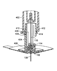
CA 3066834 2020-01-07
20
the introducer needle 110 in a single motion and provides the user with a
mechanism to
protect from an accidental needle stick.
[0059] Fig. 41 illustrates an alternative embodiment of the spring clip 216
having a
transverse portion 230. The transverse portion 230 reduces the elasticity of
the spring clip
216, thereby increasing the bias strength of the spring clip 216.
[0060] Fig. 42 is a cross-sectional view of another embodiment of a needle
shield device
232 fully engaged with the fluid connector 126 and the base 102, piercing the
septum 124
and catheter with the introducer needle 110 and ready for placement on the
skin. Fig. 43 is
another cross-sectional view of the needle shield device 232 taken at 90
degrees with
respect to Fig. 42. The needle shield device 232 includes a cavity 234 over
the fluid
connector 126 for housing a spring element or biased spring 236. In the
example of Figs.
42 and 43, the spring 236 is biased in contact with the fluid connector 126
and a surface
238 at the proximal end of the cavity 234.
[0061] Fig. 44 is a cross-sectional view of the spring 232 showing that it
includes slotted
holes 240 for the introducer needle 110 to pass through. The slotted holes 240
are axially
aligned with the septum 124. The spring 236 also includes a small hole 242 and
a tip
pocket 244 at the distal end of the spring 236, which is in contact with the
fluid connector
126.
[0062] Figs. 45 and 46 illustrate the removal of the needle shield device 232.
In Fig. 45,
when the needle shield device 232 is retracted to withdraw the introducer
needle 110 from
the fluid connector 126 and the base 102, the biased spring 236 unbiases and
expands
along the axis of the introducer needle 110. According to one embodiment, the
slotted
holes 240 are longitudinal slots aligned to accommodate the introducer needle
110 as the
spring 236 expands.
[0063] Fig. 46 illustrates that the needle shield device 232 is fully
retracted and the spring
236 extends over the tip of the introducer needle 110, so that the tip is
disposed in the tip
pocket 244. Further, according to one embodiment, the small hole 242 is biased
to one side
of the spring 236 so that when the tip of the introducer needle 110 passes
through the hole
242, the spring 244 transversely unbiases so that the hole 242 is not axially
aligned with
CA 3066834 2020-01-07
21
the introducer needle 110. That is, a barrier is formed over the tip of the
introducer needle
110 when the spring 236 unbiases and substantially covers the introducer
needle 110.
[0064] According to one embodiment, the spring 236 can be a compression spring
(for
example, a spiral or coil spring) or a tapered compression spring to provide
the force
necessary to shield the tip of the introducer needle 110. In such an
embodiment, a tip shield
can be disposed at the distal end of the spring to cover the tip of the
introducer needle 110
after expansion of the spring.
[0065] The needle shield device 232 automatically releases the transverse
barrier formed
by the tip pocket 244 and hole 242 during retraction of the introducer needle
110 in a
single motion and provides the user with a mechanism to protect the user from
an
accidental needle stick.
[0066] Figs. 47-52 illustrate another exemplary embodiment of a needle shield
device 246.
Fig. 47 is a perspective view of the needle shield device 246 with a shield
248 that is
disposed over the extension tubing 134 prior to insertion. The shield 248
includes a hinge
249 that may be formed by molding the needle shield device 246 as a single
piece. In an
alternative exemplary embodiment, the hinge 249 may be molded separately and
fixed to
the needle shield device 246. The shield 248 generally has a U-shaped profile
and is
deployed after removal from the fluid connector 126 and/or base 102 by
rotating the shield
248 about the hinge 249. Figs. 48 and 49 illustrate the shield 248 after
deployment. The
shield 248 includes latches 250 on an inner surface to keep the shield 248 in
a substantially
fixed position after deployment.
[0067] Fig. 50 is a cross-sectional view of the needle shield device 246 and
illustrates that
the shield 248 has an inner cavity 252 that is press fit and latches with a
base 254 of the
needle shield device 246. According to one embodiment, the shield 248 latches
to the base
254 without contacting or bending the needle 110. Fig. 51 is a cross-sectional
view of the
needle shield device 246 after deployment, and illustrates that the distal end
of the shield
248 extends beyond the tip of the introducer needle 110. Fig. 52 is another
cross-sectional
view at 90 degrees with respect to Fig. 51 and illustrates that a gap 256 of
the shield 248 is
smaller than the average finger, to prevent an accidental needle stick.
CA 3066834 2020-01-07
22
[0068] Conventional one-piece protective shields generally have more than a 90-
degree
rotation and may require the user to bend or break the needle. This requires
extra force
and/or extra manufacturing (e.g., a notch in the introducer needle to
facilitate bending or
breaking). Such conventional designs also result in a wider needle holder that
is physically
larger and more difficult to dispose of. The above-described exemplary
embodiment is
easier to manufacture by reducing manufacturing steps and also is easier to
operate because
the integrity of the introducer needle is maintained. Further, the above-
described
exemplary embodiment is physically smaller than conventional shields and is
therefore
easier to dispose of.
[0069] Figs. 53 and 54 are perspective views of another embodiment of a needle
shield
device 74. The introducer needle 110 is omitted from Fig. 53 for clarity. The
needle shield
device 74 includes a needle hub 76 and a shield 78 rotatably connected to the
needle hub
76 by a hinge 82, such as a living hinge. The shield 78 includes one or more
cantilevered
needle-engaging flaps 80. When the user rotates the shield 78 to enclose the
introducer
needle 110, as shown in Fig. 54, the flaps 80 are disposed to permit the flaps
80 to pass the
introducer needle 110 in one direction, but prevent the shield from passing
the introducer
needle in the opposite direction thereafter. In other words, once the shield
78 is deployed to
cover the shield, the flaps engage the introducer needle 110 to prevent the
shield 78 from
returning to its initial position.
[0070] Figs. 55-59 depict a locking fluid connector 258 that a user may place
in, for
example, six different rotational positions. Fig. 55 is a cross-sectional view
of the locking
fluid connector 258 and includes a base 260 that the locking fluid connector
258 locks
onto. The fluid connector 258 includes a tubing portion 259 with a tubing port
for
connecting tubing thereto, and a hub portion 261 for connection with the base
260. The hub
portion 261 has a domed portion 263. Fig. 56 is a cross-sectional view of the
locking fluid
connector 258 and includes engagement fingers 262 protruding radially inward
at the distal
opening of the domed portion. The molded cannula 140 extends from the domed
portion
and is in fluid communication with the tubing port. The engagement fingers 262
fix a
tensile element such as, for example, a spring 264 within the locking fluid
connector 258.
The spring 264 in Fig. 56 may be biased or, alternatively, may be unbiased
when the
locking fluid connector 258 is not connected to the base 260. Fig. 57 is a
bottom
CA 3066834 2020-01-07
23
perspective view of the locking fluid connector 258 that illustrates that the
spring 264 and
the fingers are separate.
[0071] Figs. 58-60 illustrate an alternative exemplary embodiment of the
locking fluid
connector 258. Fig. 58 is a cross-sectional view of the locking fluid
connector 258 and
illustrates that a connector 266 is disposed in the locking fluid connector
258. Fig. 59
illustrates that the connector 266 has fingers 268 and a leaf spring 270
integral therewith,
in other words. In other words, in the embodiment of Fig. 59, the fingers and
spring are
integrally formed as one body. Fig. 60 is a bottom view of the locking fluid
connector 258
with the connector 266 placed therein and illustrates that the leaf springs
270 are not biased
when the locking fluid connector 258 is not connected to the base 260.
[0072] Fig. 61 depicts the base 260 in more detail. The base 260 has a column
that
includes inverted J-shaped engagement structures or protrusions 273 having
cantilevered
ends and tapered edges that protrude in the radial direction from the column.
The
protrusions 273 include a notch to form engagement pockets 272 and are
separated by slots
274. In the exemplary embodiment, the engagement pockets 272 and the slots 274
are
configured to receive the engagement fingers of the locking fluid hub
retention set 258 and
lock it into a predetermined number of discrete rotational orientations (e.g.,
6, 8, etc.).
[0073] Fig. 62 illustrates that, in the event that the engagement fingers 262
of the locking
fluid connector 258 are aligned and placed into the slots 274, the spring 264
is in contact
with a tapered top surface 276 of the base 260. A user then applies a force to
bias the
spring 264, causing the engagement fingers 262 to travel into the slots 274.
The user then
rotates locking fluid connector 258 and releases it, thereby allowing the
spring 264 to
partially unbias and move the engagement fingers 262 to move into contact with
the base
260 via the engagement pockets 272. In this configuration, the locking fluid
connector 258
is locked into base 260 via the engagement pockets 272 and can only be removed
by
pressing on the locking fluid connector 258, rotating it to align the
engagement fingers 262
with the slots 274, and releasing the locking fluid connector 258.
[0074] The exemplary embodiment provides a locking retention fluid set 268
that can be
easily removed and provides a fixed number of rotational positions to allow
the user to
select the best position of the extension tubing 134. The base 260 may also be
made of a
rigid or flexible material via a die-cut process, molding process, or a two-
step molding
CA 3066834 2020-01-07
24
process. Various shapes of shrouds may also be used to contour and minimize
the potential
to snag on objects (e.g., clothing, furniture, etc.) and optimized for ease of
handling and
intuitive use.
[0075] Fig. 63 illustrates another exemplary embodiment of a locking fluid
connector 278
having levers 280 with a latch 282 integral thereto. Fig. 64 illustrates the
locking fluid
connector 278 fully engaged with the base 102, and ready for placement on the
skin. The
locking fluid connector 278 is locked into a substantially fixed position via
a
predetermined number of catches 284 disposed on the surface of the base 102.
Fig. 65
illustrates a cross-sectional view of the locking fluid connector 278 and
depicts one
example of a latch 282 engaged with a catch 284.
[0076] Fig. 66 illustrates a base 102 having six different catches 284 to
allow a user to
place the locking fluid connector 278 in six different rotational positions.
To remove the
locking fluid connector 278, the user squeezes the levers 280 together to
cause the latches
282 to disengage from the catches 284. The user then rotates the locking fluid
connector
278 such that the latches 282 are not aligned with the catches 284 and
releases the levers
280 and lifts off the locking fluid connector 278. In an alternative
embodiment, the levers
280 can displace sufficiently to disengage the latches 282 from the catches
284 to permit
the user to simply lift the fluid connector 278 from the base 102 without
rotation.
[0077] . The levers 280 are sufficiently displaceable that, if correctly
aligned, the fluid
connector 278 can be axially lowered onto the base 102 and the latches 282 can
snap into
engagement with the catches. As shown in Fig. 67 the blunt cannula 140 must be
axially
aligned with the septum 124 so that the latches 282 can be placed into the
catch 284 to
place the locking fluid connector 278 into a fixed position.
[0078] In an alternative embodiment, to lock the locking fluid connector 278,
the user
places the latches 282 such that they are not aligned with the catches 284,
and then rotates
the locking fluid connector 278 until at least one of the latches 282 engage
with at least one
of the catches 284
[0079] The squeeze action of the locking fluid connector 278 is more user
friendly and
intuitive compared to depressing a single button. Further, the release
mechanism is more
reliable because it requires fewer assembly tolerances.
CA 3066834 2020-01-07
25
[0080] Fig. 68 is a cross-sectional view of exemplary embodiment of a fluid
connector 286
fully engaged with the base 102, piercing the septum 124 and catheter 108 with
the
introducer needle 110 and ready for placement on the skin. Fig. 69 depicts the
bottom of
the fluid connector 286 and Fig. 70 illustrates a base latch 288 that the
fluid connector 286
connects to. The fluid connector 286 includes at least one snap latch 290
having an angular
profile to snap over the base latch 288. For example, Fig. 70 illustrates a
spherically shaped
post or ball 289 on the base latch 288 to receive the snap latches 290. The
snap latches 290
are configured to unlatch from the ball 289 in the event that the fluid
connector 286
experiences undesired force that would normally remove the catheter from the
user. That
is, the fluid connector 286 is designed to separate from the base 102 to
prevent inadvertent
removal of the catheter from the user's body in the event that, for example,
the extension
tubing 134 is accidentally pulled by an external object (e.g., a doorknob,
furniture, etc.).
[0081] Fig. 71 illustrates the fluid connector 286 connected to the base latch
288 and is
able to rotate 360 degrees around the base latch 288, which may be partially
exposed
between the snap latches 290. The fluid connector 286 includes a sheath 292
that receives
the extension tubing 134 and has a sheath base 294 that extends along the
sheath 292. The
sheath base 294 protrudes from the bottom surface of the sheath 292 toward the
base 102.
In the embodiment of Fig. 71, the sheath 292 and the sheath base 294 generally
do not
extend beyond the radius of the base 102. The sheath base 294 provides support
to prevent
the fluid connector 286 from unlatching in the event an undesired force is
asserted on the
sheath 292 or extension tubing 134.
[0082] Fig. 72 is a cross-sectional view that illustrates another exemplary
embodiment of
the fluid connector 286 having a shroud 296 with a hemispherical dome shape
that extends
over and substantially encloses the snap fingers 290. In Fig. 72, the radius
of the shroud
296 is less than the radius of the base 102. Fig. 73 illustrates an exemplary
embodiment of
a tapered shroud 298 that tapers the edges to be substantially planar with the
surface of the
base 102. The purpose of the shroud 296 is to prevent the user from squeezing
the snap
latches 290 during removal, which would make it difficult to remove the fluid
connector
286 from the base 102. Also, the low profile of the shroud 296 minimizes the
potential for
unintentionally snagging the fluid connector 286 on clothing or objects.
CA 3066834 2020-01-07
26
[0083] Fig. 74 illustrates another exemplary embodiment of a tapered shroud
298 with
tapered edges that are substantially planar with the surface of the base 102.
The tapered
shroud 298 impedes accidental disengagement of fluid connector 286 from the
user. In
another exemplary embodiment of the fluid connector 286 illustrated in Fig.
73, the radius
of the tapered shroud 298 is substantially equal to the radius of the base
102. In other
embodiments, the shape of the tapered shroud 298 may be modified in any
suitable manner
for aesthetic purposes or to enhance the user's grip on the fluid connector
286.
[0084] The exemplary embodiment provides a fluid connector that separates from
the
user's body prior to removal of the catheter in the event that the fluid
connector
experiences an undesired external force. Further, the above-described example
provides a
shroud with a tapered edge that is substantially planar with the user's skin
to facilitate
engagement and impede accidental disengagement of the fluid retention set. The
exemplary embodiment also includes a sheath with a base to prevent inadvertent
removal
of the fluid connector from the base 102 due to an undesired force experienced
by the
extension tubing.
[0085] Fig. 75 illustrates another exemplary embodiment of a fluid connector
300 having a
septum 302 disposed therein to allow priming of an introducer needle (not
shown) disposed
in the fluid connector 300 before insertion. The septum 302 is cylindrical in
shape and is
placed in the axial direction of the extension set tubing 134 such that a
fluid path 304 is
formed below the septum 302. In this exemplary embodiment, the introducer
needle is
configured to receive the fluid so that the fluid connector 300 may be primed
prior to
insertion of the introducer needle.
[0086] Alternatively, the septum 302 can be the tubing 134, so long as the
tubing material
possesses sufficient healing (sealing) properties to prevent leakage due to
the slit caused by
the introducer needle 110, while still having suitable material properties to
perform the
other functions required of the tubing 134.
[0087] Fig. 76 illustrates a passive needle shield device 400 connected to the
base 102 and
ready for placement on the skin. Fig. 77 is a cross-sectional view of the
needle shield
device 400 fully engaged with the base 102, piercing the septum 124 and the
catheter 108
with the introducer needle 110. The needle shield device 400 includes a shield
needle hub
CA 3066834 2020-01-07
27
or outer shield 402 which surrounds and encloses an inner shield 404 and the
introducer
needle 110.
[0088] Figs. 78-81 illustrate the sequence of steps that occur after the user
has inserted the
catheter 108. In other words, these figures illustrate the operation of
removing the needle
shield device 400 from the base 102. Briefly, the user simply pulls on the
outer shield 402
in a direction away from the base 102 to remove the introducer needle 110.
According to
one embodiment, the outer shield 402 and inner shield 404 are both made of
rigid plastic
materials that have some degree of flexibility.
[0089] In more detail, Fig. 78 is a quarter-sectional view illustrating an
initial state of the
needle shield device 400 and a first position of the outer shield 402 relative
to the inner
shield 404, in which an outer shield hub latch 406 contacts the base 102 and
also contacts a
cantilevered latch beam 408 of the inner shield 400 to maintain engagement of
the latch
beam 408 with the base 102 beneath the base latch 114. According to one
embodiment, the
hub latch 406 biases the latch beam 408 radially inward.
[0090] Fig. 79 illustrates the orientation of the needle shield device 400
while the user is
axially displacing the outer shield 402, but before it has completed its
stroke relative to the
inner shield 404. In this state, the outer shield 402 continues to prevent the
latch beam 408
from disengaging from the base 102. More specifically it is the hub latch 406
that holds the
latch beam 408 in place against the base 102. Therefore, according to one
embodiment, the
inner shield 404 is locked onto the base 102 while the outer shield 402 is
being axially
displaced relative to the inner shield 404.
[0091] Fig. 80 illustrates the completely displaced position of the outer
shield 402 with
respect to the inner shield 404. In this state, the hub latch 406 no longer
prevents the latch
beam 408 from disengaging from the base 102. The hub latch 406 is instead
disposed in an
indent 409 (best shown in Fig. 77) on the inner shield 404 and engaged with a
shield latch
410 formed on the inner shield 404. The shield latch 410 engages a top side of
the hub
latch 406, thereby preventing further proximal displacement of the outer
shield 402 relative
to the inner shield 404. Additionally, because the hub latch 406 is no longer
pressing on the
latch beam 408, the latch beam 408 can disengage from the base 102.
CA 3066834 2020-01-07
28
[0092] Further, a hub beam or outer shield latch 412 rides over an inner
shield latch 414
and the bottom of the hub beam 412 engages the top of the inner shield latch
414 to
prevent distal displacement of the outer shield 402 relative to the inner
shield 404.
According to one embodiment, the hub beam 412 is cantilevered.
[0093] The latch beam 408 is free to radially displace and disengage from the
base 102
once the user continues to distally displace the needle shield device 400. The
engagement
of the shield latch 410 with the hub latch 406 and the engagement of the hub
beam 412
with the inner shield latch 414 shields the introducer needle 110 and thereby
reduces the
possibility of an accidental needle stick.
[0094] According to one embodiment, the inner shield latch 414 is fixedly
disposed on the
inner shield 404. According to another embodiment, the inner shield latch 414
is disposed
on a cantilevered inner shield latch beam 416 so that both the inner shield
latch beam 416
and the hub beam 412 are cantilevered. According to yet another embodiment,
the inner
shield latch 414 is disposed on a cantilevered inner shield latch beam 416 and
the hub
beam is fixedly disposed on the outer shield 402.
[0095] In another alternative embodiment, the needle shield device 400 can
also be
attached to a fluid connector 126 and the base 102. Such an embodiment allows
a user to
prime the infusion set while it is outside the body and insert and remove the
introducer
needle 110 with the fluid connector 126 attached the entire time.
[0096] Fig. 81 illustrates a completely deployed needle shield device 400. The
latch beam
408 is removed from the base 102 as the user continues to pull on the outer
shield 402. Fig.
82 is a perspective view illustrating the needle shield 400 in the completely
deployed state,
removed from the base 102 and ready for disposal.
[0097] Fig. 83 illustrates how the introducer needle 110 within the needle
shield device
400 is prevented from contacting a user's finger 173 during handling of the
completely
deployed needle shield device 400. The inner shield 404 forms a shield well
414
surrounding the exposed introducer needle 110 such that an average finger 173
will not fit
through the shield well 414 opening and contact the exposed introducer needle
110.
[0098] In each of the herein disclosed embodiments and in other alternative
embodiments,
the components of the infusion set can be made of injection-molded
polypropylene,
CA 3066834 2020-01-07
29
polyethylene, polyesters, acrylonitrile-butadiene-styrene polymers, and/or bio-
based resins
such as polylactide, starch-filled polypropylene, or polyhydroxyalkanoates.
The catheter
can be a separate component or injection-molded as part of the base assembly,
either as a
single part or as a coinjection-molded part using two resins.
[0099] Although only a few exemplary embodiments of the present invention have
been
described in detail above, those skilled in the art will readily appreciate
that many
modifications are possible in the exemplary embodiments without materially
departing
from the novel teachings and advantages of this invention. Accordingly, all
such
modifications are intended to be included within the scope of this invention
as defined in
the appended claims and their equivalents.
CA 3066834 2020-01-07