Note: Descriptions are shown in the official language in which they were submitted.
CA 03067062 2019-12-12
WO 2018/227295
PCT/CA2018/050716
MARKERS FOR DISEASE AND DISEASE EXTENT IN INFLAMMATORY BOWEL
DISEASE
[001] The present application claims priority of U.S. provisional patent
application No.
62/520,652 filed on June 16, 2017.
Technical Field
[002] The present application relates to protein markers for inflammatory
bowel
disease (IBD), including namely ulcerative colitis (UC) and Crohn's disease
(CD).
Background
[003] Inflammatory Bowel Disease (IBD) encompasses two principal
conditions:
ulcerative colitis (UC) and Crohn's disease (CD). Some patients have features
of both
subtypes and are classified as IBD-unclassified (I BD-U) (Gastroenterology,
2007. 133(5):
p. 1670-89). UC is defined by continuous mucosal inflammation starting in the
rectum and
restricted to the colon while CD inflammation can occur anywhere in the
gastrointestinal
tract, involves full thickness of the bowel wall and often with skip lesions
(Gastroenterol
Clin North Am, 2009. 38(4): p. 611-28; Gastroenterology, 2007. 133(5): p. 1670-
89).
[004] One of the primary tools used for both diagnosis and IBD management
is
endoscopy (World J Gastrointest Endosc, 2012. 4(6): p. 201-11). Endoscopy
enables
both visualization of the mucosa and access for mucosal biopsies to diagnose
disease,
to define disease extent and activity, and to monitor disease progression. The
diagnostic
accuracy from colonoscopy ranges from 60 to 74% (J Clin Pathol, 2002. 55: p.
955-60).
Other diagnostic approaches include radiological imaging and histological
examination of
mucosal biopsies in the differentiation of IBD subtypes (e.g non-caseating
submucosal
granuloma). However, 10% of patients (Registry. Dtsch Arztebl Int 2015;112:121-
7) have
ambiguous diagnosis using these approaches and are instead classified as IBD-
unclassified (IBD-U) patients (J Pediatr Gastroenterol Nutr 2014;58:795-806).
Accurate
1
CA 03067062 2019-12-12
WO 2018/227295
PCT/CA2018/050716
and early diagnosis is essential for proper disease management. The goal of
IBD
treatment is to bring active disease into remission, prevent follow-up relapse
(flare-ups)
and prevent complications of disease. The choice of treatment depends on
disease
subtype (CD versus UC), disease location, severity of disease, disease
complications and
individual host factors (e.g. nutritional and growth status, pubertal status,
child's age and
size, medication allergies, co-morbid conditions) (J Pediatr Gastroenterol
Nutr, 2010, S1-
S13. The American Journal of Gastroenterology, 2011. 106 Suppl 1: p. S2-25;
quiz S26.
Gastroenterol Clin North Am, 2009. 38(4): p. 611-28). Current drug therapies
consist of
aminosalicylates, immune-modulators, corticosteroids, antibiotics and
biological
therapies (e.g. anti-TNFa monoclonal antibodies). One third of the cost
associated with
IBD is due to medical therapies (CCFC. 2008, report. p. 1-101) stressing the
economic
importance of an effective treatment and thereby an accurate diagnosis.
[005] Genome wide association studies in both adults and pediatric patients
have
identified novel IBD-associated genes but only define 25% of the genetic risk
for
developing IBD and excepting for very young infants (i.e. <2 years of age), no
unique
genes have been discovered that define pediatric IBD from adult-onset IBD. IBD
is a
complex polygenic disease involving multiple risk gene loci (Nature genetics,
2008. 40(8):
p. 955-62. Nature genetics, 2009. 41(12): p. 1335-40. Nature genetics, 2010.
42(4): p.
332-7). These loci encode genes involved in innate and adaptive immunity,
autophagy,
and maintenance of epithelial barrier integrity for those genes that have
known function.
[006] On average, children will suffer with IBD for at least four months
before a
diagnosis of IBD is established but oftentimes much longer, at which point the
disease
has often progressed to a more severe state ( Dtsch Arztebl Int, 2015. 112(8):
p. 121-7).
The most frequently assayed biomarker used to distinguish IBD from non-
inflammatory
disorders or from extra-intestinal inflammatory disorders is fecal
calprotectin, which
outperforms blood markers (Erythrocyte Sedimentation Rate and C - reactive
protein) in
its ability to indicate intestinal inflammation3. While limited utility has
been shown within
2
CA 03067062 2019-12-12
WO 2018/227295
PCT/CA2018/050716
the adult population, the diagnostic accuracy of calprotectin is inferior for
pediatric
patients 4 where the specificity reaches only 0.682 in the context of
suspected pediatric
IBD5. The low specificity observed for IBD is due to similarly elevated levels
of calprotectin
measured in stool from children suffering from disorders including celiac
disease, cystic
fibrosis, infection, neoplasia and p01yp56, allergic diseases7, 8, and even in
apparently
healthy children9. Fecal calprotectin has also recently been implemented for
the diagnosis
of several allergic diseases in children, highlighting its lack of specificity
in IBD diagnosis.
In addition, the level of fecal calprotectin is influenced by age, with the
highest levels
observed in children under the age of four. Therefore, an elevated (positive)
fecal
.. calprotectin result necessitates further testing for suspected IBD cases,
including
endoscopy. Non-invasive biomarkers with the ability to lower the false
positive rate
associated with fecal calprotectin would be beneficial in order to reduce the
number of
unnecessary invasive colonoscopies, and thus avoid the risk, discomfort and
economic
burden associated with endoscopy.
[007] Once an IBD diagnosis has been established, several other aspects of
the
disease must be assessed in order to select an appropriate therapeutic
strategy, including
disease severity and extent of disease (UC)19, 11. To date, biomarkers able to
determine
extent of disease have not been implemented in the clinic. Instead, this
aspect of
diagnosis is achieved by endoscopy and imaging. UC is characterized by
continuous
mucosal inflammation limited to the colon, extending proximally from the
rectum. The
extent of disease in UC is defined as the macroscopic degree of inflammation
in the colon,
as assessed by colonoscopy; disease extent may partially dictate the method
(oral or
rectal) and type of treatment administered, and the recommended time to begin
for
monitoring for colorectal cancer (i.e. 8 to 10 years post-diagnosis of
pancolitis).
[008] In light of the above, there is a need for improved diagnostic
methods for IBD,
especially in children, to decrease the number of unnecessary invasive
endoscopies
performed for IBD diagnosis. There is also a need to assess the extent of
disease in UC.
3
CA 03067062 2019-12-12
WO 2018/227295
PCT/CA2018/050716
Summary
[009] Applicant has discovered that proteins leukotriene A-4 hydrolase,
catalase,
transketolase and annexin A3 show increased expression in gut samples obtained
from
subjects with IBD when compared to corresponding protein expression levels in
gut
samples obtained from subjects without IBD. Therefore, applicant has
discovered that
these proteins may be used as reliable biomarkers to indicate if a patient has
or does not
have inflammatory bowel disease. These biomarkers may be used for disease
diagnosis,
treatment strategy and treatment responsiveness. Disease detection analysis by
measuring the relative expression of these biomarkers may be performed in gut
samples
such as, for example, mucosal luminal interface samples, and stool samples.
[0010] Moreover, Applicant has discovered that proteins leukotriene A-4
hydrolase,
thioredoxin domain containing protein 17, vasodilator-stimulated
phosphoprotein, and
thymosin beta-10 may be used as biomarkers to determine if patients with
ulcerative
colitis show a presence or an absence of pancolitis. These biomarkers may be
used for
disease diagnosis, determine the severity of the disease, determine the extent
of disease,
treatment strategy, treatment responsiveness, determining disease remission
and
determining disease relapse. Disease detection analysis by measuring the
relative
expression of these biomarkers may be performed in gut samples such as, for
example,
mucosal luminal interface samples, and stool samples.
[0011] Moreover, applicant has also discovered that thioredoxin domain
containing
protein 17, vasodilator-stimulated phosphoprotein, and thymosin beta-10 may
also be
used as biomarkers to detect IBD in a subject. These biomarkers may be
measured in
gut samples, such as mucosal lumina! interface (MLI) samples and/or stool
samples. One
or more of these biomarkers (thioredoxin domain containing protein 17,
vasodilator-
stimulated phosphoprotein, and thymosin beta-10) may also be used in
combination with
one or more of leukotriene A-4 hydrolase, catalase, transketolase and annexin
A3 to
4
CA 03067062 2019-12-12
WO 2018/227295
PCT/CA2018/050716
detect IBD, wherein the sensitivity and specificity may increase when more
than one
biomarker is used (up to 4 biomarkers may be used to reach optimal specificity
and
sensitivity).
[0012]
A first broad aspect is a method for determining a presence of
inflammatory
bowel disease in a subject. The method involves providing a gut sample
obtained from a
subject. The method also involves measuring a level in the gut sample of one
or more
proteins, wherein the one or more proteins comprises at least one of:
leukotriene A-4
hydrolase, catalase, transketolase, thioredoxin domain containing protein 17,
vasodilator-
stimulated phosphoprotein and thymosin beta-10. The method includes comparing
the
measured level to a predetermined level to provide an indication of presence
of disease.
[0013]
In some embodiments, the determining may be used to obtain an indication
on
remission of the disease.
[0014]
In some embodiments, the determining may be used to obtain an indication
of
relapse of the disease.
[0015]
In some embodiments, the one or more proteins may be leukotriene A-4
hydrolase and a measured level in the gut sample of leukotriene A-4 hydrolase
higher
than a predetermined protein level of leukotriene A-4 hydrolase corresponding
to a
healthy subject may be indicative of disease. The one or more proteins may be
catalase
and a measured level in the gut sample of catalase higher than a predetermined
protein
level of catalase corresponding to a healthy subject may be indicative of
disease. The
one or more proteins may be transketolase and wherein a measured level in the
gut
sample of transketolase higher than a predetermined protein level of
transketolase
corresponding to a healthy subject may be indicative of disease. The one or
more proteins
may be annexin A3 and wherein a measured level in the gut sample of annexin A3
higher
than a predetermined protein level of annexin A3 corresponding to a subject
patient may
be indicative of disease.
5
CA 03067062 2019-12-12
WO 2018/227295
PCT/CA2018/050716
[0016] In some embodiments, the measuring may be measuring each of
leukotriene
A-4 hydrolase, catalase, transketolase and annexin A3. In some embodiments,
the
measuring may be measuring one or more selected proteins, wherein said one or
more
selected proteins may be at least one of leukotriene A-4 hydrolase, catalase,
transketolase and annexin A3.
[0017] In some embodiments, the measuring may be measuring a level in
the gut
sample of two or more proteins, wherein the two or more proteins may be at
least two of:
leukotriene A-4 hydrolase, catalase, transketolase, annexin A3, thioredoxin
domain
containing protein 17, vasodilator-stimulated phosphoprotein and thymosin beta-
10. The
measuring may be measuring a level in the gut sample of three or more
proteins, wherein
the three or more proteins may be at least three of: leukotriene A-4
hydrolase, catalase,
transketolase, annexin A3, thioredoxin domain containing protein 17,
vasodilator-
stimulated phosphoprotein and thymosin beta-10. The measuring may be measuring
a
level in the gut sample of four proteins, or four or more proteins, wherein
the four proteins
are selected from: leukotriene A-4 hydrolase, catalase, transketolase, annexin
A3,
thioredoxin domain containing protein 17, vasodilator-stimulated
phosphoprotein and
thymosin beta-10. In some embodiments, the measuring may be measuring a level
in the
gut sample of two or more proteins, wherein the two or more proteins may be at
least two
of: leukotriene A-4 hydrolase, catalase, transketolase and annexin A3. The
measuring
may be measuring a level in the gut sample of three or more proteins, wherein
the three
or more proteins may be at least three of: leukotriene A-4 hydrolase,
catalase,
transketolase and annexin A3.
[0018] In some embodiments, the measuring may include using an
immunoassay. The
immunoassay may be ELISA. In some embodiments, the measuring may be using semi-
quantitative immunoblotting. The measuring may include using mass
spectrometry.
[0019] In some embodiments, the gut sample may be a mucosal luminal
interface
sample. The gut sample may be a stool sample.
6
CA 03067062 2019-12-12
WO 2018/227295
PCT/CA2018/050716
The subject may be a pediatric subject. The subject may be an adult subject.
[0020] A second broad aspect is a method of treating inflammatory bowel
disease in
a subject involving determining whether the subject has inflammatory bowel
disease
according to the method for determining a presence of inflammatory bowel
disease in a
subject as described herein, and administrating to the patient a compound
pharmaceutically effective against the inflammatory bowel disease. In some
embodiments, the administering may involve administering a pharmaceutically
effective
amount of a compound selected from aminosalicylates, immunomodulators, anti-
integrins, anti-cytokines, enteral feed programs, corticosteroids,
antibiotics, monoclonal
antibodies (e.g. anti-TNFa, anti-1L12/23, anti-integrin), or a combination
thereof.
[0021] In some embodiments, where the subject was determined to have, at
a time
prior to obtaining the gut sample, inflammatory bowel disease, the measuring
to provide
an indication of presence of disease may be to further determine if the
disease is in
remission or if remission is maintained.
[0022] In some embodiments, the measuring may be to further determine if
relapse of
the disease has occurred.
[0023] A third broad aspect is a method for determining presence or an
indication of
pancolitis in a subject with ulcerative colitis. The method involves providing
a gut sample
obtained from a subject with ulcerative colitis. The method entails measuring
in the gut
sample one or more proteins, wherein the one or more proteins comprises at
least one
of: leukotriene A-4 hydrolase, thioredoxin domain containing protein 17,
vasodilator-
stimulated phosphoprotein, and thymosin beta-10. The method includes comparing
the
measured level to a predetermined protein level to provide an indication of
the presence
or absence of pancolitis.
[0024] In some embodiments, the one or more proteins may be leukotriene A-4
hydrolase and a measured level in the gut sample of leukotriene A-4 hydrolase
higher
than a predetermined protein level of leukotriene A-4 hydrolase corresponding
to a
7
CA 03067062 2019-12-12
WO 2018/227295
PCT/CA2018/050716
subject without pancolitis may be indicative of pancolitis; the one or more
proteins may
be thioredoxin domain containing protein 17 and a measured level in the gut
sample of
thioredoxin domain containing protein 17 higher than a predetermined protein
level of
thioredoxin domain containing protein 17 corresponding to a subject without
pancolitis
may be indicative of pancolitis; the one or more proteins may be vasodilator-
stimulated
phosphoprotein and wherein a measured level in the gut sample of vasodilator-
stimulated
phosphoprotein higher than a predetermined protein level of vasodilator-
stimulated
phosphoprotein corresponding to a subject without pancolitis may be indicative
of
pancolitis; and/or the one or more proteins may be thymosin beta-10 and a
measured
level in the gut sample of thymosin beta-10 lower than a predetermined protein
level of
thymosin beta-10 corresponding to a subject without pancolitis may be
indicative of
pancolitis.
[0025] In some embodiments, the one or more proteins may be leukotriene
A-4
hydrolase and wherein a measured level in the gut sample of leukotriene A-4
hydrolase
higher than a predetermined protein level of leukotriene A-4 hydrolase
corresponding to
a subject without pancolitis may be indicative of pancolitis. The one or more
proteins may
be thioredoxin domain containing protein 17 and wherein a measured level in
the gut
sample of thioredoxin domain containing protein 17 higher than a predetermined
protein
level of thioredoxin domain containing protein 17 corresponding to a subject
without
pancolitis may be indicative of pancolitis. The one or more proteins may be
vasodilator-
stimulated phosphoprotein and a measured level in the gut sample of
vasodilator-
stimulated phosphoprotein higher than a predetermined protein level of
vasodilator-
stimulated phosphoprotein corresponding to a subject without pancolitis may be
indicative
of pancolitis. The one or more proteins may be thymosin beta-10 and a measured
level
in the gut sample of thymosin beta-10 lower than a predetermined protein level
of
thymosin beta-10 corresponding to a subject without pancolitis may be
indicative of
pancolitis.
8
CA 03067062 2019-12-12
WO 2018/227295
PCT/CA2018/050716
[0026] In some embodiments, the measuring may involve measuring each of
leukotriene A-4 hydrolase, thioredoxin domain containing protein 17,
vasodilator-
stimulated phosphoprotein, and thymosin beta-10. The measuring may involve
measuring a level in the gut sample of two or more proteins, wherein the two
or more
proteins may be at least two of: leukotriene A-4 hydrolase, thioredoxin domain
containing
protein 17, vasodilator-stimulated phosphoprotein, and thymosin beta-10. The
measuring
may involve measuring a level in the gut sample of three or more proteins,
wherein the
three or more proteins may be at least three of: leukotriene A-4 hydrolase,
thioredoxin
domain containing protein 17, vasodilator-stimulated phosphoprotein, and
thymosin beta-
10.
[0027] In some embodiments, the gut sample may be a mucosal luminal
interface
sample. The gut sample may be a stool sample. The subject may be a pediatric
subject.
The subject may be an adult subject.
[0028] In some embodiments, the measuring may involve using an
immunoassay. The
immunoassay may be ELISA. The measuring may involve using semi-quantitative
immunoblotting. The measuring may involve using mass spectrometry.
[0029] A fourth broad aspect is a method of treating ulcerative colitis
in a subject
involving determining whether the ulcerative colitis subject has pancolitis or
does not have
pancolitis according to the method for determining a presence or indication of
pancolitis
in a subject with ulcerative colitis as described herein, and administrating
to the patient a
compound pharmaceutically effective against: pancolitis; or ulcerative colitis
without
pancolitis, the administration tailored in accordance with the determined
presence or
absence of pancolitis. The administering may involve administering a
pharmaceutically
effective amount of a compound selected from aminosalicylates,
immunomodulators,
anti-cytokines, enteral feed programs, corticosteroids, anti-integrins,
antibiotics,
monoclonal antibodies (e.g. anti-TNFa, anti-1L12/23, anti-integrin), or a
combination
thereof.
9
CA 03067062 2019-12-12
WO 2018/227295
PCT/CA2018/050716
[0030] A fifth broad aspect is a method for determining the efficacy of
a treatment of
inflammatory bowel disease in a patient suffering from the disease, the
treatment
comprising the administration of aminosalicylates, immunomodulators, anti-
cytokines,
enteral feed programs, corticosteroids, anti-integrins, antibiotics,
monoclonal antibodies
(e.g. anti-TNFa, anti-1L12123, anti-integrin), or a combination thereof. The
method
involves measuring a level in an indicative gut sample, obtained from a
patient, of one or
more proteins, wherein the one or more proteins comprises at least one of:
leukotriene
A-4 hydrolase, catalase, transketolase, thioredoxin domain containing protein
17,
vasodilator-stimulated phosphoprotein and thymosin beta-10. In some
embodiments, two
or more of the proteins, three or more of the proteins, or four or more of the
proteins may
be measured. In some embodiments, Annexin A3 may be measured. The method
involves comparing the measured level to a corresponding protein level
measured in a
reference gut sample taken from the patient at a time prior to when the
indicative gut
sample was obtained; a predetermined protein level; a corresponding protein
level
associated with responders; and/or a corresponding protein level associated
with non-
responders. The method also involves assessing responsiveness to treatment as
a
function of the comparison.
[0031] In some embodiments, the comparing may involve further comparing
the
measured level with: corresponding protein level measured in a reference gut
sample
taken from the patient at a first time, the first time prior to the second
time, (or with
predetermined reference protein levels, such as that of controls) wherein the
measured
level in the indicative gut sample of each of the at least one of leukotriene
A-4 hydrolase,
catalase, transketolase, thioredoxin domain containing protein 17, vasodilator-
stimulated
phosphoprotein, annexin A3 lower than, and thymosin beta-10 higher than the
corresponding protein level measured in the reference gut sample is indicative
of
responsiveness to treatment; corresponding protein levels of responders,
wherein the
measured level in the indicative gut sample of each of the at least one of
leukotriene A-4
CA 03067062 2019-12-12
WO 2018/227295
PCT/CA2018/050716
hydrolase, catalase, transketolase, thioredoxin domain containing protein 17,
vasodilator-
stimulated phosphoprotein, annexin A3 equal to or lower than, and thymosin
beta-10
equal to or higher than the corresponding protein level the corresponding
protein levels
of responders is indicative of responsiveness to treatment; and/or
corresponding protein
levels of non-responders, wherein the measured level in the indicative gut
sample of each
of the at least one of leukotriene A-4 hydrolase, catalase, transketolase,
thioredoxin
domain containing protein 17, vasodilator-stimulated phosphoprotein, annexin
A3 equal
to or higher than and thymosin beta-10 equal to or lower than the
corresponding protein
levels of non-responders is indicative of non-responsiveness to treatment.
[0032]
In some embodiments, the one or more proteins may be at least one of
thioredoxin domain containing protein 17, vasodilator-stimulated
phosphoprotein,
thymosin beta-10 and leukotriene A-4 hydrolase. In some embodiments, the one
or more
proteins may be at least one of leukotriene A-4 hydrolase, catalase,
transketolase and
annexin A3.
[0033] In
some embodiments, the corresponding protein levels of responders may be
an average of responders' protein levels of the corresponding protein, and the
corresponding protein levels of non-responders may be an average of non-
responders'
protein levels of the corresponding protein.
[0034]
In some embodiments, the measuring involves performing an assay. In some
embodiments, the patient may be a pediatric patient. In some embodiments, the
patient
may be an adult patient.
[0035]
A sixth broad aspect is a method for determining the efficacy of a
treatment of
ulcerative colitis in a patient suffering from the disease, the treatment
comprising the
administration of aminosalicylates, immunomodulators, anti-cytokines, enteral
feed
programs, corticosteroids, anti-integrins, antibiotics, monoclonal antibodies
(e.g. anti-TNF
a, anti-1L12/23, anti-integrin), or a combination thereof. The method involves
measuring
a level, in a gut sample obtained from a patient, of one or more proteins,
wherein the one
11
CA 03067062 2019-12-12
WO 2018/227295
PCT/CA2018/050716
or more proteins comprises at least one of: leukotriene A-4 hydrolase,
thioredoxin domain
containing protein 17, vasodilator-stimulated phosphoprotein, and thymosin
beta-10. The
method includes comparing the measured level in the gut sample to a
predetermined
protein level to indicate the presence or absence of pancolitis. The method
includes
assessing responsiveness of treatment with reference to a prior health
condition of the
patient wherein the assessment is indicative of responsiveness to treatment
when the
prior health condition was that the patient had pancolitis and the comparing
the measured
level in the gut sample indicates an absence of pancolitis; or the assessment
is indicative
of non-responsiveness to treatment when the prior health condition was that
the patient
did not have pancolitis and the comparing the measured level in the gut sample
indicates
a presence of pancolitis.
[0036]
In some embodiments, the one or more proteins may be leukotriene A-4
hydrolase and a measured level in the gut sample of leukotriene A-4 hydrolase
higher
than a protein level of leukotriene A-4 hydrolase for a subject without
pancolitis may be
indicative of pancolitis; the one or more proteins may be thioredoxin domain
containing
protein 17 and a measured level in the gut sample of thioredoxin domain
containing
protein 17 higher than a protein level of thioredoxin domain containing
protein 17 for a
subject without pancolitis may be indicative of pancolitis; the one or more
proteins may
be vasodilator-stimulated phosphoprotein and a measured level in the gut
sample of
vasodilator-stimulated phosphoprotein higher than a protein level of
vasodilator-
stimulated phosphoprotein for a subject without pancolitis may be indicative
of pancolitis;
and/or the one or more proteins may be thymosin beta-10 and wherein a measured
level
in the gut sample of thymosin beta-10 lower than a protein level of thymosin
beta-10 for
a subject without pancolitis may be indicative of pancolitis.
[0037]
In some embodiments, the measuring may involve performing an assay. In
some embodiments, the patient may be a pediatric patient.
12
CA 03067062 2019-12-12
WO 2018/227295
PCT/CA2018/050716
[0038] Another broad aspect is a method for determining a presence of
inflammatory
bowel disease in a subject. The method includes providing a gut sample
obtained from a
subject. The method includes measuring a level in the gut sample of two or
more proteins,
wherein the two or more proteins comprises at least two of: leukotriene A-4
hydrolase,
Annexin A3, catalase, transketolase, thioredoxin domain containing protein 17,
vasodilator-stimulated phosphoprotein and thymosin beta-10. The method
includes
comparing the measured level to a predetermined protein level to provide an
indication
of presence of disease.
[0039] In some embodiments, the providing an indication of presence of
disease may
further indicate if the disease is in relapse.
[0040] In some embodiments, the providing an indication of presence of
disease may
further indicate if the disease is in remission.
Brief Description of the Drawings
[0041] The invention will be better understood by way of the following
detailed
description of embodiments of the invention with reference to the appended
drawings, in
which:
[0042] Figure 1 is a flow chart illustrating an exemplary set of steps
to generate the
IBD biomarker panel and an exemplary set of steps to obtain the biomarker
panel for UC
extent of disease.
[0043] Figure 2 is a set of graphs showing age of patients included in
cohort for the
(A) ascending colon and (B) descending colon. No significant differences were
observed
between patient subgroups by one-way ANOVA.
[0044] Figure 3 is a set of graphs of MS Data Evaluation. Median Log2
L/H normalized
ratio (MLI proteins/super-SILAC reference proteome) ratio of proteins
quantified in the (A)
ascending colon and (B) descending colon. Dotted lines indicate 10-fold ratio
threshold.
13
CA 03067062 2019-12-12
WO 2018/227295
PCT/CA2018/050716
Number of proteins quantified per patient in the (C) ascending colon and (D)
descending
colon. No significant differences in the number of proteins were observed
between patient
subgroups by one-way AN OVA.
[0045] Figure 4 illustrates MS Data Evaluation. Namely, Pearson
correlations of Q75
proteome 10g2 (light/heavy) are shown in (A) for ascending colon and in (B)
for
descending colon of MLI samples. Hierarchical clustering of Pearson
correlations are
shown in (C) for ascending colon and in (D) for descending colon.
[0046] Figure 5 is a set of graphs illustrating proteomic landscape
alterations in
treatment-naïve pediatric IBD: PCA of Q75 proteins from (A) ascending colon
and (B)
descending colon. CoN = without macroscopic inflammation, CoA = with
macroscopic
inflammation.
[0047] Figure 6 is a set of graphs illustrating proteomic landscape
evaluation at the
colonic MLI. PCA of Q75 from (A) ascending colon and (B) descending colon.
Intestinal
MLI aspirate samples do not segregate according to gender in either colon sub-
region.
[0048] Figure 7 illustrates proteomic landscape evaluation at the colonic
MLI. (A)
number of features identified in ascending colon (left circle) and descending
colon (right
circle) between control patients and IBD patients with macroscopic evidence of
inflammation. Top ten biological processes of discriminant features identified
by
comparison of control and IBD CoA patient samples by PLS-DA, enrichment is
relative to
Q75 from the (B) ascending colon and (C) descending colon.
[0049] Figure 8 illustrates discriminant features identified by PLS-DA.
(A) Number of
features identified in the ascending colon (left circle) and in the descending
colon (right
circle) between control and IBD patient samples without macroscopic evidence
of
inflammation (CoN). Top biological processes in the (B) ascending colon and
(C)
descending colon of discriminant features identified by comparison of control
and IBD
CoN patient samples by PLS-DA; enrichment is relative to the Q75.
14
CA 03067062 2019-12-12
WO 2018/227295
PCT/CA2018/050716
[0050] Figure 9 is a protein interaction network (identified by the
proteins' respective
gene names) of features identified by PLS-DA of Control vs IBD CoA, common to
both
the current colonic mucosal-luminal interface (MLI) dataset and biopsy
dataset. Grouping
is based on relative IBD CoA/control expression levels between the two
datasets. Border
-- shading indicates relative expression in the biopsy data, whereas the
internal shading
represents the relative expression level from the MLI. High expression
indicates elevated
protein expression in IBD CoA compared to control, whereas low expression
indicates
decreased protein expression in IBD CoA compared to control. Squared boxes
represent
proteins involved in immune response. Small shape (22/26) indicates proteins
that
localize to the extracellular region. Arrows indicate protein-protein
interaction.
[0051] Figure 10 is a graph illustrating IBD Biomarker panel generation.
The minimum
number of proteins required to achieve maximum sensitivity and specificity in
both colon
sub-regions is indicated by the dotted line. Protein biomarker candidates were
added
based on highest combined (ascending colon and descending colon) AUC values,
using
the PLS-DA model to classify controls from IBD CoA.
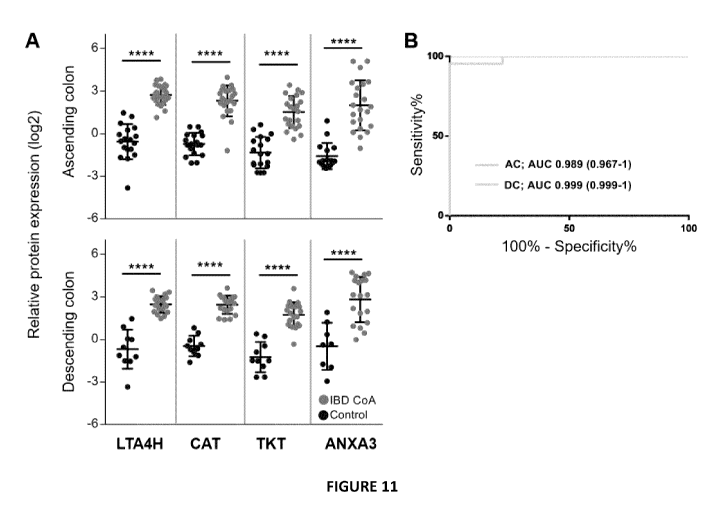
[0052] Figure 11 is a set of graphs illustrating an exemplary biomarker
panel for
suspected pediatric IBD diagnosis: (A) Relative expression levels of proteins
included in
IBD diagnosis biomarker panel. P values were generated by t-test ****p<
0.0001. (B)
Receiver operating characteristics curve utilizing panel of features listed in
Table 3 for
.. both the ascending colon and the descending colon. (C) Predictive class
probabilities in
each colon sub-region wherein samples predicted to be control are to the left
of 0.5 and
those predicted to be IBD are on the right of 0.5.
[0053] Figure 12 is a set of graphs illustrating relative expression of
proteins featured
in the IBD diagnosis biomarker panel; (A) comparison with MLI samples lacking
-- macroscopic evidence of inflammation (CoN) and (B) between CD (CoA) and UC
(CoA).
One-way ANOVA with Tukey's multiple comparison test. * p < 0.05, ** p < 0.01,
*** p <
0.001, **** p < 0.0001.
CA 03067062 2019-12-12
WO 2018/227295
PCT/CA2018/050716
[0054] Figure 13 is a set of graphs illustrating predictive class
probabilities of
Calprotectin (S100-A8 and S100-A9) using PLS-DA at the MLI in the (A)
ascending colon
and (B) descending colon.
[0055] Figure 14 is a set of graphs illustrating an exemplary biomarker
panel for extent
of disease in UC (pancolitis vs non-pancolitis): (A) Relative expression
levels of proteins
included in the UC extent of disease biomarker panel. P values were generated
by t-test
***p <0.001. (B) Receiver operating characteristics curve for differentiation
of pancolitis
from non-pancolitis utilizing the expression of proteins in UC extent of
disease biomarker
panel. (C) Predictive class probabilities of inflammatory status in the
ascending colon
(pancolitis vs non-pancolitis) wherein samples predicted to be inflamed are to
the left of
0.5 and those predicted to be non-inflamed are on the right of 0.5.
[0056] Figure 15 is a graph illustrating a minimum number of proteins
required to
achieve maximum sensitivity and specificity (indicated by the dotted line) for
extent of
disease biomarker panel (pancolitis vs non-pancolitis). Protein biomarker
candidates
added based on highest AUC values, using the PLS-DA model to classify UC CoN
from
UC CoA in the ascending colon.
[0057] Figure 16 illustrates results with respect to the determination
of select
biomarkers in MLI samples and in stool: (A) Annexin A3 was validated by
immunoblotting
of descending colon MLI samples from an independent MLI cohort. (B) Stool
samples
obtained from a subset of IBD and control patients were analyzed by immunoblot
(Annexin A3) and ELISA (LTA4H, Calprotectin), with quantitative data shown.
Annexin
A3 is shown relative to total protein, whereas ELISA results provide the
absolute amount
of protein. The dotted line indicates values above which Calprotectin is
considered a
positive result according to the manufacturer. The one patient that had
commenced
treatment prior stool collection is indicated by a (#).
[0058] Figure 17 is a set of graphs illustrating the validation of
select biomarkers in
stool: (A) Catalase, (B) Leukotriene A-4 hydrolase and (C) transketolase,
biomarker
16
CA 03067062 2019-12-12
WO 2018/227295
PCT/CA2018/050716
candidates proposed for the diagnosis of pediatric IBD, were validated by
ELISA from a
cohort consisting of independent patients and patients for which their MLI
samples were
utilized to develop the biomarker panel. P values were calculated using the
Mann Whitney
test. (D) The expression level of LTA4H in stool correlates with the PUCDAI.
Analysis
performed using Spearman two-tailed test.
[0059] Figure 18 is a set of graphs illustrating the relative protein
expression of
biomarker candidates A) Annexin A3, B) Catalase, C) leukotriene A-4 hydrolase
and D)
transketolase in biopsy samples.
[0060] Figure 19 is a graph of ROC curve for Control vs IBD CoA using a
panel of
.. proteins consisting of LTA4H, TXNDC17, TMSB10 and VASP, using AC MLI
samples.
Detailed Description
[0061] The present application relates to novel biomarkers that have
been identified
to determine the presence of inflammatory bowel disease (IBD) in a human
subject.
Namely, the relative protein expression levels of leukotriene A-4 hydrolase,
catalase,
transketolase and annexin A3 were measured reliably in gut samples of subjects
with IBD
to be higher than in gut samples of normal control subjects. Therefore, these
proteins
were identified as biomarkers to determine the presence of IBD in a subject.
By
determining the presence of IBD, it is meant determining if a subject has IBD
or does not
have IBD (e.g. is healthy with respect to IBD).
[0062] The Applicant has also discovered that thioredoxin domain containing
protein
17, vasodilator-stimulated phosphoprotein, and thymosin beta-10 may also be
used as
biomarkers to identify the presence of IBD in a subject.
[0063] Furthermore, it is shown that measuring one of these biomarkers
alone may be
sufficient to provide a reliable indication of IBD in a patient. However,
using more than
one biomarker may increase sensitivity and specificity of the test. When four
biomarkers
17
CA 03067062 2019-12-12
WO 2018/227295
PCT/CA2018/050716
are used, the test may obtain a higher level of specificity and sensitivity in
detection of
IBD in a subject.
[0064] The applicant has also discovered that the relative protein
expression levels of
these IBD-detection biomarkers may be observed, in most cases, in both the
ascending
colon and in the descending colon. Moreover, given that mucosal luminal
interface
samples include proteins present in the colonic lumen, these biomarkers may
also be
found in stool as the stool transits through the lumen of the colon.
Therefore, a less
location-specific gut sample, such as a stool sample, may be used to measure
protein
expression levels of these biomarkers to detect IBD, instead and/or in
addition to more
localized samples, such as a mucosal luminal interface sample. For example, as
described herein, similarly high relative expression levels of leukotriene A-4
hydrolase,
catalase, annexin A3 and transketolase are measured in stool samples from
subjects with
IBD. Therefore, it is possible to utilize a stool sample, an example of a non-
invasive
sample, to measure the relative protein expression levels of these biomarkers
to detect
IBD. The higher relative protein expression level of transketolase may also be
measured
in a stool sample, such as by performing mass spectrometry when analyzing the
stool
sample. It will also be understood that the relative protein expression levels
of thioredoxin
domain containing protein 17, vasodilator-stimulated phosphoprotein, and
thymosin beta-
10 may also be measured in stool samples. As the relative expression levels of
these
biomarkers were measured in the mucosal luminal interface samples, these
samples
including proteins present in the colonic lumen, and given that stool transits
with the lumen
of the colon, the levels may also be measured in stool samples.
[0065] In some examples, the gut sample may be a biopsy sample, as shown
by the
relative protein expression levels of respectively leukotriene A-4 hydrolase,
catalase,
transketolase and annexin A3 shown in Figure 18.
[0066] The expression level of the biomarkers as measured in a gut
sample of a
subject may be compared to a predetermined protein level to determine the
presence of
18
CA 03067062 2019-12-12
WO 2018/227295
PCT/CA2018/050716
IBD. This predetermined protein level of the biomarkers may be a threshold or
a
reference. The threshold or reference may be related to protein expression
level to
distinguish between patients with disease and healthy. The threshold or
reference may
be related to protein expression levels in, for example, healthy subjects or
controls,
subjects with the disease, subjects in remission, etc. This comparison may
provide an
indication relative to the, e.g., presence, severity, remission, relapse of
the disease. The
predetermined protein level may also be, or related to, an absolute value of
protein
expression corresponding, for instance, to subjects with the disease, or
healthy subjects.
[0067] Moreover, an increased relative protein expression level of the
biomarkers may
also be measured in patients without macro-inflammatory IBD and those with
macro-
inflammatory IBD. Therefore, these biomarkers may provide sufficient
sensitivity to detect
IBD in a patient without macro-inflammation.
[0068] In some examples, the biomarkers may also be used to establish a
treatment
fora patient having IBD, or to determine the patient's responsiveness to a
given treatment
for the disease. For instance, responsiveness may be established by taking one
or more
samples from the IBD-positive patient obtained at distinct times. A sample
taken later may
be compared to a sample obtained earlier from the same patient. A Protein
expression
level may be analyzed for the earlier sample, the later sample, and compared
to a control
sample for a normal patient. In some examples, only one sample may be needed,
where
the measured relative protein expression level may be compared to a
predetermined
threshold, such as that of a healthy patient (or that of responders) to
determine
responsiveness to treatment.
[0069] In some examples, a control sample is not needed, where the
relative protein
expression levels (or e.g. absolute protein concentrations of the biomarkers)
of the
biomarkers may be compared between the earlier sample and the later sample, or
using
the absolute protein concentration to compare with a predetermined level of
the
biomarkers, for instance, corresponding to healthy subjects (i.e. a
reference). The
19
CA 03067062 2019-12-12
WO 2018/227295
PCT/CA2018/050716
difference in protein expression levels of the biomarkers between the earlier
sample and
the later sample may assist in telling if the patient is responding to the
treatment. The
results from the test to establish responsiveness may also be correlated with
a disease
severity index to establish if the disease is receding, maintaining the same
severity or
worsening.
[0070] In some examples, to determine responsiveness of treatment, the
patient's
sample may be compared with the biomarker expression levels taken from
responders
and/or non-responders. If the biomarker protein expression levels of the
patient are
similar to those of the responders, then this may serve as an indication of
responsiveness
to the treatment. However, if the biomarker protein expression levels are
similar to those
of the non-responders, then this may serve as an indication that the patient
is not
responding to the treatment. In some examples, an average or a median protein
expression level for responders or non-responders may be used. The results
from the
test to establish responsiveness may also be correlated with a disease
severity index to
establish if the disease is receding, maintaining the same severity or
worsening.
[0071] In other embodiments, comparing the relative protein expression
levels of the
biomarkers with a predetermined protein level may be used to assess if
remission has
been induced in the patient (e.g. such as in response to a given treatment),
or if remission
is maintained in the patient. For instance, the predetermined protein level
may be that of
one or more of the biomarkers as observed in remission cases (e.g. a
predetermined level
achieved by analyzing relative protein expression levels of the biomarkers in
IBD
remission patients, or healthy patients).
In other embodiments, comparing the relative protein expression levels of the
biomarkers
with a predetermined protein level may be used to assess if relapse of
inflammatory bowel
disease has occurred in the patient.
[0072] Moreover, the present application identifies novel biomarkers for
determining if
a subject with ulcerative colitis has pancolitis (where inflammation is
present in the
CA 03067062 2019-12-12
WO 2018/227295
PCT/CA2018/050716
ascending colon) or non-pancolitis). These protein biomarkers are leukotriene
A-4
hydrolase, thioredoxin domain containing protein 17, vasodilator-stimulated
phosphoprotein, and thymosin beta-10.
[0073] In some embodiments, the relative protein expression levels of
leukotriene A-4
hydrolase, thioredoxin domain containing protein 17, vasodilator-stimulated
phosphoprotein, and thymosin beta-10 may be used to determine the presence of
macro-
inflammation in a patient.
[0074] Therefore, these biomarkers show different relative protein
expression levels
in gut samples from ulcerative colitis subjects with pancolitis when compared
to
corresponding protein expression levels from gut samples of subjects without
pancolitis.
[0075] Measuring a single one of these biomarkers may be sufficiently
reliable to
indicate if an ulcerative colitis patient has or does not have pancolitis.
However, it will be
understood that measuring more than one of these biomarkers may increase the
sensitivity and the specificity of the test. If all four or these biomarkers
are used, the test
may obtain an optimal level of specificity and sensitivity.
[0076] In some examples, these biomarkers may be used to assess the
responsiveness of a patient with ulcerative colitis to treatment by monitoring
the
development or disappearance of pancolitis. A first sample may be taken at a
first time
(e.g. before treatment) to assess protein expression levels of the biomarkers
as described
herein to determine if the patient has or does not have pancolitis. A second
sample may
then be taken at a second time, analyzed for biomarker protein expression
level as
described herein, and compared to the results of the first sample. For
instance, if the first
sample indicated that the patient has pancolitis, but the second sample
indicates instead
that the patient no longer has pancolitis, then this may serve as an
indication that
inflammation has receded and that the patient may be responding to treatment.
However,
the results of the analysis made on the first sample may show instead that the
patient
has, for example, only a mild case of ulcerative colitis without pancolitis,
but in the second
21
CA 03067062 2019-12-12
WO 2018/227295
PCT/CA2018/050716
sample it is determined that the patient has developed pancolitis, then this
may be an
indication that the patient may not be responding to treatment and that the
patient's
ulcerative colitis is in fact increasing in severity. The results from the
test to establish
responsiveness may also be correlated with a disease severity index to
establish if the
disease is receding, maintaining the same severity or worsening.
[0077] In other embodiments, comparing the relative protein expression
levels of the
biomarkers with a predetermined protein level may be used to assess if relapse
of
pancolitis has occurred.
[0078] DEFINITIONS:
[0079] In the present application, by subjects having "inflammatory
bowel disease" it
is meant subjects with ulcerative colitis (UC), subjects with Crohn's disease
(CD) and/or
subjects with IBD-unclassified (IBD-U).
[0080] By "gut sample" it is meant a sample that has originated from a
subject's
gastrointestinal track. In some examples, the gut sample may be specific to
the colon. In
other examples, the gut sample may be specific to the ascending colon or the
descending
colon. A gut sample may be a stool sample or any other non-invasive sample
that has
originated from the patient's gut. In some examples, the gut sample may
involve obtaining
a mucosal lumina! interface (MLI) sample from the subject. In some examples,
the gut
sample may be a biopsy sample taken from a subject, obtained during, for
instance, a
colonoscopy.
[0081] By "subject" it is meant a pediatric subject and/or an adult
subject. Even though
the exemplary studies presented herein are conducted on pediatric subjects,
there is
considerable similarity in pathology between pediatric IBD and adult-onset
IBD. The
pediatric subjects of the exemplary studies were between eight and eighteen
years of
age. The age of the subjects did not have an impact on the results of the
studies (i.e. the
relative biomarker protein expression level). Therefore, it will be
appreciated that similar
22
CA 03067062 2019-12-12
WO 2018/227295
PCT/CA2018/050716
relative biomarker protein expression levels may be observed in adult
subjects, wherein
the biomarkers may also be used, for instance, to detect IBD in an adult
subject, or to
determine if the adult subject has or does not have pancolitis.
[0082] By "severity of disease" it is meant if the disease is mild,
moderate or severe
(e.g. mild, moderate and severe may be defined in accordance with the
Pediatric
Ulcerative Colitis Activity Index, the Truelove and Witts' severity index, the
Harvey-
Bradshaw Index, or another diagnostic severity index). In some embodiments,
"severity
of the disease" may also mean determining whether the subject has an absence
of
disease (e.g. in some examples, indicating that the subject is in remission).
[0083] By "extent of disease" it is meant the proportion of the affected
colon (i.e. the
affected area), as defined, e.g., by the Paris Classification, where extent
may be defined
as El, E2, E3 and E4.
[0084] By "measuring" a protein sample as used herein, it is meant
conducting an
analysis of a sample to determine a protein expression level (or relative
protein
expression level) using such techniques as an immunoassay (e.g. ELISA), semi-
quantitative immunoblotting, mass spectrometry, and other techniques that are
known in
the art to quantitatively and/or qualitatively analyze the contents of a
sample obtained
from a patient.
[0085] Moreover, it will be understood that a protein name or its
corresponding gene
name may be used interchangeably herein to refer to the protein. Reference is
made to
Table 3 that correlates the different protein names with their respective gene
names.
[0086] THE PERFORMED STUDY:
[0087] A quantitative proteomic analysis of the colonic MLI in treatment-
naive pediatric
IBD patients at two distinct colon sub-regions was performed. Analysis of the
proteomic
data identified candidate biomarkers that outperform the accuracy of
calprotectin at the
MLI for classification of IBD patients with colonic involvement compared with
controls with
normal appearing colons. In addition, biomarkers which can indicate extent of
disease in
23
CA 03067062 2019-12-12
WO 2018/227295
PCT/CA2018/050716
UC (pancolitis vs non-pancolitis) were also identified. Finally, it is showed
that the
biomarker candidates LTA4H, Annexin A3 and CAT identified from the MLI
proteome
exhibited consistent results when assessed in stool samples.
[0088] MATERIALS AND METHODS
[0089] Patient Cohort
[0090] A cross-sectional study of patients less than 18 years old
undergoing diagnostic
colonoscopy for IBD was performed. In order to assess the host proteomic
landscape
alterations in IBD, the following exclusion criteria were chosen as they are
known to alter
the intestinal microbiota and thus influence the host response: (1) body mass
index > 95th
percentile; (2) diabetes mellitus (3) infectious gastroenteritis within the
preceding 2
months; and (4) use of any antibiotics, probiotics or immunosuppressives
within 1 month
of colonoscopy. Moreover, patients with inconclusive IBD diagnosis at the time
of sample
collection were excluded from analysis.
[0091] It will be understood that even though the study focused on
pediatric patients,
the relative expression level of the identified biomarkers described herein
may also be
observed in adult patients to indicate the presence of IBD due to the
significant overlap
of disease etiology between pediatric IBD and adult IBD which results in
similar gene and
protein expression patterns.
[0092] Reference Standard Test Methods
[0093] IBD was diagnosed by clinical examination, endoscopy, imaging and
laboratory
testing 3. The Pediatric Crohn's Disease Activity Index (PCDAI) was utilized
for CD 13 and
the Pediatric Ulcerative Colitis Activity Index (PUCAI) was utilized for UC
14. Inflammation
of the mucosa of the ascending colon (AC) or descending colon (DC) was
assessed by
visual appearance at colonoscopy. Extent of macroscopically inflamed mucosa
was
classified using the Paris modification of the Montreal Classification for IBD
15.
[0094] Colonic mucosal-luminal interface (MLI) aspirate sample
collection
24
CA 03067062 2019-12-12
WO 2018/227295
PCT/CA2018/050716
[0095] Colonic mucosal lumina! interface (MLI) aspirates were obtained
at time of
diagnostic colonoscopy following a standard 1 day clean-out preparation 16.
Sampling
occurred at the mid-ascending colon (AC) and/or at the site of the lower
descending colon
and upper sigmoid colon region (DC), and annotated to be from a normal, non-
inflamed
(CoN) or an affected, inflamed region (CoA) based on macroscopic evaluation.
Briefly,
upon insertion of the colonoscope, initial fluid and debris in the fluid were
aspirated away.
Thereafter sterile water was flushed onto the mucosa of the selected region
and the fluid
was aspirated into sterile collection vials as the MLI aspirate sample. These
samples were
used for analysis and the order of collection was distal to proximal sites.
The samples
were put on ice in the endoscopy room and immediately delivered to laboratory
for further
processing. A complete protease inhibitor cocktail (Roche Diagnostic GmbH,
Mannheim,
Germany) was added to the intestinal aspirates upon receipt in the lab.
Following debris
depletion by centrifugation at 700g for 5 minutes at 4 C, the supernatant was
subsequently subjected to 14,000g centrifugation for 20 minutes at 4 C to
remove
bacteria. The resultant supernatant was then filtered through a 0.2pm syringe
driven filter
for removal of any residual bacterial cells and stored at -80 C.
[0096] Heavy isotopic-labeled reference proteome
[0097] Heavy reference proteins for quantification were prepared from 5
isotopically-
labeled commercially available human cell lines, namely lymphocytic Jurkat
(ATCC),
HEK-293 (ATCC), colorectal carcinoma HCT 116 (ATCC), monocytic THP-1 (ATCC)
and
hepatic HuH-7 (JCRB Cell Bank). HCT116, HuH-7 and HEK293 cells were grown in
custom prepared media17. THP-1 and Jurkat cells were grown in RPM! media
(#0422
AthenaES Baltimore, MD, USA) supplemented with 15 mg/L methionine, 40 mg/L
[13C6,15N2]-L-lysine, 200 mg/L [13C6,15N4]-L-arginine (Sigma Aldrich,
Oakville, ON,
Can), 10% dialyzed FBS (GIBCO-Invitrogen; Burlington, ON ,CAN), 1 mM sodium
pyruvate (Gibco-lnvitrogen), 0.0059g/L Phenol Red (Sigma Aldrich, Oakville,
ON, Can)
and 28 pg/mL gentamicin (Gibco-Invitrogen). Heavy amino acid incorporation
(>95%) was
CA 03067062 2019-12-12
WO 2018/227295
PCT/CA2018/050716
confirmed by MS analysis18. For protein isolation, cells were lysed in lysis
buffer (4% SDS,
50 mM Tris, pH 8.0, protease inhibitor cocktail (Roche Diagnostic GmbH,
Mannheim,
Germany)) and sonicated three times in 10 second pulses with a 30 second
incubation
on ice between pulse intervals. Lysates were centrifuged at 10,000g for 10
minutes,
supernatants collected and protein concentrations were quantified by DC
(detergent
compatible) protein assay (BIORAD, California, USA). Protein aliquots were
stored at -
80 C.
[0098] Processing of colonic MLI aspirate proteins for mass spectrometry
analysis
[0099] Filtered colonic MLI aspirates were thawed on ice, and proteins
precipitated
overnight with trichloroacetic acid (20% v/v). Protein pellets were washed
three times with
acetone (100%) and dried before resuspension and sonication in lysis buffer.
Protein
concentration was quantified by DC protein assay (BIORAD, California, USA).
Colonic
aspirate proteins (45pg) were combined with an equal amount of heavy isotopic-
labeled
cell lysate (9pg of each cell type), used as a representative internal
standard to permit
quantitative proteomic data, an approach known as Super-SILAC (stable-isotope
labeling
by amino acids in cell culture) 19. The mixture containing proteins from
colonic aspirates
and internal reference cells was digested with trypsin by filter aided sample
preparation
method (FASP) 29, fractionated into 5 fractions (pH 4, 6, 8, 10 and 12) using
SCX resin
(Agilent Technologies, CA, USA), and desalted with a 10 pm AQUA-C18 resin (Dr
Maisch,
GmbH, Ammerbuch, Germany).
[00100] LC-MS/MS and Bioinformatic analysis
[00101] High-performance liquid chromatography/electrospray ionization tandem
mass
spectrometry (HPLC-ESI-MS/MS) was performed 12 using an Ekspert nanoLC 400
(Eksigent, Dublin, CA, USA) coupled to an LTQ Velos Pro Orbitrap Elite MS
(ThermoFisher Scientific, San Jose, CA).
[00102] Peptides were assigned and quantified using MaxQuant version 1.5.3.30
21 in
a single run against the human Uniprot database (downloaded 2012/07/11).
Patients with
26
CA 03067062 2019-12-12
WO 2018/227295
PCT/CA2018/050716
inconclusive IBD diagnosis at the time of sample collection were excluded from
downstream bioinformatics analysis. The following parameters were used: a
multiplicity
of two with Arg10 and Lys8 selected as the heavy labels; a specific digestion
mode was
implemented with trypsin selected as the enzyme with a maximum of two missed
cleavages; cysteine carbamidomethylation as a fixed modification; methionine
oxidation
and acetylation (protein N-termini) as variable modifications; the re-quantify
and match
between runs parameters were enabled; minimum peptide length of seven amino
acids;
ion mass tolerance of 0.5 Da; protein and peptide false discovery rate (FDR)
of 1%. The
AC and DC proteomes were analyzed separately post database search.
[00103] The MaxQuant output (Ratio H/L normalized) was uploaded into Perseus
v1.5.2.6 for heavy/light inversion, 10g2 transformation, Pearson correlation
coefficient
determination and hierarchical clustering of correlation values, while data
filtering was
performed in Excel. Partial least squares discriminant analysis (PLS-DA) and
receiver
operating characteristics (ROC) curve analyses were performed on k-nearest
neighbor
(knn) imputed data in the Biomarker Analysis module of MetaboAnalyst 3.022.
Using
predictive class probability values generated in MetaboAnalyst 3.0, ROC curves
and
predictive class probability plots were plotted in GraphPad Prism 7 and ROC
curve AUC
calculated in GraphPad Prism 7. ROC, sensitivity and specificity confidence
intervals
were calculated in GraphPad Prism 7 using predictive class probability values
generated
.. in MetaboAnalyst 3Ø Individual protein AUC and associated confidence
interval values
were calculated using knn imputed MS results in GraphPad Prism7. PCA was
performed
using the prcomp argument in R studio. Protein interaction networks were
generated
using STRING version 10.0 and Cytoscape version 3.4Ø For suspected pediatric
IBD
biomarker panel generation, proteins identified by PLS-DA in both the AC and
DC with
the highest combined area under the curve (AUC) upon ROC curve analysis were
considered, and biomarker panel assembled based on iterative analysis. If a
protein lead
to a decrease in sensitivity or specificity without an increase in its
counterpart in either
27
CA 03067062 2019-12-12
WO 2018/227295
PCT/CA2018/050716
colon sub-region it was skipped. Biomarker panel generation for extent of
disease in UC
considered features with the highest AUC from comparison of ascending colon
with and
without macroscopic evidence of inflammation. Relative protein expression
graphs with
statistical analyses were generated in GraphPad Prism 7. Biomarker panels and
Calprotectin (S100-A8 & S100-A9) were evaluated using the ROC curve based
model
evaluation module of MetaboAnalyst 3Ø Gene ontology was performed using
DAVID
Bioinformatics Resources 6.823 and plotted in GraphPad Prism 7.
[00104] Study data were collected and managed using REDCap electronic data
capture
tools hosted at the CHEO Research Institute. REDCap (Research Electronic Data
Capture) 24.
[00105] Stool collection and protein extraction from stool
[00106] Stool samples were collected from a treatment-naive cohort consisting
of both
independent samples from the discovery cohort and patients whose corresponding
MLI
sample was used for biomarker discovery. Stool samples were collected within 8
weeks
of diagnostic colonoscopy with 82.2% collected within 4 days of colonoscopy.
Stool
samples were frozen after collection and brought into the clinic on ice.
Extraction buffer
(50mM Tris, pH 7.2, 150mM NaCI, protease inhibitor cocktail (Complete, Mini
(Roche
Diagnostic GmbH, Mannheim, Germany)) was added to stool in a 5:1 ratio
(extraction
buffer volume: stool weight). The resultant slurry was mixed by agitation for
30 seconds,
followed by rotation at 4 C for 20 minutes. Following centrifugation for 20
minutes at
10,000g at 4 C to remove large sediment, the supernatant was filtered (0.21Jm)
and the
protein-containing filtrate was collected and stored at -80 C. Protein
concentration was
assessed by BCA protein assay kit (ThermoFisher Scientific, San Jose, CA).
[00107] I mmunoblotting
[00108] Proteins from intestinal aspirates were precipitated, lysed and
quantified.
Intestinal aspirate proteins (30pg) or stool protein extracts (50pg) were
separated by a 4-
15% TGX Stain-free gel (BIORAD, California, USA) under reducing conditions.
The gels
28
CA 03067062 2019-12-12
WO 2018/227295
PCT/CA2018/050716
were exposed to UV light for 1 minute to enable fluorescent labeling of
proteins, followed
by electroblotting to LF PVDF membranes (BIORAD, California, USA) using the
Trans-
Blot Turbo Transfer system (BIORAD, California, USA), followed by another
round of UV
exposure. Standard immunoblotting procedures were applied, using antibodies
for
Annexin A3 (ab33068 Abcam Ltd, Cambridge, England) with anti-rabbit secondary
antibody (GE Healthcare Life Sciences, Massachusetts, USA). Proteins were
detected by
ECL substrate (BIORAD, California, USA) and blots were imaged using the
ChemiDoc
MP system (BIORAD, California, USA). Quantification was performed using Image
Lab
5.2.1 (BIORAD, California, USA) utilizing the full lane intensity as reference
for
normalization.
[00109] Enzyme-linked immunosorbent assay (ELISA)
[00110] Leukotriene A-4 hydrolase (abx572445 Abbexa Ltd, Cambridge, UK) and
Catalase (ab171572 Abcam, Cambridge, UK) from stool samples was measured from
5Oug and 0.5pg of protein, respectively, by ELISA which was then performed
according
to the manufacturer's protocol. A reference standard was added to all plates
for inter-
plate normalization.
[00111] RESULTS
[00112] Patient cohort
[00113] The colonic MLI aspirates of 93 colon sub-regions (57 AC [18 control,
39 IBD],
36 DC [10 control, 26 IBD]) were collected during diagnostic endoscopy from 60
patients
prior to the administration of any treatment. For 33 patients (control=10,
CD=14, UC=9)
both colon sub-regions were analyzed, whereas the AC or DC proteomes were
exclusively analyzed for 24 (control=8, CD=7, UC=9) and 3 (CD=1, UC=2)
patients,
respectively (Figure 1). The patient characteristics are summarized in Table
1. No
significant difference in age was observed between groups in either colon sub-
region
(supplementary figures 1A, B) or between sexes in the IBD group of the AC and
DC and
the control group of the DC, although a significantly higher number of females
in the AC
29
CA 03067062 2019-12-12
WO 2018/227295
PCT/CA2018/050716
control group was observed (Binomial one-tailed; p = 0.0481). In accordance
with
previous reports, the majority (70%) of pediatric UC patients in the cohort
presented with
extensive disease (Paris E3/E4 classification) 25. Similarly, the majority
(77%) of CD
patients had disease involvement that was either isolated to the colon (L2) or
had
ileocolonic (L3) involvement 26.
Controls (n=18) CD (n=22) UC (n=20)
Region AC (n=18) DC (n=10) AC (n=21) DC (n=15) AC (n=18) DC
(n=11)
# Females (%) 13(72.2%) 7(70.0%) 5(23.8%) 3(20.0%) 18 (55.6%)
4(36.4%)
Median Age, years 15.4 15.7 13.8 14.1 15.4 15.8
(IQR) (11.3-16.6) (12.7-16.2) (11-16.3) (12.3-16.1) (11.1-16.2)
(14.9-16.8)
Paris Classification
Ala NA NA 4(19.0%) 2(13.3%) NA NA
Alb NA NA 16(76.2%) 12(80.0%) NA NA
A2 NA NA 1(4.8%) 1(6.7%) NA NA
Location
Ll NA NA 4(19.0%) 3(20.0%) NA NA
L2 NA NA 5(23.8%) 4(26.7%) NA NA
L3 NA NA 11(52.4%) 7(46.7%) NA NA
L4a NA NA 10(46.6%) 6(40.0%) NA NA
L4b NA NA 3(14.3%) 3(20.0%) NA NA
Behavior
B1 NA NA 21(100%) 14(93.3%) NA NA
B2 NA NA 0 0 NA NA
CA 03067062 2019-12-12
WO 2018/227295
PCT/CA2018/050716
B3 NA NA 0 1(6.7%) NA NA
P NA NA 3(14.3%) 5(33.3%) NA NA
Growth
GO NA NA M(66.7%) 11(73.3%) NA NA
G1 NA NA 7(33.3%) 4(26.7%) NA NA
Extent
El NA NA NA NA 1(5.6%)
1(9.1%)
E2 NA NA NA NA 4(22.2%) 4(36.4%)
E3 NA NA NA NA 2(11.1%) 2(18.2%)
E4 NA NA NA NA
11(611%) 4(36.4%)
Severity
SO NA NA NA NA 15(83.3%) 9(8L8%)
S1 NA NA NA NA 3(16.7%) 2(18.2%)
Table 1: Patient characteristics (AC = ascending colon; DC = descending colon)
[00114] Proteomic dataset assessment
[00115] Samples were processed and analyzed by MS over a period of 22 months.
A
super-SI LAC approach was implemented for accurate quantification of proteins
obtained
from colonic MLI aspirate samples, and for use as a consistent reference over
time. The
majority (87.7% (AC), 88.5% (DC)) of proteins were quantified within a 10-fold
median
ratio (normalized MLI proteins/super-SILAC reference proteome). Furthermore,
there
were no significant differences observed in the total number of quantified
proteins
between groups in either colon sub-region. A total of 3,537 and 3,132 proteins
were
quantified from the AC and DC MLI, respectively, of which 972 (AC) and 995
(DC) proteins
were quantified in 75% of samples (Q75) with 80% protein ID overlap between
regions.
31
CA 03067062 2019-12-12
WO 2018/227295
PCT/CA2018/050716
Pearson correlation analysis of the Q75 was performed on the 10g2 light/heavy
ratios
(Figures 3A and 3B), yielding 89% and 84% of values with a correlation > 0.75
in the AC
and DC respectively, indicating consistent MS performance, sample preparation
and that
the majority of the proteome remains unaltered in the disease state.
Hierarchical
clustering of the Pearson correlation values tended to segregate samples
according to
diagnosis and inflammatory status rather than by MS batch analysis (Figures 4C
and 4D).
[00116] Proteomic landscape alterations in treatment naïve pediatric I BD
[00117] To assess the proteomic alterations in an unbiased manner, principal
component analysis (PCA) was performed on the Q75. The control samples
generally
clustered separately from those IBD samples that were deemed by endoscopy to
have
macroscopic evidence of inflammation (IBD CoA) (Figure 5). In the AC, the IBD
samples
with no evidence of macroscopic inflammation (IBD CoN) distributed between
control
samples and IBD CoA samples, whereas in the DC the IBD CoN samples tended to
cluster with the IBD CoA. Segregation according to sex was not observed in
either colon
sub-regions (Figure 6). In order to identify discriminate features between
control patients
and IBD patients with active disease (IBD CoA), a multivariate analysis
approach using
PLS-DA was applied, identifying 130 and 208 proteins in the AC and DC
respectively.
There were 78 proteins common to both the AC and DC (Figure 7A), which
included
proteins known to be altered in IBD such as the S100-A8 subunit of
calprotectin. Gene
ontology enrichment of the PLS-DA results yielded several expected biological
processes
related to IBD including defense response to bacterium, innate immune
response,
inflammatory response, phagocytosis and neutrophil chemotaxis (Figures 7B and
7C).
Upon PLS-DA analysis of controls and IBD samples without macroscopic evidence
of
inflammation (CoN), pathways related to inflammation were observed (Figure 8),
possibly
indicating the presence of microscopic inflammation. Notably, of the 130
proteins in the
current AC IBD CoA vs control data set, 26 were also identified 12 (figure 9).
AC biopsies
were analyzed, wherein 225 proteins were identified as discriminant features
of IBD CoA
32
CA 03067062 2019-12-12
WO 2018/227295
PCT/CA2018/050716
vs control by PLS-DA analysis of the discovery cohort. Among these 26 common
proteins,
11(42%) and 9 (35%) were increased and decreased, respectively, in both
datasets. The
remaining 6 (23%) common proteins exhibited opposite relative expression
between the
biopsy and MLI datasets, with the majority (4/6) being elevated in the IBD
samples relative
to control in the MLI proteome versus a relative reduction in the biopsy
proteome. Within
these 26 proteins, 22 localize to the extracellular region and an enrichment
of proteins
related to the immune response was identified (11/26). The expression levels
of the 26
proteins in biopsy samples versus mucosal luminal interface samples are listed
in Table
2:
MLI
expression
ratio log 2
of IBD Biopsy
CoA/ expression
Gene Control of 10g2 (IBD
name AC CoA/Control)
ANPEP -1.98915 -0.6181
ANXA3 4.216674 1.563199
C3 1.356244 -0.24447
CBR1 -2.0791 -0.57351
CLCA1 -1.33476 -0.60407
CORO1A 2.680741 -0.01155
CTSG 3.547383 0.604387
ELANE 4.154011 1.545775
HBB 1.602313 -0.20574
IGJ -1.09184 -0.4301
ITGB2 2.013377 0.941676
33
CA 03067062 2019-12-12
WO 2018/227295
PCT/CA2018/050716
LAP3 -1.4226 0.89655
LCP1 2.330294 0.539129
LGALS3 -2.6157 -0.54731
LRPPRC -1.58638 -0.74149
MT2A -2.0682 -0.2031
NAMPT 2.595841 1.007
PGD 2.369553 0.653318
PIGR -3.22623 -0.43575
REG1A -1.48006 -0.63364
S100A11 1.800538 0.79001
S100P 2.540318 2.302368
SERPINA1 -0.91455 0.315549
SERPINB1 1.7999 0.254763
TF 1.438568 -0.26176
WARS 1.39081 1.680909
Table 2: the expression levels of 26 proteins in MLI samples and biopsy
samples, showing
opposite relative expression for 6 of the proteins.
[00118] Biomarker panel for suspected pediatric I BD diagnosis
[00119] To identify biomarkers of active IBD, discriminant features identified
by PLS-
DA (control vs IBD CoA) were further considered for biomarker panel
generation.
Proteins with the highest combined area under the curve (AUC) values from both
colon
sub-regions were considered for biomarker panel assembly. A maximum
sensitivity and
specificity utilizing the minimum number of features was reached in the AC
using a panel
of 4 features, whereas 2 features were sufficient in the DC to reach maximum
sensitivity
and specificity; the 4 panel proteins ultimately utilized for IBD CoA vs
Control are listed in
Table 3. The relative expression levels are depicted in Figure 11 and compared
to that of
IBD CoN in supplemental Figure 12. While not included in the development of
the panel,
34
CA 03067062 2019-12-12
WO 2018/227295
PCT/CA2018/050716
IBD CoN levels of all panel proteins were significantly different from control
samples with
the exception of transketolase in the AC. However, it is observed that the
relative
expression level of these biomarkers in subjects with IBD CoN are higher than
those for
the controls. Therefore, a skilled person will understand that these
biomarkers may also
be used to detect the presence of IBD CoN in a subject.
AUC Fold change AUC
Fold change
(CI)
(CI) [UC CoA/
[IBD CoA/Ctrl]
[UC CoN vs
[IBD CoA vs Control] UC CoN]
UC CoA]
Protein GeneAC DC AC DC AC AC
name
0.995 1.0
0.961
Leukotriene A-4
LTA4H 8.05 6.66 4.54
hydrolase
(0.982-1.008) (1.0-1.0) (0.881-1.041)
0.967 1.0
Catalase CAT 8.83 7.31 N/A N/A
(0.903-1.031) (1.0-1.0)
0.967 0.989
Transketolase TKT 7.23 7.39 N/A N/A
(0.922-1.012) (0.962-1.017)
0.972 0.937
Annexin A3 ANXA3 18.59 9.35 N/A N/A
(0.929-1.015) (0.849-1.025)
Thioredoxin
0.961
domain-containing TXNDC17 N/A N/A N/A N/A 3.17
protein 17
(0.881-1.041)
0.948
Thymosin beta-10 TMSB10 N/A N/A N/A N/A 0.23
(0.851-1.045)
CA 03067062 2019-12-12
WO 2018/227295
PCT/CA2018/050716
Vasodilator- 0.935
stimulated VASP N/A N/A N/A N/A 5.83
phosphoprotein
(0.824-1.046)
Table 3: Proteins in panel for pediatric IBD diagnosis and for extent of
disease in UC
(pancolitis vs non-pancolitis)
[00120] Applying a receiver operating characteristics (ROC) curve utilizing
this panel of
4 proteins achieves an AUC value of 0.989 (95% Cl: 0.967-1.0) and 0.999 (95%
Cl: 0.999-
1.0) for the AC and DC, respectively (Figure 11B). Predictive class
probabilities yielded a
sensitivity of 0.954 (95% Cl: 0.7716-0.9988) and 1.0 (95% Cl: 0.8235-1.0) for
the AC and
DC, respectively and a specificity of >0.999 (AC 95% Cl: 0.8147-1.0; DC 95%
Cl: 0.6915-
1.0) for both the AC and DC, providing a classification accuracy of 97.5% and
100% in
the AC and DC, respectively. This panel of biomarkers for suspected pediatric
IBD
diagnosis outperformed the classification accuracy of calprotectin (S100-A8 &
S100-A9)
by a direct comparison of MS data obtained from the MLI in both colon sub-
regions.
Calprotectin yielded a sensitivity of 0.682 (95% Cl: 0.4513-0.8614) and 0.684
(95% Cl:
0.4345-0.8742) for the AC and DC respectively, and a specificity of 0.833 (95%
Cl:
0.5858-0.9642) and 0.700 (95% CI:O. 3475-0.9333) in the AC and DC,
respectively, which
yielded classification accuracies of 75.0% and 69.0% in the AC and DC,
respectively
(Figure 13).
[00121] Biomarker panel for extent of disease in UC (pancolitis vs non-
pancolitis)
[00122] The majority (70%) of new-onset pediatric UC patients recruited in the
discovery cohort displayed extensive disease (E3 or E4). To generate a
biomarker panel
to evaluate disease extent, features identified by PLS-DA upon comparison of
colonic
aspirate proteins arising from the AC of patients with evidence of macroscopic
inflammation (UC CoA) versus proteins from the AC of patients without evidence
of
macroscopic inflammation (UC CoN) were further considered. As listed in Table
3, a panel
of 4 proteins achieved a sensitivity of >0.9999 (95% Cl: 0.5904-1.0) and
specificity of
36
CA 03067062 2019-12-12
WO 2018/227295
PCT/CA2018/050716
>0.9999 (95% Cl: 0.7151-1.0) and yielded a classification accuracy of 100%
(Figures 14
and 15).
[00123] Validation of biomarkers identified at the MLI in stool samples
[00124] In order to implement easily accessible biomarkers that would be more
readily
.. translatable to the clinic, the expression of 2 of the IBD biomarker
proteins identified
herein, namely CAT and LTA4H, was assessed in stool. ELISA was performed on
stool
from 39 and 38 children for CAT and LTA4H, respectively. This cohort
encompasses both
independent participants (n= 26) and participants for which their MLI samples
were
utilized for biomarker discovery (n=15). The increased expression in IBD
compared to
control observed by proteomics at the MLI was reflected by ELISA in stool, in
which the
expression of CAT (p < 0.0001), LTA4H (p =0.0002) and transketolase (p =
0.0076) were
significantly different in IBD patients compared to controls (Figures 17A, 17B
and 17C).
Furthermore, the expression of LTA4H in stool correlated with the PUCAI
(Spearman
r=0.567 (95% Cl: 0.1378-0.817; p=0.0114) (Figure 17D). LTA4H also correlated
with
albumin (r=-0.3696; p=0.0265) but did not correlate with ESR or HCT, which are
used in
the calculation of the PCDAI, nor with PCDAI. The expression of CAT in stool
correlated
with the level of albumin measured in serum (r=-0.4645; p=0.0038) but not with
PCDAI,
PUCAI, HCT or ESR.
[00125] The increased expression of Annexin A3 in IBD patients with active
disease
compared to the controls was validated by immunoblotting using DC MLI samples
independent of the discovery cohort (Figure 16A). Quantitation of immunoblots
are shown
relative to total protein. To investigate whether Annexin A3 could be detected
in a non-
invasive bio-specimen, and thus more readily translatable to the clinic,
immunoblotting on
stool samples from patients included in the discovery cohort in addition to
independent
samples was performed, wherein a single patient had commenced therapy upon
stool
collection. In accordance with the MLI samples, Annexin A3 was detected in the
IBD stool
samples (Figure 16B). For direct comparison, the level of fecal calprotectin
in the same
37
CA 03067062 2019-12-12
WO 2018/227295
PCT/CA2018/050716
samples was measured using an ELISA kit that has been applied in the clinical
setting.
Notably, one control patient had levels of fecal calprotectin considered to be
positive for
IBD diagnosis. Therefore, the calprotectin results yielded a false positive
IBD result for a
given patient. In contrast, the immunoblots correctly identified all control
patients as non-
IBD, this determination based upon the relative level of Annexin A3 (Figure
16B).
[00126] Moreover, the levels of LTA4H were evaluated by ELISA from the same
stool
samples as calprotectin and Annexin A3. There was a significant increase
("p=0.004) in
the levels of LTA4H in stool samples obtained from IBD patients compared to
control
patients (Figure 16C). Furthermore, the expression level of LTA4H in the stool
samples
correctly classified all the patients as either controls or IBD, in contrast
to when using the
calprotectin results.
[00127] DISCUSSION OF THE RESULTS
[00128] The study identified biomarkers that improve upon current IBD
standards in
diagnosis, and propose novel biomarkers for disease extent in UC. Biomarkers
currently
applied in the clinic, including fecal calprotectin, are limited in their
ability to diagnose and
monitor IBD, and thus can only be successfully used when in conjunction with
endoscopy.
[00129] An IBD biomarker panel of four proteins was identified that was
capable of
accurately classifying >95% of patients as IBD or control using MLI aspirates.
One or
more of these biomarkers may also be used to reliably classify patients as IBD-
positive
or IBD-negative. Furthermore, it was shown that the significantly different
levels of select
biomarker panel proteins were confirmed in a non-invasive bio-specimen
(stool). For
transketolase, the increased levels of protein expression in a stool sample
may also be
shown, such as by using mass spectrometry.
[00130] An instance wherein a control patient exhibited high calprotectin
expression in
stool whereas the expression of two of our novel IBD biomarkers, Annexin A3
and LTA4H,
had correctly identified the patient as a control was observed. Annexin A3 and
LTA4H
may have increased specificity when compared with calprotectin.
38
CA 03067062 2019-12-12
WO 2018/227295
PCT/CA2018/050716
[00131] It was observed that a portion (23%) of proteins demonstrate opposite
relative
expression for IBD compared to control between the two proteomic datasets.
This can be
explained in part by the difference in subcellular localization under
assessment in each
dataset. For example, complement C3 is increased in IBD within the MLI but
decreased
in the biopsy dataset, and the tissue macrophages or epithelial cells within
the biopsy
may be releasing C3 into the extracellular space, hence being detected at
relatively higher
levels within the MLI of IBD patient samples than in control samples,
highlighting the
insight gained by the multi-omic comparison. Amongst the 26 proteins, 5
(ELANE,
CORO1A, CTSG, ANXA3 and C3) are involved in leukocyte mediated immunity.
[00132] The elevated expression of TKT that was observed in IBD patients
compared
to controls may be a response toward reducing the ROS-induced damage that is
observed within the colon of IBD patients 33.
[00133] As for catalase, a significant difference in catalase expression
between CD and
UC at the MLI was not observed (Figure 12B). However, there is significant
elevation of
catalase in MLI from both macroscopic (CoA) and microscopic (CoN) inflammatory
aspirates from IBD patients when compared with controls. Similar to TKT, the
elevated
levels of catalase may be to protect cells from the damage caused by hydrogen
peroxide
which is elevated in the inflamed mucosa of IBD patients 36.
[00134] Leukotriene A-4 hydrolase (LTA4H) is the only protein included in both
the IBD
and UC extent biomarker panels. LTA4H catalyzes the biosynthesis of
leukotriene B4,
which in turn is a potent neutrophilic chemoattractant and has been implicated
in chronic
inflammation 37.
[00135] Proteins TXNDC17, TMSB10 and VASP are also identified as biomarkers
for
IBD diagnosis, as part of the panel for disease extent in UC. Moreover,
proteins
TXNDC17, TMSB10 and VASP may also be used to indicate the presence of IBD in a
patient, due to its different relative expression levels when compared to that
of a healthy
subject, as shown in Figure 19. Interestingly, TXNDC17 can regulate TNF-a
signaling 38;
39
CA 03067062 2019-12-12
WO 2018/227295
PCT/CA2018/050716
TNF-a is an important signaling molecule in inflammation, and is the target of
anti-TNF-a
agents that are utilized for the induction and maintenance of remission in CD.
Unlike the
role of TXNDC17, TMSB10 and VASP are involved in cytoskeleton organization 39,
40.
Perhaps elevated VASP observed in inflamed UC is associated with altered host-
microbe
interactions. Despite both having roles in cytoskeletal organization, they
demonstrate
opposite expression trends in inflamed tissue compared to uninflamed tissue
(Figure 14).
[00136] The extent of disease in UC patients can indicate response to therapy,
since
healing progresses proximally to distally through the colon. Considering that
many
pediatric UC patients have panc01iti525, a non-invasive biomarker indicating a
status of
pancolitis vs. non-pancolitis is beneficial as it may permit assessment of
response to
therapy without the need for colonoscopy.
[00137] In summary, this study identified biomarkers that are better at
predicting IBD
than calprotectin at the MLI. Elevated expression of CAT, Annexin A3 and
LTA4H,
identified as biomarker candidates at the MLI was reflected in stool samples
showing that
non-invasively obtained samples may also show similar protein expression
levels of the
four biomarkers for determination of the presence of IBD. Further, a panel of
UC extent
of disease biomarkers was identified, classifying 100% of patients with
pan/non-
pancolitis; this panel was developed by comparison of UC patients with and
without
macroscopic inflammation, and thus at a level similar in nature to what is
observed by
endoscopy.
[00138] The description of the present invention has been presented for
purposes of
illustration but is not intended to be exhaustive or limited to the disclosed
embodiments.
Many modifications and variations will be apparent to those of ordinary skill
in the art.
CA 03067062 2019-12-12
WO 2018/227295
PCT/CA2018/050716
[00139] References:
1. Vizcaino JA, Csordas A, Del-Toro N, et al. 2016 update of the PRIDE
database
and its related tools. Nucleic Acids Res 2016;44:11033.
2. Benchimol El, Fortinsky KJ, Gozdyra P, et al. Epidemiology of pediatric
inflammatory bowel disease: a systematic review of international trends.
Inflamm
Bowel Dis 2011;17:423-39.
3. Levine A, Koletzko S, Turner D, et al. ESPGHAN revised porto criteria
for the
diagnosis of inflammatory bowel disease in children and adolescents. J Pediatr
Gastroenterol Nutr 2014;58:795-806.
4. van Rheenen PF, Van de Vijver E, Fidler V. Faecal calprotectin for
screening of
patients with suspected inflammatory bowel disease: diagnostic meta-analysis.
BMJ 2010;341:c3369.
5. Henderson P, Anderson NH, Wilson DC. The diagnostic accuracy of fecal
calprotectin during the investigation of suspected pediatric inflammatory
bowel
disease: a systematic review and meta-analysis. Am J Gastroenterol
2014;109:637-45.
6. Mendoza JL, Abreu MT. Biological markers in inflammatory bowel disease:
practical consideration for clinicians. Gastroenterol Clin Biol 2009;33 Suppl
3:S158-73.
7. Merras-Salmio L, Kolho KL, Pelkonen AS, et al. Markers of gut mucosal
inflammation and cow's milk specific immunoglobulins in non-IgE cow's milk
allergy. Clin Trans! Allergy 2014;4:8.
8. Orivuori L, Mustonen K, de Goffau MC, et al. High level of fecal
calprotectin at
age 2 months as a marker of intestinal inflammation predicts atopic dermatitis
and asthma by age 6. Clin Exp Allergy 2015;45:928-39.
9. Zhu Q, Li F, Wang J, et al. Fecal Calprotectin in Healthy Children Aged
1-4
Years. PLoS One 2016;11:e0150725.
10. Danese S, Fiocchi C. Ulcerative colitis. N Engl J Med 2011;365:1713-25.
41
CA 03067062 2019-12-12
WO 2018/227295
PCT/CA2018/050716
11. Stange EF, Travis SP, Vermeire S, et al. European evidence-based
Consensus
on the diagnosis and management of ulcerative colitis: Definitions and
diagnosis.
J Crohns Colitis 2008;2:1-23.
12. Starr AE, Deeke SA, Ning Z, et al. Proteomic analysis of ascending
colon
biopsies from a paediatric inflammatory bowel disease inception cohort
identifies
protein biomarkers that differentiate Crohn's disease from UC. Gut 2016.
13. Hyams J, Markowitz J, Otley A, et al. Evaluation of the pediatric crohn
disease
activity index: a prospective multicenter experience. J Pediatr Gastroenterol
Nutr
2005;41:416-21.
14. Turner D, Otley AR, Mack D, et al. Development, validation, and
evaluation of a
pediatric ulcerative colitis activity index: a prospective multicenter study.
Gastroenterology 2007;133:423-32.
15. Levine A, Griffiths A, Markowitz J, et al. Pediatric modification of
the Montreal
classification for inflammatory bowel disease: the Paris classification.
Inflamm
Bowel Dis 2011;17:1314-21.
16. Jimenez-Rivera C, Haas D, Boland M, et al. Comparison of two common
outpatient preparations for colonoscopy in children and youth. Gastroenterol
Res
Pract 2009;2009:518932.
17. Mottawea W, Chiang C-K, Miihlbauer M, et al. Altered intestinal
microbiota-host
mitochondria crosstalk in new onset Crohn's disease. Nature Communications
2016;7:13419.
18. Ong SE, Mann M. A practical recipe for stable isotope labeling by amino
acids in
cell culture (SILAC). Nat Protoc 2006;1:2650-60.
19. Geiger T, Cox J, Ostasiewicz P, et al. Super-SILAC mix for quantitative
proteomics of human tumor tissue. Nat Methods 2010;7:383-5.
20. Wisniewski JR, Zougman A, Mann M. Combination of FASP and StageTip-
based
fractionation allows in-depth analysis of the hippocampal membrane proteome. J
Proteome Res 2009;8:5674-8.
21. Cox J, Matic I, Hilger M, et al. A practical guide to the MaxQuant
computational
platform for SILAC-based quantitative proteomics. Nat Protoc 2009;4:698-705.
42
CA 03067062 2019-12-12
WO 2018/227295
PCT/CA2018/050716
22. Xia J, Sinelnikov IV, Han B, et al. MetaboAnalyst 3.0--making
metabolomics
more meaningful. Nucleic Acids Res 2015;43:W251-7.
23. Huang da W, Sherman BT, Lempicki RA. Bioinformatics enrichment tools:
paths
toward the comprehensive functional analysis of large gene lists. Nucleic
Acids
Res 2009;37:1-13.
24. Harris PA, Taylor R, Thielke R, et al. Research electronic data capture
(REDCap)--a metadata-driven methodology and workflow process for providing
translational research informatics support. J Biomed Inform 2009;42:377-81.
25. Sauer CG, Kugathasan S. Pediatric inflammatory bowel disease:
highlighting
pediatric differences in IBD. Gastroenterol Clin North Am 2009;38:611-28.
26. Vernier-Massouille G, BaIde M, Salleron J, et al. Natural history of
pediatric
Crohn's disease: a population-based cohort study. Gastroenterology
2008;135:1106-13.
27. Wlodarska M, Kostic AD, Xavier RJ. An integrative view of microbiome-
host
interactions in inflammatory bowel diseases. Cell Host Microbe 2015;17:577-91.
28. Rifai N, Gillette MA, Carr SA. Protein biomarker discovery and
validation: the
long and uncertain path to clinical utility. Nat Biotechnol 2006;24:971-83.
29. Presley LL, Ye J, Li X, et al. Host-microbe relationships in
inflammatory bowel
disease detected by bacterial and metaproteomic analysis of the mucosal-
luminal
interface. Inflamm Bowel Dis 2012;18:409-17.
30. Marshall KW, Mohr S, Khettabi FE, et al. A blood-based biomarker panel
for
stratifying current risk for colorectal cancer. Int J Cancer 2010;126:1177-86.
31. Lassen N, Black WJ, Estey T, et al. The role of corneal crystallins in
the cellular
defense mechanisms against oxidative stress. Semin Cell Dev Biol 2008;19:100-
12.
32. Bentz S, Pesch T, Wolfram L, et al. Lack of transketolase-like (TKTL) 1
aggravates murine experimental colitis. Am J Physiol Gastrointest Liver
Physiol
2011;300:G598-607.
43
CA 03067062 2019-12-12
WO 2018/227295
PCT/CA2018/050716
33. Cao W, Vrees MD, Kirber MT, et al. Hydrogen peroxide contributes to
motor
dysfunction in ulcerative colitis. Am J Physiol Gastrointest Liver Physiol
2004;286:G833-43.
34. Roozendaal C, Zhao MH, Horst G, et al. Catalase and alpha-enolase: two
novel
granulocyte autoantigens in inflammatory bowel disease (IBD). Clin Exp Immunol
1998;112:10-6.
35. Sabery N, Bass D. Use of serologic markers as a screening tool in
inflammatory
bowel disease compared with elevated erythrocyte sedimentation rate and
anemia. Pediatrics 2007;119:E193-E199.
36. Strus M, Gosiewski T, Fyderek K, et al. A role of hydrogen peroxide
producing
commensal bacteria present in colon of adolescents with inflammatory bowel
disease in perpetuation of the inflammatory process. J Physiol Pharmacol
2009;60 Suppl 6:49-54.
37. Palmer RM, Stepney RJ, Higgs GA, et al. Chemokinetic activity of
arachidonic
and lipoxygenase products on leuocyctes of different species. Prostaglandins
1980;20:411-8.
38. Jung Y, Kim H, Min SH, et al. Dynein light chain LC8 negatively
regulates NF-
kappaB through the redox-dependent interaction with IkappaBalpha. J Biol Chem
2008;283:23863-71.
39. Yu FX, Lin SC, Morrison-Bogorad M, et al. Effects of thymosin beta 4
and
thymosin beta 10 on actin structures in living cells. Cell Motil Cytoskeleton
1994;27:13-25.
40. Ke Y, Tan Y, Wei N, et al. Yersinia protein kinase A phosphorylates
vasodilator-
stimulated phosphoprotein to modify the host cytoskeleton. Cell Microbiol
2015;17:473-85.
41. Lee SY, Gertler FB, Goldberg MB. Vasodilator-stimulated phosphoprotein
restricts cell-to-cell spread of Shigella flexneri at the cell periphery.
Microbiology
2015;161:2149-60.
44
CA 03067062 2019-12-12
WO 2018/227295
PCT/CA2018/050716
42.
Castellano F, Le Clainche C, Patin D, et al. A WASp-VASP complex regulates
actin polymerization at the plasma membrane. Embo Journal 2001;20:5603-
5614.
45