Note: Descriptions are shown in the official language in which they were submitted.
CA 03067181 2019-12-11
WO 2019/204229 PCT/US2019/027540
METHODS OF ENCAPSULATING SINGLE CELLS, THE ENCAPSULATED CELLS AND USES THEREOF
FIELD
[0001] Systems, methods, and compositions provided herein relate to
hollow
beads encapsulating a single cell, methods making the hollow beads, and
methods of using
the hollow beads for conducting multiple co-assays on the encapsulated cell,
including, for
example, spatial index sequencing and nucleic acid library preparation.
BACKGROUND
[0002] The detection of specific nucleic acid sequences present in a
biological
sample has been used, for example, as a method for identifying and classifying
microorganisms, diagnosing infectious diseases, detecting and characterizing
genetic
abnormalities, identifying genetic changes associated with cancer, studying
genetic
susceptibility to disease, and measuring response to various types of
treatment. A common
technique for detecting specific nucleic acid sequences in a biological sample
is nucleic acid
sequencing.
[0003] Next generation sequencers are powerful tools that generate
large amounts
of genomic data per sequencing run. Interpreting and analyzing this large
amount of data can
be challenging. Single cell DNA sequencing is emerging as one tool for
studying genomic
heterogeneity. Specifically, the microbiome, which carries multiple repeated
genomic
regions, can be sequenced by obtaining DNA sequences from only a single cell.
Performing
multiple enzymatic reactions on a single cell is unreliable due to the
challenges in confining
and accessing intracellular biomolecules within a single cell over multiple
assays. For
example, many cell based assays fail to secure intracellular molecules,
resulting in loss of
biomolecules during performance of the assay.
SUMMARY
[0004] The present disclosure is related to systems, methods, and
compositions
for performing multiple co-assays on a single cell, wherein the single cell is
encapsulated
within a hollow bead, such that the single cell is confined in order to
perform multiple co-
assays, including, for example, lysis, DNA analysis, RNA analysis, protein
analysis,
-1-
CA 03067181 2019-12-11
WO 2019/204229 PCT/US2019/027540
tagmentation, nucleic acid amplification, nucleic acid sequencing, DNA library
preparation,
assay for transposase accessible chromatic using sequencing (ATAC-seq),
contiguity-
preserving transposition (CPT-seq), single cell combinatorial indexed
sequencing (SCI-seq),
or single cell genome amplification, or any combination thereof performed
sequentially on a
single cell.
[0005] Some embodiments provided herein relate to a hollow bead
encapsulating
a single cell. In some embodiments, the hollow bead includes a polymer shell
and a single
cell disposed within the polymer shell. In some embodiments, the polymer shell
includes
pores that allow diffusion of a reagent through the polymer shell while
retaining the single
cell.
[0006] Further embodiments relate to a method of performing multiple
sequential
co-assays on a single cell encapsulated within a hollow bead. In some
embodiments, the
method includes obtaining a hollow bead as described herein, wherein the
hollow bead
encapsulates a single cell and sequentially contacting the single cell with
reagents to perform
multiple sequential co-assays.
[0007] Some embodiments provided herein relate to a method of
encapsulating a
single cell within a hollow bead. In some embodiments, the method includes
mixing a single
cell with a polymer to form a mixture and mixing the mixture with a
crosslinking oil to form
a polymer shell encapsulating the single cell, wherein the polymer shell
includes pores that
allow diffusion of a reagent through the polymer shell while retaining the
single cell.
BRIEF DESCRIPTION OF THE DRAWINGS
[0008] FIG. 1 is a schematic diagram of an embodiment of a microfluidic
droplet
generator system that can be used to prepare hollow beads encapsulating a
cell.
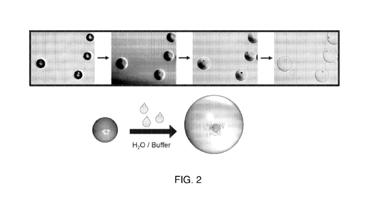
[0009] FIG. 2 depicts micrograph images and a schematic representation
of an
embodiment of hollow beads that are capable of expanding in size in the
presence of an
aqueous buffer. The micrograph images depict beads in various states,
including (from left to
right): dehydrated beads; beads initially contacted with an aqueous phase;
beads beginning to
swell; and completely swollen beads.
[0010] FIG. 3 is a schematic that illustrates an embodiment for
increasing bead
porosity by temperature or chemical means. As shown in FIG. 3, increasing bead
porosity
-2-
CA 03067181 2019-12-11
WO 2019/204229 PCT/US2019/027540
can increase the flow of particles into or out of the bead, and controlling
the porosity
provides control of particles (including particle size) that flow into or out
of the bead.
[0011] FIG. 4 is a schematic that illustrates an embodiment of a method
of
device-free cell encapsulation by depositing monomer units of a polymer over a
single cell.
[0012] FIG. 5 is a schematic that illustrates an embodiment of an
encapsulated
cell within a polymer formed by device-free methods, wherein the polymer shell
is attached
to the cell through specific interactions with a specific binding molecule.
[0013] FIG. 6 depicts micrograph images of beads stained with
fluorescent dyes,
depicting cells encapsulated within the beads. The micrograph images depict
(from left to
right): staining with thiol-specific fluorophores, targeting free PEG-
maleimide; staining with
Texas Red fluorophores targeting free ¨SH groups; and nuclei staining with
Hoechst
staining.
[0014] FIG. 7 is a schematic that illustrates an embodiment of a method
of
performing multiple co-assays on a hollow bead encapsulating a cell. FIG. 7
demonstrates an
embodiment of performing tagmentation to generate ATAC-seq fragments, reverse
transcription to initiate cDNA synthesis, and PCR reactions to generate an
indexed library.
[0015] FIG. 8 is a schematic that illustrates an embodiment of a method
of
performing multiple co-assays on a hollow bead encapsulating a cell. FIG. 8
demonstrates an
embodiment of performing tagmentation to generate ATAC-seq fragments, reverse
transcription to initiate cDNA synthesis, and PCR reactions with random
extension for full
length RNA sequencing to generate an indexed library.
[0016] FIG. 9 is a schematic that illustrates an embodiment of a method
of
performing multiple co-assays on a hollow bead encapsulating a cell. FIG. 9
demonstrates a
three-tier combinatorial indexing approach using two rounds of indexed split
ligation and one
round of indexed PCR.
[0017] FIG. 10 is a schematic that illustrates an embodiment of a
method of
performing multiple co-assays on a hollow bead encapsulating a cell. FIG. 10
demonstrates
multiple co-assays for combinatorial indexing with indexed transposomes.
[0018] FIG. 11 is a schematic that illustrates an embodiment of a
method of
performing multiple co-assays on a hollow bead encapsulating a cell. FIG. 11
demonstrates
multiple co-assays for single cell whole genome amplification.
-3-
CA 03067181 2019-12-11
WO 2019/204229 PCT/US2019/027540
[0019] FIG. 12 is a flow diagram that illustrates an embodiment of a
method of
preparing a whole genome library with a hollow bead encapsulating a cell.
DETAILED DESCRIPTION
[0020] In the following detailed description, reference is made to the
accompanying drawings, which form a part hereof. In the drawings, similar
symbols
typically identify similar components, unless context dictates otherwise. The
illustrative
embodiments described in the detailed description, drawings, and claims are
not meant to be
limiting. Other embodiments may be utilized, and other changes may be made,
without
departing from the spirit or scope of the subject matter presented herein. It
will be readily
understood that the aspects of the present disclosure, as generally described
herein, and
illustrated in the Figures, can be arranged, substituted, combined, separated,
and designed in
a wide variety of different configurations, all of which are explicitly
contemplated herein.
[0021] Embodiments provided herein relate to hollow beads encapsulating
a
single cell, methods of making the hollow beads, and methods of performing
multiple co-
assays on a single cell encapsulated within a hollow bead. The hollow beads
may include
hydrogel polymers and crosslinkers that are mixed in the presence of a cell,
and which form
hollow beads that encapsulate the cell. The hollow beads may include pores
that allow
diffusion of reagents through the hollow bead while retaining the cell within
the hollow bead,
thereby allowing reactions to take place within the hollow beads. In some
embodiments, the
hollow beads enable co-assays to be performed on the same single cell while
maintaining
cellular contiguity. In particular, the methods, systems, and compositions
provided herein
allow confining and accessing intracellular biomolecules within the
encapsulated cells.
Accordingly, in some embodiments, the hollow beads described herein are
referred to herein
as contiguity particles. Thus, the term "contiguity particle" as used herein
refers to a hollow
bead encapsulating a single cell.
[0022] The contiguity particles described herein may be used for next
generation
cell compartmentalization approaches and allow multi-analyte assays performed
on an
individual cell. The contiguity particles and methods of use described herein
efficiently allow
millions of cells to be analyzed individually thereby reducing the cost of
sample preparation
and maintaining sample contiguity. The compositions and methods described
herein maintain
-4-
CA 03067181 2019-12-11
WO 2019/204229 PCT/US2019/027540
cellular contiguity without the use of external compartmentalization
strategies (microfluidics)
such as emulsions, immobilization, or other micro-compartments.
[0023] In some embodiments, the contiguity particles as described
herein can be
used in assays to analyze a single cell. Assays that may be performed on the
single cell may
include, for example, cellular lysis, DNA analysis, RNA analysis, nucleic acid
sequencing,
protein analysis, tagmentation, nucleic acid amplification, DNA library
preparation, assay for
transposase accessible chromatic using sequencing (ATAC-seq), contiguity-
preserving
transposition (CPT-seq), single cell combinatorial indexed sequencing (SCI-
seq), or single
cell genome amplification, or any combination thereof performed sequentially.
[0024] The use of contiguity particles for performing one or more
assays on a
single cell may be used simultaneously on multiple contiguity particles in
order to
simultaneously perform co-assays on a number of single cells, for example from
10,000 to 1
million single cells, such as 10,000, 50,000, 100,000, 500,000, or 1 million
single cells.
[0025] The pore size of the contiguity particle can be engineered to
allow the
diffusion of reagents, such as enzymes, chemicals, and smaller sized primers
(< 50bps),
while retaining the single cell itself or while retaining other larger
particles, such as larger
nucleic acids (>300bps), inside the hollow bead during the performance of one
or more
assays.
[0026] As used herein, the term "reagent" describes an agent or a
mixture of two
or more agents useful for reacting with, interacting with, diluting, or adding
to a sample, and
may include agents used in assays described herein, including agents for
lysis, nucleic acid
analysis, nucleic acid amplification reactions, protein analysis, tagmentation
reactions,
ATAC-seq, CPT-seq, or SCI-seq reactions, or other assays. Thus, reagents may
include, for
example, buffers, chemicals, enzymes, polymerase, primers having a size of
less than 50 base
pairs, template nucleic acids, nucleotides, labels, dyes, or nucleases. In
some embodiments,
the reagent includes lysozyme, proteinase K, random hexamers, polymerase (for
example,
029 DNA polymerase, Taq polymerase, Bsu polymerase), transposase (for example,
Tn5),
primers (for example, P5 and P7 adaptor sequences), ligase, catalyzing enzyme,
deoxynucleotide triphosphates, buffers, or divalent cations.
-5-
CA 03067181 2019-12-11
WO 2019/204229 PCT/US2019/027540
Contiguity Particles
[0027] In some embodiments, the hollow bead has a polymer shell that
was
prepared from a hydrogel composition. As used herein, the term "hydrogel"
refers to a
substance formed when an organic polymer (natural or synthetic) is cross-
linked via
covalent, ionic, or hydrogen bonds to create a three-dimensional open-lattice
structure that
entraps water molecules to form a gel. In some embodiments, the hydrogel may
be a
biocompatible hydrogel. As used herein, the term "biocompatible hydrogel"
refers to a
polymer that forms a gel that is not toxic to living cells and allows
sufficient diffusion of
oxygen and nutrients to entrapped cells to maintain viability. In some
embodiments, the
hydrogel material includes alginate, acrylamide, or poly-ethylene glycol
(PEG), PEG-
acrylate, PEG-amine, PEG-carboxylate, PEG-dithiol, PEG-epoxide, PEG-
isocyanate, PEG-
maleimide, polyacrylic acid (PAA), poly(methyl methacrylate) (PMMA),
polystyrene (PS),
polystyrene sulfonate (PSS), polyvinylpyrrolidone (PVPON), N,N'-
bis(acryloyl)cystamine,
polypropylene oxide (PPO), poly(hydroxyethyl methacrylate) (PHEMA), poly(N-
isopropylacrylamide) (PNIPAAm), poly(lactic acid) (PLA), poly(lactic-co-
glycolic acid)
(PLGA), polycaprolactone (PCL), poly(vinylsulfonic acid) (PVSA), poly(L-
aspartic acid),
poly(L-glutamic acid), polylysine, agar, agarose, heparin, alginate sulfate,
dextran sulfate,
hyaluronan, pectin, carrageenan, gelatin, chitosan, cellulose, collagen,
bisacrylamide,
diacrylate, diallylamine, triallylamine, divinyl sulfone, diethyleneglycol
diallyl ether,
ethyleneglycol diacrylate, polymethyleneglycol diacrylate, polyethyleneglycol
diacrylate,
trimethylopropoane trimethacrylate, ethoxylated trimethylol triacrylate, or
ethoxylated
pentaerythritol tetracrylate, or combinations or mixtures thereof. In some
embodiments, the
hydrogel is an alginate, acrylamide, or PEG based material. In some
embodiments, the
hydrogel is a PEG based material with acrylate-dithiol, epoxide-amine reaction
chemistries.
In some embodiments, the hydrogel forms a polymer shell that includes PEG-
maleimide/dithiol oil, PEG-epoxide/amine oil, PEG-epoxide/PEG-amine, or PEG-
dithiol/PEG-acrylate. In some embodiments, the hydrogel material is selected
in order to
avoid generation of free radicals that have the potential to damage
intracellular biomolecules.
In some embodiments, the hydrogel polymer includes 60-90% fluid, such as
water, and 10-
30% polymer. In certain embodiments, the water content of hydrogel is about 70-
80%. As
used herein, the term "about" or "approximately", when modifying a numerical
value, refers
-6-
CA 03067181 2019-12-11
WO 2019/204229 PCT/US2019/027540
to variations that can occur in the numerical value. For example, variations
can occur
through differences in the manufacture of a particular substrate or component.
In one
embodiment, the term "about" means within 1%, 5%, or up to 10% of the recited
numerical
value.
[0028] As
used herein, the polymer shell is a polymer surface of a hollow bead,
having a shell with an interior that encapsulates a single cell. Due to the
nature of the hollow
beads described herein, the contiguity particles can retain genetic material
after multiple
assays and can be released by physical force, cleaving chemicals, or by
generating osmotic
imbalance depending on the thickness of the polymer shell.
[0029]
Hydrogels may be prepared by cross-linking hydrophilic biopolymers or
synthetic polymers. Thus, in some embodiments, the hydrogel may include a
crosslinker. As
used herein, the term "crosslinker" refers to a molecule that can form a three-
dimensional
network when reacted with the appropriate base monomers. Examples of the
hydrogel
polymers, which may include one or more crosslinkers, include but are not
limited to,
hyaluronans, chitosans, agar, heparin, sulfate, cellulose, alginates
(including alginate
sulfate), collagen, dextrans (including dextran sulfate), pectin, carrageenan,
polylysine,
gelatins (including gelatin type A), agarose, (meth)acrylate-oligolactide-PEO-
oligolactide-
(meth)acrylate, PEO-PPO-PEO copolymers (Pluronics),
poly(phosphazene),
poly(methacrylates), poly(N-vinylpyrrolidone), PL(G)A-PEO-PL(G)A copolymers,
poly(ethylene imine), polyethylene glycol (PEG)-thiol, PEG-acrylate,
acrylamide, N,N'-
bis(acryloyl)cystamine, PEG, polypropylene oxide (PPO), polyacrylic acid,
poly(hydroxyethyl methacrylate) (PHEMA), poly(methyl methacrylate) (PMMA),
poly(N-
isopropylacrylamide) (PNIPAAm), poly(lactic acid) (PLA), poly(lactic-co-
glycolic acid)
(PLGA), polycaprolactone (PCL), poly(vinylsulfonic acid) (PVSA), poly(L-
aspartic acid),
poly(L-glutamic acid), bisacrylamide, diacrylate, diallylamine, triallylamine,
divinyl sulfone,
diethyleneglycol diallyl ether, ethyleneglycol diacrylate, polymethyleneglycol
diacrylate,
polyethyleneglycol diacrylate, trimethylopropoane trimethacrylate, ethoxylated
trimethylol
triacrylate, or ethoxylated pentaerythritol tetracrylate, or combinations
thereof. Thus, for
example, a combination may include a polymer and a crosslinker, for example
polyethylene
glycol (PEG)-thiol/PEG-acrylate, acrylamide/N,N'-bis(acryloyl)cystamine
(BACy), or
PEG/polypropylene oxide (PPO). In some embodiments, the polymer shell includes
a four-
-7-
CA 03067181 2019-12-11
WO 2019/204229 PCT/US2019/027540
arm polyethylene glycol (PEG). In some embodiments, the four-arm polyethylene
glycol
(PEG) is selected from the group consisting of PEG-acrylate, PEG-amine, PEG-
carboxylate,
PEG-dithiol, PEG-epoxide, PEG-isocyanate, and PEG-maleimide
[0030] In some embodiment, the crosslinker is an instantaneous
crosslinker or a
slow crosslinker. An instantaneous crosslinker is a crosslinker that instantly
crosslinks the
hydrogel polymer, and is also referred to herein as click chemistry.
Instantaneous
crosslinkers may include dithiol oil + PEG-maleimide or PEG epoxide + amine
oil. A slow
crosslinker is a crosslinker that slowly crosslinks the hydrogel polymer, and
may include
PEG-epoxide + PEG-amine or PEG-dithiol + PEG-acrylate. A slow crosslinker may
take
more than several hours to crosslink, for example more than 2, 3, 4, 5, 6, 7,
8, 9, 10, 11, or 12
hours to crosslink. In some embodiments provided herein, contiguity particles
are formulated
by an instantaneous crosslinker, and thereby preserve the cell state better
compared to a slow
crosslinker. Without wishing to be bound by theory, cells may possible undergo
physiological changes by intracellular signaling mechanisms during longer
crosslinking
times.
[0031] In some embodiments, a crosslinker forms a disulfide bond in the
hydrogel
polymer, thereby linking hydrogel polymers. In some embodiments, the hydrogel
polymers
form a hydrogel matrix having pores (for example, a porous hydrogel matrix).
These pores
are capable of retaining sufficiently large particles, such as a single cell
or nucleic acids
extracted therefrom within the polymer shell, but allow other materials, such
as reagents, to
pass through the pores, thereby passing in and out of the hollow beads. In
some
embodiments, the pore size of the polymer shell is finely tuned by varying the
ratio of the
concentration of polymer to the concentration of crosslinker. In some
embodiments, the ratio
of polymer to crosslinker is 30:1, 25:1, 20:1, 19:1, 18:1, 17:1, 16:1, 15:1,
14:1, 13:1, 12:1,
11:1, 10:1, 9:1, 8:1, 7:1, 6:1, 5:1, 4:1, 3:1, 2:1, 1:1, 1:2, 1:3, 1:4, 1:5,
1:6, 1:7, 1:8, 1:9, 1:10,
1:15, 1:20, or 1:30, or a ratio within a range defined by any two of the
aforementioned ratios.
In some embodiments, additional functions such as DNA primer, or charged
chemical groups
can be grafted to polymer matrix to meet the requirements of different
applications.
[0032] As used herein, the term "porosity" means the fractional volume
(dimension-less) of a hydrogel that is composed of open space, for example,
pores or other
openings. Therefore, porosity measures void spaces in a material and is a
fraction of volume
-8-
CA 03067181 2019-12-11
WO 2019/204229 PCT/US2019/027540
of voids over the total volume, as a percentage between 0 and 100% (or between
0 and 1).
Porosity of the hydrogel may range from 0.5 to 0.99, from about 0.75 to about
0.99, or from
about 0.8 to about 0.95.
[0033] The polymer shell can have any pore size that allows for
sufficient
diffusion of reagents while concomitantly retaining the single cell or nucleic
acids extracted
therefrom. As used herein, the term "pore size" refers to a diameter or an
effective diameter
of a cross-section of the pores. The term "pore size" can also refer to an
average diameter or
an average effective diameter of a cross-section of the pores, based on the
measurements of a
plurality of pores. The effective diameter of a cross-section that is not
circular equals the
diameter of a circular cross-section that has the same cross-sectional area as
that of the non-
circular cross-section. In some embodiments, the hydrogel can be swollen when
the hydrogel
is hydrated. The sizes of the pores size can then change depending on the
water content in the
hydrogel. In some embodiments, the pores of the hydrogel can have a pore of
sufficient size
to retain the encapsulated cell within the hydrogel but allow reagents to pass
through. In
some embodiments, the interior of the polymer shell is an aqueous environment.
In some
embodiments, the single cell disposed within the polymer shell is free from
interaction with
the polymer shell and/or is not in contact with the polymer shell. In some
embodiments, a
polymer shell is formed around a cell (as described herein in greater detail),
and the cell is in
contact with the polymer shell due to the polymer shell being brought to the
cell surface due
to passive adsorption or in a targeted manner, such as by being attached to an
antibody or
other specific binding molecule.
[0034] In some embodiments, the contiguity particles is of a sufficient
size to
encapsulate a single cell. In some embodiments, the contiguity particle has a
diameter of
about 20 [tm to about 200 [tm, such as 20, 30, 40, 50, 60, 70, 80, 90, 100,
110, 120, 130, 140,
150, 160, 170, 180, 190, or 200 [tm, or a diameter within a range defined by
any two of the
aforementioned values. The size of the contiguity particle may change due to
environmental
factors. In some embodiments, the contiguity particles expand when they are
separated from
continuous oil phase and immersed in an aqueous phase, as shown in FIG. 2. In
some
embodiments, expansion of the contiguity particles increases the efficiency of
performing
assays on the genetic material inside the encapsulated cells. In some
embodiments, expansion
-9-
CA 03067181 2019-12-11
WO 2019/204229 PCT/US2019/027540
of the contiguity particles creates a larger environment for indexed inserts
to be amplified
during PCR, which may otherwise be restricted in current cell based assays.
[0035] In
some embodiments, pore size is increased sufficiently to retain the
encapsulated cell, but to allow extracted nucleic acids to diffuse through the
polymer shell, as
depicted in FIG. 3. In some embodiments, the pore size of the contiguity
particles can be
controlled by altering the crosslinking chemistry. The final crosslinked pore
size can further
be altered by changing the environment of the contiguity particle, for
example, by changing
salt concentration, pH, or temperature, thereby allowing immobilized molecules
to be
released from the contiguity particle.
[0036] In
some embodiments, the crosslinker is a reversible crosslinker. In some
embodiments, a reversible crosslinker is capable of reversibly crosslinking
the hydrogel
polymer and is capable of being un-crosslinked in the presence of a cleaver.
In some
embodiments, a crosslinker can be cleaved by the presence of a reducing agent,
by elevated
temperature, or by an electric field. In some embodiments, the reversible
crosslinker may be
N,N'-bis(acryloyl)cystamine, a reversible crosslinker for polyacrylamide gels,
wherein a
disulfide linkage may be broken in the presence of a suitable reducing agent.
As shown in
FIG. 3, bead porosity may be increased by temperature or chemical means,
thereby releasing
contacting the crosslinker with a reducing agent cleaves the disulfide bonds
of the
crosslinker, breaking down the hollow beads. The hollow beads degrade, and
release the
contents, such as nucleic acids that were retained therein. In some
embodiments, the
crosslinker is cleaved by increasing the temperature to greater than 50, 55,
60, 65, 70, 75, 80,
85, 90, 95, or 100 C. In some embodiments, the crosslinker is cleaved by
contacting the
hollow beads with a reducing agent. In some embodiments, the reducing agents
include
phosphine compounds, water soluble phosphines, nitrogen containing phosphines
and salts
and derivatives thereof, dithioerythritol (DTE), dithiothreitol (DTT) (cis and
trans isomers,
respectively, of 2,3-dihydroxy-1,4-dithiolbutane), 2-mercaptoethanol or P-
mercaptoethanol
(BME), 2-mercaptoethanol or aminoethanethiol, glutathione, thioglycolate or
thioglycolic
acid, 2,3 -dimercaptopropanol, tris(2-carboxyethyl)phosphine
(TCEP),
tris(hydroxymethyl)phosphine (THP), or P4tris(hydroxymethyl)phosphine]
propionic acid
(THPP).
-10-
CA 03067181 2019-12-11
WO 2019/204229 PCT/US2019/027540
[0037] In some embodiments, elevating the temperature to increase
diffusion or
contacting with a reducing agent degrades the crosslinker, thereby releasing
encapsulated
genetic material from the contiguity particle.
[0038] In some embodiments, the crosslinking of the crosslinker
establishes pores
within the contiguity particle. In some embodiments, the size of the pores in
the polymer
shell are regulatable and are formulated to encapsulate cells or nucleic acids
of greater than
about 300 base pairs, but to allow smaller particles, such as reagents, or
smaller sized nucleic
acids of less than about 50 base pairs, such as primers, to pass through the
pores. In some
embodiments, the reagents including reagents for processing genetic material,
such as
reagents for isolating nucleic acids from a cell, for amplifying or sequencing
nucleic acids, or
for preparation of nucleic acid libraries. In some embodiments, reagents
include, for
example, lysozyme, proteinase K, random hexamers, polymerase (for example, 029
DNA
polymerase, Taq polymerase, Bsu polymerase), transposase (for example, Tn5),
primers (for
example, P5 and P7 adaptor sequences), ligase, catalyzing enzyme,
deoxynucleotide
triphosphates, buffers, or divalent cations.
[0039] Exemplary cell types that can be used within a contiguity
particle may
include, for example, cells isolated from a tissue biopsy (e.g., from a tissue
having a disease
such as colon, breast, prostate, lung, skin cancer, or infected with a
pathogen etc.) and normal
cells from the same tissue, e.g., from the same patient; cells grown in tissue
culture that are
immortal (e.g., cells with a proliferative mutation or an immortalizing
transgene), infected
with a pathogen, or treated (e.g., with environmental or chemical agents such
as peptides,
hormones, altered temperature, growth condition, physical stress, cellular
transformation,
etc.), and normal cells (e.g., cells that are otherwise identical to the
experimental cells except
that they are not immortalized, infected, or treated, etc.); cells isolated
from a mammal with a
cancer, a disease, a geriatric mammal, or a mammal exposed to a condition, and
cells from a
mammal of the same species, e.g., from the same family, that is healthy or
young; and
differentiated cells and non-differentiated cells from the same mammal (e.g.,
one cell being
the progenitor of the other in a mammal, for example). In one embodiment,
cells of different
types, e.g., neuronal and non-neuronal cells, or cells of different status
(e.g., before and after
a stimulus on the cells) may be compared. In another embodiment, the
experimental material
is cells susceptible to infection by a pathogen such as a virus, e.g., human
immunodeficiency
-11-
CA 03067181 2019-12-11
WO 2019/204229 PCT/US2019/027540
virus (HIV), etc., and the control material is cells resistant to infection by
the pathogen. In
another embodiment of the invention, the sample pair is represented by
undifferentiated cells,
e.g., stem cells, and differentiated cells. Cells from yeast, plants and
animals, such as fish,
birds, reptiles, amphibians and mammals may be used in the subject methods. In
certain
embodiments, mammalian cells, i.e., cells from mice, rabbits, primates, or
humans, or
cultured derivatives thereof, may be used.
Methods of Making Contiguity Particles
[0040] Some
embodiments provided herein relate to methods of encapsulating a
single cell within a polymer shell to form a contiguity particle. In some
embodiments, a
sample containing cells to be encapsulated within the contiguity particle is
obtained. In some
embodiments, the cells can be fixed with a fixative prior to encapsulation
within the
contiguity particle. As used herein, a fixative generally refers to an agent
that can fix cells.
For example, fixed cells can stabilize protein complexes, nucleic acid
complexes, or protein-
nucleic acid complexes in the cell. Suitable fixatives and cross-linkers can
include, alcohol or
aldehyde based fixatives, formaldehyde, glutaraldehyde, ethanol-based
fixatives, methanol-
based fixatives, acetone, acetic acid, osmium tetraoxide, potassium
dichromate, chromic acid,
potassium permanganate, mercurials, picrates, formalin, paraformaldehyde,
amine-reactive
NHS-ester crosslinkers such as bis[sulfosuccinimidyl] suberate (B53), 3,3'-
dithiob is [sulfosuccinimidylpropionate] (DTS SP),
ethylene glycol
bis [sulfosuccinimidylsuccinate] (sulfo-EGS), disuccinimidyl
glutarate (DSG),
dithiobis[succinimidyl propionate] (DSP), disuccinimidyl suberate (DSS),
ethylene glycol
bis[succinimidylsuccinate] (EGS), NHS-ester/diazirine crosslinkers such as NHS-
diazirine,
NHS-LC-diazirine, NHS-SS-diazirine, sulfo-NHS-diazirine, sulfo-NHS-LC-
diazirine, and
sulfo-NHS-SS-diazirine. In some embodiments, fixing a cell preserves the
internal state of
the cell thereby preventing modification of the cell during encapsulation of
the cell within the
contiguity particle.
[0041] In
some embodiments, a contiguity particle is prepared by static means,
such as by single cell microwell/microarray methods or microdissection
methods, without the
need of a microfluidic device. Thus, in some embodiments, the contiguity
particles described
herein are prepared by a device-free method. Device-free methods may include
initiation of
polymerization on a cell surface by contacting the cell with a polymer,
forming a polymer
-12-
CA 03067181 2019-12-11
WO 2019/204229 PCT/US2019/027540
shell around the cell. Initiation of polymerization can occur by chemical
reaction of active
group on monomer units with specific moieties of membrane proteins, glycans or
other small
molecules. Initial step of monomer polymerization can be followed by one or
several rounds
of monomer units deposition promoted by either electrostatic or hydrophobic
forces. Some of
monomer layers can contain functional groups such as biotin or other ligands
which can be
used later for specific capture of encapsulated cells. For example, embedded
cells can contain
biotin and coating the embedded cells with magnetic streptavidin beads would
make them
magnetic allowing automated and easy processing, as shown in FIG. 4.
Alternatively, live or
fixed cell encapsulation can be initialized by light, or other physical or
chemical means when
initiator molecules are brought to cell surface either by passive adsorption,
or in a targeted
manner, for example attached to an antibody or other specific binding molecule
and promote
localized polymer amplification, as shown in FIG. 5.
[0042] Encapsulating cells in a targeted surface-initiated
polymerization method
enables the user to specifically select cells of interest and simultaneously
prepares the cells
for subsequent downstream assays, thereby combining flow sorting with single
cell assays.
Enrichment of specific cell types can be performed using cell separation kits
utilizing
magnetically labeled beads or custom binding molecules specific to certain
cell membrane
proteins conjugated to an enrichment surface.
[0043] In some embodiments, a contiguity particle is prepared by
dynamic means,
such as by vortex assisted emulsion, microfluidic droplet generation, or valve
based
microfluidics. As used herein, vortex assisted emulsion refers to vortexing a
hydrogel
polymer with a cell, including a fixed cell as described herein, in a
container, such as in a
tube, vial, or reaction vessel. The components can be mixed, for example by
manual or
mechanical vortexing or shaking. In some embodiments, manual mixing results in
hollow
beads that encapsulate genetic material having a size of 20, 25, 30, 35, 40,
45, 50, 55, 60, 65,
70, 75, 80, 85, 90, 95, 100, 110, 120, 130, 140, 150, 160, 170, 180, 190, or
200 um in
diameter, or a size within a range defined by any two of the aforementioned
values. In some
embodiments, the size of the beads is non-uniform, and thus, the size of the
beads includes
beads of various diameters.
[0044] In some embodiments, the contiguity particles are prepared by
microfluidic flow techniques. Microfluidic flow includes use of a microfluidic
device for
-13-
CA 03067181 2019-12-11
WO 2019/204229 PCT/US2019/027540
assisted gel emulsion generation, as shown in FIG. 1. In some embodiments, the
microfluidic
device includes microchannels configured to produce a contiguity particle of a
desired size
and configured to encapsulate a single cell per contiguity particle. In some
embodiments, the
microfluidic device has a height of 50, 60, 70, 80, 90, 100, 110, 120, 130,
140, 150, 160, 170,
180, 190, or 200 [tm, or a height within a range defined by any two of the
aforementioned
values. In some embodiments, the microfluidic device includes one or more
channels. In
some embodiments, the microfluidic device includes a channel for introducing a
cell,
including a fixed cell, that has been introduced to a polymer, a channel for
introducing a
crosslinker, and a channel for an immiscible fluid. In some embodiments, the
width of the
one or more channels is identical. In some embodiments, the width of the one
or more
channels is different. In some embodiments, the width of the one or more
channels is 20, 30,
40, 45, 50, 55, 60, 65, 70, 75, 80, 85, 90, 95, 100, 105, 110, 115, 120, 125,
130, 135, 140,
145, or 150 [tm, or a width within a range defined by any two of the
aforementioned values.
The width and height of the channel is not necessarily restricted to the
values described
herein and a person of skill in the art will recognize that the size of the
contiguity particle
will be dependent in part on the size of the channels of the microfluidic
device. Thus, the size
of the contiguity particle may be tuned in part by modifying the size of the
channels. In
addition to the size of the microfluidic device and the width of the channels,
the flow rate of
the channels may also affect the size of the contiguity particles, and may
also effect the
number of cells encapsulated within each contiguity particle.
[0045] In some embodiments, the flow rate of the cell in the polymer,
including a
fixed cell mixed with a polymer, through the microfluidic channel is 1, 2, 5,
10, 20, 30, 40,
50, 60, 70, 80, 90, 100, 110, 120, 130, 140, or 150 [IL/min, or a rate within
a range defined
by any two of the aforementioned values. In some embodiments, the flow rate of
the
crosslinker in the microfluidic channel is 1, 2, 5, 10, 20, 30, 40, 50, 60,
70, 80, 90, 100, 110,
120, 130, 140, or 150 [IL/min, or a rate within a range defined by any two of
the
aforementioned values. In some embodiments, the flow rate of the immiscible
fluid in the
microfluidic channel is 20, 30, 50, 80, 100, 150, 160, 170, 180, 190, 200,
225, 250, 275, 300,
325, 350, 375, or 400 [IL/min, or a rate within a range defined by any two of
the
aforementioned values. In some embodiments, the cells mixed with the polymer,
including
fixed cells mixed with a polymer, and the crosslinker contact one another in
the microfluidic
-14-
CA 03067181 2019-12-11
WO 2019/204229 PCT/US2019/027540
droplet generator upstream of the immiscible fluid. The contiguity particles
begin to form
upon contact with the crosslinker, encapsulating cells within a polymer shell.
The forming
contiguity particles continue to flow through the microfluidic droplet
generator into an
immiscible fluid, such as a spacer oil and/or a crosslinking oil, at a flow
rate less than the
flow rate of the immiscible fluid, thereby forming droplets. In some
embodiments, the
immiscible fluid is introduced in two stages, as shown in FIG. 1, including as
a spacer oil and
as a crosslinker oil. In some embodiments, the spacer oil is a mineral oil, a
hydrocarbon oil, a
silicon oil, a fluorocarbon oil, or a polydimethylsiloxane oil, or mixtures
thereof. The spacer
oil as used herein is used to avoid crosslinking of the polymer at the channel
aqueous-oil
interphase.
[0046] In some embodiments, the contiguity particles are formed
instantaneously
by crosslinking with an instantaneous crosslinker. For example, cells
encapsulated with a
polymers like four-arm PEG maleimide or epoxide using a microfluidic droplet
generator can
be instantaneously crosslinked using crosslinkers that are dissolvable in oils
such as mineral
oil or fluorocarbon oils like FIFE-7500, forming a crosslinking oil. In some
embodiments, the
crosslinking oil includes toluene, acetone, tetrahydrofuran with dithiol,
amine functional
groups as in the case of toluene 3, 4 dithiol, 2, 4 diaminotoluene, hexane
dithiol, which
readily diffuse into the forming droplets thereby instantaneously crosslinking
the contiguity
particles.
[0047] In some embodiments, the contiguity particles are formulated in
a uniform
size distribution. In some embodiments, the size of the contiguity particles
is finely tuned by
adjusting the size of the microfluidic device, the size of the one or more
channels, or the flow
rate through the microfluidic channels. In some embodiments, the resulting
contiguity
particle has a diameter ranging from 20 to 200 [tm, for example, 20, 25, 30,
35, 40, 45, 50,
55, 60, 65, 70, 75, 80, 85, 90, 95, 100, 110, 120, 130, 140, 150, 160, 170,
180, 190, or 200
[tm, or a diameter within a range defined by any two of the aforementioned
values.
[0048] In some embodiments, the size and uniformity of the contiguity
particles
can be further controlled by contacting a hydrogel polymer prior to particle
formation with a
fluidic modifier, such as with an alcohol, including isopropyl alcohol. In the
absence of
isopropyl alcohol, contiguity particles form at a greater diameter than
contiguity particles
-15-
CA 03067181 2019-12-11
WO 2019/204229 PCT/US2019/027540
formed in the presence of isopropyl alcohol. Isopropyl alcohol influences the
fluidic property
of the hydrogel polymer, allowing modulation of the size of contiguity
particles.
[0049] As will be recognized by those of skill in the art, the
microfluidic device
depicted in FIG. 1 is exemplary of a three channel microfluidic device, but
the microfluidic
device may be modified, varied, or altered to generate contiguity particles of
a particular size
or to generate contiguity particles formed from varied hydrogel materials or
crosslinkers.
[0050] In some embodiments, a contiguity particle, whether prepared by
vortex
assisted emulsion or microfluidic inertial flow assisted emulsion, encapsulate
a single cell,
including a single fixed cell as described herein. In some embodiments, the
number of cells
within a contiguity particle can be controlled by diluting or concentration
the solution
containing the cell within the inputted sample. The sample including the cell
is mixed with
hydrogel polymer, and the hydrogel polymer containing the cells is submitted
to vortex
assisted emulsion or microfluidic flow assisted emulsion, as described herein.
[0051] In some embodiments, the contiguity particles are functionalized
with a
nucleotide. In some embodiments, the nucleotide is an oligonucleotide or polyT
nucleotide.
In some embodiments, the nucleotide is bound to the contiguity particles, and
the
functionalized contiguity particles can be used for targeted capture of a
nucleotide of interest.
[0052] In some embodiments, the contiguity particles encapsulating a
single cell
are cured to sustain performing multiple co-assays on a single contiguity
particle, including
multiple buffer washes, multiple reagent exchanges, and multiple analyses
based on the assay
being performed. The formulated contiguity particles, prepared by any of the
methods
described herein, including surface-initiated polymerization techniques,
vortexing, or by the
microfluidic techniques may be loaded or seeded onto a patterned flow cell, a
microarray, a
plate with wells, an etched surface, a microfluidic channel, a bead, a column,
or other surface
for performing multiple co-assays on the encapsulated cell.
Methods of Performing Multiple Co-Assays on Cells Encapsulated within
Contiguity
Particles
[0053] Some embodiments provided herein relate to methods of performing
multiple sequential co-assays on a single cell encapsulated within a
contiguity particle. In
some embodiments, the method includes obtaining a contiguity particle as
described herein
-16-
CA 03067181 2019-12-11
WO 2019/204229 PCT/US2019/027540
and sequentially contacting the single cell encapsulated within the contiguity
particle with
reagents to perform multiple sequential co-assays.
[0054] In some embodiments, the contiguity particles are prepared as
described
herein, and contiguity particles are loaded onto a surface, such as a flow
cell device, a well of
a plate, a slide, or a patterned surface. In some embodiments, the surface is
a flow cell
device, and includes an insert having microwells or micropillars in an array
for distribution
of the contiguity particles for spatial indexing in a flow cell device. In
some embodiments,
the contiguity particles are loaded onto a welled plate with a single
contiguity particle in each
well. A welled plate may include, for example, a 12 well plate, a 24 well
plate, a 48 well
plate, a 96 well plate, a 384 well plate, a 1536 well plate, a 3456 well
plate, or a 9600 well
plate, or any number of wells in a plate, with a single contiguity particle,
and thus a single
cell, deposited into each well. In some embodiments, the contiguity particles
are
permeabilized with a permeabilization agent. In some embodiments, a
permeabilization agent
includes a mild detergent such as TritonX-100. In some embodiments, the
permeabilization
agent permeabilizes the cell membrane of the cell within the contiguity
particle to increase
accessibility to cellular nuclear acids, such as to genomic DNA and RNA. In
some
embodiments, the welled plate having a single contiguity particle deposited
within each well
is subjected to multiple co-assays in sequence, including, for example, buffer
washes, lysis,
DNA analysis, RNA analysis, protein analysis, tagmentation, nucleic acid
amplification,
nucleic acid sequencing, DNA library preparation, assay for transposase
accessible chromatic
using sequencing (ATAC-seq), contiguity-preserving transposition (CPT-seq),
single cell
combinatorial indexed sequencing (SCI-seq), or single cell genome
amplification, or any
combination thereof performed sequentially.
[0055] In some embodiments, the contiguity particle encapsulating a
cell or viral
particle is treated to purify and isolate nucleic acids from the cell. Thus,
for example the
contiguity particle is contacted with a lysis buffer. As used herein, "lysis"
means perturbation
or alteration to a cell wall or viral particle facilitating access to or
release of the cellular RNA
or DNA. Neither complete disruption nor breakage of the cell wall is an
essential
requirement for lysis. By the term "lysis buffer" is meant a buffer that
contains at least one
lysing agent. Typical enzymatic lysing agents include, but are not limited to,
lysozyme,
glucolase, zymolose, lyticase, proteinase K, proteinase E, and viral
endolysins and exolysins.
-17-
CA 03067181 2019-12-11
WO 2019/204229 PCT/US2019/027540
Thus, for example, lysis of cells in the contiguity particles may be performed
by introducing
lysing agents, such as lysozyme and proteinase K into the contiguity
particles. The gDNA
from the cells is now contained within the contiguity particles. In some
embodiments,
following lysis treatment, isolated nucleic acid is retained within the
contiguity particle, and
may be used for further processing.
[0056] DNA analysis refers to any technique used to amplify, sequence,
or
otherwise analyze DNA contained within the encapsulated cell. DNA
amplification can be
accomplished using PCR techniques or pyrosequencing. DNA analysis may also
comprise
non-targeted, non-PCR based DNA sequencing (e.g., metagenomics) techniques. As
a non-
limiting example, DNA analysis may include sequencing the hyper-variable
region of
the 16S rDNA (ribosomal DNA) and using the sequencing for species
identification via
DNA.
[0057] RNA analysis refers to any technique used to amplify, sequence,
or
otherwise analyze RNA contained within the encapsulated cell. The same
techniques used to
analyze DNA can be used to amplify and sequence RNA. RNA, which is less stable
than
DNA is the translation of DNA in response to a stimuli. Therefore, RNA
analysis may
provide a more accurate picture of the metabolically active members of the
community and
may be used to provide information about the community function of organisms
in a sample.
Nucleic acid sequencing refers to use of sequencing to determine the order of
nucleotides in a
sequence of a nucleic acid molecule, such as DNA or RNA.
[0058] The term "sequencing," as used herein, refers to a method by
which the
identity of at least 10 consecutive nucleotides (e.g., the identity of at
least 20, at least 50, at
least 100 or at least 200 or more consecutive nucleotides) of a polynucleotide
is obtained.
[0059] The terms "next-generation sequencing" or "high-throughput
sequencing"
or "NGS" generally refers to high throughput sequencing technologies,
including, but not
limited to, massively parallel signature sequencing, high throughput
sequencing, sequencing
by ligation (e.g., SOLiD sequencing), proton ion semiconductor sequencing, DNA
nanoball
sequencing, single molecule sequencing, and nanopore sequencing and may refer
to the
parallelized sequencing-by-synthesis or sequencing-by-ligation platforms
currently employed
by Illumina, Life Technologies, or Roche, etc. Next-generation sequencing
methods may also
include nanopore sequencing methods or electronic-detection based methods such
as Ion
-18-
CA 03067181 2019-12-11
WO 2019/204229 PCT/US2019/027540
Torrent technology commercialized by Life Technologies or single molecule
fluorescence-
based method commercialized by Pacific Biosciences.
[0060] Protein analysis refers to the study of proteins in an
encapsulated cell, and
may include proteomic analysis, determination of post-translational
modification of proteins
of interest, determination of protein expression levels, or determination of
protein
interactions with other molecules, including with other proteins or with
nucleic acids.
[0061] As used herein, the term "tagmentation" refers to the
modification of DNA
by a transposome complex comprising transposase enzyme complexed with adaptors
comprising transposon end sequence. Tagmentation results in the simultaneous
fragmentation
of the DNA and ligation of the adaptors to the 5' ends of both strands of
duplex fragments.
Following a purification step to remove the transposase enzyme, additional
sequences can be
added to the ends of the adapted fragments, for example by PCR, ligation, or
any other
suitable methodology known to those of skill in the art.
[0062] Assay for transposase accessible chromatic using sequencing
(ATAC-seq)
refers to a rapid and sensitive method of integrative epigenomic analysis.
ATAC-seq captures
open chromatin sites and reveals interplay between genomic locations of open
chromatin,
DNA binding proteins, individual nucleosomes, and higher-order compaction at
regulatory
regions with nucleotide resolution. Classes of DNA binding factor that
strictly avoid, can
tolerate, or tend to overlap with nucleosomes have been discovered. Using ATAC-
seq, the
serial daily epigenomes of resting human T cells was measured and evaluated
from a pro
band via standard blood draws, demonstrating the feasibility of reading
personal epigenomes
in clinical timescales for monitoring health and disease. More specifically,
ATAC-seq may
be performed by treating chromatin from a single encapsulated cell within a
contiguity
particles with an insertional enzyme complex to produce tagged fragments of
genomic DNA.
In this step, the chromatin is tagmented (for example, fragmented and tagged
in the same
reaction) using an insertional enzyme such as Tn5 or MuA that cleaves the
genomic DNA in
open regions in the chromatin and adds adaptors to both ends of the fragments.
[0063] In some cases, the conditions may be adjusted to obtain a
desirable level
of insertion in the chromatin (e.g., an insertion that occurs, on average,
every 50 to 200 base
pairs in open regions). The chromatin used in the method may be made by any
suitable
method. In some embodiments, nuclei may be isolated, lysed, and the chromatin
may be
-19-
CA 03067181 2019-12-11
WO 2019/204229 PCT/US2019/027540
further purified, e.g., from the nuclear envelope. In other embodiments, the
chromatin may
be isolated by contacting isolated nuclei with the reaction buffer. In these
embodiments, the
isolated nuclei may lyse when it makes contact with the reaction buffer (which
comprises
insertional enzyme complexes and other necessary reagents), which allows the
insertional
enzyme complexes access to the chromatin. In these embodiments, the method may
comprise
isolating nuclei from a population of cells; and combining the isolated nuclei
with the
transposase and adaptors, wherein the combining results in both lysis of the
nuclei to release
said chromatin and production of the adaptor-tagged fragments of genomic DNA.
The
chromatin does not require cross-linking as in other methods (e.g., ChIP-SEQ
methods).
[0064] After the chromatin has been fragmented and tagged to produce
tagged
fragments of genomic DNA, at least some of the adaptor tagged fragments are
sequenced to
produce a plurality of sequence reads. The fragments may be sequenced using
any suitable
method. For example, the fragments may be sequenced using Illumina's
reversible terminator
method, Roche's pyrosequencing method (454), Life Technologies' sequencing by
ligation
(the SOLiD platform) or Life Technologies' Ion Torrent platform. Examples of
such methods
are described in the following references: Margulies et al. (Nature 2005 437:
376-80);
Ronaghi et al. (Analytical Biochemistry 1996 242: 84-9); Shendure et al.
(Science 2005 309:
1728-32); Imelfort et al. (Brief Bioinform. 2009 10:609-18); Fox et al.
(Methods Mol Biol.
2009;553:79-108); Appleby et al. (Methods Mol Biol. 2009; 513:19-39) and
Morozova et al.
(Genomics. 2008 92:255-64), which are incorporated by reference herein for the
general
descriptions of the methods and the particular steps of the methods, including
all starting
products, methods for library preparation, reagents, and final products for
each of the steps.
As would be apparent, forward and reverse sequencing primer sites that are
compatible with
a selected next generation sequencing platform can be added to the ends of the
fragments
during the amplification step. In certain embodiments, the fragments may be
amplified using
PCR primers that hybridize to the tags that have been added to the fragments,
where the
primer used for PCR have 5' tails that are compatible with a particular
sequencing platform.
Methods of performing ATAC-seq are set forth in PCT Application No.
PCT/U52014/038825, which is incorporated by reference herein in its entirety.
[0065] The term "chromatin," as used herein, refers to a complex of
molecules
including proteins and polynucleotides (e.g. DNA, RNA), as found in a nucleus
of a
-20-
CA 03067181 2019-12-11
WO 2019/204229 PCT/US2019/027540
eukaryotic cell. Chromatin is composed in part of histone proteins that form
nucleosomes,
genomic DNA, and other DNA binding proteins (e.g., transcription factors) that
are generally
bound to the genomic DNA.
[0066] Contiguity-preserving transposition sequencing (CPT-seq) refers
to a
method of sequencing while preserving contiguity information by the use of
transposase to
maintain the association of template nucleic acid fragments adjacent in the
target nucleic
acid. For example, CPT may be carried out on a nucleic acid, such as on DNA or
RNA. The
CPT-nucleic acid can be captured by hybridization of complimentary
oligonucleotides
having unique indexes or barcodes and immobilized on a solid support. In some
embodiments, the oligonucleotide immobilized on the solid support may further
comprise
primer binding sites, unique molecular indices, in addition to barcodes.
Advantageously,
such use of transposomes to maintain physical proximity of fragmented nucleic
acids
increases the likelihood that fragmented nucleic acids from the same original
molecule, e.g.,
chromosome, will receive the same unique barcode and index information from
the
oligonucleotides immobilized on a solid support. This will result in a
contiguously-linked
sequencing library with unique barcodes. The contiguously-linked sequencing
library can be
sequenced to derive contiguous sequence information. The contiguity particles
described
herein may be contacted with the CPT-seq reagents for performance of CPT-seq
on nucleic
acids extracted from the encapsulated cell.
[0067] As used herein the term "contiguity information" refers to a
spatial
relationship between two or more DNA fragments based on shared information.
The shared
aspect of the information can be with respect to adjacent, compartmental and
distance spatial
relationships. Information regarding these relationships in tum facilitates
hierarchical
assembly or mapping of sequence reads derived from the DNA fragments. This
contiguity
information improves the efficiency and accuracy of such assembly or mapping
because
traditional assembly or mapping methods used in association with conventional
shotgun
sequencing do not take into account the relative genomic origins or
coordinates of the
individual sequence reads as they relate to the spatial relationship between
the two or more
DNA fragments from which the individual sequence reads were derived.
[0068] Therefore, according to the embodiments described herein,
methods of
capturing contiguity information may be accomplished by short range contiguity
methods to
-21-
CA 03067181 2019-12-11
WO 2019/204229 PCT/US2019/027540
determine adjacent spatial relationships, mid-range contiguity methods to
determine
compartmental spatial relationships, or long range contiguity methods to
determine distance
spatial relationships. These methods facilitate the accuracy and quality of
DNA sequence
assembly or mapping, and may be used with any sequencing method, such as those
described
herein.
[0069] Contiguity information includes the relative genomic origins or
coordinates of the individual sequence reads as they relate to the spatial
relationship between
the two or more DNA fragments from which the individual sequence reads were
derived. In
some embodiments, contiguity information includes sequence information from
non-
overlapping sequence reads.
[0070] In some embodiments, the contiguity information of a target
nucleic acid
sequence is indicative of haplotype information. In some embodiments, the
contiguity
information of a target nucleic acid sequence is indicative of genomic
variants.
[0071] Single cell combinatorial indexed sequencing (SCI-seq) is a
sequencing
technique for simultaneously generating thousands of low-pass single cell
libraries for
somatic copy number variant detection.
[0072] Accordingly, multiple co-assays may be performed on a single
cell for
purposes of analyzing the cell or nucleic acids of the cell, including assays
described herein,
alone or in combination with any other assay.
[0073] The indexed contiguity particles can also be loaded directly
onto the flow
cells held through an array of posts/microwells. The indexed libraries
released from the
contiguity particles (chemical/temperature release) and bind to the flow cell.
This allows a
powered indexing approach where the first level of indexing comes from spatial
location and
then the next level comes from the indexed libraries from a single contiguity
particle.
Alternatively, indexed libraries extracted from the contiguity particles can
be collectively
loaded onto the flow cell.
[0074] In some embodiments, a cell encapsulated within a contiguity
particle is
contacted with one or more reagents for nucleic acid processing. In some
embodiments, the
cell is retained within the contiguity particle, and reagents are able to pass
through the pores
of the contiguity particle. In some embodiments, reagents can include lysis
agents, nucleic
acid purification agents, DNA amplification agents, tagmentation agents, PCR
agents, or
-22-
CA 03067181 2019-12-11
WO 2019/204229 PCT/US2019/027540
other agents used in processing of genetic materials. Thus, the contiguity
particle provides a
microenvironment for controlled reactions of cells, including nucleic acids
extracted from the
cells, within the contiguity particle by allowing a barrier for reagents to
pass in and out of the
polymer shell, while retaining the cell itself within the contiguity particle.
In some
embodiments, pores of the polymer shell are modulated to allow the flow of
reagents and
also the flow of nucleic acids, such as DNA and RNA extracted from the cell
through the
polymer shell.
[0075] In some embodiments, entire DNA library preparation can be
accomplished seamlessly inside the contiguity particle with multiple reagent
exchanges by
passing through the porous hydrogel while retaining the gDNA and its library
products
within the polymer shell. The hydrogel may be resistant to high temperatures
up to 95 C for
several hours to support different biochemical reactions.
[0076] As used herein, the terms "isolated," "to isolate," "isolation,"
"purified,"
"to purify," "purification," and grammatical equivalents thereof as used
herein, unless
specified otherwise, refer to the reduction in the amount of at least one
contaminant (such as
protein and/or nucleic acid sequence) from a sample or from a source (e.g., a
cell) from
which the material is isolated. Thus purification results in an "enrichment,"
for example, an
increase in the amount of a desirable protein and/or nucleic acid sequence in
the sample.
[0077] Following lysis and isolation of nucleic acids, amplification
may be
performed, such as multiple displacement amplification (MDA), which is a
widely used
technique for amplifying low quantities of DNA, especially from single cells.
In some
embodiments, the encapsulated nucleic acids are amplified, sequenced, or used
for the
preparation of nucleic acid libraries. As used herein, the terms "amplify" or
"amplified"
amplifying" as used in reference to a nucleic acid or nucleic acid reactions,
refer to in vitro
methods of making copies of a particular nucleic acid, such as a target
nucleic acid, or a
nucleic acid encapsulated within a contiguity particle, for example, by an
embodiment of the
present invention. Numerous methods of amplifying nucleic acids are known in
the art, and
amplification reactions include polymerase chain reactions, ligase chain
reactions, strand
displacement amplification reactions, rolling circle amplification reactions,
multiple
annealing and looping based amplification cycles (MALBAC), transcription-
mediated
amplification methods such as NASBA, loop mediated amplification methods
(e.g., "LAMP"
-23-
CA 03067181 2019-12-11
WO 2019/204229 PCT/US2019/027540
amplification using loop-forming sequences. The nucleic acid that is amplified
can be DNA
comprising, consisting of, or derived from DNA or RNA or a mixture of DNA and
RNA,
including modified DNA and/or RNA. The products resulting from amplification
of a nucleic
acid molecule or molecules (for example, "amplification products"), whether
the starting
nucleic acid is DNA, RNA or both, can be either DNA or RNA, or a mixture of
both DNA
and RNA nucleosides or nucleotides, or they can comprise modified DNA or RNA
nucleosides or nucleotides. A "copy" does not necessarily mean perfect
sequence
complementarity or identity to the target sequence. For example, copies can
include
nucleotide analogs such as deoxyinosine or deoxyuridine, intentional sequence
alterations
(such as sequence alterations introduced through a primer comprising a
sequence that is
hybridizable, but not complementary, to the target sequence, and/or sequence
errors that
occur during amplification.
[0078] The encapsulated nucleic acids that are isolated within the
contiguity
particle can be amplified according to any suitable amplification methodology
known in the
art. In some embodiments, the encapsulated nucleic acids are amplified within
the contiguity
particle. In some embodiments, the contiguity particle is captured on a solid
support and
degraded, wherein the encapsulated nucleic acids are released onto the solid
support, and the
nucleic acids are amplified on the solid support.
[0079] In some embodiments, the encapsulated nucleic acids are
amplified within
the contiguity particle. For example, in some embodiments, amplification
primers and
enzymes pass through the pores of the contiguity particle and hybridize to the
encapsulated
nucleic acids.
[0080] It will be appreciated that any of the amplification
methodologies
described herein or generally known in the art can be utilized with universal
or target-specific
primers to amplify encapsulated nucleic acids. Suitable methods for
amplification include,
but are not limited to, the polymerase chain reaction (PCR), strand
displacement
amplification (SDA), transcription mediated amplification (TMA) and nucleic
acid sequence
based amplification (NASBA), as described in U.S. Pat. No. 8,003,354, which is
incorporated herein by reference in its entirety. The above amplification
methods can be
employed to amplify one or more nucleic acids of interest. For example, PCR,
including
multiplex PCR, SDA, TMA, NASBA and the like can be utilized to amplify
encapsulated
-24-
CA 03067181 2019-12-11
WO 2019/204229 PCT/US2019/027540
nucleic acids. In some embodiments, primers directed specifically to the
nucleic acid of
interest are included in the amplification reaction.
[0081] Other suitable methods for amplification of nucleic acids can
include
oligonucleotide extension and ligation, rolling circle amplification (RCA)
(Lizardi et al., Nat.
Genet. 19:225-232 (1998), which is incorporated herein by reference) and
oligonucleotide
ligation assay (OLA) technologies (See generally U.S. Pat. Nos. 7,582,420,
5,185,243,
5,679,524 and 5,573,907; EP 0 320 308 Bl; EP 0 336 731 Bl; EP 0 439 182 Bl; WO
90/01069; WO 89/12696; and WO 89/09835, all of which are incorporated by
reference). It
will be appreciated that these amplification methodologies can be designed to
amplify
encapsulated nucleic acids. For example, in some embodiments, the
amplification method
can include ligation probe amplification or oligonucleotide ligation assay
(OLA) reactions
that contain primers directed specifically to the nucleic acid of interest. In
some
embodiments, the amplification method can include a primer extension-ligation
reaction that
contains primers directed specifically to the nucleic acid of interest, and
which are capable of
passing through the hydrogel pores. As a non-limiting example of primer
extension and
ligation primers that can be specifically designed to amplify a nucleic acid
of interest, the
amplification can include primers used for the GoldenGate assay (I1lumina,
Inc., San Diego,
Calif.) as exemplified by U.S. Pat. Nos. 7,582,420 and 7,611,869, each of
which is
incorporated herein by reference in its entirety. In each of the methods
described, the
reagents and components involved in the nucleic acid reaction are capable of
passing through
the pores of the contiguity particle while retaining the nucleic acid itself
within the contiguity
particle.
[0082] In some embodiments, the encapsulated nucleic acids are
amplified using
cluster amplification methodologies as exemplified by the disclosures of U.S.
Pat. Nos.
7,985,565 and 7,115,400, the contents of each of which are incorporated herein
by reference
in their entirety. The incorporated materials of U.S. Pat. Nos. 7,985,565 and
7,115,400
describe methods of nucleic acid amplification which allow amplification
products to be
immobilized on a solid support in order to form arrays comprised of clusters
or "colonies" of
immobilized nucleic acid molecules. Each cluster or colony on such an array is
formed from
a plurality of identical immobilized polynucleotide strands and a plurality of
identical
immobilized complementary polynucleotide strands. The arrays so-formed are
generally
-25-
CA 03067181 2019-12-11
WO 2019/204229 PCT/US2019/027540
referred to herein as "clustered arrays". The products of solid-phase
amplification reactions
such as those described in U.S. Pat. Nos. 7,985,565 and 7,115,400 are so-
called "bridged"
structures formed by annealing of pairs of immobilized polynucleotide strands
and
immobilized complementary strands, both strands being immobilized on the solid
support at
the 5' end, preferably via a covalent attachment. Cluster amplification
methodologies are
examples of methods wherein an immobilized nucleic acid template is used to
produce
immobilized amplicons. Other suitable methodologies can also be used to
produce
immobilized amplicons from immobilized DNA fragments produced according to the
methods provided herein. For example one or more clusters or colonies can be
formed via
solid-phase PCR whether one or both primers of each pair of amplification
primers are
immobilized. In some embodiments, the encapsulated nucleic acids are amplified
within the
contiguity particle, and then deposited in an array or on a solid support in a
cluster.
[0083] Additional amplification methods include isothermal
amplification.
Exemplary isothermal amplification methods that can be used include, but are
not limited to,
multiple displacement amplification (MDA) as exemplified by, for example Dean
et al., Proc.
Natl. Acad. Sci. USA 99:5261-66 (2002) or isothermal strand displacement
nucleic acid
amplification exemplified by, for example U.S. Pat. No. 6,214,587, each of
which is
incorporated herein by reference in its entirety. Other non-PCR-based methods
that can be
used in the present disclosure include, for example, strand displacement
amplification (SDA)
which is described in, for example Walker et al., Molecular Methods for Virus
Detection,
Academic Press, Inc., 1995; U.S. Pat. Nos. 5,455,166, and 5,130,238, and
Walker et al.,
Nucl. Acids Res. 20:1691-96 (1992) or hyperbranched strand displacement
amplification
which is described in, for example Lage et al., Genome Research 13:294-307
(2003), each of
which is incorporated herein by reference in its entirety. Isothermal
amplification methods
can be used with the strand-displacing Phi 29 polymerase or Bst DNA polymerase
large
fragment, 5'->3' exo¨ for random primer amplification of genomic DNA. The use
of these
polymerases takes advantage of their high processivity and strand displacing
activity. High
processivity allows the polymerases to produce fragments that are 10-20 kb in
length. As set
forth above, smaller fragments can be produced under isothermal conditions
using
polymerases having low processivity and strand-displacing activity such as
Klenow
polymerase. Additional description of amplification reactions, conditions and
components
-26-
CA 03067181 2019-12-11
WO 2019/204229 PCT/US2019/027540
are set forth in detail in the disclosure of U.S. Pat. No. 7,670,810, which is
incorporated
herein by reference in its entirety. In some embodiments, the polymerases,
reagents, and
components required to perform these amplification reactions are capable of
passing through
the pores of the contiguity particles to interact with the encapsulated
nucleic acids, thereby
amplifying the nucleic acids within the contiguity particles. In some
embodiments, random
hexamers are annealed to the denatured DNA followed by strand displacement
synthesis at a
constant temperature in the presence of a catalyzing enzyme, Phi 29. This
results in DNA
amplification within the contiguity particles as confirmed by an increase in
the fluorescence
intensity (DNA was stained with SYTOX) after MDA. Independently, Nextera based
tagmentation after lysis and clean up and subsequent gDNA amplification via
PCR as
indicated by a substantial increase in fluorescence intensity within the
contiguity particles
after Nextera tagmentation and PCR may also be performed. After this Nextera
library
preparation, the contiguity particles may be heated to 80 C for 3 minutes to
release the
contents of the contiguity particles namely, the sequencing ready library
products from a cell.
[0084] Another nucleic acid amplification method that is useful in the
present
disclosure is Tagged PCR which uses a population of two-domain primers having
a constant
5' region followed by a random 3' region as described, for example, in
Grothues et al.
Nucleic Acids Res. 21(5):1321-2 (1993), incorporated herein by reference in
its entirety. The
first rounds of amplification are carried out to allow a multitude of
initiations on heat
denatured DNA based on individual hybridization from the randomly-synthesized
3' region.
Due to the nature of the 3' region, the sites of initiation are contemplated
to be random
throughout the genome. Thereafter, the unbound primers can be removed and
further
replication can take place using primers complementary to the constant 5'
region.
[0085] In some embodiments, the encapsulated nucleic acids are
sequenced in full
or in part within the contiguity particles. The encapsulated nucleic acids can
be sequenced
according to any suitable sequencing methodology, such as direct sequencing,
including
sequencing by synthesis, sequencing by ligation, sequencing by hybridization,
nanopore
sequencing and the like.
[0086] One sequencing methodology is sequencing-by-synthesis (SBS). In
SBS,
extension of a nucleic acid primer along a nucleic acid template (e.g. a
target nucleic acid or
amplicon thereof) is monitored to determine the sequence of nucleotides in the
template. The
-27-
CA 03067181 2019-12-11
WO 2019/204229 PCT/US2019/027540
underlying chemical process can be polymerization (e.g. as catalyzed by a
polymerase
enzyme). In a particular polymerase-based SBS embodiment, fluorescently
labeled
nucleotides are added to a primer (thereby extending the primer) in a template
dependent
fashion such that detection of the order and type of nucleotides added to the
primer can be
used to determine the sequence of the template.
[0087] One or more amplified encapsulated nucleic acids can be
subjected to an
SBS or other detection technique that involves repeated delivery of reagents
in cycles. For
example, to initiate a first SBS cycle, one or more labeled nucleotides, DNA
polymerase,
etc., can be flowed into/through a contiguity particle that houses one or more
amplified
nucleic acid molecules. Those sites where primer extension causes a labeled
nucleotide to be
incorporated can be detected. Optionally, the nucleotides can further include
a reversible
termination property that terminates further primer extension once a
nucleotide has been
added to a primer. For example, a nucleotide analog having a reversible
terminator moiety
can be added to a primer such that subsequent extension cannot occur until a
deblocking
agent is delivered to remove the moiety. Thus, for embodiments that use
reversible
termination, a deblocking reagent can be delivered to the flow cell (before or
after detection
occurs). Washes can be carried out between the various delivery steps. The
cycle can then be
repeated n times to extend the primer by n nucleotides, thereby detecting a
sequence of
length n. Exemplary SBS procedures, fluidic systems and detection platforms
that can be
readily adapted for use with amplicons produced by the methods of the present
disclosure are
described, for example, in Bentley et al., Nature 456:53-59 (2008), WO
04/018497; U.S. Pat.
No. 7,057,026; WO 91/06678; WO 07/123744; U.S. Pat. No. 7,329,492; U.S. Pat.
No.
7,211,414; U.S. Pat. No. 7,315,019; U.S. Pat. No. 7,405,281, and US
2008/0108082, each of
which is incorporated herein by reference.
[0088] Other sequencing procedures that use cyclic reactions can be
used, such as
pyrosequencing. Pyrosequencing detects the release of inorganic pyrophosphate
(PPi) as
particular nucleotides are incorporated into a nascent nucleic acid strand
(Ronaghi, et
al., Analytical Biochemistry 242(1), 84-9 (1996); Ronaghi, Genome Res. 11(1),
3-11 (2001);
Ronaghi et al. Science 281(5375), 363 (1998); U.S. Pat. No. 6,210,891; U.S.
Pat. No.
6,258,568 and U.S. Pat. No. 6,274,320, each of which is incorporated herein by
reference). In
pyrosequencing, released PPi can be detected by being immediately converted to
adenosine
-28-
CA 03067181 2019-12-11
WO 2019/204229 PCT/US2019/027540
triphosphate (ATP) by ATP sulfurylase, and the level of ATP generated can be
detected via
luciferase-produced photons. Thus, the sequencing reaction can be monitored
via a
luminescence detection system. Excitation radiation sources used for
fluorescence based
detection systems are not necessary for pyrosequencing procedures. Useful
fluidic systems,
detectors and procedures that can be adapted for application of pyrosequencing
to amplicons
produced according to the present disclosure are described, for example, in
WIPO Pat. App.
Ser. No. PCT/US11/57111, US 2005/0191698 Al, U.S. Pat. No. 7,595,883, and U.S.
Pat.
No. 7,244,559, each of which is incorporated herein by reference.
[0089] Some embodiments can utilize methods involving the real-time
monitoring of DNA polymerase activity. For example, nucleotide incorporations
can be
detected through fluorescence resonance energy transfer (FRET) interactions
between a
fluorophore-bearing polymerase and y-phosphate-labeled nucleotides, or with
zero mode
waveguides (ZMVVs). Techniques and reagents for FRET-based sequencing are
described,
for example, in Levene et al. Science 299, 682-686 (2003); Lundquist et al.
Opt. Lett. 33,
1026-1028 (2008); Korlach et al. Proc. Natl. Acad. Sci. USA 105, 1176-1181
(2008), the
disclosures of which are incorporated herein by reference.
[0090] Some SBS embodiments include detection of a proton released upon
incorporation of a nucleotide into an extension product. For example,
sequencing based on
detection of released protons can use an electrical detector and associated
techniques that are
commercially available. Examples of such sequencing systems are pyrosequencing
(e.g.
commercially available platform from 454 Life Sciences a subsidiary of Roche),
sequencing
using y-phosphate-labeled nucleotides (e.g. commercially available platform
from Pacific
Biosciences) and sequencing using proton detection (e.g. commercially
available platform
from Ion Torrent subsidiary of Life Technologies) or sequencing methods and
systems
described in US 2009/0026082 Al; US 2009/0127589 Al; US 2010/0137143 Al; or US
2010/0282617 Al, each of which is incorporated herein by reference. Methods
set forth
herein for amplifying target nucleic acids using kinetic exclusion can be
readily applied to
substrates used for detecting protons. More specifically, methods set forth
herein can be used
to produce clonal populations of amplicons that are used to detect protons.
[0091] Another sequencing technique is nanopore sequencing (see, for
example,
Deamer et al. Trends Biotechnol. 18, 147-151 (2000); Deamer et al. Acc. Chem.
Res.
-29-
CA 03067181 2019-12-11
WO 2019/204229 PCT/US2019/027540
35:817-825 (2002); Li et al. Nat. Mater. 2:611-615 (2003), the disclosures of
which are
incorporated herein by reference). In some nanopore embodiments, the target
nucleic acid or
individual nucleotides removed from a target nucleic acid pass through a
nanopore. As the
nucleic acid or nucleotide passes through the nanopore, each nucleotide type
can be
identified by measuring fluctuations in the electrical conductance of the
pore. (U.S. Pat. No.
7,001,792; Soni et al. Clin. Chem. 53, 1996-2001 (2007); Healy, Nanomed. 2,
459-481
(2007); Cockroft et al. J. Am. Chem. Soc. 130, 818-820 (2008), the disclosures
of which are
incorporated herein by reference).
[0092] Exemplary methods for array-based expression and genotyping
analysis
that can be applied to detection according to the present disclosure are
described in U.S. Pat.
No. 7,582,420; 6,890,741; 6,913,884 or 6,355,431 or US Pat. Pub. Nos.
2005/0053980 Al;
2009/0186349 Al or US 2005/0181440 Al, each of which is incorporated herein by
reference.
[0093] In the methods of isolating nucleic acids, amplification, and
sequencing as
described herein, various reagents are used for nucleic acid isolation and
preparation. Such
reagents may include, for example, lysozyme, proteinase K, random hexamers,
polymerase
(for example, 029 DNA polymerase, Taq polymerase, Bsu polymerase), transposase
(for
example, Tn5), primers (for example, P5 and P7 adaptor sequences), ligase,
catalyzing
enzyme, deoxynucleotide triphosphates, buffers, or divalent cations. These
reagents pass
through the pores of the contiguity particles, whereas the genetic material is
retained within
the contiguity particles. An advantage of the methods set forth herein is that
they provide for
an encapsulated microenvironment for the processing of nucleic acids within a
contiguity
particle. This enables single cell processing for rapid and efficient
processing of a target
nucleic acid.
[0094] Adaptors can include sequencing primer sites, amplification
primer sites,
and indexes. As used herein an "index" can include a sequence of nucleotides
that can be
used as a molecular identifier and/or barcode to tag a nucleic acid, and/or to
identify the
source of a nucleic acid. In some embodiments, an index can be used to
identify a single
nucleic acid, or a subpopulation of nucleic acids. In some embodiments,
nucleic acid libraries
can be prepared within a contiguity particle. In some embodiments, a single
cell encapsulated
within a contiguity particle may be used for combinatorial indexing of the
single cell, for
-30-
CA 03067181 2019-12-11
WO 2019/204229 PCT/US2019/027540
example, using a contiguity preserving transposition (CPT-seq) approach. In
some
embodiments, DNA from a single cell may be barcoded by encapsulation of single
cell after
WGA amplification with another contiguity particle carrying barcoded
transposons and
dissolving the gel matrix by contacting it with a reducing agent, for example,
to release
genomic DNA for barcoding.
[0095] Embodiments of the "spatial indexing" methods and techniques
described
herein shortens data analysis and simplifies the process of library
preparation from single
cells and long DNA molecules. Existing protocols for single cell sequencing
requires
efficient physical separation of the cells and uniquely barcoding each
isolated cell and
pooling everything back together to do sequencing. Current protocols for
synthetic long reads
also requires cumbersome barcoding steps, and pooling each barcoded fragments
together for
sequencing and letting data analysis to distinguish genetic information coming
from each
barcoded cell. During these long processes there is also loss of genetic
material which causes
dropouts in the sequences. Embodiments described herein not only shorten the
process but
also increase data resolution for single cells. Furthermore, embodiments
provided herein
simplify the assembly of genomes of new organisms. Embodiments described
herein may be
used to reveal rare genetic variations and co-occurrence of mutations. In some
embodiments,
DNA library confined in the contiguity particles until release provide the
opportunity to
control the size of the fragments that is released on the surface by
controlling the release
process and hydrogel formulation.
[0096] In some embodiments, the library may be amplified using primer
sites in
the adaptor sequences, and sequenced using sequencing primer sites in the
adaptor
sequences. In some embodiments the adaptor sequences can include indexes to
identify the
source of the nucleic acids. The efficiency of subsequent amplification steps
can be reduced
by the formation of primer-dimers. To increase the efficiency of subsequent
amplification
steps, non-ligated single-stranded adaptors can be removed from ligation
products.
Preparing Nucleic Acid Libraries with Contiguity Particles
[0097] Some embodiments of the systems, methods and compositions
provided
herein include methods in which adaptors are ligated to target nucleic acids.
Adaptors can
include sequencing primer binding sites, amplification primer binding sites,
and indexes. For
-31-
CA 03067181 2019-12-11
WO 2019/204229 PCT/US2019/027540
example, an adaptor can include a P5 sequence, a P7 sequence, or a complement
thereof. As
used herein a P5 sequence comprises a sequence defined by SEQ ID NO: 1
(AATGATACGGCGACCACCGA) and a P7 sequence comprises a sequence defined by
SEQ ID NO: 2 (CAAGCAGAAGACGGCATACGA). In some embodiments, the P5 or P7
sequence can further include a spacer polynucleotide, which may be from 1 to
20, such as 1
to 15, or 1 to 10, nucleotides, such as 2, 3, 4, 5, 6, 7, 8, 9, or 10
nucleotides in length. In some
embodiments, the spacer includes 10 nucleotides. In some embodiments, the
spacer is a
polyT spacer, such as a 10T spacer. Spacer nucleotides may be included at the
5' ends of
polynucleotides, which may be attached to a suitable support via a linkage
with the 5' end of
the polynucleotide. Attachment can be achieved through a sulphur-containing
nucleophile,
such as phosphorothioate, present at the 5' end of the polynucleotide. In some
embodiments,
the polynucleotide will include a polyT spacer and a 5' phosphorothioate
group. Thus, in
some embodiments, the P5 sequence is
5'phosphorothioate-
TTTTTTTTTTAATGATACGGCGACCACCGA-3' (SEQ ID NO: 3), and in some
embodiments, the P7 sequence is 5 '
phosphorothioate-TTTTTTTTTT
CAAGCAGAAGACGGCATACGA-3' (SEQ ID NO: 4).
[0098]
Indexes can be useful to identify the source of a nucleic acid molecule. In
some embodiments, an adaptor can be modified to prevent the formation of
concatemers, for
example by the addition of blocking groups that prevent extension of the
adaptor at one or
both ends. Examples of 3' blocking groups include a 3'-spacer C3, a
dideoxynucleotide, and
attachment to a substrate. Examples of 5' blocking groups include a
dephosphorylated 5'
nucleotide, and attachment to a substrate.
[0099]
Adaptors include nucleic acids, such as single-stranded nucleic acids.
Adaptors can include short nucleic acids having a length less than, greater
than, or equal to
about 5 nucleotides, 10 nucleotides, 20 nucleotides, 30 nucleotides, 40
nucleotides, 50
nucleotides, 60 nucleotides, 70 nucleotides, 80 nucleotides, 90 nucleotides,
100 nucleotides,
or a range between any two of the foregoing sizes. In some embodiments, the
adaptors are of
sufficient size to pass through the pores of the contiguity particles. Target
nucleic acids
include DNA, such as genomic or cDNA; RNA, such as mRNA, sRNA or rRNA; or a
hybrid
of DNA and RNA. The nucleic acid can be isolated from a single cell
encapsulated within a
contiguity particle. A nucleic acid can contain phosphodiester bonds, and can
include other
-32-
CA 03067181 2019-12-11
WO 2019/204229 PCT/US2019/027540
types of backbones, comprising, for example, phosphoramide, phosphorothioate,
phosphorodithioate, 0-methylphosphoroamidite and peptide nucleic acid
backbones and
linkages. A nucleic acid can contain any combination of deoxyribo- and
ribonucleotides, and
any combination of bases, including uracil, adenine, thymine, cytosine,
guanine, inosine,
xanthanine, hypoxanthanine, isocytosine, isoguanine, and base analogs such as
nitropyrrole
(including 3-nitropyrrole) and nitroindole (including 5-nitroindole). In some
embodiments, a
nucleic acid can include at least one promiscuous base. A promiscuous base can
base-pair
with more than one different type of base and can be useful, for example, when
included in
oligonucleotide primers or inserts that are used for random hybridization in
complex nucleic
acid samples such as genomic DNA samples. An example of a promiscuous base
includes
inosine that may pair with adenine, thymine, or cytosine. Other examples
include
hypoxanthine, 5-nitroindole, acylic 5-nitroindole, 4-nitropyrazole, 4-
nitroimidazole and 3-
nitropyrrole. Promiscuous bases that can base-pair with at least two, three,
four or more types
of bases can be used.
[0100] Target nucleic acids can include a sample in which the average
size of a
nucleic acid in the sample is less than, greater than, or equal to about 2 kb,
1 kb, 500 bp, 400
bp, 200 bp, 100 bp, 50 bp, or a range between any two of the foregoing sizes.
In some
embodiments, the average size of a nucleic acid in the sample is less than,
greater than, or
equal to about 2000 nucleotides, 1000 nucleotides, 500 nucleotides, 400
nucleotides, 200
nucleotides, 100 nucleotides, 50 nucleotides, or a range between any two of
the foregoing
sizes. In some embodiments, the nucleic acid is of sufficient size that the
nucleic acid is
entrapped within the contiguity particle such that it cannot pass through the
pores of the
contiguity particle.
[0101] An example method includes dephosphorylating the 5' ends of
target
nucleic acids to prevent the formation of concatemers in subsequent ligation
steps; ligating
first adaptors to the 3' ends of the dephosphorylated targets using a ligase,
in which the 3'
ends of the first adaptors are blocked; re-phosphorylating of the 5' ends of
the ligated targets;
ligating a second adaptor to the 5' ends of the dephosphorylated targets using
the single-
stranded ligase, in which the 5' ends of the second adaptors are non-
phosphorylated.
[0102] Another example includes partial digestion of the nucleic acid
with a 5'
exonuclease to form a double-stranded nucleic acid with single-stranded 3'
overhangs. An
-33-
CA 03067181 2019-12-11
WO 2019/204229 PCT/US2019/027540
adaptor containing a 3' blocking group can be ligated to the 3' ends of double-
stranded
nucleic acid with 3' overhangs. The double-stranded nucleic acid with 3'
overhangs with
ligated adaptors can be dehybridized to form single-stranded nucleic acids. An
adaptor
containing a non-phosphorylated 5' end can be ligated to the 5' end of the
single-stranded
nucleic acid.
[0103] Methods to dephosphorylate nucleic acids, such as the 5'
nucleotide of a
nucleic acid include contacting a nucleic acid with a phosphatase. Examples of
phosphatases
include calf intestinal phosphatase, shrimp alkaline phosphatase, Antarctic
phosphatase, and
APEX alkaline phosphatase (Epicentre).
[0104] Methods to ligate nucleic acids include contacting nucleic acids
with a
ligase. Examples of ligases include T4 RNA ligase 1, T4 RNA ligase 2, RtcB
ligase,
Methanobacterium RNA ligase, and TS2126 RNA ligase (CIRCLIGASE).
[0105] Methods to phosphorylate nucleic acids, such as the 5'
nucleotide of a
nucleic acid include contacting a nucleic acid with a kinase. Examples of
kinases include T4
polynucleotide kinase.
[0106] Embodiments provided herein relate to preparing nucleic acids
libraries in
a contiguity particle, such that the nucleic acid library is prepared in a
single reaction
volume.
[0107] Embodiments of the systems and methods provided herein include
kits,
containing any one or more of the hydrogel polymers, crosslinkers, or
microfluidic devices
for preparing contiguity particles that encapsulate a cell, and further
including components
useful for processing of the genetic material, including reagents for cell
lysis, and nucleic
acid amplification and sequencing, or for nucleic acid library preparation,
including
lysozyme, proteinase K, random hexamers, polymerase (for example, 029 DNA
polymerase,
Taq polymerase, Bsu polymerase), transposase (for example, Tn5), primers (for
example, P5
and P7 adaptor sequences), ligase, catalyzing enzyme, deoxynucleotide
triphosphates,
buffers, or divalent cations as described herein, and as used for the
respective processing of
genetic material.
-34-
CA 03067181 2019-12-11
WO 2019/204229 PCT/US2019/027540
EXAMPLES
Example 1¨Preparation of Contiguity Particles
[0108] The following example demonstrates an embodiment of preparing
contiguity particles encapsulating a single cell using a microfluidic droplet
generator.
[0109] Samples containing cells stored at -80 C were thawed at room
temperature. 100 [IL of each sample was transferred to a sterile 1.7 mL tube,
and the sample
was washed once with 1 mL 0.85% NaCl. The sample was pelleted and wash
solution was
removed. The cell pellet was mixed with a hydrogel solution to resuspend the
cells in the
hydrogel solution.
[0110] To generate contiguity particles of a uniform size distribution,
microfluidic droplet generators were used, such as the generator illustrated
in FIG. 1. The
solution containing a hydrogel polymer and a cell was introduced into a first
channel of the
microfluidic droplet generator. Mineral oil, used as a spacer oil, was added
to a second
channel, and a crosslinker was added to a third channel. Upon contacting the
crosslinker in
the third channel, the hydrogel instantaneously formed contiguity particles
encapsulating a
single cell. As depicted in FIG. 6, contiguity particles were stained with a
fluorescent dye
that targets the ¨SH groups of the polymer shell of the contiguity particles.
The right panel of
FIG. 6 shows staining of a single cell within a contiguity particle, depicting
a 60x objective
image of a contiguity particle stained with Hoechst (blue-nuclear stain) and
AF-647 BSA that
diffuses into the contiguity particle through the polymer shell pores. The
type of crosslinking
oil was selected to tune the rapidity of crosslinking, including a slow
crosslinker or an
instantaneous crosslinker, as shown in Table 1:
-35-
CA 03067181 2019-12-11
WO 2019/204229
PCT/US2019/027540
Table 1: Crosslinking Chemistries
Contiguity Crosslinkers Crosslinking Size Bead
Particle Type Time
Homogeneity Curing
Slow PEG-epoxide + Peg-amine >12 hours Good
Complete
Crosslinker PEG-dithiol + PEG-acrylate 4 hours Average
Incomplete
Instantaneous Dithiol Oil + PEG-maleimide Instantaneous Good
Complete
Crosslinker PEG epoxide + amine oil Instantaneous
Good Complete
Example 2¨Co-Assays Performed on Contiguity Particles
[0111] The following example demonstrates exemplary assays performed on
contiguity particles from Example 1, including SCI-seq, ATAC-seq,
combinatorial indexing,
and single cell whole genome amplification.
[0112] Contiguity particles from Example 1 were obtained and deposited onto
a
plate having wells, such that a single contiguity particle was deposited into
a single well.
Cells were lysed by introducing a lysis buffer, following by wash, thereby
extracting nucleic
acids from the cells. The contiguity particles with the lysed cells were then
exposed to a
series of assays as described below.
[0113] As outlined in FIG. 7, indexed transposomes (TSM) were used to
tagment
genomic DNA, generating ATAC-seq fragments. After proteinase/SDS treatment,
the oligoT
with the same index as TSM was added to each well to initiate cDNA synthesis
by reverse
transcription (RT). The PCR adapter on the other end was introduced by
randomer extension.
This generated Index 1. Contiguity particles from each well were pooled
together then split
into an indexed PCR plate to generate Index 2. This 2-tier indexing can be
scaled up to
150,000 (384x384) cells; The final library generated was a mixture of ATAC-seq
and RNA-
seq, in which the gDNA and cDNA from the same cell was grouped by the same
index, and
the oligoT-UMI pattern served as an internal marker to distinguish RNA signal
from ATAC
signal.
[0114] In addition, random extension was also used for full length RNA-seq,
as
shown in FIG. 8. In this case the indices of TSM were different from the
indices of the
randomers. The 1-to-1 matching between these 2 indices set helped identify the
reads from a
-36-
CA 03067181 2019-12-11
WO 2019/204229 PCT/US2019/027540
single cell and differentiated DNA and RNA signals. UMI was also applied in
this method to
improve the accuracy of reads analysis.
[0115] A 3-tiered combinatorial indexing assay was also performed using
two
rounds of indexed splint-ligation and one round of indexed PCR, as outlined in
the schematic
of FIG. 9. In this method, TSM and oligoT are universal, both containing a
splintl fragment,
which enabled the index addition by splint-ligation. Three-tier indexing
achieved up to 1
million cell throughput (96x96x96). Another 3-tier combinatorial indexing for
ATAC-seq
was performed using indexed TSMs to increase cell throughput, as outlined in
FIG. 10.
Rather than using a universal TSM, TSMs contained a unique index on its B7G
and A7G
side. Two different splints were used to attach indexed adapters required for
indexed PCR. In
Figure 10, the indexing included the following components: B15 adaptor
sequence (SEQ ID
NO: 5), N6, and Linkl, together forming a B15 N6 Linkl sequence (SEQ ID NO:
11); a
Phos Link2, A7G sequence (SEQ ID NO: 9), and ME sequence (SEQ ID NO: 7),
together
forming a Phos Link2 A7G ME sequence (SEQ ID NO: 12); an A14 adaptor sequence
(SEQ ID NO: 6), N6, and Linkl, together forming an A14 N6 Linkl sequence (SEQ
ID
NO: 13); and a Phos Link2, B7G (SEQ ID NO: 10), and ME sequence (SEQ ID NO:
7),
together forming a Phos Link2 B7G ME sequence (SEQ ID NO: 14). Figure 10 also
provides an ME complementary sequence (SEQ ID NO: 8), a Splint 1 sequence (SEQ
ID
NO: 15), and a Splint 2 sequence (SEQ ID NO: 16).
[0116] Single cell whole genome amplification was also carried out
using the
contiguity particles, as outlined in FIG. 11. This was done using Indexed T7
transposition of
contiguity particles in individual wells followed by pooling and extension via
T7 in vitro
transcription (IVT) linear amplification. The beads were separated again for
indexed random
extension, pooled and split again for a final indexed PCR.
[0117] The contiguity particles showed efficient tagmentation when they
were
targeted by FAM labeled transposomes. The nuclei were stained with Hoechst
(DNA stain
with blue emission) while the transposed nuclei were stained fluorescent green
with FAM
(fluorescent dye). Lysing the cells with SDS increased the background
fluorescence of
contiguity particles while there was no signal seen from contiguity particles
without any
cells. The results demonstrate that an ATAC-seq library may be generated for
cells
-37-
CA 03067181 2019-12-11
WO 2019/204229 PCT/US2019/027540
encapsulated inside contiguity particles with only small portion of leakage of
short fragments
from tagmentation.
Example 3¨Nucleic Acid Library Preparation in Contiguity Particles
[0118] The following example demonstrates a method for whole genome
library
preparation from a cell encapsulated within a contiguity particle.
[0119] FIG. 12 outlines a schematic for performing whole genome library
on a
single cell encapsulated within a contiguity particle. As outlined in the
schematic of FIG. 12,
a contiguity particle was used for generating a Nextera whole genome library.
Contiguity
particles as prepared in Example 1 were obtained. The contiguity particles
(CPs) were loaded
on a 45 [tm cell strainer and multiple washes with PBS or Tris-Cl were
performed to remove
any non-encapsulated cells. One advantage of encapsulating a cell in a
contiguity particle is
an improved ability to handle and process them. One simple way to do this is
through use of
spin columns or filter plates. The pore size of the filter may be smaller than
the bead
diameter. Examples of filter plates include but not limited to Millipore's
MultiScreen-Mesh
Filter Plates with 20, 40, and 60 [tm pore size, Millipore's MultiScreen
Migration Invasion
and Chemotaxis Filter Plate with 8.0 [tm pores, or Pall's AcroPrep Advance 96-
Well Filter
Plates for Aqueous Filtration with 30-40 [tm pores. With these filter plates,
bead
encapsulated cells were easily separated from solution, allowing multiple
buffer exchanges.
[0120] The washed beads were suspended in buffer and removed from the
filter.
To estimate the final concentration of beads, efficiency of cell loading, and
to ensure that no
non-encapsulated cells remained, an aliquot of the beads was visualized under
microscope.
[0121] To perform Nextera tagmentation, Millipore's 20 [tm Nylon
MultiScreen-
Mesh Filter Plates were used. To limit adhesion of beads to the filter, they
were pre-wet with
Pluronic F-127. After centrifugation of the plate for 30s @ 500g, buffer
flowed through the
filter will maintaining the beads. The beads were washed twice with 200 [IL of
Tris-Cl
buffer, then suspended in lysis buffer (0.1% SDS). Beads were incubated in
lysis buffer for
1 min before removal by centrifugation. To remove residual lysis buffer, two
additional 200
[IL Tris-Cl washes were performed. Next, cells were suspended in 45 [IL of lx
Tagmentation
buffer by pipetting up and down, then transferred to a strip tube. To 45 [IL
of beads, 5 [IL of
Tagmentation DNA Enzyme (TDE, Illumina Inc.) was added and incubation in a
thermal
-38-
CA 03067181 2019-12-11
WO 2019/204229 PCT/US2019/027540
cycler was performed for lhr @ RT, 30 minutes at 55 C. Alternatively,
tagmentation could
be performed on the filter plate by suspending bead-encapsulated cells in the
tagmentation
master mix and incubating on a heat block. After tagmentation, an aliquot of
beads was
stained with Hoechst dye and visualized under microscope. Visualization
confirmed that
DNA remains in the bead.
[0122] Tagmented beads (25 [IL) were PCR amplified using Illumina's
Nextera
PCR MM (NPM, Illumina) and PCR primers. To the master PCR mix, 0.1% SDS was
added
to remove Tn5 bound to DNA. Pre-incubation was performed at 75 C to help Tn5
removal in
presence of SDS. Eleven PCR cycles were performed to generate a final library.
Following
PCR, libraries were purified using 0.9x SPRI, quantified using dsDNA qubit
and/or
BioAnalyzer, and sequenced on the MiSeq and/or NextSeq.
[0123] The method described in this example may be scaled up to perform
single
cell sequencing using an approach similar to sciSEQ (Vitak et al. Nat Meth.
2017;14:302-
308). For example, use of a 96-well filter plate to perform 96 indexed-
tagmentation reactions
simultaneously may be performed in a scale up process. After beads are added
to the plate,
successive buffers are added, then removed, by centrifugation or vacuum. After
tagmentation, beads are collected from the filter and pooled. Pooled beads are
redistributed in
a second 96-well PCR plate for multiplexed PCR. In this dual level indexing
scheme
(Tagmentation and PCR indexing) all DNA fragments from a single bead-
encapsulated cell
receive the same barcode that can be deconvoluted to reconstruct the cell's
genome.
[0124] The steps outlined in this example are amenable to automation on
liquid
handling platforms by addition of a vacuum manifold, such as Millipore's
MultiScreen HTS
Vacuum Manifold or Orochem's 96-well Plate Vacuum Manifold. These vacuum
manifolds
can be added to many liquid handling platforms, including the Biomek FX, the
Microlab
Star, the Tecan Genesis, etc. Tagmentation could be automated by transferring
the filter plate
to a thermal block. The plate is then transferred back to the vacuum manifold
for post
tagmentation washes.
[0125] The embodiments, examples, and figures described herein provide
compositions, methods, and systems for retaining genetic material in
physically confined
space during the process from lysis to library generation. Some embodiments
provide
libraries originated from single long DNA molecule or a single cell to be
released on a
-39-
CA 03067181 2019-12-11
WO 2019/204229 PCT/US2019/027540
surface of a flow cell in confined space. Once the library from a single DNA
molecule or
single cell in the individual compartments are released to the surface of the
flow cell, the
library from each compartment gets seeded at close proximity to each other.
[0126] The term "comprising" as used herein is synonymous with
"including,"
"containing," or "characterized by," and is inclusive or open-ended and does
not exclude
additional, unrecited elements or method steps.
[0127] The above description discloses several methods and materials of
the
present invention. This invention is susceptible to modifications in the
methods and
materials, as well as alterations in the fabrication methods and equipment.
Such
modifications will become apparent to those skilled in the art from a
consideration of this
disclosure or practice of the invention disclosed herein. Consequently, it is
not intended that
this invention be limited to the specific embodiments disclosed herein, but
that it cover all
modifications and alternatives coming within the true scope and spirit of the
invention.
[0128] All references cited herein, including but not limited to
published and
unpublished applications, patents, and literature references, are incorporated
herein by
reference in their entirety and are hereby made a part of this specification.
To the extent
publications and patents or patent applications incorporated by reference
contradict the
disclosure contained in the specification, the specification is intended to
supersede and/or
take precedence over any such contradictory material.
-40-