Note: Descriptions are shown in the official language in which they were submitted.
CA 03067382 2019-12-13
WO 2018/232356
PCT/US2018/037919
TARGETED NON-VIRAL DNA INSERTIONS
PRIOR RELATED APPLICATIONS
[0001] This application claims the benefit of U.S. Provisional Application
No.
62/520,117 filed on June 15, 2017 and U.S. Provisional Application No.
62/552,180 filed on
August 30, 2017, both of which are hereby incorporated by reference in their
entireties.
STATEMENT AS TO RIGHTS TO INVENTIONS MADE UNDER
FEDERALLY SPONSORED RESEARCH AND DEVELOPMENT
[0002] This invention was made with government support under grant no. P50
GM082250 awarded by the National Institutes of Health. The government has
certain rights
in the invention.
BACKGROUND OF THE INVENTION
[0003] The ability to introduce small mutations (indels) at targeted sites
in the genome of
cells by electroporating a Cas9-gRNA complex (RNP) into the cells has been
developed.
However, since these mutations are random and introduced by non-homologous end
joining,
they can cause a protein to be knocked out of frame (Schumann et al. PNAS
112(33): 10437-
10442 (2015)). Other methods have been developed to introduce a defined DNA
sequence at
a specified target site in the genome by electroporating a small ssDNA
oligonucleotide
(ssODN) produced by chemical synthesis. This allows for integration of very
small amounts
of exogenous DNA (usually from aboutl base pair (bp) to about 30 base pairs
(bps)) via
Homology Directed Repair (termed HDR), which is less efficient than NHEJ, but
allows for
the final sequence to be defined. However, since the size of these
oligonucleotides is limited
to the length of DNA that can be chemically synthesized (< about 200 bps), and
a large
fraction of that is taken up by homology arms, many applications cannot be
served by this
method due to the limited size of integrations. In addition to size
limitations, it is well
established that electroporation of naked DNA, in particular, naked DNA larger
than about
200 bps, into cells often leads to massive cell death owing to the activation
of intrinsic
cellular defense mechanism (Comu et al. Nat. Med. 23: 415-423 (2017); Hornung
and Latz,
Nature Reviews Immunology 10: 123-130 (2010); Zhao et al., Mol. Ther. 13(1):
151-159
1
CA 03067382 2019-12-13
WO 2018/232356
PCT/US2018/037919
(2006)). Although non-integrating viral vectors, such as integrase defective
lentiviral vectors
or adeno-associated viral (AAV) vectors, have been used to deliver large donor
nucleic acid
sequences to cells, these vectors require viral infection and cause off-target
effects.
Therefore, compositions and methods for targeted insertion of large nucleotide
sequences into
the genome of a cell are needed.
BRIEF SUMMARY OF THE INVENTION
[0004] The present invention is directed to compositions and methods for
editing the
genome of a cell. The inventors have discovered that large nucleotide
sequences, for
example, sequences greater than about 200 nucleotides in length, can be
inserted into a
targeted region in the genome of a cell. In some methods, integration of
sequences greater
than about 200 nucleotides in length occurs while reducing off-target effects
and/or reducing
loss of cell viability.
[0005] In some embodiments, the present invention provides a method of editing
the
genome of a cell, the method comprising: a) providing a Cas9 ribonucleoprotein
complex
(RNP)-DNA template complex comprising: (i) the RNP, wherein the RNP comprises
a Cas9
nuclease domain and a guide RNA, wherein the guide RNA specifically hybridizes
to a target
region of the genome of the cell, and wherein the Cas9 nuclease domain cleaves
the target
region to create an insertion site in the genome of the cell; and (ii) a
double-stranded or
single-stranded DNA template, wherein the size of the DNA template is greater
than about
200 nucleotides, wherein the 5' and 3' ends of the DNA template comprise
nucleotide
sequences that are homologous to genomic sequences flanking the insertion
site,and wherein
the molar ratio of RNP to DNA template in the complex is from about 3:1 to
about 100:1; and
b) introducing the RNP-DNA template complex into the cell.
[0006] In some embodiments, the DNA template is a linear DNA template. In some
examples, the DNA template is a single-stranded DNA template. In certain
embodiments, the
single-stranded DNA template is a pure single-stranded DNA template.
[0007] In some embodiments, the RNP-DNA template complex is formed by
incubating
the RNP with the DNA template for about one to about thirty minutes, at a
temperature of
about 20 to 25 C. In some embodiments, the RNP-DNA template complex and the
cell are
mixed prior to introducing the RNP-DNA template complex into the cell.
2
CA 03067382 2019-12-13
WO 2018/232356
PCT/US2018/037919
[0008] In some embodiments, the RNP comprises a Cas9 nuclease. In some
embodiments,
the RNP comprises a Cas9 nickase. In some embodiments, the RNP-DNA template
complex
comprises at least two structurally different RNP complexes. In some
embodiments, the at
least two structurally different RNP complexes contain structurally different
Cas9 nuclease
domains In some embodiments, the at least two structurally different RNP
complexes contain
structurally different guide RNAs. In some embodiments, wherein the at least
two
structurally different RNP complexes contain structurally different guide
RNAs, each of the
structurally different RNP complexes comprises a Cas9 nickase, and the
structurally different
guide RNAs hybridize to opposite strands of the target region.
[0009] In some embodiments, introducing the RNP-DNA template complex into the
cell
comprises electroporation. In some embodiments, the molar ratio of of RNP to
DNA
template is from about 5:1 to about 15:1. In some embodiments, the molar ratio
of RNP to
DNA template is from about 5:1 to about 10:1. In some embodiments, the molar
ratio of
RNP to DNA template is from about 8:1 to about 12:1. In some embodiments, the
DNA
template is at a concentration of about 2.5 pM to about 25 pM. In some
embodiments, the
size of the DNA template is greater than about lkb. In some embodiments, the
amount of
DNA template is about 1 lig to about 101,tg.
[0010] In some embodiments, the RNP-DNA template complex is introduced into
about 1
x 105to about 2 x 10⁶ cells. In some embodiments, the cell is a primary
hematopoietic cell or
a primary hematopoietic stem cell. In some embodiments, the primary
hematopoietic cell is
an immune cell. In some embodiments, the immune cell is a T cell. In some
embodiments,
the T cell is a regulatory T cell, an effector T cell, or a naïve T cell. In
some embodiments,
the T cell is a CD8+ T cell. In some embodiments, the T cell is a CD4+CD8+ T
cell.
BRIEF DESCRIPTION OF THE DRAWINGS
[0011] The present application includes the following figures. The figures
are intended
to illustrate certain embodiments and/or features of the compositions and
methods, and to
supplement any description(s) of the compositions and methods. The figures do
not limit the
scope of the compositions and methods, unless the written description
expressly indicates that
such is the case.
[0012] Figure 1 shows low cell viability after electroporation of high
concentrations of
naked DNA necessary to achieve a workable editing efficiency in a cell.
3
CA 03067382 2019-12-13
WO 2018/232356
PCT/US2018/037919
[0013] Figure 2 shows that complexing the DNA template (plasmid) with the
RNP, by a
brief room temperature incubation prior to addition of cells when
electroporating, reduces the
viability loss normally seen upon electroporation of an amount of long,
plasmid dsDNA.
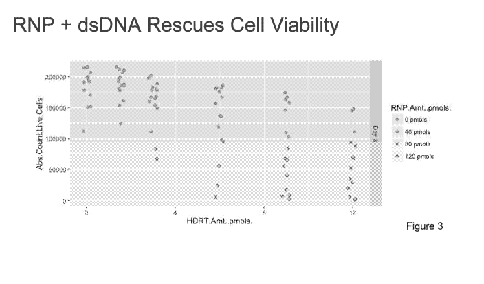
[0014] Figure 3 shows that complexing the DNA template (linear, double-
stranded DNA
(dsDNA) template) with the RNP, by a brief room temperature incubation prior
to addition of
cells when electroporating, reduces the viability loss normally seen upon
electroporation of
long, linear, double-stranded DNA.
[0015] Figure 4 shows that an exemplary molar ratio of about 10:1 RNP to
DNA template
maintains both efficiency of integration as well as viability, post
electroporation.
[0016] Figure 5 shows that an exemplary molar ratio of about 10:1 RNP to
DNA template
balances the effects of viability loss and efficiency, and maximizes the
number of integration
positive cells.
[0017] Figure 6 shows that an exemplary molar ratio of about 10:1 RNP to
DNA template
allows for high efficiency insertion of large templates greater than about 750
base pairs in
size.
[0018] Figure 7 shows that insertion of long DNA templates can still result
in an amount
of off-target integration.
[0019] Figure 8 shows that off-target integration can be reduced by using a
long single-
stranded DNA (ssDNA) template as a donor..
[0020] Figure 9 shows that the non-viral integrations disclosed herein can
be inserted
using two gRNAs and a Cas9 nickase (D10A), which prevents off target dsDNA
breaks.
[0021] Figures 10A-F show that CRISPR/Cas9 RNP co-electroporation reduces
dsDNA
induced viability loss. (A) A linear dsDNA template (a homology directed
repair template,
¨1350 bps long, targeting a GFP fusion to RAB11A, Fig. 11A) electroporated
into primary
human T cells cause marked viability loss with increasing amounts of
template. Electroporation of the same amount of dsDNA template along with 100
pmols of
RNP surprisingly increased viability. (B) For both plasmid and linear dsDNA
templates,
addition of an RNP increased viability post electroporation. Of note, no loss
in viability was
seen with short ssDNA oligo donor nucleotides (ssODNs). (C) RNPs must be
delivered
concurrently with DNA to see increased viability. T cells from two donors were
each
electroporated twice with an eight hour rest in between electroporations.
While two
electroporations so closely interspersed caused a high degree of cell death,
delivery of the
RNP and linear dsDNA template could be delivered separately. However, an
initial RNP
4
CA 03067382 2019-12-13
WO 2018/232356
PCT/US2018/037919
electroporation did not increase viability when a DNA template was
subsequently
electroporated in comparison to cells that received DNA first and RNP second.
(D-F) Given
that the RNP and DNA needed to be introduced concurrently, we assayed whether
additional
pre-incubation together before electroporation would further increase
viability. No difference
in viability was seen with increasing pre-incubation time (0 to 15 minutes),
but surprisingly if
the RNP and cells were mixed first and the DNA template was added immediately
prior to
elecroporation (RNP + Cells; + HDRT) viability was increased (E). However, in
wells where
the RNP and the DNA HDR template were mixed together prior to adding the cells
(RNP +
HDRT; + Cells), no matter how long the RNP and DNA template were preincubated,
there
was a drastic increase in HDR percentage (GFP+ cells). Viability was measured
2 days
following electroporation and GFP expression was measured at day 4. Graphs (B,
D, F)
display data from 2 healthy human donors.
[0022] Figures 11A-F show the development of efficient large non-viral gene
targeting.
(A) Systematic analysis of the effects of cell culture and stimulation
conditions, RNP and
DNA template formulations, and electroporation conditions via 96-well high-
throughput
electroporations enabled rapid optimization of both cell viability (total
number of live cells in
culture) and HDR efficiency (% of cells GFP positive). (B) Schematic of a long
(1350bp)
linear dsDNA template encoding a GFP sequence flanked by regions homologous to
the N-
terminus of the housekeeping gene RAB11A (not drawn to scale). When a dsDNA
break is
induced at the N-terminus of RABI1A, the GFP sequence can be seamlessly
introduced via
homology directed repair (HDR) to generate an endogenously-tagged RAB11A-GFP
fusion
protein. (C) Primary human T cells were cultured for 2 days using varying
combinations of
T cell receptor (TCR) stimulation and cytokines prior to electroporation of
RABI1A targeting
RNP and HDR template, followed by varying culture conditions for 5 days post-
electroporation. (D) Among RNP and HDR template concentrations tested here,
optimal GFP
insertion into RAB11A was achieved at intermediate concentrations of the
reagents. Further
testing (Fig. 16) narrowed optimal concentrations to 50 pmols of RNP and 4 ugs
of dsDNA
HDRT. (E) Arrayed testing of electroporation pulse conditions showed that, in
general,
conditions yielding higher HDR efficiency decreased viability. EH115 was
selected to
optimize HDR, while still maintaining sufficient viability. (F) Using
parameters optimized in
C-D, high-efficiency insertion of GFP into the endogenous RAB11A gene was
achieved by
non-viral targeting in both primary human CD4+ and CD8+ T cells. Viability and
efficiency
were assayed 3 days (E) or 5 days (C, D, and F) after electroporation.
Individual points
CA 03067382 2019-12-13
WO 2018/232356
PCT/US2018/037919
represent individual blood donors (C and D) or the mean plus standard
deviation in two
individual donors (E). Green highlights indicate conditions ultimately chosen
for the non-
viral gene targeting protocol.
[0023] Figures 12A-B show non-viral gene targeting enables rapid and
efficient
genetic engineering in primary human T cells. (A) Diagrammatic timeline of non-
viral gene
targeting. Approximately one week is required to design, order from commercial
suppliers,
and assemble any novel combination of genomic editing reagents (gRNA along
with
homology directed repair template). Two days prior to electroporation, primary
human T
cells isolated from blood or various other sources (Fig. 15) are stimulated.
dsDNA HDR
templates can be made easily by PCR followed by a SPRI purification to achieve
a highly
concentrated product suitable for electroporation. On the day of
electroporation, the gRNA
complexed to an RNP, the HDR template, and harvested stimulated T cells are
mixed and
electroporated, a process taking approximately one and a half hours. After
electroporation,
engineered T cells can be readily expanded for an additional two weeks. (B)
Viability is used
to refer to the percentage of live cells relative to an equivalent population
that went through
all protocol steps except for the actual electroporation (No electroporation
control). The
trough in live cells after electroporation was empirically determined to come
two days
following, and all viability measures have been recorded at that time point
unless otherwise
noted. The term efficiency is used to refer to the percentage of live cells in
culture expressing
the "knocked in" exogenous sequence (such as GFP). Finally, the total number
of cells
positive for the desired integration was calculated by multiplying the
efficiency by the
absolute cell count. Methodological changes that maximized efficiency often
were not
always optimal for the total number of positive cells, and vice-versa.
[0024] Figures 13A-D show optimization of primary human T cell stimulation
for non-
viral gene targeting. (A) Alternative pre-electroporation stimulation
conditions were applied
for two days prior to electroporation. CD3/CD28 bead bound stimulation along
with a
cytokine stimulation cocktail of IL-2, IL-7, and IL-15 achieved higher
viability, rates of
editing, and total positive cells than plate bound antibody stimulation. (B)
Alternative ratios
of beads to cells showed an optimal 1:1 ratio along with removal of beads
prior to
electroporation. (C) Non-bead based CD3/CD28/CD2 stimulation yielded lower
editing
efficiencies than CD3/CD28 beads at optimal ratio. (D) Commercial XVivo15
media
achieved similar viability but higher editing efficiencies compared to RPMI.
Of interest, the
serum-free Immunocult media also enabled high-efficiency editing of human
primary CD3+
6
CA 03067382 2019-12-13
WO 2018/232356
PCT/US2018/037919
T cells. Efficiency of GFP insertion (dsDNA RAB11A-GFP HDRT) and the absolute
count
of total GFP+ cells was performed 4 days following electroporation. Two dots
per condition
represent the values obtained from two healthy blood donors.
[0025] Figures 14A-D show optimization of primary human T cell handling
post-
electroporation. (A) Electroporation of CD3+ T cells from healthy human donors
at day 2 or
day 3 post stimulation achieved efficient targeted GFP integration. Dual
electroporations at
both days, while increasing efficiency slightly, drastically reduced the
viability when a DNA
template was included in the two electroporations (Fig. 10). (B) Additional
CD3/CD28
stimulation after electroporation reduced proliferative potential. (C) High
doses of IL-2 post-
electroporation improved both efficiency and viability. Further addition of IL-
7 and IL-15,
unlike during pre-electroporation stimulation (Fig. 13) did not contribute to
improved editing.
(D) Post culture density has little effects on insertion efficiency.
Efficiency of GFP insertion
(dsDNA RAB11A-GFP HDRT) and the absolute count of total GFP+ cells was
performed 4
days following electroporation. Two dots per condition represent the values
obtained from
two healthy blood donors.
[0026] Figures 15A-B show efficient non-viral gene targeting in fresh and
frozen T cells
isolated from multiple sources. (A) A dsDNA RAB11A-GFP HDR template was
inserted
into both fresh and frozen T cells from two healthy donors. High rates of GFP
insertion were
seen in both conditions, demonstrating the adaptability of non-viral gene
targeting to research
or clinical protocols that require freezing of cells. (B) Similarly, high
efficiencies of GFP
targeted integration were seen in primary human CD3+ T cells isolated from
whole blood, a
plasma apheresis residual, as well as leukapheresis.
[0027] Figures 16A-B show optimization of RNP and HDR template formulations
for
non-viral gene targeting. (A) Across three donors, a consistent trend appeared
that
electroporation of increasing amounts of dsDNA HDR template (RAB11A-GFP)
gradually
reduced cell viability, while also increasing efficiency, but that
intermediate concentrations
tested of both HDR template and RNP gave the greatest total number of GFP+
cells. (B)
Further targeted optimization series in three additional donors yielded an
optimal formulation
of 4 ugs of HDR template electroporated concurrently with 50 pmols of RNP.
Efficiency of
GFP insertion and the absolute count of total GFP+ cells was performed 4 days
following
electroporation. Multiple dots per graph (B) represent technical replicates.
[0028] Figures 17A-C show optimization of electroporation parameters for
delivery of
large non-viral HDR templates. (A) Raw data shown here is summarized in Fig
7
CA 03067382 2019-12-13
WO 2018/232356
PCT/US2018/037919
11E. Systematic variation of electroporation conditions on a Lonza 4D
nucleofector. The
ultimately chosen pulse code, EH115, was consistently the most efficient code
when using
the electroporation buffer Lonza P3. Other alternative codes, such as EO-148
optimized for
total positive cells. (B) Confirmatory testing of a subset of electroporation
conditions also
identified pulse code EO-155 in OMEM buffer as a moderate efficiency but high
total
positive cell combination. (C) Electroporating a total volume (RNP + HDRT +
Cells) of 24
uL made a large contribution to cell viability and maintained high efficiency.
Electroporation
volumes above 24 uL commonly cause electroporation failures. Efficiency of
dsDNA
RAB11A-GFP insertion (A, C) or dsDNA BATF-GFP insertion (B) and the absolute
count of
total GFP+ cells was performed 4 days following electroporation.
[0029] Figures 18A-D show the diverse applications of non-viral gene
targeting in
primary human T cells. (A) High efficiency genome targeting with GFP-fusion
constructs
could be achieved in multiple endogenous genes in primary human T cells using
non-viral
HDR templates and corresponding RNPs. (B) Confocal microscopy of living,
primary
human T cells 7 days after electroporation of the indicated HDR template
confirmed the
specificity of fusion-protein targeting. Scale bar in each image is 5 um. (C)
Non-viral
targeting of GFP-fusion constructs to the RAB11A and CD4 genes in bulk human
primary T
cells. RAB11A-fusions were GFP positive in both CD4+ and CD8+ cells, whereas
CD4+-
fusions were only positive in CD4+ T cells (representative flow cytometry
above,
quantification below). (D) Primary human T cells were engineered to express
GFP fused to
the endogenous transcription factor, BATF. At 11 Days post electroporation,
nuclei were
isolated and CUT&RUN was performed. GFP-BATF and total BATF chromatin
interaction
sites were identified using anti-GFP or anti-BATF antibodies. Flow cytometry
to assay
viability and efficiency was performed at 4 days after electroporation (A, C,
D). Displayed
data is representative of at least two different donors.
[0030] Figures 19A-B show reproducible non-viral gene targeting across
target loci.
(A) Four days after electroporation of one of five different GFP templates
along with a
corresponding RNP into primary CD3+CD8+ T cells from six healthy donors, GFP
expression is observed across both templates and donors. Note the consistency
in GFP
expression levels within GFP positive cells across donors for each of the five
loci (higher in
TUBA1B and ACTB, lower in RAB11A and FBL tags). (B) Graphical summary of the
percentage of GFP insertion in (A).
8
CA 03067382 2019-12-13
WO 2018/232356
PCT/US2018/037919
[0031] Figures 20A-B show reproducible non-viral gene targeting in a cohort
of healthy
donors. (A) A constant dsDNA RAB11A-GFP HDR template and RNP was
electroporated
using the optimized conditions developed for non-viral gene targeting in cells
obtained from
a cohort of twelve healthy donors. While there was significant variability in
GFP insertion
percentage among individual donors, all achieved robust integration of GFP
(range 22% to
57% in CD8+ T cells). Some GFP expression was seen in cells electroporated
with the
dsDNA RAB11A-GFP HDR template with an off-target RNP targeting CXCR4 compared
to
no-electroporation controls. (B) Summary graph of GFP insertion percentages in
(A). Across the 12 healthy donor cohort slightly higher rates of in GFP
expression was seen
in CD3+CD8+ T cells (mean 42.0%) compared to CD3+CD4+ T cells (mean 35.2%).
[0032] Figure 21 shows endogenous tagging of transcription factor BATF for
analysis of
chromatin occupancy. Anti-BATF, anti-GFP, and no antibody heatmaps of CUT&RUN
data
obtained from primary human T cell populations electroporated with GFP-BATF
fusion HDR
template (untagged cells were not electroporated). Aligned CUT&RUN binding
profiles for
each sample were centered on BATF CUT&RUN peaks in untagged cells and ordered
by
BATF peak intensity in untagged cells.
[0033] Figures 22A-E show combinatorial non-viral gene targeting. (A)
Simultaneous
electroporation of HDR templates to create RAB11A-GFP and/or RAB11A-mCherry
fusions
in primary human T cells. A distinct population of dual GFP+ mCherry+ cells
was found
when both templates are introduced concurrently, consistent with bi-allelic
targeting. (B) The
potential genotypes for individual cells in the quadrants are defined by
expression of the two
fluorophores. The observed level of bi-allelic integrations is higher in cells
that acquire at
least one integration than expected by chance (Fig. 23). Individual points
represent replicates
where the combination of the genes encoding the fluorescent proteins was
varied (GFP +
mCherry, GFP + BFP, mCherry + BFP) as was the amount of HDR template (3 to 6
ugs). (C-D) Multiplexed integration of HDR Templates at two separate genomic
loci in the
same primary human T cells. 2 ugs of each template (4 ugs total per
electroporation) were
electroporated together with 25 pmols of each RNP (50 pmols total). Cells
positive for
integration at one site (e.g. GFP+) were much more likely to have an
integration at the second
site (e.g. also be mCherry+) than cells lacking the first integration. (E)
Simultaneous non-
viral gene targeting of large insertions to three distinct genomic loci. 1.5
ugs of each
template (4.5 ugs total) were electroporated together with 20 pmols of each
corresponding
RNP (60 pmols total). Similarly to two site multiplexing, cells positive for a
single
9
CA 03067382 2019-12-13
WO 2018/232356
PCT/US2018/037919
integration (mCherry+ in Q-II and GFP+ in Q-III) were more likely to have a
second
integration (BFP+) compared to those without (Q-I). Cells positive for two
integrations
(GFP+ and mCherry+, Q-IV) are even more likely to have an integration of the
third gene
(BFP+). Below is a bar graph quantification of cells that are single, double
and triple positive
for fluorophores. All fluorescent readouts were performed 4 days post-
electroporation.
Displayed data are representative of at least two different donors except
panel E (one donor).
[0034] Figures 23 A-G show modeling and analysis of bi-allelic HDR
integrations by
insertion of multiple fluorescent proteins into the same locus. (A) The
possible cellular
phenotypes when two fluorescent proteins are inserted into the same locus. (B)
The
genotypes of two of these phenotypic populations are immediately known. Cells
without any
functional insertions (bottom left quadrant, genotype A), must have a NA/NA
genotype
(where NA indicates an allele without HDR, including WT alleles and NHEJ
edited
alleles). Dual fluorescent cells (top right quadrant, genotype E) must have
acquired one copy
of each template (assuming an autosomal target locus and no off-target
integrations), and
would have a genotype of GFP/RFP. The two single positive populations though
will be
mixed between cells heterozygous for HDR insertion (Genotypes B and C) or
homozygous
but for two copies of the same fluorescent template (Genotypes D and F). (C)
The total
percentage of cells with bi-allelic HDR integrations must be the sum of
genotypes D, E, and
F. While the proportion of cells with genotype E (dual fluor positives) is
immediately
apparent from the phenotypes, genotypes D and F are not. Application of a
simple
probability model allow for the de-convolution of the multiple genotypes in
the single fluor
positive phenotypes, and thus an estimation of the true percentage of cells
homozygous for
HDR. (D) Bi-allelic HDR analysis applied across a variety of fluorophore
permutations
inserted into the RAB11A locus. (E-F) Dual fluorescence bi-allelic
integrations were seen
across target loci. While the total percentage of cells with an insertion
varied with the
efficiency of each target site, the fold enrichment in the observed percentage
of homozygous
cells over that predicted by random chance was consistent across loci. (G)
Attempted
integration of three distinct fluorophores by HDR into the same locus. As a
max of two
targeted insertions are possible (at the locus' two alleles; assuming a
diploid genome), no
cells positive for all three loci should be observed (triple positives).
Indeed, while large
numbers of single fluorophore integrations are observed (single positives), as
well as cells
positive for the various permutations of two fluors (double positives), there
is a 30 fold
reduction in the number of triple positive cells compared to double positives.
All flow
CA 03067382 2019-12-13
WO 2018/232356
PCT/US2018/037919
cytometric analysis of fluorescent protein expression was performed 4 days
following
electroporation. Displays are representative of multiple technical replicates
from one (E, F)
or two (D, G) healthy human donors. Bar graphs display mean + standard
deviation.
[0035] Figures 24A-B show multiplexed integrations showing that acquisition
of HDR
integration at one locus increases likelihood of HDR at additional loci. (A)
Two HDR
template permutations from a set of six dsDNA HDR templates (targeting RAB11A,
CD4,
and CLTA; each site with GFP or RFP) were electroporated into CD3+ T cells
isolated from
healthy human donors. Four days after electroporation of the indicated two HDR
templates
along with their two respective on-target RNPs, the percentage of cells
positive for each
template was analyzed when gating on cells either positive or negative for the
other
template. Not only was two-template multiplexing possible across a variety of
template
combinations, but gating on cells positive for one template (Template 1+
Cells,) yielded an
enriched population of cells more likely to be positive for the second
template compared to
cells negative for the first (Template 1- Cells, Black). 2 ugs of each
template, along with 30
pmols of each associated RNP, were electroporated for dual multiplexing
experiments. (B)
Electroporation of an additional template allows for 3 site multiplexing using
a variety of
HDR template combinations. Cells positive for the third template can be
further enriched by
gating on cells positive for both other templates when compared to single
positive
cells. Displayed data are means + standard deviation from multiple technical
replicates from
two healthy human donors.
[0036] Figures 25A-F show DlOA nickase and ssDNA HDR templates reduce off-
target
integrations. (A) Combinations of Cas9 RNPs and a RAB11A-GFP dsDNA HDR
template
were electroporated into primary human T cells. dsDNA template alone, or with
an RNP
containing a scrambled gRNA matching no sequence in the human genome yielded
small but
detectable amounts of GFP expression, which was noticeably increased when a
dsDNA
template is electroporated with a gRNA targeting a site different from the
targeted RAB11A-
GFP integration site (the "off-target RNP" targets CXCR4 Exon 1). (B) Off-
target
integrations were consistently present in cells from different donors when the
RAB11A-GFP
dsDNA HDR template was electroporated with the off-target RNP, and fewer off-
target
integrations occurred when the dsDNA HDR template alone was electroporated.
(C) Cas9
nuclease variants DlOA (nickase) and inactive dCas9 significantly decreased
off-target
integrations when a single off-target CXCR4 gRNA was used, but Dl OA nickase
(with an
"On-target" pair of gRNAs in a PAM-out orientation) led to efficient on-target
integration of
11
CA 03067382 2019-12-13
WO 2018/232356
PCT/US2018/037919
the RAB11A dsDNA HDR template. (D) Electroporation of a ssDNA HDR template
reduced
the off-target integrations to the limit of detection (comparable to levels
with no template
electroporated) both with no nuclease added and at induced off-target dsDNA
breaks (Off-
target gRNA + Cas9). (E-F) For integration of GFP fusion at the RAB11A site,
use of a Dl OA
nickase with a ssDNA HDR template reduced the on-target HDR (GFP integration
with on-
target gRNA) compared to Cas9 with a dsDNA template, but strongly reduced off-
target
integrations to undetectable levels. All fluorescent readouts were performed 4
days post-
electroporation. Displayed data is representative of at least two different
donors (A and E) or
the averages of two different donors (C, D, and F) with standard deviation
shown
[0037] Figures 26A-D show fluorescent estimation and quantification of off-
target
integration events across multiple HDR templates. (A) Diagram of HDR mediated
insertions
at the N-terminus of a target locus (not drawn to scale). The homology arms
specify the
exact sequence where the insert (a GFP tag in this case) will be inserted,
allowing for scarless
integration of exogenous sequences. As a GFP fusion protein is created, GFP
fluorescence
will be seen as a result of this on-target integration, dependent on an RNP
cutting adjacent to
the integration site. (B) Double stranded DNA can be integrated via homology-
independent
repair mechanisms at off-target sites through either random integration at
naturally occurring
dsDNA breaks, or potentially at induced double stranded breaks, such as those
at the off-
target cut sites of the RNP. This effect can be harnessed to allow for
targeted integration of a
dsDNA sequence at a desired induced dsDNA break (HITI) in senescent cell types
lacking
the ability to do HDR, but crucially the entirety of the dsDNA template is
integrated,
including any potential homology arms. In the case that the homology arms
contain a
promoter sequence (such as for N-terminal fusion tags), these off target
integrations can drive
observable expression of the inserted sequence without the desired correct HDR
insertion. (C) Bars represent real GFP+ percentages from human CD3+ T cells
electorporated with the indicated components. Flow cytometry for fluorescent
protein
expression can be used to rapidly evaluate functional off-target integrations.
The increase in
the percentage of fluorescent cells over the limit of detection when the
template alone is
electroporated likely represents random integrations at naturally occurring
dsDNA
breaks. Not every off-target integration will yield fluorescent protein
expression, but the
relative differences in functional off-target expression between different
templates can be
assayed. Inclusion of an RNP targeting CXCR4 dramatically increases the
observed off-
target homology-independent integrations, likely through a HITI-type insertion
event. The
12
CA 03067382 2019-12-13
WO 2018/232356
PCT/US2018/037919
largest increase (from 1% to >30% in this donor) comes though through
electroporation of
the correct RNP and HDR mediated insertion. (D) Comparisons of on-target GFP
expression
(right column) vs functional off-target integrations (middle column) across
five
templates. Mean expression (bars) of two biologic donors (dots) are graphed.
[0038] Figures 27A-B show GFP expression across a HDR template versus gRNA
matrix. (A) GFP expression was analyzed in CD3+CD4+ primary human T cells from
a
healthy donor 7 days following electroporation of a matrix of dsDNA HDR
templates and
their corresponding gRNAs, along with a CXCR4 gRNA and a no RNP control. As
expected
with a dsDNA template, off-target integrations were seen across combinations,
but for all
gRNAs and HDR templates the highest GFP expression was seen in the on-target
condition.
(B) Heat map summary of flow cytometry data in (A). One HDR template, a C-
terminal GFP
fusion tag into the nuclear factor FBL, had consistently higher off-target
expression across
gRNAs.
[0039] Figures 28A-D show efficient HDR in primary human T cells using a
Cas9
nickase. (A) Diagram of the genomic locus containing the first exon of RAB11A.
Use of
spCas9 with a single guide RNA (gRNA 1) along with a dsDNA HDR template
integrating a
GFP in frame with RAB11A directly after the start codon results in efficient
GFP expression
(Fig 11F). Use of a Cas9 nickase (D10A variant) with two gRNAs could reduce
the chances
of off-target cutting. (B-C) A series of single gRNAs as well as dual gRNA
combinations
were tested for GFP insertion efficiency at the RAB11A N-terminal locus. As
expected, no
gRNAs showed appreciable levels of GFP insertion when using a nuclease dead
Cas9
(dCas9). Multiple single gRNAs cutting adjacent to the insertion site showed
GFP
integration when using Cas9, but none as efficiently as gRNA 1. The DlOA
nickase showed
little to no GFP integration with single guides, but with multiple two-guide
combinations
showed efficient GFP integration. Only in gRNA combinations where the two PAM
sequences were directed away from each other (PAM Out) was GFP integration
seen. (D)
Raw data presented in (Fig 25C) demonstrating lower levels of functional off-
target
integrations when electroporating an off-target gRNA (targeting CXCR4), likely
due to the
requirement for the DlOA nickase to have two gRNAs binding in close proximity
to induce a
dsDNA break. Dots in all displays (B-D) represent technical replicates in the
labeled two
healthy donors.
[0040] Figures
29A-H show reduced Treg frequencies and defective Treg suppressive
capacity in subjects with two loss of function IL2RA mutations. (A) CD3+CD4+ T
cells
13
CA 03067382 2019-12-13
WO 2018/232356
PCT/US2018/037919
from a healthy donor and all family members, including IL2RA heterozygotes
(c.530 het 1,
c.800 hets 1-3) as well as compound heterozygote children (Comp. Hets 1-3),
with loss-of-
function IL2RA mutations were analyzed by flow cytometry to assess presence of
CD25hiCD12710 Tregs. (B) In healthy donors and single hets, CD4+FoxP3+ T cells
are
predominantly CD25hiCD1271o. In the compound heterozygotes, a CD12710
CD4+FoxP3+
population is present, but does not express IL2RA. (C) Clinical phenotyping
performed at
two separate sites confirms compound heterozygotes possess normal frequencies
of CD12710
FoxP3+ cells. (D) Deficiency in IL2RA surface expression in compound
heterozygote 3 led
to aberrant downstream signalling as measured by pStat5 expression after
stimulation with
IL-2, but not IL-7 or IL-15. (E) Due to the inability to sort CD25hi Tregs
from the CD25-
deficient compound heterozygotes, an alternate gating strategy was established
to enrich for
FoxP3+ cells from CD3+CD4+ T cells using the surface markers
CD1271oCD45R0+TIGIT+. Intracellular FoxP3 staining from the indicated gated
population is shown. (F) While these CD3+CD4+CD1271oCD45R0+TIGIT+ potential
"Tregs" were highly enriched for FoxP3 and showed some suppressive capacity
when
cultured with CF SE-labeled stimulated responder T cells (Tresps) from healthy
donors,
CD3+CD4+CD1271oCD45R0+TIGIT+ from the compound heterozygotes showed no
suppressive ability. Stimulated Tresp population (Solid curves), non-
stimulated Tresp
(Dashed curve). (G) Correction of either CD25 mutation in the compound
heterozygotes
individually would still leave the other mutation, leaving the cells as single
heterozygotes. To confirm that such a potential correction would result in
some level of
functional suppression, CD4+CD25hiCD12710 Tregs from the c.530 and c.800
single
heterozygote family members were isolated and their suppressive ability was
assayed as in
(F). (H) Dot plot summaries of Treg suppressive ability in cells from healthy
donor, CD25-
deficient compound heterozygotes (F) and CD25+/- c.530 or c.800 heterozygotes
(G). While
CD3+CD4+CD1271oCD45R0+TIGIT+ "Tregs" from compound heterozygotes showed no
suppressive ability, conventional CD4+CD25hiCD12710 Tregs from the single
heterozygote
family members showed some suppressive capacity, consistent with their lack of
pronounced
clinical phenotype compared to the compound hets.
[0041] Figures 30A-E show monogenic autoimmune mutation corrected by non-
viral
gene targeting in primary human T cells. (A) Three siblings in a family carry
two different
IL2RA (encoding high-affinity IL-2 receptor, CD25) mutations (c.530A>G
creating a stop
codon in IL2RA exon 4; c.800delA, creating a frameshift mutation in IL2RA exon
8 which
14
CA 03067382 2019-12-13
WO 2018/232356
PCT/US2018/037919
causes an almost 100 amino acid run-on). (B) These three compound heterozygote
siblings
show greatly reduced, but not completely absent, cell surface expression of
IL2RA on their
primary T cells. Non-viral gene targeting of the c.530 mutation by
electroporation of a Cas9
RNP and a dsDNA HDR template containing the correct IL2RA sequence (along with
a silent
mutation in the targeted PAM sequence) successfully rescued IL2RA cell surface
expression
in a portion of T cells from each compound heterozygote sibling 2 days
following
electroporation. (C) 7 days after non-viral gene targeting, targeted T cells
showed increased
phosphorylation levels of Stat5 upon IL-2 stimulation compared to non-targeted
controls. (D) 9 days following non-viral gene targeting to correct the c.530
mutation,
IL2RA+ T cells from the three compound heterozygote donors include an
increased level of
FoxP3+ cells compared to non-targeted cells or healthy donor cells. (E) Non-
viral gene
targeting and correction of the c.530 mutation is possible and efficient using
an optimized
therapeutic reagent set (D10A nickase along with ssDNA HDR template). T cells
from one
compound heterozygote donor were stained for IL2RA surface expression after 9
days of ex-
vivo expansion following electroporation (2 days following re-stimulation).
[0042] Figures 31A-D show identification of compound heterozygous mutations
in
IL2RA and design of corrective CRISPR-Cas9 genome targeting reagents. (A)
Initial genetic
testing of the proband using an in-house targeted next-generation sequencing
multi-gene
panel of over 40 genes known to be involved in monogenic forms of diabetes was
negative.
Subsequent exome sequencing in the trio of proband and parents revealed two
causative
mutations in the IL2RA gene. The mother possessed a single heterozygous
mutation
(c.530G>A) in exon 4 of IL2RA (SEQ ID NO: 1) (AGACAAGGTRGACCCAGCC),
resulting in a premature stop codon. (B) The father possessed a single
heterozygous mutation
(c.800delA) in exon 8 of IL2RA (SEQ ID NO: 2 (ACAGGAGGARRRKWRRARAA),
resulting in a frameshift mutation resulting in a 95 amino acid long run-on.
Sanger
sequencing confirmed that the proband was a compound heterozygote for both
mutations. (C) A linear depiction of the IL2RA protein annotated with
approximate locations
of the two identified IL2RA mutations. SD', sushi domain 1; 5D2, sushi domain
2; TM,
transmembrane; C, cytoplasmic. (D) The genomic sequences including the
specified
mutations ((SEQ ID NO: 3)(CAAAATGACCCACGGGAAGACAAGGTAGACCC) for
c.530G>A allele and SEQ ID NO: 4 (GACTTTGTTACACCACTACAGGAGGAGAGTA)
for c.800delA Allele)) were used to design CRISPR-Cas9 genome targeting
reagents to
correct the two IL2RA mutations. A gRNA was designed to cut adjacent to the
site of each
CA 03067382 2019-12-13
WO 2018/232356
PCT/US2018/037919
mutation, 8 bps away for c.530 mutation, and 7 bps away for c.800. For each
mutation, an
HDR template ((SEQ ID NO: 5) (ACAAGATGGACCC) for c.530 mutation and (SEQ ID
NO: 6)(AGGAGAAAGAGTA for c.800)) was designed including the corrected sequence
as
well as a silent mutation in a degenerate base to disrupt the PAM sequence
("NGG") for each
guide RNA. The corrected allele + silent PAM disruption sequence for c.530
(CAAAATGACCCACGGGAAGACAAGATGGACCC) (SEQ ID NO: 7) and c.800 (SEQ
ID NO: 8) (GACTTTGTTACACCACTACAGGAGAAAGAGTA) are shown. Displayed
genomic regions (not to scale) for c.530 mutation site (hg38 ch10:6021526-
6021557) and
c800 mutation site (hg38 ch10:6012886-6012917). Both ssODN HDR Templates
(ssDNA
with 60 bp homology arms), and large dsDNA or ssDNA HDR Templates (as
displayed, with
¨300 bp homology arms) were used.
100431 Figures 32A-C show HDR mediated correction of IL2RA c.530A>G loss of
function mutation. (A) Unlike the gRNA targeting the c.800delA mutation at the
C-terminus
of IL2RA, the gRNA targeting the c.530A>G mutation (causing a stop codon in an
interior
exon) results in substantial (-90%) knockdown of IL2RA in a healthy donor and
single
heterozygotes (c.800 Het 2 and 3) 2 days following electroporation of the RNP
alone (Blue)
into CD3+ T cells. While starting from a very small IL2RA+ percentage,
knockdown was
also observed in all three compound heterozygotes, potentially as some small
amount of
protein can be surface expressed off of the c.800delA allele. This reduced
CD25 expression
can be partially rescued by inclusion of an ssODN HDR template and even more
substantially
rescued using a large dsDNA HDR template. Both template types contained the
corrected
sequence, a silent mutation to remove the gRNA's PAM sequence, and either 60
bp
(ssODNs) or ¨300 bp (large dsDNA) homology arms (Fig. 32). Unlike targeting of
the
c.800delA mutation for correction, CD25 surface expression in T cells from the
compound
heterozygotes is only seen when an HDR template is included. In all three
compound
heterozygotes, the dsDNA HDR template yielded greater percentages of CD25+
cells. (B)
Increased pStat5 signaling in response to IL-2 stimulation (200 U/mL) 7 days
following
electroporation in CD3+ T cells from compound heterozygote patients undergoing
HDR-
mediated mutation correction compared to no electroporation or RNP only
controls. (C)
Similarly, increased proportions of CD25+ FoxP3+ cells are seen 9 days
following
electroporation in the HDR correction conditions in compound heterozygote
patients. Lower
percentages of correction were seen when targeting the c.530 mutation for HDR
correction in
compound heterozygote 3, potentially due altered cell-state associated with
the patient's
16
CA 03067382 2019-12-13
WO 2018/232356
PCT/US2018/037919
disease or the patient's immunosuppressive drug regimen. Electroporations were
performed
according to optimized non-viral genome targeting protocol set forth in the
examples. For
ssODN electroporations, 100 pmols in luL H20 were electroporated.
[0044] Figures 33A-C show non-HDR mediated correction of IL2RA c.800delA
frameshift loss of function mutation. (A) Histograms of CD25 surface
expression in CD3+ T
cells in all children from a family carrying two loss-of-function IL2RA
mutations, including
three compound heterozygotes that express minimal amounts of IL2RA on their
surface (No
electroporation, Grey). Two days following electroporation of an RNP
containing a gRNA
against the site of one of the two mutations, a one base pair deletion in the
final exon of
IL2RA (c.800delA) causing a run-on past the normal stop codon, CD3+ T cells
from a healthy
donor and single hets (c.800 Het 2 and 3) show slight increase in CD25- cells
(RNP only,
Blue). Low knock-out is potentially due to the gRNA targeting the C-terminus
of the protein
where small indels may cause less pronounced loss of surface protein
expression. Surprisingly, the RNP alone resulted in CD25 surface expression in
almost 50%
of edited T cells in all three compound heterozygotes. Increases in the
percent of cells with
CD25 correction compared to RNP only could be achieved by inclusion of an
ssODN HDR
template sequence with the mutation correction (RNP+ssODN, Purple), and
further increased
when using a longer dsDNA HDR template to correct the mutation (RNP + dsDNA
HDRT,
Green) (Fig. 32). (B) Phospho Stat5 signaling in response to high dose IL-2
stimulation (200
U/mL) in edited CD3+ T cells following 7 days of expansion post-
electroporation. Increased
numbers of pStat5+ cells correlated with increases in CD25 surface expression
(A). (C)
Following 9 days of expansion post-electroporation, intracellular FoxP3
staining reveals a
dramatically increased proportion of CD25+ FoxP3+ cells in CD3+ T cells
compared to no
electroporation controls, approaching the proportion of CD25+ FoxP3+ cells
seen in a
healthy donor similarly cultured. Electroporations were performed according to
optimized
non-viral genome targeting protocol (Examples). For ssODN electroporations,
100 pmols in
luL H20 were electroporated.
[0045] Figures 34A-B show diminished HDR potential and altered clinical
phenotype in
compound heterozygote IL2RA loss-of-function patient receiving
immunosuppressants. (A)
Flow cytometric analysis of GFP expression 6 days following electroporation of
a positive
HDR control RAB11A-GFP dsDNA HDR template into CD3+ T cells from the indicated
patients revealed lower GFP expression in the three compound heterozygotes
compared to
their two c.800 heterozygote siblings. Compared to a cohort of twelve healthy
donors
17
CA 03067382 2019-12-13
WO 2018/232356
PCT/US2018/037919
similarly edited (Fig. 20), both c.800 heterozygotes as well as compound het 1
and 2 were
within the general range observed across healthy donors, whereas compound het
3 had lower
GFP expression than any healthy donor analyzed. Of note, while in compound het
3 HDR
mediated correction at the c.530 mutation was substantially lower than the
other two
compound hets (Fig 31A), CD25 surface expression after electroporation of the
c.800delA
targeting RNP alone was similar. Unlike HDR mediated repair, a NHEJ mediated
frameshift
correction at c.800delA may not require cell proliferation, consistent with
compound het 3
being the only compound heterozygote patient on active immunosuppressants at
the time of
blood draw and T cell isolation. (B) Altered cell-state associated with the
patient's disease
could also be contributory to diminished HDR rates. TIGIT and CTLA4 expression
levels in
non-edited, isolated CD4+ T cells from each indicated patient measured by flow
cytometry. Consistent with altered activation state, cells from compound het 3
had a distinct
phenotype, with increased TIGIT and CTLA4 expression compared both to healthy
donors,
the heterozygous family members, as well the other two compound heterozygous
siblings.
[0046] Figures 35A-B show multiple methods to produce long ssDNA HDR
templates.
(A) If a large enough amount of long single stranded DNA sequence could be
produced for
electroporation, off-target integrations could be reduced without overly
compromising on-
target efficiency. One method involves a two-step selective exonuclease
digestion that
specifically degrades one strand of a PCR product that has been labeled by 5'
phosphorylation, easily added onto a PCR primer prior to amplification. (B) A
second
ssDNA production method based on sequential in-vitro transcription (IVT) and
reverse
transcription (RT) reaction was also applied. A PCR product with a short T7
promoter
appended serves as an IVT template to produce a ssRNA product. Following
annealing of an
RT primer and reverse transcription, an RNA/DNA hybrid is formed which can be
easily
transformed into a long ssDNA template by incubation in sodium hydroxide
(selectively
degrades RNA strand). (C) At 2 days post-electroporation, viability in CD3+ T
cells
electroporated with only a ssDNA template was higher than those electroporated
with only a
dsDNA template (Fig. 11). (D) A ssDNA RAB11A-GFP HDR template showed high
efficiency GFP integration similar to dsDNA templates, and maintained high
efficiency
integrations at higher molar amounts of template, potentially due to increased
viability (C) as
well as less mass per mole of DNA template. Individual points represent at
least two healthy
donors (C, D)
18
CA 03067382 2019-12-13
WO 2018/232356
PCT/US2018/037919
Definitions
[0047] As used in this specification and the appended claims, the singular
forms "a,"
"an," and "the" include plural reference unless the context clearly dictates
otherwise.
[0048] The term "nucleic acid" or "nucleotide" refers to deoxyribonucleic
acids (DNA)
or ribonucleic acids (RNA) and polymers thereof in either single- or double-
stranded form.
Unless specifically limited, the term encompasses nucleic acids containing
known analogues
of natural nucleotides that have similar binding properties as the reference
nucleic acid and
are metabolized in a manner similar to naturally occurring nucleotides. Unless
otherwise
indicated, a particular nucleic acid sequence also implicitly encompasses
conservatively
modified variants thereof (e.g., degenerate codon substitutions), alleles,
orthologs, SNPs, and
complementary sequences as well as the sequence explicitly indicated.
Specifically,
degenerate codon substitutions may be achieved by generating sequences in
which the third
position of one or more selected (or all) codons is substituted with mixed-
base and/or
deoxyinosine residues (Batzer et al., Nucleic Acid Res. 19:5081 (1991);
Ohtsuka et al., J.
Biol. Chem. 260:2605-2608 (1985); and Rossolini et al., Mol. Cell. Probes 8:91-
98 (1994)).
The term nucleic acid is used interchangeably with gene, cDNA, and mRNA
encoded by a
gene.
[0049] The term "gene" can refer to the segment of DNA involved in
producing or
encoding a polypeptide chain. It may include regions preceding and following
the coding
region (leader and trailer) as well as intervening sequences (introns) between
individual
coding segments (exons). Alternatively, the term "gene" can refer to the
segment of DNA
involved in producing or encoding a non-translated RNA, such as an rRNA, tRNA,
guide
RNA (e.g., a small guide RNA), or micro RNA
[0050] "Treating" refers to any indicia of success in the treatment or
amelioration or
prevention of the disease, condition, or disorder, including any objective or
subjective
parameter such as abatement; remission; diminishing of symptoms or making the
disease
condition more tolerable to the patient; slowing in the rate of degeneration
or decline; or
making the final point of degeneration less debilitating. The treatment or
amelioration of
symptoms can be based on objective or subjective parameters; including the
results of an
examination by a physician. Accordingly, the term "treating" includes the
administration of
the compounds or agents of the present invention to prevent or delay, to
alleviate, or to arrest
or inhibit development of the symptoms or conditions associated with a
disease, condition or
disorder as described herein. The term "therapeutic effect" refers to the
reduction,
19
CA 03067382 2019-12-13
WO 2018/232356
PCT/US2018/037919
elimination, or prevention of the disease, symptoms of the disease, or side
effects of the
disease in the subject. "Treating" or "treatment" using the methods of the
present invention
includes preventing the onset of symptoms in a subject that can be at
increased risk of a
disease or disorder associated with a disease, condition or disorder as
described herein, but
does not yet experience or exhibit symptoms, inhibiting the symptoms of a
disease or
disorder (slowing or arresting its development), providing relief from the
symptoms or side-
effects of a disease (including palliative treatment), and relieving the
symptoms of a disease
(causing regression). Treatment can be prophylactic (to prevent or delay the
onset of the
disease, or to prevent the manifestation of clinical or subclinical symptoms
thereof) or
therapeutic suppression or alleviation of symptoms after the manifestation of
the disease or
condition. The term "treatment," as used herein, includes preventative (e.g.,
prophylactic),
curative or palliative treatment.
[0051] A "promoter" is defined as one or more a nucleic acid control
sequences that
direct transcription of a nucleic acid. As used herein, a promoter includes
necessary nucleic
acid sequences near the start site of transcription, such as, in the case of a
polymerase II type
promoter, a TATA element. A promoter also optionally includes distal enhancer
or repressor
elements, which can be located as much as several thousand base pairs from the
start site of
transcription.
[0052] "Polypeptide," "peptide," and "protein" are used interchangeably
herein to refer to
a polymer of amino acid residues. As used herein, the terms encompass amino
acid chains of
any length, including full-length proteins, wherein the amino acid residues
are linked by
covalent peptide bonds.
[0053] As used herein, the term "complementary" or "complementarity" refers
to specific
base pairing between nucleotides or nucleic acids. Complementary nucleotides
are,
generally, A and T (or A and U), and G and C..
[0054] As used throughout, by subject is meant an individual. For example,
the subject is
a mammal, such as a primate, and, more specifically, a human. Non-human
primates are
subjects as well. The term subject includes domesticated animals, such as
cats, dogs, etc.,
livestock (for example, cattle, horses, pigs, sheep, goats, etc.) and
laboratory animals (for
example, ferret, chinchilla, mouse, rabbit, rat, gerbil, guinea pig, etc.).
Thus, veterinary uses
and medical uses and formulations are contemplated herein. The term does not
denote a
particular age or sex. Thus, adult and newborn subjects, whether male or
female, are
CA 03067382 2019-12-13
WO 2018/232356
PCT/US2018/037919
intended to be covered. As used herein, patient or subject may be used
interchangeably and
can refer to a subject afflicted with a disease or disorder.
[0055] The "CRISPR/Cas" system refers to a widespread class of bacterial
systems for
defense against foreign nucleic acid. CRISPR/Cas systems are found in a wide
range of
eubacterial and archaeal organisms. CRISPR/Cas systems include type I, II, and
III sub-
types. Wild-type type II CRISPR/Cas systems utilize an RNA-mediated
nuclease,Cas9 in
complex with guide and activating RNA to recognize and cleave foreign nucleic
acid. Guide
RNAs having the activity of both a guide RNA and an activating RNA are also
known in the
art. In some cases, such dual activity guide RNAs are referred to as a small
guide RNA
(sgRNA).
[0056] Cas9 homologs are found in a wide variety of eubacteria, including,
but not
limited to bacteria of the following taxonomic groups: Actinobacteria,
Aquificae,
Bacteroidetes-Chlorobi, Chlamydiae-Verrucomicrobia, Chlroflexi, Cyanobacteria,
Firmicutes, Proteobacteria, Spirochaetes, and Thermotogae. An exemplary Cas9
protein is
the Streptococcus pyo genes Cas9 protein. Additional Cas9 proteins and
homologs thereof are
described in, e.g., Chylinksi, etal., RNA Biol. 2013 May 1; 10(5): 726-737;
Nat. Rev.
Microbiol. 2011 June; 9(6): 467-477; Hou, etal., Proc Natl Acad Sci U S A.
2013 Sep
24;110(39):15644-9; Sampson etal., Nature. 2013 May 9;497(7448):254-7; and
Jinek, etal.,
Science. 2012 Aug 17;337(6096):816-21. The Cas9 nuclease domain can be
optimized for
efficient activity or enhanced stability in the host cell.
[0057] As used herein, the term "Cas9" refers to an RNA-mediated nuclease
(e.g., of
bacterial or archeal orgin, or derived therefrom). Exemplary RNA-mediated
nuclases include
the foregoing Cas9 proteins and homologs thereof, and include but are not
limited to, CPF1
(See, e.g., Zetsche etal., Cell, Volume 163, Issue 3, p759-771, 22 October
2015). Similarly,
as used herein, the term "Cas9 ribonucleoprotein" complex and the like refers
to a complex
between the Cas9 protein, and a crRNA (e.g., guide RNA or small guide RNA),
the Cas9
protein and a trans-activating crRNA (tracrRNA), the Cas9 protein and a small
guide RNA,
or a combination thereof (e.g., a complex containing the Cas9 protein, a
tracrRNA, and a
crRNA guide RNA).
[0058] As used herein, the phrase "editing" in the context of editing of a
genome of a cell
refers to inducing a structural change in the sequence of the genome at a
target genomic
region. For example, the editing can take the form of inserting a nucleotide
sequence into the
genome of the cell. The nucleotide sequence can encode a polypeptide or a
fragment thereof
21
CA 03067382 2019-12-13
WO 2018/232356
PCT/US2018/037919
Such editing can be performed by inducing a double stranded break within a
target genomic
region, or a pair of single stranded nicks on opposite strands and flanking
the target genomic
region. Methods for inducing single or double stranded breaks at or within a
target genomic
region include the use of a Cas9 nuclease domain, or a derivative thereof, and
a guide RNA,
or pair of guide RNAs, directed to the target genomic region.
[0059] As used herein, the phrase "introducing" in the context of
introducing a RNP-
DNA template complex refers to the translocation of the RNP-DNA template
complex from
outside a cell to inside the cell. In some cases, introducing refers to
translocation of the RNP-
DNA template complex from outside the cell to inside the nucleus of the cell.
Various
methods of such translocation are contemplated, including but not limited to,
electroporation,
contact with nanowires or nanotubes, receptor mediated internalization,
translocation via cell
penetrating peptides, liposome mediated translocation, and the like.
[0060] As used herein the phrase "heterologous" refers to what is not
normally found in
nature. The term "heterologous sequence" refers to a sequence not normally
found in a given
cell in nature. As such, a heterologous nucleotide or protein sequence may be:
(a) foreign to
its host cell (i.e., is exogenous to the cell); (b) naturally found in the
host cell (i.e.,
endogenous) but present at an unnatural quantity in the cell (i.e., greater or
lesser quantity
than naturally found in the host cell); or (c) be naturally found in the host
cell but positioned
outside of its natural locus.
[0061] As used herein, the phrase "primary" in the context of a primary
cell or primary
stem cell refers to a cell that has not been transformed or immortalized. Such
primary cells
can be cultured, sub-cultured, or passaged a limited number of times (e.g.,
cultured 0, 1, 2, 3,
4, 5, 6, 7, 8, 9, 10, 11, 12, 13, 14, 15, 16, 17, 18, 19, or 20 times). In
some cases, the primary
cells are adapted to in vitro culture conditions. In some cases, the primary
cells are isolated
from an organism, system, organ, or tissue, optionally sorted, and utilized
directly without
culturing or sub-culturing. In some cases, the primary cells are stimulated,
activated, or
differentiated. For example, primary T cells can be activated by contact with
(e.g., culturing
in the presence of) CD3, CD28 agonists, IL-2, IFN-y, or a combination thereof
[0062] As used herein, the phrase "hematopoietic stem cell" refers to a
type of stem cell
that can give rise to a blood cell. Hematopoietic stem cells can give rise to
cells of the
myeloid or lymphoid lineages, or a combination thereof Hematopoietic stem
cells are
predominantly found in the bone marrow, although they can be isolated from
peripheral
blood, or a fraction thereof Various cell surface markers can be used to
identify, sort, or
22
CA 03067382 2019-12-13
WO 2018/232356
PCT/US2018/037919
purify hematopoietic stem cells. In some cases, hematopoietic stem cells are
identified as c-
kit+ and lin-. In some cases, human hematopoietic stem cells are identified as
CD34+, CD59+,
Thyl/CD90+, CD381 /-, C-kit/CD117+, lin-. In some cases, human hematopoietic
stem cells
are identified as CD34-, CD59+, Thyl/CD90+, CD381 /-, C-kit/CD117+, lin-. In
some cases,
human hematopoietic stem cells are identified as CD133+, CD59+, Thy1/CD90+,
CD381 /-, C-
kit/CD117+, lin-. In some cases, mouse hematopoietic stem cells are identified
as CD341 /-,
SCA-1+, Thy1+/1 , CD38+, C-kit +, lin-. In some cases, the hematopoietic stem
cells are
CD150+CD48-CD244-.
[0063] As used herein, the phrase "hematopoietic cell" refers to a cell
derived from a
hematopoietic stem cell. The hematopoietic cell may be obtained or provided by
isolation
from an organism, system, organ, or tissue (e.g., blood, or a fraction
thereof). Alternatively,
an hematopoietic stem cell can be isolated and the hematopoietic cell obtained
or provided by
differentiating the stem cell. Hematopoietic cells include cells with limited
potential to
differentiate into further cell types. Such hematopoietic cells include, but
are not limited to,
multipotent progenitor cells, lineage-restricted progenitor cells, common
myeloid progenitor
cells, granulocyte-macrophage progenitor cells, or megakaryocyte-erythroid
progenitor cells.
Hematopoietic cells include cells of the lymphoid and myeloid lineages, such
as
lymphocytes, erythrocytes, granulocytes, monocytes, and thrombocytes. In some
embodiments, the hematopoietic cell is an immune cell, such as a T cell, B
cell, macrophage,
a natural killer (NK) cell or dendritic cell. In some embodiments the cell is
an innate immune
cell.
[0064] As used herein, the phrase "T cell" refers to a lymphoid cell that
expresses a T cell
receptor molecule. T cells include, but are not limited to, naïve T cells,
stimulated T cells,
primary T cells (e.g., uncultured), cultured T cells, immortalized T cells,
helper T cells,
cytotoxic T cells, memory T cells, regulatory T cells, natural killer T cells,
combinations
thereof, or sub-populations thereof T cells can be CD4+, CD8+, or CD4+ and
CD8+. T cells
can be helper cells, for example helper cells of type Thl, Th2, Th3, Th9,
Th17, or TFH. T cells
can be cytotoxic T cells. Regulatory T cells can be FOXP3+ or FOXP3-. T cells
can be
alpha/Beta T cells or gamma/delta T cells. In some cases, the T cell is a
CD4+CD25h1CD12710 regulatory T cell. In some cases, the T cell is a regulatory
T cell
selected from the group consisting of Trl, Th3, CD8+CD28-, Treg17, and Qa-1
restricted T
cells, or a combination or sub-population thereof In some cases, the T cell is
a FOXP3+ T
23
CA 03067382 2019-12-13
WO 2018/232356
PCT/US2018/037919
cell. In some cases, the T cell is a CD4+CD2510CD127' effector T cell. In some
cases, the T
cell is a CD4+CD2510CD127hiCD45RAhiCD45R0- naïve T cell.
[0065] A T cell can be a recombinant T cell that has been genetically
manipulated. In
some cases, the recombinant T cell has a recombinant (e.g., mutated or
heterologous) T cell
receptor or a chimeric antigen receptor (CAR). For example, the T cell
receptor can have one
or more mutations in a complementarity determining region of a T cell receptor
to alter
antigen specificity. As another example, the T cell receptor can be mutated
(e.g., in the
endodomain) to increase or decrease signaling. As yet another example, the T
cell receptor
can be replaced with a heterologous T cell receptor. As yet another example,
the T cell
receptor can be replaced with a polypeptide having a different receptor
domain, such as an
antibody or antibody fragment. In some cases, the T cell receptor is a
chimeric receptor
containing a targeting domain (e.g., an antibody fragment), a transmembrane
domain, and an
intracellular or endodomain domain. The endodomain can contain one or more
signaling
domains and/or adaptor domains to provide robust T cell activation and anti-
antigen activity.
[0066] As used herein, the term "non-homologous end joining" or NHEJ refers
to a
cellular process in which cut or nicked ends of a DNA strand are directly
ligated without the
need for a homologous template nucleic acid. NHEJ can lead to the addition,
the deletion,
substitution, or a combination thereof, of one or more nucleotides at the
repair site.
[0067] As used herein, the term homology directed repair (HDR) refers to a
cellular
process in which cut or nicked ends of a DNA strand are repaired by
polymerization from a
homologous template nucleic acid. Thus, the original sequence is replaced with
the sequence
of the template. The homologous template nucleic acid can be provided by
homologous
sequences elsewhere in the genome (sister chromatids, homologous chromosomes,
or
repeated regions on the same or different chromosomes). Alternatively, an
exogenous
template nucleic acid can be introduced to obtain a specific HDR-induced
change of the
sequence at the target site. In this way, specific mutations can be introduced
at the cut site.
[0068] As used herein, a single-stranded DNA template or a double-stranded
DNA
template refers to a DNA oligonucleotide that can be used by a cell as a
template for HDR.
Generally, the single-stranded DNA template or a double-stranded DNA template
has at least
one region of homology to a target site. In some cases, the single-stranded
DNA template or
double-stranded DNA template has two homologous regions flanking a region that
contains a
heterologous sequence to be inserted at a target cut site.
24
CA 03067382 2019-12-13
WO 2018/232356
PCT/US2018/037919
DETAILED DESCRIPTION OF THE INVENTION
[0069] The following description recites various aspects and embodiments of
the present
compositions and methods. No particular embodiment is intended to define the
scope of the
compositions and methods. Rather, the embodiments merely provide non-limiting
examples
of various compositions and methods that are at least included within the
scope of the
disclosed compositions and methods. The description is to be read from the
perspective of
one of ordinary skill in the art; therefore, information well known to the
skilled artisan is not
necessarily included.
[0070] Provided herein are compositions and methods for editing the genome of
a cell. The
inventors have surprisingly discovered that large nucleotide sequences, for
example,
nucleotide sequences greater than about 200 nucleotides or base pairs in
length, can be
inserted into the genome of a cell, in the absence of a viral vector. In some
embodiments, the
nucleotide sequence greater than about 200 nucleotides or base pairs in
length, can be
inserted into the genome of a primary immune cell, in the absence of a viral
vector
[0071] Integration of large nucleic acids, for example nucleic acids greater
than 200
nucleotides in size, into cells, can be limited by low efficiency of
integration, off-target
effects and/or loss of cell viability. Described herein are methods and
compositions for
achieving integration of a nucleotide sequence, for example, a nucleotide
sequence greater
than about 200 nucleotides in size, into the genome of a cell. In some methods
the efficiency
of integration is increased, off-target effects are reduced and/or loss of
cell viability is
reduced.
Methods
[0072] Methods for editing the genome of a cell can include a) providing a
Cas9
ribonucleoprotein complex (RNP)-DNA template complex comprising: (i) the RNP,
wherein
the RNP comprises a Cas9 nuclease domain and a guide RNA, wherein the guide
RNA
specifically hybridizes to a target region of the genome of the cell, and
wherein the Cas9
nuclease domain cleaves the target region to create an insertion site in the
genome of the cell;
and (ii) a double-stranded or single-stranded DNA template, wherein the size
of the DNA
template is greater than about 200 nucleotides, wherein the 5' and 3' ends of
the DNA
template comprise nucleotide sequences that are homologous to genomic
sequences flanking
the insertion site,and wherein the molar ratio of RNP to DNA template in the
complex is
CA 03067382 2019-12-13
WO 2018/232356
PCT/US2018/037919
from about 3:1 to about 100:1; and b) introducing the RNP-DNA template complex
into the
cell.
[0073] In some embodiments, the methods described herein provide an efficiency
of
delivery of the RNP-DNA template complex of at least about 20%, 25%, 30%, 35%,
40%,
45%, 50%, 55%, 60%, 65%, 70%, 75%, 80%, 85%, 90%, 95%, 97.5%, 99%, 99.5%, 99%,
or
higher. In some cases, the efficiency is determined with respect to cells that
are viable after
introducing the RNP-DNA template into the cell. In some cases, the efficiency
is determined
with respect to the total number of cells (viable or non-viable) in which the
RNP-DNA
template is introduced into the cell.
[0074] As another example, the efficiency of delivery can be determined by
quantifying the
number of genome edited cells in a population of cells (as compared to total
cells or total
viable cells obtained after the introducing step). Various methods for
quantifying genome
editing can be utilized. These methods include, but are not limited to, the
use of a mismatch-
specific nuclease, such as T7 endonuclease I; sequencing of one or more target
loci (e.g., by
sanger sequencing of cloned target locus amplification fragments); and high-
throughput deep
sequencing.
[0075] In some embodiments, loss of cell viability is reduced as compared to
loss of cell
viability after introduction of naked DNA into a cell or introduction of DNA
into a cell using
a viral vector. The reduction can be a reduction of at least 10%, 20%, 30%,
40%, 50%, 60%,
70%, 80%, 90%,100% or any percentage in between these percentages. In some
embodiments, off-target effects of integration are reduced as compared to off-
target
integration after introduction of naked DNA into a cell or introduction of DNA
into a cell
using a viral vector. The reduction can be a reduction of at least 10%, 20%,
30%, 40%, 50%,
60%, 70%, 80%, 90%, 100% or any percentage in between these percentages.
[0076] In some cases, the methods described herein provide for high cell
viability of cells
to which the RNP-DNA template has been introduced. In some cases, the
viability of the
cells to which the RNP-DNA template has been introduced is at least about 20%,
25%, 30%,
35%, 40%, 45%, 50%, 55%, 60%, 65%, 70%, 75%, 80%, 85%, 90%, 95%, 97.5%, 99%,
99.5%, 99%, or higher. In some cases, the viability of the cells to which the
RNP-DNA
template has been introduced is from about 20% to about 99%, from about 30% to
about
90%, from about 35% to about 85% or 90% or higher, from about 40% to about 85%
or 90%
or higher, from about 50% to about 85% or 90% or higher, from about 50% to
about 85% or
26
CA 03067382 2019-12-13
WO 2018/232356
PCT/US2018/037919
90% or higher, from about 60% to about 85% or 90% or higher, or from about 70%
to about
85% or 90% or higher.
[0077] In the methods provided herein, the molar ratio of RNP to DNA template
can be
from about 3:1 to about 100:1. For example, the molar ratio can be from about
5:1 to 10:1,
from about 5:1 to about 15:1, 5:1 to about 20:1; 5:1 to about 25:1; from about
8:1 to about
12:1; from about 8:1 to about 15:1, from about 8:1 to about 20:1, or from
about 8:1 to about
25:1.
[0078] In some embodiments, the DNA template is at a concentration of about
2.5 pM to
about 25 pM. For example, the concentration of DNA template can be about 2.5,
3, 3.5, 4,
4.5, 5, 5.5, 6, 6.5, 7, 7.5, 8, 8.5, 9, 9.5, 10, 10.5, 11, 11.5, 12, 12.5, 13,
13.5, 14, 14.5, 15,
15.5, 16, 16.5, 17, 17.5, 18, 18.5, 19, 19.5, 20, 20.5, 21, 21.5, 22, 22.5,
23, 23.5, 24, 24.5, 25
pM or any concentration in between these concentrations. In some embodiments,
the size or
length of the DNA template is greater than about 200 bp, 250 bp, 300 bp, 350
bp, 400 bp, 450
bp, 500 bp, 550 bp, 600 bp, 650 bp, 700 bp, 750 bp, 800 bp, 850 bp, 900 bp,
lkb, 1.1 kb, 1.2
kb, 1.3 kb, 1.4 kb, 1.5 kb, 1.6 kb, 1.7 kb, 1.8 kb, 1.9 kb, 2.0 kb, 2.1 kb,
2.2 kb, 2.3 kb, 2.4 kb,
2.5 kb, 2.6 kb, 2.7 kb, 2.8 kb, 2.9 kb, 3 kb, 3.1 kb, 3.2 kb, 3.3 kb, 3.4 kb,
3.5 kb, 3.6 kb, 3.7
kb, 3.8 kb, 3.9 kb, 4.0 kb, 4.1 kb, 4.2 kb, 4.3 kb, 4.4 kb, 4.5 kb, 4.6 kb,
4.7 kb, 4.8 kb, 4.9 kb,
5.0 kb or any size of DNA template in between these sizes. For example, the
size of the
DNA template can be about 200 bp to about 500 bp, about 200 bp to about 750
bp, about 200
bp to about 1 kb, about 200 bp to about 1.5 kb, about 200 bp to about 2.0 kb,
about 200 bp to
about 2.5 kb, about 200 bp to about 3.0 kb, about 200 bp to about 3.5 kb,
about 200 bp to
about 4.0 kb, about 200 bp to about 4.5 kb, about 200 bp to about 5.0 kb. In
some
embodiments, the amount of DNA template is about 1 ug to about 10 fig. For
example, the
amount of DNA template can be about 1 ug to about 2 fig, about 1 ug to about 3
ug, about 1
ug to about 4 ug, about 1 ug to about 5 ug, about 1 ug to about 6 ug, about 1
ug to about 7
fig, about 1 ug to about 8 fig, about 1 ug to about 9 ug, about 1 ug to about
10 fig. In some
embodiments the amount of DNA template is about 2 ug to about 3 ug, about 2 ug
to about 4
ug, about 2 ug to about 5 ug, about 2 ug to about 6 ug, about 2 ug to about 7
ug, about 2 ug
to about 8 fig, about 2 ug to about 9 fig, or 2 ug to about 10 fig. In some
embodiments the
amount of DNA template is about 3 ug to about 4 fig, about 3 ug to about 5
fig, about 3 ug to
about 6 fig, about 3 ug to about 7 fig, about 3 ug to about 8 fig, about 3 ug
to about 9 fig, or
about 3 ug to about 10 fig. In some embodiments, the amount of DNA template is
about 4 ug
to about 5 ug, about 4 ug to about 6 ug, about 4 ug to about 7 ug, about 4 ug
to about 8 ug,
27
CA 03067382 2019-12-13
WO 2018/232356
PCT/US2018/037919
about 4 pg to about 9 pg, or about 4 pg to about 10 pg. In some embodiments,
the amount of
DNA template is about 5 pg to about 6 pg, about 5 pg to about 7 pg, about 5 pg
to about 8
pg, about 5 pg to about 9 pg, or about 5 pg to about 10 pg. In some
embodiments, the
amount of DNA template is about 6 pg to about 7 pg, about 6 pg to about 8 pg,
about 6 pg to
about 9 pg, or about 6 pg to about 10 pg. In some embodiments, the amount of
DNA
template is about 7 pg to about 8 pg, about 7 pg to about 9 pg, or about 7 pg
to about 10 pg.
In some embodiments, the amount of DNA template is about 8 pg to about 9 pg,
or about 8
pg to about 10 pg. In some embodiments, the amount of DNA template is about 9
pg to
about 10 pg. In some cases, the size of the DNA template is large enough and
in sufficient
quantity to be lethal as naked DNA. In some embodiments, the DNA template
encodes a
heterologous protein or a fragment thereof In some embodiments, the DNA
template
includes regulatory sequences, for example, a promoter sequence and/or an
enhancer
sequence to regulate expression of the heterologous protein or fragment
thereof after insertion
into the genome of a cell.
[0079] In some cases, the DNA template is a linear DNA template. In some
cases, the
DNA template is a single-stranded DNA template. In some cases, the single-
stranded DNA
template is a pure single-stranded DNA template. As used herein, by "pure
single-stranded
DNA" is meant single-stranded DNA that substantially lacks the other or
opposite strand of
DNA. By "substantially lacks" is meant that the pure single-stranded DNA lacks
at least 100-
fold more of one strand than another strand of DNA.
[0080] In some cases, the RNP-DNA template complex is formed by incubating the
RNP
with the DNA template for less than about one minute to about thirty minutes,
at a
temperature of about 20 C to about 25 C. For example, the RNP can be
incubated with the
DNA template for about 5 seconds, 10 seconds, 15 seconds, 20 seconds, 25
seconds, 30
seconds, 35 seconds, 40 seconds, 45 seconds, 50 seconds, 55 seconds, 1 minute,
2 minutes, 3
minutes, 4 minutes, 5 minutes, 6 minutes, 7 minutes, 8 minutes, 9 minutes, 10
minutes, 11
minutes, 12 minutes, 13 minutes, 14 minutes, 15 minutes, 16 minutes, 17
minutes, 18
minutes, 19 minutes, 20 minutes, 21 minutes, 22 minutes, 23 minutes, 24
minutes, 25
minutes, 26 minutes, 27 minutes, 28 minutes, 29 minutes or 30 minutes or any
amount of
time in between these times, at a temperature of about 20 C, 21 C, 22 C, 23 C,
24 C or
25 C. In another example, the RNP can be incubated with the DNA template for
less than
about one minute to about one minute, for less than about one minute to about
5 minutes, for
less than about 1 minute to about 10 minutes, for about 5 minutes to 10
minutes, for about 5
28
CA 03067382 2019-12-13
WO 2018/232356
PCT/US2018/037919
minutes to 15 minutes, for about 10 to about 15 minutes, for about 10 minutes
to about 20
minutes, or for about 10 minutes to about 30 minutes, at a temperature of
about 20 C to
about 25 C. In some embodiments, the RNP-DNA template complex and the cell
are mixed
prior to introducing the RNP-DNA template complex into the cell.
[0081] In some embodiments introducing the RNP-DNA template complex comprises
electroporation. Methods, compositions, and devices for electroporating cells
to introduce a
RNP-DNA template complex can include those described in the examples herein.
Additional
or alternative methods, compositions, and devices for electroporating cells to
introduce a
RNP-DNA template complex can include those described in WO/2006/001614 or Kim,
J.A.
etal. Biosens. Bioelectron. 23, 1353-1360 (2008). Additional or alternative
methods,
compositions, and devices for electroporating cells to introduce a RNP-DNA
template
complex can include those described in U.S. Patent Appl. Pub. Nos.
2006/0094095;
2005/0064596; or 2006/0087522. Additional or alternative methods,
compositions, and
devices for electroporating cells to introduce a RNP-DNA template complex can
include
those described in Li, L.H. etal. Cancer Res. Treat. 1, 341-350 (2002); U.S.
Patent Nos.:
6,773,669; 7,186,559; 7,771,984; 7,991,559; 6485961; 7029916; and U.S. Patent
Appl. Pub.
Nos: 2014/0017213; and 2012/0088842. Additional or alternative methods,
compositions,
and devices for electroporating cells to introduce a RNP-DNA template complex
can include
those described in Geng, T. etal.. J. Control Release 144, 91-100 (2010); and
Wang, J., etal.
Lab. Chip 10, 2057-2061 (2010).
[0082] In some embodiments, the Cas9 protein can be in an active endonuclease
form, such
that when bound to target nucleic acid as part of a complex with a guide RNA
or part of a
complex with a DNA template, a double strand break is introduced into the
target nucleic
acid. The double strand break can be repaired by NHEJ to introduce random
mutations, or
HDR to introduce specific mutations. Various Cas9 nucleases can be utilized in
the methods
described herein. For example, a Cas9 nuclease that requires an NGG
protospacer adjacent
motif (PAM) immediately 3' of the region targeted by the guide RNA can be
utilized. Such
Cas9 nucleases can be targeted to any region of a genome that contains an NGG
sequence.
As another example, Cas9 proteins with orthogonal PAM motif requirements can
be utilized
to target sequences that do not have an adjacent NGG PAM sequence. Exemplary
Cas9
proteins with orthogonal PAM sequence specificities include, but are not
limited to, CFP1,
those described in Nature Methods 10, 1116-1121(2013), and those described in
Zetsche et
al., Cell, Volume 163, Issue 3, p759-771, 22 October 2015.
29
CA 03067382 2019-12-13
WO 2018/232356
PCT/US2018/037919
[0083] In some cases, the Cas9 protein is a nickase, such that when bound to
target nucleic
acid as part of a complex with a guide RNA, a single strand break or nick is
introduced into
the target nucleic acid. A pair of Cas9 nickases, each bound to a structurally
different guide
RNA, can be targeted to two proximal sites of a target genomic region and thus
introduce a
pair of proximal single stranded breaks into the target genomic region.
Nickase pairs can
provide enhanced specificity because off-target effects are likely to result
in single nicks,
which are generally repaired without lesion by base-excision repair
mechanisms. Exemplary
Cas9 nickases include Cas9 nucleases having a DlOA or H840A mutation.
[0084] In some embodiments, the RNP comprises a Cas9 nuclease. In some
embodiments,
the RNP comprises a Cas9 nickase. In some embodiments, the RNP-DNA template
complex
comprises at least two structurally different RNP complexes. In some
embodiments, the at
least two structurally different RNP complexes contain structurally different
Cas9 nuclease
domains In some embodiments, the at least two structurally different RNP
complexes contain
structurally different guide RNAs. In some embodiments, wherein the at least
two
structurally different RNP complexes contain structurally different guide
RNAs, each of the
structurally different RNP complexes comprises a Cas9 nickase, and the
structurally different
guide RNAs hybridize to opposite strands of the target region.
[0085] In some cases, a plurality of RNP-DNA templates comprising structurally
different
ribonucleoprotein complexes is introduced into the cell. For example a Cas9
protein can be
complexed with a plurality (e.g., 2, 3, 4, 5, or more, e.g., 2-10, 5-100, 20-
100) of structurally
different guide RNAs to target insertion of a DNA template at a plurality of
structurally
different target genomic regions.
[0086] In the methods and compositions provided herein, cells include, but are
not limited
to, eukaryotic cells, prokaryotic cells, animal cells, plant cells, fungal
cells and the like.
Optionally, the cell is a mammalian cell, for example, a human cell. The cell
can be in vitro,
ex vivo or in vivo. The cell can also be a primary cell, a germ cell, a stem
cell or a precursor
cell. The precursor cell can be, for example, a pluripotent stem cell, or a
hematopoietic stem
cell. In some embodiments, the cell is a primary hematopoietic cell or a
primary
hematopoietic stem cell. In some embodiments, the primary hematopoietic cell
is an immune
cell. In some embodiments, the immune cell is a T cell. In some embodiments,
the T cell is a
regulatory T cell, an effector T cell, or a naïve T cell. In some embodiments,
the T cell is a
CD4+ T cell. In some embodiments, the T cell is a CD8+ T cell. In some
embodiments, the T
CA 03067382 2019-12-13
WO 2018/232356
PCT/US2018/037919
cell is a CD4+CD8+ T cell. In some embodiments, the T cell is a CD4-CD8- T
cell.
Populations of any of the cells modified by any of the methods described
herein are also
provided. In some embodiments, the methods further comprise expanding the
population of
modified cells.
[0087] In some cases, the cells are removed from a subject, modified using any
of the
methods described herein and administered to the patient. In other cases, any
of the
constructs described herein is delivered to the patient in vivo. See, for
example, U.S. Patent
No. 9737604 and Zhang et al. "Lipid nanoparticle-mediated efficient delivery
of
CRISPR/Cas9 for tumor therapy," NPG Asia Materials Volume 9, page e441 (2017).
[0088] In some embodiments, the RNP- DNA template complex is introduced into
about 1
x 105to about 2 x 10⁶ cells. For example, the RNP- DNA template complex can be
introduced into about 1 x 10⁵to about 5 x 10⁵ cells, about 1 x 10⁵to about 1 x
106, 1 x 10⁵to
about 1.5 x 10⁶, 1 x 10⁵to about 2 x 10⁶, about 1 x 10⁶ to about 1.5 x 10⁶
cells or about 1 x
106 to about 2 x 10⁶.
[0089] In some cases, the methods and compositions described herein can be
used for
generation, modification, use, or control of recombinant T cells, such as
chimeric antigen
receptor T cells (CAR T cells). Such CAR T cells can be used to treat or
prevent cancer, an
infectious disease, or autoimmune disease in a subject. For example, in some
embodiments,
one or more gene products are inserted or knocked-in to a T cell to express a
heterologous
protein (e.g., a chimeric antigen receptor (CAR)).
Compositions
[0090] Also provided herein is a plurality of cells, wherein the genome of at
least 20%,
30%, 40%, 50%, 60%, 70%, 80%, 90%, 95%, 99% or greater of the cells comprises
a
targeted insertion of a heterologous DNA template, wherein the DNA template is
at least
about 200 bps in size. In some embodiments, the plurality of cells comprises
primary
hematopoietic cells or primary hematopoietic stem cells. In some embodiments,
the primary
hematopoietic cells are immune cells. In some embodiments, the immune cells
are T cells.
In some embodiments, the T cells are regulatory T cells, effector T cells, or
naïve T cells. In
some embodiments, the T cells are CD8+ T cells. In some embodiments, the T
cells are
CD4+CD8+ T cells.
31
CA 03067382 2019-12-13
WO 2018/232356
PCT/US2018/037919
[0091] Disclosed are materials, compositions, and components that can be used
for, can be
used in conjunction with, can be used in preparation for, or are products of
the disclosed
methods and compositions. These and other materials are disclosed herein, and
it is
understood that when combinations, subsets, interactions, groups, etc. of
these materials are
disclosed that while specific reference of each various individual and
collective combinations
and permutations of these compounds may not be explicitly disclosed, each is
specifically
contemplated and described herein. For example, if a method is disclosed and
discussed and
a number of modifications that can be made to one or more molecules including
in the
method are discussed, each and every combination and permutation of the
method, and the
modifications that are possible are specifically contemplated unless
specifically indicated to
the contrary. Likewise, any subset or combination of these is also
specifically contemplated
and disclosed. This concept applies to all aspects of this disclosure
including, but not limited
to, steps in methods using the disclosed compositions. Thus, if there are a
variety of
additional steps that can be performed, it is understood that each of these
additional steps can
be performed with any specific method steps or combination of method steps of
the disclosed
methods, and that each such combination or subset of combinations is
specifically
contemplated and should be considered disclosed.
[0092] Publications cited herein and the material for which they are cited are
hereby
specifically incorporated by reference in their entireties.
EXAMPLES
[0093] The following examples are provided by way of illustration only and
not by way
of limitation. Those of skill in the art will readily recognize a variety of
non-critical
parameters that could be changed or modified to yield essentially the same or
similar results.
Example I
[0094] The data provided in Example I were generated as outlined in the
protocol below.
= Clinical protocols/donor consent were established
= Isolation of PBMCs was performed with SepMate using the manufacturer's
protocol.
= Isolation of Bulk T Cells was performed with EasySep using the
manufacturer's
protocol.
= Freezing was performed with Bambanker medium using the manufacturer's
protocol.
o 20 million cells per mL
= Thawing
32
CA 03067382 2019-12-13
WO 2018/232356
PCT/US2018/037919
o lmL Roswell Park Memorial Institute Medium (RPMI) added on top of
thawed cells, which were then combined and washed in media
o Cells rested in media only overnight prior to stimulation
Primary T Cell Culture
= Media
o RPMI + 10% FBS
o XVivo15 + 5% FBS; or
o Immunocult (Serum free)
= Useful for culturing cells in a serum free environment
= Stimulation
o 1:1 CD3/CD28 magnetic Dynabeads
= Ratios of 0.25:1 up to 2:1 can be used
= Magnetic bead removal prior to electroporation can improve efficiency
= Cytokines
= Pre-electroporation
= IL-2 at 200 U/mL (essential)
= IL-7 at 5 ng/mL (non-essential)
= IL-15 at 5 ng/mL (non-essential)
o Post-Electroporation
= IL-2 at 500 U/mL (essential)
= Culture Density
o Pre-electroporation
= 1 X 10⁶ cells per mL of culture media
= Commonly 1 mL into 24 well plates, 30 mL into T75 flask, or
70 mL into T175 flask
o Post-electroporation
= Day 0- 0.25 106 electroporated cells into 1 well of 96 well round
bottom plate in 200 uL media
= Day 2- Wells topped up with 100 uL fresh media (with post-
electroporation cytokines at 3X concentration)
= Day 4- Transferred into 500 uL media in 48 Well plates with fresh
cytokines for further expansion, subsequently split every 2-3 days to
keep at ¨1 X 10⁶ cells per mL culture, each time adding fresh
cytokines
33
CA 03067382 2019-12-13
WO 2018/232356
PCT/US2018/037919
RNP Production
= 160 uM crRNA mixed 1:1 with 160 uM tracrRNA
o aliquoted stocks stored at -80 C
= Lyophilized RNA resuspended in Tris-HCL (7.4 pH) with 150 mM
MgCl
o crRNA and tracrRNAs purchased from either Dharmacon or IDT, tracrRNA
from respective manufacturer always used with its crRNAs
= Incubated for 30 Min at 37 C
o Produces 80uM gRNA
= 80 uM gRNA mixed 1:1 with 40 uM Cas9
o Tube mixed by tapping side until Cas9 precipitate comes into solution
= Incubated for 15 Min at 37 C
o Produces 20 uM RNP
= RNP can be immediately used, stored briefly at 4 C prior to use, or
stored long term at
-80 C and used after thawing
Homology directed repair template (HDRT) Production
= Construction
o HDRT sequences were constructed from PCR products and GeneBlocks (IDT)
using Gibson Assemblies to place the final HDRT including 5' and 3'
homology arms and the desired insert into a cloning vector for future
propagation
= Production
o Linear dsDNA HDRT sequences were produced by high-output PCR
amplification (Kapa Hotstart polymerase)
o PCR amplicons were SPRI purified and concentrated into a final volume of
4
uL H20 per 100 uL of PCR reaction input
o Concentrations of HDRTs were analyzed by nanodrop with a 1:20 dilution
o Purity was assayed by gel electrophoresis
Primary T Cell Electroporations
= Electroporation Parameters
o Cell Number - 1,000,000 (as low as 200,000 or as high as 2,000,000 will
work)
34
CA 03067382 2019-12-13
WO 2018/232356
PCT/US2018/037919
o Cell Volume- 20 uL (this amount can vary between about 10 ill and about
20
= Cells spun for 10 minutes at 90G, aspirated, and resuspended in
electroporation buffer immediately prior to electroporation
o Electroporation Buffer- P3
= Alternate buffers, including P2 and OMEM also work, but have
different optimal pulse codes
= Buffer P2 yields higher viability with lower efficiencies, but similar
total number of positive cells
= OMEM Buffer with optimal pulse code (E0155) yields similar
viabilities and efficiencies as P3 with its optimal pulse code
o RNP Volume- about.5 uL (50 pmols)
= As low 1 uL (20 pmols) and as high as 5 uL (100 pmols) work
= Optimal RNP amount varies with the amount of HDRT, however, an
exemplary molar ratio of approximately 10:1 RNP to HDRT works.
o HDRT Volume- aboutl uL
= Volumes can vary between about 0.5 [IL and about 2 uLs.
o HDRT Total Amount- about 5 pmols
= Lower and higher amounts are both possible with varying efficiencies
o Total electroporation volume- about 24 uL
o Pulse Code- EH115
= Many other pulse codes possible, but EH115 has proven to be the most
efficient
= Electroporation Protocol
o First, HDRTs were aliquoted into wells of a 96-well polypropylene V-
Bottom
plate corresponding to the wells of the 96-well electroporation plate
o Indicated RNP were then similarly added to the 96-well polypropylene V-
Bottom plate
o HDRTs and RNPs were incubated together at RT for 5 minutes
= As little as 30 seconds shows no difference in efficacy
= It is important that HDRT and RNP are incubated together before cells
are added
CA 03067382 2019-12-13
WO 2018/232356
PCT/US2018/037919
o Finally, cells were resuspended in electroporation buffer, and 20 uL of
cells
were added to each well of the 96-well polypropylene V-Bottom plate and
mixed by pipetting up and down three times with the HDRT and RNP already
in the well
o 24 uL of Cell+RNP+HDRT mixture was transferred from each well into the
corresponding well of the 96 well electroporation plate for electroporation
= Post-electroporation handling
o Immediately following electroporation, 80 uL of pre-warmed media was
added to each well of the electroporation plate
o Plate was incubated in a 37 C incubator for 15 minutes
= No post-electroporation incubation was slightly less efficient.
Incubations of about 15 minutes to about 60 minutes are possible
without loss of efficiency.
o Cells were transferred from electroporation plate into culture plates at
densities described above
= Commonly, electroporation plate was split into 4 identical, 96 well
round bottom plates prefilled with media and cytokines.
Results
[0095] It would be useful to make longer DNA constructs, for example, by
using PCR to
produce linear dsDNA constructs that allow for large insertion sizes (>1kb).
This can be
done at high through-put, however, until the present invention, this was not
possible because
introduction of DNA is highly toxic and leads to massive cell death. As shown
in Figure 1, at
the concentrations of naked DNA necessary to achieve a workable editing
efficiency, cell
viability is so low that the method is non-workable.
Complexing long DNA templates with RNPs rescues cell viability
[0096] When electroporating an amount of long dsDNA (either plasmid or
linear dsDNA)
that causes large amounts of cell death, the inventors discovered that
complexing the DNA
with an RNP to form an RNP-DNA template complex (by a brief room temperature
incubation, prior to addition of cells when electroporating) reduces viability
loss. This was
true for plasmid templates (Figure 2) and linear dsDNA templates (Figure 3).
As the amount
of DNA electroporated is increased, the amount of RNP was also increased to
maintain
viability (Figure 3).
36
CA 03067382 2019-12-13
WO 2018/232356
PCT/US2018/037919
Ratios of Cas9 to DNA template for viability and efficiency of integration
[0097] A molar ratio of about 10:1 RNP to DNA template maintained both
efficiency of
integration as well as viability, post electroporation (Figure 4). However,
ratios ranging from
3:1 to about 100:1 also worked. Using a ratio of about 10:1 RNP to DNA
template balanced
the effects of viability loss and efficiency, and achieved the maximal number
of integration
positive cells (Figure 5). This ratio also allowed for high efficiency
insertion of large
templates (>750 bps) (Figure 6).
dsDNA templates have some off-target integrations which is reduced using ssDNA
templates
[0098] Insertion of long DNA templates can result in a small amount of off-
target
integration (Figure 7), which is similar to off-target integration seen when
using an AAV as
the donor template. However, some of the methods provided herein use a long
ssDNA
template as the donor, which results in reduced off-target integrations
(Figure 8).
Using a Cas9 Nickase prevents off-target dsDNA breaks
[0099] Another issue in addition to off-target integrations is off-target
dsDNA breaks
(which can be repaired via NHEJ as mutations) introduced by Cas9. As shown
herein, the
high efficiency non-viral integrations disclosed herein can be inserted using
two gRNAs and
a Cas9 nickase (D10A) (Figure 9), which prevents off target dsDNA breaks.
Example II
Isolation of Human Primary T Cells For Gene Targeting
[0100] Primary human T cells were isolated from healthy human donors either
from fresh
whole blood samples, residuals from leukoreduction chambers after Trima
Apheresis (Blood
Centers of the Pacific), or leukapheresis products (StemCell). Peripheral
blood mononuclear
cells (PBMCs) were isolated from whole blood samples by Ficoll centrifugation
using
SepMate tubes (STEMCELL, per manufacturer's instructions). T cells were
isolated from
PBMCs from all cell sources by magnetic negative selection using an EasySep
Human T Cell
Isolation Kit (STEMCELL, per manufacturer's instructions). Unless otherwise
noted,
isolated T cells were stimulated and used directly (fresh). When frozen cells
were used,
previously isolated T cells that had been frozen in Bambanker freezing medium
(Bulldog
Bio) per manufacturer's instructions were thawed, cultured in media without
stimulation for 1
day, and then stimulated and handled as described for freshly isolated
samples. Fresh healthy
37
CA 03067382 2019-12-13
WO 2018/232356
PCT/US2018/037919
human blood donors were consented under protocol approved by the UCSF
Committee on
Human Research (CHR). Patient samples for gene editing were obtained under a
protocol
approved by the Yale Internal Review Board (IRB).
Primary T Cell Culture
[0101] Unless otherwise noted, bulk T cells were cultured in XVivo15 medium
(STEMCELL) with 5% Fetal Bovine Serum, 50 mM 2-mercaptoethanol, and 10 mM N-
Acetyl L-Cystine. Serum free media (ImmunoCult XF T cell expansion media,
STEMCELL)
without additives, as well as RPMI + 10% FBS were used in indicated
experiments (Fig.
15). Immediately following isolation, T cells were stimulated for 2 days with
anti-human
CD3/CD28 magnetic dynabeads (ThermoFisher) at a beads to cells concentration
of 1:1,
along with a cytokine cocktail of IL-2 at 200 U/mL (UCSF Pharmacy), IL-7 at 5
ng/mL
(ThermoFisher), and IL-15 at 5 ng/mL (Life Tech). Following electroporation, T
cells were
cultured in media with IL-2 at 500 U/mL. Throughout culture T cells were
maintained at an
approximate density of 1 million cells per mL of media. Every 2-3 days post-
electroporation
additional media was added, along with additional fresh IL-2 to bring the
final concentration
to 500 U/mL, and cells were transferred to larger culture vessels as necessary
to maintain a
density of 1 million cells/mL.
RNP Production
[0102] RNPs were produced by annealing of a two-component gRNA to Cas9, as
previously described (7, 16). Briefly, crRNAs and tracrRNAs were chemically
synthesized
(Dharmacon, IDT), and recombinant Cas9-NLS, D10A-NLS, or dCas9-NLS were
recombinantly produced and purified (QB3 Macrolab). Lyophilized RNA was
resuspended
in Tris-HCL (7.4 pH) with 150 mM KC1 at a concentration of 160 uM, and stored
in aliquots
at -80C. crRNA and tracrRNA aliquots were thawed, mixed 1:1 by volume, and
incubated at
37C for 30 min to form an 80 uM gRNA solution. Recombinant Cas9 and variants,
stored at
40 uM in 20 mM HEPES-KOH pH 7.5, 150 mM KC1, 10% glycerol, 1 mM DTT, were then
mixed 1:1 by volume with the 80 uM gRNA (2:1 gRNA to Cas9 molar ratio) at 37C
for 15
min to form an RNP at 20 uM. RNPs were generally electroporated immediately
after
complexing.
38
CA 03067382 2019-12-13
WO 2018/232356
PCT/US2018/037919
dsDNA HDRT Production
[0103] Double stranded DNA HDRT sequences were generated from PCR
products. Novel HDR sequences were constructed using Gibson Assemblies to
place the
HDR template sequence, consisting of the homology arms (commonly synthesized
as
gBlocks from IDT) and the desired insert (such as GFP) into a cloning vector
for sequence
confirmation and future propagation. These plasmids were used as templates for
high-output
PCR amplification (Kapa Hotstart polymerase). PCR amplicons (the dsDNA HDRT)
were
SPRI purified (1.0X) and eluted into a final volume of 3 uL H20 per 100 uL of
PCR reaction
input. Concentrations of HDRTs were analyzed by nanodrop with a 1:20 dilution.
The size
of the amplified HDRT was confirmed by gel electrophoresis in a 1.0% agarose
gel.
ssDNA HDRT Production by Exonuclease Digestion
[0104] To produce long ssDNA as HDR donors, the DNA of interest was
amplified via
PCR using one regular, non-modified PCR primer and a second phosphorylated PCR
primer.
The DNA strand that will be amplified using the phosphorylated primer, will be
the strand
that will be degraded using this method. This allows to either prepare a
single stranded sense
or single stranded antisense DNA using the respective phosphorylated PCR
primer. To
produce the ssDNA strand of interest, the phosphorylated strand of the PCR
product was
degraded via subsequent treatment with two enzymes, Strandase Mix A and
Strandase Mix B,
for 5 minutes (per lkb) at 37C, respectively. Enzymes were deactivated by a 5
minute
incubation at 80C. Resulting ssDNA HDR templates were SPRI purified (1.0X) and
eluted in
H20. A more detailed protocol for the GuideitTM Long ssDNA Production System
(Takara
Bio USA, Inc. #632644) can be found at the manufacturer's website.
ssDNA HDRT Production by Reverse Synthesis
[0105] ssDNA donors were synthesized by reverse transcription of an RNA
intermediate
followed by hydrolysis of the RNA strand in the resulting RNA:DNA hybrid
product, as
described in (28). Briefly, the desired HDR donor was first cloned downstream
of a T7
promoter and the T7-HDR donor sequence amplified by PCR. RNA was synthesized
by in
vitro transcription using HiScribe T7 RNA polymerase (New England Biolabs) and
reverse-
transcribed using TGIRT-III (InGex). Following reverse transcription, NaOH and
EDTA
were added to 0.2 M and 0.1 M respectively and RNA hydrolysis carried out at
95 C for 10
min. The reaction was quenched with HC1, the final ssDNA product purified
using Ampure
XP magnetic beads (Beckman Coulter) and eluted in sterile RNAse-free H20.
ssDNA quality
was analyzed by capillary electrophoresis (Bioanalyzer, Agilent).
39
CA 03067382 2019-12-13
WO 2018/232356
PCT/US2018/037919
Primary T Cell Electroporations
[0106] RNPs and HDR templates were electroporated 2 days following initial
T cell
stimulation. T cells were harvested from their culture vessels and magnetic
CD3/CD28
dynabeads were removed by placing cells on a magnet for 2 minutes. Immediately
prior to
electroporation, de-beaded cells were centrifuged for 10 minutes at 90g,
aspirated, and
resuspended in the Lonza electroporation buffer P3 at 20 uL buffer per one
million cells. For
optimal editing, one million T cells were electroporated per well using a
Lonza 4D 96-well
electroporation system with pulse code EH115. Alternate cell concentrations
from 200,000
up to 2 million cells per well showed lower efficiencies. Alternate
electroporation buffers
were used as indicated, but had different optimal pulse settings (E0155 for
OMEM
buffer). Unless otherwise indicated, 2.5 uLs of RNPs (50 pmols total) were
electroporated,
along with 2 uLs of HDR Template at 2 ugs/uL (4 ugs HDR Template total).
[0107] The order of cell, RNP, and HDRT addition appeared to matter (Fig.
10). For 96-
well experiments, HDRTs were first aliquoted into wells of a 96-well
polypropylene V-
bottom plate. RNPs were then added to the HDRTs and allowed to incubate
together at RT
for at least 30 seconds. Finally, cells resuspended in electroporation buffer
were added,
briefly mixed by pipetting with the HDRT and RNP, and 24 uLs of total volume
(cells + RNP
+ HDRT) was transferred into a 96 well electroporation cuvette plate.
Immediately following
electroporation, 80 uLs of pre-warmed media (without cytokines) was added to
each well,
and cells were allowed to rest for 15 minutes at 37oC in a cell culture
incubator while
remaining in the electroporation cuvettes. After 15 minutes, cells were moved
to final culture
vessels.
Flow Cytometry
[0108] Flow cytometric analysis was performed on an Attune NxT Accustic
Focusing
Cytometer (ThermoFisher). Surface staining for CD3-APC-eFluor 780 (SK7,
eBiosciences),
CD4-PerCP (SK3, Tonbo), CD8-PE-Cy7 (SK1, BD), IL2RA/CD25-APC (BC96, Tonbo).
Intracellular phosphorylation staining was performed using pStat5(Y694)-
PacBlue (clone 47,
BD). Intracellular cytokine staining for FoxP3 was performed using FoxP3-AF488
(206D,
Biolegend).
Confocal Microscopy
[0109] Samples were prepared by drop casting 10 ill of suspended live T
cells solution
onto a 3x1" microscope slide onto which a 25 mm2 coverslip was placed. Imaging
was
CA 03067382 2019-12-13
WO 2018/232356
PCT/US2018/037919
performed on an upright configuration Nikon Air laser scanning confocal
microscope.
Excitation was achieved through a 488 nm OBIS laser (Coherent). A long working
distance
(LWD) 60x Plan Apo 1.20 NA water immersion objective was used with additional
digital
zoom achieved through the NIS-Elements software. Images were acquired under
"Galvano"
mirror settings with 2x line averaging enabled and exported as TIFF to be
analyzed in FIJI
(ImageJ, NIH).
CUT&RUN
[0110] CUT&RUN was performed on epitope-tagged primary human T cells 11
days
after electroporation and 4 days after re-stimulation with anti-CD3/anti-CD28
beads
(untagged cells were not electroporated). Approximately 20% and 10% of
electroporated
cells showed GFP-BATF expression as determined by flow cytometry in donor 1
and donor 2
samples, respectively. CUT&RUN was performed as described in (18) using anti-
GFP
(ab290), anti-BATF (sc-100974), and rabbit anti-mouse (ab46540) antibodies.
Briefly, 6
million cells (30 million cells for anti-GFP CUT&RUN in GFP-BATF-containing
cells) were
collected and washed. Nuclei were isolated and incubated rotating with primary
antibody
(GFP or BATF) for 2 hours at 4C. BATF CUT&RUN samples were incubated an
additional
hour with rabbit anti-mouse antibody. Next, nuclei were incubated with
proteinA-
micrococcal nuclease (kindly provided by the Henikoff lab) for one hour at 4C.
Nuclei were
equilibrated to OC and and MNase digestion was allowed to proceed for 30
minutes.
Solubilized chromatin CUT&RUN fragments were isolated and purified. Paired-end
sequencing libraries were prepared and run on Illumina Nextseq machines and
sequencing
data was processed as described in Skene and Henikoff, "An efficient targeted
nuclease
strategy for high resolution mapping of DNA binding sites," Elife 6 (2017)
doi:
10.7554/eLife.21856. For peak calling and heatmap generation, reads mapping to
centromeres were filtered out.
TLA sequencing and analysis
[0111] TLA sequencing was performed by Cergentis as previously described16.
Similarly,
data analysis of integration sites and transgene fusions was performed by
Cergentis as
previously described16. TLA sequencing was performed in two healthy donors,
each edited at
the RAB11A locus with either a dsDNA or ssDNA HDR template to integrate a GFP
fusion
41
CA 03067382 2019-12-13
WO 2018/232356
PCT/US2018/037919
Sequencing reads showing evidence of primer dimers or primer bias (i.e.
greater than 99% of
observed reads came from single primer set) were removed.
In vitro Treg suppression assay
[0112] CD4+ T cells were enriched using the EasySep Human CD4+ T cell
enrichment
kit (STEMCELL Technologies). CD3+CD4+CD1271oCD45R0+TIGIT+ Treg-enriched cells
from IL2RA-deficient subjects and HD as well as CD3+CD4+CD25hiCD12710 Tregs
from
CD25+/- individuals were sorted by flow cytometry. CD3+CD4+CD25-CD127+
responder T
cells (Tresps) were labeled with CellTrace CFSE (Invitrogen) at 5 jtM. Tregs
and HD Tresps
were co-cultured at a 1:1 ratio in the presence of beads loaded with anti-CD2,
anti-CD3 and
anti-CD28 (Treg Suppression Inspector; Miltenyi Biotec) at a 1 bead: 1 cell
ratio. On days
3.5 to 4.5, co-cultures were analyzed by FACS for CFSE dilution. % inhibition
is calculated
using the following formula: 1 ¨ (% proliferation with Tregs / % proliferation
of stimulated
Tresps without Tregs).
Sorting and TSDR analysis of corrected Tregs
[0113] Ex-vivo expanded Tregs and T effector cells from a healthy control and
a patient with
IL2RA compound heterozygous mutations (D6) were thawed and stained. Live cells
were
sorted based on expression of CD25 and CD62L markers directly into
ZymoResearch M-
digestion Buffer (2x) (cat# D5021-9) supplemented with proteinase K. The
lysate was
incubated at 65 C for greater than 2 hours and then frozen. Bisulfite
conversion and
pyrosequencing of the samples was performed by EpigenDx (assay ID AD5783-F52)
to
interrogate the methylation status of 9 CpG sites intron 1 of the FOXP3 gene,
spanning -2330
to -2263 from ATG.
Heterozygous/Homozygous integration prediction model
[0114] An estimation of the percentage of cells with bi-allelic insertions
at a single
autosomal genomic locus (two potential alleles) can be made from only
fluorescent
phenotypes if two HDR templates integrating different fluorescent proteins
into that same site
are introduced into the cell (electroporated). A simple probability model
requires only two
assumptions.
Assumption 1: There are no off-target integrations at other sites besides the
target
locus that contribute to fluorescent phenotypes.
42
CA 03067382 2019-12-13
WO 2018/232356
PCT/US2018/037919
Assumption 2: Integration of a specific second fluorescent protein (i.e. RFP)
does not
depend on which fluorescent protein was integrated at the cell's other allele
(i.e. GFP
or RFP integrations one the first allele are equally likely to have an RFP
integration at
the second).
[0115] Following the labeling in Fig. 26A-C, the percentages of four
different phenotypic
populations are known:
= % GFP-RFP-
= % GFP+RFP-
= % GFP-RFP+
= % GFP+RFP+
[0116] From these, immediately two genotypes are known:
1) Genotype A = NA/NA = % GFP-RFP-
2) Genotype E = GFP/RFP = % GFP+RFP+
[0117] The four remaining genotypes sum to the two remaining single fluor
positive
phenotypes:
3) Genotype B + Genotype D = GFP/NA + GFP/GFP = % GFP+RFP-
4) Genotype C + Genotype F = RFP/NA + RFP/RFP = % GFP-RFP+
[0118] The probabilities that a RFP+ cell will also be GFP+, and vice
versa, are also
known from the phenotypes:
5) Probability of being GFP + given being RFP+ = P(GFP1RFP) = (% GFP+RFP+)
/ (%
RFP+ + %GFP+RFP+)
6) Probability of being RFP + given being GFP + = P(RFP1GFP) = (% GFP+RFP+)
/ (%
GFP + + %GFP+RFP+)
[0119] Following from assumption 2, if the probability that a cell receives
a GFP
integration at its second allele is independent of whether the first
integration was a GFP or
RFP, then a relationship between the single positive genotypes can be
determined (Fig 26):
43
CA 03067382 2019-12-13
WO 2018/232356
PCT/US2018/037919
7) D = P(GFP1RFP) * B
8) F = P(RFP1GFP) * C
[0120] Inserting the equations 7 and 8 into equations 3 and 4 respectively
and simplifying
solves for the remaining genotypes in terms of the known phenotypes:
9) B = % GFP+RFP- / (1 + (% GFP+RFP+) / (% RFP+ + %GFP+RFP+) )
10) C = % GFP-RFP + / (1 + (% GFP+RFP+) / (% GFP + + %GFP+RFP+) )
11) D = % GFP+RFP-- B
12) F = % GFP-RFP + - C
[0121] From the known genotypes, the observed % of cells that are have mono-
allelic or
bi-allelic insertions, as well as other statistics, can be calculated readily:
= Observed % Cells Heterozygous = B + C
= Observed % Cells Homozygous = D + E + F
= Observed % Cells with at least 1 insertion =B+C+D+E+F =1- A=1-% GFP-
RFP-
= Observed % Alleles that have a GFP = (B + E + 2D) / 2
= Observed % Alleles that have a RNP = (C + E + 2F) /2
= Observed % Alleles with an insertion = % AllelesGFp + % AllelesRFp
[0122] An expected % of cells homozygous if the HDR alleles were
distributed randomly
(in essence at Hardy-Weinberg Equilibrium) can be calculated from the observed
% of cells
with at least one insertion (HDR):
= p = HDR allele (GFP or RFP)
= q = non-HDR allele (NA)
= X = % of cells observed to have at least one HDR
13)p+q=1
14) p2 + 2*p*q + q2 = 1
[0123] As any cell that has an HDR (GFP or RFP) allele will show the
phenotype (in this
case GFP+ or RFP+):
15) X = p2 + 2*p*q
44
CA 03067382 2019-12-13
WO 2018/232356
PCT/US2018/037919
[0124] Substituting X into equation 14 and simplifying:
16) q = (1 - X)1/2
17) p = 1 - q
18) p = 1 -(1 - X)1/2
p2 will give then give the expected % of cells homozygous for HDR integration
if HDR
template insertion was random among the target alleles:
19) p2 = 2 - 2(1 - - X
[0125] As X is known, the expected % of homozygous cells can be calculated
directly
from the observed total % of cells with at least one HDR, and can then be
compared the
observed % of homozygous cells calculated by taking into account the
information provided
by integration of two separate fluorophores.
Clinical History of Family with Autoimmunity/Immune Dys regulation
[0126] The proband is a Caucasian infant who presented at 15 weeks of age
after
vomiting, fussiness and tachypnea led to medical evaluation that revealed
severe diabetic
ketoacidosis and serum glucose level of 920 mg/dL. A week after diagnosis,
testing for
GAD65, IA-2 and insulin autoantibodies was negative; however, autoimmune
diabetes was
confirmed when repeat antibody tests at 5-7 months of age in three different
laboratories
showed positive results for IA-2 and insulin autoantibodies, as well as very
high levels of
GAD65 antibodies in two of the laboratories [42.8 nmol/L (<0.02) at Mayo
Laboratories and
896 IU/mL (0.0-5.0) at Barbara Davis Center]. Testing for thyroid dysfunction
and celiac
disease has been negative but mildly low IgA levels suggest partial IgA
deficiency. C-peptide
testing was repeatedly completely undetectable, including at 7 months of age
when measured
90 minutes after a feed with a serum glucose level of 202 mg/dL, at which time
proinsulin
was also undetectable. After the initial DKA was treated with intravenous
insulin, he was
discharged on multiple daily injections of subcutaneous insulin (glargine and
lispro) initially
and later transitioned to an insulin pump with continuous glucose monitoring.
He consistently
required a high replacement dose of insulin in the range of 0.8-0.9
units/kg/day (48% basal at
7 months of age). He had been delivered by repeat c-section at 37 weeks
gestation with a
birth weight of 3.629 kg (75th percentile) without any complications and there
have been no
CA 03067382 2019-12-13
WO 2018/232356
PCT/US2018/037919
concerns about his developmental progress and his medical history has
otherwise been
unremarkable. His parents have disparate Caucasian ancestry and denied
consanguinity.
[0127] Clinical information on family members is provided in Table 1. More
detailed
information is as follows:
1. Mother (37):
a. Pneumonia as a child ¨ explained as viral
b. Ear infections as a child treated with antibiotics
c. Tooth problems (perhaps related to antibiotics)
d. Her father developed insulin dependent diabetes in his 30's. He had a low
WBC and
also had nummular dermatitis of the scalp.
e. Her mother had lupus
2. Father (44)
a. Moroccan descent
b. No major medical problems
c. Some possible concern this his response time to common viral infections may
be
prolonged.
3. Affected child (14)
a. Immune thrombocytopenic purpura: (+ anti-platelet antibodies)
b. Neutropenia (anti-neutrophil Ab)
c. Autoimmune hemolytic anemia (DAT+ i.e. direct Coombs+)
d. Nummular dermatitis of the scalp
e. Hypercellular bone marrow: inverted CD4/CD8 ratio (0.36).
f Mouth ulcers
g. Ear infections treated with tubes
h. Diarrhea as a child
i. 46,0( ¨ no known chromosomal abnormality
j. Flow cytometry of peripheral blood: 82.7% of CD45+ cells are CD3+ and
5.9% are
CD19+. CD19+CD5+ cells are the deficient B cells. 43.6% of CD45+ cells are
CD8+
with an inverted CD4/CD8 ratio (0.6). There is a relative increase in
TCR(alpha
beta)+ CD3+ CD4- CD8- T lymphocytes (26% of TCR alpha beta+ CD3+ cells and
5% of CD45+ leukocytes).
46
CA 03067382 2019-12-13
WO 2018/232356
PCT/US2018/037919
k. Has been treated with immunosuppression including prednisone (20 mg), IgG-
pro-
IgA, Flonase nasal spray and topical steroids and Symbicort. Also treated with
Neupogen.
4. Affected child
a. 3+ diabetes autoantibodies (anti-GAD, MIAA, ICA, negative ZnT8 and
ICA512/IA-2
) normal OGTT
b. Ear infections treated with tubes at 1 yr.
c. Eczema in the winter
5. Unaffected daughter (15)
a. Allergies, but otherwise healthy
6. Affected son (4)
a. Eczema in winter
b. Positive test for HSV
c. Insulin dependent diabetes within the first year of life, C-peptide <
0.1 at presentation,
anti-GAD ab+ (>30 (nl<1U/m1) 1 yr after dx but negative at dx, ICA512 Ab+ (1.3
(nl<1.0)) 1 yr after dx but negative at dx
7. Unaffected daughter (9)
a. Asthma
Genetic Testing to Identify IL2RA Mutations
[0128] Initial genetic testing of the proband using an in-house targeted
next-generation
sequencing multi-gene panel of over 40 genes known to be involved in monogenic
forms of
diabetes was negative. Subsequent exome sequencing in the trio pf proband and
parents
revealed the causative compound heterozygous mutations in the IL2RA gene. Two
siblings
carry only one mutation, but the other two with both mutations have evidence
for
autoimmunity: an older male sibling was found (at 4 or 5 years of age) to have
positive
diabetes autoantibodies in the absence of hyperglycemia and an older female
sibling was
diagnosed with autoimmune mediated pancytopenia at age 11 years. CD25
expression was
markedly reduced in the three compound heterozygous children.
47
CA 03067382 2019-12-13
WO 2018/232356
PCT/US2018/037919
Clinical Phenotyping of IL2RA Patients
[0129] The CD25-deficient children have an almost complete loss of IL2-RA
cell surface
expression on T cells and therefore virtually no detectable
CD3+CD4+CD25hiCD12710
Tregs in their blood, whereas family relatives carrying heterozygous IL2RA
mutation display
decreased CD25 expression on their Tregs (Fig. 34). However, frequencies of
CD3+CD4+CD1271oFOXP3+ T cells in CD25-deficient subjects resemble those in HD
and
CD25+/- individuals, thereby suggesting that Tregs may develop in the absence
of IL2-Ra
function (Fig. 34). Using a strategy to isolate Tregs without CD25 expression,
we found that
CD3+CD4+CD1271oCD45R0+TIGIT+ Treg-enriched cells from CD25-deficient subjects
showed a defective ability to suppress the proliferation of responder T cells
(Tresps) as
compared to HD counterparts (Fig. 34). In contrast, Tregs from relatives with
a single
heterozygous IL2RA mutation could inhibit Tresp proliferation, although with
suboptimum
capacity (Fig. 34). Hence, correcting functional IL2-Ra expression on the
surface of FOXP3+
T cells from these patients may represent a valuable approach for developing
an ex vivo gene
therapy.
Results
[0130] Human T cells can be purified from blood, engineered ex vivo, and
then returned
to circulation through autologous transplantation. Engineered T cells are
being developed to
treat cancer and infectious diseases (Fesnak et al. "Engineered T cells: the
promise and
challenges of cancer immunotherapy," Nat. Rev. Cancer 16, 566-581 (2016); and
Esensten et
al. "Engineering Therapeutic T Cells: From Synthetic Biology to Clinical
Trials,"Annu. Rev.
Pathol. 12, 305-330 (2017)).
[0131] These cell-based treatments depend on the ability to genetically
reprogram T cells,
for example to enhance their ability to recognize and attack specific antigens
(Roybal et al.
"Synthetic Immunology: Hacking Immune Cells to Expand Their Therapeutic
Capabilities,"
Annu. Rev. Immunol. 35, 229-253 (2017). Cell-based therapies involving
modified regulatory
T cells (Tregs) designed to suppress inflammation are being developed for
autoimmune
diseases and organ transplantation (Bluestone et al. "Type 1 diabetes
immunotherapy using
polyclonal regulatory T cells," Sci. Transl. Med. 7, 315ra189 (2015).
[0132] A variety of approaches have been used to modify the genomes of
primary human
T cells. Long DNA sequences (multiple kilobases) can be inserted using
lentiviral vectors,
but the integration sites are non-targeted (Verhoeyen et al. in Methods in
Molecular Biology
48
CA 03067382 2019-12-13
WO 2018/232356
PCT/US2018/037919
(2009), pp. 97-114). Lentiviruses have been the primary means to introduce
gene constructs
such as chimeric antigen receptors (CARs) (Kalos et al., "T cells with
chimeric antigen
receptors have potent antitumor effects and can establish memory in patients
with advanced
leukemia" Sci. Transl. Med. 3, 95ra73 (2011). To knock out specific endogenous
genes,
sequence specific nucleases such as Cas9, TALEN or zinc-finger nuclease (ZFN)
can be
electroporated into T cells (Schumann et al., "Generation of knock-in primary
human T cells
using Cas9 ribo nucleoproteins," Proceedings of the National Academy of
Sciences. 112,
10437-10442 (2015); and Perez et al. "Establishment of HIV-1 resistance in
CD4+ T cells by
genome editing using zinc-finger nucleases," Nat. Biotechnol. 26, 808-816
(2008))
generating double-stranded breaks that result in a non-random spectrum of
insertions and
deletion mutations through non-homologous end-joining (NHEJ) (van Overbeek et
al., "DNA
Repair Profiling Reveals Nonrandom Outcomes at Cas9-Mediated Breaks,"Mol.
Cell. 63,
633-646 (2016). Co-delivery of small (<200 bp) chemically synthesized ssDNA
oligos
(ssODNs) that have homology to the sequences flanking a specific nuclease
cleavage site has
been used to modify short DNA sequences via homology directed repair (Schumann
et al.
(2015)).
[0133] The targeted integration of much longer DNA sequences would enable
more
diverse applications. This has recently been achieved by electroporation of a
sequence-
specific nuclease followed by infection with an integrase-deficient adeno-
associated vector
(AAV) containing an HDR template (Sather et al., "Efficient modification of
CCR5 in
primary human hematopoietic cells using a megaTAL nuclease and AAV donor
template,"
Sci. Transl. Med. 7, 307ra156 (2015); and Hubbard et al. "Targeted gene
editing restores
regulated CD4OL function in X-linked hyper-IgM syndrome." Blood 127, 2513-2522
(2016)).. This electroporation and infection approach has enabled novel
therapeutic T cell
engineering strategies (Eyquem et al., "Targeting a CAR to the TRAC locus with
CRISPR/Cas9 enhances tumour rejection," Nature 543, 113-117 (2017)) but causes
off-
target integrations, necessitates a potentially undesirable viral infection,
and is limited in
throughput due to challenges in viral production.
[0134] Cell culture conditions, concentrations of Cas9 RNPs and HDR
templates and
electroporation parameters were tested to develop methods for high-efficiency
non-viral
genome targeting. Conditions where high concentrations of Cas9 RNPs and long
DNA
templates (>1 Kb) could be co-delivered into multiple loci in primary human T
cells with
limited effects on cell viability were identified.
49
CA 03067382 2019-12-13
WO 2018/232356
PCT/US2018/037919
[0135] Non-viral targeting could be used to correct a pathogenic mutation
that causes
Treg dysfunction and monogenic autoimmune disease. Described herein is a
family where
two children have developed early onset autoimmune disease and a third has
autoantibodies
suggesting a very high risk of type 1 diabetes (T1D) and identified the causal
loss-of-function
mutations in IL2RA through exome sequencing. IL2RA is critical for regulatory
T cell
function and immune homeostasis. With the non-viral CRISPR genome targeting
methods
provided herein, efficient mutation correction, which restored cell surface
expression of
IL2RA along with functional downstream signaling, was achieved. Non-viral
genome
targeting in primary human immune cells will enable functional studies and
correction of
mutations in cells from patients. Cell therapies coupled with improved gene
targeting (non-
viral templates, high efficiency and specificity, and long targeting
constructs) hold enormous
promise for treatment of autoimmune diseases as well as immune deficiencies,
infectious
diseases, organ transplantation and cancer immunotherapy.
Development of non-viral human T cell genome targeting
[0136] A major limitation for genome targeting in human T cells has been
that DNA
delivery leads to cell death (Cornu et al., "Refining strategies to translate
genome editing to
the clinic," Nat. Med. 23, 415-423 (2017)).). While the introduction of short
single-stranded
oligodeoxynucleotide (ssODN) HDR templates did not cause significant loss of
viability in T
cells, larger linear dsDNA templates led to extensive toxicity (Y. Zhao et
al., "High-
Efficiency Transfection of Primary Human and Mouse T Lymphocytes Using RNA
Electroporation,"Mol. Ther. 13, 151-159 (2006); and Hornung et al.
"Intracellular DNA
recognition," 110, 123-130 (2010)).
[0137] As shown herein, long (>1kb) linear dsDNA templates were less toxic
when they
were co-electroporated with a CRISPR-Cas9 ribonucleoprotein (Cas9 RNP)
(Fig.10). This
suggested that co-delivery of an appropriate mixture of Cas9 RNPs and long
dsDNA would
enable HDR and preserve cell viability.
[0138] Non-viral genome targeting was optimized in primary human T cells.
The
protocol was adjusted for efficiency of target integration, cell viability,
and the total number
of integration-positive cells (Fig. 11A and Fig. 12). Cas9 RNPs were
electroporated along
with a dsDNA HDR template designed to introduce an N-terminal GFP-fusion to
the
housekeeping gene RAB11A (Fig. 11B). High-throughput flow cytometry performed
3-5
days after electroporation was used to monitor integration and cell viability.
First, stimulation
CA 03067382 2019-12-13
WO 2018/232356
PCT/US2018/037919
and cytokine treatments, both before and after electroporation, that markedly
increased rates
of gene targeting (Fig. 11C and Fig. 13 and 14) were identified. These
conditions allowed
efficient targeting in fresh or frozen primary T cells isolated from a variety
of sources (Fig.
15). Varying ratios of Cas9 RNP and HDR template concentrations were tested at
different
amounts in these well-stimulated T cells, (Fig. 11D and Fig. 16), and
appropriate
concentrations were identified that enabled efficient gene targeting. Finally,
electroporation
conditions to maximize gene targeting while preserving high levels of cell
viability (Fig. 11E
and Fig. 17) were tested. Non-viral gene targeting could achieve introduction
of a GFP
fusion to the endogenous RAB11A housekeeping gene in over 50% of cells in both
primary
human CD4+ and CD8+ T cells (Fig. 11F).
Rapid and combinatorial gene targeting applications
[0139] The simplicity and speed of non-viral gene targeting application of
the methods
provided herein across genomic sites and human blood donors (Fig. 18 and Fig.
12). Constructs encoding GFP-fusions with homologous flanking sequences were
efficiently
and reproducibly targeted to a variety of sites throughout the genome (Fig.
18A and Fig.
19). These targeted GFP fusions labeled a variety of sub-cellular structures
(Leonetti et al.
"A scalable strategy for high-throughput GFP tagging of endogenous human
proteins," Proc.
Natl. Acad. Sci. U S. A. 113, E3501-8 (2016)). Confocal microscopy confirmed
the
specificity of the fusion proteins produced by targeting diverse genes, and
also demonstrated
that targeting endogenous genes with GFP enabled imaging of protein
localization in living
human T cells (Fig. 18B). In cells from a cohort of a dozen healthy human
donors, targeting
GFP integrations into diverse genes proved highly reproducible in primary
human T cells
(Fig. 19 and 20). The specificity of the targeted integrations and the cell-
type specific
expression pattern of the tagged genes was confirmed further by tagging the
endogenously-
encoded CD4 surface receptor with GFP. A linear relationship between CD4 and
GFP
expression specifically in tagged CD4+ T cells but not in CD8+ T cells (Fig.
18C) was
observed. Taken together, these findings establish that non-viral genome
targeting can be
used to modify endogenous genes by inserting large DNA sequences into targeted
sites in the
genome.
[0140] Fusion tags not only permitted imaging of endogenous proteins, but
also could be
used for biochemical targeting of specific proteins. For example, ChIP-Seq,
and more
recently CUT & RUN (Skene and Henikoff, "An efficient targeted nuclease
strategy for high
resolution mapping of DNA binding sites," Elife 6(2017),
doi:10.7554/eLife.21856.), are
51
CA 03067382 2019-12-13
WO 2018/232356
PCT/US2018/037919
widely used to map transcription factor binding sites; however these assays
are often limited
by availability of effective and specific antibodies. As a proof-of-principle
anti-GFP
antibodies were used to perform CUT & RUN in primary T cells where the
endogenous gene
encoding BATF, a critical TF, had been targeted to generate a GFP-fusion.
Binding sites
identified with anti-GFP CUT & RUN closely matched the sites identified with
anti-BATF
antibody (Fig. 18D and Fig. 21).
[0141] Targeting two alleles of the same gene with two distinct
fluorophores would
provide a way to quantify and enrich cells with bi-allelic gene modifications.
Two distinct
fluorescent proteins targeting the same site at the RAB11A gene (Fig. 22A and
Fig. 23) were
introduced and showed that >5% of cells had successful bi-allelic
integrations. Importantly,
the number of cells that express both fluorescent proteins underestimates the
percentage of
cells with bi-allelic integrations because some cells will have received
either GFP or mCherry
on both alleles. A model was constructed to account for homozygous
integrations of the same
fluorescent protein (Fig. 22B, Fig. 23). This model estimates that there were
bi-allelic
integrations in the RAB11A gene in up to ¨10% of cells. This suggests that
cells with one
RAB11A integration are more likely to have also undergone a second targeted
integration,
and this effect was observed across three genomic loci (Fig. 23). Co-delivery
of three
fluorescent-tags targeting the RAB11A locus demonstrated very low rates of
cells that
express all three fluorophores, consistent with low rates of off-target
integrations in these
experiments (Fig. 23G). In summary, using multiple non-viral constructs to
targeting the
same locus allows identification of bi-allelic genome editing in human T
cells.
[0142] Multiplex editing of combinatorial sets of genomic sites would offer
expanded
research and therapeutic applications. Whether multiple non-viral HDR
templates could be
co-delivered with multiple RNPs to generate primary cells with more than one
modified locus
was tested. It was found that, not only is multiplexed gene targeting possible
(Fig. 22C), but
cells with two modifications were enriched by gating on the cells that had one
modification
(Fig. 22D and Fig. 24) (Agudelo et al., "Marker-free coselection for CRISPR-
driven genome
editing in human cells," Nat. Methods. 14, 615-620 (2017)). Triple gene
targeting was also
achieved and could significantly enrich for cells that had a third
modification by gating on the
cells with two targeted insertions (Fig. 22E and Fig. 24). Overall, non-viral
gene targeting can
be used to enable complex genetic modifications of primary T cells that could
be used for a
variety of research and therapeutic applications.
52
CA 03067382 2019-12-13
WO 2018/232356
PCT/US2018/037919
[0143] DlOA nickase and ssDNA HDR templates reduce off-target integrations
[0144] One of the major concerns using HDR templates, especially for
therapeutic
applications, is the potential for off-target integrations. This has been
observed even when
integrase-deficient AAVs were used as donor templates (Dever et al.,
"CRISPR/Cas9 (3-
globin gene targeting in human haematopoietic stem cells," Nature 539, 384-389
(2016)). Similar evidence of functional off-target integrations using a linear
dsDNA template
for non-viral gene targeting was found here. Double-stranded DNA templates can
integrate
in an HDR-independent manner at sites of naturally occurring dsDNA breaks
(Murnane et al.
"Recombination events during integration of transfected DNA into normal human
cells,"
Nucleic Acids Res. 18, 2733-2738 (1990)), as well as at the specific dsDNA
breaks induced
by targeted nucleases such as Cas9, an effect called Homology-Independent
Targeted
Integration ((Auer et al. "Highly efficient CRISPR/Cas9-mediated knock-in in
zebrafish by
homology-independent DNA repair," Genome Res. 24, 142-153 (2014); and Suzuki
et al. "In
vivo genome editing via CRISPR/Cas9 mediated homology-independent targeted
integration," Nature 540, 144-149 (2016)). Unintended non-homologous
integrations using
an N-terminal GFP-RAB11A fusion construct which contained the endogenous
RAB11A
promoter sequence within its 5' homology arm were looked for; this construct
can drive GFP
expression at off-target integration sites (Fig. 25A and Fig. 26). It was
found that functional
off-target integrations were present in cells from different biological donors
(Fig. 25B), and
were seen in experiments with different target sequences and HDR templates
(Fig. 26 and
27). Off-target integrations must be minimized in cells destined for
therapeutic use to ensure
that integrated sequences remain under proper endogenous regulation and that
off-target sites
are not disrupted.
[0145] To reduce off-target integrations caused by off-target Cas9 cutting,
non-viral gene
targeting was performed using the DlOA Cas9 nickase variant. This variant
requires that two
gRNAs bind and cleave in close proximity to produce a double strand break,
thus reducing
the number of off-target dsDNA breaks (Miyaoka et al., "Systematic
quantification of HDR
and NHEJ reveals effects of locus, nuclease, and cell type on genome-
editing,"Sci. Rep. 6
(2016), doi:10.1038/srep23549; Vriend et al., "Distinct genetic control of
homologous
recombination repair of Cas9-induced double-strand breaks, nicks and paired
nicks," Nucleic
Acids Res. 44, 5204-5217 (2016); and Bothmer et al., "Characterization of the
interplay
between DNA repair and CRISPR/Cas9-induced DNA lesions at an endogenous
locus," Nat.
53
CA 03067382 2019-12-13
WO 2018/232356
PCT/US2018/037919
Commun. 8, 13905 (2017)). A series of gRNA combinations at the RAB11A locus
for GFP
integration were tested, a set of "PAM-Out" guides that showed efficient
introduction of GFP
when using the DlOA nickase (Fig. 28) were found. As expected, use of DlOA
with a single
off-target guide showed markedly reduced functional off-target integrations
when compared
to Cas9, equivalent to the level seen when nuclease-incompetent dCas9 was used
(Fig. 25C).
[0146] Even using the DlOA nickase, dsDNA HDR templates still gave rise to
rare but
observable off-target integrations (comparable to the rate observed with no
Cas9 nuclease),
perhaps at naturally occurring dsDNA breaks (Fig. 25A and C). It was reasoned
that the
remaining off-target integrations could be eliminated by replacing the dsDNA
HDR
templates with long ssDNA HDR templates, which cannot integrate non-
specifically at
double strand breaks (Quadros et al., "Easi-CRISPR: a robust method for one-
step generation
of mice carrying conditional and insertion alleles using long ssDNA donors and
CRISPR
ribonucleoproteins," Genome Biol. 18, 92 (2017); and Leonetti et al.
httpbiorxiv.org/content/early/2017/08/21/178905).
[0147] To test this hypothesis, ssDNA HDR templates were generated with two
methods
that produce the large amounts of long ssDNA required for electroporation
(Fig. 35). ssDNA
HDR templates reduced functional off-target integrations approximately 100-
fold, while
maintaining efficient on-target integration (Fig. 25D). It was possible to use
DlOA Cas9
nickase with ssDNA templates. In these experiments, although on-target
integration rates
were reduced, non-specific integrations were reduced to background levels seen
without
template (Fig. 25E and F). For sites where potential off-target activity is a
concern, DlOA
Cas9 nickase and ssDNA HDR templates can be employed to reduce the rates of
integration
arising from off-target induced double strand breaks and naturally occurring
breaks
respectively, which may make this an attractive method for therapeutic
modification of
patient T cells.
Therapeutic mutation correction by non-viral gene targeting
[0148] Application of non-viral gene targeting to correct the mutations
that cause
monogenic immune dysregulation in T cells from patients was pursued. A family
with
monogenic primary immune dysregulation with autoimmune disease caused by
recessive
loss-of-function mutations in the gene encoding the IL-2 alpha receptor
(IL2RA), also known
as CD25 (Sharfe et al. "Human immune disorder arising from mutation of the
alpha chain of
the interleukin-2 receptor," Proc. Natl. Acad. Sci. U S. A. 94, 3168-3171
(1997); Caudy et
54
CA 03067382 2019-12-13
WO 2018/232356
PCT/US2018/037919
al. "CD25 deficiency causes an immune dysregulation, polyendocrinopathy,
enteropathy, X-
linked¨like syndrome, and defective IL-10 expression from CD4 lymphocytes," J.
Allergy
Clin. Immunol. 119, 482-487 (2007); and Goudy et al., "Human IL2RA null
mutation
mediates immunodeficiency with lymphoproliferation and autoimmunity," Clin.
Immunol.
146, 248-261 (2013)) was identified. IL2RA is essential for Tregs and immune
homeostasis
( Sakaguchi et al. "Immunologic self-tolerance maintained by activated T cells
expressing IL-
2 receptor alpha-chains (CD25). Breakdown of a single mechanism of self-
tolerance causes
various autoimmune diseases,"1 Immunol. 155, 1151-1164 (1995); and Rudensky et
al.
"Regulatory T cells and Foxp3," Immunol. Rev. 241, 260-268 (2011)), and the
children in the
family who are compound heterozygotes with two loss-of-function mutations have
pleiotropic autoimmune manifestations (Table 1). One is affected by neonatal-
onset type 1
diabetes (Ti D) and another has developed recalcitrant autoimmune cytopenias
during
childhood. All three IL2RA-deficient family members demonstrated pathologic
serum
autoantibodies. The IL2RA-deficient children have an almost complete loss of
IL2RA cell
surface expression and therefore virtually no detectable CD3+CD4+CD25hiCD12710
Tregs
in their blood, whereas family relatives carrying heterozygous IL2RA mutations
display
decreased IL2RA expression on their Tregs (Fig. 30). However, frequencies of
CD3+CD4+CD1271oFOXP3+ T cells in the IL2RA-deficient subjects resemble those
in
healthy donors (HD) and heterozygous family members, suggesting that Treg-like
cells
develop and persist despite the IL2RA mutations. Using a strategy to isolate
Tregs without
CD25 expression, it was found that CD3+CD4+CD1271oCD45R0+TIGIT+ Treg-enriched
cells from CD25-deficient subjects showed a defective ability to suppress the
proliferation of
responder T cells (Tresps) as compared to HD counterparts (Fig. 29). In
contrast, Tregs from
relatives with a single heterozygous IL2RA mutation could inhibit Tresp
proliferation, albeit
with suboptimum capacity (Fig. 29). Hence, correcting functional IL2RA
expression on the
surface of FOXP3+ T cells from these patients may represent a valuable
approach for
developing an ex vivo gene therapy.
CA 03067382 2019-12-13
WO 2018/232356 PCT/US2018/037919
[0149] Table 1
___________
...............................................................................
...............................................................................
........................................
Hst 1 =i.ik/c0tc''? F c...53C,A>S
fiG33cthoxe;33- mf=e<3.#<µn:s Wow.
,cA.0 Hst vaT r4pna Nom
Comp 4,0 1. t.amsgo, 7:14., 1 Mabee/ tm0.111.
a/spssdent 4,31344*-4 141 renti.-W30. ICA5?2
tint raw 0 fits =
*MO. Nittl F.o;.51*.00 o.0904,14 Immisrss. Ttwosrstsrstytapettia Ezt
mft4ctwm 3+.muipic: artt-pisetft,t
P%$rpcso:..Rotoirnaturte ,3rom$a*,' Nors'oulUt
orrr,,givv.
Nc.nropem.% tx0va ;Mr rVii
COAMin MM4th
t.t3tsrs
[0150] Whole exome sequencing revealed that the IL2RA deficient children
harbored
compound heterozygous mutations in IL2RA (Fig. 30A and Fig. 31). One mutation
at
c.530A>G creates a premature stop codon. Improvements in cell culture and
electroporation
methodologies made it possible to efficiently correct the mutation using ¨120
bp chemically
synthesized ssDNA HDR templates (Fig. 32). Rates were even higher using a
longer dsDNA
template (Fig. 30B and Fig. 32 and 33). The corrected patient-derived T cells
expressed
IL2RA on their surface. Although correction was successful in all three
siblings, lower rates
of IL2RA expression were seen in compound het 3, which could be due to altered
cell-state
associated with the patient's disease or the fact she was on the only sibling
treated with
immunosuppression (Table 1 and Fig. 34). The second mutation, c.800delA,
causes a
frameshift in the reading frame of the final IL2RA exon resulting in
misreading of the portion
of the gene encoded in the final exon as well as run-on translation past the
normal stop codon.
This frameshift could be ameliorated even without an HDR template (Fig. 33).
At this site,
genomic cutting caused by a Cas9 RNP alone was sufficient to cause productive
cell surface
expression of IL2RA, likely by restoring the correct frame with
insertion/deletion mutations
(Fig. 33). Taken together, these data show how distinct mutations can be
corrected in patient
T cells with HDR template-dependent and non-HDR template-dependent repair
mechanisms.
[0151] One potential therapeutic strategy for patients from this family
with monogenic
Treg defects would be ex vivo T cell gene correction followed by transfusion
of autologous
corrected Tregs. Treg cells produced by targeted correction could limit some
of the potential
risks of hematopoietic stem cell transplantation. Whether correcting one of
the IL2RA
mutations led to productive signaling and whether or not correction occurred
in a meaningful
56
CA 03067382 2019-12-13
WO 2018/232356
PCT/US2018/037919
fraction of FOXP3+ Tregs was tested. Following correction of the c.530A>G
mutation, cells
were able to functionally signal through IL2RA, the high-affinity IL-2
receptor. In response
to IL-2 treatment, the modified cells demonstrated increased STAT5
phosphorylation, a
hallmark of productive signaling (Fig. 31C and Fig. 33 and 34). In addition,
flow cytometry
confirmed that a fraction of IL2RA corrected cells expressed FOXP3, a critical
transcriptional factor in Tregs (Fig. 30D and Fig. 32 and 33).
[0152] The endogenous gene encoding IL2RA is under tight control by
multiple cis-
regulatory elements that constitute a super-enhancer ( Farh et al., "Genetic
and epigenetic
fine mapping of causal autoimmune disease variants," Nature 518, 337-343
(2015); and
Simeonov et al. "Discovery of Stimulation-Responsive Immune Enhancers with
Unbiased
CRISPR Activation," Nature 549 (7670): 111-115 (2017). Therefore, therapeutic
correction
of IL2RA is likely to depend on specific repair of the gene in its endogenous
genomic locus.
Given that GFP insertions with Cas9 and dsDNA showed that there is a potential
for non-
specific integrations of dsDNA, we used DlOA Cas9 nickase and a long ssDNA
template to
specifically repair the c.530A>G patient mutation. Using these reagents is was
possible to
specifically and selectively correct the mutant gene in ¨20% of the T cells
from the patient
(Fig. 30E).
[0153] Non-viral gene targeting enables efficient insertion of defined
sequences
throughout the genome of primary human T cells. These insertions can range
from the
introduction or correction of single base pair mutations, to integration of
large functional
sequences and tags at endogenous loci, and multiplexed integrations throughout
the genome
are possible. For therapeutic applications of engineered T cells, off-target
integrations can be
significantly reduced by using DlOA Cas9 nickase and a ssDNA HDR template. The
methods and results provided herein will enable the accelerated development of
engineered T
cell therapies and the treatment of genetic disease.
57