Note: Descriptions are shown in the official language in which they were submitted.
VALIDATION METHODS AND SYSTEMS FOR SEQUENCE VARIANT CALLS
CROSS REFERENCE TO RELATED APPLICATIONS
[00011 The present application claims priority to U.S. Provisional Application
No.
62/593,095, entitled "VALIDATION METHODS AND SYSTEMS FOR SEQUENCE
VARIANT CALLS" and filed November 30, 2017.
BACKGROUND
[0002] The present disclosure relates generally to the field of data related
to biological
samples, such as sequence data. More particularly, the disclosure relates to
techniques
for validating sequence variant calls based on sequencing data acquired during
sequencing operations.
[0003] Genetic sequencing has become an increasingly important area of genetic
research, promising future uses in diagnostic and other applications. In
general, genetic
sequencing involves detelinining the order of nucleotides for a nucleic acid
such as a
fragment of RNA or DNA. Next-generation sequencing (NGS) offers an ability to
identify sequence variants in a biological sample. The NGS test includes a DNA
workflow for the identification of single nucleotide variants (SNVs), small
insertions and
deletions (indels), multiple nucleotide variants (MNVs), gene amplifications
(CNVs).
The NGS test also includes a RNA workflow for the identification of splice
variants and
gene fusions. A sequence variant is identified when a sample nucleic acid
sequence is
determined to different from a reference or baseline sequence at one or more
base pair
positions along the sequence. Identification of one or more sequence variants
may in turn
be used to characterize a patient sample, diagnose a clinical condition,
and/or classify
disease (e.g., cancer) progression.
[0004] However, validation of sequence variants is complex. Certain sequencing
techniques experience false positives in connection with variant calling. For
example, the
1
Date Recue/Date Received 2021-06-16
technique may incorrectly determine that a variant is present in a sample
sequence at a
particular location (base pair) and/or incorrectly identify the type of
variant, which leads
to false positives in identified sequence variants. False positive sequence
variants may be
the result of error introduced into the sample itself at the sample
preparation stage and/or
may be the result of systematic errors introduced during amplification or
sequence
acquisition. Further, certain types of samples (e.g., FFPE samples) may be
more prone to
error. A need remains for sequencing methods and systems that can accurately
identify
DNA variants while reducing a number of false positives in an efficient and
cost-
effective manner.
DEFINITIONS
[0005]
[0006] The term "chromosome" refers to the heredity-bearing gene carrier of a
living
cell, which is derived from chromatin strands comprising DNA and protein
components
(especially histones). The conventional internationally recognized individual
human
genome chromosome numbering system is employed herein.
[0007] The term "site" refers to a unique position (e.g., chromosome ID,
chromosome
position and orientation) on a reference genome. In some embodiments, a site
may be a
residue, a sequence tag, or a segment's position on a sequence. The term
"locus" may be
used to refer to the specific location of a nucleic acid sequence or
polymorphism on a
reference chromosome.
2
Date Recue/Date Received 2021-06-16
CA 03067425 2019-3.1-29
WO 2019/108972
PCT/US2018/063372
[0008] The term "sample" or "biological sample" herein refers to a sample,
typically
derived from a biological fluid, cell, tissue, organ, or organism containing a
nucleic acid
or a mixture of nucleic acids containing at least one nucleic acid sequence
that is to be
sequenced and/or phased. Such samples include, but are not limited to
sputum/oral fluid,
amniotic fluid, blood, a blood fraction, fine needle biopsy samples (e.g.,
surgical biopsy,
fine needle biopsy, etc.), urine, peritoneal fluid, pleural fluid, tissue
explant, organ culture
and any other tissue or cell preparation, or fraction or derivative thereof or
isolated
therefrom. Although the sample is often taken from a human subject (e.g.,
patient),
samples can be taken from any organism having chromosomes, including, but not
limited
to dogs, cats, horses, goats, sheep, cattle, pigs, etc. The sample may be used
directly as
obtained from the biological source or following a pretreatment to modify the
character
of the sample. For example, such pretreatment may include preparing plasma
from blood,
diluting viscous fluids and so forth. Methods of pretreatment may also
involve, but are
not limited to, filtration, precipitation, dilution, distillation, mixing,
centrifugation,
freezing, lyophilization, concentration, amplification, nucleic acid
fragmentation,
inactivation of interfering components, the addition of reagents, lysing, etc.
[0009] The term "sequence" includes or represents a strand of nucleotides
coupled to
each other. The nucleotides may be based on DNA or RNA. It should be
understood that
one sequence may include multiple sub-sequences. For example, a single
sequence (e.g.,
of a PCR amplicon) may have 350 nucleotides. The sample read may include
multiple
sub-sequences within these 350 nucleotides. For instance, the sample read may
include
first and second flanking subsequences having, for example, 20-50 nucleotides.
The first
and second flanking sub-sequences may be located on either side of a
repetitive segment
having a corresponding sub-sequence (e.g., 40-100 nucleotides). Each of the
flanking
sub-sequences may include (or include portions of) a primer sub-sequence
(e.g., 10-30
nucleotides). For ease of reading, the term "sub-sequence" will be referred to
as
"sequence," but it is understood that two sequences are not necessarily
separate from
each other on a common strand. To differentiate the various sequences
described herein,
the sequences may be given different labels (e.g., target sequence, primer
sequence,
3
CA 03067425 2019-11-29
WO 2019/108972
PCT/US2018/063372
flanking sequence, genomic sequence, sample sequence, reference sequence, and
the
like). Other terms, such as "allele," may be given different labels to
differentiate between
like objects.
[00101 The term "paired-end sequencing" refers to sequencing methods
that sequence both ends of a target fragment. Paired-end sequencing may
facilitate
detection of genomic rearrangements and repetitive segments, as well as gene
fusions and
novel transcripts. Methodology for paired-end sequencing are described in PCT
publication W007010252, PCT application Serial No. PCTGB2007/003798 and US
patent application publication US 2009/0088327.
In one example, a series of operations may be performed as follows; (a)
generate clusters of nucleic acids; (b) linearize the nucleic acids; (c)
hybridize a first
sequencing primer and carry out repeated cycles of extension, scanning and
deblocking,
as set forth above; (d) invert the target nucleic acids on the flow cell
surface by
synthesizing a complimentary copy; (e) linearize the resynthesized strand; and
(1)
hybridize a second sequencing primer and carry out repeated cycles of
extension,
scanning and deblocking, as set forth above. The inversion operation can be
carried out
be delivering reagents as set forth above for a single cycle of bridge
amplification.
[00111 The term "reference genome", "reference sequence", or "baseline
sequence"
refers to any particular known genome sequence, whether partial or complete,
of any
organism which may be used to reference identified sequences from a subject
and relative
to which one or more sequence variants may be determined. For example, a
reference
genome used for human subjects as well as many other organisms is found at the
National Center for Biotechnology Information at ncbi.nlm.nih.gov. A "genome"
or
genomic sequence refers to the complete genetic information of an organism or
virus,
expressed in nucleic acid sequences. A genome includes both the genes and the
non-
coding sequences of the DNA. The reference sequence may be larger than the
reads that
are aligned to it. For example, it may be at least about 100 times larger, or
at least about
1000 times larger, or at least about 10,000 times larger, or at least about
105 times larger,
4
Date Recue/Date Received 2021-06-16
CA 03067425 2019-3.1-29
WO 2019/108972
PCT/US2018/063372
or at least about 106 times larger, or at least about 107times larger. In one
example, the
reference genome sequence is that of a full length human genome. In another
example,
the reference genome sequence is limited to a specific human chromosome. Such
sequences may be referred to as chromosome reference sequences, although the
term
reference genome is intended to cover such sequences. Other examples of
reference
sequences include genomes of other species, as well as chromosomes, sub-
chromosomal
regions (such as strands), etc., of any species. In another embodiment, the
reference
sequence may include sequence information for a subset of the genome that
aligns with a
targeted sequencing panel. In various embodiments, the reference genome is a
consensus sequence or other combination derived from multiple individuals.
That is, the
reference sequence may be a hypothetical or representative sequence. However,
in
certain applications, the reference sequence may be taken from a particular
individual. In
one embodiment, the reference sequence is a normal sequence and the sample of
interest
is a matched tumor sequence from the same individual. In another embodiment, a
reference sequence is taken at a first time point and the sample sequence is
taken at a
second, subsequent, time point. As provided herein, a reference sequence may
be used
as a basis relative to which sequence variants are determined. The reference
sequence
may be provided as a stored data file that may be accessed and/or operated on
according
to processor-executed instructions. Further, a system as provided herein may
include a
stored set of different reference sequences that may be selected based on user
input
related to the sample of interest and/or the sequencing type (whole genome,
targeted
sequencing). In one embodiment, a sample from an individual user may
sequenced, and
an appropriate reference sequence may be accessed (e.g., from a cloud
computing
environment) as an input to a sequence variant operation on the genomic
sequence data.
[0012] The term "read" or "sequence read" refers to a collection of sequence
data that
describes a fragment of a nucleotide template sample or reference. The
fragment may be
a fragment generated during sample preparation. The term "read" may refer to a
sample
read (from a biological sample of interest) and/or a reference read (a
sequence read
acquired as part of sequencing a reference sample). A read may represent a
short sequence of contiguous base pairs in the sample or reference. The read
may be
represented symbolically by the base pair sequence (in ATCG) of the sample or
reference
fragment. It may be stored in a memory device and processed as appropriate to
determine
whether the read matches or has differences relative to a reference sequence
or meets
other criteria. A sequence read may be obtained directly from a sequencing
apparatus or
may be accessed from stored sequence information concerning the sample. In
some cases,
a read is a DNA sequence of sufficient length (e.g., at least about 25 bp)
that can be used
to identify a larger sequence or region, e.g., that can be aligned, e.g.,
stitched together,
and specifically assigned to a chromosome or genomic region or gene as part of
genome
assembly. The terms "sample read", "sample sequence" or "sample fragment"
refer
to sequence data of a genomic sequence of interest from a sample. For example,
in one
embodiment, the sample read includes sequence data from a PCR amplicon having
a
forward and reverse primer sequence. The sequence data can be obtained from
any
appropriate sequence methodology. The sample read can be, for example, from a
sequencing-by-synthesis (SBS) reaction, a sequencing-by-ligation reaction, or
any other
suitable sequencing methodology for which it is desired to determine the
length and/or
identity of a repetitive element. The sample read can be a consensus (e.g.,
averaged or
weighted) or collapsed sequence derived from multiple sample reads.
100131 Next-generation sequencing (NGS) methods include, for example,
sequencing by
synthesis technology (Illuminal), pyrosequencing (454), ion semiconductor
technology
(Ion Torrent sequencing), single-molecule real-time sequencing (Pacific
BiosciencesTm)
and sequencing by ligation (SOLiD sequencing). Depending on the sequencing
methods,
the length of each read may vary from about 30 bp to more than 10,000 bp. For
example,
an Illumina sequencing method using SOLiD sequencer generates nucleic acid
reads of
about 50 bp. In another example, Ion Torrent Sequencing generates nucleic acid
reads of
up to 400 bp and 454 pyrosequencing generates nucleic acid reads of about 700
bp. In
yet another example, single-molecule real-time sequencing methods may generate
reads
of 10,000 bp to 15,000 bp. Therefore, in certain embodiments, the reads as
provided
herein have a length of 30-100 bp, 50-200 bp, or 50-400 bp.
6
Date Recue/Date Received 2023-01-05
CA 03067425 2019-3.1-29
WO 2019/108972
PCT/US2018/063372
[0014] The terms "aligned," "alignment," or "aligning" refer to the process of
comparing
a read or tag to a reference sequence and thereby determining whether the
reference sequence contains the read sequence. If the reference sequence
contains the
read, the read may be mapped to the reference sequence or, in certain
embodiments, to a
particular location in the reference sequence. In some cases, alignment simply
tells
whether or not a read is a member of a particular reference sequence (i.e.,
whether the
read is present or absent in the reference sequence). In some cases, an
alignment
additionally indicates a location in the reference sequence where the read or
tag maps to.
For example, if the reference sequence is the whole human genome sequence, an
alignment may indicate that a read is present on a particular chromosome, and
may
further indicate that the read is on a particular strand and/or site of the
chromosome.
[0015] The term
"variant" or "sequence variant" refers to a nucleic acid sequence that
is different from a reference sequence. Typical nucleic acid sequence variant
includes
without limitation single nucleotide polymorphism (SNP), short deletion and
insertion
polymorphisms (Indel), copy number variation (CNV), microsatellite markers or
short
tandem repeats and structural variation. Variants may also occur at
homopolymer regions
with at least 4 repetitive nucleotides, e.g., AAAA, GGGG, CCCC, TTTT.
Somatic variant calling, sequence variant calling, or variant calling as
provided herein
refers to identification and/or validation of sequence variants present in a
sample of
interest. In one embodiment, variant calling may be used to characterize
cancer
progression. For example, a single nucleotide variation might be seen in a
certain
percentage of the reads covering a given base.
[0016] The term "indel" refers to the insertion and/or the deletion of bases
in the DNA of
an organism. A micro-indel represents an indel that results in a net change of
1 to 50
nucleotides. In coding regions of the genome, unless the length of an indel is
a multiple
of 3, it will produce a frameshift mutation. Indels can be contrasted with
point mutations.
An indel inserts and deletes nucleotides from a sequence, while a point
mutation is a form
of substitution that replaces one of the nucleotides without changing the
overall number
7
CA 03067425 2019-3.1-29
WO 2019/108972
PCT/US2018/063372
in the DNA. Indels can also be contrasted with a Tandem Base Mutation (TBM),
which
may be defined as substitution at adjacent nucleotides (primarily
substitutions at two
adjacent nucleotides, but substitutions at three adjacent nucleotides have
been observed.
[0017] The term "variant frequency" or "variant allele frequency" represents
the relative
frequency of an allele (variant of a gene) at a particular locus in a
population, expressed
as a fraction or percentage. For example, the fraction or percentage may be
the fraction of
all chromosomes in the population that carry that allele. By way of example,
sample variant frequency represents the relative frequency of an
allele/variant at a
particular locus/position along a genomic sequence of interest over a
"population"
corresponding to the number of reads and/or samples obtained for the
genomic sequence of interest from an individual. As another example, a
baseline variant
frequency represents the relative frequency of an allele/variant at a
particular
locus/position along one or more baseline genomic sequences where the
"population"
corresponding to the number of reads and/or samples obtained for the one or
more
baseline genomic sequences from a population of normal individuals.
[0018] The terms "position", "designated position", and "locus" refer to a
location or
coordinate of one or more nucleotides within a sequence of nucleotides. The
terms
"position", "designated position", and "locus" also refer to a location or
coordinate of one
or more base pairs in a sequence of nucleotides.
[0019] The term "haplotype" refers to a combination of alleles at adjacent
sites on a
chromosome that are inherited together. A haplotype may be one locus, several
loci, or an
entire chromosome depending on the number of recombination events that have
occurred
between a given set of loci, if any occurred.
[0020] The term "threshold" herein refers to a numeric or non-numeric value
that is used
as a cutoff to characterize a sample, a nucleic acid, or portion thereof
(e.g., a read). A
threshold may be varied based upon empirical analysis. The threshold may be
compared
to a measured or calculated value to determine whether the source giving rise
to such
8
CA 03067425 2019-3.1-29
WO 2019/108972
PCT/US2018/063372
value suggests should be classified in a particular manner. Threshold values
can be
identified empirically or analytically. The choice of a threshold is dependent
on the level
of confidence that the user wishes to have to make the classification. The
threshold may
be chosen for a particular purpose (e.g., to balance sensitivity and
selectivity). As used
herein, the term "threshold" indicates a point at which a course of analysis
may be
changed and/or a point at which an action may be triggered. A threshold is not
required to
be a predetermined number. Instead, the threshold may be, for instance, a
function that is
based on a plurality of factors. The threshold may be adaptive to the
circumstances.
Moreover, a threshold may indicate an upper limit, a lower limit, or a range
between
limits.
[0021] In some
embodiments, a metric or score that is based on sequencing data may
be compared to the threshold. As used herein, the terms "metric" or "score"
may include
values or results that were determined from the sequencing data or may include
functions
that are based on the values or results that were determined from the
sequencing data.
Like a threshold, the metric or score may be adaptive to the circumstances.
For instance,
the metric or score may be a normalized value. As an example of a score or
metric, one or
more embodiments may use count scores when analyzing the data. A count score
may be
based on number of sample reads. The sample reads may have undergone one or
more
filtering stages such that the sample reads have at least one common
characteristic or
quality. For example, each of the sample reads that are used to determine a
count score
may have been aligned with a reference sequence or may be assigned as a
potential allele.
The number of sample reads having a common characteristic may be counted to
determine a read count. Count scores may be based on the read count. In some
embodiments, the count score may be a value that is equal to the read count.
In other
embodiments, the count score may be based on the read count and other
information. For
example, a count score may be based on the read count for a particular allele
of a genetic
locus and a total number of reads for the genetic locus. In some embodiments,
the count
score may be based on the read count and previously-obtained data for the
genetic locus.
In some embodiments, the count scores may be normalized scores between
9
CA 03067425 2019-3.1-29
WO 2019/108972
PCT/US2018/063372
predetermined values. The count score may also be a function of read counts
from other
loci of a sample or a function of read counts from other samples that were
concurrently
run with the sample-of-interest. For instance, the count score may be a
function of the
read count of a particular allele and the read counts of other loci in the
sample and/or the
read counts from other samples. As one example, the read counts from other
loci and/or
the read counts from other samples may be used to normalize the count score
for the
particular allele. A "likelihood score" is a score per variant site given the
error rate
estimate according to the disclosed embodiments, and may also be based in part
on an
alternative read count (count of number of variant sample reads) and a total
read count
for the variant site in question. In one embodiment, an error rate is based on
a total count
of sequence reads deteimined to have sequence errors as provided herein. A
biological
sample having a high total count may be considered to have a higher error rate
than
another biological sample having a lower total count
100221 The terms
"coverage", "sequence coverage", "read coverage", or "fragment
coverage" refer to a count or other measure of a number of sample reads for
the same
fragment of a sequence. A sequence read count may represent a count of the
number of
reads that cover a corresponding fragment. Alternatively, the coverage may be
determined by multiplying the read count by a designated factor that is based
on
historical knowledge, knowledge of the sample, knowledge of the locus, etc.
100231 "Allele
quality" (AQ) is the quality score of observed allele frequency in test
sample against baseline or reference samples.
100241 Unique molecular indices or unique molecular identifiers (UMIs) are
sequences of
nucleotides applied to or identified in nucleic acid molecules that may be
used to
distinguish individual nucleic acid molecules from one another. UMIs may be
sequenced
along with the nucleic acid molecules with which they are associated to
determine
whether the read sequences are those of one source nucleic acid molecule or
another. The
term "UMI" may be used herein to refer to both the sequence information of a
polynucleotide and the physical polynucleotide per se. UMIs are similar to bar
codes,
CA 03067425 2019-3.1-29
WO 2019/108972
PCT/US2018/063372
which are commonly used to distinguish reads of one sample from reads of other
samples, but UMIs are instead used to distinguish nucleic acid template
fragments from
another when many fragments from an individual sample are sequenced together.
The
UMIs may be single or double-stranded, and may be at least 5 bases, at least 6
bases, at
least 7 bases, at least 8 bases, or more. In certain embodiments, the UMIs are
5-8 bases,
5-10 bases, 5-15 bases, 5-25 bases, 8-10 bases, 8-12 bases, 8-15 bases, or 8-
25 bases in
length, etc. Further, in certain embodiments, the UMIs are no more than 30
bases, no
more than 25 bases, no more than 20 bases, no more than 15 bases in length. It
should be
understood that the length of the UMI sequences as provided herein may refer
to the
unique/distinguishable portions of the sequences and may exclude adjacent
common or
adapter sequences (e.g., p5, p7) that may serve as sequencing primers and that
are
common between multiple UMIs having different identifier sequences.
BRIEF DESCRIPTION
100251 The present
disclosure provides a novel approach for detection of sequence
variants and/or validation of identified sequence variants in a biological
sample. The
disclosed techniques harness sequence information used for sequence assembly
and/or
analysis to extract a sequence data error rate that is characteristic of
overall sequencing
errors present in the sequence data. Such techniques enhance or may be used in
conjunction with other techniques for reducing error. For example, certain
techniques
involve reducing error in a read group, a group of sequence reads that all
include or are
associated with the same unique molecular identifier (UMI). As provided
herein, the
present techniques track, and in some embodiments characterize, errors
identified within
multiple individual read groups of genomic sequence data to generate a
characteristic
error rate for the genomic sequence data. The error rate may in turn be used
to determine
if individual potential sequence variants are valid. For example, for genomic
sequence
data having a relatively high overall error rate, potential sequence variants
may be subject
to more stringent read coverage thresholds before being validated. For genomic
sequence
data having a relatively low overall error rate, lower read coverage
thresholds may be
11
CA 03067425 2019-3.1-29
WO 2019/108972
PCT/US2018/063372
permitted in such samples to validate an individual potential sequence
variant. In this
manner, the validation of sequence variants may be dependent on the quality of
the
genomic sequence data as exhibited by the error rate.
[0026] The present
techniques improve efficiency and accuracy in identification and
validation of sequence variants. In certain embodiments, the present
techniques permit
variant calling even in the context of low read coverage and/or the absence of
a qualified
duplex strand for samples identified as having appropriate error rates. In
certain
embodiments, the present techniques reduce a number of identified false
positive
sequence variants by identifying genome sequence data, or sites within such
data, likely
to contain false positives. Further, the present techniques harness data
typically
disregarded during consensus sequence determination to extract meaningful
information,
thereby improving the efficiency of variant calling. That is, rather than
simply
eliminating outlier sequences within a read group, the present techniques
identify these
eliminated sequences to determine the number and, in embodiments, nature of
the
sequence errors present. Based on an overall or global error rate for all
sequencing errors
or for certain types of sequencing errors in the sequence data of a particular
sample,
individual variants may be validated. The validation conditions may be set
based on the
error rate for each type of change. If a particular sample is associated with
a high rate of
sequencing errors of a certain type of nucleotide change (e.g., C to T),
identified variants
with alternative C to T sequences may have more stringent validation
conditions relative
to variants with alternative sequences associated with a lower error rate
within the
sample.
100271 As such, a
characteristic error rate (or error rates) for an individual sample may
be determined on a sample-to-sample basis. While the presence of errors in
genomic
sequence data may be related to a variety of error sources that are complex to
predict, the
disclosed embodiments facilitate determination of more accurate sequence
variant
information in a customized manner to account for such error sources and error
variability.
12
CA 03067425 2019-3.1-29
WO 2019/108972
PCT/US2018/063372
[0028] In an
embodiment, a computer-implemented method is provided. The method
is performed under control of a processor executing instructions. The method
includes
the step of receiving genomic sequence data of a biological sample, wherein
the genomic
sequence data comprises a plurality of sequence reads, each sequence read
being
associated with a unique molecular identifier of a plurality of unique
molecular
identifiers. The method also includes the step of identifying errors in the
genomic
sequence data based on sequence disagreement within a first subset of the
plurality of
sequence reads associated with a first unique molecular identifier, sequence
disagreement
between the first subset and a second subset of the plurality of sequence
reads having a
second unique molecular identifier complementary to the first unique molecular
identifier, or both, to generate an error rate of the genomic sequence data.
The method
also includes the steps of identifying a plurality of potential sequence
variants in the
genomic sequence data relative to a reference sequence, classifying false
positive
sequence variants of the plurality of potential sequence variants based on the
error rate of
the genomic sequence data; and eliminating the false positive sequence
variants from the
plurality of potential sequence variants to yield a plurality of sequence
variants.
[0029] In an
embodiment, a computer-implemented method is provided. The method
is performed under control of a processor executing instructions. The method
includes
the step of receiving genomic sequence data of a first biological sample,
wherein the
genomic sequence data comprises a plurality of sequence reads, each sequence
read being
associated with a unique molecular identifier of a plurality of unique
molecular
identifiers. The method also includes the step of identifying first sequence
differences
within a first subset of the plurality of sequence reads associated with a
first unique
molecular identifier. The method also includes the step of collapsing the
first subset to
yield a collapsed first subset sequence read, wherein the collapsing comprises
eliminating
sequence differences present in a minority of the sequencing reads of the
first subset. The
method also includes the step of identifying second sequence differences
within a second
subset of the plurality of sequence reads associated with a second unique
molecular
identifier, the second unique molecular identifier being complementary at
least in part to
13
CA 03067425 2019-3.1-29
WO 2019/108972
PCT/US2018/063372
the first unique molecular identifier. The method also includes the step of
collapsing the
second subset to yield a collapsed second subset sequence read, wherein the
collapsing
comprises eliminating sequence differences present in a minority of the
sequencing reads
of the second subset. The method also includes the step of determining that a
sequence
variant relative to a baseline in the collapsed first subset, the collapsed
second subset, or a
duplex of the collapsed first subset and the collapsed second subset is valid
based on a
function of an error rate of the genomic sequence data, wherein the error rate
is
determined based in part on the identified first sequence differences and the
identified
second sequence differences.
[0030] In an
embodiment, sequencing device configured to identify sequence variants
in genomic sequence data of a biological sample is provided. The device
includes a
memory device including executable application instructions stored therein and
a
processor configured to execute the application instructions stored in the
memory device.
The application instructions comprise instructions that cause the processor to
receive
genomic sequence data of a biological sample, wherein the genomic sequence
data
comprises a plurality of sequence reads, each sequence read being associated
with a
unique molecular identifier of a plurality of unique molecular identifiers;
identify a
plurality of errors in the genomic sequence data based on sequence
disagreement between
sequence reads associated with each unique molecular identifier of the
plurality of unique
molecular identifiers to generate an error rate of the genomic sequence data;
identify a
plurality of potential sequence variants in the genomic sequence data relative
to a
reference sequence; and determine a validity of the plurality of potential
sequence
variants based at least in part on the error rate.
DRAWINGS
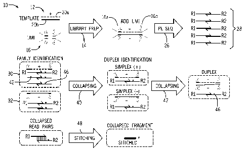
[0031] FIG. 1 is a diagrammatical overview of a workflow for identifying a
genomic
sequence error rate in accordance with the present techniques;
14
CA 03067425 2019-3.1-29
WO 2019/108972
PCT/US2018/063372
[0032] FIG. 2 is a flow diagram of a technique for sequence variant validation
in
accordance with the present techniques;
[0033] FIG. 3 is a flow diagram of a technique for sequence variant
identification in
accordance with the present techniques;
[0034] FIG. 4 is a flow diagram of a technique for determining a genomic
sequence data
error date in accordance with the present techniques;
[0035] FIG. 5 shows stratified error rates for a variety of source samples and
error types;
[0036] FIG. 6 is a flow diagram of a technique for determining stratified
error rates and
sequence variant validation in accordance with the present techniques;
[0037] FIG. 7 shows stratified error types for source samples, including a
sample with a
high error rate;
[0038] FIG. 8 shows stratified error rates for a variety of error types for
the high error
rate sample of FIG. 7;
[0039] FIG. 9 is a plot showing improved specificity relative to a decision
tree technique;
[0040] FIG. 10 is a table showing sensitivity and specificity results relative
to a default
decision tree technique; and
[0041] FIG. 11 is a block diagram of a sequencing device in accordance with
the present
techniques.
DETAILED DESCRIPTION
[0042] The present
techniques are directed to analysis and processing of sequencing
data for improved sequence variation detection and/or validation. To that end,
the
disclosed techniques eliminate or reduce designation of false positive
sequence variants
and also permit improved limits of detection of sequence variants for certain
samples.
CA 03067425 2019-3.1-29
WO 2019/108972
PCT/US2018/063372
FIG. 1 is a schematic workflow diagram 10 showing a sample preparation and
sequence
acquisition workflow.
[0043] A template
12 derived from a biological sample of interest, undergoes library
preparation (step 14) to incorporate one or more UMIs 16. The template 12 may
represent
a plurality of nucleic acid fragments. Each template 12 incorporates an
individual UMI
16 (which may include one or more identifier sequences) of a plurality of
UMIs, such that
the different source templates 12 are each associated with distinguishable
UMIs 16 have
different sequences. For example, the depicted diagram 10 is shown in the
context of
forked paired-end sequencing adapters including unique molecular identifiers
(UMIs) 16
configured to couple to the 5' and 3' ends of a nucleic acid template fragment
12 and
such that the template 12 is flanked by different portions 16a, 16b of the
U1VII 16.
Further, the positive strand 20a includes a first LTMI sequence or sequences
while the
negative strand 20b includes a second UMI sequence complementary to the first.
The first
UMI sequence and the second UMI sequence may be considered to be part of a
single
UMI 16 or different UMIs 16. By identifying the complementary sequences of the
UMI
or UMIs 16, the sequences of the positive strand 20a and the negative strand
20b may be
associated with one another.
[0044] Subsequent
to library preparation, genomic sequence data of the sample
(including a plurality of templates 12) is acquired by any suitable sequencing
technique,
depicted here as paired-end sequencing (step 26). Paired-end sequencing yields
a
plurality of sequence reads 28, which may be in turn divided or separated by
template
source via the respective UMIs 16. For example, a first read group 30
including a first
subset of the acquired sequence reads 28 may be associated with a first UMI 16
while a
second read group 32 including a second subset of the acquired sequence reads
28 may
be associated within a second UMI 16 complementary to the first UMI 16. As
noted, the
complementary UMIs may also be considered to be a single UMI.
[0045] Generally, sequence reads on the same strand within a single read group
(e.g., the
first read group 30, the second read group 32) should be identical to one
another, as the
16
CA 03067425 2019-3.1-29
WO 2019/108972
PCT/US2018/063372
associated UMI 16 links a subset of the sequence reads 28 to a single source
template 12.
Deviation or differences within the group are indicative of sample preparation
or
sequence acquisition errors. Identification and elimination of outlier reads
within a read
group to collapse the read group to a consensus sequence or collapsed sequence
(step 40)
may serve to prevent introduced sequence errors from propagating into the
sequence data
to yield false positive variants. As provided herein, such outlier
differences, such as
difference 42, that are not present in other sequence reads within the first
read group 30,
may be considered to be due to sequence error. Any identified differences or
variations
within a read group are provided as input to determining an overall error rate
for the
sample.
[0046] Any
differences that pass through consensus sequence building, e.g., difference
46, may further be compared to sequence reads associated with a complementary
strand
of the UMI 16. That is, the sequences of the first read group 30 and the
second read
group 32 may be assembled as a duplex. Again, any differences between the
groups 30,
32 may be identified before a consensus duplex of the complementary strands is
assembled (step 47). Such differences may also be tracked as part of the error
rate. In
addition, the collapsed simplex or duplex groups may be stitched together at
overlapping
regions (step 48) to generate a collapsed longer fragment as part of sequence
assembly.
Stitching may be used to determine a frequency of any potential sequence
variants.
[0047] While the
depicted diagram shows a single template 12 (e.g., a nucleic acid
fragment), the disclosed techniques track error throughout the genomic
sequence data to
generate a global or overall error rate or rates. In particular, FIG. 2 is a
flow diagram of a
method 50 of receiving genomic sequence data of a biological sample, wherein
the
genomic sequence data comprises a plurality of sequence reads, each sequence
read being
associated with a unique molecular identifier of a plurality of unique
molecular
identifiers; The method includes the step of receiving genomic sequence data
of an
individual biological sample (block 52).
17
CA 03067425 2019-3.1-29
WO 2019/108972
PCT/US2018/063372
[0048] The received sequence data may be received subsequent to sample
preparation
and sequencing of the biological sample as provided herein. Further, the
received
genomic sequence data may be stored or retrospective sequence data. The
genomic
sequence data may include include customer information, biological sample
organism
information, biological sample type information (e.g. information identifying
whether the
sample is fresh, frozen, or preserved), tissue type, sequence device type, and
sequencing
assay type (whole genome, targeted panel).
[0049] The genomic
sequence data is operated on to determine an error rate of the
genomic sequence data (block 54). The error rate is characteristic of the
sample itself and
its associated genomic sequence data. Accordingly, the error rate may be
calculated de
novo for each sequencing run of a biological sample of interest. An error rate
for
samples taken from a same individual at different times may exhibit different
characteristic error rates that depend on sample preparation variabilities,
sequencing
device settings, etc.
[0050] The method
may also identify potential sequence variants in the genomic
sequence date (block 56). Potential sequence variants may be identified
relative to a
reference sequence. Potential sequence variant identification may include
locus mapping
of sequence reads and assignment to corresponding genetic loci. The sample
reads may
be assigned to corresponding genetic loci based on the sequence of the
nucleotides of the
sample read or, in other words, the order of nucleotides within the sample
read (e.g., A,
C, G, T). Based on this analysis, the sample read may be designated as
including a
possible variant/allele of a particular genetic locus. The sample read may be
collected (or
aggregated or binned) with other sample reads that have been designated as
including
possible variants/alleles of the genetic locus. The sample reads may be
analyzed to locate
one or more identifying sequences (e.g., UMIs 16) of nucleotides that
differentiate the
sample read from other sample reads.
[0051] The mapped
sample reads are analyzed relative to the reference sequence to
identify potential sequence variants. Among other things, the results of the
analysis
18
identify the potential variant call, a sample variant frequency, a reference
sequence and a
position within the genomic sequence of interest at which the variant
occurred. For
example, if a genetic locus is known for including SNPs, then the assigned
reads that
have been called for the genetic locus may undergo analysis to identify the
SNPs of the
assigned reads. If the genetic locus is known for including polymorphic
repetitive DNA
elements, then the assigned reads may be analyzed to identify or characterize
the
polymorphic repetitive DNA elements within the sample reads. In some
embodiments, if
an assigned read effectively matches with an STR locus and an SNP locus, a
warning or
flag may be assigned to the sample read. The sample read may be designated as
both an
STR locus and an SNP locus. The analyzing may include aligning the assigned
reads in
accordance with an alignment protocol to determine sequences and/or lengths of
the
assigned reads. The alignment protocol may include the method described in
International Application No. PCT/US2013/030867 (Publication No. WO
2014/142831),
filed on Mar. 15, 2013. The
analysis may also count a number of reads having a particular potential
variant allele
relative to a total coverage for a particular locus.
100521 Once
identified, the potential sequence variants are operated on by a function
that takes into account the determined error rate to distinguish between true
positives and
false positives (block 58). In on embodiment, for individual potential
sequence variant, a
likelihood score is determined based on a likelihood ratio:
Likelihood ratio (L) = Likelihood (observed variant is errorlcoverage, error
rate)/Likelihood (observed variant is true positive coverage, variant allele
frequency),
where the variant allele frequency (VAF) = max (observed VAF, limit of
detection).
The likelihood score is a function of the error rate, the read coverage at the
particular site,
and the frequency that the potential sequence variant occurs in the reads. For
example,
lower frequency variants may be less likely to be validated. The likelihood
score or ratio
may have adjustable thresholds that are set by the user or the system based on
user inputs
and/or sample type. Potential sequence variants may be validated based on a
likelihood
19
Date Recue/Date Received 2021-06-16
CA 03067425 2019-3.1-29
WO 2019/108972
PCT/US2018/063372
score above or below a threshold or within a range. For example, a likelihood
score or
ratio below 0.01 and above 0.0001 or between 10' to 10-2 may be indicative of
a pass. In
another embodiment, the thresholds may be set based on a calculated
specificity goal.
[0053] Once
identified, the validated sequence variants may be provided (block 60) to
a user. For example, the validated sequence may be provided as a generated
report, e.g.,
stored as a report file or displayed on a graphical user interface for user
interaction.
Alternatively, when the validation operation invalidates or disqualifies
potential variant
call, the validation operation may also report or store a corresponding
indication (e.g., a
negative indicator, a no call indicator, an in-valid call indicator) as part
of the report. The
validation also may provide the likelihood score related to a degree of
confidence that the
variant call is correct or the invalid call designation is correct.
[0054] FIG. 3 is a
flow diagram of a method 64 that operates on received genomic
sequence data of a biological sample (block 66) to determine sequence
variants. The
genomic sequence data includes sequences of UMIs, whereby each sequence read
is
associated with one UMI of a plurality of UMIs used in the sequencing run. The
sequence reads may be separated into read groups, whereby each read group is a
subset of
the sequence reads that are associated with a common UMI (block 70).
Accordingly,
each sequence read should be present in only one read group. Once separated,
errors in
the genomic sequence data are identified based on sequence disagreement
between the
subset of sequence reads within the read group. Each sequence read for a
particular UMI
should be identical. Further, for paired end sequencing, sequenced strands in
both
directions should align. The presence of sequence variability within a
particular read
group is indicative of systemic error. Accordingly, based on the overall
errors identified
within each different read group (block 72), an overall error rate of the
genomic sequence
data may be determined (block 74). The error rate may in turn be used to
identify and/or
validate sequence variants in the genomic sequence data (block 76).
[0055] FIG. 4 is a
flow diagram of a method 80 for generating an error rate as
provided herein. The method 80 operates on received genomic sequence data of a
CA 03067425 2019-3.1-29
WO 2019/108972
PCT/US2018/063372
biological sample (block 82) that has been separated into subsets based on a
common
unique molecular identifier (block 84). As part of generating a consensus or
collapsed
sequence, sequence differences within the subset are identified (block 86).
The collapsed
sequence may be determined based on a majority voting rule, whereby sequence
differences that are in a minority of sequence reads in a particular subset
(i.e., read group)
are designated as sequence errors (block 88) but sequence differences that are
in a
majority of the sequence reads pass through to build the consensus or
collapsed sequence
(block 90). Based on the identified sequence errors, the error rate is
identified (block 92).
However, not all sequence differences in each subgroup necessarily contribute
to the
error rate. Sequence differences in the majority of sequence reads (see
difference 46 of
FIG. 1) are distinguished from sequence differences in the minority.
[0056] While certain embodiment are disclosed in the context of a global or
overall error
rate for genomic sequence data, the error rate may, additionally or
alternatively, be
stratified based on a type of nucleotide change. In this manner, systemic
error that is
biased towards particular nucleotide changes is identified. FIG. 5 is a panel
of error rates
separated out by type of change. The error rates are compared between
different sample
types, including 24 single cell free DNA (cfDNA) BRN samples, nucleosome prep
of
seven cancer cell lines and 6 0.2% zoo mix samples, and genomic pipDNA
including
three healthy samples and 21 HD753 titrated samples. Further, the inputs to
the error rate
determination are separated by duplex, simplex, stitched, and unstitched
sequence reads
in various combination. As noted with reference to FIG. 1, duplex building and
stitching
corrects errors in template sequences by eliminating sequence differences that
are
associated with error.
[0057] As observed, the error rates of each type of error vary based on sample
type. For
example, in cell free DNA and nucleosomePrep, deamination and resultant G to A
errors
are present in relatively higher levels. Oxidation is dominant in pipDNA,
resulting in
observed higher error rates of G to T changes. Accordingly, in certain
embodiments,
certain biological sample types may be associated with particular
characteristic errors.
21
CA 03067425 2019-3.1-29
WO 2019/108972
PCT/US2018/063372
In one embodiment, the sequence variant determination may include a weighting
factor to
weigh against potential variants that are associated with error for the sample
type in
question.
[00581 FIG. 6 is a flow diagram of a method 100 of determining stratified
error rates, as
shown in FIG. 5. For sequence reads that are part of a single read group,
individual reads
having sequence differences within the group and in a minority of strands are
eliminated
to correct the template, These eliminated sequence reads may be further
analyzed to
identify the types of erroneous sequence changes that occur at each locus
(block 102).
The nucleotide change forming the erroneous sequence change is considered
relative to
the majority sequence read in the group to identify the type of nucleotide
change. For
example, if the majority sequence read includes a G at position (n) of the
read, and the
minority read or reads include an A at position (n), the type of change may be
binned as a
G>A change. The change may be a single nucleotide change or an indel. This
process is
applied to all individual read groups including minority sequence reads having
sequence
differences to generate stratified error rates of each type of nucleotide
change throughout
the genomic sequence data (block 104), whereby the nucleotide changes are
based on
disagreement within the genomic sequence data itself, Using the stratified
error rates, a
potential sequence variant may be validated. Once received (block 106) as part
of a
variant identification operation, the potential sequence variant in the
genomic sequence
data is classified according to the type of nucleotide change relative to a
reference
sequence (block 108). In particular, while the error rates are calculated
using a measure
internal to the genomic sequence data (internal sequence disagreement between
sequence
reads of a read group as provided herein), the sequence variants are
determined relative to
a reference sequence. If the potential variant sequence is a G>A change
relative to the
reference sequence, the G>A error rate (and not the other error rates for the
other types of
nucleotide changes) are used to determine that the potential sequence variant
is a true
positive or a false positive (block 110), e.g., as part of a likelihood ratio
deteimination.
In this manner, a biological sample having a relatively low G>A error rate may
validate a
G>A sequence variant while the same biological sample, with a relatively high
G>T error
22
CA 03067425 2019-3.1-29
WO 2019/108972
PCT/US2018/063372
rate may apply more stringent conditions to validating potential G>T sequence
variants.
In one embodiment, a weighting factor for each type of error may be generated
based on
the stratified error rates.
[0059] FIG. 7 shows a comparison of error rates in different cell free DNA
samples
relative to one another and associated specificity of sequence variant
identification of
each sample. The highlighted sample, BRN022, exhibits a significant increase
in C>T
errors relative to the sample cohort. However, the sample cohort generally
shows
relatively higher C>T errors relative to other error types, which are
indicative of C>T or
G>A deamination changes. Nonetheless, the specificity in the sample with high
C>T or
G>A error rates is about or greater than 99.95%, indicating a high specificity
in the
context of a biological sample and genomic sequence data having a high
sequence error
rate.
[0060] FIG. 8 shows stratified error rates for a variety of error types for
the high error
rate sample of FIG. 7 for duplex and simplex (positive and negative) data,
stitched and
unstitched. The template correction in the stitched data appears to be
associated with
different error identification relative to the unstitched data. However, the
positive and
negative strand errors appear to correlate, with the C>T error appearing as
G>A in the
opposing strand. Similarly, the identified peak in T>C error appears as a peak
in A>G
error in the opposing strand. The identified high error C>T and G>A changes
are
examined relative to a default technique that does not calculate error rate as
provided
herein. The default technique identified 257 C>T and G>A false positives in
the
BRN022 sample, while the stratified error rate method identified 24 and 14
(depending
on the limit of detection thresholds), showing a significant decrease in false
positive
identification for a high error rate sample.
[0061] FIG. 9 is a plot showing improved specificity relative to a decision
tree technique.
Such a technique may be a technique as provided in PCT publication
W02018093780
and that involves one or more quality scores based on weighting fragment
types. In
contrast to the decision tree technique, the disclosed techniques may
determine error rates
23
on an per-sample basis rather than using a predetermined weighting factor. For
example,
certain samples may exhibit higher error in positive strands vs. negative
strands.
Accordingly, the error may also be stratified based on fragment type as
calculated de
novo. As shown in FIG. 9, the error rate techniques as provided herein, the
likelihood
model, result in higher specificity relative to a decision tree technique for
all three sample
types examined. FIG. 10 is a table showing sensitivity and specificity results
relative to a
default decision tree technique for the nucleosomePrep samples, including a
percentage
of zoo mix, showing sensitivity in line with the decision tree technique. The
likelihood
(based on error rate) technique exhibits high specificity, indicating an
improvement in
variant calling and a reduction in false positive identification.
[0062] FIG. 11 is a schematic diagram of a sequencing device 160 that may be
used in
conjunction with the disclosed embodiments for acquiring sequencing data that
is used to
identify and/or validate sequence variant calls as provided herein. The
sequence device
160 may be implemented according to any sequencing technique, such as those
incorporating sequencing-by-synthesis methods described in U.S. Patent
Publication Nos.
2007/0166705; 2006/0188901; 2006/0240439; 2006/0281109; 2005/0100900; U.S.
Pat.
No. 7,057,026; WO 05/065814; WO 06/064199; WO 07/010,251.
Alternatively, sequencing
by ligation techniques may be used in the sequencing device 160. Such
techniques use
DNA ligase to incorporate oligonucleotides and identify the incorporation of
such
oligonucleotides and are described in U.S. Pat. No. 6,969,488; U.S. Pat, No.
6,172,218;
and U.S. Pat. No. 6,306,597.
Some embodiments can utilize nanopore sequencing,
whereby target nucleic acid strands, or nucleotides exonucleolytically removed
from
target nucleic acids, pass through a nanopore. As the target nucleic acids or
nucleotides
pass through the nanopore, each type of base can be identified by measuring
fluctuations
in the electrical conductance of the pore (U.S. Patent No. 7,001,792; Soni &
Meller,
Chem. 53, 1996-2001 (2007); Healy, Nanomed. 2, 459-481 (2007); and Cockroft,
et al.
.1. Am. Chem. Soc. 130, 818-820 (2008)).
24
Date Recue/Date Received 2021-06-16
Yet other embodiments include detection of a proton released upon
incorporation of a
nucleotide into an extension product. For example, sequencing based on
detection of
released protons can use an electrical detector and associated techniques that
are
commercially available from Ion Torrent (Guilford, CT, a Life Technologies
subsidiary)
or sequencing methods and systems described in US 2009/0026082 Al; US
2009/0127589 Al; US 2010/0137143 Al; or US 2010/0282617 Al.
Particular embodiments can utilize methods involving the real-time monitoring
of DNA
polymerase activity. Nucleotide incorporations can be detected through
fluorescence
resonance energy transfer (FRET) interactions between a fluorophore-bearing
polymerase
and 7-phosphate-labeled nucleotides, or with zeromode waveguides as described,
for
example, in Levene et al. Science 299, 682-686 (2003); Lundquist et al. Opt.
Lett. 33,
1026-1028 (2008); Korlach et al. Proc. Natl. Acad. S'ci. USA 105, 1176-1181
(2008).
Other suitable alternative techniques include, for example, fluorescent in
situ sequencing
(FISSEQ), and Massively Parallel Signature Sequencing (MPSS). In particular
embodiments, the sequencing device 160 may be a HiSee, MiSee, or HiScanSQ
from IlluminaTm (La Jolla, CA). In other embodiment, the sequencing device 160
may be
configured to operate using a CMOS sensor with nanowells fabricated over
photodiodes
such that DNA deposition is aligned one-to-one with each photodiode.
[0063] The sequencing device 160 may be "one-channel" a detection device, in
which
only two of four nucleotides are labeled and detectable for any given image.
For
example, thymine may have a permanent fluorescent label, while adenine uses
the same
fluorescent label in a detachable form. Guanine may be permanently dark, and
cytosine
may be initially dark but capable of having a label added during the cycle.
Accordingly,
each cycle may involve an initial image and a second image in which dye is
cleaved from
any adenines and added to any cytosines such that only thymine and adenine are
detectable in the initial image but only thymine and cytosine are detectable
in the second
image. Any base that is dark through both images in guanine and any base that
is
Date Recue/Date Received 2023-01-05
CA 03067425 2019-3.1-29
WO 2019/108972
PCT/US2018/063372
detectable through both images is thymine. A base that is detectable in the
first image
but not the second is adenine, and a base that is not detectable in the first
image but
detectable in the second image is cytosine. By combining the information from
the initial
image and the second image, all four bases are able to be discriminated using
one
channel.
100641 In the depicted embodiment, the sequencing device 160 includes a
separate
sample processing device 162 and an associated computer 164. However, as
noted, these
may be implemented as a single device. Further, the associated computer 164
may be
local to or networked with the sample processing device 162. In the depicted
embodiment, the biological sample may be loaded into the sample processing
device 162
on a sample substrate 170, e.g., a flow cell or slide, that is imaged to
generate sequence
data. For example,
reagents that interact with the biological sample fluoresce at
particular wavelengths in response to an excitation beam generated by an
imaging
module 172 and thereby return radiation for imaging. For instance, the
fluorescent
components may be generated by fluorescently tagged nucleic acids that
hybridize to
complementary molecules of the components or to fluorescently tagged
nucleotides that
are incorporated into an oligonucleotide using a polymerase. As will be
appreciated by
those skilled in the art, the wavelength at which the dyes of the sample are
excited and
the wavelength at which they fluoresce will depend upon the absorption and
emission
spectra of the specific dyes. Such returned radiation may propagate back
through the
directing optics. This retrobeam may generally be directed toward detection
optics of the
imaging module 172.
[0065] The imaging module detection optics may be based upon any suitable
technology,
and may be, for example, a charged coupled device (CCD) sensor that generates
pixilated
image data based upon photons impacting locations in the device. However, it
will be
understood that any of a variety of other detectors may also be used
including, but not
limited to, a detector array configured for time delay integration (TDI)
operation, a
complementary metal oxide semiconductor (CMOS) detector, an avalanche
photodiode
26
(APD) detector, a Geiger-mode photon counter, or any other suitable detector.
TDI mode
detection can be coupled with line scanning as described in U.S. Patent No.
7,329,860.
Other useful detectors are described, for
example, in the references provided previously herein in the context of
various nucleic
acid sequencing methodologies.
100661 The imaging module 172 may be under processor control, e.g., via a
processor
174, and the sample receiving device 162 may also include I/0 controls 176, an
internal
bus 78, non-volatile memory 180, RAM 182 and any other memory structure such
that
the memory is capable of storing executable instructions, and other suitable
hardware
components that may be similar to those described with regard to FIG. 11.
Further, the
associated computer 164 may also include a processor 184, I/O controls 186, a
communications module 184, and a memory architecture including RAM 188 and non-
volatile memory 190, such that the memory architecture is capable of storing
executable
instructions 192. The hardware components may be linked by an internal bus
194, which
may also link to the display 196. In embodiments in which the sequencing
device 160 is
implemented as an all-in-one device, certain redundant hardware elements may
be
eliminated.
100671 The processor 184 may be programmed to operate on the genomic sequence
data
as provided herein. In particular embodiments, based on the image data
acquired by the
imaging module 172, the sequencing device 160 may be configured to generate
sequencing data that includes base calls for each base of a sequence read.
Further, based
on the image data, even for sequence reads that are performed in series, the
individual
reads may be linked to the same location via the image data and, therefore, to
the same
template strand. The processor 184 may also be programmed to perform
downstream
analysis on the sequences corresponding to the inserts for a particular sample
subsequent
to assignment of sequence reads to the sample. The processor 184 may be
configured to
operate on sequence data in the form of a BAM file and to output the variant
calls in
various formats, such as in a .VCF or .GVCF file.
27
Date Recue/Date Received 2021-06-16
[0068] Although preferred embodiments of the invention have been disclosed for
illustrative purposes, those skilled in the art will appreciate that many
additions,
modifications, and substitutions are possible and that the scope of the claims
should not
be limited by the embodiments set forth herein, but should be given the
broadest
interpretation consistent with the description as a whole.
28
Date Recue/Date Received 2023-01-05