Note: Descriptions are shown in the official language in which they were submitted.
CA 03067514 2019--16
WO 2019/034710
PCT/EP2018/072155
1
ELECTROSURGICAL APPARATUS FOR DELIVERING RF AND/OR MICROWAVE
ENERGY INTO BIOLOGICAL TISSUE
FIELD OF THE INVENTION
The invention relates to an electrosurgical apparatus and
device for delivering radiofrequency and/or microwave
frequency energy into biological tissue. In particular, the
invention relates to an electrosurgical instrument capable of
delivering radiofrequency (RF) energy for cutting tissue
and/or microwave frequency energy for haemostasis (i.e.
promoting blood coagulation). The invention may be
particularly suitable in gastrointestinal (GI) procedures
associated with the lower and upper GI tract, e.g. to remove
polyps on the bowel, i.e. for endoscopic mucosal resection, or
endoscopic submucosal dissection. The invention may also lend
itself to other procedure, e.g. in general surgery or
laparoscopic surgery. The invention may find use in ear,
nose and throat procedures and liver resection. The device may
also be used to address procedures associated with the
pancreas, e.g. to resect or remove tumours or abnormalities in
close proximity to the portal vein or the pancreatic duct.
BACKGROUND TO THE INVENTION
Surgical resection is a means of removing sections of
organs from within the human or animal body. Such organs may
be highly vascular. When tissue is cut (divided or
transected) small blood vessels called arterioles are damaged
or ruptured. Initial bleeding is followed by a coagulation
cascade where the blood is turned into a clot in an attempt to
plug the bleeding point. During an operation, it is desirable
for a patient to lose as little blood as possible, so various
devices have been developed in an attempt to provide blood
free cutting. For endoscopic procedures, bleeds are also
undesirable, and need to be dealt with in an expedient manner,
CA 03067514 2019-12-16
WO 2019/034710
PCT/EP2018/072155
2
since the blood flow may obscure the operator's vision, which
may prolong surgery and potentially lead to the procedure
needing to be terminated and another method used instead, e.g.
open surgery.
Electrosurgical generators are prevalent in hospital
operating theatres, often for use in open and laparoscopic
procedures, and increasingly for use with surgical scoping
devices, e.g. an endoscope or the like. In endoscopic
procedures the electrosurgical accessory is typically inserted
through a lumen inside an endoscope. Considered against the
equivalent access channel for laparoscopic surgery, such a
lumen is comparatively narrow in bore and greater in length.
Instead of a sharp blade, it is known to use
radiofrequency (RF) energy to cut biological tissue. The
method of cutting using RF energy operates using the principle
that as an electric current passes through a tissue matrix
(aided by the ionic contents of the cells and the
intercellular electrolytes), the impedance to the flow of
electrons across the tissue generates heat. In practice, an
instrument is arranged to apply an RF voltage across the
tissue matrix that is sufficient to generate heat within the
cells to vaporise the water content of the tissue. However,
as a result of this increasing desiccation, particularly
adjacent to the RF emitting region of the instrument (which
has the highest current density of the current path through
tissue), direct physical contact between the tissue and
instrument can be lost. The applied voltage then manifests
itself as a voltage drop across this small void, which causes
ionisation in the void that leads to a plasma. Plasma has a
very high volume resistivity compared with tissue. The energy
supplied to the instrument maintains the plasma, i.e.
completes the electrical circuit between the instrument and
the tissue. Volatile material entering the plasma can be
vaporised and the perception is therefore of a tissue
dissecting plasma.
CA 03067514 2019-12-16
WO 2019/034710
PCT/EP2018/072155
3
GB 2 523 246 describes an electrosurgical instrument for
applying to biological tissue RF electromagnetic energy and/or
microwave frequency EM energy. The instrument comprises a
shaft insertable through an instrument channel of a surgical
scoping device. At a distal end of the shaft there is an
instrument tip comprising a planar transmission line formed
from a sheet of a first dielectric material having first and
second conductive layers on opposite surfaces thereof. The
planar transmission line is connected to a coaxial cable
conveyed by the shaft. The coaxial cable is arranged to
deliver either microwave or RF energy to the planar
transmission line. The coaxial cable comprises an inner
conductor, an outer conductor coaxial with the inner
conductor, and a second dielectric material separating the
outer and inner conductors, the inner and outer conductors
extending beyond the second dielectric at a connection
interface to overlap opposite surfaces of the transmission
line and electrically contact the first conductive layer and
second conductive layer respectively. The instrument further
comprises a protective hull with a smoothly contoured convex
undersurface facing away from the planar transmission line.
The undersurface comprises a longitudinally extending recessed
channel formed therein. A retractable needle is mounted
within the instrument, and operable to extend through the
recessed channel to protrude from a distal end of the
instrument. The needle can be used to inject fluid into a
treatment zone before the RF or microwave energy is applied.
SUMMARY OF THE INVENTION
At its most general, the present invention provides a
development to the concept discussed in GB 2 523 246. The
development may include forming the protective hull as a
shaped piece of electrically conductive bio-compatible
material having a low coefficient of friction with biological
CA 03067514 2019-12-16
WO 2019/034710
PCT/EP2018/072155
4
tissue (e.g. stainless steel) which has the dual function of
(i) physically protecting tissue that lies underneath the
active tip, and (ii) providing an electrical connection
between a coaxial feed line and the active tip.
The protective hull may be particularly useful in
procedures performed in the gastrointestinal tract, where
bowel perforation is a concern, or in the pancreas, where
damage to the portal vein or the pancreatic duct may occur
when a tumour or other abnormality is being resected,
dissected or removed.
The protective hull may be applied to planar instrument
tips adapted for different functions. For example, aspects of
the invention contemplated herein include: an instrument
adapted to deliver radiofrequency (RF) energy for cutting
biological tissue; an instrument adapted to deliver both RF
and microwave frequency energy separately or simultaneously;
and an instrument adapted to deliver RF and/or microwave
energy and having a retractable needle for delivering or
removing fluid (liquid or gas) to or from the treatment site.
For example, the needle may be used to introduce a gas, e.g.
argon, to produce thermal or non-thermal plasma for surface
coagulation (thermal) or sterilisation (non-thermal). The RF
and/or microwave field may be used to strike and sustain or
create this plasma. The protective hull may include a
passageway, e.g. recessed channel, through which the
retractable needle travels or through which fluid can be
delivered without the use of a needle, e.g. for clinical or
cleaning purposes.
According to the invention, there is provided an
electrosurgical instrument for delivering electromagnetic
energy to biological tissue, the instrument comprising: a
distal end assembly comprising: an active tip comprising a
planar body made of a first dielectric material separating a
first conductive element on a first surface thereof from a
second conductive element on a second surface thereof, the
CA 03067514 2019-12-16
WO 2019/034710
PCT/EP2018/072155
second surface facing in the opposite direction to the first
surface; an electrically conductive protective hull mounted on
an underside of the active tip, the protective hull having a
smoothly contoured convex undersurface facing away from the
5 planar body; and a coaxial feed cable comprising an inner
conductor, an outer conductor coaxial with the inner conductor
and a second dielectric material separating the inner and
outer conductors, the coaxial feed cable being for conveying
RF EM energy or microwave EM energy, wherein the inner
conductor is electrically connected to the first conductive
element and the outer conductor is electrically connected to
the second conductive element via the protective hull to
enable the instrument tip to receive the RF and/or the
microwave signal, and wherein the first and second conductive
elements are arranged to emit the RF EM energy or the
microwave EM energy from the coaxial cable at a distal side
portion of the planar body. With this arrangement, the
protective hull itself provide a conductive path between the
coaxial cable and the active tip, so that no additional
connecting components are required.
The first and second conductive elements may be arranged
to act as either or both of (i) active and return electrodes
to emit RF EM energy, or (ii) an antenna structure for
emitting and microwave EM energy from the distal side portion
of the active tip.
The protective hull may be formed from a conductive
material having a low coefficient of friction with biological
tissue, and which is biocompatible. Stainless steel may be
preferred.
The protective hull may be soldered to the second
conductive element to provide the requisite electrical
connection. The soldering may be performed after the
protective hull and active tip are positioned together. The
soldering may be induction soldering. A solder preform may be
mounted between the protective hull and active tip to provide
CA 03067514 2019-12-16
WO 2019/034710
PCT/EP2018/072155
6
the material for the soldered joint. The protective hull may
comprise an upper surface for abutting the second surface of
the planar body. The upper surface may comprise a recess for
retaining the solder preform. The recess may be rectangular.
The recess may have side edges that are set back from side
edges of the planar body. This can ensure that solder does
not flow or leak to the sides of the active tip, where it
could interfere with delivery of the RF EM energy or microwave
EM energy.
The protective hull may have a U-shaped recess for
receiving a portion of the outer conductor. For example, the
outer conductor may be exposed along a length of the coaxial
cable where it engages the protective hull. The coaxial cable
may be retained in the U-shaped recess by an interference fit.
In one example, the coaxial cable may be deformed, e.g. by a
crimp or the like to cause it to abut and engage the U-shaped
recess. The coaxial cable may be squashed so that its cross-
section belong oval, whereby it engages side walls of the U-
shaped recess.
The undersurface of the protective hull may smoothly
taper at its perimeter to meet the underside of the planar
body. The thickness of the protective hull may also decrease
towards the distal end of the instrument tip. Thus, the outer
portion of the protective hull may have a convex profile. The
undersurface may have a longitudinally extending recessed
channel formed therein. The tapering edge profile and
recessed channel may cause the undersurface of the protective
hull to comprise a pair of ridges. The tapered conformal
flowing form of the hull may reduce the risk of the instrument
digging into collateral tissue aiding its ability to glide.
For example, this shape may reduce the risk of the instrument
digging into the bowel wall and causing a bowel perforation or
may protect the portal vein or pancreatic duct from being
damaged. The particular dimensions of the hull (e.g. length,
CA 03067514 2019-12-16
WO 2019/034710
PCT/EP2018/072155
7
width, thickness, etc.) may be adapted to suit the intended
use and intended area of the body to be operated on.
The distal end assembly may comprise a flexible shaft
connected to a proximal end of the protective hull, the shaft
defining a lumen for conveying the coaxial cable.
The flexible shaft may comprise a proximal cannula tube
having braids formed therein to assist in the transfer of
torque from its proximal end to the distal end assembly, and a
distal unbraided tubular portion bonded to a distal end of the
cannula tube. The braids may extend in a longitudinal
direction. The braids may be made from metal. Providing the
unbraided tubular portion can prevent the braids from
interfering with the transfer of energy from the coaxial cable
to the active tip.
The flexible shaft may comprise a support tube mounted at
a junction between the proximal cannula tube and the unbraided
tubular portion. The support tube may provide mechanical
strength to the junction. The support tube may be a polymer
sleeve to which the proximal cannula tube and the unbraided
tubular portion are bonded. Additionally or alternatively,
the junction between the proximal cannula tube and the
unbraided tubular portion may be wrapped in a heat shrink
sleeve.
The first and second conductive elements may be arranged
to provide a local return path for RF energy, i.e. a low
impedance route for RF energy to be transported between the
first and second conductive elements. Meanwhile, for a
microwave signal, the instrument tip may be modelled as a
parallel plate transmission line with the planar body
representing dielectric material separating two conductive
plates.
The first and second conductive elements may each
comprise a layer of metallisation being formed on opposite
surfaces of the first dielectric material. The first and
second conductive elements may be arranged to set up a local
CA 03067514 2019-12-16
WO 2019/034710
PCT/EP2018/072155
8
electric field at a contact region in which the instrument tip
makes contact with the biological tissue. The local electric
field can be extremely high, which may cause a microplasma
(i.e. a hot thermal plasma) to be formed at the distal side
portion of the planar body, e.g. where contact is made with
the biological tissue. The microplasma may be desirable in
terms of achieving efficient cutting. The first and second
conductive elements may include portions, e.g. plated regions
at and adjacent the distal side portion, made from conductive
material having a high melting point, e.g. 1500 C or more,
such as titanium, tungsten or the like. Using such materials
may prevent the high temperatures of the microplasma from
eroding the first and second conductive elements. The first
and second conductive elements may also include connecting
portions made from conductive materials having lower melting
points (e.g. silver, gold and the like) deposited or plated on
the higher melting point conductors. The connecting portions
may facilitate connection of the inner and outer conductors of
the coaxial cable, e.g. by soldering or the like. In one
embodiment, a titanium tungsten (TiW) seed layer may be used
with a layer of silver (Ag) or gold (Au) deposited on the top.
For example, the seed layer may have a thickness of 30 nm, and
each layer of metallisation may have a thickness of 0.03 mm.
Preferably, each layer of metallisation is deposited on the
seed layer in two steps. In a first step, a 760 nm layer of
silver or gold may be sputtered onto the seed layer. In a
second step, a 29 pm thick layer of silver or gold may be
deposited by electrolysis. The lower melting point material
may be deposited onto the higher melting point material only
in the region where the inner conductor and protective hull
are to be attached, i.e. at the proximal end of the active tip
only, and not along the sides thereof, where the microplasma
will be generated.
In one embodiment, the first dielectric material
separating the conductive elements may provide the
CA 03067514 2019--16
WO 2019/034710
PCT/EP2018/072155
9
preferential return path between the inner conductor (active)
and the outer conductor (return). RF tissue cutting may be
produced at the distal side portion of the instrument tip if
the first dielectric material has a high dielectric constant
(e.g. greater than that of air) and the thickness of the first
dielectric material at the distal side portion, i.e. the
separation of the first and second conductive elements at the
distal side portion edge, is small, i.e. less than 1 mm. This
arrangement may provide the necessary preferential return path
for the current to flow.
The layers of metallisation may be set back (e.g. by 0.2
mm) from the side edges of the first dielectric material in a
proximal region of the planar body, to reduce the field
strength at this region. The proximal region may comprise the
region of the planar body proximal to the distal end.
In some embodiments, the first dielectric material
forming the planar body may be a biocompatible material such
as ceramic, preferably alumina. For example, the first
dielectric material may be at least 99% pure alumina having a
polished surface for robust adhesion to the metallisation
layers which may form the first conductive element and the
second conductive element.
The distal end assembly may include a fluid feed conduit
for conveying fluid for delivery out of the instrument. The
undersurface of the protective hull may have a longitudinally
extending recessed channel formed therein. The fluid feed
conduit may be mounted within the longitudinally extending
recessed channel. The coaxial feed cable may form part of a
multi-lumen conduit assembly for delivering RF and/or
microwave frequency energy and fluid (liquid or gas) to the
instrument. The fluid may be conveyed through a corresponding
passageway formed within the multi-lumen conduit assembly.
The fluid feed conduit may also be used to deliver other
material to the treatment site, e.g. a gas or a solid (e.g.
powder). In one embodiment, injection of fluid (saline or the
CA 03067514 2019-12-16
WO 2019/034710
PCT/EP2018/072155
like) is used to plump up the biological tissue at the
treatment site. This may be particularly useful where the
instrument is used to treat the wall of the bowel or the wall
of the oesophagus or for protecting the portal vein or the
5 pancreatic duct when a tumour or other abnormality located in
close proximity, in order to protect these structures and
create a cushion of fluid. Plumping up the tissue in this
manner may help to reduce the risk of bowel perforation,
damage to the wall of the oesophagus or leakage of from the
10 pancreatic duct or damage to the portal vein, etc. This
arrangement may enable the instrument to treat other
conditions where the abnormality (tumour, growth, lump, etc.)
is close to a sensitive biological structure.
The fluid feed conduit may comprise a needle guide tube
having a retractable needle slidably mounted therein. The
needle may be slidably movable with respect to the protective
hull through one or more control wires, which may be actuated
via a suitable slide actuator at a proximal end of the
instrument. Preferably, the needle is slidable back and forth
with respect to a fluid supply passageway which conveys the
fluid to the needle for delivery. The fluid supply passageway
may be an integral part of the sleeve, or may be a tube
statically mounted in the sleeve. The ability to move the
needle back and forth while conveying fluid to the needle
through a conduit which does not move relatively to the sleeve
enables a retractable needle to be provided within a smaller
diameter sleeve than a device in which a fluid delivery tube
must slide along the length of the sleeve.
The term "surgical scoping device" may be used herein to
mean any surgical device provided with an insertion tube that
is a rigid or flexible (e.g. steerable) conduit that is
introduced into a patient's body during an invasive procedure.
The insertion tube may include the instrument channel and an
optical channel (e.g. for transmitting light to illuminate
and/or capture images of a treatment site at the distal end of
CA 03067514 2019-12-16
WO 2019/034710
PCT/EP2018/072155
11
the insertion tube. The instrument channel may have a
diameter suitable for receiving invasive surgical tools. The
diameter of the instrument channel may be 5 mm or less.
Herein, the term "inner" means radially closer to the
centre (e.g. axis) of the instrument channel and/or coaxial
cable. The term "outer" means radially further from the centre
(axis) of the instrument channel and/or coaxial cable.
The term "conductive" is used herein to mean electrically
conductive, unless the context dictates otherwise.
Herein, the terms "proximal" and "distal" refer to the
ends of the elongate probe. In use the proximal end is closer
to a generator for providing the RF and/or microwave energy,
whereas the distal end is further from the generator.
In this specification "microwave" may be used broadly to
indicate a frequency range of 400 MHz to 100 GHz, but
preferably the range 1 GHz to 60 GHz. Specific frequencies
that have been considered are: 915 MHz, 2.45 GHz, 3.3 GHz, 5.8
GHz, 10 GHz, 14.5 GHz and 24 GHz. In contrast, this
specification uses "radiofrequency" or "RF" to indicate a
frequency range that is at least three orders of magnitude
lower, e.g. up to 300 MHz, preferably 10 kHz to 1 MHz, and
most preferably 400 kHz.
The electrosurgical instrument discussed herein may be
capable of delivering radiofrequency (RF) electromagnetic (EM)
energy and/or microwave EM energy into biological tissue. In
particular, the electrosurgical instrument may be capable of
delivering radiofrequency (RF) energy for cutting tissue
and/or microwave frequency energy for haemostasis (i.e.
sealing broken blood vessels by promoting blood coagulation).
The invention may be particularly suitable in gastrointestinal
(GI) procedures associated with the lower and upper GI tract,
e.g. to remove polyps on the bowel, i.e. for endoscopic sub-
mucosal resection. The invention may also lend itself to
precision endoscopic procedures, i.e. precision endoscopic
resection, and may be used in ear, nose and throat procedures
CA 03067514 2019-12-16
WO 2019/034710
PCT/EP2018/072155
12
and liver resection. The device may also be used to address
procedures associated with the pancreas, e.g. to resect or
remove tumours or abnormalities in close proximity to the
portal vein or the pancreatic duct.
BRIEF DESCRIPTION OF THE DRAWINGS
Examples embodying the invention are discussed in detail
below with reference to the accompanying drawings, in which:
Fig. 1 is a schematic view of a complete electrosurgery
system in which the present invention is applied;
Fig. 2 is an exploded view of a distal end of an
electrosurgical instrument that is an embodiment of the
invention;
Fig. 3 is a partly transparent perspective view of a
distal end of an electrosurgical instrument that is an
embodiment of the invention;
Figs. 4A and 4B are a top view and a cross-sectional side
view respectively of a protective hull member suitable for use
with the present invention; and
Fig. 5 is a cross-sectional side view of a distal tip
assembly of an electrosurgical instrument that is an
embodiment of the invention.
DETAILED DESCRIPTION; FURTHER OPTIONS AND PREFERENCES
Various aspects of the present inventions are presented
below in the context of an electrosurgery system that provides
an electrosurgical invasive instrument for use in endoscopic
procedures for the removal of polyps and malignant growths
through the controlled delivery of both microwave and RF
energy. However, it is to be understood that the aspects of
the invention presented herein need not be limited to this
particular application. They may be equally applicable in
CA 03067514 2019-12-16
WO 2019/034710
PCT/EP2018/072155
13
embodiments where only RF energy is required, or where only RF
energy and fluid delivery is required.
Fig. 1 is a schematic diagram of a complete
electrosurgery system 100 that is capable of selectively
supplying to the distal end of an invasive electrosurgical
instrument any or all of RF energy, microwave energy and
fluid, e.g. saline or hyaluronic acid. The system 100
comprises a generator 102 for controllable supplying RF
electromagnetic (EM) energy and/or microwave frequency EM
energy. A suitable generator for this purpose is described in
WO 2012/076844, which is incorporated herein by reference.
The generator 102 is connected to an interface joint 106
by an interface cable 104. The interface joint 106 is also
connected to receive a fluid supply 107 from a fluid delivery
device 108, such as a syringe. The interface joint 106 houses
a needle movement mechanism that is operable by sliding a
trigger 110. The function of the interface joint 106 is to
combine the inputs from the generator 102, fluid delivery
device 108 and needle movement mechanism into a single
flexible shaft 112, which extends from the distal end of the
interface joint 106. The internal configuration of the
interface joint 106 is discussed in more detail below.
The flexible shaft 112 is insertable through the entire
length of an instrument (working) channel of a surgical
scoping device 114. A torque transfer unit 116 is mounted on
a proximal length of the shaft 112 between the interface joint
106 and surgical scoping device 114. The torque transfer unit
116 engages the shaft to permit it to be rotated within the
instrument channel of the surgical scoping device 114.
The flexible shaft 112 has an electrosurgical instrument
tip 118 that is shaped to pass through the instrument channel
of the surgical scoping device 114 and protrude (e.g. inside
the patient) at the distal end of the endoscope's tube. The
instrument tip includes an active tip for delivering RF EM
energy and/or microwave EM energy into biological tissue and a
CA 03067514 2019-12-16
WO 2019/034710
PCT/EP2018/072155
14
retractable hypodermic needle for delivering fluid. These
combined technologies provide a unique solution for cutting
and destroying unwanted tissue and the ability to seal blood
vessels around the targeted area. Through use of the
retractable hypodermic needle, the surgeon is able to inject
saline and/or hyaluronic acid with added marker dye between
tissues layers in order to distend and mark the position of a
lesion to be treated. The
injection of fluid in this manner
lifts and separates the tissue layers making it both easier to
resect around the lesion and plane through the submucosal
layer, reducing the risk of bowel wall perforation and
unnecessary thermal damage to the muscle layer.
As discussed in more detail below, the instrument tip 118
further includes a protective hull positioned under the active
tip to assist a tissue planing type resection action, again
helping to protect against inadvertent perforation and ensure
viability of the remaining tissue, which in turn facilitates
more rapid healing and post operation recovery.
The structure of the instrument tip discussed below may
be particularly designed for use with a conventional steerable
flexible endoscope having a working channel with an internal
diameters of at least 3.3 mm and a channel length of between
60 cm and 170 cm. As such the majority of the comparatively
small diameter (less than 3 mm) instrument is housed within
the lumen of a much larger and predominantly polymer
insulating device, i.e. the flexible endoscope channel, which
typically has an outer diameter of 11 mm to 13 mm. In
practice, only 15 mm to 25 mm of the distal assembly protrudes
from the distal end of the endoscope channel, in order not to
block the field of view or adversely affect camera focussing.
The protruding part of the distal assembly is the only portion
of the instrument that ever makes direct contact with the
patient.
At the proximal end of the endoscope working channel,
which is typically held 50 cm to 80 cm from the patient, the
CA 03067514 2019-12-16
WO 2019/034710
PCT/EP2018/072155
flexible shaft 112 emerges from the working channel port and
extends a further 30 cm to 100 cm to the interface joint 106.
In use, the interface joint 106 is typically held by a gloved
assistant throughout the procedure. The interface joint 106
5 is designed and manufactured from polymer materials in such a
way as to provide primary and secondary electrical insulation
with extended creepage and clearance distances. The interface
cable 104 is connected to the generator 102 using a QMA-type
coaxial interface, which is designed to allow continuous
10 clockwise or counter clockwise rotation. This permits the
interface joint 106 to rotate with the torque transfer unit
116 under the control of the user. The assistant supports the
interface joint 106 throughout the procedure in order to
assist the user with sympathetic instrument rotation, needle
15 control and fluid injection.
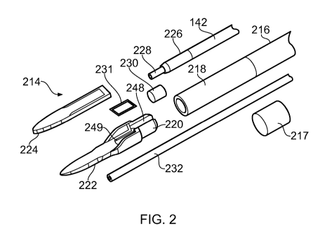
Fig. 2 shows an exploded view of the distal end assembly
214 (sometimes referred to as an instrument tip) of an
electrosurgical instrument that is an embodiment of the
invention. The distal end assembly 214 is mounted at the
distal end of an outer cannula tube 216 of a flexible shaft,
e.g. which correspond to the flexible shaft 112 discussed
above with reference to Fig. 1. The cannula tube 216 forms a
flexible sleeve defining a lumen for transporting fluid to the
instrument tip, the instrument tip being secured at its distal
end. In order to provide a torque transfer function, the outer
cannula tube 216 is formed of a braided tube, e.g. comprising
a braided wire (e.g. stainless steel) wrap mounted between a
radially inner polymer layer and a radially outer polymer
layer, wherein the polymer may be e.g. Pebax0.
In this embodiment, the outer cannula tube 216 is
connected at its distal end to an unbraided tubular portion
218, which may be a flexible conduit. The tubular portion 218
may be formed from any suitable polymer material, e.g. Pebax0
or the like. The tubular portion 218 may have an axial length
(i.e. length in line with the shaft axis equal to or greater
CA 03067514 2019-12-16
WO 2019/034710
PCT/EP2018/072155
16
than 1 mm. This may ensure that a safe distance is introduced
between the end of the braiding and the proximal edge of the
distal end assembly 214 in order to avoid any risk of heating
of the braid as a result of capacitive conductance during use
of microwave energy. This arrangement may also prevent the
two plates of the planar transmission line or the two
conductors in the coaxial transmission line from becoming
shorted or connected together.
The tubular portion 218 may be referred to as a 'soft
tip' 218. The soft tip 218 may in some embodiments be an
additional length of polymer tube which is bonded to the
distal end of the sleeve or cannula tube 216. The bonding may
use any suitable adhesive, e.g. epoxy or the like. A support
tube 217 may be mounted over the junction between the tubular
portion 218 and cannula tube 216 to reinforce the joint by
providing additional mechanical strength. The support tube
217 may be a short section of polymer tubing within which the
both the tubular portion 218 and the cannula tube 216 are
secured, e.g. by bonding. The support tube 217 may be
flexible and/or may have a length selected to ensure that it
does not adversely affect the flexibility of the shaft.
The junction of the tubular portion 218, cannula tube 216
and support tube 217 may also be captured within a heat shrink
sleeve (not shown) to provide further structural strength at
the distal end of the shaft.
The braiding within the cannula tube 216 enables torque
applied to the proximal end of the shaft to be transformed
into rotational movement of the instrument tip. For
convenience, some of the accompanying illustrations show the
tubular portion 218 and cannula tube 216 as transparent. In
practical embodiments, the shaft may be opaque.
A distal end of the tubular portion 218 is arranged to
fit over a corresponding proximal part 220 of a protective
hull 222. The protective hull is formed from a metallic
material having low friction with biological tissue, e.g.
CA 03067514 2019-12-16
WO 2019/034710
PCT/EP2018/072155
17
stainless steel, and is shaped to perform a number of
functions, i.e.
- mount the distal end assembly 214 on the flexible
shaft,
- provide a protective undersurface for an active tip
structure that delivers energy into surrounding biological
tissue,
- provide a protective housing and supporting frame for a
retractable needle, and
- locate the active tip structure relative to the coaxial
cable during assembly and subsequent use.
The parts of the structure of the hull 222 that perform
these functions are discussed in more detail below.
The distal end assembly 214 includes an active tip 224,
which is a planar piece of dielectric material 221 (e.g.
alumina) having conductive layers (e.g. layers of
metallisation) on its upper and lower surfaces. The
conductive layers are each electrically connected to a
respective one of an inner conductor 228 and an outer
conductor 226 of a coaxial cable 142 that is conveyed by the
cannula tube 216. At a distal end of the coaxial cable 142,
its outer sheath is removed to expose a length of the outer
conductor 226. The inner conductor 228 of the coaxial cable
extends beyond the distal end of the outer conductor 226. The
coaxial cable 142 and the active tip 224 are mounted relative
to one another so that the protruding part of the inner
conductor 228 lies on a first conductive layer of the active
tip, while the outer conductor 226 is brought into electrical
connection with a second conductive layer via the protective
hull 222, as discussed below. The first conductive layer is
isolated from the outer conductor 226 and the second
conductive layer is isolated from the inner conductor 228.
The conductive layers may be formed from high melting
point conductors, e.g. W or Ti. However, in one example, to
facilitate the use of solder in the electrical connection
CA 03067514 2019--16
WO 2019/034710
PCT/EP2018/072155
18
between the inner and outer conductors of the coaxial cable
142 and the active tip 224, lower melting point conductors may
be deposited at proximal regions on the conductive layers
where the electrical connections are made. The lower melting
point conductors may be silver (Ag) or gold (Au).
The distal end of the active tip 224 is curved to avoid
presenting sharp corners within the patient.
The outer conductor 226 is electrically connected to a
lower conductive layer on the underside of the active tip 224
via the protective hull 222. A proximal end of the protective
hull 222 is formed with a U-shaped channel 248 for receiving
and supporting a distal end of the coaxial feed cable 142.
The distal end assembly is configured so that the exposed
portion of the outer conductor 226 sits in the U-shaped
channel 248. An electrically conductive element 230, such as
a sleeve or collar, is used to crimp the exposed portion of
the outer conductor 226. The compression caused by the crimp
means that the coaxial cable deforms in the region where it is
received by the protective hull 222. For example, the portion
of the coaxial cable where the outer conductor 226 is exposed
may have an oval cross-section, whereby it abuts and forms a
robust electrical contact with the sides of the U-shaped
channel 248. The crimped outer conductor 226 may thus be
retained by the hull via an interference fit.
To complete the electrical connection between the outer
conductor 226 and lower conductive layer 229 on the active tip
224, the protective hull 222 is electrically coupled to the
lower conductive layer, e.g. by soldering (see e.g. Fig. 5).
In this embodiment, a solder preform 231 is provided for this
purpose. the solder preform 231 is shaped to be receivable
within a recess 249 formed in an upper surface of the
protective hull 222. In this example, the recess 49 is
rectangular, and the solder preform 231 has a corresponding
shape, but any suitable shape may be used. The recess 249 is
set back from the edges of the protective hull in a manner
CA 03067514 2019-12-16
WO 2019/034710
PCT/EP2018/072155
19
that ensures solder is only present between the lower surface
of the active tip 224 and the protective hull 222, i.e. it
does not flow to the side edges of the active tip 224. When
assembled, the solder preform 231 may be aligned with a region
on the lower surface of the active tip 224 that is coated in a
lower melting point conductor as discussed above (e.g. gold).
A suitable flex (not shown) may be provided with the solder
preform when the components are assembled to facilitate the
soldering process. The soldering process itself may be
induction soldering. The induction soldering effect may be
confined to a region of the active tip 224 and protective hull
222 at the solder preform 231.
The above configuration is advantageous because the
protective hull 222 retains all of (i) the active tip 224,
(ii) the solder preform 231, and (iii) the coaxial cable 142
in a fixed spatial relationship which ensures accurate and
repeatable assembly.
The distal end assembly further comprises a needle guide
232 that is retained within a recess formed in the
undersurface of the protective hull 222. The needle guide 232
is a hollow tube (e.g. a ferrule), e.g. made of polyimide,
within which a hypodermic needle 234 is slidably mounted. The
needle 234 is in fluid communication with the internal volume
of the cannula tube 216 in order to receive liquid present
therein for delivery to the treatment site.
After the distal end assembly 214 is assembled, it may be
secured within the distal end of the tubular portion 218 by an
interference fit and an adhesive (e.g. epoxy). The adhesive
may also form a plug for the distal end of the tubular portion
218 to provide a fluid tight seal that means the only exit for
fluid introduced at the interface joint is through the needle
234. Similarly, the junction (e.g. soldered joint) between
the inner conductor 228 and the upper conductive layer 227 may
have a protective cover 251 (see Fig. 5) that may be formed
from a suitable adhesive (e.g. epoxy). The protective cover
CA 03067514 2019-12-16
WO 2019/034710
PCT/EP2018/072155
251 may strengthen the connection between the protective hull
222 and active tip 224, while also forming an end plug for the
tubular portion 218, i.e. a fluid tight seal that means the
only exit for fluid introduced at the interface joint is
5 through the needle.
In use the active tip 224 makes an intimate contact with
the patient. The needle 234 can be extended beyond the distal
end of the active tip 224 and retracted to a position back
inside the guide tube 232 via control of the slider mechanism
10 on the interface joint which acts on a control wire 235 (see
Fig. 3) to deploy and retract the needle 234. In its extended
position, the needle is used to inject fluid for the purpose
of locally distending and/or marking tissue. The conductive
layers on the active tip 224 form bi-polar electrodes for
15 delivering RF and/or microwave electromagnetic energy.
The needle guide 232 extends back inside and proximal to
the distal assembly to provide extended creepage clearance to
ensure RF/microwave activation only occurs across the distal
tip region of the active tip 224.
20 Fig. 3
shows the distal end assembly 214 in an assembled
configuration. The tubular portion 218, support sleeve 217
and cannula tube 216 are shown as transparent so that the
inner components are visible. Parts of the protective hull
222 within the tubular portion 218 are omitted to show how the
electrically conductive element 230 fits over the outer
conductor 226.
Figs. 4A and 4B show the shape of a protective hull
structure 222 that can be used in embodiments of the
invention. A distal end of the protective hull 222 has a flat
upper surface 250 for contacting the lower conductive layer
229 on the active tip 224. As discussed above, a rectangular
recess 249 is formed towards the proximal end of the flat
upper surface 250 for receiving the solder preform 231.
The proximal end of the protective hull 222 is formed
with a U-shaped channel 248 for receiving and support the
CA 03067514 2019-12-16
WO 2019/034710
PCT/EP2018/072155
21
distal end of the coaxial feed cable 142. A similar channel
is formed on the underside of the proximal end of the
protective hull 222 to receive the guide tube 232 of the
retractable needle 234. The outer surface of the proximal end
of the protective hull 222 is cylindrical, with a diameter
selected to fit inside the distal end of the tubular portion
218.
At the sides of the protective hull 222 between the
proximal and distal ends, there are a pair of upstanding wing
portions 244, whose inner surfaces engage with respective side
edges of the active tip 224 and whose outer surface engage in
an interference fit with the inner surface of the tubular
portion 218.
The protective hull 222 is preferably made from a
metallic material having a low coefficient of friction with
biological tissue, such as stainless steel.
The distal end of the hull is shaped to permit the active
tip 224 to overhang it by around 0.2 mm around the distal edge
except at the distal tip. The surface that contacts the
underside of the active tip therefore has a maximum width of 2
mm, which narrows to 1.6 mm in an intermediate portion 223
before tapering to its distal tip in a distal portion 225.
The distal tip may be a single radiused curve, e.g. having a
radius of 0.2 mm.
Meanwhile the proximal end of the hull defines an oblong
recess for receiving the proximal end of the active tip. The
oblong recess is bordered by a pair of wings 244 on each side,
which act to retain and align the active tip as well as define
a volume for receiving the protective cover 251 that covers
the exposed inner conductor 228 of the coaxial cable 142.
Fig. 5 is a cross-section view through the distal end
assembly 214 when fully assembled. Features described above
are given the same reference number. In this drawings, the
soldered conductive connection provided by the solder preform
231 is visible between the protective hull 222 and the lower
CA 03067514 2019-12-16
WO 2019/034710
PCT/EP2018/072155
22
conductive layer 229. To ensure secure bonding between the
protective hull 222 and active tip 224, the lower conductive
layer 229 may be bonded to the flat surface 250 where they
abut distally from the soldered area.