Note: Descriptions are shown in the official language in which they were submitted.
CA 02849771 2014-03-21
WO 2013/043922 PCT/US2012/056416
COMPOSITIONS AND METHODS FOR ANALYZING HETEROGENEOUS SAMPLES
CROSS-REFERENCE TO RELATED APPLICATIONS
[0001] This application claims the benefit under 35 U.S.C. 119(e) of U.S.
Provisional Application No.
61/537,E75, filed on September 22, 2011; U.S. Provisional Application No.
61/554,0E6, filed on
November 1,2011; and U.S. Provisional Application No. 61/608,442, filed on
March 8, 2012.
BACKGROUND OF THE INVENTION
[0002] Nucleic acids associated with certain pathological or physiological
processes are sometimes
released into the blood or other bodily fluids of a subject. For example,
nucleic acids derived from tumors
may be found in bodily fluids of subjects suffering from cancer. Additionally,
nucleic acids derived from
an unborn fetus may be found in the bodily fluids of pregnant subjects, while
nucleic acids derived from
donor organs may be found in certain bodily fluids of transplant recipients.
As a result, bodily fluids of a
subject may contain a heterogeneous mix of nucleic acids from different
genomic sources.
[0003] Noise or background signal from the genome of a host subject can often
make it difficult to detect
or distinguish a foreign genome within a biological sample taken from the
host. There is thus a need for
improved methods for the detection of certain nucleic acids within a
heterogeneous sample.
SUMMARY OF THE INVENTION
[0004] Disclosed herein, in some embodiments, is a method comprising (a)
obtaining a sample from a
subject who is the recipient of transplanted tissue; (b) inserting the sample
into a device that generates a
size profile of a set of molecules derived from the transplanted tissue; and
(c) using the size profile to
evaluate the level of necrosis in the transplanted tissue. In some instances,
the size profile is generated by
paired-end sequencing, single molecule sequencing, gel electrophoresis,
capillary electrophoresis,
amplification reaction, or arrays. In some instances, the subject is
undergoing a rejection of the
transplanted tissue. In some instances, the transplanted tissue is a solid
organ. In some instances, the
method further comprises determining whether the rejection is at least
partially caused by an infectious
process within the transplanted tissue. In some instances, the method further
comprises deteimining
whether the rejection is at least partially caused by an immune reaction to
the transplanted tissue. In some
instances, the immune reaction is a cell-mediated immune reaction. In some
instances, the immune
reaction is an antibody-mediated immune reaction. In some instances, the
method further comprises
comparing the size profile of the set of molecules with the size profile
expected if the molecules were
derived from necrotic tissue. In some instances, the method further comprises
determining the ratio of
apoptotic versus necrotic tissue. In some instances, the method further
comprises determining the overall
levels of a molecule derived from the transplanted tissue. In sonic instances,
the set of molecules are
-1-
CA 3067612 2020-01-13
CA 02849771 2014-03-21
WO 2013/043922 PCT/US2012/056416
nucleic acids. In some instances, the nucleic acids are DNA molecules. In some
instances, the method
further comprises conducting a sequencing reaction to detect the sequence of
an infectious agent. In some
instances, the infectious agent is a virus. In some instances, the infectious
agent is a bacterium. In some
instances, the method further comprises conducting a sequencing reaction to
detect the sequences of
molecules from the immune repertoire. In some instances, the transplant
recipient has received a kidney
transplant, a pancreas transplant, a liver transplant, a heart transplant, a
lung transplant, an intestine
transplant, a pancreas after kidney transplant, or a simultaneous pancreas-
kidney transplant from the
organ donor. In some instances, the sample is blood, plasma, a blood fraction,
saliva, sputum, urine,
semen, transvaginal fluid, cerebrospinal fluid, stool, a cell or a tissue
biopsy. In some instances, the
sample is blood or plasma. In some instances, the sample is urine.
100051 Further disclosed herein, in some embodiments, is method comprising (a)
obtaining a sample of
biological fluid from a subject who is the recipient of transplanted tissue;
(b) inserting the sample into a
device that detects a set of molecules derived from the transplanted tissue;
and (c) evaluating the level of
necrosis in the transplanted tissue based on the detection of the set of
molecules derived from the
transplanted tissue. In some instances, the subject is undergoing a rejection
of the transplanted tissue. In
some instances, the transplanted tissue is a solid organ. hi some instances,
the method further comprises
determining whether the rejection is at least partially caused by an
infectious process within the
transplanted tissue. In some instances, the method further comprises
determining whether the rejection is
at least partially caused by an immune reaction to the transplanted tissue. In
some instances, the immune
reaction is a cell-mediated immune reaction. In some instances, the immune
reaction is an antibody-
mediated immune reaction. In some instances, the method further comprises
determining the overall
levels of a molecule derived from the transplanted tissue. In some instances,
the set of molecules are
nucleic acids. In some instances, the nucleic acids are DNA molecules. in some
instances, the method
further comprises conducting a sequencing reaction to detect the sequence of
an infectious agent. In some
instances, the infectious agent is a virus. In some instances, the infectious
agent is a bacterium. In some
instances, the method further comprises conducting a sequencing reaction to
detect the sequences of
molecules from the immune repertoire. In some instances, the transplant
recipient has received a kidney
transplant, a pancreas transplant, a liver transplant, a heart transplant, a
lung transplant, an intestine
transplant, a pancreas after kidney transplant, or a simultaneous pancreas-
kidney transplant from the
organ donor. In some instances, the sample is blood, plasma, a blood fraction,
saliva, sputum, urine,
semen, transvaginal fluid, cerebrospinal fluid, stool, a cell or a tissue
biopsy. In some instances, the
sample is blood or plasma. In some instances, the sample is urine.
(0006] Disclosed herein, in some embodiments, is a method comprising: (a)
obtaining a sample from a
subject who is the recipient of transplanted tissue; (b) inserting the sample
into a device that generates a
-2-
CA 3067612 2020-01-13
CA 02849771 2014-03-21
WO 2013/043922 PCT/US2012/056416
size profile of a set of molecules derived from the transplanted tissue; and
(c) using the size profile to
evaluate the level of apoptosis in the transplanted tissue in order to detect
or evaluate the risk of rejection
of the transplanted tissue. In some instances, the subject is undergoing a
rejection of the transplanted
tissue. In some instances, the transplanted tissue is a solid organ. In some
instances, the method further
comprises determining whether the rejection is at least partially caused by an
infectious process within the
transplanted tissue. In some instances, the method further comprises
determining whether the rejection is
at least partially caused by an immune reaction to the transplanted tissue. In
some instances, the immune
reaction is a cell-mediated immune reaction. In some instances, the immune
reaction is an antibody-
mediated immune reaction. In some instances, the method further comprises
comparing the size profile of
the set of molecules with the size profile expected if the molecules were
derived from apoptotic tissue. In
some instances, the method further comprises determining the ratio of
apoptotic versus necrotic tissue. In
some instances, the method further comprises determining the overall levels of
a molecule derived from
the transplanted tissue. In some instances, the set of molecules are nucleic
acids. In some instances, the
nucleic acids are DNA molecules. In some instances, the method further
comprises conducting a
sequencing reaction to detect the sequence of an infectious agent. In some
instances, the infectious agent
is a virus. In some instances, the infectious agent is a bacterium. In some
instances, the method further
comprises conducting a sequencing reaction to detect the sequences of
molecules from the immune
repertoire. In some instances, the transplant recipient has received a kidney
transplant, a pancreas
transplant, a liver transplant, a heart transplant, a lung transplant, an
intestine transplant, a pancreas after
kidney transplant, or a simultaneous pancreas-kidney transplant from the organ
donor. In some instances,
the sample is blood, plasma, a blood fraction, saliva, sputum, urine, semen,
transvaginal fluid,
cerebrospinal fluid, stool, a cell or a tissue biopsy. In some instances, the
sample is blood or plasma. In
some instances, the sample is urine.
100071 Further disclosed herein, in some embodiments, is a method of
differential detection of whole
genomes, or unique regions thereof, in a biological sample comprising a
mixture of genetic material from
different genomic sources, the method comprising the steps of: (a) isolating
nucleic acid from the
biological sample comprising a mixture of genetic material from different
genomic sources to obtain a
heterogeneous nucleic acid sample; (b) directly sequencing the heterogeneous
nucleic acid sample
without diluting or distributing the sample into discrete sub-samples or
individual molecules; (c) counting
the number of unique sequences in the heterogeneous nucleic acid sample; and
(d) conducting an analysis
that compares the ratios of unique sequences to determine the relative amounts
of the different genomes
in the biological sample. In some instances, the unique region of the genome
comprises one or more
variable number tandem repeats (VNTRs), short tandem repeat (STRs), SNP
patterns, hypervariable
regions, minisatellites, dinucleotide repeats, trinucleotide repeats,
tetranucleotide repeats, and simple
-3-
CA 3067612 2020-01-13
CA 02849771 2014-03-21
WO 2013/043922 PCT/US2012/056416
sequence repeats. In some instances, the sequencing step is performed using
long-read sequencing
technology. In some instances, the long-read sequencing technology is selected
from the group consisting
of: the SMRTIm sequencing system, the SOLiDTM sequencing system, the SOLEXATM
sequencing
system, the Ion TorrentTm sequencing system, or the Genome Sequencer FLX
system. In some instances,
the biological sample is blood, a blood fraction, saliva, sputum, urine,
semen, transvaginal
cerebrospinal fluid, stool, a cell or a tissue biopsy. In some instances, the
blood is peripheral blood
derived from a subject diagnosed with or suspected of having cancer, or a
fraction thereof. In some
instances, the blood is peripheral blood derived from a transplant recipient,
or a fraction thereof In some
instances, the different genomic sources are selected from the group
consisting of: a pregnant female and
a fetus, an organ donor and a transplant recipient, cancerous cell and a non-
cancerous cell. In some
instances, the transplant recipient has received a kidney transplant, a
pancreas transplant, a liver
transplant, a heart transplant, a lung transplant, an intestine transplant, a
pancreas after kidney transplant,
or a simultaneous pancreas-kidney transplant from the organ donor. In some
instances, the cancer is
prostate, breast, ovarian, lung, colon, pancreatic, or skin cancer.
100081 Disclosed herein, in some embodiments, is a method of treating a
subject, the method comprising
the steps of: (a) administering a therapeutic regimen to the subject; (b)
obtaining a biological sample from
the subject and detecting a quantity of nucleic acid from at least one
different genomic source within the
sample, wherein the at least one different genomic source is different from
the subject; and (c) adjusting
the therapeutic regimen administered to the subject based on the amount of
nucleic acids from the at least
one different genomic source, wherein the therapeutic regimen is increased if
the percentage of nucleic
acids from the at least one different genomic source is greater than 0.5% of
the total nucleic acids in the
biological sample. In some instances, the percentage of nucleic acids from the
at least one different
genomic source is less than 1 .5% of the total nucleic acids in the biological
sample. In some instances, the
percentage of nucleic acids from the at least one different genomic source is
greater than 1% of the total
nucleic acids in the biological sample. In some instances, the subject is a
recipient of a heart transplant. In
some instances, the biological sample is blood, a blood fraction, saliva,
sputum, urine, semen,
transvaginal fluid, cerebrospinal fluid, stool, a cell or a tissue biopsy. In
some instances, the biological
sample is blood. In some instances, the blood is peripheral blood derived from
a transplant recipient, or a
fraction thereof. In some instances, the blood is peripheral blood derived
from a subject diagnosed with or
suspected of having cancer, or a fradion thereof In some instances, the
subject is a transplant recipient or
afflicted with cancer. In some instances, the transplant recipient has
received a kidney transplant, a
pancreas transplant, a liver transplant, a heart transplant, a lung
transplant, an intestine transplant, a
pancreas after kidney transplant, or a simultaneous pancreas-kidney transplant
from the organ donor. In
some instances, the cancer is prostate, breast, ovarian, lung, colon,
pancreatic, or skin cancer. In some
-4-
CA 3067612 2020-01-13
CA 02849771 2014-03-21
WO 2013/043922 PCTJUS2012/056416
instances, the therapeutic regimen is an immune suppression regimen. In some
instances, the therapeutic
regimen is reduced by at least 50%. In some instances, the immune suppression
regimen comprises
administering to the subject a glucocorticoid, a cytostatic agent, an anti-
metabolite, an antibody, or drugs
acting on immunophilins to the subject. In some instances, the immune
suppression regimen comprises
administering to the subject an anti-1L2 antibody. In some instances, the
immune suppression regimen
comprises administering to the subject cyclophilin, mycophenolate,
basiliximab, daclizumab, tacrolimus,
sirolimus, sacrolimus, interferon, opioid, TNF-a (tumor necrosis factor-alpha)
binding protein, fingolimod
or myriocin. In some instances, the immune suppression regimen comprises
administering to the subject
CellCept, ProGraf, Simulect, Zenapax, Rapamune, or Nulojix. In some instances,
the therapeutic regimen
is a chemotherapeutic regimen, a radiation therapy regimen, a monoclonal
antibody regimen, an anti-
angiogenic regimen, an oligonucleotide therapeutic regimen, or any combination
thereof. In some
instances, the oligonucleotide therapeutic regimen comprises the
administration of an antisense
oligonucleotide, miRNA, siRNA, aptamer, or RNA-based therapeutic to the
subject. In some instances,
the method further comprises sequencing the nucleic acids. In some instances,
the sequencing is
performed using long-read sequencing technology. In some instances, the long-
read sequencing
technology is selected from the group consisting of: the SMRTTm sequencing
system, the SOLiDTM
sequencing system, the SOLEXATM sequencing system, the Ion Torrentim
sequencing system, or the
Genome Sequencer FLX system. In some instances, the method further comprises
the steps of counting
the number of unique sequences of nucleic acids; and conducting an analysis
that compares the ratios of
unique sequences to determine the relative amounts of the different genomes in
the biological sample. In
some instances, the unique region of the genome comprises one or more variable
number tandem repeats
(VNTRs), short tandem repeat (STRs), SNP patterns, hypervariable regions,
minisatellites, dinucleotide
repeats, trinucleotide repeats, tetranucleotide repeats, and simple sequence
repeats.
100091 Further disclosed herein, in some embodiments, is a method of treating
a subject, the method
comprising the steps of: (a) administering a therapeutic regimen to the
subject; (b) at a first point of time,
obtaining a first biological sample from the subject; (c) detecting a first
quantity of nucleic acids from at
least one different genomic source within the first biological sample, wherein
the at least one different
genomic source is different from the subject; (d) at a second point of time,
obtaining a second biological
sample from the subject at a point of time wherein a transplant rejection is
detectable by a biopsy and
wherein the second point of time is within a three-month period after the
obtaining of the first biological
sample from the subject; (e) detecting a second quantity of the nucleic acids
from the at least one genomic
source within the second biological sample; and (f) adjusting the therapeutic
regimen administered to the
subject based on the first and second quantities, wherein the therapeutic
regimen is increased if the second
quantity of nucleic acids is greater than 2.5-fold higher than the first
quantity of nucleic acids. In some
-5 -
CA 3067612 2020-01-13
CA 02849771 2014-03-21
WO 2013/043922 PCT/US2012/056416
instances, the biological sample is blood, a blood fraction, saliva, sputum,
urine, semen, transvaginal
fluid, cerebrospinal fluid, stool, a cell or a tissue biopsy. In some
instances, the biological sample is
blood. In some instances, the blood is peripheral blood derived from a
transplant recipient, or a fraction
thereof In some instances, the blood is peripheral blood derived from a
subject diagnosed with or
suspected of having cancer, or a fraction thereof. In some instances, the
subject is a transplant recipient or
afflicted with cancer. In some instances, the transplant recipient has
received a kidney transplant, a
pancreas transplant, a liver transplant, a heart transplant, a lung
transplant, an intestine transplant, a
pancreas after kidney transplant, or a simultaneous pancreas-kidney transplant
from the organ donor. In
some instances, the cancer is prostate, breast, ovarian, lung, colon,
pancreatic, or skin cancer. In some
instances, the therapeutic regimen is an immune suppression regimen. In some
instances, the therapeutic
regimen is reduced by at least 50%. In some instances, the immune suppression
regimen comprises
administering to the subject a glucocorticoid, a cytostatic agent, an anti-
metabolite, an antibody, or drugs
acting on immunophilins to the subject. In some instances, the immune
suppression regimen comprises
administering to the subject an anti-1L2 antibody. In some instances, the
immune suppression regimen
comprises administering to the subject cyclophilin, mycophenolate,
basiliximab, daclizumab, tacrolimus,
sirolimus, sacrolimus, interferon, opioid, TNF-ci (tumor necrosis factor-
alpha) binding protein, fingolimod
or myriocin. In some instances, the immune suppression regimen comprises
administering to the subject
CeWept, ProGraf, Simulect, Zenapax, Rapamune, or Nulojix. In some instances,
the therapeutic regimen
is a chemotherapeutic regimen, a radiation therapy regimen, a monoclonal
antibody regimen, an anti-
angiogenic regimen, an oligonucleotide therapeutic regimen, or any combination
thereof. In some
instances, the oligonucleotide therapeutic regimen comprises the
administration of an antisense
oligonucleotide, miRNA, siRNA, aptamer, or RNA-based therapeutic to the
subject. In some instances,
the method further comprises sequencing the nucleic acids. In some instances,
the sequencing is
Performed using long-read sequencing technology. In some instances, the long-
read sequencing
technology is selected from the group consisting of: the SMRTTm sequencing
system, the SOLiDTM
sequencing system, the SOLEXATM sequencing system, the ion TorrentTm
sequencing system, or the
Genome Sequencer FLX system. In some instances, the method further comprises
the steps of counting
the number of unique sequences of nucleic acids; and conducting an analysis
that compares the ratios of
unique sequences to determine the relative amounts of the different genomes in
the biological sample. In
some instances, the unique region of the genome comprises one or more variable
number tandem repeats
(VNTRs), short tandem repeat (STRs), SNP patterns, hypervariable regions,
minisatellites, dinucleotide
repeats, trinucleotide repeats, tetranucleotide repeats, and simple sequence
repeats.
100101 Further disclosed herein, in some embodiments, is a method of treating
a subject, the method
comprising the steps of (a) administering a therapeutic regimen to the
subject; (b) isolating nucleic acid
-6-
CA 3067612 2020-01-13
CA 02849771 2014-03-21
WO 2013/043922 PCT/US2012/056416
from a biological sample obtained from the subject and detecting within the
biological sample a quantity
of nucleic acids from at least one different genomic source, wherein the at
least one different genomic
source is different from the subject; and (c) adjusting the therapeutic
regimen administered to the subject
based on the amount of nucleic acids from different genomic sources detected
in said biological sample,
wherein the therapeutic regimen is reduced or stopped if the percentage of
nucleic acids from the at least
one different genomic source is less than 1% of the total nucleic acids in
said biological sample. In some
instances, the subject is a recipient of a lung transplant, a kidney
transplant or a liver transplant. In some
instances, the percentage of nucleic acids from the at least one different
genomic source is less than 0.5%
of the total nucleic acids in said biological sample. In some instances, the
biological sample is blood, a
blood fraction, saliva, sputum, urine, semen, transvaginal fluid,
cerebrospinal fluid, stool, a cell or a tissue
biopsy. hi some instances, the biological sample is blood. In some instances,
the blood is peripheral blood
derived from a transplant recipient, or a fraction thereof. In some instances,
the blood is peripheral blood
derived from a subject diagnosed with or suspected of having cancer, or a
fraction thereof. In some
instances, the subject is a transplant recipient or afflicted with cancer. In
some instances, the transplant
recipient has received a kidney transplant, a pancreas transplant, a liver
transplant, a heart transplant, a
lung transplant, an intestine transplant, a pancreas after kidney transplant,
or a simultaneous pancreas-
kidney transplant from the organ donor. In sonic instances, the cancer is
prostate, breast, ovarian, lung,
colon, pancreatic, or skin cancer. In some instances, the therapeutic regimen
is an immune suppression
regimen. In some instances, the therapeutic regimen is reduced by at least
50%. In some instances, the
immune suppression regimen comprises administering to the subject a
glucocorticoid, a cytostatic agent,
an anti-metabolite, an antibody, or drugs acting on immunophilins to the
subject. In some instances, the
immune suppression regimen comprises administering to the subject an anti-IL2
antibody. In some
instances, the immune suppression regimen comprises administering to the
subject cyclophilin,
mycophenolate, basiliximab, daclizumab, tacrolimus, sirolimus, sacrolimus,
interferon, opioid,
(tumor necrosis factor-alpha) binding protein, fingolimod or myriocin. In some
instances, the immune
suppression regimen comprises administering to the subject CellCept, ProGraf,
Simulect, Zenapax,
Rapamune, or Nulojix. In some instances, the therapeutic regimen is a
chemotherapeutic regimen, a
radiation therapy regimen, a monoclonal antibody regimen, an anti-angiogenic
regimen, an
oligonucleotide therapeutic regimen, or any combination thereof. In some
instances, the oligonucleotide
therapeutic regimen comprises the administration of an antisense
oligonucleotide, miRNA, siRNA,
aptamer, or RNA-based therapeutic to the subject. In some instances, the
method further comprises
sequencing the nucleic acids. In some instances, the sequencing is performed
using long-read sequencing
technology. In some instances, the long-read sequencing technology is selected
from the group consisting
-7-
CA 3067612 2020-01-13
CA 02849771 2014-03-21
WO 2013/043922 PCT/1JS2012/056416
of: the SMRTml sequencing system, the SOLjDTM sequencing system, the SOLEXATm
sequencing
system, the Ion Torrenti'm sequencing system, or the Genome Sequencer FLX
system.
[0011] Further disclosed herein, in some embodiments, is a method of
monitoring the immune system in a
subject, said method comprising the steps of (a) administering a therapeutic
regimen to the subject; (b) at
a first point of time, obtaining a first biological sample from the subject
and detecting a first quantity of
nucleic acid from at least one different genomic source within the biological
sample, wherein the at least
one different genomic source is different from the subject; (c) at a second
point of time, obtaining a
second biological sample from the subject at a point of time within three
months after the first point of
time; (d) detecting a second quantity of nucleic acids from the at least one
genomic source within the
second biological sample; and (e) adjusting the therapeutic regimen
administered to the subject based on
the first and second quantities of nucleic acids, wherein the therapeutic
regimen is increased if the second
quantity of nucleic acids is greater than five-fold higher than the first
quantity of nucleic acids. In some
instances, the biological sample is blood, a blood fraction, saliva, sputum,
urine, semen, transvaginal
fluid, cerebrospinal fluid, stool, a cell or a tissue biopsy. In some
instances, the biological sample is
blood. In some instances, the blood is peripheral blood derived from a
transplant recipient, or a fraction
thereof. In some instances, the blood is peripheral blood derived from a
subject diagnosed with or
suspected of having cancer, or a fraction thereof. In some instances, the
subject is a transplant recipient or
afflicted with cancer. In some instances, the transplant recipient has
received a kidney transplant, a
pancreas transplant, a liver transplant, a heart transplant, a lung
transplant, an intestine transplant, a
pancreas after kidney transplant, or a simultaneous pancreas-kidney transplant
from the organ donor. In
some instances, the cancer is prostate, breast, ovarian, lung, colon,
pancreatic, or skin cancer. In some
instances, the therapeutic regimen is an immune suppression regimen. In some
instances, the therapeutic
regimen is reduced by at least 50%. In some instances, the immune suppression
regimen comprises
administering to the subject a glucocorticoid, a cytostatic agent, an anti-
metabolite, an antibody, or drugs
acting on immunophilins to the subject. In some instances, the immune
suppression regimen comprises
administering to the subject an anti-IL2 antibody. In some instances;the
immune suppression regimen
comprises administering to the subject cyclophilin, mycophenolate,
basiliximab, daclizumab, tacrolimus,
sirolimus, sacrolimus, interferon, opioid, TNF-a. (tumor necrosis factor-
alpha) binding protein, fingolimod
or myriocin. In some instances, the immune suppression regimen comprises
administering to the subject
CeliCept, ProGraf, Simulect, Zenapax, Rapamune, or Nulojix. In some instances,
the therapeutic regimen
is a chemotherapeutic regimen, a radiation therapy regimen, a monoclonal
antibody regimen, an anti-
angiogenic regimen, an oligonucleotide therapeutic regimen, or any combination
thereof. In some
instances, the oligonucleotide therapeutic regimen comprises the
administration of an antisense
oligonucleotide, miRNA, siRNA, aptamer, or RNA-based therapeutic to the
subject. In some instances,
-8-
CA 3067612 2020-01-13
CA 02849771 2014-03-21
WO 2013/043922 PCT/US2012/056416
the method further comprises sequencing the nucleic acids. In some instances,
the sequencing is
performed using long-read sequencing technology. In some instances, the long-
read sequencing
technology is selected from the group consisting of: the SMRTTm sequencing
system, the SOLiDTM
sequencing system, the SOLEXATM sequencing system, the Ion ToiTentTm
sequencing system, or the
Genome Sequencer FLX system.
[0012] Further disclosed herein, in some embodiments, is a method of treating
a subject who has received
a lung transplant from a donor comprising the steps of (a) providing a
biological sample from the subject;
(b) detecting within the biological sample a quantity of nucleic acids derived
from the donor; and (c)
administering a therapeutic regimen to the subject wherein at least 1% of the
total nucleic acids in the
biological sample comprise the donor nucleic acids. In some instances, at
least 3% of the total nucleic
acids in the biological sample comprise the donor nucleic acids.
[0013] Further disclosed herein, in some embodiments, is a method of treating
a subject who has received
a lung transplant from a donor comprising the steps of (a) administering a
therapeutic regimen to the
subject; (b) obtaining a biological sample from the subject from at last two
different time points; (c)
determining a quantity of nucleic acids derived from the donor at the at least
two different time points;
and (d) reducing or stopping the therapeutic regimen when the percentage of
the total nucleic acids in the
sample comprising the donor nucleic acids is less than 1.5%. In some
instances, the percentage of the total
nucleic acids in the sample comprising donor nucleic acids is less than 0.5%.
[0014] Further disclosed herein, in some embodiments, is a method of treating
a subject who has received
a liver transplant from a donor comprising the steps of (a) providing a
biological sample from the subject;
(b) detecting within the biological sample a quantity of nucleic acids derived
from the donor; and (c)
administering a therapeutic regimen to the subject wherein at least 1.5% of
the total nucleic acids in the
biological sample comprise the donor nucleic acids. In some instances, at
least 4% of the total nucleic
acids in the biological sample comprise the donor nucleic acids.
[0015] Further disclosed herein, in some embodiments, is a method of treating
a subject who has received
a liver transplant from a donor comprising the steps of (a) administering a
therapeutic regimen to the
subject; (b) obtaining a biological sample from the subject from at last two
different time points; (c)
determining a quantity of nucleic acids derived from the donor at the at least
two different time points;
and (d) reducing or stopping the therapeutic regimen when the percentage of
the total nucleic acids in the
sample comprising the donor nucleic acids is less than 2%. In some instances,
the percentage of the total
nucleic acids in the sample comprising the donor nucleic acids is less than
0.75%.
[0016] Further disclosed herein, in some embodiments, is a method of treating
a subject who has received
a transplant from a donor comprising the steps of: (a) providing a biological
sample from the subject; (b)
detecting within the biological sample a quantity of nucleic acids derived
from the donor; and (c)
-9-
CA 3067612 2020-01-13
CA 02849771 2014-03-21
WO 2013/043922 PCT/US2012/056416
administering a therapeutic regimen to the subject when the quantity of donor
nucleic acids increases by
greater than 2.5 fold over at least a one-month period. In some instances, the
2.5-fold increase is
predictive that a transplant rejection will occur within at least one month.
In some instances, the 2.5-fold
increase is predictive that a transplant rejection will occur within at least
three months.
[0017] Also disclosed herein, in some embodiments, is a method comprising (a)
obtaining a sample from
a subject who is the recipient of a transplanted tissue; (b) conducting a
reaction on the sample to detect a
molecule from a pathogen, wherein the reaction comprises a sequencing
reaction; and (c) diagnosing,
predicting, or monitoring a status or outcome of a condition in the subject
based on the detection of the
molecule from a pathogen. In some instances, the pathogen is a virus. In some
instances, the pathogen is a
bacterium. In some instances, the pathogen is derived from the transplanted
tissue. In some instances, the
pathogen is introduced as a result of the subject receiving the transplanted
tissue. In some instances, the
sample is a biological fluid. In some instances, the biological fluid is blood
or plasma. In some instances,
the biological fluid is urine.
100181 Also disclosed herein, in some embodiments, is a method comprising (a)
obtaining a sample from
a subject who is the recipient of a transplanted tissue; (b) conducting a
reaction on the sample to detect a
molecule from a pathogen, wherein the reaction comprises attaching one or more
unique identifiers to the
molecule from a pathogen; and (c) diagnosing, predicting, or monitoring a
status or outcome of a
condition in the subject based on the detection of the molecule from a
pathogen. In some instances, the
pathogen is a virus. In some instances, the pathogen is a bacterium. In some
instances, the unique
identifiers comprise nucleic acids. In some instances, the reaction is a
sequencing reaction. In some
instances, the pathogen is derived from the transplanted tissue. In some
instances, the pathogen is
introduced as a result of the subject receiving the transplanted tissue. In
some instances, the sample is a
biological fluid. In some instances, the biological fluid is blood or plasma.
In some instances, the
biological fluid is urine.
10019] Additional aspects and advantages of the present disclosure will become
readily apparent to those
skilled in this art from the following detailed description, wherein only
illustrative instances of the present
disclosure are shown and described. As will be realized, the present
disclosure is capable of other and
different instances, and its several details are capable of modifications in
various obvious respects, all
without departing from the disclosure. Accordingly, the drawings and
description are to be regarded as
illustrative in nature, and not as restrictive.
-10
CA 3067612 2020-01-13
CA 02849771 2014-03-21
WO 2013/043922 PCT/US2012/056416
BRIEF DESCRIPTION OF THE DRAWINGS
100211 The novel features of the invention are set forth with particularity in
the appended claims. A better
understanding of the features and advantages of the present invention will be
obtained by reference to the
following detailed description that sets forth illustrative instances, in
which the principles of the invention
are utilized, and the accompanying drawings of which:
100221 FIGURE 1 Illustration of a workflow for detecting molecules within a
heterogeneous sample
100231 FIGURE 2 Illustration of a workflow for detecting foreign molecules
DETAILED DESCRIPTION OF THE INVENTION
I. Overview
100241 This disclosure provides methods, compositions, and systems for
detecting molecules (e.g.,
nucleic acids, proteins, etc.) in a heterogeneous sample, such as a sample
comprising nucleic acids
derived from at least two different genomic sources. The heterogeneous sample
may be a biological
sample obtained from a subject and may comprise both the subject's molecules
and foreign molecules
(e.g., nucleic acids, proteins, etc.) that originated from donor tissue, a
pathogen, a fetus, or other source.
However, in some cases, the so-called foreign molecules are derived from the
subject's own tissue that
has transformed in some way-----such as by becoming cancerous, or experiencing
cellular death (e.g., by
necrosis or apoptosis). In such cases, the heterogeneous sample may comprise
molecules derived from the
subject's healthy tissue as well as molecules derived from tissue that has
undergone such change or
transformation.
100251 This disclosure is particularly useful for the differential diagnosis
of a condition. For example,
often the origin of a graft injury experienced by an organ transplant
recipient is difficult to determine. A
graft injury can also be caused by more than one factor. The present
disclosure can enable detection,
approximation, or identification of the cause (or multiple causes) of the
injury. It also discloses methods
of distinguishing between different causes. There are many potential causes of
a graft injury including,
but not limited to: (1) an immune-mediated rejection of the transplanted
tissue and (2) a pathogenic
infection. This disclosure provides methods of evaluating a heterogeneous
sample in order to evaluate the
level of necrosis or apoptosis in the transplanted tissue (or surrounding
tissue). The relative levels of
necrosis and apoptosis can then be used to assess whether the injury is due to
a pathogenic infection
(which may con-elate with higher levels of necrotic tissue) or a cellular
immune response (which may
con-elate with higher levels of apoptotic tissue). The present disclosure also
provides therapeutic
regimens, diagnostics, prognostics, and methods of monitoring a condition.
-11 -
CA 3067612 2020-01-13
CA 02849771 2014-03-21
WO 2013/043922 PCT/US2012/056416
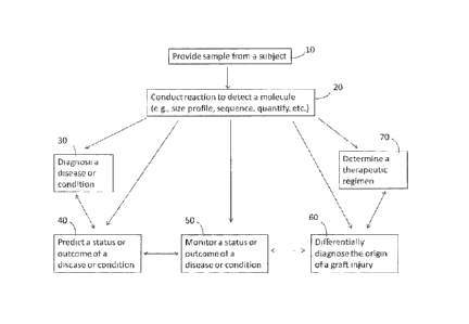
100261 Figure 1 provides a general overview of the flow of many of the methods
provided herein.
Generally, the method comprises providing a sample from a subject (10),
conducting a reaction to detect a
molecule (20), and then diagnosing a disease or condition (30), predicting the
status or outcome of a
disease or condition (40), monitoring the status or outcome of a disease or
condition (50), differentially
diagnosing the origin of a graft injury (60), or determining a therapeutic
regimen (70). Different
combinations of steps can be used, and the steps can be performed in different
orders as well. Also
provided are methods for detecting, monitoring, and/or measuring whole
genomes, or unique regions
thereof, within the heterogeneous sample. The genomes (or genotypic patterns)
may derive from a subject
or from a foreign source.
100271 In some instances, the methods further comprise the use of a computer,
computer software,
and/or algorithm for analyzing one or more molecules in the sample. In other
instances, the methods
further comprise generating a report.
100281 Figure 2 outlines some additional embodiments of the methods provided
herein. The methods
may generally comprise: (a) obtaining a sample containing nucleic acids from
different genomic sources
(210); (b) optionally, sequencing the nucleic acids, e.g., by long-read
sequencing (220); (c) optionally,
counting the number of unique sequences within the nucleic acid sample (e.g.,
via sequence reads) (230);
and (d) optionally, analyzing (e.g., comparing) the ratios of unique sequences
to determine the relative
amounts of the different genomes in the biological sample (240). Different
combinations of steps can be
used, and the steps can be performed in different orders as well, or combined
with steps described herein
related to other methods.
100291 The methods, compositions, and systems of the disclosure may be
especially useful for
noninvasive detection of organ rejection in a transplant recipient, cancer in
a subject, fetal genetic
disorders in a fetus (via the maternal blood), and infection by foreign
pathogens. The methods provided
herein are also useful for the detection of single nucleotide polymorphisms
(SNPs), as well as the
detection of any genomic instability, such as a point mutation or an
aneuploidy (e.g., trisomy, monosomy,
duplication, deletion, addition, rearrangement, translocation, or inversion)
within a foreign and/or host
genome.
Organ/Tissue Transplantation
Introduction
100301 This disclosure provides methods for detecting circulating molecules
(e.g., nucleic acids,
proteins, etc.) in a subject who has received a transplant (e.g., organ
transplant, tissue transplant, stem cell
transplant) in order to diagnose, monitor, predict, or evaluate the status or
outcome of the transplant.
Moreover, this disclosure provides methods of determining or evaluating
potential causes of transplant
rejection, or threatened-rejection.
-12-
CA 3067612 2020-01-13
CA 02849771 2014-03-21
WO 2013/043922 PCT/US2012/056416
[0031] Often, a biological sample containing blood (or other bodily fluid such
as urine) obtained from a
transplant recipient is a heterogeneous sample containing molecules derived
both from the donor and the
recipient. The method may comprise specifically detecting, profiling, or
quantitating molecules (e.g.,
nucleic acids, DNA. RNA, protein, etc.) that are within the biological sample
and that derive from the
donor or donor tissue. In some cases, the method comprises detecting nucleic
acids (or other molecules)
that are derived from the transplant recipient's tissue (as opposed to the
donor tissue)¨either alone or in
addition to molecules derived from donor tissue.
100321 A relative rise in the level of certain circulating nucleic acids,
particularly those derived from the
donor organ or tissue, generally indicates an increased risk of rejection - or
actual rejection - of the
transplanted tissue. Since cell-free DNA or RNA can arise from dying cells
(e.g., apoptotic celLs or
necrotic cells), the relative amount of donor-specific sequences in
circulating nucleic acids may provide a
predictive measure of on-coming organ failure in transplant patients for many
types of solid organ
transplantation including, but not limited to, heart, lung, liver, kidney and
skin. Thus, transplant rejection
can be detected or predicted using partial or whole genome analysis of
circulating nucleic acids derived
from the donor as compared to the recipient's genome.
a. Differential Diagnosis of Graft Injuries
i. Types of Tissue Transplant Outcomes (or Statuses)
[0033] A subject who has received a tissue or organ transplant has a number of
different possible
outcomes. Under optimal circumstances, the status or outcome of the tissue
transplant is transplant
tolerance. Transplant tolerance includes situations where the subject does not
reject a graft organ, tissue
or cell(s) that has been introduced into/onto the subject. In other words, the
subject tolerates or maintains
the organ, tissue or cell(s) that has been transplanted to it.
[0034] Less-favorable statuses or outcomes may involve immunological rejection
(e.g., acute cellular
rejection, antibody-mediated rejection) of the transplant, transplant (or
graft) injury (either non-rejection-
based, or due to rejection), decreased or impaired transplant function,
decreased transplant survival,
and/or chronic transplant injury. Even worse statuses or outcomes include
organ failure and death of the
organism. Organ failure can involve failure of the whole organ, or a portion
thereof. Organ failure may
also involve one organ, or multiple organs, e.g., greater than 1, 2, 3, 4,5,
6, 7, 8,9, or 10 organs.
[0035] Transplant rejection encompasses both acute and chronic transplant
rejection. Acute rejection
(AR) may occur when the immune system of a tissue transplant recipient rejects
transplanted tissue,
usually because it is immunologically foreign. Acute rejection may be
characterized by infiltration of the
transplanted tissue by immune cells of the recipient, which carry out their
effector function and destroy
the transplanted tissue. The onset of acute rejection may be rapid and
generally occurs in humans within a
few weeks or a few months after transplant surgery, but in some cases acute
rejection may occur several
-13-
CA 3 0 6 7 6 1 2 2 0 2 0 -0 1 -1 3
CA 02849771 2014-03-21
WO 2013/043922 PCT/US2012/05641.6
months after transplant surgery or even years after transplant surgery.
Generally, acute rejection can be
inhibited or suppressed with immunosuppressive drugs such as rapamycin,
cyclosporin A, anti-CD4OL
monoclonal antibody and the like. Chronic transplant rejection (CR) generally
occurs in humans within
several months to years after engraftment, and can occur even in the presence
of successful
immunosuppression of acute rejection. Fibrosis is a common factor in chronic
rejection of all types of
organ transplants. Chronic rejection can typically be described by a range of
specific disorders that are
characteristic of the particular organ. For example, in lung transplants, such
disorders include
fibroproliferative destruction of the airway (bronchiolitis obliterans); in
heart transplants or transplants of
cardiac tissue, such as valve replacements, such disorders include cardiac
allograft vasculopathy and
fibrotic atherosclerosis; in kidney transplants, such disorders include
obstructive nephropathy,
nephrosclerorsis, tubulointerstitial nephropathy; and in liver transplants,
such disorders include
disappearing bile duct syndrome. Chronic rejection can also be characterized
by ischemic insult,
denervation of the transplanted tissue, hyperlipidemia and hypertension
associated with
immunosuppressive drugs. In some instances, chronic rejection comprises
inflammation at the site of a
graft and/or surrounding vasculature. In some instances, chronic rejection
comprises injury to a graft
and/or surrounding vasculature. Chronic rejection can be caused by a
pathogenic infection (e.g., viral,
bacterial, fungal, microbial). For example, a viral infection can cause a
chronic rejection. Alternatively, a
bacterial infection can cause a chronic rejection. Chronic rejection can be
characterized by a slow
accumulation of injury. Chronic rejection can occur over a prolonged period of
time, such as over several
weeks (e.g., about 2 weeks, about 4 weeks, about 6 weeks, about 8 weeks, about
10 weeks, about 12
weeks). In some instances, chronic rejection occurs over several months (e.g.,
about 3 months, about 6
months, about 9 months, about 12 months). Chronic rejection can occur over
several years (e.g., about 1.5
years, about 2 years, about 2.5 years, about 3 years, about 3.5 years, about 4
years, about 4.5 years, about
years). In some instances, the immune activity of a transplant recipient
continues, extends, or prolongs
the duration of the chronic rejection.
[0036] Examples of non-rejection based transplant injury (e.g., allograft
injury) include, but are not
limited to, ischemic injury, pathogenic infection (e.g., viral infection,
bacterial infection, fungal
infection), perioperative ischemia, reperfusion injury, hypertension,
physiological stress, injuries due to
reactive oxygen species and injuries caused by pharmaceutical agents (e.g.,
immunosuppressive drugs,
etc.). Transplant status or outcome may also involve vascular complications or
neoplastic involvement of
the transplanted organ. The outcome or status of a transplant can be affected
by the dose, titer or level of
therapies used to treat the subject, such as the level of immunosuppressive
agents administered to the
subject. For example, a high dose of immunosuppressive drugs may result in
transplant injury, while a
dose that is too low may result in rejection of the transplant.
-14-
CA 3067612 2020-01-13
CA 02849771 2014-03-21
WO 2013/043922 PCT/US2012/056416
ii. Circulating Donor Molecules and Cellular Death by Apoptosis or
Necrosis
[00371 The present disclosure provides methods for identifying a source or
cause of a transplant/graft
injury or of transplant rejection, including by measuring levels of
circulating donor molecules and/or by
evaluating the size distribution of such molecules. As described further
herein, information from
circulating donor molecules can be used either alone, or in combination with
other markers of transplant
injury, such as markers derived from the subject's own tissue (including the
subject's immune repertoire)
or markers derived from a foreign source such as a pathogen.
100381 Provided herein are methods of determining an origin of a graft injury
by discriminating
between rejection and infection. In some instances, the fragment length of the
donor molecule is used to
discriminate between an immunologic rejection (which may be associated with an
increase in apoptotic
tissue) and an infection (which may manifest in an increase in necrotic
tissue). Cell-free DNA is released
from both apoptotic and necrotic cells, but the size distribution of the DNA
fragments may differ in these
two cases.
100391 The methods provided herein may comprise determining a relative level
of apoptotic cell death
in a donor tissue or organ by evaluating a size profile of circulating DNA
(e.g., circulating donor DNA) or
other molecule (e.g., nucleic acid, RNA, protein, etc.). The method may
further comprise using the level
(or relative level) of apoptotic cell death to determine the presence or
degree of an immune response to
transplanted tissue or a transplanted organ. The immune response may be a
cellular immune response
and/or an antibody-mediated immune response. Apoptotic cell death usually
involves nuclease digestion
of the genomic DNA while still bound to nucleosomes prior to release from the
cell. Consequently, as a =
result of apoptosis, the circulating DNA may present as a set of small
fragments, often separated by a
uniform, or near-uniform periodicity. If the DNA is run on an electrophoretic
gel, it may appear as a
ladder of fragments of different sizes. The methods provided herein may
comprise detecting the level (or
relative level) of a set of fragments comprising fragments of size 180 bp, 360
bp, 540 bp, and 720 bp, 900
bp, etc. with the majority of molecules at the smallest sizes. The method may
comprise detecting the level
(or relative level) of a set of fragments comprising molecules of sizes that
are smaller than 200 bp, e.g., a
set comprising fragments of sizes of 195 bp, 190 bp, 185 bp, 180 bp, 175 bp,
170 bp, 165 bp, 160 bp, 155
bp, 150 bp, 145 bp, 140 bp, 135 bp, 130 bp, 125 bp, 120 bp, 110 bp, 100 bp, 90
bp, 80 bp, 70 bp, 60 bp,
50 bp, 40 bp, 30 bp, 20 bp, and/or 10 bp, or any combination thereof. The set
of fragments may also
comprise molecules that are within plus or minus 1, 2, 3,4, or 5 bp of these
values. In some cases, the set
of fragments comprises fragments of sizes less than 300 bp, 250 bp, 240 bp,
230 bp, 220 bp, 210 bp, 200
bp, 190 bp, 180 bp, 170 bp, 160 bp, or 150 bp. The set of molecules may be
spaced at a periodicity of
about 5, 6, 7, 8,9, 10, 11, 12, 13, 14, 15, 16, 17, 18, 19, 20, 30, 40, 50,
60, 70, 80, 90, 100, 120, 130, 140,
150, 160, 170, 180, 190, 200, 210, or 220 bp. For example, a set of molecules
spaced at a periodicity of
-15-
CA 3067612 2020-01-13
CA 02849771 2014-03-21
WO 2013/043922 PCT/US2012/056416
about 10 lop can comprise fragments of sizes of 10 bp, 20 bp, 30 bp, 40 bp, 50
bp, 60 bp, 70 bp, 80 bp, 90
bp, 100 bp, 110 bp, 120 bp, 130 bp, 140 bp, 150 bp, 160 bp, or 170 bp. In some
instances, apoptotic cell -
death in a donor tissue or organ is characterized by a size profile of donor-
derived DNA wherein a
majority of DNA fragments are less than about 250 bp. In some instances, the
size profile is characterized
by an increase in DNA fragments of about 166 bp, when compared to (1) the size
profile of DNA from a
different tissue type, such as blood; and/or (2) the size profile of DNA from
the same tissue type, where
the tissue is known to be either healthy or diseased. In other cases, the size
profile is characterized by a
decrease in DNA fragments with sizes of about 166 bp. In some instances, the
size profile is characterized
by a decrease in DNA fragments of less than about 120 bp.
(00401 The methods provided herein may comprise determining a relative level
of apoptotic or necrotic
cell death in a donor tissue or organ by evaluating the quantity of
circulating RNA derived from the donor
tissue or organ. The method may further comprise using the level (or relative
level) of apoptotic or
necrotic cell death to determine the presence or degree of an immune response
to transplanted tissue or a
transplanted organ; to determine or predict the degree of tissue or organ
rejection or damage; and/or to
identify or predict the presence of a pathogenic infection. In some cases, a
relative increase in circulating
donor RNA indicates a higher risk of rejection. In some cases, a relative
decrease in circulating donor
RNA indicates a higher risk of rejection. In some cases, a relative increase
in circulating donor RNA may
indicate a higher risk of pathogenic infection; in other cases, a relative
decrease in circulating donor RNA
indicates a higher risk of pathogenic infection. The relative increase may be
at least 1-fold, 1.25-fold, 1.5-
fold, 1.75-fold, 2-fold, 2.25-fold, 2.5-fold, 2.75-fold, 3-fold, 3.25-fold,
3.5-fold, 3.75-fold, 4-fold, 5-fold,
6-fold, 7-fold, 8-fold, 9-fold, 10-fold, 50-fold, 100-fold, 500-fold, 1000-
fold or more. Alternatively, the
relative decrease is at least 1-fold, 1.25-fold, 1.5- fold, 1.75-fold, 2-fold,
2.25-fold, 2.5-fold, 2.75-fold, 3-
fold, 3.25-fold, 3.5-fold, 3.75-fold, 4-fold, 5-f)ld, 6-fold, 7-fold, 8-fold,
9-fold, 10-fold, 50-fold, 100-fold,
500-fold, 1000-fold or more. In some instances, the relative increase is at
most about 1-fold, 1.25-fold,
1.5- fold, 1.75-fold, 2-fold, 2.25-fold, 2.5-fold, 2.75-fold, 3-fold, 3.25-
fold, 3.5-fold, 3.75-fold, 4-fold, 5-
fold, 6-fold, 7-fold, 8-fold, 9-fold, 10-fold, 50-fold, 100-fold, 500-fold,
1000-fold or more. Alternatively,
the relative decrease is at most about 1-fold, 1.25-fold, 1.5- fold, 1.75-
fold, 2-fold, 2.25-fold, 2.5-fold,
2.75-fold, 3-fold, 3.25-fold, 3.5-fold, 3.75-fold, 4-fold, 5-fold, 6-fold, 7-
fold, 8-fold, 9-fold, 10-fold, 50-
fold, 100-fold, 500-fold, 1000-fold or more. The increase or decrease may be
relative to the quantity of
circulating donor RNA at a particular time point or may occur over a
particular time period. For example,
the increase or decrease is a 2-fold increase over a 5-day time period. In
another example, the increase or
decrease is a 2.5-fold increase over, or within, a I-month time period, a 2-
month time period, a 3-month
time period, a 4-month time period, a 5-month time period, or a 6-month time
period. In some cases, the
increase or decrease is a 3-fold increase over, or within, a 1-month time
period, a 2-month time period, a
-16-
CA 3067612 2020-01-13
CA 02849771 2014-03-21
WO 2013/043922 PCT/US2012/056416
3-month time period, a 4-month time period, a 5-month time period, or a 6-
month time period. In some
cases, tlie particular time period is about 0.5, 1, 2, 3, 4, 5, 6, 7, 8, 9, or
10 days; 1, 2, 3, 4, 5, 6, 7, 8, 9, or
weeks; 1, 2, 3, 4, 5, 6, 7, 8, 9, or 10 months; or 1 or 2 years, or, in some
cases, even longer. In some
cases, the particular time period is at most 0.5, 1, 2, 3,4, 5, 6, 7, 8, 9, or
10 days. Alternatively, the
particular time period is at most 1,2, 3,4, 5,6, 7, 8,9, or 10 months. In some
cases, the particular time
period is at most 0.5, 1, 2, 3, 4, 5, 6, 7, 8, 9, or 10 years.
[0041] The methods provided herein may comprise determining a relative level
of necrotic cell death in
a donor tissue or organ by evaluating a size profile of circulating DNA (e.g.,
circulating donor DNA) or
of a different molecule (e.g., RNA or other nucleic acid, protein, etc.). The
method may further comprise
using the level (or relative level) of necrotic cell death to determine the
presence or degree of a
pathogenic infection associated with transplanted tissue or a transplanted
organ. Necrotic cell death is not
as orderly as apoptotic cell death. Moreover, DNA released from necrotic cells
is generally longer than
that released from apoptotic celLs. The methods provided herein may comprise
detecting the level (or
relative level) of a set of fragments comprising fragments of relatively large
size. The set of fragments
may comprise fragments that are greater than 300 bp, 400 bp, 500 bp, 1000 bp,
1500 bp, 2000 bp, 2500
bp, 3000 bp, 3500 bp, 4000 bp, 4500 bp, 5000 bp, 5500 bp, 6000 bp, 6500 bp,
7000 bp, 7500 bp, 8000 bp,
8500 bp, 9000 bp, 9500 bp, 10000 bp, 10500 bp, 11000 bp, 11500 bp, 12000 bp,
12500 bp, 13000 bp,
13500 bp, 14000 bp, 14500 bp, or 15000 bp.
[0042] In some cases, necrotic cell death in a donor tissue or organ is
characterized by an increase in
smaller-sized DNA fragments, particularly after the donor-derived DNA is
digested (e.g., digestion by
restriction enzymes). Such increase may be an increase of small-sized donor
DNA fragments when
compared with digested DNA from healthy tissue, such as healthy recipient
tissue. In some cases, such
increase is an increase of small-sized DNA fragments when compared with
digested donor DNA from a
different time-point, or over a particular time period (such as, e.g., 1, 2,
3, 4, 5, 6, 7, 8, 9, 10, 11, or 12
hours; 1, 2, 3, 4, 5, 6, 7, 8, 9, 10, 11, or 12 days; 1, 2, 3, 4, 5, 6, 7, 8,
9, 10, 11, or 12 months; orl , 2, or 3
years or longer. The increase may be a 1-fold, 2-fold, 3-fold, 4-fold, 5-fold,
6-fold, 7-fold, 8-fold, 9-fold,
10-fold, 50-fold, 100-fold, 500-fold, 1000-fold increase, or even more. in
some instances, the smaller-
sized DNA fragments are less than about 150 bp, about 140 bp, about 130 bp,
about 120 bp, about 110 bp,
or about 100 bp. For example, necrotic cell death can be characterized by an
increase of DNA fragments
of about 120 bp and/or a decrease in DNA fragments of about 166 bp,
particularly after digestion.
[0043] In some cases, the method comprises identifying whether the fragments
have a uniform, or near-
uniform, periodicity in size versus whether the sizes of the fragments appear
to be more randomly-sized.
For example, in some cases, the method may comprise determining whether a size
profile of DNA
fragments has well-defined peaks of sizes (e.g., as would be more indicative
of apoptotic cell death)
-17-
CA 3067612 2020-01-13
CA 02849771 2014-03-21
WO 2013/043922 PCT/US2012/056416
versus less-defined sizes. The DNA fragments may derive from necrotic DNA if
they fail to appear as a
ladder with distinct sizes separated by a uniform or near-uniform size
periodicity or if they appear as a
smear when run on an electrophoretic gel. The methods herein, therefore, may
comprise using these
factors (size, periodicity, etc.) to determine whether the originating DNA is
derived from apoptotic versus
necrotic tissue. For example, in some instances, necrotic cell death is
characterized by a size profile
comprising irregular or randomly-sized DNA fragments, whereas apoptotic cell
death is characterized by
a size profile comprising DNA fragments of a certain periodicity (e.g., 5 bp,
10 bp, 20 bp, etc). In some
cases, the size profile characterizing apoptotic cell death is an even
distribution of DNA fragments across
a spectrum of sizes. The quantity of donor DNA fragments across a given size
profile may vary, on
average, by less than 1%, 2%, 3%, 4 A, 5%, 6%, 7%, 80/0, 9%, 100/o, 20%, 30%,
40%, 50%, 60%, 70%,
80%, 90%, 100%, 150%, 200%, 300%, 400%, 500%, or 1000%. For example, for a
size profile that
contains only DNA fragments that are 50 bp, and 1000 bp in length and where
the quantity of 50-b1,
fragments is half the quantity of 1000 bp fragments, the average quantity
variation is less than 100%. In
some cases, necrotic cell death is characterized by a size profile comprising
indistinguishable or non-
discrete DNA fragments (e.g., DNA fragments appear as a smear on an
electrophoresis gel).
[0044] Methods of obtaining a size profile are described further in other
sections herein. Briefly, a size
profile can be obtained by any one of a number different techniques,
including, but not limited to,
sequencing (e.g., paired-end sequencing, single molecule sequencing),
electrophoresis (e.g., gel
electrophoresis, agarose gel electrophoresis, polyacrylamide electrophoresis,
capillary electrophoresis,
alkaline gel electrophoresis, pulsed field gel electrophoresis), amplification
(e.g., PCR-based
amplification, non-PCR based amplification), and arrays. In some instances,
devices, including, but not
limited to, a sequencing machine, electrophoresis chamber, electrophoresis
machine, thermal cycler, PCR
machine, plate reader, fluorometer, luminometer, microscope, and computer are
used to obtain a size
profile.
[0045] The method may further comprise obtaining a "Death Mode Ratio" by
comparing the relative
level of circulating DNA fragments of a certain size (or size pattern, ladder,
or profile) associated with
apoptosis with the relative level of circulating DNA fragments of a certain
size (or size pattern, ladder, or
profile) associated with necrosis. Often, the circulating DNA fragments used
to obtain the Death Mode
Ratio may derive from the donor tissue; but the DNA fragments may also derive
from recipient tissue, or
some combination of donor and recipient tissue (e.g., necrotic recipient DNA,
necrotic donor DNA,
apoptotic donor DNA, or apoptotic recipient DNA).
[0046] The methods provided herein may comprise correlating a Death Mode Ratio
with a control
Death Mode Ratio that characterizes a particular known condition, such as a
condition or transplant status
described herein (e.g., tolerance, immunologic rejection, pathogenic infection
(of donor tissue, recipient
-18-
CA 3067612 2020-01-13
CA 02849771 2014-03-21
WO 2013/043922 PCT/US2012/056416
tissue, or both), specific graft injury, graft injury due to pharmacological
agent, etc.). Thus, a method may
comprise determining a control Death Mode Ratio, determined by measuring
levels of circulating DNA in
subjects with a known condition, such as a known immunologic rejection of
transplanted tissue or a
known pathogenic infection of transplanted tissue. In some cases, the control
Death Ratio may be
determined in a subject known not to have received a transplant, or who is
known to have tolerated a
transplant. The subject used to determine the control Death Mode Ratio may be
a subject different from
the subject used for the Death Mode Ratio or the same as the subject used for
the Death Mode Ratio. In
some cases, multiple subjects are used to determine the control Death Mode
Ratio.
[0047] The method may further comprise comparing the Death Mode Ratio of a
subject with an
unknown condition (e.g., it is unknown whether transplanted tissue has
triggered an immunologic
rejection) with the control Death Mode Ratio in order to determine, or help
determine, whether the subject
is experiencing an immunologic rejection or pathogenic infection associated
with transplanted tissue.
Such method may also determine, or help determine, whether a known case of
transplant rejection is
worsening or improving. The method may further comprise evaluating or
analyzing the comparison of the
Death Mode Ratio with the control Death Mode Ratio in order to determine the
existence of, risk of, level
of, or status of immunologic rejection within the subject with the unknown
condition. For example, a
Death Mode Ratio of a sample from a transplant recipient can be obtained by
determining the quantity of
DNA fragments (e.g., donor DNA) that is between 160-170 bp in size and
comparing that to the quantity
of DNA fragments (e.g., donor DNA) across a broader size range, such as DNA
fragments present
between 100-250 bp. A Death Mode Ratio of a control sample can be determined
in a similar manner.
The Death Mode Ratio of the sample from a transplant recipient then can be
compared to the Death Mode
Ratio of the control sample. A Death Mode Ratio of a sample from the
transplant recipient greater than
the Death Mode Ratio of the control sample can be indicative of apoptosis
within the donor organ or
tissue, whereas, a Death Mode Ratio of a sample from the transplant recipient
less than the Death Mode
Ratio of the control sample can be indicative of necrosis.
[0048] Often, the method comprises determining a Death Mode Ratio after
determining the relative
level of circulating donor nucleic acids within the transplant recipient. For
example, the method may
comprise (a) evaluating the level of circulating donor nucleic acids (e.g.,
DNA, RNA) in a transplant
recipient and then, if the level has reached a certain threshold level, (b)
evaluating a size profile of
circulating nucleic acids and/or calculation of a Death Mode Ratio. The
threshold level may be a level
known to indicate a particular status or outcome: e.g., rejection, threatened
rejection, organ failure, organ
damage, or risk of the foregoing. In some cases, the threshold level reflects
a level of circulating nucleic
acids disclosed herein in any section of this disclosure. In some cases, the
threshold level is determined on
a patient-specific basis. For example, the threshold level may be determined
based on a review of a
-19-
CA 3067612 2020-01-13
CA 02849771 2014-03-21
WO 2013/043922 PCT/US2012/056416
patient's (or other subject's) past history of organ tolerance, rejection or
threatened rejection and
correlation of such previous events with the level of circulating molecules
(e.g, nucleic acids) in the
patient. In some instances, an increase in donor-derived molecules (e.g., DNA
and/or RNA) and an
increase in apoptotic cell death as determined by a Death Mode ratio relative
to values determined from
previously obtained samples from a transplant recipient are indicative of a
rejection or an increased
likelihood of rejection in the transplant recipient.
iii. Detecting an Immune Response
100491 In addition to detecting circulating donor molecules, discriminating
between rejection and
infection may further comprise measuring an immune response in a subject, such
as by immune repertoire
profiling of T cells and/or B cells. The method may comprise detecting,
monitoring, or evaluating an
immune response within a transplant recipient, particularly an immune response
to transplanted organ,
tissue, cells, or molecules. In some cases, an immunological rejection is
indicated (or at increased risk)
when the inunune repertoire profiling reveals the presence of B-cell clones or
T cells that are capable of
targeting an antigen associated with the transplanted cells, tissues, organ,
or molecules. In some cases, an
immunological rejection is less indicated (or at reduced risk) when the immune
repertoire profiling
reveals the absence or reduction of B-cell clones or T cells that are capable
of targeting an antigen
associated with the transplanted cells, tissues, organ, or molecules.
10050] In some cases, the method comprises determining that a cellular
rejection of transplanted tissue
or organ is occurring, has an increased risk of occurring, or is worsening,
where there is a relative
increase in one or both of the following (a) circulating molecules (e.g., DNA)
associated with apoptotic
donor tissue and (b) immune response as measured by evaluating the immune
repertoire. In some cases,
the method comprises determining that a cellular rejection of transplanted
tissue or organ is not occurring,
is at decreased risk of occurring, or is improving, where there is a relative
decrease in one or both of the
following (a) circulating molecules (e.g., DNA) associated with (or derived
from) apoptotic donor tissue
and (b) immune response as measured by evaluating the immune repertoire.
Although in preferred
embodiments the circulating molecules are derived from apoptotic donor tissue,
in some cases they may
derive from apoptotic recipient tissue.
100511 The method may further comprise detecting, monitoring, or evaluating an
immune response to a
pathogenic infection associated with the transplant. As described herein, the
immune response may be
detected, monitored, or evaluated by measuring the immune repertoire. The
method may comprise
predicting an increased chance that an organ or tissue rejection is due to
pathogenic infection, where an
increased immune response to a pathogen is detected. For example, the immune
repertoire profiling can
reveal the presence of (or increased number of, or increased activity of) a
large number of B-cell clones
producing antibodies to a pathogen, thereby indicating an infection. In some
cases, the method may
-20-
CA 3067612 2020-01-13
CA 02849771 2014-03-21
WO 2013/043922 PCT/US2012/056416
comprise predicting a decreased chance that an organ or tissue rejection is
due to infection, where a
decreased immune response to a pathogen is detected. For example, immune
repertoire profiling can
reveal the absence of (or reduction of) T cells (or B cells) targeting a
pathogen, thereby indicating the
absence of infection by that pathogen and/or possibly the presence of an
immunologic rejection episode.
100521 A method that involves detecting an immune response to a pathogen may
also comprise using
this information along with information regarding relative levels of necrosis
or apoptosis to evaluate,
predict, monitor or diagnose the risk of, or existence of, a pathogenic
infection. For example, the method
may comprise determining that there is an increased chance that a transplant
rejection is due to infection
where a transplant recipient demonstrates a relative increase in one or more
of the following: (a)
circulating molecules (e.g., DNA) associated with necrosis (e.g., necrotic
donor tissue or necrotic
recipient tissue) and (b) immune response to a pathogen. Although in preferred
embodiments the
circulating molecules are derived from necrotic donor tissue; in some cases,
they may derive from
necrotic recipient tissue.
100531 Detection of the T cell and/or B cell repertoire can comprise
sequencing (e.g., high-throughput
sequencing), amplifying, and/or quantifying the T cell and/or B cell
repertoire. Exemplary methods of
measuring the immune repertoire are described in PCT publication No.
WO/2011/140433 entitled:
Measurement and Comparison of Immune Diversity by High-Throughput Sequencing,
filed May 6, 2011.
iv. Detecting Pathogenic Infections
100541 The method may further comprise detecting, monitoring, or evaluating a
pathogenic infection
within a transplant recipient; particularly a pathogenic infection associated
with, or caused by, the
transplant (or introduced as a result of the transplantation of a tissue or
organ). As described further
herein, a pathogenic infection can be detected via numerous methods including
by sequencing nucleic
acids (e.g., DNA or RNA) or proteins from the pathogen, by amplifying nucleic
acids from a pathogen
(e.g., by applying a PCR reaction to a sample taken from the transplant
recipient, or by using an antibody
to detect a particular pathogen). The method may also comprise determining the
amount of pathogen
(e.g., viral load) in a sample, or otherwise quantifying the pathogen. The
method may comprise predicting
an increased chance that an organ or tissue rejection is due to pathogenic
infection, where a pathogen is
detected within a sample taken from a transplant recipient. The method may
comprise predicting a
decreased chance that an organ or tissue rejection is due to infection, where
a pathogen is not detected in a
sample taken from a transplant recipient. The method may further comprise
using this information along
with information regarding relative levels of necrosis or apoptosis, as
described herein. For example, the
method may comprise determining that there is an increased chance that a
transplant rejection is due to
infection where a pathogen is detected in a sample taken from a transplant
recipient and a transplant
-"?1-
CA 3067612 2020-01-13
CA 02849771 2014-03-21
WO 2013/043922 PCT/US2012/056416
recipient demonstrates a relative increase in one or more of the following:
(a) circulating molecules (e.g.,
DNA) associated with necrosis and (b) immune response to a pathogen.
10055] b. Therapeutic Regimens for Organ/Tissue Transplant Recipients
i. General
100561 The methods disclosed herein may further comprise administering,
adjusting, or terminating a
therapeutic regimen based on the discrimination between rejection and
infection. For example, if an
infection is indicated, then a therapeutic regimen comprising an anti-
microbial (e.g., antibiotic, antiviral,
antifungal) is administered, increased, or adjusted (e.g., by making a change
in the number or types of
pharmacological agents administered). In some cases, if an infection is
indicated, then a therapeutic
regimen comprising an immunosuppressive drug is reduced, terminated or
adjusted (e.g., by making a
change in the number or types of pharmacological agents administered). In some
cases, if a rejection is
indicated, then a therapeutic regimen comprising an iMmunosuppressive drug is
administered, increased
or adjusted (e.g., by making a change in the number or types of
pharmacological agents administered).
ii. Predicting or diagnosing transplant rejection
10057] This disclosure provides methods of predicting or diagnosing transplant
survival (or rejection) in
a subject that has received a transplant. The prediction or diagnosis may
involve detecting circulating
molecules associated with the transplant graft. In many cases, the prediction
or diagnosis may take into
account other factors as well, such as other indicia of organ failure or
reduced function. For example, the
prediction or diagnosis may involve monitoring proteinuria in a kidney
transplant recipient in addition to
the methods described herein.
10058] In some cases, the disclosure provides methods of diagnosing or
predicting the length of time that
transplanted tissue, organ(s), or cells, will survive, such as the presence of
long-term graft survival. By
"long-term" graft survival is meant graft survival for at least about five
years beyond current sampling,
despite the occurrence of one or more prior episodes of acute rejection. In
some cases, graft survival is
predicted to be at least I, 2, 3, 4, 5, 6, 7, 8, 9, 10, 11, 12, 13, 14, 15,
20, 25 or 30 years. In some cases,
graft survival is predicted to be at least, 1, 2, 3, 4, 5, 6, 7, or 8 weeks,
or 1, 2, 3, 4, 5, 6, 7, 8, 10, 11, or 12
months. In other cases, graft survival is predicted to be less than any of
these time periods, e.g., less than
1, 2, 3, 4, 5, 6, 7, or 8 weeks, or 1, 2, 3, 4, 5, 6, 7, 8, 10, 11, or 12
months or less than 1,2, 3, 4, 5, 6, 7, 8,
9, 10, 11, 12, 13, 14, 15, 20, 25 0r30 years.
100591 This disclosure also provides methods of determining or predicting
transplant survival following
acute rejection. In certain embodiments, transplant survival is determined for
patients in which at least
one episode of acute rejection has occurred; in some cases, the subject has
experienced at least 1,2, 3,4,
or 5 episodes of rejection of transplanted tissue. Transplant survival is
determined or predicted in certain
-2/-
CA 3067612 2020-01-13
CA 02849771 2014-03-21
= WO 2013/043922
PCT/US2012/056416
embodiments in the context of transplant therapy, e.g., immunosuppressive
therapy, where
immunosuppressive therapies are known in the art.
10060] Similarly, the methods provided herein may involve determining,
diagnosing, detecting,
predicting, or monitoring the risk of, or existence of, a transplant
rejection. In yet other embodiments,
methods of determining the class and/or severity of rejection (e.g., acute
rejection) (and not just the
presence thereof) are provided. In some instances, methods of determining the
cause of a rejection are
provided herein.
100611 in some instances, predicting a status or outcome of an organ
transplant comprises predicting a
risk of transplant rejection. Alternatively, predicting a status or outcome of
an organ transplant comprises
predicting or detecting organ failure. Monitoring a status or outcome of an
organ transplant may comprise
monitoring efficacy of an immunosuppressive regimen. In addition, predicting a
status or outcome of an
organ transplant may comprise identifying or predicting responders to an
immunosuppressive therapy.
[0062] In some instances, the method comprises predicting or diagnosing the
presence of, risk of, or
degree of transplant rejection after detecting an increase in foreign (e.g.,
donor-derived) molecules, such
as circulating donor-derived molecules. A transplant rejection may be
indicated, or an increased risk of
rejection may be indicted, where donor-derived molecules may increase by at
least about 30%, at least
about 40%, at least about 50%, at least about 60%, at least about 70%, at
least about 80%, or at least
about 90%. In other instances, the rejection may be indicated, or at an
increased risk, where the donor-
derived molecules increase by at least about 1.25-fold, at least about 1.5-
fold, at least about 1.75-fold, at
least about 2-fold, at least about 2.25-fold, at least about 2.5-fold, at
least about 2.75-fold, at least about 3-
fold, at least about 3.25-fold, at least about 3.5-fold, at least about 3.75-
fold, at least about 4-fold, at least
about 4.5-fold, at least about 5-fold, at least about 6-fold, at least about 7-
fold, at least about 8-fold, at
least about 9-fold, at least about 10-fold, at least about 15-fold, at least
about 20-fold, at least about 50-
fold, or at least about 100-fold. The increase in donor-derived molecules may
be detected over a period of
time. For example, the increase in donor-derived molecules may occur within
about a 2-day period, 5-day
period, 10-day period, 14-day period, 21-day period, 28-day period, 1-month
period, 2-month period, or
3-month period. In another example, the increase in donor-derived molecules
can occur within about a 4-
month period, 5-month period, 6-month period, 7-month period, 8-month period,
9-month period, 10-
month period, 11-month period, 12-month period, 13-month period, 14-month
period, 15-month period,
16-month period, 17-month period, 18-month period, or 24-month period. In some
instances, the increase
in donor-derived molecules occurs within at most a 1, 2, 3, 4, 5, 6, 7, 8,9,
or 10-month period. In some
instances, the increase in donor-derived molecules occurs within at most a 1,
2, 3, 4, 5,6, 7, 8, 9, or 10-
year period. In some instances, an increase in donor-derived molecules
relative to a consensus normal or
control at about the time of transplantation (e.g., time = 0) is indicative of
rejection or increased risk of
-23-
CA 3067612 2020-01-13
CA 02849771 2014-03-21
WO 2013/043922 PCT/US2012/056416
rejection. In some cases, a certain increase in donor-derived molecules is
predictive of a transplant
rejection with a certainty of at least 25%, 50%, 60%, 70%, 75%, 80%, 85%, 90%,
95%, 96%, 97%, 98%,
99% or 99.5%. The time period may indicate the presence of, or risk of
transplant rejection. For example,
an increase of about two-fold of donor-derived molecules within a 10 day
period may indicate a rejection
or increased risk of rejection. As another example, an increase in donor-
derived molecules above an
established baseline level over several months may indicate a rejection or
increased risk of rejection.
100631 In some instances, a gradual increase in foreign molecules (e.g., donor-
derived molecules) is
indicative of chronic rejection. In some instances, chronic rejection, or risk
thereof, is indicated when the
foreign molecules increase by less than about 20%, less than about 15%, less
than about 12%, less than
about 10%, less than about 7%, less than about 5%, over a period of time.
Preferably, the foreign
molecules increase by less than about 5%, less than about 4%, less than about
3%, less than about 2%,
less than about 1%, less than about 0.5%, less than about 0.25% over a period
of time. hi other instances,
the presence of the foreign molecules increases by less than about 1.25-fold,
1.5-fold, less than about
1.75-fold, less than about 2-fold, less than about 2.25-fold, less than about
2.5-fold, less than about 2.75-
fold, less than about 3-fold, less than about 3.25-fold, less than about 3.5-
fold, less than about 3.75-fold,
less than about 4-fold, less than about 5-fold, less than about 6-fold, less
than about 7-fold, less than about
8-fold, less than about 9-fold, less than about 10-fold, less than about 15-
fold, less than about 20-fold, less
than about 50-fold, or less than about 100-fold.
100641 In some instances, the methods disclosed herein predict transplant
rejection at least about 1, at
least about 2, at least about 3, at least about 4, at least about 5, at least
about 6, at least about 7, at least
about 8, at least about 9, at least about 10, at least about 11, at least
about 12, at least about 13, or at least
about 14 days earlier than a biopsy-predicted transplant rejection.
Alternatively, the methods disclosed
herein predict transplant rejection at least about 1, at least about 2, at
least about 3, at least about 4, at
least about 5, at least about 6, at least about 7, at least about 8, at least
about 9, at least about 10, at least
about 11, at least about 12, at least about 13, or at least about 14 weeks
earlier than a biopsy-predicted
transplant rejection. The methods disclosed herein may predict transplant
rejection at least about 1, at
least about 2, at least about 3, at least about 4, at least about 5, at least
about 6, at least about 7, at least
about 8, at least about 9, at least about 10, at least about 11, at least
about 12, at least about 13, or at least
about 14 months earlier than a biopsy-predicted transplant rejection. The
methods disclosed herein may
predict transplant rejection at least about 1, 1.25, 1.5, 1.75, 2, 2.25, 2.5,
2.75, 3, 3.25, 3.5, or 3.75 years
prior to transplant rejection.
109651 In some instances, the methods disclosed herein predict the cause of a
transplant rejection by
detecting the presence or absence of foreign nucleic acids, wherein the
foreign nucleic acids are not
donor-derived nucleic acids. In some instances, the presence of the foreign
nucleic acids is indicative of a
CA 3067612 2020-01-13
CA 02849771 2014-03-21
WO 2013/043922 PCT/1JS2012/056416
pathogenic infection. In some instances, the foreign nucleic acids are viral
nucleic acids. Alternatively,
the foreign nucleic acids are bacterial nucleic acids. In some instances, the
presence of the foreign nucleic
acids is indicative of an infection within the transplanted tissue or organ.
In some instances, the presence
of foreign nucleic acids indicates that the rejection is at least partially
caused by an infection. In some
instances, the infection is a viral infection. In other instances, the
infection is a bacterial infection. In
sonic instances, the method further comprises conducting a sequencing reaction
on the foreign nucleic
acids. In some instances, the absence of foreign nucleic acids is indicates
that the rejection is at least
partially caused by an immune reaction. In some instances, the immune reaction
is a cell-mediated
immune response. Alternatively, the immune reaction is an antibody-mediated
immune reaction.
100661 In some instances, the methods disclosed herein predict transplant
tolerance by monitoring the
fold-change of donor nucleic acids, or the percentage change of donor nucleic
acids relative to total
nucleic acids. In some instances, a fold-increase of not more than about
0.0001-fold, 0.005-fold, 0.01-
fold, 0.05-fold, 0.1-fold, 0.15-fold, 0.2-fold, 0.25-fold, 0.3-fold, 0.35-
fold, 0.4-fold, 0.45-fold, 0.5-fold,
0.55-fold, 0.6-fold, 0.65-fold, 0.7-fold, 0.75-fold, 0.8-fold, 1-fold, 1.1-
fold, 1.2-fold, 1.3-fold, 1.4-fold,
1..5-fold, 1..6-fold, 1.7-fold, 1.8-fold, 1.9-fold, or 2-fold over a period of
time, or within a period of time,
is indicative of tolerance. In some instances, tolerance is indicated when
donor nucleic acids make up not
more than about 0.4%, 0.45%, 0.5%, 0.55%, 0.6%, 0.65%, 0.7%, 0.75%, 0.8% of
total nucleic acids
within a period of time. In some instances, the period of time is over, or
within, 1, 2, 3, 4, 5, 6, 7, 8, 9, 10,
11, 12, 13, 14, 15, 16, 17, 18, 19, 20, 21, 22, 23, 24, 25, 26, 27, 28, 29, or
30 weeks. In some instances,
the period of time is over, or within, 1, 2, 3,4, 5, 6, 7, 8, 9, 10, 11, or 12
months. Alternatively, the period
of time is over, or within, 1,2, 3, 4, 5, 6, 7, 8,9, or 10 years.
100671 In some instances, diagnosing, predicting, or monitoring the status or
outcome of a disease or
condition comprises determining patient-specific baselines and/or thresholds.
For example, the patient-
specific baselines and/or thresholds can provide ranges for predicting a
transplant rejection. In another
example, the patient-specific baselines and/or thresholds can provide ranges
for diagnosing a disease or
condition. In another example, the baselines and thresholds in a pediatric
heart recipient may be higher
than in an adult heart recipient. Similarly, the baselines and thresholds in a
recipient receiving a partial
liver transplant as in living donor transplantation can differ from the
baselines and thresholds in a
recipient receiving a complete liver. Alternatively, the baselines and
thresholds in a recipient receiving a
single-lung transplant may differ from the baselines or thresholds in a
recipient receiving a double-lung
transplant. In some instances, patient-specific baselines and/or thresholds
determined at about time of
transplantation (e.g., time = 0) are compared to baselines and/or thresholds
for a group (e.g., children,
adults, males, females, ethnic groups). In some instances, comparison of
patient-specific baselines and/or
thresholds to group-specific baselines and/or thresholds is used to predict
outcome and/or guide therapies.
-25-
CA 3067612 2020-01-13
CA 02849771 2014-03-21
WO 2013/043922 MT/1)82012/056.116
For example, the threshold percentage of donor nucleic acids can be predictive
of rejection. In some
instances, the threshold percentage of total nucleic acids within a sample
that are donor nucleic acids
varies depending on the organ. For example, a threshold percentage for a liver
transplant where at least
about 0.1%, 0.2%, 0.3%, 0.4%, 0.5%, 0.75%, 1%, 1.25%, 1.5%, 2%, 2.2%, 2.5%,
2.7%, 3%, 3.25%,
3.5%, 3.75%, 4%, 4.25%, 4.5%, 4.75%, 5%, 5.25%, 5.5%, 5.75%, or 6% of total
nucleic acids are donor
nucleic acids is indicative of rejection. In sonic instances, in a liver
transplant, a percentage of total.
nucleic acids in a sample that are donor nucleic acids that is at least about
1%, 2%, 3%, or 4% may be
indicative of rejection. In another example, a percentage for a lung
transplant of at least 0.1%, 0.2%,
0.3%, 0.4%, 0.5%, 0.75%, 1%, 1.25%, 1.5%, 2%, 2.25%, 2.5%, 2.75%, 3%, 3.25%,
3.5%, 3.75%, 4%,
4.25%, 4.5%, 4.75% or 5% of total nucleic acids that are donor nucleic acids
is indicative of rejection. In
some instances, a percentage for a lung transplant of at least about 3% of
total nucleic acids that are donor
nucleic acids is indicative of rejection. In another example, a rejection is
indicated in a kidney or heart
transplant where at least 0.10/), 0.2%, 0.3%, 0.4%, 0.5%, 0.75%, 1%, 1.25%,
1.5%, 1.75%, 2%, 2.25%,
2.5%, 2.75%, 3%, 3.25%, 3.5%, 3.75%, or 4% of total nucleic acids within a
sample are donor nucleic
acids. In some instances, rejection is indicated in a heart or kidney
transplant wherein the donor nucleic
acids make up at least about 0.2%, 0.3%, 0.5%, 1%, or 2% of the total nucleic
acids within a sample. In
some instances, rejection is indicated in a heart or kidney transplant wherein
the percentage of total
nucleic acids within a sample that are donor nucleic acids is within a range
of greater than or equal to
0.1% through less than 2%. In some instances, such percentage is within a
range of greater than or equal
to 0.2% through less than 1.5%. In some instances, such percentage is within a
range of greater than or
equal to 0.3% through less than 1.5%. In some instances, such percentage is
within a range of greater than
or equal to 0.4% through less than 1.5%. In some instances, such percentage is
within a range of greater
than or equal to 0.5% through less than 1.5%.
100681 In some instances, the patient-specific baselines and/or thresholds can
provide ranges for
predicting transplant tolerance. In some instances, the threshold percentage
of donor nucleic acids within
a population of total nucleic acids in a sample varies depending on the organ.
For example, in a liver
transplant, a threshold percentage of total nucleic acids in a sample that are
donor nucleic acids that is not
more than about 0.5%, 0.75%, 1.0%, 1.1%, 1.2%, 1.3%, 1.4%, 1.5%, 1.6%, 1.7%,
1.75%, 1.8%, 1.85%,
1.9%, 1.95%, 2%, 2.05%, 2.1%, 2.15%, 2.2%, 2.25%, 2.3%, 2.35%, 2.4%, 2.45%,
2.5%, 2.55%, 2.6%,
2.65%, 2.7%, 2.75%, or 2.8% is indicative of tolerance. In another example, in
a lung transplant, a
threshold percentage of total nucleic acids that are donor nucleic acids of at
least about, or not more than,
0.3%, 0.5%, 0.75%, 1.0%, 1.1%, 1.2%, 1.25%, 1.3%, 1.35%, 1.4%, 1.45%, 1.5%,
1.55%, 1.6%, 1.65%,
1.7%, 1.75%, 1.8%, 1.85%, 1.9%, 1.95%, or 2% may be indicative of tolerance.
In another example, the
threshold percentage for a kidney or heart transplant of at least about, or
not more than, 0.05%, 0.1%,
-26-
CA 3067612 2020-01-13
CA 02849771 2014-03-21
WO 2013/043922 PCT/US2012/056416
0.15%, 0.2%, 0.25%, 0.3%, 0.35%, 0.4%, 0.45%, 0.5%, 0.55%, 0.60%, 0.65%, 0.7%,
0.75%, 0.8%,
0.85%, 0.9%, 0.95%, 1%, 1.05%, 1.1%, 1.15%, 1.2%, 1.25%, 1.3%, 1.35%, 1.4%,
1.45%, or 1.5% donor
nucleic acids, within a population of total nucleic acids in a sample, is
indicative of tolerance.
100691 The patient-specific baselines and/or thresholds may be determined
based on the presence or
absence of the foreign molecules. In some instances, the patient-specific
baselines and/or thresholds are
determined by calculating the absolute percent of foreign molecules in a
sample. For example, the
presence of at least about 1% of foreign molecules (e.g., donor-derived
molecules) in a sample
comprising foreign molecules and subject-derived molecules may be predictive
of a transplant rejection.
In some instances, the patient-specific baseline and/or threshold is when at
least about 0.1%, at least about
0.2%, at least about 0.3%, at least about 0.4%, at least about 0.5%, at least
about 0.6%, at least about
0.7%, at least about 0.8%, at least about 0.9%, at least about 1%, at least
about 2%, at least about 3%, at
least about 5%, at least about 7%, at least about 10%, at least about 12%, at
least about 15%, at least
about 20%, at least about 25%, at least about 30%, at least about 35%, at
least about 40%, at least about
45%, at least about 50%, at least about 55%, at least about 60%, at least
about 65%, or at least about 70%
of the total molecules (e.g., nucleic acids, DNA) in a sample are foreign
molecules (e.g., nucleic acids,
DNA).
Immunosappressive Therapies
100701 in some instances, the methods, compositions, and systems of the
invention disclosed herein are
used to determine a therapeutic regimen for a transplant recipient.
Determining a therapeutic regimen may
comprise administering one or more immunosuppressive therapies. In some cases,
the
immunosuppressive therapy is administered along with an antimicrobial agent,
or instead of an
antimicrobial agent. In some cases, the immunosuppressive therapy is
administered along with a different
pharmaceutical agent (e.g., cancer drug) or in place of such different
pharmaceutical agent.
100711 Determining a therapeutic regimen may comprise modifying, recommending,
or initiating an
immunosuppressive regimen. Modifying a therapeutic regimen may comprise
continuing, discontinuing,
increasing, or decreasing an immunosuppressive therapy. In some instances,
determining a therapeutic
regimen comprises preventing, or reducing the risk of, a transplant rejection
by administering or
modifying an immunosuppressive regimen. In some instances, determining a
therapeutic regimen
comprises modifying a dosage of a therapeutic drug based on the presence or
absence of foreign
molecules. Alternatively, determining a therapeutic regimen comprises dose
control of a therapeutic drug.
Determining a therapeutic regimen may also comprise adjusting the frequency of
dosage. In some
instances, a therapeutic regimen is administered, modified, or initiated once
the donor-derived molecules
reach a certain percentage of the total molecules. For example, a therapeutic
regime is administered,
increased, or initiated when the donor-derived molecule is at least about
0.1%, at least about 0.5%, at least
-27-
CA 3067612 2020-01-13
CA 02849771 2014-03-21
WO 2013/043922 PCT/US2012/056416
about 1%, at least about 2%, at least about 3%, at least about 4%, at least
about 5%, at least about 7%, at
least about 10%, at least about 15%, or at least about 20% of the total
molecules in the sample. In another
example, if the presence of the foreign molecules increases, then the dosage
of the therapeutic drug
increases. Alternatively, if the presence of the foreign molecules increases,
then a new therapeutic drug is
administered. The foreign molecules can increase by at least about 30%, at
least about 40%, at least about
50%, at least about 60%, at least about 70%, at least about 80%, at least
about 90%, at least about 95%, or
at least about 100%. The dosage of the therapeutic drug can increase by at
least about 5%, at least about
10%, at least about 20%, at least about 30%, at least about 40%, at least
about 50%, at least about 60%, at
least about 70%, or at least about 80%. In some instances, the dosage of the
therapeutic drug can increase
by at least about 5-fold, 10-fold, 20-fold, 30-fold, 40-fold, 50-fold, 60-
fold, 70-fold, 80-fold, 90-fold,
100-fold, 120-fold, 140-fold, 160-fold, 180-fold, 200-fold, 250-fold, 300-
fold, 350-fold, 400-fold, 450-
fold, or 500-fold. The donor-derived molecules may increase by at least about
30%, at least about 40%, at
least about 50%, at least about 60%, at least about 70%, at least about 80%,
or at least about 90%. In other
instances, the donor-derived molecules may increase by at least about 1.5-
fold, at least about 2-fold, at
least about 3-fold, at least about 4-fold, at least about 5-fold, at least
about 6-fold, at least about 7-fold, at
least about 8-fold, at least about 9-fold, or at least about 10-fold.
(00721 in some instances, determining a therapeutic regimen comprises
terminating or reducing an
immunosuppressive regimen. In some instances, a fold-increase in molecules
(e.g., donor nucleic acids,
donor DNA) of not more than about 0.0001-fold, 0.005-fold, 0.01-fold, 0.05-
fold, 0.1-fold, 0.15-fold, 0.2-
fold, 0.25-fold, 0.3-fold, 0.35-fold, 0.4-fold, 0.45-fold, 0.5-fold, 0.55-
fold, 0.6-fold, 0.65-fold, 0.7-fold,
0.75-fold, 0.8-fold, 1.0-fold, 1.1-fold, 1.2-fold, 1.3-fold, 1.4-fold, 1.5-
fold, 1.6-fold, 1.7-fold, 1.8 fold,
1.9-fold, or 2-fold over a period of time is indicative that an
immunosuppressive regimen should be
reduced or terminated, hi sonic instances, termination or reduction of an
immunosuppressive regime is
indicated when the quantity of donor nucleic acids reach a certain percentage
of the total nucleic acids
within a sample within a given period of time, such as greater than, or less
than, 0.001%, 0.005%, 0.01%,
- 0.05%, 0.1%, 0.2%, 0.3%, 0.4%, 0.45%, 0.5%, 0.55%, 0.6%, 0.65%, 0.7%, 0.75%,
0.8%, 0.9%, 1.0%,
1,2%, 1.3%, 1.4%, 1.5%, or 1.8% within a given period of time. . In some
instances, the given period of
time is over, or within, 1, 2, 3, 4, 5, 6, 7, 8, 9, 10, 11, 12, 13, 14, 15,
16, 17, 18, 19, 20, 21, 22, 23, 24, 25,
26, 27, 28, 29, or 30 weeks. In some instances, the given period of time is
over, or within, 1, 2, 3, 4, 5, 6,
7, 8, 9, 10, 11, or 12 months. Alternatively, the period of time is over, or
within, 1,2, 3,4, 5, 6, 7, 8, 9, or
years.
100731 The increase in donor-derived molecules may occur over a period of
time. For example, the
increase in donor-derived molecules may occur in about a 2-day period, 5-day
period, 10-day period, 14-
day period, 21-day period, 28-day period, 1-month period, 2-month period, or 3-
month period.
-2.8-
CA 3 0 6 7 6 1 2 2 0 2 0 -0 1-1 3
CA 02849771 2014-03-21
WO 2013/043922 PCT/US2012/056416
Alternatively, the increase in donor-derived molecules can occur in about a 4-
month period, 5-month
period, 6-month period, 7-month period, 8-month period, 9-month period, 10-
month period, 11 -month
period, 12-month period, 13-month period, 14-month period, 15-month period, 16-
month period, 17-
month period, 18-month period, or 24-month period.
100741 In some cases, if the presence of the foreign molecules increases, then
the frequency of the
dosage of the therapeutic drug increases. The frequency of the dosage of the
therapeutic drug may
increase from once a day to twice a day, three times a day, or four times a
day. Alternatively, the
frequency of the dosage of the therapeutic drug may increase to weekly,
biweekly, every other day, daily,
or multiple times a day. In some instances, the route of administration of the
therapeutic drug is altered in
response to an increase in the presence of the foreign molecules. For example,
the route of administration
of the therapeutic drug can change from oral to intravenous, or from oral to
yet a different route of
administration such as intraarterial, intramuscular, intracardiac,
intraosseous infusion, intrathecal,
intraperitoneal, intravesical infusion, intravitreal, nasal, intradermal or
subcutaneous.
100751 In some instances, the method comprises decreasing the dosage of a
therapeutic drug if the level
of the foreign molecules decreases. The foreign molecules can decrease by at
least about 30%, at least
about 40%, at least about 45%, at least about 50%, at least about 55%, at
least about 60%, at least about
65%, at least about 70%, at least about 75%, at least about 80%, at least
about 85%, at least about 90%, at
least about 95%, or at least about 97%. The dosage of the therapeutic drug can
decrease by at least about
5%, at least about 10%, at least about 20%, at least about 30%, at least about
40%, at least about 50%, at
least about 60%, at least about 70%, or at least about 80%. In another
example, if the presence of the
foreign molecules decreases, then the frequency of the dosage of the
therapeutic drug decreases. The
frequency of the dosage of the therapeutic drug may decrease to four times a
day, three times a day, two
times a day, one time a day, every other day, biweekly, weekly, or monthly.
Alternatively, if the
percentage of foreign molecules within a population of total molecules is less
than about 10%, less than
about 7%, less than about 5%, less than about 4%, less than about 3%, less
than about 2%, less than about
1%, less than about 0.9%, less than about 0.8%, less than about 0.7%, less
than about 0.6%, less than
about 0.5%, less than about 0.4%, less than about 0.3%, less than about 0.2%,
or less than about 0.1% in
the sample, then the therapeutic regimen is decreased or terminated.
Alternatively, if the percentage of
foreign molecules is less than about 10%, less than about 7%, less than about
5%, less than about 4%, less
than about 3%, less than about 2%, less than about 1%, less than about 0.9%,
less than about 0.8%, less
than about 0.7%, less than about 0.6%, less than about 0.5%, less than about
0.4%, less than about 0.3%,
less than about 0.2%, or less than about 0.1% in the sample, then the
frequency of dosage of the
therapeutic drug is decreased. In some instances, the route of administration
of the therapeutic drug is
altered in response to a decrease in the presence of the foreign molecules.
For example, the route of
-29-
CA 3067612 2020-01-13
CA 02849771 2014-03-21
WO 2013/043922 PCT/US2012/05641.6
administration of the therapeutic drug can change from oral to intravenous, or
from oral to yet a different
route of administration such as intraarterial, intramuscular, intracardiac,
intraosseous infusion, intrathecal,
intraperitoneal, intravesical infusion, intravitreal, nasal, intradermal or
subcutaneous.
100761 An immunosuppressive regimen may comprise one or more immunosuppressive
therapies.
Examples of immunosuppressive therapies include, but are not limited to,
glucocorticoids, cytostatics,
antibodies, drugs acting on immunophilins, other drugs, and any combination
thereof, hi some instances,
the .immunosuppressive therapy may comprise a glucocorticoid. Glucocorticoids
(GC) are a class of
steroid hormones that bind to the glueocorticoid receptor (GR). Examples of
glucocorticoids include, but
are not limited to, hydrocortisone (cortisol), cortisone acetate, prednisone,
prednisolone,
methylprednisolone, dexamethasone, betamethasone, triamcinolone,
beclometasone, fludrocortisone
acetate, deoxycorticosterone acetate (DOCA), and aldosterone.
100771 Alternatively, the immunosuppressive therapy may comprise a cytostatic
drug. Cytostatic drugs
may inhibit cell division and may affect the proliferation of both T cells and
B cells. Cytostatic drugs may
be alk-ylating agents, antimetabolites, or cytotoxic antibiotics. The
alkylating agents used in
immunotherapy may be nitrogen mustards (cyclophosphamide), nitrosoureas,
platinum compounds, and
others.
100781 Cytostatic drugs such as antimetabolites may interfere with the
synthesis of nucleic acids. In
some instances, the anti-metabolite is an inhibitor of de novo purine
synthesis, such as mycophenolic acid
(MPA) or mycophenolate mofetil (MMF, CellCept, Myfortic) or an inhibitor of de
novo pyramidine
synthesis, such as leflunomide. Antimetabolites may include folic acid
analogues, such as methotrexate;
purine analogues such as azathioprine and mercaptopurine; pyrirnidine
analogues; and protein synthesis
inhibitors. Methotrexate is a folic acid analogue. Methotrexate may bind to
dihydrofolate reductase and
prevents synthesis of tetrahydrofolate. Azathioprine may be used to control
transplant rejection reactions.
It may be rionenzymatically cleaved to mercaptopurine that acts as a purine
analogue and an inhibitor of
DNA synthesis. Mercaptopurine itself can also be administered directly.
100791 Cytotoxic antibiotics are another example of an immunosuppressive
therapy. Cytotoxic
antibiotics may prevent the clonal expansion of lymphocytes in the induction
phase of the immune
response, thereby affecting both the cell and the humoral immunity. Examples
of cytotoxic antibiotics
include, but are not limited to, dactinomycin, anthracyclines, mitomycin C,
bleomycin, and mithramycin.
100801 Antibodies are sometimes used as an immunosuppressive therapy.
Heterologous polyclonal
antibodies may be obtained from the serum of animals (e.g., rabbit, horse),
and injected with the patient's
thymocytes or lymphocytes. Polyclonal antibodies may inhibit T lymphocytes and
may cause their lysis,
which may be complement-mediated cytolysis and cell-mediated opsonization
followed by removal of
reticuloendothelial cells from the circulation in the spleen and liver.
Examples of polyclonal antibodies
-30-
CA 3067612 2020-01-13
CA 02849771 2014-03-21
WO 2013/043922 PCT/US2012/056416
include, but are not limited to, atgam and thymoglobuline. Polyclonal
antibodies may be administered
with highly-purified serum fractions. Alternatively, polyclonal antibodies may
be administered in
combination with other immunosuppressants, for example, calcinemin inhibitors,
cytostatics and
corticosteroids. Preferably, combination therapy comprises antibodies and
ciclosporin.
100811 Monoclonal antibodies are another example of antibody therapies and may
be directed towards
exactly defined antigens. Examples of monoclonal antibodies include, but are
not limited to, IL-2
receptor- (CD25-) and CD3-directed antibodies. Muromonab-CD3 is a murine anti-
CD3 monoclonal
antibody of the IgG2a type that may prevent T-cell activation and
proliferation by binding the T-cell
receptor complex present on all differentiated T cells. Monoclonal antibody
may be administered to
control the steroid- andlor polyclonal antibodies-resistant acute rejection
episodes. Monoclonal antibodies
may also be used prophylactically in transplantations.
100821 In some instances, the immunosuppressive therapy may comprise an anti-
IL-2 antibody.
Examples of anti-IL-2 antibodies include basiliximab (Simulect) and daclizumab
(Zenapax). They may be
used in the prophylaxis of the acute organ rejection.
100831 Additional examples of immunosuppressive therapy or therapies comprise
drugs that act on
immunophilins. Drugs that act on immunophilins include ciclosporin,
tacrolimus, and sirolimus.
Tacrolimus (trade name Prograf) is macrolide lactone and is a product of the
bacterium Streptomyces
tsulathaensis. Preferably, tacrolimus is used in liver and kidney
transplantations. Alternatively, tacrolimus
may be used heart, lung, and heart/lung transplantations. Like tacrolimus,
ciclosporin is an
immunosuppressive therapy. It is a cyclic fungal peptide, composed of 11 amino
acids. Ciclosporin (or
cyclosporin) may bind to the cytosolic protein cyclophilin (an immunophilin)
of immunocompetent
lymphocytes, especially T-Iymphocytes. This complex of ciclosporin and
cyclophilin inhibits the
phosphatase calcineurin, which under normal circumstances induces the
transcription of interleukin-2.
Cyclosporin may also inhibit lymphokine production and interleukin release,
leading to a reduced
function of effector 'f-cells. Ciclosporin can be used in the treatment of
acute rejection reactions.
Sirolimus (rapamycin, trade name Rapamune) is a macrolide lactone and is
produced by the actinomycete
bacterium Streptomyces hygroscopicus. Sirolimus may be used to prevent
rejection reactions. Although
sirolimus is a structural analogue of tacrolimus, it may act somewhat
differently. Sirolimus may affect the
second phase, namely signal transduction and lymphocyte clonal proliferation
and may inhibit mTOR.
Therefore, sirolimus may act synergistically with ciclosporin and tacrolimus.
Also, sirolimus may
indirectly inhibit several T lymphocyte-specific kinases and phosphatases,
hence preventing their
transition from G1 to S phase of the cell cycle. In a similar manner,
sirolimus may prevent B cell
differentiation into plasma cells, reducing production of IgM, IgG, and IgA
antibodies.
-31-
CA 3067612 2020-01-13
CA 02849771 2014-03-21
WO 2013/043922 PCT/US2012/056416
[0084] Additional immunosuppressive drugs or therapies may comprise
interferons, opioids, TNF-a,
(tumor necrosis factor-alpha') binding protein, mycophenolic acid, and small
biological agents.
Interferons, such as IFN-13 and IFN-y may be used as immunosuppressive
therapy. IFN43 may suppress
the production of Thl cytokines and the activation of monocytes. I.FN-y may
trigger lymphocytic
apoptosis.
100851 Alternatively, immunosuppressive drugs or therapies may comprise
inhibition or blockage of T-
cell stimulation. In some instances, the immunosuppressive drugs or therapies
bind CD80 or C086.
Examples of immunosuppressive drugs or therapies that bind to CD80 and/or CD86
include, but are not
limited to, anti-CD80 antibodies, anti-CD86 antibodies, CTLA4-ig, XENP9523,
and belatacept. In other
instances, the immunosuppressive drug or therapy comprises a fusion protein.
The fusion protein may
comprise an Fe fragment of a human immunoglobulin linked to an extracellular
domain of CTLA-4. Non-
limiting examples of fusion proteins include alefacept (Ameviva), etanercept
(Enbreln and atacicept.
In some instances, the immunosuppressive drug or therapy is belatacept
(Nulojix0).
100861 TNF-a. (tumor necrosis factor-alpha) binding proteins may also act as
immunosuppressants. A
TNF-a (tumor necrosis factor-alpha) binding protein may be a monoclonal
antibody or a circulating
receptor such as infliximab (Remicade), etanercept (Enbrel), or adalimumab
(Humira) that may bind to
TNF-ot and may prevent TNF-a, from inducing the synthesis of IL-1 and TL-6 and
the adhesion of
lymphocyte-activating molecules. TNF or the effects of TNF may also be
suppressed by various natural
compounds, including curcumin (an ingredient in turmeric) and catechins (in
green tea).
100871 Another type of immunosuppressive therapy is mycophenolic acid.
Myeophenolic acid may act
as a non-competitive, selective, and reversible inhibitor of Inosine-5'-
monophosphate dehydrogenase
(IMPDH), which is a key enzyme in the de novo guanosine nucleotide synthesis.
Lymphocytes B and 1'
are very dependent on de novo guanosine nucleotide synthesis.
100881 Small biological agents such as fingolimod and myriocin may also be
used as
immunosuppressive therapies. Fingolimod is a synthetic immunosuppressant. It
may increase the
expression or changes the function of certain adhesion molecules (a4/137
integrin) in lymphocytes, so they
accumulate in the lymphatic tissue (lymphatic nodes) and their number in the
circulation is diminished.
Myriocin, also known as antibiotic ISP-1 and thermozymocidin, is an atypical
amino acid and an
antibiotic derived from certain thermophilic fungi. Among the producing
strains are Mycelia sterilia and
Isarta sinetairii. Myriocin may inhibit serine palmitoyltransferase, the first
step in sphingosine
biosynthesis. Myriocin may inhibit the proliferation of an IL-2-dependent
mouse cytotoxic T cell line and
possesses immunosuppressant activity.
-32-
CA 3067612 2020-01-13
CA 02849771 2014-03-21
WO 2013/043922 PCT/US2012/056416
iv. Reducing the risk of or avoiding, over-suppression
100891 In some instances, a subject is treated with a therapeutic drug and the
dosage or frequency of
dosage of the therapeutic drug is higher or more frequent than what is
necessary to achieve a therapeutic
or prophylactic effect. In a transplant recipient, the over-dosage or high
frequency of dosage results in
over-suppression of the immune system. Detection of the foreign molecules
(e.g., donor-derived
molecules) can provide insight into or determine the effective therapeutic
dosage or frequency of dosage
of an immunosuppressive drug.
100901 In some instances, diagnosing, predicting, or monitoring the status or
outcome of a disease
comprises preventing over-suppression therapy based on the presence or absence
of foreign molecules
(e.g., donor-derived molecules). Preventing over-suppression therapy may
comprise maintaining,
adjusting, or terminating an immunosuppressive therapy. For example, if the
presence of the foreign
molecules decreases, then the dosage or the frequency of dosage of an
immunosuppressive therapy is
reduced, thereby preventing over-suppression therapy. Similarly, if the
presence of the foreign molecules
is unchanged over a period of time, then the dosage or the frequency of dosage
of the immunosuppressive
therapy can be reduced. Following the reduction in the dosage or dosage
frequency, the presence of the
foreign molecules may be assayed. The detection of the foreign molecules can
be used to determine the
minimal effective dosage and frequency of dosage that is necessary to achieve
therapeutic efficacy and
prevent over-suppression therapy. Often, the level of foreign molecules in a
subject may be monitored
over time in order to determine a patient-specific threshold or baseline.
v. Reducing the risk of or avoiding, toxicity
100911 In some instances, a subject is treated with a therapeutic drug and the
dosage or frequency of
dosage of the therapeutic drug is toxic to the subject. In a transplant
recipient, the over-dosage or high
frequency of dosage results in increased toxicity or risk of death. Detection
of the foreign molecules can
provide insight into or determine the effective therapeutic dosage or
frequency of dosage of an
immunosuppressive drug to minimize or reduce toxicity.
100921 In some instances, diagnosing, predicting, or monitoring the status or
outcome of a disease
comprises preventing or reducing toxicity of a therapeutic drug based on the
presence or absence of
foreign molecules. Preventing or reducing toxicity may comprise maintaining,
adjusting, or terminating
an immunosuppressive therapy. For example, if the presence of the foreign
molecules decreases, then the
dosage or the frequency of dosage of an immunosuppressive therapy is reduced,
thereby preventing or
reducing toxicity. Similarly, if the presence of the foreign molecules is
unchanged over a period of time,
then the dosage or the frequency of dosage of the immunosuppressive therapy
can be reduced. Following
the reduction in the dosage or dosage frequency, the presence of the foreign
molecules may be assayed.
The detection of the foreign molecules can be used to determine the minimal
effective dosage and
-33-
CA 3067612 2020-01-13
CA 02849771 2014-03-21
WO 2013/043922 PCT/US2012/056416
frequency of dosage that is necessary to achieve therapeutic efficacy and
prevent or reducing toxicity.
Often, the level of foreign molecules in a subject may be monitored over time
in order to determine a
patient-specific threshold or baseline.
[0093] c. Tissue and Organ types
100941 As described further herein, heterogeneous samples taken from a
transplant recipient may
comprise a mix of molecules, some derived from the recipient and others
derived from the transplanted
organ(s), tissue(s) or cell(s). The transplant organ(s), tissue(s) or cell(s)
may be allogeneic or xenogeneic,
such that the grafts may be allografts Or xenografts. In some instances, the
transplant organ(s), tissue(s) or
cell(s) are allogeneic. In other instances, the transplant organ(s),
.tissue(s) or cell(s) are xenogeneic.
[0095] In some cases, the transplant organ(s), tissue(s) or cell(s) are
derived from the subject; but in most
cases, the transplant organ(s), tissue(s) or cell(s) are derived from a
different subject. In some cases, the
transplant organ(s), tissue(s) or cell(s) are derived from a human, mammal,
non-human mammal, ape,
orangutan, monkey, chimpanzee, cow, pig, horse, rodent, bird, reptile, or
other animal.
100961 The transplant graft may be any solid organ, hollow organ, bone man-ow
or skin transplant. The
transplanted tissue can be whole organs, or portions of organs. Examples of
organ transplants that can be
analyzed by the methods described herein include but are not limited to kidney
transplant, pancreas
transplant, liver transplant, heart transplant, lung transplant, intestine
transplant, bladder transplant,
pancreas after kidney transplant, simultaneous pancreas-kidney transplant,
blood transfusion, or bone
marrow transplantation. The organ transplant may also be part of
reconstructive surgery, such as a
cartilage or tendon transplant.
100971 Examples of donor organs (or portions of organs) include, but are not
limited to: adrenal gland,
appendix, bladder, brain, ear, esophagus, eye, gall bladder, heart, kidney,
large intestine, liver, lung,
mouth, muscle, nose, pancreas, parathyroid gland, pineal gland, pituitary
gland, skin, small intestine,
spleen, stomach, thymus, thyroid gland, trachea, uterus, vermiform appendix,
cornea, skin, heart valve,
artery, or vein. In some cases, the organ is a gland organ. For example, the
organ may be an organ of the
digestive or endocrine system; in some cases, the organ can be both an
endocrine gland and a digestive
organ. In some cases, the organ may be derived from endoderm, ectoderm,
primitive endoderm, or
mesoderm. In other instances, donor cells are derived from the bone marrow,
particularly where the
heterogeneous samples are from a bone marrow transplant recipient.
[0098] In some cases, organ, tissue or cell transplant (or foreign molecules
derived therefrom) is an
intact organ, a fragment (or portion) of an intact organ, a disrupted organ,
or a cell from any of the organs
disclosed herein. Donor cells may be derived from any of the donor organs
disclosed herein (e.g.,
pancreatic cell, hepatic cell, glioma, etc). For example, the transplanted
tissue may comprise disrupted
brain tissue and may comprise neurons (e.g., nerve cells) and/or glial cells
(e.g., astrocytes,
-34-
CA 3067612 2020-01-13
CA 02849771 2014-03-21
WO 2013/043922 PCT/US2012/056416
oligodendrocytes, ependymal cells). The transplanted tissue may also comprise
stem cells (e.g.,
multipotent stem cells, pluripotent stem cells, neuronal stem cells, heart
stem cells, induced pluripotent
stem cells, embryonic stem cells, cells derived from cord blood, etc.). In
some cases, the transplanted
organ, tissues or cells may comprise cholecystocytes, cardiomyocytes, valves,
glomerulus cells (e.g.,
parietal, podocyte), kidney proximal tubule brush border cells, Loop of Henle
thin segment cell, thick
ascending limb cell, kidney distal tubule cell, kidney collecting ductal cell,
or interstitial kidney cell,
enterocytes, goblet cells, enterocytes, caveolated tuft cells, enteroendocrine
cells, ganglion neuron,
parenchymal cells, non-parenchymal cells, hepatocytes, sinusoidal endothelial
cells, kupffer cells, hepatic
stellate cells, tendon, cartilage, bone, blood, lymph, myocytes, muscle
fibres, pancreatic beta cells,
endothelial cells, or exocrine cells.
100991 Exemplary tissues include but are not limited to: connective tissue,
epithelial tissue, muscular
tissue, nervous tissue, fat tissue, dense fibrous tissue, skeletal muscle,
cardiac muscle, or smooth muscle.
The muscle tissue may comprise muscle fibres or myocytes. In some cases, the
tissue is a bone or tendon
(both referred to as musculoskeletal grafts).
1001001 Often, the donor tissue is derived from an adult. The donor tissue,
organ, or cells may also be
derived from a fetus, embryo, embryonic stem cells, induced pluripotent stem
cells, child, or teenager.
The donor tissue may be from a male or a female.
1001011 The donor organ, tissue, or cells may be derived from a subject who
has certain similarities or
compatibilities with the recipient subject. For example, the donor organ,
tissue, or cells may be derived
from a donor subject who is age-matched, ethnicity-matched, gender-matched,
blood-type compatible, or
HLA-type compatible with the recipient subject.
100102] d. Transplant Recipients
100103] The subjects disclosed anywhere in this disclosure including the
transplant recipients described
herein may be mammals or non-mammals. Preferably the subjects are a mammal,
such as, a human, non-
human primate (e.g., apes, monkeys, chimpanzees), cat, dog, rabbit, goat,
horse, cow, pig, and sheep.
Even more preferably, the subject is a human. The subject may be male or
female; the subject may be a
fetus, infant, child, adolescent, teenager or adult. Non-mammals include, but
are not limited to, reptiles,
amphibians, avians, and fish. A reptile may be a lizard, snake, alligator,
turtle, crocodile, and tortoise. An
amphibian may be a toad, frog, newt, and salamander. Examples of avians
include, but are not limited to,
ducks, geese, penguins, ostriches, and owls. Examples of fish include, but are
not limited to, catfish, eels,
sharks, and swordfish.
1001041 Often, the subject is a patient or other individual undergoing a
treatment regimen, or being
evaluated for a treatment regimen (e.g., immunosuppressive therapy). However,
in some cases, the
subject is not undergoing a treatment regimen. A feature of the graft tolerant
phenotype detected or
-35-
CA 3067612 2020-01-13
CA 02849771 2014-03-21
WO 2013/043922 PCT/11S2012/056416
identified by the subject methods is that it is a phenotype which occurs
without immunosuppressive
therapy, e.g., it is present in a host that is not undergoing
immtmosuppressive therapy such that
inmiunosuppressive agents are not being administered to the host.
Pathogenic Infeeti ns
[00105] As described herein, this disclosure provides methods of detecting,
monitoring, quantitating, or
evaluating a presence of pathogen-derived molecules (e.g., viral, bacterial,
fungal) in order to discriminate
between a rejection and an infection in a recipient of a tissue or organ
transplant. This disclosure also
provides general methods of detecting monitoring, quantitating, or evaluating
pathogen-derived molecules
outside of an organ or tissue transplant setting.
[00106] The methods may comprise detecting the pathogen in a subject who shows
signs or symptoms
of an infection, who has been exposed to an infectious pathogen, who is
suspected of having a pathogenic
infection, who is at risk of having a pathogenic infection, who has undergone
surgery or who is suffering
from a disease or disorder (e.g., cancer). In other cases, the method may
comprise detecting a pathogen in
a healthy subject, or a subject who shows no signs or symptoms of disease. The
pathogen-derived
molecules can be detected by any methods known in the art and can include
amplifying, sequencing,
detecting by antibody, and/or quantifying the pathogen-derived molecules.
[00107] Often, the presence of pathogen-derived molecules is indicative of an
infection. This disclosure
also provides non-invasive diagnostics for the detection of infection by
organisms using the methods
described herein to detect the "foreign genome" within the host genome. For
example, some viruses, such
as retroviruses or lentiviruses, arc able to integrate into a host genome; the
integrated viral nucleic acids
may then become part of the circulating molecules within the bodily fluid of a
subject. By regular
monitoring of circulating nucleic.: acids in bodily fluid (e.g., blood, or
urine) using the genotyping methods
described herein, one can detect the presence of infection by a virus or
microorganism (e.g., bacteria,
fungi, archae, protists).
[00108] In some instances, the methods (e.g., a method of discriminating
between a rejection and
infection) may comprise detection of a foreign (e.g., pathogen-derived,
subject-derived, donor-derived,
fetal-derived, cancer-derived) molecule. Detection of the foreign molecules
may comprise the attachment
of one or more barcodes to the foreign molecules. The barcode can comprise a
unique sequence and/or
primer sequence. The barcode can be used to amplify, sequence, quantify,
and/or distinguish the foreign
molecules.
(00109J Detection of the foreign molecules may comprise the use of foreign-
molecule specific primers.
For example, the foreign-molecule specific primers comprise a pathogen-
specific primer (e.g., viral or
bacterial-specific primer), donor-specific primer, fetal-specific primer, or
cancer-specific primer.
-36-
CA 3067612 2020-01-13
CA 02849771 2014-03-21
WO 2013/043922 PCT/1JS2012/056416
10011011[n some instances, the heterogeneous sample is from a subject
suffering from a disease or
condition caused by a pathogen and the heterogeneous sample comprises foreign
molecules derived from
a pathogen and molecules derived from the subject. In some instances, the
pathogen is a bacterium, fungi,
virus, or protozoan.
[00111] Exemplary pathogens include but are not limited to: Bordetella,
Borrelia, Brucelia,
Campylobacter, Chlamydia, Chlamydophila, Clostridium, Cotynebacterium,
Enterococcus, Escherichia,
Francisellaõ Haemophilus, Helicobacter, Legionella, Leptospira, Listeria,
Mycobacterium, Mycoplasma,
Neisseria, Pseudomonas, Rickettsia, Salmonella, Shigella, Staphylococcus,
Streptococcus, Treponema,
Vibrioõ or Yersinia. In some cases, the disease or condition caused by the
pathogen is tuberculosis and the
heterogeneous sample comprises foreign molecules derived from the bacterium
Mycobacterium
tuberculosis and molecules derived from the subject. In some instances, the
disease or condition is caused
by a bacterium is tuberculosis, pneumonia, which can be caused by bacteria
such as Streptococcus and
Pseudomonas, a foodborne illness, which can be caused by bacteria such as
Shigella, Campylobacter and
Salmonella, and an infection such as tetanus, typhoid fever, diphtheria,
syphilis and leprosy. The disease
or condition may be bacterial vaginosis, a disease of the vagina caused by an
imbalance of naturally
occurring bacterial flora. Alternatively, the disease or condition is a
bacterial meningitis, a bacterial
inflammation of the meninges (e.g., the protective membranes covering the
brain and spinal cord). Other
diseases or conditions caused by bacteria include, but are not limited to,
bacterial pneumonia, a urinary
tract infection, bacterial gastroenteritis, and bacterial skin infection.
Examples of bacterial skin infections
include, but are not limited to, impetigo which may be caused by
Staphylococcus aureus or Streptococcus
pyogenes; erysipelas which may be caused by a streptococcus bacterial
infection of the deep epidermis
with lymphatic spread; and cellulitis which may be caused by normal skin flora
or by exogenous bacteria.
[00112] The pathogen may be a fungus, such as, Candida, Aspergillus,
Cryptocaccus, Histoplasma,
Pneumocystis, and Stachybottys. Examples of diseases or conditions caused by a
fungus include, but are
not limited to, jock itch, yeast infection, ringworm, and athlete's foot.
100113] The pathogen may be a virus. Examples of viruses include, but are not
limited to, adenovirus,
coxsackievirus, Epstein-Barr virus, Hepatitis virus (e.g., Hepatitis A, B, and
C), herpes simplex virus
(type 1 and 2), cytomegalovirus, herpes virus, HIV, influenza virus, measles
virus, mumps virus,
papillomavirus, paraintluenza virus, poliovirus, respiratory syncytial virus,
rubella virus, and varicella-
zoster virus. Examples of diseases or conditions caused by viruses include,
but are not limited to, cold,
flu, hepatitis, AIDS, chicken pox, rubella, mumps, measles, warts, and
poliomyelitis.
100114] The pathogen may be a protozoan, such as Acanthamoeba (e.g., A.
astronyxis, A. castellanii, A.
culbertsoni, A. hatchetti, A. polyp haga, A, rhysodes, A. healyi, A.
dtvionensis), .Brachiola (e.g., B connori,
B. vesiculartim), Cryptosporidium (e.g., C. parvum), Cyclospora (e.g., C.
cayetanensis), Encephalitozoon
-37-
CA 3067612 2020-01-13
CA 02849771 2014-03-21
WO 2013/043922 PCT/13S2012/056416
(e.g., E. cuniculi, E. he//em, E. inte,slinalis), Entamoeba (e.g., E.
histolytica), Enterocytozoon (e.g., E.
bieneusi), Giardia (e.g., G. lamblia), Isospora (e.g, 1. bell,), ),
Microsporidium (e.g., Al. africanum, M.
ceylonensts), Naegleria (e.g.., AT. 'Owlet?), Nosema (e.g., N. algerae, N.
ocularum), Pleistophora,
Trachipleistophora (e.g., T. anthropophthera, T. hominis), and Vittalbrma
(e.g., V. corneae).
1001151 The detection of foreign molecules (e.g., donor-derived molecules) in
a subject suffering from a
pathogenic infection may be used in the diagnosis, prediction, or monitoring
of a status or outcome of a
pathogenic infection. For example, diagnosing, predicting, or monitoring a
status or outcome of a
pathogenic infection may comprise diagnosing or detecting a pathogenic
infection. In other instances,
diagnosing, predicting, or monitoring a status or outcome of a pathogenic
infection may comprise
predicting the risk of recurrence. Alternatively, diagnosing, predicting, or
monitoring a status or outcome
of a pathogenic infection may comprise predicting mortality or morbidity.
Diagnosing, predicting, or
monitoring a status or outcome of a-pathogenic infection may comprise treating
a pathogenic infection or
preventing disease progression. In addition, diagnosing, predicting, or
monitoring a status or outcome of a
pathogenic infection may comprise identifying or predicting responders to an
antimicrobial therapy.
100116] In some instances, diagnosing, predicting, or monitoring may comprise
determining a therapeutic
regimen. Determining a therapeutic regimen may comprise administering an anti-
microbial therapy.
Alternatively, determining a therapeutic regimen may comprise modifying,
recommending, continuing or
discontinuing an antimicrobial regimen. An antimicrobial regimen may comprise
one or more
antimicrobial therapies.
[00117] An antimicrobial is a substance that kills or inhibits the growth of
microorganisms such as
bacteria, fungi, virus, or protozoans. Antimicrobial drugs either kill
microbes (microbicidal) or prevent
the growth of microbes (microbiostatic). There are mainly two classes of
antimicrobial drugs, those
obtained from natural sources (e.g., antibiotics, protein synthesis inhibitors
(such as aminoglycosides,
macrolides, tetracyclines, chloramphenicol, polypeptides)) and synthetic
agents (e.g., sulphonamides,
cotrimoxazole, quinolones). In some instances, the antimicrobial drug is an
antibiotic, anti-viral, anti-
fungal, anti-malarial, anti-tuberculosis chug, anti-leprotic, or anti-
protozoal.
1001181Antibiotics are generally used to treat bacterial infections.
Antibiotics may be divided into two
categories: bactericidal antibiotics and bacteriostatic antibiotics.
Generally, bactericidaLs may kill bacteria
directly where bacteriostatics may prevent them from dividing. Antibiotics may
be derived from living
organisms or may include synthetic antimicrobials, such as the sulfonamides.
Antibiotics may include
aminoglycosides, such as amikacin, gentamicin, kanamycin, neomycin,
netilmicin, tobramycin, and
paromomycin. Alternatively, antibiotics may be ansamycins (e.g., geklanamycin,
herbimycin),
cabacephems (e.g., loracarbel), carbapenems (e.g., ertapenem, doripenem,
imipenem, cilastatin,
meropenem), glycopeptides (e.g., teicoplanin, vancomycin, telavancin),
lincosamides (e.g., clindamycin,
-38-
CA 3067612 2020-01-13
CA 02849771 2014-03-21
WO 2013/043922 PCT/US2012/05641.6
lincomyein, daptomycin), macrolides (e.g., azithromycin, clarithromycin,
dirithromycin, erythromycin,
roxithromycin, troleandomycin, telithromycin, spectinomycin, spiramycin),
nitrofurans (e.g.,
furazolidone, nitrofurantoin), and polypeptides (e.g., bacitracin, colistin,
polymyxin B).
100119] In some instances, the antibiotic therapy includes cephalosporins such
as cefadroxil, cefazolin,
cefalotin, cefalexin, cefaclor, cefamandole, cefoxitin, cefprozil, cefuroxime,
cefixime, cefdinir,
cefditoren, cefoperazone, cefotaxime, cefpodoxime, ceftazidime, ceftibuten,
ceftizoxime, ceftriaxone,
cefepime, ceftaroline fosamil, and ceftobiprole.
1001201The antibiotic therapy may also include penicillins. Examples of
penicillins include amoxicillin,
ampicillin, azlocillin, carbenicillin, cloxacillin, dicloxacillin,
flucloxacillin, mezlocillin, methicillin,
nafcillin, oxacillin, penicillin g, penicillin v, piperacillin, temocillin,
and ticarcillin.
1001211 Alternatively, quinolines may be used to treat a bacterial infection.
Examples of quinilones
include ciprofloxacin, enoxacin, gatifloxacin, levofloxacin, lomefloxacin,
moxifloxacin, nalidixic acid,
norfloxacin, ofloxacin, trovafloxacin, grepaflox.acin, sparfloxacin, and
temafloxacin.
[001221In some instances, the antibiotic therapy comprises a combination of
two or more therapies. For
example, amoxicillin and clavulanate, ampicillin and sulbactam, piperacillin
and tazobactam, or ticarcillin
and clavulanate may be used to treat a bacterial infection.
1001231 Sulfonamides may also be used to treat bacterial infections. Examples
of sulfonamides include,
but are not limited to, mafenide, sulfonamidochrysoidine, sulfacetamide,
sulfadiazine, silver sulfadiazine,
sulfamethizole, sulfamethoxazole, sulfandimide, sulfasalazine, sulfisoxazole,
trimethoprim, and
trimethoprim-sulfamethoxazole (co-trimoxazole) (tmp-smx).
1001241 Tetracyclines are another example of antibiotics. Tetracyclines may
inhibit the binding of
aminoacyl-tRNA to the mRNA-ribosome complex by binding to the 30S ribosomal
subunit in the .inRNA
translation complex. Tetracyclines include demeclocycline, doxycycline,
minocycline, oxytetracycline,
and tetracycline. Additional antibiotics that may be used to treat bacterial
infections include
arsphenamine, chloramphenicol, fosfomycin, fusidic acid, linezolid,
metronidazole, mupirocin,
platensimycin, quinupristinklalfopristin, rifaximin, thiamphenicol,
.figecycline, tinidazole, cl.ofazimine,
dapsone, capreomycin, cycloserine, ethambutol, ethionamide, isoniazid,
pyrazinamide, rifampicin,
rifamycin, rifabutin, rifapentine, and streptomycin.
100125] Antiviral therapies are a class of medication used specifically for
treating viral infections. Like
antibiotics, specific antivirals are used for specific viruses. They are
relatively harmless to the host, and
therefore can be used to treat infections. Antiviral therapies may inhibit
various stages of the viral life
cycle. For example, an antiviral therapy may inhibit attachment of the virus
to a cellular receptor. Such
antiviral therapies may include agents that mimic the virus associated protein
(VAP and bind to the
cellular receptors. Other antiviral therapies may inhibit viral entry, viral
uncoating (e.g., amantadine,
-39-
CA 3067612 2020-01-13
CA 02849771 2014-03-21
WO 2013/043922 PCT/1JS2012/056416
rimantadine, pleconaril), viral synthesis, viral integration, viral
transcription, or viral translation (e.g.,
fomivirsen). In some instances, the antiviral therapy is a morpholino
antisense. Antiviral therapies should
be distinguished from viricides, which actively deactivate virus particles
outside the body.
[00126] Many of the antiviral drugs available are designed to treat infections
by retroviruses, mostly HIV.
Antiretroviral drugs may include the class of protease inhibitors, reverse
transcriptase inhibitors, and
integrase inhibitors. Drugs to treat HIV may include a protease inhibitor
(e.g., invirasc, saquinayir,
kaletra, lopinavir, lex iva, fosamprenavir, nondr, ritonavir, prezista,
duranavir, reyataz, viracept ),
integrase inhibitor (e.g., raltegravir), transcriptase inhibitor (e.g.,
abacavir, ziagen, agenerase, amprenavir,
aptivus, tipranavir, crixivan, indinavir, fortovase, saquinavir, hitelencem,
etravirine, isentress, viread),
reverse transcriptase inhibitor (e.g., delavirdine, efavirenz, epivir, livid,
nevirapine, retrovir, AZT,
stuvadine, truvada, videx), fusion inhibitor (e.g., fuzeon, enfuvirtide),
chemokine coreceptor antagonist
(e.g., selzentry, emtriva, emtricitabine, epzicom, or trizi.vir).
Alternatively, antiretroviral therarapies may
be combination therapies, such as atripla (e.g., efavirenz, emtricitabine, and
tenofovira disoproxil
fumarate) and completer (embricitabinc, rilpivirine, and tenofovir disoproxil
fumarate). Herpes viruses,
best known for causing cold sores and genital herpes, are usually treated with
the nucleoside analogue
acyclovir. Viral hepatitis (A-E) are caused by five unrelated hepatotropic
viruses and are also commonly
treated with antiviral drugs depending on the type of infection. Influenza A
and B viruses are important
targets for the development of new influenza treatments to overcome the
resistance to existing
neuraminidase inhibitors such as oseltamivir.
100127111 some instances, the antiviral therapy may comprise a reverse
transcriptase inhibitor. Reverse
transcriptase inhibitors may be nucleoside reverse transcriptase inhibitors or
non-nucleoside reverse
transcriptase inhibitors. Nucleoside reverse transcriptase inhibitors may
include, but are not limited to,
combivir, emtriva, epivir, epzicom, hivid, retrovir, trizivir, truvada, videx
ec, videx, viread, zerit, and
ziagen. Non-nucleoside reverse transcriptase inhibitors may comprise edtu-ant,
intelence, rescriptor,
sustiva, and viramune (immediate release or extended release).
1001281 Protease inhibitors are another example of antiviral drugs and may
include, but are not limited to,
agenerase, aptivus, crixivan, fortovase, invirase, kaletra, lexiva, norvir,
prezista, reyataz, and viracept.
Alternatively, the antiviral therapy may comprise a fusion inhibitor (e.g.,
enfuviride) or an entry inhibitor
(e.g., maraviroc).
[00129] Additional examples of antiviral drugs include abacavir, acyclovir,
adefovir, amantadine,
amprenavir, ampligen, arbidol, atazanavir, atripla, boceprevir, cidofovir,
combivir, darunavir, delavirdine,
didanosine, docosanol, edoxudine, efavirenz, emtricitabine, enfuvirtide,
entecavir, famciclovir,
fomiviisen, fosamprenavir, fosca.met, fosfonet, fusion inhibitors,
ganciclovir, ibacitabine, imun.ovir,
idoxuridine, imiquimod, indinavir, inosine, integrase inhibitor, interferons
(e.g., interferon type I. II, III),
-40-
CA 3067612 2020-01-13
CA 02849771 2014-03-21
WO 2013/043922 PCT/1JS2012/056416
lamivudine, lopinavir, loviride, maraviroc, moroxydine, meth.isazone,
nelfinavir, nevirapine, nexavir,
nucleoside analogues, oseltamivir, peg-interferon alfa-2a, penciclovir,
peramivir, pleconaril,
podophyllotoxin, protease inhibitors, raltegravir, reverse transcriptase
inhibitors, ribavirin, rimantadine,
ritonavir, pyramidine, saquinavir, stavudine, tea tree oil, tenofovir,
tenofovir disoproxil, tipranavir,
trifluridine, trizivir, tromantadine, truvada, valaciclovir, valganciclovir,
vicri.viroc, vidarabine, viramidine,
zalcitabine, zanamivir, and zidovudine.
1001301An antifungal drug is medication that may be used to treat fungal
infections such as athlete's foot,
ringworm, candidiasis (thrush), serious systemic infections such as
cryptococcal meningitis, and others.
Antifimgals work by exploiting differences between mammalian and fungal cells
to kill off the fungal
organism. Unlike bacteria, both fungi and humans are eukaryotes. Thus, fungal
and human cells are
similar at the molecular level, making it more difficult to find a target for
an antifungal drug to attack that
does not also exist in the infected organism.
[00131]Antiparasitics are a class of medications which are indicated for the
treatment of infection by
parasites, such as nematodes, cestodes, trematodes, infectious protozoa, and
amoebae. Like antifungals,
they must kill the infecting pest without serious damage to the host.
IV. Additional Applications
[00132] a. Cancer
1001331This disclosure provides highly sensitive, non-invasive diagnostics for
the detection, monitoring
or prognosis of cancer using partial or whole genome analysis of circulating
nucleic acids derived from
tumors as compared to the patient's genome. In some instances, the presence of
sequences differing from
a patient's normal genotype can be used to detect disease. In cancer, genetic
variations such as gene
mutations or copy number changes can be predictive of the advance of the
disease.
1001341 This disclosure provides methods for the detection, monitoring and/or
prognosis of cancer in a
subject. In some instances, the method comprises detection and/or quantifying
a foreign molecule. In
some instances, the foreign molecule is a cell-free nucleic acid derived from
necrotic or apoptotic cells
from tumor circulating within the subject's blood. Methods of evaluating
whether a circulating nucleic
acid derived from necrotic, apoptotic or normal tissue are provided herein in
other sections. Such methods
can also be used to detect or monitor necrotic or apoptotic tissue associated
with cancer. For example, the
method comprises detecting nucleic acids associated with necrotic tissue that
are derived from cancerous
tissue. In some instances, the method comprises detecting nucleic acids
associated with apoptotic tissue
that are derived from cancerous tissue. The method may further comprise
predicting, evaluating,
monitoring, diagnosing, or prognosing the existence of, stage of, or risk of,
cancer in the subject based on
the level of such nucleic acids (e.g., necrotic nucleic acids or apoptotic
nucleic acids). The method may
also comprise calculating a Death Mode Ratio, as described herein, to evaluate
the relative level of
-41-
CA 3067612 2020-01-13
CA 02849771 2014-03-21
WO 2013/043922 PCT/US2012/056416
apoptosis or necrosis. The method may also comprise comparing the level of
nucleic acids associated with
apoptotic tissue with either or both: (a) a healthy subject who does not have
cancer and/or (b) a subject
known to have cancer. Similarly, the method may comprise calculating a control
Death Mode Ratio, as
described further herein in other sections.
1001351The method may further comprise evaluating circulating nucleic acids,
such as those derived from
cancerous cells, for certain hallmark characteristics of disease, such as
cancer. This information can be
used with, or in place of, information related to apoptosis or necrosis. For
example, the circulating nucleic
acids can be evaluated for one or more of the following signs of cancer:
mutations in oncogenes,
microsatellite alterations, and/or viral genomic sequences (which are relevant
to cancers caused by viral
pathogens such as the human papilloma virus).
1001361Examples of tumor-associated circulating nucleic acids include, but are
not limited to, those
derived from prostate, breast, ovarian, uterine, cervical, lung, colon,
uterine, pancreatic, bladder, brain,
liver, kidney, and skin cancer. The disclosure further provides methods for
monitoring response to
radiation treatment and/or chemotherapeutic drugs, and monitoring cancer
remission and recurrence.
1001371ln some cases, the heterogeneous samples are from a subject suffering
from a cancer and
heterogeneous sample comprises foreign molecules derived from a cancerous cell
or tumor and molecules
derived from a non-cancerous cell. The sample may comprise malignant tissue,
benign tissue, or a
mixture thereof The cancer may be a recurrent and/or refractory cancer.
Examples of cancers include, but
are not limited to, sarcomas, carcinomas, lymphomas or leukemias.
[00138] Sarcomas are cancers of the bone, cartilage, fat, muscle, blood
vessels, or other connective or
supportive tissue. Sarcomas include, but are not limited to, bone cancer,
fibrosarcoma, chondrosarcoma,
EWing's sarcoma, malignant hemangioendothelioma, malignant schwannonia,
bilateral vestibular
schwannoma, osteosarcoma, soft tissue sarcomas (e.g. alveolar soft part
sarcoma, angiosarcoma,
cystosarcoma phylloides, dermatofibrosarcoma, desmoid tumor, epithelioid
sarcoma, extraskeletal
osteosarcoma, fibrosarcoma, hemangiopericytoma, hemangiosarcoma, Kaposi's
sarcoma,
leiomyosarcoma, liposarcoma, lymphangiosarcoma, lymphosarcoma, malignant
fibrous histiocytoma,
neurofibrosarcoma, rhabdomyosarcoma, and synovial sarcoma).
1001.391Carcinomas are cancers that begin in the epithelial cells, which are
cells that cover the surface of
the body, produce hormones, and make up glands. By way of non-limiting
example, carcinomas include
breast cancer, pancreatic cancer, lung cancer, colon cancer, colorectal
cancer, rectal cancer, kidney
cancer, bladder cancer, stomach cancer, prostate cancer, liver cancer, ovarian
cancer, brain cancer,
vaginal cancer, vulvar cancer, uterine cancer, oral cancer, penile cancer,
testicular cancer, esophageal
cancer, skin cancer, cancer of the fallopian tubes, head and neck cancer,
gastrointestinal stromal cancer,
adenocarcinoma, cutaneous or intraocular melanoma, cancer of the anal region,
cancer of the small
-42-
CA 3067612 2020-01-13
CA 02849771 2014-03-21
WO 2013/043922 PCT/US2012/056416
intestine, cancer of the endocrine system, cancer of the thyroid gland, cancer
of the parathyroid gland,
cancer of the adrenal gland, cancer of the urethra, cancer of the renal
pelvis, cancer of the ureter, cancer
of the endometrium, cancer of the cervix, cancer of the pituitary gland,
neoplasms of the central nervous
system (CNS), primary CNS lymphoma, brain stem glioma, and spinal axis tumors.
In some instances,
the cancer is a skin cancer, such as a basal cell carcinoma, squamous,
melanoma, nonmelanoma, or
actinic (solar) keratosis.
1001401 In some instances, the cancer is a lung cancer. Lung cancer can start
in the airways that branch off
the trachea to supply the lungs (bronchi) or the small air sacs of the lung
(the alveoli). Lung cancers
include non-small cell lung carcinoma (NSCLC), small cell lung carcinoma, and
mesotheliomia.
Examples of NSCLC include squamous cell carcinoma, adenocarcinoma, and large
cell carcinoma. The
mesothetioma may be a cancerous tumor of the lining of the lung and chest
cavitity (pleura) or lining of
the abdomen (peritoneum). The mesothelioma may be due to asbestos exposure.
The cancer may be a
brain cancer, such as a glioblastoma.
1001411 Alternatively, the cancer may be a central nervous system (CNS) tumor.
CNS tumors may be
classified as gliomas or nongliomas. The glioma may be malignant glioma, high
grade glioma, diffuse
intrinsic pontine glioma. Examples of gliomas include astrocytomas,
oligodendrogliomas (or mixtures of
oligodendroglioma and astocytoma elements), and ependymomas. Astrocytomas
include, but are not
limited to, low-grade astrocytomas, anaplastic astrocytomas, glioblastoma
m.ultifonne, pilocytic
astrocytoma, pleomorphic xanthoastrocytoma, and subependymal giant cell
astrocytoma.
Oligodendrogliomas include low-grade oligodendrogliomas (or oligoastrocytomas)
and anaplastic
oligodendriogliomas. Nongliomas include meningiomas, pituitary adenomas,
primary CNS lymphomas,
and medulloblastomas. In some instances, the cancer is a meningioma.
1001421 The leukemia may be an acute lymphocytic leukemia, acute myelocytic
leukemia, chronic
lymphocytic leukemia, or chronic myelocytic leukemia. Additional types of
leukemias include hairy cell
leukemia, chronic myelomonocytic leukemia, and juvenile myelomonocytic
leukemia.
1001.43] Lymphomas are cancers of the lymphocytes and may develop from either
B or T lymphocytes.
The two major types of lymphoma are Hodgkin's lymphoma, previously known as
Hodgkin's disease, and
non-Hodgkin's lymphoma. Hodgkin's lymphoma is marked by the presence of the
Reed-Sternberg cell.
Non-Hodgkin's lymphomas are all lymphomas which are not Hodgkin's lymphoma.
Non-Hodgkin
lymphomas may be indolent lymphomas and aggressive lymphomas. Non-Hodgkin's
lymphomas include,
but are not limited to, diffuse large B cell lymphoma, follicular lymphoma,
mucosa-associated lymphatic
tissue lymphoma (MALT), small cell lymphocytic lymphoma, mantle cell lymphoma.
Burkitt's
lymphoma, mediastinal large B cell lymphoma, Waldenstrom .macroglobulinemia,
nodal marginal zone B
cell lymphoma (NMZL), splenic marginal zone lymphoma (SMZL), extranodal
marginal zone B cell.
-43-
CA 3067612 2020-01-13
CA 02849771 2014-03-21
WO 2013/043922 PCT/US2012/056416
lymphoma, intravascular large B cell lymphoma, primary effusion lymphoma, and
lymphomatoid
granulomatosis.
1001.441Detection of foreign molecules (e.g., cancer-derived molecules) in a
subject suffering from cancer
may be used in the diagnosis, prediction, or monitoring of a status or outcome
of a cancer. For example,
diagnosing, predicting, or monitoring a status or outcome of a cancer may
comprise diagnosing or
detecting a cancer, cancer metastasis, or stage of a cancer. In other
instances, diagnosing, predicting, or
monitoring a status or outcome of a cancer may comprise predicting the risk of
cancer recurrence. In
some cases, diagnosing, predicting, or monitoring a status or outcome of a
cancer may comprise
predicting mortality or morbidity. The methods provided herein may comprise
treating a cancer or
preventing a cancer progression. In addition, diagnosing, predicting, or
monitoring a status or outcome of
a cancer may comprise identifying or predicting responders to an anti-cancer
therapy.
100145] In some instances, the method comprises determining a therapeutic
regimen. Determining a
therapeutic regimen may comprise administering an anti-cancer therapy.
Alternatively, determining a
therapeutic regimen may comprise modifying, recommending, continuing or
discontinuing an anti-cancer
regimen. An anti-cancer regimen may comprise one or more anti-cancer
therapies. Examples of anti-
cancer therapies include surgery, chemotherapy, radiation therapy,
immunotherapy/biological therapy,
photodynamic therapy.
1001461 Surgical oncology uses surgical methods to diagnose, stage, and treat
cancer, and to relieve
certain cancer-related symptoms. Surgery may be used to remove the tumor
(e.g., excisions, resections,
debulking surgery), reconstruct a part of the body (e.g., restorative
surgery), and/or to relieve symptoms
such as pain (e.g., palliative surgery). Surgery may also include cryosurgery.
Cryosurgery (also called
cryotherapy) may use extreme cold produced by liquid nitrogen (or argon gas)
to destroy abnormal tissue.
Cryosurgery can be used to treat external tumors, such as those on the skin.
For external tumors, liquid
nitrogen can be applied directly to the cancer cells with a cotton swab or
spraying device. Cryosurgery
may also be used to treat tumors inside the body (internal tumors and tumors
in the bone). For internal
tumors, liquid nitrogen or argon gas may be circulated through a hollow
instrument called a cryoprobe,
which is placed in contact with the tumor. An ultrasound or MRI may be used to
guide the cryoprobe and
monitor the freezing of the cells, thus limiting damage to nearby healthy
tissue. A ball of ice crystals may
form around the probe, freezing nearby cells. Sometimes more than one probe is
used to deliver the liquid
nitrogen to various parts of the tumor. The probes may be put into the tumor
during surgery or through
the skin (percutaneously). After cryosurgery, the frozen tissue thaws and may
be naturally absorbed by
the body (for internal tumors), or may dissolve and form a scab (for external
tumors).
1001471Chemotherapeutic agents may also be used for the treatment of cancer.
Examples of
chemotherapeutic agents include alkylating agents, anti-metabolites, plant
alkaloids and terpenoids, vinca
-44-
CA 3067612 2020-01-13
CA 02849771 2014-03-21
WO 2013/043922 PCT/US2012/056416
alkaloids, podophyllotoxin, taxanes, topoisomerase inhibitors, and cytotoxic
antibiotics. Cisplatin,
carboplatin, and oxaliplatin are examples of alkylating agents. Other
alkylating agents include
mechlorethamine, cyclophosphamide, chlorambucil, ifosfamide. Alkylating agents
may impair cell
function by forming covalent bonds with the amino, carboxyl, sulfhydryl, and
phosphate groups in
biologically important molecules. Alternatively, alkylating agents may
chemically modify a cell's DNA.
1001481 Anti-metabolites arc another example of chemotherapeutic agents. Anti-
metabolites may
masquerade as purities or pyrimidines and may prevent purines and pyrimidines
from becoming
incorporated in to DNA during the "S" phase (of the cell cycle), thereby
stopping normal development
and division. Antimetabolites may also affect RNA synthesis. Examples of
metabolites include
azathioprine and mercaptopurine.
100149] Alkaloids may be derived from plants, block cell division, and may
also be used for the treatment
of cancer. Alkaloids may prevent microtubule function. Examples of alkaloids
are vinca alkaloids and
taxanes. Vinca alkaloids may bind to specific sites on tubulin and inhibit the
assembly of tubulin into
microtubules (M phase of the cell cycle). The vinca alkaloids may be derived
from the Madagascar
periwinkle, Catharanthus roseus (formerly known as Vinca rosea). Examples of
vinca alkaloids include,
but are not limited to, vincristine, vinblastine, vinorelbine, or vindesine.
Taxanes are diterpenes produced
by the plants of the genus Taxus (yews). Taxanes may be derived from natural
sources or synthesized
Taxanes include paclitaxel (Taxol) and docetaxel (Taxotere). Taxanes may
disrupt
microtubule function. Microtubules are essential to cell division, and taxanes
may stabilize GDP-bound
tubulin in the microtubule, thereby inhibiting the process of cell division.
Thus, in essence, taxanes may
be mitotic inhibitors. Taxanes may also be radiosensitizing and often contain
numerous chiral centers.
1001501 Alternative chemotherapeutic agents include pod ophyllotoxin and
warfarin (coumadin,
dicoumarol). Podophyllotoxin is a plant-derived compound that may help with
digestion and may be used
to produce cytostatic drugs such as etoposide and teniposide. They may prevent
the cell from entering the
G1 phase (the start of DNA replication) and the replication of DNA (the S
phase). Warfarin is a synthetic
derivative of dicoumarol, a 4-hydroxycoumarin-derived mycotoxin anticoagulant.
1001511 Topoisomerases are essential enzymes that maintain the topology of
DNA. Inhibition of type I or
type II topoisomerases may interfere with both transcription and replication
of DNA by upsetting proper
DNA supercoiling. Some chemotherapeutic agents may inhibit topoisomerases. For
example, some type I
topoisomerase inhibitors include camptothecins: irinotecan and topotecan.
Examples of type II inhibitors
include amsacrine, etoposide, etoposide phosphate, and teniposide.
Alternatively, the anti-cancer agent
comprises a proteasome inhibitor. Examples of proteasome inhibitors include
boitezomib, disulfiram,
epigallocatechin-3-gallage, salinosporamide A, carfilzomib, ONX912, CEP-18770,
and MLN9708.
-45-
CA 3067612 2020-01-13
CA 02849771 2014-03-21
WO 2013/043922 PCT/US2012/056416
1001521Another example of chemotherapeutic agents is cytotoxic antibiotics.
Cytotoxic antibiotics are a
group of antibiotics that are used for the treatment of cancer because they
may interfere with I.T\TA
replication and/or protein synthesis. Cytotoxic antiobiotics include, but are
not limited to, actinomycin,
anthracyclines, doxorubicin, daunorubicin, valrubicin, idarubicin, epirubicin,
bleomycin, plicamycin, and
mitomycin.
100153] In some instances, the anti-cancer treatment may comprise radiation
therapy. Radiation can come
from a machine outside the body (external-beam radiation therapy) or from
radioactive material placed in
the body near cancer cells (internal radiation therapy, more commonly called
brachytherapy). Systemic
radiation therapy uses a radioactive substance, given by mouth or into a vein
that travels in the blood to
tissues throughout the body.
1001541External-beam radiation therapy may be delivered in the form of photon
beams (either x-rays or
gamma rays). A photon is the basic unit of light and other forms of
electromagnetic radiation. An
example of external-beam radiation therapy is called 3-dimensional conformal
radiation therapy (3D-
CRT). 3D-CRT may use computer software and advanced treatment machines to
deliver radiation to very
precisely shaped target areas. Many other methods of external-beam radiation
therapy are currently being
tested and used in cancer treatment. These methods include, but are not
limited to, intensity-modulated
radiation therapy (IMRT), image-guided radiation therapy (IGRT), Stereotactic
radiosurgery (SRS),
Stereotactic body radiation therapy (SBRT), and proton therapy.
[00155] Intensity-modulated radiation therapy (ll\ART) is an example of
external-beam radiation and may
use hundreds of tiny radiation beam-shaping devices, called collimators, to
deliver a single dose of
radiation. The collimators can be stationary or can move during treatment,
allowing the intensity of the
radiation beams to change during treatment sessions. This kind of dose
modulation allows different areas
of a tumor or nearby tissues to receive different doses of radiation. IMRT is
planned in reverse (called
inverse treatment planning). In inverse treatment planning, the radiation
doses to different areas of the
tumor and surrounding tissue are planned in advance, and then a high-powered
computer program
calculates the required number of beams and angles of the radiation treatment.
In contrast, during
traditional (forward) treatment planning, the number and angles of the
radiation beams are chosen in
advance and computers calculate how much dose will be delivered from each of
the planned beams. The
goal of IMRT is to increase the radiation dose to the areas that need it and
reduce radiation exposure to
specific sensitive areas of surrounding normal tissue.
[001561Another example of external-beam radiation is image-guided radiation
therapy (IGRT). In TGRT,
repeated imaging scans (CT, MRI, or PET) may be performed during treatment.
These imaging scans may
be processed by computers to identify changes in a tumor's size and location
due to treatment and to
allow the position of the patient or the planned radiation dose to be adjusted
during treatment as needed.
-46-
CA 3 0 6 7 6 1 2 2 0 2 0-0 1 -1 3
CA 02849771 2014-03-21
WO 2013/043922 PCT/US2012/056416
Repeated imaging can increase the accuracy of radiation treatment and may
allow reductions in the
planned volume of tissue to be treated, thereby decreasing the total radiation
dose to normal tissue.
100157] Tomotherapy is a type of image-guided 1MRT. A tomotherapy machine is a
hybrid between a CT
imaging scanner and an external-beam radiation therapy machine. The part of
the tomotherapy machine
that delivers radiation for both imaging and treatment can rotate completely
around the patient in the same
manner as a normal CT scanner. Tomotherapy machines can capture CT images of
the patient's tumor
immediately before treatment sessions, to allow for very precise tumor
targeting and sparing of normal
tissue.
1001581 Stereotactic radiosurgery (SRS) can deliver one or more high doses of
radiation to a small tumor.
SRS uses extremely accurate image-guided tumor targeting and patient
positioning. Therefore, a high
dose of radiation can be given without excess damage to normal tissue. SRS can
be used to treat small
tumors with well-defined edges. It is most commonly used in the treatment of
brain or spinal tumors and
brain metastases from other cancer types. For the treatment of some brain
metastases, patients may
receive radiation therapy to the entire brain (called whole-brain radiation
therapy) in addition to SRS.
SRS requires the use of a head frame or other device to immobilize the patient
during treatment to ensure
that the high dose of radiation is delivered accurately.
1001591 Stereotactic body radiation therapy (SBRT) delivers radiation therapy
in fewer sessions, using
smaller radiation fields and higher doses than 3D-CRT in most eases. SBRT may
treat tumors that lie
outside the brain and spinal cord. Because these tumors are more likely to
move with the normal motion
of the body, and therefore cannot be targeted as accurately as tumors within
the brain or spine, SBRT is
usually given in more than one dose. SBRT can be used to treat small, isolated
tumors, including cancers
in the lung and liver. SBRT systems may be known by their brand names, such as
the CyberKnife .
100160] In proton therapy, external-beam radiation therapy may be delivered by
proton. Protons are a type
of charged particle. Proton beams differ from photon beams mainly in the way
they deposit energy in
living tissue. Whereas photons deposit energy in small packets all along their
path through tissue, protons
deposit much of their energy at the end of their path (called the Bragg peak)
and deposit less energy along
the way. Use of protons may reduce the exposure of normal tissue to radiation,
possibly allowing the
delivery of higher doses of radiation to a tumor.
100161] Other charged particle beams such as electron beans may be used to
irradiate superficial tumors,
such as skin cancer or tumors near the surface of the body, but they cannot
travel very far through tissue.
1001621 Internal radiation therapy (brachytherapy) is radiation delivered from
radiation sources
(radioactive materials) placed inside or on the body. Several brachytherapy
techniques are used in cancer
treatment. Interstitial brachytherapy may use a radiation source placed within
tumor tissue, such as within
a prostate tumor. Intracavitary brachytherapy may use a source placed within a
surgical cavity or a body
-47-
CA 3067612 2020-01-13
CA 02849771 2014-03-21
WO 2013/043922 PCT/US2012/056.116
cavity, such as the chest cavity, near a tumor. Episcleral brachytherapy,
which may be used to treat
melanoma inside the eye, may use a source that is attached to the eye. In
brachytherapy, radioactive
isotopes can be sealed in tiny pellets or "seeds." These seeds may be placed
in patients using delivery
devices, such as needles, catheters, or some other type of carrier. As the
isotopes decay naturally, they
give off radiation that may damage nearby cancer cells. .Brachytherapy may be
able to deliver higher
doses of radiation to some cancers than external-beam radiation therapy while
causing less damage to
normal tissue.
1001631 Brachytherapy can be given as a low-dose-rate or a high-dose-rate
treatment. In low-close-rate
treatment, cancer cells receive continuous low-dose radiation from the source
over a period of several
days. In high-dose-rate treatment, a robotic machine attached to delivery
tubes placed inside the body
may guide one or more radioactive sources into or near a tumor, and then
removes the sources at the end
of each treatment session. High-dose-rate treatment can be given in one or
more treatment sessions. An
example of a high-dose-rate treatment is the Mam.moSite system. Brachytherapy
may be used to treat
patients with breast cancer who have undergone breast-conserving surgery.
1001641 The placement of brachytherapy sources can be temporary or permanent.
For permament
brachytherapy, the sources may be surgically sealed within the body and left
there, even after all of the
radiation has been given off. In some instances, the remaining material (in
which the radioactive isotopes
were sealed) does not cause any discomfort or harm to the patient. Permanent
brachytherapy is a type of
low-dose-rate brachytherapy. For temporary brachytherapy, tubes (catheters) or
other carriers are used to
deliver the radiation sources, and both the carriers and the radiation sources
are removed after treatment.
Temporary brachytherapy can be either low-dose-rate or high-dose-rate
treatment. Brachytherapy may be
used alone or in addition to external-beam radiation therapy to provide a
"boost" of radiation to a tumor
while sparing surrounding normal tissue.
1001651In systemic radiation therapy, a patient may swallow or receive an
injection of a radioactive
substance, such as radioactive iodine or a radioactive substance bound to a
monoclonal antibody.
Radioactive iodine (1311) is a type of systemic radiation therapy commonly
used to help treat cancer,
such as thyroid cancer. Thyroid cells naturally take up radioactive iodine.
For systemic radiation therapy
for some other types of cancer, a monoclonal antibody may help target the
radioactive substance to the
right place. The antibody joined to the radioactive substance travels through
the blood, locating and
killing tumor cells. For example, the drug ibrihnnomab tiuxetan (Zevalinct)
may be used for the treatment
of certain types of B-cell non-Hodgkin lymphoma (NHL). The antibody part of
this drug recognizes and
binds to a protein found on the surface of B lymphocytes. The combination drug
regimen of tosittimomab
and iodine 1131 tosituinomab (Bexxare) may be used for the treatment of
certain types of cancer, such as
NHL. In this regimen, nonradioactive tositumomab antibodies may be given to
patients first, followed by
-48-
CA 30 67 61 2 2 02 0-01-1 3
CA 02849771 2014-03-21
WO 2013/043922 PCT/US2012/056416
treatment with tositumomab antibodies that have 1311 attached. Tositumomab may
recognize and bind to
the same protein on B lymphocytes as ibritumomab. The nonradioactive form of
the antibody may help
protect normal B lymphocytes from being damaged by radiation from 1311.
[00166] Some systemic radiation therapy drugs relieve pain from cancer that
has spread to the bone (bone
metastases). This is a type of palliative radiation therapy. The radioactive
drugs samarium-I 53-
lexidronam (Quadramett) and strontium-89 chloride (Metastrorrt) are examples
of radiopharmaceuticals
may be used to treat pain from bone metastases.
1001671Biological therapy (sometimes called immunotherapy, biotherapy, or
biological response
modifier (BRM) therapy) uses the body's immune system, either directly or
indirectly, to fight cancer or
to lessen the side effects that may be caused by some cancer treatments.
Biological therapies include
imerferons, interleukins, colony-stimulating factors, monoclonal antibodies,
vaccines, gene therapy, and
nonspecific immunomodulating agents.
[00168] Interferons (IFNs) are types of cytokines that occur naturally in the
body. Interferon alpha,
interferon beta, and interferon gamma are examples of interferons that may be
used in cancer treatment.
1001691Like interferons, interleukins (ILs) are cytokines that occur naturally
in the body and can be made
in the laboratory. Many interleukins have been identified for the treatment of
cancer. For example,
interleukin-2 (IL--2 or aldesleukin), interleukin 7, and interleukin 12 have
may be used as an anti-cancer
treatment. IL-2 may stimulate the growth and activity of many immune cells,
such as lymphocytes, that
can destroy cancer cells. Imerleukins may be used to treat a number of
cancers, including leukemia,
lymphoma, and brain, colorectal, ovarian, breast, kidney and prostate cancers.
1001701Colony-stimulating factors (CSFs) (sometimes called hematopoietic
growth factors) may also be
used for the treatment of cancer. Some examples of CSFs include, but are not
limited to, G-CSF
(filgrastim) and GM-CSF (sargramostim). CSFs may promote the division of bone
marrow stem cells and
their development into white blood cells, platelets, and red blood cells. Bone
marrow is critical to the
body's immune system because it is the source of all blood cells. Because
anticancer drugs can damage
the body's ability to make white blood cells, red blood cells, and platelets,
stimulation of the immune
system by CSFs may benefit patients undergoing other anti-cancer treatment,
thus CSFs may be
combined with other anti-cancer therapies, such as chemotherapy. CSFs may be
used to treat a large
variety of cancers, including lymphoma, leukemia, multiple myeloma, melanoma,
and cancers of the
brain, lung, esophagus, breast, uterus, ovary, prostate, kidney, colon, and
rectum.
100171]Mother type of biological therapy includes monoclonal antibodies (MOABs
or MoABs). These
antibodies may be produced by a single type of cell and may be specific for a
particular antigen. To create
MOABs, a human cancer cells may be injected into mice. In response, the mouse
immune system can
make antibodies against these cancer cells. The mouse plasma cells that
produce antibodies may be
-49-
CA 3 0 6 7 6 1 2 2 0 2 0 ¨0 1-1 3
CA 02849771 2014-03-21
WO 2013/043922 PCT/US2012/05641.6
isolated and fused with laboratory-grown cells to create "hybrid" cells called
hybridomas. Hybridomas
can indefinitely produce large quantities of these pure antibodies, or MOABs.
MOABs may be used in
cancer treatment in a number of ways. For instance, MOABs that react with
specific types of cancer may
enhance a patient's immune response to the cancer. MOABs can be programmed to
act against cell growth
factors, thus interfering with the growth of cancer cells.
[001721M0ABs may be linked to other anti-cancer therapies such as
chemotherapeutics, radioisotopes
(radioactive substances), other biological therapies, or other toxins. When
the antibodies latch onto cancer
cells, they deliver these anti-cancer therapies directly to the tumor, helping
to destroy it. MOABs carrying
radioisotopes may also prove useful in diagnosing certain cancers, such as
colorectal, ovarian, and
prostate.
1001731Rituxan (rituximab) and Hercepting (ttastuzumab) are examples of MOABs
that may be used
as a biological therapy. Ritaxan may be used for the treatment of non-Hodgkin
lymphoma. Herceptin can
be used to treat metastatic breast cancer in patients with tumors that produce
excess amounts of a protein
called HER.2. Alternatively, MOABs may be used to treat lymphoma, leukemia,
melanoma, and cancers
of the brain, breast, lung, kidney, colon, rectum, ovary, prostate, and other
areas.
10017411 Cancer vaccines are another form of biological therapy. Cancer
vaccines may be designed to
encourage the patient's immune system to recognize cancer cells. Cancer
vaccines may be designed to
treat existing cancers (therapeutic vaccines) or to prevent the development of
cancer (prophylactic
vaccines). Therapeutic vaccines may be injected in a person after cancer is
diagnosed. These vaccines
may stop the growth of existing tumors, prevent cancer from recurring, or
eliminate cancer cells not killed
by prior treatments. Cancer vaccines given when the tumor is small may be able
to eradicate the cancer.
On the other hand, prophylactic vaccines are given to healthy individuals
before cancer develops. These
vaccines are designed to stimulate the immune system to attack viruses that
can cause cancer. By
targeting these cancer-causing viruses, development of certain cancers may be
prevented. For example,
cervarix and gardasil are vaccines to treat human papilloma virus and may
prevent cervical cancer.
Therapeutic vaccines may be used to treat melanoma, lymphoma, leukemia, and
cancers of the brain,
breast, lung, kidney, ovary, prostate, pancreas, colon, and rectum. Cancer
vaccines can be used in
combination with other anti-cancer therapies.
1001751 Gene therapy is another example of a biological therapy. Gene therapy
may involve introducing
genetic material into a person's cells to fight disease. Gene therapy methods
may improve a patient's
immune response to cancer. For example, a gene may be inserted into an immune
cell to enhance its
ability to recognize and attack cancer cells. In another approach, cancer
cells may be injected with genes
that cause the cancer cells to produce cytokines and stimulate the immune
system.
-50-
CA 3067612 2020-01-13
CA 02849771 2014-03-21
WO 2013/043922
PCT/US2012/056416
[00176] In some instances, biological therapy includes nonspecific
immunomodulating agents.
Nonspecific immunomodulating agents are substances that stimulate or
indirectly augment the immune
system. Often, these agents target key immune system cells and may cause
secondary responses such as
increased production of cytokines and immunoglobulins. Two nonspecific
immunomodulating agents
used in cancer treatment are bacillus Calmette-Guerin (BCG) and levamisole.
BCG may be used in the
treatment of superficial bladder cancer following surgery. BCG may work by
stimulating an
inflammatory, and possibly an immune, response. A solution of BCG may be
instilled in the bladder.
Levamisole is sometimes used along with fluorouracil (5¨FU) chemotherapy in
the treatment of stage III
(Dukes C) colon cancer following surgery. Levamisole may act to restore
depressed immune function.
1001771Photodynamic therapy (PDT) is an anti-cancer treatment that may use a
drug, called a
photosensitizer or photosensitizing agent, and a particular type of light.
When photosensitizers are
exposed to a specific wavelength of light, they may produce a form of oxygen
that kills nearby cells. A
photosensitizer may be activated by light of a specific wavelength. This
wavelength determines how far
the light can travel into the body. Thus, photosensitizers and wavelengths of
light may be used to treat
different areas of the body with PDT.
[001781in the first step of PDT for cancer treatment, a photosensitizing agent
may be injected into the
bloodstream. The agent may be absorbed by cells all over the body but may stay
in cancer cells longer
than it does in normal cells. Approximately 24 to 72 hours after injection,
when most of the agent has left
normal cells but remains in cancer cells, the tumor can be exposed to light.
The photosensitizer in the
tumor can absorb the light and produces an active form of oxygen that destroys
nearby cancer cells. In
addition to directly killing cancer cells, PDT may shrink or destroy tumors in
two other ways. The
photosensitizer can damage blood vessels in the tumor, thereby preventing the
cancer from receiving
necessary nutrients. PDT may also activate the immune system to attack the
tumor cells.
1001791The light used for PDT can come from a laser or other sources. Laser
light can be directed
through fiber optic cables (thin fibers that transmit light) to deliver light
to areas inside the body. For
example, a fiber optic cable can be inserted through an endoscope (a thin,
lighted tube used to look at
tissues inside the body) into the lungs or esophagus to treat cancer in these
organs. Other light sources
include light-emitting diodes (LEDs), which may be used for surface tumors,
such as skin cancer. PDT is
usually performed as an outpatient procedure. PDT may also be repeated and may
be used with other
therapies, such as surgery, radiation, or chemotherapy.
1001801Extracorporeal photopheresis (ECP) is a type of PDT in which a machine
may be used to collect
the patient's blood cells. The patient's blood cells may be treated outside
the body with a photosensitizing
agent, exposed to light, and then returned to the patient. ECP may be used to
help lessen the severity of
-51-
CA 3067612 2020-01-13
CA 02849771 2014-03-21
WO 2013/043922 PCTICS2012/056416
skin symptoms of cutaneous T-cell lymphoma that has not responded to other
therapies. ECP may be used
to treat other blood cancers, and may also help reduce rejection after
transplants.
100181]Additionally, photosensitizing agent, such as porfimer sodium or
Photofrira, may be used in
PDT to treat or relieve the symptoms of esophageal cancer and non-small cell
lung cancer. Porfimer
sodium may relieve symptoms of esophageal cancer when the cancer obstructs the
esophagus or when the
cancer cannot be satisfactorily treated with laser therapy alone. Porfimer
sodium may be used to treat
non-small cell lung cancer in patients for whom the usual treatments are not
appropriate, and to relieve
symptoms in patients with non-small cell lung cancer that obstructs the
airways. Porfimer sodium may
also be used for the treatment of precancerous lesions in patients with
Barrett esophagus, a condition that
can lead to esophageal cancer.
1001821Laser therapy may use high-intensity light to treat cancer and other
illnesses. Lasers can be used
to shrink or destroy tumors or precancerous growths. Lasers are most commonly
used to treat superficial
cancers (cancers on the surface of the body or the lining of internal organs)
such as basal cell skin cancer
and the very early stages of some cancers, such as cervical, penile, vaginal,
vulvar, and non-small cell
lung cancer.
1001831 Lasers may also be used to relieve certain symptoms of cancer, such as
bleeding or obstruction.
For example, lasers can be used to shrink or destroy a tumor that is blocking
a patient's trachea
(windpipe) or esophagus. Lasers also can be used to remove colon polyps or
tumors that are blocking the
colon or stomach.
1001841Laser therapy is often given through a flexible endoseopc (a thin,
lighted tube used to look at
tissues inside the body). The endoseope is fitted with optical fibers (thin
fibers that transmit light). It is
inserted through an opening in the body, such as the mouth, nose, anus, or
vagina. Laser light is then
precisely aimed to cut or destroy a tumor.
(00185] Laser-induced interstitial thermotherapy (LITT), or interstitial laser
photocoagulation, also uses
lasers to treat some cancers. LITT is similar to a cancer treatment called
hyperthermia, which uses heat to
shrink tumors by damaging or killing cancer cells. During LITT, an optical
fiber is inserted into a tumor.
Laser light at the tip of the fiber raises the temperature of the tumor cells
and damages or destroys them.
LITT is sometimes used to shrink tumors in the liver.
1001861 Laser therapy can be used alone, but most often it is combined with
other treatments, such as
surgery, chemotherapy, or radiation therapy. hi addition, lasers can seal
nerve endings to reduce pain after
surgery and seal lymph vesseLs to reduce swelling and limit the spread of
tumor cells.
100187]Lasers used to treat cancer may include carbon dioxide (CO2) lasers,
argon lasers, and
neodymium:yttrium-aluminum-garnet (Nd:YAG) lasers. Each of these can shrink or
destroy tumors and
can be used with endoscopes. CO2 and argon lasers can cut the skin's surface
without going into deeper
-52-
CA 30 67 612 2 02 0-01 ¨1 3
CA 02849771 2014-03-21
WO 2013/043922 PCT/US2012/056416
layers. Thus, they can be used to remove superficial cancers, such as skin
cancer. In contrast, the
Nd:YA.G laser is more commonly applied through an endoscope to treat internal
organs, such as the
uterus, esophagus, and colon. Nd:YAG laser light can also travel through
optical fibers into specific areas
of the body during LITT. Argon lasers are often used to activate the drugs
used in PDT.
1001881 b. Other Diseases, Disorders or Conditions
[00189] This disclosure also provides methods for detecting, monitoring,
diagnosing and/or predicting
diseases or disorders in a subject, including non-cancerous diseases or
disorders. The methods provided
herein may be particularly useful for detecting, monitoring, diagnosing and/or
predicting diseases or
disorders that are characterized by in an increased in cell death, including
apoptotic and/or necrotic cell
death. Examples of such diseases and disorders include but are not limited to:
atherosclerosis,
inflammatory diseases, autoimmune diseases, rheumatic heart disease. Examples
of inflammatory
diseases include, but are not limited to, acne vuigaris. Alzheimer's,
ankylosing spondylitis, arthritis
(osteoarthritis, rheumatoid arthritis (RA), psoriatic arthritis), asthma,
atherosclerosis, celiac disease,
chronic prostatitis, Crolm's disease, colitis, dermatitis, diverticulitis,
fibromyalgia, glomemlonephritis,
hepatitis, irritable bowel syndrome (IBS), systemic lupus erythematous (SLE),
nephritis, Parkinson's
disease, pelvic inflammatory disease, sarcoidosis, ulcerative colitis, and
vasculitis. Examples of
autoimmune diseases include, but are not limited to, acute disseminated
encephalomyelitis (ADEM),
Addison's disease, agammaglobulinemia, alopecia areata, amyotrophic Lateral
Sclerosis, ankylosing
spondylitis, antiphospholipid syndrome, antisynthetase syndrome, atopic
allergy, atopic dermatitis,
autoimmune aplastic anemia, autoimmune cardiomyopathy, autoimmune enteropathy,
autoimmune
hemolytic anemia, autoimmune hepatitis, autoimmune inner car disease,
autoimmune lymphoproliferative
syndrome, autoimmune peripheral neuropathy, autoimmune pancreatitis,
autoimmune polyendocrine
syndrome, autoimmune progesterone dermatitis, autoimmune thrombocytopenic
purpura, autoimmune
unicatia, autoimmune uveitis, Balo disease/Balo concentric sclerosis, Behcet's
disease, Berger's disease,
Bickerstaffs encephalitis, Blau syndrome, buttons pemphigoid, Castleman's
disease, celiac disease,
Chagas disease, chronic inflammatory demyelinating polyneuropathy, chronic
recurrent multifocal
osteomyelitis, chronic obstructive pulmonary disease, Churg-Strauss syndrome,
cicatricial. peinphigoid,
Cogan syndrome, cold agglutinin disease, complement component 2 deficiency,
contact dermatitis,
cranial arteritis, CREST syndrome, Crohn's disease, Cushing's syndrome,
cutaneous leukocytoclastic
angiitis, Dego's diseasevDercum's disease, dermatitis herpetifonnis,
dermatomyositis, diabetes mellitus
type I, diffuse cutaneous systemic sclerosis, Dressler's syndrome, drug-
induced lupus, discoid lupus
erythematosus, eczema, endometriosis, enthesitis-related arthritis,
eosinophilic fasciitis, eosinophilic
gastroenteritisvepidermolysis bullosa acquisita, erythema nodostun,
erythroblastosis fetalis, essential
mixed cryoglobulinemia, Evan's syndrome, fibrodysplasia ossificans
progressiva, fibrosing alveolitis (or
-53-
CA 3067612 2020-01-13
CA 02849771 2014-03-21
WO 2013/043922 PCT/US2012/05641.6
idiopathic pulmonaiy fibrosis) , gastritis, gastrointestinal pemphigoid, giant
cell arteritis,
glomerulonephritis, Goodpasture's syndrome, Graves disease, GuiIlain-Barre
syndrome (GBS) ,
Hashimoto's encephalopathyõ Hashimoto's thyroiditisvHenoch-Schonlein
purpuravherpes gestationis aka
gestational pemphigoid, hidradenitis suppurativa, Hughes-Stovin syndrome,
hypogarnmaglobulinemia,
idiopathic inflammatory demyelinating diseases, idiopathic pulmonary fibrosis,
ligA nephropathy,
inclusion body myositis, chronic inflammatory demyelinating
polyneuropathyvinterstitial cystitis,
juvenile idiopathic arthritis aka juvenile rheumatoid arthritis, Kawasaki's
disease, Lambert-Eaton
myasthenic syndrome, leukocytoclastic vasculitis, Lichen planus, Lichen
sclerosus, linear IgA disease
(LAD) , Lou Gehrig's disease (Also Amyotrophic lateral sclerosis) , lupoid
hepatitis aka autoimmune
hepatitis, lupus erythematosus, Majeed syndrome, Meniere's disease,
microscopic polyangiitis, mixed
connective tissue disease, morphea, Mucha-Habermann disease, multiple
sclerosis, myasthenia gravis,
myositis, neuromyelitis optica (also Devic's disease), neuromyotonia, occular
cicatricial pemphigoid,
opsoclonus myoclonus syndrome, Ord's thyroiditis, palindromic rheumatism,
PANDAS (pediatric
autoinunune neuropsychiatfic disorders associated with streptococcus) ,
paraneoplastic cerebellar
degeneration, paroxysmal nocturnal hemoglobinuria (PNH), Parry Romberg
syndrome, Parsonage-Turner
syndrome, Pars planitis, pemphigus vulgaris, pernicious anaemia, perivenous
encephalomyelitis, POEMS
syndrome, polyarteritis nodosa, polymyalgia rheumatica, polymyositis, primary
binary cirrhosis, primary
sclerosing cholangitis, progressive inflammatory neuropathy, psoriasis,
psoriatic arthritis, pyoderma
gangrenosum, pure red cell aplasia, Rasmussen's encephalitis, Raynaud
phenomenon, relapsing
polychondritis, Reiter's syndrome, restless leg syndrome, retroperitoneal
fibrosis, rheumatoid arthritis,
rheumatic fever, sarcoidosis, Schmidt syndrome another form of APS, Schnitzler
syndrome, scleritis,
scleroderma, scrum sickness; Sjogren's syndrome, spondyloarthropathy. Stiff
person syndrome, subacute
bacterial endocarditis (SHE), Susac's syndrome, Sweet's syndrome, sympathetic
ophthalmia, Takayasu's
arteritis, temporal arteritis (also known as "giant cell arteritis"),
thrombocytopenia, Tolosa-Hunt
syndrome, transverse myelitis, ulcerative colitis, undifferentiated connective
tissue disease different from
mixed connective tissue disease, undifferentiated spondyloarthropathy,
urticarial vasculitis, vasculitis,
vitiligo, and Wegener's granulomatosis. The methods provided herein may also
be useful for detecting,
monitoring, diagnosing and/or predicting a subject's response to an implanted
device. For example, the
methods may comprise detecting dying or infected tissue near the implanted
device using methods
described herein. In another example, the methods may comprise detecting
subject molecules (e.g., RNA,
DNA, protein) from an immune cell (e.g., B-cell, NK cell) using methods
described herein.
Exemplary medical devices include but are not limited to stents, replacement
heart valves, implanted
cerebella stimulators, hip replacement joints, breast implants, and knee
implants.
-54-
CA 3067612 2020-01-13
CA 02849771 2014-03-21
WO 2013/043922 PCT/US2012/05641.6
[00190] Methods of evaluating whether a circulating nucleic acid derived from
necrotic, apoptotic or
normal tissue are provided herein in other sections. Such methods can also be
used to detect or monitor
necrotic or apoptotic tissue associated with diseases other than cancer or
organ transplantation. For
example, the method may comprise detecting nucleic acids associated with
apoptotic tissue that may have
derived from dying tissue related to a particular disease or condition. The
method may further comprise
predicting, evaluating, monitoring, diagnosing, or prognosing the existence
of, stage of, or risk of, the
disease or disorder in the subject based on the level of such nucleic acids.
The method may also comprise
calculating a Death Mode Ratio, as described herein, to evaluate the relative
level of apoptosis or
necrosis. The method may also comprise comparing the level of nucleic acids
associated with apoptotic
tissue with either or both: (a) a healthy subject who does not have the
disease or disorder and/or (b)
subject known to have the disease or disorder. Similarly, the method may
comprise calculating a control
Death Mode Ratio, as described further herein in other sections.
[00191Ic. Fetal .illolecules
[00192] In some embodiments, the disclosure provides highly sensitive, non-
invasive diagnostics for
monitoring the health of a fetus using whole or partial genome analysis of
nucleic acids derived from a
fetus, as compared to the maternal genome. For example, circulating DNA can be
useful in healthy
patients for fetal diagnostics, with fetal DNA circulating in maternal blood
serving as a marker for gentler,
rhesus D status, fetal aneuploidy, and sex-linked disorders. In some
instances, the methods, compositions,
and systems disclosed herein can replace more invasive and risky techniques
such as amniocentesis or
chorionic villus sampling. In some instances, the nucleic acids derived from
the fetus are RNA molecules.
Alternatively, the nucleic acids derived from the fetus are DNA molecules. The
nucleic acids derived
from the fetus can be cell-free nucleic acids. Alternatively, the nucleic
acids derived from the fetus are
from a cell. In some instances, a size profile of fetal molecules is used for
fetal diagnostics. The size
profile of the fetal molecules may be produced by any of the methods disclosed
herein. The size profile of
the fetal molecules can be indicative of apoptotic cell death. Alternatively,
the size profile of the fetal
molecules can be indicative of necrotic cell death.
[00193] The methods of the disclosure may involve analysis of mixed fetal and
maternal nucleic acids
(e.g., DNA., RNA) in the maternal blood to identify fetal mutations or genetic
abnormalities from the
background of maternal DNA. Differential detection of the fetal nucleic acid
is achieved using whole
genome sequencing to differentially detect and quantitate the genetic
fingerprint of the fetus as compared
to the maternal genome.
[00194] In a particular embodiment of the methods described herein, the
starting material is maternal
blood. In order to obtain sufficient DNA for testing, it is preferred that 10-
20 mi, of blood be drawn, in
order to obtain at least 10,000 genome equivalents of total DNA. This sample
size is based on an estimate
-55-
CA 3067612 2020-01-13
CA 02849771 2014-03-21
WO 2013/043922 PCT/1JS2012/056416
of fetal DNA being present as roughly 25 genome equivalents/mL of maternal
plasma in early pregnancy,
and a fetal DNA concentration of about 3.4% of total plasma DNA. However, less
blood may be drawn
for a genetic screen in which less statistical significance is required, or in
which the DNA sample is
enriched for fetal DNA.
1001951While the present description refers throughout to fetal DNA, fetal RNA
found in maternal blood
may be analyzed as well. As described in Ng et at., "mRNA of placental origin
is readily detectable in
maternal plasma," Proc. Nat. Acad. Sci. 100(8): 4748-4753 (2003), hPL (human
placental lactogen) and
liCG (human chorionic gonadotropin) mRNA transcripts were detectable in
maternal plasma, as analyzed
using the respective real-time RT-PCR assays.
[001961The maternal blood may be processed to enrich the fetal DNA
concentration in the total DNA, as
described in Li et al., supra. Briefly, circulatory DNA is extracted from 5 to
10 mL, maternal plasma using
commercial column technology (Roche High Pure Template DNA Purification Kit;
Roche, Basel,
Switzerland) in combination with a vacuum pump. After extraction, the DNA is
separated by agarose gel
(1%) electrophoresis (Invitrogen, Basel, Switzerland), and the gel fraction
containing circulatory DNA
with a size of approximately 300 bp is carefully excised. The DNA is extracted
from this gel slice by
using an extraction kit (QIAEX II Gel Extraction Kit; Qiagen, Basel,
Switzerland) and elated into a final
volume of 401AL sterile 10-mM trishydrochloric acid, pH 8.0 (Roche).
1001971 DNA and/or RNA may be concentrated by known methods, including
centrifugation and various
enzyme inhibitors. The DNA and/or RNA is bound to a selective membrane (e.g.,
silica) to separate it
from contaminants. The DNA and/or RNA is preferably enriched for fragments
circulating in the plasma,
which are less than 1000 base pairs in length, generally less than 300 bp.
This size selection is done on a
DNA and/or RNA size separation medium, such as an electrophoretic gel or
chromatography material.
Such a material is described in Huber et al.., "High-resolution liquid
chromatography of DNA fragments
on non-porous poly(styrene-divinylbenzene) particles," Nucleic Acids Res. 1993
Mar. 11; 21(5): 1061-
1066, gel filtration chromatography. TSK. gel, as described in Kato et al., "A
New Packing for Separation
of DNA Restriction Fragments by High Performance Liquid Chromatography," J.
Biochem, 1984, Vol.
95, No. 1 83-86.
1001981United States Patent Application 20040137470 also reports an enrichment
procedure for fetal
DNA. In this enrichment procedure, blood is collected into 9 ml EDTA Vacuette
tubes (catalog number
NC9897284), 0.225 ml of 10% neutral buffered solution containing formaldehyde
(4% w/v) is added to
each tube, and each tube gently is inverted. The tubes are stored at 4 C until
ready for processing.
100199] Agents that impede cell lysis or stabilize cell membranes can be added
to the tubes including but
not limited to formaldehyde, and derivatives of formaldehyde, formalin,
glutaraldehyde, and derivatives
of glutaraldehyde, crosslinkers, primary amine reactive crosslinkers,
sulfhydiy1 reactive crosslinkers,
-56-
CA 3067612 2020-01-13
CA 02849771 2014-03-21
WO 2013/043922 PCT/US2012/056416
sulthydryl addition or disulfide reduction, carbohydrate reactive
crosslinkers, carboxyl reactive
crosslinkers, photoreactive crosslinkers, cleavable crosslinkers, etc. Any
concentration of agent that
stabilizes cell membranes or impedes cell lysis can be added. In a preferred
embodiment, the agent that
stabilizes cell membranes or impedes cell lysis is added at a concentration
that does not impede or hinder
subsequent reactions.
100200] Flow cytometry techniques can also be used to enrich fetal cells
(Herzenberg et al., PNAS 76:
1453-1455 (1979); Bianchi et al., PNAS 87: 3279-3283 (1990); Bruth et al.,
Prenatal Diagnosis 11: 787-
798 (1991)). U.S. Pat. No. 5,432,054 also describes a technique for separation
of fetal nucleated red blood
cells, using a tube having a wide top and a narrow, capillary bottom made of
polyethylene. Centrifugation
using a variable speed program results in a stacking of red blood cells in the
capillary based on the density
of the molecules. The density fraction containing low-density red blood cells,
including fetal red blood
cells, is recovered and then differentially hemoly7ed to preferentially
destroy maternal red blood cells. A
density gradient in a hypertonic medium is used to separate red blood cells,
now enriched in the fetal red
blood cells from lymphocytes and ruptured maternal cells. The use of a
hypertonic solution shrinks the
red blood cells, which increases their density, and facilitates purification
from the more dense
lymphocytes. After the fetal cells have been isolated, fetal DNA and/or RNA
can be purified using
standard techniques in the art.
100201]Further, an agent that stabilizes cell membranes may be added to the
maternal blood to reduce
maternal cell lysis including but not limited to aldehydes, urea formaldehyde,
phenol formaldehyde,
DMAE (dimethylaminoethanol), cholesterol, cholesterol derivatives, high
concentrations of magnesium,
vitamin E, and vitamin E derivatives, calcium, calcium gluconate, taurine,
niacin, hydroxylamine
derivatives, bimoclomol, sucrose, astaxanthin, glucose, amitriptyline, isomer
A hopane tetra'
phenylacetate, isomer B hopane tetral phenylacetate, citicoline, inositol,
vitamin B, vitamin B complex,
cholesterol hemisuccinate, sorbitol, calcium, coenzyme Q, ubiquinone, vitamin
K, vitamin K complex,
menaquinone, zonegran, zinc, ginkgo biloba extract, diphenylhydantoin,
perftoran, polyvinylpyrrolidone,
phosphatidylserine, tegretol, PABA, disodium cromglycate, nedocromil sodium,
phenyloin, zinc citrate,
mexitil, dilantin, sodium hyaluronate, or polaxamer 188.
1002021 An example of a protocol for using this agent is as follows: The blood
is stored at 4 C until
processing. The tubes are spun at 1000 rpm for ten minutes in a centrifuge
with braking power set at zero.
The tubes are spun a second time at 1000 rpm for ten minutes. The supernatant
(the plasma) of each
sample is transferred to a new tube and spun at 3000 rpm for tell minutes with
the brake set at zero. The
supernatant is transferred to a new tube and stored at -80 C. Approximately
two milliliters of the "buffy
coat,'' which contains maternal cells, is placed into a separate tube and
stored at -80 C.
-57-
CA 3067612 2020-01-13
CA 02849771 2014-03-21
W() 2013/043922 PCT/US2012/056416
100203]In addition, enrichment may be accomplished by suppression of certain
alleles through the use of
peptide nucleic acids (PNAs), which bind to their complementary target
sequences, but do not amplify.
1002041Plasma RNA extraction is described in Enders et al., "The Concentration
of Circulating
Corticotropin-releasing Hormone mRNA in Maternal Plasma Is Increased in
Preeclampsia," Clinical
Chemistry 49: 727-731, 2003. As described there, plasma harvested after
centrifugation steps is mixed
Trizol LS reagent (Invitrogen) and chloroform. The mixture is centrifuged, and
the aqueous layer
transferred to new tubes. Ethanol is added to the aqueous layer. The mixture
is then applied to an RNeasy
mini column (Qiagen) and processed according to the manufacturer's
recommendations.
100205]Detection of foreign molecules (e.g., fetal-derived DNA and/or RNA
molecules) in a pregnant
female may be used in the diagnosis, prediction, or monitoring of genetic
abnormaity of the fetus.
Examples of fetal genetic abnormalities include, but are not limited to,
aneuploidy and other genetic
variations, such as mutations, insertions, additions, deletions, h-
anslocations, inversions, point mutation,
ttinucleotide repeat disorders and/or single nucleotide polymorphisms (SNPs),
as well as control targets
not associated with fetal genetic abnormalities.
1002061 Often the methods and compositions described herein can enable
detection of extra or missing
chromosomes, particularly those typically associated with birth defects or
miscarriage. For example, the
diagnosis, prediction or monitoring of autosomal trisomies (e.g., Trisomy 13,
15, I 6, 18, 21, or 22) may
be based on the detection of foreign molecules. In some cases the trisomy may
be associated with an
increased chance of miscarriage (e.g., Trisomy 15, 16, or 22). In other cases,
the trisomy that is detected
is a livebom trisomy that may indicate that an infant will be born with birth
defects (e.g., Trisomy 13
(Patau Syndrome), Trisomy 18 (Edwards Syndrome), and Trisomy 21 (Down
Syndrome)). The
abnormality may also be of a sex chromosome (e.g., XXY (Klinefelter's
Syndrome), XYY (Jacobs
Syndrome), or XXX (Trisomy X). ht certain preferred instances, the foreign
molecule(s) to be detected is
on one or more of the following chromosomes: 13, 18, 21, X, or Y. For example,
the foreign molecule
may be on chromosome 21 and/or on chromosome 18, and/or on chromosome 13. The
foreign molecules
may comprise multiple sites on multiple chromosomes.
1002071 Further fetal conditions that can be determined based on the methods
and systems herein include
monosomy of one or more chromosomes (X chromosome monosomy, also known as
Turner's syndrome),
trisomy of one or more chromosomes (13, 18, 21, and X), tetrasomy and
pentasomy of one or more
chromosomes (which in humans is most commonly observed in the sex chromosomes,
e.g. XXXX,
XXYY, XXXY, XYYY, XXXXX, XXXXY, XXXYY, XYYYY and XXYYY), monoploidy, triploidy
(three of every chromosome, e.g. 69 chromosomes in humans), tetraploidy (four
of every chromosome,
e.g. 92 chromosomes in humans), pentaploidy and multiploidy.
-58-
CA 3067612 2020-01-13
CA 02849771 2014-03-21
WO 2013/043922
PCT/US2012/056416
[00208]In some cases, the genetic target comprises more than 1, 2, 3, 4, 5, 6,
7, 8, 9, 10, 11, 12, 13, 14,
15, 16, 17, 18, 19, 20, 21, 22, 23, 24, 25, 26, 27, 28, 29, 30, 31, 32, 33,
34, 35, 36, 37, 38, 39, 40, 41, 42,
43, 44, 45, 46, 47, 48, 49, 50, 75, 100, 125, 150, 175, 200, 225, 250, 300,
350, 400, 450, 500, 1,000,
5,000, 10,000, 20,000, 30,000, 40,000, 50,000, 60,000, 70,000, 80,000, 90,000
or 100,000 sites on a
specific chromosome. In some cases, the genetic target comprises targets on
more than 1, 2, 3, 4, 5, 6, 7,
8, 9, 10, 11, 12, 13, 14, 15, 16, 17, 18, 19, 20,21, or 22 different
chromosomes. In some cases the genetic
target comprises targets on less than 2, 3, 4, 5, 6, 7, 8, 9, 10, 11, 12, 13,
14, 15, 16, 17, 18, 19, 20, 21, 22,
or 23 chromosomes. In some cases, the genetic target comprises a gene that is
known to be mutated in an
inherited genetic disorder, including autosomal dominant and recessive
disorders, and sex-linked
dominant and recessive disorders. Non-limiting examples include genetic
mutations that give rise to
autoimmune diseases, neurodegenerative diseases, cancers, and metabolic
disorders. In some instances,
the method detects the presence of a genetic target associated with a genetic
abnormality (such as
trisomy), by comparing it in reference to a genetic target not associated with
a genetic abnormality (such
as a gene located on a normal diploid chromosome).
V. General Therapeutic Diaenoses/Predictions/Reuimens
[002091In some instances, the methods, compositions, and systems disclosed
here are used to diagnose a
disease or condition in a subject. Diagnosing a disease or condition may
comprise diagnosing a disease or
condition such as cancer, pathogenic condition (e.g., viral infection,
bacterial infection), transplant
rejection, or genetic disorder. Diagnosing a disease or condition may comprise
confirming a preliminary
diagnosis. In some cases, diagnosing a disease or condition comprises
determining the stage or level of
severity of a disease, such as cancer; or otherwise classifying a disease. In
another example, diagnosing a
disease or condition comprises diagnosing a fetal genetic disorder in a fetus.
In a particular embodiment,
the methods of the invention provide the capability for sensitive, non-
invasive, high throughput screening
for diseases or conditions associated with the release of circulating nucleic
acids into the bloodstream of a
subject. The disease or condition may be a cancer, an organ transplant, a
pathogenic infection, or
pregnancy. The circulating nucleic acids are foreign nucleic acids and may be
from a cancerous cell or
tissue, a donor organ, a pathogen, or a fetus. In some instances, the
circulating nucleic acids are host-
derived nucleic acids and may be from a non-cancerous cell or a subject
tissue, organ, or cell.
10021011n other instances, the methods, compositions, and system disclosed
herein are used to predict a
status or outcome of a disease or condition. Predicting a status or outcome of
a disease or condition may
comprise predicting the risk of disease or injury. Predicting a status or
outcome of a disease or condition
may comprise predicting the risk of recurrence. Alternatively, predicting a
status or outcome of a disease
or condition may comprise predicting mortality or morbidity. In some
instances, predicting a status or
outcome of a disease or condition comprises identifying or predicting
therapeutic responders. In other
-59-
CA 3067612 2020-01-13
CA 02849771 2014-03-21
WO 2013/043922 PCT/US2012/056416
instances, predicting a status or outcome of a disease or condition comprises
predicting risk of drug
resistance.
100211] In some instances, predicting a status or outcome of a disease or
condition comprises predicting a.
transplant rejection or risk of a transplant rejection. Alternatively,
predicting a status or outcome of a
disease or condition comprises predicting the risk of cancer recurrence.
Predicting a status or outcome of
a disease or condition can also comprise predicting the risk of infection or
injury. In some instances,
predicting a status or outcome of a disease or condition comprises predicting
an effectiveness of a
therapeutic regimen or predicting a response to a therapeutic regimen.
100212] Monitoring a status or outcome of a disease or condition may comprise
preventing progression of
the disease or condition. Alternatively, monitoring a status or outcome of a
disease or condition comprises
determining an efficacy of a therapeutic drug or regimen. The efficacy of a
therapeutic regimen can be
determined by detecting foreign molecules before, during and/or after a
therapeutic drug or regimen is
administered. In some instances, a decrease in foreign molecules is indicative
of drug efficacy. For
example, if the foreign molecules decrease by at least about 5%, at least
about 10%, at least about 20%, at
least about 30%, at least about 40%, at least about 50%, at least about 60%,
at least about 70%, at least
about 80%, at least about 90%, or at least about 95%, then the therapeutic
regimen is deemed effective.
Alternatively, if the foreign molecules decrease by at least about 20-fold, at
least about 15-fold, at least
about 10-fold, at least about 7-fold, at least about 5-fold, at least about 4-
fold, at least about 3-fold, at
least about 2-fold, at least about 1.5-fold, or at least about 1-fold, then
the therapeutic regimen is deemed
effective. In another example, if the foreign molecules comprise less than
about 10%, less than about 7%,
less than about 5%, less than about 4%, less than about 3%, less than about
2%, less than about 1.5%, less
than about 1%, less than about 0.9%, less than about 0.8%, less than about
0.7%, less than about 0.6%,
less than about 0.5%, less than about 0.4%, less than about 0.3%, less than
about 0.2%, or less than about
0.1% of the total molecules in the sample, than the drug is deemed effective.
Alternatively, an increase in
foreign molecules is indicative of drug failure, inefficiency, or refraction.
For example, if the foreign
molecules of increases by at least about 5%, at least about 10%, at least
about 20%, at least about 30%, at
least about 40%, at least about 50%, at least about 60%, at least about 70%,
at least about 80%, at least
about 90%, or at least about 95%, then the therapeutic regimen is deemed a
failure or ineffective, or the
subject is deemed refractory to the drug. Alternatively, if the foreign
molecules increase by at least about
1-fold, at least about 1.5-fold, at least about 2-fold, at least about 3-fold,
at least about 4-fold, at least
about 5-fold, at least about 7-fold, at least about .10-fold, at least about
15-fold, or at least about 20-fold,
then the therapeutic regimen is deemed a failure or ineffective, or the
subject is deemed refractory to the
drug. hi another example, if the foreign molecules comprise greater than about
0.1%, greater than about
0.2%, greater than about 0.3%, greater than about 0.4%, greater than about
0.5%, greater than about 0.6%,
=
-60-
CA 3067612 2020-01-13
CA 02849771 2014-03-21
WO 2013/043922 PCT/US2012/05641.6
greater than about 0.7%, greater than about 0.8%, greater than about 0.9%,
greater than about 1%, greater
than about 2%, greater than about 3%, greater than about 4%, greater than
about 5%, greater than about
7%, greater than about 10%, greater than about 15%, or greater than about 20%
of the total molecules in
the sample, than the drug is deemed a failure or ineffective, or the subject
is deemed refractory to the
drug.
100213] In some instances, the quantitative measurement of cell-free nucleic
acids found within the
various biological samples obtained from the subject, as described herein, is
indicative of whether the
therapeutic regimen is effective to treat a particular disease or condition
(e.g., cancer, transplant rejection,
pathogenic infection, or chimerism), or whether it needs to be adjusted to
increase efficacy, or to avoid
over-administration of harsh or harmful drugs or agents to the subject. In
certain embodiments, the
therapeutic regimen is increased if the percentage of cell-free nucleic acids
from different genomic
sources is greater than 1-2% of the total nucleic acids, preferably greater
than or equal to 1% of the total
nucleic acids, in the biological sample obtained from the subject. In other
certain embodiments, the
therapeutic regimen is decreased if the percentage of nucleic acids from
different genomic sources is less
than 1%, 0.9%, 0.8%, 0.7%, 0.6%, 0.5%, 0.4%, 0.3%, 0.2%, or 0.1% of the total
nucleic acids in the
biological sample.
1002141 In certain embodiments, the therapeutic drug is titrated and the
percentage of cell-free nucleic
acids (e.g., DNA, RNA) from different genomic sources is determined for a
plurality of titration points.
The percentage of cell-free nucleic acids for the plurality of titration
points can be used to inform the
proper dose of the therapeutic treatment regimen. The concentration endpoint
may be specific to a
particular organ, or particular individual, or both.
1002151 In some instances, the methods, compositions and systems disclosed
herein are used to determine
a treatment regimen. Determining a treatment regimen may comprise
administering a drug (e.g.,
iminunosuppressive therapy, anti-cancer drug, anti-microbial). Alternatively,
determining a treatment
regimen may comprise modifying, recommending, or initiating a therapeutic
regimen. Modifying a
therapeutic regimen comprises continuing, discontinuing, increasing, or
decreasing a therapeutic regimen.
In some instances, determining a treatment regimen comprises determining an
optimal dose and/or
optimal dosing schedule based on the presence or absence of foreign molecules.
A therapeutic regimen
may comprise one or more therapeutic drugs. The therapeutic regimen may
comprise at least about 1, at
least about 2, at least about 3, at least about 4, at least about 5, at least
about 6, at least about 7, at least
about 8, at least about 9, or at least about 10 therapeutic drugs.
10021.61ln some instances, determining a therapeutic regimen comprises
determining drug-specific
baselines and/or thresholds. The drug-specific baselines and/or thresholds can
provide ranges for
modifying (e.g., adjusting, maintaining, initiating, or terminating) a
therapeutic regimen. In some
-61-
CA 3067612 2020-01-13
CA 02849771 2014-03-21
WO 2013/043922 PCT/1JS2012/056416
instances, modifying a therapeutic regimen may comprise increasing or
decreasing a dosage of a
therapeutic drug based on the presence or absence of foreign molecules. The
dosage of therapeutic drug
may be increased if the percentage of foreign molecules in the sample
increases. The dosage of the
therapeutic drug may be increased by at least about 2%, 5%, 7%, 10%, 15%, 20%,
30%, 40%, 50%, 60%,
70%, 80%, 90%, 95%, and 97%. Alternatively, the dosage of the therapeutic drug
may be decreased if the
percentage of foreign molecules in the sample decreases. The dosage of the
therapeutic drug may be
decreased by at least about 2%, 5%, 7%, 10%, 15%, 20%, 30%, 40%, 50%, 60%,
70%, 80%, 90%, 95%,
and 97%.
10021711n certain cases, the therapeutic regimen is modified if the percentage
of nucleic acids from
different genomic sources (e.g., foreign nucleic acids) is greater than about
1% of the total molecules
(e.g., nucleic acids) in the biological sample obtained from the subject. In
some instances, modifying a
therapeutic regimen comprises increasing a therapeutic regimen. The
therapeutic regimen may be
increased if the percentage of nucleic acids from different genomic sources
(e.g., foreign nucleic acids) is
greater than about 0.1%, greater than about 0.2%, greater than about 0.3%,
greater than about 0.4%,
greater than about 0.5%, greater than about 0.6%, 0.7%, greater than about
0.8%, greater than about 0.9%,
greater than about 1%, greater than about 2%, greater than about 3%, greater
than about 4%, greater than
about 5%, greater than about 6%, greater than about 7%, greater than about 8%,
greater than about 9%, or
greater than about 10% of the total molecules (e.g., nucleic acids) in the
sample. Alternatively, if the
percentage of foreign molecules is greater than about 0.1%, greater than about
0.2%, greater than about
0.3%, greater than about 0.4%, greater than about 0.5%, greater than about
0.6%, greater than about 0.7%,
greater than about 0.8%, greater than about 0.9%, greater than about 1%,
greater than about 2%, greater
than about 3%, greater than about 4%, greater than about 5%, greater than
about 6%, greater than about
7%, greater than about 8%, greater than about 0%, or greater than about 10% of
the total molecules in the
sample, then the therapeutic regimen is administered, increased, or initiated.
Alternatively, if the
percentage of foreign molecules is greater than about 0.1%, greater than about
0.2%, greater than about
0.3%, greater than about 0.4%, greater than about 0.5%, greater than about
0.6%, greater than about 0.7%,
greater than about 0.8%, greater than about 0.9%, greater than about 1%,
greater than about 2%, greater
than about 3%, greater than about 4%, greater than about 5%, greater than
about 6%, greater than about
7%, greater than about 8%, greater than about 9%, or greater than about 10% of
the total molecules in the
sample, then the frequency of dosage of the therapeutic drug is increased.
1002181In other certain embodiments, the therapeutic regimen is modified if
the percentage of nucleic
acids from different genomic sources (e.g., foreign nucleic acids) is less
than about 4%, 3%, 2%, 1%,
0.9%, 0.8%, 0.7%, 0.6%, 0.5%, 0.4%, 0.3%, 0.2%, or 0.1% of the total molecules
(e.g., nucleic acids) in
the biological sample obtained from the subject. In some instances, modifying
a therapeutic regimen
-62-
CA 3067612 2020-01-13
CA 02849771 2014-03-21
WO 2013/043922 PCT/US2012/056416
comprises decreasing a therapeutic regimen. The therapeutic regimen may be
decreased if the percentage
of nucleic acids from different genomic sources (e.g., foreign nucleic acids)
is less than about 10%, less
than about 7%, less than about 5%, less than about 3%, less than about 2%,
less than about 1.5%, less
than about 1%, less than about 0.7%, less than about 0.5%, or less than about
0.1% of the total molecules
(e.g., nucleic acids) in the sample. Alternatively, if the percentage of
foreign molecules is less than about
10%, less than about 7%, less than about 5%, less than about 4%, less than
about 3%, less than about 2%,
less than about 1%, less than about 0.9%, less than about 0.8%, less than
about 0.7%, less than about
0.6%, less than about 0.5%, less than about 0.4 A, less than about 0.3%, less
than about 0.2%, or less than
about 0.1% of the total molecules (e.g, nucleic acids, DNA) in the sample,
then the therapeutic regimen is
decreased or terminated. Alternatively, if the percentage of foreign molecules
is less than about 10%, less
than about 7%, less than about 5%, less than about 4%, less than about 3%,
less than about 2%, less than
about 1%, less than about 0.9%, less than about 0.8%, less than about 0.7%,
less than about 0.6%, less
than about 0.5%, less than about 0.4%, less than about 0.3%, less than about
0.2%, or less than about
0.1% of the total molecules (e.g., nucleic acids, DNA) in the sample, then the
frequency of dosage of the
therapeutic drug is decreased.
10021911n some instances, the foreign molecules increase by at least about
30%, 40%, 50%, 60%, 70%,
80%, 90%, 95%, or 100%. In other instances, the foreign molecules increase by
at least about I .5-fold, at
least about 2-fold, at least about 2.5-fold, at least about 3-fold, at least
about 3.5-fold, at least about 4-
fold, at least about 4.5-fold, at least about 5-fold, at least about 6-fold,
at least about 7-fold, at least about
8-fold, at least about 9-fold, or at least about 10-fold. Alternatively, the
foreign molecules increase by at
least about 20-fold, at least about 30-fold, at least about 40-fold, at least
about 50-fold, at least about 60-
fold, at least about 70-fold, at least about 80-fold, at least about 90-fold,
or at least about 100-fold.
[00220] In some instances, the foreign molecules decrease by at least about
30%, 40%, 50%, 60%, 70%,
80%, 90%, 95%, or 100%. In other instances, the foreign molecules decrease by
at least about 1.5-fold, at
least about 2-fold, at least about 2.5-fold, at least about 3-fold, at least
about 3.5-fold, at least about 4-
fold, at least about 4.5-fold, at least about 5-fold, at least about 6-fold,
at least about 7-fold, at least about
8-fold, at least about 9-fold, or at least about 10-fold. Alternatively, the
foreign molecules decrease by at
least about 20-fold, at least about 30-fold, at least about 40-fold, at least
about 50-fold, at least about 60-
fold, at least about 70-fold, at least about 80-fold, at least about 90-fold,
or at least about 100-fold.
100221] in some instances, determining a therapeutic regimen comprises
administering a drug. The
method may further comprise administering a test prior to administration of
the drug. In some instances,
the test comprises detecting the presence or absence of a foreign molecule.
The test may comprise a
diagnostic assay. For example, the test may comprise a viral detection test to
diagnose a viral infection in
a subject. Alternatively, the test comprises a bacterial detection test to
diagnose a bacterial infection in a
-63-
CA 3067612 2020-01-13
CA 02849771 2014-03-21
WO 2013/043922 PCT/US2012/05641.6
subject. In another instance, the test is a genetic test. For example, the
subject is a pregnant female and the
genetic test is administered to detect a fetal genetic abnormality. If a fetal
genetic abnormality is detected,
a drug may be administered to the subject.
[00222] In some instances, the methods, compositions, and systems disclosed
herein are used as a
companion diagnostic, whereby molecular assays that measure levels of
proteins, nucleic acids, genes or
specific mutations are used to provide a specific therapy for an individual's
condition by stratifying
disease status, selecting the proper medication and tailoring dosages to that
patient's specific needs.
Alternatively, such methods might be used to monitor the efficacy and/or
toxicity of an
immunosuppressive therapy administered to a subject suffering from a
transplant rejection. The
immunosuppressive therapy may be increased, decreased, or terminated based on
the results of
monitoring. In other instances, a new immunosuppressive therapy may be
administered based on the
results of the monitoring. Additionally, such methods might be used to assess
a patient's risk factor for a
number of conditions and tailor individual preventative treatments such as
nutritional immunology
approaches. Tissue-derived molecular information might be combined with an
individual's personal
medical history, family history, and data from imaging, and other laboratory
tests to develop more
effective treatments for a wider variety of conditions.
100223]Determining a therapeutic regimen may comprise administering,
modifying, initiating, or
terminating a therapeutic regimen. Modifying a therapeutic regimen may
comprise increasing or
decreasing the dose of a therapeutic drug(s) and/or frequency of dosage of a
therapeutic drug(s).
Alternatively, modifying a therapeutic regimen can comprise adding or removing
one or more therapeutic
drugs. In some instances,: determining a therapeutic regimen comprises
determining the effective dose of a
therapeutic drug. Alternatively, determining a therapeutic regimen comprises
determining a dosing
schedule for a therapeutic drug. The therapeutic regimen may be an anti-cancer
regimen,
immunosuppressive regimen, anti-pathogenic regimen (e.g., antibacterial,
antiviral, antifungal).
Alternatively, therapeutic regimen is a chemotherapeutic regimen, a radiation
therapy regimen, a
monoclonal antibody regimen, an anti-angiogenic regimen, an oligonucleotide
therapeutic regimen, or
any combination thereof. In some instances, the oligonucleotide therapeutic
regimen comprises an
antisense oligonucleotide, an miRNA, an siRNA, an aptamer or an RNA-based
therapeutic. Often, the
therapeutic regimens provided herein have better performance than standard
regimens since circulating
nucleic acids can serve as a better marker for the diagnosis, prediction, or
monitoring of a status or
outcome of a disease or condition as compared to standard markers (e.g., serum
creatinine levels for
kidney function or transaminase levels for liver function).
-64-
CA 3067612 2020-01-13
CA 02849771 2014-03-21
WO 2013/043922 PCT/US2012/056416
Detection of molecules
1002241 Types of Molecules Detected
100225]The methods disclosed herein often comprise conducting a reaction to
detect a molecule (e.g.,
foreign molecule) in a heterogeneous sample from a subject. The method may
further comprise detecting
a molecule derived from a subject. The molecule (e.g., foreign molecule or
subject-derived molecule)
may be a biomolecule. Examples of biomolecules include, but are not limited
to, a protein, a polypeptide,
a peptide, a nucleic acid molecule, a nucleotide, an oligonucleotide, a
polynucleotide, a saccharide, a
polysaccharide, a cytokine, a growth factor, a morphogen, an antibody, a
peptibody, or any fragment
thereof. In some instances, the foreign molecule is a nucleic acid molecule or
fragment thereof.
Additionally, the subject molecule is a nucleic acid molecule or fragment
thereof. The nucleic acid
molecule may be a DNA molecule, RNA molecule (e.g. mRNA, cRNA or miRNA), and
DNA/RNA
hybrids. Examples of DNA molecules include, but are not limited to, double-
stranded DNA, single-
stranded DNA, single-stranded DNA hairpins, cDNA, genomic DNA. The nucleic
acid may be an RNA
molecule, such as a double-stranded RNA, single-stranded RNA, neRNA, RNA
hairpin, and mRNA.
Examples of ncRNA include, but are not limited to, siRNA, miRNA, snoRNA,
piRNA, tiRNA, PASR,
TASR, aTASR, TSSa-RNA, snRNA, RE-RNA, uaRNA, x-ncRNA, hY RNA, usRNA, snaR, and
vtRNA.
100226] The DNA or RNA can be cell-free DNA or cell-free RNA. The DNA or RNA
can be circulating,
such as circulating within a bodily fluid (e.g., blood, urine). Circulating
nucleic acids in bodily fluids such
as blood can arise from necrotic or apoptotic cells. In some particular cases,
the method comprises
detecting or isolating cell-free RNA present in human plasma (Tong, Y.K. Lo,
Cl.in Chim Acta,
363, 187-196 (2006)) and cDNA sequencing of transcripts, thereby providing
another option to detect
circulating nucleic acids arising from foreign genomes.
100227] In sonic instances, the foreign molecules are circulating or cell-free
nucleic acids (e.g., cell-free
DNA, cell-free RNA). In other instances, the subject molecules are circulating
or cell-free nucleic acids
(e.g., cell-free DNA, cell-free RNA).
100228] As disclosed herein, the molecules may be from circulating foreign
cells (e.g., donor cells,
bacterial cells, virally infected cells, cancer cells). In some instances, the
molecules are from circulating
non-foreign cells. In other instances, the molecule is a circulating cell-free
molecule. Alternatively, or
additionally, the molecules are from an apoptotic cell or a necrotic cell. The
molecules may be from a
mitotic, post-mitotic, differentiated, redifferentiated, dedifferentiated,
stem, pluripotent, or progenitor cell.
10022911h some instances, the foreign molecule is a protein, polypeptide,
peptide, or fragment thereof.
Additionally, the subject molecule is a protein, polypeptide, peptide, or
fragment thereof. Proteins,
polypeptides, peptides may comprise cell surface markers (e.g., carbohydrates
on bacterial cell walls,
receptors), antibodies, transcription factors, translation factors, cell cycle
regulators, enzymes, or kinases.
-65-
CA 3067612 2020-01-13
CA 02849771 2014-03-21
WO 2013/043922 PCT/US2012/056416
[002301in some instances, the nucleic acid comprises a genetic variation such
as a polymorphism. A
polymorphism may comprise one or more base changes, an insertion, a repeat, or
a deletion of one or
more bases. Copy number variants (CNVs), transversions and other
rearrangements are also forms of
genetic variation. Polymorphic markers include single nucleotide polymorphisms
(SNPs), restriction
fragment length polymorphisms, variable number of tandem repeats (VNTRs),
hypervariable regions,
minisatellites, dinucleotide repeats, trinucleotide repeats, tetranucleotide
repeats, simple sequence repeats,
and insertion elements such as Alu.
[00231] The methods disclosed herein often involve conducting a reaction to
detect a foreign molecule. In
some instances, conducting a reaction to detect a molecule comprises detecting
a foreign molecule such as
a molecule derived from donor tissue that was transplanted into a subject.
Alternatively, conducting a
reaction to detect a molecule comprises detecting a molecule that is not a
foreign molecule but is derived
from a subject, such as a subject who is a transplant recipient. The reaction
to detect the molecules may
comprise dilution or distribution of a mixture of molecules in the biological
sample into discrete sub-
samples or individual molecules. For example, the reaction to detect the
foreign molecules may comprise
a digital PCR reaction. Alternatively, the reaction to detect the molecules
does not comprise dilution or
distribution of mixture of molecules in the biological sample into discrete
sub-samples or individual
molecules. For example, the reaction to detect the molecules may comprise
direct sequencing of the
foreign molecules in the sample comprising a plurality of molecules (e.g.,
foreign molecules and/or
subject molecules).
[00232] In some instances, the methods, compositions and systems disclosed
herein comprise the isolation
of the molecule(s). By way of example only, nucleic acids or proteins are
"isolated" when such nucleic
acids or proteins are free of at least some of the cellular components with
which it is associated in the
natural state, or that the nucleic acid or protein has been concentrated to a
level greater than the
concentration of its in vivo or in vitro production. Nucleic acid can be
isolated from the heterogeneous
biological sample using techniques well known to those of ordinary skill in
the art. See Sambrook, Fritsch
and Maniatis, Molecular Cloning: A Laboratory Manual, 2nd edition (1989). In
certain embodiments,
genomic DNA is isolated from plasma using commercially available kits (e.g.,
Qiagen Midi Kit or
QIAAmp Circulating Nucleic Acid Kit) for purification of DNA from blood cells,
following the
manufacturer's instructions (QIAmp DNA Blood Midi Kit, Catalog number 51183).
DNA is eluted in
1001.11 of distilled water. The Qiagen Midi Kit can also be used to isolate
DNA contained in the "buffy
coat." In some cases, conducting a reaction can comprise isolation of a
foreign nucleic acid from a
heterogeneous sample. The isolated foreign nucleic acid can be directly
sequenced, thereby digitally
separating the foreign genome from the host genome.
-66-
CA 3067612 2020-01-13
CA 02849771 2014-03-21
WO 2013/043922 PCT/US2012/056-116
(0023311h sonic instances, conducting a reaction to detect a foreign molecule
or subject molecule
comprises generating a size profile of the molecules, sequencing the
molecules, quantifying the
molecules, or any combination thereof. For example, methods of the invention
comprise the detection of
fragments of molecules and conducting a size profile of the molecules. The
methods of the invention may
also comprise sequencing the foreign molecules. In another example, methods of
the invention comprise
the use of long-read sequencing technology. In some instances, long-sequencing
read technology is used
to digitally count whole genomes, or unique regions thereof, contained in a
heterogeneous sample.
1002341Detection Methods
1002351 Sequencing
1002361 Many different methods may be used to detect a molecule (e.g., subject
molecule or foreign
molecule) in a sample. In some instances, the molecules in a heterogeneous
sample are detected by
sequencing. The methods, compositions, and systems of the invention may
comprise sequencing the
foreign molecule (e.g., molecules from a pathogen, molecules from a
transplanted organ or tissue,
molecules from a cancerous cell or tissue, molecules from an unborn fetus).
Additionally, subject
molecules (e.g., molecules derived from an infected host, molecules derived
from a transplant recipient,
molecules derived from a non-cancerous cell or tissue, molecules derived from
a pregnant female) are
sequenced. Any technique for sequencing a nucleic acid known to those skilled
in the art can be used in
the methods of the provided invention. Sequencing may allow for the presence
of multiple genotypes to
be detected and quantified in a biological sample containing a mixture of
genetic material from different
genomic sources. Whole genomes, or unique regions thereof (e.g., genotype
patterns such as variable
number tandem repeats (VNTRs), short tandem repeats (STRs), and SNP patterns),
can be detected and
quantified.
(00237] In a particular embodiment, the nucleic acid is directly sequenced
without diluting the genetic
material within the sample or distributing the mixture of genetic material
into discrete reaction samples.
Sequencing methods may comprise whole genome sequencing or exome sequencing.
Sequencing
methods such as Maxim-Gilbert, chain-termination, or high-throughput systems
may also be used.
Additional, suitable sequencing techniques include classic dideoxy sequencing
reactions (Sanger method)
using labeled terminators or primers and gel separation in slab or capillary,
sequencing by synthesis using
reversibly terminated labeled nucleotides, pyrosequencing, 454 sequencing,
allele specific hybridization
to a library of labeled oligonucleotide probes, sequencing by synthesis using
allele specific hybridization
to a library of labeled clones that is followed by ligation, real time
monitoring of the incorporation of
labeled nucleotides during a polymerization step, and SOLiD sequencing.
100238] Preferably, the sequencing technique used in the methods of the
invention generates at least 100
reads per run, at least 200 reads per run, at least 300 reads per run, at
least 400 reads per run, at least 500
-67-
CA 3067612 2020-01-13
CA 02849771 2014-03-21
WO 2013/043922 .PCT/US2012/056416
reads per run, at least 600 reads per run, at least 700 reads per run, at
least 800 reads per run, at least 900
reads per run, at least 1000 reads per run, at least 5,000 reads per run, at
least 10,000 reads per run, at
least 50,000 reads per run, at least 100,000 reads per run, at least 500,000
reads per run, or at least
1,000,000 reads per run. Alternatively, the sequencing technique used in the
methods of the invention
generates at least 1,500,000 reads per run, at least 2,000,000 reads per run,
at least 2,500,000 reads per
run, at least 3,000,000 reads per run, at least 3,500,000 reads per run, at
least 4,000,000 reads per run, at
least 4,500,000 reads per run, or at least 5,000,000 reads per run.
1002391 Preferably, the sequencing technique used in the methods of the
invention can generate at least
about 30 bp, at least about 40 bp, at least about 50 bp, at least about 60 bp,
at least about 70 bp, at least
about 80 bp, at least about 90 bp, at least about 100 bp, at least about 110,
at least about 120 bp per read,
at least about 150 bp, at least about 200 bp, at least about 250 bp, at least
about 300 bp, at least about 350
bp, at least about 400 bp, at least about 450 bp, at least about 500 bp, at
least about 550 bp, at least about
600 bp, at least about 700 bp, at least about 800 bp, at least about 900 bp,
or at least about 1,000 bp per
read. Alternatively, the sequencing technique used in the methods of the
invention can generate long
sequencing reads. In some instances, the sequencing technique used in the
methods of the invention can
generate at least about 1,200 bp per read, at least about 1,500 bp per read,
at least about 1,800 bp per read,
at least about 2,000 bp per read, at least about 2,500 bp per read, at least
about 3,000 bp per read, at least
about 3,500 bp per read, at least about 4,000 bp per read, at least about
4,500 bp per read, at least about
5,000 bp per read, at least about 6,000 bp per read, at least about 7,000 bp
per read, at Least about 8,000
bp per read, at least about 9,000 bp per read, or at least about 10,000 bp per
read.
1002401 High-throughput sequencing systems may allow detection of a sequenced
nucleotide immediately
after or upon its incorporation into a growing strand, i.e., detection of
sequence in real time or
substantially real time. In some cases, high throughput sequencing generates
at least 1,000, at least 5,000,
at least 10,000, at least 20,000, at least 30,000, at least 40,000, at least
50,000, at least 100,000 or at least
500,000 sequence reads per hour; with each read being at least 50, at least
60, at least 70, at least 80, at
least 90, at least 100, at least 120, at least 150, at least 200, at least
250, at least 300, at least 350, at least
400, at least 450, or at least 500 bases per read. Sequencing can be performed
using nucleic acids
described herein such as genomic DNA, cDNA derived from RNA transcripts or RNA
as a template.
100241] Examples of high throughput sequencing methods include, but are not
limited to, Lynx
Therapeutics' Massively Parallel Signature Sequencing (MPSS), Polony-
sequencing, 454 pyrosequencing,
Illumina (Solexa) sequencing, SOLiD sequencing, Ion Torrentirm, Ion
semiconductor sequencing, DNA
nanoball sequencing, Helioscopelm single molecule sequencing, Single Molecule
SMRT(TM)
sequencing, Single Molecule real time (RNAP) sequencing, Nanopore DNA
sequencing, and VisiGen
Biotechnologies approach.
-68-
CA 3067612 2020-01-13
CA 02849771 2014-03-21
WO 2013/043922 PCT/US2012/056416
[00242] Suitable sequencing platforms that are useful with methods of the
invention include, but are not
limited to, True Single Molecule Sequencing (tSMS") technology such as the
HeliScopeTM Sequencer
offered by Helicos Inc. (Cambridge, MA), Single Molecule Real Time (SMRT")
technology, such as the
PacBio RS system offered by Pacific Biosciences (California) and the Solexa
Sequencer, Genome
Analyzer Ilx, HiSeq, and MiSeq offered by Illumina. In the tSMS technique, a
DNA sample is cleaved
into strands of approximately 100 to 200 nucleotides, and a polyA sequence is
added to the 3' end of each
DNA strand. Helicos True Single Molecule Sequencing (tSMS) (Harris T. D. et
at. (2008) Science
320:106-109). Each strand is labeled by the addition of a .fluorescently
labeled adenosine nucleotide. The
DNA strands are then hybridized to a flow cell, which contains millions of
oligo-T capture sites that are
immobilized to the flow cell surface. The templates can be at a density of
about 100 million
templatesicm2. The flow cell is then loaded into an instrument, e.g.,
HeliScopelm sequencer, and a laser
illuminates the surface of the flow cell, revealing the position of each
template. A CCD camera can map
the position of the templates on the flow cell surface. The template
fluorescent label is then cleaved and
washed away. The sequencing reaction begins by introducing a DNA polymerase
and a fluorescently
labeled nucleotide. The oligo-T nucleic acid serves as a primer. The
polymerase incorporates the labeled
nucleotides to the primer in a template directed manner. The polymerase and
unincorporated nucleotides
are removed. The templates that have directed incorporation of the
fluorescently labeled nucleotide are
detected by imaging the flow cell surface. After imaging, a cleavage step
removes the fluorescent label,
and the process is repeated with other fluorescently labeled nucleotides until
the desired read length is
achieved. Sequence information is collected with each nucleotide addition
step.
100243] Another example of a sequencing technology that can be used in the
methods of the provided
invention includes the single molecule, real-time (SMRTTm) technology of
Pacific Biosciences. In
SMRTTm, each of the four DNA bases is attached to one of four different
fluorescent dyes. These dyes are
phospholinked. A single DNA polymerase is immobilized with a single molecule
of template single
stranded DNA at the bottom of a zero-mode vvaveguide (ZMW). A ZMW is a
confinement structure
which enables observation of incorporation of a single nucleotide by DNA
polymerase against the
background of fluorescent nucleotides that rapidly diffuse in an out of the
ZMW (in microseconds). It
takes several milliseconds to incorporate a nucleotide into a growing strand.
During this time, the
fluorescent label is excited and produces a fluorescent signal, and the
fluorescent tag is cleaved off.
Detection of the corresponding fluorescence of the dye indicates which base
was incorporated. The
process is repeated.
1002441Another example of a sequencing technology that can be used in the
methods of the provided
invention is SOLEXA sequencing (Illumina). SOLEXA sequencing is based on the
amplification of DNA
on a solid surface using fold-back PCR and anchored primers. Genoinic DNA is
fragmented, and adapters
-69-
CA 3067612 2020-01-13
CA 02849771 2014-03-21
WO 2013/043922 PCT/1JS2012/056416
are added to the 5 and 3' ends of the fragments. DNA fragments that are
attached to the surface of flow
cell channels are extended and bridge amplified. The fragments become double
stranded, and the double
stranded molecules are denatured. Multiple cycles of the solid-phase
amplification followed by
denaturation can create several million clusters of approximately 1,000 copies
of single-stranded DNA
molecules of the same template in each channel of the flow cell. Primers, DNA
polymerase and four
fluorophore-labeled, reversibly terminating nucleotides are used to perform
sequential sequencing. After
nucleotide incorporation, a laser is used to excite the fluorophores, and an
image is captured and the
identity of the first base is recorded. The 3' terminators and fluorophores
from each incorporated base are
removed and the incorporation, detection and identification steps are
repeated.
1002451Another example of a DNA sequencing technique that can be used in the
methods of the provided
invention is 454 sequencing (Roche) (Margulies, M et al. 2005, Nature, 437,
376-380). 454 sequencing
involves two steps. In the first step, DNA is sheared into fragments of
approximately 300-800 base pairs,
and the fragments are blunt ended. Oligonucleotide adaptors are then ligated
to the ends of the fragments.
The adaptors serve as primers for amplification and sequencing of the
fragments. The fragments can be
attached to DNA capture beads, e.g., streptavidin-coated beads using, e.g.,
Adaptor B, which contains 5'-
biotin tag. The fragments attached to the beads are PCR amplified within
droplets of an oil-water
emulsion. The result is multiple copies of clonally amplified DNA fragments on
each bead. In the second
step, the beads are captured in wells (pico-liter sized). Pyrosequencing is
performed on each DNA
fragment in parallel. Addition of one or more nucleotides generates a Light
signal that is recorded by a
CCD camera in a sequencing instrument. The signal strength is proportional to
the number of nucleotides
incorporated. Pyrosequencing makes use of pyrophosphate (PPi) which is
released upon nucleotide
addition. PPi is converted to ATP by ATP sulfurylase in the presence of
adenosine 5' phosphosulfate.
Luciferase uses ATP to convert luciferin to oxyluciferin, and this reaction
generates light that is detected
and analyzed.
100246]Another example of a DNA sequencing technique that can be used in the
methods of the
invention includes the Genome Sequencer FLX systems (Roche/454). The Genome
Sequences FLX
systems (e.g., GS FLX/FLX+, GS Junior) offer more than 1 million high-quality
reads per run and read
lengths of 400 bases. These systems are ideally suited for de novo sequencing
of whole genomes and
transcriptomes of any size, metagenomic characterization of complex samples,
or resequencing studies.
1002471SOLiDrm Sequencing is another example of a DNA sequencing technique
that can be used in the
methods. In SOLiD sequencing, genomic DNA is sheared into fragments, and
adaptors are attached to the
5' and 3' ends of the fragments to generate a fragment library. Alternatively,
internal adaptors can be
introduced by ligating adaptors to the 5' and 3' ends of the fragments,
circularizing the fragments,
digesting the circularized fragment to generate an internal adaptor, and
attaching adaptors to the 5' and 3'
-70-
CA 3067612 2020-01-13
CA 02849771 2014-03-21
WO 2013/043922 PCTOUS2012/056416
ends of the resulting fragments to generate a mate-paired library. Next,
clonal bead populations are
prepared in microreactors containing beads, primers, template, and PCR
components. Following PCR, the
templates are denatured and beads are enriched to separate the beads with
extended templates. Templates
on the selected beads are subjected to a 3' modification that permits bonding
to a glass slide. The
sequence can be determined by sequential hybridization and ligation of
partially random ofigonucleotides
with a central determined base (or pair of bases) that is identified by a
specific fluorophore. After a color
is recorded, the ligated oligonucleotide is cleaved and removed and the
process is then repeated.
1002481In some instances, sequencing comprises paired-end sequencing. Paired-
end sequencing can
comprise a modification to the standard single-read DNA library preparation,
facilitating reading both the
forward and reverse template strands of each cluster during one paired-end
read. In addition to sequence
information, both reads contain long-range positional information, allowing
for highly precise alignment
of reads and determination of molecule length. The Paired-End Sequencing Assay
can utilize a
combination of cBot (or the Cluster Station) and the Paired-End Module
followed by paired-end
sequencing on the Genome Analyzerll, or HiSeq or MiScq. The paired-end
sequencing protocol can also
allow the end user to choose the length of the insert (200-500 bp) and
sequence either end of the insert,
generating highly quality, alignable sequence data. A typical paired-end run
can achieve 2 x 150 bp reads
and up to 200 million reads.
100249] Nanopore sequencing is another example of a sequencing technique that
can be used. A nanopore
is a small hole, of the order of 1 nanometer in diameter. Immersion of a
nanopore in a conducting fluid
and application of a potential across it results in a slight electrical
current due to conduction of ions
through the nanoporc. The amount of current which flows is sensitive to the
size of the nanopore. As a
DNA molecule passes through a nanopore, each nucleotide on the DNA molecule
obstructs the nanopore
to a different degree. Thus, the change in the current passing through the
nanopore as the DNA molecule
passes through the nanopore represents a reading of the DNA sequence.
100250] Another example of a sequencing technique that can be used in the
methods of the provided
invention involves using a chemical-sensitive field effect transistor
(chemFET) array to sequence DNA
(for example, as described in US Patent Application Publication No.
20090026082). In one example of
the technique, DNA molecules can be placed into reaction chambers, and the
template molecules can be
hybridized to a sequencing primer bound to a polymerase. Incorporation of one
or more triphosphates into
a new nucleic acid strand at the 3 end of the sequencing primer can be
detected by a change in current by
a chemFET. An array can have multiple chemFET sensors. In another example,
single nucleic acids can
be attached to beads, and the nucleic acids can be amplified on the bead, and
the individual beads can be
transferred to individual reaction chambers on a cheinFET array, with each
chamber having a chemFET
sensor, and the nucleic acids can be sequenced.
-71-
CA 3067612 2020-01-13
CA 02849771 2014-03-21
WO 2013/043922 PCTRUS2012/056416
[00251] Another example of a sequencing technique that can be used in the
methods of the provided
invention involves using an electron microscope (Moudrianakis E. N. and Beer
M. Proc Natl Acad Sci
USA. 1965 March; 53:564-71). In one example of the technique, individual DNA
molecules are labeled
using metallic labels that are distinguishable using an electron microscope.
These molecules are then
stretched on a flat surface and imaged using an electron microscope to measure
sequences.
100252] In some instances, high-throughput sequencing involves the use of
technology available by
Helicos Biosciences Corporation (Cambridge, Massachusetts) such as the Single
Molecule Sequencing by
Synthesis (SMSS) method. SMSS is unique because it allows for sequencing the
entire human genome
with no pre-amplification step needed. Thus, distortion and nonlin.earity in
the measurement of nucleic
acids are reduced. Sequencing methods may also allow for detection of a SNP
nucleotide in a sequence in
substantially real time or real time.
[00253] Alternatively, high-throughput sequencing involves the use of
technology available by 454
Lifesciences, Inc. (Branford, Connecticut) such as the Pico Titer Plate device
which includes a fiber optic
plate that transmits chemiluminescent signal generated by the sequencing
reaction to be recorded by a
CCD camera in the instrument. This use of fiber optics allows for the
detection of a minimum of 20
million base pairs in 4.5 hours.
1002541 High-throughput sequencing may be performed using Clonal Single
Molecule An-ay (Solexa,
Inc.) or sequencing-by-synthesis (SBS) utilizing reversible terminator
chemistry. High-throughput
sequencing of RNA or DNA can take place using Anyllot.chips (Genovoxx,
Germany), which allows for
the monitoring of biological processes (e.g., miRNA expression or allele
variability (SNP detection). In
particular, the AnyDot-chips allow for 10x - 50x enhancement of nucleotide
fluorescence signal
detection.
[00255] Other high-throughput sequencing systems include those disclosed in
Venter, J., et al. Science 16
February 2001; Adams, M. et al, Science 24 March 2000; and M. J. Levene, et
al. Science 299:682-686,
January 2003; as well as US Publication Application No. 20030044781 and
2006/0078937. In general,
high-throughput sequencing systems involve sequencing a target nucleic acid
molecule having a plurality
of bases by the temporal addition of bases via a polymerization reaction that
is measured on a molecule of
nucleic acid, e.g., the activity of a nucleic acid polymerizing enzyme on the
template nucleic acid
molecule to be sequenced is followed in real time. Sequence can then be
deduced by identifying which
base is being incorporated into the growing complementary strand of the target
nucleic acid by the
catalytic activity of the nucleic acid polymerizing enzyme at each step in the
sequence of base additions.
A polymerase on the target nucleic acid molecule complex is provided in a
position suitable lo move
along the target nucleic acid molecule and extend the oligonucleotide primer
at an active site. A plurality
of labeled types of nucleotide analogs are provided proximate to the active
site, with each distinguishably
-72-
CA 3067612 2020-01-13
CA 02849771 2014-03-21
WO 2013/043922 PCT/US2012/056416
type of nucleotide analog being complementary to a different nucleotide in the
target nucleic acid
sequence. The growing nucleic acid strand is extended by using the polymerase
to add a nucleotide
analog to the nucleic acid strand at the active site, where the nucleotide
analog being added is
complementary to the nucleotide of the target nucleic acid at the active site.
The nucleotide analog added
to the oligonucleotide primer as a result of the polymerizing step is
identified. The steps of providing
labeled nucleotide analogs, polymerizing the growing nucleic acid strand, and
identifying the added
nucleotide analog are repeated so that the nucleic acid strand is further
extended and the sequence of the
target nucleic acid is determined.
1002561 Shotgun sequencing may be performed to detect molecules in a
heterogeneous sample. In shotgun
sequencing, DNA is broken up randomly into numerous small segments, which are
sequenced using the
chain termination method to obtain reads. Multiple overlapping reads for the
target DNA are obtained by
performing several rounds of this fragmentation and sequencing. Computer
programs then use the
overlapping ends of different reads to assemble them into a continuous
sequence
1002571 Sequencing techniques may also be used for detection and quantitatiOn
of SNPs. In this case, one
can estimate the sensitivity of detection. There are two components to
sensitivity: (i) the number of
molecules analyzed (depth of sequencing) and (ii) the error rate of the
sequencing process. Regarding the
depth of sequencing, a frequent estimate for the variation between individuals
is that about one base per
thousand differs. Currently, sequencers such as the Illumina Genome Analyzer
have read lengths
exceeding 36 base pairs. Without intending to be limited to any theory or
specific embodiment, this
means that roughly one in 30 molecules analyzed will have a potential SNP.
While the fraction of foreign
nucleic acid molecules in a heterogeneous sample from a subject is currently
not well determined and will
depend on organ type, one can take 1% as a baseline estimate based on the
literature and applicants own
studies with heart transplant patients. At this fraction of foreign nucleic
acid molecules, approximately
one in 3,000 molecules analyzed will be from the foreign subject (e.g., donor,
pathogen, cancer) and
informative about donor genotype. On the Genome Analyzer one can obtain about
10 million molecules
per analysis channel and there are 8 analysis channels per instrument run.
Therefore, if one sample is
loaded per channel, one should be able to detect about 3,000 molecules that
can be identified as from the
donor in origin, more than enough to make a precise determination of the
fraction of donor DNA using
the above parameters. If one wants to establish a lower limit of sensitivity
for this method by requiring at
least 100 donor molecules to be detected, then it should have a sensitivity
capable of detecting donor
molecules when the donor fraction is as low as 0.03%. Higher sensitivity can
be achieved simply by
sequencing more molecules, i.e. using more channels.
1002581 The sequencing error rate also affects the sensitivity of this
technique. For an average error rate of
6, the chance of a single SNP being accidentally identified as of donor origin
as a result of a misread is
-73-
CA 3067612 2020-01-13
CA 02849771 2014-03-21
WO 2013/043922 PCT/US2012/05641.6
roughly E/3. For each individual read, this establishes a lower limit of
sensitivity of one's ability to
determine whether the read is due to donor or recipient. Typical sequencing
error rates for base
substitutions vary between platforms, but are between 0.5-1.5%. This places a
potential limit on
sensitivity of 0.16 to 0.50%. However, it is possible to systematically lower
the sequencing error rate by
resequencing the sample template multiple times, as has been demonstrated by
Helicos Biosciences
(Harris, T.D., et al., Science, 320, 106-109 (2008)). A single application of
resequencing would reduce
the expected error rate of donor SNP detection to c2/9 or less than .003%.
[002591 Genotyping
[002601In some embodiments, the methods of the invention are used to genotype
the donor and/or the
recipient before transplantation to enable the detection of donor-specific
nucleic acids such as DNA or
RNA in bodily fluids such as blood or urine from the organ recipient after
transplantation. Sequencing
performed On the nucleic acid recovered from plasma or other biological
samples may directly quantitate
the percentage of donor-specific species within the sample. This approach
allows for a reliable
identification of sequences arising solely from the organ transplantation that
can be made in a manner that
is independent of the genders of donor and recipient.
1002611The sequencing methods described herein are useful for generating a
genetic fingerprint for the
donor organ, tissue, or cell and/or a genetic fingerprint for the transplant
recipient. Genotyping of
transplant donors and transplant recipients prior to transplantation
establishes a profile, using
distinguishable markers, for detecting donor nucleic acids (e.g. circulating
cell-free nucleic acid or nucleic
acids from circulating donor cells). In some embodiments, for xenotransplants,
nucleic acids from the
donors can be mapped to the genome of the donor species.
[002621Following transplantation, samples as described herein can be drawn
from the patient and
analyzed for the donor genetic fingerprint and/or the transplant recipient
genetic fingerprint. The
proportion of donor nucleic acids can be monitored over time and an increase
in this proportion can be
used to determine transplant status or outcome (e.g. transplant rejection).
[002631In some embodiments, genotyping comprises whole genome sequencing and
quantitation of
nucleic acids from circulating transplant donor cells or circulating cell-free
nucleic acids._In some
embodiments, genotyping comprises detection and quantitation of polymorphic
markers. Examples of
polymorphic markers include single nucleotide polymorphisms (SNPs),
restriction fragment length
polymorphisms (RFLPs), variable number of tandem repeats (VNTRs), short tandem
repeats (STRs),
hypervariable regions, minisatellites, dinucleotide repeats, trinucleotide
repeats, tetranucleotide repeats,
simple sequence repeats, and insertion elements such as Alu. In some
embodiments, genotyping
comprises detection and quantitation of STRs. In some embodiments, genotyping
comprises detection and
quantitation of VNTRs.
-74-
CA 3067612 2020-01-13
CA 02849771 2014-03-21
WO 2013/043922 PCT/US2012/05641.6
[00264] In some instances, genotyping comprises genotyping foreign molecules.
In some instances,
genotyping comprises genotyping non-foreign molecules. In some instances, non-
foreign molecules are
genotyped and homozygous non-foreign positions are monitored for foreign
bases. In some instances, the
foreign molecules are not genotyped. In some embodiments, genotyping comprises
detection and
quantitation of SNPs. Without intending to be limited to any theory, any donor
and recipient will vary at
roughly three million SNP positions if fully genotyped. Usable SNPs must be
homozygous for the
recipient and ideally homozygous for the donor as well. While the majority of
these positions will contain
SNPs that are heterozygous for either the donor or the recipient, over 10% (or
hundreds of thousands) will
be homozygous for both donor and recipient meaning a direct read of that SNP
position can distinguish
donor DNA from recipient DNA. For example, after genotyping a transplant donor
and transplant
recipient, using existing genotyping platforms know in the art including the
one described herein, one
could identify approximately 1.2 million total variations between a transplant
donor and tTansplant
recipient. Usable SNPs may comprise approximately 500,000 heterozygous donor
SNPs and
approximately 160,000 homozygous donor SNPs.
1002651 Due to the low number of expected reads for any individual nucleic
acid (e.g. SNP) in patient
samples, some preamplification of the sample material may be required before
analysis to increase signal
levels, but using either preamplification, sampling more target nucleic acid
positions (e.g. SNP positions),
or both, will provide a reliable read-out of the transplant donor nucleic acid
fraction. Preamplification can
be performed using any suitable method known in the art such as multiple
displacement amplification
(MDA) (Gonzalez et al. Environ Microbiol; 7(7); 1024-8 (2005)) or
amplification with outer primers in a
nested PCR approach. This permits detection and analysis of donor nucleic
acids even if the total amount
of donor nucleic acid in the sample (e.g. blood from transplant patient) is
only up to 1000 ng, 500 ng,
200 rig, 100 ng, 50 ng, 40 ng, 30 ng, 20 ng, 10 ng, 5 rig, 1 ng, 500 pg, 200
pg, 100 pg, 50 pg, 40 pg, 30 pg,
20 p, 10 pg, 5 pg, or I pg or between 1-5 ug, 5-10 pig, or 10-50 jig.
[00266] Methods, compositions and systems disclosed herein can provide for
digital counting of whole
genom.es, or unique regions thereof. Methods, compositions and systems
disclosed herein can afford more
data via long sequence reads than traditional methods of analysis, such as
PCR, SNP arrays, restriction
fragment length polymorphism identification (RFLPI) of genomic DNA, random
amplified polymorphic
detection (RAPD) of genomic DNA, and amplified fragment length polymorphism
detection (AFLPD).
As such, methods of the invention can deliver a higher detection rate of
circulating nucleic acids in a host
subject and improved clinical performance compared to conventional screening
methods.
[00267] Amplification
[00268] In some instances, the methods disclosed herein comprise amplification
of the foreign molecules.
Additionally, or alternatively, the methods disclosed herein comprise
amplification of the subject-derived
-75-
CA 3067612 2020-01-13
CA 02849771 2014-03-21
WO 2013/043922 PCT/US2012/056,11.6
molecules. The RNA or DNA molecules may be cell-free. Alternatively, the RNA
or DNA molecules are
isolated from a cell. Amplification methods disclosed herein may be combined
with reverse transcription
to quantify and detect a foreign and/or subject-derived RNA molecule. In some
instances, amplification
comprises a PCR-based method. In some instances, the PCR-based method is PCR,
quantitative PCR,
emulsion PCR, droplet PCR, hot start PCR, in situ PCR, inverse PCR, multiplex
PCR, Variable Number
of Tandem Repeats (VNTR) PCR, asymmetric PCR, long PCR, nested PCR, hemi-
nested PCR,
touchdown PCR, assembly PCR, or colony PCR.
1002691 in other instances, amplification comprises a non-PCR-based method. In
some instances, the non-
PCR-based method is multiple displacement amplification (MDA), transcription-
mediated amplification
(TMA), nucleic acid sequence-based amplification (NASBA), strand displacement
amplification (SDA),
real-time SDA, rolling circle amplification, or circle-to-circle
amplification.
100270.1 Quantitative methods
1002711In some instances, the methods, compositions and systems disclosed
herein comprise the use of
quantitative methods for the detection of the molecules (e.g., DNA, RNA) in
the sample. In some
instances, the methods, compositions and systems disclosed herein comprise the
use of quantitative
methods for the detection of foreign molecules. Alternatively, or
additionally, the methods, compositions,
and systems disclosed herein comprise the use of quantitative methods for the
detection of subject-derived
molecules. Foreign molecules and subject-derived molecules may be RNA or DNA
molecules. The RNA
or DNA molecules may be cell-free. Alternatively, the RNA or DNA Molecules are
isolated from a cell.
Examples of quantitative methods include, but arc not limited to, qPCR,
digital PCR, nanoreporters, and
chromatography. Quantitative methods can be used to determine the percentage
of molecules in a
heterogeneous sample. The relative or absolute amounts of the molecules may
also be determined by the
quantitative methods disclosed herein.
1002721Quantitative PCR (e.g., qPCR, RTQ-PCR) may be used to determine the
amount of the foreign
molecules in a heterogeneous sample. Alternatively, or additionally,
quantitative PCR (e.g., qPCR, RTQ-
PCR) may be used to determine the amount of subject-derived molecules in a
heterogeneous sample.
Generally, qPCR is used to amplify and simultaneously quantify a DNA molecule.
In some instances,
qPCR may be combined with reverse transcription to quantify and detect an RNA
molecule. qPCR
follows the same general principle of polymerase chain reaction, however, qPCR
allows real time
detection of the amplified molecule (e.g., as the reaction progresses, the
amplified product may be
detected). Two common methods for detection of products in qPCR are: (1) non-
specific fluorescent dyes
1
that intercalate with any double-stranded DNA, and (2) sequence-specific DNA
probes consisting of
oligonucleotides that are labeled with a fluorescent reporter which permits
detection only after
hybridization of the probe with its complementary DNA target. qPCR can be used
to quantify nucleic
-76-
CA 3067612 2020-01-13
CA 02849771 2014-03-21
WO 2013/043922 PCT/US2012/05641.6
acids by two methods: relative quantification and absolute quantification.
However, both relative and
absolute quantification are comparative methods. Relative quantification is
based on internal reference
genes to determine fold-differences in expression of the target gene. Absolute
quantification gives the
exact number of target DNA molecules by comparison with DNA standards.
1002731Digital PCR and/or next-generation sequencing methods allows for the
presence of multiple
genotypes to be detected and quantified in a biological sample containing a
mixture of genetic material
from different genomic sources. Partial or whole genomes, or unique regions
thereof (e.g., genotype
patterns such as variable number tandem repeats (VNTRs), short tandem repeats
(STRs), and SNP
patterns), can be detected and quantified.
1002741Digital PCR (dPCR) may be used to directly quantify and clonally
amplify nucleic acids
including DNA, cDNA or RNA. dPCR also carries out a single reaction within a
sample, however the
sample is separated into a large number of partitions and the reaction is
carried out in each partition
individually. The partitioning of the sample allows one to count the molecules
by estimating according to
Poisson. As a result, each part will contain "0" or "1" molecules, or a
negative or positive reaction,
respectively. After PCR amplification, nucleic acids may be quantified by
counting the regions that
contain PCR end-product, positive reactions. In conventional PCR, starting
copy number is proportional
to the number of PCR amplification cycles. dPCR, however, is not dependent on
the number of
amplification cycles to determine the initial sample amount, eliminating the
reliance on uncertain
exponential data to quantify target nucleic acids and providing absolute
quantification. Unlike µPCR,
absolute quantification by dPCR is not a comparative method.
1002751In some instances, quantitation of molecules comprises absolute
quantitation. Absolute
quantitation may comprise use of one or more control oligonucleotide species.
The one or more control
oligonucleotide species can be added to a sample from the subject. The one or
more control
oligonucleotide species can be of comparable size to the molecules expected in
the cell-free molecules
(e.g., DNA, RNA) samples. The one or more control oligonucleotide species can
have distinct sequence
identity. The one or more control oligonucleotide species can have non-natural
sequence identity. The one
or more control oligonucleotide species may comprise at least one
deoxyribonucleotide and/or
ribonucleotide. The one or more control oligonucleotide species can have one
or more non-natural
nucleotides. The samples with the control oligonucleotide species can be
prepared and sequenced by any
of the methods disclosed herein. The percentage of foreign molecules and the
percentage of control
oligonucleotide species can be calculated. The number of observed control
oligonucleotide species can be
used to calculate the number of molecules in the starting sample.
Consequently, the absolute number of
foreign molecules in the starting sample can be calculated. Control
oligonucleotide species ratios and/or
control oligonucleotide species lengths can be used to account for the biases
resulting from different
-77-
CA 3067612 2020-01-13
CA 02849771 2014-03-21
WO 2013/043922 PCT/11S2012/056416
sample inputs. Alternatively, or additionally, the percentage of subject-
derived molecules in the starting
sample can be calculated.
100276] Another method to quantify molecules may comprise the use of unique
identifiers (e.g.,
barcodes, nanoreporters, labels). Generally, the molecules are labeled with
unique identifiers and the
uniquely labeled molecules may be detected by methods such as sequencing,
ELISAs, and arrays and
quantified. In some instances, the unique identifiers are nanoreporters, such
as those described in US
patent number 7,473,767, US publication number 2007/0166708, US application
number 11/645,270, and
PCT application number US06/049274. The unique identifiers may comprise
nucleic acids, biomolecules,
peptides, enzymes, kinases, proteins, antibodies, or antigens. The unique
identifiers may be attached to
the molecules and attachment may occur by ligation, binding, covalent
attachment, hybridization or PCR.
For example, a unique identifier comprising a nucleic acid may be ligated to a
molecule (e.g., nucleic
acid). In another example, a unique identifier comprising an antibody may be
bound to a molecule (e.g.,
protein). Alternatively, the unique identifier is hybridized to the molecule.
10027711h some instances, the molecule may be fragmented. Fragmentation of the
molecules can occur by
sonication, needle shear, nebulisation, shearing (e.g., acoustic shearing,
mechanical shearing, point-sink
shearing), passage through a French pressure cell, or enzymatic digestion.
Enzymatic digestion may occur
by nuclease digestion (e.g., micrococcal nuclease digestion, endonucleases,
exonucleases, RNAse H or
DNase 1). Fragmentation of the target nucleic acid may result in fragment
sized of about 100 bp to about
2000 bp, about 200 bp to about 1500 bp, about 200 bp to about 1000 bp, about
200 bp to about 500 bp,
about 500 bp to about 1500 bp, and about 500 bp to about 1000 bp.
1002781The detection, identification and/or quantitation of the molecules can
be performed using arrays
(e.g. SNP arrays). Results can be visualized using a scanner that enables the
viewing of intensity of data
collected and software to detect and quantify nucleic acid. Such methods are
disclosed in part US Patent
No. 6,505,125. Another method contemplated by the present invention to detect
and quantify nucleic
acids involves the use of beads as is commercially available by Illumina, Inc.
(San Diego) and as
described in US Patent Nos. 7,035,740; 7033,754; 7,025,935, 6,998,274;
6,942,968; 6,913,884;
6,890,764; 6,890,741; 6,858,394; 6,812,005; 6,770,441; 6,620,584; 6,544,732;
6,429,027; 6,396,995;
6,355,431 and US Publication Application Nos. 20060019258; 20050266432;
20050244870;
20050216207; 20050181394; 20050164246; 20040224353; 20040185482; 20030198573;
20030175773;
20030003490; 20020187515; and 20020177141; and in B. E. Stranger, et al.,
Public Library of Science-
Genetics, 1(6), December 2005; Thigh Cai, et al., Stem Cells, published online
November 17, 2005; C.M.
Schwartz, et al., Stem Cells and Development, f 4, 517-534, 2005; Barnes, M.,
J. et al., Nucleic Acids
Research, 33 (1 81, 5914-5923, October 2005; and Bibikova M, et al. Clinical
Chemistry, Volume 50,
No. 112, 2384-2386, December 2004. Additional description for preparing RNA
for bead arrays is
-78-
CA 3067612 2020-01-13
CA 02849771 2014-03-21
WO 2013/043922 PCT/US2012/056416
described in Kacharmina JE, et at., Methods Enzymol. 303: 3-18, 1999; Pabon C,
et at., Biotechn.iques 3
1(4): 8769,2001; Van Gelder RN, et al., Proc Natl Acad Sci USA 87: 1663-7
(1990); and Murray, SS.
BMC Genetics B(SupplI):SX5 (2005).
[00279] When analyzing SNPs according to the methods described herein, the
foreign and/or subject
nucleic acids can be labeled and hybridized with a DNA microarray (e.g., 100K
Set Array or other array).
Results can be visualized using a scanner that enables the viewing of
intensity of data collected and
software "calls" the SNP present at each of the positions analyzed. Computer
implemented methods for
determining genotype using data mapping arrays are disclosed, for example, in
Liu, et at., Bioinformatics
19:2397-2403,2003; and Di et al., Bioinformatics 21: 1958-63,2005. Computer
implemented methods for
linkage analysis using mapping array data are disclosed, for example, in
Ruschendorf and Nusnberg,
Bioinforrnatics 21:2123-5, 2005; and Leykin et at., BMC Genet. 6:7,2005; and
in US Patent No.
5,733,729.
[00280] In some instances, genotyping microarrays that are used to detect SNPs
can be used in
combination with molecular inversion probes (MIPs) as described in Hardenbol
et al., Genome Res. 15(2)
:269-275,2005; Hardenbol, P. et al. Nature Biotechnology 21(6), 673-8,2003;
Faham M, et al. Hum Mot
Genet. 10(16):1657-64,2001; Maneesh Jain, Ph.D., et at. Genetic Engineering
News V24: No. 18, 2004;
Fakhrai-Ra.d II, et al. Genome Res. Jul; 14(7): 1404-12, 2004; and in U.S.
Pat. No. 5,858,412. Universal
tag arrays and reagent kits for performing such locus specific genotyping
using panels of custom M1Ps are
available from Affymetrix and Par Allele. MIP technology involves the use of
enzymological reactions
that can score up to 10,000; 20,000; 50,000; 100,000; 200,000; 500,000;
1,000,000; 2,000,000 or
5,000,000 SNPs (target nucleic acids) in a single assay. The enzymological
reactions are insensitive to
cross-reactivity among multiple probe molecules and there is no need for pre-
amplification prior to
hybridization of the probe with the genomic DNA. In some instances, the
molecules or SNPs can be
obtained from a single cell.
1002811Electrophoretic methods may also be used to detect and/or quantify
molecules. In some instances,
the methods, compositions and systems disclosed herein comprise
electrophoretic detection and/or
quantitation of foreign molecules. Alternatively, or additionally, the
methods, compositions and systems
disclosed herein comprise electrophoretic detection and/or quantitation of
subject-derived molecules. For
example, gel electrophoresis is used to detect nucleic acids and proteins and
includes overlay gel
electrophoresis, charge shift method, band shift assay, countermigration
electrophoresis, affinophoresis,
affinity electrophoresis, rocket immunoelectrophoresis, and crossed
immunoelectrophoresis. Another
example of an electrophoretic method is capillary electrophoresis. Affinity
capillary electrophoresis has
demonstrated its value in the measurement of binding constants, the estimation
of kinetic rate constants,
and the determination of stoithiometry of biomolecular interactions. It offers
short analysis time, requires
-79-
CA 3 0 6 7 6 1 2 2 0 2 0 -0 1-1 3
CA 02849771 2014-03-21
WO 2013/043922 PCT/US2012/056416
minute amounts of protein samples, usually involves no radiolabeled compounds,
and, most importantly,
is carried out in solution. SDS-PAGE may also be used to detect nucleic acid
molecules. Electrophoretic
methods may also be used to generate a size profile of the molecules, thereby
detecting foreign molecules.
[00282] Additional methods for quantifying molecules include, but are not
limited to, gas
chromatography, supercritical fluid chromatography, liquid chromatography
(including partition
chromatography, adsorption chromatography, ion exchange chromatography, size
exclusion
chromatography, thin-layer chromatography, and affinity chromatography),
electrophoresis (including
capillary electrophoresis, capillary zone electrophoresis, capillary
isoelectric focusing, capillary
electrochromatography, micellar electrokinetic capillary chromatography,
isotachophoresis, transient
isotachophoresis and capillary gel electrophoresis), comparative genomic
hybridization (CGH),
microarrays, bead arrays, and high-throughput genotyping such as with the use
of molecular inversion
probe (mrP). Southern blot and Northern blot may also be used to detect and/or
quantify nucleic acid
molecules. ELISA, immunofluorescence, and Western blot are additional methods
that may be used to
detect and/or quantify biomolecules (e.g., proteins).
1002831 The quantification method may involve amplification; although, in some
cases, the method may
be amplification independent. Cylindrical illumination confocal spectroscopy
and molecular barcoding
are examples of quantification methods that can be conducted in an
amplification-independent manner.
[00284] Alternatively, fluorescent dyes may also be used for the detection
and/or quantification of
molecules. Fluorescent dyes may typically be divided into families, such as
fluorescein and its
derivatives; rhodamine and its derivatives; cyanine and its derivatives;
coumarin and its derivatives;
Cascade BlueTM and its derivatives; Lucifer Yellow and its derivatives; BODIPY
and its derivatives; and
the like. Exemplary fluorophores include indocarbocyanine (C3),
indodicarbocyanine (C5), Cy3, Cy3.5,
Cy5, Cy5.5, Cy7, Texas Red, Pacific Blue, Oregon Green 488, Alexa Fluor0-355,
Alexa Fluor 488, Alexa
Fluor 532, Alexa Fluor 546, Alexa Fluor-555, Alexa Fluor 568, Alexa Fluor 594,
Alexa Fluor 647, Alexa
Fluor 660, Alexa Fluor 680, JOE, Lissamine, Rhodamine Green, BODIPY,
fluorescein isothiocyanate
(F1TC), carboxy-fluorescein (FAM), phycoerythrin, rhodamine, dichlororhodamine
(dRhodamine),
carboxy tetramethylrhodamine (TAMRA), carboxy-X-rhodamine (ROX.TM.), LIZTM,
VICTm., NEDTM,
PETTm, SYBR, PicoGreen, RiboGreen, and the like. Descriptions of fluorophores
and their use, can be
found in, among other places, R. Haugland, Handbook of Fluorescent Probes and
Research Products,
9th ed. (2002), Molecular Probes, Eugene, Oreg.; M. Schena, .Microairay
Analysis (2003), John
Wiley & Sons, Hoboken, N.J.; Synthetic Medicinal Chemistry 2003/2004 Catalog,
Berry and Associates,
Ann Arbor, Mich.; G. Hermanson, Bioconjugate Techniques, Academic Press
(1996); and Glen Research
2002 Catalog, Sterling, Va. Near-infrared dyes are expressly within the
intended meaning of the terms
fluorophore and fluorescent reporter group.
-80-
CA 3067612 2020-01-13
CA 02849771 2014-03-21
WO 2013/043922 PCT/US2012/056416
100285] In another aspect of the invention, a branched-DNA (bDNA) approach is
used to increase the
detection sensitivity. In some instances, bDNA approach is applied to an array
detection assay. The array
detection assay can be any array assay known in the art, including the array
assays described herein.
bDNA approach amplifies the signals through a branched DNA that are attached
by tens or hundreds of
alkaline phosphatase molecules. Thus, the signals are significantly amplified
while the fidelity of the
original nucleic acid target abundance is maintained.
1002861Marker prOle
1002871 In some instances, detection of the molecules may comprise producing a
marker profile. The
marker profile may comprise one or more molecules or fragments thereof. In
some instances, producing a
marker profile comprises sequencing the foreign molecules. The foreign
molecules may be DNA or RNA
molecules. The DNA or RNA molecules may be from a cell. Alternatively, the DNA
or RNA molecules
are cell-free molecules. The marker profile may comprise at least about 1, at
least about 2, at least about
3, at least about 4, at least about 5, at least about 10, at least about 15,
at least about 20, at least about 30,
at least about 40, at least about 50, at least about 100, at least about 200,
at least about 300, at least about
400, at least about 500, at least about 600, at least about 700, at least
about 800, at least about 900, or at
least about 1000 foreign molecules. The foreign molecules may be identical,
similar, or different.
Identical foreign molecules may comprise the same sequence (nucleotide or
peptide sequence). In some
instances, the sequence of two or more foreign molecules in the marker profile
are at least about 10%,
20%, 30%, 40%, 50%, 60%, 70%, 75%, 80%, 90%, 95%, or 97% identical. The
sequence of two or more
foreign may overlap (partially or fully). The sequence of two or more foreign
molecules may be different.
In some instances, the sequence of two or more foreign molecules may be less
than about 1%, 5%, 10%,
15%, 20%, 25%, 30%, 35%, 40%, 50%, 60%, 70%, 75%, 80%, 90%, 95%, or 97%
identical.
[00288] Alternatively, or additionally, producing a marker profile comprises
sequencing the subject
molecules. The subject molecules may be DNA or RNA molecules. The DNA or RNA
molecules may be
from a cell. Alternatively, the DNA or RNA molecules are cell-free molecules.
The marker profile may
comprise at least about 1, at least about 2, at least about 3, at least about
4, at least about 5, at least about
10, at least about 15, at least about 20, at least about 30, at least about
40, at least about 50, at least about
100, at least about 200, at least about 300, at least about 400, at least
about 500, at least about 600, at least
about 700, at least about 800, at least about 900, or at least about 1000
subject molecules.
[00289] The methods disclosed herein may comprise the detection of at least
about 10%, 20%, 30%, 40%,
50%, 60%, 70%, 80%, 90%, 95%, or 97% of the foreign molecules in the
heterogeneous sample. In some
instances, the foreign molecules comprise less than about 90%, 80%, 70%, 60%,
50%, 40%, 30%, or 20%
of the total molecules in the sample. Preferably, the foreign molecules
comprise less than about 15%,
10%, 9%, 8%, 7%, 6%, 5%, 4%, 3%, 2%, or 1% of the total molecules in the
sample. In some instances,
-81-
CA 3067612 2020-01-13
CA 02849771 2014-03-21
WO 2013/043922 PCT/1JS2012/056416
the foreign molecules comprise less than about 1%, 0.5%, 0.25%, 0.1% of the
total molecules in the
sample.
1002901The methods disclosed herein may comprise the detection of at least
about 10%, 20%, 30%, 40%,
50%, 60%, 70%, 80%, 90%, 95%, or 97% of the subject molecules in the
heterogeneous sample. In some
instances, the subject molecules comprise less than about 90%, 80%, 70%, 60%,
50%, 40%, 30%, or 20%
of the total molecules in the sample. Alternatively, the subject molecules may
comprise less than about
15%, 10%, 9%, 8%, 7%, 6%, 5%, 4%, 3%, 2%, or 1% of the total molecules in the
sample. In some
instances, the subject molecules comprise greater than about 95%, 96%, 97%,
98%, 98.5%, 99%, 99.5%,
or 99.9% of the total molecules in the sample.
100291] Size PI Ole
10029211n some instances, the methods disclosed herein comprise generating a
size profile of the
molecules. Generating a size profile may comprise electrophoretic separation
of the molecules and/or
sequencing the molecules. The size profile may comprise nucleic acid fragments
(e.g., DNA fragments,
RNA fragments). In some instances, the nucleic acid fragments comprise DNA
fragments. Alternatively,
the nucleic acid fragments comprise RNA fragments. In some instances, the
nucleic acid fragments are
cell-free nucleic acid fragments. Alternatively, the nucleic acid fragments
are from a cell. The nucleic
acid fragments may be foreign molecules. The nucleic acid molecules may be
subject-derived molecules.
The nucleic acid fragments may form a ladder of sizes. In some instances, the
ladder of sizes comprises
nucleic acid fragments of about 180bp increments. For example, the ladder may
comprise of nucleic acid
fragments of about 180bp, about 360bp, about 540bp, about 720bp, about 900bp,
about 1080bp, about
1260bp, about 1440bp, about 1620bp, about 1800bp, etc.
100293] In some instances, for a urine sample, a different use of sizing may
involve filtering DNA from
the urinary system from DNA coming from other parts of the body (e.g., an
organ such as a heart, lung, or
liver). Healthy kidneys can normally function to filter out DNA from the
blood, though, in some
instances, small DNA fragments pass through the kidney. Donor-derived signal
can be enriched by
isolating sequences, either experimentally or informatically, that are less
than about 25, 50, 75, 100, 125,
or 150 base pairs. Similarly, kidney-derived or other urinary tract DNA could
be enriched by selecting
molecules that are at least about 150, 200, 300, or 500 base pairs in length.
In some embodiments, the use
of both small and/or large DNA fragments may be used to inform patient
treatment. For example, the
amount of donor DNA observed in small fragments reveals that the donor graft
is healthy, thereby
resulting in a reduction in an immunosuppressive therapy. In another example,
an increase in kidney-
derived DNA is indicative of drug nephrotoxicity, thereby resulting in a
reduction in an
immunosuppressive therapy and/or administration of a new immunosuppressive
therapy.
-82-
CA 3067612 2020-01-13
CA 02849771 2014-03-21
WO 2013/043922 PCT/US2012/056416
[0029411n some instances, the size distribution of the foreign molecules is
assayed by sequencing. In
addition, the subject-derived molecules are also assayed by sequencing.
Sequencing, such as paired-end
sequencing, can be performed on the molecules. Identification of subject and
foreign-specific SNPs can
be used to identify all foreign-derived molecules. The size of the foreign
molecules can be calculated
from the paired-end sequence alignment. A histogram of sizes can be made. In
some instances, features of
the size distribution, including mean/median size and the extent of apoptotic
ladder-like patterning, is
used to determine the ratio of apoptotic versus necrotic contribution. The
size distribution and/or the ratio
of apoptotic versus necrotic contribution can be used to perform differential
diagnosis of different causes
of graft injury. Optionally, overall foreign molecule levels, sequences from
an infectious agent, and/or
sequences from an immune repertoire are also used to perform differential
diagnosis of different causes of
graft injury.
1002951 In some instances, the size profile comprises at least about 1, at
least about 2, at least about 3, at
least about 4, at least about 5, at least about 10, at least about 15, at
least about 20, at least about 25, at
least about 30, at least about 35, at least about 40, at least about 45, at
least about 50, at least about 60, at
least about 70, at least about 80, at least about 90, or at least about 100
molecules. Alternatively, the size
profile comprises at least about 500, at least about 1,000, at least about
2,000, at least about 3,000, at least
about 4,000, at least about 5,000, at least about 6000, at least about 7,000,
at least about 8,000, at least
about 9,000, at least about 10,000, at least about 15,000, at least about
20,000, at least about 25,000, at
least about 30,000, at least about 40,000, at least about 50,000, at least
about 75,000, or at least about
100,000 molecules. Additional information about size profiles can be found in
other sections of the
present disclosure, such as sections relating to organ transplantation. Such
size profiles can also be used
for diseases or conditions outside of the organ transplantation setting.
1002961Analysis
1002971After digitally counting the number of genomes and/or genotype patterns
present in the
heterogeneous biological sample, ratios of the different genomes and/or
genotype patterns can then be
compared to determine the relative amounts of the various genotypes and/or
genotype patterns in the
biological sample. By counting the number of genomes and/or genotype patterns,
the over- or
unden-epresentation of any foreign genome within a given individual can be
detected. It should be noted
that methods of the invention do not require the differentiation of foreign
versus host nucleic acid, and
with large enough sequence counts, methods of the invention can be applied to
arbitrarily small fractions
of foreign nucleic acid.
100298] High-throughput analysis can be achieved using one or more
bioinformatics tools, such as
ALLPATHS (a whole genome shotgun assembler that can generate high quality
assemblies from short
reads), Arachne (a tool for assembling genome sequences from whole genome
shotgun reads, mostly in
-83-
CA 3067612 2020-01-13
CA 02849771 2014-03-21
WO 2013/043922 PCT/US2012/056416
forward and reverse pairs obtained by sequencing cloned ends, BACCardl (a
graphical tool for the
validation of genomic assemblies, assisting gnome finishing and intergenome
comparison), CCRaVAT
& QuTie (enables analysis of rare variants in large-scale case control and
quantitative trait association
studies), CNV-seq (a method to detect copy number variation using high
throughput sequencing), Elvira
(a set of tools/procedures for high throughput assembly of small genomes
(e.g., viruses)), Glimmer (a
system for finding genes in microbial DNA, especially the genomes of bacteria,
archaea and viruses),
gnumap (a program designed to accurately map sequence data obtained from
nextgeneration sequencing
machines), Goseq (an R library for performing Gene Ontology and other category
based tests on
RNA-seq data which corrects for selection bias), ICAtools (a set of programs
useful for medium to large
scale sequencing projects), LOCAS (a program for assembling short reads of
second generation
sequencing technology), Maq (builds assembly by mapping short reads to
reference sequences, MEME
(motif-based sequence analysis tools, NGSView (allows for visualization and
manipulation of millions of -
sequences simultaneously on a desktop computer, through a graphical interface,
OSLay (Optimal
Syntenic Layout of Unfinished Assemblies), Perm (efficient mapping for short
sequencing reads with
periodic full sensitive spaced seeds, Projector (automatic contig mapping for
gap closure purposes),
Qpalma (an alignment tool targeted to align spliced reads produced by
sequencing platforms such as
Illumina, Solexa, or 454), RazerS (fast read mapping with sensitivity
control), SHARCGS (SHort read
Assembler based on Robust Contig extension for Genome Sequencing; a DNA
assembly program
designed for de novo assembly of 25-40mer input fragments and deep sequence
coverage), Tablet (next
generation sequence assembly visualization), and Velvet (sequence assembler
for very short reads).
[002991The methods described herein are used to detect and/or quantify whole
genomes or genomic
DNA regions. In some embodiments, the methods described herein can
discriminate and quantitate
genomic DNA regions. The methods described herein can discriminate and
quantitate at least 1; 2; 3; 4; 5;
10,20; 50; 100; 200; 500; 1,000; 2,000; 5,000; 10,000, 20,000; 50,000;
100,000; 200,000; 300,000;
400,000; 500,000; 600,000; 700,000; 800,000; 900,000; 1,000,000; 2,000,000 or
3,000,000 different
genomic DNA regions. The methods described herein can discriminate and
quantitate genomic DNA
regions varying by 1 nt or more than 1, 2, 3, 5, 10, 15, 20, 21, 22, 24, 25,
30, 40, 50, 60, 70, 80, 90, 100,
200, 300, 400 or 500 nt.
(003001In some cases, the methods described herein are used to detect and/or
quantify genomic DNA
regions such as a region containing a DNA polymorphism. A polymorphism refers
to the occurrence of
two or more genetically determined alternative sequences or alleles in a
population. A polymorphic
marker or site is the locus at which divergence occurs. Preferred markers have
at least two alleles, each
occurring at a frequency of preferably greater than 1%, and more preferably
greater than 10% or 20% of a
selected population. A polymorphism may comprise one or more base changes, an
insertion, a repeat, or a
-84-
CA 3067612 2020-01-13
CA 02849771 2014-03-21
WO 2013/043922 PCT/US2012/056416
deletion. A polymorphic locus may be as small as one base pair. Polymorphic
markers include single
nucleotide polymorphisms (SNPs), restriction fragment length polymorphisms
(RFLPs), short tandem
repeats (STRs), variable number of tandem repeats (VNTRs), hypervariable
regions, minisatellites,
dinucleotide repeats, trinucleotide repeats, tetranucleotide repeats, simple
sequence repeats, and insertion
elements such as Alu. A polymorphism between two nucleic acids can occur
naturally, or be caused by
exposure to or contact with chemicals, enzymes, or other agents, or exposure
to agents that cause damage
to nucleic acids, for example, ultraviolet radiation, mutagens or carcinogens.
(003011 hi some cases, the methods described herein can discriminate and
quantitate a DNA region
containing a DNA polymorphism. The methods described herein can discriminate
and quantitate of at
least 1; 2; 3; 4; 5; 10,20; 50; 100; 200; 500; 1,000; 2,000; 5,000;
10,000,20,000; 50,000; 100,000;
200,000; 300,000; 400,000; 500,000; 600,000; 700,000; 800,000; 900,000;
1,000,000; 2,000,000 or
3,000,000 DNA polymorphism. In some embodiments, the methods described herein
can discriminate
and quantitate at least 1; 2; 3; 4; 5; 10; 20; 50; 100; 200; 500; 1,000;
2,000; 5,000; 10,000; 20,000;
50,000; 100,000; 200,000; 300,000; 400,000; 500,000; 600,000; 700,000;
800,000; 900,000; 1,000,000;
2,000,000 or 3,000,000 different polymorphic markers...hi some embodiments,
the methods described
herein can discriminate and quantitate at least]. ; 2; 3; 4; 5; 10; 20; 50;
100; 200; 500; 1,000; 2,000; 5,000;
10,000; 20,000; 50,000; 100,000; 200,000; 300,000; 400,000; 500,000; 600,000;
700,000; 800,000;
900,000; 1,000,000; 2,000,000 or 3,000,000 different SNIPs.
1003021ln some cases, the Methods described herein are used to detect and/or
quantify gene expression.
In some embodiments, the methods described herein provide high discriminative
and quantitative analysis
of multiple genes. The methods described herein can discriminate and
quantitate the expression of at least
1, 2, 3,4, 5, 10, 20, 50, 100, 200, 500, 1,000, 2,000, 5,000, 10,000, 20,000,
50,000, 100,000, different
target nucleic acids. In some instances, the target nucleic acids are DNA
molecules. The DNA molecules
may comprise introns and/or exons. The DNA molecules may be cDNA molecules.
Alternatively, the
target nucleic acids are RNA molecules. In some instances, the RNA molecules
are mRNA molecules.
The mRNA molecules may be immature mRNA molecules. Alternatively, the mRNA
molecules are
mature mRNA molecules. The DNA and/or RNA molecules may be from a subject.
Alternatively, or
additionally, the DNA and/or RNA molecules may be from a foreign source.
00303] Gene expression of one or more DNA and/or RNA molecules can be used to
determine the health
of particular cells, tissues, or organs. For example, some genes may only be
expressed, or may be
primarily expressed, in heart tissue, and the quantification of RNA from these
genes would give a signal
regarding the status of the heart. In some instances, the cell, tissue or
organ is from a transplant donor.
Alternatively, the cell, tissue, or organ is from the subject. Gene expression
of one or more DNA and/or
RNA molecules can be used to determine the health of a subject. For example,
gene expression of one or
-85-
CA 3067612 2020-01-13
CA 02849771 2014-03-21
WO 2013/043922 PCT/US2012/056416
more DNA and/or RNA molecules from a cancerous cell can be used to determine
the health of a subject
suffering from a cancer. In another example, gene expression of one or more
DNA and/or RNA molecules
from a pathogen and/or pathogen-infected subject can be used to determine the
health of a subject
suffering from a pathogenic infection. Alternatively, gene expression of one
or more DNA and/or RNA
molecules can be used to determine the health of a fetus. The signal may
comprise the presence/absence
of RNAs from a particular gene or several genes. The signal may also represent
an increase, or decrease,
in the level of a particular gene or several genes.
[003041 in some embodiments, the methods described herein are used to detect
and/or quantify gene
expression of genes with similar sequences. The methods described herein can
discriminate and quantitate
the expression of genes varying by 1 nt or more than 1, 2, 3, 5, 10, 15, 20,
21, 22, 24, 25, 30,40, 50, 60,
70, 80, 90, 100, 200, 300, 400 or 500 nt.
1003051I11 some embodiments, the methods described herein are used to detect
and/or quantify genomic
DNA regions by mapping the region to the genome of a species in the case where
the transplant donor
and the transplant recipient are not from the same species (e.g.,
xenotransplants). In some embodiments,
the methods described herein can discriminate and quantitate a DNA region from
a species. The methods
described herein can discriminate and quantitate of at least 1; 2; 3; 4; 5;
10,20; 50; 100; 200; 500; 1,000;
2,000; 5,000; 10,000, 20,000; 50,000; 100,000; 200,000; 300,000; 400,000;
500,000; 600,000; 700,000;
800,000; 900,000; 1,000,000; 2,000,000 or 3,000,000 DNA regions from a
species.
1003061 In some instances, the foreign molecules are detected in a multiplexed
reaction. For example, at
least about 2, at least about 3, at least about 4, at least about 5, at least
about 10, at least about 15, at least
about 20, at least about 30, at least about 40, at least about 50, at least
about 100, at least about 200, at
least about 300, at least about 400, at least about 500, at least about 600,
at least about 700, at least about
800, at least about 900, or at least about 1000 molecules are detected in a
single reaction or a single
reaction container. In another example, at least about 2000, at least about
5000, at least about 10000, at
least about 15000, at least about 20000, at least about 30000, at least about
40000, at least about 50000, at
least about 100000, at least about 200000, at least about 300000, at least
about 400000, at least about
500000, at least about 600000, at least about 700000, at least about 800000,
at least about 900000, or at
least about 1000000 molecules are detected in a single reaction or a single
reaction container.
1003071Detection of the foreign molecules may comprise the detection of
genetic variants. In some
instances, at least about 2, at least about 3, at least about 4, at least
about 5, at least about 10, at least
about 15, at least about 20, at least about 30, at least about 40, at least
about 50, at least about 100, at least
about 200, at least about 300, at least about 400, at least about 500, at
least about 600, at least about 700,
at least about 800, at least about 900, or at least about 1000 genetic
variants are detected in a single
reaction. In another example, at least about 2000, at least about 5000, at
least about 10000, at least about
-86-
CA 30 67 612 2 02 0-01 -1 3
CA 02849771 2014-03-21
WO 2013/043922 PCT/US2012/056416
15000, at least about 20000, at least about 30000, at least about 40000, at
least about 50000, at least about
100000, at least about 200000, at least about 300000, at least about 400000,
at least about 500000, at least
about 600000, at least about 700000, at least about 800000, at least about
900000, or at least about
1000000 genetic variants are detected in a single reaction.
1003081In some instances, detection of molecules comprises physically
separating the molecules into a
single target molecule in a reaction. Alternatively, the detection of
molecules does not comprise dilution
or distribution of the target molecules in the sample into discrete sub-
samples or individual molecules.
Often, detection of the target molecules occurs in a single reaction volume.
The target molecules can be
detected simultaneously or sequentially. In some instances, target molecules
derived from a foreign
genotype are detected. Alternatively, target molecules from multiple genotypes
are detected. For example,
foreign molecules and subject molecules are detected.
1003091111 some instances, the presence or absence of molecules is determined
by the use of an integral
detector. Generally, an integral detector measures the accumulated quantity of
sample component(s) that
reach the detector. Alternatively, the presence or absence of molecules is
determined by the use of a
differential detector. Generally, differential detection is an encoding and
detection technique that uses
phase changes in the carrier to signal binary ones and zeros.
VII. Heterogeneous Samples
1003101 The present disclosure provides methods and compositions for the
detection of foreign molecules
within a heterogeneous sample (e.g., a sample comprising at least two
different genomic sources).
Heterogeneous samples may be from a transplant recipient, a chimeric
individual, a subject suffering from
cancer, a subject suffering from a disease or condition caused by a pathogen,
or a subject with a different
disease, disorder or condition.
[0031 II The heterogeneous sample may be from a tissue, organ, or bodily fluid
of a subject. The
, heterogeneous biological sample can be blood, a blood fraction (e.g.,
plasma, serum), saliva, sputum,
urine, semen, transvaginal fluid, vaginal flow, cerebrospinal fluid, brain
fluid, sweat, breast milk, breast
fluid (e.g., breast nipple aspirate), stool, bile, secretions, lymph fluid,
tears, ear flow, lymph, bone marrow
suspension, or ascites. Preferably, the sample is from blood. In some
instances, the biological sample is
from whole blood, plasma, or serum. In some cases, the biological sample is
urine.
1003121111 some cases, the heterogeneous biological sample is derived from
secretions of the respiratory,
intestinal or genitourinary tracts or from a lavage of a tissue or organ (e.g.
lung) or tissue which has been
removed from organs. The heterogeneous biological sample may be a cell or a
tissue biopsy, or a sample
taken as a smear. In a particular embodiment, the biological sample is drawn
blood and circulating nucleic
acids (or other molecules such as proteins) from different genomic sources is
found in the blood or
plasma, rather than in cells.
-87-
CA 3067612 2020-01-13
CA 02849771 2014-03-21
WO 2013/043922
PCT/US2012/056416
1003131The heterogeneous sample can comprise a mixture of molecules (e.g.,
genetic material, nucleic
acids, proteins) from at least two different genomic sources. The different
genomic sources contributing
the genetic material or other molecules to the biological sample can be from
any of the following sets of
sources: a pregnant female and a fetus; a non-cancerous cell or a tissue and a
cancerous cell or tissue; a
donor tissue and a transplant recipient tissue; a healthy cell or tissue and
diseased cell or tissue; or from a
pathogen (e.g, virus, bacterium, fungus) and an infected subject.
1003141 In some cases, the heterogeneous samples are from a chimeric
individual. The chimeric
individual may be a pregnant subject and the heterogeneous sample may comprise
a foreign molecule
from a fetus and a molecule from the pregnant subject. In other instances, the
chimeric individual is a
recipient of a blood transfusion and the heterogeneous sample comprises a
foreign molecule from a blood
donor and a molecule from the recipient.
1003151 The sample can be obtained by a health care provider, for example, a
physician, physician
assistant, nurse, veterinarian, dermatologist, rheumatologist, dentist,
paramedic, or surgeon. The sample
can be obtained by a research technician. In some cases, information related
to the sample such as the
name of the subject, gender, ethnicity, national origin, race, disease-status,
site where sample was
obtained, name of person who obtained the sample, etc. is entered into a
computer or database. In some
cases, the information is transmitted to a different location.
1003161In some instances, more than one sample from a subject is obtained. In
some instances, at least 2,
3, 4, 5,6, 7, 8, 9, 10, 11, 12, 13, 14, 15, 20, 25, or 30 samples are obtained
from the subject. In some
instances, the multiple samples arc obtained over a period of time. For
example, the multiple samples are
obtained over a 1, 2, 3, 4, 5, 6, 7, 8, 9, 10, 11, 12, 13, 14, 15, 16, 17, 18,
19, 20, 21, 22, 23, 24, 25, 26, 27,
28, 29, or 30-week period. Alternatively, the multiple samples are obtained
over a I, 2, 3, 4, 5, 6, 7, 8, 9,
10, 11, 12, 13, 14, 15,16, 17, 18, 19, 20, 21, 22, 23, 24, 25, 26, 27, 28, 29,
or 30-month period. In some
instances, the multiple samples are obtained over a 1, 1.5, 2, 2.5, 3, 3.5, 4,
4.5, 5, 6, 7, 8,9, or 10-year
period. In some instances, the multiple samples are obtained about every year,
6-months, 4-months, 3-
months, 2-months, 1-month, 4-weeks, 3-weeks, 2-weeks, week, 4-days, 3-clays, 2-
days, day, 24-hours,
20-hours, 15-hours, 12-hours, 10-hours, 8-hours, 6-hours, 4-hours, 3-hours, 2-
hours, or hour.
1003171The biological sample may optionally be enriched for nucleic acid from
one or more of the
contributing genomic sources using techniques described herein. In the methods
of the provided
invention, the amount of RNA or DNA from a subject that can be analyzed
includes, for example, as low
as a single cell in some applications (e.g., a calibration test) and as many
as 10 millions of cells or more
translating to a range of DNA of 6 pg-60 ug, and RNA of approximately 1 pg-10
ug.
1003181 In some embodiments, less than 1 pg, 5 pg, 10 pg, 20 pg, 30 pg, 40 pg,
50 pg, 100 pg, 200 pg,
500 pg, 1 ng, 5 ng, 10 ng, 20 ng, 30 ng, 40 ng, 50 ng, 100 ng, 200 ng, 500 ng,
lug, 5 ug, 10 ug, 20 ug,
-88-
CA 3067612 2020-01-13
CA 02849771 2014-03-21
WO 2013/043922 PCT/US2012/056416
30 ug, 40 ug, 50 ug, 100 ug, 200 ug, 500 ug or 1 nig of nucleic acids are
obtained from the heterogeneous
biological sample for genotyping analysis. In some cases, about 1-5 pg, 5-10
pg, 10-100 pg, 100 pg -1 ng,
1-5 ng, 5-10 ng, 10-100 ng, 100 ng-lug of nucleic acids are obtained from the
sample for genotyping
analysis.
VIII. Exemplary Methods
[003191In some instances, the method comprises generally the steps of: (a)
providing a heterogeneous
sample from a subject in need thereof; (b) conducting a reaction on the
heterogeneous sample to detect
one or more foreign molecules; (c) optionally, diagnosing, predicting, or
monitoring a status or outcome
of a disease or condition based on the detection of one or more foreign
molecules; and (d) optionally,
determining a therapeutic regimen based on the detection of one or more
foreign molecules.
10032011n some instances, the method comprises (a) providing a biological
sample containing genetic
material from different genomic sources (e.g., a heterogeneous biological
sample); (b) optionally,
isolating one or more molecules (e.g., DNA, RNA or genomic DNA) from the
heterogeneous biological
sample; (c) optionally, amplifying the isolated nucleic acid molecule; (d)
optionally, directly sequencing
the isolated nucleic acid without diluting the genetic material within the
sample or distributing the
mixture of genetic material into discrete reaction samples; (e) optionally,
counting the number of
sequences for each genome in the heterogeneous sample; and (0 optionally,
conducting an analysis that
compares the ratios of the various unique sequences to determine relative
amounts of the unique
sequences and/or different genomes in the heterogeneous biological sample.
Counting the number of
sequences for each gnome may be achieved via sequence reads. Alternatively,
counting the number of
sequences for each genome may comprise labeling the molecules. Labeling may
comprise the use of
barcodes. The method may further comprise mapping one or more unique sequences
to one or more
genomes represented within the sample. The methods may further comprise
diagnosing a disease or
condition, predicting the status or outcome of a disease or condition,
monitoring the status or outcome of
a disease or condition, differentially diagnosing the origin of a graft
injury, or determining a therapeutic
regimen. In some instances, the methods further comprise the use of a
computer, computer software,
and/or algorithm for analyzing one or more molecules in the sample. hi other
instances, the methods
further comprise generating a report,
1003211 The reaction may comprise sequencing the foreign molecules.
Alternatively, the reaction
comprises hybridizing the foreign molecules to an array. In some instances,
the reaction comprises
quantifying the foreign molecules. Quantifying the foreign molecules may
comprise determining the
absolute amount of a foreign molecule. Quantifying the foreign molecules may
comprise a comparative
method or a non-comparative method. Quantifying the foreign molecules may
comprise quantitative PCR.
Alternatively, quantifying the foreign molecules may comprise labeling the
foreign molecules with
-89-
CA 3067612 2020-01-13
CA 02849771 2014-03-21
WO 2013/043922 PCT/US2012/056416
barcodes or labels. The reaction may comprise generating a size profile of the
foreign molecules. In some
instances, the reaction comprises amplifying the foreign molecules.
Amplification may comprise a PCR-
based method or a non-PCR based method. Alternatively, the reaction may
comprise an amplification-
free reaction. The reaction may comprise hybridizing the foreign molecules to
a solid support. In some
instances, the solid support is a microarray. In some instances, the solid
support is a bead. The reaction
may comprise a multiplex reaction. Alternatively, the reaction may comprise
two or more sequential
reactions.
1003221In some instances, the disease or condition is organ transplant
rejection. The disease or condition
may be a cancer. Alternatively, the disease or condition is fetal genetic
disorder. The disease or condition
may also be pathogenic infection. Examples of pathogenic infections include,
but are not limited to,
bacterial infections, viral infections, fungal infections, and protozoan
infections.
1003231 Determining a therapeutic regimen may comprise administering a
therapeutic drug. Alternatively,
determining a therapeutic regimen comprises modifying, continuing, or
initiating a therapeutic regime.
Alternatively, determining a therapeutic regimen comprises treating the
disease or condition. In some
instances, the therapy is an immunosuppressive therapy, anticancer therapy, or
antimicrobial therapy. In
other instances, diagnosing, predicting, or monitoring a disease or condition
comprises determining the
efficacy of a therapeutic regimen. Diagnosing, predicting, or monitoring a
disease or condition comprises
determining drug resistance. In some instances, monitoring a disease or
condition comprises detecting
transplant rejection. Predicting a disease or condition can comprise
predicting the risk of a transplant
rejection. Diagnosing a disease or condition may comprise diagnosing a fetal
genetic disorder. In some
instances, predicting a disease or condition comprises determining the risk of
a fetal genetic disorder.
IX. Performance
1003241 In some embodiments, the invention provides highly sensitive, non-
invasive diagnostics for
monitoring the health of a transplanted organ and managing the overall-health
of the recipient using
partial or whole genome analysis of circulating nucleic acids derived from
tumors as compared to the
patient's genome. Often, the methods of the invention deliver a higher
detection rate of molecules (e.g.,
circulating nucleic acids, foreign molecules) in a host subject. For example,
the methods of the invention
may detect at least about 1.5-fold, 2-fold, 3-fold, 4-fold, 5-fold, 6-fold, 7-
fold, or 10-fold more molecules
than current methods.
100325] The methods of the invention often provide improved clinical
performance compared to
conventional screening methods. In some instances, improved clinical
performance comprises earlier
diagnosis of a disease or condition. Alternatively, improved clinical
performance comprises improved
prediction of a status or outcome.
-90-
CA 30 67 61 2 2 02 0 ¨01-1 3
CA 02849771 2014-03-21
WO 2013/043922 PCT/US2012/056416
1003261 In some instances, the accuracy of the diagnosis, prediction, or
monitoring a status or outcome of
a disease or condition is at least about 50%, at least about 55%, at least
about 60%, at least about 65%, at
least about 70%, or at least about 75%. Preferably, the accuracy of the
diagnosis, prediction, or
monitoring of a disease or condition is at least about 80%, at least about
85%, at least about 90%, at least
about 95%, or at least about 97%.
1003271In some instances, the methods, compositions, and systems disclosed
herein further comprise the
use of a proportional¨integral--derivative controller (PID controller).
Generally, a PID controller is a
generic control loop feedback mechanism (controller). A PID controller can
calculate an "en-or" value as
the difference between a measured process variable and a desired setpoint. The
controller can attempt to
minimize the error by adjusting the process control inputs. Generally, the PID
controller calculation
(algorithm) involves three separate constant parameters, and is accordingly
sometimes called three-tenn
control: the proportional, the integral and derivative values, denoted P,
[,and D. These values can be
interpreted in terms of time: P depends on the present error, Ion the
accumulation of past errors, and D is
a prediction of future errors, based on current rate of change. The weighted
sum of these three actions can
be used to adjust the process via a control element.
1003281 Ranges can be expressed herein as from "about" one particular value,
and/or to "about" another
particular value. When such a range is expressed, another embodiment includes
from the one particular
value and/or to the other particular value. Similarly, when values are
expressed as approximations, by use
of the antecedent "about," it will be understood that the particular value
forms another embodiment. It
will be further understood that the endpoints of each of the ranges are
significant both in relation to the
other endpoint, and independently of the other endpoint. The term "about" as
used herein refers to a range
that is 15% plus or minus from a stated numerical value within the context of
the particular usage. For
example, about 10 would include a range from 8.5 to 11.5.
Examples
1003291 Example 1. Differential diagnosis of the origin of a graft injury
1003301in response to a rise in donor DNA levels, differential diagnosis of
the origin of graft injury can
be performed by looking at the size profile of donor DNA molecules identified
in the cell-free fraction.
Cell-free DNA is released from both apoptotic and necrotic cells, but the size
distribution of DNAs differs
in these two cases. Apoptotic cell death involves nuclease digestion of the
genomic DNA while still
bound to nucleosomes prior to release from the cell. Consequently, apoptotic
contributions to the cell-free
DNA are small fragments that form a ladder of sizes starting around 180 bp,
then 360 bp, then 540 bp,
and so on with the majority of molecules at the smallest sizes. Necrotic cell
death is not as orderly and the
released DNA is generally of a larger size, and is not digested into a smooth
ladder of sizes but instead is
-91-
CA 3067612 2020-01-13
CA 02849771 2014-03-21
WO 2013/043922 PCT/US2012/056416
a smear. Different causes of graft injury can give a different proportion of
apoptotic and necrotic cell
death. Infectious pathogens, for example, often express RNA messages that turn
off cellular apoptotic
processes, and increased necrotic contribution of cellular DNA is expected in
such cases.
[00331]To assay the size distribution of donor DNA molecules, paired-end
sequencing of cell-free DNA
libraries made from fluids, including but not limited to plasma and urine, is
performed. These libraries
may be prepared with size selection, to assay smaller/larger ranges than
normal, or without additional size
selection after preparation. Following sequencing, the recipient/donor SNP
approach is used to identify all
molecules of donor origin, and the size of the inserts for this population
calculated from the paired-end
sequence alignment. A histogram of sizes can be made and features of this
distribution, including
mean/median size and the extent of apoptotic ladder-like patterning, is used
to determine the ratio of
apoptotic versus necrotic contribution. This ratio is used (possibly in
combination with other signals
including overall donor DNA levels, sequences from an infectious agent, or
sequences from the immune
repertoire) to perform the differential diagnosis of different causes of graft
injury.
100332]Example 2. Predicting transplant rejection
1003331A blood sample from a transplant recipient is analyzed for donor-
derived DNA in order to predict
a risk of transplant rejection. The donor-derived cell-free DNA is detected
via sequencing. Long-
sequencing technology is used to sequence at least about 1500 bp of the donor-
derived cell-free DNA.
The amount of donor-derived cell-free DNA is quantified by counting the number
of sequence reads. A
transplant rejection is predicted if the total percentage of the donor-derived
cell-free DNA is greater than
about 1% of the total DNA in the sample.
1003341Example 3. Modifying an immunosuppressive regimen
1003351A urine sample from a liver transplant recipient treated with an
immunosuppressive regimen is
analyzed for donor-derived DNA in order to monitor an immunosuppressive
regimen. A small fragment
sample is generated by isolating DNA fragments less than about 150 base pairs
from the urine sample.
The amount of donor-derived DNA in the small fragment sample is determined by
quantitative PCR. The
amount of donor-derived DNA is less than about 0.5% of the total DNA in the
small fragment sample,
indicating that the donor graft is healthy. As a result of the healthy graft,
one or more immunosuppressive
drugs in the immunosuppressive regimen are reduced.
1003361Alternatively, multiple urine samples are obtained from a heart
transplant recipient treated with
an immunosuppressive regimen are analyzed for kidney-derived DNA in order to
monitor an
immunosuppressive regimen. The urine samples are collected at different time
points (e.g., 0 days, 7 days,
14 days, and 21 days after administration of an immunosuppressive regimen).
For each urine sample at
each time point, a large fragment sample is generated by isolating DNA
fragments greater than about 150
base pairs from each urine sample. The amount of kidney-derived DNA in the
large fragment sample is
-9")-
CA 3067612 2020-01-13
CA 02849771 2014-03-21
WO 2013/043922 PCT/1152012/056416
determined by digital PCR. The amount of kidney-derived DNA in the large
fragment increases over
time, indicating increased drug nephrotoxicity. As a result of the
nephrotoxicity, one or more
immunosuppressive therapies in the immunosuppressive regimen are reduced or
removed from the
regimen. Optionally, one or more immunosuppressive therapies in the
immunosuppressive regimen are
replaced with an alternative therapy.
100337] Example 4. Determining an anti-cancer regimen
1003381 A serum sample from a subject suffering from a breast cancer is
analyzed for cancer cell-derived
RNA. The nucleic acids in the sample are mixed with a plurality of unique
barcodes to produce barcoded-
RNA molecules. The barcoded-RNA molecules are reverse transcribed to produce
barcoded-DNA
molecules. The barcoded-DNA molecules are sequenced and the quantity of the
nucleic acids is
determined by counting the number of unique barcodes for each sequence. If the
amount of the cancer
cell-derived RNA increases by at least about 2-fold, then an anti-cancer
regimen is administered or
increased.
1003391Example 5. Identification of drug responders
1003401A urine sample from a subject suffering from a bacterial infection is
analyzed for the presence of
bacterial DNA. Multiple urine samples are collected from the subject suffering
from the bacterial
infection. The subject is concurrently treated with an antibiotic and the
amount of bacterial 'DNA in the
urine sample is detected over time. If the amount of bacterial DNA in the
sample decreases over time,
then the subject is identified as a responder to the antibiotic therapy.
1003411 Example 6. Determining an antiviral regimen
1003421 A scrum sample from a subject suffering from a viral infection is
analyzed for the presence of
viral RNA. The viral RNA is isolated from the serum sample and reverse
transcribed into DNA. The viral
DNA is sequenced and the strain of the virus is determined. An antiviral
regimen is administered based on
the strain of the virus. Seven days after administration of the antiviral
regimen, a second serum sample is
obtained from the subject. A size profile of the subject-derived DNA is
generated. The size profile
indicates high necrotic cell death. The antiviral regimen is terminated and a
new antiviral regimen is
administered. Seven days after administration of the second antiviral regimen,
a third serum sample is
obtained from the subject. A size profile of the subject-derived DNA is
generated. The size profile reveals
normal levels of necrotic cell death. The second antiviral regimen is
maintained until the viral infection is
no longer detected.
-93-
CA 3067612 2020-01-13