Note: Descriptions are shown in the official language in which they were submitted.
CA 03068215 2019-12-20
- 1 -
Description
Title of Invention
NANOPARTICLE, CONTRAST AGENT FOR MAGNETIC
RESONANCE IMAGING CONTAINING SAME, AND LIGAND
COMPOUND
Technical Field
[0001]
The present invention relates to a novel nanoparticle, a
contrast agent for magnetic resonance imaging containing
the same, and a ligand compound used for production of the
nanop article.
Background Art
[0002]
Magnetic resonance imaging (MRI), which has been
playing an important role in clinical diagnostic imaging, is
becoming an important tool also in the field of biomedical
research in recent years.
[0003]
Diagnostic imaging and a contrast agent used for the
diagnostic imaging are a technology used for examination of
an organ, tissue, and the like of a living organism. MRI, in
particular, is a technology which, on the basis of magnetic
properties of atoms, creates an elaborate cross-sectional
CA 03068215 2019-12-20
- 2 -
image and an elaborate three-dimensional image of a tissue
and an organ of a living organism with use of an intense
magnetic field and a high-frequency radio signal.
[0004]
MRI is an effective technique for obtaining a two- or
three-dimensional image of all water-containing tissues and
organs.
[0005]
When converged electromagnetic wave pulses enter
hydrogen atoms that are aligned by magnetism in a target
tissue, the hydrogen atoms return signals as a result of
relaxation of protons. On the basis of a slight difference
between signals from various tissues, MRI can identify an
organ and indicate a potential contrast between a benign
tissue and a malignant tissue. MRI is useful for detection of
a tumor, bleeding, an edema, and the like.
[00061
Note that a "contrast agent for MRI" refers to a drug
which enables detection of a lesion area or examination of a
blood flow in a blood vessel, a function of each organ, and the
like, by (i) changing relaxation times (Ti, T2) of water in a
living organism mainly by shortening the relaxation times
(T1, T2) and (ii) thus enhancing a contrast between different
tissues.
[0007]
CA 03068215 2019-12-20
- 3 -
The contrast agent for MRI is expected to have the
following properties: that the contrast agent exhibits a
contrast effect quickly after administration; that the contrast
agent has no adverse effect on a living organism; and that
100% of the contrast agent is eliminated from the living
organism. The contrast agent for MRI can be distributed in
blood and extracellular fluid by, for example, intravenous
administration. Then, the contrast agent is excreted to urine
via the river preferably within 2 hours, more preferably
within 1 hour. The contrast agent distributed in the
extracellular fluid is in itself not directly imaged by MRI. The
contrast agent promotes relaxation of protons in tissues in
the area in which the contrast agent has been distributed.
This is mainly called a Ti-shortening effect, and allows the
contrast agent to exhibit a contrast effect in a Ti -weighted
image (signals are enhanced). The contrast agent causes a
change in relaxation time of a tissue occupied by the contrast
agent.
[0008]
On the other hand, in a case where a concentration of
the contrast agent is increased to a certain level or higher,
the signal is attenuated by T2- and T2*-shortening effects. As
such, an optimum concentration for allowing signal intensity
to be increased varies depending on the purpose of
performing contrast imaging.
CA 03068215 2019-12-20
- 4 -
[0009]
Degrees of Ti- and T2-relaxation shortening effects in a
magnetic body, i.e., efficiencies in shortening relaxation
times of protons are represented as relaxation rate (R). A
relaxation rate R1 and a relaxation rate R2 are represented as
a reciprocal of a longitudinal relaxation time Ti and a
reciprocal of a transverse relaxation time T2, respectively, of
MRI (Ri = 1/T1, R2 = 1/T2). A relaxation rate per unit
concentration is represented as relaxivity (r). Longitudinal
relaxivity is represented as ri, and transverse relaxivity is
represented as r2. An Ri/R2 ratio and an ri/r2 ratio are each
used as a parameter for evaluating a relaxivity of a contrast
agent for MRI.
[0010]
In particular, a contrast agent which utilizes Ti
relaxation and is used for the purpose of enhancing signals
on a Ti-weighted image is referred to as a Ti shortening
contrast agent or a positive contrast agent. The positive
contrast agent causes a signal increase in tissues occupied
by the positive contrast agent. A contrast agent which
utilizes T2 relaxation and is used for the purpose of
attenuating signals on a T2-weighted image is referred to as a
T2 shortening contrast agent or a negative contrast agent.
The negative contrast agent causes a signal decrease in
tissues occupied by the negative contrast agent.
CA 03068215 2019-12-20
- 5 -
Ti-weighted MRI has been attracting attention in recent years
because, as compared to T2-weighted MRI, Ti-weighted MRI
has a small artifact and exhibits a high spatial resolution. In
order to obtain a Ti-weighted MR image exhibiting high
contrast, it is essential to use the positive contrast agent
which enhances MRI contrast by changing relaxation times of
water protons.
[0011]
In particular, an ri /r2 ratio of a contrast agent is an
important value for evaluation of the contrast agent. A high
ri/r2 ratio of a positive contrast agent enables providing a
good Ti-weighted MR image.
[0012]
A gadolinium (Gd)-based chelate and a gadolinium
oxide nanoparticle can be clinically used as a positive
contrast agent, and exhibits excellent Ti contrast due to
having high ri and low r2 (i.e., a high rl/r2 ratio). However,
Gd-based compounds are known to have toxicity to an elderly
person and a patient with renal failure.
[0013]
Iron oxide-based compounds, on the other hand, have
an extremely low toxicity as compared with the Gd-based
compounds. As such, research and development is being
conducted on iron oxide-based nanoparticles as an
alternative material to Gd, which is the current mainstream
CA 03068215 2019-12-20
- 6 -
in the market (Non-Patent Literature 1).
[0014]
So far, research and development has been conducted
on nanoparticles to be applied to medical uses (e.g., for
diagnosis, treatment, or the like). As an aspect of a
nanoparticle to be applied to a living organism, there is
known a nanoparticle including (i) a core particle consisting
of a metal material and (ii) a molecule of various kinds, such
as a polymer, with which a surface of the core particle is
coated. For example, there have been reported (i) a method
for producing iron oxide particles (ESIONs) having a size of 4
nm or less and (ii) a positive contrast agent for MRI which
positive contrast agent contains nanoparticles including (a)
ESIONs and (b) polyethylene glycol phosphate (PO-PEG) with
which the ESIONs are coated (Non-patent Literature 2). There
has also been reported a nanoparticle having a structure in
which zwitterionic dopamine sulfonate (ZDS) is bound to a
surface of an iron oxide nanoparticle serving as a core
particle (Non-Patent Literature 3 and Patent Literature 1).
Properties of such nanoparticles (ZDS-SPIONs) when used as
a positive contrast agent have also been reported (Patent
Literature 2 and Non-patent Literature 4).
Citation list
[Patent Literatures]
CA 03068215 2019-12-20
- 7 -
[0015]
[Patent Literature 1]
International Publication No. W02013/090601
(Publication Date: June 20, 2013)
[Patent Literature 2]
International Publication No. W02016/044068
(Publication Date: March 24, 2016)
[Non-patent Literatures]
[0016]
[Non-patent Literature 1]
Corot et al., Advanced Drug Delivery Reviews, 58,
1471-1504, 2006
[Non-patent Literature 2]
Byung Hyo Kim et al., J Am. Chem. Sci., 133,
12624-12631, 2011
[Non-patent Literature 3]
He Wei et al., Integr. Biol., 5, 108-114, 2013
[Non-patent Literature 41
He Wei et al., Proc. Natr. Acad. Sci., 114(9),
2325-2330, 2017
Summary of Invention
Technical Problem
[0017]
There is still a demand for (i) a novel nanoparticle that
CA 03068215 2019-12-20
- 8 -
sufficiently meets the following conditions: exhibiting a
behavioral stability in a living organism while having an
excellent contrast ability; having a low toxicity to a living
organism; and having a good storage stability and (ii) a ligand
compound for coating the nanoparticle. Further, there is a
need for development of a contrast agent for magnetic
resonance imaging containing the nanoparticle.
Solution to Problem
[0018]
In order to solve the above problem, the present
invention includes in its scope any one aspect below.
<1> A nanoparticle, including: a metal particle
containing iron oxide; and a ligand which is bound to a metal
atom on a surface of the metal particle and is represented by
formula (3):
[0019]
M + 9
-------------------- -0
1 _
Me m
(3)
[0020]
where m is an integer of 1 to 4, and a broken line
represents a coordinate bond with a metal atom on the
surface of the metal particle.
<2>
(3,4 -dihydroxyphenyl) (dimethyl)(3- sulfonate
CA 03068215 2019-12-20
- 9 -
propyl)ammonium.
[0021]
Note that a structural formula
of
(3,4-dihydroxyphenyl)(dimethyl)(3-sulfonate
propyl)ammonium is represented by formula (2). In the
specification of the present application, the above compound
may be abbreviated as "DDSA", and a ligand represented by
formula (1) (described later) which ligand is the above
compound in a state in which the above compound is bound
to a metal atom on a surface of a metal particle may also be
abbreviated as "DDSA".
[0022]
M + 9
HO
I ot_:0
Me
H
(2)
Advantageous Effects of Invention
[0023]
The present invention is expected to enable providing
(i) a novel nanoparticle, (ii) a contrast agent for magnetic
resonance imaging containing the same, in particular, a
positive contrast agent having a good relaxivity, and (iii) a
novel ligand compound used for production of the
nanop article.
CA 03068215 2019-12-20
.
- 10 -
Brief Description of Drawings
[0024]
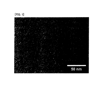
Fig. 1 is an image of a nanoparticle (iron oxide particle
(SNP)-ligand (DDSA)) of the present invention observed by a
transmission electron microscope (TEM).
Fig. 2 is views illustrating results of estimation of
relaxivity of SNP-DDSA in PBS in a case where a 1 tesla (T)
MRI is used, the SNP-DDSA including an iron oxide particle
of 1.8 nm in diameter as a core. (a) of Fig. 2 illustrates a
result of measurement of relaxation times in PBS of
SNP-DDSA obtained by diluting SNP-DDSA in sequence. (b) of
Fig. 2 is views each obtained by plotting a relaxation time
with respect to an iron atom concentration in SNP-DDSA. (c)
of Fig. 2 shows values of relaxivities r 1 and r2 determined
from an inclination of the plotted line in (b) of Fig. 2, and a
ri/r2 value.
(a) of Fig. 3 shows images of a bladder of a mouse to
which a contrast agent containing SNP-DDSA of Example 2
was administered, which images were obtained as a result of
MRI measurement carried out over time, respectively at the
following timings: prior to the administration (pre),
immediately after the administration (post), 30 minutes after
the administration (30min), and 3 hours after the
administration (3h). (b) of Fig. 3 shows images of the bladder
of the mouse to which the contrast agent containing
CA 03068215 2019-12-20
11 -
SNP-DDSA of Example 2 was administered, which images
were obtained as a result of MRI measurement carried out
over time, respectively at the following timings: prior to the
administration (pre), immediately after the administration
(post), 1 hour after the administration (1h), and 2 hours after
the administration (2h).
(a) of Fig. 4 shows images of a kidney of a mouse to
which a contrast agent containing SNP-DDSA of Example 2
was administered, which images were obtained as a result of
MRI measurement carried out over time, respectively at the
following timings: prior to the administration (pre),
immediately after the administration (post), 30 minutes after
the administration (30min), and 3 hours after the
administration (3h). (b) of Fig. 4 shows images of the kidney
of the mouse to which the contrast agent containing
SNP-DDSA of Example 2 was administered, which images
were obtained as a result of MRI measurement carried out
over time, respectively at the following timings: prior to the
administration (pre), immediately after the administration
(post), 1 hour after the administration (1h), and 2 hours after
the administration (2h).
Fig. 5 shows images of a liver of a mouse to which a
contrast agent containing SNP-DDSA of Example 2 was
administered, which images were obtained as a result of MRI
measurement carried out over time.
CA 03068215 2019-12-20
- 12 -
Fig. 6 shows images of blood vessels of a mouse to
which a contrast agent containing SNP-DDSA of Example 2
was administered, which images were obtained as a result of
MR angiography carried out over time.
Description of Embodiments
[0025]
The description below deals with an embodiment of the
present invention in detail.
[0026]
[Definitions of Terms]
Generally, the term "nanoparticle" refers to a particle
having a particle diameter in an order of nanometers, and
ordinarily refers to a particle having a particle diameter of
less than 1 p.m. Details of particle diameter will be discussed
later in a section of particle diameter.
[0027]
The term "ligand" or "ligand compound" refers to a
compound which (i) has a group capable of forming a
coordinate bond with a metal atom on a surface of a metal
particle and (ii) is used as a modifier on the surface of the
metal particle for allowing the metal particle to be stably
dispersed in water. As used herein, the term "ligand" or
"ligand compound" refers to (i) a case in which the compound
has not been bound by a coordinate bond to a surface of a
CA 03068215 2019-12-20
- 13 -
metal particle and/or (ii) a case in which the compound has a
molecular structure in which the compound has been bound
by a coordinate bond to a surface of a metal particle.
[0028]
As used herein, the term "subject" refers to a given
organism to which a contrast agent for MRI, a nanoparticle,
or a composition containing the nanoparticle of the present
invention can be administered for the purpose of, for
example, experiment, diagnosis, and/or treatment. As an
example, the subject is a human.
[0029]
The following description will discuss a
nanoparticle, a contrast agent for MRI, and a compound in
accordance with the present invention.
[0030]
[1. Nanoparticle]
The nanoparticle in accordance with the present
invention is a nanoparticle including: a metal particle
containing iron oxide; and a ligand which is bound to a metal
atom on a surface of the metal particle and is represented by
the following formula (3), wherein the ligand is preferably a
ligand represented by the following formula (1).
[0031]
CA 03068215 2019-12-20
- 14 -
M + 9
-------------------- 0
I
Me m 0-
(3)
[0032]
In the above formula (3), m is an integer of 1 to 4, and
a broken line represents a coordinate bond with a metal atom
on the surface of the metal particle.
[0033]
M + 9
I
Me 0-
(i)
[0034]
In the above formula (1), a broken line represents a
coordinate bond with the metal atom on the surface of the
metal particle.
Further, a nanoparticle in accordance with another
aspect of the present invention is a nanoparticle including: a
metal particle containing iron oxide; and a ligand which is
bound to a metal atom on a surface of the metal particle and
is represented by the above formula (3), where m is an integer
of 1, 2, or 4.
[0035]
In an embodiment of the present invention, the
CA 03068215 2019-12-20
- 15 -
nanoparticle of the present invention is a nanoparticle
including: a metal particle containing iron oxide; and a
ligand which is bound to a metal atom on a surface of the
metal particle and is represented by the above formula (3),
where m is 2 or 4, more preferably 4.
[0036]
That is, the nanoparticle in accordance with the
present invention is a particle which includes a metal
particle in a center part (core) of the particle and in which a
ligand compound is bound to an outer surface of the metal
particle so as to coat the metal particle.
[0037]
The nanoparticle of the present invention enables
prevention of agglomeration of nanoparticles, and exhibits
stable particle properties even in, for example, a solution
containing the nanoparticle at a high concentration. Such a
nanoparticle can be expected to both (i) ensure low
saturation magnetization and thus enable obtaining a clear
Ti-weighted image and (ii) facilitate renal excretion and thus
enable good renal clearance.
[00381
(Metal particle)
The metal particle contains iron oxide. In an
embodiment of the present invention, the metal particle is an
iron oxide particle containing only iron oxide.
CA 03068215 2019-12-20
- 16 -
[0039]
In an embodiment of the present invention, the metal
particle may contain iron oxide and at least one metal
derivative other than iron oxide. Further, the metal particle
may contain at least one metal element other than iron (Fe).
As the other metal element, the metal particle may further
contain, as necessary, at least one selected from the group
consisting of gadolinium (Gd), manganese (Mn), cobalt (Co),
nickel (Ni), and zinc (Zn).
[0040]
In still another embodiment of the present invention,
the metal particle may consist of iron oxide alone or may
contain ferrite derived from iron oxide. Ferrite is an oxide
represented by formula: (Fe2+, M)304 where M is preferably a
transition metal ion selected from Zn2+, Co2+, Mn2+, and Ni2+.
[0041]
A material known as super paramagnetic iron oxide
(SPIO) may be also suitably used. Such a material is
represented by general formula: f Fe203).[Fe203(M2+0)]1-x
(where x = 0 or 1). M2+ may be a divalent metal ion of, for
example, Fe, Mn, Ni, Co, Zn, magnesium (Mg), copper (Cu), or
a combination thereof. Note that the material is magnetite
(Fe304) in a case where the metal ion (M2+) is a ferrous iron
(Fe2+) and x = 0, and the material is maghemite (y-Fe2O3) in a
case where x = 1.
CA 03068215 2019-12-20
- 17 -
[0042]
In an embodiment of the present invention, iron oxide
is magnetic oxide of iron, and may be magnetite (Fe304),
maghemite (y-Fe2O3), or a mixture thereof. A particle of the
magnetic iron oxide is a super paramagnetic nanoparticle.
10043)
In still another embodiment of the present invention, in
a case where the iron oxide particle contains derivative(s) of
one or more metallic elements other than iron, the
derivative(s) of the respective metal element(s) may differ in
kind. That is, the iron oxide particle may contain an oxide, a
nitride, and the like. In another embodiment of the present
invention, a core particle may contain a derivative (e.g., FePt
and FeB) of iron other than iron oxide which derivative has an
iron element other than iron oxide.
[0044]
A metal particle in accordance with an embodiment of
the present invention may be a metal particle produced by a
well-known method such as a method disclosed in Patent
Literature 1, Non-patent Literature 2, Non-patent Literature
3, or the like, or may be a commercially available metal
particle. For example, the metal particle may be an iron oxide
particle produced by a coprecipitation method or a reduction
method.
[0045]
CA 03068215 2019-12-20
- 18 -
(Particle diameter of metal particle)
As used herein, the term "particle diameter" refers to
an "average particle diameter" unless otherwise noted.
[0046]
As used herein, the term "particle diameter" means a
diameter of a maximum inscribed circle of a two-dimensional
shape of a particle observed with use of a transmission
electron microscope (TEM). For example, in a case where the
two-dimensional shape of the particle is substantially a
circle, the "particle diameter" means a diameter of the circle.
In a case where the two-dimensional shape of the particle is
substantially an ellipse, the "particle diameter" means a
minor axis of the ellipse. In a case where the two-dimensional
shape of the particle is substantially a square, the "particle
diameter" means a length of a side of the square. In a case
where the two-dimensional shape of the particle is
substantially a rectangle, the "particle diameter" means a
length of a short side of the rectangle.
[0047]
Examples of a method for confirming a value of an
average particle diameter is in a predetermined range include
a method of observing 100 particles with use of a
transmission electron microscope (TEM) to measure the
particle diameter of each particle and find an average value of
the particle diameters of the 100 particles.
CA 03068215 2019-12-20
1
- 19 -
[0048]
An iron oxide particle in accordance with an
embodiment of the present invention preferably has a
diameter of 5 nm or less, more preferably has a diameter of 4
nm or less, more preferably has a diameter of 3 nm or less,
even more preferably has a diameter of 2 nm or less, and most
preferably has a particle diameter of 1 nm or less. Having a
particle diameter of 2 nm or less makes the iron oxide
particle more useful as a positive contrast agent for
high-field MRI of 3 tesla (T) or more. Further, an iron oxide
particle having a particle diameter of 2 nm or less, preferably
1 nm or less, enables achieving a higher signal-to-noise ratio
when used for high-magnetic field MRI of 7 T or more. This
may enable measurement with a higher spatial resolution
and in a shorter period of time.
(0049)
An iron oxide particle of the present invention has an
average particle diameter of preferably 5 nm or less, more
preferably 4 nm or less, more preferably 3 nm or less, even
more preferably 2 nm or less. As an example, the average
particle diameter is 1.8 nm. It is preferable that the average
particle diameter of the iron oxide particle be as small as
possible. As an example, the average particle diameter is 0.5
nm or more, or 0.6 nm or more.
(0050)
CA 03068215 2019-12-20
- 20 -
In an embodiment of the present invention, it is
preferable that properties of the nanoparticle contained in
the contrast agent for MRI are as uniform as possible among
the individual nanoparticles. Accordingly, it is preferable
that the metal particle serving as the core of the nanoparticle
be uniform in size and shape. As an example, a uniformity of
the metal particle in particle diameter is within a range of 1
nm of the average particle diameter of the metal particle. As
another example, the uniformity of the metal particle in
particle diameter is within a range of 0.5 nm of the average
particle diameter of the metal particle. In another
embodiment of the present invention, it is preferable that as
many small particles as possible be contained each as the
metal particle which serves as the core of the nanoparticle
contained in the contrast agent for MRI. As an example, a
ratio of the number of metal particles having a particle size of
5 nm or more to the number of all the metal particles is 30%
or less, preferably 10% or less, more preferably 5% or less. As
another example, a ratio of the number of metal particles
having a particle size of 4 nm or more to the number of all the
metal particles is 30% or less, preferably 10% or less, more
preferably 5% or less. As yet another example, a ratio of the
number of metal particles having a particle size of 3 nm or
more to the number of all the metal particles is 30% or less,
preferably 10% or less, more preferably 5% or less.
CA 03068215 2019-12-20
- 21 -
[0051]
(Particle diameter of nanoparticle)
The particle diameter of the nanoparticle increases as a
thickness of the ligand with which the metal particle is
coated increases. Measurement of the particle diameter of
the nanoparticle, however, is difficult. Ordinarily, a
hydrodynamic diameter (HD) of the nanoparticle as measured
in a solution of the nanoparticle is treated as an index for the
size of the nanoparticle. As an example, the nanoparticle has
an average HD of 30 nm or less, preferably 10 nm or less. As
another example, the nanoparticle has an average HD of 7 nm
or less, preferably 6 nm or less, preferably 5 nm or less,
preferably 4 nm or less, more preferably 3 nm or less.
[0052]
Note that it has been confirmed that the contrast
ability of the contrast agent for MRI is affected by the particle
diameter of the metal particle serving as the core.
[0053]
(Ligand)
The ligand compound in accordance with the present
invention is a compound represented by the following
formula (4):
[0054]
CA 03068215 2019-12-20
Me +
HO
Me
(4)
where n is an integer of 1 to 4.
In an aspect of the present invention, the ligand
compound in accordance with the present invention is
(3, 4-dihydroxyphenyl) (dimethyl) (3 -sulfonate
propyl)ammonium (DDSA) represented by formula (2) below.
In a ligand substitution reaction (described later), hydrogen
ions are desorbed from two hydroxyl groups of the compound,
and each remaining oxygen atom forms a coordinate bond
with a metal atom on the surface of the metal particle. Thus
produced is the nanoparticle of the present invention. The
ligand bound by a coordinate bond to the metal atom on the
surface of the metal particle has a structure represented by
the above formula (1).
[00551
9
HO5
HO y=c)
Me 0
(2)
[0056]
Note that the metal atom with which the oxygen atom of
the ligand of the present invention forms a coordinate bond is
CA 03068215 2019-12-20
- 23 -
an atom located on the surface of the metal particle serving
the core. For example, the metal atom is an iron atom.
[0057)
The ligand of the present invention has a structure in
which an ammonium group is directly bonded to a benzene
ring. This allows the ligand of the present invention to have a
molecular chain shorter than that of a conventionally known
ligand, and accordingly allows a ligand layer to be thinner.
Further, it is a characteristic of the ligand of the present
invention that the ligand has a positive charge on a metal
particle side and a negative charge on the outer surface of the
core particle. As such, it can be expected that the
nanoparticle of the present invention is less likely to undergo
agglomeration of core particles in body fluid and thus is
highly stable. Further, thinness of the ligand layer of the
present invention reduces a distance from the metal atom. It
can be accordingly expected that the nanoparticle of the
present invention exhibits an excellent contrast ability
resulting from an increase in the number of water molecules
affected by the core particle, and the like.
[0058]
The number (the number of ligands) of ligand molecules
coordinated on the surface of the metal particle varies
depending on a size, surface area, and the like of the metal
particle. For example, in a case where the metal particle has
CA 03068215 2019-12-20
- 24 -
a particle diameter of 1.8 nm, the number of ligands per
metal particle is preferably 5 to 200, more preferably 10 to
50.
[0059]
(Method for producing ligand)
A method for producing the ligand is not particularly
limited. The ligand can be produced easily from a well-known
raw material compound by a reaction well known to a person
skilled in the art. For example, the ligand can be produced
with reference to a method described in Wei H. et al., Nano
Lett. 12, 22-25, 2012.
[0060]
As an example, a synthesis method described in
Examples can be suitably employed.
[0061]
(Compound bound to metal particle other than ligand)
The nanoparticle of the present invention may contain
a component other than the ligand of the present invention.
In an embodiment of the present invention, the nanoparticle
may be (i) a nanoparticle in which a core particle itself has a
fluorescent property or (ii) a nanoparticle which further
contains a molecule such as a fluorescent molecule, a dye
molecule, or the like bound to a surface of the core particle.
In a case where the core particle itself has a fluorescent
property or in a case where a fluorescent molecule or a dye
CA 03068215 2019-12-20
- 25 -
molecule is introduced in the nanoparticle, the nanoparticle
can be used not only as a contrast agent for MRI but also as
a contrast agent for an optical image. In another embodiment
of the present invention, the nanoparticle of the present
invention may include a fluorescent molecule or a dye
molecule which is bound by a covalent bond to the ligand of
the present invention and is linked to the iron oxide particle
via the ligand. After the nanoparticle is injected into a body,
the fluorescent molecule is present on the surface of the iron
oxide particle. The fluorescent molecule can thus be utilized
for microscopic imaging and examination of localization of
the nanoparticle. Examples of the fluorescent molecule and
the dye molecule include rhodamine, fluorescein,
nitrobenzoxadiazole (NBD), cyanine, green fluorescence
protein (GFP), coumarin, and a derivative thereof.
[00621
In another embodiment of the present invention, the
nanoparticle of the present invention may include at least
one substance bound to the surface of the metal particle.
Examples of such a substance include, but are not limited to,
a peptide, a nucleic acid, a small molecule, or the like.
[0063]
Further, another ligand other than the ligand of the
present invention may be bound to the surface of the
nanoparticle. For example, in a case where a ligand having a
CA 03068215 2019-12-20
- 26 -
property of being accumulated specifically to a tumor is
bound to the nanoparticle of the present invention, the
nanoparticle can have a tumor-selective binding property.
[0064]
Imparting such a tissue specificity to the contrast
agent is preferable in order to (i) enhance a signal at a
portion that is a subject of MRI measurement and (ii) thereby
obtain information of a specific pathological condition or the
like. A distribution of the contrast agent in a living organism
depends on particle diameter, charge, surface chemistry,
route of administration, and route of elimination.
[00651
Further, the nanoparticle of the present invention has
an extremely low toxicity to a living organism. Accordingly,
the nanoparticle is highly safe and faces few limitations in
order to be put to various uses.
[0066]
[2. Method for producing nanoparticle]
The following description will discuss a method for
producing the nanoparticle. The method for producing the
nanoparticle is not particularly limited, and can be a
well-known method.
[0067]
For example, the nanoparticle can be produced with
reference to a method disclosed in Kim et al., J Am. Chem.
CA 03068215 2019-12-20
- 27 -
Sci. 2011, 133, 12624-12631, Kim et al., J Am. Chem.
Sci.2013, 135, 2407-2410, and a method disclosed in Hyeon
et al., J. Am. Chem. Soc., 133, 12624, 2011.
[0068]
A method in accordance with an embodiment of the
present invention for producing the nanoparticle includes
the steps of (a) reacting a metal salt with an alkali metal salt
of a carboxylic acid having 18 carbon atoms to form a
metal-carboxylic acid complex, (b) heating the
metal-carboxylic acid complex to synthesize a metal particle
which serves as a core of the nanoparticle and whose surface
is coated with a hydrophobic ligand, (c) converting the
hydrophobic ligand on the surface of the metal particle
serving as the core into a hydrophilic ligand having a
carboxyl group to form a particle dispersible in a highly-polar
solvent, and (d) reacting the metal particle coated with the
hydrophilic ligand with the ligand compound of the present
invention to substitute the hydrophilic ligand on the surface
of the metal particle with the ligand of the present invention.
The following describes each step in detail.
[0069]
(Step (a))
The step (a) is a step in which a metal salt is reacted
with an alkali metal salt of a carboxylic acid having 18
carbon atoms to form a metal-carboxylic acid complex.
CA 03068215 2019-12-20
- 28 -
[0070]
Frist, a metal salt and an alkali metal salt of a
carboxylic acid having 18 carbon atoms are dispersed in a
solvent. Examples of the metal salt used for preparation of
the metal-carboxylic acid complex include iron(III) chloride
hexahydrate [FeC13=6H20]. Examples of the alkali metal salt
of the carboxylic acid having 18 carbon atoms include sodium
oleate. Examples of the solvent include ethanol, water,
hexane, and a mixture thereof. As an example, iron(III)
chloride hexahydrate and sodium oleate are dispersed in a
mixture of ethanol, water, and hexane. Subsequently, a
resultant solution is stirred while being heated, preferably at
70 C, for 1 hour to 10 hours, preferably for 4 hours, and an
organic layer is collected. The organic layer is washed with
water once or more, more preferably 3 times to 4 times. The
organic layer obtained is optionally dried.
[0071]
(Step (b))
The step (b) is a step in which the complex obtained in
the step (a) is reacted with a hydrophobic ligand to
synthesize a nanoparticle in which a surface of a metal
particle serving as a core is coated with the hydrophobic
ligand.
[0072]
For example, in an atmosphere of a gas selected from
CA 03068215 2019-12-20
- 29 -
argon (Ar) and nitrogen, the following (i) and (ii) are added to
the complex obtained in the step (a): (i) at least one detergent
selected from the group consisting of a fatty acid having 18
carbon atoms, aliphatic alcohol having 18 carbon atoms, and
aliphatic amine having 18 carbon atoms and (ii) a solvent
selected from diphenyl ether and phenyloctyl ether. As an
example, the detergent may be oleyl alcohol and the solvent
may be diphenyl ether. Subsequently, a mixture thus
obtained is heated from room temperature to a temperature
of 180 C to 300 C, and then is optionally stirred in this state
for 10 minutes to several hours. As an example, the mixture
is heated from 30 C to 250 C at a rate of 10 C /min, and is
stirred at 250 C for 30 minutes. As another example, the
mixture is heated from 30 C to 200 C at a rate of 10 C /min,
and is stirred at 200 C for 30 minutes.
[0073]
A resultant reaction solution is cooled down to room
temperature. Then, acetone is added, a resultant mixture is
centrifuged, and a supernatant is removed. This operation is
repeated 2 times to 3 times, preferably 4 times to 5 times. A
solution thus obtained is optionally dried. As an example, the
operation of adding acetone, performing centrifugation, and
removing the supernatant is repeated 3 times.
[0074]
(Step (c))
CA 03068215 2019-12-20
- 30 -
The step (c) is a step in which the hydrophobic ligand,
with which the surface of the nanoparticle obtained in the
step (b) is coated, is substituted with a hydrophilic ligand
having a carboxyl group to form a particle dispersible in a
highly-polar solvent.
[0075]
For example, in an atmosphere of a gas selected from Ar
and nitrogen, the nanoparticle coated with the hydrophobic
ligand is dispersed in a solvent, and then a hydrophilic
ligand having a carboxyl group is added. Examples of the
hydrophilic ligand having a carboxyl group include
2-[2-(2-methoxyethoxy)ethoxy]acetic acid (MEEA). Methanol
is suitable as the solvent.
(00761
A reaction solution is reacted at room temperature or
while being heated, preferably at 25 C to 80 C for
approximately 1 hour to 15 hours, preferably 5 hours to 10
hours. As an example, the reaction is carried out by stirring
the reaction solution at 50 C for 7 hours. As an example, the
reaction is carried out by stirring the reaction solution at
70 C for 10 hours. As yet another example, the reaction is
carried out by stirring the reaction solution at 70 C for 5
hours.
[0077]
The reaction solution is cooled down to room
CA 03068215 2019-12-20
- 31 -
temperature. Then, a solvent selected from acetone and
hexane is added, a resultant mixture is centrifuged, and a
supernatant is removed. This operation can be repeated 2
times to 3 times, preferably 4 times to 5 times. A solution
thus obtained may optionally be dried. As an example, the
above operation is repeated 3 times.
[0078]
(Step (d))
The step (d) is a step in which the metal particle
obtained in the step (c) and coated with the hydrophilic
ligand is reacted with the ligand compound of the present
invention to obtain a nanoparticle in which a surface of the
metal particle is coated with the ligand compound of the
present invention.
[0079]
Note that the metal particle coated with the hydrophilic
ligand is reacted with the ligand compound of the present
invention by being stirred for 1 hour to several tens of hours
in an atmosphere of a gas selected from Ar and nitrogen and
at room temperature or while being heated. As an example,
the above reaction is carried out in an Ar atmosphere. A
reaction temperature is 25 C to 80 C as an example, and
50 C to 70 C as another example. A stirring time is 5 hours to
7 hours as an example, and 24 hours as another example. As
an example, stirring is performed at 70 C for 12 hours.
CA 03068215 2019-12-20
- 32 -
Subsequently, a resultant reaction solution is cooled down to
room temperature, and a solvent is added. A resultant
mixture is centrifuged, and a supernatant is removed. The
solvent is not particularly limited, and may be selected from
acetone, hexane, and the like. As an example, the solvent is
acetone. The operation of adding the solvent, performing
centrifugation, and removing the supernatant can be
repeated a plurality of times. For example, the operation may
be repeated 4 times to 5 times. As an example, this operation
is repeated 3 times. Subsequently, a resultant solution
containing the nanoparticle coated with the ligand compound
of the present invention can be concentrated with use of a
concentration column or the like of a centrifugal ultrafilter or
the like. This concentration operation can be repeated a
plurality of times, during which a solution such as PBS may
be added at some point, and then the concentration operation
may be repeated.
(00801
As an aspect of the present invention, the following
description will discuss another method for producing a
nanoparticle having an iron oxide particle as a core.
[0081]
An iron oxide particle (SNP-0A) coated with oleic acid
is suspended in a hexane solution. A resultant suspension is
mixed with 1.7% tetramethylammonium hydroxide (TMA(OH))
CA 03068215 2019-12-20
- 33 -
aqueous solution, and is vigorously shaken. A resultant
solution is centrifuged to separate an aqueous layer, and
acetone is added. A resultant mixture is centrifuged at 8000
rpm to 12000 rpm for 5 minutes to 10 minutes, and a
supernatant is removed to obtain a precipitate. 2 mL of 0.1%
TMA(OH) solution is added and dispersed in the precipitate,
acetone is added again in an amount of 10 mL, and a
resultant mixture is left for precipitation. This operation can
be repeated a plurality of times, and is repeated preferably 3
times to 4 times. A solution thus obtained is dispersed in
0.1% TMA(OH) solution and stored.
[0082]
To 0.1% TMA(OH) solution thus prepared in accordance
with the above procedure, a solution of the ligand compound,
which solution is prepared with use of 0.1% to 2% TMA(OH)
solution so as to achieve a pH of approximately 8 to 12, is
added. A resultant solution is stirred at room temperature for
6 hours to 24 hours, and acetone is added. A resultant
mixture is left for precipitation and is centrifuged at 8000
rpm to 12000 rpm for 3 minutes to 10 minutes, and a
supernatant is removed. A precipitate thus obtained is
dispersed in a phosphate buffer, and a resultant solution is
centrifuged at 7000 rpm to 12000 rpm with use of a
concentration column to reduce an amount of the solution.
The phosphate buffer is added again, and a resultant mixture
CA 03068215 2019-12-20
- 34 -
is centrifuged at 7000 rpm to 12000 rpm for 10 minutes to 20
minutes for concentration. This operation can be repeated a
plurality of times, and is repeated preferably 3 times to 4
times, more preferably 5 times to 10 times. Thus obtained is
a solution of an iron oxide particle coated with the ligand.
The solution may be diluted with PBS and stored.
[0083]
[3. Contrast agent for magnetic resonance imaging
(contrast agent for MRI)]
The present invention also provides a contrast agent
for magnetic resonance imaging which contrast agent
includes the above-described nanoparticle.
[00841
The following description will discuss the contrast
agent for MRI in detail.
10085)
(Various components contained in contrast agent for
MRI)
= Nanoparticle =
In an embodiment of the present invention, the
contrast agent for MRI of the present invention is
characterized by containing at least one kind of the
above-described nanoparticle. In another embodiment of the
present invention, the contrast agent for MRI of the present
invention may include a combination of two or more kinds of
CA 03068215 2019-12-20
- 35 -
the above-described nanoparticle.
[0086]
Further, the contrast agent for MRI may contain, if
necessary, a solvent and a pharmacologically acceptable
additive in addition to the nanoparticle. In an embodiment of
the contrast agent for MRI of the present invention, the
contrast agent may further contain a suitable solvent and/or
at least one selected from additives such as a carrier, a
vehicle, a complex and the like.
[0087]
= Solvent =
Examples of the solvent contained in the contrast agent
for MRI include water, a buffer solution, and the like.
Further, examples of the buffer solution include
physiological saline, phosphate buffer, tris buffer, boric acid
buffer, Ringer's solution, and the like. In a case where a
dosage form is an injection, examples of a preferable solvent
include water, Ringer's solution, physiological saline, and
the like.
[0088]
That is, the contrast agent for MRI in accordance with
the present invention may be a solution obtained by
suspending the nanoparticle in accordance with the present
invention in a solution having a desired composition.
Specifically, the contrast agent may be in the form of a buffer
CA 03068215 2019-12-20
- 36 -
solution such as phosphate buffer, tris buffer, or boric acid
buffer in which the nanoparticle is suspended.
[0089]
= Additive =
Examples of the additive such as a carrier, a complex,
and a vehicle contained in the contrast agent for MRI include
a carrier, a vehicle, and the like which are generally used in
the fields of pharmaceuticals and biotechnology. Examples of
the carrier include a polymer such as polyethylene glycol, a
metal fine particle, and the like. Examples of the complex
include diethylenetriaminepentaacetic acid (DTPA),
1, 4, 7, 10-tetraazacyclododecane- 1,4,7, 10-tetraacetic
acid
(DOTA), and the like. Examples of the vehicle include lime,
soda ash, sodium silicate, starch, glue, gelatin, tannin,
quebracho, and the like.
[0090]
Further, the contrast agent for MRI of the present
invention may further contain an excipient, a lubricant, a
wetting agent, an emulsifier, a suspension, a preservative, a
pH adjusting agent, an osmotic pressure controlling agent,
and the like.
[0091]
(Dosage form)
A dosage form of the contrast agent for MRI of the
present invention is not particularly limited, and may be
CA 03068215 2019-12-20
- 37 -
liquid, solid or semisolid, or semiliquid. These dosage forms
can be produced easily in accordance with a method well
known to a person skilled in the art. In a case where the
dosage form is a liquid, the liquid may be one which is
obtained by dispersing, suspending, or dissolving the
nanoparticle in accordance with the present invention in, for
example, an aqueous solvent so that the liquid contains the
nanoparticle. Further, the contrast agent may be in the form
of a lyophilized agent, and be dispersed, suspended, or
dissolved when used.
[0092]
(Concentration of nanoparticle)
A concentration of the nanoparticle in the contrast
agent for MRI is determined as appropriate in accordance
with a purpose, a tissue to be imaged, and the like. For
example, a concentration is selected such that the selected
concentration is in a range within which (i) an adequate
contrast ability is exhibited and (ii) a degree of influence on a
living organism is tolerable.
[0093]
The nanoparticle of the present invention, even when
contained at a high concentration, is less likely to be
agglomerated and thus is capable of maintaining the
stability. Accordingly, the nanoparticle of the present
invention can maintain, stably and for a long period of time,
CA 03068215 2019-12-20
- 38 -
a higher MRI contrast ability than a well-known
nanop article.
[0094]
For example, in a case where the contrast agent for MRI
is a liquid that is an aqueous solution, examples of a
concentration of the nanoparticle in the liquid when, for
example, the liquid is used as a general injection include 0.1
mM Fe/mL to 1000 mM Fe/mL, preferably 1.0 mM Fe/mL to
500 mM Fe/mL, more preferably 5.0 mM Fe/mL to 100 mM
Fe/mL, and, in an aspect, 10 mM Fe/mL to 500 mM Fe/mL,
and, in another aspect, 5.0 mM Fe/mL to 50 mM Fe/mL.
[0095]
(Administration target)
An administration target to which the contrast agent in
accordance with the present invention is administered can
be, for example, a given organism that is not a human, or a
human. Examples of the organism that is not a human
include, but not limited to, mammals (e.g., rodents, mice,
rats, rabbits, monkeys, dogs, cats, sheep, cows, primates,
pigs, and the like), birds, reptiles, amphibians, fish, insects,
and plants. In an aspect, the animal can be a transgenic
animal, a genetically-engineered animal, or a clone animal.
Further, the administration target can be one that is not a
living organism, for example, a tissue sample or a biological
material which includes a cell.
CA 03068215 2019-12-20
- 39 -
[0096]
(Uses to which contrast agent for MRI is applied)
As described above, there are two types of contrast
agents for MRI, namely, a positive contrast agent and a
negative contrast agent.
[0097]
In an embodiment of the present invention, the
contrast agent for MRI of the present invention is a positive
contrast agent. In another embodiment, the contrast agent is
a negative contrast agent.
[0098]
The contrast agent for MRI of the present invention is
used for, for example, diagnosis of a lesion and a tumor and
the like using an MRI apparatus. For example, the contrast
agent can be suitably used for examination of renal function,
detection of liver tumors, hepatic angiography, and the like.
Note that the MRI apparatus may be a given apparatus, and a
well-known MRI apparatus can be used. A magnetic field to
be applied may be, for example, 1 T, 1.5 T, 3 T, and 7 T. An
example of a diagnosis method using the contrast agent of the
present invention includes the steps of: administering a
positive contrast agent to a living subject such as a human in
vivo or in vitro; and subsequently forming an image of the
subject with use of an MRI apparatus.
[0099]
CA 03068215 2019-12-20
- 40 -
Among conventionally known contrast agents for MRI,
a paramagnetic compound is used as a positive contrast
agent, and a super paramagnetic nanoparticle is used as a
negative contrast agent. The nanoparticle of the present
invention is super paramagnetic, but can be used also as a
positive contrast agent. Super paramagnetism is generated
when a region containing a crystal having unpaired spins is
large enough to be regarded as a single, thermodynamically
independent domain particle called a "magnetic domain". The
magnetic domain is a net magnetic dipole which is greater
than a sum of individual unpaired electrons in the magnetic
domain. While no magnetic field is applied, all magnetic
domains are randomly oriented, and there is no net
magnetization, accordingly. When an external magnetic field
is applied, dipole moments in all magnetic domains are
realigned. As a result, a net magnetic moment is generated.
Ti, T2, and T2* relaxation processes are shortened by
magnetic particles. In an embodiment of the present
invention, the contrast agent in accordance with the present
invention has a contrast ability represented by an r2
relaxivity of 15 mM-Is-1 to 19 mM-Is-1 and an ri relaxivity of 9
mM-Is-1 to 12 mM-1s-1, at room temperature and with a
magnetic field of 1 T. In another embodiment of the present
invention, the contrast agent in accordance with the present
invention has a contrast ability represented by an r2
CA 03068215 2019-12-20
- 41 -
relaxivity of 5 mM-Is-1 to 7 mM-Is-1 and an ri relaxivity of 3
mm- Is-1,
to 5 mM-Is-1, at room temperature and in a magnetic
field of 1 T.
[0100]
The relaxivity depends on various factors such as (i) a
particle diameter of the metal particle in the nanoparticle of
the contrast agent for MRI, (ii) a composition of the metal
particle, (iii) a charge and properties of the surface of the
particle, (iv) particle stability, and (v) agglomeration and
connectivity to tissues in a living organism. A relaxivity ratio
ri/r2 is generally used for quantification of a type of a
contrast generated in MRI, and can serve as an index for
performance of the contrast agent.
[01011
It is preferable that an ri/r2 value of the positive
contrast agent for MRI of the present invention in a case
where a magnetic field of 1 T is externally applied be as high
as possible. For example, the ri/r2 value in a case where the
magnetic field is 1 T is preferably 0.5 or more, more
preferably 0.6 or more, and even more preferably 0.7 or more.
In a case where the ri/r2 value is 0.5 or more, the positive
contrast agent exhibits an excellent Ti (positive) effect and,
even in MRI measurement with a higher magnetic field,
exhibits a high contrast effect with a high resolution. From
the viewpoint of significantly increasing the contrast effect
CA 03068215 2019-12-20
- 42 -
and reducing an amount of the positive contrast agent for
MRI to be administered, the ri /r2 value is preferably 0.7 or
more.
[0102]
In the nanoparticle of the present invention, a
molecular chain length of the ligand is shorter than that of a
conventional ligand, and a ligand shell with which the core is
coated is thinner. Thinness of the ligand shell reduces a
distance between the metal particle serving as the core and a
water molecule outside, and allows the relaxivity to be
efficiently exhibited.
[0103]
In the contrast agent for MRI of the present invention,
the metal particle can have a particle diameter of 2 nm or
less, or in an example, 1 nm or less. The contrast agent for
MRI of the present invention can thus be utilized as a positive
contrast agent with an MRI apparatus of 7 T or more. As an
example, the contrast agent for MRI of the present invention
encompasses a positive contrast agent for MRI to be used
with an MRI apparatus of 7 T or less. As an example, the
contrast agent for MRI of the present invention encompasses
a positive contrast agent for MRI to be used with an MRI
apparatus of 3 T or less.
[0104]
(Toxicity and stability)
CA 03068215 2019-12-20
- 43 -
The contrast agent for MRI of the present invention
exhibits a high stability of the nanoparticle. As shown in
Example 4 (described later), it has been confirmed that the
contrast agent can be stored in a solution for a long period of
time at room temperature or at 4 C without undergoing
agglomeration. Further, the contrast agent has a low toxicity
to organisms. This allows for long-term and continuous
application of the contrast agent to a living organism.
10105]
[4. Ligand compound]
The present invention also relates to
(3,4-dihydroxyphenyl)(dimethyl)(3-sulfonate
propyl)ammonium represented by the above formula (2) and
use of
(3,4-dihydroxyphenyl)(dimethyl)(3-sulfonate
propyl)ammonium for production of the nanoparticle.
[0106]
The above compound can be used as a ligand for
production of the nanoparticle of the present invention.
Specifically, the compound is reacted with a metal particle
coated with a hydrophilic ligand or the like to cause a ligand
substitution reaction. This provides a nanoparticle in which
the metal particle is coated with the ligand of the present
invention which ligand has a structure represented by the
following formula (1):
[0107]
CA 03068215 2019-12-20
M + 9
I
Me 0-
(i)
[0108]
where a broken line represents a coordinate bond with
a metal atom on the surface of the metal particle.
In an embodiment of the present invention, the
compound in accordance with the present invention can be
used as a ligand which is bound to a metal particle serving as
a core in a nanoparticle made of (i) a metal selected from Fe,
Gd, and Mn, (ii) a metal derivative thereof, and (iii) a
combination of (i) and (ii). Examples of the metal derivative
include an oxide, a nitride, a carbide, and a sulfide. For
example, the metal particle and the compound are bound to
each other by a coordinate bond between a metal atom on a
surface of the metal particle and an oxygen atom.
[0109]
In another aspect of the ligand compound in
accordance with the present invention, the ligand compound
is a compound represented by formula (4) below. In the above
ligand substitution reaction, hydrogen ions are desorbed
from two hydroxyl groups of the compound, and each
remaining oxygen atom forms a coordinate bond with a metal
atom on the surface of the metal particle. Thus produced is
CA 03068215 2019-12-20
- 45 -
the nanoparticle of another aspect of the present invention.
[0110]
M +
HO
Me n
H
(4)
[0111]
where n is an integer of 1 to 4.
A compound of another aspect of the present invention
is a compound which is represented by the above formula (4)
where n is 1, 2, or 4, preferably 2 or 4, and more preferably 4.
[0112]
The compound represented by the above formula (4) is
suitably used as a material for production of the nanoparticle
of another aspect of the present invention which nanoparticle
includes (i) a metal particle containing iron oxide and (ii) a
ligand which is bound to a metal atom on a surface of the
metal particle and is represented by the above formula (3).
[0113]
[5. Examples of specific aspects in accordance with the
present invention]
In order to solve the above problem, the present
invention includes in its scope any one aspect below.
<1> A nanoparticle including: a metal particle
containing iron oxide; and a ligand which is bound to a metal
CA 03068215 2019-12-20
- 46 -
atom on a surface of the metal particle and is represented by
formula (3):
[0114]
Meõ +
.................... -0 9_10
..' 1 1 "
I Me , /17.1 0-
.0
(3)
[0115]
where m is an integer of 1 to 4, and a broken line
represents a coordinate bond with a metal atom on the
surface of the metal particle.
<2> The nanoparticle as set forth in <1> above, wherein
the ligand bound to the metal atom on the surface of the
metal particle is a ligand represented by the following
formula (1):
[0116]
M + 9
I
Me 0-
(1)
[0117]
where a broken line represents a coordinate bond with
the metal atom on the surface of the metal particle.
<3> The nanoparticle as set forth in <1> above, wherein
m is 1, 2, or 4 in the above formula (3).
CA 03068215 2019-12-20
- 47 -
<4> The nanoparticle as set forth in any one of <1>
through <3> above, wherein the metal particle containing the
iron oxide is an iron oxide particle.
<5> The nanoparticle as set forth in any one of <1>
through <4> above, wherein the metal particle has an average
particle diameter of 5 nm or less.
<6> The nanoparticle as set forth in <5> above, wherein
the metal particle has an average particle diameter of 4 nm or
less.
<7> The nanoparticle as set forth in <5> above, wherein
the metal particle has an average particle diameter of 3 nm or
less.
<8> A contrast agent for magnetic resonance imaging,
containing a nanoparticle recited in any one of <1> through
<7> above.
<9> The contrast agent as set forth in <8> above,
wherein the contrast agent is a positive contrast agent.
<10> Use of
(3,4-dihydroxyphenyl)(dimethyl)(3-sulfonate
propyl)ammonium for production of a nanoparticle recited in
<2> above.
<11> (3,4-
dihydroxyphenyl)(dimethyl)(3-sulfonate
propyl)ammonium.
[0118]
Further, the present invention includes in its scope the
CA 03068215 2019-12-20
- 48 -
following aspects as other aspects of the present invention.
<12> A nanoparticle, including: a metal particle
containing iron oxide; and a ligand which is bound to a metal
atom on a surface of the metal particle and is represented by
formula (1):
(01191
M + 9
------------------- -0
, r
Me
(i)
[0120]
where a broken line represents a coordinate bond with
the metal atom on the surface of the metal particle.
<13> The nanoparticle as set forth in <12> above,
wherein the metal particle containing the iron oxide is an
iron oxide particle.
<14> The nanoparticle as set forth in <12> or <13>
above, wherein the metal particle has an average particle
diameter of 5 nm or less.
<15> A contrast agent for magnetic resonance imaging,
containing a nanoparticle recited in any one of <12> through
<14> above.
<16> The contrast agent as set forth in <15> above,
wherein the contrast agent is a positive contrast agent.
<17> Use of
CA 03068215 2019-12-20
- 49 -
(3,4-dihydroxyphenyl)(dimethyl)(3-sulfonate
propyl)ammonium for production of a nanoparticle recited in
any one of <12> through <14> above.
<18> (3,4-dihydroxyphenyl)(dimethyl)(3-sulfonate
propyl)ammonium.
[0121]
The present invention is not limited to the
embodiments, but can be altered by a skilled person in the
art within the scope of the claims. The present invention also
encompasses, in its technical scope, any embodiment derived
by combining technical means disclosed in differing
embodiments. Further, it is possible to form a new technical
feature by combining the technical means disclosed in the
respective embodiments.
Examples
[0122]
The following will provide Examples to describe the
present invention in further detail.
[0123]
[Example 1. Synthesis 1 of ligand compound]
According to the following Scheme 1,
(3,4-dihydroxyphenyl)(dimethyl)(3-sulfonate
propyl)ammonium (DDSA; Compound 3 of Scheme 1), which
is a ligand compound of the present invention, was
CA 03068215 2019-12-20
- 50 -
synthesized.
[0124]
Scheme 1
Me0 me
Me0 410 H2 '1-2 Me0 03HMel Me0 -
Me
MeCN meo iir KaCO3
Me0H 2 3
[0125]
The following describes each step in detail.
[0126]
1,3-propane sultone (5.98 g, 49.0 mmol) was added to
acetonitrile (100 mL) solution of 3,4-dimethoxyaniline (5.00
g, 32.6 mmol), and a resultant mixture was stirred at room
temperature in an argon atmosphere for 48 hours. A reaction
mixture was filtered out, washed with acetonitrile, and then
dried. Thus obtained
was
3-(3,4-dimethoxyanilino)propane-1-sulfonic acid (Compound
1) in the form of grey powder (4.97 g, yield: 55%).
[0127]
Compound 1 thus obtained (2.00 g, 7.26 mmol),
potassium carbonate (2.01 g, 14.5 mmol), and iodomethane
(8.25 g, 58.1 mmol) were dissolved in methanol (50 mL), and
a resultant mixture was heated to reflux in an argon
atmosphere for 12 hours. A reaction mixture was
concentrated, and was purified by reversed phase column
chromatography (water/acetonitrile). Thus obtained was
(3,4-dimethoxyphenyl)(dimethyl)(3-sulfonate
CA 03068215 2019-12-20
- 51 -
propyl)ammonium (Compound 2) (2.16 g, yield: 98%).
[0128]
Compound 2 thus obtained (1.34 g, 4.42 mmol) was
dissolved in hydriodic acid (10 mL), and a resultant mixture
was heated to reflux in an argon atmosphere for 12 hours. A
reaction mixture was heat-vacuum dried, and then water (10
mL) was added. Again, a solution thus obtained was
heat-vacuum dried. Then again a resultant residue was
dissolved in water (5 mL), and acetone (300 mL) was added. A
resultant mixture was left for precipitation, and then a
precipitate was filtered out. Thus obtained was
(3, 4-dihydroxyphenyl) (dimethyl)(3-sulfonate
propyl)ammonium (DDSA, Compound 3) in the form of white
powder (420 mg, yield: 35%).
10129]
[Example 2. Production 1 of nanoparticle]
According to a procedure shown in Scheme 2, a
nanoparticle (SNP-DDSA) which (i) included an iron oxide
nanoparticle (SNP) having an average particle diameter of 1.8
nm and serving as a core particle and (ii) was coated with
DDSA, was produced.
101301
CA 03068215 2019-12-20
- 52 -
Scheme 2
FeCI3 ) ( ) -
(ab
TT
Fe0A3
Oleic Acid (OA) MEEA
(c) + DDSA (d)
*DOS =
=
[0131]
The following describes steps (a) through (d) of Scheme
2 in detail.
[0132]
<Step (a)>
The step (a) is a step in which oleic acid (OA) is added
to iron(III) chloride to produce a complex (Fe0A3) consisting
of oleic acid and an iron ion.
[0133]
Iron(III) chloride hexahydrate (2.16 g, 8 mmol), sodium
oleate (7.3 g, 24 mmol), 16 mL of ethanol, 12 mL of water, and
28 mL of hexane were mixed in a 100-mL flask, and a
resultant mixture was stirred at 70 C for 4 hours. An organic
layer was collected and transferred to a separatory funnel, 30
mL of water was added, the separatory funnel was vigorously
shaken, and an organic layer was collected. This operation
was repeated 3 times, and an organic layer obtained was
dried. Thus obtained was a complex (Fe0A3) consisting of
CA 03068215 2019-12-20
- 53 -
oleic acid and an iron ion.
[0134]
<Step (b)>
The step (b) is a step in which Fe0A3 is reacted with
oleyl alcohol to produce an iron oxide particle (SNP-0A)
whose surface is coated with oleic acid.
[0135]
To Fe0A3 (1.8 g, 2 mmol) obtained in the step (a), oleyl
alcohol (3.22 g, 12 mmol) and 10 g of diphenyl ether were
added in an Ar atmosphere. A resultant mixture was
degassed at 90 C while being stirred, and then was heated to
200 C at a rate of 10 C /min. Stirring was continued at 200 C
for 30 minutes. Then, the mixture was cooled to room
temperature, and 50 mL of acetone was added. A resultant
mixture was centrifuged at 8000 rpm for 20 minutes, and a
supernatant was removed. Until a precipitate obtained was
completely dispersed, chloroform was added (approximately
0.5 mL). Further, 10 mL of acetone was added, then a
resultant mixture was centrifuged at 8000 rpm for 20
minutes, and a supernatant was removed. This operation was
repeated 3 times, and a supernatant obtained was dried.
[0136]
<Step (c)>
The step (c) is a step in which oleyl acid, with which the
surface of the SNP-OA obtained in the step (b) is coated, is
CA 03068215 2019-12-20
- 54 -
substituted with 2-[2-(2-methoxyethoxy)ethoxy]acetic acid
(MEEA) to produce a nanoparticle (SNP-MEEA) coated with a
hydrophilic ligand.
[0137]
In an Ar atmosphere, 10 mg of the SNP-OA was
dispersed in 0.9 mL of methanol, and 0.1 mL of MEEA was
added. A resultant mixture was stirred at 70 C for 4 hours. A
resultant solution was cooled to room temperature, and then
8 mL of acetone and 2 mL of hexane were added. A resultant
mixture was centrifuged at 5800 rpm for 3 minutes, and a
supernatant was removed. This operation was repeated 3
times, and a supernatant obtained was dried. Thus obtained
was SNP-MEEA. Further, 300 pL of water and 600 pL of DMF
were added to the SNP-MEEA. A resultant solution is
hereinafter referred to as a "SNP-MEEA solution".
<Step (d)>
The step (d) is a step in which the SNP-MEEA obtained
in the step (c) is reacted
with
(3 ,4-dihydroxyphenyl) (dimethyl) (3 -sulfonate
propyl)ammonium (DDSA) to produce a nanoparticle
(SNP-DDSA) in which an iron oxide particle is coated with
DDSA. Note that a DDSA ligand with which a surface of the
iron oxide particle is coated in the nanoparticle (SNP-DDSA)
has a structure represented by the following formula (1).
[0138]
CA 03068215 2019-12-20
M + 9
I
Me 0-
(1)
[0139]
where a broken line represents a coordinate bond
between an iron atom on the surface of the iron oxide particle
and an oxygen atom.
85 mg of DDSA was added as a ligand compound to 1
mL of the SNP-MEEA solution in an Ar atmosphere, and a
resultant mixture was stirred at 50 C for 12 hours. Then, the
mixture was cooled to room temperature, and 20 mL of
acetone was added. A resultant mixture was centrifuged at
5800 rpm for 3 minutes, and a supernatant was removed. A
precipitate obtained was dispersed in 2 mL of phosphate
buffered saline (PBS). A solution obtained was centrifuged at
8000 rpm for approximately 30 minutes with use of Amicon
Ultra centrifuge 3K filter (Merck Millipore, hereinafter
abbreviated as "3K filter") to reduce a volume of the solution
to approximately 1/5. PBS was added so that a total volume
of a resultant solution was approximately 2 mL, and the
solution was centrifuged. This operation was repeated
approximately 5 times to 8 times until a solution dripping
form the filter is completely colorless. A solution obtained
was diluted with PBS so that a resultant solution had a
CA 03068215 2019-12-20
- 56 -
volume of 1 mL to 1.5 mL. Thus obtained was an SNP-DDSA
solution.
[0140]
The SNP-DDSA solution obtained in the step (d) was
stored at 4 C. Further, an iron concentration in the
SNP-DDSA solution was determined by inductively coupled
plasma-atomic emission spectroscopy (ICP-AES). Fig. 1 is an
image of SNP-DDSA observed by a transmission electron
microscope (TEM). From a result of observation with the TEM,
it was estimated that, based on an average value of core
diameters of 100 particles, the obtained SNP-DDSA had a
diameter of an iron oxide particle, which serves as a core, of
1.8 nm on average.
[0141]
[Example 3. Evaluative measurement of MR relaxivity
of nanoparticle]
The nanoparticle obtained in Example 2, SNP-DDSA,
which included an iron oxide particle of 1.8 nm in diameter
as a core, was used in an experiment below.
[0142]
First, SNP-DDSA was diluted in PBS so as to change a
concentration of SNP-DDSA in sequence. Solutions thus
obtained were used as test samples. For each sample, a
relaxivity was estimated by 1 T MRI.
[0143]
CA 03068215 2019-12-20
- 57 -
First, Ti-weighted image was obtained in 1 T MRI. Ti
and T2 measurement conditions are as follows.
[0144]
<1 T MRI>
Ti-weighted image
Pulse Sequence: MSME, TR = 400 msec, TE = 10 msec, Slice
Thickness = 2 mm, Number of Slice = 1, Matrix Size = 256 x
256, FOV = 38.4 x 38.4 mm2, scan time = 1 min 42 sec.
T2 measurement (multi echo spin echo technique)
Pulse Sequence: MSME, TR = 15,000 msec, TE = 20 msec (a
cycle of TR and TE was repeated 256 times (using mao
pulses)), Slice Thickness = 2 mm, Number of Slice = 1, FOV =
38.4 x 38.4 mm2, Matrix Size = 64 x 64, Scan Time = 16 min
00 sec.
Ti measurement (inversion recovery)
Pulse: SE-RARE, TR = 20,000 sec, TE = 17 msec, NEX = 1,
RARE Factor = 4, Number of slice = 1, slice thickness = 2 mm,
FOV = 38.4 x 38.4 mm2, Matrix Size = 64 x 64. Scan Time per
scan = 21 min 20 sec, Inversion Time = 45, 100, 200, 400,
800, 1600, 3200, 6400, 8000, 10000, 12000 (11
measurements)
Results are shown in Fig. 2. (a) of Fig. 2 illustrates a
result of measurement of relaxation times in the PBS
solutions of SNP-DDSA obtained by diluting SNP-DDSA in
sequence. (b) of Fig. 2 is views each obtained by plotting a
CA 03068215 2019-12-20
- 58 -
relaxation time with respect to an iron atom concentration in
SNP-DDSA. It was confirmed from (b) of Fig. 2 that Ti and T2
were each in linear correlation with SNP concentration. (c) of
Fig. 2 shows values of relaxivities ri and r2 determined from
an inclination of the plotted line in (b) of Fig. 2, and a ri/r2
value.
[0145]
According to results of the above, the ri/r2 value at 1 T
was 0.71. This value is the highest among r 1 /r2 values
obtained with conventionally reported SNPs including an iron
oxide particle as a core, after an influence of a magnetic field
strength is corrected. This indicates that SNP-DDSA is
promising to be applied to use as a positive contrast agent.
[0146]
[Example 4. Stability evaluation test]
In order for a contrast agent containing a nanoparticle
to exhibit an expected performance, it is necessary that the
nanoparticle be stably dispersed in a solution. It is also
desirable that dispersion of the nanoparticle is maintained
for a long period of time even in a state where the
nanoparticle is contained at a high concentration.
[0147]
In general, a dispersion stability of a nanoparticle is
evaluated by size exclusion chromatography (SEC) or
dynamic light scattering (DLS).
CA 03068215 2019-12-20
- 59 -
[0148]
SEC is an analysis technique in which (i) a sample is
caused to run through a column filled with a carrier having
pores and (ii) a size of the sample is estimated on the basis of
a time it takes for the sample to be discharged from the
column. Large aggregates do not enter the pores of the
carrier, and therefore are quickly discharged from the
column. Small nanoparticles pass through the pores of the
carrier, and therefore are slowly discharged from the column
due to following a longer route before being discharged from
the column. It is thus possible to examine an agglomeration
behavior on the basis of a change in time it takes for the
sample to be discharged.
[0149]
DLS is a method of estimating a hydrodynamic radius
of an object in a solution on the basis of rates of temporal
change in intensity and direction of light scattered by the
object in the solution. It is possible to examine an
agglomeration behavior on the basis of a distribution and an
average value of the hydrodynamic radius obtained by this
measurement.
[01501
In order to examine the stability of the nanoparticle,
SNP-DDSA obtained in Example 2 above was freeze-dried and
then was dispersed in PBS so as to achieve an Fe ion
CA 03068215 2019-12-20
- 60 -
concentration of 100 mM. A solution thus obtained was used
as a test sample.
[0151]
The test sample was left to stand still at 4 C and at
room temperature (20 C), respectively. 1 day, 7 days, and 28
days later, each test sample was subjected to SEC and DLS to
check a degree of agglomeration. SEC measurement
conditions and DLS measurement conditions were as follows.
<SEC conditions>
Flow rate: 0.3 mL/min
Eluent: PBS
Column: Shodex KW403-4F
Detector: UV 280 nm, PDA 200 nm to 650 nm
<DLS conditions>
Apparatus: Malvern Zetasizer nano
The solution was diluted so as to achieve an Fe ion
concentration of approximately 1 mM, and was subjected to
the measurement.
Observation of the subject by SEC and DLS for 28 days
showed that, at both 4 C and room temperature, (i) neither
emergence of a new peak nor a shift of a peak occurred in the
measurement by SEC and (ii) a distribution and an average
value of the hydrodynamic radius hardly changed in the
measurement by DLS. No agglomeration of SNP-DDSA was
thus observed, and it was confirmed that the nanoparticle
CA 03068215 2019-12-20
- 61 -
had an excellent stability.
[0152]
[Example 5. MRI measurement using mouse]
A contrast agent containing SNP-DDSA (a nanoparticle
including an iron oxide particle of 1.8 nm in diameter as a
core) produced in Example 2 was administered to a rat, and
Ti-weighted images were obtained with use of an MRI device
of 1 T. Measurement conditions were as follows.
Animal: C57BL/6j jms mouse, male, having a body weight of
27.8 g
Concentration of administered nanoparticle: 40 mM
Dosage: 200 pL
Magnetic field strength: 1 T
Imaging mode: Ti-weighted (Figs. 3 through 5), MR
angiography (Fig. 6)
Device used: 1 T MRI system (manufactured by Bruker
Biospin, ICON, solenoid coil)
<1 T MRI>
Ti-weighted images
Pulse Sequence: MSME (Mulch Slice Mulch Echo), Slice
Orient = Axial, TE/TR = 10.464 / 400 msec, Field of view = 40
x 40 mm2, matrix size = 256 x 256, Number of Slice = 15, Slice
thickness = 1 mm, Slice Gap = 2 mm, Number of averages = 8,
Scan Time = 13 min 39 sec
<MR angiography>
CA 03068215 2019-12-20
_
- 62 -
Pulse Sequence: FLASH (Fast Low Angle Shot), Slice Orient =
Axial, TE/TR = 5.954 / 15 msec, Field of view = 28.8 x 28.8
mm2, matrix size = 192 x 192, Number of Slice = 1, Slice
thickness = 1 mm, Number of averages = 32, Scan Time = 1
min 32 sec
Note that one slice of image was taken at 3 portions
separately, since carrying out imaging at once would
diminish signals from blood.
[0153]
Imaging was carried out before the administration of
the contrast agent, and then 200 pl. of 40 mM SNP-DDSA
solution was intravenously administered. Subsequently,
imaging was carried out at different time points to conduct
follow-up observation up to 3 hours after the administration.
[0154]
Results are shown in Figs. 3 through 6.
(a) of Fig. 3 shows images of a bladder of the mouse to
which the contrast agent containing SNP-DDSA of Example 2
was administered, which images were obtained as a result of
MRI measurement carried out over time, respectively at the
following timings: prior to the administration (pre),
immediately after the administration (post), 30 minutes after
the administration (30min), and 3 hours after the
administration (3h). (b) of Fig. 3 shows images of the bladder
of the mouse to which the contrast agent containing
CA 03068215 2019-12-20
- 63 -
SNP-DDSA of Example 2 was administered, which images
were obtained as a result of MRI measurement carried out
over time, respectively at the following timings: prior to the
administration (pre), immediately after the administration
(post), 1 hour after the administration (1h), and 2 hours after
the administration (2h). The fact that accumulation of urine
was observed from immediately after the administration
suggested that the contrast agent was excreted as urine from
the kidney.
[0155]
(a) of Fig. 4 shows images of a kidney of the mouse to
which the contrast agent containing SNP-DDSA of Example 2
was administered, which images were obtained as a result of
MRI measurement carried out over time, respectively at the
following timings: prior to the administration (pre),
immediately after the administration (post), 30 minutes after
the administration (30min), and 3 hours after the
administration (3h). (b) of Fig. 4 shows images of the kidney
of the mouse to which the contrast agent containing
SNP-DDSA of Example 2 was administered, which images
were obtained as a result of MRI measurement carried out
over time, respectively at the following timings: prior to the
administration (pre), immediately after the administration
(post), 1 hour after the administration (1h), and 2 hours after
the administration (2h). The fact that both signals from the
CA 03068215 2019-12-20
- 64 -
renal pelvis and signals from the renal cortex increased
immediately after the administration suggested that the
contrast agent was excreted as urine via the kidney. Further,
observation of these changes in signals suggested that the
contrast agent can be potentially used in a renal function
test.
[0156]
Fig. 5 shows images of a liver of the mouse to which the
contrast agent containing SNP-DDSA of Example 2 was
administered, which images were obtained as a result of MRI
measurement carried out over time. Signals from the liver
gradually decreased from immediately after the
administration (in the images, portions that had originally
looked whitish gradually darkened from immediately after
the administration). This signal decrease is believed to have
been caused by shortening of T2 relaxation time due to
concentration and agglomeration of the contrast agent in a
reticuloendothelial system of the liver. Since this
phenomenon does not occur in the case of hepatoma, it was
suggested that the contrast agent is useful for detecting a
liver tumor.
(0157)
Further, signals from many of the blood vessels of the
liver increased. This suggested that the contrasting agent
enables high-contrast imaging of blood vessels of the liver.
CA 03068215 2019-12-20
- 65 -
[0158]
Fig. 6 shows images of blood vessels of the mouse to
which the contrast agent containing SNP-DDSA of Example 2
was administered, which images were obtained as a result of
MR angiography carried out over time.
[0159]
In angiography, as indicated in the cross sections in
Fig. 6, signals from veins increased immediately after the
administration, and this state of signal increase continued
for not less than 30 minutes.
[0160]
[Example 6. Synthesis 2 of ligand compound]
According to the following Scheme
3,
(3,4-dihydroxyphenyl)(dimethyl)(4-sulfonate
butyl)ammonium (C4-DDSA; Compound 6), which is a ligand
compound of another aspect of the present invention, in
which aspect n = 4 in the above formula (4), was synthesized.
[0161]
Scheme 3
XH
Me a H2 1,..) Me0
03H Mel WO . HI _____
_
MIe
Me0 "I MeCN meo 4 K,CO, me e -
H -
4 Me0H 5 6
[0162]
The following describes each step in detail.
[0163]
CA 03068215 2019-12-20
- 66 -1,4-butanesultone (1.60 mL) was added to a mixture of
3,4-dimethoxyaniline (2.00 g) and acetonitrile (50 mL), and a
resultant mixture was stirred at 115 C. While stirring,
1,4-butanesultone (1.33 mL) was added twice. The stirring
was performed for a total of 24 hours. After the mixture was
cooled to room temperature, a solid was filtered out, was
washed with acetonitrile, and then was dried at 50 C under
reduced pressure to
obtain
4-(3,4-dimethoxyanilino)butane-1-sulfonic acid (2.97g).
MASS (ESI+): 290
[0164]
To a mixture of thus obtained Compound 4 (2.97 g),
potassium carbonate (3.40 g), and methanol (45 mL),
iodomethane (5.76 mL) was added. A resultant mixture was
stirred at 50 C for 3 days. The mixture was cooled to room
temperature, and then was concentrated. An obtained
residue was purified by reversed phase silica gel column
chromatography (acetonitrile /water) to
obtain
(3,4-dimethoxyphenyl)(dimethyl)(4-sulfonate
butyl)ammonium (3.12 g) .
MASS (ESI+): 318
10165]
A mixture of thus obtained Compound 5 (3.12 g) and
57% hydriodic acid (13 mL) was stirred at 110 C. While
stirring, 57% hydriodic acid (13 mL) was added. The stirring
CA 03068215 2019-12-20
- 67 -
was performed for a total of 16 hours. After a resultant
mixture was cooled to room temperature, water (20 mL) was
added and a resultant mixture was concentrated. Water (20
mL) was added again, and a resultant mixture was
concentrated. To an obtained residue, water (2 mL) and
acetone (35 mL) were added. A resultant mixture was stirred
for 30 minutes under ice-cooling, and a supernatant was
discarded. Further, to an obtained residue, water (2 mL) and
acetone (25 mL) were added, and a resultant mixture was
stirred for 30 minutes under ice-cooling, and a supernatant
was discarded. This operation was repeated one more time,
and a resultant product was dried at 50 C under reduced
pressure to
obtain
(3,4-dihydroxyphenyl)(dimethyl)(4-sulfonate
butyl)ammonium (C4-DDSA, Compound 6) (2.50 g).
MASS (ESI+): 290
1H NMR (DMSO-d6) 8 ppm 1.36 - 1.57 (4 H, m) 2.32 - 2.41 (2
H, m) 3.41 - 3.48 (6 H, m) 3.69 - 3.86 (2 H, m) 6.86 (1 H, d, J
= 8.8 Hz) 7.07 (1 H, dd, J = 8.8, 3.1 Hz) 7.23 (1 H, d, J = 3.1
Hz) 9.57 (1 H, br s) 9.80 (1 H, br s)
[0166]
[Example 7. Production 2 of nanoparticle]
According to the following Scheme 4, a nanoparticle
(SNP-C4-DDSA) which included an iron oxide nanoparticle
(SNP) serving as a core particle and was coated with C4-DDSA
CA 03068215 2019-12-20
- 68 -
was produced.
[0167]
Scheme 4
FeCl3 (a) (b)
-
Fe0A3 4. Hot0õ..... ",,O,
me
_
Oleic Acid (OA)
MEEA
o
õ (d)
PAE
+ DSA
[0168]
The following describes steps (a) through (d) of Scheme
4 above in detail.
[0169]
<Step (a)>
The step (a) is a step in which oleic acid (OA) is added
to iron(III) chloride to produce a complex (Fe0A3) consisting
of oleic acid and an iron ion. The step (a) can be carried out
in accordance with the step (a) in Scheme 2 described above.
[0170]
<Step (b)>
The step (b) is a step in which Fe0A3 is reacted with
oleyl alcohol to produce SNP-0A.
[0171]
Fe0A3 (6.00 g), ()ley' alcohol (10.7 g), and diphenyl
ether (33.5 g) were added. A resultant mixture was degassed
CA 03068215 2019-12-20
- 69 -
at 90 C under reduced pressure for 2 hours while being
stirred. Then, the pressure was changed to normal pressure
with use of argon, and the mixture was heated to 230 C over
a period of 16 minutes and was stirred at 230 C for 37
minutes (for 30 minutes after an internal temperature
exceeded 220 C). Then, the mixture was cooled to room
temperature, and then hexane (5 mL) and acetone (150 mL)
were added. A resultant mixture was centrifuged at 8000 rpm
and 10 C for 10 minutes, and a supernatant was removed.
Hexane (24 mL) and acetone (150 mL) were added, a resultant
mixture was centrifuged under the same conditions, and a
supernatant was removed. This operation was repeated one
more time to obtain SNP-OA (1.02 g).
[01721
<Step (c)>
The step (c) is a step in which ley' acid, with which the
surface of the SNP-OA obtained in the step (b) is coated, is
substituted with MEEA to produce SNP-MEEA.
[01731
In an Ar atmosphere, a mixture of SNP-OA (20 mg),
MEEA (500 uL), and methanol (1.5 mL) was stirred at 70 C for
6 hours. After the mixture was cooled to room temperature,
acetone (4 mL) and hexane (16mL) were added, a resultant
mixture was centrifuged at 7800 rpm and 10 C for 10
minutes, and a supernatant was removed. This operation was
CA 03068215 2019-12-20
- 70 -
repeated 3 times with use of acetone (1 mL) and hexane (4
mL) to obtain SNP-MEEA.
<Step (d)>
The step (d) is a step in which the SNP-MEEA obtained
in the step (c) is reacted with C4-DDSA to produce a
nanoparticle (SNP-C4-DDSA) in which an iron oxide particle
is coated with C4-DDSA. Note that a DDSA ligand with which
a surface of the iron oxide particle is coated in the
nanoparticle (SNP-C4-DDSA) has a structure represented by
the following formula (5).
[0174]
M +
------------------- -0
I ip
Me
------------------- -0 (5)
[0175]
where a broken line represents a coordinate bond between an
iron atom on the surface of the iron oxide particle and an
oxygen atom.
C4-DDSA (250 mg) was dissolved in water (3.3 mL) while
being heated, and sodium hydrogen carbonate (50 mg) was
added. A resultant solution was added to the SNP-MEEA
obtained in the step (c), and DMF (6.7 mL) was further added.
A resultant mixture was stirred overnight at 50 C. The
mixture was cooled to room temperature, and then water (1.5
CA 03068215 2019-12-20
- 71 -
mL) and acetone (60 mL) were added. A resultant mixture was
divided into two portions, each of which was centrifuged at
7800 rpm and 10 C for 10 minutes. A supernatant was
removed. An obtained precipitate was dispersed in PBS, and a
resultant solution was centrifuged at 5800 rpm and 10 C for
30 minutes with use of Amicon Ultra centrifuge 100K filter
(Merck Millipore). PBS was further added, and a resultant
solution was centrifuged. This operation was repeated 3 more
times. An obtained filtrate was centrifuged at 5800 rpm and
10 C for 30 minutes with use of Amicon Ultra centrifuge 10K
filter (Merck Millipore, hereinafter abbreviated as "10K
filter"). Water was further added, and a resultant solution
was centrifuged. This operation was repeated 3 more times. A
resultant concentrated liquid was filtered through a YMC
Duo-Filter (XQ DUO 15, pore size: 0.2 p.m) and was
freeze-dried to obtain SNP-C4-DDSA (10 K) (1.9 mg). A filtrate
from the 10K filter was centrifuged at 5800 rpm and 10 C for
1 hour with use of a 3K filter. Water was further added, and
a resultant solution was centrifuged. This operation was
repeated 8 more times. A resultant concentrated liquid was
filtered through a YMC Duo-Filter and was freeze-dried to
obtain SNP-C4-DDSA (3 K) (0.5 mg). Note that "(10K)" and
"(3K)" following the term "SNP-C4-DDSA" each indicate a type
of filter that was used last.
10176]
CA 03068215 2019-12-20
- 72 -
In a case
where
(3 ,4-dihydroxyphenyl) (dimethyl) (1 -sulfonate
methyl)ammonium (C 1-DDSA), which is represented by the
above formula (4) where n = 1 or
(3 ,4-dihydroxyphenyl)(dimethyl)(2-sulfonate
methyl)ammonium (C2-DDSA), which is represented by the
above formula (4) where n = 2 are used as a ligand and
combined with the steps of Scheme 2 or 4 above or with an
equivalent or well-known technique, it is possible to produce
(i) a nanoparticle (SNP-C 1-DDSA) which includes an iron
oxide nanoparticle (SNP) serving as a core particle and is
coated with C 1-DDSA or (ii) a nanoparticle (SNP-C2-DDSA)
which includes an iron oxide nanoparticle (SNP) serving as a
core particle and is coated with C2-DDSA, respectively.
Industrial Applicability
[0177]
A contrast agent for MRI of the present invention can
be suitably used as a contrast agent for MRI in a medical
field. A nanoparticle and a compound of the present
invention are applicable to various pharmaceutical
compositions and the like, including a contrast agent for
MRI, and can be used widely in the fields of pharmaceuticals,
biotechnology, and the like, including various diagnosis
methods and examination reagents.