Note: Descriptions are shown in the official language in which they were submitted.
FLUID TRANSFER DEVICES AND METHODS OF USE
RELATED APPLICATIONS
[0001] This application claims the benefit under 35 U.S.C. 119(e)
of U.S.
Provisional Patent Application No. 61/229,701, filed July 29, 2009, and
entitled FLUID
TRANSFER DEVICE, and U.S. Provisional Patent Application No. 61/354,648, filed
June
14,2010, and entitled FLUID TRANSFER DEVICE.
BACKGROUND OF THE DISCLOSURE
Field of the Disclosure
[0002] Some embodiments of the invention relate generally to
devices and
methods for transferring fluid and specifically to devices and method for
transferring medical
fluids.
Background of the Disclosure
[0003] In some circumstances it can be desirable to transfer one or
more
fluids between containers. In the medical field, it is often desirable to
dispense fluids in
precise amounts and to store and to transport potentially dangerous fluids.
Current fluid
transfer devices and methods in the medical field suffer from various
drawbacks,
including high cost, low efficiency, intensive labor demands, and excessive
fluid or vapor
leakage. Some embodiments disclosed herein overcome one or more of these
disadvantages.
SUMMARY OF SOME EMBODIMENTS
[0004] Some embodiments disclosed herein related to devices for
transferring
precise amounts of fluid from a source container to a target container. In
some embodiments,
the fluid is first transferred from the source container through a connector
to an intermediate
measuring container (e.g., a syringe). The precisely measured amount of fluid
can then be
transferred from the intermediate measuring container to the target container.
[0005] In some embodiments, methods and devices for providing a
substantially
entirely closed system for the transfer of medical fluids between or among
different medical
fluid containers include a fluid transfer module that can be removably
attached to an
electronically controlled fluid dispensing system. The fluid transfer module
can comprise
-1-
CA 3068441 2020-01-16
first and second interfaces connected respectively to fluid source and fluid
destination containers.
The first and second interfaces can comprise selectively openable and
closeable apertures that can
substantially entirely prevent fluid within the fluid transfer module from
escaping through the
apertures when closed. An intermediate container can be part of or connected
to the fluid transfer
module. One or more valves within the fluid transfer module can permit fluid
to move from the
fluid source to the intermediate container, but can generally obstruct the
fluid from moving from
the intermediate container to the fluid source, and can permit fluid to move
from the intermediate
container to the fluid destination, but can generally obstruct the fluid from
moving from the fluid
destination to the intermediate container. In some embodiments, the fluid
transfer module can be
attached to an electronically controlled fluid dispensing system, and the
fluid transfer module can
include an interaction portion configured to permit the electronically
controlled fluid dispensing
system to indicate that at least a portion of the fluid transfer module is
attached to the electronically
controlled fluid dispensing system. In some embodiments, the electronically
controlled fluid
dispensing system can include an interactive user interface and can be
configured to dispense
precise amounts of medical fluid.
[0005a]
According to an aspect of the invention is a method of providing a custom-
filled IV bag for a patient comprising:
obtaining an electronically controlled fluid dispensing system, the fluid
dispensing system
comprising:
a display being configured to receive user input and to convey information to
a user,
a pump motor being configured to transfer medical fluid between a source
container and a
destination container,
a memory being configured to store information,
a printer,
a scanner;
a sensor being configured to detect a presence of a connector functionally
attached to the
electronic fluid transfer station; and
an electronic controller being in electrical communication with the pump
motor, the
display, and the memory;
attaching a disposable fluid transfer tubing to the fluid dispensing system
such that the
pump motor is configured to transfer medical fluid from the source container
to the destination
-2-
Date Recue/Date Received 2023-11-27
container through the fluid transfer tubing, the fluid transfer tubing
comprising first and
second selectively closeable ports forming a closed system;
attaching the source container with a selectively closeable port in fluid
communication with
the first selectively closeable port of the fluid transfer tubing;
attaching the destination container with a selectively closeable port in fluid
communication
with the second selectively closeable port of the fluid transfer tubing;
using the scanner to provide information to the memory of the fluid dispensing
system
regarding the contents of the source container;
transferring fluid from the source container to the destination container in a
closed system;
and
detaching the destination container from the fluid dispensing system for
delivery to a
patient;
wherein the source container, the fluid transfer tubing, and the destination
container
comprise a closed system during fluid transfer, and
wherein each of the source container, the fluid transfer tubing, and the
destination container
comprise a closed container when detached from the fluid dispensing system.
[0005b]
According to an aspect of the invention is an electronically controlled fluid
dispensing system comprising:
an electronic fluid transfer station comprising:
a pump motor;
a display being configured to receive user input and to convey information to
a user;
a memory being configured to store information;
an electronic controller being in electrical communication with the pump
motor, the
display, and the memory;
a scanner in communication with the electronic controller, the scanner being
configured to
transmit information to the memory; and
a sensor being configured to detect a connector in use with the electronic
fluid transfer
station;
a fluid transfer tubing being configured to be functionally connected to the
pump motor
such that the pump motor is configured to transfer medical fluid through the
fluid transfer tubing,
the fluid transfer tubing comprising at least two resealable needleless fluid
ports;
-2a-
Date Recue/Date Received 2023-11-27
a source container with a resealable needleless fluid port attachable to one
of the at least
two resealable needleless fluid ports of the fluid transfer tubing;
a destination container with a resealable needleless fluid port attachable to
one of the at
least two resealable needleless fluid ports of the fluid transfer tubing, such
that the source
container, the fluid transfer tubing, and the destination container, when
connected, form a closed
fluid system; and
a weight sensor being configured to measure a weight of the destination
container,
wherein the electronic controller is configured to store a value relating to
the weight of the
destination container in the memory and to receive the value from the memory
to determine how
much fluid to transfer to the destination container.
[0005c] According to an aspect of the invention is an electronically
controlled fluid
dispensing station comprising:
a pump driven by a pump motor and configured to transfer medical fluid;
a display being configured to receive user input and to convey information to
a user;
a memory being configured to store information;
a scanner configured to transmit information regarding the fluid dispensing
system to the
memory;
a printer;
an electronic controller being in electrical communication with the pump
motor, the
display, and the memory;
a first holder configured to receive a closed-system fluid transfer module;
a second holder configured to receive a closed-system fluid destination
assembly; and
a sensor being configured to detect a presence of a connector functionally
attached to the
electronically controlled fluid dispensing station,
wherein the pump motor is configured to transfer fluid from a fluid source
assembly
through the fluid transfer module to the fluid destination assembly in a
closed system.
[0005d] According to an aspect of the invention is a method of
providing a fluid-
dispensing system to enable a health-care provider to provide custom-filled IV
bags for patients,
the method comprising:
-2b-
Date Recue/Date Received 2023-11-27
method of providing a fluid-dispensing system to enable a health-care provider
to provide
custom-filled IV bags for patients, the method comprising:
providing an electronically controlled fluid dispensing system comprising:
a display being configured to receive user input and to convey information to
a user,
a pump motor being configured to transfer medical fluid between a source
container and a
target container,
a memory being configured to store information,
a weight sensor being configured to measure a weight of the target container,
a sensor being configured to detect a connector in use with the electronically
controlled
fluid dispensing system, and
an electronic controller being in electrical communication with the pump
motor, the
display, and the memory;
instructing a user to insert a disposable, closed-system fluid transfer module
into the
electronically controlled fluid dispensing system to enable transfer of fluid;
storing a value relating to the weight of the target container in the memory;
and
transferring fluid to the target container at least partially based on the
value relating to the
weight of the target container.
[0005e1 Further aspects of the invention are directed to:
1. A connector for use in a fluid transfer system that transfers
precise amounts
of fluid from a source container to a target container, the connector
comprising:
a main housing defining a main interior chamber;
a source connector portion configured to provide at least a portion of a
source fluid
pathway between the main interior chamber and a source container;
an intermediate connector portion configured to provide at least a portion of
an
inteimediate fluid pathway between the main interior chamber and an
intermediate
container; and
a target connector portion configured to provide at least a portion of a
target fluid
pathway between the main interior chamber and a target container;
-2c-
Date Recue/Date Received 2023-11-27
wherein the target connector portion comprises a valve member movable between
a closed
position and an open position, the valve member configured to be in the closed
position when the
target container is detached from the target connector portion, and the valve
member configured
to be in the open position when the target container is attached to the target
connector portion;
wherein the target connector portion is configured to align with at least one
optical
sensor when the connector is attached to the fluid transfer system, wherein at
least a portion
of the target connector portion is substantially transparent to allow light
from the optical
sensor to enter the target connector portion to detect whether the valve
member is in the
open position or the closed position.
2. The connector as herein described, further comprising a source check
valve
configured to allow fluid to flow from the source connector portion to the
intermediate connector
portion and block fluid from flowing from the intermediate connector portion
to the source
connector portion.
3. The connector as herein described, further comprising a target check
valve
configured to allow fluid to flow from the intermediate connector portion to
the target connector
portion and block fluid from flowing from the target connector portion to the
intermediate
connector portion.
4. The connector as herein described in any aspect, wherein at least a
portion of the
valve member is substantially opaque, wherein the target connector portion is
configured to align
with the at least one optical sensor when the connector is attached to the
fluid transfer system such
that the light from the at least one optical sensor does not intersect the
opaque portion of the valve
member when the valve member is in the closed position, and such that the
light from the at least
one optical sensor intersects the opaque portion of the valve member when the
valve member is in
the open position.
5. The connector as herein described in any aspect, wherein at least a
portion of the
valve member is substantially opaque, wherein the target connector portion is
configured to align
with the at least one optical sensor when the connector is attached to the
fluid transfer system such
that the light from the at least one optical sensor does not intersect the
opaque portion of the valve
member when the valve member is in the open position, and such that the light
from the at least
-2d-
Date Recue/Date Received 2023-11-27
one optical sensor intersects the opaque portion of the valve member when the
valve
member is in the closed position.
6. The connector as herein described in any aspect, further comprising a
source
container attached to the source connector portion of the connector.
7. The connector as herein described, wherein the source container
comprises a vial
and a vial adapter, the vial containing a fluid and having a septum, the vial
adapter being attached
to the source connector portion of the connector and having a spike configured
to penetrate the
septum to provide access to the fluid.
8. The connector as herein described, wherein the source container
comprises a vial
that contains a fluid and includes a septum, and the source connector portion
comprises a spike
configured to penetrate the septum to provide access to the fluid.
9. The connector as herein described in any aspect, further comprising a
target
container attached to the target connector portion of the connector.
10. The connector as herein described, wherein the target container
comprises an IV
bag.
11. The connector as herein described in any aspect, further comprising an
intermediate
container attached to the intermediate connector portion of the connector.
12. The connector as herein described, wherein the intermediate container
comprises a
syringe configured to measure precise amounts of fluid to be transferred.
13. The connector as herein described in any aspect, further comprising a
fluid transfer
system configured to receive the connector.
14. The connector as herein described, wherein the fluid transfer system
comprises at
least one optical sensor, the optical sensor comprising a light source
positioned on a first side of
the target connector portion and a light detector positioned on a second side
of the target connector
portion opposite the light source.
15. The connector as herein described, wherein at least a portion of the
valve member
is substantially opaque, and wherein the opaque portion of the valve member is
not disposed
between the light source and light detector when the valve member is in the
closed configuration
such that light is permitted to travel from the light source, through the
target connector portion, to
the light detector, and wherein the opaque portion of the valve member is
disposed between the
-2e-
Date Recue/Date Received 2023-11-27
light source and the light detector when the valve member is in the open
configuration
such that light is blocked from traveling from the light source to the light
detector.
16. The connector as herein described, wherein the fluid transfer system
comprises a
controller in communication with the at least one optical sensor, the
controller configured to allow
fluid to be transferred when the at least one optical sensor detects that the
valve member is in the
open state indicating that the target container is attached thereto, and the
controller being
configured to not allow fluid to be transferred when the at least one optical
sensor detects that the
valve member is in the closed configuration indicating that the target
container is not attached
thereto.
17. A method of attaching a connector to a fluid transfer system, the
method
comprising:
providing a fluid transfer system that comprises at least one optical sensor;
providing a connector that comprises a source connector portion, an
intelinediate
connector portion, and a target connector portion, the target connector
portion comprising
a valve member movable between an open position and a closed position; and
attaching the connector to the fluid transfer system such that the target
connector
portion aligns with the at least one optical sensor;
wherein at least a portion of the target connector portion is substantially
transparent
to allow light from the at least one optical sensor to enter the target
connector portion to
detect whether the valve member is in the open position or the closed
position.
18. The method as herein described, further comprising attaching a source
container to
the source connector portion of the connector.
19. The method as herein described, wherein the source container comprises
a vial and
a vial adapter, the vial containing a fluid and having a septum, the vial
adapter being attached to
the source connector portion of the connector and having a spike configured to
penetrate the
septum to provide access to the fluid.
20. The method as herein described, wherein the source container comprises
a vial that
contains a fluid and includes a septum, and the source connector portion
comprises a spike
configured to penetrate the septum to provide access to the fluid.
21. The method as herein described, further comprising attaching a syringe
to the
intermediate connector portion of the connector, the syringe having a body and
a plunger.
-2f-
Date Recue/Date Received 2023-11-27
22. The method as herein described, further comprising attaching the
plunger of the
syringe to an actuator on the fluid transfer system, the actuator being
coupled to a motor and
configured to move the plunger of the syringe to transfer fluid from a source
container through the
connector and into the syringe as the plunger is withdrawn and to transfer
fluid from the syringe
through the connector and into a target container as the plunger is advanced.
23. The method as herein described in any aspect, further comprising
attaching an IV
bag to the target connector portion of the connector.
24. A method of providing a connector for use in a system for transferring
precise
amounts of fluid from a source container to a target container, the method
comprising:
providing a main housing defining a main interior chamber;
providing a source connector portion configured to form at least a portion of
a
source fluid pathway between the main interior chamber and a source container;
providing an intermediate connector portion configured to form at least a
portion
of an intermediate fluid pathway between the main interior chamber and an
intermediate
container;
providing a target connector portion configured to form at least a portion of
a target
fluid pathway between the main interior chamber and a target container; and
positioning a valve member inside the target connector portion, the valve
member
movable between a closed position and an open position, the valve member
configured to
be in the closed position when the target container is detached from the
target connector
portion, and the valve member configured to be in the open position when the
target
container is attached to the target connector portion;
wherein the target connector portion is configured to align with at least one
optical
sensor when the connector is attached to the fluid transfer system, wherein at
least a portion
of the target connector portion is substantially transparent to allow light
from the at least
one optical sensor to enter the target connector portion to detect whether the
valve member
is in the open position or the closed position.
25. The method as herein described, further comprising positioning a source
check
valve between the source connector portion and intermediate connector portion,
the source check
valve configured to allow fluid to flow from the source connector portion to
the intermediate
-2g-
Date Recue/Date Received 2023-11-27
connector portion and block fluid from flowing from the intermediate connector
portion
to the source connector portion.
26. The method as herein described, further comprising positioning a target
check valve
between the intermediate connector portion and the target connector portion,
the target check valve
configured to allow fluid to flow from the intermediate connector portion to
the target connector
portion and block fluid from flowing from the target connector portion to the
intermediate
connector portion.
27. The method as herein described, wherein at least a portion of the valve
member is
substantially opaque, wherein the target connector portion is configured to
align with the at least
one optical sensor when the connector is attached to the fluid transfer system
such that the light
from the at least one optical sensor does not intersect the opaque portion of
the valve member
when the valve member is in the closed position, and such that the light from
the at least one optical
sensor intersects the opaque portion of the valve member when the valve member
is in the open
position.
28. The method as herein described, wherein at least a portion of the valve
member is
substantially opaque, wherein the target connector portion is configured to
align with the at least
one optical sensor when the connector is attached to the fluid transfer system
such that the light
from the at least one optical sensor does not intersect the opaque portion of
the valve member
when the valve member is in the open position, and such that the light from
the at least one optical
sensor intersects the opaque portion of the valve member when the valve member
is in the closed
position.
BRIEF DESCRIPTION OF THE DRAWINGS
[0006] Certain embodiments of the invention will now be discussed in
detail with
reference to the following figures. These figures are provided for
illustrative purposes only, and
the embodiments are not limited to the subject matter illustrated in the
figures.
[0007] Figure 1 schematically shows an embodiment of an automated
system for
transferring precise amounts of fluid.
[0008] Figure 2 schematically shows an embodiment of an automated
system for
compounding mixtures of precise amounts of fluid.
[0009] Figure 3A is a perspective view of a subsystem for transferring
fluid.
[0010] Figure 3B is an exploded perspective view of the subsystem of
Figure 3A.
-2h-
Date Recue/Date Received 2023-11-27
100111 Figure 4A is an exploded perspective view of the connector of
Figure 3A.
[0012] Figure 4B is a cross sectional view of the connector of Figure
4A.
[0013] Figure 5A is a perspective view of the source connector portion
of Figure 4A
adjacent to the vial of Fig 3A.
-2i-
Date Recue/Date Received 2023-11-27
[0014] Figure 5B is another perspective view of the source
connector portion of
Figure 4A and the vial of Fig. 3A.
[0015] Figure 5C is a cross-sectional view of the source connector
portion and
vial of Figure 5A in engagement.
[0016] Figure 5D is a cross-sectional view of the source connector
portion and
vial of Figure 5B in a subsequent stage.
[0017] Figure 6A is a perspective view of the target connector
portion of
Figure 4A.
[0018] Figure 6B is an exploded perspective view of the target
connector portion
of Figure 6A.
[0019] Figure 6C is a top view of a housing portion of the target
connector
portion.
[0020] Figure 6D is a cross-sectional view of the target connector
portion and the
female connector in an unengaged configuration.
[0021] Figure 6E is a cross-sectional detail view of the target
connector portion
and the female connector in an engaged configuration.
[0022] Figure 7A is a perspective view of the syringe connector
portion of Figure
4A adjacent to the syringe of Figure 3A.
[0023] Figure 7B is a top view of the syringe connector portion and
the syringe of
Figure 7A in engagement.
[0024] Figure 7C is a cross-sectional view of the syringe connector
portion and
syringe of Figure 7A in engagement.
[0025] Figure 8A is a perspective view of the source check valve of
Figure 4B.
[0026] Figure 8B is another perspective view of the source check
valve of Figure
8A.
[0027] Figure 9A is an exploded cross sectional view of the source
connector
portion and main body of Figure 4A and the source check valve of Figure 8A.
[0028] Figure 9B is a cross sectional view of the source connector
portion, main
body, and source check valve shown in Figure 9A in an assembled configuration.
-3-
CA 3068441 2020-01-16
[0029] Figure 10A is a side view of the main body coupled to the
source
connector portion of Figure 4A.
[0030] Figure 10B is a cross sectional view of the source connector
portion of
Figure 4A and the source check valve of Figure 8A disposed therein.
[0031] Figure 10C is a partial cross-sectional view of the source
connector and
source check valve shown in Figure 10B.
[0032] Figure 10D is a side cross sectional view showing the source
connector
portion and the source check valve of Figure 10B.
[0033] Figure 11 is a side cross sectional view of the source check
valve of Figure
10B positioned against a side wall of a chamber.
[0034] Figure 12 is another side cross sectional view of the source
check valve of
Figure 10B positioned against a side wall of a chamber.
[0035] Figure 13A is an exploded cross sectional view of the main
body, target
connector portion, and target check valve of Figure 4B,
[0036] Figure 13B is a cross sectional view of the main body, target
connector
portion, and target check valve of Figure 13A.
[0037] Figure 14A is a cross sectional view of the fluid transfer
system of Figure
3A with the source check valve in an open configuration and the target check
valve in a
closed configuration.
[0038] Figure 14B is a cross sectional view of the fluid transfer
system of Figure
3A with the source check valve in a closed configuration and the target check
valve in an
open configuration.
[0039] Figure 15 is a perspective view of an automated system for
transferring
fluid having multiple transfer stations.
[0040] Figure 16A is perspective view of a transfer station of the
system shown in
Figure 15.
[0041] Figure 16B is a side view of the fluid transfer system shown
in Figure 15.
[0042] Figure 16C is a front view of the transfer station shown in
Figure 16A.
-4-
CA 3068441 2020-01-16
[0043] Figure 17 is a perspective view of the top connector piece of
the transfer
station shown in Figure 16A with the top portion thereof removed to show a
light source and
photodetector disposed therein.
[0044] Figure 18 is a cross sectional view of the syringe and
connector of Figure
15 showing regions where the light from the light source of Figure 17 can
intersect the
connector.
[0045] Figure 19A is a perspective view of another embodiment of a
top
connector piece.
[0046] Figure 19B is an exploded perspective view of the top
connector piece of
Figure 19A.
[0047] Figure 19C is a side view of a connector for use in
transferring fluid.
[0048] Figure 19D is a cross sectional view of the connector of
Figure 19C in
which the target connector portion is closed.
[0049] Figure 19E is a cross sectional view of the connector of
Figure 19C in
which the target connector portion is open.
[0050] Figure 20 is a perspective view schematically showing another
embodiment of an automated fluid transfer system wherein the system includes a
support bar
assembly attached to the housing.
[0051] Figure 21 is a side view of an attachment piece and arm of
figure 20.
[0052] Figure 22 is a partial perspective view schematically showing
another
embodiment of an automated fluid transfer system wherein one or more of the
transfer
stations include a support arm.
[0053] Figure 22A is a perspective view of a fluid transfer system
that includes a
support tray for supporting an IV bag.
[0054] Figure 23 is a flowchart that shows an embodiment of a method
of
operation for an automated fluid transfer system.
[0055] Figure 24 is a flowchart that shows an embodiment of a method
for
transferring fluid.
[0056] Figure 25 is a flowchart that shows an embodiment of a method
for
confirming the successful transfer of fluid by checking the IV bag weight.
-5-
CA 3068441 2020-01-16
[0057] Figure 26 is a cross sectional view of another embodiment of
a connector
for transferring fluid.
[0058] Figure 27A is a perspective view of another embodiment of a
connector
for transferring fluid.
[0059] Figure 27B is another perspective view of the connector of
Figure 27A.
[0060] Figure 28A is an exploded perspective view of the connector
of Figure
27A.
[0061] Figure 28B is another exploded perspective view of the
connector of
Figure 27A.
[0062] Figure 29A is a perspective view of a duckbill check valve.
[0063] Figure 29B is another perspective view of the duckbill check
valve of
Figure 29A.
[0064] Figure 29C is a cross sectional view of the duckbill check
valve of Figure
29A in a closed configuration.
[0065] Figure 29D is a cross sectional view of the duckbill check
valve of Figure
29A in an open configuration.
[0066] Figure 30A is a perspective view of the connector of Figure
27A, and a
syringe, and a vial in an unassembled configuration.
[0067] Figure 30B is a perspective view of the connector of Figure
27A, and a
syringe, and a vial in an assembled configuration.
[0068] Figure 30C is a front view of the connector of Figure 27A.
[0069] Figure 31A is a cross sectional view of the connector of
Figure 27A, a
vial, and a syringe as fluid is drawn from the vial, through the connector,
and into the syringe.
[0070] Figure 31B is a cross sectional view of the connector of
Figure 27A, a vial,
and a syringe as fluid is driven from the syringe, through the connector, and
into an IV bag.
[0071] Figure 32A is a perspective view of another embodiment of a
connector
for transferring fluid.
[0072] Figure 32B is another perspective view of the connector of
Figure 32A.
[0073] Figure 33A is an exploded perspective view of the connector
of Figure
32A.
-6-
CA 3068441 2020-01-16
[0074] Figure 33B is another exploded perspective view of the
connector of
Figure 32A.
[0075] Figure 34A is a cross sectional view of the connector of
Figure 32A, a
vial, and a syringe as fluid is drawn from the vial, through the connector,
and into the syringe.
[0076] Figure 34B is a cross sectional view of the connector of
Figure 32A, a vial,
and a syringe as fluid is driven from the syringe, through the connector, and
into an IV bag.
[0077] Figure 35A is a perspective view of another embodiment of a
connector
for transferring fluid.
[0078] Figure 35B is another perspective view of the connector of
Figure 35A.
[0079] Figure 36A is an exploded perspective view of the connector
of Figure
35A.
[0080] Figure 36B is another exploded perspective view of the
connector of
Figure 35A.
[0081] Figure 37 is a perspective view of a check valve assembly
that can be used
with the connector of Figure 35A.
[0082] Figure 38A is a cross sectional view of the connector of
Figure 35A, a
vial, and a syringe as fluid is drawn from the vial, through the connector,
and into the syringe.
[0083] Figure 38B is a cross sectional view of the connector of
Figure 35A, a vial,
and a syringe as fluid is driven from the syringe, through the connector, and
into an IV bag.
[0084] Figure 39 is a perspective view of a system for transferring
precise
amounts of fluid.
[0085] Figure 40 is a perspective view of a fluidics assembly for
use with the
system of Figure 39.
[0086] Figure 41 is an exploded perspective view of the fluidics
assembly of
Figure 40.
[0087] Figure 42 is an exploded perspective view of a vial adapter.
[0088] Figure 43 is a cross sectional view of the vial adapter of
Figure 42.
[0089] Figure 44 is a perspective view of a connector of the
fluidics assembly of
Figure 40.
[0090] Figure 45 is another perspective view of the connector of
Figure 44.
-7-
CA 3068441 2020-01-16
[0091] Figures 46-51 show various views of the connector of Figure
44.
[0092] Figures 52-53 are exploded perspective views of the connector
of Figure
44.
[0093] Figures 54-55 are cross sectional views of the connector and
syringe of the
fluidics assembly of Figure 40.
[0094] Figure 56 is a perspective view of the IV bag assembly of the
fluidics
system of Figure 40.
[0095] Figure 57 is an exploded perspective view of an another
sample
embodiment of an IV bag assembly.
[0096] Figure 58 is a perspective view of a top connector of the
system of Figure
39.
[0097] Figure 59 is a perspective exploded view of the top connector
of Figure
58.
[0098] Figures 60-65 show various views of the top connector of
Figure 58.
[0099] Figures 66-71 show various views of the cassette of the top
connector of
Figure 58.
[0100] Figures 72-77 show various views of the base member of the
top
connector of Figure 58.
[0101] Figure 78 is a cross sectional view of the second male
connector of the
connector of Figure 44.
[0102] Figure 79-81 are perspective views of the top connector that
are cut and
separated to illustrate the interior of the top connector.
[0103] Figure 82 is a top-down view of the top connector and syringe
of Figure
81,
[0104] Figure 83 is a side view of a tray attached to the top
connector.
[0105] Figure 84 is a side view of the tray and top connector in a
disengaged
configuration.
[0106] Figure 85 is a flowchart showing an embodiment for priming
the fluidics
assembly of Figure 40.
[0107] Figure 86 is a flowchart showing an embodiment for transfer
fluid.
-8-
CA 3068441 2020-01-16
[0108] Figure 87 is a flowchart showing an example embodiment for
replacing a
vial during the transfer of fluid.
[0109] Figure 88 is a perspective view of another example embodiment
of a
system for transferring fluid.
[0110] Figure 89 is a perspective view of a top connector from a
fluid transfer
station of the system of Figure 88.
[0111] Figure 90 is a perspective view of the tray associated with
the top
connector of Figure 89.
[0112] Figure 91 is a perspective view of the top connector of
Figure 89 with the
tray attached thereto in a first configuration.
[0113] Figure 92 is a perspective view of the top connector of
Figure 89 with the
tray attached thereto in a second configuration.
[0114] Figure 93 is a split perspective view of the top connector of
Figure 89 and
the tray.
[0115] Figure 94 is a cross sectional view of the top connector of
Figure 89 and
the tray.
[0116] Figure 95 is a perspective view of the cassette from the top
connector of
Figure 89.
[0117] Figure 96 is a front view of the cassette of Figure 95.
[0118] Figure 97 is a cross sectional view of the connector shown in
Figure 88
with an outline of the cassette from Figure 95.
[0119] Figure 98 is a perspective view of another example embodiment
of a
connector for transferring fluid.
[0120] Figures 99-104 are cross sectional views of the target
connector piece
taken along the line 99-99 of Figure 97 with the housing positioned as various
different
rotational positions.
[0121] Figure 105 is a side view of another example embodiment of a
connector
that can be used to transfer fluid.
[0122] Figure 106 is a cross sectional view of the target connector
portion of the
connector of Figure 105.
-9-
CA 3068441 2020-01-16
[0123] Figure 107 is a perspective view of another example
embodiment of a
connector that can be used to transfer fluid.
[0124] Figure 108 is a cross sectional view of the target connector
portion of the
connector of Figure 107 with the valve member in the closed position and an
unobstructed
light path.
[0125] Figure 109 is a cross sectional view of the target connector
portion of the
connector of Figure 107 with the valve member in the open position and an
obstructed light
path.
[0126] Figure 110 is a cross sectional view of the target connector
portion of the
connector of Figure 107 with the valve member in the closed position and an
obstructed light
path.
[0127] Figure 111 is a cross sectional view of the target connector
portion of the
connector of Figure 107 with the valve member in the open position and an
unobstructed
light path.
DETAILED DESCRIPTION OF SOME EXAMPLE EMBODIMENTS
[0128] The following detailed description is now directed to certain
specific
example embodiments of the disclosure. In this description, reference is made
to the
drawings wherein like parts are designated with like numerals throughout the
description and
the drawings.
[0129] In many circumstances fluid is transferred from a source
container to a
target container. In some instances, it can be desirable to transfer precise
amounts of a fluid
such as a medication into the target container. For example, in some
embodiments a
medication can be stored in a vial or other container, and a precise dosage
amount of the
medication can be extracted and transferred to a target device so that the
dosage amount can
be delivered to a patient. In some embodiments, fluid from multiple source
containers can be
combined, or compounded, into a single target container. For example, in some
embodiments a mixture of medications can be created in the target container,
or a
concentrated medication can be combined with a diluent in the target
container. To achieve
the desired proportions of fluids, it can be desirable to precisely measure
the amounts of
fluids transferred into the target container. Also, precisely measuring the
amount of fluid
-10-
CA 3068441 2020-01-16
transferred from the source container to the target container can reduce the
amount of fluid
wasted (e.g., when more fluid than necessary is withdrawn from the source
container).
Reduction of waste is desirable because in some instances the fluid being
transferred can be
expensive.
[0130] Some embodiments disclosed herein provide a fluid transfer
device for
transferring precise amounts of fluid from one or more source containers into
one or more
target containers.
[0131] In some embodiments, it can be desirable to transfer fluids
from a source
container to a target container using a sealed system. In some embodiments,
exposing the
fluid to ambient air can allow contaminants to enter the fluid or cause an
undesirable reaction
with the fluid. Some medications (e.g., chemotherapy medications) can be
harmful to a
healthy individual. Therefore, it can be desirable to prevent or reduce
exposure of the fluid
being transferred to the ambient air or area outside the fluid transfer
system. In some
embodiments, a fluid transfer system that prevents or reduces exposure of the
fluid to the area
outside the fluid transfer system can render other expensive equipment (e.g.,
a clean room)
unnecessary, thereby reducing the cost associated with transferring the
fluids.
[0132] Some embodiments disclosed herein provide a fluid transfer
device for
transferring fluid while preventing, reducing, or minimizing the amount of
contact the fluid
has with the ambient air or area outside the fluid transfer system.
[0133] Figure 1 schematically shows an embodiment of an automated
fluid
transfer system 100. The system 100 can include a housing 102 enclosing a
controller 104
and a memory module 106. The system 100 can also include a user interface 108,
which can
be, for example, external to the housing 102. The user interface 108 can also
be integrated
into the housing 102 in some cases. The user interface 108 can include, for
example, a
display, a keypad, and/or a touch screen display. The user interface 108 can
be configured to
receive instructions from the user, for example, regarding the amounts of
fluid to be
transferred and the types of fluids to be transferred. The user interface can
also be configured
to provide information to the user, such as error messages, alerts, or
instructions (e.g., to
replace an empty vial). The system 100 can also include a bar code scanner 110
in
communication with the controller 104. Although in the embodiment shown, the
-11-
CA 3068441 2020-01-16
controller 104 and memory module 106 are contained within the housing 102, a
variety of
other configurations are possible. For example, controller 104 can be external
to the housing
102, and can be, for example contained within a second housing which also
contains the user
interface 108. In some embodiments, the system 100 can include a communication
interface
105 configured to receive information (e.g., instructions) from a remote
source such as a
terminal or an automated management system, etc. In
some embodiments, the
communication interface can also send information (e.g., results or alerts) to
the remote
source. In some embodiments, the system 100 does not include a communication
interface
105 and does not communicate with a remote source.
[0134]
The system 100 can include multiple transfer stations 112a-c. In the
embodiment shown, the system 100 includes three transfer stations 112a-c, but
a different
number of transfer stations can be used. For example, in some embodiments, the
system may
include a single transfer station. In other embodiments, the system may
include two, four,
five, six, seven, eight, or more transfer stations depending on the number of
different fluid
types the system is designed to handle and the amount of fluid to be
transferred.
10135]
Each transfer station 112a-c can include a fluid source container 114a-c,
which can be, for example, a medical vial or other suitable container such as
a bag, a bottle,
or a vat, etc. Although many embodiments disclosed herein discuss using a vial
as the source
container, it will be understood the other containers can be used even when
not specifically
mentioned. In some embodiments, each of the source containers 114a-c can
contain a unique
fluid, providing a variety of fluids that the user can select for transfer. In
other embodiments,
two or more of the source containers 114a-c can contain the same fluid. In
some
embodiments, the source containers 114a-c include bar codes that identify the
types of fluid
contained therein. The bar codes can be scanned by the scanner 110 so that the
identities of
the fluids contained by source containers 114a-c can be stored within memory
module 106.
In some embodiments, the fluid transfer stations 112a-c are configured to
transfer precise
amounts of fluid from source containers 114a-c to target containers 116a-c,
which can be, for
example IV bags. It will be understood that in various embodiments described
herein, a
different type of target connector or destination container can be used
instead of an IV bag
(e.g., a syringe, a bottle, a vial, etc.) even when not specifically
mentioned. In some
-12-
CA 3068441 2020-01-16
embodiments the fluid can first be transferred from source containers 114a-c
to intermediate
measuring containers 118a-c so that a precise amount of fluid can be measured.
The
intermediate measuring containers 118a-c can be, for example, syringes. After
being
measured, the fluid can be transferred from intermediate measuring containers
118a-c to the
target containers 116a-c. In some embodiments, one or more of the transfer
stations 112a-c
can include one or more pairs of male and female fluid connectors configured
to be attached
to each other to selectively permit the passage of fluid. When fluid transfer
is completed, the
connectors can be detached or disconnected. In some embodiments, the
connectors can be
configured to automatically close. The fluid module can be removed while
retaining
substantially entirely or entirely all of the remaining interior fluid within
the respective
connectors and the rest of the fluid module, thus permitting the transfer to
occur in a
substantially entirely or entirely closed system, thereby diminishing the risk
of damage
caused by liquid or vapor leakage from the fluid module after disconnection
and from the
fluid source and the fluid destination after disconnection.
[0136] In some embodiments, the system 100 can be configured to be
compatible
with a variety of sizes of syringes. For example, larger volume syringes can
be used to
transfer larger volumes of fluid in shorter amounts of time. Smaller volume
syringes can be
used to increase the accuracy and precision with which amounts of fluid can be
transferred.
In some embodiments, the syringes can include a bar code which identifies the
volume of the
syringe. The bar code can be scanned by a bar code scanner 110, so that the
sizes of the
syringes used by the different transfer stations 112a-c can be stored within
memory module
106 for use by the controller 104.
[0137] In some embodiments, connectors 120a-c connect the source
containers
114a-c, the intermediate containers 118a-c, and the target containers 116a-c.
In some
embodiments, the connectors 120a-c can include first check valves (not shown)
configured to
allow fluid to flow from the source containers 114a-c into the connector 120a-
c, and block
fluid from flowing connector 120a-c into the source containers 114a-c, as
shown by single-
headed arrows. The connectors 120a-c can also include second check valves (not
shown)
configured to allow fluid to flow from connectors 120a-c into target
containers 116a-c, but
block fluid from flowing from target containers 116a-c into connectors 120a-c,
as shown by
-13-
CA 3068441 2020-01-16
single-headed arrows. In some embodiments, the connectors 120a-c can be in two-
way fluid
communication with the intermediate containers 118a-c, as shown by double-
headed arrows.
[0138] In some embodiments, the system 100 can include mounting
modules
122a-c for mounting the transfer stations 112a-c onto the housing 102. For
example, in some
embodiments the mounting modules 122a-c can be configured to securely receive
intermediate measuring containers 118a-c as shown in Figure 1. The system 100
can also
include motors 124a-c, which can be for example, contained within housing 102.
The motors
104a-c can be configured to actuate the plungers on the syringes 118a-c to
draw fluid into the
syringes and to dispel fluid therefrom. The motors 124a-c can be in
communication with the
controller 104, and can receive actuation instructions from the controller
104.
[0139] In some embodiments, the system can include fluid detectors
126a-c
configured to detect a presence or absence of fluid in connectors 120a-c. The
fluid detectors
126a-c can be in communication with the controller 104 so that when the
detectors 126a-c
detect an absence of fluid in connectors 120a-c, indicating that source fluid
containers 114a-c
have run dry, they can send a signal to controller 104 that a source container
114a-c needs to
be replaced. The fluid detectors 126a-c can be for example an infrared LED and
photo
detector, or other type of electronic eye, as will be discussed in more detail
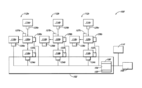
below. In the
embodiment shown, fluid detectors 126a-c are shown connected to connectors
128a-c, but
other configurations are possible. For example, fluid detectors 126a-c can be
connected to
fluid source containers 114a-c themselves.
[0140] In some embodiments, the system 100 can include compatibility
mechanisms 127a-c for ensuring that an approved connector 120a-c has been
placed in
communication with the system 100 to ensure the accuracy of the amount of
fluid transferred.
The compatibility mechanisms 127a-c can be, for example, a specifically shaped
mounting
feature configured to correspond to a portion of the connector 120a-c.
[0141] In some embodiments, the system 100 can include source
adapters 129a-c
configured to receive the source containers 114a-c and removably connect to
the connectors
120a-c. Thus, when a source container 114a-c runs out of fluid, the empty
source container
114a-c and its corresponding adapter 129a-c can be removed and replaced
without removing
the associated connector 120a-c from the system 100. In some embodiments,
source adapters
-14-
CA 3068441 2020-01-16
129a-c can be omitted, and the source containers 114a-c can be directly
received by the
connectors 120a-c.
[0142] In some embodiments the system 100 can include sensors 128a-c
for
detecting the presence of target containers 116a-c. Sensors 128a-c can be in
communication
with the controller 104 so as to prevent the system 100 from attempting to
transfer fluid when
no target container 116a-c is connected. A variety of sensor types can be used
for sensors
128a-c. For example, sensors 128a-c can be weight sensors or infrared sensors
or other form
of electronic eye. In some embodiments, weight sensors 128a-c can also be used
to measure
the weight of the target containers 116a-c after fluid has been transferred.
The final weight of
a target container 116a-c can be compared to an expected weight by the
controller 104 to
confirm that the proper amount of fluid was transferred into the target
container 116a-c.
Sensors 128a-c can be a variety of other sensor types, for example sensor pads
or other sensor
types able to detect the presence of target containers 116a-c.
[0143] Figure 2 schematically illustrates a system 200 for automated
precise
transfer of fluids. System 200 can be the same as or similar to the system 100
in some
regards. Some features shown in Figure 1, such as the adapters 129a-c and
compatibility
mechanisms 127a-c, are not shown specifically in the system 200, but it will
be understood
that system 200 can include corresponding features. The system 200 can include
a housing
202, a controller 204, a memory 206, a user interface 208, a scanner 210, and
a
communication interface 205, similar to those describe above in connection
with the system
100. System 100 is configured to transfer individual fluids from the source
containers 114a-c
to target containers 116a-c. System 200, on the other hand, is configured to
transfer and
combine fluids from source containers 214a-c into a common target container
216. Thus,
system 200 can be used for compounding mixtures of fluids. In some
embodiments, a single
system can be configured both for compounding mixtures of fluids and for the
transfer of
individual fluids from a single-source container to a single-target container.
For example, a
system containing six fluid transfer stations can be configured so that
transfer stations 1-3 are
dedicated to compounding mixtures of fluids into a single common target
container, while
fluid transfer stations 4-6 can be configured to each transfer fluid from a
single source
container to a single target container. Other configurations are possible. In
the embodiment
-15-
CA 3068441 2020-01-16
shown in Figure 2, the system 200 can include sensors 228a-c for detecting
whether or not
the connectors 220a-c are connected to the common target container 216. The
system 200
can also include a sensor 229 for detecting the presence of the common target
container 216.
In some embodiments, the sensor 229 can measure the weight of the common
target container
216 and can report the weight to the controller 104. The controller 104 is
then able to
compare the final weight of the common target container 216 with an expected
weight to
confirm that the common target container 152 was filled with the correct
amount of fluids.
[0144] Figures 3A and 3B show a subsystem, or fluidics assembly, 300
for
transferring precise amounts of fluid from a medical vial 314 to an IV bag
316. Figure 3A is
a perspective view of subsystem 300, and Figure 3B is an exploded perspective
view of
subsystem 300. The subsystem 300 can include a syringe 318 for measuring
precise amounts
of fluid to be transferred. In some embodiments, the system includes an IV bag
assembly
330. The IV bag assembly 330 can include the IV bag 316, a connector 332, and
a piece of
tubing 334 connecting the IV bag 316 to the connector 332. The connector 332
can be, for
example, a female medical connector. The connector 332 illustrated in Figures
3A-B is a
version of the Clove connector manufactured by ICU Medical, Inc., of San
Clemente,
California. Various embodiments of a connector of this type are described in
U.S. Patent No.
5,685,866 (the '866 Patent"). The subsystem 300 can also include a connector
320, for
interconnecting the vial 314, the syringe 318, and the IV bag assembly 330.
[0145] Turning now to Figures 4A and 4B, Figure 4A shows an exploded
perspective view of a fluid transfer module in the form of connector 320, and
Figure 4B
shows a cross-sectional view of the connector 320. The connector 320 can
include a first
interface or source connector portion 336 configured to provide fluid
communication
between the connector 320 and the vial 314, a second interface of target
connector portion
338 configured to provide fluid communication between the connector 320 and
the IV bag
assembly 330, and an intermediate connector portion 340 configured to provide
fluid
communication between the connector 320 and the syringe 318. The connector can
also
include a main body 342. In the embodiment shown in Figures 4A-B, the
intermediate
connector portion 340 is integrally formed as part of the main body 342.
-16-
CA 3068441 2020-01-16
[0146] In some embodiments, the connector 320 can be a T-connector.
In the
embodiment shown, the fluid path leading to the IV bag assembly 330 is
substantially
perpendicular to the fluid path between the vial 314 and the syringe 318. A
variety of other
configurations are possible. For example, the fluid pathways can be arranged
to intersect at
an oblique angle.
[0147] In some embodiments, the source connector portion 336
includes a female
connector portion 344 having a slightly tapered internal surface. The main
body 342 of the
connector can have a corresponding male connector portion 346 having a
similarly tapered
outer surface. The female connector portion 344 and male connector portion 346
can be
configured such that when the male connector portion 346 is fully inserted
into the female
connector portion 344 (i.e., the tapered surfaces prevents further insertion),
a chamber 348 is
defined between the end of the male connector portion 346 and the base of the
female
connector portion 344. The male connector portion 346 can be secured to the
female
connector portion 344 by applying a plastic welding adhesive (such as
Dichloromethane) to
the outer surface of the male connector portion 346 and/or to the inner
surface of the female
connector portion 344 before insertion. The Dichloromethane can chemically
weld the outer
surface of the male connector portion 346 to the inner surface of the female
connector portion
344. Other methods can be used to connect the male connector portion 346 to
the female
connector portion 344, such as sonic welding, threading, adhesives, etc. In
some
embodiments, the connection between the main body 342 and the source connector
portion
336 is hermetically sealed, and in some embodiments includes a sealing member
(not shown),
such as an 0-ring, to provide the hermetic seal.
[0148] In some embodiments, the target connector portion 338 can be
similarly
attached to the main body 342. The main body 342 can include a female
connector portion
350 with a tapered inner surface, and the target connector portion 338 can
include a male
connector portion 352 with a tapered outer surface. When the male connector
portion 352 is
inserted fully into the female connector portion 350 (i.e., the tapered
surfaces prevent further
insertion), a chamber 354 is defined between the end of the male connector
portion 352 and
the base of the female connector portion 350. The connector portions 350, 352
can be
secured to one another using Dichloromethane or any of the other methods
discussed above.
-17-
CA 3068441 2020-01-16
In some embodiments, the connection between the main body 342 and the target
connector
portion 338 is hermetically sealed, and in some embodiments, the connection
can include a
sealing member.
[0149] The connector 320 can include a source check valve 356
disposed inside
the chamber 348. The check valve 356 can be configured to allow fluid to flow
from the vial
314 into the connector 320, but block fluid from flowing from the connector
320 into the vial
314. The connector can also include a target check valve 358 disposed inside
chamber 354.
Check valve 358 can be configured to allow fluid to flow from the connector
320 into the IV
bag assembly, but blocks fluid from flowing from the IV bag assembly into the
connector
320. The check valves 356, 358 will be discussed in greater detail below.
[0150] The main body 342 can be constructed from a variety of
materials. The
main body 342 can be constructed from a rigid material such as polycarbonate
or other
polymeric materials. In some embodiments, at least a portion of the main body
342 can be
formed from a substantially transparent material as discussed below.
[0151] Figure 5A shows a perspective view of the source connector
portion 336
and vial 314 in an unengaged configuration. Figure 5B is another perspective
view of the
source connector portion 336 and vial 314, also in an unengaged configuration.
Figure 5C is
a cross-sectional view of the source connector portion 336 and vial 314 in an
engaged
configuration. Figure 5D is a cross-sectional view of the source connector
portion 336 and
vial 314 after a portion of the fluid has been withdrawn from the vial 314.
Although Figures
5A-5D shown the source connector portion 336 of the connector 320 separated
from the
remainder of the connector 320 for simplicity, it should be understood that
the source
connector portion 336 can be connected to the remainder of the connector 320
when in use.
[0152] With reference now to Figures 5A-D, the vial 314 can comprise
any
suitable container for storing medical fluids, and can be for example a
medical vial such as
those produced by Abbott Laboratories of Abbott Park, Illinois. In some
embodiments, the
vial 314 includes a body 357 and a cap 359. In some instances, the vial 314
can be
configured to be hermetically sealed. The body 357 can comprise a rigid
substantially
impervious material such as plastic or glass. In some embodiments the cap 359
includes a
septum 360 and casing 362. The septum 360 can be made of an elastomeric
material capable
-18-
CA 3068441 2020-01-16
of deforming in such a way that when punctured by an item, it forms a
substantially airtight
seal around that item. For example, in some instances the septum 360 comprises
silicone
rubber or butyl rubber. The casing 362 can surround the septum 360 and can be
made from
any suitable material for sealing the vial 314. In some instances, the casing
362 comprises a
metal that is crimped around the septum 360 and an end portion of the vial
body 357 in order
to form an airtight seal between the septum 360 and the vial body 357. In some
embodiments, casing 362 can include a substantially flat mounting surface 364.
The vial 314
can include a fluid 366, such as a medical fluid (e.g., a chemotherapy drug)
contained within
its internal volume. The vial 314 can also include a relatively small amount
of sterilized air
368 also contained within the internal volume.
[0153] The
source connector portion 336 can include a piercing member 370
which can comprise a sheath 372 and a pointed tip 374. The sheath 372 can be
cylindrical in
shape, or it can be a variety of other suitable shapes. For example, in some
embodiments, the
sheath 372 can be generally conical in shape and taper toward the pointed tip
374. The
piercing member 370 can comprise a rigid material such as metal or plastic,
suitable for
insertion through the septum 360, such as a polycarbonate plastic. In some
instances the
pointed tip 374 is separable from the sheath 372. In other embodiments, the
pointed tip 374
and sheath 372 can be integrally formed or permanently joined. The pointed tip
374 can be
configured to facilitate piercing of the septum 360. The source connector
portion 336 can
also include a cap connector 376 configured to secure the source connector
portion 336 to the
vial 314. In some embodiments, the cap connector 376 can include an adhesive
378, such as
a double-sided tape, disposed on the surface of the cap connector 376. A
removable covering
380 (shown partially pealed away in Figure 5B) can be disposed over the
adhesive 378 until it
is ready to be used. The vial 314 can be secured to the cap connector 376 by
removing the
covering 380 from the adhesive 378 and pressing the vial 314 down onto the
source
connector portion 336 so that the piercing member 370 pierces the septum 360
and the
mounting surface 364 comes into contact with the adhesive 378. A variety of
other
connection types can be used to secure the vial 314 to the source connection
portion 336 of
the connector 220.
-19-
CA 3068441 2020-01-16
[0154] In some embodiments, the source connector portion 336 can be
configured
to automatically equalize pressure within the vial 314 as fluid 366 is
withdrawn. For
example, the source connector portion 336 can be a version of the Genie
closed vial access
device manufactured by ICU Medical, Inc. of San Clemente, California. Certain
embodiments of closed vial access devices of this type are disclosed in U.S.
Provisional
Patent Application No. 61/090,561 (the "561 Application"). For example, the
'561
Application discloses other methods by which the vial 314 can be connected to
the source
connector portion 336.
[0155] In some embodiments, the source connection portion 336 can
include a
fluid extraction channel 382. The fluid extraction channel 382 can include an
upper portion
384 that extends from an extraction aperture 383 formed in the side wall of
the piercing
member 370 through a portion of the piercing member 370. The fluid extraction
channel 382
can also include and a lower portion 386 that extends through the female
connection portion
344. In certain embodiments, the lower portion 386 can be wider than the upper
portion 384,
defining a shoulder 388 at the transition from the lower portion 386 to the
upper portion 384.
[0156] In some embodiments, the sheath 372 can be hollow defining a
regulator
channel 390 that extends through the sheath 372 and through the cap connector
376 to a
regulator aperture 392 formed on a position of the source connector portion
344 that remains
exposed to the ambient air when the vial 324 is secured to the source
connector portion 336.
In some embodiments, a bag 394 can be enclosed within the regulator channel
390. The bag
can define an inner volume 395 that is in fluid communication with the
regulator channel
390. In some embodiments, the bag can include a connection region 396 that
forms an
airtight seal with the walls of the regulator channel 390 so that air cannot
move past the
connection region 396 unless it enters the inner volume 395 of the bag 394. In
some
embodiments, the connection region 396 of the bag 394 can be secured to the
sheath 372 by
an adhesive, or by any other suitable manner.
[0157] The bag 394 can be folded up inside the regulator channel 390
so that it
occupies a relatively small volume compared to its unfolded state. The bag 394
can be
configured to be able to fill all, or a substantial portion, of the internal
volume of the vial 314.
In some embodiments, the bag 394 can comprise a elastomeric material, such as
Mylar ,
-20-
CA 3068441 2020-01-16
polyester, polyethylene, polypropylene, saran, latex rubber, polyisoprene,
silicone rubber,
polyurethane, and latex-free silicone that can allow the bag 394 to unfold,
expand, and/or
contract. In some embodiments, the bag 394 can comprise a non-expandable
material that is
flexible enough to allow the bag to unfold. In some circumstances, the bag 394
can comprise
a material that is impervious to liquid and air and inert with respect to the
fluid 366.
[0158] Figure 5C illustrates an embodiment of the source connector
portion 336
coupled to the vial 314 at a stage before any of the fluid 366 is extracted.
By comparison,
Figure 5D illustrates an embodiment of the source connector portion 336
coupled to the vial
314 at a stage with the bag 394 deployed after some of the fluid 366 has been
extracted.
Although not shown in Figures 5C and 5D, the fluid extraction channel 382 of
the source
connector portion 336 can be in fluid communication with the syringe 318 or
other medical
instrument capable of creating a negative pressure to extract fluid 366 from
the vial 314. In
some circumstances, a volume of the fluid 366 can be withdrawn from the vial
314 by the
syringe causing the pressure within the vial 314 to drop. The reduced pressure
in the vial can
cause the tip 374 to disengage from the sheath 372, so that the bag 394 is
free to emerge from
the sheath 372. As the fluid 366 flows out of the vial 314 and toward the
syringe 318,
ambient air flows into the inner volume 395 of the bag 394 by way of the
regulator channel
390 and the regulator aperture 392. In some circumstances the inner volume 395
of the bag
394 expands (by the bag unfolding and/or expanding) to compensate for the
reduced pressure
inside the vial 314.
[0159] Thus, the source connector portion 336 can be configured to
allow the
fluid 366 to be withdrawn from the vial 314 while regulating the pressure
within the vial 314.
In some embodiments, the source connector portion 336 maintains a
substantially constant
pressure within the vial 314 as the fluid 366 is withdrawn therefrom. In some
embodiments,
the pressure within the vial 314 changes by no more than about 1-5 psi as the
fluid 366 is
withdrawn. The '561 Application discloses additional details and various
alternatives that
can be applied to the source connector portion 336 and vial 314.
[0160] Figure 6A shows a perspective view of the target connector
portion 388.
Figure 613 is an exploded perspective view of the target connector portion
388. Figure 6C
shows a top view of a housing portion of the target connector portion 388.
Figure 6D shows
-21-
CA 3068441 2020-01-16
a cross-sectional view of the target connector portion 388 and the female
connector 332 in an
unengaged configuration. Figure 6E shows a cross-sectional view of the target
portion 338
and the female connector 332 in an engaged configuration. Although the target
connector
portion 338 is shown separated from the remainder of the connector 320 in
Figures 6A-6E, it
should be understood that the target connector portion 338 can be connected to
the remainder
of the connector 320 when in use.
[01611 With reference now to Figures 6A-6E, the target connector
portion 338 of
the connector 320 can be a closeable male luer connector that is configured to
prevent fluid
from escaping from or entering into the connector when it is not engaged with
a
corresponding female connector, but allow fluid to flow when it is engaged
with a
corresponding female connector 332. In the embodiments shown, the target
connector
portion 338 can be a version of the Spiros closeable male connector
manufactured by ICU
Medical, Inc., of San Clemente, California. Various embodiments of connectors
of this type
are described in U.S. Patent Publication No. 2008/0287920 (the "'920
Publication").
Although the embodiments illustrated in Figures 6A-6E show the connector 332
as being a
female connector and the target connector portion 338 as being a male
connector, it should be
noted that other configurations are possible. For example, the connector 332
can be a male
connector while the target connector portion 338 can be a female connector. In
some
embodiments, a substantially entirely or entirely closed system can be
achieved, at least in
part, by providing corresponding automatically closeable male and female
connectors at
various (or all) connection points within the fluid transfer system 100,
thereby causing the
stationary fluid to substantially entirely remain within the fluid source, the
fluid module, and
the fluid target, respectively, upon disconnection and to not generally leak
or vaporize outside
of the system. For example, in some embodiments, corresponding pairs of
automatically
closing connectors (e.g., male and female connectors) can be provided at the
interfaces
between the fluid source and the fluid module, the fluid module and the
intermediate
container, and/or the fluid module and the destination or target container.
[01621 The target connector portion 338 can include a housing 398, a
valve
member 400, a resilient member 402, a sealing ring 404, an end cap 406, and an
0-ring 407.
The housing 398 can be generally tubular in shape, and can include a
passageway 408 that
-22-
CA 3068441 2020-01-16
extends axially through the housing. As illustrated, the passageway 408
includes apertures
on each side of the connector. The housing 398 can include a male luer tip 410
that connects
to the rest of the housing 398 at a base 412. The luer tip 410 can be
generally tubular in shape
so that a portion of the passageway 408 is defined therein, and the luer tip
410 can include a
hole 414 at its end providing access to the passageway 408. In some
embodiments, the luer
tip 410 includes a shelf 416 that extends radially inwardly toward the axis of
the passageway
408. The shelf 416 can be located adjacent to the hole 414, so that the
passageway 408 is
narrowed at the end of the luer tip 410. In some embodiments, the surface of
the shelf 416
that faces radially inwardly is tapered so that the passageway 408 is
narrowest immediately
adjacent to the hole 414. In some circumstances, the shelf 416 can be
configured to seal the
passageway when a portion of the valve member 400 is abutted against it. As
illustrated, in
some embodiments, connectors can be used to substantially entirely prevent
fluid therein to
leak, vaporize, or otherwise escape through apertures in the fluid pathway
when the
connectors are closed.
[0163] The luer tip 410 can be surrounded by a shroud 418. In some
embodiments, the luer tip 410 extends some distance beyond the edge 420 of the
shroud. The
shroud 418 can include inner threads 422 on its interior surface. The inner
threads 422 can
be used for securing a female connector 332. The shroud can include an
indented portion 424
that has a smaller outer diameter than the other portions of the housing. The
indented portion
424 can be configured to engage a portion of the resilient member 402.
[0164] The housing 398 can include two wall sections 426a, 426b
separated by
two gaps 428a, 428b. The gaps 428a, 428b can be configured to receive portions
of the
resilient member 402. The wall sections 426a, 426b can be configured to engage
the end cap
406.
[0165] In some embodiments, the housing 398 includes a middle
portion 430
located substantially between the wall sections 426a, 426b, and connected to
the wall sections
426a, 426b near the gaps 428a, 428b. In some embodiments, holes 432a, 432b are
defined
between the middle portion 430 and the wall sections 426a, 426b (as shown in
Figure 6C). In
some embodiments, the luer tip 410 connects to the middle portion 430 at its
base 412. In
some embodiments, the middle portion defines a portion of the passageway 408
therein. In
-23-
CA 3068441 2020-01-16
some embodiments, portions 434 of the outer surface of the middle portion 430
are exposed
by the gaps 428a, 428b. The portions 434 can include notches 436a, 436b and
through-holes
438a, 438b. The notches 436a, 436b can be generally rectangular in shape, and
can be
tapered such that the notches 436a, 436b are narrower near their bases than
near their
surfaces. The through-holes 438a, 438b can also be generally rectangular in
shape.
10166]
The housing 398 can be constructed from a variety of materials. The
housing 398 can be constructed from a rigid material such as polycarbonate or
other
polymeric materials. In some embodiments, the housing 398 can be constructed
from a
hydrophobic material such as Bayer Malcrolon, or any other suitable material.
In some
embodiments, the housing 398 can be formed from a substantially transparent
material.
10167]
The valve member 400 can include a fluid passageway 440 extending
axially from an opening formed in a base portion 444 and into a tube 446. In
some
embodiments, the passageway 440 can be wider in the base portion 444 than in
the tube 446.
In some embodiments, the tube 446 includes a narrowed tip 448. In some
embodiments, the
tip 448 can have a tapered outer surface. The tip 448 can be tapered to
substantially the same
degree as the radially inwardly facing surface of the shelf 416 and can be
sized so that the tip
448 can form a fluid seal with the shelf 416 when abutted against it. In some
embodiments,
the tip 448 can be made from a flexible or compressible material, such as
silicone rubber to
facilitate formation of the fluid seal between the tip 448 and the shelf 416.
In some
embodiments, the tube can include one or more holes 450 for providing access
to the fluid
passageway 440. The holes 450 can be formed, for example, in the tip 448 of
the tube 446.
[0168] In
some embodiments, the valve member 400 can include two struts 452a,
452b extending out from the base 444 and positioned on either side of tube
446, so that an
open space is defined on either side of the tube. In some embodiments, the
tube 446 can
extend axially past the ends of the struts 452a, 452b.
[0169]
The base 444 of the valve member 400 can include a plurality of
protrusions 454 extending radially outwardly from its external surface. In
some
embodiments, the protrusions 454 can be positioned so as to define two
channels 456a, 456b
therebetween. In some embodiments, the protrusions 454 do not extend across
the full length
-24-
CA 3068441 2020-01-16
of the base 444, leaving a lower portion 458 of the base 444 that has a
substantially smooth
outer surface.
[0170] The valve member 400 can be constructed from a variety of
materials,
such as polycarbonate or other polymeric materials. In some embodiments, the
valve
member 400 can be constructed from the same material as the housing 398. In
some
embodiments, the valve member 400 and housing 398 can be constructed from
different
materials. In some embodiments, the valve member 400 can be constructed from
multiple
materials or from multiple pieces. For example, the tip 448 can be constructed
from a
material that is more flexible than the remainder of the valve member 400. In
some
embodiments, the valve member 400 can be formed from a substantially opaque
material.
10171] The resilient member 402 can include a first ring 460 and a
second ring
462 connected to each other by elastic members 464a, 464b. The elastic members
464a,
464b can be made from an elastic material that exerts a restoring force when
stretched, such
as silicon rubber. Thus, if the rings 460, 462 are pulled apart, the elastic
members 464a, 464b
function to restore the rings 460, 462 to their relaxed configuration. In some
embodiments,
the rings 460, 462 are also constructed from an elastic material, such as the
same material
used to form the elastic members 464a, 464b. In some embodiments, the second
ring 462 can
have a greater diameter than the first ring 460. In some embodiments, the
second ring 462
can have a tapered outer surface so that the end of the second ring 462 that
is closest to the
first ring 460 is wider than the end of the second ring 462 that is furthest
from the first ring
460.
[0172] The sealing ring 404 can be generally cylindrical in shape,
and can have a
bore 466 extending axially therethrough. The sealing ring 404 can have a
cylindrical body
section 468 and an 0-ring 470 located at one end of the body section 468. In
some
embodiments, the thickest portion of the 0-ring 470 can be thicker than the
body section 468
so that the thickest portion of the 0-ring 470 extends radially inwardly
toward the axis of the
bore 466 a distance past the inner surface of the body section 468. Thus, the
bore 466 can be
narrower at the thickest part of the 0-ring 470 than in the body section 468.
In some
embodiments, the thickest portion of the 0-ring 470 also extends radially
outwardly a
distance past the outer surface of the body section 468. The sealing ring 404
can include two
-25-
CA 3068441 2020-01-16
protrusions 472a, 472b that extend radially outwardly from the body section
468. In some
embodiments, the protrusions 472a, 472b can be generally rectangular in shape.
[01731 The sealing ring 404 can be constructed from a variety of
materials. In
some embodiments, the sealing ring 404 can be constructed from a deformable or
elastic
material such as a silicone rubber. In some embodiments, the sealing ring 404
can be
constructed from the same material used for form the resilient member 402. In
some
embodiments, the sealing ring 404 can be constructed from a material capable
of forming a
fluid seal against a rigid plastic or other rigid polymeric material.
[0174] The end cap 406 can include a first end cap member 405 and a
second end
cap member 409. The second end cap member 409 can include a male connector
352, a
plunger 474, and a disk portion 476 located between the male connector 352 and
the plunger
474. The second end cap member 409 can have a fluid passageway 478 axially
positioned
therein. In some embodiments, the plunger 474 can be generally tubular in
shape. In some
embodiments, the outer surface of the plunger 474 includes an indented region
480, which
can be configured to receive the 0-ring 407 therein. The 0-ring 407 can be
constructed from
an elastic material such as silicone rubber so that it can be stretched over
the edge 482 of the
plunger 474 and be seated in the indented region 480. In some embodiments, the
0-ring 407
can be constructed from the same material as the resilient member 402 and/or
the sealing ring
404. In some embodiments, the 0-ring 407 can be sized so that when seated in
the indented
region 480, the thickest portion of the 0-ring 407 extends radially outwardly
a distance past
the outer surface of the plunger 474.
[01751 In some embodiments, the passageway 478 can have a
substantially
constant width throughout the second end cap member 409. In some embodiments,
the
passageway 478 can be tapered so that it is wider in the male connector 352
than in the
plunger 474. In some embodiments, the passageway 478 can narrow near the end
of the
plunger 474, for example, to accommodate the indented region 480.
[0176] The first end cap member 405 can be generally frustoconical
in shape and
can have a central opening 471 therein. When assembled, the plunger 474 can
extend
through the central opening 471. A ridge 473 can extend inward into the
central opening
471. The ridge 473 can be received into a channel 475 formed between the base
of the
-26-
CA 3068441 2020-01-16
plunger 474 and the disk portion 476 on the second end cap member 409 to
secure the first
end cap member 405 to the second end cap member 409. The ridge 473 and
corresponding
channel 475 can allow the first end cap member 405 to rotate about a
longitudinal axis with
respect to the second end cap member 409. Thus, the first end cap member 405
and the
second end cap member 409 can join to form the end cap 406.
[0177] The valve end cap 406 can be constructed from a variety of
materials,
such as polycarbonate or other rigid polymeric materials. In some embodiments,
the end cap
406 can be constructed from the same material as the housing 398 and/or the
valve
member 400.
[0178] In some embodiments, the end cap 406 can be constructed from
a different
material than the valve member 400 and/or the housing 398. The first end cap
member 405
can be formed from the same material as the second end cap member 409, or
different
materials can be used. In some embodiments, the first end cap member 405 or
the second
end cap member 409 or both can be substantially transparent.
[0179] Certain interconnections between various parts of the target
connector
portion 338 will now be discussed in further detail. The sealing ring 404 can
be positioned
inside the middle portion 430 of the housing 398. The protrusions 472a, 472b
can be sized
and positioned so that they engage the through-holes 438a, 438b. Thus, the
sealing ring 404
can be secured to the housing 398 so that it does not rotate or move axially
with respect to the
tube 446.
[0180] The valve member 400 can be slidably inserted into the
housing 398 so
that the tube 446 enters the passageway 408. The narrowed tip 448 of the tube
446 can pass
through the bore 466 of the sealing ring 404 and into the male luer tip 410
until it abuts
against the shelf 416. The tube 446 can have a width that substantially fills
the bore 446 and
presses against the 0-ring 470 portion of the sealing ring 404 to form a fluid
seal
therebetween. The struts 452a, 452b can pass through the holes 432a, 432b in
the housing
398 respectively, so that the struts 452a, 452b are positioned between the
male luer tip 410
and the shroud 418.
[0181] The resilient member 402 can function to bias the valve
member 400
against the housing 398. The first ring 460 can fit onto the lower portion 458
of the base 444
-27-
CA 3068441 2020-01-16
of the valve member 400, so that a surface of the ring 460 abuts against the
protrusions 454.
The second ring 462 can fit into the indented portion 424 of the housing. The
elastic
members 464a, 464b can be positioned in the channels 456a, 456b respectively,
and can pass
through the respective gaps 428a, 428b between the wall sections 426a, 426b of
the housing
398.
[01821
The 0-ring 407 can be seated onto the indented region 480 of the end cap
406, as discussed above, and the plunger 474 can be slidably inserted at least
partially into the
passageway 440 of the valve member. In some embodiments, the thickest portion
of the 0-
ring 407 can be wider than the portion of the passageway 440 formed in the
base 444 of the
valve member 400, so that the 0-ring 407 forms a fluid seal against the inner
surface of the
passageway 440. The plunger 474 can be inserted into the valve member 400
until the disk
portion 476 of the end cap 406 comes into contact with the ends of the wall
sections 426a,
426b of the housing 398.
[0183] In
some embodiments, the wall sections 426a, 426b can be secured to the
top surface 477 of the first end cap member 405 by sonic welding, snap fit
structures (not
shown), a pressure or friction fitting, or other suitable connection type.
As mentioned
above, the first end cap member 405 can be secured to the second end cap
member 409 in a
manner that allows the first end cap member 405 to rotate relative to the
second end cap
member 409. Thus, once the target connector portion 338 is assembled, the
housing 398,
sealing ring 404, resilient member 402, valve member 400, and first end cap
member 405 can
rotate relative to the second end cap member 409 about the longitudinal axis.
[0184]
With reference now to Figures 6D-6E, the target connector portion 338
can be configured to engage a female connector 332. A variety of types of
female connectors
332 can be used. The female connector 332 shown is a closable female luer
connector that
includes a housing 490, a spike 492, a base 494, and a resilient seal element
496. A fluid
passageway 498 can pass through the base 494 and through the spike 492. The
spike 492 can
include one or more holes 500 providing fluid communication between the
passageway 498
and the area outside the spike 492. The seal element 496 can be shaped and
positioned to
substantially surround the spike 492. The seal element 496 can include a
closable aperture
502 or slit that can open to allow the tip of the spike 492 to pass through
then end of the seal
-28-
CA 3068441 2020-01-16
element 496 when the seal element 496 is compressed (as shown in Figure 6E).
The housing
can include external threads 504 configured to engage the inner threads 422 on
the housing
398 of the target connector portion 338. An end of the tubing 334 can be
connected to the
end of the female connector 332 by an adhesive, clamp, friction or pressure
fitting, or other
suitable manner to form a fluid tight connection.
[0185] As discussed above, in some embodiments, the housing 398,
sealing ring
404, resilient member 402, valve member 400, and first end cap member 405 can
rotate about
the longitudinal axis with respect to the second end cap member 409. Thus, as
the female
connector 332 of the IV bag assembly is attached to the target connector
portion 338, the
female connector 332 can be held still while the housing 398 of the target
connector portion
338 can rotate causing the threads 504, 422 to engage. Because the female
connector 322 is
not required to rotate during engagement and disengagement with the target
connector
portion 338, the tubing 334 can avoid being twisted or kinked and the user is
not required to
twist the IV Bag to accommodate rotation of the female connector 322.
Embodiments of the
connectors with this rotational capability are disclosed in greater detail in
the '920
Publication.
[0186] When not engaged with the female connector 332 (as shown in
Figure
6D), the target connector portion 338 can be sealed. In some embodiments,
fluid can enter
the target connector portion 338 at the male connector 352 and pass through
the passageway
478 of the end cap 406, through the passageway 440 of the valve member 400,
through the
holes 450, and into the portion of the passageway 408 defined by the male luer
tip 410. But
the fluid seal created by the tip 448 of the valve member 400 pressing against
the shelf 416 of
the male luer tip 410 prevents the fluid from exiting the target connector
portion 338. In
some embodiments, an increase in pressure, such as when additional fluid is
forced into the
target connector portion 338, causes the tip 448 to press more firmly against
the shelf 416,
thereby improving the fluid seal.
[0187] When the target connector portion 338 is engaged with the
female
connector 332 (as shown in Figure 6E), the external threads 504 of the female
luer connector
332 can engage the inner threads 422 on the shroud 418, securing the female
connector 332
to the target connector portion 338. The edge of the male luer tip 410 can
press against and
-29-
CA 3068441 2020-01-16
compress the resilient seal element 496 so that the spike 492 passes through
the aperture 502
until the holes 500 are exposed. The end of the housing 490 of the female luer
connector 332
can enter the space between the male luer tip 410 and the shroud 418 until it
contacts the
struts 452a, 452b. As the female luer connector 332 further engages the target
connector
portion 338, it can push on the struts 452a, 452b causing the entire valve
member 400 to
retract. As the valve member 400 retracts, the elastic members 464a, 464b of
the resilient
member 402 stretch. When the valve member 400 retracts, the tip 448 disengages
from the
shelf 416, breaking the fluid seal and allowing fluid pass from the passageway
408 in the
housing 398 of the target connector portion 338 to the passageway 498 in the
female
connector 332 via the holes 500. When engaged, the resilient seal element 496
exerts a
restoring force toward the target connector portion 338 that presses the end
of the seal
element 496 against the end of the male luer tip 410, forming a fluid seal
therebetween.
Thus, the fluid can be kept isolated from the external environment while it is
transferred from
the target connector portion 338 to the female connector 332.
[0188] The female connector 332 can be disengaged from the target
connector
portion 338. The restoring force exerted by the resilient seal element 496 of
the female
connector 332 causes it to return to its closed position, sealing off its
passageway 498. The
elastic members 464a, 464b of the resilient member 402 exert a restoring force
on the valve
member 400, causing the valve member 400 to return to its closed position with
its tip 448
abutted against the shelf 416 as the female connector 332 is disengaged.
[0189] The '920 Publication discloses additional details and various
alternatives
that can be applied to the target connector portion 338 of the connector 320.
[0190] Figure 7A shows a perspective view of the syringe 318 and the
intermediate connector portion 340 of the connector 320 in an =engaged
configuration.
Figure 7B is a top view of the syringe 318 and intermediate connector portion
in an engaged
configuration. Figure 7C is a cross-sectional view of the syringe 318 and
intermediate
connector portion 340 in an engaged configuration. Although Figures 7A-7C show
the main
body 342 of the connector 320 separated from the remainder of the connector
320 for
simplicity, it should be understood that the main body 342 can be connected to
the remainder
of the connector 320 when in use.
-30-
CA 3068441 2020-01-16
[0191] In the embodiment shown, the intermediate connector portion
340 is an
integral part of the main body 342 of the connector 320. Other configurations
are possible.
For example, in some embodiments, the intermediate connector portion 340 can a
separate
piece connected to the main body 342. The intermediate connector portion 340
can include a
female connector 506. In some embodiments, the female connector 506 can have a
tapered
inner surface. The external surface of the female connector 506 can include
external threads
508.
[0192] The syringe 318 can have a hollow syringe body 510 defining
an internal
volume 511. The syringe can include a male luer tip 512 at one end and a
shroud 514
surrounding the male luer tip 512. The shroud 514 can have internal threads
516. The male
luer tip 512 and threaded shroud 514 can be configured to securely mate with
the female
connector 506 on the intermediate connector portion 340 of the connector 320,
forming a
fluid tight connection therebetween. The syringe body 510 can include a body
flange 518
positioned at the end of the body opposite the male luer tip 512. The syringe
also includes a
plunger 520 that can be slidably received into the internal volume of the
syringe body 510.
The plunger 522 can include a stopper 522 or other sealing member configured
to form a
fluid tight seal against the inner surface of the syringe body 510. A plunger
flange 524 can be
positioned on the plunger 520 at the end opposite the stopper 522.
[0193] In some embodiments, the female connector 506 and the male
luer tip 512
can be open to the atmosphere when unengaged. Other configurations are
possible. For
example, in some embodiments, the female connector 506 can be a sealing female
connector
similar to the female connector 332 described above, and can be for example a
version of the
Claw connector. Similarly, the syringe 318 can include a sealing male
connector, or a
sealing male connector can be connected between the syringe 318 and the female
connector
506. In some embodiments the sealing male connector can be a version of the
SpirosTm
connector. Thus, in some embodiments, the fluid in the syringe 318 and in the
connector 320
can be isolated from the environment even when they are disengaged from each
other.
[0194] In some embodiments, when the syringe 318 is engaged with the
connector
320 (as shown in Figure 7B) the internal volume 511 of the syringe 318 can be
in two way
fluid communication with the connector 320. Thus, as the plunger 520 is
retracted a fluid can
-31-
CA 3068441 2020-01-16
be drawn from the connector 320 into the internal volume 511 of the syringe
318. Then as
the plunger 520 is advanced the fluid can be directed out of the internal
volume 511 and into
the connector 320.
[0195] As discussed briefly above, the connector 320 can include a
source check
valve 356 and a target check valve 358. The check valves 356, 358 can function
so that when
the plunger 520 is retracted the source check valve 356 opens and the target
check valve 358
closes, allowing fluid to flow from the vial 314 through the connector 320 and
into the
syringe 318. Then, when the plunger 520 is advanced the source check valve 356
can close
and the target check valve 358 can open, allowing fluid to flow from the
syringe 318 through
the connector 320 and into the IV bag 316.
[0196] Figure 8A is a perspective view showing the source check
valve 356.
Figure 8B is another perspective view showing the source check valve 356 from
a different
angle. The source check valve 356 can include a disk shaped base 526. A
plurality of feet
528 can extend axially from one side of the base 526. In the embodiment shown,
the source
check valve 356 includes four feet 528, but other numbers of feet 528 can be
used, such as
three feet, or five feet, or another suitable number of feet 528. In some
embodiments, the feet
528 can each be generally cylindrical in shape and can each include a rounded
tip 530. Other
shapes and configurations are possible. The source check valve 356 can have a
sealing
surface 531 located on the side opposite the feet 528.
[0197] Figure 9A shows the source connector portion 336, the source
check valve
356, and the main body 342 in an exploded cross-sectional view. Figure 9B is a
cross-
sectional view of the source connector portion 336, the source check valve
356, and the main
body 342 in an assembled configuration with the check valve 356 in an open
position. As
discussed above, the source connector portion 336 can include a fluid
extraction channel 382
having an upper, narrow portion 384 and a lower, wide portion 386. A shoulder
388 can be
defined at the transition from the upper portion 384 to the lower portion 386
of the fluid
extraction channel 382. In some embodiments, the lower portion 386 of the
extraction
channel 382 can have a tapered inner surface, so that the lower portion 386
narrows near the
shoulder 388. The upper portion 384 can also have a tapered inner surface, so
that the upper
portion 384 widens near the shoulder 388. In some embodiments, the upper
portion 384
-32-
CA 3068441 2020-01-16
and/or the lower portion 386 can be substantially cylindrical or can assume a
variety of other
shapes having a substantially uniform cross-sectional area.
[01981 The main body 342 can include a first fluid passageway 532
leading from
the end 534 of the male connector 346 to the end 534 of the intermediate
connector portion
340. The first fluid passageway 532 can include an upper portion 536 and a
lower portion
538. The lower portion 538 can be wider than the upper portion 536 defining a
shoulder 540.
The upper portion 536 and lower portion 538 can have tapered or untapered
inner surfaces.
When assembled, the source check valve 356 can be positioned in the chamber
348 located
between the end 534 of the male connector 346 and the shoulder 388 of the
fluid extraction
channel 382. The source check valve 356 can be positioned so that the feet 528
face toward
the end 534 of the male connector 346, while the sealing surface 531 can face
toward the
shoulder 388. In some configurations, when the pressure in the fluid
passageway 332 is
sufficiently higher than the pressure in the extraction channel 382, such as
when the plunger
520 of the syringe 318 is advanced forcing fluid into the fluid passageway
332, the source
check valve 356 is pushed away from the main body 342 and the sealing surface
531 engages
the shoulder 388 forming a fluid tight seal that prevents fluid from flowing
from the first
fluid passageway 532 into the upper portion 384 of the extraction channel 382.
In some
configurations, when the pressure in the fluid passageway 332 is sufficiently
lower than the
pressure in the extraction channel 382, such as when the plunger 520 of the
syringe 318 is
retracted drawing fluid out of the fluid passageway 332, the source check
valve 356 is pulled
away from the shoulder 388 and the feet 528 rest against the end 534 of the
male connector
346 in an open position.
101991 Figure 10A is a side view of the main body 342 coupled to the
source
connector portion 336 of the connector 320. Figure 10B is a cross sectional
view of the
source connector portion 336 of the connector 320 and the source check valve
356 disposed
therein. Figure 10C is a partial cross-sectional view showing the source check
valve 356
positioned in a chamber 348 defined radially by the walls of the female
connector 344.
Figure 10D is another cross sectional view showing the source check valve 356
positioned in
the chamber 348.
-33-
CA 3068441 2020-01-16
[0200] With reference to Figures 10A-10D, the base 526 of the source
check
valve 356 can be disk shaped and can have a diameter d1 that is less than the
diameter d2 of
the chamber 438, defining a space 542 between the side edges of the source
check valve 356
and the inner walls of the chamber 348 through which fluid can pass. Also, the
feet 528 can
be spaced so that open areas 544 are defined between adjacent feet 528.
[0201] Thus, when the source check valve 356 is in the open
position, fluid can
flow from the upper portion 384 of the extraction channel 382, into the
chamber 348, through
the space 542 between the side edges of the source check valve 356 and the
inner walls of the
chamber 348, through the open areas 544 between the feet 528, and into the
upper portion
536 of the first fluid passageway 532.
[0202] In some embodiments, the source check valve 356 can be
configured to
allow a substantially open flow around the check valve 356 without significant
bottlenecking.
For example, the space 542 between the side edges of the source check valve
356 and the
inner walls of the chamber 348 can have a cross-sectional area Ai that is at
least large as the
cross-sectional area A2 of the upper portion 483 of the extraction channel 382
taken near the
chamber 348. This relationship can be expressed as equation (1) below.
AI A2 (1)
102031 In some embodiments, the chamber 348 and the source check
valve 356
can both be substantially cylindrical, having diameters d2 and di respectively
(as shown in
Figures 10C and 10D). The cross sectional area Ai of the space the space 542
between the
side edges of the source check valve 356 and the inner walls of the chamber
348 can then be
defined by equation (2) below.
2 (
= d2 ¨ 2)
, 2 ) 2)
In some embodiments, the upper portion 483 of the extraction channel 382 taken
near the
chamber 348 can be substantially cylindrical and can have a diameter d3 (as
shown in Figure
10D). The area A2 can then be defined by equation (3) below.
(3)
A2 = r
, 2
By substituting equations (2) and (3), equation (1) can be rewritten as
equation (4) below.
-34-
CA 3068441 2020-01-16
jd2 71.(611 r(d3`2 (4)
-1 2 ) 2) 2 ,
By solving equation (4) for di, equation (4) can be rewritten as equation (5)
below.
di 5.. Vd22 - d32 (5)
Thus, when the diameter d2 of the chamber 348 and the diameter d3 of the upper
portion 483
of the extraction channel 382 are known, the source check valve 356 can be
having a
diameter that satisfies equation (5) to avoid bottlenecking of fluid as it
flows through the
space 542.
102041 As shown in Figure 10D, in some embodiments, when the source
check
valve 356 is in the open position a space 546 having a height hi is defined
between the
sealing surface 531 and the shoulder 388. The space 546 can allow fluid to
flow
therethrough. In some embodiments, the source check valve 356 and the chamber
348 can be
configured to prevent bottlenecking as fluid flows through the space 546. For
example, in the
embodiment shown, the smallest area of the space 546 through which the fluid
flows can be
described as an open an imaginary open cylinder (shown by dotted lines in
Figure 10D)
having a height of hi, a diameter of d3, and a surface area A3. If the surface
area A3 of the
imaginary cylinder is at least as great as the cross-sectional area A2 of the
upper portion 483
of the extraction channel 382 taken near the chamber 348, bottlenecking can be
reduced.
This relationship can be expressed as equation (6) below.
A3 A2 (6)
The surface area A3 of the imaginary open cylinder can be expressed as
equation (7) below.
A3 = /03 hi (7)
By substituting equations (3) and (7), equation (6) can be rewritten as
equation (8) below.
\2 (8)
lid3hI n=P
2 ,
By solving for hi, equation (8) can be rewritten as equation (9) below.
(9)
-35-
CA 3068441 2020-01-16
Thus, when the diameter d3 of the upper portion 483 of the extraction channel
382 is known,
the source check valve 356 can be made to have a total height that is shorter
than the height
of the chamber 348 by at least to
reduce bottlenecking as the fluid flows from the upper
4
portion 483 of the extraction channel 382 into the space 546 between the
source check valve
356 and the shoulder 388.
[0205]
The source check valve 356 can be configured to reduce bottlenecking of
the fluid as it flows through the open areas 544 (shown in Figure 10C) between
the feet 528.
For example, if the total area A4 of the open areas 544 between the feet 528
is at least as great
as the cross-sectional area A2 of the upper portion 483 of the extraction
channel 382 taken
near the chamber 348, bottlenecking can be reduced as the fluid flows from the
extraction
channel 382 and around the check valve 356. This relationship can be expressed
by equation
(10) below.
A4 A2 (10)
In the embodiment shown, the feet 528 are arranged so that an imaginary open
cylinder
(shown by a dotted line in Figure 10C) can be placed so that its edge
intersects each of the
feet 528. The feet 528 can be positioned so that the imaginary cylinder has a
diameter di. In
some embodiments, the source check valve 356 includes a number n of feet that
each have
substantially equal diameters ds and substantially equal heights 112. The area
total area A4 of
the open areas 544 can be defined by equation (11) below. It should be noted
that because
the feet 528 have rounded tips 530, the area A4 can be slightly greater than
represented by
equation (11). In some embodiments, the feet 528 do not have rounded tips and
can be
substantially cylindrical.
A4 = gd4h2 ¨nd5 (11)
By substituting equations (3) and (11), equation (10) can be rewritten as
equation (12) below.
d )2 (12)
nd4h2 nds
2
By using feet 528 that satisfy equation (12), bottlenecking can be reduced.
For example, if
the number n of feed or the diameter ds is increased, the height h2 of the
feet can be
-36-
CA 3068441 2020-01-16
increased, or the feet can be moved closer to the peripheral edge (increasing
di) to
compensate.
[0206] In some embodiments, the source check valve 356 can be
configured to
provide a substantially uniform flow of fluid. For example, the space 542
between the side
edges of the source check valve 356 and the inner walls of the chamber 348 can
have a cross-
sectional area Ai that is substantially equal to the cross-sectional area A2
of the upper portion
483 of the extraction channel 382 taken near the chamber 348. Similarly, the
surface area A3
of the fist imaginary cylinder can be substantially equal to the cross-
sectional area A2 of the
upper portion 483 of the extraction channel 382 taken near the chamber 348.
Likewise, the
total area A4 of the open areas 544 between the feet 528 can be substantially
equal to the
cross-sectional area A2 of the upper portion 483 of the extraction channel 382
taken near the
chamber 348. The source check valve 356 and chamber 348 can be configured so
that other
areas of flow also have an area that is substantially equal to the cross-
sectional area A2 of the
upper portion 483 of the extraction channel 382 taken near the chamber 348.
For example, in
some embodiments, the shoulder 388 or the sealing surface 531 of the check
valve 356 can be
tapered so that the height of the space 546 is smaller near the side space 542
than near the
upper portion 483 of the extraction channel 382. In some embodiments, the
areas discussed
herein can be considered to be substantially equal if they vary by an amount
less than an
acceptable tolerance T. In some embodiments, the acceptable tolerance T can be
less than
about 1 mm, 0.5mm, 0.1mm, 0.05mm, or 0.01mm. In some embodiments, the flow
areas
around the check valve 356 (e.g., AI, A3, and A4) can be smaller than A2 by an
amount no
larger than tolerance T. Thus, in some embodiments, a small but acceptable
amount of
bottlenecking can occur as the fluid flows around the source check valve 356.
[0207] In embodiments where the diameter d2 of the chamber 348 is
greater than
the diameter d1 of the source check valve 356, the source check valve 356 can
move not only
axially within the chamber, but also radially within the chamber. For example,
Figure 11
shows a cross-sectional view of the source check valve 356 positioned against
one side of the
chamber 348 when in a closed position. The diameter di of the check valve 356
can be large
enough to allow the check valve 356 to adequately seal off the chamber 348
when positioned
against one side of the chamber 348. For example, in some embodiments, the
chamber 348
-37-
CA 3068441 2020-01-16
can be generally symmetrical so that the shoulder 388 has a substantially
uniform width, and
the diameter d1 of the check valve 356 can be chosen to satisfy the equation
(13) below.
d d d, > +
(13)
' 2 2
[0208] In some embodiments, the upper portion 536 of the first fluid
passageway
532 can be generally cylindrical in shape and can have a diameter d6. In some
embodiments,
the feet 528 are positioned near enough to the peripheral edges of the check
valve 356 so the
feet do not drop into the upper portion 536 of the first fluid passageway 532
when the check
valve 356 is positioned against the side of the chamber 348. For example,
Figure 12 shows a
cross-sectional view of the source check valve 356 positioned against one side
of the
chamber 348 in an open position. The feet 528 are positioned so that when the
check valve
356 is positioned against one side of the chamber 348 the foot 528a closest to
the first
passageway 532 does not drop down into the first passageway 532. In some
embodiments,
the feet 528 can be positioned along a circle concentric with the check valve
356, the circle
having a diameter da that satisfies the equation (14) below.
d4 do + d2 ¨ d1
(14)
[0209] In some embodiments, the source check valve 356 can have a
diameter of
about 2mm to about 20mm, although diameters outside this range can also be
used. A variety
of other configurations are possible. For example, the source check valve 356,
the chamber
348, the extraction channel 382 and/or the first fluid passageway 532 can have
non-circular
cross sections.
[0210] Turning now to Figures 13A-B, Figure 13A shows an exploded
cross-
sectional view of the target connector portion 338, the target check valve
358, and the main
body 342. Figure 13B shows a cross-sectional view of the target connector
portion 338, the
target check valve 358, and the main body 342 in an assembled configuration
with the target
check valve 358 in an open position. The main body 342 can include a second
fluid
passageway 548 that intersects the first fluid passageway 532 at a junction
550. In some
embodiments, the second fluid passageway 548 can intersect the upper portion
536 of the
first fluid passageway 532. In the embodiment shown the second fluid
passageway 548
intersects the first fluid passageway 532 at a substantially right angle.
Other configurations
-38-
CA 3068441 2020-01-16
are also possible. For example, the fluid passageways 532, 548 can intersect
at an oblique
angle. The second fluid passageway 548 can have a narrow portion 552 and a
wide portion
554 that define a shoulder 556. In some embodiments, the narrow portion 552
can have a
width that is substantially the same as the width of the upper portion 536 of
the first fluid
passageway 532 near the junction 550, while in other embodiments the narrow
portion 552
can have a width that is smaller or larger than the width of the upper portion
536 of the first
fluid passageway 532 near the junction 550. In some embodiments, the narrow
portion 552
and/or the wide portion 554 of the second fluid passageway 548 can have
tapered interior
surfaces. For example, the wide portion 554 can be tapered so as to receive a
tapered male
connector 352, as discussed above.
[0211] When assembled, the target check valve 358 can be positioned
in the
chamber 354 formed between the male connector 352 and the shoulder 556. In
some
embodiments, the target check valve 358 can be similar to the source check
valve 356
described above, having a disk shaped base 558, a plurality of feet 560, and a
sealing surface
562. The target check valve 358 can be positioned with the feet 560 facing the
male
connector 352 and the sealing surface 562 facing the shoulder 556. Thus, when
the pressure
in the second fluid passageway 548 is sufficiently higher than the pressure
inside the male
connector 352, such as when the plunger 520 of the syringe 318 is advanced
forcing fluid into
the main body 342, the target check valve 358 can be pushed toward the male
connector 352
so that the feet 560 rest against the end of the male connector 352 in an open
position. When
the pressure in the second fluid passageway 548 is sufficiently lower than the
pressure inside
the male connector 352, such as when the plunger 520 of the syringe 318 is
retracted drawing
fluid out of the main body 342, the target check valve 358 can be pulled away
from the main
body 342 so that the sealing surface 562 engages the shoulder 556 forming a
fluid tight seal
that prevents fluid from flowing from the chamber 354 into the narrow portion
552 of the
second fluid channel 548.
[0212] In some embodiments, the target check valve 358 and the
chamber 354 can
be configured to reduce bottlenecking as fluid flows around the target check
valve 358 in its
open position. For example, the target check valve 358 and chamber 354 can be
configured
similarly in many ways to the source check valve 358 and chamber 348 described
above.
-39-
CA 3068441 2020-01-16
[0213] The check valves 356, 358 can work together to direct fluid
through the
system. Figure 14A shows the flow of fluid (by flow lines) as the plunger 520
is retracted.
Fluid is drawn out of the vial 314 through the upper portion 384 of the fluid
extraction
channel 382. The fluid flows into the chamber 348 and around the source check
valve 356,
which is in the open position. The fluid flows through the first fluid
passageway 532 and into
the syringe 318. The fluid can enter the narrow portion 552 of the second
fluid passageway
548, but the target check valve 358 is in the closed position and prevents the
fluid from
entering the chamber 354.
[0214] Figure 14B shows the flow of fluid (by flow lines) as the
plunger 520 is
advanced. Fluid is expelled from the syringe 318, into the first fluid
passageway 532,
through the narrow portion 552 of the second fluid passageway 548, into the
chamber 354,
around the target check valve 358 (which is in the open position), through the
target
connector portion 338, toward the IV bag 316. The fluid can travel up the
first fluid
passageway 532 and into the chamber 348, but the source check valve 356 is in
the closed
position and prevents the fluid from advancing back into the vial 314. In some
embodiments,
the force of the fluid pressing against the source check valve 356 is strong
enough the
overcome the force of gravity pulling the source check valve 356 downward so
as to maintain
the source check valve 356 in the closed position.
[0215] The check valves 356, 358 can be formed from rigid, semi-
rigid, or
deformable materials. In some embodiments, at least the sealing surfaces 531,
562 of the
check valves 356, 358 can be formed from a material capable of forming a fluid
tight seal
against a plastic or other rigid material. In some embodiments, the check
valves can include
a silicon-based deformable material, or a rubber. In some embodiments, the
feet 528, 560
can be formed from different material than the disk shaped base 526, 558. In
some
embodiments, the feet 528, 560 can be formed from a rigid polycarbonate or
other polymeric
material.
[0216] Figure 15 is a perspective view of an automated system 600
for
transferring fluid, which can be similar to or the same as the other automated
fluid transfer
systems (e.g., 100, 200) disclosed herein. The system 600 can include a base
housing 602,
and six transfer stations 604a-f, located on a front side of the base housing
602. In some
-40-
CA 3068441 2020-01-16
embodiments, the system 600 can include a different number of transfer
stations 604a-f (e.g.,
one, two, four, five, eight, or more transfer stations). In some embodiments,
the transfer
stations 604a-f can be distributed on multiple sides of the base housing 602.
Transfer
stations 604b-f are shown in an empty state having no syringe attached
thereto. Transfer
station 604a is shown having a syringe 606 and a connector 608 attached
thereto. During
operation, a vial (not shown) can be attached to the top of the connector 608
and an IV bag
(not shown) can be placed in fluid connection with the connector 608 so that
fluid can be
transferred from the vial to the syringe 606 and then from the syringe 606
into the IV bag, as
discussed above. Also, during operation, some or all of the transfer stations
604a-f can be
equipped similarly to transfer station 604a. In some embodiments, multiple
transfer stations
604a-f can operate simultaneously. In some embodiments, multiple transfer
stations 604a-f
can be placed in fluid communication with a single IV bag so that fluid from
multiple vials
can be combined into a single IV bag. In some embodiments, one or more of the
transfer
stations 604a-f can include a dedicated IV bag so that fluid from only a
single transfer
stations can be transferred into the dedicated IV bag.
[0217] Turning now to Figures 16A-16C, and 17, a transfer station
604a is shown
in greater detail. Figure 16A shows a partial perspective view of the transfer
station 604a,
with the syringe 606 and connector 608 in an unengaged configuration. Figure
16B shows a
left-side view of the transfer station 604a, with the syringe 606 and
connector 608 in an
unengaged configuration. Figure 16C shows a front-side view of the transfer
station 604a,
with the syringe 606 and connector 608 omitted from view. The transfer station
604a can
include an auxiliary housing 610 connected to the base housing 602. The
transfer station
604a can also include a top connector piece 612 attached to the base housing
602 above the
auxiliary housing 610, and a bottom connector piece 614 attached to the base
housing 602
below the auxiliary housing 610. The top connector piece 612 and the bottom
connector
piece 614 can extend out a distance past the auxiliary housing 610, and a pair
of shafts 616a-
b can extend vertically between the top connector piece 612 and the bottom
connector piece
614. A middle connector piece 618 can be attached to the shafts 616a-b.
[0218] The middle connector piece 618 can have a recess 620
configured to
receive the syringe body 624. For example, if the syringe body 624 is
generally cylindrical,
-41-
CA 3068441 2020-01-16
the recess 620 can in the shape of a half cylinder (as shown). The middle
connector piece
618 can also include a slit 622 configured to receive the body flange 626 of
the syringe 606.
The top connector piece 612 can have a recess 628 configured to receive the
shroud 630 of
the syringe 606 and a portion of the connector 608. In some embodiments, the
middle
connector piece 618 can be removable, so that it can be interchanged with
additional middle
pieces (not shown) to provide compatibility with different sizes and shapes of
syringes. Also,
in some embodiments, the position of the middle connector piece 618 can be
adjustable. For
example, the middle connector piece 618 can be slid up and down the shafts
616a-b and
secured in a variety of location, providing compatibility with syringes of
different lengths. In
some embodiments, the position of the middle connector piece 618 can be fixed.
[0219] The transfer station 604a can include an actuator 632
configured to retract
and advance the plunger 634 of the syringe 606. In the embodiment shown, the
actuator 632
includes an actuator base 636. Two shafts 648a-b can be positioned at the back
of the
actuator base 636 and can extend upward from the actuator base 636 into the
auxiliary
housing 610. Another shaft 640 can be positioned at the front of the actuator
base 636 and
can extend upward in front of the auxiliary housing 610. An end piece 642 can
be attached to
the end of the shaft 640 opposite the actuator base 636. The end piece 642 can
include a
horizontal slit 644 configured to receive the plunger flange 648 of the
syringe 606. The end
piece 642 can also be configured to receive a portion of the plunger shaft 650
that is near the
plunger flange 648. For example, if the plunger shaft 650 includes four
longitudinal ribs (as
shown), the end piece 642 can include a vertical slit 646 configured to
receive one of the
longitudinal ribs. The end piece 642 can also include a thumb screw 652 which
can be
tightened to apply pressure to the plunger flange 648 and prevent the syringe
606 from
accidentally disengaging from the transfer station 604a.
[0220] In some embodiments, a motor (not shown) is located inside
the auxiliary
housing 610. The motor can be an electric motor, a pneumatic motor, a
hydraulic motor, or
other suitable type of motor capable of moving the actuator 632. In some
embodiments, the
motor can be a piston type motor. In some embodiments, the motor is contained
within the
base housing 602 rather than in the auxiliary housing 610. In some
embodiments, each
transfer station 604a-f has an individual motor dedicated to the individual
transfer station
-42-
CA 3068441 2020-01-16
604a-f. In some embodiments, one or more of the transfer stations 604a-f share
a motor, and
in some embodiments, the system 600 includes a single motor used to drive all
the transfer
stations 604a-f. The motor can drive the shafts 638a-b downward out of the
auxiliary
housing 610, which in turn drives the rest of the actuator 632 downward
causing the plunger
634 to retract from the syringe body 624 to draw fluid into the syringe. The
motor can also
draw the shafts 638a-b upward into the auxiliary housing 610, which in turn
drives the rest of
the actuator 632 upward causing the plunger 632 to advance into the syringe
body 624 to
expel fluid from the syringe.
[0221] In some embodiments, the transfer station 604a can include a
label 654
that uniquely identifies the specific transfer station 604a. In some
embodiments the label 654
can be prominently displayed at the top of the transfer station 604a. The
label 654 can be
colored, and each of the transfer stations 604a-f can have a different colored
label.
[0222] The system 600 can include a controller, for controlling the
operations of
the transfer stations 604a-f, The controller can start and stop the motor(s)
of the system 600
to control the amount of fluid that is transferred from the vial to the IV bag
at each transfer
station 604a-f. The controller can be one or more microprocessors or other
suitable type of
controller. The controller can be a general purpose computer processor or a
special purpose
processor specially designed to control the functions of the system 600. The
controller can
include, or be in communication with, a memory module that includes a software
algorithm
for controlling the operations of the system 600. The controller can be
contained within the
base housing 602. In some embodiments, the controller can be external to the
base housing
602, and can be for example the processor of a general purpose computer that
is in wired or
wireless communication with components of the system 600.
[0223] In some embodiments, the transfer station 604a includes a
sensor (hidden
from view in Figures 16A-C) configured to determine when the liquid in the
vial (not shown)
has run out. If the plunger 634 is retracted to draw fluid into the syringe
606 when the vial
contains no more fluid, air is drawn out of the vial and travels into the
connector 608 toward
the syringe. Air may also be drawn into the connector 608 when the vial still
contains a small
amount of fluid, but the fluid level is low enough that air is drawn out of
the vial along with
the fluid (e.g., as an air bubble). In some embodiments, the sensor can detect
air in the
-43-
CA 3068441 2020-01-16
connector 608. For example, the sensor can be an infrared light source (e.g.,
an LED) and a
photodetector, or other form of electric eye.
[0224] In some embodiments, the sensor can be located inside the top
connector
piece 612. The top connector piece 612 can be made from a bottom portion 656
and a top
portion 658. Figure 17 shows a perspective view of the bottom portion 656 of
the top
connector piece 612, with the top portion 658 removed. The bottom portion 656
can include
a central cavity 660 and a pair of grooves 662a-b, one on either side of the
recess 628.
Grooves 664a-b can connect the grooves 662a-b to the central cavity 660. In
some
embodiments, the grooves 662a-b, 664a-b can have semi-circular cross sections.
In other
embodiments, the grooves can be V-grooves, or any other suitably shaped
grooves. The
grooves 662a-b can be open at the ends furthest from the recess 628. In some
embodiments,
the grooves 662a-b can also be open at the ends closest to the recess 628. In
some
embodiments, walls 665a-b can separate the grooves 662a-b from the recess 628,
except that
the walls 665a-b can have holes 666a-b that connect the grooves 662a-b to the
recess 628.
[0225] A light source 668 can be located in the groove 662a, and a
photodetector
670 can be located in the groove 662b. In some embodiments, the light source
668 can be a
laser light source that is aligned to direct a laser beam of light through the
hole 666a, across
the recess 628, into the hole 666b, and onto the photodetector 670. In some
embodiments,
the light source 668 can be an LED or other type of light source. In some
embodiments, the
light source 668, can emit light in many directions, so that some of the light
passes through
the hole 666a, across the recess 628, into the hole 666b, and onto the
photodetector 670. A
wire 672 can be connected to the light source 668 and can run along the groove
664a and
through the central cavity 660. The wire 672 can provide power or other
electric signals from
the controller to the light source 668. A wire 674 can be connected to the
photodetector 670
and can run along the groove 664b and through the central cavity 660. The wire
674 can
carry electric signals from the photodetector 670 to the controller.
[0226] In some embodiments, the top portion 658 (not shown in Figure
17) of the
top connector piece 612 can have grooves and/or cavities that correspond to
the grooves
and/or cavities formed in the bottom portion 656. In some embodiments, the top
portion 658
can have a generally flat underside so as to act as a lid to the grooves
and/or cavities that are
-44-
CA 3068441 2020-01-16
formed in the bottom portion 656. The top portion 658 can be attached to the
bottom portion
656 by an adhesive, a clamp, snap or friction fit structures, or various other
manners known
in the art or yet to be devised. In some embodiments, the top portion 658 is
removably
attached to the bottom portion 656 so that the user can access the light
source 668 and
photodetector 670 for calibration, repair, replacement, etc.
[0227] When the syringe 606 and connector 608 are attached to the
transfer
station 604a, the connector 608 (not shown in Figure 17) can be positioned in
the path of
light 676 traveling from the light source 668 to the photodetector 670. In
some
embodiments, the at least a portion of the connector 608 can be made from a
substantially
transparent plastic or other suitably material that allows the light 676 to
pass through the
walls of the connector 608. Figure 18 is a side-view of the syringe 606 and
connector 608
and illustrates the location on the connector 608 that intersects the light
676. In some
embodiments, the connector 608 can be positioned so that the light 676 passes
through the
connector 608 at a location that is below the lower end of the source
connector portion 677,
but above the male luer tip 678 of the syringe 606. This area is marked as
region 680 in
Figure 18. In some embodiments, the connector 608 can be positioned so that
light 676
passes through the connector 608 above the external shoulder 682 of the
connector 608
(shown as region 684). In some embodiments, the connector 608 can be
positioned so that
light 676 passes through the first fluid passageway 686 at a location above
the junction to the
second fluid passageway 688 (shown as region 690). In some embodiments, the
light 676
passes through the connector 608 near the midpoint between the lower end of
the source
connector portion 677 and the top of the junction, so that turbulence created
as fluid flows in
and out of the second fluid passageway 688 does not causes errors in the
sensor's readings.
In some embodiments, the light 676 passes through the connector 608 at a
location that is far
enough from the male luer tip 678 of the syringe 606 so that when air is
detected as fluid is
being drawn into the syringe 606, the flow can be stopped before the air
reaches the male luer
tip 678.
[0228] In some embodiments, the beam of light 676 travelling from
the light
source 668 to the photodetector 670 is large enough to cover substantially the
entire width of
the first fluid passageway 686, so that an air bubble cannot travel down into
the syringe 606
-45-
CA 3068441 2020-01-16
without crossing the beam of light 676. In some embodiments, the holes 666a-b
shown in
Figure 17 can be larger than as shown, or they can be horizontal slits that
allow light to
intersect substantially the entire width of the first fluid passageway 686.
[0229] The light source 668 and photodetector 670 can be configured
to detect the
presence of air using absorption spectroscopy, emission spectroscopy,
scattering
spectroscopy, fluorescence spectroscopy, or other suitable manner of
distinguishing between
the presence of air and the presence of fluid in the path of the beam of light
676.
[02301 Figure 19A is a perspective view of another embodiment of a
top
connector piece 1900 which can be similar in some regards to the top connector
piece 612
described above. Figure 19B is an exploded view of the top connector piece.
The top
connector piece 1900 can be used in place of the top connector piece 612 in
connection with
the automated fluid transfer system 600. For example, the top connector piece
1900 can be
connected to the base housing 602 and can function to receive a portion of the
syringe 606 or
a portion of the connector 608.
[0231] The top connector piece 1900 can include a base member 1902
and a
cassette 1904. In some embodiments, the base member 1902 can be made of metal,
such as
aluminum, although other materials can be used. The cassette 1904 can be made
from
plastic, although other materials can be used. The cassette 1904 can include a
bore 1906
configured to align with a bore 1908 formed in the base member 1902 such that
the cassette
1904 can be secured to the base member 1902 by inserting a bolt, screw, or
other fastener
through the bores 1906, 1908. In some embodiments, one or both of the bores
1906, 1908
can be threaded to mate with corresponding threads on the bolt or other
fastener. The bore
1906 can include a widened upper portion to receive the head of the bolt
therein. The
cassette 1904 can also be secured to the base member 1902 by a snap-fit, or
friction-fit, or in
any other suitable manner.
[0232] The base member 1902 can include a cutout region 1910
configured to
receive the cassette 1904 such that the top surface of the cassette aligns
substantially flush
with the top surface of the base member 1902. One or more bores 1912a-c can
extend from
the back surface of the base member 1902 to the cutout region 1910. In the
illustrated
embodiment three bores 1912a-c are shown, although it will be understood that
other
-46-
CA 3068441 2020-01-16
numbers of bores can be used. The outer bores 1912a, 1912c can receive pins or
other
fasteners used to secure the base member 1902 to the housing 602 of the fluid
transfer system
600. The inner bore 1912b can provide a channel that allows wires 1914a-b,
1916a-b to pass
from the cutout region 1910 through the base member 1902 and to the housing
602. Many
other configurations are possible. For example, a single bore can be used for
securing the
base member 1902 to the housing 602 and for providing a channel for the wires
1914a-b,
1916a-b.
[02331 A first light source 1918a and a corresponding first
photodetector 1920a
can be positioned inside the top connector piece 1900. The first light source
1918a and first
photodetector 1920a can be similar to the light source 668 and photodetector
760 discussed
above. Although the first light source 1918a and first photodetector 1920a are
located in the
cutout region 1910 in Figure 19B, it will be understood that the first light
source 1918a and
first photodetector 1920a can be positioned inside of the slots 1922a-b formed
in the cassette
1904. The first light source 1918a can be configured to direct light 1924
through a hole
1926a formed in the cassette 1902, across a recess 1928a, through a second
hole 1926b
formed in the cassette 1902 on the other side of the recess 1928a, and to the
first
photodetector 1920a. The wire 1914a can provide power or other electric
signals from the
controller to the first light source 1918a. A wire 1916a can carry electric
signals from the
first photodetector 1920a to the controller.
[0234] The first light source 1918a and first photodetector 1920a
can be
configured to detect air in the connector 608 similar to the light source 668
and photodetector
760 discussed above. The recess 1928a, 1928b can be configured to receive the
syringe 606
and/or connector 608 such that a transparent portion of the connector 608 is
positioned in the
path of the light 1924 such that the light 1924 passes through a portion of
the fluid pathway
between the vial and the syringe 606 (e.g., as discussed above in connection
with Figure 18).
The first light source 1918a and first photodetector 1920a can be configured
to detect air in
the fluid pathway and provide a signal to the controller indicating that the
vial may need to be
replaced.
[0235] The portion of the recess 1928a formed by the cassette can be
substantially
semicircular in shape to conform to the portion of the connector 608
configured to assign
-47-
CA 3068441 2020-01-16
therewith. The portion of the recess 1928b formed by the base member 1902 can
be further
enclosed than the portion of the recess 1928a formed by the cassette, such
that a step 1930 is
formed on either side of the recess 1928b. The steps 1930 can facilitate the
proper securing
and alignment of the connector 608 with the top connector piece 1900.
[0236] A second light source 1918b and a corresponding second
photodetector
1920b can be positioned inside the top connector piece 1900. The second light
source 1918b
and second photodetector 1920b can be similar to the light source 668 and
photodetector 760
discussed above. Although the second light source 1918b and second
photodetector 1920b
are located in the cutout region 1910 in Figure 19B, it will be understood
that the second light
source 1918b and second photodetector 1920b can be positioned inside of the
slots 1922a-b
formed in the cassette 1904. The cassette 1904 can have a pair of arms 1934a-b
that extend
outwardly, and the slots 1922a-b can extend along the arms 1934a-b. The base
member 1902
can have corresponding arms 1936a-b positioned under the arms 1934a-b of the
cassette
1904. The second light source 1918b can be configured to direct light 1938
through a hole
1932a formed in a first arm 1934a of the cassette 1902, across a gap formed
between the
arms 1934a-b, through a second hole 1932b formed in the second arm 1934b of
the cassette
1902, and to the second photodetector 1920b. The wire 1914b can provide power
or other
electric signals from the controller to the second light source 1918b. A wire
1916b can carry
electric signals from the second photodetector 1920b to the controller.
[0237] In some embodiments, the cassette 1904 can be removable from
the base
member 1902, providing access to the light sources 1918a-b, photodetectors
1920a-b, and
wires 1914a-b, 1916a-b for repair or replacement. In some embodiments, the
light sources
1918a-b and/or photodetectors 1920a-b can be secured to the cassette 1904 and
the cassette
1904 can be interchanged with a replacement cassette if a light source 1918a-b
or
photodetector 1920a-b breaks or if different functionality (e.g., a different
wavelength of
light) is desired.
[0238] The second light source 1918b and the second photodetector
1920b can be
configured to determine whether an IV bag assembly is connected to the
connector 608. In
some embodiments, the controller can be configured to abort a command from a
user to
transfer fluid to an IV bag for a particular transfer station if the
controller determines that no
48-
CA 3068441 2020-01-16
IV bag is attached to the particular transfer station, thereby preventing
waste of the fluid to be
transferred and preventing exposure to potentially hazardous fluids. The
controller can also
display an error message or alert on the user interface when a command is
aborted in this
fashion. It should be understood that in some embodiments, a portion of the
connector 608
(e.g., target connector portion 338) can be closed when no IV bag assembly is
attached
thereto, so that the connector can prevent fluid from escaping when no IV bag
assembly is
attached. However, if the fluid transfer station is permitted to infuse fluid
into the closed
connector, high pressure can build up in the connector which can compromise
the closed seal
of the connector allowing fluid to escape, or can cause damage to the system
600. The
second light source 1918b and the second photodetector 1920b are one example
of a sensor
configured to determine whether an IV bag assembly is attached to the
connector 608, and it
will be understood that other sensor types (e.g., weight sensors) can also be
used for detecting
the presence of the IV bag assembly.
[02391 The manner in which the second light source 1918b and the
second
photodetector 1920b detect the presence of an IV bag assembly will be
described in
connection with Figures 19C-E. Figure 19C is a side view of a connector 1950
which can be
similar to the connector 320 or any other connector described herein. The
connector 1950
can include a source connector portion 1952 and a target connector portion
1954. In the
illustrated embodiment, the source connector portion 1952 and the target
connector portion
1954 can be attached to a main body piece 1956 which can have an intermediate
connector
portion 1958 configured to receive a syringe or other intermediate measuring
container.
[0240] Figure 19D is a cross sectional view of the connector 1950
that shows the
target connector portion 1954 in a closed state. Figure 19E is a cross
sectional view of the
connector 1950 that shows the target connector portion 1954 in an open state.
The target
connector portion 1954 can be similar to the target connector portion 338
described herein.
The target connector portion 1954 can include a housing 1960 and an end cap
1962 that
includes an elongate plunger 1964. A valve member 1966 can be slidably engaged
with the
plunger 1964 such that when the valve is in the closed position, as shown in
Figure 19D, the
base 1968 of the valve member 1966 overlaps only the end of the plunger 1964,
leaving at
least a portion of the plunger 1964 exposed. When the connector 1965 of the IV
bag
-49-
CA 3068441 2020-01-16
assembly is attached to the target connector portion 1954 (e.g., as described
in connection
with Figures 6D-E), the valve member 1966 is displaced toward the end cap 1962
as shown
in Figure 19E.
[0241] The second light source 1918b and the second photodetector
1920b are
shown schematically in Figures 19D-E. In some embodiments, at least a portion
of the
housing 1960 and at least a portion of the plunger 1964 can be made of a
material that is
transparent to the light 1938 emitted by the second light source 1918b, while
the valve
member 1966 can be made of a material that is opaque to the light 1938, or
otherwise
prevents the light 1938 from reaching the second photodetector 1920b when
placed in the
path of the light 1938. Thus, when no IV bag assembly is attached to the
connector 1950 and
the target connector portion 1954 is in the closed configuration (as shown in
Figure 19D), the
light 1938 can pass through the transparent housing 1960, through the
transparent plunger
1964, and to the second photodetector 1920b. When the second photodetector
1920b detects
the light 1938 it can send a signal to the system controller indicating that
no IV bag assembly
is attached to the target connector portion 1954. When the connector of an IV
bag assembly
is attached to the target connector portion 1954 the base 1958 of the valve
member 1966 can
intersect the path of the light 1938 and prevent the light 1938 from reaching
the second
photodetector 1920b, as shown in Figure 19E. When the second photodetector
1920b does
not detect light 1938, it can send a signal to the system controller
indicating that target
connector portion 1954 is in the open configuration and an IV bag assembly is
attached
thereto.
[0242] In some embodiments, the connector 1950 can be aligned so
that the light
1938 passes through the open space 1970 next to the plunger 1964 without
intersecting the
plunger 1964. Thus, in some embodiments, the plunger 1964 can be made of a
material that
not transparent to the light 1938. In the open configuration, as shown in
Figure 19E, the base
1968 of the valve member 1966 fills the space 1970 adjacent to the plunger
1964 to block the
light 1938. Thus, in some embodiments, the light 1938 does not pass through
the fluid flow
path 1972 formed through the target connector portion 1954, which can be
advantageous in
certain circumstances such as when a fluid is transported through the
connector that would
prevent the light 1938 from reaching the second photodetector 1920b.
-50-
CA 3068441 2020-01-16
[0243] Figures 19D-E also illustrate the light 1924 emitted by the
first light
source 1918a being transmitted through the fluid flow path 1974 formed between
the vial and
the syringe to the first photodetector 1920a, as described above.
[0244] Returning now to Figure 15, the system 600 can include a user
interface
692 for receiving information and commands from the user and for providing
information to
the user. The user interface 692 can be part of an external unit 694, or it
can be integrated
into or attached to the base housing 602. The user interface 692 can include,
for example, a
touch screen display. The user interface 692 can be in wired or wireless
communication with
the controller. In some embodiments, a cable 696 connects the external unit
694 to the base
housing 602 and provides a communication link between the user interface 692
and the
controller. In some embodiments, the controller can be contained in the
external unit 694
along with the user interface 692 and the controller can send and receive
signals to and from
components (e.g., the motors) of the system 600 through the cable 696. The
user interface
692 can be configured to receive instructions from the user regarding the
amounts of fluids to
be transferred by the transfer stations 604a-604f. The user interface 692 can
deliver the
instructions to the controller to be stored in a memory and/or used to actuate
the motor(s) to
transfer the desired amount of fluids.
[0245] In some embodiments, the system 600 can include a
communication
interface (shown schematically in Figure 15 as antenna 691). The communication
interface
691 can be configured to provide a communication link between the controller
and a remote
source, such as a remote terminal or an automated management system. The
communication
link can be provided by a wireless signal or a cable or combination of the
two. The
communication link can make use of a network such as a WAN, LAN, or the
internet. In
some embodiments, the communication interface can be configured to receive
input (e.g.,
fluid transfer commands) from the remote source and can provide information
(e.g., results or
alerts) from the controller to the remote source. In some embodiments, the
remote source can
be an automated management system which can coordinate actions between
multiple
automated fluid transfer systems (e.g., 100, 200, and 600).
[0246] The system 600 can also include a bar code scanner 698, in
communication with the controller and/or memory. The bar code scanner 698 can
be used to
-51-
CA 3068441 2020-01-16
provide information about the system 600 to the controller and/or the memory.
For example,
the syringe 606 can include a bar code that identifies the size and type of
the syringe 606.
The user can scan the syringe 606 with the bar code scanner 698 and then scan
a bar code
associated with the transfer station 604a to inform the controller of the size
of the syringe 606
that is attached to the transfer station 604a. Different sizes of syringes can
hold different
volumes of fluid when their plungers are withdrawn by the same distance. Thus,
when the
controller is tasked with filling the syringe 606 with a predetermined amount
of fluid, the
controller can determine how far the plunger is to be withdrawn to fill the
particular type of
syringe with the predetermined amount of fluid. The vials (not shown) can also
include bar
codes that indicate the type of fluid contained therein. The user can scan a
vial and then scan
the bar code associated with the particular transfer station the vial is to be
installed onto.
Thus, the controller can be aware of what fluids are controlled by which
transfer stations to
facilitate automated transfer of fluids. Other components of the system 600
can also include
bar codes readable by the bar code scanner 698 for providing information about
the
components to the controller and/or memory. In some embodiments, the user
interface 692
can be configured to allow the user to input data relating to the size of the
syringe 606, the
type of fluid contained in a vial, etc. instead of using the bar code scanner
698.
[0247] Figure 20 is a perspective view that schematically shows
another
embodiment of an automated fluid transfer system 2000. Some aspects of the
automated
fluid transfer system 2000 can be similar to or the same as the other
automated fluid transfer
systems (e.g., 100, 200, and 600) described above. The automated fluid
transfer system 600
can include a base housing 2002, and six transfer stations 2004a-f (although
the system 600
can have other numbers of transfer stations). In Figure 20, the transfer
stations 2004a-f are
shown schematically as boxes, but it should be understood that each of the
transfer stations
2004a-f can include structure similar to or the same as that described above
in connection
with the transfer station 604a. For example, each transfer station can include
a fluid transfer
subsystem (e.g., subsystem 300 or 1900) including a vial, a syringe, and an IV
bag assembly.
[0248] The automated fluid transfer system 2000 can include a
support bar
assembly 2050. Figure 21 is a side view schematically showing a portion of the
support bar
assembly. With reference now to Figures 20 and 21, the support bar assembly
2050 can
-52-
CA 3068441 2020-01-16
include a substantially horizontal support bar 2052, supported on either side
by an arm 2054.
Each arm 2054 can be attached to the side of the base housing 2002 by an
attachment piece
2056. In some embodiments the attachment piece can be integrally formed with
the base
housing 2002 or secured thereto, for example, by an adhesive or by one or more
screws 2055
or other fasteners. The arm 2054 can be attached to the attachment piece 2056
by a shoulder
bolt 2058, so that the arm 2054 can pivot on the shoulder bolt 2058. The
rotational range of
the arm 2054 can be limited by an upper dowel pin 2060 and a lower dowel pin
2062. A
spring plunger 2064 can be positioned on the arm 2054 and can be configured to
slide into
one or more locking holes (hidden from view in Figures 20 and 21) to lock the
arm 2054, and
the support bar 2052, in position. The spring plunger 1064 can be pulled out
of the locking
hole to release the arm 2054 from the locked position. In Figures 20 and 21,
the arms 2054
and support bar 2052 are shown locked in an upward position with the arm 2054
positioned
adjacent to the upper dowel 2060. The support bar 2052 can be configured to
hold or
otherwise support at least a portion of the one or more fluid transfer
subsystems of the fluid
transfer stations 2004a-f. For example, when locked in the upward position,
the support bar
2052 can be positioned so that the target connector portion, the female
connector attached to
the target connector portion, the IV bag, or other portion of the IV bag
assembly can rest on
the support bar 2052 to reduce the amount of stress placed on the connector.
[0249]
Figure 22 is a partial perspective view that schematically shows another
embodiment of an automated fluid transfer system 2200 that, in some regards,
can be the
same as or similar to the other automated fluid transfer systems (e.g., 100,
200, 600, and
2000) disclosed herein. In some embodiments, one or more of the transfer
stations (e.g.,
2204a) can include a support arm 2250. The support arm 2250 can be integrally
formed with
or attached to the top connector piece 2212. Alternatively, the support arm
2250 can be
separate from the top connector piece 2212 and can be secured, for example,
directly to the
base housing 2202 by one or more screws or other fasteners. In some
embodiments, the
support arm 2250 can be substantially "L" shaped, having an elongate extension
portion 2252
and a support platform 2254. The support platform 2254 can be configured to
hold or
otherwise support at least a portion of the fluid transfer subsystems of the
fluid transfer
station 2204a. For example, the support platform 2254 can be positioned so
that the target
-53-
CA 3068441 2020-01-16
connector portion 2236, the female connector (not shown in Figure 22) attached
to the target
connector portion 2236, the IV bag (not shown in Figure 22), or other portion
of the IV bag
assembly can rest on the support platform 2254 to reduce the amount of stress
placed on the
connector.
[0250] In some embodiments, the support arm 2250 can include a
weight sensor
2256, or other type of sensor, capable of determining whether an IV bag
assembly (not shown
in Figure 22) is connected to the target connector portion 2236. For example,
the weight
sensor 2256 can "feel" the weight of the IV bag as the support arm 2250
provides support
thereto. The weight sensor 2256 can be in electronic communication with the
controller so
that the controller can confirm that an IV bag assembly is attached to the
target connector
portion 2236 before transferring fluid into the IV bag.
[0251] In some embodiments, the weight sensor 2256 can be used to
confirm that
the correct amount of fluid was transferred to the IV bag. The controller can
be configured to
calculate an expected weight for the IV bag from the instructions received
from the user and
from information stored in a memory, e.g., the amount of fluid to be
transferred, the density
of the fluid to be transferred, the starting weight of the empty IV bag, etc.
Once the transfer
of fluid is complete the controller can measure the final weight of the IV bag
using the weight
sensor and can compare the final weight to the expected weight. If the final
weight differs
from the expected weight by more than an acceptable tolerance amount (e.g.,
determined by
the accuracy of the weight sensor), the controller can send an error message
or alert to the
user interface informing the user that an error likely occurred in the fluid
transfer (e.g., the
wrong fluid type was transferred or the wrong amount of fluid was
transferred).
[0252] Figure 22A is a partial perspective view of another
embodiment of an
automated fluid transfer system 2270 that, in some regards, can be the same as
or similar to
the other automated fluid transfer systems (e.g., 100, 200, 600, 2000, and
2200) disclosed
herein. The system 2270 can include a tray 2272 extending out from the housing
2274. The
tray 2272 can be configured to support the IV bag 2276. The tray 2272 can have
flat base
2278 and sides 2280a-b that turn up (e.g., by about 30' to about 60 ) to
prevent the IV bag
2276 from sliding off the side of the tray 2272. The end 2282 of the tray 2272
furthest from
the housing 2274 can be open, having no turned up side, so that the IV bag can
hang over the
-54-
CA 3068441 2020-01-16
edge of the tray 2272. A support foot 2279 can extend from the base of the
housing 2274 to
prevent the system 2270 from tipping forward under the weight of the IV bag
2276.
[0253] The tray 2272 can include a hole or cutout 2284 configured to
align with
the target connector portion 2286 of the connector (which can be similar to
the connector 320
or any other connector disclosed herein). In some embodiments, the outer
housing 2288 of
the target connector portion 2286 can rotate relative to the connector 2290
(which can be
similar to the female connector 322) of the IV bag assembly. Because at least
a portion of the
target connector portion 2286 is rotatable, the connector 2290 is not required
to rotate when it
is attached or detached to the target connector portion 2286, so that the
tubing 2292 is not
twisted or kinked and the IV bag 2276 need not be twisted. In some
embodiments, the target
connector portion 2286 can rotate to engage the connector 2290 in a manner
similar to that
described above in connection with Figures 6D-E, although it will be
understood that any
rotating connector can be used. The hole or cutout 2284 formed in the tray
2272 can be
configured to allow a user's hand to pass though therethrough when rotating
the housing
2288 of the target connector portion 2286.
[0254] The tray 2272 can be removably secured to the housing 2274.
In some
embodiments, the tray 2272 can be bolted, screwed, or otherwise fastened to
the housing
2274. A snap fit connection or a friction-fit connection can also be used. In
some
embodiments, the end of the tray can fit between the top connector piece 2294
and the
auxiliary housing 2296 of the transfer station with which the tray 2272 is
associated. The
embodiment illustrated in Figure 22A shows a single tray 2272 attached to a
transfer station
of the system 2270, but it will be understood that a plurality of individual
trays can be used,
each tray being associated with one of the transfer stations. In some
embodiments, a single
tray can be used for more than one or all the transfer stations,
[0255] Figure 23 is a flowchart that schematically shows a method
2300 of
operation for an automated fluid transfer system (e.g., 100, 200, 600, 2000,
and 2200). At
block 2302, the system receives a fluid transfer command. The fluid transfer
command can
be received, for example, via a user interface from inputs provided by a user,
or via a
communication interface from a remote terminal or an automated management
system. The
fluid transfer command can include information such as a fluid type to be
transferred, an
-55-
CA 3068441 2020-01-16
amount of the fluid to be transferred, and a desired concentration of the
fluid. In some
embodiments, a fluid transfer command can include information for multiple
fluids to be
combined into a compounded mixture.
10256] At block 2304, the controller determines whether the fluid
transfer stations
of the system are currently equipped to transfer the requested fluids. In some
embodiments,
the system includes a memory that includes, for example, a database or lookup
table so that
the controller can determine the type of fluids associated with each transfer
station. If the
fluid transfer stations do not have the specified fluid, the method can
proceed to block 2306
wherein the user interface can prompt the user to change the fluid(s) of the
fluid transfer
station(s). In some embodiments, the controller can determine a recommended
fluid to
replace (e.g., using a history of usage stored in the memory) and provide the
recommendation
to the user via the user interface. After the user makes the changes to the
fluid transfer
station(s), the method 2300 can return to block 2304 to confirm that the
transfer station(s) are
properly equipped.
[0257] In some embodiments, the user can specify one or more
transfer stations to
use for the fluid transfer, rather than specifying the types of fluids
desired. Thus in some
embodiments, blocks 2304 and 2306 can be omitted. In some embodiments, the
user
interface can display to the user the types of fluids associated with the
different transfer
stations to aid the user in selecting the transfer stations to use for the
fluid transfer.
[0258] In some embodiments, the system can contain concentrated
fluids in the
source containers and in some circumstances the fluids are to be diluted with
a diluent prior
to delivery to the patient. Therefore, in some instances, the controller can
determine a desired
amount of diluent based upon the concentration of the fluid in the source
container, the
desired concentration, and the amount of fluid to be transferred. The user
interface can
prompt the user to fill the target IV bag with the desired amount diluent.
Alternatively one or
more of the transfer stations of the system can include diluents. Thus, in
some embodiments,
the controller will determine whether transfer stations are equipped with the
desired
medication and the desired diluent.
[0259] If the fluid transfer stations are properly equipped, the
method 2300 can
proceed to block 2308 where the controller determines whether the IV bag
assembly is
-56-
CA 3068441 2020-01-16
properly attached. In some embodiments, the system can include, for example, a
weight
sensor or IR sensor capable of determining whether the target connector
portion for a transfer
station is connected to an IV bag assembly. In some embodiments, the weight
sensor and
controller can determine whether the IV has been filled with a desired amount
of diluent. In
some embodiments, the memory can include a database or lookup table indicating
which
transfer stations are associated with which IV bags (which can be especially
useful when
multiple transfer stations are associated with a single IV bag). The
information can be input
by the user via the user interface or by scanning bar codes on the IV bags and
transfer
stations. If the controller determines that the IV bag assembly is not
properly attached (e.g.,
no IV bag attached, or incorrect IV bag weight for desired diluent, or a wrong
combination of
transfer stations associated with the IV bag), the user interface can prompt
the user to attach
an IV bag or otherwise change the IV bag configuration. After the user makes
the changes,
the process 2300 can return to block 2308 to confirm that the IV bag assembly
is properly
attached and configured.
[0260] If the IV bag assembly is properly attached, the process 2300
proceeds to
block 2312 where the system transfers fluid(s) from the transfer station(s) to
the IV bag, as
will be described in greater detail below.
[0261] Figure 24 is a flowchart that schematically shows an
embodiment of a
method 2400 for transferring an amount of fluid from a vial to an IV bag. At
block 2402, the
controller determines an amount of fluid to be transferred. In some
embodiments, the amount
can be specified directly by the fluid transfer command. In some embodiments,
the amount
of fluid (e.g., medication or diluent) can be affected by the desired
concentration and the
concentration of the fluid contained in the vial.
[0262] At block 2404, the controller determines whether the transfer
amount is
greater than the effective maximum volume of the syringe associated with the
transfer station.
In some embodiments, the memory can include a database or lookup table that
stores the
sizes of the syringes associated with the different transfer stations. The
information can be
input by the user via the user interface or by scanning bar codes on the
syringes and transfer
stations. In some embodiments, the effective maximum volume of a syringe is
the volume of
the syringe when the plunger is substantially fully retracted. In some
embodiments, the
-57-
CA 3068441 2020-01-16
effective maximum volume of the syringe is the volume of the syringe when the
plunger is
retracted by the maximum amount that the actuator is able to retract.
[0263] If the amount to be transferred is greater than the
effective maximum
volume of the syringe, the method 2400 proceeds to block 2406 where the
controller causes
the plunger of the syringe to be withdrawn so as to draw the effective maximum
volume of
fluid from the vial into the syringe. As the fluid is transferred to the
syringe in block 2406,
the system can monitor for air bubbles, in block 2408, which can indicate that
the fluid in the
vial has run out. If a bubble is detected at block 2408, the method 2400 can
interrupt block
2406 and prompt the user to replace the empty vial at block 2410. Once the
vial has been
replaced, the method 2400 can return to block 2406 and finish filling the
syringe.
[0264] Once the syringe has been filled the method can proceed to
block 2412
where the system determines whether an IV bag is attached to the target
connector portion of
the relevant transfer station. In some embodiments, a weight or IR sensor can
be used to
detect the presence of an IV bag or a connector attached to the target
connector portion.
Because an IV bag can be disconnected by mistake during a fluid transfer, in
some
embodiments the system can be configured to check for a connected IV bag each
time the
plunger of the syringe is to be advanced to drive fluid out of the syringe. In
some
embodiments, the system checks for an attached IV bag only at the start of the
fluid transfer,
so blocks 2412 and 2414 can be omitted. If the IV bag is not attached, the
method 2400 can
proceed to block 2414 where the user interface can prompt the user to reattach
the IV bag. In
some embodiments, the UI can provide an alert message to the user indicating
that an error
has likely occurred (e.g., an IV bag was removed prematurely). Once the
changes have been
made, the method 2400 can return to block 2412 to confirm that the IV bag is
properly
attached. In some embodiments, if the IV bag is not properly attached, the
method 2400 can
abort the fluid transfer, rather than proceeding to block 2414, and display an
error message or
alert to the user.
[0265] Once the system determines that the IV bag is attached, the
method 2400
can advance to block 2416 where the controller can cause the actuator to
advance the plunger
of the syringe to drive the fluid out of the syringe and into the IV bag. At
block 2418, the
method can subtract the effective max volume of the syringe (i.e., the amount
added to the IV
-58-
CA 3068441 2020-01-16
bag at block 2416) from the amount of fluid to be transferred. Then the method
2400 can
return to block 2404.
[0266] If, at block 2404, the controller determines that the amount
to be
transferred is less than the effective maximum volume of the syringe, the
method 2400 can
advance to block 2420 where the controller causes the actuator to withdraw the
plunger of the
syringe by a distance to draw the remaining transfer amount of fluid into the
syringe. The
controller can be configured to determine the distance to draw back the
plunger based on the
amount fluid remaining to be transferred and by the size of the syringe, which
can be stored
in a database or lookup table in the memory.
[0267] At block 2422, the system can monitor for air bubbles
similarly to block
2408. If an air bubble is detected, the process 2400 can interrupt block 2420
and proceed to
block 2424 where the user interface can prompt the user to replace the empty
vial. Once the
vial has been replace the method 2400 can return to block 2420 and finish
filling the syringe
with the desired amount of fluid.
[0268] Once the syringe contains the remaining fluid to be
transferred, the process
can advance to block 2426, where the system determines whether an IV bad is
attached
similar to block 2412. If no IV bag is properly attached, the method 2400 can
advance to
block 2428, where the user interface can prompt the user to reattach the IV
bag. Once the
changes have been made the method 2400 can return to block 2426 to confirm
that an IV bag
is properly attached. Then the method 2400 can advance to block 2430 where the
controller
can cause the actuator to advance the plunger of the syringe to drive the
fluid from the
syringe into the IV bag.
[0269] The method 2400 can end at block 2432. In some embodiments,
the
method 2400 can repeat for one or more additional fluids (e.g., a diluent or
additional
medication for a compounding procedure) transferred from one or more
additional transfer
stations. In addition, the blocks and order illustrated are exemplary methods.
Modification is
also possible. For example, the system can detect whether a bag is attached
(e.g., blocks
2412, 2426) prior to drawing fluid into the syringe (e.g., blocks 2406, 2420).
[0270] Figure 25 is a flowchart that schematically shows an
embodiment of a
method 2500 for confirming the successful transfer of fluid by checking the
weight of the
-59-
CA 3068441 2020-01-16
final IV bag. At block 2502, the controller can determine an expected IV bag
weight for the
final IV bag filled with the transferred fluid. The expected weight can be
determined by the
starting weight of the empty IV bag (or the starting weight of the IV bag with
diluent), and
the amount and density of fluid to be transferred into the IV bag.
[0271] At block 2504, the system can measure the actual IV bag
weight. In some
embodiments, the system can include a weight sensor and can automatically
measure the
weight of the IV bag once the fluid transfer is complete. In some embodiments,
the user
interface can prompt the user to weigh the IV bag and enter the weight. In
some
embodiments, the user interface can prompt the user that the transfer is
complete and display
the expected weight for the IV bag. The user can then weigh the IV bag and
compare the
actual weight against the displayed expected weight.
[0272] At block 2506, the controller can compare the actual IV bag
weight to the
expected IV bag weight. If the actual IV bag weight differs from the expected
IV bag weight
by more than a threshold tolerance amount, the method 2500 can determine that
an error
occurred during the fluid transfer and advance to block 2510. At block 2510,
the controller
can attempt to determine possible causes of the fluid transfer failure. Many
circumstances
can lead to a fluid transfer failure. For example, if the user changes the
type of fluid for a
fluid transfer station without properly updating the database, the IV bag can
contain the
correct amount of fluid but since the fluid can have a different density the
final weight of the
IV bag can be different from the expected amount. If the user changes the
syringe size for the
transfer station without properly updating the database the actuation of the
plunger can
transfer an amount of fluid different than intended and the final weight of
the IV bag can
differ from the expected weight. The controller can be configured determine
possible causes
for the failure based at least in part on the amount by which the actual IV
bag weight differs
from the expected weight. At block 2512, the user interface can inform the
user of the failure
and can display one or more possible causes for the failure to aid the user in
trouble shooting
the problem.
[0273] If the actual IV bag weight is within the threshold tolerance
amount of the
expected weight, the system can conclude that the fluid was transferred
successfully, and the
method can advance to block 2508. At block 2508, the user interface can inform
the user that
-60-
CA 3068441 2020-01-16
the fluid was transferred successfully. The threshold tolerance amount can be
determined by
several factors, including the precision of the weight sensors, the amount of
fluid transferred,
and the accuracy provided by the syringe(s) used. It should be noted that some
fluid transfer
errors can go undetected by checking the weight of the IV bag. For example, if
an incorrect
fluid is used that has the same density as the correct fluid, the final IV bag
will weigh the
correct amount. However, by checking the weight of the IV bag, many errors can
be
detected.
102741 Figure 26 is a partial sectional view that schematically
shows another
embodiment of a fluid transfer subsystem 2600 that can includes a vial 2614, a
syringe 2618,
and a connector 2620. In some embodiments, the vial 2614, syringe 2618, and
connector
2620 shown in Figure 26 can be the same as or similar to, for example, to the
vial 314,
syringe 318, and connector 320 described above. In some embodiments, the
connector 2620
can include a main body portion 2642, a source connector portion 2636
configured to connect
to the vial 2614, a target connector portion 2638 (partially shown in Figure
26) configured to
connect to an IV bag assembly (not shown in Figure 26), and an intermediate
connector
portion 2640 configured to connect to the syringe 2618.
[0275] In some embodiments, the source connector portion 2636 can
similar to
the source connector portion 336 described above. The source connector portion
2636 can be
integrally formed with the main body portion 2642 of the connector 2620, or
the source
connector portion 2636 can be separately formed and secured to the main body
portion 2642,
for example, by a plastic welding adhesive or other manner as described above.
In some
embodiments, the source connector portion 2636 includes a piercing member 2670
which can
include an elongate shaft 2672 and pointed tip 2674. The piercing member 2670
can be
configured to puncture a septum 2660 formed in a cap 2659 of the vial 2614
when the vial
2614 is pressed onto the connector 2620.
[0276] In some embodiments, the source connector portion can include
a fluid
extraction channel 2682 extending from an extraction aperture 2683 formed in a
portion of
the piercing member 2670 to the main body portion 2642 of the connector 2620.
The fluid
extraction channel 2682 can be configured to allow fluid 2666 to flow out of
the vial 2614
and into the connector 2620, e.g., when the plunger 2619 of the syringe 2618
is withdrawn.
-61-
CA 3068441 2020-01-16
In some embodiments, the connector 2620 can include a source check valve 2656
formed
therein and configured to allow fluid to flow from the vial into the connector
2620 and
prevent fluid from flowing from the connector 2620 into the vial 2614. In some
embodiments, the source check valve 2656 can be similar to the check valve 356
described
above or it can be a duckbill valve formed in the fluid extraction channel
2682, as
schematically shown in Figure 26. Many other variations are possible.
[0277] The source connector portion 2636 can also include a
regulator channel
2690 extending from a regulator aperture 2692 up through a portion of the
elongate shaft
2672 to an opening 2693 formed in the piercing member 2670. The regulator
channel 2690
can allow air to enter the connector 2620 and flow into the vial 2614 as the
fluid 2666 is
withdrawn, thereby maintaining a substantially constant pressure inside the
vial 2614. In
some embodiments, a regulator check valve 2655 can be formed in the regulator
channel
2690 to prevent fluid 2666 from escaping from the vial 2614 via the regulator
channel 2690.
The connector 2620 can also include a filter 2661 formed over the regulator
aperture 2692 to
prevent contaminants or other foreign particles from entering the regulator
channel 2690 and
contacting the fluid 2666. In some embodiments, the filter 2661 can be
permeable to air so
that air is permitted to enter the vial 2614 via the regulator channel 2690.
In some
embodiments, the filter 2661 can be impermeable to the fluid 2666 and can be
used in
conjunction with, or in place of, the regulator check valve 2655 to prevent
fluid 2666 from
exiting the vial 2614 via the regulator channel 2690.
[0278] In some embodiments, the source connector portion 2636 can
differ from
the source connector portion 336 by not including a bag to hold the air that
enters the vial
2614. Thus, the air that enters the vial 2614 can directly contact the fluid
2666 contained
therein. In some embodiments, the connector portion 2636 is only used for
vials 2614
containing fluid 2666 that will not react with, or otherwise be adversely
affected by, the air.
In some embodiments, the filter 2661 and/or regulator check valve 2655 can be
configured to
allow only certain gases, which will not adversely affect the fluid 2666, to
enter the vial
2614.
10279] The target connector portion 2638 can be similar to the
target connector
portion 338 described above, the disclosure of which applies to the embodiment
shown in
-62-
CA 3068441 2020-01-16
Figure 26. Only the mail connector portion 2652 of the target connector
portion 2638 is
shown in Figure 26. The target connector portion can be configured to provide
fluid
communication between the connector 2620 and an IV bag assembly (not shown in
Figure
26) similar or the same as the IV bag assembly 330 described above. The
connector 2620 can
include a target check valve 2658 configured to allow fluid to flow from the
connector into
the IV bag assembly, e.g., when the plunger 2619 of the syringe 2618 is
advanced, and
prevent fluid from flowing from the VI bag assembly into the connector 2620.
The target
check valve 2658 can be similar or the same as the target check valve 358
described above, or
it can be a duckbill valve as shown schematically in Figure 26.
[0280] The intermediate connector portion 2640 can be configured to
removably
receive the syringe 2618 and provide a sealed fluid pathway between the
connector 2620 and
the syringe 2618. In some embodiments, the intermediate connector portion 2640
can be the
same as or similar to the intermediate connector portion 340 described above.
[0281] The fluid transfer subsystem 2600 can be used as a fluid
transfer station on
an automated fluid transfer system, which can be, for example, similar to the
automated fluid
transfer system 600 described above.
[0282] Figure 27A is a perspective view of an embodiment of a fluid
transfer
module in the form of a connector 2700, which can be similar in many regards
to the
connector 320 or any other connector disclosed herein. Figure 27B is another
perspective
view of the connector 2700. The connector 2700 can be used to transfer fluid
from a source
container (e.g., a vial) to an intermediate measuring container (e.g., a
syringe) and then to a
target container (e.g., an IV bag). The connector 2700 can include a source
connector portion
2702 configured to interface with the source container (e.g., a vial), an
intermediate
connector portion 2704 configured to interface with the intermediate measuring
container
(e.g., a syringe), and a target connector portion 2706 configured to interface
with the target
container (e.g., an IV bag assembly).
[0283] The connector 2700 can function to transfer fluid from the
source
container to the target container similarly to the connector 320 or the
connector 2600 or any
other connector disclosed herein. Fluid can be extracted from a vial (not
shown) through the
fluid extraction aperture 2708, and air can enter the vial via the air inlet
2710 and air outlet
-63-
CA 3068441 2020-01-16
2712 to replace the volume of extracted fluid. The fluid extracted from the
vial can be drawn
through the connector 2700 and into the syringe (not shown) via the opening
2714 formed in
the intermediate connector portion 2704. A source check valve (hidden from
view in Figures
27A-B) can be configured to allow fluid to flow from the fluid extraction
aperture 2708 to the
opening 2714 in the intermediate connector portion 2704 while preventing fluid
from flowing
in the reverse direction back into the vial. The fluid can be driven from the
syringe into the
connector 2700 via the opening 2714, and the fluid can be directed into the
target connector
portion 2706 and into an IV bag assembly (not shown) attached to the target
connector
portion 2706. A target check valve (hidden from view in Figures 27A-B) can be
configured
to allow the fluid to flow from the opening 2714 in the intermediate connector
portion 2704
to the target connector portion 2706 while preventing fluid from flowing in
the reverse
direction.
[0284] Figure 28A is an exploded perspective view of the connector
2700. Figure
28B is another exploded perspective view of the connector 2700. The connector
2700 can
include an upper housing member 2720 and a lower housing member 2722. The
upper
housing member 2720 can include the source connector portion 2702 of the
connector 2700,
and the lower housing member 2722 can include the intermediate connector
portion 2704 of
the connector 2700.
[0285] The upper housing member 2720 can include a piercing member
2724
made up of an elongate substantially cylindrical shaft 2726 and a pointed tip
2728. The
piercing member 2724 can be configured to pierce the septum of a vial (not
shown) when the
vial is attached thereto. The upper housing member 2720 can include retaining
arms 2730a-b
configured to secure the vial to the connector 2700, as described herein. The
piercing
member 2724 can include a fluid extraction aperture 2708 formed on one side
thereof. The
fluid extraction aperture can be a slit that extends from near the end of the
pointed tip 2728
down onto the shaft 2726, although openings of other shapes can also be used.
In some
embodiments, the slit shape can facilitate the full extraction of fluid from
the vial. A fluid
pathway 2732 can extend from the fluid extraction aperture 2708 to a fluid
outlet opening
2734 formed in the bottom surface of the base 2736 of the upper housing member
2720. The
piercing member 2724 can also included an air outlet 2712 that allows air to
enter the vial as
-64-
CA 3068441 2020-01-16
fluid is extracted therefrom to equalize the pressure differential caused by
the extraction of
fluid. The air outlet 2712 can receive air from an air pathway 2738 that
extends through the
shaft 2726 and through the base 2736 and to an air inlet opening 2740 formed
in the base
2736 of the upper housing 2720.
[0286] The upper housing member 2720 can include a female end 2742
configured to receive a male end 2744 of the target connector portion 2706.
The target
connector portion 2706 can be similar to the other target connector portions
described herein
(e.g., 338), the disclosure of which applies also to the target connector
portion 2706. The
male end 2744 can be secured to the female end 2742 by applying a plastic
welding adhesive
(such as Dichloromethane) to the outer surface of the male end 2744 and/or to
the inner
surface of the female end 2742 before insertion. The Dichloromethane can
chemically weld
the outer surface of the male end 2744 to the inner surface of the female end
2742. Other
methods can be used to connect the male end 2744 to the female end 2742, such
as sonic
welding, threading, adhesives, etc. It will also be understood that the target
connector portion
can include the female end of the interface while the top housing member can
include the
male end thereof. Indeed, any suitable interface for securing the target
connector portion
2706 to the upper housing member 2702 can be used. In some embodiments, the
connection
between the male end 2744 and the female end 2742 is hermetically sealed, and
in some
embodiments includes a sealing member (not shown), such as an 0-ring, to
provide the
hermetic seal. A fluid pathway 2746 can extend from the opening in the female
end 2742 to
a fluid inlet opening 2748 formed in the bottom surface of the base 2736 of
the upper housing
member 2720.
[0287] The lower housing member 2722 can include a chamber 2750
enclosed by
a base wall 2752 and by side walls 2754 having an open top. The chamber 2750
can be
configured to receive the base 2736 of the upper housing member 2720 when the
top housing
member 2720 is secured to the bottom housing member 2722. The side walls 2754
can
include projections 2756a-b formed near the top thereof, which can be
configured to mate
with corresponding slots 2758a-b formed in the upper portion of the base 2736
for provide a
snap-fit connection between the top housing member 2720 and the bottom housing
member
2722. It will be understood that the top housing member 2720 can be secured to
the bottom
-65-
CA 3068441 2020-01-16
housing member 2722 using various other techniques including an adhesive,
sonic welding, a
friction-fit, or any other suitable manner. The side walls 2754 of the lower
housing member
2722 can include a front cutout 2760 configured to receive a portion of the
female end 2742
therein. The side walls 2754 can also include a back cutout 2762 which can be
align with the
air inlet opening 2740 so that air is allowed to flow enter the air pathway
2738 by passing
through the back cutout 2762 and through the air inlet opening 2740.
[0288] A shaft 2764 can extend downward from the base wall 2752 of
the lower
housing member 2722, and the shaft 2764 can have a female end 2766 configured
to receive
the male end of a syringe (not shown). The female end 2766 can include
external threads
2768 configured to mate with internal threads of the syringe for securing the
syringe thereto.
A fluid pathway 2770 can extend from the opening formed in the female end 2766
up
through the shaft 2764. The fluid pathway 2770 can include a fork or branch
that divides the
fluid pathway 2770 so that a fluid inlet opening 2772 and a fluid outlet
opening 2774 are both
in fluid communication with the fluid pathway 2770. The shaft 2764 can include
an enlarged
portion 2776 that is wider than the female end 2766 to accommodate the fork or
branch in the
fluid pathway 2770.
[0289] When the top housing member 2720 is attached to the bottom
housing
member 2722, the fluid outlet opening 2734 of the upper housing member 2720
can align
with the fluid inlet opening 2772 of the lower housing member 2722 such that
fluid can flow
from the vial, through the fluid pathway 2732, out the fluid outlet opening
2734, in the fluid
inlet opening 2772, through the fluid pathway 2770, and into the syringe.
Also, the fluid inlet
opening 2748 of the upper housing member 2720 can align with the fluid outlet
opening 2774
of the lower housing member 2722 such that fluid can flow from the syringe,
through the
fluid pathway 2770, out the fluid outlet opening 2774, in the fluid inlet
opening 2748,
through the fluid pathway 2746, and to the target connector portion 2706.
[0290] A source check valve 2778 can be disposed between the top
housing
member 2720 and the lower housing member 2722, and can be configured to allow
fluid to
flow from the fluid outlet opening 2734 to the fluid inlet opening 2772 while
preventing fluid
from flowing in the reverse direction. The source check valve 2778 can be a
duckbill check
-66-
CA 3068441 2020-01-16
valve as shown in the illustrated embodiment, or any other form of check valve
capable of
allowing fluid to flow in one direction while preventing fluid flow in the
opposite direction.
[0291] A target check valve 2780 can also be disposed between the
top housing
member 2720 and the lower housing member 2722, and can be configured to allow
fluid to
flow from the fluid outlet opening 2774 to the fluid inlet opening 2748 while
preventing fluid
from flowing in the reverse direction. The target check valve 2780 can be a
duckbill check
valve as shown in the illustrated embodiment, or any other form of check valve
capable of
allowing fluid to flow in one direction while preventing fluid flow in the
opposite direction.
[02921 An air check valve 2782 can be disposed between the base 2736
of the
upper housing member 2720 and a side wall 2754 of the lower housing member
2722. The
check valve 2782 can be positioned between the back cutout 2762 and the air
inlet opening
2740 such that air is permitted to flow from the back cutout 2762 to the air
inlet opening
2740, but air and fluid are not allowed to flow out of the air inlet opening
2740. The air
check valve 2782 can be a duckbill check valve as shown in the illustrated
embodiment, or
any other form of check valve capable of allowing fluid to flow in one
direction while
preventing fluid flow in the opposite direction. In some embodiments, a filter
(not shown)
can be used in conjunction with or in place of the air check valve 2782. The
filter can be
placed between, or within one of, the back cutout 2762 and the air inlet
opening 2740. The
filter can be permeable to air so that air is permitted to enter the air
passageway 2738. In
some embodiments, the filter can be impermeable to the fluid to prevent fluid
from exiting
the vial via the air pathway 2738. In some embodiments, a bag (not shown) at
least partially
disposed within the air passageway 2738 can be used to prevent the air that
enters the vial
from mixing with the fluid. For example, the piercing member 2724 can include
a bag and
can be similar to the piercing member 370 discussed above in connection with
Figures 5A-D.
[0293] Figure 29A is a perspective view of a check valve 2900 which
can be used
as the source check valve 2778, the target check valve 2780, and/or the air
check valve 2782.
In some embodiments, the source check valve 2778, the target check valve 2780,
and the air
check valve 2782 can each have the same shape and size so that they are
interchangeable,
thereby reducing the cost (e.g., mold creation) that would be required to
produce two or three
distinct check valve designs. The check valve 2900 can include a base 2902,
which can by
-67-
CA 3068441 2020-01-16
cylindrical in shape, although other shapes can also be used. A pair of
generally opposing
bill members 2904a-b can extend upward from the base 2902. The bill members
2904a-b can
abut against one another at their ends furthest from the base 2902 forming a
slit 2906
therebetween. In the check valve's 2900 relaxed state, the slit 2906 can be
closed as shown
in Figures 29A-B. The base 2902 can include an opening 2908 in fluid
communication with
a chamber 2910 formed between portions of the bill members 2904a-b.
[0294] Figure 29C is a cross sectional view of the check valve 2900
in the closed
configuration. When the slit 2906 is closed and fluid is directed to the check
valve 2900 in
the direction that the check valve 2900 is configured to block, as shown in
Figure 29C by
fluid flow lines, the resulting pressure applied to the outside surfaces of
the bill members
forces the slit closed. Thus, as greater pressure is applied, the slit 2906
closes more strongly
to prevent fluid flow in the undesired direction. Likewise, when fluid is
withdrawn from the
fluid chamber 2910, the bill members 2904a-b are also drawn together causing
the slit 2906
to seal more tightly. Figure 29D shows the check valve 2900 in the open
configuration as
fluid is directed through the check valve 2900 in the desired direction, as
shown by fluid
lines. When fluid is directed through the opening 2908 and into the chamber
2910, the
resulting pressure applied to the inside surfaces of the bill members 2904a-b
causes the bill
members 2904a-b to move away from one another forcing the slit 2906 to open.
Likewise,
when fluid is drawn away from the outside surfaces of the bill members 2904a-b
(with flow
in the opposite direction of the flow lines shown in Figure 29C), the
resulting pressure can
pull the bill members 2904a-b apart to open the slit 2906. The check valve
2900 can be
formed from silicone or any other suitable resilient material.
[0295] Returning now to Figures 28A-B, the fluid inlet opening 2772
can be wide
enough to receive the duckbill portion of the source check valve 2778, and the
fluid inlet
opening 2748 can be wide enough to receive the duckbill portion of the target
check valve
2780. Thus, in some embodiments, the fluid inlet opening 2772 can be wider
than the fluid
outlet opening 2774, and the fluid inlet opening 2748 can be wider than the
fluid outlet
opening 2734. The fluid outlet opening 2734 can include a widened end portion
that
produces a step 2735. The widened portion and the step 2735 can be configured
to receive
the base of the source check valve 2778. The step 2735 can have a height that
is less than the
-68-
CA 3068441 2020-01-16
height of the base of the source check valve 2778 so that the base of the
check valve 2778 can
be compressed between the top housing member 2720 and the lower housing member
2722
when they are attached. Thus, the compressed base of the check valve 2778 can
function to
seal off the interface between the fluid outlet opening 2734 and the fluid
inlet opening 2772
so that fluid can flow through the check valve 2778 without escaping. This can
be
particularly advantageous when a chemotherapy drug or other hazardous fluid is
transported
through the connector 2700. The fluid inlet opening 2748 can also have a
widened end
portion that creates a step 2749 to receive and compress the base of the
target check valve
2780 to seal the interface between the fluid outlet opening 2774 and the fluid
inlet opening
2748. The air inlet opening 2740 can also include a widened end portion that
forms a step
2741 and receives the base of the air check valve 2782 to seal the interface
between the back
cutout 2762 and the air inlet opening 2740. In some embodiments, all fluid
flow paths
through the connector are sealed (e.g., hermetically sealed) such that no
fluid (e.g.,
chemotherapy drugs or other hazardous materials) can escape during operation.
[0296] Figure 30A shows the connector 2700, a vial 3000, and a
syringe 3050 in
an unattached configuration. Figure 30B shows the connector 2700, the vial
3000, and the
syringe 3050 in an attached configuration. Figure 30C shows a front view of
the connector
2700. In Figures 30A-C, the connector 2700 is illustrated without the target
connector
portion 2706. The vial 3000 can include a body 3002, and a cap 3004, with a
septum 3006
(hidden from view in Figures 30A-B) disposed within the cap 3004. The vial can
include a
securing ring 3008 formed on the neck of the body 3002, and/or the cap 3004
can overhang
over the edge of the body 3002 forming a securing step 3010. The vial 3000 can
be similar to
the vial 314 described herein or any other medical vial or any other suitable
container of
fluid. It will be understood that various vial shapes and sizes can be used
other than the vials
shown herein. For example, the vial 3000 can be much larger than the vials
(e.g., 314 or
3000) shown. Also, in some embodiments, other fluid containers can be used in
place the
vials shown.
[0297] As mentioned above, the connector 2700 can include retaining
arms
2730a-b for securing the vial 3000 to the connector 2700. The manner of
securing the vial
3000 to the connector 2700 will be discussed in greater detail with reference
to Figures 30A-
-69-
CA 3068441 2020-01-16
C. The retainer arms 2730a-b can be general z-shaped, having a lower portion
2784a-b, a
middle portion 2786a-b, and an upper portion 2788a-b. The lower portions 2784a-
b can
extend outward from the base 2736 of the upper housing member 2720. As can
best be seen
in Figure 30C, the lower portions 2784a-b can be slightly curved and can angle
upward
slightly (e.g., at an angle of at least about 100 and/or no more than about
200, and in some
embodiments at an angle of about 150, from the horizontal plane). The middle
portions
2786a-b can extend inwardly from the ends of the lower portions 2784a-b and
can angle
upward at an angle of at least about 30 and/or no more than about 60 , and in
some
embodiments by an angle of about 45 , from the horizontal plane. The upper
portions 2788a-
b can extend outwardly from the ends of the middle portions and can angle
upward at an
angle of at least about 30 and/or no more than about 60 , and in some
embodiments by an
angle of about 45 , from the horizontal plane. In some embodiments, the ends
of the upward
portions 2788a-b can be curved as best seen in Figure 30C. Securing
projections 2790a-b can
be located at the junctions between the middle portions 2786a-b and the upper
portions
2788a-b.
[0298] The
retaining arms 2730a-b can be formed of a material and thickness
such that the retaining arms can resiliently bend outwardly, causing the
distance between the
securing projections 2790a-b to increase. To attach the vial 3000 to the
connector 2700, the
vial 3000 can be positioned as shown in Figure 30A, and the vial 3000 can be
pushed toward
the connector 2700 such that the piercing member 2724 punctures through the
septum 3006
of the vial 3000. As the cap 3004 of the vial 3000 contacts presses against
the top/inner
surfaces of the upper portions 2788a-b of the retainer arms 2730a-b, the
retainer arms
2730a-b can be flexed away from one another until the cap 3004 slips past the
securing
projections 2790a-b, at which point the retaining arms 2730a-b snap back. When
the
retaining arms 2730a-b snap back, the securing projections 2790a-b can engage
the securing
step 3010 on the side of the cap 3004 facing the body 3002 of the vial 3000.
In some
embodiments, the vial can be advanced until the securing projections 2790a-b
engages with
the securing step 3010 on the cap 3004 (as shown in Figure 30B) or with the
securing ring
3008. In some embodiments, the retaining arms 2730a-b can include indentations
2792a-b
that can be configured to receive a portion of the vial body 3002 prevent the
vial 3000 from
-70-
CA 3068441 2020-01-16
shifting once secured to the connector 2700. If the securing step 3010 on the
cap 3004
engages the securing projections 2790a-b, the securing ring 3008 can engage
the indentations
2792a-b (as shown in Figure 30B). If the securing ring 3008 engages the
securing projections
2790a-b, the portion of the vial 3000 where the neck widens to the body 3002
can be received
by the indentations 2792a-b.
[0299] As shown in Figure 30B, the piercing member 2724 can extend
into the
body 3002 of the vial 3000 such that the fluid extraction aperture 2708 is
place into contact
with the fluid inside the vial 3000. In some embodiments, the slit shape of
the fluid
extraction aperture 2708 can allow the fluid to remain in contact with the
fluid extraction
aperture 2708 as the fluid is emptied from the vial 3000. For example, in some
embodiments, a portion of the fluid extraction aperture 2708 does not fully
pass through the
septum so that when the vial 3000 is nearly empty, the little remaining fluid
can still be
withdrawn through the fluid extraction aperture 2708. In some embodiments, at
least a
portion of the septum of the vial can be thicker than the length of the fluid
extraction aperture
2708 so that when the piercing member 2724 is inserted through the septum the
fluid
extraction aperture 2708 is not in simultaneous communication with both the
interior and
exterior of the vial.
[0300] In some embodiments, the connector can include a slit 2894
that extends
through a portion of the base 2736 along a midline between the retainer arms
2730a-b. The
slit 2794 can facilitate the flexing of the retainer arms 2730a-b so that the
slit can widen as
the arms 2730a-b are separated from each other. In some embodiments, the
piercing member
2724 can connect to the base 2736 of the upper housing member 2720 within an
indentation
2796 formed in the upper surface of the base 2736. The indentation 2796 can
also facilitate
the flexing of the retainer arms 2730a-b because the arms 2730 can flex
without directly
applying pressure to the piercing member 2708. In some embodiments, the slit
2794 can
extend out from the front and back sides of the indentation 2796.
[0301] With further reference to Figures 30A-C, the syringe 3050 can
be similar
to the syringe 318 discussed above, or any other syringe discussed herein. The
syringe 3050
can include a body 3052, a male luer tip 3054, and a shroud 3056 surrounding
the male luer
-71-
CA 3068441 2020-01-16
tip 3054. Internal threads 3058 can be formed on the inside surface of the
shroud 3056 to
mate with the external threads 2768 formed on the outside surface of the
female end 2766.
103021 It will be understood that the connector 2700 can be used in
connection
with an automated fluid transfer system (e.g., system 600). When attached to a
fluid transfer
station, the connector 2700 can align with sensors for optically detecting the
presence of air
in the fluid pathway between the vial 3000 and the syringe 3050 as discussed
above in
connection with Figures 17-19D. With further reference now to Figures 30B-C,
in some
embodiments the connector 2700 can be aligned such that the light (e.g., light
676 or 1924)
passes through the fluid pathway 2770 (hidden from view in Figure 30C) formed
in the shaft
2764 within the region 2798 between the enlarged portion 2776 of the shaft
2764 and the
location where the upper end of the syringe shroud 3056 ends when the syringe
is attached
(e.g., as shown in Figure 30B). In some embodiments, all or a portion of the
lower housing
member 2722 can be made from a material that is transparent to the light
transmitted through
the region 2798. In some embodiments, the entire shaft 2764 or the entire
portion of the shaft
below the enlarged portion 2776 thereof can be transparent. In some
embodiments, the shaft
2764 includes a transparent window portion that covers all or a portion of the
region 2798,
with the remainder of the lower housing member 2722 being made from a material
that is
opaque to the light.
103031 Figure 31A shows a cross sectional view of the connector
2700, the vial
3000, and the syringe 3050 as fluid is drawn through the connector 2700 from
the vial 3000
to the syringe 3050. As the plunger (not shown) of the syringe 3050 is
withdrawn, fluid can
be drawn into the body 3052 of the syringe 3050 from the fluid pathway 2770
formed in the
shaft 2764. The fluid pathway 2770 can fork or branch so that both the source
check valve
2778 and the target check valve 2780 are exposed to the pressure differential
caused by the
fluid being withdrawn from the fluid pathway 2770. The slit of the target
check valve 2780
closes more tightly as fluid is drawn away from it and towards the syringe
3050. The slit of
the source check valve 2778 opens as the fluid is drawn toward the syringe.
When the source
check valve 2778 opens, fluid can be drawn from the source container (e.g.,
vial 3000)
toward the syringe 3050 to compensate for the pressure differential. Fluid can
enter the fluid
pathway 2732 via the fluid extraction aperture 2708, and flow through the
source check valve
-72-
CA 3068441 2020-01-16
2778, into the fluid pathway 2770, and down into the syringe 3050. As fluid is
extracted
from the vial 3000, air can be drawn into the vial to compensate for the loss
of fluid volume.
The air can pass through the back cutout 2762, through the air check valve
2782, through the
air pathway 2738, and through the air outlet 2712 into the body 3002 of the
vial 3000.
[0304] Figure 31B shows a cross sectional view of the connector
2700, the vial
3000, and the syringe 3050 as fluid is driven through the connector 2700 from
the syringe
3050 to the target connector portion 2706 which leads to the IV bad assembly
(not shown).
As the plunger (not shown) of the syringe 3050 is advanced, fluid can be
driven from the
body 3052 of the syringe 3050 into the fluid pathway 2770 formed in the shaft
2764. The
fluid pathway 2770 can fork or branch so that both the source check valve 2778
and the target
check valve 2780 are exposed to the pressure differential caused by the fluid
being driven
into the fluid pathway 2770. The slit of the source check valve 2778 closes
more tightly as
fluid is pressed against the outside surfaces of its bill members. The slit of
the target check
valve 2780 opens as the fluid pushed into its chamber and its bill members are
pushed away
from each other. When the target check valve 2780 opens, fluid can pass
through the target
check valve 2780, through the fluid pathway 2746, and into the male end 2744
of the target
connector portion 2706. Although not shown in Figure 31B, it will be
understood that the
fluid can be driven through the target connector portion 2706 and into an IV
bag that is
attached thereto.
[0305] It will be understood that many variations and modifications
can be made
to the connector 2700. For example, although the illustrated embodiment is
shown having an
upper housing member 2720 and a lower housing member 2722, it will be
understood that the
main housing can be made up of a different number of housing members. Some
features that
are shown as integrated components can be separately formed, and vice versa.
For example,
in some embodiments, the retaining arms 2730a-b can be separately formed and
attachable to
the upper housing member 2720. Also, features and elements that are shown as
part of the
upper housing member 2720 may, in some embodiments, be formed as part of the
lower
housing member 2722 and vice versa. For example, female end 2742 that is
configured to
receive the target connector portion 2706 can be formed as part of the lower
housing member
2702. Many other variations are also possible.
-73-
CA 3068441 2020-01-16
[0306] Figure 32A is a perspective view of an embodiment of a fluid
transfer
module in the form of a connector 3200, which can be similar in many regards
to the
connector 320 or any other connector disclosed herein. Figure 32B is another
perspective
view of the connector 3200. The connector 3200 can be used to transfer fluid
from a source
container (e.g., a vial) to an intermediate measuring container (e.g., a
syringe) and then to a
target container (e.g., an IV bag). The connector 3200 can include a source
connector portion
3202 configured to interface with the source container (e.g., a vial), an
intermediate
connector portion 3204 configured to interface with the intermediate measuring
container
(e.g., a syringe), and a target connector portion 3206 configured to interface
with the target
container (e.g., an IV bag assembly).
[0307] The connector 3200 can function to transfer fluid from the
source
container to the target container similarly to the connector 320 or the
connector 2700 or any
other connector disclosed herein. Fluid can be extracted from a vial (not
shown) through the
fluid extraction aperture 3208, and air can enter the vial via the air inlet
3210 and air outlet
3212 to replace the volume of extracted fluid. The fluid extracted from the
vial can be drawn
through the connector 3200 and into the syringe (not shown) via the opening
3214 formed in
the intermediate connector portion 3204. A source cheek valve (hidden from
view in Figures
32A-B) can be configured to allow fluid to flow from the fluid extraction
aperture 3208 to the
opening 3214 in the intermediate connector portion 3204 while preventing fluid
from flowing
in the reverse direction back into the vial. The fluid can be driven from the
syringe into the
connector 3200 via the opening 3214, and the fluid can be directed into the
target connector
portion 3206 and into an IV bag assembly (not shown) attached to the target
connector
portion 3206. A target check valve (hidden from view in Figures 32A-B) can be
configured
to allow the fluid to flow from the opening 3214 in the intermediate connector
portion 3204
to the target connector portion 3206 while preventing fluid from flowing in
the reverse
direction.
[0308] Figure 33A is an exploded perspective view of the connector
3200. Figure
33B is another exploded perspective view of the connector 3200. The connector
3200 can
include an upper housing member 3220 and a lower housing member 3222. The
upper
housing member 3220 can include the source connector portion 3202 of the
connector 3200,
-74-
CA 3068441 2020-01-16
and the lower housing member 3222 can include the intermediate connector
portion 3204 of
the connector 3200.
[0309] The upper housing member 3220 can include a piercing member
3224
made up of an elongate substantially cylindrical shaft 3226 and a pointed tip
3228. The
piercing member 3224 can be configured to pierce the septum of a vial (not
shown) when the
vial is attached thereto. The piercing member 3224 can include a fluid
extraction aperture
3208 formed on one side thereof. The fluid extraction aperture can be a slit
that extends from
near the end of the pointed tip 3228 down onto the shaft 3226, although
openings of other
shapes can also be used. The piercing member 3224 can also included an air
outlet 3212 that
allows air to enter the vial as fluid is extracted therefrom to equalize the
pressure differential
caused by the extraction of fluid. The air outlet 3212 can receive air from an
air pathway
3238a that extends through the shaft 3226 and through the base 3236 and to an
air inlet
opening 3240 formed in the base 3236 of the upper housing 3220.
[0310] The upper housing member 3220 can include a male end 3242
configured
to receive a female end 3244 of the target connector portion 3206. The target
connector
portion 3206 can be similar to the other target connector portions described
herein (e.g., 338),
the disclosure of which applies also to the target connector portion 3206. In
the illustrated
embodiment, the target connector portion can include the female end 3244 of
the interface
while the top housing member can include the male end 3242 thereof. Indeed,
any suitable
interface for securing the target connector portion 3206 to the upper housing
member 3202
can be used. The male end 3242 can be secured to the female end 3244 by
applying a plastic
welding adhesive (such as Dichloromethane) to the outer surface of the male
end 3242 and/or
to the inner surface of the female end 3244 before insertion. The
Dichloromethane can
chemically weld the outer surface of the male end 3242 to the inner surface of
the female end
3244. Other methods can be used to connect the male end 3242 to the female end
3244, such
as sonic welding, threading, adhesives, etc. In some embodiments, the
connection between
the male end 3242 and the female end 3244 is hermetically sealed, and in some
embodiments
includes a sealing member (not shown), such as an 0-ring, to provide the
hermetic seal. A
fluid pathway 3246 can extend from the opening in the male end 3242 to a fluid
inlet opening
3248 formed in the bottom surface of the base 3236 of the upper housing member
3220.
-75-
CA 3068441 2020-01-16
[0311] The lower housing member 3222 can include a base 3250
configured to
mate with the base 3236 of the upper housing member 3220. The base 3236 of the
upper
housing member 3220 can include a lip 3254 on the bottom surface thereof,
forming an
indentation. The periphery of the top surface of the base 3250 of the lower
housing member
3222 can be configured to contact the bottom surface of the lip 3254 when
attached. The
upper housing member 3220 can be secured to the lower housing member 3222
using an
adhesive, or plastic welding material, or sonic welding, or a snap-fit, or any
other suitable
technique.
[0312] The lower housing member 3222 can include an air inlet 3210
and an air
outlet opening 3262 with a fluid pathway 3238b extending therebetween. A shaft
3264 can
extend downward from the base 3250 of the lower housing member 3222, and the
shaft 3264
can have a female end 3266 configured to receive the male end of a syringe
(not shown). The
female end 3266 can include external threads 3268 configured to mate with
internal threads
of the syringe for securing the syringe thereto. A fluid pathway 3270 can
extend from the
opening formed in the female end 3266 up through the shaft 3264. The fluid
pathway 3270
can include a channel 3271 that diverts from the main flow path. Thus the
fluid pathway
3270 can provide a fluid inlet opening 3272 and a fluid outlet opening 3274.
[03131 When the top housing member 3220 is attached to the bottom
housing
member 3222, the fluid outlet opening 3234 of the upper housing member 3220
can align
with the fluid inlet opening 3272 of the lower housing member 3222 such that
fluid can flow
from the vial, through the fluid pathway 3232, out the fluid outlet opening
3234, in the fluid
inlet opening 3272, through the fluid pathway 3270, and into the syringe.
Also, the fluid inlet
opening 3248 of the upper housing member 3220 can align with the fluid outlet
opening 3274
of the lower housing member 3222 such that fluid can flow from the syringe,
through the
fluid pathway 3270, out the fluid outlet opening 3274, in the fluid inlet
opening 3248,
through the fluid pathway 3246, and to the target connector portion 3206.
Also, the air outlet
opening 3262 can align with the air inlet opening 3240 so that air is allowed
to enter through
the air inlet 3210, flow through the air pathway 3238b, out the air outlet
opening 3262, in the
air inlet opening 3240, through the air pathway 3238a, through the air outlet
3212 and into
the vial.
-76-
CA 3068441 2020-01-16
[0314] A check valve assembly 3277 can be disposed between the top
housing
member 3220 and the lower housing member 3222. The check valve assembly 3277
can
include a base which can be shaped to fit into the indentation formed by the
lip 3254. The
check valve assembly 3277 can include a source check valve 3278 configured to
allow fluid
to flow from the fluid outlet opening 3234 to the fluid inlet opening 3272
while preventing
fluid from flowing in the reverse direction. The source check valve 3278 can
be a dome
valve as shown in the illustrated embodiment, or any other form of check valve
capable of
allowing fluid to flow in one direction while preventing fluid flow in the
opposite direction.
[0315] The check valve assembly 3277 can include a target check
valve 3280
configured to allow fluid to flow from the fluid outlet opening 3274 to the
fluid inlet opening
3248 while preventing fluid from flowing in the reverse direction. The target
check valve
3280 can be a domed check valve as shown in the illustrated embodiment, or any
other form
of check valve capable of allowing fluid to flow in one direction while
preventing fluid flow
in the opposite direction.
[0316] The check valve assembly 3277 can include an air check valve
3282
configured such that air is permitted to flow from the air outlet 3262 to the
air inlet opening
3240, but air and fluid are not allowed to flow out of the air inlet opening
3240. The air
check valve 3282 can be a domed check valve as shown in the illustrated
embodiment, or any
other form of check valve capable of allowing fluid to flow in one direction
while preventing
fluid flow in the opposite direction. In some embodiments, a filter (not
shown) can be used
in conjunction with or in place of the air check valve 3282. The filter can be
placed in or
near the air inlet, or within the air pathways 3238a-b. The filter can be
permeable to air so
that air is permitted to enter the air passageway 3238a-b. In some
embodiments, the filter can
be impermeable to the fluid to prevent fluid from exiting the vial via the air
pathway 3238a-b.
In some embodiments, a bag (not shown) at least partially disposed within the
air passageway
3238a can be used to prevent the air that enters the vial from mixing with the
fluid. For
example, the piercing member 3224 can include a bag and can be similar to the
piercing
member 370 discussed above in connection with Figures 5A-D.
[03171 Although the domed check valves 3278, 3280, 3282 are shown as
being
interconnected by the base 3279, it will be understood that the domed check
valves 3278,
-77-
CA 3068441 2020-01-16
3280, 3282 can be separately formed. A domed check valve can include a dome
having a
convex side and a concave side. One or more slits 3281 can be formed in the
dome.
Although a single slit is shown in the illustrated embodiment, it will be
understood that two
crossing slits, or various other slit configurations can be used. In the domed
check valve's
relaxed state, the slit can be closed.
[0318] When the slit 3281 is closed and fluid is directed to the
check valve 3278,
3280, 3282 in the direction that the check valve 3278, 3280, 3282 is
configured to block, the
resulting pressure that pushes on the convex side forces the slit 3281 closed.
Thus, as greater
pressure is applied, the slit 3281 closes more strongly to prevent fluid flow
in the undesired
direction. Likewise, when fluid is withdrawn from the concave side, the slit
3281 is sealed
more tightly. When fluid is pushed toward the concave side, the resulting
pressure causes the
dome to flex outwardly such that the slit 3281 opens. Likewise, when fluid is
drawn away
from the convex side, the resulting pressure can pull the dome members such
that they flex
outwardly and the slit 3281 opens. The check valve assembly 3277 can be formed
from
silicone or any other suitable resilient material.
[0319] With further reference to Figures 33A-B, the fluid inlet
opening 3272 can
be wide enough to receive the dome portion of the source check valve 3278, and
the fluid
inlet opening 3248 can be wide enough to receive the dome portion of the
target check valve
3280. Thus, in some embodiments, the fluid inlet opening 3272 can be wider
than the
channel 3271 that functions as the fluid outlet opening 3274, and the fluid
inlet opening 3248
can be wider than the fluid outlet opening 3234. The indentation formed by the
lip 3254 can
have a height that is less than the height of the base 3279 of the check valve
assembly 3277
so that the base 3279 can be compressed between the top housing member 3220
and the
lower housing member 3222 when they are attached. Thus, the compressed base
3279 of the
check valve assembly 3277 can function to seal off the interfaces between the
upper housing
member 3220 and the lower housing member 3222 so that fluid can flow
therethrough
without escaping. This can be particularly advantageous when a chemotherapy
drug or other
hazardous fluid is transported through the connector 3200. In some
embodiments, all fluid
flow paths through the connector 3200 are sealed (e.g., hermetically sealed)
such that no fluid
(e.g., chemotherapy drugs or other hazardous materials) can escape during
operation.
-78-
CA 3068441 2020-01-16
[0320] Figure 34A shows a cross sectional view of the connector
3200, the vial
3000, and the syringe 3050 as fluid is drawn through the connector 3200 from
the vial 3000
to the syringe 3050. As the plunger (not shown) of the syringe 3050 is
withdrawn, fluid can
be drawn into the body 3052 of the syringe 3050 from the fluid pathway 3270
formed in the
shaft 3264. The fluid can be drawn in from the pathway 3270 including the
channel 3271 so
that both the source check valve 3278 and the target check valve 3280 are
exposed to the
pressure differential caused by the fluid being withdrawn from the fluid
pathway 3270. The
slit of the target check valve 3280 closes more tightly as fluid is drawn away
from it and
towards the syringe 3050. The slit of the source check valve 3278 opens as the
fluid is drawn
toward the syringe. When the source check valve 3278 opens, fluid can be drawn
from the
source container (e.g., vial 3000) toward the syringe 3050 to compensate for
the pressure
differential. Fluid can enter the fluid pathway 3232 via the fluid extraction
aperture 3208,
and flow through the source check valve 3278, into the fluid pathway 3270, and
down into
the syringe 3050. As fluid is extracted from the vial 3000, air can be drawn
into the vial 3000
to compensate for the loss of fluid volume. The air can pass through the air
inlet 3210,
through the air pathway 3238b, through the air check valve 3282, through the
air pathway
3238a, and through the air outlet 3212 into the body 3002 of the vial 3000.
[0321] Figure 34B shows a cross sectional view of the connector
3200, the vial
3000, and the syringe 3050 as fluid is driven through the connector 3200 from
the syringe
3050 to the target connector portion 3206 which leads to the IV bad assembly
(not shown).
As the plunger (not shown) of the syringe 3050 is advanced, fluid can be
driven from the
body 3052 of the syringe 3050 into the fluid pathway 3270 formed in the shaft
3264. The
fluid can enter the channel 3271 so that both the source check valve 3278 and
the target
check valve 3280 are exposed to the pressure differential caused by the fluid
being driven
into the fluid pathway 3270. The slit of the source check valve 3278 closes
more tightly as
fluid is pressed against the convex surface of its dome. The slit of the
target check valve
3280 opens as the fluid pushed against the concave surface of its dome. When
the target
check valve 3280 opens, fluid can pass through the target check valve 3280,
through the fluid
pathway 3246, and into the female end 3244 of the target connector portion
3206. Although
-79-
CA 3068441 2020-01-16
not shown in Figure 34B, it will be understood that the fluid can be driven
through the target
connector portion 3206 and into an IV bag that is attached thereto.
[0322] It will be understood that the connector 3200 can be used in
connection
with an automated fluid transfer system (e.g., system 600). When attached to a
fluid transfer
station, the connector 3200 can align with sensors for optically detecting the
presence of air
in the fluid pathway between the vial 3000 and the syringe 3050 as discussed
above in
connection with Figures 17-19D. With further reference now to Figures 34A-B,
in some
embodiments the connector 3200 can be aligned such that the light (e.g., light
676 or 1924)
passes through the fluid pathway 3270 formed in the shaft 3264 within the
region 3298 above
the location where the upper end of the syringe shroud 3056 ends when the
syringe 3050 is
attached. In some embodiments, all or a portion of the lower housing member
3222 can be
made from a material that is transparent to the light transmitted through the
region 3298. In
some embodiments, the entire shaft 3264 can be transparent. In some
embodiments, the shaft
3264 includes a transparent window portion that covers all or a portion of the
region 3298,
with the remainder of the lower housing member 3222 being made from a material
that is
opaque to the light.
[0323] It will be understood that many variations and modifications
can be made
to the connector 3200. For example, although the illustrated embodiment is
shown having an
upper housing member 3220 and a lower housing member 3222, it will be
understood that the
main housing can be made up of a different number of housing members. Also,
features and
elements that are shown as part of the upper housing member 3220 may, in some
embodiments, be formed as part of the lower housing member 3222 and vice
versa.
[0324] Figure 35A is a perspective view of an embodiment of a
connector 3500,
which can be similar in many regards to the connector 350 or any other
connector disclosed
herein. Figure 35B is another perspective view of the connector 3500. The
connector 3500
can be used to transfer fluid from a source container (e.g., a vial) to an
intermediate
measuring container (e.g., a syringe) and then to a target container (e.g., an
IV bag). The
connector 3500 can include a source connector portion 3502 configured to
interface with the
source container (e.g., a vial), an intermediate connector portion 3504
configured to interface
-80-
CA 3068441 2020-01-16
with the intermediate measuring container (e.g., a syringe), and a target
connector portion
3506 configured to interface with the target container (e.g., an IV bag
assembly).
[0325] The connector 3500 can function to transfer fluid from the
source
container to the target container similarly to the connector 350 or the
connector 2700 or any
other connector disclosed herein. Fluid can be extracted from a vial (not
shown) through the
fluid extraction aperture 3508, and air can enter the vial via the air inlet
3510 and air outlet
3512 to replace the volume of extracted fluid. The fluid extracted from the
vial can be drawn
through the connector 3500 and into the syringe (not shown) via the opening
3514 formed in
the intermediate connector portion 3504. A source check valve (hidden from
view in Figures
35A-B) can be configured to allow fluid to flow from the fluid extraction
aperture 3508 to the
opening 3514 in the intermediate connector portion 3504 while preventing fluid
from flowing
in the reverse direction back into the vial. The fluid can be driven from the
syringe into the
connector 3500 via the opening 3514, and the fluid can be directed into the
target connector
portion 3506 and into an IV bag assembly (not shown) attached to the target
connector
portion 3506. A target check valve (hidden from view in Figures 35A-B) can be
configured
to allow the fluid to flow from the opening 3514 in the intermediate connector
portion 3504
to the target connector portion 3506 while preventing fluid from flowing in
the reverse
direction.
[0326] Figure 36A is an exploded perspective view of the connector
3500. Figure
36B is another exploded perspective view of the connector 3500. The connector
3500 can
include an upper housing member 3520 and a lower housing member 3522. The
upper
housing member 3520 can include the source connector portion 3502 of the
connector 3500,
and the lower housing member 3522 can include the intermediate connector
portion 3504 of
the connector 3500,
[0327] The upper housing member 3520 can include a piercing member
3524
made up of an elongate substantially cylindrical shaft 3526 and a pointed tip
3528. The
piercing member 3524 can be configured to pierce the septum of a vial (not
shown) when the
vial is attached thereto. The upper housing member 3220 can include retaining
arms 3230a-b
configured to secure the vial to the connector 2700 in a manner similar to
that described in
connection with the retaining arms 2730a-b. The piercing member 3524 can
include a fluid
-81-
CA 3068441 2020-01-16
extraction aperture 3508 formed on one side thereof. The fluid extraction
aperture can be a
slit that extends from near the end of the pointed tip 3528 down onto the
shaft 3526, although
openings of other shapes can also be used. The piercing member 3524 can also
included an
air outlet 3512 that allows air to enter the vial as fluid is extracted
therefrom to equalize the
pressure differential caused by the extraction of fluid. The air outlet 3512
can receive air
from an air pathway 3538a that extends through the shaft 3526 and through the
base 3536
and to an air inlet opening 3540 formed in the base 3536 of the upper housing
3520.
[0328] The upper housing member 3520 can include a female end 3542
configured to receive a male end 3544 of the target connector portion 3506.
The target
connector portion 3506 can be similar to the other target connector portions
described herein
(e.g., 338), the disclosure of which applies also to the target connector
portion 3506. Any
suitable interface for securing the target connector portion 3506 to the upper
housing member
3502 can be used. The female end 3542 can be secured to the male end 3544 by
applying a
plastic welding adhesive (such as Dichloromethane) to the outer surface of the
male end 3544
and/or to the inner surface of the female end 3542 before insertion. The
Dichloromethane
can chemically weld the outer surface of the male end 3544 to the inner
surface of the female
end 3542. Other methods can be used to connect the male end 3544 to the female
end 3542,
such as sonic welding, threading, adhesives, etc. In some embodiments, the
connection
between the male end 3544 and the female end 3542 is hermetically sealed, and
in some
embodiments includes a sealing member (not shown), such as an 0-ring, to
provide the
hermetic seal. A fluid pathway 3546 can extend from the opening in the female
end 3542 to
a fluid inlet opening 3548 formed in the bottom surface of the base 3536 of
the upper housing
member 3520.
[0329] The lower housing member 3522 can include a chamber 3550
enclosed by
a base wall 3252 and by side walls 3254 and can have an open top. The chamber
3250 can be
configured to receive the base 3536 of the upper housing member 2720 when the
top housing
member 3520 is secured to the bottom housing member 3522. The side walls 3554
can
include a lip 3556 near the top thereof which can be configured to mate with
corresponding
slots 3558 formed in the upper portion of the base 3536 for provide a snap-fit
connection
between the top housing member 3520 and the bottom housing member 3522. It
will be
-82-
CA 3068441 2020-01-16
understood that the top housing member 3520 can be secured to the bottom
housing member
3522 using various other techniques including an adhesive, sonic welding, a
friction-fit, or
any other suitable manner. The side walls 3554 of the lower housing member
3522 can
include a front cutout 3560 configured to receive a portion of the female end
3542 therein.
[03301 The lower housing member 3522 can include an air inlet 3510
and an air
outlet opening 3562 with a fluid pathway 3538b extending therebetween. A shaft
3564 can
extend downward from the base wall 3552 of the lower housing member 3522, and
the shaft
3564 can have a female end 3566 configured to receive the male end of a
syringe (not
shown). The female end 3566 can include external threads 3568 configured to
mate with
internal threads of the syringe for securing the syringe thereto. A fluid
pathway 3570 can
extend from the opening formed in the female end 3566 up through the shaft
3564. The fluid
pathway 3570 can include a fork or branch that divides the fluid pathway 3570
so that a fluid
inlet opening 3572 and a fluid outlet opening 3574 are both in fluid
communication with the
fluid pathway 3570.
[03311 When the top housing member 3520 is attached to the bottom
housing
member 3522, the fluid outlet opening 3534 of the upper housing member 3520
can align
with the fluid inlet opening 3572 of the lower housing member 3522 such that
fluid can flow
from the vial, through the fluid pathway 3532, out the fluid outlet opening
3534, in the fluid
inlet opening 3572, through the fluid pathway 3570, and into the syringe.
Also, the fluid inlet
opening 3548 of the upper housing member 3520 can align with the fluid outlet
opening 3574
of the lower housing member 3522 such that fluid can flow from the syringe,
through the
fluid pathway 3570, out the fluid outlet opening 3574, in the fluid inlet
opening 3548,
through the fluid pathway 3546, and to the target connector portion 3506.
Also, the air outlet
opening 3562 can align with the air inlet opening 3540 so that air is allowed
to enter through
the air inlet 3510, flow through the air pathway 3538b, out the air outlet
opening 3562, in the
air inlet opening 3540, through the air pathway 3538a, through the air outlet
3512 and into
the vial.
[03321 A check valve assembly 3577 can be disposed between the top
housing
member 3520 and the lower housing member 3522. The check valve assembly 3577
can
include a source check valve 3578 configured to allow fluid to flow from the
fluid outlet
-83-
CA 3068441 2020-01-16
opening 3534 to the fluid inlet opening 3572 while preventing fluid from
flowing in the
reverse direction. The source check valve 3578 can be a flap check valve as
shown in the
illustrated embodiment, or any other form of check valve capable of allowing
fluid to flow in
one direction while preventing fluid flow in the opposite direction.
[0333] The check valve assembly 3577 can include a target check
valve 3580
configured to allow fluid to flow from the fluid outlet opening 3574 to the
fluid inlet opening
3548 while preventing fluid from flowing in the reverse direction. The target
check valve
3580 can be a flap check valve as shown in the illustrated embodiment, or any
other form of
check valve capable of allowing fluid to flow in one direction while
preventing fluid flow in
the opposite direction.
[0334] The check valve assembly 3577 can include an air check valve
3582
configured such that air is permitted to flow from the air outlet 3562 to the
air inlet opening
3540, but air and fluid are not allowed to flow out of the air inlet opening
3540. The air
check valve 3582 can be a flap check valve as shown in the illustrated
embodiment, or any
other form of check valve capable of allowing fluid to flow in one direction
while preventing
fluid flow in the opposite direction. In some embodiments, a filter (not
shown) can be used
in conjunction with or in place of the air check valve 3582. The filter can be
placed in or
near the air inlet 3510, or within the air pathway 3538a-b. The filter can be
permeable to air
so that air is permitted to enter the air pathway 3538a-b. In some
embodiments, the filter can
be impermeable to the fluid to prevent fluid from exiting the vial via the air
pathway 3538a-b.
In some embodiments, a bag (not shown) at least partially disposed within the
air pathway
3538a can be used to prevent the air that enters the vial from mixing with the
fluid. For
example, the piercing member 3524 can include a bag and can be similar to the
piercing
member 370 discussed above in connection with Figures 5A-D.
[0335] Figure 37 is a perspective view of a check valve assembly
3700 which can
be used as the check valve assembly 3577 discussed herein. The check valve
assembly 3577
can include a base 3702 with a right opening 3704, a central opening 3706, and
a left opening
3708 formed therethrough. A series of raised ridges 3722a can outline the
openings 3704,
3706, 3708 on the top side of the base 3702, and a series of raised ridges
3722b can outline
the openings 3704, 3706, 3708 on the bottom side of the base 3702. A right
divider 3710 can
-84-
CA 3068441 2020-01-16
divide the right opening 3704 from the central opening 3706. A left divider
3712 can divide
the left opening 3708 from the central opening 3706.
[0336] A right flap 3714 can extend from the right divider 3710
into the right
opening 3704. The right flap 3714 can be sized so as to cover a substantial
portion of the
right opening 3704 but leaving a narrow open area surrounding the right flap
3714. A left
flap 3716 can extend from the left divider 3712 into the left opening 3708.
The left flap 3716
can be sized so as to cover a substantial portion of the left opening 3708 but
leaving a narrow
open area surrounding the left flap 3716. A first central flap 3718 can extend
from the right
divider 3710 into the central opening 3706. A second central flap 3720 can
extend from the
left divider 3712 into the central opening 3706. The first and second central
flaps 3718, 3720
can be configured to fill a substantial portion of the central opening 3706
but leaving a
narrow open area surrounding the first and second central flaps 3718, 3720.
[0337] The flaps 3714, 3716, 3718, 3720 can resiliently deform to
open a fluid
pathway. The flaps 3714, 3716, 3718, 3720 are shown in Figure 37 in relaxed
positions.
However, if a force (e.g., fluid pressure) is applied to one side of a flap
3714, 3716, 3718,
3720, the flap 3714, 3716, 3718, 3720 can be displaced in the direction of the
applied force.
In some embodiments, the flaps 3714, 3716, 3718, 3720 can pivot or hinge on
the dividers
3710, 3712 and/or the flaps 3714, 3716, 3718, 3720 themselves can bend to
assume a curved
shape. The manner in which the flaps 3714, 3716, 3718, 3720 operation as check
valves will
be described in greater detail below.
[0338] In some embodiments, the check valve assembly 3700 can by
symmetrical
across the x-y plane, the x-z plane, and/or the y-z plane. This symmetry can
facilitate
assembly of the connector because the check valve assembly 3700 cannot be
inserted
backwards or upside-down.
[0339] Returning now to figures 36A-B, the check valve assembly
3577 can
include a source check valve 3578 (e.g., second central flap 3720), and a
target check valve
3580 (e.g., right flap 3714), and an air check valve 3582 (e.g., left flap
3716). In some
embodiments, the check valve assembly 3577 can include an extra flap 3583
(e.g., first
central flap 3718) that does not function as a check valve. The extra flap
3581 can be
-85-
CA 3068441 2020-01-16
included to maintain the symmetry of the check valve assembly 3577 to simplify
assembly of
the connector 2500.
103401 With further reference to Figures 33A-B, the fluid inlet
opening 3572 can
be wide enough to allow the source check valve 3578 to swing open, but the
fluid outlet
opening 3534 can fit flush against the flap of the source check valve 3578,
thereby allowing
the flap of the source check valve 3578 to open only in the direction toward
the fluid pathway
2770. The fluid inlet opening 3548 can be wide enough to allow the target
check 3580 valve
to swing open, but the fluid outlet opening 3574 can fit flush against the
flap of the target
check valve 3580, thereby allowing the flap of the target check valve 3580 to
open only in the
direction toward the fluid pathway 3546. The air inlet opening 3540 can be
wide enough to
allow the air check valve 3582 to swing open, but the air outlet opening 3562
can fit flush
against the flap of the air check valve 3582, thereby allowing the flap of the
air check valve
3582 to open only in the direction toward the fluid pathway 3538a. The
functionality of the
check valves 3578, 3580, and 3582 can also be seen in Figures 38A-B which will
be
discussed below.
[0341] The height of the base 3702 and/or ridges 3722a-b of the
check valve
assembly 2577 can be configured such that the base 3702 and/or ridges 3722a-b
are
compressed between the top housing member 3520 and the lower housing member
3522
when they are attached. Thus, the compressed base 3702 and/or ridges 3722a-b
of the check
valve assembly 2577 can function to seal off the interfaces between the upper
housing
member 3520 and the lower housing member 3522 so that fluid can flow
therethrough
without escaping. This can be particularly advantageous when a chemotherapy
drug or other
hazardous fluid is transported through the connector 3500. In some
embodiments, all fluid
flow paths through the connector 3500 are sealed (e.g., hermetically sealed)
such that no fluid
(e.g., chemotherapy drugs or other hazardous materials) can escape during
operation.
[0342] Figure 38A shows a cross sectional view of the connector
3500, the vial
3000, and the syringe 3050 as fluid is drawn through the connector 3500 from
the vial 3000
to the syringe 3050. As the plunger (not shown) of the syringe 3050 is
withdrawn, fluid can
be drawn into the body 3052 of the syringe 3050 from the fluid pathway 3570
formed in the
shaft 3564. Because the fluid pathway 3570 forks or branches, both the source
check valve
-86-
CA 3068441 2020-01-16
3578 and the target check valve 3580 are exposed to the pressure differential
caused by the
fluid being withdrawn from the fluid pathway 3570. The pressure differential
caused by the
fluid being withdrawn from the fluid pathway 3570 pulls the flap of the target
check valve
3580 more firmly closed against the base wall 3552 because the fluid outlet
opening 3574 is
not wide enough to accommodate the flap. The pressure differential can pull
the flap of the
source check valve 3578 open. When the source check valve 3578 opens, fluid
can be drawn
from the source container (e.g., vial 3000) toward the syringe 3050 to
compensate for the
pressure differential. Fluid can enter the fluid pathway 3532 via the fluid
extraction aperture
3508, and flow past the source check valve 3578, into the fluid pathway 3570,
and down into
the syringe 3050. The extra flap 3583 can also be pulled down into the fluid
inlet opening
3572 toward the fluid pathway 3570. In some embodiments, the extra flap 3583
does not
function as a check valve and does not substantially affect the flow of fluid
in either the
relaxed or deformed configuration. In some embodiments, the extra flap 3583
can be
omitted. As fluid is extracted from the vial 3000, air can be drawn into the
vial 3000 to
compensate for the loss of fluid volume. The air can pass through the air
inlet 3510, through
the air pathway 3538b, past the air check valve 3582, through the air pathway
3538a, and
through the air outlet 3512 into the body 3002 of the vial 3000.
[0343]
Figure 38B shows a cross sectional view of the connector 3500, the vial
3000, and the syringe 3050 as fluid is driven through the connector 3500 from
the syringe
3050 to the target connector portion 3506 which leads to the IV bad assembly
(not shown).
As the plunger (not shown) of the syringe 3050 is advanced, fluid can be
driven from the
body 3052 of the syringe 3050 into the fluid pathway 3570 formed in the shaft
3564. The
fluid pathway 3570 can fork or branch so that both the source check valve 3578
and the target
check valve 3580 are exposed to the pressure differential caused by the fluid
being driven
into the fluid pathway 3570. The pressure differential caused by the fluid
being driven into
the fluid pathway 3570 can push the flap of the source check valve 3578 more
firmly closed
against the bottom surface of the base 2536 because the fluid outlet opening
3534 is not wide
enough to accommodate the flap. The flap of the target check valve 3580 can
swing open as
the fluid pushed against the flap. When the target check valve 3580 opens,
fluid can flow
past the target check valve 3580, through the fluid pathway 3546, and into the
male end 3544
-87-
CA 3068441 2020-01-16
of the target connector portion 3506. Although not shown in Figure 388, it
will be
understood that the fluid can be driven through the target connector portion
3506 and into an
IV bag that is attached thereto.
[0344] It will be understood that the connector 3500 can be used in
connection
with an automated fluid transfer system (e.g., system 600). When attached to a
fluid transfer
station, the connector 3500 can align with sensors for optically detecting the
presence of air
in the fluid pathway between the vial 3000 and the syringe 3050 as discussed
above in
connection with Figures 17-19D. With further reference now to Figures 38A-B,
in some
embodiments the connector 3500 can be aligned such that the light (e.g., light
676 or 1924)
passes through the fluid pathway 3570 formed in the shaft 3564 within the
region 3598 above
the location where the upper end of the syringe shroud 3056 ends when the
syringe 3050 is
attached. In some embodiments, all or a portion of the lower housing member
3522 can be
made from a material that is transparent to the light transmitted through the
region 3598. In
some embodiments, the entire shaft 3564 can be transparent. In some
embodiments, the shaft
3564 includes a transparent window portion that covers all or a portion of the
region 3598,
with the remainder of the lower housing member 3522 being made from a material
that is
opaque to the light.
[0345] It will be understood that many variations and modifications
can be made
to the connector 3500. For example, although the illustrated embodiment is
shown having an
upper housing member 3520 and a lower housing member 3522, it will be
understood that the
main housing can be made up of a different number of housing members. Also,
features and
elements that are shown as part of the upper housing member 3520 may, in some
embodiments, be formed as part of the lower housing member 3522 and vice
versa.
[0346] Several connectors for transferring fluid are described
herein (e.g.,
connectors 320, 2600, 2700, 3200, 3500, 3910). It will be understood that many
of the
features described in connection with one connector can also be applied to the
other
connectors disclosed herein. Many components of the connectors can be
interchangeable
with corresponding components of the other connectors. For example, The
connectors 2700
and 3500 are shown as having retaining arms for securing a vial thereto, and
the retaining
arms can similarly be incorporated into the other connectors (e.g., 320 or
3200). Indeed, in
-88-
CA 3068441 2020-01-16
some embodiments, the retaining arms can be removably attachable and can slide
over the
piercing member and snap into place into a groove formed in the base of the
shaft of the
piercing member (see Figure 32A). Each of the connectors can be modified to
incorporate
the check valve types disclosed in connection with each of the other
connectors. In some
embodiments, a single connector can use different check valve types for
different check
valves. One possible configuration is to use a series of three duckbill check
valves (e.g., as
shown in connector 2700) but integrated into a single check valve assembly and
oriented
similar to the check valve assembly of the connector 3200. Many other
modifications are
possible.
[03471 Figure 39 is a perspective view of another example
embodiment of a fluid
transfer system 3900. The fluid transfer station 3900 can be similar to, or
the same as, fluid
transfer systems 100 or 600 or any other fluid transfer system discussed
herein. Thus, the
discussion associated with many features of other fluid transfer systems
described herein is
also applicable to the fluid transfer system 3900, even when not specifically
identified.
[03481 The fluid transfer system can include a main housing 3902
that supports
two transfer stations 3904a-b, although any other suitable number of transfer
stations can be
used (e.g. one, three, four, five, or more transfer stations). The transfer
stations 3904a-b can
be similar to, or the same as, the transfer stations 604a-f discussed above.
Although only
transfer station 604a is discussed in further detail below, it should be
understood that the
transfer station 604b can be the same as transfer station 604a, or the
transfer stations 604a-b
can vary (e.g., having different sized syringes).
[0349] The transfer station 3904a can be configured to receive a
fluidics assembly
3906 in a manner similar to that described in connection with transfer station
604a. The
fluidics assembly 3906 can include a vial (not shown in Figure 39), a vial
adapter 3908, a
fluid transfer module or connector 3910, a syringe 3912, and an IV bag
assembly 3914
(partially shown in Figure 39). The transfer station can be configured to
secure the syringe
3912 and/or connector 3910 using, for example, a top connector 3916, a middle
connector
3918, and an end piece 3920. The transfer station 3904a can include a motor
(inside the
housing 3902) to cause the end piece 3920 to move with respect to the middle
connector
3918, thus withdrawing or advancing the plunger of the syringe 3912. In some
embodiments,
-89-
CA 3068441 2020-01-16
the motor can be a high precision stepping motor able to withdraw the plunger
of the syringe
3912 by a precise distance, thereby facilitating precision fluid transfer.
In some
embodiments, the system 3900 can transfer amounts of fluid in increments
within the range
of approximately 0.05 milliliters to approximately 0.3 milliliters. In some
embodiments, the
system 3900 can transfer amounts of fluid in increments of about 0.1
milliliters. In some
embodiments, the system 3900 can transfer fluid at a rate in the range of
about 10 to 70
milliliters per minute for each transfer station. In some embodiments, the
rate can be about
30 milliliters per minute for each fluid transfer station. In some
embodiments, the system
3900 can transfer fluid with an error rate in the range of about 0% to about
8% when
transferring a volume of more than 1 milliliter. In some embodiments, the
error rate can be
about 3%.
[0350] In
some embodiments fluid transfer station 3904a can include a
compatibility mechanism configured to ensure that an approved connector is
used, to provide
reliable accurate fluid transfer. The compatibility mechanism can be a
mounting feature (e.g.,
of the top connector 3916) that is configured specifically to fit with a
portion of the
connector 3910. In some embodiments, the fluid transfer module or connector
3910 can be a
single-use, disposable portion. The fluid transfer module 3910 can be provided
with
instructions to the user for inserting the fluid transfer module 3910 into the
electronically
controlled fluid dispensing system to properly position and align the various
components to
allow for fluid transfer and safety features. The fluid transfer module 3910
also can be
provided with instructions to the user for disconnecting the fluid transfer
module 3910 after
fluid transfer is completed. In some embodiments, the user instructions can
include
information indicating that the fluid transfer module should be disposed of in
a biohazard
receptacle after a single use.
[0351]
The fluid transfer station 3904a can include a tray 3922 to support the IV
bag assembly 3914. The tray 3922 can be similar to, or the same as the tray
2272 described
above. In some embodiments, the tray 3922 can be secured to the top connector
3916 or
other portion of the housing 3902 using screws or the tray 3922 can be
inserted into a slot.
Other supports can be used. In some embodiments, the tray 3922 can pivot down
when not in
use, as will be discussed in greater detail below.
-90-
CA 3068441 2020-01-16
[0352] An electronically controlled fluid dispensing system, such
as the fluid
transfer system 3900 can include a power switch 3926, and various input and/or
output ports
3928 for connecting external devices (e.g., a keypad, touchscreen, controller,
printer, barcode
scanner, monitor, or computer). In some embodiments a foot pedal can connect
to one of the
ports 3928. The foot pedal can include a button or switch to start and stop
the fluid
transfer process. The housing 3902 can have support feet 3930 extending
therefrom, and
handles 3932.
[0353] Figure 40 is a perspective view of the fluidics assembly
3906 in an
assembled configuration. Figure 41 is a perspective exploded view of the
fluidics assembly
3906 from a different angle than that shown in Figure 40. The fluid assembly
3906 can be
used to transfer precise amounts of fluid from the vial 3907 to the IV bag
3914. The fluidics
assembly 3906 includes a vial 3907, a vial adapter 3908 configured to provide
fluid
communication with the fluid (e.g., chemotherapy drug or other medication)
contained within
the vial, a syringe 3912, an IV bag assembly 3914, and a connector 3910 for
directing fluid
from the vial adapter 3908 into the syringe 3912 and from the syringe toward
the IV bag
assembly. In some embodiments, the fluidics assembly 3906 can have features
similar to, or
the same as, those of the other fluidics systems disclosed. In some
embodiments, the fluidics
assembly 3096 can be configured to allow the vial 3907 and vial adapter 3908
to be replaced
(e.g., when the vial runs out of fluid) without replacing the connector 3910
or syringe 3912.
Unlike many of the connectors disclosed herein, in the fluidics assembly 3906,
air enters the
vial 3907 via the vial adapter 3908 rather than through the connector 3910.
[0354] Figure 42 is a perspective view showing the vial adapter
3908 and the vial
3907 in a separated configuration, such as before the vial 3907 is attached to
the vial adapter
3908. The vial adapter can have a top portion 3940 that is similar to, or the
same as, the top
of the connector 2700, the connector 3500, or any of the other connectors
described as being
able to access fluid in a vial (or bag or other fluid source container). For
example, the top
portion 3940 can include a spike 3942 configured to piece the septum on the
cap of the vial
3907 and arms 3942 to retain the vial 3907 onto the vial adapter 3908.
[0355] Opposite the upper portion 3940, the vial adapter can
include a connector,
which can be, for example, a female connector 3944. The connector 3944 can be,
for
-91-
CA 3068441 2020-01-16
example, a version of the Clave connector manufactured by ICU Medical, Inc.,
of San
Clemente, California. Various embodiments of a connector of this type are
described in the
'866 Patent. The female connector 3944 can seal the end of the vial adapter
3908 such that
no fluid is allowed to escape from the vial adapter 3908 until a male
connector is attached to
the female connector 3944. It should be understood that in many embodiments
discussed
herein, the male and female connectors can be switched. For example, the vial
adapter 3908
can include a male connector which is configured to mate with a female
connector on the
connector 3910.
[0356] The vial adapter 3908 can include an air intake channel 3946
configured to
direct air into the vial 3907 to compensate for fluid removed from the vial
3907 to reduce the
pressure differential. The air intake channel 3946 can include a filter 3948
configured to
allow air to pass through the filter 3948 and toward the vial 3907 while also
preventing fluid
from passing through the filter. For example, the filter 3948 can include an
air permeable but
fluid impermeable membrane. The filter 3948 can be a hydrophobic filter. In
some
embodiments, the vial adapter 3908 can include a check valve in place of or in
addition to the
filter 3948. The vial adapter 3908 can also have a bag that is configured to
increase in
volume while preventing the input air to contact the fluid inside the vial
3907, similar to the
bag 394 discussed above. Thus, the vial 3907 can be vented by a mechanism
independent of
the connector 3910.
103571 Figure 43 is a cross sectional view of the vial 3907 and
vial adapter 3908
in an assembled configuration. As shown by the flow lines in Figure 43. Air
can pass
through the filter 3948, through the air inlet channel 3946, and into the vial
3907 to
compensate for the fluid that is drawn out of the vial 3907 through a fluid
channel 3950. The
fluid channel 3950 can pass through the spike 3942, and down through the
female connector
3944 as shown. Although the female connector 3944 is shown in a closed
configuration in
Figure 43, it will be understood that the female connector 3944 can be opened
by the first
male connector 3964 of the connector 3910 to allow fluid to pass from the vial
adapter 3908
to the connector 3910.
[0358] Figure 44 is a perspective view of the connector 3910.
Figure 45 is a
perspective view of the connector taken from a different angle than the view
of Figure 44.
-92-
CA 3068441 2020-01-16
Figure 46 is a right-side view of the connector 3910. Figure 47 is a back view
of the
connector 3910. Figure 48 is a view of the connector 3910. Figure 49 is a top-
down view of
the connector 3910. Figure 50 is a bottom-up view of the connector 3910.
Figure 51 is a
left-side view of the connector 3910.
[0359] The connector 3910 can have features similar to, or the same
as, those of
the connector 2700 or any other connector disclosed here. The connector 3910
can include
an upper housing portion 3960 and a lower housing portion 3962. A first male
connector
3964 can be attached to a female end 3966 of the upper housing portion. A
second male
connector 3964 can be attached to a female end 3968 of the lower housing
portions 3962.
The male connectors 3964, 3968 can be a version of the Spiros closeable male
connector
manufactured by ICU Medical, Inc., of San Clemente, California. Various
embodiments of
connectors of this type are described in the '920 Publication. A syringe
interface 3972 can
extend down from the bottom of the lower housing portion 3962 to receive the
syringe 3912.
A sensor region 3974 can also be positioned at the base of the lower housing
portion 3962
and can be configured to allow light to pass through the fluid pathway in the
connector 3910
to detect the presence of bubbles, which can indicate that the vial 3907 has
run out of fluid.
In some embodiments, the surface of the sensor region can be flat to allow
light to pass
through the wall of the sensor region 3974 at an angle that is perpendicular
to the surface,
thereby allowing the light to more reliably strike the corresponding sensor.
[0360] Figure 52 is an exploded perspective view of the connector
3910. Figure
53 is an exploded perspective view of the connector 3910 taken from a
different view than
Figure 52. The connector 3910 can be similar to the connector 2700 in many
respects.
However, instead of including a vial adapter built into the upper housing
portion, as is the
case for the connector 2700, the connector 3910 includes the first male
connector 3964 which
is configured to removably interface with the female connector 3944 of the
separate vial
adapter 3908. Thus, when the vial 3907 runs out of fluid, the vial 3907 and
vial adapter 3908
can be replaced without replacing the connector 3910, syringe 3912, or any
other part of the
fluidics assembly 3906. This can provide the benefit of reducing the amount of
disposable
pieces and fluid sent to waste during a vial replacement. Because the vial
adapter is not part
of the connector 3910, the connector 3910 also differs from the connector 2700
in that the
-93-
CA 3068441 2020-01-16
connector 3910 does not include an air inlet channel or an air check valve.
Other connectors
which are described herein as having an integrated vial adapter (e.g., the
connectors 320,
3200, 3500) can be similarly modified to be compatible with a separate vial
adapter.
[0361] When the vial 3907, vial adapter 3908, connector 3910,
syringe 3912, and
IV bag assembly 3914 are connected, a source fluid pathway can be formed
between the vial
3907 and the syringe 3912, and a target fluid pathway can be formed between
the syringe
3912 and the IV bag. The connector 3910 can include a source check valve 3976
positioned
in the source fluid pathway to allow fluid to flow from the vial 3907 into the
syringe and
prevent fluid from flowing back into the vial 3907. The connector 3910 can
also include a
target check valve 3978 positioned in the target fluid pathway to allow fluid
to flow from the
syringe 3912 to the IV bag and prevent fluid from flowing from the IV bag back
toward the
syringe 3912. The source and target check valves 3976, 3978 can be duck bill
check valves
similar to the check valve 2900 discussed herein, although dome check valves
or disc check
valves or any other suitable check valve can be used.
[0362] Figure 54 is a cross sectional view of the connector 3910
and syringe 3912
showing fluid flowing through the connector 3910 from the vial 3907 to the
syringe 3912.
As the plunger of the syringe 3912 is withdrawn, fluid is drawn into the
syringe. The
pressure causes the source check valve 3976 to open so that fluid is allowed
to flow from the
vial 3907 to the syringe 3912. The pressure also causes the sides of the
target check valve
3978 to bear against each other to maintain the target check valve 3978
closed. Thus, fluid
drawn into the syringe 3912 will be drawn from the vial 3907 and not the IV
bag. As fluid is
drawn out of the vial 3907, air can enter the vial 3907 through the air inlet
channel 3946 as
described above in connection with Figure 43.
[0363] Figure 55 is a cross sectional view of the connector 3910
and syringe 3912
showing fluid flowing through the connector 3910 from the syringe 3912 toward
the IV bag
assembly 3914. As the plunger of the syringe 3912 is advanced, fluid is driven
out of the
syringe. The pressure causes the target check valve 3978 to open so that fluid
is allowed to
flow from the syringe 3912 toward the IV bag assembly 3914. The pressure also
causes the
sides of the source check valve 3976 to bear against each other to maintain
the source check
-94-
CA 3068441 2020-01-16
valve 3976 closed. Thus, fluid driven out the syringe 3912 will be directed to
the IV bag and
not back into the vial 3907.
[0364] Figure 56 is a perspective view of the IV bag assembly 3914.
The IV bag
assembly 3914 can include an IV bag 3980, a length of tubing 3982, and a
female connector
3984. The female connector 3984 can be removably or irremovably attached to
the tubing
3982. The female connector 3984 can function to seal off the IV bag assembly
3914 so that
no fluid can escape from the IV bag 3980 except when a male connector is
attached thereto.
[0365] Figure 57 is an alternative IV bag assembly 5700 which may
be used with
the fluidics assembly 3906 or with various other embodiments discussed herein.
The IV bag
assembly 5700 can include an IV bag 5702 and a length of tubing attached
thereto 5704. A
spike port 5706 can be positioned at the end of the tubing 5704, and the spike
port 5706 can
include a piercing membrane or barrier that when closed prevents fluid from
entering or
exiting the IV bag 5702. The female connector 5708 can have a spike 5710
attached thereto.
The spike 5710 can be inserted into the spike port 5706 until it pierces the
membrane or
barrier thereby providing access to the interior of the IV bag.
[0366] Figure 58 is a perspective view of the top connector 3916
which includes a
base member 4002 and a cassette 4004 in an engaged configuration. Figure 59 is
an
exploded perspective view of the top connector 3916 with the base member and
cassette 4004
in a disengaged configuration. Figure 60 is a right-side view of the top
connector 3916.
Figure 61 is a front view of the top connector 3916. Figure 62 is a back view
of the top
connector 3916. Figure 63 is a left-side view of the top connector 3916.
Figure 64 is a top-
down view of the top connector 3916. Figure 65 is a bottom-up view of the top
connector
3916. Figure 60 is a right-side view of the top connector 3916. Figure 60 is a
right-side view
of the top connector 3916. Figure 61 is a front view of the top connector
3916. Figure 62 is
a back view of the top connector 3916. Figure 63 is a left-side view of the
top connector
3916. Figure 64 is a top-down view of the top connector 3916. Figure 65 is a
bottom-up
view of the top connector 3916.
[0367] Figure 66 is a front view of the cassette 4004. Figure 67 is
a back view of
the cassette 4004. Figure 68 is a right-side view of the cassette 4004. Figure
69 is a top-
-95-
CA 3068441 2020-01-16
down view of the cassette 4004. Figure 70 is a bottom-up view of the cassette
4004. Figure 71 is
a left-side view of the cassette 4004.
[0368] Figure 72 is a front view of the base member 4002. Figure 73 is
a back view of
the base member 4002. Figure 74 is a right-side view of the base member 4002.
Figure 75 is a
top-down view of the base member 4002. Figure 76 is a bottom-up view of the
base member 4002.
Figure 77 is a left-view of the base member 4002.
[0369] The top connector 3916 can have features that are similar to, or
the same as, the
top connector 1900, or any other suitable top connector discussed herein. For
example, the top
connector can include a light source and sensor to detect an air bubble in the
connector 3910, which
can be an indication that the vial 3907 is empty. In some instances, infrared
light can be used to
detect the presence of air in the connector 3910. For example, in some
embodiments, light having
a wavelength of at least about 980 nanometers and/or no more than about 1180
nanometers, or of
at least about 1050 nanometers and/or no more than about 1110 nanometers, or
of approximately
1080 nanometers can be effective for detecting air in the connector 3910.
Other wavelengths of
light can also be used, such as light having a wavelength of at least about
850 nanometers and/or
no more than about 1050 nanometers, or of at lest about 920 nanometers and/or
no more than about
980 nanometers, or of approximately 950 nanometers. Light can be used that has
a wavelength of
at least about 1380 nanometers and/or no more than about 1580 nanometers, at
least about 1450
nanometers and/or no more than about 1510 nanometers, or about 1480
nanometers. One suitable
optical sensor that can be used is the DL2OJJTM 1480nm sensor available from
STM Sensor
Technologie Munchen GmbH of Germany. Light can be directed between hole 4006a
and hole
4006b (hidden from view). The sensor region 3974 of the connector 3910 can be
positioned
between hole 4006a and hole 4006b when it is properly attached to the top
connector 3916.
[0370] In various embodiments disclosed herein which use a light source
and a light
sensor (e.g., to detect air or to detect the presence of an IV bag), the light
source can pulse or flash
at a predetermined frequency, and the light sensor can be configured to
synchronize with the
pulsing light source. In some embodiments, the light sensor can be configured
to ignore light that
is not pulsed at the predetermined frequency. Thus, the light
-96-
Date Recue/Date Received 2021-06-21
sensor can differentiate between light emitted by the corresponding light
sensor (which is pulsed
at the predetermined frequency) and light emitted from other sources (e.g.,
light from a different
sensor that is pulsed at a different frequency, or ambient light). In some
embodiments, light
sources can used that provide a constant beam of light.
[0371] The top connector 3916 can also include a light source and
sensor configured
to detect whether an IV bag assembly 3914 is attached to the connector 3910.
Light can be directed
from hole 4008a to hole 4008b (hidden from view) and can intersect the second
male connector
3968 at a location that is not obstructed when the second male connector 3968
is closed (when no
IV bag is attached) and is obstructed when the second male connector 3968 is
open (when an IV
bag is attached). For example the location where the light intersects the
second male connector
3968 can be the location 4012 shown in Figure 78. Figure 78 is a cross
sectional view of the
second male connector 3968 in the closed configuration, with no IV bag
assembly attached thereto.
The light can pass through the clear housing 4016 unobstructed when the second
male connector
3968 is in the open configuration. When the light reaches the corresponding
detector, a signal can
be generated that indicates that no IV bag is attached to the second male
connector 3968. When
the valve member 4018 of the second male connector 3968 is pushed back to the
open
configuration (when the IV bag is attached), the opaque valve member 4018 is
positioned to
occupy the location 4012 and obstruct the light from reaching the
corresponding detector. When
no light reaches the detector, a signal can be generated that indicates that
the second male
connector 3968 is in the open configuration and the IV bag assembly 3914 is
attached.
[0372] One suitable optical sensor that can be used with some
embodiments for
detecting the presence of IV bag or other target container is the DL20RMI'm
645nm sensor
available from STM Sensor Technologie Munchen GmbH of Germany. In some
embodiments, an
amplifier can be used to amplify the signal of the light detector so that a
relatively small amount
of light can trigger the sensor. Thus, the amplifier can allow the sensor to
accurately identify a
closed valve member 4018 in the second male connector 3968 even when a portion
of the light is
reflected or refracted or otherwise redirected away from the light detector.
One suitable amplifier
that can be used is the V8-C or V8-D amplifier available from STM Sensor
Technolgie Munchen
GmbH of Germany.
..97..
Date Recue/Date Received 2021-06-21
[0373] The top connector 3916 can also include a light source and
detector
configured to detect the presence of the second male connector 3968 regardless
of whether it
is open or closed. Light can be directed between hole 4010a to hole 4010b
which is aligned
with an opaque portion of the second male connector 3968, e.g., at location
4014 as shown in
Figure 78. When light passes unobstructed between hole 4010a and hole 4010b
(hidden from
view) the detector can generate a signal indicating that the connector 3910
(of which the
second male connector 3968 is a part) is not present. When the light is
obstructed by the
plunger at location 4014 and does not reach the detector, a signal can be
generated that
indicates that the second male connector 3968, and the rest of the connector
3910 is present.
[0374] In some embodiments, the two optical sensors can both
function to detect
whether an IV bag is attached. As further described below, if the light from
one of the optical
sensors is unintentionally blocked from reaching the corresponding light
detector when the
valve member is closed and no IV bag is present, the light from the other
optical sensor can
reach the corresponding light detector to provide an indication that the valve
member is
closed.
[0375] Figure 79 is a perspective view showing the top connector
3916 cut to
reveal the inner channels used to route wires for the light sources and
detectors described
above. Figure 80 is a perspective view showing the top connector 3916 cut
along a different
axis to further reveal the channels used to route wires. Wires can pass from
the main housing
3902 to the top connector 3916 via the hole 4020. The wires can then enter the
channel 4016
which leads to the holes 4006a-b. As seen in Figure 80, the channels 4016 turn
upward and
lead to the holes 4008a-b and the holes 4010a-b.
[0376] In some embodiments, the cassette 4004 can be shaped or
otherwise
configured to be compatible with only authorized connectors 3910. For example,
as can best
be seen in Figure 61 (front view of the top connector 3916), the side walls
4003 of the
cassette 4004 are slanted. The slanted side walls can correspond to the
slanted side walls of
the lower housing portion 3962 of the connector 3910. When an authorized
connector 3910
specifically designed for use with the fluid transfer system 3900 is attached
to the top
connector 3916, the tapered walls can fit snuggly to properly position the
connector 3910. If
an unauthorized connector of different size or shape were to be connected to
the top
-98-
CA 3068441 2020-01-16
connector, it would not fit properly with the top connector 3016. The tapered
walls can
reliably position the connector 3910 with little or no freedom of movement in
the vertical
direction when the connector 3910 is attached to the top connector 3916. The
side walls can
also restrict the freedom of movement of the connector along a horizontal
direction that
intersects the side walls.
[0377] It can be beneficial to limit the connectors that can be
used with the system
3900 to ensure accurate and reliable transfer of fluid. For example, as
discussed below, in
some embodiments, the proper priming of the connector 3910 relies in part on
the internal
volume of the connector 3910. Thus, if a different connector 3910 having a
different internal
volume were used, the system 3900 may improperly prime the connector 3910.
[0378] In some embodiments, the top connector 3916 can be
configured to hold
the fluidics assembly 3906 in place using a securing mechanism. Figure 81 is a
perspective
view of the base member 4002 of the top connector 3916 and the syringe 3912
cut and
separated to reveal a channel 4022. Figure 82 is a top-down view taken at the
cutting plane
of Figure 81. The channel 4022 can be positioned such that when the syringe
3912 is fully
attached to the top connector 3916, the central axis of the syringe 3912 is
positioned slightly
past the central axis through the channel 4022. As shown in Figure 82, one or
more securing
mechanisms 4024 can be positioned in the channel 4022. In their relaxed
position, the
securing mechanisms 4024 can protrude partially past the channel 4022 and into
the space
shown occupied by the syringe 3912. The securing mechanisms 4024 can be
resiliently
movable along the axis down the channel 4022. As the syringe 3912 is slid into
the top
connector 3916, the outer walls of the syringe 3912 contact the securing
mechanisms 4024
and displace them into the channel 4022. Once the widest portion of the
syringe 3912 clears
the securing mechanisms 4024, the securing mechanisms 4024 return at least
partially to their
previous position, thereby securing the syringe 3912, and the rest of the
fluidics assembly
3906 in place. The securing mechanisms 4024 can attach the fluidics assembly
3906 to the
top connector 3916 with little or no freedom of movement in the horizontal
direction that is
substantially perpendicular to the channel 4022. By restricting the freedom of
movement of
the connector 3910, the connector 3910 can reliably be aligned with respect to
the optical
sensors when it is attached to the top connector 3916.
-99-
CA 3068441 2020-01-16
[0379] In some embodiments, the tray 3922 can be positioned as shown
in Figure
39 when in use and can be pivoted downward when not in use. The base member
4002 can
be configured to facilitate the pivoting of the tray 3922. Figure 83 is a
right-side view of the
base member 4002 with the tray 3922 attached thereto. Figure 84 is a right-
side view of the
base member 4002 and the tray 3922 in a disengaged configuration. The tray
3922 can have
a rear connector 4026 and a front connector 4028. The base member 4002 can
include a rear
connection slot 4030 that turns rearward and a front connection slot 4032 that
turn forward.
It will be understood that the other side of the tray 3922 and base member
4002 can be
symmetrical or similarly configured. To attach the tray 3922 to the base
member 4002, the
rear connector 4026 can be inserted into the read connection slot 4030 until
the rear
connector 4026 reaches the rear depression 4034. At this point the tray 3922
can hand from
the top connector base member 4002 in the pivoted-down, unused position. The
tray 3922
can be pivoted up until the forward connector 4028 enters the forward
connection slot 4032,
and the tray can be shifted forward to the in-use position shown in Figure 83
where the
forward connector 4028 engages the forward depression 4036.
[0380] In some embodiments, the system 3900 (or other systems
described
herein) can prime the fluidics assembly 3906 before the desired volume of
fluid is transferred
from the vial 3907 to the IV bag 3980. When the user first assemblies the
fluidics assembly,
the internal volumes contain air. Figure 85 is flowchart that schematically
shows an example
embodiment of a method 8500 for priming a fluidics assembly.
[0381] At block 8504 a prime command is received. In some
embodiments, the
user can initiate the prime by providing an instruction to the system 3900 to
prime the
fluidics assembly. In some embodiments, the system 3900 can ask the user (via
a user
interface) whether the fluidics assembly should be primed. In some
embodiments, the system
can recognize when a new fluidics assembly has been attached to the system.
For example
the sensor that detects the presence of the second male connector can indicate
when a fluids
assembly was added to the system. Also, in some embodiments, other sensors can
be used.
The sensor for detecting air in the connector can also be configured to
recognize whether the
connector itself is present in the light path. Other sensor types are also
possible. For
example the securing mechanisms discussed above can include a sensor for
detecting whether
-100-
CA 3068441 2020-01-16
they are displaced, indicate that the connector is present. In some
embodiments, the sensor
that is used to detect air for determining whether vial has run empty can also
be used to
indicate whether the connector has already been primed by determining whether
air is present
in the connector. Thus, the system can be configured to determine when to
automatically
prime the fluidics assembly and when to prompt the user to decide whether to
prime.
[0382] At block 8506 the method determines whether the fluidics
assembly is
properly attached. For example, the sensors discussed above can be used to
determine
whether the fluidics assembly is present and whether a prime is needed. In
some
embodiments, this step is performed before block 8504, as discussed above. If
the fluidics
assembly is not properly attached, block 8508 can inform the user to attach or
correct the
fluidics assembly. If the fluidics assembly is properly attached, the method
8500 advances to
block 8510.
[0383] At block 8510, the syringe plunger is withdrawn by the
distance necessary
to draw the priming volume into the syringe. The system can ignore the signal
from the air
detector when priming the fluidics assembly. Normally, the air detector can be
used to
prevent air from being drawn into the syringe. However, during the priming
process, air can
be drawn into the syringe before the fluid reaches the syringe.
[0384] In some embodiments, the priming volume is the volume of the
fluidics
assembly between (and excluding) the vial and the IV bag assembly when the
syringe plunger
is fully advanced. The priming volume can be the volume of air in the fluidics
assembly that
needs to be pushed into the IV bag in order to bring the leading edge of fluid
up to the
entrance to the IV bag, which may be the end of a connector attached to the
bag via a length
of tubing. Thus, using the system 3900 as an example, the priming volume can,
for example,
be equal to the internal volume of the vial adapter 3908, plus the internal
volume of the
connector 3910 (which includes the internal volume of the both male connectors
3964, 3968,
the internal volume in the internal chamber with the check valves, and the
internal volume of
the syringe interface that is not occupied by the syringe). In some
embodiments, the internal
volume of the IV bag assembly is excluded from the priming volume. However, in
some
embodiments the internal volume of the female connector 3984 and the tubing
3982 and any
other portions of the IV bag assembly other than the IV bag itself are
included. This can be
-101-
CA 3068441 2020-01-16
useful if the parts of the IV bag assembly need to be replaced or removed
prior to patient
delivery. In some embodiments, the priming volume can include a portion of the
syringe's
internal volume, such as the internal volume of the syringe tip above the
plunger's end. In
some embodiments, the vial adapter can be self priming, in which case, the
internal volume
of the vial adapter can be excluded from the priming volume. For example, in
some
embodiments, the air in the fluid pathway of the vial can rise up into the
vial such that the
fluid from the vial advances to the end of the female connector of the vial
adapter.
[0385] In some embodiments, the system 3900 can calculate the
priming volume
based on information acquired from the user or from sensors or otherwise. For
example, the
priming volume may vary depending on the model of vial adapter that is used or
the model of
syringe being used. The system 3900 can prompt the user for information to be
used for
calculating the priming volume. In some embodiments, the priming volume can be
a
predetermined amount. For example, the priming volume can about 0.7
milliliters.
[0386] At 8512 the system determines whether the IV bag is
attached, for
example. If the IV bag is not attached properly, the system prompts the user
to properly
attached the IV bag at 8514. If the IV bag is attached, the method 8500
advances to Block
8516. At 8516, the syringe drive the priming volume into the connector,
through the second
male connector, and into the IV bag assembly. In some embodiments, the priming
volume
that is drawn into and expelled from the syringe contains both air and fluid.
If calculated and
executed properly, in some embodiments, the leading edge of the fluid from the
vial will be
positioned at the entrance to the IV bag assembly, or in some cases at the
entrance to the IV
bag itself. At block 8518 the method can optionally prompt the user that the
fluidics
assembly was successfully primed.
103871 The method 8500 can be varied in many ways. For example, the
checks at
blocks 8506 and 8512 can be omitted or performed together or performed before
block 8504.
In some embodiments, the system does not perform a separate priming procedure.
Instead
the system can merely add the priming volume to the first volume of fluid that
is transferred
through the fluidics assembly.
[0388] Figure 86 is a flowchart schematically showing a sample
embodiment of a
method 8600 for transferring fluid from a vial to an IV bag. This method can
be similar in
-102-
CA 3068441 2020-01-16
some ways to the method 2400 discussed above. At block 8602, the amount of
fluid to be
transferred is determined. At block 8604, the system determines whether the
amount
remaining to be transferred is greater than the maximum volume that can be
transferred by
the syringe. If that remaining volume to be transferred is larger than the
maximum volume of
the syringe, the method proceeds to block 8606 where the system fills the
syringe with the
maximum syringe fluid volume. As fluid is drawn into the syringe, the air
detector monitors
for the presence of air in the connector, as will be discussed in greater
detail in connection
with Figure 87.
[03891 At block 8608, the fluid is transferred from the syringe
into the IV bag. In
some embodiments the system can first perform a check to ensure that the IV
bag is properly
attached before advancing the plunger of the syringe. At block 8610, the
maximum volume
of the syringe is subtracted from the volume to be transferred, and the
process returns to
Block 8604.
[03901 Once the amount of volume to be transferred is less than the
maximum
volume of the syringe, the process advances to block 8612 where the system
fills the syringe
with the remaining amount of volume to be transferred. Again, while the fluid
is drawn into
the syringe, the air detector monitors for the presence of air in the
connector, as will be
discussed in greater detail in connection with Figure 87. At block 8614 the
fluid is driven
from the syringe into the IV bag. In some embodiments, the system can perform
a check to
ensure that the IV bag is properly attached before pushing fluid into the IV
bag. The process
then ends at block 8616.
[0391] Figure 87 is a flowchart that schematically illustrates an
example
embodiment of a method for replacing a vial of fluid to be transferred. At
block 8702, the air
detector identifies air in the connector, and at block 8704 the system stops
the transfer of
fluid. In some embodiments, the system can prompt the user that air was
detected and ask the
user to check the vial, In some embodiments, the user interface can allow the
user to indicate
that the vial is not yet empty, in which case, the detected air was likely
merely a small bubble.
If the system receives notification that the vial is not empty at block 8706,
the process will
then continue transferring the fluid at block 8708.
-103-
CA 3068441 2020-01-16
[0392] If the vial was indeed empty, the user can replace the vial
and the
corresponding vial adapter. In some embodiments, the user can press a button
or otherwise
indicate that the vial has been replaced. Once notification is received that
the vial has been
replaced at block 8712, the system then adds a replacement volume amount to
the target fluid
transfer amount to compensate for the volume of air that was drawn from the
vial before the
air was detected. In some embodiments, the vial replacement volume can be
substantially
equal to the internal volume of the flow path through the vial adapter,
through the first male
connector, and through the portion of the connector that is on the syringe
side of the target
check valve and before the sensing location where the air was detected. In
some
embodiments, the volume of the flow path through the new vial adapter should
also be added
to the vial replacement volume since the air in the new vial adapter will also
be drawn into
the syringe and then pushed to the IV bag. As discussed above, variations are
possible. For
example, for a self priming vial adapter, the volume for the replacement vial
adapter does not
need to be included. In some embodiments, the vial replacement volume can be
0.3
milliliters.
[0393] At block 8716 the method continues with the fluid transfer
process. In
some embodiments, the system can ignore air detected in the connector for a
short time after
the vial is replaced. In some embodiments, after the vial replacement volume
has been added
to the total transfer volume, the system can reevaluate whether an additional
syringe draw
will be needed to reach the desired total fluid transfer amount.
[0394] Figure 88 is a perspective view of another example
embodiment of a fluid
transfer system 8800. The fluid transfer station 8800 can be similar to, or
the same as, fluid
transfer systems 3900, 100, or 600 or any other fluid transfer system
discussed herein. Thus,
the discussion associated with many features of other fluid transfer systems
described herein
is also applicable to the fluid transfer system 8800, even when not
specifically identified.
[0395] The fluid transfer system 8800 can include a main housing
8802 that
supports four fluid transfer stations 8804a-d, although any other suitable
number of fluid
transfer stations can be used. In the illustrated embodiment, the fluid
transfer stations 8804a-
b are configured to receive larger syringes than the fluid transfer stations
8804c-d. For
example, fluid transfer stations 8804a-b can be configured to use 20
milliliter syringes and
-104-
CA 3068441 2020-01-16
fluid transfer stations 8804c-d can be configured to use 10 milliliter
syringes, although other
sizes of syringes can also be used. In some embodiments, a larger syringe
(e.g., 20
milliliters) can allow fluid to be transferred from the source container to
the target container
at a faster rate, while a smaller syringe (e.g., 10 milliliters) can allow
fluid to be transferred
from the source container to the target container with greater precision. It
will be understood
that the fluid transfer stations 8804a-d can be configured to use various
other syringe sizes,
such as syringes of sizes between about 1 milliliter and about 100 milliliters
or even syringes
outside these ranges.
[0396] The fluid transfer station 8804d is shown as having a
fluidics assembly
8806 attached thereto. The fluidics assembly can include a vial (not shown in
Figure 88), a
vial adapter 8808, a connector 8810, a syringe 8812, and an IV bag assembly
8814 (partially
shown in Figure 88), which can be similar to, or the same as, the
corresponding components
discussed in connection with the embodiment shown in Figure 39, or any other
embodiments
disclosed herein. The transfer station 8804d can be configured to receive the
syringe 8812
and/or the connector 8810 using, for example, a top connector 8816, a middle
connector
8818, and a lower connector end piece 8820. A motor (hidden from view in
Figure 88) can
cause the lower connector 8820 to move to withdraw and advance the plunger of
the syringe
8812. As discussed above, the motor can be a high precision stepping motor.
[0397] The fluid transfer station 8804d can include a tray 8822 to
support the IV
bag (not shown in Figure 88). The tray 8822 can be attached to the top
connector 8816 by a
tray arm 8824 as will be discussed in greater detail below. The housing 8802
can include a
step or foot 8830 positioned at the base thereof to provide increased
stability to the housing
8802, for example to prevent the weight of the IV bags from tipping the
housing 8802
forward.
[0398] Figure 89 is a perspective view of the top connector piece
8816. The top
connector piece can be similar to, or the same as the top connector pieces
3916 or 1900 or
any other top connector piece described herein. The top connector 8816 can
include a base
member 8902 and a removable cassette 8904. The base member 8902 can include a
tray hole
8906 that is configured to receive the tray arm 8824 therein. The tray hole
8906 can be
positioned near a side edge of the base member 8902 and the tray arm 8824 can
similarly be
-105-
CA 3068441 2020-01-16
attached near a side edge of the tray 8822 (as seen in Figure 90). Thus, the
tray 8822 can be
positioned substantially centered in front of the top connector 8816 while the
tray arm 8824 is
offset to the side so that the tray arm 8824 does not interfere with the
attaching and detaching
of the IV bag assembly.
[0399] With further reference to Figure 90, the tray arm can have a
substantially
circular cross-sectional shape, or can otherwise be configured to allow the
tray arm 8824 to
rotate within the tray hole 8906. The tray arm 8824 can include a notch 8826
formed in the
end opposite the tray 8822. The tray arm 8824 can also include a groove 8828
that extends
around all or part of the circumference of the tray arm 8824.
[0400] Figure 91 shows a rear perspective view of the top connector
8816 with
the tray 8822 attached thereto in a first configuration wherein the tray 8822
is positioned to
support an IV bag. Figure 92 shows another rear perspective view of the top
connector 8816
with the tray 8822 attached thereto in a second configuration wherein the tray
8822 is pivoted
by about 900 to provide unobstructed access to the cassette 8904. The user
can, for example,
pivot the tray 8822 out of the way to the second configuration (shown in
Figure 92) when
attaching the syringe 8812 and/or the connector 8810 to the fluid transfer
station 8804d.
Then the user can pivot the tray 8822 back to the first configuration (shown
in Figure 91) and
place the IV bag onto the tray 8822.
[0401] The top connector 8816 can include a stop plate 8908, which
can be
positioned to occupy a portion of the tray hole 8906. The stop plate 8908 can
be secured to
the back surface of the base member 8902 using, for example, a screw 8910, and
the back
surface of the base member 8902 can have a recess shaped to receive the stop
plate 8908
therein. The stop plate 8908 can have a thickness that is configured to fit
into the notch 8826.
When the tray 8822 is in the first configuration (shown in Figure 91), the
wall of the notch
8826 abuts against the side surface of the stop plate 8908 to prevent the tray
8822 from
rotating past the first configuration. When the tray 8822 is rotated to the
second
configuration (shown in Figure 92), the wall of the notch 8826 abuts against
the bottom
surface of the stop plate 8908 to prevent the tray 8822 from pivoting past the
second
configuration. In the illustrated embodiment, the stop plate 8908 is generally
square shaped,
such that the tray 8822 pivots by at least about 75 and/or no more than about
105 , or in
-106-
CA 3068441 2020-01-16
some cases about 90 between the first configuration and the second
configuration. The
shape of the stop plate 8908 and/or the shape of the notch 8826 can be
modified to change the
rotational distance between the first and second tray configurations. For
example, in some
embodiments, the tray can pivot by about 1800, or by any angular distance,
between the first
and second configurations. Also, the notch and/or the stop 8826 plate 8908 can
be moved or
modified so that the tray 8822 rotates in the opposite direction of that shown
in Figures 91-
92.
[0402] Figure 93 is a perspective view of the top connector 8816
and the tray arm
8824 cut along a vertical plane that intersects the axis of the tray hole
8906. A top hole 8912
can be formed in the base member 8902 and can intersect the tray hole 8906.
When the tray
arm 8824 is inserted into the tray hole 8906, the groove 8826 can align with
the top hole
8912. A securing mechanism 8914 can be positioned in the top hole 8912 so that
the
securing mechanism 8914 can interface with the groove 8826 to secure the tray
arm 8824 into
the tray hole 8906. The securing mechanism 8914 can have a tip 8916 that is
attached to a
spring such that the tip 8916 can be axially displaced along the top hole 8912
in a direction
away from the tray hole 8906 to compress the spring. When the tray arm 8824 is
inserted
into the tray hole 8906, the tray arm 8824 displaces the tip 8916 of the
securing mechanism
8914 and compresses the spring. Once the tray arm 8824 is inserted far enough
for the
groove 8826 to align with the securing mechanism 8914, the tip 8916 can snap
down into the
groove 8826. Thus, the securing mechanism 8914 can prevent the tray arm 8824
from being
accidentally removed from the tray hole 8806. To remove the tray arm 8824 from
the tray
hole 8906, the user can pull the tray arm 8824 with enough force to compress
the spring the
drive the tip 8916 out of the groove 8826. The groove 8826 can be V-shaped to
facilitate the
removal of the tray arm 8824.
[0403] Although not shown in the illustrated embodiment, the groove
8826 can
include deepened portions that are configured to receive the tip 8916 when the
tray 8822 is in
the first configuration and in the second configuration, so that the tray 8822
can be "locked"
into the first configuration or into the second configuration. To break the
"lock" and allow
the tray 8822 to pivot, the user can apply a rotational force that is
sufficient to compress the
spring and drive the tip 8916 out of the deepened portion of the groove 8826.
In some
-107-
CA 3068441 2020-01-16
embodiments, the groove 8826 can be omitted, and the tray arm 8824 can include
two holes
configured to receive the tip 8916 when in one of the first and second
configurations.
[0404] With further reference to Figure 93, a cap 8918 can be
placed over the top
opening of the top hole 8912 to prevent debris from entering the hole 8912.
Two bushings
8920, 8922 can be positioned in the arm hole 8906, one near the stop plate
8908, and the
other near the opening of the arm hole 8906. Other numbers of bushings can be
used, or the
bushings can be omitted. The bushings 8920, 8922 can be made from a
compressible
material and can have openings that are slightly smaller than the diameter of
the tray arm
8824. Thus, the tray arm 8824 can compress the bushings 8920, 8922 as the tray
arm 8824 is
inserted into the tray hole 8906. The pressure applied to the tray arm 8824 by
the bushings
8920, 8922 can provide additional stability to the tray 8824 to prevent
rattling or accidental
rotation.
[0405] Figure 94 is a cross sectional view of the top connector
8816 and tray arm
8824 taken along a horizontal plane that intersects the axis of the tray hole
8906. A channel
8824 can extend through the base member 8902, and securing mechanisms 8926,
8928 can be
positioned in the channel 8924 so that the tips 8930, 8932 thereof extend out
from the
channel 8824. In the illustrated embodiment, the channel 8924 can intersect
the tray hole
8906. As similarly discussed in connection with Figure 82, when a syringe is
attached to the
top connector 8816, the syringe can displace the tips 8930, 8932 into the
channel 8824 to
compress the springs of the securing mechanisms 8826, 8828. Once the widest
portion of the
syringe passes the tips 8930, 8932, the springs can drive the tips 8930, 8932
toward each
other to secure the syringe to the top connector 8816. Securing mechanisms can
similarly be
used to secure other portions of the fluidics assembly 8806 (e.g., the
connector 8810, or vial
adapter 8808) to the transfer station 8804d.
[0406] Figure 95 is a perspective view of the cassette 8904, which
can be similar
to, or the same as, the cassette 4004, 1904, or any other suitable cassette
described herein.
The cassette 8904 can include holes 8940a-b that are configured to provide
light path
between a light source and a light sensor configured to detect air in the
connector 8810. The
cassette 8904 can also provide holes 8942a-b and holes 8944a-b to provide
light paths
between corresponding light sources and light detectors for detecting the
presence of an IV
-108-
CA 3068441 2020-01-16
bag assembly. The cassette 8904 can include channels 8946 configured to
provide a path for
wires to reach the light sources and light detectors. The wires can pass
through a hole in the
base member 8902 (not shown in Figure 95) and through a hole 8948 that leads
to the
channels 8946. One channel can lead to the holes 8942b and 8944b used in
detecting the
presence of the IV bag, and another channel can branch off and lead to the
hole 8940b used
for detecting air. The other side of the cassette 8904 can have similar
channels leading to the
holes 8904a, 8942a, and 8944a. As discussed herein, the cassette 8904 can be
removably
attachable (e.g., using a screw) to the base member 8902, so that the cassette
8904 can be
detached to provide access to the channels 8946 and to the light sources and
light detector, if,
for example, a component needs to be repaired or replaced.
[0407] The cassette 8904 can have side walls 8950 that are tapered
similar to the
cassette 4004 disclosed above. In the illustrated embodiment, the cassette
8904 has vertical
side walls 8950 that are not tapered (as can be seen in Figure 96).
[0408] Figure 97 is a cross sectional view of the connector 8810
with an outline
of the cassette 8904 shown in dotted lines. In the illustrated embodiment, the
hole 8940a for
the air sensor aligns with the fluid pathway through the transition between
the source
connector piece 8952 and the main connector body 8954. Thus, the light used to
detect air
passes through a wall of the female end 8956, through a wall of the male end
8958, through
the fluid pathway 8960, then through an opposite wall of the male end 8958,
and through an
opposite wall of the female end 8956. At least a portion of the female end
8956 and at least a
portion of the male end 8958 can be substantially transparent to the light
used for the air
sensor. In some cases, at least the entire pieces that are integrally formed
with the female end
8956 and the male end 8958 can be substantially transparent to the light of
the air sensor.
[0409] The air detection light can intersect the fluid pathway at a
location of the
fluid pathway between the source check valve 8962 and the source container
(not visible in
Figure 97). In some cases, detecting air bubbles at a location upstream from
the source check
valve 8962 can reduce the occurrence of false air bubble reads which can
result from the
turbulent flow of fluid through the source check valve 8962 even when the
source container
has not run dry. In some embodiments, the light for the air sensor can pass
through a fluid
passageway that is less than about 4 millimeters wide, or less than about 2
millimeters wide;
-109-
CA 3068441 2020-01-16
and the fluid passageway can be less than about quadruple the size, less than
about triple the
size, less than about double the size, or no larger than the hole 8940a
associated with the light
for the air sensor. By causing the light from the air sensor to cover a large
portion of the fluid
pathway, the sensor can more reliably identify the leading edge of air when
the source
container has run dry.
[0410] Figure 98 is a perspective view of a connector 9800 which
can be similar
to the connector 8810, or any other connector disclosed herein. A male end
9806 of the
source connector piece 9804 can connect to a female end 9808 of the main body
portion 9802
of the connector 9800. The female end 9808 can have substantially flat outer
surfaces 9810
where the light from the air sensor intersects the female end 9808 to enter
the connector
9800, so that the light enters the connector at a direction that is
substantially normal to the
surface 9810 (e.g., within about 100 or 5 or less of a direction normal to
the surface 9810),
thereby reducing the likelihood that the light will be refracted, or otherwise
misdirected, away
from the light sensor.
[0411] In the embodiment illustrated in Figure 98, the inner
surface of the female
end 9808 is curved and tapered so as to receive the curved and tapered outer
surface of the
male end 9806. However, in some embodiments, additional surfaces that
intersect the light
from the air sensor can be flat. For example, at least a portion of the outer
surfaces and the
inner surfaces of the male end 9806 and at least a portion of the inner
surfaces of the female
end can also be flat. In some embodiments, each surface that the light for the
air sensor
passes through on the female end 9810 and the male end 9806 is a flat surface.
In some
embodiments, the male end 9806 and the female end 9808 can be substantially
index
matched when they are mated together, thereby reducing refraction, or other
misdirection, of
the light away from the corresponding sensor.
[0412] Returning now to Figure 97, the target connector piece 8964
can align with
the holes 8942a and 8944a which are associated with two optical sensors used
for detecting
an IV bag. In the illustrated embodiment, two optical sensors can be used to
determine
whether an IV bag is attached to the target connector piece 8964. As shown in
Figure 97 by
the positions of the holes 8942a and 8944a, a first light path can pass
through the target
connector piece 8964 at a location above the outside surface of the plunger
8966, and a
-110-
CA 3068441 2020-01-16
second light path can pass through the side wall of the plunger 8966. As
similarly explained
in connection with Figures 19D, when no IV bag is attached to the target
connector 8964, the
valve member 8970 can be positioned in an open position, as shown in Figure
97, to allow
light to pass through the transparent components of the target connector piece
8964 to the
corresponding light detectors. When the light detectors detect the light, they
can provide a
signal indicating that the no target container is attached to the target
connector piece 8964. In
response to that signal, a controller can stop or prevent the transfer of
fluid thereby
preventing fluid (e.g., hazardous chemotherapy drugs) from being sprayed out
of the target
connector piece 8964 when no IV bag is attached thereto. In a manner similar
to that
discussed in connection with Figure 19E, when a connector of an IV bag
assembly is attached
to the target connector piece 8964, the valve member 8970 can be displaced to
an open
position in which an opaque portion of the valve member 8970 is positioned in
between the
holes 8942a and 8942b and also between the holes 8944a and 8944b, to block
light of the
optical sensors from reaching the light detectors. When the light detectors do
not detect the
light, they can provide a signal indicating that a target container is
attached to the target
connector piece 8964. In response to the signal, a controller can begin,
resume, or allow the
transfer of fluid through the connector.
104131 In some embodiments, the connector 8810 can attach to the
transfer station
with some freedom of movement. Thus, in some instances, the light paths may
not align at
the precise locations shown. In some instances, one of the light paths may
intersect the fluid
pathway 8968 through the plunger 8966. Accordingly, a frequency of light can
be used that is
not blocked by the fluid (e.g., chemotherapy drugs) being transferred through
the connector
8810. In some embodiments, a wavelength of light can be used that transmits
well through
water or saline, which can be used as a solvent or diluent for the drugs. In
some
embodiments, visible light can be used (e.g., red colored light). In some
embodiments, light
can be used for IV bag detection that has a wavelength of at least about 545
nanometers
and/or no more than about 745 milometers, or of at least about 615 nanometers
and/or no
more than about 675 nanometers, or of about 645 nanometers.
[0414] The embodiment of Figure 97 includes two optical sensors for
detecting an
IV bag, and the controller can be configured to only allow fluid to be
transferred through the
-111-
CA 3068441 2020-01-16
target connector piece 8964 when both of the light detectors do not detect
light from their
corresponding light sources. While no IV bag is attached, if light from one of
the optical
sensors is unintentionally blocked or diverted away from the corresponding
light detector, the
light from the other optical sensor can reach its corresponding light
detector, thereby
preventing a false read in which the controller receives a signal that an IV
bag is attached
when no IV bag is present. Light from one of the optical sensors can be
unintentionally
blocked or diverted by various different causes.
[0415] As mentioned above, in some cases, the connector 8810 can
connect to the
fluid transfer station with some freedom of movement. Thus, in some instances,
one of the
light beams from one of the optical sensors may strike the curved housing 8972
of the target
connector piece 8964 at a location other than at the locations shown in Figure
97 associated
with the holes 8942a and 8944a. If the connector is shifted enough from the
position shown
in Figure 97, one of the light beams can strike the curved housing 8972 at a
sufficiently
oblique angle so that the light is reflected, refracted, or otherwise
unintentionally diverted
from its normal substantially linear path through the target connector piece
8964. Thus, the
light can fail to reach the corresponding light detector even when the valve
member 8970 is
in the closed position.
[0416] The light path formed between the holes 8942a and 8942b can
be spaced
from the light path formed between the holes 8944a and 8944b in a direction
transverse to the
longitudinal axis of the target connector portion. The distance can be
sufficient so that if one
of the light paths intersects the curved housing 8972 at an angle that is
oblique enough to
divert the light, the other light path will travel through the target
connector piece 8964 at a
location close enough to the longitudinal axis so that the light strikes the
curved housing
8972 at an angle that is close enough to normal so that the light is not
diverted away from the
corresponding light detector. For example, the holes 8944a and 8944b can be
positioned
substantially directly below the holes 8942a and 8942b. The hole 8944a can be
spaced away
from the hole 8942a by a distance of at least about 2 millimeters and/or no
more than about 6
millimeters, or by about 4 millimeters. The hole 8944b can be spaced away from
the hole
89421, by substantially the same distance,
-112-
CA 3068441 2020-01-16
104171 As similarly discussed above, in some embodiments, the
connector 8810
can be secured to the top connector 8816 such that it has little or no freedom
of movement so
that the connector 8810 can reliably be aligned with the optical sensors.
104181 Figures 99-104 are cross sectional views of the target
connector piece
8964 taken along the line 99-99 in Figure 97. Figures 99-104 show how
different rotational
positions for the housing 8972 can affect the light of the two optical
sensors. As previously
discussed, the housing 8972 of the target connector piece 8964 can have gaps
8974a-b
formed therein. In some embodiments, the light of one of the optical sensors
can be
scattered, reflected, refracted, or otherwise unintentionally blocked from
reaching the
corresponding light detector when an edge of one of the gaps 8974a-b is
positioned between
the light source and light detector. For example, the edges of the housing
8972 at the gaps
8974a-b can have a generally rough surface that scatters light so that the
edges are
substantially opaque to the light from the optical sensors.
[0419] The optical sensors and the corresponding holes 8942a-b and
8944a-b can
be positioned such that if one light path is obstructed by one of the gaps
8974a-b, the other
light path will not be obstructed. For example, in some embodiments, the light
paths can be
spaced from the center of the target connector piece 8964 by different
amounts. For example,
a first light path can be spaced about 3 millimeters from the center of the
target connector
piece 8964 and a second light path can be space about 1 millimeter from the
center of the
target connector piece 8964 in the opposite direction. Other orientations are
also possible.
[0420] When the housing 8972 is oriented as shown in Figure 99, the
light from
the first light source 8976a can travel through the target connector piece
8964 to the first light
detector 8978a without obstruction. Similarly, light from the second light
source 8976b can
travel through the target connector piece 8964 to the second light detector
8978b without
obstruction. It will be understood that although the light can refract as it
passes through
certain surfaces of the target connector piece 8964, the light can follow a
substantially linear
pathway between the light sources 8976a-b and the corresponding light
detectors 8978a-b, as
shown by the dotted lines in Figure 99.
[0421] If the housing 8972 is rotated to the position shown in
Figure 100, the light
from the first light source 8976a can strike an edge of the gap 8974b and be
blocked from
113-
CA 3068441 2020-01-16
reaching the first light detector 8978a. However, the light from the second
light source
8976b can pass through the target connector piece 8964 to the second light
detector
unobstructed.
[0422] If the housing 8972 is further rotated to the position shown
in Figure 101,
the light from the second light source 8976b can be obstructed by an edge of
the gap 8974a.
However, in this orientation, the light from the first light source 8976a can
pass through the
gap 8974b without being obstructed by the edges thereof.
[0423] If the housing 8972 is further rotated to the position shown
in Figure 102,
the light from the first light source 8976a can be obstructed by an edge of
the gap 8974b.
However, the light from the second light source 8976b can pass through the gap
8974a
without being obstructed by the edges thereof.
[0424] If the housing 8972 is further rotated to the position shown
in Figure 103,
the light from the second light source 8976b is obstructed by an edge of the
gap 8974a.
However, the light from the first light source 8976a can pass through the
target connector
piece 8964 to the first light detector 8978a without being obstructed, as
shown.
[0425] If the housing 8972 is further rotated to the position shown
in Figure 104,
the light from both light sources 8976a-b can pass through the target
connector portion 8964
to the corresponding light detectors 8978a-b, as shown.
[0426] In some embodiments, the target connector portion can be
configured to be
used with a single optical sensor for detecting whether the valve member is
open or closed.
For example, the target connector portion can be modified so that the gaps
between the walls
of the housing do not intersect the light path of the optical sensor.
[0427] Figure 105 is a side view of another example embodiment of a
connector
9000 which can be similar to, or the same as, the connector 8810, the
connector 3910, the
connector 320, or any other suitable connector discussed herein. The connector
9000 can
include a main body portion 9002, a source connector portion 9004, and a
target connector
portion 9006, which can be similar to, or the same as, the corresponding
components in, for
example, the connector 8810, the connector 3910, or the connector 320. The
target connector
portion 9006 can be similar to the target connector portion 338 discussed
above, and much of
-114-
CA 3068441 2020-01-16
the disclosure relating to the target connector portion 338 also applies to
the target connector
portion 9006. Figure 106 is a cross sectional view of the target connector
portion 9006.
104281 With further reference to Figures 105 and 106, the target
connector portion
9006 can include a housing 9008, a sealing ring 9009, a valve member 9010, a
resilient
member 9012, a first end cap member 9014, and a second end cap member 9016.
The
sealing ring 9009, valve member 9010, resilient member 9012, and second end
cap member
9016 can be the same as the corresponding components of the target connector
portion 338.
The first end cap member 9014 can be a modified version of the first end cap
member 405 of
the target connector portion 338. The first end cap member 9014 can have
forward wall
portion 9022 that surrounds a portion of the plunger 9024 on the second end
cap member
9016 when assembled. The housing 9006 can include a first wall 9018a and a
second wall
9018b with gaps 9020a-b formed therebetween to accommodate the elongate
elastic members
of the resilient member 9012.
[0429] The housing 9006 can attach to the ends of the forward wall
portion 9022
by sonic welding, adhesive, mechanical attachments, or any other suitable
manner. The
target connector portion can be attached to a corresponding fluid transfer
station that includes
one or more optical sensors so that the light path of the optical sensor
passes through the
forward wall portion 9022. The first end cap member 9014 can be substantially
transparent,
and in some cases, the second end cap member 9016 can be substantially
transparent as well.
For example, the light path can pass through the target connector portion 9006
at a location
within the area 9026 shown in dotted lines in Figure 106. In some cases, the
light path can
pass through the target connector portion 9006 at about the centerline through
the connector
(e.g., at location 9028) such that the light enters and exits the curved
surfaces of the forward
wall portion 9022 at a direction that is substantially normal to the surfaces,
thereby reducing
the occurrence of unintentional redirecting of the light. Because the housing
9008 does not
extend back into the light path, the gaps 9020a-b in the housing 9008 do not
obstruct the
light. The forward wall portion 9022 can be an unbroken, generally cylindrical
wall, at least
in the area that intersects the light path of the optical sensor. Thus, a
single optical sensor can
be used to determine whether the valve member 9010 is in the open or closed
configuration.
-115-
CA 3068441 2020-01-16
[0430]
Many different connector types can be used for the source connector
portion and/or the target connector portion of the various connectors
disclosed herein.
Various other connector types can include a valve member, or other movable
component, that
can be transitioned in and out of the light path of an optical sensor to
indicate whether an IV
bag is attached to the connector. Figure 107 is a perspective view of an
example embodiment
of a connector 9100. The connector 9100 can include a main body portion 9102,
a source
connector portion 9104, and a target connector portion 9106. The connector
9100 that can be
similar to the connector 3910 or 8810 except that the target connector portion
9106 can be a
version of the Clavee connector manufactured by ICU Medical, Inc., of San
Clemente,
California. Various embodiments of a connector of this type are described in
the '866 Patent.
Additional details and alternatives are also provided in U.S. Provisional
Patent Application
No. 61/345,554, filed May 17, 2010.
[0431]
The target connector portion 9106 can include a valve member 9108
disposed therein, which can transition between a closed position when no IV
bag is attached
thereto and an open position when an IV bag is attached thereto.
[0432]
Figure 108 is a cross sectional view of the target connector portion 9106
with the valve member 9108 in the closed configuration. Figure 109 is a cross
sectional view
of the target connector portion 9106 with the valve member 9108 in the open
configuration.
[0433] A
housing member 9110 can attach to a base 9112 to define an interior
chamber 9114 therein. The base can have a spike 9116 extending into the
interior chamber
9114 and a male end 9118 extending generally opposite the spike 9116. A fluid
pathway
9120 can run through the spike 9116 and male end 9118.The valve member 9108
can have a
head 9122 that includes a slit 9124 therein, A resiliently compressible valve
body 9126 can
include a series of accordion sections or 0-rings to bias the valve member
9108 toward the
closed position. The end of the housing 9110 can be a female luer 9130
configured to receive
a male luer end 9132 associated with, for example, an IV bag assembly.
[0434] In
some embodiments, the housing member 9110, or at least a portion
thereof, can be substantially transparent, and the valve member, or at least a
portion thereof,
can be substantially opaque. Light from an optical sensor can pass through the
housing 9110
and the interior chamber 9114 at a location 9128. When the valve member 9108
is in the
-116-
CA 3068441 2020-01-16
closed configuration, the light can travel through the target connector
portion 9106
substantially unobstructed, to provide a signal indicating that the valve
member 9108 is
closed and no target container is attached. When the valve member 9108 is in
the open
configuration, it can be positioned in the light path such that the light is
blocked from
reaching the light detector. The light detector can then provide a signal
indicating that the
valve member 9108 is in the open configuration and a target container is
attached thereto.
[0435] In
some embodiments, the target connector portion and the optical sensor
can be configured such that light is obstructed when the valve member is in
the closed
configuration and the light is permitted to pass to the light detector
substantially unobstructed
when the valve member is in the open configuration. For example, Figure 110 is
a cross
sectional view of the target connector portion 9106 with the light path of the
optical sensor
passing through the target connector portion 9106 at a location 9134 that is
blocked by the
valve member 9108 when the valve member 9108 is closed (as shown in Figure
110) and is
substantially unobstructed when the valve member 9108 is open (as shown in
Figure 111).
Accordingly, the controller can be configured to allow fluid transfer when the
light detector is
able to detect light transmitted through the target connector portion 9106
indicating that a
source container is present, and the controller does not allow fluid transfer
when the light
detector does not detect the light. [0436] It
will be understood that various other types of
connectors can be used for the target connector portion 9106 and can have a
location where a
light path is obstructed when the connector is in a first state (e.g., open or
closed) and the
light path is substantially unobstructed when the connector is in a second
state (e.g., closed or
open). Other variations are possible. In some embodiments, the optical sensor
can be
positioned to align with the connector of the IV bag assembly, or some other
opaque portion
of the IV bag assembly, such that when the IV bag assembly is present, the
light is blocked
from reaching the light detector to thereby generate a signal to allow fluid
transfer.
[0437]
Although many features of the embodiments shown in the Figures are
specifically called out and described, it will be understood that additional
features,
dimensions, proportions, relational positions of elements, etc. shown in the
drawings are
intended to make up a part of this disclosure even when not specifically
called out or
described. Although forming part of the disclosure, it will also be understood
that the
-117-
CA 3068441 2020-01-16
specific dimensions, proportions, relational positions of elements, etc. can
be varied from
those shown in the illustrated embodiments.
[0438] Embodiments have been described in connection with the
accompanying
drawings. However, it should be understood that the foregoing embodiments have
been
described at a level of detail to allow one of ordinary skill in the art to
make and use the
devices, systems, etc. described herein. A wide variety of variation is
possible. Components,
elements, and/or steps may be altered, added, removed, or rearranged.
Additionally,
processing steps may be added, removed, or reordered. While certain
embodiments have
been explicitly described, other embodiments will also be apparent to those of
ordinary skill
in the art based on this disclosure.
[0439] Some aspects of the systems and methods described herein can
advantageously be implemented using, for example, computer software, hardware,
firmware,
or any combination of software, hardware, and firmware. Software can comprise
computer
executable code for performing the functions described herein. In some
embodiments,
computer-executable code is executed by one or more general purpose computers.
However,
a skilled artisan will appreciate, in light of this disclosure, that any
module that can be
implemented using software to be executed on a general purpose computer can
also be
implemented using a different combination of hardware, software, or firmware.
For example,
such a module can be implemented completely in hardware using a combination of
integrated
circuits. Alternatively or additionally, such a module can be implemented
completely or
partially using specialized computers designed to perform the particular
functions described
herein rather than by general purpose computers.
[0440] While certain embodiments have been explicitly described,
other
embodiments will become apparent to those of ordinary skill in the art based
on this
disclosure. Therefore, the scope of the invention is intended to be defined by
reference to the
claims as ultimately published in one or more publications or issued in one or
more patents
and not simply with regard to the explicitly described embodiments.
-118-
CA 3068441 2020-01-16