Note: Descriptions are shown in the official language in which they were submitted.
CA 03069017 2020-01-03
WO 2019/010219 PCT/US2018/040777
IMMUNOSTIMULATORY FUSION MOLECULES AND USES THEREOF
CROSS-REFERENCE TO RELATED APPLICATIONS
This application claims priority to and the benefit of U.S. Provisional
Application Nos.
62/528,411 filed July 3, 2017, 62/598,433 filed December 13, 2017, 62/620,418
filed January
22, 2018, 62/620,107 filed January 22, 2018 and 62/657,455 filed April 13,
2018, the entire
disclosure of each of which is incorporated herein by reference.
SEQUENCE LISTING
The ASCII text file submitted herewith via EFS-Web, entitled
"174285 010800 sequence.txt" created on July 3, 2018, having a size of 231,504
bytes, is
hereby incorporated by reference in its entirety.
FIELD
The present disclosure relates generally to immunostimulatory fusion molecule
engineered to comprise an immune stimulating moiety and an immune cell
targeting moiety.
Methods for making and using the same are also provided.
BACKGROUND
Cytokines participate in the regulation of the immune system. When used in
cancer
therapy, cytokines can act as immunomodulatory agents that have anti-tumor
effects and which
can increase the immune response towards some types of tumors. However, rapid
blood
clearance and lack of tumor specificity require systemic administration of
high doses of the
cytokine in order to achieve a concentration of the cytokine at the tumor site
and other relevant
tissues (e.g., lymph nodes and spleen) sufficient to activate an immune
response or have an anti-
tumor effect. These high levels of systemic cytokine can lead to severe
toxicity and adverse
reactions.
Thus, the need still exists for cytokine compositions with improved
properties, e.g.,
having greater therapeutic effectiveness and a reduction in the number and
severity of the side
effects of these products (e.g., toxicity, destruction of non-tumor cells,
among others).
SUMMARY
The present disclosure provides, inter alia, an immunostimulatory fusion
molecule (IFM;
used interchangeably with "tethered fusion") comprising an immune stimulating
moiety and an
immune cell targeting moiety.
In one aspect, an immunostimulatory fusion molecule is provided, comprising:
CA 03069017 2020-01-03
WO 2019/010219 PCT/US2018/040777
(a) an immunostimulatory cytokine molecule; and
(b) an immune cell targeting moiety comprising an antigen-binding fragment
of an
antibody having an affinity to an antigen on the surface of a target immune
cell,
wherein the immunostimulatory cytokine molecule is operably linked to the
antigen-
binding fragment.
In another aspect, an immunostimulatory fusion molecule is provided,
comprising:
(a) an immunostimulatory cytokine molecule; and
(b) an immune cell targeting moiety comprising an antibody having an
antigen-
binding site specific for an antigen on the surface of a target immune cell,
wherein the antibody
comprises a light chain having a C-terminus and an N-terminus, and a heavy
chain having a C-
terminus and an N-terminus, wherein the light chain is linked to the heavy
chain by a disulfide
bond,
wherein the immunostimulatory cytokine molecule is operably linked to the
antibody at
the C-terminus of the light chain, the N-terminus of the light chain, or the N-
terminus of the
heavy chain portion.
In another aspect, an immunostimulatory fusion molecule is provided,
comprising:
(a) an IL-12 molecule; and
(b) a T cell targeting moiety comprising a Fab fragment having an antigen-
binding
site specific for a CD45 cell surface receptor;
wherein the Fab fragment and the IL-12 molecule are operably linked together
as a
fusion molecule.
In some embodiments, the immune cell targeting moiety targets a T cell
selected from an
effector T cell, a CD4+ T cell, a CD8+ T cell, and a CTL. In some embodiments,
the antigen is
a CD45 receptor expressed on the cell surface of the T cell. In some
embodiments, the immune
cell targeting moiety comprises a Fab fragment, F(ab')2, Fv, a single chain
FAT of anti-CD45
antibodies BC8, 4B2, GAP8.3 or 9.4, or humanized version of any of the
foregoing. In some
embodiments, the immunostimulatory cytokine molecule comprises an IL-12, a
single chain IL-
12, a subunit of IL-12, or a variant form any of the foregoing. The
immunostimulatory fusion
molecule can further include a single-chain FAT having an affinity to an
antigen on the surface of
the target immune cell, wherein optionally the single-chain FAT has an
affinity to the same
antigen as the antigen-binding fragment. In some embodiments, the single-chain
FAT has an
affinity to a different antigen than the antigen-binding fragment. In some
embodiments, the
antigen-binding fragment is a Fab fragment, which optionally comprises a light
chain and a
heavy chain fragment optionally linked by a disulfide bond, and wherein the
immunostimulatory
cytokine molecule is operably linked to the Fab fragment at a C-terminus of
the light chain, an
2
CA 03069017 2020-01-03
WO 2019/010219 PCT/US2018/040777
N-terminus of the light chain, a C-terminus of the heavy chain fragment, or an
N-terminus of the
heavy chain fragment.
In some embodiments, the immunostimulatory cytokine molecule is operably
linked to
the antigen-binding fragment by a linker. In some embodiments, the linker is
selected from a
cleavable linker, a non-cleavable linker, a peptide linker, a flexible linker,
a rigid linker, a helical
linker, and a non-helical linker, e.g., a peptide linker comprising a Gly and
a Ser. In some
embodiments, the peptide linker is a (GGGS)N (SEQ ID NO: 124) or (GGGGS)N (SEQ
ID NO:
125) linker, wherein N indicates the number of repeats of the motif and is an
integer selected
from 1-10.
In some embodiments, the antigen-binding fragment has an affinity to a CD45
receptor
and comprises:
(a) a light chain variable amino acid sequence corresponding to the
variable domain
in the antibody portion of the amino acid sequence shown in SEQ ID NO: 82, or
an amino acid
sequence at least 85%, 90%, 95%, or higher identity to the variable domain of
SEQ ID NO: 82;
and/or
(b) a heavy chain variable amino acid sequence corresponding to the
variable domain
of amino acid sequence shown in SEQ ID NO: 79, or an amino acid sequence at
least 85%, 90%,
95%, or higher identity to the variable domain of SEQ ID NO: 79.
In some embodiments, the cytokine molecule comprises an IL-12 molecule having
an
amino acid sequence corresponding to the amino acid sequence shown in SEQ ID
NO: 50, or an
amino acid sequence at least 85%, 90%, 95%, or higher identity to the cytokine
portion of SEQ
ID NO: 50.
In some embodiments, the cytokine molecule comprises a single-chain IL-12
molecule
having an IL-12A subunit linked to an IL-12B subunit through a linker having
an amino acid
sequence corresponding to the amino acid sequence shown in SEQ ID NO: 70, or
an amino acid
sequence at least 85%, 90%, 95%, or higher identity to the cytokine portion of
SEQ ID NO: 70.
In some embodiments, the linker comprises a peptide linker having an amino
acid
sequence corresponding to the amino acid sequence in SEQ ID NO: 36, or an
amino acid
sequence at least 85%, 90%, 95%, or higher identity to the cytokine portion of
SEQ ID NO: 36.
In some embodiments, the single-chain Fv has an amino acid sequence
corresponding to
the Fv portion of SEQ ID NO: 80, or an amino acid sequence at least 85%, 90%,
95%, or higher
identity to the Fv portion of SEQ ID NO: 80.
In some embodiments, the Fab fragment comprises a light chain having a
variable
domain (VL) and a constant domain (CL) and a heavy chain fragment having a
variable domain
(VH) and a constant domain (CH1), wherein the light chain and heavy chain
fragment are
3
CA 03069017 2020-01-03
WO 2019/010219
PCT/US2018/040777
optionally linked by a disulfide bond, and wherein the light chain and heavy
chain fragment
each comprise a C-terminus and an N-terminus. In some embodiments, the IL-12
molecule is
operably linked to the C-terminus or the N-terminus of the light chain or the
heavy chain
fragment.
In some embodiments, the immunostimulatory fusion molecule further comprises a
peptide linker having a first terminus fused to the IL-12 molecule and a
second terminus is fused
to the Fab fragment, thereby operably linking the IL-12 molecule and the Fab
fragment.
Also provided herein is an isolated nucleic acid molecule encoding any one of
the
immunostimulatory fusion molecule disclosed herein.
Also provided herein is a vector comprising one or more nucleic acids encoding
a
polypeptide corresponding to the amino acid sequence of SEQ ID NO: 36, 50, 70,
79, 80, or 82,
or an amino acid sequence at least 85%, 90%, 95%, or higher identity to SEQ ID
NO: 36, 50, 70,
79, 80, or 82.
Also provided herein is a host cell comprising the nucleic acid molecule or
the vector
disclosed herein.
A further aspect relates to a modified immune cell comprising:
(a) an immunostimulatory fusion molecule comprising
(i) an immunostimulatory cytokine molecule; and
(ii) an immune cell targeting moiety having an affinity to a cell surface
antigen;
and
(b) a target immune cell expressing or otherwise displaying the cell
surface antigen,
wherein the immunostimulatory fusion molecule is bound to the surface of the
immune
cell through interaction with the cell surface antigen.
Another aspect relates to a modified immune cell comprising a healthy and/or
non-
malignant immune cell and the immunostimulatory fusion molecule disclosed
herein bound
thereto.
Another aspect relates to a method of preparing modified immune cells,
comprising:
(a) providing a population of immune cells; and
(b) incubating the immunostimulatory fusion molecule of disclosed herein
with the
population of immune cells so as to permit targeted binding of the
immunostimulatory fusion
molecule thereto, thereby producing a population of immune cells having
immunostimulatory
fusion molecules bound on the cell surface.
4
CA 03069017 2020-01-03
WO 2019/010219 PCT/US2018/040777
Another aspect relates to a composition for use in immune cell therapy, the
composition
comprising:
(a) a plurality of immunostimulatory fusion molecules, each fusion
molecule
comprising
(i) an immunostimulatory cytokine molecule; and
(ii) an immune cell targeting moiety having an affinity to a cell surface
antigen of
a T cell;
(b) a population of T cells expressing or otherwise displaying the
cell surface
antigen, wherein the plurality of immunostimulatory fusion molecules are bound
to the surface
of the T cells through interaction with the cell surface antigen; and
(c) a pharmaceutically acceptable carrier, excipient, or
stabilizer.
Another aspect relates to a pharmaceutical composition comprising the
immunostimulatory fusion molecule disclosed herein and a pharmaceutically
acceptable carrier,
excipient, or stabilizer.
Also provided herein is a method for the treatment of cancer in a human
subject, the
method comprising administering to the human subject a cell therapeutic
composition, the
.. composition comprising:
(a) a plurality of immunostimulatory fusion molecules, each fusion
molecule
comprising
(i) an immunostimulatory cytokine molecule; and
(ii) an immune cell targeting moiety having an affinity to a cell surface
antigen of
a T cell; and
(b) a population of T cells that homes to a cancer cells or a
tissue in which cancer
cells exist, and wherein the T cells express the cell surface antigen,
wherein the plurality of immunostimulatory fusion molecules are bound to the
surface of
the T cells, and wherein the cytokine molecule acts in vivo upon the
population of T cells and/or
other immune cells in the human subject to stimulate an immune response
against the cancer.
In some embodiments, the population of T cells comprise primary T cells,
expanded
primary T cells, T cells derived from PBMC cells, T cells derived from cord
blood cells, T cells
autologous to the human subject, T cells allogeneic to the human subject,
genetically-engineered
T cells, CAR-T cells, effector T cells, activated T cells, CD8+ T cells, CD4+
T cells, and/or
CTLs. In some embodiments, the cell therapeutic composition is administered to
the human
5
CA 03069017 2020-01-03
WO 2019/010219 PCT/US2018/040777
subject in a cell therapy course selected from an adoptive cell therapy, CAR-T
cell therapy,
engineered TCR T cell therapy, an antigen-trained T cell therapy, or an
enriched antigen-specific
T cell therapy.
In some embodiments, the immunostimulatory fusion molecule disclosed herein
can
further include a nanoparticle, a liposome and/or a biodegradable polymer. In
some
embodiments, the nanoparticle comprises a protein nanogel, a nucleotide
nanogel, a polymer
nanoparticle, or a solid nanoparticle. In some embodiments, the nanoparticle
comprises a protein
nanogel. In some embodiments, the nanoparticle optionally comprises at least
one polymer,
cationic polymer, or cationic block co-polymer on the nanoparticle surface. In
some
embodiments, the nanoparticle comprises a nanogel that is cross linked by a
reversible linker
that is sensitive to redox (disulfide) or pH (hydrolysable groups) or enzymes
(proteases).
In an aspect, a composition is provided, comprising:
(a) an immunostimulatory fusion molecule comprising
(i) an immunostimulatory cytokine molecule; and
(ii) an immune cell targeting moiety having an affinity to a cell surface
antigen;
(b) a target immune cell expressing or otherwise displaying the
cell surface antigen,
wherein the immunostimulatory fusion molecule is bound to the surface of the
immune cell
through interaction with the cell surface antigen; and
(c) a nanoparticle, nanogel, or liposome.
In another aspect, an immunostimulatory fusion molecule (IFM) is provided,
comprising:
(i) a cytokine molecule selected from one or more of IL-15, IL-2, IL-6, IL-7,
IL-12, IL-
18, IL-21, IL-23, or IL-27, including; and
(ii) an immune cell targeting moiety having an affinity with an immune cell
surface
receptor on the immune cell, wherein the immune cell targeting moiety is
selected from an
antibody or antigen-binding fragment thereof, a non-antibody scaffold, or a
ligand that binds to
the immune cell surface receptor, wherein the immune cell surface receptor is
selected from one
or more of CD45, CD4, CD8, CD3, CD11a, CD11b, CD11c, CD18, CD25, CD127, CD19,
CD20, CD22, HLA-DR, CD197, CD38, CD27, CD196, CXCR3, CXCR4, CXCR5, CD84,
CD229, CCR1, CCR5, CCR4, CCR6, CCR8, CCR10, CD16, CD56, CD137, 0X40, or GITR;
wherein the cytokine molecule and the immune cell targeting moiety are
operably linked
together as a fusion molecule.
In some embodiments, the cytokine molecule can comprise IL-15 and/or IL-12.
The
immune cell targeting moiety can comprise an antibody or a ligand that binds
to CD45.
6
CA 03069017 2020-01-03
WO 2019/010219 PCT/US2018/040777
In another aspect, an immunostimulatory fusion molecule (IFM) is provided,
comprising:
(i) a cytokine molecule selected from IL-12 and/or IL-15, or an variant form
thereof; and
(ii) an immune cell targeting moiety having an affinity with an immune cell
surface
receptor on the immune cell, wherein the immune cell targeting moiety is
selected from an
antibody or antigen-binding fragment thereof, a non-antibody scaffold, or a
ligand that binds to
the immune cell surface receptor, wherein the immune cell surface receptor is
CD45;
wherein the cytokine molecule and the immune cell targeting moiety are
operably linked
together as a fusion molecule.
In some embodiments, the cytokine molecule is IL-12 and/or IL-15. The immune
cell
targeting moiety can comprise an antibody or antigen-binding fragment thereof
that binds to
CD45.
In some embodiments, the immune cell is a healthy and/or non-malignant immune
cell.
In various embodiments, the IFM can further include a linker for operably
linking the targeting
moiety and the cytokine molecule. For example, the linker can be selected
from: a cleavable
linker, a non-cleavable linker, a peptide linker, a flexible linker, a rigid
linker, a helical linker, or
a non-helical linker, preferably a peptide linker that optionally comprises
Gly and Ser, wherein
preferably the peptide linker is a (GGGS)N or (GGGGS)N linker, wherein N
indicates the number
of repeats of the motif and is an integer selected from 1-10.
Also provided herein is a pharmaceutical composition comprising the IFM
disclosed
herein and a pharmaceutically acceptable carrier, excipient, or stabilizer.
Another aspect relates to a modified immune cell, comprising a healthy and/or
non-
malignant immune cell and the IFM disclosed herein bound or targeted thereto.
A further aspect relates to a method of in vitro preparation of modified
immune cells,
comprising:
providing a plurality of healthy and/or non-malignant immune cells; and
incubating the IFM disclosed herein with the plurality of healthy and/or non-
malignant immune cells so as to permit targeted binding of the IFM thereto,
thereby
producing a plurality of modified immune cells.
Another aspect relates to a method of providing a cell therapy, comprising:
providing a plurality of healthy and/or non-malignant immune cells;
incubating the IFM disclosed herein with the plurality of healthy and/or non-
malignant immune cells so as to permit targeted binding of the IFM thereto,
thereby
producing a plurality of modified immune cells; and
administering the plurality of modified immune cells to a subject in need
thereof;
7
CA 03069017 2020-01-03
WO 2019/010219 PCT/US2018/040777
wherein preferably the cell therapy is administered in the absence of pre-
conditioning of the subject, wherein said pre-conditioning comprises CPX
(cyclophosphamide) or other lymphodepletion conditioning chemotherapy.
In some embodiments, the cell therapy can be used for treating a cancer,
preferably a
solid tumor cancer or a hematological cancer. The solid tumor cancer can be
one or more of
ovarian cancer, rectal cancer, stomach cancer, testicular cancer, cancer of
the anal region, uterine
cancer, colon cancer, rectal cancer, renal-cell carcinoma, liver cancer, non-
small cell carcinoma
of the lung, cancer of the small intestine, cancer of the esophagus, melanoma,
Kaposi's sarcoma,
cancer of the endocrine system, cancer of the thyroid gland, cancer of the
parathyroid gland,
.. cancer of the adrenal gland, bone cancer, pancreatic cancer, skin cancer,
cancer of the head or
neck, cutaneous or intraocular malignant melanoma, uterine cancer, brain stem
glioma, pituitary
adenoma, epidermoid cancer, carcinoma of the cervix squamous cell cancer,
carcinoma of the
fallopian tubes, carcinoma of the endometrium, carcinoma of the vagina,
sarcoma of soft tissue,
cancer of the urethra, carcinoma of the vulva, cancer of the penis, cancer of
the bladder, cancer
.. of the kidney or ureter, carcinoma of the renal pelvis, spinal axis tumor,
neoplasm of the central
nervous system (CNS), primary CNS lymphoma, tumor angiogenesis, metastatic
lesions of said
cancers, or combinations thereof The hematological cancer can be one or more
of leukemia
(e.g., acute lymphoblastic leukemia (ALL), acute myeloid leukemia (AML),
chronic
lymphocytic leukemia (CLL), chronic myelogenous leukemia (CML), hairy cell
leukemia, acute
monocytic leukemia (AMoL), chronic myelomonocytic leukemia (CMML), juvenile
myelomonocytic leukemia (JMML), or large granular lymphocytic leukemia),
lymphoma (e.g.,
AIDS-related lymphoma, cutaneous T-cell lymphoma, Hodgkin lymphoma (e.g.,
classical
Hodgkin lymphoma or nodular lymphocyte-predominant Hodgkin lymphoma), mycosis
fungoides, non-Hodgkin lymphoma (e.g., B-cell non-Hodgkin lymphoma (e.g.,
Burkitt
lymphoma, small lymphocytic lymphoma (CLL/SLL), diffuse large B-cell lymphoma,
follicular
lymphoma, immunoblastic large cell lymphoma, precursor B-lymphoblastic
lymphoma, or
mantle cell lymphoma) or T-cell non-Hodgkin lymphoma (mycosis fungoides,
anaplastic large
cell lymphoma, or precursor T-lymphoblastic lymphoma)), primary central
nervous system
lymphoma, Sezary syndrome, Waldenstrom macroglobulinemia), chronic
myeloproliferative
neoplasm, Langerhans cell histiocytosis, multiple myeloma/plasma cell
neoplasm,
myelodysplastic syndrome, or myelodysplastic/myeloproliferative neoplasm.
In various embodiments, the cell therapy can be selected from an adoptive cell
therapy,
CAR-T cell therapy, engineered TCR T cell therapy, a tumor infiltrating
lymphocyte therapy, an
antigen-trained T cell therapy, an enriched antigen-specific T cell therapy or
NK cell therapy. In
certain embodiments, the plurality of healthy and/or non-malignant immune
cells are autologous
8
CA 03069017 2020-01-03
WO 2019/010219 PCT/US2018/040777
to the subject.
In another aspect, the disclosure provides a particle, e.g., a nanoparticle,
that comprises
an IFM as described herein, e.g., nanoparticle that comprises a protein (e.g.,
a protein nanogel).
In one embodiment, the particle comprises the same IFM. In other embodiments,
the particle
comprises one or more different types of IFM. Nanoparticles and methods of
making are
disclosed in PCT International Application No. PCT/US2017/037249 filed June
13, 2017, e.g.,
on pages 57-79, which is incorporated herein by reference in its entirety. In
certain
embodiments, such nanoparticles can be used in connection with the backpack
technology as
disclosed in, e.g., U.S. Publication No. 2017/0080104, U.S Patent No.
9,603,944, U.S.
Publication No. 2014/0081012, and PCT Application No. PCT/US2017/037249, each
of which
is incorporated herein by reference in its entirety.
In some embodiments, the immune stimulating moiety is chosen from a cytokine
molecule, an agonist of a costimulatory molecule, or an inhibitor of a
negative immune
regulator, e.g., an inhibitor of a checkpoint inhibitor.
In some embodiments, the immune stimulating moiety is a cytokine molecule. In
certain
embodiments, the cytokine molecule includes a cytokine, e.g., includes a
cytokine chosen from
one or more of IL-2, IL-6, IL-7, IL-9, IL-12, IL-15, IL-18, IL-21, IL-23, or
IL-27, including
variant forms thereof (e.g., a cytokine derivative, a complex comprising the
cytokine molecule
with a polypeptide, e.g., a cytokine receptor complex, and other agonist forms
thereof). In one
embodiment, the cytokine molecule is an IL-15 molecule.
In other embodiments, the immune stimulating moiety is an agonist of a
costimulatory
molecule, e.g., a costimulatory molecule chosen from CD137, 0X40, CD28, GITR,
VISTA,
anti-CD40, or CD3. In some embodiments, the agonist of the immune stimulatory
molecule is
an agonist antibody molecule against, or an agonist ligand of, CD137, 0X40,
GITR, CD3, or
CD28.
In yet other embodiments, the immune stimulating moiety is an inhibitor of a
negative
immune regulator, e.g., an inhibitor of a checkpoint inhibitor, e.g., a
checkpoint inhibitor chosen
from PD-1, PD-L1, LAG-3, TIM-3, or CTLA-4. In some embodiments, the inhibitor
of the
negative immune regulator is an antibody molecule or a ligand. For example,
the inhibitor of
the checkpoint inhibitor, e.g., the antibody molecule, binds to and/or
inhibits PD-1, PD-L1,
LAG-3, TIM-3, or CTLA-4.
In some embodiments, the immune cell targeting moiety is capable of binding to
an
immune cell surface target, thereby targeting the immune stimulating moiety to
the immune cell,
e.g., an immune effector cell (e.g., a lymphocyte). Without wishing to be
bound by theory,
9
CA 03069017 2020-01-03
WO 2019/010219 PCT/US2018/040777
binding of the immune cell targeting moiety to the immune cell surface target
is believed to
increase the concentration, e.g., the concentration over time, of the immune
stimulating moiety,
e.g., cytokine molecule, with its corresponding receptor, e.g., a cytokine
receptor, on the surface
of the immune cell, e.g., relative to the association of the free cytokine
molecule with its
cytokine receptor. In some embodiments, the immune cell surface target is
abundantly present
on the surface of an immune cell (e.g., outnumbers the number of receptors for
the cytokine
molecule present on the immune cell surface). In some embodiments, the immune
cell targeting
moiety can be chosen from an antibody molecule or a ligand molecule that binds
to an immune
cell surface target, e.g., a target chosen from CD4, CD8, CD11 a, CD19, CD20
or CD45. In one
embodiment, the immune cell targeting moiety comprises an antibody molecule or
a ligand
molecule that binds to CD45. In other embodiments, the immune cell targeting
moiety binds to
an immune checkpoint inhibitor, such as PD-1, PD-L1, LAG-3, TIM-3, or CTLA-4.
In
embodiments where an immune checkpoint inhibitor is targeted, the immune cell
targeting
moiety may bind to, or may bind to and inhibit, the immune checkpoint
inhibitor. In
.. embodiments, the targeting moiety is believed to specifically deliver
and/or increase the
concentration of the cytokine molecule to the surface of an immune cell,
thereby resulting in one
or more of increased localization, distribution and/or enhancing the cell
surface availability of
the cytokine molecule. In embodiments, the IFM does not substantially
interfere with the
signaling function of the cytokine molecule. Such targeting effect results in
localized and
prolonged stimulation of proliferation and activation of the immune cells,
thus inducing the
controlled expansion and activation of an immune response.
Thus, provided herein are, inter alia, IFMs that include the aforesaid
moieties,
pharmaceutical compositions thereof and formulations, e.g., nanoparticles that
comprise the
IFMs (e.g., a protein nanogel as described herein), nucleic acids encoding the
same, methods of
producing the aforesaid molecules, and methods of treating a disorder, e.g., a
cancer, an
infectious disorder or an autoimmune disorder, using the aforesaid IFMs and
compositions
thereof
Accordingly, in one aspect, the disclosure provides an immunostimulatory
fusion
molecule (IFM) comprising an immune stimulating moiety (e.g., a cytokine
molecule, an agonist
.. of a costimulatory molecule, or an inhibitor of a negative immune
regulator), and an immune
cell targeting moiety.
In some embodiments, the immune stimulating moiety, e.g., the cytokine
molecule, is
connected to, e.g., covalently linked to, the immune cell targeting moiety
(e.g., directly or
indirectly, e.g., via a peptide linker). In some embodiments, the immune cell
targeting moiety of
the IFM binds to a surface target, e.g., surface receptor, on an immune cell,
e.g., an immune
CA 03069017 2020-01-03
WO 2019/010219 PCT/US2018/040777
effector cell. In embodiments, the IFM associates, e.g., links together, the
immune stimulating
moiety, e.g., the cytokine molecule, and the immune cell targeting moiety to
the immune cell,
e.g., the effector immune cell. In some embodiments, the IFM increases the
concentration of the
cytokine molecule of the IFM (e.g., the concentration of the cytokine molecule
of the IFM over
time, e.g., a specified period of time) on the surface of the immune cell. In
embodiments, the
increased concentration of the cytokine molecule of the IFM on the surface of
the immune cell
results in one or more of: (i) increased localization (e.g., level) of the
cytokine molecule of the
IFM to the immune cell surface, e.g., relative to the free cytokine molecule;
(ii) enhanced cell
surface availability (e.g., concentration (e.g., level or amount) and/or
duration of exposure) of
the cytokine molecule of the IFM, e.g., relative to the free cytokine
molecule; (iii) increased
cytokine signaling in a targeted population of immune cells, e.g., a
population of cells
expressing a preselected surface target, e.g., a surface target as described
herein, e.g., relative to
the free cytokine molecule; (iv) prolongs cytokine signaling in the targeted
cell population (e.g.,
increases the duration of cytokine signaling by at least 8 hours, e.g, 24
hours), e.g., relative to
the free cytokine molecule; (v) causes immunostimulation; (vi) increases
immune cell activation
of and/or expansion, e.g., of the targeted population of immune cells; or
(vii) shows reduced side
effects, e.g., a lower systemic toxicity, compared to the free cytokine
molecule. In some
embodiments, the IFM changes, e.g., increases, any of (i)-(vii) to a greater
extent than the free
cytokine molecule, e.g., by at least 8 hours, e.g., 24 hours. In one
embodiment, the cytokine
molecule is an IL-15 molecule as described herein, and the immune cell
targeting moiety is an
anti-CD45 antibody molecule, e.g., an antibody or antibody fragment that binds
to CD45 as
described herein.
In a related aspect, the disclosure provides a composition, e.g., an IFM,
comprising a
cytokine molecule, e.g., an IL-15 molecule, coupled to, e.g., fused to, an
immune cell targeting
moiety. In embodiments, the immune cell targeting moiety binds to a target or
a receptor on the
immune cell. In embodiments, the immune cell targeting moiety includes, or is,
an antibody
molecule, e.g., an antibody or an antibody fragment, e.g., an anti-CD45
antibody molecule (e.g.,
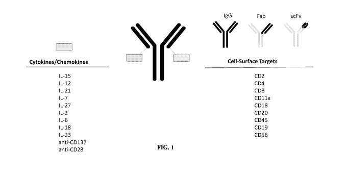
an IgG, a Fab, scFv), that binds a CD45 receptor on a cell, e.g., an immune
cell (e.g., an immune
effector cell, such as a lymphocyte). In embodiments, the composition, e.g.,
an IFM, associates,
e.g., links together, the cytokine molecule and the immune cell targeting
moiety to the immune
cell, e.g., the effector immune cell. In embodiments, the anti-CD45 antibody
binding to the cell
increases the association of the IL-15 molecule with the cell and improves one
or more of IL-15
signaling, immunostimulation, over time, e.g., relative to a free IL-15
molecule (an IL-15
molecule not found in the composition). In embodiments, the signaling and/or
11
CA 03069017 2020-01-03
WO 2019/010219 PCT/US2018/040777
immunostimulation occurs over a period of time, e.g., minutes, hours, days
e.g., by at least 8
hours, e.g., 24 hours.
In another aspect, the disclosure provides a particle, e.g., a nanoparticle,
that comprises
an IFM as described herein, e.g., nanoparticle that comprises a protein (e.g.,
a protein nanogel as
described herein). In one embodiment, the particle comprises the same IFM. In
other
embodiments, the particle comprises one or more different types of IFM.
Compositions, e.g., pharmaceutical compositions, comprising the IFMs and/or
the
particles disclosed herein, are also disclosed. In embodiments, the
pharmaceutical compositions
further include a pharmaceutically acceptable carrier, excipient, or
stabilizer.
In yet another aspect, the disclosure provides an isolated nucleic acid
molecule
comprising the nucleotide sequence encoding the IFM disclosed herein, or
comprising a
nucleotide sequences substantially identical thereto (e.g., at least 95%
identical thereto), as well
as a vector, e.g., an expression vector, and a host cell comprising the
nucleic acid molecule
disclosed herein.
Methods of making, e.g., producing, the IFM disclosed herein are also
disclosed. In
embodiments, the method includes culturing the host cell comprising the
nucleic acid molecules
disclosed herein, under suitable growth conditions.
In yet another aspect, the disclosure provides a method of treating a disorder
or condition
in a subject, e.g., a human. The method includes administering to the subject,
in need of
treatment, a composition, e.g., an IFM or a particle as described herein, in
an amount effective to
treat the disorder or condition. In some embodiments, the disorder is a
cancer, e.g., a solid
tumor or a hematological cancer. In other embodiments, the disorder is an
infection, e.g., a
viral, bacterial or yeast infection. In yet other embodiments, the disorder is
an autoimmune
disorder. The compositions disclosed herein can be used alone, or in
combination with, but not
limited to, cell therapy, and as a combination with chemotherapy or
radiotherapy. In some
embodiments, the cell therapy is chosen from an adoptive cell therapy, CAR-T
cell therapy,
engineered TCR T cell therapy, a tumor infiltrating lymphocyte therapy, an
antigen-trained T
cell therapy, or an enriched antigen-specific T cell therapy.
In a related aspect, the disclosure provides a composition, e.g., an IFM
and/or a particle
disclosed herein, for use in a medicament, e.g., for use in treating a
disorder or condition in a
subject, e.g., a mammal (e.g., a human). Alternatively, the use of a
composition, e.g., an IFM, in
the manufacture of a medicament for treating a disorder or condition in a
subject, e.g., a
mammal (e.g., a human) is disclosed. In some embodiments, the disorder is a
cancer, e.g., a
solid tumor or a hematological cancer. In other embodiments, the disorder is
an infection, e.g., a
viral (e.g., HIV), bacterial or yeast infection. In yet other embodiments, the
disorder is an
12
CA 03069017 2020-01-03
WO 2019/010219
PCT/US2018/040777
autoimmune disorder. The compositions disclosed herein can be used alone, or
in combination
with, but not limited to, cell therapy, and as a combination with chemotherapy
or radiotherapy.
In some embodiments, the cell therapy is chosen from an adoptive cell therapy,
CAR-T cell
therapy, engineered TCR T cell therapy, a tumor infiltrating lymphocyte
therapy, an antigen-
trained T cell therapy, or an enriched antigen-specific T cell therapy.
In another aspect, the disclosure provides a method of selectively delivering
a cytokine
molecule, e.g., an IL-15 molecule, to an immune cell in a subject, e.g., a
mammal (e.g., a
human). The method includes administering an IFM as described herein to the
subject, wherein
the cytokine molecule is selectively delivered to the immune cell.
In embodiments of the therapeutic and delivery methods disclosed herein, the
subject
herein is in need of a cell-based therapy, e.g., an immune cell therapy. For
example, the subject
is in need of a cell therapy chosen from an adoptive cell therapy, CAR-T cell
therapy,
engineered TCR T cell therapy, a tumor infiltrating lymphocyte therapy, an
antigen-trained T
cell therapy, or an enriched antigen-specific T cell therapy. In some
embodiments, the subject is
a patient, e.g., a human patient. In some embodiments, the subject has a
disease chosen from
cancer, diabetes, an autoimmune disease, allergies or allergic conditions,
asthma or a
cardiovascular disease. In an embodiment, the subject is in need of a
transplant.
The IFMs described herein can be administered directly to a subject suffering
from the
disorder to be treated (e.g., cancer) via e.g., intravenous or subcutaneous
administration. In
some embodiments, the immune cell targeting moiety of the IFM delivers the
cytokine molecule
to the surface of an immune cell, thereby increasing the concentration of the
cytokine molecule
at the surface of the immune cell. In embodiments, the IFM results in one or
more of: localizes
the distribution and/or enhances the cell surface availability of the cytokine
molecule, thereby
activating and/or stimulating the immune cell.
In other embodiments, the IFMs described herein can be administered in
combination
with an immune cell therapy in order to activate and/or stimulate the immune
cell therapy either
in vivo or in vitro. For example, an IFM described herein may be co-
administered with a cell
based therapy to a subject suffering from the disorder to be treated (e.g.,
cancer) via e.g.,
intravenous or subcutaneous administration. In other embodiments, a cell
therapy is pulsed in
vitro with an IFM described herein prior to administration. In some
embodiments, the cell
therapy is chosen from an adoptive cell therapy, CAR-T cell therapy,
engineered TCR T cell
therapy, a tumor infiltrating lymphocyte therapy, an antigen-trained T cell
therapy, or an
enriched antigen-specific T cell therapy.
13
CA 03069017 2020-01-03
WO 2019/010219 PCT/US2018/040777
Additional features and embodiments of any of the IFMs, compositions,
nanoparticles,
methods, uses, nucleic acids, vectors, and host cells, disclosed herein
include one or more of the
following.
In some embodiments, the IFM is a bifunctional or bispecific molecule, e.g.,
it has at
least two different kinds of members, e.g., with different functions and/or
binding specificities.
For example, the IFM comprises, or consists of, the immune stimulating moiety,
e.g., the
cytokine molecule, and the immune cell targeting moiety, wherein the immune
stimulating
moiety and the immune cell target moiety bind to two different cell surface
targets or receptors
in the same or different cells, e.g., one or more immune cells. In
embodiments, the immune
stimulating moiety and the immune cell target moiety bind to two different
targets on the same
immune cell, e.g., the same immune effector cell. A bifunctional or bispecific
molecule can
further comprise additional moieties, e.g., further binding and/or functional
moieties. For
example, the IFM can be a multifunctional or multispecific molecule, e.g., it
is a trifunctional or
trispecific, or a tetrafunctional or tetraspecific, fusion molecule.
In certain embodiments, the IFM can be represented with the following formula
in an N
to C terminal orientation: R1-(optionally L1)-R2 or R2-(optionally L1)-R1;
wherein R1
comprises an immune cell targeting moiety, Li comprises a linker (e.g., a
peptide linker
described herein), and R2 comprises an immune stimulating moiety, e.g., a
cytokine molecule.
In some embodiments, the immune stimulating moiety, e.g., the cytokine
molecule, is
functionally linked, e.g., covalently linked (e.g., by chemical coupling,
genetic or protein fusion,
noncovalent association or otherwise) to the immune cell targeting moiety. For
example, the
immune stimulating moiety can be covalently coupled indirectly, e.g., via a
linker to the immune
cell targeting moiety. In embodiments, the linker is chosen from: a cleavable
linker, a non-
cleavable linker, a peptide linker, a flexible linker, a rigid linker, a
helical linker, or a non-helical
linker. In some embodiments, the linker is a peptide linker. The peptide
linker can be 5-20, 8-
18, 10-15, or about 8, 9, 10, 11, 12, 13, 14, 15-20, 20-25, or 25-30 amino
acids long. In some
embodiments the peptide linker can be 30 amino acids or longer; e.g., 30-35,
35-40, 40-50 50-60
amino acids long. In some embodiments, the peptide linker comprises Gly and
Ser, e.g., a linker
comprising the amino acid sequence (Gly3-Ser)11 or (Gly4-Ser)., wherein n
indicates the number
of repeats of the motif, e.g., n=1, 2, 3, 4 or 5 (e.g., a (Gly3-Ser)2 or
(Gly4Ser)2, or a (Gly3-Ser)3 or
a (Gly4Ser)3 linker). In some embodiments, the linker comprises the amino acid
sequence of
SEQ ID NO: 36, 37, 38, or 39, or an amino acid sequence substantially
identical thereto (e.g.,
having 1, 2, 3, 4, or 5 amino acid substitutions). In one embodiment, the
linker comprises an
amino acid sequence GGGSGGGS (SEQ ID NO: 37). In another embodiment, the
linker
comprises amino acids derived from an antibody hinge region. In certain
embodiments the linker
14
CA 03069017 2020-01-03
WO 2019/010219 PCT/US2018/040777
comprises amino acids derived from the hinge regions of IgGl, IgG2, IgG3,
IgG4, IgGM, or
IgGA antibodies. In embodiments, the linker comprises amino acids derived from
an IgG hinge
region, e.g., an IgGl, IgG2 or IgG4 hinge region. For example, the linker
comprises a variant
amino acid sequence from an IgG hinge, e.g., a variant having one or more
cysteines replaced,
e.g., with serines. In some embodiments, the linker comprises DKTHTCPPSCAPE
(SEQ ID
NO: 126), having one or both cysteines replaced with another amino acid, e.g.,
a serine. In some
embodiments, the linker comprises amino acids DKTHTSPPSPAP (SEQ ID NO: 38),
EPKSSDKTHTSPPSPAPE (SEQ ID NO: 127), or a derivative thereof In embodiments,
the
linker comprises amino acids derived from an IgG2 hinge region, e.g., amino
acids SVESPPSP
(SEQ ID NO: 128), ERKSSVESPPSP (SEQ ID NO: 129), or a derivative thereof In
embodiments, the linker comprises amino acids derived from an IgG4 hinge
region, e.g., amino
acids PPSPSSP (SEQ ID NO: 130), ESKYGPPSPSSP (SEQ ID NO: 131), or a derivative
thereof
In other embodiments, the linker is a non-peptide, chemical linker. For
example, the
immune stimulating moiety is covalently coupled to the immune cell targeting
moiety by
crosslinking. Suitable crosslinkers include those that are heterobifunctional,
having two
distinctly reactive groups separated by an appropriate spacer (e.g., m-
maleimidobenzoyl-N-
hydroxysuccinimide ester) or homobifunctional (e.g., disuccinimidyl suberate).
In yet other
embodiments, the immune stimulating moiety is directly covalently coupled to
the immune cell
targeting moiety, without a linker. In yet other embodiments, the immune
stimulating moiety
and the immune cell targeting moiety of the IFM are not covalently linked,
e.g., are non-
covalently associated.
In other embodiments, the linker can be a protein or a fragment or derivative
thereof,
e.g., human albumin or an Fc domain, or a fragment or derivative thereof In
some
embodiments, the immune cell targeting moiety is linked to the N-terminus and
the immune
stimulating moiety is linked to the C-terminus.
In other embodiments, the linker non-covalently associates the immune cell
targeting
moiety to the immune stimulating moiety. For example, the linker comprises a
dimerization
domain, e.g., a coiled coil or a leucine zipper.
Fusion based on other noncovalent interactions can also be used, e.g., using
the high
affinity of the IL-15/sushi interaction to join two other proteins.
Immune cells
In embodiments, the immune cell is a nucleated cell, e.g., a nucleated cell as
described
herein below.
CA 03069017 2020-01-03
WO 2019/010219 PCT/US2018/040777
In some embodiments, the immune cell is a population of immune effector cells,
e.g., a
population of immune effector cells chosen from one or more of: T cells, e.g.,
CD4 T cells,
CD8 T cells, alpha T cells, beta T cells, gamma T cells, and delta T cells; B
cells; natural killer
(NK) cells; natural killer T (NKT) cells; or dendritic cells. In embodiments,
the immune cell,
e.g., the immune effector cell, displays a cell surface receptor that binds
the immune cell
targeting moiety.
In more particular embodiments, the immune cell, e.g., an immune effector
cell, (e.g., an
immune cell chosen from a lymphocyte, T cell, B cell, or a Natural Killer
cell), or a
hematopoietic stem cell). In embodiments, the immune cell comprises a
lymphocyte. In
embodiments, the immune cell comprises a T cell. In embodiments, the immune
cell comprises
a B cell. In embodiments, the immune cell comprises a Natural Killer (NK)
cell. In
embodiments, the immune cell comprises a hematopoietic stem cell. In some
embodiments, the
immune cell is an immune cell (e.g., T cell or NK cell) that comprises, e.g.,
expresses, a
Chimeric Antigen Receptor (CAR), e.g., a CAR that binds to a cancer antigen.
In other
embodiment, the immune cell expresses an exogenous high affinity Fc receptor.
In some
embodiments, the immune cell comprises, e.g., expresses an engineered T-cell
receptor. In some
embodiments, the immune cell is a tumor infiltrating lymphocyte. In some
embodiments, the
immune cell is a cytotoxic T cell (e.g., a CD8 T cell). In some embodiments,
the immune cell is
a regulatory T-cell ("Treg").
In embodiments, the immune cell is an immune cell, e.g., an NK cell, acquired
from a
patient, e.g., a patient's blood. In other embodiments, the immune cell is an
immune cell, e.g.,
an NK cell, acquired from a healthy donor. In some embodiments, the NK cell
population is
purified, e.g., depleted, of T cells, e.g., allogeneic T cells, before
infusion to a patient. In
embodiments, the immune cell is an immune cell, e.g., an NK cell, from an
embryonic stem cell
.. and/or an iPSC cell. In some embodiments, the immune cell is a cell line,
e.g., a stable or an
immortalized cell line (e.g., an NK cell immortalized cell line). In some
embodiments, the
immune cell is acquired from a patient, e.g., a patient with a hematological
cancer, e.g., a
leukemia or a lymphoma. In embodiments, the immune cell is an NK cell line,
e.g., an NK cell
line chosen from NK-92 (e.g., ATCC cat. no. CRL-2407), NK-YS, KHYG-1, NKL,
NKG, SNK-
6, IMC-1, e.g., as described in Klingemann, H. etal. (2016) Frontiers in
Immunology Vol.
7(Art. 91): 1-7, incorporated by reference herein. In one embodiment, the
immune cell is an
NK92 cell line, e.g., a variant NK92 cell that expresses a high affinity Fc
receptor, e.g., Fc
gamma RIIIa-expressing cell (e.g., 158V). In other embodiments, the NK92 cell
line comprises
a CAR that binds to a cancer antigen, e.g., also as described in in Klingemann
etal. supra.
16
CA 03069017 2020-01-03
WO 2019/010219 PCT/US2018/040777
Cytokine molecules
In some embodiments, the cytokine molecule of the IFM includes an
immunomodulatory
cytokine, e.g., a pro-inflammatory cytokine or an anti-inflammatory cytokine.
In some
embodiments, the cytokine is a member of the common y-chain (yc) family of
cytokines. In
some embodiments, the cytokine molecule comprises a cytokine chosen from one
or more of
interleukin-15 (IL-15), interleukin-1, e.g., interleukin-1 alpha (IL-1a) or
interleukin-1 beta (IL-
113), interleukin-2 (IL-2), interleukin-4 (IL-4), interleukin-5 (IL-5),
interleukin-6 (IL-6),
interleukin-7 (IL-7), interleukin-9 (IL-9), interleukin-10 (IL-10),
interleukin-12 (IL-12),
interleukin-13 (IL-13), interleukin-18 (IL-18), interleukin-21 (IL-21),
interleukin-23 (IL-23),
interleukin-27 (IL-27), interleukin-35 (IL-35), IFNy, TNFa, IFNa, IFNO, GM-
CSF, or GCSF,
including variant forms thereof (e.g., a cytokine derivative, a complex
comprising the cytokine
molecule with a polypeptide, e.g., a cytokine receptor complex, and other
agonist forms thereof).
In some embodiments, the cytokine molecule is a pro-inflammatory cytokine
molecule chosen
from an IL-1, IL-2, IL-6, IL-12, IL-15, IL-18, IL-21, IL-23, or IL-27 cytokine
molecule. In some
embodiments, the cytokine molecule is an anti-inflammatory cytokine molecule
chosen from an
IL-4, IL-10, IL-13, IL-35 cytokine molecule. In some embodiments, the cytokine
molecule is
chosen from IL-2, IL-6, IL-7, IL-12, IL-15, IL-21 or IL-27, including variant
forms thereof (e.g.,
a cytokine derivative, a complex comprising the cytokine molecule with a
polypeptide, e.g., a
cytokine receptor complex, and other agonist forms thereof, e.g., a non-
neutralizing anti-
cytokine antibody molecule). In some embodiments, the cytokine molecule is a
superagonist
(SA), e.g., as described herein. For example, the superagonist can have
increased cytokine
activity, e.g., by at least 10%, 20%, or 30%, compared to the naturally-
occurring cytokine. In
some embodiments, the cytokine molecule is a monomer or a dimer. In
embodiments, the
cytokine molecule further comprises a receptor or a fragment thereof, e.g., a
cytokine receptor
domain.
In one embodiment, the cytokine molecule comprises a wild type cytokine, e.g.,
a wild
type, e.g., human amino acid sequence. In other embodiments, the cytokine
molecule comprises
an amino acid sequence substantially identical to the wild-type cytokine
sequence, e.g., the
human cytokine sequence. In some embodiments, the cytokine molecule comprises
an amino
acid sequence at least 95% to 100% identical, or having at least 1, 2, 3, 4,
5, 6, 7, 8, 9, or 10 or
more amino acid alterations (e.g., substitutions, deletions, or insertions,
e.g., conservative
substitutions) relative to a wild-type cytokine sequence, e.g., a human
cytokine sequence. In
embodiments, the cytokine molecule comprises no more than five, ten or fifteen
alterations (e.g.,
17
CA 03069017 2020-01-03
WO 2019/010219 PCT/US2018/040777
substitutions, deletions, or insertions, e.g., conservative substitutions)
relative to the wild-type
cytokine sequence, e.g., the human cytokine sequence.
Exemplary cytokine amino acid sequences are disclosed herein, for example, the
amino
acid of IL-15 is provided as, e.g., SEQ ID NO:10 and SEQ ID NO:40; the amino
acid of IL-7 is
provided as, e.g., SEQ ID NO:42 and SEQ ID NO:43; the amino acid of IL-21 is
provided as,
e.g., SEQ ID NO:44 and SEQ ID NO:45; the amino acid of IL-12A is provided as,
e.g., SEQ ID
NO:46 and SEQ ID NO:47; the amino acid of IL-12B is provided as, e.g., SEQ ID
NO:48 and
SEQ ID NO:49; exemplary fusions of IL-12A and IL-12B are disclosed as e.g.,
SEQ ID NO:50
and SEQ ID NO:51. Any of the cytokine sequences disclosed herein and
substantially identical
sequences (e.g., at least 90%, 95% or higher sequence identity) can be used in
the IFM disclosed
herein.
In one embodiment, the cytokine molecule is an IL-15 molecule. In one
embodiment,
the IL-15 molecule (e.g., IL-15 polypeptide molecule) comprises a wild-type IL-
15 amino acid
sequence, e.g., a human IL-15 amino acid sequence, e.g., includes the amino
acid sequence of
SEQ ID NO: 10. In some embodiments, the cytokine molecule is an IL-15
molecule, e.g., a full
length, a fragment or a variant of IL-15, e.g., human IL-15, that retains IL-
15 activity. In other
embodiments, the IL-15 molecule is a variant of human IL-5, e.g., having one
or more amino
acid alterations, e.g., substitutions, to the human IL-15 amino acid sequence.
In some
embodiments, the IL-15 variant comprises, or consists of, a mutation at
position 45, 51, 52, or
72, e.g., as described in US 2016/0184399. In some embodiments, the IL-15
variant comprises,
or consists of, an N, S or L to one of D, E, A Y or P substitution. In some
embodiments, the
mutation is chosen from L45D, L45E, S51D, L52D, N72D, N72E, N72A, N725, N72Y,
or
N72P (in reference to the sequence of human IL-15, SEQ ID NO: 11).
In embodiments, the IL-15 molecule comprises an IL-15 variant, e.g., a human
IL-15
polypeptide having one or more amino acid substitutions. In some embodiments,
the IL-15
molecule comprises a substitution at position 72, e.g., an N to D
substitution. In one
embodiment, the IL-15 molecule is an IL-15N72D polypeptide of SEQ ID NO: 11 or
an amino
acid sequence having at least 80%, 85%, 90%, 95%, 96%, 97%, 98%, or 99%
identity thereto,
which has IL-15Ra binding activity.
In other embodiments, the IL-15 molecule comprises a complex of IL-15 (e.g.,
an IL-15
wild type or variant sequence) and an IL-15 binding fragment of an IL-15
receptor, e.g., IL-15
receptor alpha or an IL-15 binding fragment thereof In some embodiments, the
IL-15 molecule
comprises a complex of the IL-15 and an IL-15 receptor fragment, e.g., an
extracellular domain
of an IL-15 receptor or a fragment thereof In some embodiments, the IL-15
molecule and the
18
CA 03069017 2020-01-03
WO 2019/010219 PCT/US2018/040777
IL-15 binding fragment of the IL-15 receptor are not covalently linked, e.g.,
are non-covalently
associated.
In one embodiment, the IL-15 receptor fragment comprises, or consists of, the
amino
acid sequence of SEQ ID NO: 9 or SEQ ID NO: 52, or a fragment thereof, or an
amino acid
sequence substantially identical thereto (e.g., at least 95% identical
thereto, or having at least
one, two, three, four, five amino acid alterations, but not more than ten,
fifteen, twenty
alterations (e.g., substitutions, deletions, or insertions, e.g., conservative
substitutions) to the
amino acid sequence of SEQ ID NO: 9 or SEQ ID NO: 52, or a fragment thereof,
and having IL-
binding activity.
10 In some embodiments, the IL-15 molecule comprises a complex of the IL-15
and an IL-
15 receptor fragment, e.g., a sushi domain (e.g., as described herein). In
some embodiments, the
sushi domain is from human or a non-human animal, e.g., mammal, e.g., non-
human primate. In
one embodiment, the sushi domain includes a human sequence, or a substantially
identical
thereto (e.g., at least 95% identical thereto). In embodiments, the
extracellular domain of IL-15
15 Receptor alpha comprises a domain referred to as herein as "the sushi
domain," which binds IL-
15. In embodiments, the sushi domain is provided as a 62-amino acid referred
to herein as a
"minimal domain" (e.g., having the amino acid sequence of SEQ ID NO: 52), or a
65-amino
acid extended domain (e.g., having the amino acid sequence of SEQ ID NO: 9),
or comprises a
77-amino acid domain comprising, e.g., consisting of, amino acids 31-107 or
SEQ ID NO: 63.
In some embodiments, the IL-15 receptor fragment has a wild type sequence. In
other
embodiments, the IL-15 receptor fragment has a mutated sequence, e.g., a
substitution at
position 77, e.g., an L77I substitution (with the numbering referring to the
wild-type IL-15Ra of
SEQ ID NO: 4). In some embodiments, the sushi domain comprises the amino acid
sequence of
SEQ ID NO: 9, SEQ ID NO: 52, SEQ ID NO: 65, or SEQ ID NO: 66, or an amino acid
sequence substantially identical thereto (e.g., at least 95% identical
thereto, or having at least
one, two, three, four, five amino acid alterations, but not more than ten or
fifteen alterations
(e.g., substitutions, deletions, or insertions, e.g., conservative
substitutions) to the amino acid
sequence of SEQ ID NO: 9, SEQ ID NO: 52, SEQ ID NO: 65, or SEQ ID NO: 66, and
having
IL-15 binding activity.
In some embodiments, an IL-15 receptor alpha extracellular domain consists of
62-171
amino acids of SEQ ID NO: 63 or an amino acid sequence substantially identical
thereto (e.g., at
least 95% identical thereto 96%, 97%, 98%, or 99% identity thereto or having
up to 1, 2, 3, 4, 5,
6, 7, 8, 9, 10, or 15 alterations (e.g., substitutions) relative thereto, and
having IL-15 binding
activity). In some embodiments, an IL-15 receptor alpha extracellular domain
consists of 65-
171 amino acids of SEQ ID NO: 63 or an amino acid sequence substantially
identical thereto
19
CA 03069017 2020-01-03
WO 2019/010219 PCT/US2018/040777
(e.g., at least 95% identical thereto 96%, 97%, 98%, or 99% identity thereto
or having up to 1, 2,
3, 4, 5, 6, 7, 8, 9, 10, or 15 alterations (e.g., substitutions) relative
thereto, and having IL-15
binding activity). In some embodiments, an IL-15 receptor alpha extracellular
domain consists
of up to 171 amino acids of SEQ ID NO: 63 or an amino acid sequence
substantially identical
thereto (e.g., at least 95% identical thereto 96%, 97%, 98%, or 99% identity
thereto or having up
to 1, 2, 3, 4, 5, 6, 7, 8, 9, 10, or 15 alterations (e.g., substitutions)
relative thereto, and having IL-
binding activity). In some embodiments, an IL-15 receptor alpha extracellular
domain
consists of 62-171, 62-160, 62-150, 62-140, 62-130, 62-120, 62-110, 62-100, 62-
90, 62-80, 62-
70, 65-171, 65-160, 65-150, 65-140, 65-130, 65-120, 65-110, 65-100, 65-90, 65-
80, 65-70, or
10 65-77 amino acids of SEQ ID NO: 63 or a sequence having at least 95%
identity thereto 96%,
97%, 98%, or 99% or having up to 1, 2, 3, 4, 5, 6, 7, 8, 9, 10, or 15
alterations (e.g.,
substitutions) relative thereto, and having IL-15 binding activity. In some
embodiments, an IL-
15 receptor alpha extracellular domain consists of 62-171, 62-160, 62-150, 62-
140, 62-130, 62-
120, 62-110, 62-100, 62-90, 62-80, 62-70, 65-171, 65-160, 65-150, 65-140, 65-
130, 65-120, 65-
15 110, 65-100, 65-90, 65-80, 65-70, or 65-77 amino acids of SEQ ID NO: 63.
In some embodiments, the IFM containing an IL-15 molecule can further comprise
a
polypeptide, e.g., a cytokine receptor, e.g., a cytokine receptor domain, and
a second,
heterologous domain. In one embodiment, the heterologous domain is an
immunoglobulin Fc
region. In other embodiments, the heterologous domain is an antibody molecule,
e.g., a Fab
fragment, a FAB2fragment, a scFv fragment, or an affibody fragment or
derivative, e.g. a sdAb
(nanobody) fragment, a heavy chain antibody fragment. In some embodiments, the
polypeptide
also comprises a third heterologous domain. In some embodiments, the cytokine
receptor
domain is N-terminal of the second domain, and in other embodiments, the
cytokine receptor
domain is C-terminal of the second domain.
In some embodiments, the cytokine molecule comprises a complex of a cytokine
and an
anti-cytokine antibody molecule. For example, the cytokine can be IL-2 and the
anti-cytokine
antibody can be a non-neutralizing anti-IL-2 antibody molecule (see e.g.,
Spangler, J.B., et al.
(2015) www.dx.doi.org/10.1016/j.immuni.2015.04.015; Letourneau, S. et al.
(2010) PNAS Vol
107(5): 2171-2176; Boyman, 0. et al. (2006) Science 311, 1924); and Spangler,
J.B. (2015)
Annu Rev Immunol 33:139-167, the contents of which are entirely incorporated
by reference).
Immune cell targeting moieties
In some embodiments, the immune cell targeting moiety of the IFM includes an
antibody
molecule or a ligand that selectively binds to an immune cell surface target,
e.g., an immune cell
surface receptor. In some embodiments, the immune cell surface target or
receptor can have
CA 03069017 2020-01-03
WO 2019/010219
PCT/US2018/040777
one, two, three or more of the following properties: (i) is abundantly present
on the surface of
an immune cell (e.g., outnumbers the number of receptors for the cytokine
molecule present on
the immune cell surface); (ii) shows a slow downregulation, internalization,
and/or cell surface
turnover, e.g., relative to the receptors activated by the cytokine of the
IFM; (iii) is present on
the surface of the immune cell for a prolonged period of time, e.g., relative
to the receptors
activated by the cytokine of the IFM; or (iv) once internalized is
substantially recycled back to
the cell surface, e.g., at least 25%, 50%, 60%, 70%, 80%, 90% or more of the
immune cell
surface target is recycled back to the cell surface.
In some embodiments, the immune cell targeting moiety of the IFM binds to a
recycling
cell surface receptor. Without being bound by theory, it is believed that
binding to the recycling
cell surface receptor mediates internalization of the receptor and the IFM.
For example, the IFM
internalized along with the receptor may be sequestered into early endosomes
and subsequently
recycled back to the cell surface, instead of advancing to subsequent
degradation (e.g. via either
clathrin-mediated and clathrin-independent endocytosis). The return of the
IFM/receptor to the
cell surface can improve cytokine signaling by restoring the cytokine molecule
of the IFM to the
cell surface, thus increasing the time and availability of the cytokine
molecule to bind its own
cell-surface receptor. Additionally, signaling events that are initiated at
the surface membrane
by binding of a fusion protein of the disclosure may continue from endosomal
compartments.
In some embodiments, the immune cell surface target or receptor is present on
the
surface of an immune cell, but not present on a cancer or tumor cell, e.g., a
solid tumor or
hematological cancer cell. In some embodiments, the immune cell surface target
or receptor is
predominantly present on the surface of an immune cell compared to its
presence on a cancer or
tumor cell, e.g., is present at least 5:1, 10:1, 15:1, 20:1 higher ratio on
the immune cell relative
to the cancer or tumor cell.
In some embodiments, the immune cell targeting moiety of the IFM binds to a
receptor
expressed on a cell (e.g., an immune cell), e.g. the surface membrane of the
cell, and further the
cell also expresses a cytokine receptor (e.g., a receptor to the cytokine
molecule of the IFM).
In some embodiments, the immune cell targeting moiety of the IFM can be chosen
from
an antibody molecule or a ligand molecule that binds to an immune cell surface
target, e.g., a
target chosen from CD16, CD45, CD4, CD8, CD3, CD11 a, CD11b, CD11c, CD18,
CD25,
CD127, CD56, CD19, CD20, CD22, HLA-DR, CD197, CD38, CD27, CD137, 0X40, GITR,
CD56, CD196, CXCR3, CXCR4, CXCR5, CD84, CD229, CCR1, CCR5, CCR4, CCR6, CCR8,
or CCR10. In some embodiments, the immune cell targeting moiety binds to CD4,
CD8,
CD11a, CD18, CD20, CD56, or CD45. In other embodiments, the immune cell
surface target is
chosen from CD19, CD20, or CD22. In one embodiment, the immune cell targeting
moiety
21
CA 03069017 2020-01-03
WO 2019/010219 PCT/US2018/040777
comprises an antibody molecule or a ligand molecule that binds to CD45 (also
interchangeably
referred to herein as "CD45 receptor" or "CD45R"). In some embodiments, the
target is CD45
(e.g., a CD45 isoform chosen from CD45RA, CD45RB, CD45RC or CD45R0). In
embodiments, CD45 is primarily expressed on T cells. For example, CD45RA is
primarily
expressed on naive T cells; CD45R0 is primarily expressed on activated and
memory T cells.
In some embodiments, the immune cell targeting moiety of the IFM (e.g., an
antibody
molecule) binds a checkpoint inhibitor such as PD-1, PD-L1, LAG-3, TIM-3, or
CTLA-4. In
embodiments, the checkpoint inhibitor is present on an immune effector cell,
e.g., a T cell or NK
cell.
In other embodiments, the immune cell targeting moiety of the IFM comprises an
antibody
molecule (e.g., an antigen binding domain), a receptor molecule (e.g., a
receptor, a receptor
fragment or functional variant thereof), or a ligand molecule (e.g., a ligand,
a ligand fragment or
functional variant thereof), or a combination thereof, that binds to the
immune cell target or
receptor.
In some embodiments, the antibody molecule of the immune cell targeting moiety
of the
IFM comprises a full antibody (e.g., an antibody that includes at least one,
and preferably two,
complete heavy chains, and at least one, and preferably two, complete light
chains), or an
antigen-binding fragment (e.g., a Fab, F(ab')2, Fv, a single chain Fv, a
single domain antibody, a
diabody (dAb), a bivalent antibody, or bispecific antibody or fragment
thereof, a single domain
variant thereof, or a camelid antibody)) that binds to the immune cell target
or receptor.
The heavy chain constant region of the antibody molecule can be chosen from
IgGl, IgG2,
IgG3, or IgG4, or a fragment thereof, and more typically, IgGl, IgG2 or IgG4.
In some
embodiments, the Fc region of the heavy chain can include one or more
alterations, e.g.,
substitutions, to increase or decrease one or more of: Fc receptor binding,
neonatal-Fc receptor
binding, antibody glycosylation, the number of cysteine residues, effector
cell function,
complement function, or stabilize antibody formation (e.g., stabilize IgG4).
For example, the
heavy chain constant region for an IgG4, e.g., a human IgG4, can include a
substitution at
position 228 (e.g., a Ser to Pro substitution) (see e.g., Angal, S, King, DJ,
etal. (1993)Mol
Immunol 30:105-108 (initially described as S241P using a different numbering
system); Owens,
R, Ball, E, etal. (1997) Immunotechnology 3:107-116).
The light chain constant region of the antibody molecule can be chosen from
the light
chain constant regions of kappa or lambda, or a fragment thereof
The antibody molecule of the immune cell targeting moiety of the IFM can bind
to the
target antigen with a dissociation constant of less than about 100 nM, 50nM,
25nM, 10 nM, e.g.,
less than 1 nM (e.g., about 10 ¨ 100 pM). In embodiments, the antibody
molecule binds to a
22
CA 03069017 2020-01-03
WO 2019/010219 PCT/US2018/040777
conformational or a linear epitope on the antigen. In certain embodiments, the
antigen bound by
the antibody molecule of the immune cell targeting moiety is stably expressed
on the surface of
the immune cell. In embodiments, the antigen is a cell surface receptor that
is more abundant on
the cell surface relative to a receptor for the cytokine molecule of the IFM
on the cell surface.
In some embodiments, the immune cell targeting moiety is chosen from an
antibody
molecule (e.g., a full antibody (e.g., an antibody that includes at least one,
and preferably two,
complete heavy chains, and at least one, and preferably two, complete light
chains), or an
antigen-binding fragment (e.g., a Fab, F(ab')2, Fv, a single chain Fv, a
single domain antibody, a
diabody (dAb), a bivalent antibody, or bispecific or multispecific antibody or
fragment thereof, a
.. single domain variant thereof, or a camelid antibody)), or a non-antibody
scaffold, or a ligand
that binds to an immune cell surface target or ligand. In some embodiments,
the immune cell
targeting moiety is an antibody molecule or a ligand that binds to CD45, CD2,
CD4, CD8, CD11
(e.g., CD11 a (integrin alpha-L), CD11b, or CD11c), CD18 (integrin beta-2),
CD19, CD20, or
CD25, or a combination thereof, e.g., a bispecific antibody that binds to two
or more of the
aforesaid targets.
In some embodiments, the antibody molecule (e.g., mono- or bi-specific
antibodies) binds
to one or more of CD45, CD8, CD18 or CD11a, e.g., it is an IgG, e.g., human
IgG4, or an
antigen binding domain, e.g., a Fab, a F(ab')2, Fv, a single chain Fv, that
binds to CD45, CD8,
CD18 or CD11a. In some embodiments, the antibody molecule is a human, a
humanized or a
chimeric antibody. In embodiments, the antibody molecule is a recombinant
antibody.
In some embodiments, the anti-CD45 antibody is a human anti-CD45 antibody, a
humanized anti-CD45 antibody, or a chimeric anti-CD45 antibody. In some
embodiments, the
anti-CD45 antibody is an anti-CD45 monoclonal antibody. Exemplary anti-CD45
antibodies
include antibodies BC8, 4B2, GAP 8.3 or 9.4. Antibodies against other immune
cell surface
targets are also disclosed, e.g., anti-CD8 antibodies, such as OKT8 monoclonal
antibodies, anti-
CD18 antibodies, such as 1B4 monoclonal antibodies, and anti-CD11 a
antibodies, such as
MHM24 antibodies.
Also encompassed by the present disclosure are antibody molecules having the
amino acid
sequences disclosed herein, or an amino acid sequence substantially identical
thereof), nucleic
acid molecules encoding the same, host cells and vectors comprising the
nucleic acid molecules.
In one embodiment, the antibody molecule that binds to CD45 is specific to one
CD45
isoform or binds to more than on CD45 isoforms, e.g., is a pan-CD45 antibody.
In some
embodiments, the anti-CD45 antibody molecule binds to CD45RA and CD45RO. In
one
embodiment, the anti-CD45 antibody molecule is a BC8 antibody. In some
embodiments, the
BC8 antibody binds to CD45RA and CD45RO. In other embodiments, the anti-CD45
antibody
23
CA 03069017 2020-01-03
WO 2019/010219 PCT/US2018/040777
molecule is CD45RO-specific or is a pan-CD45 antibody molecule, e.g., it binds
to activated
and memory T cells. Additional examples of anti-CD45 antibody molecules
includes, but is not
limited to, GAP8.3, 4B2, and 9.4.
In one embodiment, the anti-CD45 antibody molecule is a BC8 antibody, e.g., a
chimeric
or humanized BC8 antibody. In some embodiments, the chimeric BC8 antibody
comprises:
(i) the light chain variable amino acid sequence (optionally, further
including a kappa light
chain sequence) corresponding to the antibody portion of the amino acid
sequence shown in
SEQ ID NO:1, 2, 3, 4, 7, 21, or 22, or an amino acid sequence substantially
identical thereof
(e.g., an amino acid sequence at least 85%, 90%, 95% or higher identical to
the antibody portion
of SEQ ID NO: 1,2, 3,4, 7,21, or 22); and/or
(ii) the heavy chain variable amino acid sequence (optionally, further
including a human
IgG1 heavy chain sequence or a human IgG4 sequence having an 5228P
substitution) of the
amino acid sequence shown in SEQ ID NO:5, 6, or 8, respectively, or an amino
acid sequence
substantially identical thereto (e.g., an amino acid sequence at least 85%,
90%, 95% or higher
identical to SEQ ID NOs: 5, 6, or 8, respectively). In embodiments, the BC8
antibody
comprises one, two, or all three CDR1, CDR2 or CDR3 of the light chain
variable region, and/or
the heavy chain variable region, of the BC8 antibody, e.g., according to the
Kabat definition, or
a closely related CDR, e.g., CDRs which have at least one amino acid
alteration, but not more
than two, three or four alterations (e.g., substitutions, deletions, or
insertions, e.g., conservative
substitutions) from the CDR sequence of SEQ ID NO: 1-4, 7, 21, or 22, or 5-6,
or 8.
In other embodiments, the amino acid of SEQ ID NO:1-4, or an amino acid
substantially
identical thereto (e.g., an amino acid sequence at least 85%, 90%, 95% or
higher identical to
SEQ ID NO: 1-4) (optionally, further including a kappa light chain sequence),
includes,
optionally via a linker, an IL-15 cytokine or receptor, e.g., a sushi domain
as described herein
(e.g., SEQ ID NO: 9 or an amino acid substantially identical thereto (e.g., an
amino acid
sequence at least 85%, 90%, 95% or higher identical to SEQ ID NO: 9). In one
embodiment,
the IL-15 cytokine or receptor is coupled to the N-terminus of the light chain
of the BC8
antibody of SEQ ID NO:1-4. In other embodiments, the IL-15 cytokine or
receptor is coupled to
the C-terminus of the light chain of the BC8 antibody of SEQ ID NO:1-4. In one
embodiment,
the linker comprises, or consists of, SEQ ID NO: 37 or 38.
In other embodiments, the amino acid of SEQ ID NO:21 or 22, or an amino acid
substantially identical thereto (e.g., an amino acid sequence at least 85%,
90%, 95% or higher
identical to SEQ ID NO: 21 or 22) (optionally, further including a kappa light
chain sequence),
includes, optionally via a linker, an IL-15 cytokine or receptor, e.g., a
sushi domain as described
herein (e.g., SEQ ID NO: 9 or an amino acid substantially identical thereto
(e.g., an amino acid
24
CA 03069017 2020-01-03
WO 2019/010219 PCT/US2018/040777
sequence at least 85%, 90%, 95% or higher identical to SEQ ID NO: 9). In one
embodiment,
the IL-15 cytokine or receptor is coupled to the N-terminus of the light chain
of the Bc8
antibody of SEQ ID NO:21 or 22 via a linker of SEQ ID NO: 37 or 38. In other
embodiments,
the IL-15 cytokine or receptor is coupled to the C-terminus of the light chain
of the Bc8
antibody of SEQ ID NO:21 or 22 via a linker of SEQ ID NO: 37 or 38.
In some embodiments, the amino acid of SEQ ID NO: 6 or 8, or an amino acid
substantially identical thereto (e.g., an amino acid sequence at least 85%,
90%, 95% or higher
identical to SEQ ID NO: 6 or 8), includes a heavy chain constant region. In
some embodiments,
the amino acid of SEQ ID NO 6 or 8, or an amino acid substantially identical
thereto (e.g., an
amino acid sequence at least 85%, 90%, 95% or higher identical to SEQ ID NO: 6
or 8),
includes an IL-15 cytokine or receptor, e.g., a sushi domain as described
herein (e.g., SEQ ID
NO: 9 or an amino acid substantially identical thereto (e.g., an amino acid
sequence at least
85%, 90%, 95% or higher identical to SEQ ID NO: 9). In one embodiment, the IL-
15 cytokine
or receptor is coupled to the N-terminus of the light chain of the Bc8
antibody of SEQ ID NO:6
or 8. In other embodiments, the IL-15 cytokine or receptor is coupled to the C-
terminus of the
light chain of the Bc8 antibody of SEQ ID NO:6 or 8.
In other embodiments, the humanized BC8 antibody comprises:
(i) the light chain variable amino acid sequence (optionally, further
including a kappa light
chain sequence) corresponding to the antibody portion of the amino acid
sequence shown in
SEQ ID NO:19, or an amino acid sequence substantially identical thereof (e.g.,
an amino acid
sequence at least 85%, 90%, 95% or higher identical to the antibody portion of
SEQ ID NO: 19);
and/or
(ii) the heavy chain variable amino acid sequence (optionally, further
including a human
IgG1 heavy chain sequence) of the amino acid sequence shown in SEQ ID NO:18,
or an amino
acid sequence substantially identical thereto (e.g., an amino acid sequence at
least 85%, 90%,
95% or higher identical to SEQ ID NO:18). In embodiments, the BC8 antibody
comprises one,
two, or all three CDR1, CDR2 or CDR3 of the light chain variable region,
and/or the heavy
chain variable region, of the BC8 antibody, e.g., according to the Kabat
definition, or a closely
related CDR, e.g., CDRs which have at least one amino acid alteration, but not
more than two,
three or four alterations (e.g., substitutions, deletions, or insertions,
e.g., conservative
substitutions) from the CDR sequence of SEQ ID NO: 18 or 19.
In some embodiments, the amino acid sequence of SEQ ID NO:19, or an amino acid
sequence substantially identical thereof (e.g., an amino acid sequence at
least 85%, 90%, 95% or
higher identical to SEQ ID NO: 19) includes an IL-15 cytokine molecule, e.g.,
an IL-15
cytokine, coupled to the N- or C-terminus of SEQ ID NO: 19, optionally, via a
linker (e.g., a
CA 03069017 2020-01-03
WO 2019/010219 PCT/US2018/040777
linker comprising the amino acid sequence of SEQ ID NO: 36-39, or an amino
acid sequence
substantially identical thereof (e.g., an amino acid sequence at least 85%,
90%, 95% or higher
identical to SEQ ID NO: 36-39).
Alternatively or in combination, the amino acid sequence of SEQ ID NO:18, or
an amino
acid sequence substantially identical thereof (e.g., an amino acid sequence at
least 85%, 90%,
95% or higher identical to SEQ ID NO: 18) includes an IL-15 cytokine molecule,
e.g., an IL-15
cytokine, coupled to the N- or C-terminus of SEQ ID NO: 18, optionally, via a
linker (e.g., a
linker comprising the amino acid sequence of SEQ ID NO: 36-39, or an amino
acid sequence
substantially identical thereof (e.g., an amino acid sequence at least 85%,
90%, 95% or higher
identical to SEQ ID NO: 36-39).
In other embodiments, the antibody molecule that binds to CD45 is a 9.4
antibody, e.g., a
chimeric or humanized 9.4 antibody. In some embodiments, the chimeric 9.4
antibody
comprises:
(i) the light chain variable amino acid sequence (optionally, further
including a kappa light
chain sequence) corresponding to the antibody portion of the amino acid
sequence shown in
SEQ ID NO:15, or an amino acid sequence substantially identical thereto (e.g.,
an amino acid
sequence at least 85%, 90%, 95% or higher identical to the antibody portion of
SEQ ID NO:15);
and/or
(ii) the heavy chain variable amino acid sequence (optionally, further
including a human
IgG1 heavy chain sequence) of the amino acid sequence shown in SEQ ID NO:14,
respectively,
or an amino acid sequence substantially identical thereto (e.g., an amino acid
sequence at least
85%, 90%, 95% or higher identical to SEQ ID NO: 14, respectively). In
embodiments, the 9.4
antibody comprises one, two, or all three CDR1, CDR2 or CDR3 of the light
chain variable
region, and/or the heavy chain variable region, of the 9.4 antibody, e.g.,
according to the Kabat
definition, or a closely related CDR, e.g., CDRs which have at least one amino
acid alteration,
but not more than two, three or four alterations (e.g., substitutions,
deletions, or insertions, e.g.,
conservative substitutions) from the CDR sequence of SEQ ID NO:14 or 15.
In some embodiments, the amino acid sequence of SEQ ID NO:15, or an amino acid
sequence substantially identical thereof (e.g., an amino acid sequence at
least 85%, 90%, 95% or
higher identical to SEQ ID NO: 15) includes an IL-15 cytokine molecule, e.g.,
an IL-15
cytokine, coupled to the N- or C-terminus of SEQ ID NO: 15, optionally, via a
linker (e.g., a
linker comprising the amino acid sequence of SEQ ID NO: 36-39, or an amino
acid sequence
substantially identical thereof (e.g., an amino acid sequence at least 85%,
90%, 95% or higher
identical to SEQ ID NO: 36-39).
26
CA 03069017 2020-01-03
WO 2019/010219 PCT/US2018/040777
Alternatively or in combination, the amino acid sequence of SEQ ID NO:14, or
an amino
acid sequence substantially identical thereof (e.g., an amino acid sequence at
least 85%, 90%,
95% or higher identical to SEQ ID NO: 14) includes an IL-15 cytokine molecule,
e.g., an IL-15
cytokine, coupled to the N- or C-terminus of SEQ ID NO: 14, optionally, via a
linker (e.g., a
.. linker comprising the amino acid sequence of SEQ ID NO: 36-39, or an amino
acid sequence
substantially identical thereof (e.g., an amino acid sequence at least 85%,
90%, 95% or higher
identical to SEQ ID NO: 36-39).
In other embodiments, the antibody molecule that binds to CD45 is a 4B2
antibody, e.g., a
chimeric or humanized 4B2 antibody. In some embodiments, the chimeric 4B2
antibody
comprises:
(i) the light chain variable amino acid sequence (optionally, further
including a kappa light
chain sequence) corresponding to the antibody portion of the amino acid
sequence shown in
SEQ ID NO:17, or an amino acid sequence substantially identical thereto (e.g.,
an amino acid
sequence at least 85%, 90%, 95% or higher identical to the antibody portion of
SEQ ID NO:17);
and/or
(ii) the heavy chain variable amino acid sequence (optionally, further
including a human
IgG1 heavy chain sequence) of the amino acid sequence shown in SEQ ID NO:16,
respectively,
or an amino acid sequence substantially identical thereto (e.g., an amino acid
sequence at least
85%, 90%, 95% or higher identical to SEQ ID NO: 16, respectively). In
embodiments, the 4B2
antibody comprises one, two, or all three CDR1, CDR2 or CDR3 of the light
chain variable
region, and/or the heavy chain variable region, of the 4B2 antibody, e.g.,
according to the Kabat
definition, or a closely related CDR, e.g., CDRs which have at least one amino
acid alteration,
but not more than two, three or four alterations (e.g., substitutions,
deletions, or insertions, e.g.,
conservative substitutions) from the CDR sequence of SEQ ID NO:16 or 17.
In some embodiments, the amino acid sequence of SEQ ID NO:17, or an amino acid
sequence substantially identical thereof (e.g., an amino acid sequence at
least 85%, 90%, 95% or
higher identical to SEQ ID NO: 17) includes an IL-15 cytokine molecule, e.g.,
an IL-15
cytokine, coupled to the N- or C-terminus of SEQ ID NO: 17, optionally, via a
linker (e.g., a
linker comprising the amino acid sequence of SEQ ID NO: 36-39, or an amino
acid sequence
substantially identical thereof (e.g., an amino acid sequence at least 85%,
90%, 95% or higher
identical to SEQ ID NO: 36-39).
Alternatively or in combination, the amino acid sequence of SEQ ID NO:16, or
an amino
acid sequence substantially identical thereof (e.g., an amino acid sequence at
least 85%, 90%,
95% or higher identical to SEQ ID NO: 16) includes an IL-15 cytokine molecule,
e.g., an IL-15
cytokine, coupled to the N- or C-terminus of SEQ ID NO: 16, optionally, via a
linker (e.g., a
27
CA 03069017 2020-01-03
WO 2019/010219 PCT/US2018/040777
linker comprising the amino acid sequence of SEQ ID NO: 36-39, or an amino
acid sequence
substantially identical thereof (e.g., an amino acid sequence at least 85%,
90%, 95% or higher
identical to SEQ ID NO: 36-39).
In another embodiment, the antibody molecule that binds to CD8 is an OKT8
antibody,
e.g., a chimeric or humanized OKT8 antibody. In some embodiments, the chimeric
OKT8
antibody comprises:
(i) the light chain variable amino acid sequence (optionally, further
including a kappa light
chain sequence) corresponding to the antibody portion of the amino acid
sequence shown in
SEQ ID NO:31, or an amino acid sequence substantially identical thereof (e.g.,
an amino acid
sequence at least 85%, 90%, 95% or higher identical to the antibody portion of
SEQ ID NO:31);
and/or
(ii) the heavy chain variable amino acid sequence (optionally, further
including a human
IgG1 heavy chain sequence) of the amino acid sequence shown in SEQ ID NO:30,
or an amino
acid sequence substantially identical thereto (e.g., an amino acid sequence at
least 85%, 90%,
95% or higher identical to SEQ ID NO:30). In embodiments, the OKT8 antibody
comprises
one, two, or all three CDR1, CDR2 or CDR3 of the light chain variable region,
and/or the heavy
chain variable region, of the OKT8 antibody, e.g., according to the Kabat
definition, or a closely
related CDR, e.g., CDRs which have at least one amino acid alteration, but not
more than two,
three or four alterations (e.g., substitutions, deletions, or insertions,
e.g., conservative
substitutions) from the CDR sequence of SEQ ID NO: 30 or 31.
In some embodiments, the amino acid sequence of SEQ ID NO:31, or an amino acid
sequence substantially identical thereof (e.g., an amino acid sequence at
least 85%, 90%, 95% or
higher identical to SEQ ID NO: 31) includes an IL-15 cytokine molecule, e.g.,
an IL-15
cytokine, coupled to the N- or C-terminus of SEQ ID NO: 31, optionally, via a
linker (e.g., a
linker comprising the amino acid sequence of SEQ ID NO: 36-39, or an amino
acid sequence
substantially identical thereof (e.g., an amino acid sequence at least 85%,
90%, 95% or higher
identical to SEQ ID NO: 36-39).
Alternatively or in combination, the amino acid sequence of SEQ ID NO:30, or
an amino
acid sequence substantially identical thereof (e.g., an amino acid sequence at
least 85%, 90%,
95% or higher identical to SEQ ID NO: 30) includes an IL-15 cytokine molecule,
e.g., an IL-15
cytokine, coupled to the N- or C-terminus of SEQ ID NO: 30, optionally, via a
linker (e.g., a
linker comprising the amino acid sequence of SEQ ID NO: 36-39, or an amino
acid sequence
substantially identical thereof (e.g., an amino acid sequence at least 85%,
90%, 95% or higher
identical to SEQ ID NO: 36-39).
28
CA 03069017 2020-01-03
WO 2019/010219 PCT/US2018/040777
In another embodiment, the antibody molecule that binds to CD18 is a 1B4
antibody, e.g.,
a chimeric or humanized 1B4 antibody. In some embodiments, the chimeric 1B4
antibody
comprises:
(i) the light chain variable amino acid sequence (optionally, further
including a kappa light
chain sequence) corresponding to the antibody portion of the amino acid
sequence shown in
SEQ ID NO:29, or an amino acid sequence substantially identical thereof (e.g.,
an amino acid
sequence at least 85%, 90%, 95% or higher identical to the antibody portion of
SEQ ID NO:31);
and/or
(ii) the heavy chain variable amino acid sequence (optionally, further
including a human
IgG1 heavy chain sequence) of the amino acid sequence shown in SEQ ID NO:28,
or an amino
acid sequence substantially identical thereto (e.g., an amino acid sequence at
least 85%, 90%,
95% or higher identical to SEQ ID NO:28). In embodiments, the 1B4 antibody
comprises one,
two, or all three CDR1, CDR2 or CDR3 of the light chain variable region,
and/or the heavy
chain variable region, of the 1B4 antibody, e.g., according to the Kabat
definition, or a closely
related CDR, e.g., CDRs which have at least one amino acid alteration, but not
more than two,
three or four alterations (e.g., substitutions, deletions, or insertions,
e.g., conservative
substitutions) from the CDR sequence of SEQ ID NO: 28 or 29.
In some embodiments, the amino acid sequence of SEQ ID NO:29, or an amino acid
sequence substantially identical thereof (e.g., an amino acid sequence at
least 85%, 90%, 95% or
higher identical to SEQ ID NO: 29) includes an IL-15 cytokine molecule, e.g.,
an IL-15
cytokine, coupled to the N- or C-terminus of SEQ ID NO: 29, optionally, via a
linker (e.g., a
linker comprising the amino acid sequence of SEQ ID NO: 36-39, or an amino
acid sequence
substantially identical thereof (e.g., an amino acid sequence at least 85%,
90%, 95% or higher
identical to SEQ ID NO: 36-39).
Alternatively or in combination, the amino acid sequence of SEQ ID NO:28, or
an amino
acid sequence substantially identical thereof (e.g., an amino acid sequence at
least 85%, 90%,
95% or higher identical to SEQ ID NO: 28) includes an IL-15 cytokine molecule,
e.g., an IL-15
cytokine, coupled to the N- or C-terminus of SEQ ID NO: 28, optionally, via a
linker (e.g., a
linker comprising the amino acid sequence of SEQ ID NO: 36-39, or an amino
acid sequence
substantially identical thereof (e.g., an amino acid sequence at least 85%,
90%, 95% or higher
identical to SEQ ID NO: 36-39).
In yet another embodiment, the antibody molecule that binds to CD11 a is an
MHM24
antibody, e.g., a chimeric or humanized MHM24 antibody. In some embodiments,
the chimeric
MHM24 antibody comprises:
29
CA 03069017 2020-01-03
WO 2019/010219 PCT/US2018/040777
(i) the light chain variable amino acid sequence (optionally, further
including a kappa light
chain sequence) corresponding to the antibody portion of the amino acid
sequence shown in
SEQ ID NO:27, or an amino acid sequence substantially identical thereof (e.g.,
an amino acid
sequence at least 85%, 90%, 95% or higher identical to the antibody portion of
SEQ ID NO:27);
and/or
(ii) the heavy chain variable amino acid sequence (optionally, further
including a human
IgG1 heavy chain sequence) of the amino acid sequence shown in SEQ ID NO:26,
or an amino
acid sequence substantially identical thereto (e.g., an amino acid sequence at
least 85%, 90%,
95% or higher identical to SEQ ID NO:26). In embodiments, the MHM24 antibody
comprises
one, two, or all three CDR1, CDR2 or CDR3 of the light chain variable region,
and/or the heavy
chain variable region, of the MHM24 antibody, e.g., according to the Kabat
definition, or a
closely related CDR, e.g., CDRs which have at least one amino acid alteration,
but not more
than two, three or four alterations (e.g., substitutions, deletions, or
insertions, e.g., conservative
substitutions) from the CDR sequence of SEQ ID NO: 26 or 27.
In some embodiments, the amino acid sequence of SEQ ID NO:27, or an amino acid
sequence substantially identical thereof (e.g., an amino acid sequence at
least 85%, 90%, 95% or
higher identical to SEQ ID NO: 27) includes an IL-15 cytokine molecule, e.g.,
an IL-15
cytokine, coupled to the N- or C-terminus of SEQ ID NO: 27, optionally, via a
linker (e.g., a
linker comprising the amino acid sequence of SEQ ID NO: 36-39, or an amino
acid sequence
substantially identical thereof (e.g., an amino acid sequence at least 85%,
90%, 95% or higher
identical to SEQ ID NO: 36-39).
Alternatively or in combination, the amino acid sequence of SEQ ID NO:26, or
an amino
acid sequence substantially identical thereof (e.g., an amino acid sequence at
least 85%, 90%,
95% or higher identical to SEQ ID NO: 26) includes an IL-15 cytokine molecule,
e.g., an IL-15
.. cytokine, coupled to the N- or C-terminus of SEQ ID NO: 26, optionally, via
a linker (e.g., a
linker comprising the amino acid sequence of SEQ ID NO: 36-39, or an amino
acid sequence
substantially identical thereof (e.g., an amino acid sequence at least 85%,
90%, 95% or higher
identical to SEQ ID NO: 36-39).
Exemplary IFM formats
Exemplary formats for the IFM include a cytokine molecule (e.g., one or more
cytokine
molecules) coupled to an antibody molecule that binds to an immune cell
surface target (e.g.,
immunoglobulin moiety (Ig), for example an antibody (e.g., a full antibody,
IgG) or antibody
fragment (Fab, scFv, a half antibody, or a single domain antibody and the
like). In
embodiments, the IFM includes a fusion to the amino-terminus (N-terminus) or
carboxy-
CA 03069017 2020-01-03
WO 2019/010219
PCT/US2018/040777
terminus (C-terminus) of the antibody molecule, typically, the C-terminus. The
cytokine
molecule can be coupled to the antibody molecule, optionally, via a linker. In
some
embodiments, the cytokine or cytokine receptor, e.g., receptor fragment (e.g.,
sushi domain) is
coupled to the N-terminus or the C-terminus of the antibody molecule. In
embodiments, the
cytokine or cytokine receptor, e.g., receptor fragment (e.g., sushi domain) is
coupled to the N-
terminus or the C-terminus of the light chain of the antibody molecule (e.g.,
a full antibody or
fragment thereof, e.g., a Fab, a scFv, or a half antibody). Alternatively or
in combination, the
cytokine or cytokine receptor, e.g., receptor fragment (e.g., sushi domain) is
coupled to the N-
terminus or the C-terminus of the heavy chain of the antibody molecule (e.g.,
a full antibody or
fragment thereof, e.g., a Fab, a scFv, a half antibody, or a single domain
antibody (VH)).
Examples of the formats are provided in, e.g., FIG. 1, 2A, 3A-3D, 6A, 11A and
12A.
In some embodiments, the IFM includes a light chain of the anti-immune cell
targeting
antibody molecule, e.g., anti-CD45 (e.g., BC8 antibody), anti-CD8, anti-CD18,
or anti-CD11a,
that is coupled to, e.g., covalently linked, to a cytokine molecule, e.g., IL-
15, IL-2, IL-6, IL-7,
IL-12, IL-21, IL-27, or a cytokine receptor, e.g., IL-15Ralpha sushi domain,
at either the N-
terminal region or the C-terminal region of the light chain or heavy chain (as
depicted in, e.g.,
FIG. 1, 2A, 3A-3D, 6A, 11A and 12A. Any combination of orientation of light
chain- or heavy
chain-cytokine molecule can be present in the IFM.
For example, the IFM can include a full antibody or a tetramer of two
identical half-
antibodies, e.g., a first and second antibody, each having a light chain and a
heavy chain of the
anti-immune cell targeting antibody molecule, e.g., anti-CD45 (e.g., BC8
antibody), anti-CD8,
anti-CD18, or anti-CD11a, that is coupled to, e.g., covalently linked, to a
cytokine molecule,
e.g., IL-15 or an IL-15 sushi domain, at the N-terminal region of the light
chain (e.g., a depicted
in FIGS. 3A and 3C)
In other embodiments, the IFM can include a full antibody or a tetramer of two
identical
half-antibodies, e.g., a first and second antibodies, each having a light
chain and a heavy chain
of the anti-immune cell targeting antibody molecule, e.g., anti-CD45 (e.g.,
BC8 antibody), anti-
CD8, anti-CD18, or anti-CD11a, that is coupled to, e.g., covalently linked, to
a cytokine
molecule, e.g., IL-15 or an IL-15 sushi domain, at the C-terminal region of
the light chain (e.g.,
a depicted in FIGS. 2A, 3A, 3C and 6A).
Alternatively, or in combination with the aforesaid formats, the IFM can
include a full
antibody or a tetramer of two identical half-antibodies, e.g., a first and
second antibodies, each
having a light chain and a heavy chain of the anti-immune cell targeting
antibody molecule, e.g.,
anti-CD45 (e.g., BC8 antibody), anti-CD8, anti-CD18, or anti-CD11 a, that is
coupled to, e.g.,
31
CA 03069017 2020-01-03
WO 2019/010219 PCT/US2018/040777
covalently linked, to a cytokine molecule, e.g., IL-15 or an IL-15 sushi
domain, at the C-
terminal region of the heavy chain (e.g., a depicted in FIG. 6A).
Alternatively, or in combination with the aforesaid format, the IFM can
include a full
antibody or a tetramer of two identical half-antibodies, e.g., a first and
second antibodies, each
having a light chain and a heavy chain of the anti-immune cell targeting
antibody molecule, e.g.,
anti-CD45 (e.g., BC8 antibody), anti-CD8, anti-CD18, or anti-CD11 a, that is
coupled to, e.g.,
covalently linked, to a cytokine molecule, e.g., IL-15 or an IL-15 sushi
domain, at the N-
terminal region of the heavy chain.
Alternatively, or in combination with the aforesaid formats, the IFM can
include an
antibody fragment (e.g., a scFv, a Fab) having a light chain variable domain
and a heavy chain
variable domain of the anti-immune cell targeting antibody molecule, e.g.,
anti-CD45 (e.g., BC8
antibody), anti-CD8, anti-CD18, or anti-CD11 a, that is coupled to, e.g.,
covalently linked, to a
cytokine molecule, e.g., IL-15 or an IL-15 sushi domain, at the N- or C-
terminal region of the
heavy chain variable domain (e.g., a depicted in FIGS. 2A, 3B, 3D, 11A, and
12A).
Alternatively, or in combination with the aforesaid formats, the IFM can
include an
antibody fragment (e.g., a scFv, a Fab) having a light chain variable domain
and a heavy chain
variable domain of the anti-immune cell targeting antibody molecule, e.g.,
anti-CD45 (e.g., BC8
antibody), anti-CD8, anti-CD18, or anti-CD11 a, that is coupled to, e.g.,
covalently linked, to a
cytokine molecule, e.g., IL-15 or an IL-15 sushi domain, at the N- or C-
terminal region of the
light chain variable domain.
Alternatively, the IFM includes a tetramer of two different half-antibodies,
e.g., a first and
second antibodies, wherein the first antibody has a light chain and a heavy
chain (or an fragment
thereof, e.g., scFv or Fab) of the anti-immune cell targeting antibody
molecule, e.g., anti-CD45
(e.g., BC8 antibody), anti-CD8, anti-CD18, or anti-CD11a, that is coupled to,
e.g., covalently
linked, to a cytokine molecule, e.g., IL-15 or an IL-15 sushi domain, at the N-
terminal region of
the light chain; and the second antibody has a light chain and a heavy chain
(or an fragment
thereof, e.g., scFv or Fab) of the anti-immune cell targeting antibody
molecule, e.g., anti-CD45
(e.g., BC8 antibody), anti-CD8, anti-CD18, or anti-CD1la that is coupled to,
e.g., covalently
linked, to a cytokine molecule, e.g., IL-15 or an IL-15 sushi domain, at the C-
terminal region of
the light chain or the heavy chain. Any pairing of antibodies to the same or
different targets can
be used, e.g., CD45:CD8, CD45:CD18, CD45:CD11a, CD8:CD18, CD8:CD11a, or
CD18:CD11a,
In other embodiments, the IFM includes a heavy chain of the anti-immune cell
targeting
antibody molecule, e.g., anti-CD45 (e.g., BC8 antibody) or anti-CD8, that is
coupled to, e.g.,
covalently linked, to a cytokine molecule, e.g., IL-15 or an IL-15 sushi
domain, at either the N-
32
CA 03069017 2020-01-03
WO 2019/010219 PCT/US2018/040777
terminal region or the C-terminal region of the heavy chain (as depicted in,
e.g., FIG. 6A), and
the corresponding light chain. In embodiments, the cytokine molecule is
covalently linked to
the C-terminus of the heavy chain.
In other embodiments, the IFM includes a heavy chain of the anti-immune cell
targeting
antibody molecule, e.g., anti-CD45 (e.g., BC8 antibody) or anti-CD8, that is
coupled to, e.g.,
covalently linked, to a non-cytokine immunostimulatory molecule, e.g., an
agonist of an
immunostimulatory molecule. In some embodiments, the immune stimulatory
molecule is an
agonist of CD137, 0X40, GITR, CD3, or CD28. In some embodiments, the agonist
of the
immune stimulatory molecule is an antibody molecule or a ligand. For example,
the agonist of
.. the immune stimulatory molecule is an agonist antibody molecule against
CD137, 0X40, GITR,
CD3, or CD28. In some embodiments, the immune stimulatory molecule is linked
at either the
N-terminal region or the C-terminal region of the heavy chain and the
corresponding light chain.
In embodiments, the immunostimulatory molecule is covalently linked to the C-
terminus of the
heavy chain.
In yet other embodiments, the IFM includes a heavy chain of the anti-immune
cell
targeting antibody molecule, e.g., anti-CD45 (e.g., BC8 antibody) or anti-CD8,
that is coupled
to, e.g., covalently linked, to an inhibitor of a negative immune regulator,
e.g., an inhibitor of a
checkpoint inhibitor. In some embodiments, the inhibitor of the negative
immune regulator is an
inhibitor of a checkpoint inhibitor chosen from PD-1, PD-L1, LAG-3, TIM-3, or
CTLA-4. In
some embodiments, the agonist of the immune stimulatory molecule is an
antibody molecule or
a ligand. For example, the inhibitor of the checkpoint inhibitor, e.g., the
antibody molecule,
binds to and/or inhibits PD-1, PD-L1, LAG-3, TIM-3, or CTLA-4.
In embodiments, the IFM described herein (LC-chBC8-sushi, sushi-LC-chBC8, LC-
chBC8-L1-IL15, IL15-LC-chBC8 and HC-chBC8-Fab, HC-chBC8-IgG4S228P) can
comprise:
(i) the amino acid sequence of a light chain selected from SEQ ID NO: 1,2, 3,
or 4, or an
amino acid sequence substantially identical thereof (e.g., an amino acid
sequence at least 85%,
90%, 95%, 96%, 97%, 98%, 99% or higher identity to SEQ ID NO: 1, or having at
least 1, 2, 3,
4, 5, 6, 7, 8, 9, or 10 or more amino acid alterations (e.g., substitutions,
deletions, or insertions,
e.g., conservative substitutions) relative to SEQ ID NO: 1, 2, 3, or 4. In
embodiments, the IFM
comprises no more than five, ten or fifteen alterations (e.g., substitutions,
deletions, or
insertions, e.g., conservative substitutions) relative to SEQ ID NO: 1, 2, 3,
or 4; and
(ii) the amino acid sequence of the heavy chain selected from SEQ ID NO: 5 or
6, or an
amino acid sequence substantially identical thereof (e.g., an amino acid
sequence at least 85%,
90%, 95%, 96%, 97%, 98%, 99% or higher identity to SEQ ID NO: 5 or 6, or
having at least 1,
2, 3, 4, 5, 6, 7, 8, 9, or 10 or more amino acid alterations (e.g.,
substitutions, deletions, or
33
CA 03069017 2020-01-03
WO 2019/010219 PCT/US2018/040777
insertions, e.g., conservative substitutions) relative to SEQ ID NO: 5 or 6.
In embodiments, the
IFM comprises no more than five, ten or fifteen alterations (e.g.,
substitutions, deletions, or
insertions, e.g., conservative substitutions) relative to SEQ ID NO: 5 or 6.
In other embodiments, the IFM described herein (e.g., HC-chBC8-IgG4S228P-IL15
and
LC-chBC8) can comprise:
(i) the light chain variable amino acid sequence (optionally, further
including a constant
domain sequence from human kappa) shown in SEQ ID NO:7, or an amino acid
sequence
substantially identical thereto (e.g., an amino acid sequence at least 85%,
90%, 95% or higher
identical to SEQ ID NO:7), or having at least 1, 2, 3, 4, 5, 6, 7, 8, 9, or 10
or more amino acid
alterations (e.g., substitutions, deletions, or insertions, e.g., conservative
substitutions) relative to
SEQ ID NO: 7; and
(ii) the amino acid sequence of SEQ ID NO: 8, or an amino acid sequence
substantially
identical thereof (e.g., an amino acid sequence at least 85%, 90%, 95%, 96%,
97%, 98%, 99% or
higher identity to SEQ ID NO: 8, or having at least 1, 2, 3, 4, 5, 6, 7, 8, 9,
or 10 or more amino
acid alterations (e.g., substitutions, deletions, or insertions, e.g.,
conservative substitutions)
relative to SEQ ID NO: 8. In embodiments, the IFM comprises no more than five,
ten or fifteen
alterations (e.g., substitutions, deletions, or insertions, e.g., conservative
substitutions) relative to
SEQ ID NO: 7 or 8.
In yet other embodiments, the IFM described herein (e.g., HC-ch9.4-Fab and LC-
ch9.4-
IL15) can comprise:
(i) the amino acid sequence shown in SEQ ID NO:15, or an amino acid sequence
substantially identical thereto (e.g., an amino acid sequence at least 85%,
90%, 95% or higher
identical to the antibody portion of SEQ ID NO:15), or having at least 1,2,
3,4, 5, 6, 7, 8,9, or
10 or more amino acid alterations (e.g., substitutions, deletions, or
insertions, e.g., conservative
substitutions) relative to SEQ ID NO: 15; and
(ii) the amino acid sequence shown in SEQ ID NO:14, respectively, or an amino
acid
sequence substantially identical thereto (e.g., an amino acid sequence at
least 85%, 90%, 95% or
higher identical to SEQ ID NO: 14, respectively), or having at least 1, 2, 3,
4, 5, 6, 7, 8, 9, or 10
or more amino acid alterations (e.g., substitutions, deletions, or insertions,
e.g., conservative
substitutions) relative to SEQ ID NO: 15. In embodiments, the IFM comprises no
more than
five, ten or fifteen alterations (e.g., substitutions, deletions, or
insertions, e.g., conservative
substitutions) relative to SEQ ID NO: 14 or 15.
In yet other embodiments, the IFM described herein (e.g., HC-ch4B2-Fab and LC-
ch4B2-
IL15) can comprise:
34
CA 03069017 2020-01-03
WO 2019/010219 PCT/US2018/040777
(i) the amino acid sequence shown in SEQ ID NO:16, or an amino acid sequence
substantially identical thereto (e.g., an amino acid sequence at least 85%,
90%, 95% or higher
identical to the antibody portion of SEQ ID NO:16), or having at least 1,2,
3,4, 5, 6, 7, 8,9, or
or more amino acid alterations (e.g., substitutions, deletions, or insertions,
e.g., conservative
5 substitutions) relative to SEQ ID NO: 16; and
(ii) the amino acid sequence shown in SEQ ID NO:17, respectively, or an amino
acid
sequence substantially identical thereto (e.g., an amino acid sequence at
least 85%, 90%, 95% or
higher identical to SEQ ID NO: 17, respectively), or having at least 1, 2, 3,
4, 5, 6, 7, 8, 9, or 10
or more amino acid alterations (e.g., substitutions, deletions, or insertions,
e.g., conservative
10 substitutions) relative to SEQ ID NO: 17. In embodiments, the IFM
comprises no more than
five, ten or fifteen alterations (e.g., substitutions, deletions, or
insertions, e.g., conservative
substitutions) relative to SEQ ID NO: 16 or 17.
In other embodiments, the IFM described herein (e.g., HC-hBC8(23)-Fab and LC-
hBC8(23)-IL15) can comprise:
(i) the amino acid sequence shown in SEQ ID NO:18, or an amino acid sequence
substantially identical thereto (e.g., an amino acid sequence at least 85%,
90%, 95% or higher
identical to the antibody portion of SEQ ID NO:18), or having at least 1,2,
3,4, 5, 6, 7, 8,9, or
10 or more amino acid alterations (e.g., substitutions, deletions, or
insertions, e.g., conservative
substitutions) relative to SEQ ID NO: 18; and
(ii) the amino acid sequence shown in SEQ ID NO:19, respectively, or an amino
acid
sequence substantially identical thereto (e.g., an amino acid sequence at
least 85%, 90%, 95% or
higher identical to SEQ ID NO: 19, respectively), or having at least 1, 2, 3,
4, 5, 6, 7, 8, 9, or 10
or more amino acid alterations (e.g., substitutions, deletions, or insertions,
e.g., conservative
substitutions) relative to SEQ ID NO: 19. In embodiments, the IFM comprises no
more than
five, ten or fifteen alterations (e.g., substitutions, deletions, or
insertions, e.g., conservative
substitutions) relative to SEQ ID NO: 18 or 19.
In other embodiments, the IFM described herein (e.g., HC-chBC8-Fab and LC-
chBC8-L2-
IL15) can comprise:
(i) the amino acid sequence shown in SEQ ID NO:5, or an amino acid sequence
substantially identical thereto (e.g., an amino acid sequence at least 85%,
90%, 95% or higher
identical to the antibody portion of SEQ ID NO:5), or having at least 1, 2, 3,
4, 5, 6, 7, 8, 9, or
10 or more amino acid alterations (e.g., substitutions, deletions, or
insertions, e.g., conservative
substitutions) relative to SEQ ID NO: 5; and
(ii) the amino acid sequence shown in SEQ ID NO:21, respectively, or an amino
acid
sequence substantially identical thereto (e.g., an amino acid sequence at
least 85%, 90%, 95% or
CA 03069017 2020-01-03
WO 2019/010219 PCT/US2018/040777
higher identical to SEQ ID NO: 21, respectively), or having at least 1, 2, 3,
4, 5, 6, 7, 8, 9, or 10
or more amino acid alterations (e.g., substitutions, deletions, or insertions,
e.g., conservative
substitutions) relative to SEQ ID NO: 21. In embodiments, the IFM comprises no
more than
five, ten or fifteen alterations (e.g., substitutions, deletions, or
insertions, e.g., conservative
substitutions) relative to SEQ ID NO: 21 or S.
In other embodiments, the IFM described herein (e.g., HC-chBC8-Fab and LC-
chBC8-
L3-IL15) can comprise:
(i) the amino acid sequence shown in SEQ ID NO:5, or an amino acid sequence
substantially identical thereto (e.g., an amino acid sequence at least 85%,
90%, 95% or higher
identical to the antibody portion of SEQ ID NO:5), or having at least 1, 2, 3,
4, 5, 6, 7, 8, 9, or
10 or more amino acid alterations (e.g., substitutions, deletions, or
insertions, e.g., conservative
substitutions) relative to SEQ ID NO: 5; and
(ii) the amino acid sequence shown in SEQ ID NO:22, respectively, or an amino
acid
sequence substantially identical thereto (e.g., an amino acid sequence at
least 85%, 90%, 95% or
higher identical to SEQ ID NO: 22, respectively), or having at least 1, 2, 3,
4, 5, 6, 7, 8, 9, or 10
or more amino acid alterations (e.g., substitutions, deletions, or insertions,
e.g., conservative
substitutions) relative to SEQ ID NO: 22. In embodiments, the IFM comprises no
more than
five, ten or fifteen alterations (e.g., substitutions, deletions, or
insertions, e.g., conservative
substitutions) relative to SEQ ID NO: 22 or 5.
In other embodiments, the IFM described herein (e.g., HC-chMHM24-Fab and LC-
chMHM24-IL15) can comprise:
(i) the amino acid sequence shown in SEQ ID NO:26, or an amino acid sequence
substantially identical thereto (e.g., an amino acid sequence at least 85%,
90%, 95% or higher
identical to the antibody portion of SEQ ID NO:26), or having at least 1,2,
3,4, 5, 6, 7, 8,9, or
10 or more amino acid alterations (e.g., substitutions, deletions, or
insertions, e.g., conservative
substitutions) relative to SEQ ID NO: 26; and
(ii) the amino acid sequence shown in SEQ ID NO:27, respectively, or an amino
acid
sequence substantially identical thereto (e.g., an amino acid sequence at
least 85%, 90%, 95% or
higher identical to SEQ ID NO: 27, respectively), or having at least 1, 2, 3,
4, 5, 6, 7, 8, 9, or 10
or more amino acid alterations (e.g., substitutions, deletions, or insertions,
e.g., conservative
substitutions) relative to SEQ ID NO: 27. In embodiments, the IFM comprises no
more than
five, ten or fifteen alterations (e.g., substitutions, deletions, or
insertions, e.g., conservative
substitutions) relative to SEQ ID NO: 26 or 27.
In other embodiments, the IFM described herein (e.g., HC-chMHM24-Fab and LC-
chMHM24-scIL12p70) can comprise:
36
CA 03069017 2020-01-03
WO 2019/010219 PCT/US2018/040777
(i) the amino acid sequence shown in SEQ ID NO:26, or an amino acid sequence
substantially identical thereto (e.g., an amino acid sequence at least 85%,
90%, 95% or higher
identical to the antibody portion of SEQ ID NO:26), or having at least 1,2,
3,4, 5, 6, 7, 8, 9, or
or more amino acid alterations (e.g., substitutions, deletions, or insertions,
e.g., conservative
5 substitutions) relative to SEQ ID NO: 26; and
(ii) the amino acid sequence shown in SEQ ID NO:50 or 51, respectively, or an
amino acid
sequence substantially identical thereto (e.g., an amino acid sequence at
least 85%, 90%, 95% or
higher identical to SEQ ID NO: 50 or 51, respectively), or having at least 1,
2, 3, 4, 5, 6, 7, 8, 9,
or 10 or more amino acid alterations (e.g., substitutions, deletions, or
insertions, e.g.,
10 conservative substitutions) relative to SEQ ID NO: 50 or 51. In
embodiments, the IFM
comprises no more than five, ten or fifteen alterations (e.g., substitutions,
deletions, or
insertions, e.g., conservative substitutions) relative to SEQ ID NO: 50 or 51
or 26.
In other embodiments, the IFM described herein (e.g., HC-chMHM24-Fab and LC-
chMHM24-IL7) can comprise:
(i) the amino acid sequence shown in SEQ ID NO:26, or an amino acid sequence
substantially identical thereto (e.g., an amino acid sequence at least 85%,
90%, 95% or higher
identical to the antibody portion of SEQ ID NO:26), or having at least 1, 2,
3, 4, 5, 6, 7, 8, 9, or
10 or more amino acid alterations (e.g., substitutions, deletions, or
insertions, e.g., conservative
substitutions) relative to SEQ ID NO: 26; and
(ii) the amino acid sequence shown in SEQ ID NO: 42 or 43, respectively, or an
amino
acid sequence substantially identical thereto (e.g., an amino acid sequence at
least 85%, 90%,
95% or higher identical to SEQ ID NO: 42 or 43, respectively), or having at
least 1, 2, 3, 4, 5, 6,
7, 8, 9, or 10 or more amino acid alterations (e.g., substitutions, deletions,
or insertions, e.g.,
conservative substitutions) relative to SEQ ID NO: 42 or 43. In embodiments,
the IFM
comprises no more than five, ten or fifteen alterations (e.g., substitutions,
deletions, or
insertions, e.g., conservative substitutions) relative to SEQ ID NO: 42 or 43
or 26.
In other embodiments, the IFM described herein (e.g., HC-chMHM24-Fab and LC-
MHM24-IL15) can comprise:
(i) the amino acid sequence shown in SEQ ID NO:26, or an amino acid sequence
substantially identical thereto (e.g., an amino acid sequence at least 85%,
90%, 95% or higher
identical to the antibody portion of SEQ ID NO:26), or having at least 1, 2,
3, 4, 5, 6, 7, 8, 9, or
10 or more amino acid alterations (e.g., substitutions, deletions, or
insertions, e.g., conservative
substitutions) relative to SEQ ID NO: 26; and
(ii) the amino acid sequence shown in SEQ ID NO:27, respectively, or an amino
acid
sequence substantially identical thereto (e.g., an amino acid sequence at
least 85%, 90%, 95% or
37
CA 03069017 2020-01-03
WO 2019/010219 PCT/US2018/040777
higher identical to SEQ ID NO: 27, respectively), or having at least 1, 2, 3,
4, 5, 6, 7, 8, 9, or 10
or more amino acid alterations (e.g., substitutions, deletions, or insertions,
e.g., conservative
substitutions) relative to SEQ ID NO: 27. In embodiments, the IFM comprises no
more than
five, ten or fifteen alterations (e.g., substitutions, deletions, or
insertions, e.g., conservative
substitutions) relative to SEQ ID NO: 27 or 26.
In other embodiments, the IFM described herein (e.g., HC-ch1B4-Fab and LC-
ch1B4-
IL15) can comprise:
(i) the amino acid sequence shown in SEQ ID NO:28, or an amino acid sequence
substantially identical thereto (e.g., an amino acid sequence at least 85%,
90%, 95% or higher
.. identical to the antibody portion of SEQ ID NO:28), or having at least 1,2,
3,4, 5, 6, 7, 8,9, or
10 or more amino acid alterations (e.g., substitutions, deletions, or
insertions, e.g., conservative
substitutions) relative to SEQ ID NO: 28; and
(ii) the amino acid sequence shown in SEQ ID NO:29, respectively, or an amino
acid
sequence substantially identical thereto (e.g., an amino acid sequence at
least 85%, 90%, 95% or
higher identical to SEQ ID NO: 29, respectively), or having at least 1, 2, 3,
4, 5, 6, 7, 8, 9, or 10
or more amino acid alterations (e.g., substitutions, deletions, or insertions,
e.g., conservative
substitutions) relative to SEQ ID NO: 29. In embodiments, the IFM comprises no
more than
five, ten or fifteen alterations (e.g., substitutions, deletions, or
insertions, e.g., conservative
substitutions) relative to SEQ ID NO: 28 or 29.
In other embodiments, the IFM described herein (e.g., HC-chOKT8-Fab and LC-
chOKT8-IL15) can comprise:
(i) the amino acid sequence shown in SEQ ID NO:30, or an amino acid sequence
substantially identical thereto (e.g., an amino acid sequence at least 85%,
90%, 95% or higher
identical to the antibody portion of SEQ ID NO:30), or having at least 1,2,
3,4, 5, 6, 7, 8,9, or
10 or more amino acid alterations (e.g., substitutions, deletions, or
insertions, e.g., conservative
substitutions) relative to SEQ ID NO:30; and
(ii) the amino acid sequence shown in SEQ ID NO:31, respectively, or an amino
acid
sequence substantially identical thereto (e.g., an amino acid sequence at
least 85%, 90%, 95% or
higher identical to SEQ ID NO: 31, respectively), or having at least 1, 2, 3,
4, 5, 6, 7, 8, 9, or 10
or more amino acid alterations (e.g., substitutions, deletions, or insertions,
e.g., conservative
substitutions) relative to SEQ ID NO: 31. In embodiments, the IFM comprises no
more than
five, ten or fifteen alterations (e.g., substitutions, deletions, or
insertions, e.g., conservative
substitutions) relative to SEQ ID NO: 30 or 31.
In other embodiments, the IFM described herein (e.g., BC8scFv-IL15) can
comprise the
amino acid sequence shown in SEQ ID NO:32, or an amino acid sequence
substantially identical
38
CA 03069017 2020-01-03
WO 2019/010219 PCT/US2018/040777
thereto (e.g., an amino acid sequence at least 85%, 90%, 95% or higher
identical to the antibody
portion of SEQ ID NO:32), or having at least 1, 2, 3, 4, 5, 6, 7, 8, 9, or 10
or more amino acid
alterations (e.g., substitutions, deletions, or insertions, e.g., conservative
substitutions) relative to
SEQ ID NO:32.
In other embodiments, the IFM described herein (e.g., a tetravalent CD137
agonist) can
comprise an anti-CD45 humanized IgG4 (e.g. humanized BC8) covalently fused to
the C-
terminus of an anti-CD137 agonist single-chain variable fragment antibody. In
some
embodiments, the IFM further includes an additional anti-CD137 scFv on the C-
terminus of the
IgG4, e.g., thus yielding a molecule that is bi-valent for CD45 and
tetravalent for CD137. In
other embodiments, the IFM can also comprise the anti-CD45 IgG4 as stipulated
above with
scFv anti-CD137 agonist fused exclusively to the C-terminus of the IgG, thus
yielding molecule
that is bi-valent for CD45 and bi-valent for CD137.
In other embodiments, the IFM described herein (e.g., bi-valent CD137 agonist)
can
comprise an anti-CD45 humanized IgG1 Fab (e.g. humanized BC8) covalently fused
to the N-
terminus of anti-CD137 agonist single-chain variable fragment antibodies via
the C-terminus of
the heavy and light chains of the Fab, thus yielding a molecule that is mono-
valent for CD45 and
bi-valent for CD137.
In another aspect, the disclosure provides a polymeric scaffold, e.g., a
polymeric
hydrogel such as poly(lactic-co-glycolic acid) (PLGA), that comprises an IFM
as described
herein, e.g., hydrogel that comprises an IFM. The polymer can also be a
polypeptide, chitosan,
PVA (polyvinyl alcohol), PLA (polylactic acid), PEG (polyethylene glycol), or
block
copolymers. In some embodiments the polymeric scaffold comprises multiple
different
polypeptides or polymers. In one embodiment, the polymeric scaffold comprises
one IFM. In
other embodiments, the polymeric scaffold comprises one or more different
IFMs. In some
embodiments the polymeric scaffold is biodegradable. In embodiments, the
polymeric scaffold
can release the IFM over time through passive diffusion and/or through
degradation of the
polymeric scaffold.
In some embodiments, the polymeric scaffold is formed at least in part by
chemical
crosslinking, e.g., without limitation, using linkers disclosed herein. The
polymer can comprise
crosslinked polymer networks using covalent crosslinkers or non-covalent
interactions (e.g., H-
bonds, electrostatic interactions, or van der Waals interactions) between the
polymer chains. In
some embodiments the crosslinker is reversible, for example, without
limitation, by altered
redox state or altered pH of the environment, e.g. by comprising a disulfide
bond or an acid-
labile ester group. Other acid-labile linkers may be, for example, a cis-
aconitic acid linker,
acetals, ketals, activated amides such as amides of 2,3 dimethylmaleamic acid,
vinyl ether, other
39
CA 03069017 2020-01-03
WO 2019/010219
PCT/US2018/040777
activated ethers and esters such as enol or silyl ethers or esters, imines,
iminiums, enamines,
carbamates, hydrazones, and other linkages known in the art.
In embodiments the polymeric scaffold comprising one or more IFMs is
administered to
a subject in need thereof In embodiments the polymeric scaffold comprising one
or more IFMs
is administered locally (e.g. intratumorally). In embodiments the polymeric
scaffold is a liquid
that solidifies upon injection or implantation into the body, e.g., with a
viscosity increase of
1000 cP or more.
In another aspect, the disclosure provides a particle, e.g., a nanoparticle,
that comprises
an IFM as described herein, e.g., nanoparticle that comprises a protein (e.g.,
a protein nanogel as
described herein). In one embodiment, the particle comprises the same IFM. In
other
embodiments, the particle comprises one or more different types of IFM.
In embodiments, a composition herein comprises a particle, e.g., a
nanoparticle or a
microparticle, or both of a nanoparticle and a microparticle. In embodiments,
the particle, e.g.,
nanoparticle, has an average hydrodynamic diameter (e.g., measured by dynamic
light
scattering) between 30 nm and 1200 nm, between 40 nm and 1,100 nm, between 50
nm and
1,000 nm, between such as 50-500 nm, more typically, between 70 and 400 nm.
In some embodiments, the nanoparticle comprises a liposome, a protein nanogel,
a
nucleotide nanogel, a polymer nanoparticle, or a solid nanoparticle. In
embodiments, the
particle, optionally, comprises at least one polymer, cationic polymer, or
cationic block co-
polymer on the particle surface. In other embodiments, the nanoparticle
comprises a nanogel
that is cross linked by a reversible linker that is sensitive to redox
(disulfide) or pH
(hydrolysable groups) or enzymes (proteases).
In some embodiments, the one or more IFMs disclosed herein can be crosslinked
to each
other, as provided herein. In other embodiments, one or more IFMs can be
formed or self-
assemble into various nanostructures including, without limitation, protein-
hydrophilic polymer
conjugates (e.g., reversibly modified with PEG), protein-hydrophobic polymer
conjugates (e.g.,
reversibly modified PLA or PLGA), bulk crosslinked protein hydrogels,
crosslinked protein
nanogel particles, protein nanocapsules with different shell materials (e.g.,
lipid, polymers or
silica), protein-conjugated nanoparticles (e.g., micelle, polymeric
nanoparticles, inorganic
nanoparticles) and liposomes. Likewise, proteins crosslinked to each other, as
provided herein,
in some embodiments, can be formed or can self-assemble into protein
nanostructures.
In embodiments, optionally, the IFM, is covalently coupled, e.g., crosslinked,
to the
protein-hydrophilic polymer of a nanoparticle, e.g., optionally via a
degradable linker (e.g.,
disulfide linker), and remains biologically active, e.g., maintains its
function. Thus, the
CA 03069017 2020-01-03
WO 2019/010219 PCT/US2018/040777
compositions and methods disclosed herein can provide a significant benefit
for cellular therapy,
e.g., immunotherapy.
In embodiments, the particle comprising the IFM further comprises a nucleated
cell, e.g.,
an immune cell (also referred to herein as a "nucleated cell-nanoparticle
complex").
Accordingly, the disclosure provides a composition comprising an IFM and a
particle,
e.g., a nanoparticle. In some aspects, the present disclosure provides a
composition comprising
a nucleated cell and at least one IFM and/or a particle as described herein.
In embodiments, the particle is associated with the cell surface by
electrostatic attraction
to the nucleated cell. In embodiments, the nanoparticle comprises at least one
ligand, wherein
the ligand has affinity for proteins, carbohydrates or lipids on the surface
of the nucleated cell.
In embodiments, the particle is covalently conjugated, e.g., to the surface,
of the
nucleated cell, e.g., the immune cell. In embodiments, the nanoparticle is not
covalently
conjugated to the nucleated cell.
In embodiments, the particle can further include one or more additional
therapeutic
proteins chosen from one or more (e.g., 2, 3, 4, 5 or more) therapeutic
proteins, e.g., a cytokine
molecule (e.g., an IL-2, IL-7, IL-12, IL-15, IL-18, IL-21, IL-23, IL-4, IL-
lalpha, IL-lbeta, IL-
5, IFNgamma, TNFalpha (TNFa), IFNalpha, IFNbeta, GM-CSF, or GCSF, including
variant
forms thereof, e.g., mutant; complex comprising the cytokine molecule with a
polypeptide, e.g.,
a cytokine receptor complex; fusion or agonist forms thereof); growth factors;
and other
molecules, e.g., an antibody molecule or agonist ligand against a
costimulatory molecule, e.g.,
wherein the costimulatory molecule is chosen from CD137, 0X40, CD28, GITR,
VISTA, anti-
CD40, or CD3.
In other aspects, the present disclosure provides a method of making the
aforesaid
particles.
Other features, objects, and advantages of the disclosure will be apparent
from the
description and drawings, and from the claims.
Unless otherwise defined, all technical and scientific terms used herein have
the same
meaning as commonly understood by one of ordinary skill in the art to which
this disclosure
belongs. Although methods and materials similar or equivalent to those
described herein can be
used in the practice or testing of the present disclosure, suitable methods
and materials are
described below. All publications, patent applications, patents, and other
references mentioned
herein are incorporated by reference in their entirety. In addition, the
materials, methods, and
examples are illustrative only and not intended to be limiting.
41
CA 03069017 2020-01-03
WO 2019/010219 PCT/US2018/040777
BRIEF DESCRIPTION OF THE DRAWINGS
FIG. 1 depicts exemplary fusion proteins of the present disclosure combining a
cytokine
and an immunoglobulin moiety for cell-surface targeting and stimulation.
FIGS. 2A-2C depict IFMs comprising various antibody formats. FIG. 2A depicts a
schematic of IL-15 IFMs comprising various antibody formats. FIGS 2B and 2C
depict
evaluation of IFMs on CD8 T cell expansion in pulse assay using CellTiter Blue
(FIG. 2B) or
flow cytometry counting beads (FIG. 2C).
FIGS. 3A-3D depict a schematic for various fusion strategies between the IL-
15/sushi
complex and an anti-CD45 antibody. FIGS 3A and 3B depict schematics for fusion
of IL-15 to
the N- or C-terminus of chBC8 IgG (FIG. 3A) or chBC8 Fab (FIG. 3B) light-
chain. Antibody
fusions comprise a complex with IL-15Ra-sushi through a high-affinity
interaction with IL-15.
FIGS 3C and 3D depict schematics for fusion of IL-15Ra-sushi to the N- or C-
terminus of
chBC8 IgG (FIG. 3C) or chBC8Fab (FIG. 3D) light-chain. Antibody fusions
comprise a
complex with IL-15N72D through a high-affinity interaction with IL-15Ra-sushi.
FIGS. 4A-4B depict the effect of IFMs comprising various fusion strategies on
CD8 T
cell expansion. (FIGS. 4A and 4B) IFMs comprising IL-15N72D and chBC8 Fab
(FIG. 4A) or
chBC8 IgG (FIG. 4B) were evaluated for T cell expansion in pulse bioassay. CD8
T cell
proliferation was analyzed using CellTiter Blue; IL15N72D/sushiL77I-Fc and
unstimulated T-cells
(neg ctrl) were included for comparison.
FIGS. 5A-5D depict biological activity of IFMs comprising wild-type or mutated
IL-15
and various fusion strategies of IL-15/sushi complex to anti-CD45 antibody.
FIGS. 5A and 5B
depict IFMs comprising wild-type IL-15 or IL-151\72D and chBC8 Fab (FIG. 5A)
or chBC8 IgG
(FIG. 5B) that were evaluated for CD8 T cell expansion in pulse assay using
CellTiter Blue
three days after pulse incubation with IFMs. FIGS. 5C and 5D depict the
evaluation of IFMs
comprising chBC8 Fab (FIG. 5C) or chBC8 IgG (FIG. 5D) on CD8 T cell expansion
in a
constant exposure, e.g., static, assay format using CellTiter Blue following
incubation with IFMs
for three days.
FIGS. 6A-6B depict IFMs comprising C-terminal fusion to chBC8 IgG light- or
heavy-
chain. FIG. 6A depicts schematics of IFMs comprising IL-15 fused to the C-
terminus of chBC8
IgG light- or heavy-chain and a noncovalent complex between IL-15 and IL-15Ra-
sushi. FIG.
6B depicts effects of IFMs on CD8 T cell expansion in a pulse bioassay.
FIGS. 7A-7C depict IFMs require functional binding of the antibody fragment
for
improved activity. FIGS. 7A and 7B depict IL-15 IFMs comprising chBC8 Fab
(FIG.7A) or
chBC8 IgG (FIG. 7B) in the presence or absence of soluble BC8 IgG competitor
that were
42
CA 03069017 2020-01-03
WO 2019/010219 PCT/US2018/040777
evaluated for expansion of CD8 T cells in pulse assay using CellTiter Blue.
FIG. 7C depicts
evaluation of IL-15 IFM comprising a humanized BC8 Fab fragment or a humanized
BC8 Fab
fragment containing a mutated CDR-H3 that ablates binding to CD45 for CD8 T
cell expansion
in pulse bioassay.
FIGS. 8A-8C depict cell surface loading and persistence of IFMs. FIG. 8A
depicts flow
cytometry histograms showing MFI after pulse of IFMs as detected by staining
with
fluorescently labeled anti-IL15 or anti-IgG antibodies. FIG. 8B depicts
persistence of cell
surface staining of IL-15 over time. FIG. 8C depicts cell density over time,
measured using flow
cytometry counting beads.
FIG. 9A-9B depict evaluation of IFMs comprising varied linker composition
between
IL-15 and chBC8. FIG. 9A depicts amino acid sequences of the three different
linkers for fusion
of IL-15 to the C-terminus of chBC8 Fab light-chain. FIG. 9B depicts
evaluation of IFMs
comprising different linker compositions for CD8 T cell expansion in pulse
bioassay using
CellTiter Blue.
FIGS. 10A-10B depict evaluation of IFMs comprising alternative CD45 antibody
clones. FIG. 10A depicts binding affinity of various CD45 antibody clones to
CD45 expressed
on T cells. FIG. 10B depicts evaluation of IL-15 IFMs comprising different
anti-CD45 antibody
clones in pulse bioassay using CellTiter Blue.
FIGS. 11A-11B depict IFMs comprising antibodies targeting CD8, CD11 a, or
CD18.
.. FIG. 11A depicts a schematic of IL-15 IFMs comprising fusion of IL-15 to
the light-chain C-
terminus of a Fab antibody fragment targeting alternative cell surface
receptors; IFMs comprise
a noncovalent association between IL-15 and IL-15Ra-sushi. FIG. 11B depicts
evaluation of
IL-15 IFMs for CD8 T cell expansion in pulse bioassay using CellTiter Blue.
FIGS. 12A-12B depict evaluation of IFM potency comprising anti-CD8 on CD4 or
CD8
T cells. FIG. 12A depicts a schematic of IL-15 IFM comprising fusion of IL-15
to the C-
terminus of an anti-CD8 Fab light-chain, and a noncovalent association with IL-
15Ra-sushi.
FIG. 12B depicts evaluation of potency of anti-CD8 IFM on purified CD4 or CD8
T cells in
pulse bioassay using CellTiter Blue. CD4 or CD8 T cells were purified from
total T cells
(which naturally comprise mixtures of CD4 and CD8 T cells) using magnetic bead
sorting.
FIGS. 13A-13C depict graphs showing that nanoparticles comprising IL-15 IFMs
support T cell expansion. FIGS. 13A and 13B depict crosslinking of IFM into
protein nanogels
analyzed by size-exclusion chromatography (FIG. 13A); protein nanogels
comprising
IL15N72D/sushiL77I-Fc are shown for comparison (FIG. 13B). Backpacks
comprising IL-15
IFM or IL15N72D/sushiL77I-Fc were evaluated for CD8 T cell expansion in pulse
bioassay (FIG.
13C). Cell density was analyzed using flow cytometry counting beads; data are
presented as fold
43
CA 03069017 2020-01-03
WO 2019/010219
PCT/US2018/040777
expansion.
FIGS. 14A-14B show priming can improve ACT efficacy.
FIG. 15 shows example immunotargeted surface receptors and cytokines,
illustrating
versatility of the platform.
FIGS. 16A-16E show IL-15 tethered fusions improve tumor-specific T cell
therapies in
solid tumor models.
FIGS. 17A-17G show tethered fusion platform enables selective cell targeting
and
improved expansion of CD8 T cells in cultures of activated total CD3 human T
cells by pulse
incubation with CD8-targeted IFMs.
FIGS. 18A-18E shows cellular activities of various mutated forms of IL-15 and
surface
persistence of IFMs comprising IL-15 mutants.
FIGS. 19A-19C shows selective cell targeting and CD8 T cell expansion by CD8-
targeted IFMs comprising wild-type or mutated IL-15.
FIG. 20 shows surface persistence of CD45-targeted IFM comprising wild-type or
mutated IL-15.
FIG. 21 shows IL-15 tethered fusion supports persistent signaling downstream
of IL-15
receptors.
FIGS. 22A-22D illustrate 4 exemplary constructs comprising anti-CD45 antibody
and
IL-12 (also referred to as "anti-CD45-IL12-TF" or "aCD45-IL12-TF"or "IL12-
TF").
FIG. 23 show anti-CD45-IL12-TF supports strong cell loading of IL-12 and
strong
surface persistence.
FIG. 24 shows that IL-12 tethered fusions induce persistent STAT4
phosphorylation and
STAT4 phosphorylation is augmented by combined treatment with IL-12 and IL-15
tethered
fusions.
FIG. 25 shows surface loading and persistence persistence of combinations of
IL-15 and
IL-21 IFMs.
FIG. 26 shows surface loading of combinations of CD45-targeted IL-15 and CD8-
targeted IL-21 IFMs.
FIGS. 27A-27B shows STAT5 phosphorylation from combinations of IL-15 and IL-21
IFMs.
FIG. 28 shows surface loading from combinations of IL-12 and IL-15 IFMs.
FIG. 29 shows STAT4 phosphorylation from combinations of IL-12 and IL-15 IFMs.
FIG. 30 shows T cell expansion by combination of a CD45-tethered IL-15-D61H
IFM
and a CD18-tethered wild-type IL-15 IFM.
FIG. 31 shows surface loading of IL-12 or IL-15 using various humanized
antibodies
44
CA 03069017 2020-01-03
WO 2019/010219 PCT/US2018/040777
targeteing CD45, CD11 a, or CD18.
FIG. 32 shows surface persistence of mouse T cell tethered IL-12.
FIG. 33 shows quantification of mouse T cell tethered IL-12.
FIGS. 34A-34B shows schematic depicting tethered fusions can signal in cis,
trans and
by transfer to target cells, and shows activation of STAT4 phosphorylation in
loaded ("cis")
non-loaded target cells ("trans" and "transferred") by IL-12 tethered fusion.
FIG.35A shows tumor growth, mouse weight change, survival (up to day 100 post
ACT).
FIG. 35B shows Pmel cells carrying a surrogate IL12-TF lead candidate induce
transient
lymphopenia of transferred and endogenous immune cells.
FIGS. 35C-35D show proliferation (via KI67 positivity) of circulating
endogenous CD8
T cells.
FIG. 35E shows endogenous NK cell proliferation (via Ki67 positivity) and
activation
(via CD69 positivity).
FIG. 36 shows IL12-TF augments tumor-specific T cell therapy when either pre-
loaded
onto adoptively transferred T cells or when solubly co-administered.
FIG. 37A shows tumor growth curves following single or multiple doses of tumor-
specific T cells carrying IL12-TFs.
FIG. 37B shows survival from single or multiple doses of tumor-specific T
cells carrying
IL12-TFs.
FIG. 37C shows IL12-TFs enhance tumor-specific T cell expansion and
engraftment in
vivo.
FIG. 37D shows body weight changes following treatment with one or two doses
of
tumor-specific T cells carrying IL12-TFs.
FIG. 38 shows IFN-y plasma levels following ACT with Pmel carrying one of two
IL12-
TFs.
FIG. 39 shows CXCL10 plasma levels following ACT with Pmel carrying one of two
IL12-TFs.
FIGS. 40A-40D shows specific binding of CD8-targeted IFMs comprising wild-type
or
mutated IL-15 to CD8 T cells in vivo and activity of CD8-targeted IFMs on
circulating CD4 T,
.. CD8 T, and NK cells.
FIGS. 41A-41C shows toxicity of IL-15 following increasing dose or dosing
schedule,
compared with safety of CD8-targeted IFMs comprising wild-type or mutated IL-
15 variants. *
indicates after second dosing.
FIGS. 42A-42B shows anti-tumor efficacy and body weight changes from dose
escalation with IL-12, a CD8-targeted IL-12 IFM, or two different CD45-
targeted IL-12 IFMs.
CA 03069017 2020-01-03
WO 2019/010219
PCT/US2018/040777
DETAILED DESCRIPTION
The present disclosure provides, inter alia, compositions and related methods
of use of
immunostimulatory fusion molecules or IFMs. An "IFM" as described herein
includes an
immune stimulating moiety, e.g., a cytokine molecule (e.g., a biologically
active cytokine), and
an immune cell targeting moiety, e.g., an antibody molecule (e.g. an antibody
or antibody
fragment) capable of binding to an immune cell, e.g., an immune effector cell.
In embodiments,
the immune stimulating moiety and the immune cell targeting moiety are
functionally linked
(e.g., by chemical coupling, genetic fusion, noncovalent association or
otherwise). In some
embodiments, the immune cell targeting moiety is capable of binding to an
immune cell surface
target, thereby targeting the immune stimulating moiety, e.g., cytokine
molecule, to the immune
cell, e.g., an immune effector cell (e.g., a lymphocyte).
Without wishing to be bound by theory, binding of the immune cell targeting
moiety to
the immune cell surface target is believed to increase the concentration,
e.g., the concentration
over time, on the surface of the immune stimulating moiety, e.g., cytokine
molecule, with its
corresponding receptor, e.g., a cytokine receptor, on the immune cell, e.g.,
relative to the
association of the free cytokine molecule with its cytokine receptor. This can
result in an
immune effect on the immune cell itself bound by the IFMs (autocrine
signaling), and/or or on
another (e.g., neighboring) immune cell (paracrine signaling). Advantageously,
compared to
other therapeutics such as soluble cytokines, armored CAR-T (with cytokines)
and nanogel
backpacks (onto immune cells), the tethered fusions of the present disclosure
can provide
balanced, dual autocrine and paracrine activity, combining the benefits of
both sufficiently high
activity and low toxicity. In contrast, delivery of soluble cytokines while
providing systemic
activity, is known for its high toxicity. Armored CAR-T can locally secrete
cytokines that
provide paracrine and systemic activity, as well as systemic toxicities.
Nanogel backpacks such
as those described in, e.g., U.S. Publication No. 2017/0080104, U.S. Patent
No. 9,603,944, U.S.
Publication No. 2014/0081012, and PCT Application No. PCT/US2017/037249 (each
incorporated herein by reference in its entirety), are capable of providing
highly localized
autocrine activity, which may be desirable for certain cytokines (e.g., IL-
15). However, for
cytokines (e.g., IL-12) where paracrine activity is needed, the tethered
fusions disclosed herein
can be a sweet spot for balanced autocrine and paracrine activity. As shown in
FIG. 34A,
tethered fusions can signal in cis once tethered (or loaded) onto an immune
cell, in trans to a
neighboring target immune cell, and by transfer to target immune cells that
are not in close
proximity to the original backpack-loaded cells.
In embodiments, the immune cell targeting moiety results in an increase in one
or more
46
CA 03069017 2020-01-03
WO 2019/010219 PCT/US2018/040777
of: binding, availability, activation and/or signaling of the immune
stimulating moiety on the
immune cell, e.g., over a specified amount of time. In embodiments, the IFM
does not
substantially interfere with the signaling function of the cytokine molecule.
Such targeting
effect results in localized and prolonged stimulation of proliferation and
activation of the
immune cells, thus inducing the controlled expansion and activation of an
immune response.
The IFMs disclosed herein offer several advantages over art-known cytokines,
including reduced
side effects, e.g., a lower systemic toxicity, while retaining the
immunostimulatory bioactivity
(e.g., signaling activity and/or potency) of the cytokine molecule.
Prior disclosures of immunocytokines-antibody-cytokine fusion proteins are
typically
designed to target disease antigens (e.g., tumor associated antigens e.g.,
cell membrane antigens
and extracellular matrix components) via their antibody components in order to
potentiate
effector functions through their cytokine components. (Clin. Pharmacol. 2013;
5(Suppl 1): 29-
45. Thomas List and Dario Neri. Published online 2013 Aug 20. doi:
10.2147/CPAA.549231
PMCID: PMC3753206.) Exemplary barriers to the therapeutic use of cytokines
relate to their
short serum half-life and limited bioavailability. High doses of cytokines can
overcome these
barriers, but result in dose-limiting toxicities. Consequently, most cytokines
require protein
engineering approaches to reduce toxicity and increase half-life. Specific
strategies include
PEGylation, antibody complexes and fusion protein formats, and mutagenesis.
(Antibodies
2013, 2, 426-451; doi:10.3390/antib2030426 Rodrigo Vazquez-Lombardi Brendan
Roome and
Daniel Christ.)
The present disclosure provides, inter alio, fusion proteins as a covalent
conjugate of a cytokine
and a targeting moiety which functions to target the fusion protein to an
immune cell (e.g.,
healthy and/or non-malignant) with a particular composition of receptors.
Fusing a pro-
inflammatory cytokine to a targeting moiety, preferably an antibody or
antibody fragment (e.g.
single chain Fv, Fab, IgG), directs the fusion protein to a cell of interest
and enhances cell
surface availability of the cytokine. Cells of interest include, inter alio,
immune cells, especially
lymphocytes, and preferably T-cells (e.g., total CD3 T cells, CD4 T cells, or
CD8 T cells), and
can include other cell types. In some embodiments, the fusion proteins can
activate a subset of
CD8 T cells. Cells of interest, including immune cells, can be in vivo (e.g.,
in a subject), in vitro
or ex vivo (e.g., a cell based therapy).
In some embodiments, the immune cell surface target is abundantly present on
the
surface of an immune cell (e.g., outnumbers the number of receptors for the
cytokine molecule
present on the immune cell surface). In some embodiments, the immune cell
targeting moiety
can be chosen from an antibody molecule or a ligand molecule that binds to an
immune cell
surface target, e.g., a target chosen from CD4, CD8, CD18, CD11 a, CD11b, CD11
c, CD19,
47
CA 03069017 2020-01-03
WO 2019/010219 PCT/US2018/040777
CD20 or CD45. In one embodiment, the immune cell targeting moiety comprises an
antibody
molecule or a ligand molecule that binds to CD45.
CD45 is an example of an abundant receptor. CD45 is also known as leukocyte
common
antigen, is a type I transmembrane protein present on hematopoietic cells
except erythrocytes
that assists in cell activation (see e.g., Altin, JG, Immunol Cell Biol. 1997
Oct;75(5):430-45)).
Other receptors of the targeting moiety of the IFM are ideally maintained on
the cell surface and
are resistant to internalization by the cell (e.g. persistent receptors). An
example of an abundant
and persistent receptor is CD45. Alternatively, receptors of the targeting
moiety may be
constitutively turned over, e.g. internalized by the cell and recycled back to
the surface thus
.. allowing significant binding opportunities for the fusion protein, despite
their dynamic
internalization (recycling receptors). CD22 is an example of a recycling
receptor.
Expression levels of cytokine receptors can vary based on a variety of
factors, including
the (i) cell type, and (ii) the activation state of the cell. In embodiments,
the expression level can
impact one or more of cytokine signal transduction, signal strength and
duration. In one
embodiment, the receptors expressed on the immune cell surface are present in
an effective ratio
whereby the number of receptors to the targeting moiety is in excess of the
number of receptors
to the cytokine component, on the cell surface. Such an effective ratio is
realized when the
targeting moiety receptors are persistent; or alternatively; when their cell
surface density is
effectively maintained by a recycling mechanism which restores the receptors
to the cell's
surface and consequently permits binding opportunities for the targeting
component in excess of
the cytokine component. In the case of receptors that are recycled (e.g.
internalized and returned
to the cell surface), antibody receptors will be present in an effective ratio
to allow binding
opportunities for the targeting moiety in excess of binding opportunities for
the cytokine
component of the protein. Such an effective ratio allows cytokine localization
to the cell surface
.. and consequently increases the time and availability of the cytokine to
bind its own cell-surface
receptor (despite the dynamic presence, internalization and return to the
surface of the targeting
receptor).
In some cases, regulation of signaling initiated by plasma membrane receptors
is coupled
to endocytosis. Internalization of activated receptors is a means for signal
attenuation, but also
regulates the duration of receptor signaling and signaling output specificity
(reviewed in
Barbieri, P.P. Di Fiore, S. Sigismund. Endocytic control of signaling at the
plasma membrane
Curr. Opin. Cell Biol., 39 (2016), pp. 21-27). Endosomes can serve as mobile
signaling
platforms facilitating formation of multiprotein signaling assemblies and
consequently enabling
efficient signal transduction in space and time. Some signaling events, e.g.
cytokine-signaling
events, initiated at the plasma membrane may continue from endosomal
compartments.
48
CA 03069017 2020-01-03
WO 2019/010219 PCT/US2018/040777
IFMs of the disclosure can confer improved biological activity of agonistic
cytokines in
general, and of IL-15, IL-7, IL-21, and IL-12p70 in particular. Other
agonistic cytokines include
IL-2, IL-6, and IL-27. In some embodiments, the cytokine molecule includes a
pro-
inflammatory cytokine, e.g., includes a cytokine chosen from one or more of IL-
2, IL-6, IL-7,
IL-12, IL-15, IL-21 or IL-27, including variant forms thereof (e.g., a
cytokine derivative, a
complex comprising the cytokine molecule with a polypeptide, e.g., a cytokine
receptor
complex, and other agonist forms thereof). In one embodiment, the cytokine
molecule includes
IL-15 and/or IL-12 (in one IFM or two IFMs).
In one exemplary embodiment, the immune cell targeting moiety of the IFM is
derived
.. from an anti-CD45 antibody molecule and the cytokine molecule is
interleukin-15 optionally
complexed to the sushi domain of the IL-15 receptor alpha subunit (aCD45-IL15
and aCD45-
IL15/sushi). In another exemplary embodiment, the immune cell targeting moiety
of the IFM is
derived from an anti-CD45 antibody molecule and the cytokine molecule is
interleukin-12
(aCD45-IL12). The aCD45-IL15/sushi IFM and aCD45-IL12 IFM can be used together
in, e.g.,
a combination therapy.
In another embodiment, the IFMs can be tethered to different cell surface
molecules, e.g., an
IFM in which the immune cell targeting moiety is derived from an antibody
targeting an
abundant or persistence cell surface receptor other than CD45, e.g., a target
chosen from CD4,
CD8, CD18, CD11 a, CD11b, CD11c, CD19, or CD20. The IFMs can contain cytokines
such as
interleukin-15 optionally complexed to the sushi domain of the IL-15 receptor
alpha subunit
and/or interleukin-12. The IFMs can be used together in, e.g., a combination
therapy with
aCD45-IL15, aCD45-IL15/sushi, or aCD45-IL12.
In another embodiment, IFMs comprising additional cytokines tethered to the
same or
different cell surface receptors are used together, e.g., in a combination
therapy. The immune
cell targeting moiety can be chosen from an antibody molecule or a ligand
molecule that binds
to an immune cell surface target, e.g., a target chosen from CD4, CD8, CD18,
CD11a, CD11b,
CD11 c, CD19, CD20 or CD45, and a pro-inflammatory cytokine, e.g., includes a
cytokine
chosen from one or more of IL-2, IL-6, IL-7, IL-12, IL-15, IL-21 or IL-27,
including variant
forms thereof In some embodiments combinations of two different IFMs are used.
In other
embodiments combinations of three different IFMs are used. In other
embodiments
combinations of more than three IFMs are used.
Therapeutic uses for the fusion proteins of the disclosure include, inter
alio, (1) as agents
for specific delivery of therapeutic proteins via receptor mediated binding of
receptors unique to
specific cells (e.g., CD4 or CD8); (2) as ex vivo agents to induce activation
and expansion of
isolated autologous and allogenic cells prior to reintroduction to a patient;
for example, in T cell
49
CA 03069017 2020-01-03
WO 2019/010219 PCT/US2018/040777
therapies including ACT (adoptive cell transfer) and also with other important
immune cell
types, including for example, B cells, tumor infiltrating lymphocytes, NK
cells, antigen-specific
CD8 T cells, T cells genetically engineered to express chimeric antigen
receptors (CARs) or
CAR-T cells, T cells genetically engineered to express T-cell receptors
specific to an tumor
antigen, tumor infiltrating lymphocytes (TILs), and/or antigen-trained T cells
(e.g., T cells that
have been "trained" by antigen presenting cells (APCs) displaying antigens of
interest, e.g.
tumor associated antigens (TAA)); and, (3) as in vivo agents for
administration to deliver
cytokines used to support expansion of cells used in cell therapies, including
ACT.
As such, a pharmaceutical composition comprising the IFM of the present
disclosure and
a pharmaceutically acceptable carrier, excipient, or stabilizer can be used to
deliver therapeutic
proteins to a subject in need thereof A modified immune cell, comprising a
healthy and/or non-
malignant immune cell and the IFM of the present disclosure bound or targeted
thereto, is also
provided. Such modified immune cell can be prepared in vitro or in vivo.
The present disclosure also provides a method of in vitro preparation of
modified
immune cells, comprising: providing a plurality of healthy and/or non-
malignant immune cells;
and incubating the IFM of the present disclosure with the plurality of healthy
and/or non-
malignant immune cells so as to permit targeted binding of the IFM thereto,
thereby producing a
plurality of modified immune cells.
Also provided herein is a method of providing a cell therapy, comprising:
providing a
plurality of healthy and/or non-malignant immune cells; incubating the IFM of
the present
disclosure with the plurality of healthy and/or non-malignant immune cells so
as to permit
targeted binding of the IFM thereto, thereby producing a plurality of modified
immune cells; and
administering the plurality of modified immune cells to a subject in need
thereof In some
embodiments, the cell therapy is administered in the absence of pre-
conditioning of the subject,
wherein said pre-conditioning comprises CPX (cyclophosphamide) or other
lymphodepletion
conditioning chemotherapy. The elimination of pre-conditioning is advantageous
as it is well
known that pre-conditioning with chemotherapy agents can cause systemic high
toxicity to all
cells, including healthy cells, weaken the immune system and induce many
undesirable side
effects.
Definitions
Certain terms are defined herein below. Additional definitions are provided
throughout
the application.
As used herein, the articles "a" and "an" refer to one or more than one, e.g.,
to at least
one, of the grammatical object of the article. The use of the words "a" or
"an" when used in
CA 03069017 2020-01-03
WO 2019/010219 PCT/US2018/040777
conjunction with the term "comprising" herein may mean "one," but it is also
consistent with the
meaning of "one or more," "at least one," and "one or more than one."
As used herein, "about" and "approximately" generally mean an acceptable
degree of
error for the quantity measured given the nature or precision of the
measurements. Exemplary
degrees of error are within 20 percent (%), typically, within 10%, and more
typically, within 5%
of a given range of values. The term "substantially" means more than 50%,
preferably more
than 80%, and most preferably more than 90% or 95%.
As used herein the term "comprising" or "comprises" is used in reference to
compositions, methods, and respective component(s) thereof, that are present
in a given
embodiment, yet open to the inclusion of unspecified elements.
As used herein the term "consisting essentially of' refers to those elements
required for a
given embodiment. The term permits the presence of additional elements that do
not materially
affect the basic and novel or functional characteristic(s) of that embodiment
of the disclosure.
The term "consisting of' refers to compositions, methods, and respective
components
thereof as described herein, which are exclusive of any element not recited in
that description of
the embodiment.
The term "autologous" refers to any material derived from the same individual
to whom
it is later to be re-introduced into the individual. The term "allogeneic"
refers to any material
derived from a different animal of the same species as the individual to whom
the material is
introduced.
"Antibody" or "antibody molecule" as used herein refers to a protein, e.g., an
immunoglobulin chain or fragment thereof, comprising at least one
immunoglobulin variable
domain sequence. An antibody molecule encompasses antibodies (e.g., full-
length antibodies)
and antibody fragments. In an embodiment, an antibody molecule comprises an
antigen binding
or functional fragment of a full length antibody, or a full length
immunoglobulin chain. For
example, a full-length antibody is an immunoglobulin (Ig) molecule (e.g., IgG)
that is naturally
occurring or formed by normal immunoglobulin gene fragment recombinatorial
processes). In
embodiments, an antibody molecule refers to an immunologically active, antigen-
binding
portion of an immunoglobulin molecule, such as an antibody fragment. An
antibody fragment,
e.g., functional fragment, is a portion of an antibody, e.g., Fab, Fab',
F(ab')2, F(ab)2, variable
fragment (Fv), domain antibody (dAb), or single chain variable fragment
(scFv). A functional
antibody fragment binds to the same antigen as that recognized by the intact
(e.g., full-length)
antibody. The terms "antibody fragment" or "functional fragment" also include
isolated
fragments consisting of the variable regions, such as the "Fv" fragments
consisting of the
variable regions of the heavy and light chains or recombinant single chain
polypeptide
51
CA 03069017 2020-01-03
WO 2019/010219 PCT/US2018/040777
molecules in which light and heavy variable regions are connected by a peptide
linker ("scFv
proteins"). In some embodiments, an antibody fragment does not include
portions of antibodies
without antigen binding activity, such as Fc fragments or single amino acid
residues. Exemplary
antibody molecules include full length antibodies and antibody fragments,
e.g., dAb (domain
antibody), single chain, Fab, Fab', and F(ab')2 fragments, and single chain
variable fragments
(scFvs). The terms "Fab" and "Fab fragment" are used interchangeably and refer
to a region that
includes one constant and one variable domain from each heavy and light chain
of the antibody,
i.e., IVL, CL, VII, and CHI
As used herein, an "immunoglobulin variable domain sequence" refers to an
amino acid
sequence which can form the structure of an immunoglobulin variable domain.
For example,
the sequence may include all or part of the amino acid sequence of a naturally-
occurring variable
domain. For example, the sequence may or may not include one, two, or more N-
or C-terminal
amino acids, or may include other alterations that are compatible with
formation of the protein
structure.
In embodiments, an antibody molecule is monospecific, e.g., it comprises
binding
specificity for a single epitope. In some embodiments, an antibody molecule is
multispecific,
e.g., it comprises a plurality of immunoglobulin variable domain sequences,
where a first
immunoglobulin variable domain sequence has binding specificity for a first
epitope and a
second immunoglobulin variable domain sequence has binding specificity for a
second epitope.
In some embodiments, an antibody molecule is a bispecific antibody molecule.
"Bispecific
antibody molecule" as used herein refers to an antibody molecule that has
specificity for more
than one (e.g., two, three, four, or more) epitope and/or antigen.
"Antigen" (Ag) as used herein refers to a macromolecule, including all
proteins or
peptides. In some embodiments, an antigen is a molecule that can provoke an
immune response,
e.g., involving activation of certain immune cells and/or antibody generation.
Antigens are not
only involved in antibody generation. T cell receptors also recognized
antigens (albeit antigens
whose peptides or peptide fragments are complexed with an MHC molecule). Any
macromolecule, including almost all proteins or peptides, can be an antigen.
Antigens can also
be derived from genomic recombinant or DNA. For example, any DNA comprising a
nucleotide sequence or a partial nucleotide sequence that encodes a protein
capable of eliciting
an immune response encodes an "antigen." In embodiments, an antigen does not
need to be
encoded solely by a full length nucleotide sequence of a gene, nor does an
antigen need to be
encoded by a gene at all. In embodiments, an antigen can be synthesized or can
be derived from
a biological sample, e.g., a tissue sample, a tumor sample, a cell, or a fluid
with other biological
components. As used, herein a "tumor antigen" or interchangeably, a "cancer
antigen" includes
52
CA 03069017 2020-01-03
WO 2019/010219 PCT/US2018/040777
any molecule present on, or associated with, a cancer, e.g., a cancer cell or
a tumor
microenvironment that can provoke an immune response. As used, herein an
"immune cell
antigen" includes any molecule present on, or associated with, an immune cell
that can provoke
an immune response.
The "antigen-binding site" or "antigen-binding fragment" or "antigen-binding
portion"
(used interchangeably herein) of an antibody molecule refers to the part of an
antibody
molecule, e.g., an immunoglobulin (Ig) molecule such as IgG, that participates
in antigen
binding. In some embodiments, the antigen-binding site is formed by amino acid
residues of the
variable (V) regions of the heavy (H) and light (L) chains. Three highly
divergent stretches
within the variable regions of the heavy and light chains, referred to as
hypervariable regions,
are disposed between more conserved flanking stretches called "framework
regions" (FRs). FRs
are amino acid sequences that are naturally found between, and adjacent to,
hypervariable
regions in immunoglobulins. In embodiments, in an antibody molecule, the three
hypervariable
regions of a light chain and the three hypervariable regions of a heavy chain
are disposed
relative to each other in three dimensional space to form an antigen-binding
surface, which is
complementary to the three-dimensional surface of a bound antigen. The three
hypervariable
regions of each of the heavy and light chains are referred to as
"complementarity-determining
regions," or "CDRs." The framework region and CDRs have been defined and
described, e.g.,
in Kabat, E.A., et al. (1991) Sequences of Proteins of Immunological Interest,
Fifth Edition, U.S.
.. Department of Health and Human Services, NIH Publication No. 91-3242, and
Chothia, C. et al.
(1987) J. Mol. Biol. 196:901-917. Each variable chain (e.g., variable heavy
chain and variable
light chain) is typically made up of three CDRs and four FRs, arranged from
amino-terminus to
carboxy-terminus in the amino acid order: FR1, CDR1, FR2, CDR2, FR3, CDR3, and
FR4.
Variable light chain (VL) CDRs are generally defined to include residues at
positions 27-32
(CDR1), 50-56 (CDR2), and 91-97 (CDR3). Variable heavy chain (VH) CDRs are
generally
defined to include residues at positions 27-33 (CDR1), 52-56 (CDR2), and 95-
102 (CDR3). One
of ordinary skill in the art would understand that the loops can be of
different length across
antibodies and the numbering systems such as the Kabat or Chotia control so
that the
frameworks have consistent numbering across antibodies.
In some embodiments, the antigen-binding fragment of an antibody (e.g., when
included
as part of the fustion molecule of the present disclosure) can lack or be free
of a full Fc domain.
In certain embodiments, an antibody-binding fragment does not include a full
IgG or a full Fc
but may include one or more constant regions (or fragments thereof) from the
light and/or heavy
chains. In some embodiments, the antigen-binding fragment can be completely
free of any Fc
domain. In some embodiments, the antigen-binding fragment can be substantially
free of a full
53
CA 03069017 2020-01-03
WO 2019/010219 PCT/US2018/040777
Fc domain. In some embodiments, the antigen-binding fragment can include a
portion of a full
Fc domain (e.g., CH2 or CH3 domain or a portion thereof). In some embodiments,
the antigen-
binding fragment can include a full Fc domain. In some embodiments, the Fc
domain is an IgG
domain, e.g., an IgGl, IgG2, IgG3, or IgG4 Fc domain. In some embodiments, the
Fc domain
comprises a CH2 domain and a CH3 domain.
As used herein, a "cytokine molecule" refers to full length, a fragment or a
variant of a
naturally-occurring, wild type cytokine (including fragments and functional
variants thereof
having at least 10% of the activity of the naturally-occurring cytokine
molecule). In
embodiments, the cytokine molecule has at least 30, 50, or 80% of the
activity, e.g., the
immunomodulatory activity, of the naturally-occurring molecule. In
embodiments, the cytokine
molecule further comprises a receptor domain, e.g., a cytokine receptor
domain, optionally,
coupled to an immunoglobulin Fc region. In other embodiments, the cytokine
molecule is
coupled to an immunoglobulin Fc region. In other embodiments, the cytokine
molecule is
coupled to an antibody molecule (e.g., an immunoglobulin Fab or scFv fragment,
a Fab
fragment, a FAB2fragment, or an affibody fragment or derivative, e.g., a sdAb
(nanobody)
fragment, a heavy chain antibody fragment, single-domain antibody, a bi-
specific or
multispecific antibody), or non-antibody scaffolds and antibody mimetics
(e.g., lipocalins (e.g.,
anticalins), affibodies, fibronectin (e.g., monobodies or Adnectins),
knottins, ankyrin repeats
(e.g., DARPins), and A domains (e.g., avimers)).
"Cancer" as used herein can encompass all types of oncogenic processes and/or
cancerous growths. In embodiments, cancer includes primary tumors as well as
metastatic
tissues or malignantly transformed cells, tissues, or organs. In embodiments,
cancer
encompasses all histopathologies and stages, e.g., stages of
invasiveness/severity, of a cancer.
In embodiments, cancer includes relapsed and/or resistant cancer. The terms
"cancer" and
"tumor" can be used interchangeably. For example, both terms encompass solid
and liquid
tumors. As used herein, the term "cancer" or "tumor" includes premalignant, as
well as
malignant cancers and tumors.
As used herein, an "immune cell" refers to any of various cells that function
in the
immune system, e.g., to protect against agents of infection and foreign
matter. In embodiments,
this term includes leukocytes, e.g., neutrophils, eosinophils, basophils,
lymphocytes, and
monocytes. The term "immune cell" includes immune effector cells described
herein. "Immune
cell" also refers to modified versions of cells involved in an immune
response, e.g. modified
NK cells, including NK cell line NK-92 (ATCC cat. No. CRL-2407), haNK (an NK-
92 variant
that expresses the high-affinity Fc receptor FcyRIIIa (158V)) and taNK
(targeted NK-92 cells
transfected with a gene that expresses a CAR for a given tumor antigen), e.g.,
as described in
54
CA 03069017 2020-01-03
WO 2019/010219 PCT/US2018/040777
Klingemann et al. supra.
"Immune effector cell," as that term is used herein, refers to a cell that is
involved in an immune
response, e.g., in the promotion of an immune effector response. Examples of
immune effector
cells include, but are not limited to, T cells, e.g., CD4T cells, CD8 T cells,
alpha T cells, beta T
cells, gamma T cells, and delta T cells; B cells; natural killer (NK) cells;
natural killer T (NKT)
cells; dendritic cells; and mast cells. In some embodiments, the immune cell
is an immune cell
(e.g., T cell or NK cell) that comprises, e.g., expresses, a Chimeric Antigen
Receptor (CAR),
e.g., a CAR that binds to a cancer antigen. In other embodiments, the immune
cell expresses an
exogenous high affinity Fc receptor. In some embodiments, the immune cell
comprises, e.g.,
expresses, an engineered T-cell receptor. In some embodiments, the immune cell
is a tumor
infiltrating lymphocyte. In some embodiments the immune cells comprise a
population of
immune cells and comprise T cells that have been enriched for specificity for
a tumor-associated
antigen (TAA), e.g. enriched by sorting for T cells with specificity towards
MHCs displaying a
TAA of interest, e.g. MART-1. In some embodiments immune cells comprise a
population of
immune cells and comprise T cells that have been "trained" to possess
specificity against a TAA
by an antigen presenting cell (APC), e.g. a dendritic cell, displaying TAA
peptides of interest. In
some embodiments, the T cells are trained against a TAA chosen from one or
more of MART-1,
MAGE-A4, NY-ESO-1, SSX2, Survivin, or others. In some embodiments the immune
cells
comprise a population of T cells that have been "trained" to possess
specificity against a
multiple TAAs by an APC, e.g. a dendritic cell, displaying multiple TAA
peptides of interest. In
some embodiments, the immune cell is a cytotoxic T cell (e.g., a CD8 T cell).
In some
embodiments, the immune cell is a helper T cell, e.g., a CD4 T cell.
The term "effector function" or "effector response" refers to a specialized
function of a
cell. Effector function of a T cell, for example, may be cytolytic activity or
helper activity
including the secretion of cytokines.
"Cytotoxic T lymphocytes" (CTLs) as used herein refer to T cells that have the
ability to
kill a target cell. CTL activation can occur when two steps occur: 1) an
interaction between an
antigen-bound MHC molecule on the target cell and a T cell receptor on the CTL
is made; and
2) a costimulatory signal is made by engagement of costimulatory molecules on
the T cell and
the target cell. CTLs then recognize specific antigens on target cells and
induce the destruction
of these target cells, e.g., by cell lysis. In some embodiments, the CTL
expresses a CAR. In
some embodiments, the CTL expresses an engineered T-cell receptor.
The term "subject" includes living organisms in which an immune response can
be
elicited (e.g., mammals, human). In one embodiment, the subject is a patient,
e.g., a patient in
need of immune cell therapy. In another embodiment, the subject is a donor,
e.g. an allogenic
CA 03069017 2020-01-03
WO 2019/010219 PCT/US2018/040777
donor of immune cells, e.g., intended for allogenic transplantation.
The compositions and methods of the present disclosure encompass polypeptides
and nucleic
acids having the sequences specified, or sequences substantially identical or
similar thereto, e.g.,
sequences at least 85%, 90%, 95% identical or higher to the sequence
specified. In the context
of an amino acid sequence, the term "substantially identical" is used herein
to refer to a first
amino acid that contains a sufficient or minimum number of amino acid residues
that are i)
identical to, or ii) conservative substitutions of aligned amino acid residues
in a second amino
acid sequence such that the first and second amino acid sequences can have a
common structural
domain and/or common functional activity. For example, amino acid sequences
that contain a
common structural domain having at least about 85%, 90%. 91%, 92%, 93%, 94%,
95%, 96%,
97%, 98% or 99% identity to a reference sequence, e.g., a sequence provided
herein.
In the context of nucleotide sequence, the term "substantially identical" is
used herein to refer to
a first nucleic acid sequence that contains a sufficient or minimum number of
nucleotides that
are identical to aligned nucleotides in a second nucleic acid sequence such
that the first and
second nucleotide sequences encode a polypeptide having common functional
activity, or
encode a common structural polypeptide domain or a common functional
polypeptide activity.
For example, nucleotide sequences having at least about 85%, 90%, 91%, 92%,
93%, 94%,
95%, 96%, 97%, 98% or 99% identity to a reference sequence, e.g., a sequence
provided herein.
Calculations of homology or sequence identity between sequences (the terms are
used
interchangeably herein) are performed as follows.
To determine the percent identity of two amino acid sequences, or of two
nucleic acid
sequences, the sequences are aligned for optimal comparison purposes (e.g.,
gaps can be
introduced in one or both of a first and a second amino acid or nucleic acid
sequence for optimal
alignment and non-homologous sequences can be disregarded for comparison
purposes). In a
preferred embodiment, the length of a reference sequence aligned for
comparison purposes is at
least 30%, preferably at least 40%, more preferably at least 50%, 60%, and
even more preferably
at least 70%, 80%, 90%, 100% of the length of the reference sequence. The
amino acid residues
or nucleotides at corresponding amino acid positions or nucleotide positions
are then compared.
When a position in the first sequence is occupied by the same amino acid
residue or nucleotide
as the corresponding position in the second sequence, then the molecules are
identical at that
position (as used herein amino acid or nucleic acid "identity" is equivalent
to amino acid or
nucleic acid "homology").
The percent identity between the two sequences is a function of the number of
identical
positions shared by the sequences, taking into account the number of gaps, and
the length of
each gap, which need to be introduced for optimal alignment of the two
sequences.
56
CA 03069017 2020-01-03
WO 2019/010219 PCT/US2018/040777
The comparison of sequences and determination of percent identity between two
sequences can
be accomplished using a mathematical algorithm. In a preferred embodiment, the
percent
identity between two amino acid sequences is determined using the Needleman
and Wunsch
((1970) J. Mol. Biol. 48:444-453 ) algorithm which has been incorporated into
the GAP program
in the GCG software package (available at www.gcg.com), using either a Blossum
62 matrix or
a PAM250 matrix, and a gap weight of 16, 14, 12, 10, 8, 6, or 4 and a length
weight of 1, 2, 3, 4,
5, or 6. In yet another preferred embodiment, the percent identity between two
nucleotide
sequences is determined using the GAP program in the GCG software package
(available at
http://www.gcg.com), using a NWSgapdna.CMP matrix and a gap weight of 40, 50,
60, 70, or
80 and a length weight of 1, 2, 3, 4, 5, or 6. A particularly preferred set of
parameters (and the
one that should be used unless otherwise specified) are a Blossum 62 scoring
matrix with a gap
penalty of 12, a gap extend penalty of 4, and a frameshift gap penalty of 5
The percent identity between two amino acid or nucleotide sequences can be
determined using
the algorithm of E. Meyers and W. Miller ((1989) CABIOS, 4:11-17) which has
been
incorporated into the ALIGN program (version 2.0), using a PAM120 weight
residue table, a
gap length penalty of 12 and a gap penalty of 4.
The nucleic acid and protein sequences described herein can be used as a
"query
sequence" to perform a search against public databases to, for example,
identify other family
members or related sequences. Such searches can be performed using the NBLAST
and
XBLAST programs (version 2.0) of Altschul, et al. (1990) J. Mol. Biol. 215:403-
10. BLAST
nucleotide searches can be performed with the NBLAST program, score = 100,
wordlength = 12
to obtain nucleotide sequences homologous to a nucleic acid (e.g., SEQ ID NO:
1) molecules of
the disclosure. BLAST protein searches can be performed with the XBLAST
program, score =
50, wordlength = 3 to obtain amino acid sequences homologous to protein
molecules of the
disclosure. To obtain gapped alignments for comparison purposes, Gapped BLAST
can be
utilized as described in Altschul et al., (1997) Nucleic Acids Res. 25:3389-
3402. When utilizing
BLAST and Gapped BLAST programs, the default parameters of the respective
programs (e.g.,
XBLAST and NBLAST) can be used. See http://www.ncbi.nlm.nih.gov. It is
understood that
the molecules of the present disclosure may have additional conservative or
non-essential amino
acid substitutions, which do not have a substantial effect on their functions.
The term "amino acid" is intended to embrace all molecules, whether natural or
synthetic, which include both an amino functionality and an acid functionality
and capable of
being included in a polymer of naturally-occurring amino acids. Exemplary
amino acids include
naturally-occurring amino acids; analogs, derivatives and congeners thereof;
amino acid analogs
having variant side chains; and all stereoisomers of any of any of the
foregoing. As used herein
57
CA 03069017 2020-01-03
WO 2019/010219 PCT/US2018/040777
the term "amino acid" includes both the D- or L- optical isomers and
peptidomimetics.
A "conservative amino acid substitution" is one in which the amino acid
residue is
replaced with an amino acid residue having a similar side chain. Families of
amino acid
residues having similar side chains have been defined in the art. These
families include amino
acids with basic side chains (e.g., lysine, arginine, histidine), acidic side
chains (e.g., aspartic
acid, glutamic acid), uncharged polar side chains (e.g., glycine, asparagine,
glutamine, serine,
threonine, tyrosine, cysteine), nonpolar side chains (e.g., alanine, valine,
leucine, isoleucine,
proline, phenylalanine, methionine, tryptophan), beta-branched side chains
(e.g., threonine,
valine, isoleucine) and aromatic side chains (e.g., tyrosine, phenylalanine,
tryptophan, histidine).
The term "functional variant" or "variant" or "variant form" in the context of
a
polypeptide refers to a polypeptide that is capable of having at least 10% of
one or more
activities of the naturally-occurring sequence. In some embodiments, the
functional variant has
substantial amino acid sequence identity to the naturally-occurring sequence,
or is encoded by a
substantially identical nucleotide sequence, such that the functional variant
has one or more
activities of the naturally-occurring sequence.
The term "molecule" as used herein can refer to a polypeptide or a nucleic
acid encoding
a polypeptide, as indicated by the context. This term includes full length, a
fragment or a variant
of a naturally-occurring, wild type polypeptide or nucleic acid encoding the
same, e.g., a
functional variant, thereof In some embodiments, the variant is a derivative,
e.g., a mutant, of a
wild type polypeptide or nucleic acid encoding the same.
The term "isolated," as used herein, refers to material that is removed from
its original or
native environment (e.g., the natural environment if it is naturally
occurring). For example, a
naturally-occurring polynucleotide or polypeptide present in a living animal
is not isolated, but
the same polynucleotide or polypeptide, separated by human intervention from
some or all of the
.. co-existing materials in the natural system, is isolated. Such
polynucleotides could be part of a
vector and/or such polynucleotides or polypeptides could be part of a
composition, and still be
isolated in that such vector or composition is not part of the environment in
which it is found in
nature.
The terms "polypeptide", "peptide" and "protein" (if single chain) are used
interchangeably herein to refer to polymers of amino acids of any length. The
polymer may be
linear or branched, it may comprise modified amino acids, and it may be
interrupted by non-
amino acids. The terms also encompass an amino acid polymer that has been
modified; for
example, disulfide bond formation, glycosylation, lipidation, acetylation,
phosphorylation, or
any other manipulation, such as conjugation with a labeling component. The
polypeptide can be
isolated from natural sources, can be a produced by recombinant techniques
from a eukaryotic or
58
CA 03069017 2020-01-03
WO 2019/010219 PCT/US2018/040777
prokaryotic host, or can be a product of synthetic procedures.
The terms "nucleic acid," "nucleic acid sequence," "nucleotide sequence," or
"polynucleotide sequence," and "polynucleotide" are used interchangeably. They
refer to a
polymeric form of nucleotides of any length, either deoxyribonucleotides or
ribonucleotides, or
analogs thereof The polynucleotide may be either single-stranded or double-
stranded, and if
single-stranded may be the coding strand or non-coding (antisense) strand. A
polynucleotide
may comprise modified nucleotides, such as methylated nucleotides and
nucleotide analogs.
The sequence of nucleotides may be interrupted by non-nucleotide components. A
polynucleotide may be further modified after polymerization, such as by
conjugation with a
labeling component. The nucleic acid may be a recombinant polynucleotide, or a
polynucleotide
of genomic, cDNA, semisynthetic, or synthetic origin which either does not
occur in nature or is
linked to another polynucleotide in a non-natural arrangement.
The term "parent polypeptide" refers to a wild-type polypeptide and the amino
acid
sequence or nucleotide sequence of the wild-type polypeptide is part of a
publicly accessible
.. protein database (e.g., EMBL Nucleotide Sequence Database, NCBI Entrez,
ExPasy, Protein
Data Bank and the like).
The term "mutant polypeptide" or "polypeptide variant" or "mutein" refers to a
form of a
polypeptide, wherein its amino acid sequence differs from the amino acid
sequence of its
corresponding wild-type (parent) form, naturally existing form or any other
parent form. A
mutant polypeptide can contain one or more mutations, e.g., replacement,
insertion, deletion,
etc. which result in the mutant polypeptide.
The term "corresponding to a parent polypeptide" (or grammatical variations of
this
term) is used to describe a polypeptide of the present disclosure, wherein the
amino acid
sequence of the polypeptide differs from the amino acid sequence of the
corresponding parent
polypeptide only by the presence of at least amino acid variation. Typically,
the amino acid
sequences of the variant polypeptide and the parent polypeptide exhibit a high
percentage of
identity. In one example, "corresponding to a parent polypetide" means that
the amino acid
sequence of the variant polypeptide has at least about 50% identity, at least
about 60%, at least
about 70%, at least about 80%, at least about 90%, at least about 95% or at
least about 98%
identity to the amino acid sequence of the parent polypeptide. In another
example, the nucleic
acid sequence that encodes the variant polypeptide has at least about 50%
identity, at least about
60%, at least about 70%, at least about 80%, at least about 90%, at least
about 95% or at least
about 98% identity to the nucleic acid sequence encoding the parent
polypeptide.
The term "introducing (or adding etc.) a variation into a parent polypeptide"
(or
grammatical variations thereof), or "modifying a parent polypeptide" to
include a variation (or
59
CA 03069017 2020-01-03
WO 2019/010219 PCT/US2018/040777
grammatical variations thereof) do not necessarily mean that the parent
polypeptide is a physical
starting material for such conversion, but rather that the parent polypeptide
provides the guiding
amino acid sequence for the making of a variant polypeptide. In one example,
"introducing a
variant into a parent polypeptide" means that the gene for the parent
polypeptide is modified
through appropriate mutations to create a nucleotide sequence that encodes a
variant
polypeptide. In another example, "introducing a variant into a parent
polypeptide" means that
the resulting polypeptide is theoretically designed using the parent
polypeptide sequence as a
guide. The designed polypeptide may then be generated by chemical or other
means.
Various aspects of the disclosure are described in further detail below.
Additional
definitions are set out throughout the specification.
Cytokine Molecules
Cytokines are proteinaceous signaling compounds that are mediators of the
immune
response. They control many different cellular functions including
proliferation, differentiation
and cell survival/apoptosis; cytokines are also involved in several
pathophysiological processes
including viral infections and autoimmune diseases. Cytokines are synthesized
under various
stimuli by a variety of cells, including those of both the innate (monocytes,
macrophages,
dendritic cells) and adaptive (T- and B-cells) immune systems. Cytokines can
be classified into
two groups: pro- and anti-inflammatory. Pro-inflammatory cytokines, including
IFN- y, IL-1,
IL-6 and TNF-a, are predominantly derived from the innate immune cells and Thl
cells. Anti-
inflammatory cytokines, including IL-10, IL-4, IL-13 and IL-5, are synthesized
from Th2
immune cells.
The present disclosure provides, inter alia, IFMs (e.g., IFM polypeptides),
that include, e.g., are
engineered to contain, one or more cytokine molecules, e.g., immunomodulatory
(e.g.,
proinflammatory) cytokines and variants, e.g., functional variants, thereof
Accordingly, in
some embodiments, the cytokine molecule is an interleukin or a variant, e.g.,
a functional variant
thereof In some embodiments the interleukin is a proinflammatory interleukin.
In embodiments, the cytokine molecule is full length, a fragment or a variant
of a
cytokine, e.g., a cytokine comprising one or more mutations. In some
embodiments the cytokine
molecule comprises a cytokine chosen from interleukin-15 (IL-15), interleukin-
1 alpha (IL-1
alpha), interleukin-1 beta (IL-1 beta), interleukin-2 (IL-2), interleukin-4
(IL-4), interleukin-5
(IL-5), interleukin-7 (IL-7), interleukin-12 (IL-12), interleukin-18 (IL-18),
interleukin-21 (IL-
21), interleukin-23 (IL-23), interferon (IFN) a, IFN-0, IFN-y, tumor necrosis
factor alpha, GM-
CSF, GCSF, or a fragment or variant thereof, or a combination of any of the
aforesaid cytokines.
In other embodiments, the cytokine molecule is chosen from interleukin-2 (IL-
2), interleukin-12
CA 03069017 2020-01-03
WO 2019/010219
PCT/US2018/040777
(IL-12), interleukin-15 (IL-15), interleukin-18 (IL-18), interleukin-21 (IL-
21), or interferon
gamma, or a fragment or variant thereof, or a combination of any of the
aforesaid cytokines.
The cytokine molecule can be a monomer or a dimer. In embodiments, the
cytokine molecule
further comprises a receptor domain, e.g., a cytokine receptor domain.
In some embodiments the cytokine or growth factor molecule can be a Treg
inhibitory
molecule selected from one or more of Ifn-y, IL-la, IL-1(3, IL-6, IL-12, IL-
21, IL-23, IL-27 or
TNF-a. Ifn-y promotes Treg fragility, and can reduce suppression in the tumor
microenvironment. IL-1 and IL-6, IL-21 and IL-23 can induce Tregs to produce
pro-
inflammatory IL-17 and/or convert Tregs to Th17 T cell subset. IL-12 promotes
Ifn-y production
.. in Tregs, leading Treg fragility and a general pro-immunogenic environment.
TNF-a both
impairs Treg development and reduces the function of existing Tregs. Thus,
these cytokines can
impair Treg development, reduce Treg function or induce Treg trans-
differentiation into immune
activating cells.
In the context of cancer, where it is desired to reduce Treg activity, one or
more of the
.. Treg inhibitory cytokines can be delivered systemically via Treg-specific
IFMs in order to
reduce Treg suppression globally. Bi-specific targeting (e.g., CD4:CD25,
CD4:NRP1,
CD4:CD39) can be used to direct systemically injected IFMs to the Treg cells
in vivo. This
concept of targeting a specific cell population is described in the Examples.
These IFMs then
reduce Treg numbers and/or function, and drive them to a pro-inflammatory or
immunogenic
.. state.
Additionally, Treg-specific IFMs can be loaded onto anti-tumor immune cells ex
vivo
and delivered to the Tregs via trans or paracrine signaling. In this case, the
anti-tumor cells
create a local anti-suppressive environment by increasing the local
concentration of Treg
inhibitory cytokines. This can promote local as opposed to global Treg
dysfunction.
IL-15 Molecules
In some embodiments, the cytokine molecule is an IL-15 molecule, e.g., a full
length, a
fragment or a variant of IL-15, e.g., human IL-15. In embodiments, the IL-15
molecule is a
wild-type, human IL-5, e.g., having the amino acid sequence of SEQ ID NO: 10.
In other
.. embodiments, the IL-15 molecule is a variant of human IL-5, e.g., having
one or more amino
acid modifications.
In some embodiments, the IL-15 variant comprises, or consists of, a mutation
at position
45, 51, 52, or 72, e.g., as described in US 2016/0184399. In some embodiments,
the IL-15
variant comprises, or consists of, an N, S or L to one of D, E, A Y or P
substitution. In some
61
CA 03069017 2020-01-03
WO 2019/010219 PCT/US2018/040777
embodiments, the mutation is chosen from L45D, L45E, S51D, L52D, N72D, N72E,
N72A,
N72S, N72Y, or N72P (in reference to the sequence of human IL-15, SEQ ID NO:
11). As those
of skill will realize, any combination of the positions can be mutated. In
some embodiments, the
IL-15 variant comprises two or more mutations. In some embodiments, the IL-15
variant
comprises three or more mutations. In some embodiments, the IL-15 variant
comprises four,
five, or six or more mutations.
In some embodiments, the IL-15 molecule comprises a mutation, e.g., an N72D
point
mutation as shown in SEQ ID NO: 11 herein. In some embodiments, the IL-15
molecule
comprises a sequence having at least 80%, 85%, 90%, 95%, 96%, 97%, 98%, or 99%
identity to
SEQ ID NO: 11, wherein the sequence comprises an N72D mutation relative to
wild-type
human IL-15, and having IL-15Ra binding activity.
In some embodiments, the IL-15 variant comprises, or consists of, one or more
mutations
at amino acid position 8, 10, 61, 64, 65, 72, 101, or 108 (in reference to the
sequence of human
IL-15, SEQ ID NO: 11). In some embodiments the IL-15 variant possesses
increased activity as
compared with wild-type IL-15. In some embodiments the IL-15 variant possesses
decreased
activity as compared with wild-type IL-15. In some embodiments the IL-15
variant possesses
approximately two-fold, four-fold, ten-fold, 20-fold, 40-fold, 60-fold, 100-
fold, or more than
100-fold decreased activity as compared with wild-type IL-15. In some
embodiments, the
mutation is chosen from D8N, K10Q, D61N, D61H, E64H, N65H, N72A, N72H, Q101N,
Q108N, or Q108H (in reference to the sequence of human IL-15, SEQ ID NO: 11).
As those of
ordinary skill in the art would realize, any combination of the positions can
be mutated. In some
embodiments, the IL-15 variant comprises two or more mutations. In some
embodiments, the
IL-15 variant comprises three or more mutations. In some embodiments, the IL-
15 variant
comprises four, five, or six or more mutations. In some embodiments the IL-15
variant
comprises mutations at positions 61 and 64. In some embodiments the mutations
at positions 61
and 64 are D61N or D61H and E64Q or E64H. In some embodiments the IL-15
variants
comprises mutations at positions 61 and 108. In some embodiments the mutations
at positions
61 and 108 are D61N or D61H and Q108N or Q108H.
In embodiments, the cytokine molecule further comprises a receptor domain,
e.g., a
cytokine receptor domain. In one embodiment, the cytokine molecule comprises
an IL-15
receptor, or a fragment thereof (e.g., an IL-15 binding domain of an IL-15
receptor alpha) as
described herein. In some embodiments, the cytokine molecule is an IL-15
molecule, e.g., IL-15
or an IL-15 superagonist as described herein. As used herein, a "superagonist"
form of a
cytokine molecule shows increased activity, e.g., by at least 10%, 20%, 30%,
compared to the
naturally-occurring cytokine. An exemplary superagonist is an IL-15 SA. In
some
62
CA 03069017 2020-01-03
WO 2019/010219 PCT/US2018/040777
embodiments, the IL-15 SA comprises a complex of IL-15 and an IL-15 binding
fragment of an
IL-15 receptor, e.g., IL-15 receptor alpha or an IL-15 binding fragment
thereof, e.g., as
described herein. In other embodiments, the cytokine molecule further
comprises a receptor
domain, e.g., an extracellular domain of an IL-15R alpha, optionally, coupled
to an
immunoglobulin Fc or an antibody molecule. In embodiments, the cytokine
molecule is an IL-
superagonist (IL-15SA) as described in WO 2010/059253. In some embodiments,
the
cytokine molecule comprises IL-15 and a soluble IL-15 receptor alpha domain
fused to an Fc
(e.g., a sIL-15Ra-Fc fusion protein), e.g., as described in Rubinstein et al
PNAS 103:24 p. 9166-
9171 (2006).
10 The IL-15 molecule can further comprise a polypeptide, e.g., a cytokine
receptor, e.g., a
cytokine receptor domain, and a second, heterologous domain. In one
embodiment, the
heterologous domain is an immunoglobulin Fc region. In other embodiments, the
heterologous
domain is an antibody molecule, e.g., a Fab fragment, a FAB2fragment, a scFv
fragment, or an
affibody fragment or derivative, e.g. a sdAb (nanobody) fragment, a heavy
chain antibody
15 fragment. In some embodiments, the polypeptide also comprises a third
heterologous domain.
In some embodiments, the cytokine receptor domain is N-terminal of the second
domain, and in
other embodiments, the cytokine receptor domain is C-terminal of the second
domain.
The wild-type IL-15 Receptor alpha sequence and fragment and variants of this
sequence
are set out below.
Wild-type IL-15 Receptor alpha sequence (Genbank Acc. No. AAI21141.1): SEQ ID
NO: 41.
Wild-type IL-15 Receptor alpha extracellular domain (portion of accession
number
Q13261): SEQ ID NO: 63.
Isoform CRA d IL-15 Receptor alpha extracellular domain (portion of accession
number
EAW86418): SEQ ID NO: 64.
The wild-type IL-15 Receptor alpha sequence is provided above as SEQ ID NO:
41. IL-
15 receptor alpha contains an extracellular domain, a 23 amino acid
transmembrane segment,
and a 39 amino acid cytoplasmic tail. The extracellular domain of IL-15
Receptor alpha is
provided as SEQ ID NO: 63.
In other embodiments, an IL-15 agonist can be used. For example, an agonist of
an IL-
15 receptor, e.g., an antibody molecule (e.g., an agonistic antibody) to an IL-
15 receptor, that
elicits at least one activity of a naturally-occurring cytokine. In
embodiments, the IL-15 receptor
or fragment thereof is from human or a non-human animal, e.g., mammal, e.g.,
non-human
primate.
63
CA 03069017 2020-01-03
WO 2019/010219
PCT/US2018/040777
IL-15 Receptor alpha sushi Domain
The wild-type IL-15 Receptor alpha sequence is provided above as SEQ ID NO:
64. IL-
15 receptor alpha contains an extracellular domain, a 23 amino acid
transmembrane segment,
and a 39 amino acid cytoplasmic tail. The sushi domain has been described in
the literature
including, e.g., Bergamaschi etal. (2008), JBC VOL. 283, NO. 7, pp. 4189-4199;
Wei etal.
(2001), Journal of Immunology 167:277-282; Schluns etal. (2004) PNAS Vol 110
(15) 5616-
5621; US 2016/0184399 (the contents of each of which is incorporated by
reference herein).
The extracellular domain of IL-15 Receptor alpha is provided as SEQ ID NO: 63.
The
extracellular domain of IL-15 Receptor alpha comprises a domain referred to as
the sushi
domain, which binds IL-15. The general sushi domain, also referred to as
complement control
protein (CCP) modules or short consensus repeats (SCR), is a protein domain
found in several
proteins, including multiple members of the complement system. The sushi
domain adopts a
beta-sandwich fold, which is bounded by the first and fourth cysteine of four
highly conserved
cysteine residues, comprising to a sequence stretch of approximately 60 amino
acids (Norman,
Barlow, et al. J Mol Biol. 1991 Jun 20;219(4):717-25). The amino acid residues
bounded by the
first and fourth cysteines of the sushi domain in IL-15Ralpha comprise a 62
amino acid
polypeptide that we refer to as the minimal domain (SEQ ID NO: 52). Including
additional
amino acids of IL-15Ralpha at the N- and C-terminus of the minimal sushi
domain, such as
inclusion of N-terminal Ile and Thr and C-terminal Ile and Arg residues result
in a 65 sushi
amino acid domain (SEQ ID NO: 9).
A sushi domain as described herein may comprise one or more mutations relative
to a
wild-type sushi domain. For instance, residue 77 of IL-15Ra is leucine in the
wild-type gene
(and is underlined in SEQ ID NO: 41), but can be mutated to isoleucine (L77I).
Accordingly, a
minimal sushi domain comprising L77I (with the numbering referring to the wild-
type IL-15Ra
of SEQ ID NO: 41) is provided as SEQ ID NO: 65. An extended sushi domain
comprising L77I
(with the numbering referring to the wild-type IL-15Ra of SEQ ID NO: 41) is
provided as SEQ
ID NO: 66.
Minimal sushi domain, wild-type:
CPPPMSVEHADIWVKSYSLYSRERYICNSGFKRKAGTSSLTECVLNKATNVAHWTTPSL
KC (SEQ ID NO: 52)
Extended sushi domain, wild-type:
ITCPPPMSVEHADIWVKSYSLYSRERYICNSGFKRKAGTSSLTECVLNKATNVAHWTTP
SLKCIR (SEQ ID NO: 9)
64
CA 03069017 2020-01-03
WO 2019/010219 PCT/US2018/040777
Minimal sushi domain, L77I:
CPPPMSVEHADIWVKSYSLYSRERYICNSGFKRKAGTSSLTECVINKATNVAHWTTPSL
KCI (SEQ ID NO: 65)
Extended sushi domain, L77I:
ITCPPPMSVEHADIWVKSYSLYSRERYICNSGFKRKAGTSSLTECVINKATNVAHWTTPS
LKCIR (SEQ ID NO: 66)
In some embodiments, a sushi domain consists of 62-171 amino acids of SEQ ID
NO: 63
or a sequence having at least 80%, 85%, 90%, 95%, 96%, 97%, 98%, or 99%
identity thereto,
and having IL-15 binding activity. In some embodiments, a sushi domain
consists of 65-171
amino acids of SEQ ID NO: 63 or a sequence having at least 80%, 85%, 90%, 95%,
96%, 97%,
98%, or 99% identity thereto, and having IL-15 binding activity. In some
embodiments, a sushi
domain consists of up to 171 amino acids of SEQ ID NO: 63 or a sequence having
at least 80%,
85%, 90%, 95%, 96%, 97%, 98%, or 99% identity thereto, and having IL-15
binding activity.
In some embodiments, a sushi domain consists of 62-171, 62-160, 62-150, 62-
140, 62-130, 62-
120, 62-110, 62-100, 62-90, 62-80, 62-70, 65-171, 65-160, 65-150, 65-140, 65-
130, 65-120, 65-
110, 65-100, 65-90, 65-80, or 65-70 amino acids of SEQ ID NO: 63 or a sequence
having at
least 80%, 85%, 90%, 95%, 96%, 97%, 98%, or 99% identity thereto, and having
IL-15 binding
activity. In some embodiments, a sushi domain consists of 62-171, 62-160, 62-
150, 62-140, 62-
130, 62-120, 62-110, 62-100, 62-90, 62-80, 62-70, 65-171, 65-160, 65-150, 65-
140, 65-130, 65-
120, 65-110, 65-100, 65-90, 65-80, or 65-70 amino acids of SEQ ID NO: 63. In
some
embodiments, the sushi domain comprises, or consists of, an amino acid
sequence of SEQ ID
NO: 9 or SEQ ID NO: 52.
In some embodiments, a sushi domain consists of 62-171 amino acids of SEQ ID
NO: 63
or a sequence having up to 1, 2, 3, 4, 5, 6, 7, 8, 9, 10, 15, 20, 25, 30, 35,
or 40 modifications
(e.g., substitutions) relative thereto, and having IL-15 binding activity. In
some embodiments, a
sushi domain consists of up to 171 amino acids of SEQ ID NO: 5 or a sequence
having up to 1,
2, 3, 4, 5, 6, 7, 8, 9, 10, 15, 20, 25, 30, 35, or 40 modifications (e.g.,
substitutions) relative
thereto, and having IL-15 binding activity. In some embodiments, a sushi
domain consists of
62-171, 62-160, 62-150, 62-140, 62-130, 62-120, 62-110, 62-100, 62-90, 62-80,
62-70, 65-171,
65-160, 65-150, 65-140, 65-130, 65-120, 65-110, 65-100, 65-90, 65-80, or 65-70
amino acids of
SEQ ID NO: 63 or a sequence having up to 1, 2, 3, 4, 5, 6, 7, 8, 9, 10, 15,
20, 25, 30, 35, or 40
modifications (e.g., substitutions) relative thereto, and having IL-15 binding
activity.
In some embodiments, a sushi domain comprises at least 62 amino acids of SEQ
ID NO:
CA 03069017 2020-01-03
WO 2019/010219 PCT/US2018/040777
63 or a sequence having at least 80%, 85%, 90%, 95%, 96%, 97%, 98%, or 99%
identity thereto,
wherein the sequence comprises an L77I mutation relative to wild-type IL-15Ra,
and having IL-
15 binding activity. In some embodiments, a sushi domain comprises at least 65
amino acids of
SEQ ID NO: 63 or a sequence having at least 80%, 85%, 90%, 95%, 96%, 97%, 98%,
or 99%
identity thereto, wherein the sequence comprises an L77I mutation relative to
wild-type IL-
15Ra, and having IL-15 binding activity. In some embodiments, a sushi domain
comprises a
portion of SEQ ID NO: 63 or a sequence having at least 80%, 85%, 90%, 95%,
96%, 97%, 98%,
or 99% identity thereto, wherein the sequence comprises an L77I mutation
relative to wild-type
IL-15Ra, and having IL-15 binding activity. In some embodiments, the sushi
domain comprises
an amino acid sequence of SEQ ID NO: 9 or SEQ ID NO: 52.
In some embodiments, a sushi domain comprises at least 62 amino acids of SEQ
ID NO:
63 or a sequence having up to 1, 2, 3, 4, 5, 6, 7, 8, 9, 10, 15, 20, 25, 30,
35, or 40 modifications
(e.g., substitutions) relative thereto, wherein the sequence comprises an L77I
mutation relative
to wild-type IL-15Ra, and having IL-15 binding activity. In some embodiments,
a sushi domain
comprises a portion of SEQ ID NO: 66 or a sequence having up to 1, 2, 3, 4, 5,
6, 7, 8, 9, 10, 15,
20, 25, 30, 35, or 40 modifications (e.g., substitutions) relative thereto,
wherein the sequence
comprises an L77I mutation relative to wild-type IL-15Ra, and having IL-15
binding activity.
In embodiments, the sushi domain comprises at least 10, 20, 30, 40, 50, 60,
62, 65, 70,
80, 90, 100, 110, 120, 130, 140, 150, or 160 consecutive amino acids of SEQ ID
NO: 63, or a
sequence having an L77I mutation relative thereto. In embodiments, the sushi
domain consists
of 10-20, 20-30, 30-40, 40-50, 50-60, 60-70, 70-80, 80-90, 90-100, 100-110,
110-120, 120-130,
130-140, 140-150, 150-160, or 160-170 consecutive amino acids of SEQ ID NO:
63, or a
sequence having an L77I mutation relative thereto.
In embodiments, the sushi domain is a sushi domain from human or a non-human
animal, e.g., mammal, e.g., non-human primate.
In some embodiments, the polypeptide can have a second, heterologous domain,
e.g., an
Fc domain or a Fab domain.
In some embodiments, the polypeptide comprising the IL-15 receptor or fragment
thereof comprises an Fc domain. In embodiments, the Fc domain is an effector-
attenuated Fc
domain, e.g., a human IgG2 Fc domain, e.g., a human IgG2 Fc domain of SEQ ID
NO: 54 or an
amino acid sequence having at least 80%, 85%, 90%, 95%, 96%, 97%, 98%, or 99%
identity
thereto.
In embodiments, the effector-attenuated Fc domain has reduced effector
activity, e.g.,
compared to a wild-type IgG1 Fc domain, e.g., compared to a wild-type IgG1 Fc
domain of SEQ
ID NO: 67. In some embodiments, effector activity comprises antibody-dependent
cellular
66
CA 03069017 2020-01-03
WO 2019/010219
PCT/US2018/040777
toxicity (ADCC). In embodiments, the effector activity is reduced by 10%, 20%,
30%, 40%,
50%, 60%, 70%, 80%, 90%, 95%, or 99% in an ADCC assay, e.g., compared to a
wild-type
IgG1 Fc domain of SEQ ID NO: 67. In some embodiments, effector activity
comprises
complement dependent cytotoxicity (CDC). In embodiments, the effector activity
is reduced by
.. 10%, 20%, 30%, 40%, 50%, 60%, 70%, 80%, 90%, 95%, or 99% in a CDC assay
such as a
CDC assay described in Armour et al., "Recombinant human IgG molecules lacking
Fc gamma
receptor I binding and monocyte triggering activities." Eur J Immunol (1999)
29:2613-24" e.g.,
compared to a wild-type IgG1 Fc domain of SEQ ID NO: 67.
In some embodiments, the Fc domain comprises an IgG1 Fc domain of SEQ ID NO:
67
or an amino acid sequence having at least 80%, 85%, 90%, 95%, 96%, 97%, 98%,
or 99%
identity thereto.
In some embodiments, the Fc domain comprises an IgG2 constant region of SEQ ID
NO:
68 or fragment thereof, or an amino acid sequence having at least 80%, 85%,
90%, 95%, 96%,
97%, 98%, or 99% identity thereto.
In some embodiments, the Fc domain comprises an IgG2Da Fc domain of SEQ ID NO:
55 or an amino acid sequence having at least 80%, 85%, 90%, 95%, 96%, 97%,
98%, or 99%
identity thereto. In embodiments, the Fc domain comprises one or both of A3305
and P33 1S
mutations using Kabat numbering system. In embodiments, the Fc domain is one
described in
Armour et al. "Recombinant human IgG molecules lacking Fc gamma receptor I
binding and
monocyte triggering activities." Eur J Immunol (1999) 29:2613-24.
In some embodiments, the Fc domain has dimerization activity.
In some embodiments, the Fc domain is an IgG domain, e.g., an IgGl, IgG2,
IgG3, or
IgG4 Fc domain. In some embodiments, the Fc domain comprises a CH2 domain and
a CH3
domain.
In some embodiments, the nanoparticle comprises a protein having a sequence of
SEQ
ID NO: 56 (sushi-IgG2Da-Fc)or an amino acid sequence having at least 80%, 85%,
90%, 95%,
96%, 97%, 98%, or 99% identity thereto.
In some embodiments, the IFM that comprises a sushi domain described herein
(e.g., in
SEQ ID NO: 9) and an Fc domain described herein, e.g., an IgG2 Fc domain
(e.g., SEQ ID NO:
54 or an amino acid sequence having at least 80%, 85%, 90%, 95%, 96%, 97%,
98%, or 99%
identity thereto). In some embodiments, the IFM comprises a sushi domain of
SEQ ID NO: 9
and an Fc domain described herein, e.g., an IgG1 Fc domain, e.g., an Fc domain
of SEQ ID NO:
67 or an amino acid sequence having at least 80%, 85%, 90%, 95%, 96%, 97%,
98%, or 99%
identity thereto. In some embodiments, the IFM comprises a sushi domain of SEQ
ID NO: 9
and an IgG2 Fc domain, e.g., an Fc domain of SEQ ID NO: 54 or an amino acid
sequence
67
CA 03069017 2020-01-03
WO 2019/010219 PCT/US2018/040777
having at least 80%, 85%, 90%, 95%, 96%, 97%, 98%, or 99% identity thereto. In
some
embodiments, the IFM comprises a sushi domain of SEQ ID NO: 9 and an IgG1 Fc
domain,
e.g., an Fc domain of SEQ ID NO: 67 or an amino acid sequence having at least
80%, 85%,
90%, 95%, 96%, 97%, 98%, or 99% identity thereto.
In some embodiments, the IFM comprises a sushi domain of SEQ ID NO: 9 and an
IgG2Da Fc domain of SEQ ID NO: 56 or an amino acid sequence having at least
80%, 85%,
90%, 95%, 96%, 97%, 98%, or 99% identity thereto. In some embodiments, the IFM
comprises
a sushi domain of SEQ ID NO: 9 and an IgG2Da Fc domain of SEQ ID NO: 56 or an
amino acid
sequence having at least 80%, 85%, 90%, 95%, 96%, 97%, 98%, or 99% identity
thereto. In
some embodiments, the IFM comprises a sushi-IgG2Da-Fc protein having a
sequence of SEQ
ID NO: 56 or an amino acid sequence having at least 80%, 85%, 90%, 95%, 96%,
97%, 98%, or
99% identity thereto.
In embodiments, the IL-15 molecule is a molecule described in PCT
International
Application Publication No. W02017/027843, which is herein incorporated by
reference in its
entirety.
IL-12 Molecules
Interleukin-12 (IL-12) is a heterodimeric cytokine composed of p35 and p40
subunits
which are encoded by 2 separate genes, iLi2A and IL-129, respective,'. IL-12
is involved in
the differentiation of naive T cells into Thl cells. It is known as a T cell-
stimulating factor,
which can stimulate the growth and function of T cells. It stimulates the
production of
interferon-gamma (IFN-y) and tumor necrosis factor-alpha (TNF-a) from T cells
and natural
killer (NK) cells, and reduces IL-4 mediated suppression of IFN-y. T cells
that produce IL-12
have a coreceptor, CD30, which is associated with IL-12 activity.
IL-12 plays an important role in the activities of NK cells and T lymphocytes.
IL-12
mediates enhancement of the cytotoxic activity of NK cells and CD8 cytotoxic T
lymphocytes.
There also seems to be a link between IL-2 and the signal transduction of IL-
12 in NK cells. IL-
2 stimulates the expression of two IL-12 receptors, IL-12R-01 and IL-12R-02,
maintaining the
expression of a critical protein involved in IL-12 signaling in NK cells.
Enhanced functional
response is demonstrated by IFN-y production and killing of target cells.
IL-12 also has anti-angiogenic activity, which means it can block the
formation of new
blood vessels. It does this by increasing production of interferon gamma,
which in turn increases
the production of a chemokine called inducible protein-10 (IP-10 or CXCL10).
IP-10 then
mediates this anti-angiogenic effect. Because of its ability to induce immune
responses and its
anti-angiogenic activity, there has been an interest in testing IL-12 as a
possible anti-cancer
68
CA 03069017 2020-01-03
WO 2019/010219 PCT/US2018/040777
drug. There is a link that may be useful in treatment between IL-12 and the
diseases psoriasis &
inflammatory bowel disease.
IL-12 binds to the IL-12 receptor, which is a heterodimeric receptor formed by
IL-12R-
131 and IL-12R-02. IL-12R-02 is considered to play a key role in IL-12
function, since it is found
on activated T cells and is stimulated by cytokines that promote Thl cells
development and
inhibited by those that promote Th2 cells development. Upon binding, IL-12R-02
becomes
tyrosine phosphorylated and provides binding sites for kinases, Tyk2 and Jak2.
These are
important in activating critical transcription factor proteins such as STAT4
that are implicated in
IL-12 signaling in T cells and NK cells.
IL-12 is a potent cytokine with the potential to reshape the anti-inflammatory
environment in solid tumors. However, its clinical utility has been limited by
severe toxicities
both from soluble administration or from adoptively transferred T cells
engineered to secrete IL-
12. The tethered fusion (TF) disclosed herein enables improved control of
cytokine dose and
biodistribution. In in vitro model systems, the IL12-TF cytokine provides
persistent loading of
IL-12 on the surface of T cells and sustained T cell activation and signaling
downstream of the
IL-12 receptors. In turn, this can activate innate and adaptive immunity.
The IL-12 in the tethered fusion can be in the form of a single chain
containing both the
IL-12A and IL-12B subunits. In some embodiments, the IL-12 can be present as a
non single-
chain (i.e., as a heterodimer of IL-12A and IL-12B, which is the natural form
of IL-12). For
example, the TF can be made by co-expression of three protein subunits (Fab
heavy chain, Fab
light-chain w/ IL-12A (or IL-12B), and IL-12B (or IL-12A).
Immune Cell Targeting Moieties
In certain embodiments, the immunostimulatory fusion molecules disclosed
herein
include an immune cell targeting moiety. The immune cell targeting moiety can
be chosen from
an antibody molecule (e.g., an antigen binding domain as described herein), a
receptor or a
receptor fragment, or a ligand or a ligand fragment, or a combination thereof
In some
embodiments, the immune cell targeting moiety associates with, e.g., binds to,
an immune cell
(e.g., a molecule, e.g., antigen, present on the surface of the immune cell).
In certain
embodiments, the immune cell targeting moiety targets, e.g., directs the
immunostimulatory
fusion molecules disclosed herein to an immune (e.g., a lymphocyte, e.g., a T
cell).
In some embodiments, the immune cell targeting moiety is chosen from an
antibody
molecule (e.g., a full antibody (e.g., an antibody that includes at least one,
and preferably two,
complete heavy chains, and at least one, and preferably two, complete light
chains), or an
antigen-binding fragment (e.g., a Fab, F(ab')2, Fv, a single chain Fv, a
single domain antibody, a
69
CA 03069017 2020-01-03
WO 2019/010219 PCT/US2018/040777
diabody (dAb), a bivalent antibody, or bispecific antibody or fragment
thereof, a single domain
variant thereof, or a camelid antibody)), non-antibody scaffold, or ligand
that binds to the CD45
receptor.
In some embodiments, the immune cell targeting moiety targets the IFM to
persistent,
abundant, and/or recycling cell surface receptors and molecules expressed on
the surface of the
immune cell. These receptors/molecules include, e.g., CD45 (via, e.g., BC8
(ACCT: HB-10507),
9.4 (ATTC: HB- 10508), GAP8.3 (ATTC: HB-12), monoclonal antibodies), CD8 (via
OKT8
monoclonal antibody), the transmembrane integrin molecules CD1la (via MHM24
monoclonal
antibody) or CD18 (via chimeric1B4 monoclonal antibody). In other preferred
embodiments, the
targeting moiety is directed to a marker selected from the group consisting of
CD4, CD8,
CD11 a, CD18, CD19, CD20, and CD22. In some embodiments, the immune cell
targeting
moiety is chosen from an antibody molecule, e.g., an antigen binding domain,
non-antibody
scaffold, or ligand that binds to CD45, CD4, CD8, CD3, CD11a, CD11b, CD11c,
CD25,
CD127, CD137, CD19, CD20, CD22, HLA-DR, CD197, CD38, CD27, CD196, CXCR3,
CXCR4, CXCR5, CD84, CD229, CCR1, CCR5, CCR4, CCR6, CCR8, or CCR10.
"CD45," also known as leukocyte common antigen, refers to human CD45 protein
and
species, isoforms, and other sequence variants thereof Thus, CD45 can be the
native, full-length
protein or can be a truncated fragment or a sequence variant (e.g., a
naturally occurring isoform,
or recombinant variant) that retains at least one biological activity of the
native protein. CD45 is
a receptor-linked protein tyrosine phosphatase that is expressed on
leukocytes, and which plays
an important role in the function of these cells (reviewed in Altin, JG (1997)
Immunol Cell Biol.
75(5):430-45, incorporated herein by reference). For example, the
extracellular domain of CD45
is expressed in several different isoforms on T cells, and the particular
isoform(s) expressed
depends on the particular subpopulation of cell, their state of maturation,
and antigen exposure.
Expression of CD45 is important for the activation of T cells via the TCR, and
that different
CD45 isoforms display a different ability to support T cell activation.
"CD4" is a co-receptor for MHC Class II (with TCR, T-cell receptor); found on
the
surface of immune cells such as T helper cells, monocytes, macrophages, and
dendritic cells.
CD4 T cells are crucial in achieving a regulated effective immune response to
pathogens. Naive
CD4 T cells are activated after interaction with antigen-MHC complex and
differentiate into
specific subtypes depending mainly on the cytokine milieu of the
microenvironment. Besides the
classical T-helper 1 and T-helper 2, other subsets have been identified,
including T-helper 17,
regulatory T cell (Treg), follicular helper T cell, and T-helper 9, each with
a characteristic
cytokine profile. CD4 T cells carry out multiple functions, ranging from
activation of the cells of
the innate immune system, B-lymphocytes, cytotoxic T cells, as well as
nonimmune cells, and
CA 03069017 2020-01-03
WO 2019/010219 PCT/US2018/040777
also play critical role in the suppression of immune reaction. See e.g., Rishi
Vishal et al. "CD4+
T Cells: Differentiation and Functions," Clinical and Developmental
Immunology, vol. 2012,
Article ID 925135, 12 pages, 2012. doi:10.1155/2012/925135.
"CD8" is a transmembrane glycoprotein that serves as a co-receptor for the T
cell
receptor (TCR). Like the TCR, CD8 binds to a major histocompatibility complex
(MHC)
molecule, but is specific for the class I MHC protein. There are two isoforms
of the protein,
alpha and beta, each encoded by a different gene. In humans, both genes are
located on
chromosome 2 in position 2p12. The CD8 co-receptor is predominantly expressed
on the surface
of cytotoxic T cells, but can also be found on natural killer cells, cortical
thymocytes, and
dendritic cells. It is expressed in T cell lymphoblastic lymphoma and hypo-
pigmented mycosis
fungoides. To function, CD8 forms a dimer, consisting of a pair of CD8 chains.
The most
common form of CD8 is composed of a CD8-a and CD8-0 chain, both members of the
immunoglobulin superfamily with an immunoglobulin variable (IgV)-like
extracellular domain
connected to the membrane by a thin stalk, and an intracellular tail. Less-
common homodimers
of the CD8-a chain are also expressed on some cells. The extracellular IgV-
like domain of CD8-
a interacts with the a3 portion of the Class I MHC molecule. This affinity
keeps the T cell
receptor of the cytotoxic T cell and the target cell bound closely together
during antigen-specific
activation. Cytotoxic T cells with CD8 surface protein are called CD8 T cells.
See e.g., Leahy
DJ et al. (March 1992). "Crystal structure of a soluble form of the human T
cell coreceptor CD8
at 2.6 A resolution". Cell. 68 (6): 1145-62; Gao G et al. (2000). "Molecular
interactions of
coreceptor CD8 and MHC class I: the molecular basis for functional
coordination with the T-cell
receptor". Immunol Today. 21(12): 630-6; and Devine L et al. (1999).
"Orientation of the Ig
domains of CD8 alpha beta relative to MHC class I". J Immunol. 162 (2): 846-
51.
"CD11 a" also known as "Integrin Alpha L (ITGAL)" and "the alpha subunit of
LFA-1"
is a membrane glycoprotein that provides cell-cell adhesion by interaction
with ICAM-1. The
gene ITGAL encodes the integrin alpha L chain. Integrins are heterodimeric
integral membrane
proteins composed of an alpha chain and a beta chain. This I-domain containing
alpha integrin
combines with the beta 2 chain (ITGB2) to form the integrin lymphocyte
function-associated
antigen-1 (LFA-1), which is expressed on all leukocytes. LFA-1 plays an
importantrole in
leukocyte intercellular adhesion through interactions with its ligands, ICAMs
1-3 (intercellular
adhesion molecules 1 through 3), and also functions in lymphocyte
costimulatory signaling.
Two transcript variants encoding different isoforms have been found for this
gene. See e.g,
Cornwell RD et al. Description of the leukocyte function-associated antigen 1
(LFA-1 or
CD11 a) promoter. Proceedings of the National Academy of Sciences of the
United States of
America. 1993;90(9):4221-4225; and Bose TO et al. CD11 a Regulates Effector
CD8 T Cell
71
CA 03069017 2020-01-03
WO 2019/010219 PCT/US2018/040777
Differentiation and Central Memory Development in Response to Infection with
Listeria
monocytogenes. Flynn JL, ed. Infection and Immunity. 2013;81(4):1140-1151.
doi:10.1128/IAI.00749-12.
"CD18" also known as Integrin Beta 2 chain (ITGB2) is the beta subunit of four
different
structures: LFA-1 (paired with CD11 a); Macrophage-1 antigen (paired with
CD11b); Integrin
alphaXbeta2 (paired with CD11c); and Integrin alphaDbeta2 (paired with CD11d).
n humans
lack of CD18 causes Leukocyte Adhesion Deficiency, a disease defined by a lack
of leukocyte
extravasation from blood into tissues. The beta 2 integrins have also been
found in a soluble
form. The soluble beta 2 integrins are ligand binding and plasma levels are
inversely associated
with disease activity in the autoimmune disease spondyloarthritis. See e.g.,
Mazzone Al et al.
Leukocyte CD11/CD18 integrins: biological and clinical relevance.
Haematologica. 1995 Mar-
Apr;80(2):161-75; and Gjelstrup et al (8 September 2010). "Shedding of Large
Functionally
Active CD11/CD18 Integrin Complexes from Leukocyte Membranes during Synovial
Inflammation Distinguishes Three Types of Arthritis through Differential
Epitope Exposure".
The Journal of Immunology. 185 (7): 4154-4168.
"CD20" is a type III transmembrane protein found on B cells that forms a
calcium
channel in the cell membrane allowing for the influx of calcium required for
cell activation;
expressed in B-cell lymphomas, hairy cell leukemia, and B-cell chronic
lymphocytic leukemia.
Important for therapy of those diseases, as antibodies against CD20 exist:
e.g. Rituximab and
Ofatumumab, with several more in development. Similarly, anti-CD20 monoclonal
antibody
Ocrelizumab is in trials for multiple sclerosis. See e.g., Cragg MS et al
(2005). "The biology of
CD20 and its potential as a target for mAb therapy". Current Directions in
Autoimmunity.
Current Directions in Autoimmunity. 8: 140-74; and Kuijpers TW et al (January
2010). "CD20
deficiency in humans results in impaired T cell-independent antibody
responses". The Journal of
Clinical Investigation. 120 (1): 214-22.
Moieties Targeting Checkpoint Inhibitors
In some embodiments, the immunostimulatory moiety or the targeting moiety
(e.g., an
antibody molecule) binds an inhibitor of an immunosuppressor, e.g., an
inhibitor of a checkpoint
inhibitor, such as PD-1, PD-L1, LAG-3, TIM-3, CTLA-4, inhibitory KIR, CD276,
VTCN1,
BTLA/HVEM, HAVCR2 and ADORA2A, e.g., as described in US 2016/0184399
incorporated
herein by reference. In embodiments, the checkpoint inhibitor is present on an
immune effector
cell, e.g., a T cell or NK cell.
In some embodiments, the immunostimulatory or the targeting moiety binds PD-1.
For
instance, Pidilizumab and other anti-PD-1 antibodies are disclosed in
Rosenblatt, J. et al. (2011)
72
CA 03069017 2020-01-03
WO 2019/010219 PCT/US2018/040777
J Immunotherapy 34(5): 409-18, US 7,695,715, US 7,332,582, and US 8,686,119,
incorporated
by reference in their entireties. MEDI0680 and other anti-PD-1 antibodies are
disclosed in US
9,205,148 and WO 2012/145493, incorporated by reference in their entireties.
Further anti-PD-
1 antibodies include those described, e.g., in WO 2010/027827 WO 2011/066342,
WO
2015/112800, WO 2016/092419, WO 2015/085847, WO 2014/179664, WO 2014/194302,
WO
2014/209804, WO 2015/200119, US 8,735,553, US 7,488,802, US 8,927,697, US
8,993,731,
and US 9,102,727, incorporated by reference in their entireties.
In some embodiments, the immunostimulatory or the targeting moiety binds PD-
Li. For
instance, Atezolizumab and other anti-PD-Li antibodies are disclosed in US
8,217,149,
incorporated by reference in its entirety. Avelumab and other anti-PD-Li
antibodies are
disclosed in WO 2013/079174, incorporated by reference in its entirety.
Durvalumab and other
anti-PD-Li antibodies are disclosed in US 8,779,108, incorporated by reference
in its entirety.
BMS-936559 and other anti-PD-Li antibodies are disclosed in US 7,943,743 and
WO
2015/081158, incorporated by reference in their entireties. Further anti-PD-Li
antibodies
include those described, e.g., in WO 2015/181342, WO 2014/100079, WO
2016/000619, WO
2014/022758, WO 2014/055897, WO 2015/061668, WO 2013/079174, WO 2012/145493,
WO
2015/112805, WO 2015/109124, WO 2015/195163, US 8,168,179, US 8,552,154, US
8,460,927, and US 9,175,082, incorporated by reference in their entireties.
In some embodiments, the immunostimulatory or the targeting moiety binds LAG-
3.
LAG-3 antibodies have been reported in the art, e.g., BMS-986016 (Bristol-
Myers Squibb),
TSR-033 (Tesaro), MK-4280 (Merck & Co), or REGN3767 (Regeneron). IMP731 and
other
anti-LAG-3 antibodies are disclosed in WO 2008/132601 and US 9,244,059,
incorporated by
reference in their entireties. Further anti-LAG-3 antibodies include those
described, e.g., in WO
2008/132601, WO 2010/019570, WO 2014/140180, WO 2015/116539, WO 2015/200119,
WO
2016/028672, US 9,244,059, US 9,505,839, incorporated by reference in their
entireties.
In some embodiments, the immunostimulatory or the targeting moiety binds TIM-
3. In
some embodiments, the TIM-3 targeting moiety is chosen from TSR-022 (Tesaro),
or
LY3321367 (Eli Lilly). APE5137, APE5121, and other anti-TIM-3 antibodies are
disclosed in
WO 2016/161270, incorporated by reference in its entireties. Further anti-TIM-
3 antibodies
include those described, e.g., in WO 2016/111947, WO 2016/071448, WO
2016/144803, US
8,552,156, US 8,841,418, and US 9,163,087, incorporated by reference in their
entireties.
In some embodiments, the immunostimulatory or the targeting moiety binds CTLA-
4.
The antibody Ipilimumab and other anti-CTLA-4 antibodies are disclosed in US
6,984,720,
herein incorporated by reference. The antibody Tremelimumab and other anti-
CTLA-4
antibodies are disclosed in US 7,411,057, herein incorporated by reference.
73
CA 03069017 2020-01-03
WO 2019/010219 PCT/US2018/040777
Antibody Molecules
The immunostimulatory fusion molecules described herein may comprise one or
more
antibody molecule. For example, the immune cell engager may comprise an
antibody molecule.
In one embodiment, the antibody molecule binds to a cancer antigen, e.g., a
tumor antigen or a
stromal antigen. In some embodiments, the cancer antigen is, e.g., a
mammalian, e.g., a human,
cancer antigen. In other embodiments, the antibody molecule binds to an immune
cell antigen,
e.g., a mammalian, e.g., a human, immune cell antigen. For example, the
antibody molecule
binds specifically to an epitope, e.g., linear or conformational epitope, on
the cancer antigen or
the immune cell antigen.
In an embodiment, an antibody molecule is a monospecific antibody molecule and
binds
a single epitope. E.g., a monospecific antibody molecule having a plurality of
immunoglobulin
variable domain sequences, each of which binds the same epitope.
In another embodiment, an antibody molecule is a multispecific antibody
molecule, e.g.,
it comprises a plurality of immunoglobulin variable domains sequences, wherein
a first
immunoglobulin variable domain sequence of the plurality has binding
specificity for a first
epitope and a second immunoglobulin variable domain sequence of the plurality
has binding
specificity for a second epitope. In an embodiment the first and second
epitopes are on the same
antigen, e.g., the same protein (or subunit of a multimeric protein). In an
embodiment the first
and second epitopes overlap. In an embodiment the first and second epitopes do
not overlap. In
an embodiment the first and second epitopes are on different antigens, e.g.,
the different proteins
(or different subunits of a multimeric protein). In an embodiment a
multispecific antibody
molecule comprises a third, fourth or fifth immunoglobulin variable domain. In
an embodiment,
a multispecific antibody molecule is a bispecific antibody molecule, a
trispecific antibody
molecule, or a tetraspecific antibody molecule.
In an embodiment a multispecific antibody molecule is a bispecific antibody
molecule.
A bispecific antibody has specificity for no more than two antigens. A
bispecific antibody
molecule is characterized by a first immunoglobulin variable domain sequence
which has
binding specificity for a first epitope and a second immunoglobulin variable
domain sequence
that has binding specificity for a second epitope. In an embodiment the first
and second epitopes
are on the same antigen, e.g., the same protein (or subunit of a multimeric
protein). In an
embodiment the first and second epitopes overlap. In an embodiment the first
and second
epitopes do not overlap. In an embodiment the first and second epitopes are on
different
antigens, e.g., the different proteins (or different subunits of a multimeric
protein). In an
embodiment a bispecific antibody molecule comprises a heavy chain variable
domain sequence
74
CA 03069017 2020-01-03
WO 2019/010219 PCT/US2018/040777
and a light chain variable domain sequence which have binding specificity for
a first epitope and
a heavy chain variable domain sequence and a light chain variable domain
sequence which have
binding specificity for a second epitope. In an embodiment a bispecific
antibody molecule
comprises a half antibody having binding specificity for a first epitope and a
half antibody
having binding specificity for a second epitope. In an embodiment a bispecific
antibody
molecule comprises a half antibody, or fragment thereof, having binding
specificity for a first
epitope and a half antibody, or fragment thereof, having binding specificity
for a second epitope.
In an embodiment a bispecific antibody molecule comprises a scFv or a Fab, or
fragment
thereof, have binding specificity for a first epitope and a scFv or a Fab, or
fragment thereof, have
binding specificity for a second epitope.
In an embodiment, an antibody molecule comprises a diabody, and a single-chain
molecule, as well as an antigen-binding fragment of an antibody (e.g., Fab,
F(ab')2, and Fv). For
example, an antibody molecule can include a heavy (H) chain variable domain
sequence
(abbreviated herein as VH), and a light (L) chain variable domain sequence
(abbreviated herein
as VL). In an embodiment an antibody molecule comprises or consists of a heavy
chain and a
light chain (referred to herein as a half antibody. In another example, an
antibody molecule
includes two heavy (H) chain variable domain sequences and two light (L) chain
variable
domain sequence, thereby forming two antigen binding sites, such as Fab, Fab',
F(ab')2, Fc, Fd,
Fd', Fv, single chain antibodies (scFv for example), single variable domain
antibodies, diabodies
(Dab) (bivalent and mono or bispecific), triabodies (trivalent and mono or
multispecific), and
chimeric or humanized antibodies, which may be produced by the modification of
whole
antibodies or those synthesized de novo using recombinant DNA technologies.
These functional
antibody fragments retain the ability to selectively bind with their
respective antigen or receptor.
Antibodies and antibody fragments can be from any class of antibodies
including, but not
limited to, IgG, IgA, IgM, IgD, and IgE, and from any subclass (e.g., IgGl,
IgG2, IgG3, and
IgG4) of antibodies. A preparation of antibody molecules can be monoclonal or
polyclonal.
An antibody molecule can also be a human, humanized, CDR-grafted, or in vitro
generated
antibody. The antibody can have a heavy chain constant region chosen from,
e.g., IgGl, IgG2,
IgG3, or IgG4. The antibody can also have a light chain chosen from, e.g.,
kappa or lambda.
The term "immunoglobulin" (Ig) is used interchangeably with the term
"antibody" herein.
Examples of antigen-binding fragments of an antibody molecule include: (i) a
Fab
fragment, a monovalent fragment consisting of the VL, VH, CL and CH1 domains;
(ii) a F(ab')2
fragment, a bivalent fragment comprising two Fab fragments linked by a
disulfide bridge at the
hinge region; (iii) a Fd fragment consisting of the VH and CH1 domains; (iv) a
Fv fragment
.. consisting of the VL and VH domains of a single arm of an antibody, (v) a
diabody (dAb)
CA 03069017 2020-01-03
WO 2019/010219 PCT/US2018/040777
fragment, which consists of a VH domain; (vi) a camelid or camelized variable
domain; (vii) a
single chain Fv (scFv), see e.g., Bird etal. (1988) Science 242:423-426; and
Huston etal. (1988)
Proc. Natl. Acad. Sci. USA 85:5879-5883); (viii) a single domain antibody.
These antibody
fragments are obtained using conventional techniques known to those with skill
in the art, and
the fragments are screened for utility in the same manner as are intact
antibodies.
Antibody molecules include intact molecules as well as functional fragments
thereof
Constant regions of the antibody molecules can be altered, e.g., mutated, to
modify the
properties of the antibody (e.g., to increase or decrease one or more of: Fc
receptor binding,
antibody glycosylation, the number of cysteine residues, effector cell
function, or complement
.. function).
Antibody molecules can also be single domain antibodies. Single domain
antibodies can
include antibodies whose complementary determining regions are part of a
single domain
polypeptide. Examples include, but are not limited to, heavy chain antibodies,
antibodies
naturally devoid of light chains, single domain antibodies derived from
conventional 4-chain
antibodies, engineered antibodies and single domain scaffolds other than those
derived from
antibodies. Single domain antibodies may be any of the art, or any future
single domain
antibodies. Single domain antibodies may be derived from any species
including, but not
limited to mouse, human, camel, llama, fish, shark, goat, rabbit, and bovine.
According to
another aspect of the disclosure, a single domain antibody is a naturally
occurring single domain
antibody known as heavy chain antibody devoid of light chains. Such single
domain antibodies
are disclosed in WO 9404678, for example. For clarity reasons, this variable
domain derived
from a heavy chain antibody naturally devoid of light chain is known herein as
a VHH or
nanobody to distinguish it from the conventional VH of four chain
immunoglobulins. Such a
VHH molecule can be derived from antibodies raised in Camelidae species, for
example in
camel, llama, dromedary, alpaca and guanaco. Other species besides Camelidae
may produce
heavy chain antibodies naturally devoid of light chain; such VHHs are within
the scope of the
disclosure.
Antibody molecules can include non-antibody scaffolds and antibody mimetics.
Exemplary non-antibody scaffolds include: lipocalins (e.g. anticalins),
affibodies, fibronectin
(e.g. monobodies or Adnectins), knottins, ankyrin repeats (e.g. DARPins), and
A domains (e.g.
avimers).
The VH and VL regions can be subdivided into regions of hypervariability,
termed
"complementarity determining regions" (CDR), interspersed with regions that
are more
conserved, termed "framework regions" (FR or FW).
The extent of the framework region and CDRs has been precisely defined by a
number of
76
CA 03069017 2020-01-03
WO 2019/010219 PCT/US2018/040777
methods (see, Kabat, E. A., etal. (1991) Sequences of Proteins of
Immunological Interest, Fifth
Edition, U.S. Department of Health and Human Services, NIH Publication No. 91-
3242;
Chothia, C. etal. (1987)1 Mol. Biol. 196:901-917; and the AbM definition used
by Oxford
Molecular's AbM antibody modeling software. See, generally, e.g., Protein
Sequence and
Structure Analysis of Antibody Variable Domains. In: Antibody Engineering Lab
Manual (Ed.:
Duebel, S. and Kontermann, R., Springer-Verlag, Heidelberg).
The terms "complementarity determining region," and "CDR," as used herein
refer to the
sequences of amino acids within antibody variable regions which confer antigen
specificity and
binding affinity. In general, there are three CDRs in each heavy chain
variable region (HCDR1,
HCDR2, HCDR3) and three CDRs in each light chain variable region (LCDR1,
LCDR2,
LCDR3).
The precise amino acid sequence boundaries of a given CDR can be determined
using
any of a number of known schemes, including those described by Kabat etal.
(1991),
"Sequences of Proteins of Immunological Interest," 5th Ed. Public Health
Service, National
Institutes of Health, Bethesda, MD ("Kabat" numbering scheme), Al-Lazikani et
al., (1997)
JMB 273,927-948 ("Chothia" numbering scheme). As used herein, the CDRs defined
according
the "Chothia" number scheme are also sometimes referred to as "hypervariable
loops."
For example, under Kabat, the CDR amino acid residues in the heavy chain
variable
domain (VH) are numbered 31-35 (HCDR1), 50-65 (HCDR2), and 95-102 (HCDR3); and
the
CDR amino acid residues in the light chain variable domain (VL) are numbered
24-34
(LCDR1), 50-56 (LCDR2), and 89-97 (LCDR3). Under Chothia, the CDR amino acids
in the
VH are numbered 26-32 (HCDR1), 52-56 (HCDR2), and 95-102 (HCDR3); and the
amino acid
residues in VL are numbered 26-32 (LCDR1), 50-52 (LCDR2), and 91-96 (LCDR3).
Each VH and VL typically includes three CDRs and four FRs, arranged from amino-
terminus to carboxy-terminus in the following order: FR1, CDR1, FR2, CDR2,
FR3, CDR3,
FR4.
The antibody molecule can be a polyclonal or a monoclonal antibody.
The terms "monoclonal antibody" or "monoclonal antibody composition" as used
herein
refer to a preparation of antibody molecules of single molecular composition.
A monoclonal
antibody composition displays a single binding specificity and affinity for a
particular epitope.
A monoclonal antibody can be made by hybridoma technology or by methods that
do not use
hybridoma technology (e.g., recombinant methods).
The antibody can be recombinantly produced, e.g., produced by phage display or
by
combinatorial methods.
Phage display and combinatorial methods for generating antibodies are known in
the art
77
CA 03069017 2020-01-03
WO 2019/010219
PCT/US2018/040777
(as described in, e.g., Ladner etal. U.S. Patent No. 5,223,409; Kang etal.
International
Publication No. WO 92/18619; Dower etal. International Publication No. WO
91/17271;
Winter etal. International Publication WO 92/20791; Markland etal.
International Publication
No. WO 92/15679; Breitling etal. International Publication WO 93/01288;
McCafferty etal.
International Publication No. WO 92/01047; Garrard etal. International
Publication No. WO
92/09690; Ladner etal. International Publication No. WO 90/02809; Fuchs etal.
(1991)
Bio/Technology 9:1370-1372; Hay et al. (1992) Hum Antibod Hybridomas 3:81-85;
Huse et al.
(1989) Science 246:1275-1281; Griffths etal. (1993) EillB0 J12:725-734;
Hawkins etal.
(1992)J Mol Biol 226:889-896; Clackson etal. (1991) Nature 352:624-628; Gram
etal. (1992)
.. PNAS 89:3576-3580; Garrad etal. (1991) Bio/Technology 9:1373-1377;
Hoogenboom etal.
(1991) Nuc Acid Res 19:4133-4137; and Barbas etal. (1991) PNAS 88:7978-7982,
the contents
of all of which are incorporated by reference herein).
In one embodiment, the antibody is a fully human antibody (e.g., an antibody
made in a
mouse which has been genetically engineered to produce an antibody from a
human
immunoglobulin sequence), or a non-human antibody, e.g., a rodent (mouse or
rat), goat,
primate (e.g., monkey), camel antibody. Preferably, the non-human antibody is
a rodent (mouse
or rat antibody). Methods of producing rodent antibodies are known in the art.
Human monoclonal antibodies can be generated using transgenic mice carrying
the
human immunoglobulin genes rather than the mouse system. Splenocytes from
these transgenic
mice immunized with the antigen of interest are used to produce hybridomas
that secrete human
mAbs with specific affinities for epitopes from a human protein (see, e.g.,
Wood etal.
International Application WO 91/00906, Kucherlapati etal. PCT publication WO
91/10741;
Lonberg etal. International Application WO 92/03918; Kay etal. International
Application
92/03917; Lonberg, N. etal. 1994 Nature 368:856-859; Green, L.L. etal. 1994
Nature Genet.
7:13-21; Morrison, S.L. etal. 1994 Proc. Natl. Acad. Sci. USA 81:6851-6855;
Bruggeman etal.
1993 Year Immunol 7:33-40; Tuaillon etal. 1993 PNAS 90:3720-3724; Bruggeman
etal. 1991
Eur Immunol 21:1323-1326).
An antibody molecule can be one in which the variable region, or a portion
thereof, e.g.,
the CDRs, are generated in a non-human organism, e.g., a rat or mouse.
Chimeric, CDR-
grafted, and humanized antibodies are within the disclosure. Antibody
molecules generated in a
non-human organism, e.g., a rat or mouse, and then modified, e.g., in the
variable framework or
constant region, to decrease antigenicity in a human are within the
disclosure. For example,
anti-human CD45 antibodies such as 9.4, 4B2 and BC8 can be humanized using
techniques
known in the art, for making the tethered fusions disclosed herein.
An "effectively human" protein is a protein that does substantially not evoke
a
78
CA 03069017 2020-01-03
WO 2019/010219 PCT/US2018/040777
neutralizing antibody response, e.g., the human anti-murine antibody (HAMA)
response.
HAMA can be problematic in a number of circumstances, e.g., if the antibody
molecule is
administered repeatedly, e.g., in treatment of a chronic or recurrent disease
condition. A HAMA
response can make repeated antibody administration potentially ineffective
because of an
increased antibody clearance from the serum (see, e.g., Saleh et al. Cancer
Immunol.
Immunother., 32:180-190 (1990)) and also because of potential allergic
reactions (see, e.g.,
LoBuglio et al., Hybridoma, 5:5117-5123 (1986)).
Chimeric antibodies (e.g. antibodies containing mouse variable domains and
human
constant domains) can be produced by recombinant DNA techniques known in the
art (see
Robinson etal., International Patent Publication PCT/US86/02269; Akira, etal.,
European
Patent Application 184,187; Taniguchi, M., European Patent Application
171,496; Morrison et
al., European Patent Application 173,494; Neuberger etal., International
Application WO
86/01533; Cabilly et al.0 U.S. Patent No. 4,816,567; Cabilly etal., European
Patent Application
125,023; Better etal. (1988 Science 240:1041-1043); Liu etal. (1987) PNAS
84:3439-3443; Liu
etal., 1987,1 Immunol. 139:3521-3526; Sun etal. (1987) PNAS 84:214-218;
Nishimura etal.,
1987, Canc. Res. 47:999-1005; Wood etal. (1985) Nature 314:446-449; and Shaw
etal., 1988,
I Nat! Cancer Inst. 80:1553-1559).
A humanized or CDR-grafted antibody will have at least one or two but
generally all
three recipient CDRs (of heavy and or light immuoglobulin chains) replaced
with a donor CDR.
The antibody may be replaced with at least a portion of a non-human CDR or
only some of the
CDRs may be replaced with non-human CDRs. It is only necessary to replace the
number of
CDRs required for binding to the antigen. Preferably, the donor will be a
rodent antibody, e.g., a
rat or mouse antibody, and the recipient will be a human framework or a human
consensus
framework. Typically, the immunoglobulin providing the CDRs is called the
"donor" and the
immunoglobulin providing the framework is called the "acceptor." In one
embodiment, the
donor immunoglobulin is a non-human (e.g., rodent). The acceptor framework is
a naturally-
occurring (e.g., a human) framework or a consensus framework, or a sequence
about 85% or
higher, preferably 90%, 95%, 99% or higher identical thereto.
As used herein, the term "consensus sequence" refers to the sequence formed
from the most
frequently occurring amino acids (or nucleotides) in a family of related
sequences (See e.g.,
Winnaker, From Genes to Clones (Verlagsgesellschaft, Weinheim, Germany 1987).
In a family of
proteins, each position in the consensus sequence is occupied by the amino
acid occurring most
frequently at that position in the family. If two amino acids occur equally
frequently, either can be
included in the consensus sequence. A "consensus framework" refers to the
framework region in
the consensus immunoglobulin sequence.
79
CA 03069017 2020-01-03
WO 2019/010219
PCT/US2018/040777
An antibody molecule can be humanized by methods known in the art (see e.g.,
Morrison, S. L., 1985, Science 229:1202-1207, by Oi etal., 1986, BioTechniques
4:214, and by
Queen etal. US 5,585,089, US 5,693,761 and US 5,693,762, the contents of all
of which are
hereby incorporated by reference).
Humanized or CDR-grafted antibody molecules can be produced by CDR-grafting or
CDR substitution, wherein one, two, or all CDRs of an immunoglobulin chain can
be replaced.
See e.g., U.S. Patent 5,225,539; Jones etal. 1986 Nature 321:552-525;
Verhoeyan etal. 1988
Science 239:1534; Beidler etal. 1988 1 Immunol. 141:4053-4060; Winter US
5,225,539, the
contents of all of which are hereby expressly incorporated by reference.
Winter describes a
CDR-grafting method which may be used to prepare the humanized antibodies of
the present
disclosure (UK Patent Application GB 2188638A, filed on March 26, 1987; Winter
US
5,225,539), the contents of which is expressly incorporated by reference.
Also within the scope of the disclosure are humanized antibody molecules in
which
specific amino acids have been substituted, deleted or added. Criteria for
selecting amino acids
from the donor are described in US 5,585,089, e.g., columns 12-16 of US
5,585,089, e.g.,
columns 12-16 of US 5,585,089, the contents of which are hereby incorporated
by reference.
Other techniques for humanizing antibodies are described in Padlan etal. EP
519596 Al,
published on December 23, 1992.
The antibody molecule can be a single chain antibody. A single-chain antibody
(scFv)
may be engineered (see, for example, Colcher, D. etal. (1999) Ann N Y Acad Sci
880:263-80;
and Reiter, Y. (1996) Clin Cancer Res 2:245-52). The single chain antibody can
be dimerized
or multimerized to generate multivalent antibodies having specificities for
different epitopes of
the same target protein.
In yet other embodiments, the antibody molecule has a heavy chain constant
region
chosen from, e.g., the heavy chain constant regions of IgGl, IgG2, IgG3, IgG4,
IgM, IgAl,
IgA2, IgD, and IgE; particularly, chosen from, e.g., the (e.g., human) heavy
chain constant
regions of IgGl, IgG2, IgG3, and IgG4. In another embodiment, the antibody
molecule has a
light chain constant region chosen from, e.g., the (e.g., human) light chain
constant regions of
kappa or lambda. The constant region can be altered, e.g., mutated, to modify
the properties of
the antibody (e.g., to increase or decrease one or more of: Fc receptor
binding, antibody
glycosylation, the number of cysteine residues, effector cell function, and/or
complement
function). In one embodiment the antibody has: effector function; and can fix
complement. In
other embodiments the antibody does not; recruit effector cells; or fix
complement. In another
embodiment, the antibody has reduced or no ability to bind an Fc receptor. For
example, it is a
isotype or subtype, fragment or other mutant, which does not support binding
to an Fc receptor,
CA 03069017 2020-01-03
WO 2019/010219 PCT/US2018/040777
e.g., it has a mutagenized or deleted Fc receptor binding region.
Methods for altering an antibody constant region are known in the art.
Antibodies with
altered function, e.g. altered affinity for an effector ligand, such as FcR on
a cell, or the Cl
component of complement can be produced by replacing at least one amino acid
residue in the
constant portion of the antibody with a different residue (see e.g., EP
388,151 Al, U.S. Pat. No.
5,624,821 and U.S. Pat. No. 5,648,260, the contents of all of which are hereby
incorporated by
reference). Similar type of alterations could be described which if applied to
the murine, or other
species immunoglobulin would reduce or eliminate these functions.
An antibody molecule can be derivatized or linked to another functional
molecule (e.g., a
.. cytokine molecule as described herein or other chemical or proteinaceous
groups). As used
herein, a "derivatized" antibody molecule is one that has been modified.
Methods of
derivatization include but are not limited to the addition of a fluorescent
moiety, a
radionucleotide, a toxin, an enzyme or an affinity ligand such as biotin.
Accordingly, the
antibody molecules of the disclosure are intended to include derivatized and
otherwise modified
forms of the antibodies described herein, including immunoadhesion molecules.
For example, an
antibody molecule can be functionally linked (e.g., by chemical coupling,
genetic fusion,
noncovalent association or otherwise) to one or more other molecular entities,
such as a cytokine
molecule, another antibody (e.g., a bispecific antibody or a diabody), a
detectable agent, a
cytotoxic agent, a pharmaceutical agent, and/or a protein or peptide that can
mediate association
of the antibody or antibody portion with another molecule (such as a
streptavidin core region or
a polyhistidine tag).
One type of derivatized antibody molecule is produced by crosslinking an
antibody
molecule to one or more proteins, e.g., a cytokine molecule, another antibody
molecule (of the
same type or of different types, e.g., to create bispecific antibodies).
Suitable crosslinkers
.. include those that are heterobifunctional, having two distinctly reactive
groups separated by an
appropriate spacer (e.g., m-maleimidobenzoyl-N-hydroxysuccinimide ester) or
homobifunctional (e.g., disuccinimidyl suberate). Such linkers are available
from Pierce
Chemical Company, Rockford, Ill.
Exemplary Immunostimulatory Fusion Molecules and Linkers
In certain embodiments, the IFM can be represented with the following formula
in an N
to C terminal orientation: R1-(optionally L1)-R2 or R2-(optionally L1)-R1;
wherein R1
comprises an immune cell targeting moiety, Ll comprises a linker (e.g., a
peptide linker
described herein), and R2 comprises an immune stimulating moiety, e.g., a
cytokine molecule.
In some embodiments, the immune stimulating moiety, e.g., the cytokine
molecule, is
81
CA 03069017 2020-01-03
WO 2019/010219 PCT/US2018/040777
connected to, e.g., covalently linked to, the immune cell targeting moiety.
In some embodiments, the immune stimulating moiety, e.g., the cytokine
molecule, is
functionally linked, e.g., covalently linked (e.g., by chemical coupling,
fusion, noncovalent
association or otherwise) to the immune cell targeting moiety. For example,
the immune
stimulating moiety can be covalently coupled indirectly, e.g., via a linker to
the immune cell
targeting moiety.
In embodiments, the linker is chosen from: a cleavable linker, a non-cleavable
linker, a
peptide linker, a flexible linker, a rigid linker, a helical linker, or a non-
helical linker. In some
embodiments, the linker is a peptide linker. The peptide linker can be 5-20, 8-
18, 10-15, or
about 8, 9, 10, 11, 12, 13, 14, or 15 amino acids long. In some embodiments,
the peptide linker
comprises Gly and Ser, e.g., a linker comprising the amino acid sequence (Gly4-
Ser)n, wherein n
indicates the number of repeats of the motif, e.g., n=1, 2, 3, 4 or 5 (e.g., a
(Gly4Ser)2 or a
(Gly4Ser)3 linker). In some embodiments, the linker comprises the amino acid
sequence of SEQ
ID NO: 36, 37, 38, or 39, or an amino acid sequence substantially identical
thereto (e.g., having
1, 2, 3, 4, or 5 amino acid substitutions). In one embodiment, the linker
comprises an amino
acid sequence GGGSGGGS (SEQ ID NO: 37). In another embodiment, the linker
comprises
amino acids from an IgG4 hinge region, e.g., amino acids DKTHTSPPSPAP (SEQ ID
NO: 38).
In other embodiments, the linker is a non-peptide, chemical linker. For
example, the
immune stimulating moiety is covalently coupled to the immune cell targeting
moiety by
crosslinking. Suitable crosslinkers include those that are heterobifunctional,
having two
distinctly reactive groups separated by an appropriate spacer (e.g., m-
maleimidobenzoyl-N-
hydroxysuccinimide ester) or homobifunctional (e.g., disuccinimidyl suberate).
In some embodiments, the linker can be a biodegradable or cleavable linker. A
cleavable
linker allows for cleavage of the IFM such that the immune stimulating moiety,
e.g., the
cytokine molecule, can be released from the immune targeting moiety. The
cleavage of the
linker may be caused by biological activation within the relevant tissue or,
alternatively, by
external stimuli such as, e.g., electromagnetic radiation e.g., UV-radiation.
In one embodiment, the cleavable linker is configured for cleavage exterior to
a cell, e.g.,
to be cleaved in conditions associated with cell or tissue damage or disease.
Such conditions
include, for example, acidosis; the presence of intracellular enzymes (that
are normally confined
within cells), including necrotic conditions (e.g., cleaved by calpains or
other proteases that spill
out of necrotic cells); hypoxic conditions such as a reducing environment;
thrombosis (e.g., a
linker may be cleavable by thrombin or by another enzyme associated with the
blood clotting
cascade); immune system activation (e.g., a linker may be cleavable by action
of an activated
complement protein); or other condition associated with disease or injury.
82
CA 03069017 2020-01-03
WO 2019/010219 PCT/US2018/040777
In one embodiment, a cleavable linker may include an S-S linkage (disulfide
bond), or
may include a transition metal complex that falls apart when the metal is
reduced. One
embodiment of the S-S linker may have the following structure (as disclosed in
U.S. Patent No.
9,603,944, incorporated herein by reference in its entirety:
0
0 0
N
0 0 S
0 0
0
Another example pH sensitive linkers which are cleaved upon a change in pH,
e.g., at
low pH, which will facilitate hydrolysis of acid (or base) labile moieties,
e.g. acid labile ester
groups etc. Such conditions may be found in the extracellular environment,
such as acidic
conditions which may be found near cancerous cells and tissues or a reducing
environment, as
may be found near hypoxic or ischemic cells and tissues; by proteases or other
enzymes found
on the surface of cells or released near cells having a condition to be
treated, such as diseased,
apoptotic or necrotic cells and tissues; or by other conditions or factors. An
acid-labile linker
may be, for example, a cis-aconitic acid linker. Other examples of pH-
sensitive linkages include
acetals, ketals, activated amides such as amides of 2,3 dimethylmaleamic acid,
vinyl ether, other
activated ethers and esters such as enol or silyl ethers or esters, imines,
iminiums, orthoesters,
enamines, carbamates, hydrazones, and other linkages known in the art (see,
e.g., PCT
Publication No. WO 2012/155920 and Franco et al. AIMS Materials Science, 3(1):
289-323,
incorporated herein by reference). The linkers disclosed in U.S. Provisional
Application Nos.
62/554,067 filed September 5, 2017 and 62/616,221 filed January 11, 2018 can
also used and are
incorporated herein by reference. The expression "pH sensitive" refers to the
fact that the
cleavable linker in question is substantially cleaved at an acidic pH (e.g., a
pH below 6.0, such
as in the range of 4.0-6.0).
In still another embodiment, the cleavable linker is configured for cleavage
by an
enzyme, such as a protease (e.g., pepsin, trypsin, thermolysine, matrix
metalloproteinase
(MMP), a disintegrin and metalloprotease (ADAM; e.g. ADAM-10 or ADAM-17)), a
glycosidase (e.g., a-, 13-, y-amylase, a-, 0-glucosidase or lactase) or an
esterase (e.g. acetyl
cholinesterase, pseudo cholinesterase or acetyl esterase). Other enzymes which
may cleave the
cleavable linker include urokinase plasminogen activator (uPA), tissue
plasminogen activator
(tPA), granzyme A, granzyme B, lysosomal enzymes, cathepsins, prostate-
specific antigen,
Herpes simplex virus protease, cytomegalovirus protease, thrombin, caspase,
and interleukin 1
beta converting enzyme.
83
CA 03069017 2020-01-03
WO 2019/010219 PCT/US2018/040777
Still another example is over-expression of an enzyme, e.g., proteases (e.g.,
pepsin,
trypsin), in the tissue of interest, whereby a specifically designed peptide
linker will be cleaved
in upon arrival at the tissue of interest. Illustrative examples of suitable
linkers in this respect are
Gly-Phe-Ser-Gly (SEQ ID NO: 105), Gly-Lys-Val-Ser (SEQ ID NO: 106), Gly-Trp-
Ile-Gly
(SEQ ID NO: 107), Gly-Lys-Lys-Trp (SEQ ID NO: 108), Gly-Ala-Tyr-Met (SEQ ID
NO: 109).
In still another example, over-expression of an enzyme, e.g. of glycosidases
(e.g. a-
amylase), in the tissue of interest, causes a specifically designed
carbohydrate linker to be
cleaved upon arrival at the tissue of interest. Illustrative examples of
suitable linkers in this
respect are -(a-1-4-D-Glucose)n- where
In still another example, the cleavable linker is configured for cleavage by
electromagnetic radiation, e.g., UV-radiation. UV-exposure of the tissue of
interest resulting in
cleavage of the linker B can facilitate drug release or facilitate
nanoparticles uptake in the
desired tissue.
The cleavable linker may include a total of from 2 to 60 atoms, such as from 2
to 20
atoms. The cleavable linker may include amino acid residues, and may be a
peptide linkage, e.g.,
of from 1 to 30, or from 2 to 10, amino acid residues. In one variant, the
cleavable linker B
consists of from 1 to 30, such as from 2 to 10, or from 2 to 8, or from 3 to
9, or from 4-10,
amino acids. For pH sensitive linkers, the number of atoms is typically from 2
to 50, such as
from 2-30.
In some embodiments of the invention, the linker includes an aminocaproic acid
(also
termed aminohexanoic acid) linkage or a linkage composed of from 1 to 30, or
from 2 to 10
carbohydrate residues.
In one embodiment, the linker includes a peptide that can serve as a substrate
of a matrix
metalloproteinase. As the matrix metalloproteinase, for example, MMP-1
(interstitial
collagenase), MMP-2 (gelatinase A), MMP-3, MMP-7, MMP-9 (gelatinase B), and
the like are
known, and a substrate peptide that can serve as a substrate of one or more
kinds of matrix
metalloproteinases among those mentioned above can be used. For matrix
metalloproteinases,
see for example, "Molecular mechanism of cancer metastasis", Ed. by Tsuruo T.,
pp. 92-107,
Medical View Co., Ltd., published in 1993. As for the substrate peptide that
can serve as a
substrate of a matrix metalloproteinase, for example, the matrix
metalloproteinases of particular
types and substrate peptides specifically recognized thereby are explained in
Nature
Biotechnology, 19, pp. 661-667, 2001. Therefore, by referring to this
publication, a substrate
peptide specifically cleaved by a particular type of matrix metalloproteinase
can be chosen. For
example, Val-Pro-Leu-Ser-Leu-Tyr-Ser-Gly (SEQ ID NO: 110) is known as a
specific substrate
for MMP-9, and it is preferable to use the aforementioned octapeptide as a
substrate peptide that
84
CA 03069017 2020-01-03
WO 2019/010219
PCT/US2018/040777
can serve as a substrate of MMP-9. Illustrative examples of the substrate
peptide that can serve
as a substrate of a matrix metalloproteinase include Gly-Pro-Gln-Gly-Ile-Ala-
Gly-Gln (SEQ ID
NO: 111), Val-Pro-Met-Ser-Met-Arg-Gly-Gly (SEQ ID NO: 112), Ile-Pro-Val-Ser-
Leu-Arg-
Ser-Gly (SEQ ID NO: 113), Arg-Pro-Phe-Ser-Met-Ile-Met-Gly (SEQ ID NO: 114),
Val-Pro-
Leu-Ser-Leu-Thr-Met-Gly (SEQ ID NO: 115), Ile-Pro-Glu-Ser-Leu-Arg-Ala-Gly (SEQ
ID NO:
116), Arg-His-Asp, Arg-Pro-Lys-Pro-Val-Glu-Nva-Trp-Arg-Lys (SEQ ID NO: 117),
Arg-Pro-
Lys-Pro-Tyr-Ala-Nva-Trp-Met-Lys (SEQ ID NO: 118), Pro-Gln-Gly-Ile-Ala-Gly-Gln-
Arg
(SEQ ID NO: 119), Pro-Leu-Gly-Ile-Ala-Gly-Arg (SEQ ID NO: 120), Gly-Pro-Leu-
Gly-Pro
(SEQ ID NO: 121), Gly-Pro-Ile-Gly-Pro (SEQ ID NO: 122), and the like.
In another embodiment, the linker includes a peptide that can serve as a
substrate of a
disintegrase and metalloproteas (ADAM), an MMP, or a granzyme. For example,
without
limitation the linker could comprise a peptide substrate of ADAM-8, ADAM-10,
ADAM-12,
ADAM-17, MMP-1, MMP-2, MMP-3, MMP-7, MMP-9, MMP-13, granzyme A, or granzyme
B. Substrates of ADAMs, MMPs, and granzymes have been well-described (e.g. in
Miller, MA,
et al. Integrative Biology (2011) 3:422-438, Van Damme et al. Biol Chem (2010)
391:983-997,
and Casciola-Rosen, L, et al. J Biol Chem (2007) 282:4545-4552) and one
skilled in the art
would readily be able to incorporate such peptides, or variants thereof, into
a linker of interest.
The linker may, besides the substrate peptide, contain connectors, involved in
the bond
or bonds with the therapeutic protein. Such connectors may each consist of one
amino acid
residue or of an oligopeptide containing from 2 to 10, such as from 3 to 9, or
from 4 to 8, or
from 2 to 8, amino acid residues. The amino acid residue or oligopeptide as
the connectors may,
if present, bind to both ends of the substrate peptide, or may bind only to
one end of the
substrate peptide so as to represent one of the structures. Types of one amino
acid usable as the
connector(s), and amino acid residues constituting an oligopeptide usable as
the connector(s) are
not particularly limited, and one amino acid residue of an arbitrary type, or
an arbitrary
oligopeptide containing, e.g., from 2 to 8 of the same or different amino acid
residues of
arbitrary types can be used. Examples of the oligopeptide usable as the
connector(s) include, for
example, connectors that are rich in Gly amino acids. Other organic moieties
can also be used as
connectors.
In yet other embodiments, the immune stimulating moiety is directly covalently
coupled
to the immune cell targeting moiety, without a linker.
In yet other embodiments, the immune stimulating moiety and the immune cell
targeting
moiety of the IFM are not covalently linked, e.g., are non-covalently
associated.
Exemplary formats for fusion of a cytokine molecule to an antibody molecule,
e.g., an
immunoglobulin moiety (Ig), for example an antibody (IgG) or antibody fragment
(Fab, scFy
CA 03069017 2020-01-03
WO 2019/010219 PCT/US2018/040777
and the like) can include a fusion to the amino-terminus (N-terminus) or
carboxy-terminus (C-
terminus) of the antibody molecule, typically, the C-terminus of the antibody
molecule. In one
embodiment, a cytokine-Ig moiety fusion molecule comprising a cytokine
polypeptide,
cytokine-receptor complex, or a cytokine -receptor Fc complex joined to an Ig
polypeptide, a
suitable junction between the cytokine polypeptide chain and an Ig polypeptide
chain includes a
direct polypeptide bond, a junction having a polypeptide linker between the
two chains; and, a
chemical linkage between the chains. A typical junction is a flexible linker
composed of small
Gly4Ser linker (GGGGS)N, where N indicates the number of repeats of the motif
(GGGGS)2 and
(GGGGS)3 are preferred embodiments of linkers for use in the fusion constructs
of the
disclosure.
Exemplary immunostimulatory fusion molecules described herein can comprise the
amino acid sequence selected from SEQ ID NO: 1, SEQ ID NO: 2, SEQ ID NO: 3,
SEQ ID NO:
4, SEQ ID NO: 5, SEQ ID NO: 6, SEQ ID NO: 7, SEQ ID NO: 8, SEQ ID NO: 14, SEQ
ID NO:
15, SEQ ID NO: 16, SEQ ID NO: 17, SEQ ID NO: 18, SEQ ID NO: 19, SEQ ID NO: 20,
SEQ
ID NO: 21, SEQ ID NO: 22, SEQ ID NO: 23, SEQ ID NO: 24, SEQ ID NO: 25, SEQ ID
NO:
26, SEQ ID NO: 27, SEQ ID NO: 28, SEQ ID NO: 29, SEQ ID NO: 30, SEQ ID NO: 31,
SEQ
ID NO: 32, SEQ ID NO: 50, SEQ ID NO:51, SEQ ID NO:52, SEQ ID NO:53, SEQ ID
NO:54,
SEQ ID NO:55, SEQ ID NO:56, SEQ ID NO:57, SEQ ID NO:58, SEQ ID NO:59, SEQ ID
NO:60, SEQ ID NO:61, SEQ ID NO:62, SEQ ID NO:63, SEQ ID NO:64, SEQ ID NO:65,
SEQ
ID NO:66, SEQ ID NO:67, SEQ ID NO:68, SEQ ID NO:69, SEQ ID NO: 70, SEQ ID
NO:71,
SEQ ID NO:72, SEQ ID NO:73, SEQ ID NO:74, SEQ ID NO:75, SEQ ID NO:76, SEQ ID
NO:77, SEQ ID NO:78, SEQ ID NO: 79, SEQ ID NO: 80, SEQ ID NO: 81, SEQ ID NO:
82,
SEQ ID NO: 83, SEQ ID NO: 84, SEQ ID NO: 85, SEQ ID NO: 86, SEQ ID NO:87, SEQ
ID
NO:88, SEQ ID NO: 89, SEQ ID NO: 90, SEQ ID NO: 91, SEQ ID NO: 92, SEQ ID NO:
93,
SEQ ID NO: 94, SEQ ID NO: 95, SEQ ID NO: 96, SEQ ID NO:97, SEQ ID NO:98, SEQ
ID
NO: 99, SEQ ID NO: 100, SEQ ID NO: 101, SEQ ID NO: 102, SEQ ID NO: 103, SEQ ID
NO:
104, or an amino acid sequence substantially identical thereto (e.g., an amino
acid sequence at
least 85%, 90%, 95%, 96%, 97%, 98%, 99% or higher identity to any of the
aforesaid amino
acid sequences).
Described herein are exemplary immunostimulatory fusion molecules (or portion
thereof) of the present disclosure. It should be noted that in certain scFv
the arrangement is VH-
linker-VL. However, the VL-linker-VH arrangement can also be used without
affecting
functionality. In addition, while specific linkers (Linker-1, Linker-2, etc.)
were used in various
constructs, other linkers disclosed herein can also be used interchangeably.
86
CA 03069017 2020-01-03
WO 2019/010219 PCT/US2018/040777
SEQ ID NO: 1
Name: LC-chBC8-sushi
Light-chain of chimeric BC8 anti-CD45 antibody; contains variable domain from
parental BC8 mouse monoclonal antibody and human constant kappa domain;
contains wild-
type IL-15Ra-sushi domain genetically fused to antibody light-chain C-terminus
using a flexible
linker.
DIVLTQSPASLAVSLGQRATISCRASKSVSTSGYSYLHWYQQKPGQPPKLLIYLASNLES
GVPARFSGSGSGTDFTLNIHPVEEEDAATYYCQHSRELPFTFGSGTKLEIKRTVAAPSVFI
FPPSDEQLKSGTASVVCLLNNFYPREAKVQWKVDNALQSGNSQESVTEQDSKDSTYSL
SSTLTLSKADYEKHKVYACEVTHQGLSSPVTKSFNRGECGGGGSGGGGSGGGGSITCPP
PMSVEHADIWVKSYSLYSRERYICNSGFKRKAGTSSLTECVLNKATNVAHWTTPSLKCI
From N-to C-terminus, a light chain portion of a chimeric BC8 Fab is shown
fused via a
peptide linker to an IL-15-binding sushi domain. The sequence of the light
chain is shown in
normal font; the location of the peptide linker is shown by italics and single
underline; and the
sushi domain is shown by the double underline. The kappa constant region is
shown as SEQ ID
NO: 74.
In some embodiments, the chimeric BC8 antibody comprises the light chain
variable amino acid
sequence (optionally, further including a kappa light chain sequence) shown in
SEQ ID NO:1, or
an amino acid sequence substantially identical thereto (e.g., an amino acid
sequence at least 95%
or higher identical to SEQ ID NO:1). In embodiments, the chimeric BC8 antibody
comprises
one, two, or all three CDR1, CDR2 or CDR3 of the light chain variable region
of the BC8
antibody, e.g., according to the Kabat definition, or a closely related CDR,
e.g., CDRs which
have at least one amino acid alteration, but not more than two, three or four
alterations (e.g.,
substitutions, deletions, or insertions, e.g., conservative substitutions)
from any of the CDR
sequences of SEQ ID NO: 1.
In embodiments, the IFMs described herein can comprise the amino acid sequence
selected from SEQ ID NO: 1, or an amino acid sequence substantially identical
thereto (e.g., an
amino acid sequence at least 85%, 90%, 95%, 96%, 97%, 98%, 99% or higher
identity to SEQ
ID NO: 1, or having at least 1, 2, 3, 4, 5, 6, 7, 8, 9, or 10 or more amino
acid alterations (e.g.,
substitutions, deletions, or insertions, e.g., conservative substitutions)
relative to SEQ ID NO: 1.
In embodiments, the IFM comprises no more than five, ten or fifteen
alterations (e.g.,
substitutions, deletions, or insertions, e.g., conservative substitutions)
relative to SEQ ID NO: 1.
SEQ ID NO: 2
87
CA 03069017 2020-01-03
WO 2019/010219 PCT/US2018/040777
Name: sushi-LC-chBC8
Light-chain of chimeric BC8 anti-CD45 antibody; contains variable domain from
parental BC8 mouse monoclonal antibody and human constant kappa domain;
contains wild-
type IL-15Ra-sushi domain genetically fused to antibody light-chain N-terminus
using a flexible
linker.
ITCPPPMSVEHADIWVKSYSLYSRERYICNSGFKRKAGTSSLTECVLNKATNVAHWTTP
SLKCIRGGGGSGGGGSGGGGSDIVLTQSPASLAVSLGQRATISCRASKSVSTSGYSYLHW
YQQKPGQPPKLLIYLASNLESGVPARFSGSGSGTDFTLNIHPVEEEDAATYYCQHSRELP
FTFGSGTKLEIKRTVAAPSVFIFPPSDEQLKSGTASVVCLLNNFYPREAKVQWKVDNAL
QSGNSQESVTEQDSKDSTYSLSSTLTLSKADYEKHKVYACEVTHQGLSSPVTKSFNRGE
From N-to C-terminus, an IL-15-binding wild type sushi domain is fused via a
peptide
linker to alight chain portion of a chimeric BC8 Fab is shown. The sequence of
the light chain
is shown in normal font; the location of the peptide linker is shown by
italics and single
underline; and the sushi domain is shown by the double underline. The kappa
constant region is
shown as SEQ ID NO: 74.
In some embodiments, the chimeric BC8 antibody comprises the light chain
variable
amino acid sequence (optionally, further including a kappa light chain
sequence) shown in SEQ
ID NO:2, or an amino acid sequence substantially identical thereof (e.g., an
amino acid sequence
at least 85%, 90%, 95% or higher identical to SEQ ID NO:2). In embodiments,
the BC8
antibody comprises one, two, or all three CDR1, CDR2 or CDR3 of the light
chain variable
region of the BC8 antibody, e.g., according to the Kabat definition, or a
closely related CDR,
e.g., CDRs which have at least one amino acid alteration, but not more than
two, three or four
alterations (e.g., substitutions, deletions, or insertions, e.g., conservative
substitutions) from any
of the CDR sequences of SEQ ID NO: 2.
In embodiments, the IFMs described herein can comprise the amino acid sequence
selected from SEQ ID NO: 2, or an amino acid sequence substantially identical
thereto (e.g., an
amino acid sequence at least 85%, 90%, 95%, 96%, 97%, 98%, 99% or higher
identity to SEQ
ID NO: 2, or having at least 1, 2, 3, 4, 5, 6, 7, 8, 9, or 10 or more amino
acid alterations (e.g.,
substitutions, deletions, or insertions, e.g., conservative substitutions)
relative to SEQ ID NO: 2.
In embodiments, the IFM comprises no more than five, ten or fifteen
alterations (e.g.,
substitutions, deletions, or insertions, e.g., conservative substitutions)
relative to SEQ ID NO: 2.
SEQ ID NO: 3
Name: LC-chBC8-IL15 (also referred to as LC-chBC8-L1-IL15)
88
CA 03069017 2020-01-03
WO 2019/010219 PCT/US2018/040777
Light-chain of chimeric BC8 anti-CD45 antibody; contains variable domain from
parental mouse monoclonal antibody and human constant kappa domain; contains
wild-type IL-
15 genetically fused to antibody light-chain C-terminus using a flexible
linker.
DIVLTQSPASLAVSLGQRATISCRASKSVSTSGYSYLHWYQQKPGQPPKLLIYLASNLES
GVPARFSGSGSGTDFTLNIHPVEEEDAATYYCQHSRELPFTFGSGTKLEIKRTVAAPSVFI
FPPSDEQLKSGTASVVCLLNNFYPREAKVQWKVDNALQSGNSQESVTEQDSKDSTYSL
SSTLTLSKADYEKHKVYACEVTHQGLSSPVTKSFNRGECGGGGSGGGGSGGGGSNWV
NVISDLKKIEDLIQSMHIDATLYTESDVHPSCKVTAMKCFLLELQVISLESGDASIHDTVE
NLIILANNSLSSNGNVTESGCKECEELEEKNIKEFLQSFVHIVQMFINTS
From N-to C-terminus, a light chain portion of a chimeric BC8 Fab is shown
fused via a
peptide linker to an IL-15 cytokine. The sequence of the light chain is shown
in normal font; the
location of the peptide linker is shown by italics and single underline; and
the cytokine molecule
is shown by the double underline. The kappa constant region is shown as SEQ ID
NO: 74.
In some embodiments, the chimeric BC8 antibody comprises the light chain
variable
amino acid sequence (optionally, further including a kappa light chain
sequence) shown in SEQ
ID NO:3, or an amino acid sequence substantially identical thereof (e.g., an
amino acid sequence
at least 85%, 90%, 95% or higher identical to SEQ ID NO:3). In embodiments,
the BC8
antibody comprises one, two, or all three CDR1, CDR2 or CDR3 of the light
chain variable
domain of the BC8 antibody, e.g., according to the Kabat definition, or a
closely related CDR,
e.g., CDRs which have at least one amino acid alteration, but not more than
two, three or four
alterations (e.g., substitutions, deletions, or insertions, e.g., conservative
substitutions) from any
of the CDR sequences of SEQ ID NO: 3.
In embodiments, the IFMs described herein can comprise the amino acid sequence
selected from SEQ ID NO: 3, or an amino acid sequence substantially identical
thereto (e.g., an
amino acid sequence at least 85%, 90%, 95%, 96%, 97%, 98%, 99% or higher
identity to SEQ
ID NO: 3, or having at least 1, 2, 3, 4, 5, 6, 7, 8, 9, or 10 or more amino
acid alterations (e.g.,
substitutions, deletions, or insertions, e.g., conservative substitutions)
relative to SEQ ID NO: 3.
In embodiments, the IFM comprises no more than five, ten or fifteen
alterations (e.g.,
substitutions, deletions, or insertions, e.g., conservative substitutions)
relative to SEQ ID NO: 3.
SEQ ID NO: 4
Name: IL15-LC-chBC8
Light-chain of chimeric BC8 anti-CD45 antibody; contains variable domain from
parental mouse monoclonal antibody and human constant kappa domain; contains
wild-type IL-
15 genetically fused to antibody light-chain N-terminus using a flexible
linker.
89
CA 03069017 2020-01-03
WO 2019/010219 PCT/US2018/040777
NWVNVISDLKKIEDLIQSMHIDATLYTESDVHPSCKVTAMKCFLLELQVISLESGDASIH
DTVENLIILANNSLSSNGNVTESGCKECEELEEKNIKEFLQSFVHIVQMFINTS GGGGSGG
GGSGGGGSDIVLTQSPASLAVSLGQRATISCRASKSVSTSGYSYLHWYQQKPGQPPKLLI
YLASNLESGVPARFSGSGSGTDFTLNIHPVEEEDAATYYCQHSRELPFTFGSGTKLEIKR
TVAAPSVFIFPPSDEQLKSGTASVVCLLNNFYPREAKVQWKVDNALQSGNSQESVTEQD
SKDSTYSLSSTLTLSKADYEKHKVYACEVTHQGLSSPVTKSFNRGEC
From N-to C-terminus, an IL-15 cytokine fused via a peptide linker to a light
chain
portion of a chimeric BC8 Fab is shown. The sequence of the light chain is
shown in normal
font; the location of the peptide linker is shown by italics and underline;
and the cytokine
molecule is shown by the double underline. The kappa constant region is shown
as SEQ ID NO:
74.
In some embodiments, the chimeric BC8 antibody comprises the light chain
variable
amino acid sequence (optionally, further including a kappa light chain
sequence) shown in SEQ
ID NO:4, or an amino acid sequence substantially identical thereto (e.g., an
amino acid sequence
at least 95% or higher identical to SEQ ID NO:4). In embodiments, the BC8
antibody
comprises one, two, or all three CDR1, CDR2 or CDR3 of the light chain
variable domain of the
BC8 antibody, e.g., according to the Kabat definition, or a closely related
CDR, e.g., CDRs
which have at least one amino acid alteration, but not more than two, three or
four alterations
(e.g., substitutions, deletions, or insertions, e.g., conservative
substitutions) from the any of the
CDR sequences of SEQ ID NO: 4.
In embodiments, the IFMs described herein can comprise the amino acid sequence
selected from SEQ ID NO: 4, or an amino acid sequence substantially identical
thereto (e.g., an
amino acid sequence at least 85%, 90%, 95%, 96%, 97%, 98%, 99% or higher
identity to SEQ
ID NO: 2, or having at least 1, 2, 3, 4, 5, 6, 7, 8, 9, or 10 or more amino
acid alterations (e.g.,
substitutions, deletions, or insertions, e.g., conservative substitutions)
relative to SEQ ID NO: 4.
In embodiments, the IFM comprises no more than five, ten or fifteen
alterations (e.g.,
substitutions, deletions, or insertions, e.g., conservative substitutions)
relative to SEQ ID NO: 4.
An exemplary corresponding heavy chain of the chimeric BC8 antibody molecule,
e.g., a
Fab or IgG, is shown below as SEQ ID NO:5 or SEQ ID NO:6, respectively.
SEQ ID NO: 5
Name: HC-chBC8-Fab
Fab heavy-chain of chimeric BC8 anti-CD45 antibody; contains variable domain
from
parental BC8 mouse monoclonal antibody and CH1 domain from human IgGl.
CA 03069017 2020-01-03
WO 2019/010219 PCT/US2018/040777
In some embodiments, the chimeric BC8 antibody comprises the heavy chain
variable
amino acid sequence (optionally, further including a CH1 domain sequence from
human IgG1)
shown in SEQ ID NO:5, or an amino acid sequence substantially identical
thereto (e.g., an
amino acid sequence at least 85%, 90%, 95% or higher identical to SEQ ID
NO:5). In
embodiments, the BC8 antibody comprises one, two, or all three CDR1, CDR2 or
CDR3 of the
heavy chain variable domain of the BC8 antibody, e.g., according to the Kabat
definition, or a
closely related CDR, e.g., CDRs which have at least one amino acid alteration,
but not more
than two, three or four alterations (e.g., substitutions, deletions, or
insertions, e.g., conservative
substitutions) from the CDR sequence of SEQ ID NO: 5.
SEQ ID NO: 6
Name: HC-chBC8-IgG4S228P
Heavy-chain of chimeric BC8 anti-CD45 antibody; contains variable domain from
parental BC8 mouse monoclonal antibody and constant region from human IgG4
containing
5228P mutation (IgG4-5228P).
In some embodiments, the chimeric BC8 antibody comprises the heavy chain
variable
amino acid sequence (optionally, further including a constant domain sequence
from human
IgG4 containing 5228P mutation (IgG4-5228P)) shown in SEQ ID NO:6, or an amino
acid
sequence substantially identical thereto (e.g., an amino acid sequence at
least 85%, 90%, 95% or
higher identical to SEQ ID NO:6). In embodiments, the chimeric BC8 antibody
comprises one,
two, or all three CDR1, CDR2 or CDR3 of the heavy chain variable domain of the
BC8
antibody, e.g., according to the Kabat definition, or a closely related CDR,
e.g., CDRs which
have at least one amino acid alteration, but not more than two, three or four
alterations (e.g.,
substitutions, deletions, or insertions, e.g., conservative substitutions)
from any of the CDR
sequences of SEQ ID NO: 6.
SEQ ID NO: 7
Name: LC-chBC8
Light-chain of chimeric BC8 anti-CD45 antibody; contains variable domain from
parental BC8 mouse monoclonal antibody and human constant kappa domain.
In some embodiments, the BC8 antibody comprises the light chain variable amino
acid
sequence (optionally, further including a constant domain sequence from human
kappa) shown
in SEQ ID NO:7, or an amino acid sequence substantially identical thereto
(e.g., an amino acid
sequence at least 85%, 90%, 95% or higher identical to SEQ ID NO:7). In
embodiments, the
BC8 antibody comprises one, two, or all three CDR1, CDR2 or CDR3 of the light
chain variable
91
CA 03069017 2020-01-03
WO 2019/010219 PCT/US2018/040777
domain of the BC8 antibody, e.g., according to the Kabat definition, or a
closely related CDR,
e.g., CDRs which have at least one amino acid alteration, but not more than
two, three or four
alterations (e.g., substitutions, deletions, or insertions, e.g., conservative
substitutions) from any
of the CDR sequences of SEQ ID NO: 7.
SEQ ID NO: 8
Name: HC-chBC8-IgG4S228P-IL15
Heavy-chain of chimeric BC8 anti-CD45 antibody; contains variable domain from
parental BC8 mouse monoclonal antibody and constant region from human IgG4
containing
5228P mutation (IgG4-5228P); contains wild-type IL-15 genetically fused to
antibody heavy-
chain C-terminus using a flexible linker.
QVQLVESGGGLVQPGGSLKLSCAASGFDFSRYWMSWVRQAPGKGLEWIGEINPTSSTI
NFTPSLKDKVFISRDNAKNTLYLQMSKVRSEDTALYYCARGNYYRYGDAMDYWGQG
TSVTVSSASTKGPSVFPLAPCSRSTSESTAALGCLVKDYFPEPVTVSWNSGALTSGVHTF
PAVLQSSGLYSLSSVVTVPSSSLGTKTYTCNVDHKPSNTKVDKRVESKYGPPCPPCPAPE
FLGGPSVFLFPPKPKDTLMISRTPEVTCVVVDVSQEDPEVQFNWYVDGVEVHNAKTKP
REEQFNSTYRVVSVLTVLHQDWLNGKEYKCKVSNKGLPSSIEKTISKAKGQPREPQVYT
LPPSQEEMTKNQVSLTCLVKGFYPSDIAVEWESNGQPENNYKTTPPVLDSDGSFFLYSR
LTVDKSRWQEGNVFSCSVMHEALHNHYTQKSLSLSLGKGGGGSGGGGSGGGGSNWV
NVISDLKKIEDLIQSMHIDATLYTESDVHPSCKVTAMKCFLLELQVISLESGDASIHDTVE
NLIILANNSLSSNGNVTESGCKECEELEEKNIKEFLQSFVHIVQMFINTS
From N-to C-terminus, a heavy chain portion of a chimeric BC8 IgG4 fused to an
IL-15
cytokine fused via a peptide linker is shown. The sequence of the heavy chain
is shown in
normal font; the location of the peptide linker is shown by italics and
underline; and the cytokine
molecule is shown by the double underline.
In some embodiments, the BC8 antibody comprises the heavy chain variable amino
acid
sequence (optionally, further including human IgG4 constant sequence) shown in
SEQ ID NO:8,
or an amino acid sequence substantially identical thereto (e.g., an amino acid
sequence at least
95% or higher identical to SEQ ID NO:8). In embodiments, the BC8 antibody
comprises one,
two, or all three CDR1, CDR2 or CDR3 of the heavy chain variable domain of the
BC8
antibody, e.g., according to the Kabat definition, or a closely related CDR,
e.g., CDRs which
have at least one amino acid alteration, but not more than two, three or four
alterations (e.g.,
substitutions, deletions, or insertions, e.g., conservative substitutions)
from the any of the CDR
sequences of SEQ ID NO: 8.
In embodiments, the IFMs described herein can comprise the amino acid sequence
of
92
CA 03069017 2020-01-03
WO 2019/010219 PCT/US2018/040777
SEQ ID NO: 8, or an amino acid sequence substantially identical thereto (e.g.,
an amino acid
sequence at least 85%, 90%, 95%, 96%, 97%, 98%, 99% or higher identity to SEQ
ID NO: 8, or
having at least 1, 2, 3, 4, 5, 6, 7, 8, 9, or 10 or more amino acid
alterations (e.g., substitutions,
deletions, or insertions, e.g., conservative substitutions) relative to SEQ ID
NO: 8. In
embodiments, the IFM comprises no more than five, ten or fifteen alterations
(e.g., substitutions,
deletions, or insertions, e.g., conservative substitutions) relative to SEQ ID
NO: 8.
SEQ ID NO: 14
Name: HC-ch9.4-Fab
Fab heavy-chain of chimeric 9.4 anti-CD45 antibody; contains variable domain
from
parental 9.4 mouse monoclonal antibody and CH1 domain from human IgGl.
In some embodiments, the chimeric 9.4 antibody comprises the heavy chain
variable
amino acid sequence (optionally, further including a human IgG1 heavy chain
sequence) shown
in SEQ ID NO:14, or an amino acid sequence substantially identical thereto
(e.g., an amino acid
sequence at least 95% or higher identical to SEQ ID NO:14). In embodiments,
the 9.4 antibody
comprises one, two, or all three CDR1, CDR2 or CDR3 of the heavy chain
variable domain of
the 9.4 antibody, e.g., according to the Kabat definition, or a closely
related CDR, e.g., CDRs
which have at least one amino acid alteration, but not more than two, three or
four alterations
(e.g., substitutions, deletions, or insertions, e.g., conservative
substitutions) from any of the CDR
sequences of SEQ ID NO:14.
SEQ ID NO: 15
Name: LC-ch9.4-IL15
Light-chain of chimeric 9.4 anti-CD45 antibody; contains variable domain from
parental
9.4 mouse monoclonal antibody and human constant kappa domain; contains wild-
type IL-15
genetically fused to antibody light-chain C-terminus using a flexible linker.
DIVMTQAAPSVPVTPGESLSISCRSSKSLLHSSGITYLYWFLQRPGQSPQLLIYRMSNLAS
GVPDRFSGSGSGTAFTLRISRVEAEDVGVYYCMQHLEYPFTFGGGTKLEIKRTVAAPSV
FIFPPSDEQLKSGTASVVCLLNNFYPREAKVQWKVDNALQSGNSQESVTEQDSKDSTYS
LSSTLTLSKADYEKHKVYACEVTHQGLSSPVTKSFNRGECGGGGSGGGGSGGGGSNWV
NVISDLKKIEDLIQSMHIDATLYTESDVHPSCKVTAMKCFLLELQVISLESGDASIHDTVE
NLIILANNSLSSNGNVTESGCKECEELEEKNIKEFLQSFVHIVQMFINTS
From N-to C-terminus, a light chain portion of a chimeric 9.4 IgG1 fused to an
IL-15
cytokine fused via a peptide linker is shown. The sequence of the light-chain
is shown in
normal font; the location of the peptide linker is shown by italics and
underline; and the cytokine
93
CA 03069017 2020-01-03
WO 2019/010219 PCT/US2018/040777
molecule is shown by the double underline. The kappa constant region is shown
as SEQ ID NO:
74.
In some embodiments, the light chain variable amino acid sequence of the
chimeric 9.4
antibody (optionally, further including a kappa light chain sequence)
corresponding to the
antibody portion of the amino acid sequence shown in SEQ ID NO:15, or an amino
acid
sequence substantially identical thereto (e.g., an amino acid sequence at
least 85%, 90%, 95% or
higher identical to the antibody portion of SEQ ID NO:15). In embodiments, the
9.4 antibody
comprises one, two, or all three CDR1, CDR2 or CDR3 of the BC8 antibody, e.g.,
according to
the Kabat definition, or a closely related CDR, e.g., CDRs which have at least
one amino acid
alteration, but not more than two, three or four alterations (e.g.,
substitutions, deletions, or
insertions, e.g., conservative substitutions) from the any of the CDR
sequences of SEQ ID NO:
15.
In embodiments, the IFMs described herein can comprise the amino acid sequence
of
SEQ ID NO: 15, or an amino acid sequence substantially identical thereto(e.g.,
an amino acid
sequence at least 85%, 90%, 95%, 96%, 97%, 98%, 99% or higher identity to SEQ
ID NO: 15,
or having at least 1, 2, 3, 4, 5, 6, 7, 8, 9, or 10 or more amino acid
alterations (e.g., substitutions,
deletions, or insertions, e.g., conservative substitutions) relative to SEQ ID
NO: 15. In
embodiments, the IFM comprises no more than five, ten or fifteen alterations
(e.g., substitutions,
deletions, or insertions, e.g., conservative substitutions) relative to SEQ ID
NO: 15.
SEQ ID NO: 16
Name: HC-ch4B2-Fab
Fab heavy-chain of chimeric 4B2 anti-CD45 antibody; contains variable domain
from
parental 4B2 mouse monoclonal antibody and CH1 domain from human IgGl.
In some embodiments, the chimeric 4B2 antibody comprises the heavy chain
variable
amino acid sequence (optionally, further including a human IgG1 heavy chain
sequence) shown
in SEQ ID NO:16, or an amino acid sequence substantially identical thereto
(e.g., an amino acid
sequence at least 95% or higher identical to SEQ ID NO:16). In embodiments,
the 4B2 antibody
comprises one, two, or all three CDR1, CDR2 or CDR3 of the heavy chain
variable domain of
the 4B2 antibody, e.g., according to the Kabat definition, or a closely
related CDR, e.g., CDRs
which have at least one amino acid alteration, but not more than two, three or
four alterations
(e.g., substitutions, deletions, or insertions, e.g., conservative
substitutions) from any of the CDR
sequences of SEQ ID NO:16.
SEQ ID NO: 17
94
CA 03069017 2020-01-03
WO 2019/010219 PCT/US2018/040777
Name: LC-ch4B2-IL15
Light-chain of chimeric 4B2 anti-CD45 antibody; contains variable domain from
parental 4B2 mouse monoclonal antibody and human constant kappa domain;
contains wild-type
IL-15 genetically fused to antibody light-chain C-terminus using a flexible
linker.
DIVITQDELSNPVTSGESVSISCRSSKSLLYKDGKTYLNWFLQRPGQSPQLLIYLMSTRAS
GVSDRFSGSGSGTDFTLEISRVKAEDVGVYYCQQLVEYPFTFGGGTKLEVKRTVAAPSV
FIFPPSDEQLKSGTASVVCLLNNFYPREAKVQWKVDNALQSGNSQESVTEQDSKDSTYS
LSSTLTLSKADYEKHKVYACEVTHQGLSSPVTKSFNRGECGGGGSGGGGSGGGGSNWV
NVISDLKKIEDLIQSMHIDATLYTESDVHPSCKVTAMKCFLLELQVISLESGDASIHDTVE
NLIILANNSLSSNGNVTESGCKECEELEEKNIKEFLQSFVHIVQMFINTS
From N-to C-terminus, a light chain portion of a chimeric 4B2 IgG1 fused to an
IL-15
cytokine via a peptide linker is shown. The sequence of the light chain is
shown in normal font;
the location of the peptide linker is shown by italics and underline; and the
cytokine molecule is
shown by the double underline. The kappa constant region is shown as SEQ ID
NO: 74.
In some embodiments, the light chain variable amino acid sequence of the
chimeric 4B2
antibody (optionally, further including a kappa light chain sequence)
corresponding to the
antibody portion of the amino acid sequence shown in SEQ ID NO:17, or an amino
acid
sequence substantially identical thereto (e.g., an amino acid sequence at
least 85%, 90%, 95% or
higher identical to the antibody portion of SEQ ID NO:17). In embodiments, the
4B2 antibody
comprises one, two, or all three CDR1, CDR2 or CDR3 of the 4B2 antibody, e.g.,
according to
the Kabat definition, or a closely related CDR, e.g., CDRs which have at least
one amino acid
alteration, but not more than two, three or four alterations (e.g.,
substitutions, deletions, or
insertions, e.g., conservative substitutions) from the any of the CDR
sequences of SEQ ID NO:
17.
In embodiments, the IFMs described herein can comprise the amino acid sequence
of
SEQ ID NO: 17, or an amino acid sequence substantially identical thereto
(e.g., an amino acid
sequence at least 85%, 90%, 95%, 96%, 97%, 98%, 99% or higher identity to SEQ
ID NO: 17,
or having at least 1, 2, 3, 4, 5, 6, 7, 8, 9, or 10 or more amino acid
alterations (e.g., substitutions,
deletions, or insertions, e.g., conservative substitutions) relative to SEQ ID
NO: 17. In
embodiments, the IFM comprises no more than five, ten or fifteen alterations
(e.g., substitutions,
deletions, or insertions, e.g., conservative substitutions) relative to SEQ ID
NO: 17.
SEQ ID NO: 18
Name: HC-hBC8(23)-Fab
Fab heavy-chain of humanized BC8 anti-CD45 antibody; contains humanized
variable
CA 03069017 2020-01-03
WO 2019/010219 PCT/US2018/040777
domain and CH1 domain from human IgG1 .
In some embodiments, the heavy chain variable amino acid sequence of a
humanized
BC8 antibody (optionally, further including a CH1 domain sequence from human
IgG1) has the
amino acid sequence shown in SEQ ID NO:18, or an amino acid sequence
substantially identical
thereto (e.g., an amino acid sequence at least 85%, 90%, 95% or higher
identical to the antibody
portion of SEQ ID NO:18). In embodiments, the BC8 antibody comprises one, two,
or all three
CDR1, CDR2 or CDR3 of the heavy chain variable domain of the BC8 antibody,
e.g., according
to the Kabat definition, or a closely related CDR, e.g., CDRs which have at
least one amino acid
alteration, but not more than two, three or four alterations (e.g.,
substitutions, deletions, or
insertions, e.g., conservative substitutions) from any of the CDR sequences of
SEQ ID NO: 18.
SEQ ID NO: 19
Name: LC-hBC8(23)-IL15
Light-chain of humanized BC8 anti-CD45 antibody; contains humanized variable
domain and human constant kappa domain; contains wild-type IL-15 genetically
fused to
antibody light-chain C-terminus using a flexible linker.
EIVLTQSPATLSLSLGERATISCRASKSVSTSGYSYLHWYQQKPGQAPKLLIYLASNRAT
GVPARFS GS GP GTDF TLTIS SLEPEDFATYYCQHSRELPFTFGQGTKLEIKRTVAAPSVFIF
PP SDEQLKS GTASVVCLLNNFYPREAKVQWKVDNALQS GNSQESVTEQDSKDSTYSLS
STLTLSKADYEKHKVYACEVTHQGLS SPVTKSFNRGECGGGGSGGGGSGGGGSNVVVN
VISDLKKIEDLIQSMHIDATLYTESDVHP S CKVTAMKCFLLELQVIS LES GDASIHDTVEN
LIILANNSLS SNGNVTES GCKECEELEEKNIKEFLQSFVHIVQMFINTS
From N-to C-terminus, alight chain portion of a humanized BC8 IgG1 fused to an
IL-15
cytokine via a peptide linker is shown. The sequence of the light chain is
shown in normal font;
the location of the peptide linker is shown by italics and underline; and the
cytokine molecule is
shown by the double underline. The kappa constant region is shown as SEQ ID
NO: 74.
In some embodiments, the light chain variable amino acid sequence of the
humanized
BC8 antibody (optionally, further including a kappa light chain sequence)
corresponding to the
antibody portion of the amino acid sequence shown in SEQ ID NO:19, or an amino
acid
sequence substantially identical thereto (e.g., an amino acid sequence at
least 85%, 90%, 95% or
higher identical to the antibody portion of SEQ ID NO:19). In embodiments, the
BC8 antibody
comprises one, two, or all three CDR1, CDR2 or CDR3 of the light chain
variable domain of the
BC8 antibody, e.g., according to the Kabat definition, or a closely related
CDR, e.g., CDRs
which have at least one amino acid alteration, but not more than two, three or
four alterations
(e.g., substitutions, deletions, or insertions, e.g., conservative
substitutions) from any the CDR
96
CA 03069017 2020-01-03
WO 2019/010219 PCT/US2018/040777
sequences of SEQ ID NO: 19.
In embodiments, the IFMs described herein can comprise the amino acid sequence
of
SEQ ID NO: 19, or an amino acid sequence substantially identical thereto
(e.g., an amino acid
sequence at least 85%, 90%, 95%, 96%, 97%, 98%, 99% or higher identity to SEQ
ID NO: 19,
or having at least 1, 2, 3, 4, 5, 6, 7, 8, 9, or 10 or more amino acid
alterations (e.g., substitutions,
deletions, or insertions, e.g., conservative substitutions) relative to SEQ ID
NO: 19. In
embodiments, the IFM comprises no more than five, ten or fifteen alterations
(e.g., substitutions,
deletions, or insertions, e.g., conservative substitutions) relative to SEQ ID
NO: 19.
SEQ ID NO: 20
Name: HC-hBC8(23)-null-Fab
Fab heavy-chain of humanized BC8 anti-CD45 antibody; contains humanized
variable
domain, an SGGGS (SEQ ID NO: 123) substitution in CDR-H3, and CH1 domain from
human
IgGl.
SEQ ID NO: 21
Name: LC-chBC8-L2-IL15
Light-chain of chimeric BC8 anti-CD45 antibody; contains variable domain from
parental mouse monoclonal antibody and human constant kappa domain; contains
wild-type IL-
15 genetically fused to antibody light-chain C-terminus using a flexible
linker with sequence
GGGSGGGS (SEQ ID NO: 37).
DIVLTQSPASLAVSLGQRATISCRASKSVSTSGYSYLHWYQQKPGQPPKLLIYLASNLES
GVPARFSGSGSGTDFTLNIHPVEEEDAATYYCQHSRELPFTFGSGTKLEIKRTVAAPSVFI
FPPSDEQLKSGTASVVCLLNNFYPREAKVQWKVDNALQSGNSQESVTEQDSKDSTYSL
SSTLTLSKADYEKHKVYACEVTHQGLSSPVTKSFNRGECGGGSGGGSNWVNVISDLKKI
EDLIQSMHIDATLYTESDVHPSCKVTAMKCFLLELQVISLESGDASIHDTVENLIILANNS
LSSNGNVTESGCKECEELEEKNIKEFLQSFVHIVQMFINTS
From N-to C-terminus, a light chain portion of a chimeric BC8 IgG1 fused to an
IL-15
cytokine via a peptide linker (L2) is shown. The sequence of the light chain
is shown in normal
font; the location of the peptide linker is shown by italics and underline;
and the cytokine
molecule is shown by the double underline. The kappa constant region is shown
as SEQ ID NO:
74.
In some embodiments, the light chain variable amino acid sequence of the
chimeric BC8
antibody (optionally, further including a kappa light chain sequence)
corresponding to the
antibody portion of the amino acid sequence shown in SEQ ID NO:21, or an amino
acid
97
CA 03069017 2020-01-03
WO 2019/010219 PCT/US2018/040777
sequence substantially identical thereto (e.g., an amino acid sequence at
least 85%, 90%, 95% or
higher identical to the antibody portion of SEQ ID NO:21). In embodiments, the
BC8 antibody
comprises one, two, or all three CDR1, CDR2 or CDR3 of the light chain
variable domain of the
BC8 antibody, e.g., according to the Kabat definition, or a closely related
CDR, e.g., CDRs
.. which have at least one amino acid alteration, but not more than two, three
or four alterations
(e.g., substitutions, deletions, or insertions, e.g., conservative
substitutions) from any the CDR
sequences of SEQ ID NO: 21.
In embodiments, the IFMs described herein can comprise the amino acid sequence
of
SEQ ID NO: 21, or an amino acid sequence substantially identical thereto
(e.g., an amino acid
sequence at least 85%, 90%, 95%, 96%, 97%, 98%, 99% or higher identity to SEQ
ID NO:21, or
having at least 1, 2, 3, 4, 5, 6, 7, 8, 9, or 10 or more amino acid
alterations (e.g., substitutions,
deletions, or insertions, e.g., conservative substitutions) relative to SEQ ID
NO: 21. In
embodiments, the IFM comprises no more than five, ten or fifteen alterations
(e.g., substitutions,
deletions, or insertions, e.g., conservative substitutions) relative to SEQ ID
NO: 21.
SEQ ID NO: 22
Name: LC-chBC8-L3-IL15
Light-chain of chimeric BC8 anti-CD45 antibody; contains variable domain from
parental mouse monoclonal antibody and human constant kappa domain; contains
wild-type IL-
15 genetically fused to antibody light-chain C-terminus using a linker related
to the human IgG1
hinge (SEQ ID NO: 38).
DIVLTQSPASLAVSLGQRATISCRASKSVSTSGYSYLHWYQQKPGQPPKLLIYLASNLES
GVPARFSGSGSGTDFTLNIHPVEEEDAATYYCQHSRELPFTFGSGTKLEIKRTVAAPSVFI
FPPSDEQLKSGTASVVCLLNNFYPREAKVQWKVDNALQSGNSQESVTEQDSKDSTYSL
SSTLTLSKADYEKHKVYACEVTHQGLSSPVTKSFNRGECDKTHTSPPSPAPNVVVNVISD
LKKIEDLIQSMHIDATLYTESDVHPSCKVTAMKCFLLELQVISLESGDASIHDTVENLIIL
ANNSLSSNGNVTESGCKECEELEEKNIKEFLQSFVHIVQMFINTS
From N-to C-terminus, a light chain portion of a chimeric BC8 IgG1 fused to an
IL-15
cytokine via a peptide linker (L3) is shown. The sequence of the light chain
is shown in normal
font; the location of the peptide linker is shown by italics and underline;
and the cytokine
molecule is shown by the double underline. The kappa constant region is shown
as SEQ ID NO:
74.
In some embodiments, the light chain variable amino acid sequence of the
chimeric BC8
antibody (optionally, further including a kappa light chain sequence)
corresponding to the
antibody portion of the amino acid sequence shown in SEQ ID NO:22, or an amino
acid
98
CA 03069017 2020-01-03
WO 2019/010219 PCT/US2018/040777
sequence substantially identical thereto (e.g., an amino acid sequence at
least 85%, 90%, 95% or
higher identical to the antibody portion of SEQ ID NO:22). In embodiments, the
BC8 antibody
comprises one, two, or all three CDR1, CDR2 or CDR3 of the light chain
variable domain of the
BC8 antibody, e.g., according to the Kabat definition, or a closely related
CDR, e.g., CDRs
which have at least one amino acid alteration, but not more than two, three or
four alterations
(e.g., substitutions, deletions, or insertions, e.g., conservative
substitutions) from any of the CDR
sequences of SEQ ID NO: 22.
In embodiments, the IFMs described herein can comprise the amino acid sequence
of
SEQ ID NO: 22, or an amino acid sequence substantially identical thereto
(e.g., an amino acid
sequence at least 85%, 90%, 95%, 96%, 97%, 98%, 99% or higher identity to SEQ
ID NO:22, or
having at least 1, 2, 3, 4, 5, 6, 7, 8, 9, or 10 or more amino acid
alterations (e.g., substitutions,
deletions, or insertions, e.g., conservative substitutions) relative to SEQ ID
NO: 22. In
embodiments, the IFM comprises no more than five, ten or fifteen alterations
(e.g., substitutions,
deletions, or insertions, e.g., conservative substitutions) relative to SEQ ID
NO: 22.
SEQ ID NO: 25
Name: hBC8(23)-null heavy-chain variable domain
Heavy-chain variable domain of humanized BC8 anti-CD45 antibody; contains an
SGGGS substitution in CDR-H3.
In embodiments, the IFMs described herein can comprise the amino acid sequence
of
SEQ ID NO: 25, or an amino acid sequence substantially identical thereto
(e.g., an amino acid
sequence at least 85%, 90%, 95%, 96%, 97%, 98%, 99% or higher identity to SEQ
ID NO:25, or
having at least 1, 2, 3, 4, 5, 6, 7, 8, 9, or 10 or more amino acid
alterations (e.g., substitutions,
deletions, or insertions, e.g., conservative substitutions) relative to SEQ ID
NO: 25. In
embodiments, the IFM comprises no more than five, ten or fifteen alterations
(e.g., substitutions,
deletions, or insertions, e.g., conservative substitutions) relative to SEQ ID
NO: 25.
SEQ ID NO: 26
Name: HC-chMHM24-Fab
Fab heavy-chain of chimeric MHM24 anti-CD11 a antibody; contains variable
domain
from parental mouse monoclonal antibody and CH1 domain from human IgGl.
In some embodiments, the heavy chain variable amino acid sequence of a
chimeric
MHM24 antibody (optionally, further including a CH1 domain sequence from human
IgG1) has
the amino acid sequence shown in SEQ ID NO:26, or an amino acid sequence
substantially
identical thereto (e.g., an amino acid sequence at least 85%, 90%, 95% or
higher identical to the
99
CA 03069017 2020-01-03
WO 2019/010219
PCT/US2018/040777
antibody portion of SEQ ID NO:26). In embodiments, the MHM24 antibody
comprises one,
two, or all three CDR1, CDR2 or CDR3 of the heavy chain variable domain of the
MHM24
antibody, e.g., according to the Kabat definition, or a closely related CDR,
e.g., CDRs which
have at least one amino acid alteration, but not more than two, three or four
alterations (e.g.,
substitutions, deletions, or insertions, e.g., conservative substitutions)
from any of the CDR
sequences of SEQ ID NO: 26.
SEQ ID NO: 69
Name: HC-hMHM24-Fab
Fab heavy-chain of humanized MHM24 anti-CD11 a antibody; contains variable
domain
from a humanized MHM24 heavy-chain variable domain and CH1 domain from human
IgGl.
In some embodiments, the heavy chain variable amino acid sequence of a
humanized
MHM24 antibody (optionally, further including a CH1 domain sequence from human
IgG1) has
the amino acid sequence shown in SEQ ID NO: 27, or an amino acid sequence
substantially
identical thereto (e.g., an amino acid sequence at least 85%, 90%, 95% or
higher identical to the
antibody portion of SEQ ID NO: 27). In embodiments, the MHM24 antibody
comprises one,
two, or all three CDR1, CDR2 or CDR3 of the heavy chain variable domain of the
MHM24
antibody, e.g., according to the Kabat definition, or a closely related CDR,
e.g., CDRs which
have at least one amino acid alteration, but not more than two, three or four
alterations (e.g.,
substitutions, deletions, or insertions, e.g., conservative substitutions)
from any of the CDR
sequences of SEQ ID NO: 27.
SEQ ID NO: 27
Name: LC-chMHM24-IL15
Light-chain of chimeric MHM24 anti-CD11 a antibody; contains variable domain
from
parental mouse monoclonal antibody and human constant kappa domain; contains
wild-type IL-
15 genetically fused to antibody light-chain C-terminus using a flexible
linker.
DVQITQSPSYLAASPGETISINCRASKTISKYLAWYQEKPGKTNKLLIYSGSTLQSGIPSRF
SGSGSGTDFTLTISSLEPEDFAMYYCQQHNEYPLTFGTGTKLELKRTVAAPSVFIFPPSDE
QLKSGTASVVCLLNNFYPREAKVQWKVDNALQSGNSQESVTEQDSKDSTYSLSSTLTL
SKADYEKHKVYACEVTHQGLSSPVTKSFNRGECGGGGSGGGGSGGGGSNWVNVISDL
KKIEDLIQSMHIDATLYTESDVHPSCKVTAMKCFLLELQVISLESGDASIHDTVENLIILA
NNSLSSNGNVTESGCKECEELEEKNIKEFLQSFVHIVQMFINTS
From N-to C-terminus, a light chain portion of a chimeric MHM24 IgG1 fused to
an IL-
100
CA 03069017 2020-01-03
WO 2019/010219 PCT/US2018/040777
15 cytokine via a peptide linker is shown. The sequence of the light chain is
shown in normal
font; the location of the peptide linker is shown by italics and underline;
and the cytokine
molecule is shown by the double underline. The kappa constant region is shown
as SEQ ID NO:
74.
In some embodiments, the light chain variable amino acid sequence of the
chimeric
MHM24 antibody (optionally, further including a kappa light chain sequence)
corresponding to
the antibody portion of the amino acid sequence shown in SEQ ID NO:27, or an
amino acid
sequence substantially identical thereto (e.g., an amino acid sequence at
least 85%, 90%, 95% or
higher identical to the antibody portion of SEQ ID NO:27). In embodiments, the
chimeric
MHM24 antibody comprises one, two, or all three CDR1, CDR2 or CDR3 of the
light chain
variable domain of the antibody, e.g., according to the Kabat definition, or a
closely related
CDR, e.g., CDRs which have at least one amino acid alteration, but not more
than two, three or
four alterations (e.g., substitutions, deletions, or insertions, e.g.,
conservative substitutions) from
any of the CDR sequences of SEQ ID NO: 27.
In embodiments, the IFMs described herein can comprise the amino acid sequence
of
SEQ ID NO: 27, or an amino acid sequence substantially identical thereto
(e.g., an amino acid
sequence at least 85%, 90%, 95%, 96%, 97%, 98%, 99% or higher identity to SEQ
ID NO:27, or
having at least 1, 2, 3, 4, 5, 6, 7, 8, 9, or 10 or more amino acid
alterations (e.g., substitutions,
deletions, or insertions, e.g., conservative substitutions) relative to SEQ ID
NO: 27. In
embodiments, the IFM comprises no more than five, ten or fifteen alterations
(e.g., substitutions,
deletions, or insertions, e.g., conservative substitutions) relative to SEQ ID
NO: 27.
SEQ ID NO: 28
Name: HC-ch1B4-Fab
Fab heavy-chain of chimeric 1B4 anti-CD18 antibody; contains variable domain
from
parental mouse monoclonal antibody and CH1 domain from human IgGl.
In some embodiments, the heavy chain variable amino acid sequence of a
chimeric 1B4
antibody (optionally, further including a CH1 domain sequence from human IgG1)
has the
amino acid sequence shown in SEQ ID NO:28, or an amino acid sequence
substantially identical
thereto (e.g., an amino acid sequence at least 85%, 90%, 95% or higher
identical to the antibody
portion of SEQ ID NO:28). In embodiments, the chimeric 1B4 antibody comprises
one, two, or
all three CDR1, CDR2 or CDR3 of the heavy chain variable domain of the 1B4
antibody, e.g.,
according to the Kabat definition, or a closely related CDR, e.g., CDRs which
have at least one
amino acid alteration, but not more than two, three or four alterations (e.g.,
substitutions,
deletions, or insertions, e.g., conservative substitutions) from any of the
CDR sequences of SEQ
101
CA 03069017 2020-01-03
WO 2019/010219 PCT/US2018/040777
ID NO: 28.
SEQ ID NO: 29
Name: LC-ch1B4-IL15
Light-chain of chimeric 1B4 anti-CD18 antibody; contains variable domain from
parental mouse monoclonal antibody and human constant kappa domain; contains
wild-type IL-
genetically fused to antibody light-chain C-terminus using a flexible linker.
DIVLTQSPASLAVSLGQRATISCRASESVDSYGNSFMHWYQQKPGQPPKLLIYRASNLES
GIPARFSGSGSRTDFTLTINPVEADDVATYYCQQSNEDPLTFGAGTKLELKRTVAAPSVF
10 IFPPSDEQLKSGTASVVCLLNNFYPREAKVQWKVDNALQSGNSQESVTEQDSKDSTYSL
SSTLTLSKADYEKHKVYACEVTHQGLSSPVTKSFNRGECGGGGSGGGGSGGGGSNWV
NVISDLKKIEDLIQSMHIDATLYTESDVHPSCKVTAMKCFLLELQVISLESGDASIHDTVE
NLIILANNSLSSNGNVTESGCKECEELEEKNIKEFLQSFVHIVQMFINTS
From N-to C-terminus, a light chain portion of a chimeric 1B4 IgG1 fused to an
IL-15
15 cytokine via a peptide linker is shown. The sequence of the light chain
is shown in normal font;
the location of the peptide linker is shown by italics and underline; and the
cytokine molecule is
shown by the double underline. The kappa constant region is shown as SEQ ID
NO: 74.
In some embodiments, the light chain variable amino acid sequence of the
chimeric 1B4
antibody (optionally, further including a kappa light chain sequence)
corresponding to the
antibody portion of the amino acid sequence shown in SEQ ID NO:29, or an amino
acid
sequence substantially identical thereto (e.g., an amino acid sequence at
least 85%, 90%, 95% or
higher identical to the antibody portion of SEQ ID NO:29). In embodiments, the
chimeric 1B4
antibody comprises one, two, or all three CDR1, CDR2 or CDR3 of the light
chain variable
domain of the antibody, e.g., according to the Kabat definition, or a closely
related CDR, e.g.,
CDRs which have at least one amino acid alteration, but not more than two,
three or four
alterations (e.g., substitutions, deletions, or insertions, e.g., conservative
substitutions) from any
of the CDR sequences of SEQ ID NO: 29.
In embodiments, the IFMs described herein can comprise the amino acid sequence
of
SEQ ID NO: 29, or an amino acid sequence substantially identical thereto
(e.g., an amino acid
sequence at least 85%, 90%, 95%, 96%, 97%, 98%, 99% or higher identity to SEQ
ID NO:29, or
having at least 1, 2, 3, 4, 5, 6, 7, 8, 9, or 10 or more amino acid
alterations (e.g., substitutions,
deletions, or insertions, e.g., conservative substitutions) relative to SEQ ID
NO: 29. In
embodiments, the IFM comprises no more than five, ten or fifteen alterations
(e.g., substitutions,
deletions, or insertions, e.g., conservative substitutions) relative to SEQ ID
NO: 29.
102
CA 03069017 2020-01-03
WO 2019/010219 PCT/US2018/040777
SEQ ID NO: 30
Name: HC-chOKT8-Fab
Fab heavy-chain of chimeric OKT8 anti-CD8 antibody; contains variable domain
from
parental mouse monoclonal antibody and CH1 domain from human IgGl.
In some embodiments, the heavy chain variable amino acid sequence of a
chimeric
OKT8 anti-CD8 antibody (optionally, further including a CH1 domain sequence
from human
IgG1) has the amino acid sequence shown in SEQ ID NO:30, or an amino acid
sequence
substantially identical thereto (e.g., an amino acid sequence at least 85%,
90%, 95% or higher
identical to the antibody portion of SEQ ID NO:30). In embodiments, the
chimeric OKT8 anti-
CD8 antibody comprises one, two, or all three CDR1, CDR2 or CDR3 of the heavy
chain
variable domain of the OKT8 anti-CD8 antibody, e.g., according to the Kabat
definition, or a
closely related CDR, e.g., CDRs which have at least one amino acid alteration,
but not more
than two, three or four alterations (e.g., substitutions, deletions, or
insertions, e.g., conservative
substitutions) from the CDR sequence of SEQ ID NO: 30.
SEQ ID NO: 31
Name: LC-chOKT8-IL15
Light-chain of chimeric OKT8 anti-CD8 antibody; contains variable domain from
parental mouse monoclonal antibody and human constant kappa domain; contains
wild-type IL-
15 genetically fused to antibody light-chain C-terminus using a flexible
linker.
DVQINQSPSFLAASPGETITINCRTSRSISQYLAWYQEKPGKTNKLLIYSGSTLQSGIPSRF
SGSGSGTDFTLTISGLEPEDFAMYYCQQHNENPLTFGAGTKLELKRTVAAPSVFIFPPSD
EQLKSGTASVVCLLNNFYPREAKVQWKVDNALQSGNSQESVTEQDSKDSTYSLSSTLT
LSKADYEKHKVYACEVTHQGLSSPVTKSFNRGECGGGGSGGGGSGGGGSNWVNVISD
LKKIEDLIQSMHIDATLYTESDVHPSCKVTAMKCFLLELQVISLESGDASIHDTVENLIIL
ANNSLSSNGNVTESGCKECEELEEKNIKEFLQSFVHIVQMFINTS
From N-to C-terminus, alight chain portion of a chimeric OKT8 IgG1 fused to an
IL-15
cytokine via a peptide linker is shown. The sequence of the light chain is
shown in normal font;
the location of the peptide linker is shown by italics and underline; and the
cytokine molecule is
shown by the double underline. The kappa constant region is shown as SEQ ID
NO: 74.
In some embodiments, the light chain variable amino acid sequence of the
chimeric
OKT8 antibody (optionally, further including a kappa light chain sequence)
corresponding to the
antibody portion of the amino acid sequence shown in SEQ ID NO:31, or an amino
acid
sequence substantially identical thereto (e.g., an amino acid sequence at
least 85%, 90%, 95% or
higher identical to the antibody portion of SEQ ID NO:31). In embodiments, the
chimeric
103
CA 03069017 2020-01-03
WO 2019/010219
PCT/US2018/040777
OKT8 antibody comprises one, two, or all three CDR1, CDR2 or CDR3 of the light
chain
variable domain of the antibody, e.g., according to the Kabat definition, or a
closely related
CDR, e.g., CDRs which have at least one amino acid alteration, but not more
than two, three or
four alterations (e.g., substitutions, deletions, or insertions, e.g.,
conservative substitutions) from
any of the CDR sequences of SEQ ID NO: 31.
In embodiments, the IFMs described herein can comprise the amino acid sequence
of
SEQ ID NO: 31, or an amino acid sequence substantially identical thereto
(e.g., an amino acid
sequence at least 85%, 90%, 95%, 96%, 97%, 98%, 99% or higher identity to SEQ
ID NO:31, or
having at least 1, 2, 3, 4, 5, 6, 7, 8, 9, or 10 or more amino acid
alterations (e.g., substitutions,
.. deletions, or insertions, e.g., conservative substitutions) relative to SEQ
ID NO: 31. In
embodiments, the IFM comprises no more than five, ten or fifteen alterations
(e.g., substitutions,
deletions, or insertions, e.g., conservative substitutions) relative to SEQ ID
NO: 31.
SEQ ID NO: 32
Name: BC8scFv-IL15
IL-15 fused to the C-terminus of BC8-scFv using a short flexible linker and a
hexahistidine tag.
QVQLVESGGGLVQPGGSLKLSCAASGFDFSRYWMSWVRQAPGKGLEWIGEINPTSSTI
NFTPSLKDKVFISRDNAKNTLYLQMSKVRSEDTALYYCARGNYYRYGDAMDYWGQG
TSVTVSGGGGSGGGGSGGGTGDIVLTQSPASLAVSLGQRATISCRASKSVSTSGYSYLH
WYQQKPGQPPKLLIYLASNLESGVPARFSGSGSGTDFTLNIHPVEEEDAATYYCQHSRE
LPFTFGSGTKLEIKRSGSGGGGSLONWVNVISDLKKIEDLIQSMHIDATLYTESDVHPSCK
VTAMKCFLLELQVISLESGDASIHDTVENLIILANNSLSSNGNVTESGCKECEELEEKNIK
EFLQSFVHIVQMFINTSAAAHHHHHH
From N-to C-terminus, a light chain portion of a BC8 scFv fused to an IL-15
cytokine
via a peptide linker is shown. The scFv sequence is shown in normal font; the
location of the
peptide linker is shown by italics and underline; and the cytokine molecule is
shown by the
double underline.
In some embodiments, the light chain variable amino acid sequence of the BC8
scFv
antibody corresponding to the antibody portion of the amino acid sequence
shown in SEQ ID
NO:31, or an amino acid sequence substantially identical thereto (e.g., an
amino acid sequence
at least 85%, 90%, 95% or higher identical to the antibody portion of SEQ ID
NO:32). In
embodiments, the BC8 antibody comprises one, two, or all three CDR1, CDR2 or
CDR3 of the
light chain variable domain of the antibody, e.g., according to the Kabat
definition, or a closely
related CDR, e.g., CDRs which have at least one amino acid alteration, but not
more than two,
104
CA 03069017 2020-01-03
WO 2019/010219 PCT/US2018/040777
three or four alterations (e.g., substitutions, deletions, or insertions,
e.g., conservative
substitutions) from any of the CDR sequences within SEQ ID NO: 32.
In embodiments, the IFMs described herein can comprise the amino acid sequence
of
SEQ ID NO: 32, or an amino acid sequence substantially identical thereto
(e.g., an amino acid
sequence at least 85%, 90%, 95%, 96%, 97%, 98%, 99% or higher identity to SEQ
ID NO:32, or
having at least 1, 2, 3, 4, 5, 6, 7, 8, 9, or 10 or more amino acid
alterations (e.g., substitutions,
deletions, or insertions, e.g., conservative substitutions) relative to SEQ ID
NO: 32. In
embodiments, the IFM comprises no more than five, ten or fifteen alterations
(e.g., substitutions,
deletions, or insertions, e.g., conservative substitutions) relative to SEQ ID
NO: 32.
Additional sequences that can be included in the IFM of the present disclosure
are shown
in Table 1 below. In some embodiments, the IFM comprises a constant lamba or
lamda region.
Exemplary constant lamba or lamda regions include SEQ ID NOS: 74-78. In
various
embodiments, the IFMs described herein can comprise one or more of the amino
acid sequences
of SEQ ID NOS: 1-104, or an amino acid sequence substantially identical
thereto (e.g., an amino
acid sequence at least 70%, 75%, 80%, 85%, 90%, 95%, 96%, 97%, 98%, 99% or
higher
identity to any one of SEQ ID NOS: 1-104, or having at least 1, 2, 3, 4, 5, 6,
7, 8, 9, or 10 or
more amino acid alterations (e.g., substitutions, deletions, or insertions,
e.g., conservative
substitutions) relative to any one of SEQ ID NOS: 1-104. In embodiments, the
IFM comprises
no more than five, ten or fifteen alterations (e.g., substitutions, deletions,
or insertions, e.g.,
conservative substitutions) relative to any one of SEQ ID NOS: 1-104.
Table 1. Additional Sequences
SEQ Name Description/Comments
ID NO:
9 IL-15Ra-sushi (also Sushi domain from wild-type IL-15Ra.
referred as sushi)
10 IL15-WT Wild-type IL-15, corresponds to mature
polypeptide
from full IL-15 sequence described in SEQ ID NO: 40.
11 IL15-N72D IL-15 containing N72D mutation
12 sushiL77I-Fc Sushi domain from IL-15Ra fused to IgGl-Fc;
sushi
domain contains L77I mutation
13 sushi-Fc Sushi domain from IL-15Ra fused to IgGl-Fc
23 hBC8(23) heavy-chain Humanized BC8(23) heavy-chain variable
domain.
variable domain
24 hBC8(23) light-chain Humanized BC8(23) light-chain variable
domain.
variable domain
105
CA 03069017 2020-01-03
WO 2019/010219
PCT/US2018/040777
SEQ Name Description/Comments
ID NO:
33 Leader-1 Leader sequence (e.g. signal sequence, signal
peptide)
used for antibody light-chain, IL-15, N-terminal IL-15
fusions, and N-terminal fusion of sushi to Fab-light-
chain.
34 Leader-2 Leader sequence (e.g. signal sequence, signal
peptide)
used for antibody heavy-chain.
35 IL-15Ra leader sequence Leader sequence used for sushi and sushi-Fc
constructs.
36 Linker-1 (L1) (G4S)3 linker
37 Linker-2 (L2) (G3S)2 linker
38 Linker-3 (L3) Linker derived from IgG1 hinge
39 Linker-4 (L4) Linker for fusing IL-15 to C-terminus of BC8-scFv
40 Human IL-15 full Genbank Accession No. CAA62616.1
sequence
41 Human IL-15Ra full Genbank Accession No. AAI21141.1
sequence
42 IL-7 full sequence Genbank Accession No. AAA59156, AAC63047, and
NP 000871, and UniProtKB/Swiss-Prot- P13232
43 IL-7 mature sequence
44 IL-21 full sequence Genbank Accession No. AAG29348, AAH66262,
AAH69124, and EAX05226
45 IL-21 mature sequence
46 IL-12A full sequence Genbank Accession No. P29459
(also referred to as IL-
12p35)
47 IL-12A mature sequence
48 IL-12B full sequence Genbank Accession No. P29460
(also referred to as IL-
12p40)
49 IL-12B mature sequence
50 scIL-12p70-BA Synthetic sequence; IL-12B and IL-12A joined by
flexible linker
Si scIL-12p70-AB Synthetic sequence; IL-12A and IL-12B joined by
flexible linker
52 Minimal sushi domain
53 IgGl-Fc Fc domain (CH2 and CH3 domains) from human IgG1
54 IgG2-Fc Fc domain (CH2 and CH3 domains) from human IgG2
55 IgG2Da-Fc Fc domain from human IgG2 containing two point
mutations
56 sushi-IgG2Da-Fc (also Sushi domain from IL-15Ra fused to IgG2Da-Fc
referred to as sushi-
Fc2Da)
57 BC8 heavy-chain
variable domain
58 BC8 light-chain variable
domain
59 9.4 heavy-chain variable
domain
60 9.4 light-chain variable
domain
106
CA 03069017 2020-01-03
WO 2019/010219
PCT/US2018/040777
SEQ Name Description/Comments
ID NO:
61 4B2 heavy-chain variable
domain
62 4B2 light-chain variable
domain
70 Linker-5 (L5) (G3S)4 linker, e.g., as described above in SEQ ID
NOs:
50 and 51
71 Linker-6 (L6) (G45)4 linker
72 scIL-12p70-BA-L6 Synthetic sequence; IL-12B and IL-12A joined by
linker
L5.
73 scIL-12p70-AB-L5 Synthetic sequence; IL-12A and IL-12B joined by
linker
L5.
74 human immunoglobulin
kappa constant domain
75 human immunoglobulin
lambda constant 1
76 human immunoglobulin
lambda constant 2
77 human immunoglobulin
lambda constant 3
78 human immunoglobulin
lambda constant 7
79 HC-h9.4Fab Heavy-chain of a humanized anti-CD45 antibody;
contains humanized 9.4 (h9.4) heavy-chain variable
domain and the CH1 domain from human IgG1
80 HC-h9.4Fab-h9.4scFy Heavy-chain of a humanized anti-CD45 antibody
linked
to a humanized anti-CD45 scFv; contains variable
domain from h9.4 heavy-chain and the CH1 domain from
human IgGl. An h9.4 scFv is genetically fused to the
Fab heavy chain C-terminus using a flexible linker
(Linker-1, SEQ ID NO: 36).
81 HC-h9.4Fab-hBC8scFy Heavy-chain of a humanized anti-CD45 antibody
linked
to a humanized anti-CD45 scFv; contains variable
domain from h9.4 heavy-chain and the CH1 domain from
human IgGl. A humanized BC8 (hBC8) scFv is
genetically fused to the Fab heavy chain C-terminus
using a flexible linker (Linker-1, SEQ ID NO: 36).
82 LC-h9.4Fab-scIL-12p70 Light-chain of a humanized anti-CD45 antibody;
contains variable domain from h9.4 light-chain and
human constant kappa domain, a wild-type single-chain
human IL-12p70 (SEQ ID NO: 50) genetically fused to
antibody light-chain C-terminus using a flexible linker
(Linker-1, SEQ ID NO: 36); single-chain human IL-
12p70 comprises a genetic fusion of human IL-12A and
IL-12B using a flexible linker (Linker-5; SEQ ID NO:
70).
83 HC-chM1Fab Heavy-chain of chimeric M1 (chM1) anti-CD45
antibody; contains variable domain from rat monoclonal
antibody clone M1/0.3.4.HL.2 (M1) heavy-chain and the
CH1 domain from human IgGl.
107
CA 03069017 2020-01-03
WO 2019/010219
PCT/US2018/040777
SEQ Name Description/Comments
ID NO:
84 LC-chM1Fab-scIL- Light-chain of chM1 anti-CD45 antibody; contains
12p70 variable domain from rat monoclonal antibody clone
M1/0.3.4.HL.2 (M1), light-chain and human constant
kappa domain, and a wild-type single-chain mouse IL-
12p70 genetically fused to antibody light-chain C-
terminus using a flexible linker (Linker-1, SEQ ID NO:
36); single-chain mouse IL-12p70 comprises a genetic
fusion of mouse IL-12A(p40) and IL-12B(p35) using a
flexible linker (Linker-6, SEQ ID NO: 71).
85 LC-chM1Fab-IL-15 Light-chain of chM1 anti-CD45 antibody; contains
variable domain from rat monoclonal antibody clone
M1/0.3.4.HL.2 (M1) light-chain and human constant
kappa domain; contains wild-type human IL-15
genetically fused to antibody light-chain C-terminus
using a flexible linker (Linker-1, SEQ ID NO: 36).
86 HC-chM1Fab-MlscFv Heavy-chain of chM1 anti-CD45 antibody; contains
variable domain from rat monoclonal antibody clone
M1/0.3.4.HL.2 heavy-chain and the CH1 domain from
human IgGl. An M1 scFv is genetically fused to the Fab
heavy chain C-terminus using a flexible linker (Linker-1,
SEQ ID NO: 36).
87 HC-chY169Fab Heavy-chain of chimeric YTS 169.4.2.1 anti-CD8
antibody; contains variable domain from rat monoclonal
antibody clone YTS 169.4.2.1 (Y169) heavy-chain and
the CH1 domain from human IgGl.
88 LC-chY169Fab-IL-15 Light-chain of chimeric YTS 169.4.2.1 anti-CD8
antibody; contains variable domain from rat monoclonal
antibody Y169 light-chain and human constant kappa
domain; contains wild-type human IL-15 genetically
fused to antibody light-chain C-terminus using a flexible
linker (Linker-1, SEQ ID NO: 36).
89 HC-chY169Fab-MlscFv Heavy-chain of chimeric YTS 169.4.2.1 anti-CD8
antibody; contains variable domain from rat monoclonal
antibody Y169 heavy-chain and the CH1 domain from
human IgGl. An M1/0.3.4.HL.2 scFv is genetically
fused to the Fab heavy chain C-terminus using a flexible
linker (Linker-1, SEQ ID NO: 36).
90 LC-chY169Fab-scIL- Light-chain of chimeric YTS 169.4.2.1 anti-CD8
12p70 antibody; contains variable domain from rat
monoclonal
antibody Y169 light-chain and human constant kappa
domain; contains a single-chain mouse IL-12 genetically
fused to antibody light-chain C-terminus using a flexible
linker (Linker-1, SEQ ID NO: 36).
91 h9.4 scFv An scFv comprising a humanized 9.4 antibody; heavy-
chain and light-chain variable domains are genetically
fused using a flexible linker (Linker-6; SEQ ID NO: 71).
92 hBC8 scFv An scFv comprising a humanized BC8 antibody; heavy-
chain and light-chain variable domains are genetically
fused using a flexible linker (Linker-6; SEQ ID NO: 71).
108
CA 03069017 2020-01-03
WO 2019/010219 PCT/US2018/040777
SEQ Name Description/Comments
ID NO:
93 M1 scFv An scFv comprising M1 variable domains; heavy-
chain
and light-chain variable domains are genetically fused
using a flexible linker (Linker-6; SEQ ID NO: 71).
94 VH-h9.4 Heavy-chain variable domain of a humanized 9.4
variant.
95 VL-h9.4 Light-chain variable domain of a humanized 9.4
variant.
96 VH-hBC8 Heavy-chain variable domain of a humanized BC8
variant.
97 VL-hBC8 Light-chain variable domain of a humanized BC8
variant.
98 VH-M1 Heavy-chain variable domain of antibody clone
Ml.
99 VL-M1 Light-chain variable domain of antibody clone
Ml.
100 VH-Y169 Heavy-chain variable domain of antibody clone
YTS
169.4.2.1.
101 VL-Y169 Light-chain variable domain of antibody clone
YTS
169.4.2.1.
102 LC-h9.4Fab-scIL- Light-chain of a humanized anti-CD45 antibody;
12p70AB contains variable domain from h9.4 light-chain
and
human constant kappa domain; contains a single-chain
human IL-12p70 (SEQ ID NO: 51) genetically fused to
antibody light-chain C-terminus using a flexible linker
(Linker-1, SEQ ID NO: 36); single-chain human IL-
12p70 comprises a genetic fusion of human IL-12A and
IL-12B using a flexible linker (Linker-5; SEQ ID NO:
70).
103 LC-h9.4Fab-IL-12A Light-chain of a humanized anti-CD45 antibody;
contains variable domain from h9.4 light-chain and
human constant kappa domain; contains a human IL-12A
(SEQ ID NO: 47) genetically fused to antibody light-
chain C-terminus using a flexible linker (Linker-1, SEQ
ID NO: 36).
104 LC-h9.4Fab-IL-12B Light-chain of a humanized anti-CD45 antibody;
contains variable domain from h9.4 light-chain and
human constant kappa domain; contains a human IL-12B
(SEQ ID NO: 49) genetically fused to antibody light-
chain C-terminus using a flexible linker (Linker-1, SEQ
ID NO: 36).
Protein variants
Full length polypeptides and variants thereof are described below. Full-length
IL-15
sequence (SEQ ID NO: 40) is taken from Genbank Accession No. CAA62616.1;
mature IL-15
is devoid of the signal sequence and is defined in SEQ ID NO: 10. Full-length
IL-15Ra (SEQ
ID NO: 41) is taken from Genbank Accession No. AAI21141.1. The sushi domain of
IL-15Ra
(IL-15Ra-sushi) is given by SEQ ID NO: 9. A minimal sushi domain encompassing
the first and
fourth cysteines and the intervening amino acids (SEQ ID NO: 52) have also
been described
elsewhere and are plausible substitutes. Similarly, optional N-terminal
additions to the minimal
109
CA 03069017 2020-01-03
WO 2019/010219
PCT/US2018/040777
sushi domain comprising the native Thr or Ile-Thr and/or optional C-terminal
additions to the
minimal sushi domain comprising Ile or Ile-Arg residues are also plausible.
Protein variants described below specify protein subunit names and SEQ ID NOs
corresponding to the mature proteins. Each protein subunit was recombinantly
expressed with an
N-terminal signal peptide to facilitate secretion from the expressing cell.
The native IL-15Ra
signal peptide (SEQ ID NO: 35) was used for sushi, sushi-L771-Fc, and sushi-
Fc. The leader
sequence in SEQ ID NO: 33 was used to support secretion of antibody light-
chains, IL15-WT,
N-terminal IL-15 fusions, IL15-N72D, and N-terminal fusion of sushi to Fab-
light-chain. The
leader sequence in SEQ ID NO: 34 was used to support secretion of antibody
heavy chains.
For "heavy-chain" and "light-chain" nomenclature we use the standard naming
system
for antibodies. For example, in an antibody Fab fragment both chains have
approximately the
same molecular mass, but we refer to the heavy-chain as the chain of the Fab
fragment
corresponding to the heavy-chain in the full-length antibody (e.g. containing
the variable heavy-
chain and CH1 domains). Further, in the case of cytokine fusions to the light-
chain of a Fab
fragment, the light-chain would actually have a larger molecular mass than the
heavy-chain due
to the cytokine fusion; for consistency, however, we maintain the standard
naming convention in
which the variable light-chain domain and constant kappa domain and cytokine
fusion comprise
the "light-chain" while the variable heavy-chain domain and CH1 domain
comprises the "heavy-
chain".
Protein name: chBC8-IL15/sushi Fab (Also referred to as chBC8-L1-IL15/sushi
Fab).
This protein was made by coexpression of three subunits: HC-chBC8-Fab (SEQ ID
NO: 5), LC-
chBC8-IL15 (SEQ ID NO: 3), and IL-15Ra-sushi (SEQ ID NO: 9). The resulting
protein
comprises a fusion of wild-type IL-15 to the C-terminus of the chimeric BC8
Fab fragment
light-chain using linker-Li (SEQ ID NO: 36), and a noncovalent association
with IL-15Ra-sushi
(via interaction with IL-15). The chimeric BC8 Fab is an anti- human CD45R
antibody Fab
fragment comprising variable-heavy and variable-light chain domains (VH and
VL) from the
parental mouse monoclonal antibody (mAb) and constant domains from human
(human constant
kappa domain and human IgGl-CH1 domain).
Protein name: IL15-chBC8/sushi Fab
This protein was made by coexpression of three subunits: HC-chBC8-Fab (SEQ ID
NO: 5),
IL15-LC-chBC8 (SEQ ID NO: 4), and IL-15Ra-sushi (SEQ ID NO: 9). The resulting
protein
comprises a fusion of IL-15 to the N-terminus of the chimeric BC8 Fab fragment
light-chain
CA 03069017 2020-01-03
WO 2019/010219 PCT/US2018/040777
using linker-Li (SEQ ID NO: 36), and a noncovalent association between IL-15
and IL-15Ra-
sushi.
Protein name: chBC8-sushi/IL15-N72D Fab
This protein was made by coexpression of three subunits: HC-chBC8-Fab (SEQ ID
NO: 5), LC-
chBC8-sushi (SEQ ID NO: 1), and IL15-N72D (SEQ ID NO: 11). The resulting
protein
comprises a fusion of IL-15Ra-sushi to the C-terminus of the chimeric BC8 Fab
fragment light-
chain using linker-Li (SEQ ID NO: 36), and a noncovalent association between
IL15-N72D and
IL-15Ra-sushi.
Protein name: sushi-chBC8/IL15-N72D Fab
This protein was made by coexpression of three subunits: HC-chBC8-Fab (SEQ ID
NO: 5),
sushi-LC-chBC8 (SEQ ID NO: 2), and IL15-N72D (SEQ ID NO: 11). The resulting
protein
comprises a fusion of IL-15Ra-sushi to the N-terminus of the chimeric BC8 Fab
fragment light-
chain using linker-Li (SEQ ID NO: 36), and a noncovalent association between
IL15-N72D and
IL-15Ra-sushi.
Protein name: chBC8-IL15/sushi IgG
This protein was made by coexpression of three subunits: HC-chBC8-IgG4S228P
(SEQ ID NO:
6), LC-chBC8-IL15 (SEQ ID NO: 3), and IL-15Ra-sushi (SEQ ID NO: 9). The
resulting protein
comprises a fusion of wild-type IL-15 to the C-terminus of the chimeric BC8
IgG-5228P light-
chain using linker-Li (SEQ ID NO: 36), and a noncovalent association with IL-
15Ra-sushi (via
interaction with IL-15). The chimeric BC8 IgG4-5228P is an anti-human CD45R
antibody
comprising variable-heavy and variable-light chain domains (VH and VL) from
the parental
mouse mAb and constant domains from human (human constant kappa domain and
human IgG4
containing an 5228P point mutation, which reduces susceptibility of Fab-arm-
exchange of
IgG4).
Protein name: IL15-chBC8/sushi IgG
This protein was made by coexpression of three subunits: HC-chBC8-IgG4S228P
(SEQ ID NO:
6), IL15-LC-chBC8 (SEQ ID NO: 4), and IL-15Ra-sushi (SEQ ID NO: 9). The
resulting protein
comprises a fusion of wild-type IL-15 to the N-terminus of the chimeric BC8
IgG4-5228P light-
chain using linker-Li (SEQ ID NO: 36), and a noncovalent association between
IL-15 and IL-
15Ra -sushi.
111
CA 03069017 2020-01-03
WO 2019/010219 PCT/US2018/040777
Protein name: chBC8-sushi/IL15-N72D IgG
This protein was made by coexpression of three subunits: HC-chBC8-IgG4S228P
(SEQ ID NO:
6), LC-chBC8-sushi (SEQ ID NO: 1), and IL15-N72D (SEQ ID NO: 11). The
resulting protein
comprises a fusion of IL-15Ra-sushi to the C-terminus of the chimeric BC8 IgG4-
5228P light-
chain using linker-Li (SEQ ID NO: 36), and a noncovalent association with
between IL-15Ra-
sushi and IL-15.
Protein name: sushi-chBC8/IL15-N72D IgG
This protein was made by coexpression of three subunits: HC-chBC8-IgG4S228P
(SEQ ID NO:
6), sushi-LC-chBC8 (SEQ ID NO: 2), and IL15-N72D (SEQ ID NO: 11). The
resulting protein
comprises a fusion of IL-15Ra-sushi to the N-terminus of the chimeric BC8 IgG4-
5228P light-
chain using linker-Li (SEQ ID NO: 36), and a noncovalent association with
between IL-15Ra-
sushi and IL-15.
Protein name: chBC8-(HC)-IL15/sushi IgG
This protein was made by coexpression of three subunits: HC-chBC8-IgG4S228P-
IL15 (SEQ ID
NO: 8), LC-chBC8 (SEQ ID NO: 7), and IL-15Ra-sushi (SEQ ID NO: 9). The
resulting protein
comprises a fusion of IL-15 to the C-terminus of the chimeric BC8 IgG4-5228P
heavy-chain
using linker-Li (SEQ ID NO: 36), and a noncovalent association with between IL-
15 and IL-
15Ra -sushi.
Protein name: chBC8-IL15/sushi scFv
This protein was made by coexpression of two subunits: BC8scFv-IL15 (SEQ ID
NO: 32) and
IL-15Ra-sushi (SEQ ID NO: 9). The resulting protein comprises a fusion of IL-
15 to the C-
terminus of BC8-scFv using linker-Li (SEQ ID NO: 36), and a noncovalent
association with
between IL-15 and IL-15Ra-sushi. BC8-scFv comprises the variable domains from
the parental
mouse mAb joined by a flexible linker.
Protein name: IL15-N72D/sushiL771-Fc
This protein was made by coexpression of two subunits: IL15-N72D (SEQ ID NO:
11) and
sushi-L771-Fc (SEQ ID NO: 12). The resulting protein comprises IL-15Ra-sushi
containing an
L77I mutation fused to the N-terminus of human IgGl-Fc region, and a
noncovalent association
with between IL15-N72D and IL-15Ra-sushi.
112
CA 03069017 2020-01-03
WO 2019/010219 PCT/US2018/040777
Protein name: IL15-WT/sushi-Fc
This protein was made by coexpression of two subunits: IL15-WT (SEQ ID NO: 10)
and sushi-
Fc (SEQ ID NO: 13). The resulting protein comprises a fusion of IL-15Ra-sushi
to the N-
terminus of human IgGl-Fc region, and a noncovalent association with between
IL15-WT and
IL-15Ra-sushi.
Protein name: IL15-WT/sushi-IgG2Da-Fc
This protein was made by coexpression of two subunits: IL15-WT (SEQ ID NO: 10)
and sushi-
.. IgG2Da-Fc (SEQ ID NO: 56). The resulting protein comprises a fusion of IL-
15Ra-sushi to the
N-terminus of human IgG2Da-Fc region, and a noncovalent association with IL15-
WT (via
interaction with IL-15Ra-sushi).
Protein name: ch9.4-IL15/sushi Fab
This protein was made by coexpression of three subunits: HC-ch9.4-Fab (SEQ ID
NO: 14), LC-
ch9.4-IL15 (SEQ ID NO: 15), and IL-15Ra-sushi (SEQ ID NO: 9). The resulting
protein
comprises a fusion of wild-type IL-15 to the C-terminus of the chimeric 9.4
Fab fragment light-
chain using linker-Li (SEQ ID NO: 36), and a noncovalent association between
IL-15 and IL-
15Ra-sushi. The chimeric 9.4 Fab is an anti-CD45R antibody Fab fragment
comprising
variable-heavy and variable-light chain domains (VH and VL) from the parental
mouse mAb
(corresponding to ATCC hybridoma HB-10508) and constant domains from human
(human
constant kappa domain and human IgGl-CH1 domain).
Protein name: ch4B2-IL15/sushi Fab
This protein was made by coexpression of three subunits: HC-ch4B2-IL15 (SEQ ID
NO: 16),
LC-ch4B2-IL15 (SEQ ID NO: 17), and IL-15Ra-sushi (SEQ ID NO: 9). The resulting
protein
comprises a fusion of wild-type IL-15 to the C-terminus of the chimeric 4B2
Fab fragment light-
chain using linker-Li (SEQ ID NO: 36), and a noncovalent association between
IL-15 and IL-
15Ra-sushi. The chimeric 4B2 Fab is an anti-CD45R antibody Fab fragment
comprising
variable-heavy and variable-light chain domains (VH and VL) from the parental
mouse mAb
(corresponding to ATCC hybridoma HB-11186) and constant domains from human
(human
constant kappa domain and human IgGl-CH1 domain).
Protein name: hBC8(23)-IL15/sushi Fab
113
CA 03069017 2020-01-03
WO 2019/010219 PCT/US2018/040777
This protein was made by coexpression of three subunits: HC-hBC8(23)-Fab (SEQ
ID NO: 18),
LC-hBC8(23)-IL15 (SEQ ID NO: 19), and IL-15Ra-sushi (SEQ ID NO: 9). The
resulting
protein comprises a fusion of wild-type IL-15 to the C-terminus of the
humanized BC8 Fab
fragment light-chain using linker-Li (SEQ ID NO: 36), and a noncovalent
association between
IL-15 and IL-15Ra-sushi. The humanized BC8(23) Fab is an anti-CD45R antibody
Fab
fragment comprising variable-heavy and variable-light chain domains (VH and
VL) humanized
from the parental mouse mAb and constant domains from human (human constant
kappa
domain and human IgGl-CH1 domain).
Protein name: hBC8-null-IL15/sushi Fab
This protein was made by coexpression of three subunits: HC-chBC8(23)-null-Fab
(SEQ ID
NO: 20), LC-hBC8(23)-IL15 (SEQ ID NO: 19), and IL-15Ra-sushi (SEQ ID NO: 9).
The
resulting protein comprises a fusion of wild-type IL-15 to the C-terminus of
the humanized BC8
Fab fragment light-chain using linker-Li (SEQ ID NO: 36), and a noncovalent
association
between IL-15 and IL-15Ra-sushi. This protein differs from hBC8(23)-IL15/sushi
Fab in that its
heavy chain contains a GGGS substitution within the CDR-H3 loop, which results
in ablated
binding affinity towards CD45R (hence, the designation as "null" binding
variant). This IL15
fusion protein thus serves as a negative control for the effect of CD45R
engagement.
Protein name: chBC8-L2-IL15/sushi Fab
This protein was made by coexpression of three subunits: HC-chBC8-Fab (SEQ ID
NO: 5), LC-
chBC8-L2-IL15 (SEQ ID NO: 21), and IL-15Ra-sushi (SEQ ID NO: 9). The resulting
protein
comprises a fusion of IL-15 to the N-terminus of the chimeric BC8 Fab fragment
light-chain
using linker-L2 (SEQ ID NO: 37), and a noncovalent association between IL-15
and IL-15Ra-
sushi.
Protein name: chBC8-L3-IL15/sushi Fab
This protein was made by coexpression of three subunits: HC-chBC8-Fab (SEQ ID
NO: 5), LC-
chBC8-L3-IL15 (SEQ ID NO: 22), and IL-15Ra-sushi (SEQ ID NO: 9). The resulting
protein
comprises a fusion of IL-15 to the N-terminus of the chimeric BC8 Fab fragment
light-chain
using linker-L3 (SEQ ID NO: 38), and a noncovalent association between IL-15
and IL-15Ra-
sushi.
Protein name: chMHM24-IL15/sushi Fab
114
CA 03069017 2020-01-03
WO 2019/010219 PCT/US2018/040777
This protein was made by coexpression of three subunits: HC-chMHM24-Fab (SEQ
ID NO:
26), LC-MHM24-IL15 (SEQ ID NO: 27), and IL-15Ra-sushi (SEQ ID NO: 9). The
resulting
protein comprises a fusion of wild-type IL-15 to the C-terminus of the
chimeric MHM24 Fab
fragment light-chain using linker-Li (SEQ ID NO: 36), and a noncovalent
association with IL-
15Ra-sushi (via interaction with IL-15). The chimeric MHM24 Fab is an anti-
human CD1la
antibody Fab fragment comprising variable-heavy and variable-light chain
domains (VH and
VL) from the parental mouse mAb and constant domains from human (human
constant kappa
domain and human IgGl-CH1 domain).
Protein name: ch1B4-IL15/sushi Fab
This protein was made by coexpression of three subunits: HC-ch1B4-Fab (SEQ ID
NO: 28),
LC-ch1B4-IL15 (SEQ ID NO: 29), and IL-15Ra-sushi (SEQ ID NO: 9). The resulting
protein
comprises a fusion of wild-type IL-15 to the C-terminus of the chimeric 1B4
Fab fragment light-
chain using linker-Li (SEQ ID NO: 36), and a noncovalent association with IL-
15Ra-sushi (via
interaction with IL-15). The chimeric 1B4 Fab is an anti-human CD18 antibody
Fab fragment
comprising variable-heavy and variable-light chain domains (VH and VL) from
the parental
mouse mAb and constant domains from human (human constant kappa domain and
human
IgGl-CH1 domain).
.. Protein name: chOKT8-IL15/sushi Fab
This protein was made by coexpression of three subunits: HC-chOKT8-Fab (SEQ ID
NO: 30),
LC-chOKT8-IL15 (SEQ ID NO: 31), and IL-15Ra-sushi (SEQ ID NO: 9). The
resulting protein
comprises a fusion of wild-type IL-15 to the C-terminus of the chimeric OKT8
Fab fragment
light-chain using linker-Li (SEQ ID NO: 36), and a noncovalent association
with IL-15Ra-sushi
(via interaction with IL-15). The chimeric OKT8 Fab is an anti-human CD8
antibody Fab
fragment comprising variable-heavy and variable-light chain domains (VH and
VL) from the
parental mouse mAb and constant domains from human (human constant kappa
domain and
human IgGl-CH1 domain).
Protein name: h9.4Fab-scIL-12p70
This protein was made by coexpression of two subunits: HC-h9.4Fab (SEQ ID NO:
79) and LC-
h9.4Fab-scIL-12p70 (SEQ ID NO: 82). The resulting protein comprises a fusion
of a single-
chain human IL-12p70 to the C-terminus of h9.4 Fab fragment light-chain using
Linker-1 (SEQ
ID NO: 36). The h9.4 Fab is an anti-human CD45R antibody Fab fragment
comprising variable-
115
CA 03069017 2020-01-03
WO 2019/010219 PCT/US2018/040777
heavy and variable-light chain domains (VH and VL) from h9.4 and constant
domains from
human (human constant kappa domain and human IgGl-CH1 domain).
Protein name: h9.4Fab-h9.4scFv-scIL-12p70
This protein was made by coexpression of two subunits: HC-h9.4Fab-h9.4scFv
(SEQ ID NO:
80) and LC-h9.4Fab-scIL-12p70 (SEQ ID NO: 82). The resulting protein comprises
a fusion of
a single-chain human IL-12p70 to the C-terminus of h9.4 Fab fragment light-
chain using Linker-
1 (SEQ ID NO: 36) and a fusion of an h9.4 scFv to the h9.4 Fab fragment heavy-
chain using
Linker-1.
Protein name: h9.4Fab-hBC8scFv-scIL-12-p70
This protein was made by coexpression of two subunits: HC-h9.4Fab-hBC8scFv
(SEQ ID NO:
81) and LC-h9.4Fab-scIL-12p70 (SEQ ID NO: 82). The resulting protein comprises
a fusion of
a single-chain human IL-12p70 to the C-terminus of h9.4 Fab fragment light-
chain using Linker-
1 (SEQ ID NO: 36) and a fusion of an hBC8 scFv to the h9.4 Fab fragment heavy-
chain using
Linker-1 (SEQ ID NO: 36).
Protein name: h9.4Fab-scIL-12p70AB
This protein is made by coexpression of two subunits: HC-h9.4Fab (SEQ ID NO:
79) and LC-
h9.4Fab-scIL-12p70AB (SEQ ID NO: 102). The resulting protein comprises a
fusion of a single-
chain human IL-12p70 to the C-terminus of h9.4 Fab fragment light-chain using
Linker-1 (SEQ
ID NO: 36).
Protein name: h9.4Fab-h9.4scFv-scIL-12p70AB
This protein is made by coexpression of two subunits: HC-h9.4Fab-h9.4scFv (SEQ
ID NO: 80)
and LC-h9.4Fab-scIL-12p70AB (SEQ ID NO: 102). The resulting protein comprises
a fusion of
a single-chain human IL-12p70 to the C-terminus of h9.4 Fab fragment light-
chain using Linker-
1 (SEQ ID NO: 36) and a fusion of an h9.4 scFv to the h9.4 Fab fragment heavy-
chain using
Linker-1.
Protein name: h9.4Fab-hBC8scFv-scIL-12-p70AB
This protein is made by coexpression of two subunits: HC-h9.4Fab-hBC8scFv (SEQ
ID NO: 81)
and LC-h9.4Fab-scIL-12p70AB (SEQ ID NO: 102). The resulting protein comprises
a fusion of
a single-chain human IL-12p70 to the C-terminus of h9.4 Fab fragment light-
chain using Linker-
1 (SEQ ID NO: 36) and a fusion of an hBC8 scFv to the h9.4 Fab fragment heavy-
chain using
116
CA 03069017 2020-01-03
WO 2019/010219 PCT/US2018/040777
Linker-1 (SEQ ID NO: 36).
Protein name: h9.4Fab-IL-12p70AB
This protein is made by coexpression of three subunits: HC-h9.4Fab (SEQ ID NO:
79), LC-
h9.4Fab-IL-12A (SEQ ID NO: 103), and IL-12B (SEQ ID NO: 49). The resulting
protein
comprises a fusion of a human IL-12A to the C-terminus of h9.4 Fab fragment
light-chain using
Linker-1 (SEQ ID NO: 36). The full IL-12p70 is formed by association between
IL-12A and IL-
12B.
Protein name: h9.4Fab-h9.4scFv-IL-12p70AB
This protein is made by coexpression of three subunits: HC-h9.4Fab-h9.4scFv
(SEQ ID NO:
80), LC-h9.4Fab-IL-12A (SEQ ID NO: 103), and IL-12B (SEQ ID NO: 49). The
resulting
protein comprises a fusion of a human IL-12A to the C-terminus of h9.4 Fab
fragment light-
chain using Linker-1 (SEQ ID NO: 36) and a fusion of an h9.4 scFv to the h9.4
Fab fragment
heavy-chain using Linker-1. The full IL-12p70 is formed by association between
IL-12A and
IL-12B.
Protein name: h9.4Fab-hBC8scFv-IL-12-p70AB
This protein is made by coexpression of three subunits: HC-h9.4Fab-hBC8scFv
(SEQ ID NO:
81), LC-h9.4Fab-IL-12A (SEQ ID NO: 103), and IL-12B (SEQ ID NO: 49). The
resulting
protein comprises a fusion of a human IL-12A to the C-terminus of h9.4 Fab
fragment light-
chain using Linker-1 (SEQ ID NO: 36) and a fusion of an hBC8 scFv to the h9.4
Fab fragment
heavy-chain using Linker-1 (SEQ ID NO: 36). The full IL-12p70 is formed by
association
between IL-12A and IL-12B.
Protein name: h9.4Fab-IL-12p70BA
This protein is made by coexpression of three subunits: HC-h9.4Fab (SEQ ID NO:
79), LC-
h9.4Fab-IL-12B (SEQ ID NO: 104), and IL-12A (SEQ ID NO: 47). The resulting
protein
comprises a fusion of a human IL-12B to the C-terminus of h9.4 Fab fragment
light-chain using
Linker-1 (SEQ ID NO: 36). The full IL-12p70 is formed by association between
IL-12B and IL-
12A.
Protein name: h9.4Fab-h9.4scFv-IL-12p70BA
This protein is made by coexpression of three subunits: HC-h9.4Fab-h9.4scFv
(SEQ ID NO:
80), LC-h9.4Fab-IL-12B (SEQ ID NO: 104), and IL-12A (SEQ ID NO: 47). The
resulting
117
CA 03069017 2020-01-03
WO 2019/010219 PCT/US2018/040777
protein comprises a fusion of a human IL-12B to the C-terminus of h9.4 Fab
fragment light-
chain using Linker-1 (SEQ ID NO: 36) and a fusion of an h9.4 scFy to the h9.4
Fab fragment
heavy-chain using Linker-1. The full IL-12p70 is formed by association between
IL-12B and IL-
12A.
Protein name: h9.4Fab-hBC8scFv-IL-12-p70BA
This protein is made by coexpression of three subunits: HC-h9.4Fab-hBC8scFy
(SEQ ID NO:
81), LC-h9.4Fab-IL-12B (SEQ ID NO: 104), and IL-12A (SEQ ID NO: 47). The
resulting
protein comprises a fusion of a human IL-12B to the C-terminus of h9.4 Fab
fragment light-
chain using Linker-1 (SEQ ID NO: 36) and a fusion of an hBC8 scFy to the h9.4
Fab fragment
heavy-chain using Linker-1 (SEQ ID NO: 36). The full IL-12p70 is formed by
association
between IL-12B and IL-12A.
Protein name: chM1Fab-scIL-12p70
This protein was made by coexpression of two subunits: HC-chM1Fab (SEQ ID NO:
83) and
LC-chM1Fab-scIL-12p70 (SEQ ID NO: 84). The resulting protein comprises a
fusion of a
single-chain mouse IL-12p70 to the C-terminus of chM1 Fab fragment light-chain
using Linker-
1 (SEQ ID NO: 36).
Protein name: chM1Fab-MlscFv-scIL-12p70
This protein was made by coexpression of two subunits: HC-chM1Fab-MlscFy (SEQ
ID NO:
86) and LC-chM1Fab-scIL-12p70 (SEQ ID NO: 84). The resulting protein comprises
a fusion of
a single-chain mouse IL-12p70 to the C-terminus of chM1 Fab fragment light-
chain using
Linker-1 (SEQ ID NO: 36) and a fusion of an M1 scFy to the chM1 Fab fragment
heavy-chain
using Linker-1 (SEQ ID NO: 36).
Protein name: chM1Fab-IL-15/sushi
This protein was made by co-expression of three subunits: HC-chM1Fab (SEQ ID
NO: 83), LC-
chM1Fab-IL-15 (SEQ ID NO: 85), and IL-15Ra-sushi (SEQ ID NO: 9). The resulting
protein
comprises a fusion of wild-type human IL-15 to the C-terminus of chM1 Fab
fragment light-
chain using Linker-1 (SEQ ID NO: 36), and a noncovalent association with IL-
15Ra-sushi (via
interaction with IL-15).
Protein name: chM1Fab-MlscFv-IL-15/sushi
This protein was made by coexpression of three subunits: HC-chM1Fab-MlscFy
(SEQ ID NO:
118
CA 03069017 2020-01-03
WO 2019/010219
PCT/US2018/040777
86) and LC-chM1Fab-IL-15 (SEQ ID NO: 85), and IL-15Ra-sushi (SEQ ID NO: 9).
The
resulting protein comprises a fusion of wild-type human IL-15 to the C-
terminus of chM1 Fab
fragment light-chain using Linker-1 (SEQ ID NO: 36), a fusion of an M1 scFv to
the chM1 Fab
fragment heavy-chain using Linker-1 (SEQ ID NO: 36), and a noncovalent
association with IL-
15Ra-sushi (via interaction with IL-15).
Protein name: chY169Fab-IL-15/sushi
This protein was made by coexpression of three subunits: HC-chY169Fab (SEQ ID
NO: 87),
LC-chY169Fab-IL-15 (SEQ ID NO: 88), and IL-15Ra-sushi (SEQ ID NO: 9). The
resulting
protein comprises a fusion of wild-type IL-15 to the C-terminus of the
chimeric Y169 Fab
fragment light-chain using Linker-1 (SEQ ID NO: 36), and a noncovalent
association with IL-
15Ra-sushi (via interaction with IL-15).
Protein name: chY169Fab-MlscFv-IL-15/sushi
This protein was made by coexpression of three subunits: HC-chY169Fab-MlscFv
(SEQ ID
NO: 89), LC-chY169Fab-IL-15 (SEQ ID NO: 88), and IL-15Ra-sushi (SEQ ID NO: 9).
The
resulting protein comprises a fusion of wild-type IL-15 to the C-terminus of
the chimeric Y169
Fab fragment light-chain using Linker-1 (SEQ ID NO: 36), a fusion of M1 scFv
to the chY169
Fab fragment heavy-chain using Linker-1 (SEQ ID NO: 36), and a noncovalent
association with
IL-15Ra-sushi (via interaction with IL-15).
Protein name: chY169Fab-MlscFv-scIL-12p70
This protein was made by coexpression of two subunits: HC-chY169Fab-MlscFv
(SEQ ID NO:
89) and LC-chY169Fab-scIL-12p70 (SEQ ID NO: 90). The resulting protein
comprises a fusion
of a single-chain mouse IL-12p70 to the C-terminus of the chimeric Y169 Fab
fragment light-
chain using Linker-1 (SEQ ID NO: 36), and a fusion of M1 scFv to the chY169
Fab fragment
heavy-chain using Linker-1 (SEQ ID NO: 36).
Protein name: chY169Fab-scIL-12p70
.. This protein was made by coexpression of two subunits: HC-chY169Fab-(SEQ ID
NO: 87) and
LC-chY169Fab-scIL-12p70 (SEQ ID NO: 90). The resulting protein comprises a
fusion of a
single-chain mouse IL-12p70 to the C-terminus of the chimeric Y169 Fab
fragment light-chain
using Linker-1 (SEQ ID NO: 36).
119
CA 03069017 2020-01-03
WO 2019/010219 PCT/US2018/040777
Nucleic Acids/Vectors/Cells
The disclosure also features nucleic acids comprising nucleotide sequences
that encode
the immunostimulatory fusion molecules described herein. Further provided
herein are vectors
comprising the nucleotide sequences encoding an IFMs and the antibody molecule
described
herein. In one embodiment, the vectors comprise nucleotides encoding the IFMs
and the
antibody molecules described herein. In one embodiment, the vectors comprise
the nucleotide
sequences described herein. The vectors include, but are not limited to, a
virus, plasmid, cosmid,
lambda phage or a yeast artificial chromosome (YAC). Numerous vector systems
can be
employed. For example, one class of vectors utilizes DNA elements which are
derived from
animal viruses such as, for example, bovine papilloma virus, polyoma virus,
adenovirus,
vaccinia virus, baculovirus, retroviruses (Rous Sarcoma Virus, MMTV or MOMLV)
or 5V40
virus. Another class of vectors utilizes RNA elements derived from RNA viruses
such as
Semliki Forest virus, Eastern Equine Encephalitis virus and Flaviviruses.
Additionally, cells which have stably integrated the DNA into their
chromosomes may
be selected by introducing one or more markers which allow for the selection
of transfected host
cells. The marker may provide, for example, prototropy to an awcotrophic host,
biocide
resistance (e.g., antibiotics), or resistance to heavy metals such as copper,
or the like. The
selectable marker gene can be either directly linked to the DNA sequences to
be expressed, or
introduced into the same cell by cotransformation. Additional elements may
also be needed for
optimal synthesis of mRNA. These elements may include splice signals, as well
as
transcriptional promoters, enhancers, and termination signals.
Once the expression vector or DNA sequence containing the constructs has been
prepared for expression, the expression vectors may be transfected or
introduced into an
appropriate host cell. Various techniques may be employed to achieve this,
such as, for example,
protoplast fusion, calcium phosphate precipitation, electroporation,
retroviral transduction, viral
transfection, gene gun, lipid based transfection or other conventional
techniques. In the case of
protoplast fusion, the cells are grown in media and screened for the
appropriate activity.
Methods and conditions for culturing the resulting transfected cells and for
recovering
the antibody molecule produced are known to those skilled in the art, and may
be varied or
optimized depending upon the specific expression vector and mammalian host
cell employed,
based upon the present description.
In another aspect, the application features host cells and vectors containing
the nucleic acids
described herein. The nucleic acids may be present in a single vector or
separate vectors present
in the same host cell or separate host cell. The host cell can be a eukaryotic
cell, e.g., a
mammalian cell, an insect cell, a yeast cell, or a prokaryotic cell, e.g., E.
coil. For example, the
120
CA 03069017 2020-01-03
WO 2019/010219 PCT/US2018/040777
mammalian cell can be a cultured cell or a cell line. Exemplary mammalian
cells include
lymphocytic cell lines (e.g., NSO), Chinese hamster ovary cells (CHO), COS
cells, oocyte cells,
and cells from a transgenic animal, e.g., mammary epithelial cell.
The disclosure also provides host cells comprising a nucleic acid encoding an
antibody
.. molecule as described herein. In one embodiment, the host cells are
genetically engineered to
comprise nucleic acids encoding the antibody molecule. In one embodiment, the
host cells are
genetically engineered by using an expression cassette. The phrase "expression
cassette," refers
to nucleotide sequences, which are capable of affecting expression of a gene
in hosts compatible
with such sequences. Such cassettes may include a promoter, an open reading
frame with or
without introns, and a termination signal. Additional factors necessary or
helpful in effecting
expression may also be used, such as, for example, an inducible promoter. The
disclosure also
provides host cells comprising the vectors described herein. The cell can be,
but is not limited to,
a eukaryotic cell, a bacterial cell, an insect cell, or a human cell. Suitable
eukaryotic cells
include, but are not limited to, Vero cells, HeLa cells, COS cells, CHO cells,
HEK293 cells,
BHK cells and MDCKII cells. Suitable insect cells include, but are not limited
to, Sf9 cells.
Compositions
Compositions, including pharmaceutical compositions, comprising the
immunostimulatory fusion molecules are provided herein. A composition can be
formulated in
pharmaceutically-acceptable amounts and in pharmaceutically-acceptable
compositions. The
term "pharmaceutically acceptable" means a non-toxic material that does not
interfere with the
effectiveness of the biological activity of the active ingredients (e.g.,
biologically-active proteins
of the nanoparticles). Such compositions may, in some embodiments, contain
salts, buffering
agents, preservatives, and optionally other therapeutic agents. Pharmaceutical
compositions also
may contain, in some embodiments, suitable preservatives. Pharmaceutical
compositions may,
in some embodiments, be presented in unit dosage form and may be prepared by
any of the
methods well-known in the art of pharmacy. Pharmaceutical compositions
suitable for parenteral
administration, in some embodiments, comprise a sterile aqueous or non-aqueous
preparation of
the nanoparticles, which is, in some embodiments, isotonic with the blood of
the recipient
.. subject. This preparation may be formulated according to known methods. A
sterile injectable
preparation also may be a sterile injectable solution or suspension in a non-
toxic parenterally-
acceptable diluent or solvent.
Additional compositions include modified cells, such as modified immune cells
further
comprising one or more tethered fusions proteins on their cell surface. This
can be useful for ex
.. vivo preparation of a cell therapy such as an adoptive cell therapy, CAR-T
cell therapy,
121
CA 03069017 2020-01-03
WO 2019/010219 PCT/US2018/040777
engineered TCR T cell therapy, a tumor infiltrating lymphocyte therapy, an
antigen-trained T
cell therapy, an enriched antigen-specific T cell therapy, or an NK cell
therapy.
In some embodiments, the IFM of the present disclosure can be administered
directly to
a patient in need thereof, e.g., in the form of a nanogel or hydrogel or
biogel, as agents for
specific delivery of therapeutic proteins via receptor mediated binding of
receptors unique to
specific cells (e.g., CD4 or CD8). Such direct administration can be systemic
(e.g., parenteral
such as intravenous injection or infusion) or local (e.g., intratumoral, e.g.,
injection into the
tumor microenvironment). The phrases "parenteral administration" and
"administered
parenterally" as used herein refer to modes of administration other than
enteral (i.e., via the
digestive tract) and topical administration, usually by injection or infusion,
and includes, without
limitation, intravenous, intramuscular, intraarterial, intrathecal,
intracapsular, intraorbital,
intracardiac, intradermal, intraperitoneal, transtracheal, subcutaneous,
subcuticular,
intraarticular, subcapsular, subarachnoid, intraspinal, epidural and
intrasternal injection, and
infusion.
In some embodiments, the IFM of the present disclosure can be used as ex vivo
agents to
induce activation and expansion of isolated autologous and allogenic cells
prior to
administration or reintroduction to a patient, via systemic or local
administration. For example,
the expanded cells can be used in T cell therapies including ACT (adoptive
cell transfer) and
also with other important immune cell types, including for example, B cells,
tumor infiltrating
lymphocytes, NK cells, antigen-specific CD8 T cells, T cells genetically
engineered to express
chimeric antigen receptors (CARs) or CAR-T cells, T cells genetically
engineered to express T-
cell receptors specific to an tumor antigen, tumor infiltrating lymphocytes
(TILs), and/or
antigen-trained T cells (e.g., T cells that have been "trained" by antigen
presenting cells (APCs)
displaying antigens of interest, e.g. tumor associated antigens (TAA)).
Therapeutic Uses and Methods
The immunostimulatory fusion molecules and compositions containing such have
numerous therapeutic utilities, including, e.g., the treatment of cancers and
infectious diseases.
The present disclosure provides, inter alia, methods for inducing an immune
response in a
subject with a cancer in order to treat the subject having cancer. Exemplary
methods comprise
administering to the subject a therapeutically effective amount of any of the
immunostimulatory
fusion molecules described herein, wherein the IFM has been selected and
designed to increase
the cell surface availability of a cytokine and consequently potentiate its
signaling.
Methods described herein include treating a cancer in a subject by using an
IFM, e.g., an
122
CA 03069017 2020-01-03
WO 2019/010219
PCT/US2018/040777
IFM or a nanoparticle comprising the IFM as described herein, e.g., using a
pharmaceutical
composition described herein. Also provided are methods for reducing or
ameliorating a
symptom of a cancer in a subject, as well as methods for inhibiting the growth
of a cancer and/or
killing one or more cancer cells. In embodiments, the methods described herein
decrease the
size of a tumor and/or decrease the number of cancer cells in a subject
administered with a
described herein or a pharmaceutical composition described herein.
In embodiments, the cancer is a hematological cancer. In embodiments, the
hematological cancer is a leukemia or a lymphoma. As used herein, a
"hematologic cancer"
refers to a tumor of the hematopoietic or lymphoid tissues, e.g., a tumor that
affects blood, bone
marrow, or lymph nodes. Exemplary hematologic malignancies include, but are
not limited to,
leukemia (e.g., acute lymphoblastic leukemia (ALL), acute myeloid leukemia
(AML), chronic
lymphocytic leukemia (CLL), chronic myelogenous leukemia (CML), hairy cell
leukemia, acute
monocytic leukemia (AMoL), chronic myelomonocytic leukemia (CMML), juvenile
myelomonocytic leukemia (JMML), or large granular lymphocytic leukemia),
lymphoma (e.g.,
AIDS-related lymphoma, cutaneous T-cell lymphoma, Hodgkin lymphoma (e.g.,
classical
Hodgkin lymphoma or nodular lymphocyte-predominant Hodgkin lymphoma), mycosis
fungoides, non-Hodgkin lymphoma (e.g., B-cell non-Hodgkin lymphoma (e.g.,
Burkitt
lymphoma, small lymphocytic lymphoma (CLL/SLL), diffuse large B-cell lymphoma,
follicular
lymphoma, immunoblastic large cell lymphoma, precursor B-lymphoblastic
lymphoma, or
mantle cell lymphoma) or T-cell non-Hodgkin lymphoma (mycosis fungoides,
anaplastic large
cell lymphoma, or precursor T-lymphoblastic lymphoma)), primary central
nervous system
lymphoma, Sezary syndrome, Waldenstrom macroglobulinemia), chronic
myeloproliferative
neoplasm, Langerhans cell histiocytosis, multiple myeloma/plasma cell
neoplasm,
myelodysplastic syndrome, or myelodysplastic/myeloproliferative neoplasm.
In embodiments, the cancer is a solid cancer. Exemplary solid cancers include,
but are
not limited to, ovarian cancer, rectal cancer, stomach cancer, testicular
cancer, cancer of the anal
region, uterine cancer, colon cancer, rectal cancer, renal-cell carcinoma,
liver cancer, non-small
cell carcinoma of the lung, cancer of the small intestine, cancer of the
esophagus, melanoma,
Kaposi's sarcoma, cancer of the endocrine system, cancer of the thyroid gland,
cancer of the
parathyroid gland, cancer of the adrenal gland, bone cancer, pancreatic
cancer, skin cancer,
cancer of the head or neck, cutaneous or intraocular malignant melanoma,
uterine cancer, brain
stem glioma, pituitary adenoma, epidermoid cancer, carcinoma of the cervix
squamous cell
cancer, carcinoma of the fallopian tubes, carcinoma of the endometrium,
carcinoma of the
vagina, sarcoma of soft tissue, cancer of the urethra, carcinoma of the vulva,
cancer of the penis,
cancer of the bladder, cancer of the kidney or ureter, carcinoma of the renal
pelvis, spinal axis
123
CA 03069017 2020-01-03
WO 2019/010219 PCT/US2018/040777
tumor, neoplasm of the central nervous system (CNS), primary CNS lymphoma,
tumor
angiogenesis, metastatic lesions of said cancers, or combinations thereof
In embodiments, the immunostimulatory fusion molecules (or pharmaceutical
composition) are administered in a manner appropriate to the disease to be
treated or prevented.
The quantity and frequency of administration will be determined by such
factors as the condition
of the patient, and the type and severity of the patient's disease.
Appropriate dosages may be
determined by clinical trials. For example, when "an effective amount" or "a
therapeutic
amount" is indicated, the precise amount of the pharmaceutical composition (or
immunostimulatory fusion molecules) to be administered can be determined by a
physician with
consideration of individual differences in tumor size, extent of infection or
metastasis, age,
weight, and condition of the subject. In embodiments, the pharmaceutical
composition
described herein can be administered at a dosage of 104 to 109cells/kg body
weight, e.g., i05 to
106 cells/kg body weight, including all integer values within those ranges. In
embodiments, the
pharmaceutical composition described herein can be administered multiple times
at these
dosages. In embodiments, the pharmaceutical composition described herein can
be administered
using infusion techniques described in immunotherapy (see, e.g., Rosenberg et
al., New Eng. J.
of Med. 319:1676, 1988).
In embodiments, the immunostimulatory fusion molecules or pharmaceutical
composition is administered to the subject parenterally. In embodiments, the
cells are
administered to the subject intravenously, subcutaneously, intratumorally,
intranodally,
intramuscularly, intradermally, or intraperitoneally. In embodiments, the
cells are administered,
e.g., injected, directly into a tumor or lymph node. In embodiments, the cells
are administered
as an infusion (e.g., as described in Rosenberg et al., New Eng. J. of Med.
319:1676, 1988) or an
intravenous push. In embodiments, the cells are administered as an injectable
depot
formulation.
In embodiments, the subject is a mammal. In embodiments, the subject is a
human,
monkey, pig, dog, cat, cow, sheep, goat, rabbit, rat, or mouse. In
embodiments, the subject is a
human. In embodiments, the subject is a pediatric subject, e.g., less than 18
years of age, e.g.,
less than 17, 16, 15, 14, 13, 12, 11, 10, 9, 8, 7, 6, 5, 4, 3, 2, 1 or less
years of age. In
embodiments, the subject is an adult, e.g., at least 18 years of age, e.g., at
least 19, 20, 21, 22,
23, 24, 25, 25-30, 30-35, 35-40, 40-50, 50-60, 60-70, 70-80, or 80-90 years of
age.
Combination Therapies
The immunostimulatory fusion molecules disclosed herein can be used in
combination
with a second therapeutic agent or procedure.
124
CA 03069017 2020-01-03
WO 2019/010219 PCT/US2018/040777
In some embodiments, the immunostimulatory fusion molecule is administered in
combination with radiotherapy.
In some embodiments, the immunostimulatory fusion molecule is administered in
conjunction with a cell therapy, e.g., a cell therapy chosen from an adoptive
cell therapy, CAR-T
cell therapy, engineered TCR T cell therapy, a tumor infiltrating lymphocyte
therapy, an
antigen-trained T cell therapy, or an enriched antigen-specific T cell
therapy.
In embodiments, the immunostimulatory fusion molecule and the second
therapeutic
agent or procedure are administered/performed after a subject has been
diagnosed with a cancer,
e.g., before the cancer has been eliminated from the subject. In embodiments,
the
immunostimulatory fusion molecule and the second therapeutic agent or
procedure are
administered/performed simultaneously or concurrently. For example, the
delivery of one
treatment is still occurring when the delivery of the second commences, e.g.,
there is an overlap
in administration of the treatments. In other embodiments, the
immunostimulatory fusion
molecule and the second therapeutic agent or procedure are
administered/performed
sequentially. For example, the delivery of one treatment ceases before the
delivery of the other
treatment begins.
In embodiments, combination therapy can lead to more effective treatment than
monotherapy with either agent alone. In embodiments, the combination of the
first and second
treatment is more effective (e.g., leads to a greater reduction in symptoms
and/or cancer cells)
.. than the first or second treatment alone. In embodiments, the combination
therapy permits use
of a lower dose of the first or the second treatment compared to the dose of
the first or second
treatment normally required to achieve similar effects when administered as a
monotherapy. In
embodiments, the combination therapy has a partially additive effect, wholly
additive effect, or
greater than additive effect.
In one embodiment, the immunostimulatory fusion molecule is administered in
combination with a therapy, e.g., a cancer therapy (e.g., one or more of anti-
cancer agents,
immunotherapy, photodynamic therapy (PDT), surgery and/or radiation). The
terms
"chemotherapeutic," "chemotherapeutic agent," and "anti-cancer agent" are used
interchangeably herein. The administration of the immunostimulatory fusion
molecule and the
therapy, e.g., the cancer therapy, can be sequential (with or without overlap)
or simultaneous.
Administration of the immunostimulatory fusion molecule can be continuous or
intermittent
during the course of therapy (e.g., cancer therapy). Certain therapies
described herein can be
used to treat cancers and non-cancerous diseases. For example, PDT efficacy
can be enhanced
in cancerous and non-cancerous conditions (e.g., tuberculosis) using the
methods and
compositions described herein (reviewed in, e.g., Agostinis, P. etal. (2011)
CA Cancer I Clin.
125
CA 03069017 2020-01-03
WO 2019/010219 PCT/US2018/040777
61:250-281).
Anti-cancer therapies
In other embodiments, the immunostimulatory fusion molecule is administered in
combination with a low or small molecular weight chemotherapeutic agent.
Exemplary low or
small molecular weight chemotherapeutic agents include, but not limited to, 13-
cis-retinoic acid
(isotretinoin, ACCUTANEO), 2-CdA (2-chlorodeoxyadenosine, cladribine,
LEUSTATINTm), 5-
azacitidine (azacitidine, VIDAZAO), 5-fluorouracil (5-FU, fluorouracil,
ADRUCILO), 6-
mercaptopurine (6-MP, mercaptopurine, PURINETHOLO), 6-TG (6-thioguanine,
thioguanine,
THIOGUANINE TABLOID ), abraxane (paclitaxel protein-bound), actinomycin-D
(dactinomycin, COSMEGENO), alitretinoin (PANRETINO), all-transretinoic acid
(ATRA,
tretinoin, VESANOIDO), altretamine (hexamethylmelamine, HMM, HEXALENO),
amethopterin (methotrexate, methotrexate sodium, MTX, TREXALLTm, RHEUMATREXO),
amifostine (ETHYOLO), arabinosylcytosine (Ara-C, cytarabine, CYTOSAR-U ),
arsenic
trioxide (TRISENOX0), asparaginase (Erwinia L-asparaginase, L-asparaginase,
ELSPARO,
KIDROLASEO), BCNU (carmustine, BiCNUO), bendamustine (TREANDAO), bexarotene
(TARGRETINO), bleomycin (BLENOXANEO), busulfan (BUSULFEXO, MYLERANO),
calcium leucovorin (Citrovorum Factor, folinic acid, leucovorin), camptothecin-
11 (CPT-11,
irinotecan, CAMPTOSARO), capecitabine (XELODAO), carboplatin (PARAPLATINO),
carmustine wafer (prolifeprospan 20 with carmustine implant, GLIADELO wafer),
CCI-779
(temsirolimus, TORISELO), CCNU (lomustine, CeeNU), CDDP (cisplatin, PLATINOLO,
PLATINOL-AQ0), chlorambucil (leukeran), cyclophosphamide (CYTOXANO, NEOSARO),
dacarbazine (DIC, DTIC, imidazole carboxamide, DTIC-DOMED), daunomycin
(daunorubicin,
daunorubicin hydrochloride, rubidomycin hydrochloride, CERUBIDINEO),
decitabine
(DACOGENO), dexrazoxane (ZINECARDO), DHAD (mitoxantrone, NOVANTRONEO),
docetaxel (TAXOTEREO), doxorubicin (ADRIAMYCINO, RUBEXO), epirubicin
(ELLENCETm), estramustine (EMCYTO), etoposide (VP-16, etoposide phosphate,
TOPOSARO, VEPESIDO, ETOPOPHOSO), floxuridine (FUDRO), fludarabine
(FLUDARAO), fluorouracil (cream) (CARACTM, EFUDEXO, FLUOROPLEXO), gemcitabine
(GEMZARO), hydroxyurea (HYDREAO, DROXIATM, MYLOCELTm), idarubicin
(IDAMYCINO), ifosfamide (IFEXO), ixabepilone (IXEMPRATm), LCR (leurocristine,
vincristine, VCR, ONCOVINO, VINCASAR PFSO), L-PAM (L-sarcolysin, melphalan,
phenylalanine mustard, ALKERANO), mechlorethamine (mechlorethamine
hydrochloride,
mustine, nitrogen mustard, MUSTARGENO), mesna (MESNEXTm), mitomycin (mitomycin-
C,
MTC, MUTAMYCINO), nelarabine (ARRANONO), oxaliplatin (ELOXATINTm), paclitaxel
126
CA 03069017 2020-01-03
WO 2019/010219 PCT/US2018/040777
(TAXOLO, ONXALTm), pegaspargase (PEG-L-asparaginase, ONCOSPARO), PEMETREXED
(ALIMTAO), pentostatin (NIPENTO), procarbazine (MATULANEO), streptozocin
(ZANOSARO), temozolomide (TEMODARO), teniposide (VM-26, VUMONO), TESPA
(thiophosphoamide, thiotepa, TSPA, THIOPLEXO), topotecan (HYCAMTINO),
vinblastine
(vinblastine sulfate, vincaleukoblastine, VLB, ALKABAN-AQO, VELBANO),
vinorelbine
(vinorelbine tartrate, NAVELBINEO), and vorinostat (ZOLINZAO).
In another embodiment, the immunostimulatory fusion molecule is administered
in
conjunction with a biologic. Exemplary biologics include, e.g., HERCEPTINO
(trastuzumab);
FASLODEXO (fulvestrant); ARIMIDEXO (anastrozole); Aromasin0 (exemestane);
FEMARAO (letrozole); NOLVADEXO (tamoxifen),AVASTINO (bevacizumab); and
ZEVALINO (ibritumomab titmetan).
EXAMPLES
Example 1. Constructing antibody/IL-15 fusion molecules with increased
biological
persistence
To demonstrate the ability to generate IFMs with improved properties we
constructed a
series of IFMs comprising multiple formats: (i) IFMs comprising an IgG, Fab
fragment, or scFv
fragment, (ii) fusion to the N- or C-terminus of the antibody or antibody
fragment; (iii) fusion of
either IL-15 or the IL-15Ra sushi domain to the antibody or antibody fragment
(e.g., FIG. 1 and
FIGS. 2A-2C); and (iv) IFMs comprising varied linker composition between the
antibody and
cytokine.
A. IL-15 IFMs comprising multiple different antibody formats
To explore the potential for fusion molecules of IL-15 and an immune-targeted
antibody
to improve IL-15 biological activity we constructed IFMs comprising IL-15 and
mAb clone
BC8, which targets human CD45, an abundant receptor on the surface of immune
cells (Cyster
et al., EMBO Journal, Vol 10, no4, 893-902, 1991). The IFMs comprised three
different
antibody formats: scFv, Fab, and full-length IgG. Briefly, wild-type IL-15 was
genetically fused
to the C-terminus of the Fab or IgG light-chain using an amino acid linker
consisting of
(GGGGS)3 (SEQ ID NO: 36) or to the C-terminus of the scFv using the amino acid
linker
RSGSGGGGSLQ (SEQ ID NO: 39). The Fab and IgG antibody formats comprised human
constant regions and variable domains from the mouse monoclonal antibody BC8
(e.g. chBC8
Fab or chBC8 IgG). The scFv comprised BC8 heavy- and light-chain variable
domains
genetically fused with a (GGGGS)3 linker. The IL-15/antibody fusions were co-
expressed with
IL-15Ra-sushi; the high-affinity interaction between IL-15 and sushi resulted
in an IL-15-
127
CA 03069017 2020-01-03
WO 2019/010219 PCT/US2018/040777
antibody complex comprising a noncovalent association with IL-15Ra-sushi.
Together, this
resulted in three IFM variants: chBC8-IL15/sushi scFv, chBC8-IL15/sushi Fab,
and chBC8-
IL15/sushi IgG (FIG. 2A; see Protein Variants section for more details).
The effects of the IFMs on CD8 T cell expansion were evaluated using a pulse
bioassay
in which the cytokine is incubated with cells for a fixed amount of time
followed by removal of
unbound cytokine by washing. As compared with a "static" stimulation assay, in
which the
cytokine is not washed away, the pulse bioassay provides better
characterization of the
persistence of a biological effect. It also accommodates confounding factors
associated with
ligand-depletion, which is a well-appreciated challenge for quantitative
analysis of high-affinity
receptor/ligand interactions (Hulme, Tevethick, Ligand binding assays at
equilibrium: validation
and interpretation Br J Pharm 2010, 161:1219-1237). The same principles apply
to quantitative
cell-based assays of cytokines and growth factors that mediate their effects
by binding to cellular
receptors. We compared the effects of the IFMs to an IL15/sushi-Fc protein
comprising a sushi
domain containing a conservative L77I mutation fused to a human IgGl-Fc
domain, and
noncovalently associated (via interaction with the sushi domain) with an IL-15
variant
containing an N72D mutation: IL15N72D/sushiL77I-Fc. The N72D mutation is
purported to
improve agonistic properties of IL-15 (Zhu, Marcus, Xu, Lee, et al. Novel
Human Interleukin-15
Agonists. Journal of Immunology 2009). As a negative control we also evaluated
chBC8 alone
(without fusion to IL-15). IFMs and controls were incubated with CD8 T cells
for 1 hr at 37 C,
followed by three washes to remove unbound protein. Cells were then plated in
full media,
allowed to grow for three days, and then were assessed for proliferation using
both CellTiter
Blue (FIG. 2B) and flow cytometry counting beads (FIG. 2C). Both of these
methods yielded
consistent results, and demonstrated that all three antibody formats were
capable of producing
IFMs with greater persistence than the IL-15N72D/sushiL77I-Fc protein. There
was a trend of
increasing potency of IFMs containing scFv, Fab, or IgG (i.e. potency: IgG IFM
> Fab IFM >
scFv IFM). The negative control of the chBC8 antibody alone did not induce T
cell expansion
indicating the presence of IL-15 is necessary for the proliferative effects of
the IFMs. The
foregoing provides a non-limiting example of a fusion of IL-15 to an anti-CD45
antibody or
antibody fragment that improves biological persistence of IL-15. Multiple
antibody formats,
e.g., as described herein, are capable of producing such IFMs with improved
potency.
B. IFMs comprising varied fusion strategies of IL-15 to anti-CD45
To explore alternative fusion strategies between the cytokine and the antibody
of the
IFM we constructed six additional IFMs comprising fusion of IL-15 to the N- or
C-terminus of
chimeric BC8 Fab or IgG antibodies. The first two IFMs were constructed by
genetically fusing
128
CA 03069017 2020-01-03
WO 2019/010219 PCT/US2018/040777
IL-15 to the N-terminus of chBC8 Fab or chBC8 IgG light-chains and the
antibody fusions were
co-expressed with IL-15Ra to result in the IFMs: IL15-chBC8/sushi Fab and IL15-
chBC8/sushi
IgG (FIGS. 3A-3B; see "Protein Variants" section for more details). We use a
naming
convention of chBC8-IL15 in Example 1A above and IL15-chBC8 here to refer to
fusion of IL-
15 to the antibody C- or N-terminus, respectively. The four additional IFMs
were constructed by
fusing wild-type IL-15Ra-sushi to the N- or C-terminus of the chBC8 Fab or IgG
light-chain,
and co-expressed these antibody fusions with IL-151\72D (FIGS. 3C-3D): chBC8-
sushi/IL15N72D
Fab, sushi-chBC8 /IL15N72D Fab, chBC8-sushi/IL15N72D IgG, sushi-chBC8
/IL15N72D IgG (see
Protein Variants section for more details). Along with the two Fab and IgG
IFMs in Example 1A
(chBC8-IL15/sushi Fab and chBC8-IL15/sushi IgG) these eight IFMs allowed us to
test a
variety of antibody fusion strategies, genetic fusion of IL-15 vs. genetic
fusion of sushi to the
immune-targeting moiety, and IFMs comprising either wild-type or mutated IL-15
variants.
In a first experiment we evaluated the IFMs comprising IL-151\72D in the pulse
bioassay
format and found that for both the Fab and IgG fusions, as well as the N- and
C-terminal
fusions, the IFMs exhibited improved activity as compared to the IL-15 format
comprising a
complex between IL-15N72D and a sushi-Fc variant (FIGS. 4A-4B). In an
additional experiment
comparing all eight IFM variants we observed similar potencies for either IL-
15 or sushi fusion
to the antibody, and also observed similar potencies for protein variants
containing wild-type IL-
15 and IL15-N72D (FIGS. 5A-5B). We observed a trend in which fusion of IL-15
or sushi to
the C-terminus of the chBC8 IgG or Fab resulted in greater CD8 T cell
expansion than fusion to
the antibody N-terminus (FIGS. 4A-4B and FIGS. 5A-5B). However, each of the
IFM variants
(including N- -terminal fusion to the IgG or Fab) displayed greater potency
than the control
protein, IL15-N72D/sushiL77I-Fc, which does not contain CD45 receptor binding
functionality
(FIGS. 3C-3D and FIGS. 4A-4B). As a control, chimeric BC8 or the parental BC8
mouse mAb
antibodies alone did not induce T cell expansion (FIGS. 4A-4B and FIGS. 5A-
5B), consistent
with our observations above. We also evaluated the eight IFMs on CD8 T cell
proliferation in a
"static" assay format. After three days of incubation (without washing) each
of the IFMs
resulted in similar activity as IL15N72D/sushiL77I-Fc, with the exception of
the IFM comprising
N-terminal fusion of IL-15 to the Fab fragment (IL15-chBC8/sushi Fab), which
resulted in
slightly weaker potency in this assay format (FIGS. 5C-5D). The IgG fusions
displayed similar
potency to the IL-15 Fab fusions (FIG. 3B). We conclude that multiple fusions
strategies of the
IL-15/sushi complex to an antibody targeting the CD45 receptor result in IFMs
with increased
biological persistence, and that the IFM fusion strategy improves activity of
both wild-type and
mutated forms of IL-15.
129
CA 03069017 2020-01-03
WO 2019/010219 PCT/US2018/040777
C. IFMs comprising IL-15 fused to the C-terminus of an antibody heavy chain or
light chain
We constructed an additional chBC8-cytokine fusion to evaluate the ability to
fuse the
cytokine to the antibody heavy- or light-chain C-terminus. Wild-type IL-15 was
genetically
fused to the C-terminus of the chBC8 heavy-chain (HC) and this construct was
co-expressed
with IL15Ra-sushi to yield the IFM variant chBC8(HC)-IL15/sushi IgG (FIG. 6A).
We
compared chBC8(HC)-IL15/sushi IgG to the chBC8-IL15/sushi IgG described in the
previous
example, which comprises IL-15 fused to the light-chain C-terminus; for
clarity we refer to this
latter protein here as chBC8(LC)-IL15/sushi IgG in order to differentiate from
the IFM
comprising fusion to the heavy-chain C-terminus. Activated human CD8 T cells
were pulsed
with seven serial five-fold dilutions spanning 400 nM to 25.6 pM of chBC8(HC)-
IL15/sushi IgG
or chBC8-IL15(LC)/sushi IgG for 1 hr at 37 C, and then washed three times
will full media and
plated at 500,000 cells/mL. Serial dilution of media only, the parental BC8
antibody (which does
not contain a fusion with IL-15), or IL-15wT/sushi-Fc (which is devoid of
immune-targeting
function) were used as controls. Following 5 days of culture we measured CD8 T
cell expansion
using CellTiter Blue. The chBC8(HC)-IL15/sushi IgG and chBC8-IL15(LC)/sushi
IgG IFMs,
but not IL15wT/sushi-Fc or the parental BC8 antibody, each induced T cell
expansion and
exhibited similar dose-response characteristics (FIG. 6B). We conclude that
fusion of IL-15 to
the C-terminus of the antibody light- or heavy-chain are each capable of
producing IFMs with
increased persistence.
D. Functional antibody binding is required for improved potency of IFMs.
We have demonstrated above that binding to CD45 alone (e.g. with a BC8
antibody that
does not comprise a fusion with IL-15) does not explain the increased activity
of the IFMs
(FIGS. 3C-3D, FIGS. 4A-4B, and FIGS. 5A-5D). This supports the requirement of
the
cytokine for the potency of the IFM. We conducted two additional controls to
demonstrate the
functional requirement of the antibody binding to its cognate cell surface
receptor. In the first
experiment we conducted a pulse assay on activated primary human CD8 T cells
for each of the
eight IFMs described in Example 1A in the presence or absence of 1 tM BC8 as a
soluble
competitor to the CD45 cell surface receptor. Following a 1 hr pulse
incubation at 37 C cells
were washed three times and then plated in full RPMI medium at a density of
300,000 cells/mL.
Following 5 days at 37 C cell expansion was measured using CellTiter Blue;
the presence of
the BC8 competitor ablated the activity of all eight IFMs in the pulse
bioassay (FIGS. 7A-7B).
In a second experiment we generated two IFMs comprising either a functional or
non-
functional BC8 binding region. The IFM with the functional BC8 region
comprised a Fab
antibody fragment with humanized BC8 variable domain, in which CDRs from the
parental BC8
130
CA 03069017 2020-01-03
WO 2019/010219 PCT/US2018/040777
antibody were grafted onto human variable domains, and a fusion of IL-15wT to
the humanized
BC8 Fab light-chain. This complex was co-expressed with IL-15Ra-sushi to
result in the IFM
variant hBC8(23)-IL15/sushi Fab (see "Protein Variants" section for more
details). The
humanized BC8 antibody (hBC8(23)) possesses similar binding affinity as the
chimeric BC8
antibody (FIG. 10A). The IFM with the nonfunctional BC8 region (hBC8-null-
IL15/sushi Fab)
comprised the same Fab fragment, with the exception of five amino acids of
CDR3 in the
antibody heavy-chain, which were replaced with the residues SGGGS. These amino
acid
substitutions resulted in a humanized BC8 antibody that did not exhibit
detectable binding to
cellular CD45 for the concentrations tested (FIG. 10A). Each of these proteins
were pulsed with
activated human CD8 T cells (seven serial five-fold dilutions spanning a
concentration range of
400 nM to 25.6 pM), cells were then washed and plated at 500,000 cells/mL in
96-well plates.
Following three days at 37 C hBC8(23)-IL15/sushi Fab, but not hBC8-null-
IL15/sushi Fab,
supported CD8 T cell expansion (FIG. 7C). Taken together, these data
demonstrate functional
cell surface receptor binding is required for the improved potency of the
IFMs.
E. Fusion to anti-CD45 increases loading and cell surface persistence of IL-15
To explore whether the improved biological persistence of the IFMs correlates
to
increased loading and persistence of IL-15 onto cells we analyzed cell surface
IFM levels over
time. CD8 T cells at a density of 5 x 10⁷ cells/mL were pulsed with 0.75 mg/mL
hBC8(23)-
IL15/sushi Fab, IL-15/sushi-Fc2Da, or IL-15N72D/sushiL77I-Fc for 1 hr in a 1:1
mixture of PBS
and HBSS, and then washed three times with full media. Sushi-Fc2Da is a fusion
of wild-type
IL-15Ra-sushi to a variant of the human IgG2-Fc domain. A mock pulse control
in which cells
were incubated in a 1:1 mixture of PBS and HBSS was used as a negative
control. Cells were
then stained for IL-15 using a fluorescently-labeled anti-IL-15 antibody or
for IgG domains (Fab
or Fc fragments) using a fluorescently-labeled polyclonal antibody that
recognizes human IgG
light and heavy chains. These antibodies provided detection of both IL-15 or
the associated Fab
or Fc fragments on the T cell surface. The hBC8(23)-IL15/sushi Fab protein
yielded
substantially higher levels of IL-15 staining than the IL15/sushi-Fc2Da or
IL15-
N72D/sushiL77I-Fc proteins indicating a greater abundance of IL-15 on the cell
surface (FIG.
8A). We observed consistent results for IgG staining in which the IFMs
resulted in a higher
levels of staining than the IL15/sushi-Fc constructs (FIG. 8A). As a positive
control for
proliferative effects of IL-15 we added approximately 10 nM of IL15/sushi-
Fc2Da to a mock
pulse control (a "mock then saturating" condition); this corresponds to a
concentration above the
EC90 for IL-15/sushi-containing variants (see FIGS. 3A-3D) and thus serves as
a positive
control for a maximal proliferative effect by IL-15. The cells were propagated
in culture and
131
CA 03069017 2020-01-03
WO 2019/010219 PCT/US2018/040777
monitored for IL-15 surface levels for three days following the pulse
incubation. IL-15 detection
remained above background for hBC8(23)-IL15/sushi Fab, while both of the
IL15/sushi-Fc
variants were indistinguishable from background one day after the pulse
incubation (FIG. 8B).
The pulse of hBC8(23)-IL15/sushi Fab further resulted in similar cell
expansion to the saturating
amount of the IL15/sushi-Fc variant (FIG. 8C).
We reason that the IL-15 antibody that we used (clone no. 34559, R&D Systems)
likely
binds to an epitope on IL-15 that at least partially overlaps with IL-15R13,
and thus may detect
IL-15 that is not already productively engaged with IL-15 signaling receptors.
This antibody has
been described as a blocking/neutralizing antibody by several groups (see
e.g., Neely, GG,
Robbins, Amankwah, et al. (2001) J Imm 167:5011-5017; Krutzik, Hewison, Liu,
Robles, et al
(2008) J Imm 181:7115-7120; Schlaepfer, Speck (2008) PLoS ONE 3:e1999; and
Correia,
Cardoso, Pereira, Neves, et al.,J Imm (2009) 182: 6149-6159). However, it does
not appear to
mediate its neutralizing effects through competitive IL-15 binding with IL-
15Ra: IL-15 is
presented on the dendritic cell surface via its interaction with IL-15Ra, and
this antibody has
been used previously to detect such a presentation of IL-15 on dendritic cells
(Ferlazzo, Pack,
Thomas, Paludan, et al PNAS 2004). Consistent with these observations, we are
able to detect
hBC8(23)-IL15/sushi Fab and IL15/sushi-Fc variants on the T cell surface using
this antibody
(FIGS. 8A-8B; each protein variant comprises an association between IL-15Ra-
sushi and IL-
15). The antibody's neutralizing activity may therefore derive from
competitive binding with
one of the other two cell surface receptors for IL-15, IL-2/IL-15R or the
common y chain. As
such, detection of IL-15 with this antibody may reflect an excess loading of
IL-15 onto the T
cell surface beyond the levels that saturate its cell surface receptors, e.g.
IL-2/IL-15R or the
common y chain. This may also explain the low levels of cell surface IL-15
detected for the IL-
15/sushi-Fc constructs (FIGS. 8A-8B), which would only interact with T cells
through the IL-15
receptors, though the dimeric IL-15 that results from association with the
sushi-Fc fusion could
result in a slight abundance of the cytokine. Overall, we conclude that IFMs
both facilitate
greater initial loading of IL-15 on the T cell surface and support elevated
levels of biologically
available IL-15 on the cell surface over time.
F. Multiple linker compositions support enhanced IFM potency
To evaluate flexibility around linkage between IL-15 and the antibody of the
IFM we
explored multiple different polypeptide linker compositions. The IFMs explored
in Example 1
each comprised a flexible 15 amino acid linker between IL-15 or sushi and the
antibody
consisting of three tandem repeats of (GGGGS)3; Linker-1, SEQ ID NO: 36).
Here, we explored
132
CA 03069017 2020-01-03
WO 2019/010219
PCT/US2018/040777
two additional linker compositions including a flexible eight amino acid
linker ((GGGS)2;
Linker-2, SEQ ID NO: 37) and a moderately flexible 12 amino acid linker
derived from the
human IgG1 hinge region (DKTHTSPPSPAP; Linker-3, SEQ ID NO: 38, underlined
positions
denote residues of the IgG1 hinge region that were mutated from cysteine to
serine for this
linker). IL-15wT was fused to the C-terminus of the chBC8 Fab fragment light-
chain using
Linker-2 or Linker-3 and was co-expressed with IL-15Ra-sushi to generate chBC8-
L2-
IL15/sushi Fab and chBC8-L3-IL15/sushi Fab, respectively (FIG. 9A). We
previously described
chBC8-IL15/sushi Fab in Example 1; this IFM variant comprises a (GGGGS)3
linker (Linker-1,
SEQ ID NO: 36) between IL-15 and the chBC8 light-chain; for consistency we
refer to this
construct here as chBC8-L1-IL15/sushi Fab (FIG. 9A). These protein constructs
were evaluated
in the pulse bioassay with CD8 T cells as described in Example 1. Incubation
with a mock serial
dilution comprising media only or hBC8-null-IL15/sushi Fab, which comprises a
BC8 binding
domain mutated to ablate affinity to CD45 (FIG. 10A), were used as negative
controls. Three
days after the pulse incubation we analyzed cell proliferation using CellTiter
Blue and found all
three linker compositions were capable of inducing T cell expansion (FIG. 9B).
We conclude
that multiple linker compositions between the cytokine and the antibody are
capable of
generating IFMs with improved potency.
Methods
Protein expression. Proteins were produced from suspension adapted HEK 293
cells in
serum-free media. An exception was the parental mouse monoclonal antibody BC8,
which was
purified from the BC8 hybridoma culture obtained from American Type Cell
Culture (ATCC,
cat. no. HB-10507). Proteins were then purified by either protein A resin (for
Fc fusions and
full-length (IgG) antibodies), KappaSelect resin (GE Healthcare; for fusions
to antibody Fab
fragments), or histidine affinity resin (e.g. nickel-nitrilotriacetic acid, Ni-
NTA; for 6-His-tagged
scFy fusions) as appropriate. Purified proteins were buffer exchanged into
phosphate buffered
saline (PBS). Approximate molecular weights were confirmed by reducing and non-
reducing
SDS-PAGE and aggregation and multimeric status was determined by size-
exclusion
chromatography (SEC). Based on aggregation or multimeric status, proteins were
optionally
purified by SEC using a HiLoad Superdex 200 prep-grade columns (GE Life
Sciences) on an
AKTA Pure chromatography system (GE Life Sciences).
Static bioassay. CD8 T-cells isolated from human blood (from Biospecialty
Corp.) were
activated with CD3/CD28 beads (Dynabeads cat. No. 11132D, ThermoFisher
Scientific, Inc.)
according to the manufacturer's instructions and in the presence of 10 ng/mL
human IL-2 (cat.
no. 202-IL-050/CF, R&D Systems) according to the manufacturer's instructions,
plated in 6 well
133
CA 03069017 2020-01-03
WO 2019/010219 PCT/US2018/040777
9.5 cm2 plates in full RPMI media containing RPMI 1640 (cat. No. 30-2001,
American Type
Cell Culture), supplemented with 10% heat inactivated fetal bovine serum (FBS-
HI,
ThermoFisher Scientific, Inc.), penicillin/streptomycin, and 1% Glutamax (cat.
no. 35050-061,
ThermoFisher Scientific, Inc.), and cultured at 37 C and 5% CO2. After 3 days
of stimulation,
the CD3/CD28 beads were removed, and cells were allowed to rest in full RPMI
media for
24hours. The T-cells were then seeded into 96-well plates at 0.5x10⁶ cells/mL
in the presence of
six serial four-fold dilution of IFMs or control proteins spanning a
concentration range of 20 nM
to 4.9 pM, unless otherwise indicated, or with media only as a negative
control. Following three
days at 37 C and 5% CO2 cell expansion was analyzed via CellTiter-Blue (cat.
no. G8081,
Promega).
Pulse Bioassay. Human CD8 T-cells (from Biospecialty Corp.) were stimulated
with
CD3/CD28 beads (Dynabeads, ThermoFisher Scientific, Inc.) in the presence of
10 ng/mL IL-2
as described for the static bioassay. After 3 days of stimulation, the
CD3/CD28 beads were
removed, and cells allowed to rest in full RPMI media for 24 hours. Unless
otherwise indicated,
the activated T-cells were incubated at a cell density of 500,000 cells/mL
with IFMs or control
proteins at concentration range of 80 nM to 5 nM (three serial four-fold
dilutions), unless
otherwise indicated, or with media only (0 nM) as a negative control.
Following 1 hour at 37 C
and 5% CO2 cells were washed three times with full RPMI media to remove
unbound cytokine
and then plated in full RPMI media (in the absence of additional cytokines) at
0.5x10⁶ cells/mL
in 96-well plates unless otherwise indicated. Additional controls optionally
included either the
parental BC8 antibody or chBC8 IgG antibody. Following three days at 37 C and
5% CO2 T
cell expansion was analyzed via CellTiter-Blue or Flow cytometry. For flow
cytometry, 7-AAD
(cat. no. A9400-5MG, Sigma) was used to stain for (and subsequently exclude
dead cells during
the analysis) and CountBright Absolute Counting Beads (cat. no. C36950, Thermo
Fisher
.. Scientific, Inc.) were used to quantify viable cell densities according the
manufacturer's
instructions.
Measurement of IFM on cell surface. Cells were analyzed by immunofluorescent
staining to detect cell surface IFMs following 0, 1, and 3 days in culture
following pulse
incubation and washes. Briefly, cells were washed with lx phosphate buffered
saline (PBS)
containing 1 mg/mL bovine serum albumin (PBS/BSA) and then stained with a
1:100 dilution of
PE-conjugated anti-IL15 antibody (clone no. 34559; R&D Systems cat. no.
IC2471P) and a
1:100 dilution of DyLight650-conjugated anti-human IgG H+L polyclonal antibody
(ThermoFisher cat. no. Sa5-10129) in PBS/BSA. Following 20-30 min incubation
at 4 C, cells
were washed one time with PBS/BSA, and then resuspended in PBS/BSA for
analysis on a
FACSCelesta using Diva software (BD Biosciences). Data were analyzed using
Cytobank
134
CA 03069017 2020-01-03
WO 2019/010219 PCT/US2018/040777
(Cytobank, Inc) and GraphPad Prism (GraphPad Software, Inc).
Example 2. Fusion of IL15 to alternative anti-CD45 antibodies
To evaluate whether anti-CD45 antibodies other than BC8 are capable of
generating
IFMs with improved potency, we evaluated two additional anti-CD45 monoclonal
antibodies
(mAb clones 9.4 and 4B2). We first evaluated the binding affinities of
chimeric IgG forms of
these antibodies to CD45 expressed on the surface of activated CD8 T cells.
The chimeric
antibodies each comprise mouse variable domains and human constant region
(human constant
kappa domain and human IgG4 containing an 5228P mutation to stabilize the IgG4
hinge
region). For comparison with previous results we evaluated these antibodies
alongside chBC8,
hBC8(23), and a non-binding variant hBC8-null. The humanized BC8 antibody
consists of
variable domains containing CDR regions grafted from BC8, and the hBC8-null
variant contains
an SGGGS substitution in the heavy-chain CDR3, which ablates binding to CD45
(FIG. 10A).
The ch4B2 and ch9.4 antibodies exhibited the highest affinity to cellular
CD45, with apparent
dissociation constants (I(D,app) of 0.44 and 0.84 nM, respectively, while the
chBC8 and
hBC8(23) antibodies exhibited KD,app of 6.68 and 7.8 nM, respectively (FIG.
10A). The hBC8-
null antibody did not exhibit significant binding at the highest concentration
examined (500 nM;
FIG. 10A). We use the term apparent dissociation constant (KD,app) to
distinguish from a true
equilibrium dissociation constant: the bivalent nature of the IgG antibody
introduces an avidity
effect that complicates the calculation of a true equilibrium dissociation
constant for these type
of assays. Nevertheless, the KD,app is a useful metric for scoring relative
affinities of the
antibodies to the cellular receptor in its native context.
We next evaluated the potency of IFMs comprising each of these antibodies.
Briefly, IL-
15 was fused to the C-terminus of ch9.4 or ch4B2 Fab fragment light-chain and
co-expressed
with IL-15Ra-sushi to generate ch9.4-IL15/sushi Fab and ch4B2-IL15/sushi Fab,
respectively.
Construction of chBC8-IL15/sushi Fab, hBC8(23)-IL15/sushi Fab, and hBC8-null-
IL15/sushi
Fab were described in Example 1. We compared the potency of these IFMs in the
pulse
bioassay, and included a series of negative controls: hBC8-null-IL15/sushi Fab
(an IFM with
ablated CD45 binding), IL-15wT/sushi-Fc, BC8 antibody, and media only. Each of
the IFMs
induced greater T cell expansion than the negative controls as measured three
days after the
pulse incubation (FIG. 10B). The IFMs comprising 4B2 or 9.4 possessed
increased potency
compared to IFMs comprising chBC8 or hBC8(23). This is consistent with the
increased affinity
of 4B2 and 9.4 compared to chBC8 and hBC8(23), and is consistent with the idea
that increased
affinity of the antibody to the cell surface receptor results in increased
potency of the IFM.
Overall, we conclude that generation of IFMs with improved biological
persistence is not
135
CA 03069017 2020-01-03
WO 2019/010219
PCT/US2018/040777
restricted to the BC8 antibody, and that increased affinity to the cell
surface receptor can
improve the potency of the IFM.
Methods
Cell binding assay. Primary human CD8 T-cells were activated with CD3/CD28
beads
and 10 ng/mL IL-2 as described in Example 1. Following activation and an
overnight rest in full
RPMI media (see Example 1 methods) the T-cells were incubated with seven
serial five-fold
dilutions of antibodies spanning 500 nM to 6.4 pM, or with media only control
at 4 C for 1 hr.
Cells were washed with MACS buffer (PBS, 2% FBS, 1mM EDTA) three times, and
antibody
binding was detected using a PE-conjugated anti-human IgG-Fc antibody
(BioLegend, cat. no.
HP6017) for 20 min. at 4 C. Cells were washed two times with MACS buffer, and
the
resuspended in MACS buffer with live-dead stain (7-AAD). Antibody binding was
quantified
using the median fluorescence intensity (MFI) of the live cell population (via
7-AAD exclusion)
within the PE signal. Curves were generated by plotting mean fluorescent units
against antibody
concentration, and apparent dissociation constants (I(D,app) were determined
by non-linear
regression using a four-parameter logistic equation using GraphPad Prism
(GraphPad Software,
Inc.).
Pulse bioassay. The pulse bioassay was conducted as described in Example 1.
Protein expression. Proteins were produced as described in Example 1.
Example 3. IFMs targeting additional cell surface receptors facilitate
improved potency
and specificity
A. Additional cell surface receptors other than CD45 facilitate improved
cytokine potency.
To explore whether IFMs targeting cell surface receptors other than CD45 are
capable of
resulting in increased biological potency we constructed IFMs comprising
antibodies targeting
additional cell surface receptors. The alternative receptors can either be
abundant receptors on
the cell surface or exhibit persistence at the protein level (e.g. display
slow receptor
internalization or strong recycling back to the cell surface upon receptor
internalization). Ideally
the receptor is both abundant and persistent. Based on these considerations we
selected IFMs
targeting CD8, CD11a, or CD18 for evaluation. CD8 is a co-receptor for the T
cell receptor that
is expressed on cytotoxic T cells. CD8 can also be expressed in cortical
thymocytes, dendritic
cells, and natural killer cells. In cytotoxic T cells, CD8 helps maintain the
activated T cell
receptor/MHC complex on the cell surface (Wooldridge, van den berg, Glick, et
al. JBC 2005),
and once the T cell receptor is internalized the CD8 co-receptor is
preferentially maintained on
the cell surface in comparison to the T cell receptor (1989 J Imm Boyer,
Auphan, Gabert,
136
CA 03069017 2020-01-03
WO 2019/010219 PCT/US2018/040777
Schmitt-Verhulst et al Comparison of phosphorylation and internalization of
the antigen
receptor/CD3 complex, CD8, and Class I MI-IC-encoded proteins on T cells).
Lymphocyte
function-associated antigen 1 (LFA-1) is a heterodimeric cell surface receptor
composed of
integrin alpha-L (CD11 a) and integrin beta-2 (CD18) and is involved in
intercellular adhesion
and lymphocyte costimulation. Integrins generally display persistence at the
protein level: they
typically either exhibit slow internalization from the cell surface or are
recycled back to the cell
surface upon internalization (2012 J Cell Sci Integrins at a Glance and 1989
EMBO J
Endocytosis and recycling of the Fn receptor in CHO). LFA-1 is present at high
levels on
several different types of leukocytes and has been shown to exhibit slow
internalization in
multiple different cell types, including monocytic U937 cells, the
promyelocytic leukemia cell
line HL-60, and the lymphoblastoid cell line JY (1992 EMBO J Circulating
Integrins Bretscher).
In addition, preclinical studies of the CD1la antibody efalizumab demonstrated
that CD1la
exhibits persistence at the protein level in human and mouse T cells isolated
from blood;
efalizumab can, however, induce receptor internalization and degradation in
the presence of a
secondary cross-linking antibody (2004 Coffey, Pippig et al J Pharm Exp Ther).
We constructed IFMs targeting CD8 (antibody clone OKT8), CD11 a (antibody
clone
MHM24), or CD18 (antibody clone 1B4) by genetically fusing IL-15 to the C-
terminus of the
respective antibody fragment light-chain. The IL-15-antibody fusions were co-
expressed with
IL-15Ra-sushi to generate three IFMs: chMHM24-IL15/sushi Fab, ch1B4-IL15/sushi
Fab, and
chOKT8-IL15/sushi Fab (FIG. 11A). Each Fab fragment was composed of chimeric
human and
mouse regions with mouse variable region and human constant domains (kappa and
CH1). The
potency of the IFMs on CD8 T cells was analyzed in the pulse bioassay as
described in Example
1; after three days in culture following the pulse incubation cells were
analyzed for proliferation
using CellTiter Blue. The three IFMs each supported similar levels of T cell
proliferation to that
of chBC8-IL15/sushi Fab (FIG. 11B). We have demonstrated above that forms of
IL-15 that do
not contain an immune-targeting functionality exhibit minimal effects in the
pulse bioassay
(FIGS. 2A-2C, 4A-4B, 5A-5D, 6A-6B, and 8A-8C). Together this demonstrates that
additional
cell surface receptors are capable of generating IFMs with improved biological
persistence.
B. IFMs that facilitate improve potency towards CD8 T cells
To evaluate whether cell surface receptor targeting can facilitate improved
potency
towards individual cell types we compared the potency of CD8-targeted IFM on
CD4 and CD8
T cells. CD4 or CD8 cells were treated with chOKT8-IL15/sushi Fab in the pulse
bioassay
format, and cell proliferation was measured three days after the pulse
incubation. The CD8-
targeted IFM more potently stimulated CD8 T cells compared to CD4 T cells
(FIGS. 12A-12B),
137
CA 03069017 2020-01-03
WO 2019/010219
PCT/US2018/040777
supporting the ability to target specific cell types based on the antibody of
the IFM.
Methods
Pulse bioassay on CD4 or CD8 T cells. Primary human CD3 T cells, which contain
a
mixture of cells expressing CD4, CD8, or CD4 and CD8 coreceptors, were
activated using
CD3/CD28 activation beads in the presence of 10 ng/mL IL-2 as described in
Example 1.
Following three days of activation the beads were removed and cells were
rested overnight in
full RPMI media. We then purified individual populations of CD4 and CD8 T
cells using
magnetic activated cell sorting (MACS) using (cat. no. 130-045-101, and no.
130-045-201,
Miltenyi Biotec, Inc.) according the manufacturer's protocol. CD4 or CD8 T
cells were then
analyzed in the pulse bioassay as described in Example 1.
Example 4. Protein nanogels comprising IL-15 IFMs support long-term cell
expansion
Drug-loaded nanoparticles (also referred to herein as "backpacks") hold
potential for
supporting cell viability and function. We therefore explored the ability to
form nanoparticles
comprising an IFM and the effect of these nanoparticles on T cell viability
and expansion. We
crosslinked hBC8(23)-IL15/sushi Fab into a protein nanogel using a reversible,
amine-reactive,
homobifunctional cross-linker. Protein nanogels comprising IL15N72D/sushiL77I-
Fc were used as
a comparator. Both of these proteins efficiently crosslinked into protein
nanogels: by SEC
analysis the protein nanogels traveled with a faster elution time than the
free protein, consistent
with a larger species (FIGS. 13A and 13B). Comparison of HPLC traces of the
protein nanogel
with the free (noncrosslinked) protein (FIGS. 13A-13B) revealed a minimal
amount of free
protein remaining in the cross-linked nanogels, suggesting high conversion of
the proteins into
nanogels. The protein nanogels were coated with a co-block polymer of
polyethylene glycol and
polylysine and incubated with CD8 T cells. Both protein nanogels supported
long term cell
expansion in comparison to non-crosslinked protein and unstimulated controls
(FIG. 13C).
Methods
Protein nanogel synthesis. Protein nanogels comprising a crosslinked protein
nanoparticle were formed as follows. The chBC8-IL15/sushi Fab fusion protein
was crosslinked
into protein nanogels by incubating the protein at a concentration of 17 mg/mL
with a 27-fold
molar excess of a degradable crosslinker (Formula I) in the presence of a
final concentration of
2.5% polyethylene glycol with an average molecular weight of 400 Dalton (PEG-
400, Spectrum
Chemical Mfg. Corp.) and 10% glycerol (Sigma). The IL15N72D/sushiL77I-Fc
protein was
crosslinked into protein nanogels by incubating the protein at a concentration
of 17 mg/mL with
138
CA 03069017 2020-01-03
WO 2019/010219 PCT/US2018/040777
a 27-fold molar excess of a degradable crosslinker (Formula I). The cross-
linker used in this
study, Bis[2-(N-succinimidyl-oxycarbonyloxy)ethyl] disulfide, contains two N-
hydroxysuccinimide (NHS) ester groups joined together by a flexible disulfide-
containing linker
as shown in Formula I.
Formula I:
it-s-T 9
It 0 0 S = 1 14
0 t
After 30 minutes incubation at room temperature, the reactions were diluted
with
Dulbecco's phosphate buffered saline (DPBS) to a final cytokine concentration
of 1.5 mg/mL.
Protein nanogels were then purified from linker leaving groups (which comprise
molecular
fragments of the linker that are removed as part of the cross-linking
reaction) and unreacted
linker by buffer exchange into DPBS using a Zeba column (40,000 MW cut-off,
Thermo-
Fisher). Zeba columns were used according to the manufacturer's instructions,
including
equilibrating the column in DPBS by three consecutive washes with DPBS to
facilitate buffer
exchange, followed by application of the reaction products. Buffer-exchanged
protein nanogels
were analyzed by size exclusion chromatography (SEC) using a BioSepTM SEC-
s4000 column
(Phenomenex Inc.) with PBS (pH 7.2) as eluent (flow rate 0.5 mL/min) on a
Prominence HPLC
system equipped with a photodiode array (Shimadzu Corp.).
Protein nanogels at a cytokine concentration of approximately 1 - 1.5 mg/mL
were
conjugated with a polyethylene glycol-polylysine (PEG-polyK) block co-polymer:
PEG5k-
polyK30 (Alamanda Polymers cat. no. 050-KC030), which is a block co-polymer
comprising a
polyethylene glycol polymer of 5 kiloDalton (kD) average molecular weight and
a 30 amino
acid polylysine polymer (po1y1ysine30 or polyK30). PEG5k-polyK30 or were
reconstituted to 1
mg/mL in DPBS and added to 1-1.5 mg/mL of protein nanogels at a final block
copolymer
concentration of 50 ug/mL and incubated at room temperature for 30 min.
T cell expansion analysis. Protein nanogels comprising PEG5k-polyK30 at a
protein
concentration of 1-1.5 mg/mL were mixed in equal volume with 1x10⁶ CD8 T-cells
in HBSS at
a cell concentration of 100x106 cells/mL and incubated at 37 C for 1 hr. T-
cells were then
washed three times with RPMI media containing 10% FBS, penicillin/
streptomycin, and
Glutamax (all from ThermoFisher Scientific, Inc.), seeded at a cell density of
approximately 1 x
106 cells/mL, and cultured for 9 days in 24-well tissue culture plates. Cells
were split into fresh
139
CA 03069017 2020-01-03
WO 2019/010219 PCT/US2018/040777
medium at a ratio of 1:5 on three days after cell attachment of backpacks.
Cell proliferation was
measured by live-dead cell stain (7-AAD) and counting beads (CountBright
Absolute Counting
Beads, Thermo Fisher Scientific, Inc.) by flow cytometry on Days 0, 1, 2, 3,
6, and 9.
Example 5: Exemplary IFMs comprising fusions to immune regulators
In addition to fusions to cytokines, immune-cell targeted antibodies can be
coupled to
non-cytokine immune regulators, e.g., an antibody or a ligand against a
costimulatory molecule
or a checkpoint inhibitor.
In one embodiment, a construct comprising the chBC8 anti-CD45 Fab or IgG can
be
coupled to an antibody molecule, e.g., a Fab or a scFv, targeting a co-
stimulatory receptor such
as 4-1BB (CD137). In some embodiments, the antibody molecule to CD137 is an
agonist
antibody. In some embodiments, the antibody molecule to CD137 is chosen from
an scFv, Fab,
or IgG. In embodiments, the fusion comprises the antibody molecule to the co-
stimulatory
receptor fused to the N-terminal or C-terminal domain of the anti-CD45 (or
CD11, CD8, CD4,
etc.) part of the fusion. In short, all of the fusions described above and in
the figures could be
modified such that the cytokine is replaced by a Fab or scFv agonist to 4-1BB
(or agonist of any
other cell surface receptor).
In other embodiments, a construct comprising the chBC8 anti-CD45 Fab or IgG
can be
coupled to an antibody molecule, e.g., a Fab or a scFv, targeting a negative
immune regulator,
e.g., an inhibitor of a checkpoint inhibitor, e.g., an inhibitor of a
checkpoint inhibitor chosen
from PD-1, PD-L1, LAG-3, TIM-3, or CTLA-4. For example, an antagonist such as
an IgG,
Fab, or scFv antibody to CTLA-4 or PD-1 can be substituted for the cytokine
which would result
in sustained antagonism of a cell surface receptor via the fusion's
interaction with CD45, CD ii,
etc.
Example 6: Priming can improve ACT efficacy
In some embodiments, it has been shown that priming (or pre-conditioning) cell
therapeutic, tumor microenvironment, and/or the host can deliver extended
benefits for adoptive
cell therapy (ACT). In certain embodiments, even short (e.g., about 1-3 days,
about 1 day, or
less than 1 day) priming events can surprisingly have a dramatic effect on the
efficacy of cell
therapy.
As shown in FIG. 14A, priming the host and tumor microenvironment with, for
example, CPX (cyclophosphamide) or other lymphodepletion conditioning
chemotherapy
significantly suppressed tumor growth and thus, improves ACT efficacy. Pmel
transgenic
mouse strain was from Finkelstein et al., J Leukoc Biol. 2004 Aug; 76(2): 333-
337.
140
CA 03069017 2020-01-03
WO 2019/010219 PCT/US2018/040777
In some embodiments, the cell therapeutic (e.g., T cell ACT) can also be
primed to
improve or optimize T cell activation. As shown in FIG. 14B, pretreatment with
IL-21 showed
the most improvement in ACT efficacy, followed by IL-2/IL-7 and IL-7.
In certain embodiments, the immunostimulatory fusion molecules disclosed
herein can be used
to extend such priming to in vivo environments.
Example 7: Versatility of immunotargeted surface receptor
FIG. 15 illustrates various combinations of immunotargeting moieties (e.g.,
antibodies
against various cell surface receptors) and cytokine molecules. Multiple
receptors can facilitate
cytokine immunotargeting. For example, tethered fusions can be made with
various cytokines
tethered to antibodies against "abundant" cell surface receptors such as
CD11a, CD18, CD45
and/or CD2 which may be present on multiple cell types. Cytokines can also be
immunotargeted to specific cells via cell type-specific surface receptors such
as CD4, CD8 or
CD56.
Example 8: IL-15 tethered fusions improve T cell therapy in solid tumors
We evaluated the ability of IL-15 IFMs to augment the efficacy of anti-cancer
therapy
using two different tumor-specific cell therapy models. Collectively, these
studies demonstrate
the ability of IL-15 IFMs to augment tumor-specific cell therapy across tumor
models
comprising human or mouse T cells, CAR-T cells or tumor-specific TCRs, and
immune-
deficient or immunocompetent animal models. Preloading immunotargeted IL-15 on
tumor-
specific T cells supports anti-tumor activity in vivo, as shown in FIGS. 16A-
16C (immune-
competent, C57BL/6J mice bearing B16-F10 tumors) and FIGS. 16D-16E (xenogeneic
tumor
model, NSG mice bearing H1299 tumors), IL-15 tethered fusion augments efficacy
of tumor-
specific cell therapy over time.
IL-15 IFMs were first examined in the Pmel/B16-F10 model. For this model, CD8
T
cells specific to a melanoma antigen (gp100) are expanded ex vivo and then
injected into
syngeneic mice carrying the gp100-expressing B16-F10 tumor cell line. Nine
days prior to ACT
200,000 B16-F10 murine melanoma cells were injected intradermally into right
flank of
C57BL/6J mice. Melanoma antigen gp100-specific mouse CD8 T cells were isolated
from Pmel
mice, activated for two days using plate-coated antibodies against mouse CD3
and CD28
receptors, and then expanded in the presence of IL-21 for two days. Following
ex vivo
expansion, cells were loaded with chM1Fab-IL15/sushi at a concentration of
0.75 mg/mL. Cells
were washed three times in HBSS to remove excess cytokine, and then injected
as a single dose
of 6.0x10⁶ cells/mouse. Additional groups included vehicle alone (no cells),
and a single dose of
141
CA 03069017 2020-01-03
WO 2019/010219 PCT/US2018/040777
6.0x10⁶ cells that were mock-loaded with HBSS in the absence of IL-15 IFM
(Pmel alone). All
animals were also given cyclophosphamide lymphodepletion (4 mg/mouse) one day
prior to
ACT.
FIGS. 16A-16C shows tumor growth curves, mouse survival curves, and Pmel
expansion curves from the above described study. Pmel cells alone reduced
tumor volume over
the course of the study, and this effect was enhanced by pre-loading the cells
with chM1Fab-
IL15/sushi (FIG. 16A). This reduced tumor volume translated to improved
survival outcomes,
as measured by time to tumor volume of 400 mm3 (FIG. 16B). Vehicle control
mice had a
median survival of 13 days, whereas treatment with Pmel cells alone increased
this median
survival to 34 days. Pmel with pre-loaded chM1Fab-IL15/sushi extended the
median survival
time to 43 days, and led to two complete remissions (28.6%). Neither the
vehicle control, nor the
Pmel alone groups had any mice survive past 39 days. This increased tumor
control and survival
was correlated with increased numbers of circulating Pmel cells (FIG. 16C). At
peak expansion,
the chM1Fab-IL15/sushi-loaded Pmel had a 2.6-fold higher blood concentration
than the Pmel
alone and this increased concentration was maintained long past peak
expansion: for the
chM1Fab-IL15/sushi-loaded group, there were still more than 200 Pmel cells/[11
of blood as far
as 42 days post-ACT, which was substantially more than the mock-loaded Pmel
group (FIG.
16C). We conclude that pre-loading tumor-targeting T cells with the IL-15
tethered fusion
molecule enhances cell expansion in a syngeneic host, and leads to better
tumor control and
.. overall survival.
An additional mouse model comprising a human xenograft tumor and chimeric
antigen
receptor T (CAR-T) cell therapy, was used to test the ability of IL-15 IFMs to
enhance tumor-
specifc cell therapy. Briefly, CAR-T cells were generated according to
protocols similar to those
used for ex vivo expansion of clinical CAR-T cells. After transduction and
expansion of CD3
human T cells with an EGFR-targeted CAR construct (which will recognize EGFR
expressed on
the tumor cells), the cells were loaded with a human CD45-targeted IL-15 IFM
(h9.4Fab-
IL15/sushi), injected into tumor xenograft-bearing mice, and assessed for in
vivo expansion and
therapeutic efficacy. In this study, H1299 human lung cancer cell line was
acquired from the
American Type Culture Collection (ATCC, Manassas, VA) and 1.0x10⁷ tumor cells
were
injected subcutaneously (SC) into the shaved right flank of NOD. Cg-Prkdc'd
ISzJ
mice (NSG, Jackson Laboratories, Bar Harbor, ME). Human CD3 T cells were
activated with
Dynabeads, T cell Expander beads, at a 1:1 bead: cell ratio and a cell density
of lx106 cells/mL.
T cell medium (TCM, RPMI 1640 supplemented with 10% fetal bovine serums, 25 mM
HEPES
buffer, and 2 mM L-glutamine) was supplemented with IL-7 (10 ng/mL) and IL-21
(100
ng/mL), and cells were cultured at 37 C with 5% CO2. After 2 days of culture,
beads were
142
CA 03069017 2020-01-03
WO 2019/010219 PCT/US2018/040777
removed from the culture by magnet, and cells were washed with media. T cells
were
resuspended in fresh media at 2.0x10⁷ cells/mL and transduced for 30 min under
500g
centrifugation with CAR-T expressing lentivral vectors at a multiplicity of
infection of 5 with 10
ng/mL protamine sulfate. After 30 min of centrifugation, cells were diluted to
5.0x10⁶ cells/mL
with fresh TCM that was further supplemented with IL-7 and IL-21 at
concentrations of 10
ng/mL and 100 ng/mL, respectively. Cells were transduced for an additional 12
hr at 37 C with
5% CO2. After 12 hr transduction, cells were pelleted to remove virus-
containing media and
resuspended at 1.0x10⁶ cells/mL in fresh T cell media supplemented with IL-15
(50 ng/mL) and
IL-21 (100 ng/mL). Cells were cultured at 37 C with 5% CO2 for an additional 3
days and then
loaded with an IL-15 IFM at a concentration of 0.75 mg/mL. Cells were washed
three times in
HBSS to remove excess cytokine, and then injected as a single dose into H1299
tumor-bearing
mice with 5.0x10⁶ CAR-T cells/mouse. Other groups included vehicle alone (no
cells), and a
single dose of 5.0x10⁶ cells that were mock-loaded with HBSS (CAR-T alone).
The CAR-T
generation was repeated (using the same human donor) and animals were injected
with a second
dose of 5x10⁶ CAR-T cells (with or without the IL-15 IFM) on Day 37.
FIG. 16D shows tumor growth curves from the above described study. CAR-T cells
alone reduced tumor volume over the course of the study, compared to control
groups (HBSS:
no T cells; WT: activated and expanded, but non-CAR-transduced T cells), and
this effect was
enhanced by pre-loading the cells with h9.4Fab-IL15/sushi. Only the mice
treated with the IL-15
IFM-loaded CAR-T cells showed continue dtumor regression, the h9.4Fab-
IL15/sushi tethered
fusion molecule enhances CAR-T anti-tumor activity. After 8 weeks post-ACT,
tumors were
harvested and weighed; FIG. 16E shows that the IL-15 IFM decreased tumor
weight compared
to CAR-T cells alone, and also resulted in three out of seven mice with
compelete response
(tumor-free).
Example 9: Tethered fusion platform enables selective cell targeting
CD8-targeted IL-7 delivers selective loading and expansion of CD8 T cells.
FIGS. 17A-
17B shows cell-specific targeting, in which CD8-targeted-IL-7 selectively
targets CD8 T cells.
FIG. 17C shows selective expansion of CD8 T cells, but not CD4 T cells, in
cultures of activated
total CD3 human T cells by pulse incubation with CD8-targeted IL-7. Briefly,
activated human
T cells were prepared by stimulating total CD3 human T cells (comprising both
CD4 and CD8 T
cells) for three days using CD3/CD28 Dynabead Activator beads (Thermo)
according to the
manufacturer's instructions and in the presence of in the presence of 20 ng/mL
IL-7 and 100
ng/mL IL-21. The CD3/CD28 beads were then removed and cells were incubated in
full medium
143
CA 03069017 2020-01-03
WO 2019/010219 PCT/US2018/040777
(RPMI 1640 containing 10% FBS) overnight in the presence of 20 ng/mL IL-7 and
100 ng/mL
IL-21 at 37C and 5% CO2. Cells were then washed and incubated with 500 nM of a
CD8-
targeted IL-7 construct (chOKT8Fab-IL7). After 1 hr at 37C cells were washed
two times with
full medium, plated at a density of 200,000 cells/mL, and incubated at 37C and
5% CO2.
Separately, cells were analyzed for loading onto CD8 and CD4 T cells by
staining the T cells
with DyLight 650-conjugated anti-human IgG polyclonal antibody (Thermo cat.
no. SA5-10129;
DyLight 650 fluorophore can be read on the APC channel for flow cytometry, see
FIG. 17B),
FITC-conjugated anti-CD4 antibody (BioLegend cat. no. 344604), and APC/Cy7-
conjugated
anti-CD8 antibody (BioLegend cat. no. 344715). The anti-human IgG polyclonal
antibody
detects the Fab region of the CD8-targeted IFM. The anti-CD4 and anti-CD8
antibodies can
differentiate the CD4 and CD8 T cell populations (FIG. 17A). FIG. 17B shows
that the CD8-
targeted IL-7 construct selectivey loaded to greater levels on CD8 T cells
when incubated with a
mixed population of CD4 and CD8 T cells. FIG. 17C shows selective expansion of
the CD8 T
cells by the CD8-targeted IL-7 construct.
To further explore selective cell loading by different antibody clones and
antibody
configurations we compared the loading onto CD4 and CD8 T cells by two
different anti-CD8
monoclonal antibodies. Briefly, we recombinantly produced anti-human CD8
antibody clones
OKT8 and 51.1 as Fab fragments (chOKT8Fab and ch51.1Fab). We additionally
constructed
two IL-15 IFMs: one comprising the chOKT8Fab (chOKT8Fab-IL15/sushi) and a
second
comprising the chOKT8Fab and an anti-CD45 scFv (chOKT8Fab-BC8scFv-IL15/sushi).
This
latter construct contains antibody fragments against two different cell
surface receptors, CD8
and CD45, and we reasoned it may give stronger cell loading of CD8 T cells
through an avidity
effect. We evaluated the ability of these four antibody and IFM constructs to
load onto CD4 and
CD8 T cells using total CD3 T cells (comprising mixtures of CD4 and CD8 T
cells). FIG. 17D
shows strong selective loading onto CD8 T cells for each construct. In FIG.
17E we similarly
show selective loading of IL-15 onto CD8 T cells using anti-IL15 staining (Fab
constructs that
do not contain IL-15, chOKT8Fab and ch51.1Fab, are of course negative in this
assay).
To compare relative affinities against CD8 T cells, we compiled the binding
titrations
against CD8 T cells from FIG. 17D into a single plot in FIG. 17F. The
chOKT8Fab-BC8scFv
.. antibody configuration delivered the tightest binding to the CD8 T cells.
This IFM also resulted
in loading onto CD4 T cells when used at higher concentrations (FIG. 17D and
FIG. 17E). We
reason that this is due to the BC8scFv, which binds to the CD45 receptor,
which is present on
both CD4 and CD8 T cells. The relative affinity of chOKT8Fab-BC8scFv-
IL15/sushi for CD4 T
cells observed in FIG. 17D is similar to the affinity of monomeric BC8scFv
against T cells (FIG.
17G, the BC8scFv was constructed using a hexahistidine tag, which is used here
as a handle for
144
CA 03069017 2020-01-03
WO 2019/010219 PCT/US2018/040777
detection by flow cytometry); together with the minimal binding of chOKT8Fab
and
chOKT8Fab-I115/sushi to CD4 T cells, we conclude that binding of chOKT8Fab-
BC8scFv-
IL15/sushi to CD4 T cells is driven by the BC8scFv. A dimeric form of BC8scFv
results in
higher affinity to T cells, likely due to the avidity affect from bivalent
binding. We further
.. reason that the higher binding affinity of chOKT8Fab-BC8scFv-IL15/sushi to
CD8 vs CD4 T
cells ¨ as well as the higher binding to CD8 T cells as compared with the
monovalent
chOKT8Fab-IL15/sushi ¨ is similarly due an avidity effects that results from
bivalent binding to
CD8 T cells. We thus conclude that IFMs can be constructed using antibody
clones that are
specific for different cell surface receptors to improve cellular affinity and
loading. Furthermore,
.. one or more of the selected antibodies can be specific for a particular
cell type in order to enable
improved affinity while still retaining cell-selective loading. Using such a
method, we show that
it is further feasible to construct heterospecific antibodies with varied
loading efficiencies on
different cell types (e.g. in this case, stronger loading onto CD8 T cells and
weaker ¨ but
nonzero ¨ loading onto CD4 T cells).
Example 10: Cytokine engineering improves tethered fusion potency
We evaluated the ability to further augment IFM potency by engineering
cytokine
mutants with modulated biological activity. Based on the structure-function
understanding of the
interaction between IL-15 and its cellular signaling receptors (Chirifu et al.
Nat Immunol. 2007
Sep;8(9):1001-7; Ring et al. Nat Immunol. 2012 Dec;13(12):1187-95), we
constructed three IL-
15 variants comprising mutations to amino acid side-chains believed to
interact with the IL-
15/IL-2 receptor beta (specifically, IL-15-D8N, IL-15-D61N, and IL-15N72A),
and three
additional IL-15 variants comprising mutations to amino acid side-chains
believed to interact
with the gamma chain (IL-15-K10Q, IL-15-Q101N, and IL-15-Q108N). We
constructed the IL-
15 mutants as fusions to the chBC8Fab antibody fragment and evaluated their
binding and
activity on human T cells. Briefly, primary human T cells were activated as
described in
Example 1, and then incubated with five-fold serial dilutions of IFMs
comprising the IL-15
variants (without washing after IFM incubation, e.g. a 'constant' incubation
assay format). An
IFM comprising wild-type IL-15 an an IL-15/sushi-Fc construct were used as
controls for
comparison. After incubation with the IL-15 constructs for three days at 37C
and 5% CO2 cell
density was analyzed using CountBright Absolute Counting Beads (Thermo) by
flow cytometry
following the manufacturer's instructions. The IFM containing wild-type IL-15
(chBC8Fab-
IL15/sushi) exhibited similar cellular potency as the IL-15/sushi-Fc construct
(FIG. 18A and
FIG. 18B). In addition, FIG. 18A and FIG. 18B shows that three different IL-15
variants result
in stronger cellular potency than wild-type IL-15 (IL-15-K10Q, IL-15-Q101N,
and IL-15-
145
CA 03069017 2020-01-03
WO 2019/010219 PCT/US2018/040777
N72A), two IL-15 variants resulted in reduced cellular potency (IL-15-D61N and
IL-15-
Q108N), and a single IL-15 variant did not display cellular activity under the
range of
concentrations evaluated (IL-15-D8N). In a separate experiment we evaluated
the surface
persistence of the two attenuated IL-15 variants (IL-15-D61N and IL-15-Q108N)
in the pulse
assay format as described in Example 1. We found that the IFMs comprising the
attenuated
variants exhibited stronger persistence on the cell surface (FIG. 18C).
Activation of growth
factor and cytokine signaling receptors induces negative regulatory feedback
mechanisms within
the cell. One such mechanism involves internalization and degradation of
activated cytokine and
growth factor receptors (reviewed in Jones et al. Trends Biotechnol. 2008
Sep;26(9):498-505).
Without wishing to be bound by theory, we reason that the elevated surface
persistence from
cytokine variants having attenuated activity may be due to weaker induction of
such negative
regulatory feedback mechanisms within the cell.
To further explore the relationship between cytokine activity and IFM surface
persistence and potency, we designed four additional IL-15 variants comprising
mutations at the
interface with IL-15 and its IL-15/IL-2 receptor beta. In particular, we
constructed CD8-targeted
IFMs comprising the IL-15 variants IL-15-D61H, IL-15E64H, IL-15-D65H, or IL-15-
N72H.
Activated human T cells were prepared by stimulating CD3 human T cells for
three days using
CD3/CD28 Dynabead Activator beads (Thermo) according to the manufacturer's
instructions,
beads were then removed and cells were incubated in full medium (RPMI 1640
containing 10%
FBS) overnight in the presence of 20 ng/mL IL-7 and 100 ng/mL IL-21 at 37C and
5% CO2.
The activated T cells were then incubated with a serial five-fold dilution of
IFMs spanning a
concentration range of 10¨ 0.0032 nM and plated at a density of 200,000
cells/mL into 96-well
plates (without washing after IFM incubation, e.g. a 'constant' incubation
assay format). After
three days at 37C and 5% CO2 cells were harvested and evaluated for cell
expansion and IFM
surface levels. For analysis of IFM surface levels, cells were stained with
BV421-conjugated
anti-CD4 antibody (BioLegend cat. no. 344632), BV786-conjugated anti-CD8
antibody
(BioLegend cat. no. 344740), and Alexa-Fluor-647-conjugated anti-human kappa
(light-chain)
antibody. FIG. 18D demonstrates high loading of the CD8-targeted IL-15
constructs onto CD8 T
cells, and minimal loading onto CD4 T cells. The limited loading of the CD8-
targeted IFMs onto
CD4 T cells enabled a controlled evaluation of the effects of IL-15 mutations
on cellular
responses. Expansion of CD4 T cells in reponse to incubation with CD8-targeted
IL-15
constructs comprising D61H, E64H, N65H, or N72H point mutations demonstrated
attenuating
activity of D61H, E64H, and N65H mutations (FIG. 18E).
We further evaluated activity of these constructs on CD8 and CD4 T cells in
the pulse
assay format. Briefly, activated total CD3 human T cells were incubated with
the CD8-targeted
146
CA 03069017 2020-01-03
WO 2019/010219 PCT/US2018/040777
IL-15 variants for 1 hr at 37C, washed to remove unbound IFM, plated at a
density of 200,000
cells/mL, and incubated at 37C and 5% CO2. To control for effects of induced
cytokine
secretion or tethered fusion unbinding and rebinding, each of which may
support further T cell
survival or expansion, we refreshed the cell culture medium one and three days
after pulse
incubation; cells were additionaly split using a five-fold dilution ratio into
fresh medium three
days after pulse incubation to allow for futher cell expansion. Each CD8-
targeted construct
selectively loaded to higher degrees onto CD8 T cells as compared with CD4 T
cells (FIG.
19A). In addition, each of the hisitidine mutants comprising attenuated
cellular activity (D61H,
E64H, and N65H) exhibited higher surface persistence one day following the
pulse incubation
onto mixed CD4 and CD8 T cells (FIG. 19A). To evaluate the ability of the CD8-
targeted
constructs to selectively expand CD8 vs CD4 T cells we compared their activity
with a
nonselectively-loaded, CD45-targeted IL-15 IFM constructed with the same
antibody
configuration as the CD8-targeted IL-15 IFMs (h9.4Fab-BC8scFv-IL15/sushi,
which contains a
Fab-scFv format consistent with the antibody format for the CD8-targeted
constructs). Each
CD8-targeted IL-15 construct delivered selective expansion of CD8 T cells, and
the CD8-
targeted IL-15 constructs comprising the D61H and E64H mutations supported
greater CD8 T
cell expansion than the CD8-targeted construct comprising wild-type IL-15
(FIG. 19B).
Notably, in this pulse assay format each of the CD8-targeted IL-15 constructs
only weakly
expanded the CD4 T cells (FIG. 19B), consistent with their design for CD8-
specificity and
consistent with their specificity for loading onto CD8 T cells. For
comparison, a full expansion
time course is shown for the CD8-targreted constructs comprising wild-type IL-
15, IL-15-D61H,
or IL-15-E64H (FIG. 19C).
We additionally evaluated the attenuated IL-15 variant IL-15-D61H within the
context of
a CD45-targeted IFM (h9.4Fab-BC8scFv-IL15-D61H). Consistent with the data
observed for
the CD8-targeted IL-15-D61H IFM, the CD45-tethered construct delivered
stronger cell surface
persistence as compared both with a monovalent and bivalent CD45-tethered IL-
15 IFMs (FIG.
20). Collectively, we conclude that IFMs comprising attenuated IL-15 variants
can result in
higher cell surface persistence and induce stronger cell proliferation over
time.
Example 11: IL-15 tethered fusion supports persistent signaling downstream of
IL-15
receptors
We evaluated the ability for an IL-15 tethered fusion to support activation of
intracellular
signaling one day after pulse incubation with the tethered fusion. Briefly,
human T cells were
activated with CD3/CD28 Dynabeads for three days as described in the
manufacturers
instructions. Beads were removed and cells were incubated with IL-2 for 1 day
prior to pulse or
147
CA 03069017 2020-01-03
WO 2019/010219 PCT/US2018/040777
constant incubation with IL-15 constructs. Cells were incubated in biological
duplicate with PBS
(Mock condition), and IL-15/sushi-Fc construct, or hBC8Fab-IL15/sushi for 1 hr
at 37 C and
then washed three times will full media (RPMI 1640 with 10% FBS). Cells were
then plated in
full medium at a cell density of approximately 400,000 cells/mL and incubated
at 37 C, 5%
CO2. For "constant" incubation conditions cells were supplemented with the
indicated IL-15
construct at a concentration of approximately 1 pg/mL. The following day cells
were recovered,
fixed with approximately 1.5% paraformaldehyde for 10 min at room temperature,
and
permeabilized with 100% methanol for 10 min at 4 C. Cells were then
immunostained using an
antibody specific to phosphorylated STAT5 (a transcription factor activated by
signaling
molecules downstream from the IL-15 receptors) and analyzed on a FACSCelesta
using Diva
Software; data was analyzed using Cytobank. While both IL-15 constructs
induced
phosphorylation of STAT5 when incubated with the T cells in the "constant"
format, only the
tethered fusion maintained STAT5 phosphorylation one day following the pulse
incubation
(FIG. 21).
Example 12a: Exemplary immunostimulatory fusion proteins comprising IL-12
Tethered fusions (also referred to as IFMs) comprising IL-12 can be
constructed in a
similar manner to those described for IL-15. Exemplary IL-12 tethered fusions
(IL12-TFs) are
depicted in FIGS. 22A-22D. IL12-TFs for use on both human or mouse cells have
been
constructed using either human or mouse IL-12 and antibody fragments specific
to either human
or mouse CD45. The IL12-TF chM1Fab-sc-IL12p70 comprises an anti-mouse CD45 Fab
fragment fused to the mouse single-chain IL-12p70 (FIG. 22A). Mouse single-
chain IL-12p70
comprises a genetic fusion between mouse IL-12A and IL-12B. Another IL12-TF
for use in
mouse cells, chM1Fab-MlscFv-scIL-12p70, comprises a Fab-scFy fusion of anti-
mouse CD45
Fab and scFy antibody fragments and a mouse single-chain IL-12p70 (FIG. 22B).
Corresponding IL12-TFs for use with human cells have also been constructed:
h9.4Fab-scIL-
12p70 (FIG. 22C) and h9.4Fab-h9.4scFv-scIL-12p70 (FIG. 22D) comprise a Fab or
Fab-scFy
fusion specific for human CD45 and a single-chain human IL-12p70. The
respective IL-12p70
subunits IL-12A and IL-12B for all four constructs are expressed as a single-
chain molecule
with the orientation IL-12B-IL-12A, although the converse expression
orientation is also
possible (e.g. IL-12A-IL-12B). Multiple different flexible llinkers joining
the IL-12A and IL-
12B subunits are possible. IL-12p70 can also be expressed as a heterodimer of
IL-12A and IL-
12B, which is the natural form of the protein. Various linkers disclosed
herein can be used to
operably link the anti-CD45 antibody and IL-12, which act to add space
therebetween.
148
CA 03069017 2020-01-03
WO 2019/010219 PCT/US2018/040777
Example 12b: Antibody-mediated tethering of IL-12 to CD45 supports cell
loading of IL-
12 and prolonged surface persistence
We evaluated the ability of IL12-TFs to support the loading of IL-12 onto T
cells.
Briefly, human total CD3 T cells were activated with CD3/CD28 Dynabeads for
three days.
Beads were removed and cells were incubated with IL-2 for 1 day prior to pulse
incubation with
h9.4Fab-scIL-12p70 diluted in full medium (RPMI 1640 with 10% FBS). Cells were
incubated
in biological duplicate with full media (Mock condition) or h9.4Fab-scIL-12p70
for 1 hr at 37
C and then washed three times will full media (RPMI 1640 with 10% FBS) to
remove unbound
IL12-TF. Cells were then plated in full medium at a cell density of
approximately 200,000
cells/mL and incubated at 37 C, 5% CO2. Surface tethered IL-12 was detected
using flow
cytometry by immunostaining with a polyclonal anti-human IgG antibody. Cells
were counted
using CountBright Absolute flow cytometry counting beads. In each case cells
were analyzed on
a FACSCelesta using Diva Software; data was analyzed using Cytobank.
As shown in FIG. 23, pulse incubation of IL-12 fused to an anti-CD45 antibody
supports
not only significant loading and prolonged persistence of IL-12 on the T cell
surface, but also
significant T cell expansion.
Example 13: IL-12 tethered fusions induce persistent STAT4 phosphorylation
Human total CD3 T cells were activated with CD3/CD28 Dynabeads for three days.
Beads were removed and cells were incubated with IL-2 for 1 day prior to pulse
incubation with
IL-12 and IL-15 tethered fusion constructs diluted in full medium (RPMI 1640
with 10% FBS).
Cells were incubated in biological duplicate with full medium (Mock
condition), an IL-15/sushi-
Fc, h9.4Fab-scIL-12p70, h9.4Fab-IL-15/sushi, or a combination of h9.4Fab-scIL-
12p70 and
h9.4Fab-IL-15/sushi constructs (each at 500 nM) for 1 hr at 37 C and then
washed two times
will full media. Cells were then plated in full medium at a cell density of
approximately 200,000
cells/mL and incubated at 37 C, 5% CO2. For a control condition of constant
IL-15 incubation
cells were supplemented with the IL-15/sushi-Fc construct at a concentration
of approximately
25 nM. The following day cells were recovered, fixed with approximately 2%
paraformaldehyde
for 10 min at room temperature and permeabilized with 100% methanol for 10 min
at 4 C.
Cells were then immunostained using antibodies specific to phosphorylated
STAT4 or
phosphorylated STAT5 and analyzed on a FACSCelesta using Diva Software; data
was
analyzed using Cytobank.
As shown in FIG. 24, pulse incubation of the IL-15 tethered fusion induces
STAT5
phosphorylation to similar levels as constant incubation with 25 nM IL-
15/sushi-Fc, while pulse
incubation with IL-15/sushi-Fc does not induce STAT5 phosphorylation above
background
149
CA 03069017 2020-01-03
WO 2019/010219
PCT/US2018/040777
levels. The IL-12 tethered fusion induces phosphorylation of STAT4; this
activity is augmented
by combined pulsed incubation with an IL-15 tethered fusion. We conclude that
tethered fusions
can induce sustained intracellular signaling activity when used both
individually and in
combination, and that combinations of IFMs with different cytokines can
deliver increased
.. activity beyond that observed by a single IFM.
Example 14: Tethering multiple cytokines to the immune cell surface
Immune cells often sense and integrate signals from multiple different inputs.
Indeed,
many examples in the art demonstrate the ability for multiple cytokines to
elicit broader or more
potent immune responses than the individual cytokines alone. We therefore
evaluated the ability
to simultaneously tether multiple cytokines to the immune cell surface using
the IFM platform.
We examined this possibility using multiple different combinations of
cytokines and also by
tethering stimuli from multiple different cell surface receptors.
We first demonstrated the ability to simulataneously tether two cytokines onto
the
immune cell surface by tethering IL-15 and IL-21 from a single cell surface
receptor. Briefly,
total CD3 human T cells were activated using CD3/CD28 stimulation as described
in Example
13. Cells were then incubated with CD45-tethered IL-15 and IL-21 (h9.4Fab-
IL15/sushi and
h9.4Fab-IL21) at a range of molar ratios in order to examine the effects of
the relative
concentrations on cell surface cytokine loading. IL-15:IL-21 tethered fusion
(TF) ratios of 1:0,
.. 3:1, 1:1, and 1:3 were evaluated by mixing IL-15 and IL-21 TFs at
concentrations of 500 nM:0
nM, 1500 nM:500 nM, 500 nM:1500 nM, and 500 nM:1500 nM, respectively. After 1
hr
incubation at 37 C cells were washed two times in full media, followed by
staining with PE-
conjugated anti-IL-15 and Alexa-Fluor-647-conjugated anti-IL-21 antibodies
(R&D Systems
cat. no. IC2471P and BioLegend cat. no. 513006). Cells were analyzed on a
FACSCelesta flow
.. cytometer using DiVa software, and data were analyzed using Cytobank. FIG.
25 demonstrates
that both IL-15 and IL-21 can be simultaneously loaded onto the T cell surface
via tethering to
the CD45 receptor. We additionally observed, however, that increasing the
molar ratio of IL-
21:IL-15 TF resulted in increasing amounts of IL-21 on the cell surface at the
expense of
decreasing amounts of IL-15; FIG. 25 shows this effect was observed both on
the day of loading
.. (Day 0) and one day following the pulse incubation (Day 1), indicating
intial differences in
loading impacts surface levels observed over time.
We next evaluated the ability to `additively' load multiple cytokines on the
cell surface
by tethering them to different cell surface receptors. We additionally
evaluated the potential to
couple cell selective cytokine loading with TF combinations by using a CD8-
targeted IL-21
construct. IL-21 has been reported to confer a desirable stem-cell memory
phenotype on CD8 T
150
CA 03069017 2020-01-03
WO 2019/010219 PCT/US2018/040777
cells, but can undesirably repress differentiation of CD4 T cells into the
IFNg-producing Thl
phenotype while promoting differentiation of CD4 T cells to a Th2 helper
phenotype (Tian et al.
Trends Immunol. 2016 Aug; 37(8): 557-568). Hence, an IL-21 tethered fusion
specific for CD8
T cells would be desirable for anti-cancer therapy. Briefly, total human CD3 T
cells were
activated as described in Example 13. Cells were then incubated with a
combination of CD45-
tethered IL-15 (h9.4Fab-IL15/sushi) or CD8-tethered IL-21 (chOKT8Fab-BC8scFv-
IL21), each
at concentrations of 500 nM. After 1 hr incubation at 37C cells were washed
two times and then
stained with PE-conjugated anti-IL-15 and Alexa-fluor-647 conjugated anti-IL-
21 antibodies. To
enable independent quantification of cytokine loading onto CD4 or CD8 T cells,
cells were
additionally stained with PE/Cy7-conjugated anti-CD4 and BV785-conjugated anti-
CD8
antibodies (BioLegend cat. nos. 344612 and 344740). Cells were analyzed on a
FACSCelesta
flow cytometer using DiVa software, and data were analyzed using Cytobank.
FIG. 26
demonstrates that the CD45-tethered IL-15 loaded to similarly high levels on
both CD4 and
CD8 T cells. By comparison, the CD8-targeted IL-21 selectively loaded to high
levels on the
CD8 T cells. Critically, addition of the CD8-targeted IL-21 did not reduce the
levels of the
CD45-targeted IL-15 (FIG. 26), demonstrating additive cytokine loading via
tethering from
different cell surface receptors.
To explore the ability of IFM combinations to augment biological activity we
evaluated
the activation of STAT5 (via phosphorylation), a transcription factor
downstream of both the IL-
15 and IL-21 signaling receptors, following pulse incubation with IL-15 and IL-
21 TFs. Briefly,
following the pulse incubation and washing described above, cells were plated
into full medium
at a density of approximately 200,000 cells/mL, incubated overnight at 37C and
5% CO2.
Intracellular signaling is a dynamic process, in which signal is reduced over
time by negative
regulatory feedback mechanisms within the cell; we therefore included a
control condition
comprising constant incubation with saturating amounts (25 nM) of an
IL15/sushi-Fc construct
to reflect an upper limit for STAT5 signaling activity. We additionally
evaluated combinations
of CD45-tethered IL-15 and IL-21 or CD45-tethered IL-15 and CD8-tethered IL-21
(each TF
incubated at 500 nM). After one or three days incubation at 37C and 5% CO2
wells were fixed,
permeabilized, and immunostained for STAT5 phosphorylation as described in
Example 11.
Notably, one day after pulse incubation the CD45-targeted IL-15 induced STAT5
phosphorylation to levels approaching those of constant incubation with
IL15/sushi-Fc (FIG.
27A). Combination of IL-15 and IL-21 tethered fusions ¨ both by tethering each
cytokine to the
same receptor or by tethering the cytokines to separate receptors ¨ resulted
in further increased
STAT5 phosphorylation as compared with the IL-15 TF alone, and reach similar
levels as the
constant IL15/sushi-Fc control condition (FIG. 27A). The additively loaded
tethered fusions -
151
CA 03069017 2020-01-03
WO 2019/010219 PCT/US2018/040777
i.e. combination of the CD45-targeted IL-15 with the CD8-targeted IL-21 ¨
further maintained
STAT5 phosphorylation to similar levels as the saturating IL15/sushi-Fc
condition at the Day 3
time point (FIG. 27B). We conclude that tethering multiple cytokines to the
cell surface, both by
tethering to the same receptor or to different receptors, can augment cellular
responses.
Tethering from different cell surface receptors can additionally enable
additive cytokine loading
and deliver further improve cellular activity.
To further explore the modularity of cytokines and cell surface receptors for
IFM
combinations, we examined cell surface loading and cellular responses from
combinations of
IFMs comprising IL-15 and IL-12. Improved anti-tumor activity from IL-15 and
IL-12
combination has been well described in the art (Di Carlo et al. J Immunol.
2000 Sep
15;165(6):3111-8; Lasek et al. Eur Cytokine Netw. 1999 Sep;10(3):345-56). This
is likely due at
least in part from elevated expression of IL-12 receptor levels induced by IL-
15 (Wu et al. Eur J
Immunol. 1997 Jan;27(1):147-54). Hence, combinations of IFMs comprising IL-15
and IL-12
may deliver further improved therapeutic activity. We evaluated IFM
combinations in which IL-
15 and IL-12 were both tethered from the CD45 receptor as well as a
combination in which IL-
12 was tethered from CD45 while IL-15 was tethered from CD18. Briefly, total
CD3 human T
cells were activated as described in Example 13. Cells were then incubated
with a combination
of CD45-tethered IL-15 or IL-12 (h9.4Fab-IL15/sushi and h9.4Fab-scIL12p70) or
a combination
of CD45-tethered IL-12 with CD18-tethered IL-15 (ch1B4Fab-IL15/sushi). Both
combinations
were evaluated at concentrations of 25, 100, and 400 nM for each IFM. After 1
hr incubation at
37C cells were washed and then plated in full media at a density of 200,000
cells/mL and
incubated overnight at 37C and 5% CO2. Cell surface levels of IL-12 (using
Alexa-fluor-647-
conjugated anti-IL-12 antibody, BioLegend cat. no. 501818) were evaluated on
Days 0 and 1
and activation of STAT4 (using PE-conjugated anti-p-STAT4 antibody, BD
Biosciences cat. no.
558249), a transcription factor downstream of IL-12 receptors, was analyzed on
Day 1. Staining
for cell surface cytokines and intracellular signaling was performed as
described above.
Consistent with the data observed above for combination IL-15 and IL-21
loading, we observed
lower overall loading of CD45-targeted-IL-12 when combined with CD45-targeted
IL-15
(h9.4Fab-scIL12p70 and h9.4Fab-IL15/sushi combination; FIG. 28). Combination
of CD45-
targeted IL-12 with CD18-targeted IL-15, however, did not reduce the levels of
IL-12 on the cell
surface (h9.4Fab-scIL12p70 and ch1B4Fab-IL15/sushi combination, FIG. 28). The
IL-12
tethered fusion ¨ but not the IL-15 tethered fusion nor constant incubation
with 20 nM
IL15/sushi-Fc ¨ induced STAT4 phosphorylation (FIG. 29). This is consistent
with the
understanding of intracellular signaling by IL-15 and IL-12 and suggests pulse
incubation from
IFMs is capable of delivering cytokine activities typically only observed by
constant incubation
152
CA 03069017 2020-01-03
WO 2019/010219 PCT/US2018/040777
of native cytokines. Notably, STAT4 phosphorylation was augmented by
combination of IL-12
TF with the IL-15 TFs, even though the IL-15 TF did not induce STAT4 activity
on its own
(FIG. 29). We conclude that combining IFMs comprising IL-15 and IL-12 can
further augment
signaling beyond that induced by IL-12 alone. This may result from induction
of IL-12 receptor
expression by IL-15. IL-2 and IL-7 have been additionally described as
elevating IL-12 receptor
expression (Wu et al., Eur J Immunol. 1997 Jan;27(1):147-54), and combinations
of IFMs
comprising IL-2 or IL-7 with an IL-12 IFM would also likely augment IL-12 or
IL-12 IFM
activity.
We further explored the potential for IFM combinations by tethering a single
cytokine
from multiple different cell surface receptors. In particular, we evaluated
the effects of
combining a CD45-targeted IL-15 point mutant that delivers improved cell
surface persistence
with a CD18-targeted wild-type IL-15 on T cell expansion and viability.
Briefly, total CD3
human T cells were activated as described in Example 13. Cells were then
incubated with 25 or
100 nM CD45-tethered IL-15 containing D61H mutation (h9.4Fab-BC8scFv-IL15-
D61H/sushi),
100 nM CD18-tethered wild-type IL-15 (ch1B4Fab-IL15/sushi), or a combination
of the two
IFMs. After 1 hr at 37C cells were washed two times in full media, plated at a
cell density of
200,000 cells per mL and incubated at 37C and 5% CO2. Viable cell density was
monitored
over time using CountBright Absolute flow cytometry counting beads
(ThermoFisher) on a
FACSCelesta flow cytometer using DiVa software; data were analyzed using
Cytobank.
.. Combination of h9.4Fab-BC8scFv-IL15-D61H/sushi and ch1B4Fab-IL15/sushi
resulted in
greater T cell proliferation than a single tethered cytokine (FIG. 30). We
conclude that
individual cytokines or cytokine variants can be tethered from multiple cell
surface receptors to
improve biological potency.
We have shown here that cytokines can be tethered to the cell surface both
from a single
.. receptor (for example, both cytokines tethered from the CD45 receptor) or
from combinations of
different receptors. While these studies examined combination cytokine loading
using antibodies
targeting CD8, CD18, or CD45 receptors, other combinations of cell surface
receptors are
possible (optionally including, CD4, CD11 a, and/or CD2, for example).
Tethering three or more
cytokines onto a single cell is also possible and would add further benefits
(combined loading of
IL-12, IL-15, and IL-21, for example, onto a single immune cell could augment
both STAT4 and
STAT5 signaling based on our observations here). We additionally demonstrated
that tethering
cytokines from different receptors enabled 'additive' cytokine loading, in
which the second
cytokine is loaded onto the T cell surface without reducing the levels of the
first cytokine.
Without being bound by theory we reason that the additivity observed when
loading from
different receptors is the result of the antibody component of the IFMs not
having to compete for
153
CA 03069017 2020-01-03
WO 2019/010219 PCT/US2018/040777
loading onto a single cell surface receptor. We further demonstrated that
additive loading can be
combined with cell-specific targeting. We showed the ability to combine IL-15
and IL-21, for
example, wherein IL-21 was selectively loaded onto CD8 T cells, while IL-15
was loaded onto
both CD4 and CD8 T cells. This is advantageous because IL-21 has been shown to
promote
differentiation of CD4 T cells into a Th2 phenotype, while inhibiting CD4 T
cell differentiation
into the IFNg-producing Thl phenotype (Tian et al., Trends Immunol. 2016 Aug;
37(8): 557-
568). Thus, redirecting IL-21 away from CD4 T cells would be desirable for
anti-cancer therapy,
as Thl CD4 T cells are considered the more productive anti-cancer CD4 T cell
subset. Finally,
we demonstrated that individual cytokines and/or cytokine variants can be
loaded onto multiple
different receptors to facilitate further improved biological activity.
Specifically, we showed
improved cell expansion by combining CD18- and CD45-targeted tethered fusions
comprising
IL-15 and an enhanced-activity tethered fusion comprising an IL-15 mutant.
Overall, these data
support the broad versatility of the tethered fusion platform to provide
immune stimulating
activity through non-genetic engineering of the immune cell surface.
Example 15: Tethering IL-15 or IL-12 from CD11a or CD18 receptors
In Example 3 we demonstrated that multiple different abundant or persistent
cell surface
receptors are capable of generating IFMs (also referred to as tethered
fusions) with improved
biological activity and persistence. Here, we evaluated the ability of
humanized forms of
selected receptor-targeted antibodies to support high cell surface loading of
IL-15 and IL-12.
Briefly, CD3 human T cells were activated as described in Example 1. Activated
CD3 T cells
were incubated with 500 nM of a CD1la-targeted IL-12 IFM comprising humanized
MHM24
variable domains (hMHM24Fab-scIL-12p70); CD45-targeted IL-12 IFMs were
included for
comparison (h9.4Fab-scIL-12p70 and h9.4Fab-BC8scFv-scIL-12p70; each at 500
nM). After 1
hr at 37C cells were washed two times in full media and then stained for flow
cytometry analysis
with a AlexaFluor-647-conjugated anti-human IL-12 antibody. FIG. 31A shows
pulse
incubation with tethered fusions comprising the humanized antibodies supported
loading of IL-
12 onto the cell surface.
In a separate experiment we assessed the cell surface loading of IL-15
tethered fusions to
humanized antibodies targeting either the CD45, CD11a, or CD18 receptors.
Briefly, CD3
human T cells were activated as described in Example 1, and then incubated
with five-fold serial
dilutions of IFMs comprising IL-15 fused to the C-terminus of the light chain
of various
humanized antibodies: an anti-CD45 Fab fragment (h9.4Fab-IL15/sushi), an anti-
CD11 a Fab
fragment (hHMH24Fab-IL15/sushi), or an anti-CD18 Fab fragment (h1B4Fab-
IL15/sushi).
.. After 1 hr at 37C cells were washed two times in full media and then
stained for flow cytometry
154
CA 03069017 2020-01-03
WO 2019/010219 PCT/US2018/040777
analysis with a PE-conjugated anti-human IL-15 antibody to detect surface IFM
levels. FIG.
31B shows pulse incubation with the tethered fusions comprising various
humanized antibodies
supported dose-dependent loading of IL-15 onto the cell surface.
Example 16: Surface persistence and quantification of mouse T-cell tethered IL-
12
We evaluated the ability of tethered fusions comprising an antibody specific
for mouse
CD45 and the mouse scIL-12p70 cytokine to facilitate loading and persistence
of IL-12 on the
surface of mouse T cells. Prior to incubation with the mouse IL12-TFs, mouse
CD8 T cells were
isolated from Pmel mice, activated for two days using antibodies against mouse
CD3 and CD28
receptors, and then expanded in the presence of IL-21 for two days. Briefly,
isolated CD8 T cells
were incubated in full medium (containing RPMI 1640, 10% FBS, insulin-
transferrin-selenium,
penicillin/streptomycin, and 50 uM beta-mercaptoethanol) in Nunc HighBind
plates that were
previously coated with antibodies against mouse CD3 and CD28 receptors; the
CD3 and CD28
antibodes were coated at concentrations of 0.5 ug/mL and 5 ug/mL,
respectively. After
approximately 24 hr incubation on the antibody-coated plates IL-2 and IL-7
were added to a
final concentrations of 20 ng/mL and 0.5 ng/mL respectively. After a second
day of incubation,
cells were recovered from the antibody-coated plates and diluted to a density
of approximately
200,000 cells/mL into medium containing IL-21 at a final concentration of 20
ng/mL. After one
day expansion in IL-21 cells were diluted again to a density of approximately
200,00 cells/mL
into medium containing IL-21 at a final concentration of 20 ng/mL. Cells were
then recovered,
washed, and incubated with IL-12 tethered fusion (chM1Fab-scIL-12p70 or
chM1Fab-MlscFv-
scIL-12p70) at a concentration of 125 nM for 1 hr at 37 C, followed by 3
washes in full media.
Cells were plated in full media at a density of 500,000 cells/mL in 24-well
dishes. On Days 0, 1,
3, and 5 after the pulse incubation cells were immunostained using antibodies
specific to mouse
IL-12 and analyzed on a FACSCelesta using Diva Software; data was analyzed
using FlowJo.
As shown in FIG. 32, the IL-12 tethered fusions support loading of IL-12 onto
mouse T cells at
high levels. The IL-12 is detectable above the background of the Mock
condition for the
duration of the five-day experiment.
In a separate experiment we quantified the amounts of IL-12 tethered to the
mouse T cells
following the pulse incubation. Briefly, mouse CD8 T cells were pulse
incubated with serially
diluted chM1Fab-scIL-12p70 for 1 hr at 37C, and unbound tethered fusion was
removed by
washing. Cell surface bound IL-12 levels were quantified by flow cytometry
analysis with a PE-
conjugated monoclonal anti-IL-12 antibody and QUANTBriteTm PE quantification
kit (BD
Biosciences). As shown in FIG. 33, at saturating concentrations of pulsed IL-
12 tethered fusion,
approximately 3600 IL-12 tethered fusion molecules were bound to the surface
of the mouse T
155
CA 03069017 2020-01-03
WO 2019/010219 PCT/US2018/040777
cells. This corresponds to approximately 650 pg chM1Fab-scIL-12p70 per 1E6
cells, and
approximately 430 pg IL-12 p70 per one million cells.
Example 17: Activation of STAT4 phosphorylation in non-loaded target cells by
IL-12
tethered fusion
A tethered fusion can activate both the loaded cell and non-loaded target
cells (FIG.
34A). We therefore evaluated an IL-12 tethered fusion for its ability to
support activity in human
T cells in cis/autocrine, trans, and paracrine manner. Briefly, STAT4
phosphorylation was
measured in three separate assays one day after pulse incubation with an IL-12
tethered fusion
(h9.4Fab-scIL-12p70) to probe cis, trans, and paracrine activity. Total CD3 T
cells were
activated as described in Example 13. The activated human T cells were
incubated with an IL12-
TF (h9.4Fab-scIL-12p70) for 1 hr at 37 C, unbound tethered fusion was removed
by washing
and cells were seeded at a density of 4E5 cells/mL and incubated overnight at
37 C and 5%
CO2. Non-loaded cells were propagated in full media for an additional day in
the absence of
cytokine, and were used on the following day as "target" cells for the trans
and paracrine assays.
For cis-presentation/autocrine activity cells were fixed, permeabilized and
immunostained for
STAT4 phosphorylation as described above. For trans-presentation evaluation,
non-IL12-TF-
loaded target cells were labeled with CellTrace Far Red dye (ThermoFisher) in
order allow
differentiation from IL12-TF-loaded cells using flow cytometry. IL12-TF-loaded
cells were
mixed with the fluorescently labeled non-loaded cells, pelleted and incubated
together for 30
min. Cells were then fixed, permeabilized and immunostained for STAT4
phosphorylation. For
transfer/paracrine conditioned media from IL12-TF-loaded cells was recovered
one day
following pulse incubation and transferred to non-loaded cells, incubated for
30 min, and then
fixed permeabilized and immunostained for STAT4 phosphorylation. In all assays
cells were
analyzed on a FACSCelesta flow cytometer using DiVa software, and data was
analyzed using
Cytobank. For all assays the IL12-TF induces STAT4 phosphorylation above the
background of
"mock" pulsed cells, which were pulsed with media not containing a tethered
fusion (FIG. 34B).
IL-15 has been reported to augment the activity of IL-12 and we show above
that combinations
of IL-15 and IL-12 tethered fusions can augment STAT4 phosphorylation; we
therefore also
evaluated STAT4 phosphorylation for combined pulse incubation with IL-12
(h9.4Fab-scIL-
12p70) and IL-15 (h9.4Fab-IL-15/sushi) tethered fusions in the three assays
described here.
While the IL-15 tethered fusion did not induce strong STAT4 phosphorylation on
its own,
STAT4 phosphorylation was augmented by combination of IL-12 and IL-15 tethered
fusions in
the cis and transferred assays, as shown in FIG. 34B.
156
CA 03069017 2020-01-03
WO 2019/010219 PCT/US2018/040777
Example 18: Effects of IL12-TF in tumor-specifc T cell therapy
Surface tethered IL-12 was evaluated for the ability to augment tumor-specific
cell
therapy for cancer when pre-loaded on the cells prior to adoptive cell therapy
(ACT). Briefly,
C57BL/6J mice were inoculated intradermally with 400,000 B16-F10 melanoma
cells.
Separately CD8 T cells were isolated from Pmel-1 mice, which express a T cell
receptor specific
for the mouse gp100 antigen in B16-F10 melanoma cells. Cells were activated
and expanded as
described in Example 16. A mouse IL12-TF (chM1Fab-scIL-12p70) was loaded onto
activated
CD8 Pmel T cells by incubating with cells at a concentration of 50 nM; unbound
cytokine was
removed by washing and cells were resuspended in HBSS (Hanks Balanced Salt
Solution). We
also evaluated an IL15-TF (chY169Fab-MlscFv-IL15/sushi) either alone (at a
concentration of
250 nM) or in combination with the IL-12 tethered fusion (at a concentration
of 50 nM). After 1
hr incubation at 37 C with the tethered fusions cells were washed twice with
complete media,
resuspended in HBSS, and adoptively transferred (5x10⁶ cell/mouse) to the B16-
F10 tumor-
bearing mice by tail vein injection. As controls, mice were also treated with
HBSS or Pmel T
cells alone. While myeloablative lymphodepletion is commonly used for anti-
tumor T cell
therapies, all treatment conditions in this study were conducted in the
absence of myeloablative
lymphodepletion.
FIG. 35A shows tumor growth, mouse weight change, and survival (up to day 100
post
ACT). The IL-12 containing regimens (IL12-TF and IL12/15 TF) showed the
highest anti-tumor
activity, both in terms of anti-tumor growth and long-term survival. Four out
of the seven (57%)
mice from groups treated with Pmel carrying the IL12-TFs (either alone or in
combination with
the IL15-TF) exhibited complete responses. Notably, there is one long-term
survivor in the
IL15-TF-containing group. All treatment groups showed peak body weight loss
less than 5%,
and this was consistent with weight loss observed in the HBSS-treated group,
indicating an
absence of overt toxicities for the treatment strategies tested here.
Pmel cells carrying a mouse IL12-TF show signs of activity towards the
endogenous
immune system. They induce transient lymphopenia of transferred and endogenous
immune
cells including CD8 T cells and NK cells (FIG. 35B). This is followed by
proliferation (as
defined by Ki67 positivity) and differentiation of endogenous CD8 T cells
(FIG. 35C). Further
subdivision of the endogenous CD8 T cells reveals that the proliferating cells
are almost
exclusively encompassed within the antigen-experienced endogenous CD8 T cell
population (by
flow cytometry, populations are both negative for the congenic Pmel T cell
marker CD90.1 and
double-positive for CD8 and CD44), suggesting that the presence of the IL12-TF
is activating a
specific compartment of the endogenous immune system (FIG. 35D). The transient
lymphopenia
of endogenous NK cells shown in FIG. 35B is followed by their increased
proliferation (via
157
CA 03069017 2020-01-03
WO 2019/010219 PCT/US2018/040777
Ki67 positivity) and activation (via CD69 positivity) as shown in FIG. 35E. We
conclude that
tumor-specific T cells carrying IL12-TFs hold the potential to both augment
ACT for cancer and
prime the endogenous immune system.
Example 19: IL12-TF augments tumor-specific T cell therapy when either pre-
loaded onto
adoptively transferred T cells or when solubly co-administered
Surface tethered IL-12 was evaluated for the ability to augment adoptive cell
therapy
(ACT) for cancer. Briefly, C57BL/6J mice were innoculated intradermally with
400,000 B16-
F10 melanoma cells. One day prior to adoptive cell therapy with tumor-specific
T cells (9 days
after inoculation with B16-F10 cells) tumor-bearing mice were treated with 4
mg
cyclophosphamide. Separately, CD8 T cells were isolated from Pmel-1 mice,
which express a T
cell receptor specific for the gp100 antigen in B16-F10 melanoma cells, and
activated and
expanded as described for T cells in Example 16. Cells were then harvested for
ACT and
incubated with an IL-12 tethered fusion (chM1Fab-scIL-12p70) at a
concentration of 125 nM.
Unbound tethered fusion was removed by washing, cells were resuspended in
HBSS, and the
CD8 Pmel T cells were then adoptively transferred (3E6 cells/mouse) by
intravenous (i.v.)
injection into the B16-F10 tumor-bearing mice. As controls, mice were also
treated with HBSS,
CD8 Pmel T cells alone, or the CD8 Pmel T cells followed by a single dose of
soluble IL-12p70
(at dose levels of 10, 50, or 250 ng, which corresponds to 0.143, 0.715, and
3.575 pmoles of IL-
12p70) or with soluble IL-12 tethered fusion (0.143, 0.715, or 3.575 pmoles of
tethered fusion),
which was administered intravenously for all conditions. Not wishing to be
bound by theory,
based on preliminary calculations in Example 16, the highest dose tested so
far corresponds to
greater than 100-fold amounts of the surface-tethered IL-12 dose.
As shown in FIG. 36, both pre-loaded or solubly co-administered IL-12 tethered
fusions
significantly inhibited tumor growth and supported prolonged survival. In each
case the tethered
fusions more strongly inhibited tumor growth and prolonged survival than co-
administration of
free IL-12. Minimal overt toxicity in the form of body weight loss was
observed. The data are
plotted in two separate figures for clarity; in the second set of figures the
HBSS, Pmel only, and
Pmel carrying IL12-TF groups are replotted for comparison. Tumor growth
kinetics are shown
for the first 35 days after ACT or until two mice in a given group die.
Example 20: IL12-TF candidate enables further improved tumor control with
multiple cell
doses
Most cell therapies require preconditioning regimens involving myeloablative
chemotherapy prior to ACT for robust anti-tumor responses. This approach,
however, has
158
CA 03069017 2020-01-03
WO 2019/010219 PCT/US2018/040777
limitations including the inability to administer multiple cell doses due to
risks of depleting the
activity of previously administered cells by successive rounds of
preconditioning chemotherapy.
In Example 18 we demonstrated the potential for the IL12-TF to augment tumor-
specific T cell
therapy in the absence of preconditioning in a solid tumor model. Given these
effects we
evaluated the ability for IL12-TF to further augment anti-tumor control
through the use of
multiple cell doses. Briefly, C57BL/6J mice were inoculated intradermally with
400,000 B16-
F10 melanoma cells. Separately, CD8 T cells from Pmel mice were isolated,
activated,
expanded, and loaded with an IL12-TF (chM1Fab-scIL-12p70) as described in
Example 18.
Nine days following tumor inoculation mice were treated with the CD8 Pmel T
cells by i.v.
.. injection. Lymphodepletion with cyclophosphamide was used one day prior to
the first cell dose;
the second cell dose was given 14 days after the first dose in the absence of
additional
lymphodepletion. We additionally evaluated the ability of an alternative
configuration for the
IL12-TF (chM1Fab-MlscFv-scIL-12p70) to augment efficacy of a single dose of
tumor-specific
cell therapy. Both of the IL12-TFs (chM1Fab-scIL-12p70 and chM1Fab-MlscFv-scIL-
12p70)
improved the tumor growth inhibition and survival with a single cell dose
loaded ex vivo with
the tethered fusions (FIG. 37A). Multiple doses of tumor-specific T cells
loaded ex vivo with
chM1Fab-scIL-12p70 ¨ but not multiple doses of the tumor-specific T cells
alone ¨ further
augmented anti-tumor survival (FIG. 37B).
The IL12-TFs increased both the peak expansion of circulating Pmel T cells and
the their
long-term persistence (FIG. 37C). We did not observe signs of overt toxicity
in the form of body
weight loss (FIG. 37D). Any observed body weight loss appeared to be
predominantly driven by
lymphodepletion with cyclophosphamide: mice lost approximately 10% body weight
in each
treatment group (FIG. 37D), while in Example 18, which was conducted in the
absence of
lymphodepletion, we observed less than 5% body weight loss across all
treatment groups (FIG.
35A). We additionally observed modest levels of systemic IFN-y (FIG. 38) and
CXCL10 (FIG.
39) in plasma one day after ACT with Pmel carrying the IL12-TF; circulating
levels returned to
baseline within four days of the adoptive cell transfer (FIGS. 38-39).
In summary, we have demonstrated in preclinical studies that multiple
configurations of
antibody-mediated cytokine tethering enable strong loading and persistence of
IL-12 on the T
cell surface. Furthermore, surface-tethered IL-12 substantially improves
efficacy of adoptively
transferred tumor-specific T cells in an aggressive solid tumor model,
including better tumor
control and survival than >100 fold molar excess of systemically administered
IL-12. Efficacy
of tumor-specific T cells loaded with an IL12-TF in the absence of
lymphodepletion enabled
further improved efficacy through administration of multiple cell doses.
Surface-tethered IL-12 also supports activation of the endogenous immune
system -
159
CA 03069017 2020-01-03
WO 2019/010219 PCT/US2018/040777
including increased proliferation of antigen-experienced CD8 T cells ¨ with an
absence of overt
toxicities in the form of body weight loss and sustained systemic cytokine
release.
We conclude that cell surface tethered immunostimulatory cytokines are a
powerful
approach to augment the efficacy of cell therapy for cancer, including for
solid tumors. This
approach does not require genetic engineering and can be readily incorporated
onto cell
therapies that are currently under clinical exploration, such as CAR-T, TCR-T,
tumor associated
antigen-specific T cells, and NK cells.
Example 21: Tethered fusion platform enables specific cell targeting in vivo
Having established the ability for selective CD8 T cell loading in vitro using
CD8-
targeted IL-7 or IL-15 tethered fusions (see Example 9), we tested selective
targeting of CD8 T
cells in vivo using a CD8-targeted IL-15 IFM. Given our previous observations
in human T cells
demonstrating improved CD8 affinity using using a bivalent Fab-scFv construct
(Fig. 17D-Fig.
17F) we generated a mouse CD8-targeted IL-15 varaint comprising a similar Fab-
scFv antibody
configuration (chY169Fab-MlscFv-IL15/sushi). The CD8-targeting Fab is designed
to provide
specificity, while the CD45-targeting scFv improves persistence on the
targeted cell.
In the first experiment described in this example, we tested whether
intravenously
administered tethered fusion could be specifically target on mouse CD8 T cells
in vivo. Two
C57BL/6J mice/group were injected with chY169Fab-MlscFv-IL15/sushi (2
jig/mouse),
chM1Fab-IL15/sushi (CD45-targeted IFM, 2 jig/mouse), or PBS vehicle control.
One hour after
injection, blood was drawn and tethered fusion cell surface binding was
assessed by flow
cytometry. Red blood cells were lysed, and remaining cells were stained with
fluorescently
conjugated antibodies against kappa and IL-15 for detection of tethered
fusion. Antibodies
specific for mouse CD4, CD8, NK1.1, and CD45 were additionally included to
enable immune
cell subset analysis. Tethered fusion surface binding was defined by
positivity for both kappa
and IL-15. Both the vehicle control and the chM1Fab-IL15/sushi-treated animals
had minimal
positive staining cells (3.5-3.9411), while the chY169Fab-MlscFv-IL15/sushi-
treated animals
had greater than 10-fold higher concentration of circulating tethered-fusion
positive cells (FIG.
40A). The histogram plot in FIG. 40A shows a bulkd shift in fluorescence for
the chM1Fab-
IL15/sushi-treated animals; however, there was not a distinct TF-positive
population. Because
CD45 is found on all immune cells, it is likely that there was specific
binding, but the tethered
fusion signal was spread out over a much larger number of cells. For the mice
treated with the
CD8-targeted IL-15 (chY169Fab-MlscFv-IL15/sushi), while only a small
percentage of total
cells were positive for tethered fusion (1.73%), the majority of CD8 T cells
were positive (FIG.
40B). Furthermore, none of the other analyzed subsets (CD4 T cells, NK cells)
exhibited TF-
160
CA 03069017 2020-01-03
WO 2019/010219 PCT/US2018/040777
staining. Together these data show specific, high-level targeting of CD8 cells
by the
chY169Fab-MlscFv-IL15/sushi tethered fusion, and non-specific, low-level
targeting by the
CD45-specific chM1Fab-IL15/sushi tethered fusion.
In a second experiment we evaluated the effects of a single dose (2 or 10 ug)
of CD45- or
CD8-targeted IL-15 IFMs on circulating immune cells. We additionally included
a non-targeted
IL-15/sushi-Fc construct and a CD8-targeted IL-15 variant comprising D61H and
E64H
mtuations (chY169Fab-MlscFv-IL15-DHEH/sushi; see Example 9 for further
description of
these mutations). Blood was drawn on Day 3 post-injection, red blood cells
were lysed, and
remaining cells were stained with fluorescently conjugated antibodies directed
against CD45,
CD4, CD8 and NK1.1. By Day 3 post-injection there were minmal changes in CD4 T
cell
numbers for any of the tethered fusion formats or concentrations (FIG. 40C).
In contrast, CD8
numbers were increased by all of the tethered fusion formats and
concentrations with a range of
3.4-13.6 - fold. The effects of the CD45- and CD8-targeting tethered fusions
were similar on
CD8 cells and were increased relative to non-targeted IL15/sushi-Fc.
Corresponding with it's
reduced biological activity, the DHEH mutant drove less CD8 expansion than the
wildtype IL-
15 IFMs. While there was no enhancement of CD8 T cell expansion for the CD8-
targeting
tethered fusion relative to CD45, there were reduced off-target effects on NK
cells (as measured
by quantification of NK1.1+ cells in circulation). NK cells are also highly
sensitive to IL-15, and
their numbers were dramatically increased by the the IL-15/sushi-Fc and the
pan-CD45 targeting
chM1Fab-IL15/sushi tethered fusion (6-13 ¨ fold expansion relative to HBSS
treated group). In
contrast, the CD8-targeting chY169Fab-MlscFv-IL15/sushi led to only modest
increases (3-5 ¨
fold) in NK cell number. The DHEH mutant off-target effects on NK cells was
even more
attenuated with only 1.5-3 - fold expansion relative to the vehicle control.
These effects can be
seen most clearly by comparing the CD8 T cell to NK cell ratio (FIG. 40D). The
non-targeted
.. IL15/sushi actually reduces the ratio of CD8 T cells to NK cells,
suggesting that without
targeting there is a preference for NK specific activity. While the pan-CD45 ¨
targeting tethered
fusion increased CD8 T cell numbers significantly, it had comparable effects
on NK cell
numbers and the CD8:NK ratio is unchanged from vehicle treated mice. The
wildtype CD8-
targeting tethered fusion drove a substantial increase in the CD8:NK ratio
with 9.2- and 4.4-fold
increases for the 2 ug and 10 ug doses, respectively. The DHEH mutant did not
have as
dramatic effects on CD8 T cells, but it also had reduced off-target effects on
NK cells, and mice
treated with this construct had similar CD8:NK ratios as the wildtype CD8-
targeting tethered
fusion. We conclude that IFM targeting can modulate the magnitude and
selectivity of CD8 and
NK cell effects of IL-15; in particular, IL-15 activity can be biased towards
CD8 cells by
controlling the targeting (CD8 vs CD45 vs non-targeted), the dose, and
activity of IL-15 (via
161
CA 03069017 2020-01-03
WO 2019/010219 PCT/US2018/040777
attenuating IL-15 mutations).
Together these experiments indicate that systemic administration of CD8-
targeted IL-15
variants can load IL-15 onto CD8 T cells in vivo, can bias IL-15 activity
towards these cells, and
can further increase circulating levels of CD8 T cells beyond that attainable
by treatment with
IL-15 constructs described in the art, such as an IL15/sushi-Fc.
Example 22: Systemic administration of CD8-targeted IL-15 shows reduced
toxicity
We evaluated the effects of retargeting IL-15 to CD8 T cells on systemic
toxicities.
Briefly, C57BL/6J mice were given two doses administered once per week by
intravenous
injection of 10, 30 or 90 lig with an IL15/sushi-Fc construct, which is an
extended half-life form
of IL-15, or two different CD8-targeted IL-15 variants containing a D61H (DH)
or D61H and
E64H (DHEH) mutations (chY169Fab-MlscFv-IL15-DH/sushi and chY169Fab-MlscFv-
IL15-
DHEH/sushi); n=5 mice per group. We additionally evaluated a CD8-targeted IFM
containing
wild-type IL-15, chY169Fab-MlscFv-IL15/sushi at the 90 lig dose level. In
Example 21 above
we demonstrated that the CD8-targeted constructs bias the loading and activity
of IL-15 towards
CD8 T cells as compared with IL15/sushi-Fc or CD45-targeted construct, and at
the 10 lig dose
the CD8-targeted constructs induce expansion of circulating CD8 T cells as
well or better than
10 lig IL15/sushi-Fc, while inducing lesser expansion of circulating NK cells.
We also evaluated
increasing the number of doses per week at a fixed dose-level, in particular,
we examined two or
three doses per week of 10 lig IL15/sushi-Fc or the CD8-targeted IL-15 IFMs
for two weeks
(n=3 mice per group). FIG. 41A shows no overt toxicity in the form of body
weight loss over
time for the dose escalation of the CD8-targeted IL-15 variants. The
IL15/sushi-Fc construct,
however, had a maximum tolerated dose of 10 lig per week for the IL15/sushi-
Fc: the 30 and 90
lig doses induced significant toxicity and resulted in death four days post-
injection (FIG. 41A,
numbers in parentheses in figure legends indicate fraction of surviving mice
at the end of the
experiment). We further harvested the spleen from the dead mice and found
splenomegaly in the
mice treated with 30 and 90 lig IL15/sushi-Fc (FIG. 41B). By comparison, each
of the CD8-
targeted IL-15 variants were able to complete the full two-week study,
resulted in minimal body
weight loss (FIG. 41A), no deceased animals, and lesser spleen enlargement
compared to 10 lig
IL15/sushi-Fc following the full two-week dosing regimen examined here (FIG.
41B).
Furthermore, while IL15/sushi-Fc was tolerated at a dose of 10 lig one-time
per week,
increasing this dosing to two or three doses per week resulted in significant
toxicity and death
after second injection (FIG. 41C, numbers in parentheses in figure legends
indicate fraction of
surviving mice at the end of the experiment). The deceased mice also exhibited
splenomegaly
(FIG. 41B). By comparison, dosing the CD8-targeted IL-15 variants two or three
times per week
162
CA 03069017 2020-01-03
WO 2019/010219 PCT/US2018/040777
at a dose of 10 pg did not result in significant body weight loss, deceased
animals, (FIG. 41C) or
substantial spleen enlargement (FIG. 41B) over the course of the full two-week
dosing regimen.
We conclude that retargeting IL-15 to CD8 T cells enables lower toxicity and
higher dosing of
IL-15 in vivo. Expansion of NK cells by IL-15 has been shown to strongly
contribute to IL-15
toxicity in vivo (Guo et al., J Immunol. 2015 Sep 1;195(5):2353-64). Without
wishing to be
bound by theory, it is reasoned that biasing activity of IL-15 away from NK
cells in vivo can
reduce toxicity and enable stronger dosing against CD8 T cells. This is
therapeutically
advantageous and significant, given that anti-tumor efficacy of IL-15 can be
mediated by CD8 T
cells (Xu et al., Cancer Res. 2013 May 15;73(10):3075-86; Cheng et al., J
Hepatol. 2014
Dec;61(6):1297-303).
Example 23: Anti-cancer efficacy from systemic, CD8 targeted administration of
IL-12
IL-12 is a potent cytokine that induces strong anti-tumor activity in murine
tumor
models, but has suffered from high toxicity in human clinical trials. IL-12
supports
differentiation of CD4 T cells into a Thl phenotype, increases cytotoxicity of
CD8 T cells, and
activates NK cells. Clinical trials of IL-12 for cancer therapy, however, have
found that effects
of IL-12 in human patients has been most prominent on NK cells (Robertson et
al., Clin Cancer
Res. 1999 Jan;5(1):9-16; Bekaii-Saab et al., Mol Cancer Ther. 2009 Nov; 8(11):
2983-2991). It
is possible that the dominant activity of IL-12 on NK cells ¨ coupled with
toxicity associated
with activating NK cells ¨ limits the ability to effectively deliver
biological effects of IL-12 to
CD4 and CD8 T cells. To test this hypothesis we constructed a mouse IL-12 IFM
targeted to
mouse CD8 T cells (chY169Fab-MlscFv-scIL-12p70) and evaluated its safety and
anti-tumor
efficacy in a murine melanoma tumor model.
Briefly, B6D2F1/J mice were inoculated by intradermal injection with 400,000
B16-F10
melanoma cells. After 10 days tumor-bearing mice were randomized and treated
with two doses
(once weekly dosing) of 0.05, 0.25, 1, or 5 pg of recombinant IL-12 (R&D
Systems), CD45-
targeted IL-12 (chM1Fab-MlscFv-scIL12p70 and chM1Fab-scIL12p70), or CD8-
targeted IL-12
by intravenuous injection (n=5 mice per group). FIG. 42A demonstrates that
weekly injection of
the CD45-targeted IL-12 IFMs or the CD8-targeted IL-12 IFM each delivered
stronger anti-
tumor efficacy than weekly injection of IL-12. The CD8-targeted IL-12
additionally delivered
similar tumor growth inhibition as the CD45-targeted IL-12 (FIG. 42A).
Notably, mice treated
with the Fab-scFv CD45-targeted IL-12 construct suffered toxicity in the form
of body weight
loss at the 1 and 5 ug dose levels, while mice treated with the Fab-scFv CD8-
targeted IL-12 did
not exhibit similar toxicities (FIG. 42B). We conclude that IFMs comprising IL-
12 can deliver
improved anti-tumor efficacy as compared with IL-12 alone, and that cell-
specifically-targeted
163
CA 03069017 2020-01-03
WO 2019/010219 PCT/US2018/040777
IL-12 can reduce systemic toxicities.
Modifications and variations of the described methods and compositions of the
present
disclosure will be apparent to those skilled in the art without departing from
the scope and spirit
of the disclosure. Although the disclosure has been described in connection
with specific
embodiments, it should be understood that the disclosure as claimed should not
be unduly
limited to such specific embodiments. Indeed, various modifications of the
described modes for
carrying out the disclosure are intended and understood by those skilled in
the relevant field in
which this disclosure resides to be within the scope of the disclosure as
represented by the
following claims.
INCORPORATION BY REFERENCE
All patents and publications mentioned in this specification are herein
incorporated by
reference to the same extent as if each independent patent and publication was
specifically and
individually indicated to be incorporated by reference. The PCT International
Application
having Attorney Docket No. 174285-010900/PCT titled "POLYNUCLEOTIDES ENCODING
IMMUNOSTIMULATORY FUSION MOLECULES AND USES THEREOF" filed July 3,
2018 and the PCT International Application having Attorney Docket No. 174285-
011000/PCT
titled "FUSION MOLECULES TARGETING IMMUNE REGULATORY CELLS AND USES
THEREOF" filed July 3, 2018 are both incorporated herein by reference in its
entirety.
164