Note: Descriptions are shown in the official language in which they were submitted.
CA 03069523 2020-01-09
WO 2019/014398
PCT/US2018/041713
1
ENGINEERED IMMUNOSTIMULATORY BACTERIAL STRAINS
AND USES THEREOF
RELATED APPLICATIONS
Benefit of priority is claimed to U.S. Provisional Application Serial No.
62/531,327, filed July 11, 2017, to Christopher D. Thanos and Laura Hix
Glickman,
and entitled "ENGINEERED IMMUNOSTIMULATORY BACTERIAL STRAINS
AND USES THEREOF." Benefit of priority also is claimed to U.S. Provisional
Application Serial No. 62/648,380, filed March 26, 2018, to Christopher D.
Thanos,
Laura Hix Glickman, and Justin Skoble, entitled "ENGINEERED
IMMUNOSTIMULATORY BACTERIAL STRAINS AND USES THEREOF."
This application is related to U.S. Application. Serial No. (Attorney Docket
No.: 62131.01701.US03/1701), filed the same day herewith, entitled
"ENGINEERED IMMUNOSTIMULATORY BACTERIAL STRAINS AND USES
THEREOF," which claims priority to U.S. Provisional Application Serial Nos.
62/531,327 and 62/648,380.
Where permitted, the subject matter of each of these applications is
incorporated by reference in its entirety.
INCORPORATION BY REFERENCE OF SEQUENCE LISTING PROVIDED
ELECTRONICALLY
An electronic version of the Sequence Listing is filed herewith, the contents
of
which are incorporated by reference in their entirety. The electronic file was
created
on July 11,2018, is 410 kilobytes in size, and is titled 1701SEQPC001.txt.
BACKGROUND
The field of cancer immunotherapy has made great strides, as evidenced by
clinical successes of anti-CTLA4, anti-PD-1 and anti-PD-Li immune checkpoint
antibodies (see, e.g., Buchbinder et al. (2015)1 Cl/n. Invest. 125: 3377-3383;
Hodi et
at. (2015)1 Cl/n. Invest. 125:3392-4000; and Chen et al. (2015) J Cl/n.
Invest.
125:3384-3391). Tumors have evolved a profoundly immunosuppressive
environment. They initiate multiple mechanisms to evade immune surveillance,
reprogram anti-tumor immune cells to suppress immunity, and continually mutate
resistance to the latest cancer therapies (see, e.g., Mahoney et at. (2015)
Nat. Rev.
Drug Discov. 14(8):561-584). Designing immunotherapies that overcome immune
CA 03069523 2020-01-09
WO 2019/014398 PCT/US2018/041713
2
tolerance and escape, while limiting the autoimmune-related toxicilies of
current
immun.otherapies, challenges the field of immuno-oncology. Hence additional
and
innovative immunotherapies and other therapies are needed.
SUMMARY
Provided are bacteria modified to be immunostimulatory for anti-cancer
therapy. Immunostimulatory bacteria, as provided herein, provide a multi-
faceted
approach to anti-tumor therapy. As provided herein, bacteria, such as species
of
Salmonella, can be fine-tuned to have potent anti-tumor activity. Bacteria
provide a
platform in which there are numerous avenues for eliciting anti-tumor
immunostimulatory activity. The bacteria contain plasmids that encode anti-
cancer
therapeutics, such as RNA, including microRNA, shRNA, and siRNA, that are
designed to suppress, inhibit, disrupt or otherwise silence immune checkpoint
genes
and products, and other targets that play a role in pathways that are
immunosuppressive arid pathways that are immunostimulatory and improve an anti-
tumor response, such as Stimulator of Interferon Genes (STING) and'eG_AS.
Bacteria
by their nature stimulate the immune system; bacterial infection induces
immune and
inflammatory pathways and responses, some of which are desirable for anti-
tumor
treatment, and others, are undesirable. Modification of the bacteria by
deleting or
modifying genes and products that result in undesirable inflammatory response,
and
genes that induce desirable immunostimulatory anti-tumor responses can improve
the
anti-tumor activity of the bacteria. Bacteria also accumulate in tumor cells
and tissues,
and by replicating therein can lyse cells, Bacteria migrate from the sites of
administration and can accumulate other tumors and tumor cells to provide an
abscopal effect. Herein, all of these properties of bacteria are exploited to
produce
demonstrably immunostimulatory bacteria with a plurality anti-tumor activities
and
properties that can act synergistically.
Provided are compositions, uses thereof and methods that modulate immune
responses for treatment of diseases, including for treatment of cancer. The
compositions contain immunostimulatory bacteria provided herein. Methods of
treatment and uses of the bacteria for treatment also are provided. The
subjects for
= treatment include humans and other primates, pets, such as dogs and cats,
and other
animals, such as horses.
RECTIFIED SHEET (RULE 91) ISA/EP
CA 03069523 2020-01-09
WO 2019/014398
PCT/US2018/041713
3
Provided are pharmaceutical compositions containing the immunostimulatory
bacteria, and methods and uses thereof for treatment of diseases and
disorders,
particularly proliferative disorders, such as tumors, including solid tumors.
Also provided are methods of inhibiting the growth or reducing the volume of
a solid tumor by administering the immunostimulatory bacteria or
pbaimaceutical
compositions or using the compositions for treatment. For example, provided
are
methods of administering or using a composition that contains, for a single
dosage, an
effective amount of an attenuated Salmonella sp. to a subject, such a human
patient,
having a solid tumor cancer.
It is understood that all of the RNAis and modifications of the bacteria and
the
plasmids described can be combined in any desired combination. So reference to
irnmunostimulatory bacteria refers to bacteria that include RNAi against at
least one
target and that can have any or all of the modifications described herein.
Provided are immtmostimulatoiy bacteria that contain a sequence of
nucleotides encoding RNA (RNAi) that inhibits, suppresses or disrupts
expression of
an immune checkpoint or other target whose inhibition, suppression or
disruption
increases the anti-tumor immune response in a subject; the RNA is encoded on a
plasmid in the bacterium; and the irnmunostimulatory bacterium is aspartate-
sernialdehycle dehydrogenase- (asci).
For purposes herein RNAi includes all forms of double stranded RNA that can
be used to silence expression of targeted nucleic acids. RNAi includes shRNA,
siRNA
and micro RNA. Any of these forms can be interchanged in the embodiments
disclosed and described herein. In general, the RNAi is encoded on a plasmid
in the
bacterium. The plasmids can include other heterologous nucleic acids that
encode
products of interest that modulate or add activities or products to the
bacterium, or
other such products that can modulate the immune system of a subject to be
treated
with the bacterium. Bacterial genes also can be added, deleted or disrupted.
These
genes can encode products for growth and replication of the bacteria, or
products that
also modulate the immune response of the host to the bacterium.
Also provided arc immunostimulatory bacteria that contain a sequence of
nucleotides encoding RNA (R:,\TAI) that inhibits, suppresses or disrupts
expression of
three prime repair exonuclease 1 (TR.F,X1), and is auxotrophic for adenosine.
Also
RECTIFIED SHEET (RULE 91) ISA/EP
CA 03069523 2020-01-09
WO 2019/014398
PCT/US2018/041713
4
provided are immunostimulatory bacterium that contain a sequence of
nucleotides
encoding RNA that inhibits, suppresses or disrupts expression of VISTA (the
gene
encoding V-domain Ig suppressor of T cell activation), and is auxotrophic for
adenosine. Also provided are immunostimulatory bacteria that comprise a
sequence of
nucleotides encoding RNA that inhibits, suppresses, disrupts expression of
programmed death-ligand l(PD-L1).
Among these immunostimulatory bacteria arc those of Salmonella species.
These include Salmonella that contain nucleic acid that encodes an RNA that
inhibits
or suppresses, disrupts or silences expression of three prime repair
exonuelease 1
(TREX1) andlor VISTA.
Also provided are immunostimulatory bacteria that contain a sequence or
nucleotides encoding RNA that inhibits, suppresses or disrupts expression of
three
prime repair exonuclease 1 (TRFXI), and a sequence of nucleotides encoding RNA
that inhibits, suppresses or disrupts expression of PD-Ll.
Also provided are immunostimulatory bacteria that contain a sequence of
nucleotides encoding RNA that inhibits, suppresses or disrupts expression of
VISTA,
and a sequence or nucleotides encoding RNA that inhibits, suppresses or
disrupts
expression of PD-Li.
Provided are immunostimulatory bacteria, such as E typhimurium, carrying
plasmids encoding RNAi, such as rniRNA or shRNA, that mediate gene disruption
of
one or more or TREX1, VISTA and PD-Li and other such targets known to those of
skill in the art andlor enumerated or exemplified herein. Bacterial species
that carry
such plasmids, include, but are not limited to, for example, strains of
Salmonella,
Shigella, Listeria, E coli, and Bifidobacteriae. For example, species include
Shigella
sonnei, Shigella flexneri, Shigella dysenteriae, Listeria monocyogenes,
Salmonella
typhiõSalmonella typhimurium, Salmonella gallinarum, and Salmonella
enteritidis.
Species include, for example, strains of Salmonella, Shigella, E. coil,
Bifidobacteriae, Rickettsia, Vibrio, Listeria, Klebsiella, Bordetella,
Neisseria,
Aeromonas, Francisella, Cholera, Corynebacteritim, Citrobaeler, Chlamydia,
Haemophilus, Bruce/la, Mycobacterium, Mycoplasma, Legionella, Rhodococcus,
Pseudomonas, Helicobacter, Bacillus, and Erysipelothrix, or an attenuated
strain
thereof or modified strain thereof of any of the preceding list of bacterial
strains.
RECTIFIED SHEET (RULE 91) ISA/EP
CA 03069523 2020-01-09
WO 2019/014398
PCT/US2018/041713
Other suitable bacterial species include Rickettsia.. Klebsiella, Bordetella,
Neisseria, Aeromonas, Franciesella, Corynebacterium, Citrobacter, Chlamydia,
Haemophilus, Bruceila, Mycobacterium, Mycopiasma, Legionella, Rhodoeoccus,
Pseudotnonas, Helicabacter, Vibrio, Bacillus, and F,rysipelothrix. For
example,
5 Rickettsia Rikettsiae, Rickettsia prowazekii, Rickettsia tsuisugamuchi,
Rickettsia
mooseri, Rickettsia sibirica, Bordetella bronchiseptica, .Neisseria
meningitidis,
Neisseria gonorrhoeae, Aeromonas eucrenophila, Acromonas sairnonicida,
Franeiesella tularensis, Coryne bacterium pseuelotuberculosis, Citrobacter
freundil.,
Chlamydia .pneumoniae, Haemophilus sornnus, Bruce/la abortus, Mycobacterium
intracelluktre, Legioneila .pneumophila, .Rhodococcus equi, .Pseudomonas
aeruginosa,
Helicobacter mustelae, Vibrio cholerae, Bacillus subtilis, Et.71,wipelothrix
rhusiopathiae,
Yer,s'inia enterocolitica, Rochalimaea quintana, and Agrobacterturn
turnerfacium.
Salmonella is exemplified herein, and particularly Salmonella typhirnurium
strain, sueh as the strain designated YS1.646 (ATCC #202165) or VNP20009.
Other
strains include, RE88, 51,7207, x 8429, x 8431, and x 8468. Exemplary of
modified
Salmonella strains provided herein are immunostimulatory bacterium strains AST-
104, AST-105, AST-106, AST-108, AST-110, AST-112, AST-113, AST-115, AST-
117, AST-118, AST-119, AST-120, AST-121, AST-122, and AST-123. Sequences
- thereof and descriptions are provided in the detailed description,
examples and
sequence listing. The immunostimulatory bacteria can be derived from
attenuated
strains of bacteria or they become attenuated by virtue of the modifications
described
herein, such as deletion of asd, whereby replication is limited in vivo,
The immunostimulatory bacteria provided herein encode inhibitors of various
genes and/or expression of genes and/or gene products that contribute to
reduced anti-
tumoral immune responses and/or products that stimulate the immune system,
and.
thereby are immunostimulatory. As described herein, inhibition of TREX1 is
immunostimulatory, as is inhibition of PD-Ll. Adenosine auxotrophy also is
immunostimulatory. Provided are inhibitory RNA (RNAi), such as shRNA or
microRNA or siRNA, targeted for disruption or inhibition of expression of
TREX1,
PD-L1, VISTA (the gene encoding V-domain 1g suppressor of I cell activation),
.
TGF-beta, and CTNNB I (the gene that encodes 13-catenin) among others,
combinations thereof and combinations thereof with any shRNAs that inhibit or
RECTIFIED SHEET (RULE 91) ISA/EP
CA 03069523 2020-01-09
WO 2019/014398
PCT/US2018/041713
6
disrupt expression of other immune suppressive genes whose expression is
activated,
or enhanced by tumors or the tumor microenvironment (TME). Expression of these
RNA exploits two independent immunostimulatory pathways, and leads to enhanced
tumor colonization in a single therapy. The effects of this combination are
enhanced
by the strains provided herein that are auxotrophic for adenosine, which
provides
preferential accumulation in or recruitment into adenosine-rich
immunosuppressive
tumor microenvironments. Reducing adenosine in such TMEs further enhances the
immunostimulatory effects. Such combinations of traits in any of the bacterial
strains
known or that can be engineered for therapeutic administration provide similar
immunostimulatory effects.
Among the targets is TGF-beta, which has three isoforms: 1, 2 and 3. Among
the targets is TGF-beta, particularly isoforin I, and not isoforms 2 and 3.
Toxicities
are associated with isoforms 2 and 3. For example, cardiac valve toxicity is
associated
with inhibition of isofonn 2. lsofon-n 1 is present in most cancers (see,
e.g.,TCGA
database). It is advantageous to inhibit only isoform 1. RNAi can be
advantageously
employed for this purpose, since it can be designed to very specifically
recognize a
target. For TGF-beta, specific inhibition of isoform I can be effected by
targeting a
sequence unique to isofoiiii I (see, e.g., the RNA against TGF-beta isoform 1
in
Example 2) that is not present in isoform 2 or 3, or to select a sequence to
target
isoforms 1. and 3, and not 2. Also provided are immunostimulatory bacteria in
which
the plasmid encodes an shRiNA or microRNA that specifically inhibits,
suppresses or
disrupts expression of TGF-beta isoforna I but not 7170F-beta isoform 2 or ICF-
beta
isoform 3; or the plasmid encodes an shRNA or microRNA that specifically
inhibits,
suppresses or disrupts expression of TGF-beta isofoinis 1 and 3, but not
isoform 2.
Also, RNAi, such a miRNA or shRNA-inediated gene disruption of PD-L1
provided by inun.unostimidatory bacteria provided herein also improves
colonization.
It has been shown that knockout of PD-L1 enhances S. tvhimurium infection. For
example, an at least 10-fold higher bacterial load in PD-L1 knockout mice than
in
wild-type mice has been observed, indicating that PD-1,1 is protective against
S.
typhimurium infection (see, e.g., Lee et al. (2010) Irantunol. 185:2442-2449).
Engineered immunostimulatory bacteria, such as the S. typhimurium
immunostimulatory bacteria, provided herein, contain multiple synergistic
modalities
RECTIFIED SHEET (RULE 91) ISA/EP
CA 03069523 2020-01-09
WO 2019/014398
PCT/US2018/041713
7
to induce immune re-activation of cold tumors to promote tumor antigen-
specific
immune responses, while inhibiting immune checkpoint pathways that the tumor
utilizes to subvert and evade durable anti-tumor immunity. included in
embodiments
is adenosine auxotrophy and enhanced vascular disruption. This improvement in
tumor targeting through adenosine auxotrophy and enhanced vascular disruption
increases potency, while localizing the inflammation to limit systemic
cytokinc
exposure and the autoimmune toxicities observed with other immunotherapy
modalities.
Provided are immunostimulatory bacteria that are auxotrophic for adenosine
and/or target the TREX I gene, such as encoding a double-stranded RNA, such as
an
shRNA or miRNA that inhibits expression thereof, and optionally encodes
additional
RNAs, such as miRNA or shRNA, that target and inhibit expression of other
checkpoint inhibitors. Among these bacteria are immunostimulatory bacteria
that are
auxotrophie for adenosine. Methods of treatment and uses for treatment of
tumors,
including solid tumors and hematologic malignancies are provided. Among the
methods and uses are those in which the immunostimulatory bacteria are
auxotrophie
for adenosine and the uses and treatments treat tumors that are ed73+ and/or
ed73+/ed39+.
The RNAs are expressed under the control of promoters that are recognized by
the eukaryotie host cell transcription machinery, such as RNA polymerase II
(RNAPII) and RNA polymerase III (RNAPIII) promoters. RNAP III promoters
generally are constitutively expressed in a eukaryotic hest; RNAP II promoters
can be
regulated. The RNAs, such as miRNA and shRNA, are provided on plasmids stably
expressed by the bacteria. Exemplary of such bacteria are Salmonella strains,
generally attenuated strains, either attenuated by passage or other methods or
by
virtue of modifications described herein, such as adenosine auxotrophy.
Exemplary of
the bacteria are Salmonella strains. Exemplary of Salmonella strains are
modified S.
typhimurium strains that contain an asd mutation for antibiotic-free
selection. These
strains also can contain the asd mutation.
rfh_e promoters can be selected for the environment of the tumor cell, such as
a
promoter expressed in a tumor microenvironment (TME), such as a promoter
expressed in hypoxic conditions, or in conditions where the /141 is less than
7.
RECTIFIED SHEET (RULE 91) ISA/EP
CA 03069523 2020-01-09
WO 2019/014398 PCT/US2018/041713
8
Provided are strains of bacteria that contain miRNA or shRNA against the
TREX1 or VISTA gene. The TREX1 or VISTA gene can be under control of an
RNAPIII promoter, such as the H1 promoter. TREX1 knockdown induces vascular
disruption, which increases colonization, and also decreases immune
suppression. The
strains provided herein can include miRNA or shRNA that inhibits expression of
other checkpoint inhibitors, including, but not limited to PD-Li. Strains that
include a
plurality of RNAs, such as miRNA or shRNAs, generally include different
promoters,
for each RNA. For example, the bacterium can include a genetically modified S.
typhimurium strain that contains miRNA or shRNA under control of the U6
promoter
against the PD-Li gene and also contains miRNA or shRNA against TREX1 under
control of the H1 promoter. Also provided are genetically modified S.
typhimurium
strains that contain miRNA or shRNA against the SIRP-a gene under control of
the
H1 promoter. The exemplary bacteria, such as S. typhimurium strains, can
contain
miRNA or shRNA against the I3-catenin gene under control of an RNAPIII
promoter,
such as the H1 promoter and/or miRNA or shRNA against the VISTA gene under
control of an RNAPIII promoter, such as the H1 promoter. Various combinations
of
adenosine auxotrophy, miRNA or shRNA against TREX1, and/or optionally against
other immune checkpoint targets, such as RNA that inhibits, suppresses or
disrupts
PD-Li or one or both of TREX1 and PD-1 or VISTA, can be included in the
modified
immunostimulatory bacteria.
Provided are immunostimulatory bacteria that are cGAS agonists. Exemplary
of such bacteria is S. typhimurium that is one or both of a cGAS agonist and
Stimulator of Interferon Genes (STING) agonist. These can be administered, for
example, in uses and methods, such as radiotherapy and chemotherapy, in which
cytosolic DNA is produced or accumulates. STING activates innate immunity in
response to sensing nucleic acids in the cytosol. Downstream signaling is
activated
through binding of cyclic dinucleotides (CDNs), which are synthesized by
bacteria or
by host enzyme cGAS in response to binding to cytosolic dsDNA. Bacterial and
host-
produced CDNs have distinct phosphate bridge structures, which differentiates
their
capacity to activate STING. CDNs are synthesized by bacteria or by host enzyme
cGAS in response to binding cytosolic dsDNA. IFN-f3 is the signature cytokine
of
activated STING.
CA 03069523 2020-01-09
WO 2019/014398
PCT/US2018/041713
9
The plasmids in any of the bacteria described and enumerated above and
herein contain plasmids that encode the RNAi and other heterologous nucleic
acid.
Plasmids can be present in many copies or fewer. This can be controlled by
selection
of elements, such as the origin of replication. Low and high and medium copy
number
plasmids and origins of replication are well known to those of skill in the
art and can
be selected. In embodiments of the immunostimulatory bacteria here, the
plasmid can
be present in low to medium copy number, such as about 150 or 150 and fewer
copies, to low copy number which is less than about 25 or about 20 or 25
copies.
Exemplary origins are those derived from pfiR322, p15A, pSC101, pMB I, colE1,
col E2, pPS10, R6K, RE, RK2, and pLIC.
As discussed, the plasmids can include RNAi such that the RNA inhibits,
suppresses or disrupts expression of an immune checkpoint or other target and
additionally their products. Among these are sequences of nucleic acids
encoding
listeriolysin 0 (LLO) protein lacking the signal sequence (cytoLL0), a CpG
motif, a
DNA nuclear targeting sequence (D-rs), a deletion of the gene encoding a
flageilin
subunit(s), and a retinoic acid-inducible gene-I (RIG-I) binding element.
The immunostimulatory bacteria provided herein can be aspartate-
semialdehyde dehydrogenase-(dsd-), which permits growth in DAP supplemented
medium, but limits replication in vivo when administered to subjects for
treatment.
Such bacteria will be self-limiting, which can be advantageous for treatment.
The
bacterium can be asd by virtue of disruption or deletion of all or a portion
of the
endogenous gene encoding aspartate-semialdehyde dehydrogenase (asd, whereby
the
endogenous asd is not expressed. In other embodiments, the gene encoding asd
can be
included on the plasmid for expression in vivo.
Any of the immunostimulatory bacteria provided herein cart include nucleic
acid, generally on the plasmid, that includes a CpG motif or a CpG island,
wherein the
motif is recognized by toll-like receptor 9 (FLR9). Nucleic acid encoding CpG
motifs
or islands are plentiful in prokaryotes, and, thus, the CpG motif can be
included in or
part of a bacterial gene that is encoded in the plasmic!. The bacterial gene
that encodes
asd contains immunostimulatory CpGs.
The immunostimulatory bacteria provided herein can be auxotrophie for
adenosine or adenosine and adenine. Any of the bacteria herein can be rendered
RECTIFIED SHEET (RULE 91) ISA/EP
CA 03069523 2020-01-09
WO 2019/014398
PCT/US2018/041713
autotrophic for adenosine, which advantageously can increase the anti-tumor
activity,
since adenosine accumulates in many tumors, and is immunosuppressiv-e.
The immun.ostimulatory bacteria provided herein can be flagellin deficient,
where the wild-type bacterium comprises flagella. They- can be rendered
flagellin
5 deficient by disrupting or deleting all or a part of the gene or genes
that encode
flagella. For example, provided are immunostitnulatory bacteria that have
deletions in
the genes encoding one or bath of flagellin subunitsflie and //jB, whereby the
bacteria is flagella deficient.
The immunostimulatory bacteria provided herein can include a nucleic acid
10 encoding cytoLLO, which is a listeriolysin 0 (LID) protein lacking the
periplasmic
secretion signal sequence so that it accumulates in the cytoplasm. This
mutation is
advantageously combined with asd bacteria. LL() is a cholesterol-dependent
pore
forming hemolysin from Listeria monocyfogenes that mediates phagosornal escape
of
bacteria. When the autotylic strain is introduced into tumor bearing hosts,
such as
humans, the bacteria are taken up by phagocytic immune cells and enter the
vacuole.
In this environment, the lack of DAP prevents bacterial replication, and
results in
autolysis of the bacteria in the vacuole. I,ysis then releases the plasmid and
the
accumulated LLO _forms pores in the cholesterol-containing vacuole membrane
and
allows for delivery of the plasmid into the cytosol of the host cell.
The immunostimulatory bacteria can include a DNA nuclear targeting
sequence (DTS), such as an SV40 DTS, encoded on the plasmid.
The irnmunostirnulatory bacteria can have a deletion or modification in the
gene encoding endonuclease-1 (end4), whereby endil activity is inhibited or
eliminated. Exemplary of these are irnmu.nostimulatory bacteria that contain
one or
more of a CpG motif, an asd gene selectable marker for plasmid maintenance and
a
DNA nuclear targeting sequence.
The irnmunostimulatory bacteria can contain nucleic acids on the plasmid
encoding two or more different RNA molecules that inhibit, suppress or disrupt
expression of an immune checkpoint or an RNA molecule that encodes an
inhibitor of
a metabolite that is imm.unosuppressive or is in an immunosuppressive pathway.
The nucleic acids encoding the .R.NAi, such as shR.NA or miRNA or siRNA
can include a transcriptional terminator following the RNA-encoding nucleic
acid.
RECTIFIED SHEET (RULE 91) ISA/EP
CA 03069523 2020-01-09
WO 2019/014398
PCT/US2018/041713
11
In all embodiments, the RNAi encoded on the plasmid in the
irnmunostirnulatory bacteria can be short hairpin RNA (shRNA) or micro-RNA
(rniRNA).
The immunostimulatory bacteria contain RNAi that inhibits, suppresses or
disrupts expression or silences expression of immune checkpoints and other
targets
whose inhibition, disrupting or silencing is irnmunostimulatory, These targets
include,
but are not limited to, one or more of three prime repair exonuelease I
(TREX1), PD-
1, PD-Li (117-H1)õ VEGF, TOE-beta isofortn 1, Beta-eatenin, CTI,A-4, PD-L2, PD-
I,
PD-2, IDOL ID02, SIRPo., CD47, VISTA (B7-H.5), LIGHT, HVEM, CD28, LAG3,
TIM3, TIGIT, Galectin-9, CEACAM1, CD155, CD! 12, CD226, CD244 (2134), B7-
H2, B7-143, ICOS, GTR, B7-H4, B7-H6, CD27, CD40/CD4OL, CD48, CD70,
CD80, CD86, CD137( 4-1BB), CD200, CD272 (BTLA), CD160, CD39, CD73, A2a
receptor, A2b receptor, IIIILA2, ILT-2, ILT-4, gp49B, PIR-B, IILA-G, ILT-2/4,
0X40/0X-40L, BTLA, K1R, TIM1, Tim4 and STAT3, Stabilin-1 (CLEVER-1),
DNASE 11 and RNASE H2. For example, any of the immunostimulatory bacteria can
contain RNA that inhibits, suppresses or disrupts expression of one or a
combination
of TREX1, PD-L1, VISTA, TGF-beta, such as TGF-beta isoform 1 or isoforms 1 and
3, beta-catenin, SIRP-alpha, VEGF, RNase F12, DNase II, and
CLEVER-1/Stabilin-1.
Immunostimulatory bacteria where the plastnid comprises a sequence of
nucleotides that encodes RNA that inhibits, suppresses or disrupts expression
of at
least two targets, and each RNA is expressed from a different promoter, are
provided.
Exemplary of these are where the targets for inhibition, suppression or
disruption
combinations are at least two that are selected from among TREX1 and PD-L I,
TREXI and PD-1, TREXI and VISTA, TREXI and SIRP-alpha, PD-L1 and TGF-
beta isoform 1, PD-Li and beta-eatenin. PD-L1 and VISTA, TGF-beta isoform 1
and
VISTA, SIRP-alpha and VISTA and TREX1 and RNASE 112.
Other combinations of RNAi, include RNAl that inhibits, suppresses or
disrupts expression of one or a combination of TREX1, PD-Li, VISTA, TGF-beta
isoform 1, beta-eatenin, SIRP-alpha, VEGF, RNase 112, DNase II, and CLEVER-
1/Stabilin-1. Other combinations include those where the target for
inhibition,
suppression or disruption is a combination of at least two that are selected
from
RECTIFIED SHEET (RULE 91) ISA/EP
CA 03069523 2020-01-09
WO 2019/014398
PCT/US2018/041713
12
among TREX I and I, TREX1 and PD-1, TREX1 and VISTA, TREX1 and
SIRP-alpha, PD-Li and IGF-beta isoform 1, PD-L I and beta-catenin., PD-1,1 and
VISTA, TGE-beta isoform 1 and VISTA, SIRP-alpha and VISTA, TREX1 and
RNASE 112, VISTA and RNASE I-12, and VISTA and -DNASE H2, or TREX1 and
SIRPa, or 'FR.EX1. and VISTA, or TREX1 and VEGF, or PD-Ll and P-catenin, or
PD-L1 and TGF-beta isoform 1, or PD-L1 and -VEGF, or TREX and PD-1.,
The immunostimulatory bacterium can also include nucleic acids encoding
RNA that inhibits, suppresses or disrupts expression of another different
immune
checkpoint or target to be inhibited, suppressed or disrupted, selected from
among any
of CTLA.--4, PD-L1 (B7-H1), PD-2, IDOL ID02, SIRPa, CD47,
VISTA (B7-115), VEGF, TGIF-beta, LIGHT, HVEM, CD28, LAG3, TIM3, TWIT,
Galcetin-9, CEACAM1, CD155, CD112, CD226, CD244 (2B4), B7-112, B7-H3,
ICOS, GITR, 137-H4, B7-H6, CD27, CD40/CD4OL, CD48, CD70, CD80, CD86,
CD137( 4-1BB), CD200, CD272 (BTL.A), CD160, CD39, CD73, A2a receptor, A2b
receptor, HHLA2, ILT-2, 1LT-4, gp49B, PIR-B, HLA-G, ILT-2/4, 0.X40/0X-40L,
BTLA, KIR., TIM', TIM4, STAT3, CLEVER-1, DNASE Hand RNASE-H2.
Exemplary thereof are among human PD-Li (SEQ ID NO:31), human Beta-catenin
(SEQ ID NO:32), human SIRPa (SEQ ID NO:33), human TREX I (SEQ ID NO:34),
human VISTA (SEQ II) NO:35), human TGF-beta isoform 1 (SEQ ID NO:193), and
.. human VEGF (SEQ ID NO:194). RNA can target or contain a sequence in the
immune checkpoint nUeleic acid set forth in any of SEQ ID NOs.: 1-30, 36-40,
and
195-217.
The pl.asmids in any of the immunostimulatory bacteria also can encode a
sequence of nucleotides that is an agonist of reti.noic acid-inducible gene I
(RIG-I) or
a RIG-I binding element.
The immunostinaulatory bacteria can include one or more of deletions in
genes, such as one or more of purr (pterM), mshH,purD,ilageliin- (fliC/j7jB),
pagP,
adrit, C sgEr and hi The immtmostimulatory bacteria can be insb.73- For
example,
the immunostimulatory bacteria can contain a purl deletion, an trzsbB
deletion, an asd
deletion, and adr.4 deletion, and optionally-a CsgD deletion. Exemplary of
bacterial
gene deletions are any of the following:
RECTIFIED SHEET (RULE 91) ISA/EP
CA 03069523 2020-01-09
WO 2019/014398
PCT/US2018/041713
13
one or more of a mutation in a gene that alters the biosynthesis of
Iipopolysaccharide selected from among one or more of rfaL, rfaG, dal], rfctD,
tiaP,
rFb, rfa, msbB, htrB, firA, pagL, pctgP, 1pxR, arn7; eptA, and IpxT; and/or
one or more of a mutation that introduces a suicide gene and is selected from
.. one or more of sacB, nuk, ho/c, gel; kil or phIA; and/or
one or more of a mutation that introduces a bacterial lysis gene and is
selected
from one or both of hly and cly; and/or
a mutation in one or more virulence factor(s) selected from among lsyA, pag,
prg, iscAõ virG, plc and act; and/or
one or more mutations that modify the stress response selected from among
recA, htrA, htpR, hsp and groEL; and/or
a mutation in min that disrupts the cell cycle; and/or
one or more mutations that disrupt or inactivate regulatory functions selected
from among cya, crp, phoP/phoQ, and ompR.
As described, the RNAi includes shRNA and miRNA. Exemplary of an
miRNA backbone into which the RNA that encodes the target or complement
thereof
is inserted is one based on miR-I6-2 (SEQ ID NO:248), or the miRNA backbone of
SEQ ID NO:249. The immunostimulatory bacteria can include miR-I03 (SEQ ID
NO:252), where mature miR-103 comprises the sequence: 5'-
AGCAGCAUlUGUACAGOGCLTAUGA-3.1
The RNAi can be expressed under control of an RNA polymerase HI or RNA
polymerase II promoter. Generally shRNA is expressed under control of an RNAP
III
promoter; and miRNA is expressed under control of an RNAP 11 promoter, Many
RNAP liii and II promoters are known and available to those of skill in the
art. RNAP
III promoters include, for example, U3, Hl. U6, 7SK and 7SL; and RNAP H
promoters include viral promoters, a cytomegalovirus SV40 promoter, and
adenovirus
promoters. Many viral promoters, particularly later promoters, are strong
constitutive
promoters.
The immunostimulatory bacterium can be a strain of Salmonella, Shigella, E.
cull, Bilidobactericte, Rickettsia, Vibriu, Listeria, Klebsiella, Bordetella,
Neisseria,
Aeromonas, Fronciselia, Cholera, Corynebacterium, Citrobacter, Chlamydia,
Haemophilus, Bruce/la, Mycobacterium, Mycoplasma, Leg/one/la, Rhoclococcus,
RECTIFIED SHEET (RULE 91) ISA/EP
CA 03069523 2020-01-09
WO 2019/014398
PCT/US2018/041713
14
Pseudomonas, Helicobacter, Bacillus, arid Erysipelothrix, or an attenuated
strain
thereof or modified strain thereof of any of the preceding list of bacterial
strains.
Exemplary of the immunostimulatory bacteria are those where the plasmid
contains one or more of sequence of nucleic acids encoding a listeriolysin 0
(LLO)
protein lacking the signal sequence (cytoLL0), a CpG motif, a DNA nuclear
targeting
sequence (DTS), a deletion of the gene encoding a flagellin subunit(s), and a
retinoic
acid-inducible gene-I (RIG-1) binding element.
Where the plasmid contains two or mOre encoding RNAs that inhibit, suppress
or disrupt expression, each is separated by at least about 75 nucleotides, or
at least 75
nucleotides, up to about or at least 100, 150, 200, 250, 300, 350, 400,450=
500, 550,
600, 700, 800, 900, 1000, 1100, 1200, 1300, 1400, 1500 nucleotides (or base
pairs),
up to about 1600 or 1600 nucleotides (or base pairs), or between 75-1500 or
1600
nucleotides (or base pairs).
Other exemplary immunostimulatory bacteria include those that are
auxotrophic for adenosine, and comprise: a deletion in the gene(s) encoding
the
flagella; a deletion in end4; a plasmid that encodes CytoLLO; a nuclear
localization
sequence; and an asd plasmid complementation system; and encode RNA that
inhibits, suppresses or disrupts expression of an immune checkpoint or other
target
whose inhibition, suppression or disruption increases the anti-tumor immune
response
in a subject.
Such immunostimulatory bacteria include strains of Salmonella, such as
a Salmonella typhifnurium strain, such as for example, an attenuated
Salmonella
typhitnurium strain selected from among strains designated as AST-100,
VNP20009,
or strains YS1646 (ATCC *202165), RE88, 5L7207, x 8429, x 8431, and x 8468.
The immunostimulatory bacterium can contain a plasmid encoding an shRNA
encoded by the sequence of nucleotides set forth in any of SEQ ID NOs: 36-40
and
75-78, or an miRNA encoded by the sequence of nucleotides set forth in any of
SEQ.
ID NOs: 214-217.
Any of the imrnunostimulatory bacteria are those that, when grown, are
harvested at stationary phase. Methods of producing the immunostimulatory
bacteria
include those that arc cultured by standard methods, and harvested at
stationary phase.
RECTIFIED SHEET (RULE 91) ISA/EP
CA 03069523 2020-01-09
WO 2019/014398
PCT/US2018/041713
Compositions containing the immunostimulatory bacteria are provided. Such
compositions contain the bacteria and a pharmaceutically acceptable excipient
or
vehicle. A single dose is therapeutically effective for treating a disease or
disorder in
which immune stimulation effects treatment. Exemplary of such stimulation is
an
5 immune response, that includes, but is not limited to, one or both of a
specific
immune response and non-specific immune response, both specific and non-
specific
responses, innate response, primary immune response, adaptive immunity,
secondary
immune response, memory immune response, immune cell activation, immune cell
proliferation, immune cell differentiation, and cy-tokine expression.
10 Pharmaceutical compositions containing any of the iimmunostimulatory
bacteria are provided. As are uses thereof for treatment of cancers, and
methods of
treatment of cancer. Methods and uses include treating a subject who has
cancer,
comprising administering an immunostimulatory- bacterium or the pharmaceutical
composition to a subject, such as a human. A method of treating a subject who
has
15 cancer, comprising administering an immunostimulatory bacterium is
provided. The
Methods and uses include combination therapy in which a second anti-cancer
agent or
treatment is administered. The second anti-cancer agent is a chemotherapeutic
agent
that results in cytosolic DNA or radiotherapy, or an anti- immune checkpoint
inhibitor, such as an anti-PD-L or anti-PD-Li or anti-CTLA4 antibody, or CAR-T
cells or other therapeutic cells, such as stem cells, TIL cells and modified
cells for
cancer therapy.
As described herein, the immunostimulatory bacteria, such as the Salmonella
strains, that encode RNAi, such as miRNA and shRNA., against TREX1 are
complementary to therapies that are genotoxic or target or harm DNA to result
in
cytosolic DNA.
Administration can be by any suitable route, such as parenteral, and include
additional agents that can facilitate or enhance delivery. Administration can
be orai or
rectal or by aerosol into the lung or intraturnoral, intravenously,
intramuscularly, or
subcutaneously.
Cancers include solid tumors and hematologic malignancies, such as, but not
limited to, cancer of the breast, 'heart, lung, small intestine, colon,
spleen, kidney,
RECTIFIED SHEET (RULE 91) ISA/EP
CA 03069523 2020-01-09
WO 2019/014398
PCT/US2018/041713
16
bladder, uterus, head and neck, ovary, prostate, brain, pancreas, skin, bone,
liver, bone
marrow, blood, thymus, uterus, testicles, cervix or liver.
The immunostimulatory bacteria can be formulated into compositions for
administration, such as suspensions. They can he dried and stored as powders.
Combinations of the immunostimulatory bacteria with others of the anti-cancer
agents
also are provided.
Also provided are shRNA and .miRNA, such as the nucleic acid molecules
comprising the sequence of nucleic acids set forth in any of SEQ ID NOs.: 36-
40 and
75-78. Plasmids containing such DNA also are provided. The immunostimulatory
bacteria, such as :Salmonella containing the plasmids are provided.
Combination therapies for treatment of cancers and malignancies are provided.
The imrnuu.ostimulatory bacteria can be administered before, or concurrently
with
other cancer therapies, including radiotherapy, chern.otherapies, particularly
genotoxic
chemotherapies that result in cytosolie DNA, and irnmunotherapies, such as
anti-
1.5 checkpoint inhibitor antibodies, including anti-PD-L1, anti CTLA4, and
other such
imrnunotherapies.
Also provided are methods of treatment and uses for treating a subject who
has a tumor that is cd73'. The .immunostimulatory bacterium for such treatment
is
auxotrophic for adenosine; and the subject has been or is identified as having
a tumor
that is cc173' by testing .a tumor biopsy or other body tissue or fluid
sample.
Methods of increasing colonization of an immunostimulatory bacterium in a
subject are provided. These methods include administering the
immunostimulatory
bacterium to the subject; and inhibiting or suppressing expression of TREX1
and/or
the activity of the encoded product of TREXI in the subject.
The terms and expressions that are employed are used as terms of description
and not of limitation, and there is no intention that in the use of such terms
and
expressions to exclude any equivalents of the features shown and described or
portions thereof, but it is recognized that various modifications are
contemplated.
BRIEF DESCRIPTION OF THE 'DRAWINGS
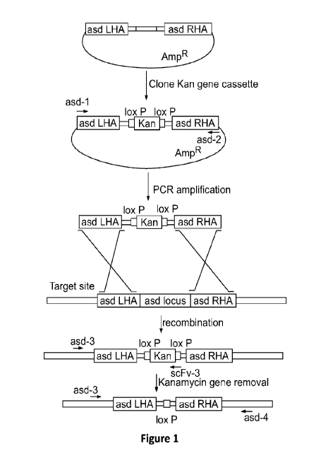
Figure 1 depicts a schematic of the process used to delete the asd gene from
strain YS1646. The asd gene from S. typhimurium strain YS1646 was deleted
using
RECTIFIED SHEET (RULE 91) ISA/EP
CA 03069523 2020-01-09
WO 2019/014398
PCT/US2018/041713
17
lambda-derived Red recombination system as described in Datsenko and Wanner
(Proc Natl Acad Sci USA 97:6640-6645 (2000)).
Figure 2 depicts the results of human PD-Li shRNA screening using qPCR
and Western blot. HEK 293 cells were co-transfected with a PD-Li cDNA
expression
plasmid and various pEQU6 plasmids encoding distinct shRNAs targeting PDLl.
Fig.
2A depicts the results of qPCR analysis to determine the level of mRNA
knockdown.
Fig. 2B depicts the Western blot analysis of human PD-Li shRNAs. Western
blotting
and densitometry were used to measure the level of PD-Li protein expression.
Figure 3 depicts the results of human TREX1 shRNA screening using qPCR
and Western blot. HEK 293 cells were co-transfected with a TREX1 cDNA
expression plasmid and various pEQU6 plasmids encoding distinct shRNAs
targeting
TREX1. Fig. 3A depicts results of qPCR analysis, used to determine the level
of
mRNA knockdown. Fig. 3B depicts results of Western blot analysis of the human
TREX1 shRNAs. Western blotting and densitometry were used to measure the level
of PD-Li protein expression.
Figure 4 depicts the results of human beta-catenin shRNA screening using
qPCR and Western blot. HEK 293 cells were co-transfected with a beta-catenin
cDNA expression plasmid and various pEQU6 plasmids encoding distinct shRNAs
targeting beta-catenin. Fig. 4A depicts results of qPCR, used to determine the
level of
mRNA knockdown. Fig. 4B depicts the results of Western blot analysis of the
human
beta-catenin shRNAs. Western blotting and densitometry were used to measure
the
level of beta-catenin protein expression.
Figure 5 depicts the results of human SIRP-alpha shRNA screening using
qPCR and Western blot. HEK 293 cells were co-transfected with a SIRP-alpha
cDNA
expression plasmid and various pEQU6 plasmids encoding distinct shRNAs
targeting
SIRP-alpha. Fig. 5A depicts results of qPCR, used to determine the level of
mRNA
knockdown. Fig. 5B depicts the results of Western blot analysis of human SIRP-
alpha
shRNAs. Western blotting and densitometry were used to measure the level of
SIRP-
alpha protein expression.
Figure 6 depicts the results of human TGF-beta isoform 1 shRNA screening
using qPCR. HEK 293 cells were co-transfected with a TGF-beta isoform 1 cDNA
CA 03069523 2020-01-09
WO 2019/014398 PCT/US2018/041713
18
expression plasmid and various pEQU6 plasmids encoding distinct shRNAs
targeting
TGF-beta. qPCR was used to determine the level of mRNA knockdown.
Figure 7 depicts the results of human VEGF shRNA screening using qPCR.
HEK 293 cells were co-transfected with a VEGF cDNA expression plasmid and
various pEQU6 plasmids encoding distinct shRNAs targeting VEGF. qPCR was used
to determine the level of mRNA knockdown.
Figure 8 depicts the results of human VISTA shRNA screening using qPCR
and Western blot. HEK 293 cells were co-transfected with a VISTA cDNA
expression plasmid and various pEQU6 plasmids encoding distinct shRNAs
targeting
VISTA. Fig. 8A depicts results of qPCR, used to determine the level of mRNA
knockdown. Fig. 8B depicts the results of Western blot analysis of human VISTA
shRNAs. Western blotting and densitometry were used to measure the level of
VISTA
protein expression.
Figure 9 depicts the results of qPCR assessment of combination gene
knockdown with HuPD-L1 + HuTREX1 RNAi's. HEK 293 cells were co-transfected
with a TREX1 cDNA expression plasmid, a PD-Li cDNA expression plasmid, and
pEQU6-H1 plasmid encoding ARI-134 shRNAs targeting PD-Li and TREX1, or
pEQU6 plasmid encoding ARI-123 shRNA targeting PD-Li alone, or pEQU6
plasmid encoding ARI-114 shRNA targeting TREX1. Fig. 9A depicts results of
qPCR, used to determine the level of PD-Li mRNA knockdown. Fig. 9B depicts
results of qPCR, used to determine the level of TREX1 mRNA knockdown.
Figure 10 depicts the results of qPCR assessment of combination gene
knockdown with HuPD-L1 + HuSIRP-alpha RNAi's. HEK 293 cells were co-
transfected with a PD-Li cDNA expression plasmid, a SIRP-alpha cDNA expression
plasmid, and pEQU6-H1 plasmid encoding ARI-135 containing shRNAs targeting
PD-Li and SIRP-alpha, or pEQU6 plasmid encoding ARI-123 shRNA targeting PD-
Li alone, or pEQU6 plasmid encoding ARI-175 shRNA targeting SIRPalpha. Fig.
10A depicts results of qPCR, used to determine the level of PD-Li mRNA
knockdown. Fig. 10B depicts results of qPCR, used to determine the level of
SIRP-
alpha mRNA knockdown.
Figure 11 depicts the results of qPCR assessment of combination gene
knockdown with HuPD-L1 + Hu beta-catenin RNAi's. HEK 293 cells were co-
CA 03069523 2020-01-09
WO 2019/014398
PCT/US2018/041713
19
transfected with a PD-IA cDNA expression plasmic', a beta-eatenin cDNA
expression
plasmid, and pEQU6411 plasmid encoding AR1-136 containing shRNAs targeting
PD-1.1 and beta-catenin, or pEQU6 plasmid encoding ARI-123 shRNA targeting PD-
LI alone, or pEQU6 plasmid encoding AR1-169 shRNA targeting beta-catenin. Fig.
HA depicts results of gPCR, used to determine the level of PD-Li tnRNA
knockdown.. Fig. 118 depicts results of qPCR, used to determine the level of
beta-
catenin m.RN¨A knockdown.
Figure 12 depicts the results of gPCR assessment of combination gene
knockdown with HuPD-L1 + HuVISTA RNAi's. HEK 293 cells were co-transfeeted
with a PD-L1 cDNA expression plasmid, a VISTA cDNA expression plastnid, and
pEQU6-H1 plasmid encoding AR1-137 (SEQ ID NO:213) containing shRNAs
targeting PD-L1 and VISTA, or pEQU6 .plasmid encoding ARI-123(SEQ ID NO:2)
shRNA. targeting PD-1,1 alone, or pEQU6 .plasmid encoding ART-195 (SEQ ID
NO:25) shRNA targeting VISTA. Fig. 12A depicts results of gPCR, used to
determine the level of PD-L1 mRNA knockdown. Fig. 12B depicts results of
ciPCR,
used to determine the level of VISTA. ml NA knockdown.
Figure 13 depicts the results of OCR assessment of combination gene
knockdown with mouse TRF.X1 + mouse PD-L1 RNAi's. HEK 293 cells were co-
transtected with a mouse TREX1 cDNA expression .plasmid, a mouse PD-L1 eDNA
expression plasmid, and pEQU6-.I-I1 plasmid encoding containing shRNA.
(designated .A.R1-128) targeting mouse TRExl and mouse PD-Li, or pEQU6 plasmid
encoding shRNA. (designated ARI-1A 5 targeting mouse PD-I., I alone, or pEQU6
plasmid encoding sh.RNA(designated ARI-108) targeting mouse TREXL Fig. 13A
depicts results of gPC.R, used to determine the level of PD-L1 raRNA
knockdown.
Fig. 138 depicts results of ciPCR, used to determine the level of TREX I
trIRNA
knockdown..
Figure 14 depicts the results of (-4PCR. assessment of combination gene
knockdown with mouse PD-L1 + mouse SIRP-alpha RNAi's. FIE( 293 cells were co-
transfeeted with a mouse PD-Li cDNA expression plasmid, a mouse SIR.P-alpha
eDNA expression plasmid, and pEQU6-H1 .plasmid encoding shRNA (designated
ART-129) targeting mouse PD-L and SIRP-alpha, or pEQU6 plasmid encoding
shRNA (designated ARI-115) targeting PD-Li alone, or pEQU6 plasmid. encoding
RECTIFIED SHEET (RULE 91) ISA/EP
CA 03069523 2020-01-09
WO 2019/014398
PCT/US2018/041713
shRNA (designated ARI-138) targeting SIRP-alpha. Fig. 14A depicts results of
qPCR, used to determine the level of PD-L I mRNA knockdown. Fig. 14B depicts
results of qPCR, used to determine the level of SIRP-alpha mRNA knockdown.
Figure 15 depicts the results of qPCR assessment of combination gene
5 knockdown with mouse PD-L I mouse VISTA RNAi's. HEK 293 cells were co-
transfected with a mouse PD-Li eDNA expression plasmid, a mouse VISTA eDNA
expression plasmid, and pEQU6-H1 plasmid encoding containing shRNA (designated
ARI-132) targeting PD-L1 and VISTA, or pEQU6 plasmid encoding shRNA
(designated ARI-115) targeting PD-Ll alone, or pEQU6 plasmid encoding shRNA
10 (designated ARI-157) targeting VISTA. Fig. 15A depicts results of qPCR,
used to
determine the level of PDL1 mRNA knockdown. Fig. 1511 depicts results of qPCR,
used to determine the level of beta-eatenin mRNA knockdown.
Figure 16 depicts the results of qPCR assessment of combination gene
knockdown with mouse TREXI+ mouse SIRP-alpha RNAi's. HEK 293 cells were
15 co-transfected with a mouse TREX I eDNA expression plasmid, a mouse
VISTA
eDNA expression plasmid, and pEQU6-1-11 plasmid encoding containing shRNA
(designated ARI-131) targeting PD-E1 and VISTA, or pEQU6 plasmid encoding
shRNA (designated ARI7108) targeting TREX1 alone, or pEQU-6 plasmid encoding
shRNA(designated ARI-138) targeting S1RP-alpha. Fig. 16A depicts results of
qPCR,
20 used to determine the level of TREX1 mRNA knockdown. Fig. 161) depicts
results of
qPCR, used to determine the level of SIRP-alpha mRNA knockdown.
Figure 17 depicts the results of qPCR assessment of combination gene
knockdown with mouse PD-Li 4.. mouse beta-catenin RNAi's. HEK 293 cells were
co-transfeeted with a mouse PD-Li eDNA expression plasmid, a mouse beta-
catenin
eDNA expression plasmid, and pEQU6-H1 plasmid encoding containing shRNA
(designated ARI-133) targeting PD-Li and VISTA, or pEQU6 plasmid encoding
shRNA(designated ARI-115) targeting PD-L I alone, or pEQU6 plasmid encoding
shRNA (designated ARI-166) targeting beta catenin. Fig. 17A depicts results of
qPCR, used to determine the level of PD-Li mRNA knockdown. Fig. 17B depicts
___________________ results of qPCR, used to detet mine the level of beta-
catenin mRNA knockdown.
Figure 18 depicts the results of qPCR assessment of combination gene
knockdown with mouse TUX! mouse VISTA RNAi's. HET{ 293 cells were co-
RECTIFIED SHEET (RULE 91) ISA/EP
CA 03069523 2020-01-09
WO 2019/014398
PCT/US2018/041713
21
transfected with a mouse TREX1 cDNA expression plasmid, a mouse VISTA cDNA
expression plasmid, and pEQU6-1I1 plasmid encoding shRNA (designated ARI-130)
targeting PD-L1 and VISTA, or pEQU6 plasmid encoding shRNA (designated AR1-
108) targeting TREX1 alone, or pEQU6 plasmid encoding shRNA (designated ART-
157) targeting VISTA. Fig. 18A depicts results of qPCR, used to determine the
level
of TREX1 mRNA knockdown. Fig. 1811 depicts results of qPCR, used to determine
the level of VISTA mRNA knockdown.
Figure 19 depicts a comparison of micro-RNA and shRNA-mediated
knockdown of mouse PD-L I . HET< 293 cells were co-transfected with a mouse PD-
Li cDNA expression plasmid and either pEQU6 plasmids encoding micro-RNA
(AR! -201) or shRNA (designated ARI-115) targeting PD-Ll. Fig. 19A depicts
results
of qPCR, used to determine the level of PD-Li mRNA knockdown. Fig. 19B depicts
results of Western blot analysis; Western blotting and densitometry were used
to
=
measure the level of PD-Li protein expression.
Figure 20 depicts a comparison of micro-RNA and shRNA-triediated
knockdown of mouse TREX1. I-TEK 293 cells were co-transfected with a mouse
TREX1 cDNA expression plasmid and pEQU6 plasmids encoding micro-RNA
(designated ART-203) or shRNA (designated ARI-108) targeting TREX1. Western
blot was used to determine the level of mRNA knockdown.
Figure 21 depicts the results of TREX1 knockdown with RNA Pot II
expression of micro-RNA. HEK 293 cells were co-transfected with a mouse TREX I
DNA expression plasmid and pEQU6 plasmid shRNA targeting mouse TREX1
(designated ART-108) or a pEQ plasmid encoding a CMV promoter and micro-.RNA
targeting mouse TREX I (designated ARI-204), Fig. 21A depicts results of qPCR,
used to determine the level of mouse TREX1 mRNA knockdown. Fig. 21B depicts
results of Western blot analysis; Western blotting and densitometry were used
to
measure the level of mouse TREX1 protein expression.
Figure 22 depicts the results of PD-L1 knockdown with RNA Pot II
expression of micro-RNA. HEK 293 cells were co-transfected with a mouse PD-Li
cDNA expression plasmid and pEQU6 plasmid shRNA targeting mouse PD-Li
(designated ART-US) or a pEQ plasmid encoding a CMV promoter and micro-RNA
targeting mouse TREX1 (designated ART-202). Fig. 22A depicts results of qPCR,
RECTIFIED SHEET (RULE 91) ISA/EP
CA 03069523 2020-01-09
WO 2019/014398
PCT/US2018/041713
22
used to determine the level of mouse PD-Li mR.N.A knockdown. Fig. 22B depicts
results of Western blot analysis; Western blotting and densitornetry were used
to
measure the level of mouse PD-Ll protein expression.
Figure 23 depicts the efficacy of systemically administered strain AST- 104 in
a CT26 colon tumor model. BALB/c mice were implanted with a single CT26 (2x10⁵
cells) subcutaneous flank tumor (n=8 per group). Mice with established tumors
were
IV injected with I x107 CFU of VS1646 strains containing either plasmid
control
(strain AST-102) or the TREX I shRNA pl.asrnid (of strain AST-104), or PBS
control,
on the days indicated by the arrows. Spaghetti plots depict tumor growth, each
line
representing an individual mouse. Tumor measurements were performed using
electronic calipers (Fowler, Newton, MA). Tumor volume was calculated using
the
modified ellipsoid :formula 1./2(length x width2). Mice were euthanized when
tumor
size reached >20% of body weight or became necrotic, as per 1A.CUC
regulations. %
Tumor Growth Inhibition (Tan was calculated as I -(mean test tumor volume/mean
control tumor volume) x 100. * p < 0.05 vs plasmid control, student's t-test.
Figure 24 depicts the correlation of strain AST-.104 mediated cytokine
changes with STING signature. BALB/c were implanted with a single CT26 (2x10⁵
cells) subcutaneous flank tumor (n--8 per group). Mice with established tumors
were
IV injected with 5x10⁶CFU of VS1646 strains containing either plasmid control
(strain AST-IO2) or the TREX1 shRNA plasmid (AST-104), or PBS control. Mice
were bled 6 brs following the first dose and systemic serum cytokines tested
on a
Isuminex 200 device (Luminex Corporation) and mouse eytorneuie bead array (BD
bead array, FACS Fortessa, ECM' software, BD Biosciences). Fig. 24A depicts
levels
of pro-inflammatory cytokines. Fig. 24B depicts levels of imrnuno-suppressive
eytoki.nes. p < 0.05, p <0.01, student's t-test.
Figure 25 depicts the efficacy of systemically administered strain AST-104 in
a MC38 colon tumor model. C57BI./6 mice (6-8 Wk old) were implanted with a
single
MC38 (2x10⁵ cells) subcutaneous flank tumor (n-10 per group). Mice with
established tumors were IV injected with 5 x10⁶CF1.1 of Y51646 strains
containing
either plasmid control (strain AST-102) or the TREX I shRNA plasmid (strain
AST-
104), or PBS control, on the days indicated by the arrows. Spaghetti plots
depict
tumor growth, each line representing an individual mouse. Tumor measurements
were
RECTIFIED SHEET (RULE 91) ISA/EP
CA 03069523 2020-01-09
WO 2019/014398 PCT/US2018/041713
23
performed using electronic calipers (Fowler, Newton, MA). Tumor volume was
calculated using the modified ellipsoid formula 1/2(length x width2). Mice
were
euthanized when tumor size reached >20% of body weight or became necrotic, as
per
IACUC regulations. TGI was calculated as 1-(mean test tumor volume/mean
control
tumor volume) x 100. *p <0.05 vs. plasmid control, student's t-test.
Figure 26 depicts the efficacy of AST-104 in a checkpoint-resistant B16.F10
melanoma model. C57B1/6 mice (6-8 wk old) were implanted with a single B16.F10
(5x10⁵ cells) subcutaneous flank tumor (n=10 per group). Mice with established
tumors were IV injected with 5x10⁶ CFU of YS1646 strains containing either
plasmid
control (AST-102) or the TREX1 shRNA plasmid (AST-104), or PBS control, on the
days indicated by the arrows. Spaghetti plots depict tumor growth, each line
representing an individual mouse. Tumor measurements were performed using
electronic calipers (Fowler, Newton, MA). Tumor volume was calculated using
the
modified ellipsoid formula 1/2(length x width2). Mice were euthanized when
tumor
size reached >20% of body weight or became necrotic, as per IACUC regulations.
TGI was calculated as 1-(mean test tumor volume/mean control tumor volume) x
100.
*p <0.05 vs. plasmid control, student's t-test.
Figure 27 depicts the efficacy of systemically administered AST-105 (shPD-
L1) in a CT26 tumor model. BALB/c (6-8 wk old) were implanted with a single
CT26
(2x10⁵ cells) subcutaneous flank tumor (n=8 per group). Mice with established
tumors
were IV injected with 5x 10⁶ CFU of YS1646 strains containing either plasmid
control
(AST-102) or the PD-Li shRNA plasmid (AST-105), or PBS control, on the days
indicated by the arrows. A separate group was administered 100 jig anti-PD-Li
antibody (clone 10F.9G2 clone, BioXCell) by IP injection weekly, beginning
with the
first IV injection. Spaghetti plots depicting tumor growth, each line
representing an
individual mouse. Tumor measurements were performed using electronic calipers
(Fowler, Newton, MA). Tumor volume was calculated using the modified ellipsoid
formula 1/2(length x width2). Mice were euthanized when tumor size reached
>20%
of body weight or became necrotic, as per IACUC regulations. TGI was
calculated as
1-(mean test tumor volume/mean control tumor volume) x 100. * p < 0.05 vs.
plasmid
control, student's t-test.
CA 03069523 2020-01-09
WO 2019/014398
PCT/US2018/041713
24
Figure 28 depicts results showing that AST-105 induces significant cytokine
responses observed over PD-L1 mAb. BALB/c mice (6-8 wk old) were implanted
with a single CT26 (2 x10⁵ cells) subcutaneous flank tumor (n=8 per group).
Mice
with established tumors were IV injected with 5x10⁶ cru- of YS1646 strains
containing either plasmid control (AST-102) or the PD-L1 shRNA plasmid (AST-
105), or PBS control, on the days indicated by the arrows. A separate group
was
administered 100 lag anti-PD-L I antibody IP (clone 1.0F.9G2 clone, .RioXCell)
weekly, beginning with the first IV injection, Mice were bled 6 hrs following
the first
dose and systemic serum eytokines tested by Luminex (BD bead array and Luminex
200) and mouse eytornetric head array (FACS Fortessa., FCAP software, all BD
Riosciences). * p < 0.05, ** p < 0.01, student's t-test.
Figure 29 depicts the effects of intratumoral administration of strains AST-
104 and AST-105 in dual flank colon tumors on tumor volume. BALB/c mice (6-8
wk
old) were implanted with dual CT26 (2x10⁵ cells) subcutaneous flank tumors on
the
right and left flanks (n=10 per group). Mice with established tumors were IT
injected.
into the right flank with 5x10⁶ CFU of YS1646 strains containing either
plasmid
control (AST-102) or the strain containing TREX.1 shRNA plasmic' (AST-104), or
PD-Li shRNA plasmid (AST-105), or PBS control, on the days indicated by the
arrows. Tumor measurements were performed using electronic calipers (Fowler,
Newton, MA). Tumor volume was calculated using the modified ellipsoid formula
1/2(length x width2). Mice were euthartized when tumor size reaches >20% of
body
weight or became necrotic, as per IACIJC regulations. % Tumor Growth
Inhibition
(TGI) is calculated as 1-(mean test tumor volume/mean control tumor volume) x
100.
The plots depict mean tumor growth of each group in the injected (left graph)
and
distal (right graph) groups, SEM. * p <0.05, * p <0.001, student's t-test.
Figure 30 depicts the curative effects of intraturnoral AST-104 administration
in dual flank colon tumors in mice. BALB/c mice (6-8 wk old) were implanted
with
dual CT26 (2 x10⁵ cells) subcutaneous flank tumors on the right and left
flanks (n-10
per group). Mice with established tumors were IT injected into the right flank
with
5x10⁶ CM of YS1646 strains containing either pl.asmid control (AST-102) or the
TREX1 shRNA plasmid (AST-104), or the shPD-L1 plasmid (AST-105), or PBS
control on days 10 and 14 after tumor implantation. Mice were euthanized when
RECTIFIED SHEET (RULE 91) ISA/EP
CA 03069523 2020-01-09
WO 2019/014398
PCT/US2018/041713
tumor size reached >20% of body weight or became necrotic, as per IACUC
regulations. The figure depicts the overall survival of the mice, ** p <0,01,
log-rank
(Mantel-Cox) test.
Figure 31 depicts the levels of tumor colonization in injected and distal
5 tumors after IT administration of AST-104. BALB/c mice (6-8 wk old) were
implanted with dual C126 (2x10⁵ cells) subcutaneous flank tumors on the right
and
left flanks (n-10 per group). Mice with established tumors were IT injected
into the
right flank with 5x10⁶ CFU of the YS1646 strain containing a TREX1 shRNA
plasmid (AST-104). At 35 days post tumor implantation (12 days after the last
dose of
10 AST-104), three mice were sacrificed, and injected and distal tumors
were
homogenized (GentleMACsTm, Milteny-i Biotee-) and plated on LB plates to
enumerate
the number of colony forming units (CFU) per gram of tumor tissue. The figure
depicts the mean CFU per gram of tissue, SD.
Figure 32 depicts that CpG scrambled plasmid has immuno-stimulatory anti-
15 tumor properties. BALB/c mice (6-8 wk old) were implanted with a single
CT26
(2x10⁵ cells) subcutaneous flank tumor (n---9 per group). Mice with
established tumors
were IV injected with 5x10⁶ CFU of the YS1646 strain (AST-100), or the YS1646
strain containing the scrambled shRNA control plasrnid (AST-103), or PBS
control,
on the days indicated by the arrows. Tumor measurements were performed using
20 electronic calipers (Fowler, Newton, MA). Tumor volume was calculated
using the
modified ellipsoid formula 1/2 (length x width2). Mice were euthanizcd when
tumor
size reached >20% of body weight or became necrotic, as per IACUC regulations.
TOT is calculated as 1-(mean test tumor volume/mean control tumor volume) x
100.
The figure depicts mean tumor growth of each group, SEM. ** p < 0.01,
student's t-
25 test
Figure 33 depicts the efficacy of AST-106 (microRNA TREX1) vs. AST-I04
(shRNA TREX1). BALB/c mice (6-8 wk old) were implanted with a single CT26
(2x10⁵ cells) subcutaneous flank tumor (11-9 per group). Mice with established
tumors
were IV injected with 5x 10⁶ CFU of the YS1646 containing the TREX1 shRNA
plasmid (AST-104) or the YS1646 strain containing a T.REX1 microRNA plasmid
(AST-106), or PBS control, on the days indicated by the arrows. Tumor
measurements were performed using electronic calipers (Fowler, Newton, MA).
RECTIFIED SHEET (RULE 91) ISA/EP
CA 03069523 2020-01-09
WO 2019/014398 PCT/US2018/041713
26
Tumor volume was calculated using the modified ellipsoid formula 1/2(length x
width2). Mice were euthanized when tumor size reached >20% of body weight or
became necrotic, as per IACUC regulations. TGI was calculated as 1-(mean test
tumor volume/mean control tumor volume) x 100. The figure depicts the mean
tumor
growth of each group, SEM. *p <0.05, student's t-test.
Figure 34 depicts a schematic of the process used to delete thefliC gene. The
flic gene was deleted from the chromosome of S. typhimurium strain AST-101
(asd
deleted strain of YS1646) using lambda-derived Red recombination system as
described in Datsenko and Wanner (Proc Natl Acad Sci USA 97:6640-6645 (2000)).
Figure 35 depicts that the Flagellin deletion strain grows normally in LB. The
figure depicts the growth of strains AST-108 ASD (pATI-shTREX1) and AST-112
ASD/FLG (pATI-shTREX1) at 37 C in LB broth, as measured by 0D600 using a
Spectramax 96 well plate reader (Molecular devices).
Figure 36 depicts that Flagellin knockout improves anti-tumor efficacy.
BALB/c mice (6-8 wk old) were implanted with a single CT26 (2x10⁵ cells)
subcutaneous flank tumor (n=9 per group). Mice with established tumors were IV
injected with 5x10⁶ CFU of the asdlfigB/fliC knockout strain containing the
pATI
shTREX1 plasmid (AST-113), or asd knockout strain containing the pATI shTREX1
plasmid (AST-110), or PBS control, on the days indicated by the arrows. Tumor
measurements were performed using electronic calipers (Fowler, Newton, MA).
Tumor volume was calculated using the modified ellipsoid formula 1/2(length x
width2). Mice were euthanized when tumor size reached >20% of body weight or
became necrotic, as per IACUC regulations. TGI was calculated as 1-(mean test
tumor volume/mean control tumor volume) x 100. The figure depicts the mean
tumor
growth of each group, SEM. *p <0.05, student's t-test.
Figure 37 depicts that Flagellin knockout shows an increased IFN-gamma
signature. BALB/c mice (6-8 wk old) were implanted with a single CT26 (2x10⁵
cells) subcutaneous flank tumor (n=9 per group). Mice with established tumors
were
IV injected with 5x10⁶ CFU of the asdlfigB/fliC knockout strain containing the
pATI
shTREX1 plasmid (AST-113), or asd knockout strain containing the pATI shTREX1
plasmid (AST-110), or PBS control. Mice were bled 6 hrs following the first
dose and
systemic serum cytokines tested by Luminex 200 device (Luminex Corporation)
and
CA 03069523 2020-01-09
WO 2019/014398
PCT/US2018/041713
27
mouse cytometric bead array (BD bead array, PACS Fortessa, R.-:AP software,
all BD
Biosciences). * p <0.05, ** p <0.01. *** p < 0.001, student's t-test.
Figure 38 depicts that Flagellin is not required for tumor colonization.
BALB/c mice (6-8 wk old) were implanted with a single CT26 (2x10⁵ cells)
subcutaneous flank tumor per group). Mice with established tumors were IV
injected with 5x10⁶ CEU of the asdifigB/fliC knockout strain containing the
pATI
shTREXI plasm:id. (AST-113), or asd knockout strain containing the pATI
shTREXI
plasmid (AST-110), or PBS control. At 35 days post tumor implantation (12 days
after the last dose of engineered Salmonella therapy), three mice per group
were
sacrificed, and tumors were homogenized (GentleM.ACsTm, Miltenyi Biotec) and
plated on LB plates to enumerate the number of colony forrning-units per gram
of
tumor tissue. The figure depicts the mean colony forming units (CFO per gram
of
tissue, SD.
Figure 39 depicts that a cytoLLO expressing strain grows normally in vitro.
The figure depicts the growth of strains AST-110 (YS1646 with asd deletion
containing (pATI-shTR.EX1)) and AST-115 (YS1646 with asd deletion and knock-in
of cytoLLO expression cassette containing (pATI-shTREX1)) at 37 C in LB
broth, as
measured by 0D600 using a Spectramax 96 well plate reader (Molecular devices).
Figure 40 depicts that AST-115 (ASD knockout + CytoLLO Knock-in strain
.. carrying shTREX1 plasmid) demonstrates potent, single-dose efficacy in a
murine
. CT26 tumor model. BALB/c mice (6-8 wk old) were implanted with a single CT26
(2 x 05 cells) subcutaneous flank tumor (n---9 per group). Mice with
established tumors
were IV injected with 5x10⁶ CPU of AST-115 (YS1646 with asd deletion and knock-
in of cytoLLO expression cassette at asd locus containing (pAT1-shTREX1), or
PBS
control, on the days indicated by the arrows. Tumor measurements were
perlbrmed
using electronic calipers (Fowler, Newton, MA). Tumor volume was calculated
using
the modified ellipsoid formula 1/2(length x width.2). Mice were euthanized
when
tumor size reached >20% of body weight or became necrotic, as per IACUC
regulations. TGI was calculated as 1-(mean test tumor volume/mean control
tumor
volume) x 100. The figure depicts the mean tumor growth of each group, SEM,
** p
<0.01, student's t-test.
RECTIFIED SHEET (RULE 91) ISA/EP
CA 03069523 2020-01-09
WO 2019/014398 PCT/US2018/041713
28
Figure 41 depicts that strain YS1646 requires tumor microenvironment levels
of adenosine for growth. Growth of strains YS1646 (purl-ImsbB-) and the wild-
type
parental strain ATCC14028 at 37 C in LB broth are shown, as measured by 0D600
using a Spectramax 96 well plate reader (Molecular devices).
Figure 42 depicts that ASD, FLG, and CytoLLO engineered strains require
high adenosine for growth. The growth of strains AST-117 (YS1646 Aasd
containing
a low copy shTREX-1 plasmid), AST-118 (YS1646 Aasd/filC/fljB containing a low
copy shTREX-1 plasmid), and AST-119 (YS1646 Aasd:LLO containing a low copy
shTREX-1 plasmid) at 37 C in LB broth are shown, as measured by 0D600 using a
Spectramax 96 well plate reader (Molecular devices).
Figure 43 depicts that a strain with a low copy origin of replication asd-
encoding plasmid has superior growth kinetics than a strain with a high copy
origin of
replication asd-encoding plasmid. The growth of strains YS1646, AST-117
(YS1646
Aasd containing a low copy shTREX-1 plasmid with a functional asd gene), AST-
104
(YS1646 containing a low copy pEQ shTREX-1 plasmid without an asd gene), and
AST-110 (YS1646 Aasd containing a high copy pATI-shTREX-1 plasmid with a
functional asd gene) at 37 C in LB broth are shown, as measured by 0D600
using a
Spectramax 96 well plate reader (Molecular devices).
Figure 44 depicts that a strain with a low copy asd plasmid is more fit than a
strain with a high copy asd plasmid in mouse tumor cells. The intracellular
growth of
strains AST-117 (YS1646 Aasd containing a low copy shTREX-1 plasmid with a
functional asd gene) and AST-110 (YS1646 Aasd containing a high copy pATI-
shTREX-1 plasmid with a functional asd gene) are shown in Bl6F.10 mouse
melanoma cells and CT26 mouse colon carcinoma cells. 5x10⁵ cells in a 24 well
dish
were infected with the S. typhimurium strains at a MOI of 5. After 30 minutes
of
infection, media was replaced with media containing gentamycin to kill
extracellular
bacteria. At indicated time points, cell monolayers were lysed by osmotic
shock the
cell lysates were diluted and plated on LB agar to enumerate CFU.
Figure 45 depicts that in vivo, asd gene complementation systems result in
retention of plasmids in S. typhimurium-infected tumors. BALB/c mice (6-8 wk
old)
were implanted with a single CT26 (2 x10⁵ cells) subcutaneous flank tumor (n=9
per
CA 03069523 2020-01-09
WO 2019/014398
PCT/US2018/041713
29
group). Mice with established tumors were IV injected with 5x1.06 CFU of the
asd
knockout strain containing the-pATI shTREX1 plasmid (AST-110) or the Y51646
containing a pEQ shTREX-1 plasmid without an asd gene (AST-104). At 35 days
post tumor implantation (12 days after the last dose of engineered Salmonella
therapy), three mice per group were sacrificed, and tumors were homogenized
using a
GentleMACsi'm homogenizer (Miltenyi Biotec) and plated on LB agar plates or LB
.
agar plates with 50ugimL of Kanamycin. The figure depicts the percentage of
Kanamyein resistant CELT in tumor tissue homogenates, SD.
Figure 46 depicts that the therapeutic efficacy of a strain containing a
plasmid
with asd gene complementation system and shTREX1 (AST-110) is improved.
BALB/c mice (6-8 wk. old) were implanted with a single CT26 (2x10⁵ cells)
subcutaneous flank tumor (n--,=9 per group). Mice with established tumors were
IV
injected with 5x I 06 CFU of the asd knockout strain containing the pATI-
shTREX1
plasmid (AST-11.0) or the asd knockout strain containing the pATI-seramble
plasmid
(AST-109), or the YS1646 strain containing a pEQ-sliTREX-I plasmid without an
asd gene (AST-104), or PBS control, on the days indicated by the arrows. Tumor
measurements were performed using electronic calipers (Fowler, Newton, MA).
Tumor volume was calculated using the modified ellipsoid formula 1/2(length x
width2). Mice were euthanized when tumor size reaches >20% of body weight or
became necrotic, as per IACLiC regulations. TGI was calculated as 1-(mean test
tumor volume/mean control tumor volume) x 100. The figure depicts the mean
tumor
growth of each group, SEM.
Figure 47 depicts that a strain containing a low copy shTREX I plasmic!
(AST-117) has superior anti-tumor properties compared to a strain containing a
high
copy plasmid (AST-110). BALB/c mice (6-8 wk old) were implanted with a single
CT26 (2x10⁵ cells) subcutaneous flank tumor (11-9 per group). Mice with
established
tumors were IV injected with 5x10⁶ CELT of the asd knockout strain containing
the
pATI-shTREX1 plasmid with a high copy number origin of replication (AST-110)
or
the. asd knockout strain containing the pATI-shTREXI. plasmid with a low copy
number origin of replication (AST-117), or PBS control., on the days indicated
by the
arrows. Tumor measurements were performed using electronic calipers (Fowler,
Newton, MA). Tumor volume was calculated using the modified ellipsoid formula
RECTIFIED SHEET (RULE 91) ISA/EP
CA 03069523 2020-01-09
WO 2019/014398
PCT/US2018/041713
1/2(length x width2). Mice were euthanized when tumor size reached >20% of
body
weight or became necrotic, as per IACUC regulations. TGI was calculated as .1-
(mean
test tumor volume/mean control tumor volume) x 100. The figure depicts the
mean
tumor growth of each group, SEM. * p <0.05, student's 1-test.
5 Figure 48 depicts that the AST-117 low copy plasmid strain colonizes
tumors
better and has a higher tumor to spleen colonization ratio than the AST-110
high copy
plasmid strain. BAII,Ble mice (6-8 wk old) were implanted with a single CT26
(2x10⁵
cells) subcutaneous .flank tumor (n=9 per group). Mice with established tumors
were
IV injected with 5x10⁶ CFI) of the asd knockout strain containing the pATI-
slaREX1
10 plasmid with a high copy number origin of replication (AST-110) or the
asd knockout
strain containing the pATI-shTREX1 plasmid with a low copy number origin of
replication (AST-117). At 35 days post tumor implantation (12 days after the
last dose
of engineered Salmonella therapy), 3 mice per group were sacrificed, and
tumors were
homogenized using a GentleMACsTm homogenizer (Miltenyi Biotec) and plated on
15 LB plates to enumerate the number of CFU per gram of tumor tissue. Fig.
48A
depicts the mean CF1i per gram of tumor tissue, Si). Fig. 48B depicts the
tumor to
spleen colonization ratios.
Figure 49 depicts that a strain grown to stationary phase is equivalently
potent, and less inflammatory than the same strain grown to log phase. BALB/c
mice
20 (6-8 wk old) were implanted with a Single CT26 (2x10⁵ cells)
subcutaneous flank
tumor (n-9 per group). Mice with established tumors were IV injected with
5x10.
CPU of the YS1646 strain containing a pEQ-shTREX- I plasmid (AST-104)
harvested
at log phase or stationary phase, or PBS control, on the days indicated by the
arrows.
Tumor measurements were performed using electronic calipers (Fowler, Newton,
25 MA). Tumor volume was calculated using the modified ellipsoid formula
112(length x
width2). Mice were euthanized when tumor size reached >20% of body weight or
became necrotic, as per IACIJC regulations. TGI was calculated as 1-(mcan test
tumor volume/mean control tumor volume) x 100. Fig. 49A depicts the mean tumor
growth of each group, SEM. * p <0.05, student's Mese Fig. 49B depicts the
levels
30 of INF-alpha and IL-6. Mice were bled 6 hrs following the first dose and
systemic
serum cytokines tested by Luminex (Luminex Corp.) and mouse cytornetric bead
array
(FACS Fortessa, FCAP software, all BD Biosciences). p <0.01, student's 1-test.
RECTIFIED SHEET (RULE 91) ISA/EP
CA 03069523 2020-01-09
WO 2019/014398 PCT/US2018/041713
31
Figure 50 depicts that autolytic strain (AST-120) cannot grow in the absence
of DAP. The figure depicts the growth of Aasd:cytoLLO strain containing a
pEQU6-
shTREX1 plasmid that does not contain an asd gene (AST-120) over time in LB
broth
alone, or in LB broth supplemented with 50 g/mL DAP, as measured by 0D600
using
a Spectramax 96 well plate reader (Molecular devices).
Figure 51 depicts the anti-tumor activity of the autolytic strain (AST-120).
BALB/c mice (6-8 wk old) were implanted with a single CT26 (2x10⁵ cells)
subcutaneous flank tumor (n=9 per group). Mice with established tumors were IV
injected with 5x10⁶ CFU of the of Aasd:cytoLLO strain containing a pEQU6-
.. shTREX1 plasmid that does not contain an asd gene (AST-120), or PBS
control, on
the days indicated by the arrows. Tumor measurements were performed using
electronic calipers (Fowler, Newton, MA). Tumor volume was calculated using
the
modified ellipsoid formula 1/2(length x width2). Mice were euthanized when
tumor
size reached >20% of body weight or became necrotic, as per IACUC regulations.
TGI was calculated as 1-(mean test tumor volume/mean control tumor volume) x
100.
The figure depicts the mean tumor growth of each group, SEM. * p < 0.05,
student's t-test.
Figure 52 depicts that TREX1 expression is increased in several human tumor
types. Analysis of the relative gene expression of the TREX1 gene using the
TCGA
database was performed from a broad array of tumor types. Tumor types with a
significant upregulation of TREX1 compared to normal tissue are displayed:
prostate,
breast, cervical, uterine and bladder (p values: BRCA - 7.7e-16; PRAD - 9.4e-
12;
UCEC - 2.5e-05; BLCA - 3.7e-03; CESC - 7.7e-03) and multiple forms of kidney
cancer (p values: KIPAN - 8.9e-39; KIRC - 9.6e-35; KIRP - 5.8e-14; KICH - 4.9e-
08).
Figure 53 depicts that radiotherapy after administration of S. typhimurium
strain AST-106 increases tumor colonization. BALB/c mice (6-8 wk old) were
inoculated subcutaneously in the right flank with lx105 mouse TSA breast
carcinoma
cells. Mice bearing established tumors were administered the following: IV
injection
of 5x10⁶CFUs of AST-106 (Y51646 transformed with pEQU6-miTREX1) followed
4 hours later with 0 Gy (3 mice), or 5 x10⁶ CFUs of AST-106 followed 4 hours
later
with 20 Gy (3 mice); 20 Gy irradiation followed 4 hours later with 5x10⁶ CFUs
of
CA 03069523 2020-01-09
WO 2019/014398
PCT/US2018/041713
32
AST-106 (3 mice), or PBS TV followed by 0 Gy radiation (I mouse). Focal
radiotherapy was administered using a small animal radiation research platform
(SARRP) device (XStrahl Life Sciences). Mice were sacrificed 24 hours later,
and
tumors were harvested and weighed. Tumors were homogenized in 10mL sterile PBS
using M tubes in a GentleMACsTm device (Mittenyi Biotec), then 10-fold serial
dilutions were performed and plated on LB agar plates containing kanamycin.
The
following day, colony forming units (CFU) were counted and CFU per gram of
tumor
tissue was calculated. * p <0.05, student's I-test.
DETAILED DESCRIPTION
OUTLINE
A. DEFINITIONS
B. OVERVIEW OF THE INIMUNOSTIMULATORY BACTERIA
C. CANCER IMMUNOTHERAPEUTICS
1. Immunotherapies
2. Adoptive Immunotherapies
3. Cancer Vaccines and Oncolytic Viruses
D. BACTERIAL CANCER IMMUNOTHERAPY
1. Bacterial therapies
2. Comparison of the Immune Responses to Bacteria and Viruses
3. Salmonella Therapy
a. Tumor-tropic Bacteria.
b. Salmonella enterica serovar typhimurium
c. Bacterial Attenuation
I. tnsh13- Mutants
ii. purl- Mutants
Combinations of Attenuating Mutations
iv. VNP20009 and Other Attenuated S. typhimurium
strains
v. Attenuated S. typhimurium Engineered To Deliver
Macromolecules
4. Enhancements of immunostimulatory Bacteria to Increase
Therapeutic Index
a. aNd Celle Deletion
b. Adenosine Auxotrophy
RECTIFIED SHEET (RULE 91) ISA/EP
CA 03069523 2020-01-09
WO 2019/014398
PCT/US2018/041713
33
c. Flagellin Deficient Strains
d. Salmonella Engineered to Escape the Salmonella
Containing Vacuole (SCV)
e. Deletions in Salmonella Genes Required for Biofilm
Formation
f. Deletions in Genes in the LPS Biosynthetic Pathway
g. Deletions of SPI-1 Genes
h. Endonuclease (endA) Mutations To Increase Plasmid
Delivery
i. RIG-I Inhibition
j. DNase II Inhibition
k. RNase H2 Inhibition
1. Stabilin-1/CLEVER-1 Inhibition
m. Bacterial Culture Conditions
E. CONSTRUCTING EXEMPLARY PLASMIDS
1. Interfering RNAs (RNAi)
a. shRNA
b. micro-RNA
2. Origin of Replication and Plasmid Copy Number
3. CpG Motifs and CpG Islands
4. Plasmid Maintenance/Selection Components
5. DNA Nuclear Targeting Sequences
F. TUMOR TARGETING IMMUNOSTIMULATORY BACTERIA
CONTAIN RNAI AGAINST EXEMPLARY IMMUNE TARGET GENES TO
STIMULATE ANTI-TUMOR IMMUNITY
1. TREX1
2. PD-Li
3. VISTA
4. SIRPa
5. I3-catenin
6. TGF-I3
7. VEGF
8. Additional Exemplary Checkpoint Targets
CA 03069523 2020-01-09
WO 2019/014398
PCT/US2018/041713
34
G. COMBINATIONS OF RNAI shRNAS TO MULTIPLE IMMUNE
TARGETS WITHIN A SINGLE THERAPEUTIC MODALITY AND
COMBINATION THERAPY
1. TREXI and Other Targets
2. TREXI and Radiotherapy
3. TREXI and Immunogenic Chemotherapy
4. Combination Therapy with Anti-Checkpoint Antibodies
H. PHARMACEUTICAL PRODUCTION, COMPOSITIONS, AND
FORMULATIONS
1. Manufacturing
a. Cell Bank Manufacturing
b. Drug Substance Manufacturing
c. Drug Product Manufacturing
2. Compositions
3. Formulations
a. Liquids, Injectables, Emulsions
b. Dried Thermostable Formulations
4. Compositions for Other Routes of Administration
5. Dosages and Administration
6. Packaging and Articles of Manufacture
I. METHODS OF TREATMENT AND USES
1. Cancers and Tumors
2. Administration
3. Monitoring
J. EXAMPLES
A. DEFINITIONS
Unless defined otherwise, all technical and scientific terms used herein have
the same meaning as is commonly understood by one of skill in the art to which
the
invention(s) belong. All patents, patent applications, published applications
and
publications, GenBank sequences, databases, websites and other published
materials
referred to throughout the entire disclosure herein, unless noted otherwise,
are
incorporated by reference in their entirety. In the event that there are a
plurality of
definitions for terms herein, those in this section prevail. Where reference
is made to a
URL or other such identifier or address, it is understood that such
identifiers can
CA 03069523 2020-01-09
WO 2019/014398
PCT/US2018/041713
change and particular information on the internet can come and go, but
equivalent
information can be found by searching the internee. Reference thereto
evidences the
availability and public dissemination of such information.
As used herein, therapeutic bacteria are bacteria that effect therapy, such as
5 cancer or anti-tumor therapy, when administered to a subject, such as a
human.
As used herein, immunostimulatory bacteria are therapeutic bacteria that,
when introduced into a subject, accumulate in immunoprivileged tissues and
cells,
such as tumors, and replicate and/or express products that are
immunostimulatory or
that result in immunostirnulation. The immunostimulatory bacteria are
attenuated in
10 the host by virtue of reduced toxicity or pathogenicity and/or by virtue
of encoded
products that reduce toxicity or pathogenicity, as the immunostimulatory
bacteria
cannot replicate and/or express products, except primarily in immunoprivileged
environments. Immunostimulatory bacteria provided herein are modified to
encode a
product or products or exhibit a trait or property that renders them
15 immunostimulatory. Such products, properties and traits include, at
least one of an
shRNA that targets, disrupts or inhibits a checkpoint gene or gene encoding
such
inhibitor or a metabolite that is immunosuppressive or is in an
immunosuppressive
pathway. These include encoding an siRNA, such as an shRNA, that targets or
inhibits TREX1 expression, a modification that renders the bacterium
auxotrophie for
20 adenosine, and/or an inhibitor or disruptor of an immune checkpoint gene
or product
thereof, such as an shRNA that disrupts or inhibits PD-Li.
As used herein, the strain designations VNP20009 (see, e.g., International
PCT application Publication No. WO 99/13053, see, also U.S. Patent No.
6,863,894)
and Y.S1646 and 41.2.9 are used interchangeably and each refer to the strain
deposited
25 with the American Type Culture Collection and assigned Accession No.
202165.
VNP20009 is a modified attenuated strain of Salmonella typhimurium, which
contains
deletions in msbB and purl, and was generated from wild type strain ATCC
14028.
As used herein, the strain designations YS1456 and 8.7 are used
interchangeably and each refer to the strain deposited with the American Type
Culture
30 Collection and assigned Accession No. 202164 (see, U.S. Patent No.
6,863,894).
RECTIFIED SHEET (RULE 91) ISA/EP
CA 03069523 2020-01-09
WO 2019/014398
PCT/US2018/041713
36
As used herein, an origin of replication is a sequence of DNA at which
replication is initiated on a chromosome, plasmid or virus. For small DNA,
including
bacterial plasmids and small viruses, a single origin is sufficient.
The origin of replication determines the vector copy number, which depends
upon the selected origin of replication. For example, if the expression vector
is
derived from the low-copy-number plasmid pBR322, it is between about 25-50
copies/cell, and if derived from the high-copy-number plasmid pUC, it can be
150-
200 copies/cell.
As used herein, medium copy number of a plasmid in cells is about or is 150
or less than 150, low copy number is 15-30, such as 20 or less than 20. Low to
medium copy number is less than 150. High copy number is greater than 150
copies/cell.
As used herein, a CpG motif is a pattern of bases that include an unmethylated
central CpG ("p" refers to the phosphodiester link between consecutive C and G
nucleotides) surrounded by at least one base flanking (on the 3' and the 5'
side of) the
central CpG. A CpG oligodeoxynucleotide is an oligodeoxynucleotide that is at
least
about ten nucleotides in length and includes an unmethylated CpG. At least the
C of
the 5' CG 3' is unmethylated.
As used herein, a RIG-I binding sequence refers to a 5'triphosphate (5'ppp)
structure directly, or that which is synthesized by RNA pol III from a poly(dA-
dT)
sequence, which by virtue of interaction with RIG-I can activate type I IFN
via the
RIG-I pathway. The RNA includes at least four A ribonucleotides (A-A-A-A); it
can
contain 4, 5, 6, 7, 8, 9, 10 or more. The RIG-I binding sequence is introduced
into a
plasmid in the bacterium for transcription into the polyA.
As used herein, a "modification" is in reference to modification of a sequence
of amino acids of a polypeptide or a sequence of nucleotides in a nucleic acid
molecule and includes deletions, insertions, and replacements of amino acids
or
nucleotides, respectively. Methods of modifying a polypeptide are routine to
those of
skill in the art, such as by using recombinant DNA methodologies.
As used herein, a modification to a bacterial genome or to a plasmid or gene
includes deletions, replacements and insertions of nucleic acid.
CA 03069523 2020-01-09
WO 2019/014398
PCT/US2018/041713
37
As used herein, RNA interference (RNAi) is a biological process in which
RNA molecules inhibit gene expression or translation, by neutralizing targeted
-
inRNA molecules to inhibit translation and thereby expression of a targeted
gene.
As used herein, RNA molecules that act via RNAi are referred to as inhibitory
by virtue of their silencing of expression of a targeted gene. Silencing
expression
means that expression of the targeted gene is reduced or suppressed or
inhibited.
As used herein., gene silencing via RNAi is said to inhibit, suppress, disrupt
or
silence expression of a targeted gene. A targeted gene contains sequences of
nucleotides that correspond to the sequences in the inhibitory RNA, whereby
the
inhibitory RNA silences expression of mRNA.
As used herein, inhibiting, suppressing, disrupting or silencing a targeted
gene
refers to processes that alter expression, such as translation, of the
targeted gene,
whereby activity or expression of the product encoded by the targeted gene is
reduced. Reduction, includes a complete knock-out or a partial knockout,
whereby
with reference to the irnmunostimulatory bacterium provided herein and
administration herein, treatment is effected.
As used herein, small interfering RNAs (siRNAs) are small pieces of double-
stranded (ds) RNA, usually about 21 nucleotides long, with 3' overhangs (2
nucleotides) at each end that can be used to "interfere" with the translation
of proteins
by binding to and promoting the degradation of messenger RNA (mRNA) at
specific
sequences. in doing so, siRNAs prevent the production of specific proteins
based on
the nucleotide sequences of their corresponding mRNAs. The process is called
RNA
interference (RNAi), and also is referred to as siRNA silencing or siRNA
knockdown.
As used herein, a short-hairpin RNA or small-hairpin RNA (shRNA) is an
artificial RNA molecule with a tight hairpin turn that can be used to silence
target
gene expression via RNA interference (RNAi). Expression of shRNA in cells is
typically accomplished by delivery of plasmids or through viral or bacterial
vectors.
As used herein, a tumor microenvironment (TME) is the cellular environment
in which the tumor exists, including surrounding blood vessels, inn-mute
cells,
fibroblasts, bone marrow-derived inflammatory cells, lymphocytes, signaling
molecules and the extracellular matrix (ECM). Conditions that exist include,
but are
RECTIFIED SHEET (RULE 91) ISA/EP
CA 03069523 2020-01-09
WO 2019/014398
PCT/US2018/041713
38
not limited to, increased vascularization, hypoxia, low pH, increased lactate
concentration, increased pyruvate concentration, increased interstitial fluid
pressure
and altered metabolites or metabolism, such as higher levels of adenosine,
indicative
of a tumor.
As used herein, human type I interferons (IFNs) are a subgroup of interferon
proteins that regulate the activity of the immune system. All type I IFNs bind
to a
specific ce.II surface receptor complex, such as the IFN-a receptor. Type I
interferons
include IFN-ct and 1FN-P, among others. IFN-f3 proteins are produced by
fibroblasts,
and have antiviral activity that is involved mainly in innate immune response.
Two
types of IFN-p are IFN-f31 (IFNB1) and IFN-133 (IFNB3).
As used herein, recitation that a nucleic acid or encoded RNA targets a gene
means that it inhibits or suppresses or silences expression of the gene by any
mechanism. Generally, such nucleic acid includes at least a portion
complementary to
the targeted gene, where the portion is sufficient to form a hybrid with the
complementary portion.
As used herein, "deletion," when referring to -a nucleic acid or -polypeptide
sequence, refers to the deletion of one or more nucleotides or amino acids
compared
to a sequence, such as a target polynucleotide or polypeptide or a native or
wild-type
sequence,
As used herein, "insertion," when referring to a nucleic acid or amino acid
sequence, describes the inclusion of one or more additional nucleotides or
amino
acids, within a target, native, wild-type or other related sequence. Thus, a
nucleic acid
molecule that contains one or more insertions compared to a wild-type
sequence,
contains one or more additional nucleotides within the linear length of the
sequence.
As used herein, "additions" to nucleic acid and amino acid sequences describe
addition of nucleotides or amino acids onto either termini compared to another
sequence.
As used herein, "substitution" or "replacement" refers to the replacing of one
or more nucleotides or amino acids in a native, target, wild-type or other
nucleic acid
.. or polypeptide sequence with an alternative nucleotide or amino acid,
without
changing the length (as described in numbers of residues) of the molecule.
Thus, one
or more substitutions in a molecule does not change the number of amino acid
RECTIFIED SHEET (RULE 91) ISA/EP
CA 03069523 2020-01-09
WO 2019/014398
PCT/US2018/041713
39
residues or nucleotides of the molecule. Amino acid replacements compared to a
particular polypeptide can be expressed in terms of the number of the amino
acid
residue along the length of the polypeptide sequence.
As used herein, "at a position corresponding to," or recitation that
nucleotides
or amino acid positions "correspond to" nucleotides or amino acid positions in
a .
disclosed sequence, such as set forth in the Sequence Listing, refers to
nucleotides or
amino acid positions identified upon alignment with the disclosed sequence to
maximize identity using a standard alignment algorithm, such as the GAP
algorithm.
By aligning the sequences, one skilled in the art can identify corresponding
residues,
for example, using conserved and identical amino acid residues as guides. In
general.,
to identify corresponding positions, the sequences of amino acids are aligned
so that
the highest order match is obtained (see, .e.g., Computational Molecular
Biology,
Lesk, A.M., ed., Oxford University Press, New York, 1988; Biocomputing:
Informatics and Genome Projects, Smith, D.W., ed., Academic Press, New York,
1993; Computer Analysis of Sequence Data, Part 1, Griffin, A.M., and Griffin,
H.C.,
eds., Humana Press, New Jersey, 1994; Sequence Analysis in Molecular B)ology,
von
Heinje, G., Academic Press, 1987; Sequence Analysis Primer, Ciribskov, M. and
Devereu.x, J., eds., M Stockton Press, New York, 1991; and Carrillo et al
(1988)
SIAM õI Applied Moth 48:1073).
As used herein, alignment of a sequence refers to the use of homology to align
two or more sequences of nucleotides or amino acids. Typically, two or more
sequences that are related by 50% or more identity are aligned. An aligned set
of
sequences refers to 2 or more sequences that are aligned at corresponding
positions
and can include aligning sequences derived from. RNAs, such as ESTs and other
eDNA.s, aligned with genornic DNA sequence. Related or variant polypeptides or
nucleic acid molecules can be aligned by any method known to those of skill in
the
art. Such methods typically maximize matches, and include methods, such as
using
manual alignments and by using the numerous alignment programs available
(e.g.,
Bi..,ASTP) and others known to those of skill in the art. By aligning the
sequences of
polypeptides or nucleic acids, one skilled in the art can identify analogous
portions or
positions, using conserved and identical amino acid residues as guides.
Further, one
skilled in the art also can employ conserved amino acid or nucleotide residues
as
RECTIFIED SHEET (RULE 91) ISA/EP
CA 03069523 2020-01-09
WO 2019/014398
PCT/US2018/041713
guides to find corresponding amino acid or nucleotide residues between and
among
human and non-human sequences. Corresponding positions also can be based on
structural alignments, for example by using computer simulated alignments of
protein
structure. In other instances, correspon.ding regions can be identified. One
skilled in
5 the art also can employ conserved amino acid residues as guides to find
corresponding
amino acid residues between and among human and non-human sequences_
As used herein, a "property" of a polypeptide, such as an antibody, refers to
any property exhibited by a polypeptide, including, but not limited to,
binding -
specificity, structural configuration or conformation, protein stability,
resistance to
10 proteolysis, coniiynnatienal stability, thermal tolerance, and tolerance
to pH
conditions. Changes in properties can alter an "activity" of the polypeptide.
For
example, a change in the binding specificity of the antibody polypeptide can
alter the
ability to bind an antigen, and/or various binding activities, such as
affinity or avidity,
or in vivo activities of the polypeptide.
15 As used herein, an "activity" or a "functional activity" of a
polypeptide, such
as an antibody, refers to any activity exhibited by the polypeptide. Such
activities can
be empirically determined. Exemplary activities incl.ude, but are not limited
to, ability
to interact with a biomolecule, for example, through antigen-binding, DNA
binding,
ligand binding, or dimerization, or enzymatic activity, for example, kinase
activity or
20 proteolytic activity. For an antibody (including antibody fragments),
activities include,
but are not limited to, the ability to specifically bind a particular antigen,
affinity of
antigen-binding (e.g., high or low affinity), avidity of antigen-binding
(e.g., high or
low avidity), on-rate, olif-rate, effector -functions, such as the ability to
promote
antigen neutralization or clearance, virus neutralization., and in vivo
activities, such as
25 the ability to prevent infection or invasion of a pathogen, or to
promote clearance, or
to penetrate a particular tissue or fluid or cell in the body. Activity can be
assessed in
vitro or in vivo using recognized assays, such as ELISA, flow cytometry,
surface
plasmon resonance or equivalent assays to measure on or off-rate,
immimohistochernistry and immunofluorescence histology and microscopy, cell-
30 based assays, flow cyu.nn.etry and binding assays (e.g., panning
assays).
As used herein, "bind," "bound" or grammatical variations thereof refers to
the participation of a molecule in any attractive interaction with another
molecule,
RECTIFIED SHEET (RULE 91) ISA/EP
CA 03069523 2020-01-09
WO 2019/014398 PCT/US2018/041713
41
resulting in a stable association in which the two molecules are in close
proximity to
one another. Binding includes, but is not limited to, non-covalent bonds,
covalent
bonds (such as reversible and irreversible covalent bonds), and includes
interactions
between molecules such as, but not limited to, proteins, nucleic acids,
carbohydrates,
lipids, and small molecules, such as chemical compounds including drugs.
As used herein, "antibody" refers to immunoglobulins and immunoglobulin
fragments, whether natural or partially or wholly synthetically, such as
recombinantly
produced, including any fragment thereof containing at least a portion of the
variable
heavy chain and light region of the immunoglobulin molecule that is sufficient
to
form an antigen binding site and, when assembled, to specifically bind an
antigen.
Hence, an antibody includes any protein having a binding domain that is
homologous
or substantially homologous to an immunoglobulin antigen-binding domain
(antibody
combining site). For example, an antibody refers to an antibody that contains
two
heavy chains (which can be denoted H and H') and two light chains (which can
be
denoted Land L'), where each heavy chain can be a full-length immunoglobulin
heavy chain or a portion thereof sufficient to form an antigen binding site
(e.g., heavy
chains include, but are not limited to, VH chains, VH-CH1 chains and VH-CH1-
CH2-
CH3 chains), and each light chain can be a full-length light chain or a
portion thereof
sufficient to form an antigen binding site (e.g., light chains include, but
are not limited
to, VL chains and VL-CL chains). Each heavy chain (H and H') pairs with one
light
chain (L and L', respectively). Typically, antibodies minimally include all or
at least a
portion of the variable heavy (VH) chain and/or the variable light (VL) chain.
The
antibody also can include all or a portion of the constant region.
For purposes herein, the term antibody includes full-length antibodies and
portions thereof including antibody fragments, such as anti-EGFR antibody
fragments. Antibody fragments, include, but are not limited to, Fab fragments,
Fab'
fragments, F(ab)2 fragments, Fv fragments, disulfide-linked Fvs (dsFv), Fd
fragments, Fd' fragments, single-chain Fvs (scFv), single-chain Fabs (scFab),
diabodies, anti-idiotypic (anti-Id) antibodies, or antigen-binding fragments
of any of
the above. Antibody also includes synthetic antibodies, recombinantly produced
antibodies, multispecific antibodies (e.g., bispecific antibodies), human
antibodies,
non-human antibodies, humanized antibodies, chimeric antibodies, and
intrabodies.
CA 03069523 2020-01-09
WO 2019/014398
PCT/US2018/041713
42
Antibodies provided herein include members of any immunoglobutin class (e.g.,
IgG,
IgM,1gD, IgE, IgA and IgY), any subclass (e.g., IgGl, 1gG2, 1gG3, IgG4, IgAl
and
IgA2) or sub-subclass (e.g., IgG2a and IgG2b).
As used herein, "nucleic acid" refers to at least two linked nucleotides or
nucleotide derivatives, including a deoxyribonucleic acid (DNA) and a
ribonucleic
acid (RNA), joined together, typically by phosphodiester linkages. Also
included in
the term "nucleic acid" are analogs of nucleic acids such as peptide nucleic
acid
(PNA), phosphorothioate DNA, and other such analogs and derivatives or
combinations thereof. Nucleic acids also include DNA and RNA derivatives
containing, for example, a nucleotide analog or a "backbone" bond other than a
phosphodiester bond, for example, a phosphotriester bond, a phosphoramidate
bond, a
phosphorothioate bond, a thioester bond, or a peptide bond (peptide nucleic
acid).
The term also includes, as equivalents, derivatives, variants and analogs of
either
RNA or DNA made from nucleotide analogs, single (sense or antisense) and
double-
stranded nucleic acids. Deoxyribonucleotides include deoxyadenosine,
deoxycytidine,
deoxyguanosine and deoxythyrnidine. For RNA, the uraeil base is uridine.
As used herein, an isolated nucleic acid molecule is one which is separated
from other nucleic acid molecules which are present in the natural source of
the
nucleic acid molecule. An ''isolated" nucleic acid molecule, such as a cDNA
molecule,
can be substantially free of other cellular material, or culture medium when
produced
by recombinant techniques, or substantially free of chemical precursors or
other
chemicals when chemically synthesized. Exemplary isolated nucleic acid
molecules
provided herein include isolated nucleic acid molecules encoding an antibody
or
antigen-binding fragments provided.
As used herein, "operably linked" with reference to nucleic acid sequences,
regions, elements or domains means that the nucleic acid regions are
functionally
related to each other. For example, a nucleic acid encoding a leader peptide
can be
operably linked to a nucleic acid encoding a polypeptide, whereby the nucleic
acids
can be transcribed and translated to express a functional fusion protein,
wherein the
leader peptide effects secretion of the fusion polypeptide. In some instances,
the
nucleic acid encoding a first polypeptide (e.g, a leader peptide) is operably
linked to a
nucleic acid encoding a second polypeptide and the nucleic acids are
transcribed as a single
RECTIFIED SHEET (RULE 91) ISA/EP
CA 03069523 2020-01-09
WO 2019/014398 PCT/US2018/041713
43
mRNA transcript, but translation of the mRNA transcript can result in one of
two
polypeptides being expressed. For example, an amber stop codon can be located
between the nucleic acid encoding the first polypeptide and the nucleic acid
encoding
the second polypeptide, such that, when introduced into a partial amber
suppressor
cell, the resulting single mRNA transcript can be translated to produce either
a fusion
protein containing the first and second polypeptides, or can be translated to
produce
only the first polypeptide. In another example, a promoter can be operably
linked to
nucleic acid encoding a polypeptide, whereby the promoter regulates or
mediates the
transcription of the nucleic acid.
As used herein, "synthetic," with reference to, for example, a synthetic
nucleic
acid molecule or a synthetic gene or a synthetic peptide refers to a nucleic
acid
molecule or polypeptide molecule that is produced by recombinant methods
and/or by
chemical synthesis methods.
As used herein, the residues of naturally occurring a-amino acids are the
residues of those 20 a-amino acids found in nature which are incorporated into
protein
by the specific recognition of the charged tRNA molecule with its cognate mRNA
codon in humans.
As used herein, "polypeptide" refers to two or more amino acids covalently
joined. The terms "polypeptide" and "protein" are used interchangeably herein.
As used herein, a "peptide" refers to a polypeptide that is from 2 to about or
40 amino acids in length.
As used herein, an "amino acid" is an organic compound containing an amino
group and a carboxylic acid group. A polypeptide contains two or more amino
acids.
For purposes herein, amino acids contained in the antibodies provided include
the
twenty naturally-occurring amino acids (see Table below), non-natural amino
acids,
and amino acid analogs (e.g., amino acids wherein the a-carbon has a side
chain). As
used herein, the amino acids, which occur in the various amino acid sequences
of
polypeptides appearing herein, are identified according to their well-known,
three-
letter or one-letter abbreviations (see Table below). The nucleotides, which
occur in
the various nucleic acid molecules and fragments, are designated with the
standard
single-letter designations used routinely in the art.
CA 03069523 2020-01-09
WO 2019/014398
PCT/US2018/041713
44
As used herein, "amino acid residue" refers to an amino acid formed upon
chemical digestion (hydrolysis) of a polypeptide at its peptide linkages. The
amino
acid residues described herein are generally in the "L" isomeric form.
Residues in the
"D" isomeric form can be substituted for any L-amino acid residue, as long as
the
desired functional property is retained by the polypeptide. NH2 refers to the
free
amino group present at the amino terminus of a polypeptide. COOH refers to the
free
carboxy group present at the carboxyl terminus of a polypeptide. In keeping
with
standard polypeptide nomenclature described in J. Biol. Chem., 243:3557-59
(1968)
and adopted at 37 C.F.R. 1.821 - 1.822, abbreviations for amino acid
residues are
shown in the following Table:
Table of Correspondence
SYMBOL
1-Letter 3-Letter AMINO ACID
Tyr Tyrosine
Gly Glycine
Phe Phenylalanine
Met Methionine
A Ala Alanine
Ser Serine
Ile Isoleucine
Leu Leucine
Thr Threonine
V Val Valine
Pro Proline
Lys Lysine
His Histidine
Gln Glutamine
Glu Glutamic acid
Glutamic Acid and/or
Glx
Glutamine
Trp Tryptophan
Arg Arginine
Asp Aspartic acid
Asn Asparagine
Aspartic Acid and/or
Asx
Asparagine
Cys Cysteine
X Xaa Unknown or other
CA 03069523 2020-01-09
WO 2019/014398 PCT/US2018/041713
All sequences of amino acid residues represented herein by a formula have a
left to right orientation in the conventional direction of amino-terminus to
carboxyl-
terminus. The phrase "amino acid residue" is defined to include the amino
acids listed
in the above Table of Correspondence, modified, non-natural and unusual amino
5 acids. A dash at the beginning or end of an amino acid residue sequence
indicates a
peptide bond to a further sequence of one or more amino acid residues or to an
amino-
terminal group such as NH2 or to a carboxyl-terminal group such as COOH.
In a peptide or protein, suitable conservative substitutions of amino acids
are
known to those of skill in the art and generally can be made without altering
a
10 biological activity of a resulting molecule. Those of skill in the art
recognize that, in
general, single amino acid substitutions in non-essential regions of a
polypeptide do
not substantially alter biological activity (see, e.g., Watson et at.,
Molecular Biology
of the Gene, 4th Edition, 1987, The Benjamin/Cummings Pub. Co., p. 224).
Such substitutions can be made in accordance with the exemplary substitutions
15 set forth in the following Table:
Exemplary conservative amino acid substitutions
Original Exemplary Conservative
residue sub stitution(s)
Ala (A) Gly; Ser
Arg (R) Lys
Asn (N) Gin; His
Cys (C) Ser
Gin (Q) Asn
Glu (E) Asp
Gly (G) Ala; Pro
His (H) Asn; Gin
Ile (I) Leu; Val
Leu (L) Ile; Val
Lys (K) Arg; Gin; Glu
Met (M) Leu; Tyr; Ile
Phe (F) Met; Leu; Tyr
Ser (S) Thr
Thr (T) Ser
Tip (W) Tyr
Tyr (Y) Tip; Phe
Val (V) Ile; Leu
Other substitutions also are permissible and can be determined empirically or
in accord with other known conservative or non-conservative substitutions.
CA 03069523 2020-01-09
WO 2019/014398 PCT/US2018/041713
46
As used herein, "naturally occurring amino acids" refer to the 20 L-amino
acids that occur in polypeptides.
As used herein, the term "non-natural amino acid" refers to an organic
compound that has a structure similar to a natural amino acid but has been
modified
structurally to mimic the structure and reactivity of a natural amino acid.
Non-
naturally occurring amino acids thus include, for example, amino acids or
analogs of
amino acids other than the 20 naturally occurring amino acids and include, but
are not
limited to, the D-stereoisomers of amino acids. Exemplary non-natural amino
acids
are known to those of skill in the art, and include, but are not limited to, 2-
Aminoadipic acid (Aad), 3-Aminoadipic acid (bAad), f3-alanine/f3 -Amino-
propionic
acid (B al a), 2-Aminobutyric acid (Abu), 4-Aminobutyric acid/piperidinic acid
(4Abu), 6-Aminocaproic acid (Acp), 2-Aminoheptanoic acid (Ahe), 2-
Aminoisobutyric acid (Aib), 3-Aminoisobutyric acid (B ai b ), 2-Aminopimelic
acid
(Apm), 2,4-Diaminobutyric acid (Dbu), Desmosine (Des), 2,2'-Diaminopimelic
acid
(Dpm), 2,3-Diaminopropionic acid (Dpr), N-Ethylglycine (EtGly), N-
Ethylasparagine
(EtAsn), Hydroxylysine (Hyl), allo-Hydroxylysine (Ahyl), 3-Hydroxyproline
(3Hyp),
4-Hydroxyproline (4Hyp), Isodesmosine (Ide), allo-Isoleucine (Aile), N-
Methylglycine, sarcosine (MeGly), N-Methylisoleucine (MeIle), 6-N-Methyllysine
(MeLys), N-Methylvaline (MeVal), Norvaline (Nva), Norleucine (Nle), and
Ornithine
(Orn).
As used herein, a DNA construct is a single or double stranded, linear or
circular DNA molecule that contains segments of DNA combined and juxtaposed in
a
manner not found in nature. DNA constructs exist as a result of human
manipulation,
and include clones and other copies of manipulated molecules.
As used herein, a DNA segment is a portion of a larger DNA molecule having
specified attributes. For example, a DNA segment encoding a specified
polypeptide is
a portion of a longer DNA molecule, such as a plasmid or plasmid fragment,
which,
when read from the 5' to 3' direction, encodes the sequence of amino acids of
the
specified polypeptide.
As used herein, the term polynucleotide means a single- or double-stranded
polymer of deoxyribonucleotides or ribonucleotide bases read from the 5' to
the 3'
end. Polynucleotides include RNA and DNA, and can be isolated from natural
CA 03069523 2020-01-09
WO 2019/014398 PCT/US2018/041713
47
sources, synthesized in vitro, or prepared from a combination of natural and
synthetic
molecules. The length of a polynucleotide molecule is given herein in terms of
nucleotides (abbreviated "nt") or base pairs (abbreviated "bp"). The term
nucleotides
is used for single- and double-stranded molecules where the context permits.
When
the term is applied to double-stranded molecules it is used to denote overall
length
and will be understood to be equivalent to the term base pairs. It will be
recognized by
those skilled in the art that the two strands of a double-stranded
polynucleotide can
differ slightly in length and that the ends thereof can be staggered; thus all
nucleotides
within a double-stranded polynucleotide molecule cannot be paired. Such
unpaired
ends will, in general, not exceed 20 nucleotides in length.
As used herein, production by recombinant means by using recombinant DNA
methods means the use of the well-known methods of molecular biology for
expressing proteins encoded by cloned DNA.
As used herein, "expression" refers to the process by which polypeptides are
produced by transcription and translation of polynucleotides. The level of
expression
of a polypeptide can be assessed using any method known in art, including, for
example, methods of determining the amount of the polypeptide produced from
the
host cell. Such methods can include, but are not limited to, quantitation of
the
polypeptide in the cell lysate by ELISA, Coomassie blue staining following gel
electrophoresis, Lowry protein assay and Bradford protein assay.
As used herein, a "host cell" is a cell that is used to receive, maintain,
reproduce and/or amplify a vector. A host cell also can be used to express the
polypeptide encoded by the vector. The nucleic acid contained in the vector is
replicated when the host cell divides, thereby amplifying the nucleic acids.
As used herein, a "vector" is a replicable nucleic acid from which one or more
heterologous proteins, can be expressed when the vector is transformed into an
appropriate host cell. Reference to a vector includes those vectors into which
a nucleic
acid encoding a polypeptide or fragment thereof can be introduced, typically
by
restriction digest and ligation. Reference to a vector also includes those
vectors that
contain nucleic acid encoding a polypeptide, such as a modified anti-EGFR
antibody.
The vector is used to introduce the nucleic acid encoding the polypeptide into
the host
cell for amplification of the nucleic acid or for expression/display of the
polypeptide
CA 03069523 2020-01-09
WO 2019/014398 PCT/US2018/041713
48
encoded by the nucleic acid. The vectors typically remain episomal, but can be
designed to effect integration of a gene or portion thereof into a chromosome
of the
genome. Also contemplated are vectors that are artificial chromosomes, such as
yeast
artificial chromosomes and mammalian artificial chromosomes. Selection and use
of
such vehicles are well-known to those of skill in the art. A vector also
includes "virus
vectors" or "viral vectors." Viral vectors are engineered viruses that are
operatively
linked to exogenous genes to transfer (as vehicles or shuttles) the exogenous
genes
into cells.
As used herein, an "expression vector" includes vectors capable of expressing
DNA that is operatively linked with regulatory sequences, such as promoter
regions,
that are capable of effecting expression of such DNA fragments. Such
additional
segments can include promoter and terminator sequences, and optionally can
include
one or more origins of replication, one or more selectable markers, an
enhancer, a
polyadenylation signal, and the like. Expression vectors are generally derived
from
plasmid or viral DNA, or can contain elements of both. Thus, an expression
vector
refers to a recombinant DNA or RNA construct, such as a plasmid, a phage,
recombinant virus or other vector that, upon introduction into an appropriate
host cell,
results in expression of the cloned DNA. Appropriate expression vectors are
well-
known to those of skill in the art and include those that are replicable in
eukaryotic
cells and/or prokaryotic cells and those that remain episomal or those which
integrate
into the host cell genome.
As used herein, "primary sequence" refers to the sequence of amino acid
residues in a polypeptide or the sequence of nucleotides in a nucleic acid
molecule.
As used herein, "sequence identity" refers to the number of identical or
similar
amino acids or nucleotide bases in a comparison between a test and a reference
poly-
peptide or polynucleotide. Sequence identity can be determined by sequence
alignment of nucleic acid or protein sequences to identify regions of
similarity or
identity. For purposes herein, sequence identity is generally determined by
alignment
to identify identical residues. The alignment can be local or global. Matches,
mismatches and gaps can be identified between compared sequences. Gaps are
null
amino acids or nucleotides inserted between the residues of aligned sequences
so that
identical or similar characters are aligned. Generally, there can be internal
and
CA 03069523 2020-01-09
WO 2019/014398 PCT/US2018/041713
49
terminal gaps. When using gap penalties, sequence identity can be determined
with no
penalty for end gaps (e.g., terminal gaps are not penalized). Alternatively,
sequence
identity can be determined without taking into account gaps as the number of
identical
positions/length of the total aligned sequence x 100.
As used herein, a "global alignment" is an alignment that aligns two sequences
from beginning to end, aligning each letter in each sequence only once. An
alignment
is produced, regardless of whether or not there is similarity or identity
between the
sequences. For example, 50% sequence identity based on "global alignment"
means
that in an alignment of the full sequence of two compared sequences each of
100
nucleotides in length, 50% of the residues are the same. It is understood that
global
alignment also can be used in determining sequence identity even when the
length of
the aligned sequences is not the same. The differences in the terminal ends of
the
sequences will be taken into account in determining sequence identity, unless
the "no
penalty for end gaps" is selected. Generally, a global alignment is used on
sequences
that share significant similarity over most of their length. Exemplary
algorithms for
performing global alignment include the Needleman-Wunsch algorithm (Needleman
et at. (1970) J Mot. Biol. 48: 443). Exemplary programs for performing global
alignment are publicly available and include the Global Sequence Alignment
Tool
available at the National Center for Biotechnology Information (NCBI) website
(ncbi.nlm.nih.gov/), and the program available at
deepc2.psi.iastate.edu/aat/align/align.html.
As used herein, a "local alignment" is an alignment that aligns two sequences,
but only aligns those portions of the sequences that share similarity or
identity.
Hence, a local alignment determines if sub-segments of one sequence are
present in
another sequence. If there is no similarity, no alignment will be returned.
Local
alignment algorithms include BLAST or Smith-Waterman algorithm (Adv. Appl.
Math. 2: 482 (1981)). For example, 50% sequence identity based on "local
alignment"
means that in an alignment of the full sequence of two compared sequences of
any
length, a region of similarity or identity of 100 nucleotides in length has
50% of the
residues that are the same in the region of similarity or identity.
For purposes herein, sequence identity can be determined by standard
alignment algorithm programs used with default gap penalties established by
each
CA 03069523 2020-01-09
WO 2019/014398 PCT/US2018/041713
supplier. Default parameters for the GAP program can include: (1) a unary
comparison matrix (containing a value of 1 for identities and 0 for non-
identities) and
the weighted comparison matrix of Gribskov et at. (1986) Nucl. Acids Res. 14:
6745,
as described by Schwartz and Dayhoff, eds., Atlas of Protein Sequence and
Structure,
5 National Biomedical Research Foundation, pp. 353-358 (1979); (2) a
penalty of 3.0
for each gap and an additional 0.10 penalty for each symbol in each gap; and
(3) no
penalty for end gaps. Whether any two nucleic acid molecules have nucleotide
sequences or any two polypeptides have amino acid sequences that are at least
80%,
85%, 90%, 95%, 96%, 97%, 98% or 99% "identical," or other similar variations
10 reciting a percent identity, can be determined using known computer
algorithms based
on local or global alignment (see e.g.,
wikipedia.org/wiki/Sequence alignment software, providing links to dozens of
known and publicly available alignment databases and programs). Generally, for
purposes herein sequence identity is determined using computer algorithms
based on
15 global alignment, such as the Needleman-Wunsch Global Sequence Alignment
tool
available from NCBUBLAST
(blast.ncbi.nlm.nih.gov/Blast.cgi?CMD=Web&Page TYPE=BlastHome); LAlign
(William Pearson implementing the Huang and Miller algorithm (Adv. Appl. Math.
(1991) 12:337-357)); and program from Xiaoqui Huang available at
20 deepc2.psi.iastate.edu/aat/align/align.html. Typically, the full-length
sequence of each
of the compared polypeptides or nucleotides is aligned across the full-length
of each
sequence in a global alignment. Local alignment also can be used when the
sequences
being compared are substantially the same length.
Therefore, as used herein, the term "identity" represents a comparison or
25 alignment between a test and a reference polypeptide or polynucleotide.
In one non-
limiting example, "at least 90% identical to" refers to percent identities
from 90 to
100% relative to the reference polypeptide or polynucleotide. Identity at a
level of
90% or more is indicative of the fact that, assuming for exemplification
purposes a
test and reference polypeptide or polynucleotide length of 100 amino acids or
30 nucleotides are compared, no more than 10% (i.e., 10 out of 100) of
amino acids or
nucleotides in the test polypeptide or polynucleotide differ from those of the
reference
polypeptide. Similar comparisons can be made between a test and reference
CA 03069523 2020-01-09
WO 2019/014398 PCT/US2018/041713
51
polynucleotides. Such differences can be represented as point mutations
randomly
distributed over the entire length of an amino acid sequence or they can be
clustered
in one or more locations of varying length up to the maximum allowable, e.g.,
10/100
amino acid difference (approximately 90% identity). Differences also can be
due to
deletions or truncations of amino acid residues. Differences are defined as
nucleic
acid or amino acid substitutions, insertions or deletions. Depending on the
length of
the compared sequences, at the level of homologies or identities above about
85-90%,
the result can be independent of the program and gap parameters set; such high
levels
of identity can be assessed readily, often without relying on software.
As used herein, "disease or disorder" refers to a pathological condition in an
organism resulting from cause or condition including, but not limited to,
infections,
acquired conditions, genetic conditions, and characterized by identifiable
symptoms.
As used herein, "treating" a subject with a disease or condition means that
the
subject's symptoms are partially or totally alleviated, or remain static
following
treatment.
As used herein, treatment refers to any effects that ameliorate symptoms of a
disease or disorder. Treatment encompasses prophylaxis, therapy and/or cure.
Treatment also encompasses any pharmaceutical use of any immunostimulatory
bacterium or composition provided herein.
As used herein, prophylaxis refers to prevention of a potential disease and/or
a
prevention of worsening of symptoms or progression of a disease.
As used herein, "prevention" or prophylaxis, and grammatically equivalent
forms thereof, refers to methods in which the risk or probability of
developing a
disease or condition is reduced.
As used herein, a "pharmaceutically effective agent" includes any therapeutic
agent or bioactive agents, including, but not limited to, for example,
anesthetics,
vasoconstrictors, dispersing agents, and conventional therapeutic drugs,
including
small molecule drugs and therapeutic proteins.
As used herein, a "therapeutic effect" means an effect resulting from
treatment
of a subject that alters, typically improves or ameliorates, the symptoms of a
disease
or condition or that cures a disease or condition.
CA 03069523 2020-01-09
WO 2019/014398 PCT/US2018/041713
52
As used herein, a "therapeutically effective amount" or a "therapeutically
effective dose" refers to the quantity of an agent, compound, material, or
composition
containing a compound that is at least sufficient to produce a therapeutic
effect
following administration to a subject. Hence, it is the quantity necessary for
preventing, curing, ameliorating, arresting or partially arresting a symptom
of a
disease or disorder.
As used herein, "therapeutic efficacy" refers to the ability of an agent,
compound, material, or composition containing a compound to produce a
therapeutic
effect in a subject to whom the agent, compound, material, or composition
containing
a compound has been administered.
As used herein, a "prophylactically effective amount" or a "prophylactically
effective dose" refers to the quantity of an agent, compound, material, or
composition
containing a compound that when administered to a subject, will have the
intended
prophylactic effect, e.g., preventing or delaying the onset, or reoccurrence,
of disease
or symptoms, reducing the likelihood of the onset, or reoccurrence, of disease
or
symptoms, or reducing the incidence of viral infection. The full prophylactic
effect
does not necessarily occur by administration of one dose, and can occur only
after
administration of a series of doses. Thus, a prophylactically effective amount
can be
administered in one or more administrations.
As used herein, amelioration of the symptoms of a particular disease or
disorder by a treatment, such as by administration of a pharmaceutical
composition or
other therapeutic, refers to any lessening, whether permanent or temporary,
lasting or
transient, of the symptoms that can be attributed to or associated with
administration
of the composition or therapeutic.
As used herein, an "anti-cancer agent" refers to any agent that is destructive
or
toxic to malignant cells and tissues. For example, anti-cancer agents include
agents
that kill cancer cells or otherwise inhibit or impair the growth of tumors or
cancer
cells. Exemplary anti-cancer agents are chemotherapeutic agents.
As used herein "therapeutic activity" refers to the in vivo activity of a
therapeutic polypeptide. Generally, the therapeutic activity is the activity
that is
associated with treatment of a disease or condition.
CA 03069523 2020-01-09
WO 2019/014398 PCT/US2018/041713
53
As used herein, the term "subject" refers to an animal, including a mammal,
such as a human being.
As used herein, a patient refers to a human subject.
As used herein, animal includes any animal, such as, but not limited to,
primates including humans, gorillas and monkeys; rodents, such as mice and
rats;
fowl, such as chickens; ruminants, such as goats, cows, deer, sheep; pigs and
other
animals. Non-human animals exclude humans as the contemplated animal. The
polypeptides provided herein are from any source, animal, plant, prokaryotic
and
fungal. Most polypeptides are of animal origin, including mammalian origin.
As used herein, a "composition" refers to any mixture. It can be a solution,
suspension, liquid, powder, paste, aqueous, non-aqueous or any combination
thereof.
As used herein, a "combination" refers to any association between or among
two or more items. The combination can be two or more separate items, such as
two
compositions or two collections, a mixture thereof, such as a single mixture
of the two
or more items, or any variation thereof. The elements of a combination are
generally
functionally associated or related.
As used herein, combination therapy refers to administration of two or more
different therapeutics. The different therapeutic agents can be provided and
administered separately, sequentially, intermittently, or can be provided in a
single
composition.
As used herein, a kit is a packaged combination that optionally includes other
elements, such as additional reagents and instructions for use of the
combination or
elements thereof, for a purpose including, but not limited to, activation,
administration, diagnosis, and assessment of a biological activity or
property.
As used herein, a "unit dose form" refers to physically discrete units
suitable
for human and animal subjects and packaged individually as is known in the
art.
As used herein, a "single dosage formulation" refers to a formulation for
direct
administration.
As used herein, a multi-dose formulation refers to a formulation that contains
multiple doses of a therapeutic agent and that can be directly administered to
provide
several single doses of the therapeutic agent. The doses can be administered
over the
course of minutes, hours, weeks, days or months. Multi-dose formulations can
allow
CA 03069523 2020-01-09
WO 2019/014398 PCT/US2018/041713
54
dose adjustment, dose-pooling and/or dose-splitting. Because multi-dose
formulations
are used over time, they generally contain one or more preservatives to
prevent
microbial growth.
As used herein, an "article of manufacture" is a product that is made and
sold.
As used throughout this application, the term is intended to encompass any of
the
compositions provided herein contained in articles of packaging.
As used herein, a "fluid" refers to any composition that can flow. Fluids thus
encompass compositions that are in the form of semi-solids, pastes, solutions,
aqueous
mixtures, gels, lotions, creams and other such compositions.
As used herein, an isolated or purified polypeptide or protein (e.g., an
isolated
antibody or antigen-binding fragment thereof) or biologically-active portion
thereof
(e.g., an isolated antigen-binding fragment) is substantially free of cellular
material or
other contaminating proteins from the cell or tissue from which the protein is
derived,
or substantially free from chemical precursors or other chemicals when
chemically
synthesized. Preparations can be determined to be substantially free if they
appear
free of readily detectable impurities as determined by standard methods of
analysis,
such as thin layer chromatography (TLC), gel electrophoresis and high
performance
liquid chromatography (HPLC), used by those of skill in the art to assess such
purity,
or sufficiently pure such that further purification does not detectably alter
the physical
and chemical properties, such as enzymatic and biological activities, of the
substance.
Methods for purification of the compounds to produce substantially chemically
pure
compounds are known to those of skill in the art. A substantially chemically
pure
compound, however, can be a mixture of stereoisomers. In such instances,
further
purification might increase the specific activity of the compound. As used
herein, a
"cellular extract" or "lysate" refers to a preparation or fraction which is
made from a
lysed or disrupted cell.
As used herein, a "control" refers to a sample that is substantially identical
to
the test sample, except that it is not treated with a test parameter, or, if
it is a plasma
sample, it can be from a normal volunteer not affected with the condition of
interest.
A control also can be an internal control.
As used herein, the singular forms "a," "an" and "the" include plural
referents
unless the context clearly dictates otherwise. Thus, for example, reference to
a
CA 03069523 2020-01-09
WO 2019/014398
PCT/US2018/041713
polypeptide, comprising an immunoglobulin domain" includes polypeptides with
one or a plurality of irnmunoglohulin domains.
As used herein, the term "or" is used to mean "and/or" unless explicitly
indicated to refer to alternatives only or the alternatives are mutually
exclusive.
5 As used herein, ranges and amounts can be expressed as "about" a
particular
value or range. About also includes the exact amount. Hence "about 5 amino
acids"
means "about 5 amino acids" and also "5 amino acids."
As used herein, "optional" or "optionally" means that the subsequently
described event or circumstance does or does not occur and that the
description
10 includes instances where said event or circumstance occurs and instances
where it
does not. For example, an optionally variant portion means that the portion is
variant
or non-variant.
As used herein, the abbreviations for any protective groups, amino acids and
other compounds, are, unless indicated otherwise, in accord with their common
usage,
15 recognized abbreviations, or the IUPAC41TB Commission on Biochemical
Nomenclature (see, Biochem. (1972) 11(9):1726-1732).
For clarity of disclosure, and not_ by way of limitation, the detailed
description
is divided into the subsections that follow.
B. OVERVIEW OF THE IMMUNOSTIMIJLATORV BACTERIA
20 Provided are modified bacteria, called immunostimulatory bacteria herein
that
accumulate and/or replicate in tumors and encode inhibitory RNAs, such as
designed
shRNAs and designed micro RNAsõ that target genes whose inhibition,
suppression or
silencing effects tumor therapy, upon expression of the RNAs in th.e treated
subject.
Strains of bacteria for modification arc any suitable for therapeutic use. The
modified
25 immunostimulatory bacteria provided herein are for use and for methods
for treating
cancer. The bacteria are modified for such uses and methods.
The imrnunostimulatory bacteria provided herein are modified by deletion or
modification of bacterial genes to attenuate their inflammatory responses, and
are
modified. to enhance anti-tumor immune responses in hosts treated with the
bacteria.
30 For example, the plasmids encoding RN Ai that inhibit checkpoint genes
in the host
are included in the bacteria, and the bacteria can be auxotrophic for
adenosine.
= Attenuation of the inflammatory response to the bacteria can be effected
by deletion
RECTIFIED SHEET (RULE 91) ISA/EP
CA 03069523 2020-01-09
WO 2019/014398
PCT/US2018/041713
56
of the msbB gene, which decreases TNF-alpha1n the host, and/or knocking out
llagellin genes. The bacteria are modified to stimulate host anti-tumor
activity, for
example, by adding plasmids encoding RNAi that target host immune checkpoints,
and by adding nucleic acid with CpCis.
Bacterial strains can be attenuated strains or strains that are attenuated by
standard methods or that by virtue of the modifications provided herein are
attenuated
in that their ability to colonize is limited primarily to immunoprivileged
tissues and
organs, particularly inumme and tumor cells, including solid tumors. Bacteria
include,
but are not limited to, for example, strains of Sairnonelia,, Shigella,
Listeria, K coli,
and Bifidobacteriae. For example, species include Shigella sonnei, Shigella
Jlexneri,
Shigella disenteriae, Listeria monoeytogenes, Salmonella typhi, Salmonella
typhimurium, Salmonella gallinaruni, and Salmonella enteritidis. Other
suitable
bacterial species include Rickettsia, .Klebsielia, b'ordetella, Neisseria,
Aer07110170.5,
Franc/se/la, Corynehacteriutn, Citrobacter, Chlamydia, Haemophilus, Brucella,
21.43. ,cobacterium, Alycopla,sma, Legionella, Rhodococcus, Psettclomonas,
Helicobacter,
Vibrio, Bacillus, and Erysipelothrix. For example, Rickettsia Rikettsiae,
Rickettsia
prowazekii, .Rickettsia tsutsugamuchi, Rickettsia mooseri, Rickettsia
sibirica,
Bordetella bronchiseptica, Neisseria meningitia'is, Neisseria gonorrhoeae,
Aeromonas eucrenophila, Aeromonas salmonicicla, Francisella tularensis,
Corynehacterium pseudotuberculosis, atrobacter freundii, Chlarnydia
pneumoniae,
Haernophilus sornnus, Bruce/la abortus, ll4ycobacteriurn intracellulare,
Leg/one/la
pneumophila, Rhodococcus equi, l'seudomonas aeruginosa, Helicobacter mustelae,
Vihrio cholerae, Bacillus subtiks, Erysipelothrix rhusiopathiae, Yersinia
enterocolitica, Rochalimaea quintana, and Agrobacterium tumerfacium.
The bacteria accumulate by virtue of one or more properties, including,
diffusion, migration and ehemotaxis to immunoprivileged tissues or organs or
environments, environments that provide nutrients or other molecules for which
they
are auxotrophic and/or environments that contain replicating cells that
provide
environments for entry and replication of bacteria. The immunostimulatory
bacteria
.. provided herein and species that effect such therapy include species of
Salmonella,
Listeria, and E. coll. The bacteria contain plasrnids that encode one or more
short
hairpin (sh) RNA construct(s), or other RNAi modalities, whose expression
inhibits or
RECTIFIED SHEET (RULE 91) ISA/EP
CA 03069523 2020-01-09
WO 2019/014398
PCT/US2018/041713
57
disrupts expression of targeted genes. The shRNA constructs are expressed
under
control of a eukaryotic promoter, such as an RNA polymerase (RNAP) II or III
promoter. Typically, RNAPIII (also referred to as POUT].) proinoters are
constitutive,
and RNAPII (also referred to as POLIO can be regulated. In some examples, the
shRNAs target the gene TREXI, to inhibit its expression. In some embodiments,
the
plasinids encode a plurality of RNAi molecules, such as shRNAs or microRNAs,
that
inhibit two or more checkpoint genes, such as shRNAs for inhibiting PD-Li,
V1S'FA,
CTNNB I, TGF-beta, and/or VECiF and any others known to those of skill in
the art. Where a plurality of shRNAs are encoded, expression of each is under
control
of different promoters.
Among the bacteria provided herein, are bacteria that are modified so that
they
are auxotrophie for adenosine. This can be achieved by modification or
deletion of
genes involved in purine synthesis, metabolism, or transport. For example,
disruption
of the tsx gene in Salmonella species, such as Salmonella typhi, results in
adenosine
auxotrophy. Adenosine is immunosuppressive and accumulates to high
concentrations
in tumors; auxotrophy for adenosine improves the anti-tumor activity of the
bacteria
because the bacteria selectively replicate in tissues rich in adenosine.
Also provided are bacteria that are modified so that they have a defective asd
gene. These bacteria for use in vivo are modified to include carrying a
functional asd
gene on the introduced plasmid; this maintains selection for the plasmid so
that an
antibiotic-based plasmid maintenance/selection system is not needed. Also
provided is
the use of asd defective strains that do not contain a functional asd gene on
a plasmid
and are thus engineered to be autolytic in the host.
Also provided are bacteria that are modified so that they are incapable of
producing flagella. This can be achieved by modifying the bacteria by means of
deleting the genes that encode the flagellin subunits. The modified bacteria
lacking
flagellin are less inflammatory and therefore better tolerated and induce a
more potent
anti-tumor response.
Also provided are bacteria that are modified to produce listeriolysin 0, which
improves plasinid delivery in phagocytic cells.
Also provided are bacteria modified to carry a low copy, CpG-containing
plasmid. The plasmid further can include other modifications, and RNAi.
RECTIFIED SHEET (RULE 91) ISA/EP
CA 03069523 2020-01-09
WO 2019/014398 PCT/US2018/041713
58
The bacteria also can be modified to grow in a manner such that the bacteria,
if a Salmonella species, expresses less of the toxic SPI-1 (Salmonella
pathogenicity
island-1) genes. In Salmonella, genes responsible for virulence, invasion,
survival,
and extra intestinal spread are located in Salmonella pathogenicity islands
(SPIs).
The bacteria include plasmids that encode RNAi, such as shRNA or
microRNA, that inhibits checkpoints, such as PD-Li or TREX1 only, or TREX1 and
one or more of a second immune checkpoint. The bacteria can be further
modified for
other desirable traits, including for selection of plasmid maintenance,
particularly for
selection without antibiotics, for preparation of the strains. The
immunostimulatory
bacteria optionally can encode therapeutic polypeptides, including anti-tumor
therapeutic polypeptides and agents.
Exemplary of the immunostimulatory bacteria provided herein are species of
Salmonella. Exemplary of bacteria for modification as described herein are
engineered
strains of Salmonella typhimurium, such as strain YS1646 (ATCC Catalog #
202165;
see, also International PCT application No Publication No. WO 99/13053, also
referred to as VNP20009) that is engineered with plasmids to complement an asd
gene knockout and antibiotic-free plasmid maintenance.
Modified immunostimulatory bacterial strains that are rendered auxotrophic
for adenosine are provided herein as are pharmaceutical compositions
containing such
strains formulated for administration to a subject, such as a human, for use
in methods
of treating tumors and cancers.
The engineered immunostimulatory bacteria provided herein contain multiple
synergistic modalities to induce immune re-activation of cold tumors and to
promote
tumor antigen-specific immune responses, while inhibiting immune checkpoint
pathways that the tumor utilizes to subvert and evade durable anti-tumor
immunity.
Improved tumor targeting through adenosine auxotrophy and enhanced vascular
disruption have improved potency, while localizing the inflammation to limit
systemic cytokine exposure and the autoimmune toxicities observed with other
immunotherapy modalities. Exemplary of the bacteria so-modified are S.
typhimurium
strains, including such modifications of the strain YS1646, particularly asd-
strains.
For example, as provided herein, are immunostimulatory bacteria that provide
for shRNA-mediated gene disruption of PD-Li. It has been shown in mice that
gene
CA 03069523 2020-01-09
WO 2019/014398 PCT/US2018/041713
59
disruption of PD-Li can improve tumor colonization. It has been shown, for
example,
that S. Ophimurium infection in PD-Li knockout mice, results in a 10-fold
higher
bacterial load than in wild-type mice. (see, Lee et at. (2010) Immunol.
185:2442-
2449). Hence, PD-Li is protective against S. Ophimurium infection. Provided
herein
are immunostimulatory bacteria, such as S. Ophimurium, carrying plasmids
capable of
RNAi-mediated gene knockdown of TREX1, PD-L1, or of PD-Li and TREX1. Such
bacteria provide anti-tumor effects due to the combination of two independent
pathways that lead to enhanced and sustained anti-tumor immune responses in a
single therapy.
C. CANCER IMMUNOTHERAPEUTICS
The immunosuppressive milieu found within the tumor microenvironment
(TME) is a driver of tumor initiation and progression. Cancers emerge after
the
immune system fails to control and contain tumors. Multiple tumor-specific
mechanisms create tumor environments wherein the immune system is forced to
tolerate tumors and their cells instead of eliminating them. The goal of
cancer
immunotherapy is to rescue the immune system's natural ability to eliminate
tumors.
Acute inflammation associated with microbial infection has been
observationally
linked with the spontaneous elimination of tumors for centuries.
1. Immunotherapies
Several clinical cancer immunotherapies have sought to perturb the balance of
immune suppression towards anti-tumor immunity. Strategies to stimulate
immunity
through directly administering cytokines such as IL-2 and IFN-a have seen
modest
clinical responses in a minority of patients, while inducing serious systemic
inflammation-related toxicities (Sharma et at. (2011) Nat Rev Cancer 11:805-
812).
The immune system has evolved several checks and balances to limit
autoimmunity,
such as upregulation of programmed cell death protein 1 (PD-1) on T cells and
its
binding to its cognate ligand, programmed death-ligand 1 (PD-L1), which is
expressed on both antigen presenting cells (APCs) and tumor cells. The binding
of
PD-Li to PD-1 interferes with CD8+ T cell signaling pathways, impairing the
proliferation and effector function of CD8+ T cells, and inducing T cell
tolerance. PD-
1 and PD-Li are two examples of numerous inhibitory "immune checkpoints,"
which
function by downregulating immune responses. Other inhibitory immune
checkpoints
CA 03069523 2020-01-09
WO 2019/014398 PCT/US2018/041713
include cytotoxic T-lymphocyte-associated protein 4 (CTLA-4), signal
regulatory
protein a (SIRPa), V-domain Ig suppressor of T cell activation (VISTA),
programmed death-ligand 2 (PD-L2), indoleamine 2,3-dioxygenase (DO) 1 and 2,
lymphocyte-activation gene 3 (LAG3), Galectin-9, T cell immunoreceptor with Ig
and
5 ITIM domains (TIGIT), T cell immunoglobulin and mucin-domain containing-3
(TIM-3, also known as hepatitis A virus cellular receptor 2 (HAVCR2)),
herpesvirus
entry mediator (HVEM), CD39, CD73, B7-H3 (also known as CD276), B7-H4,
CD47, CD48, CD80 (B7-1), CD86 (B7-2), CD155, CD160, CD244 (2B4), B- and T-
lymphocyte attenuator (BTLA, or CD272) and carcinoembryonic antigen-related
cell
10 adhesion molecule 1 (CEACAM1, or CD66a).
Antibodies designed to block immune checkpoints, such as anti-PD-1 (for
example, pembrolizumab, nivolumab) and anti-PD-Li (for example, atezolizumab,
avelumab, durvalumab), have had durable success in preventing T cell anergy
and
breaking immune tolerance. Only a fraction of treated patients demonstrate
clinical
15 benefit, and those that do often present with autoimmune-related
toxicities (see, e.g.,
Ribas (2015)N Engl J Med 373:1490-1492; Topalian et at. (2012)N Engl J Med
366:3443-3447). This is further evidence for the need for therapies, provided
herein,
that are more effective and less toxic.
Another checkpoint blockade strategy inhibits the induction of CTLA-4 on T
20 cells, which binds to and inhibits co-stimulatory receptors on APCs,
such as CD80 or
CD86, out-competing the co-stimulatory cluster differentiation 28 (CD28),
which
binds the same receptors, but with a lower affinity. This blocks the
stimulatory signal
from CD28, while the inhibitory signal from CTLA-4 is transmitted, preventing
T cell
activation (see, Phan et at. (2003) Proc. Natl. Acad. Sci. U.S.A. 100:8372-
8377). Anti-
25 CTLA-4 therapy (for example, ipilimumab) have clinical success and
durability in
some patients, whilst exhibiting an even greater incidence of severe immune-
related
adverse events (see, e.g., Hodi et al. (2010)N Engl J Med 363:711-723;
Schadendorf
et at. (2015) J Cl/n. Oncol. 33:1889-1894). It also has been shown that tumors
develop resistance to anti-immune checkpoint antibodies, highlighting the need
for
30 more durable anticancer therapies, and provided herein.
CA 03069523 2020-01-09
WO 2019/014398 PCT/US2018/041713
61
2. Adoptive Immunotherapies
In seeking to reactivate a cold tumor to become more immunogenic, a class of
immunotherapies known as adoptive cell therapy (ACT) encompasses a variety of
strategies to harness immune cells and reprogram them to have anti-tumor
activity
(Hinrichs et at. (2011) Immunol. Rev. 240:40-51). Dendritic cell-based
therapies
introduce genetically engineered dendritic cells (DCs) with more immune-
stimulatory
properties. These therapies have not been successful because they fail to
break
immune tolerance to cancer (see, e.g., Rosenberg et at. (2004) Nat. Med.
12:1279). A
method using whole irradiated tumor cells containing endogenous tumor antigens
and
granulocyte macrophage colony-stimulating factor (GM-CSF) to stimulate DC
recruitment, known as GVAX, similarly failed in the clinic due to the lack of
ability
to break tumor tolerance (Copier et at. (2010) Curr. Op/n. Mot. Ther. 12:647-
653). A
separate autologous cell-based therapy, Sipuleucel-T (Provenge), was FDA
approved
in 2010 for castration-resistant prostate cancer. It utilizes APCs retrieved
from the
patient and re-armed to express prostatic acid phosphatase (PAP) antigen to
stimulate
a T cell response, then re-introduced following lymphablation. Unfortunately,
its
broader adoption has been limited by low observed objective response rates and
high
costs, and its use is limited to only the early stages of prostate cancer
(Anassi et at.
(2011) P T 36(4):197-202). Similarly, autologous T cell therapies (ATCs)
harvest a
patient's own T cells and reactivate them ex vivo to overcome tumor tolerance,
then
reintroduce them to the patient following lymphablation. ATCs have had limited
clinical success, and only in melanoma, while generating serious safety and
feasibility
issues that limit their utility (Yee et at. (2013) Cl/n. Cancer Res. 19:1-3).
Chimeric antigen receptor T cell (CAR-T) therapies are T cells harvested
from patients that have been re-engineered to express a fusion protein between
the T
cell receptor and an antibody Ig variable extracellular domain. This confers
upon
them the antigen-recognition properties of antibodies with the cytolytic
properties of
activated T cells (Sadelain (2015) Cl/n. Invest. 125:3392-400). Success has
been
limited to B cell and hematopoietic malignancies, at the cost of deadly immune-
related adverse events (Jackson et at. (2016) Nat. Rev. Cl/n. Oncol. 13:370-
383).
Tumors can also mutate to escape recognition by a target antigen, including
CD19
(Ruella et at., (2016) Comput Struct Biotechnol 14: 357-362) and EGFRvIII
CA 03069523 2020-01-09
WO 2019/014398
PCT/US2018/041713
62
(O'Rourke et al. (2017) Sc! Trans' Med, Jul 19;9:399), thereby fostering
immune
escape. In addition, while CAR-T therapies are approved, and are approved in
the .
context of hematological malignancies, they face a significant hurdle for
feasibility to
treat solid tumors: overcoming the highly imrnunosuppressive nature of the
solid
tumor microen.viro.nment. A number of additional modifications to existing CAR-
T
therapies will be required to potentially provide feasibility against solid.
tumors
(Kakarla, el at. (2014) Cancer J. Mar-Apr; 20(2): 151-155). When the safety of
CAR-Ts is significantly improved and their efficacy expanded to solid tumors,
the
feasibility and costs associated with these labor-intensive therapies will
continue to
limit their broader adoption.
3. Cancer Vaccines and Oncolytic Viruses
Cold tumors lack T cell and dendritic cell (DC) infiltration, and arc non-T-
cell-inflamed (Sharma et al. (2017) Cell 9;168(4):707-723). In seeking to
reactivate a
cold tumor to become more immunogenic, another class of immunotherapies
harness
microorganisms that can accumulate in tumors, either naturally or by virtue of
engineering. These include viruses designed to stimulate the immune system to
express tumor antigens, thereby activating and reprogramming the immune system
to
reject the tumor. Virally-based cancer vaccines have largely failed clinically
for a
number of factors, including pre-existing or acquired immunity to the viral
vector
itself, as well as a lack of sufficient imr.n.utiogenieity to the expressed
tumor antigens
(Larocca et. al. (2011) cancer .1 17(5):359-371). Lack of proper adjuvant
activation
of APCs has also hampered other non-viral vector cancer vaccines, such as DNA
vaccines, Oncolytic viruses, in contrast, seek to preferentially replicate in
dividing
tumor cells over healthy tissue, whereupon subsequent tumor cell lysis leads
to
immunogenic tumor cell death and further viral dissemination. The oneolytic
virus
Talimogene laherparepvec (T-VEC), which uses a modified herpes simplex virus
in
combination with the DC-recruiting cytokine GM-CSF, is FDA approved for
metastatic melanoma (Bastin et at. (2016) Biamedicines 4(3):21). While
demonstrating clinical benefit in some melanoma patients, and with fewer
immune
tox.iciti.es than with other irnmunotherapies, the intraturnoral route of
administration
and manufacturing conditions have been limiting, as well as its lack of distal
tumor efficacy
and broader application to other tumor types. Other oncolytic virus (0V)-based
vaccines, such
RECTIFIED SHEET (RULE 91) ISA/EP
CA 03069523 2020-01-09
WO 2019/014398
PCT/US2018/041713
63
as those utilizing paramyxovirus, reovirus and picornavirus, among others,
have met
with similar limitations in inducing systemic anti-tumor immunity (Chiocca et
al.
(2014) Cancer Invnunal. Res. 2(4):295-300). Systemic administration of
o.ncolytic
viruses presents unique challenges. Upon IV administration, the virus is
rapidly
diluted, thus requiring high titers that can lead to hepatotoxicity. Further,
if pre-
existing immunity exists, the virus is rapidly neutralized in the blood, and
acquired.
immunity then restricts repeat dosing (Maroun et al. (2017) Future Virol.
12(4):193-
213).
Of the limitations of virally-based vaccine vectors and oncolytie viruses, the
.10 greatest limitations can be the virus itself. Viral antigens have
strikingly higher
affinities to human I cell receptors (TCR) compared to tumor antigens (Aleksic
et at.
(2012) Fur J .1tninunol. 42(12)3174-3179). Tumor antigens, presented alongside
of
viral vector antigens by M.1s1C-1 on the surface of even highly activated
A.PCs, will be
outcompeted for binding to TCRs, resulting in very poor antigen-specific anti-
tumor
immunity. A tumor-targeting immunostimulatory vector, as provided herein, that
does
not itself provide high affinity T cell epitopes can circumvent these
limitations.
D. BACTERIAL CANCER IMMUNOTHERAPV
1. Bacterial Therapies
The recognition that bacteria have anticancer activity goes back to the 1800s,
when several physicians observed regression of tumors in patients infected
with
Streptococcus pyogenes. William Coley began the first study utilizing bacteria
for the -
treatment of end stage cancers, and developed a vaccine composed of S.
pyogenes and
Serratia marcescens, which was successfully used to treat a variety of
cancers,
including sarcomas, carcinomas, lymphomas and melanomas. Since then, a number
of
bacteria, including species of Clostridium, Mycobacterium, Blfidobacteriutnõ
Listeria,
such as, L. rnonocytogenes, and Eseherichia species, have been studied as
sources of
anti-cancer vaccines (see, e.g., Published International PCT application WO
1999/013053; Published international PCT application WO/2001/025399; Bermudes
et al. (2002) Curr. Opin. Drug Discov. Devel. 5:194-199; Patyar et al. (2010)
Journal
of Biomedical Science 17:21; Pavia& etal. (2003) Lancet Onco1.4:548-556).
Bacteria can infect animal and human cells, and some possess the innate
ability to deliver DNA into the cy-tosol of cells, and these are candidate
vectors for
RECTIFIED SHEET (RULE 91) ISA/EP
CA 03069523 2020-01-09
WO 2019/014398
PCT/US2018/041713
64
gene therapy. Bacteria also are suitable for therapy because they can be
administered
orally, they propagate readily in vitro and in vivo, and they can be stored
and
transported in a lyophilized state. Bacterial genetics are readily
manipulated, and the
complete gcnomes for many strains have been fully characterized (Feigner et
al.
(2016) mbio 7(5):e01.220-16). As a result, bacteria have been used to deliver
and
express a wide variety of genes, including those that encode cytokines,
an.giogenesis
inhibitors, toxins and prodrug-converting enzymes. Salmonella, for example,
has been
used to express immune-stimulating molecules like IL-18 (Loeffler et al.
(2008)
Cancer Gene Ther, 1.5(12):787-794), LIGHT (Loeffler et al. (2007) PNAS
104(31):12879-12883), and Fas ligand (Loeffler et al. (2008)1 Natl. Cancer
Inst.
100:1113-1116) in tumors. Bacterial vectors also are cheaper and easier to
produce
than viral vectors, and bacterial delivery is favorable over viral delivery
because it can
be quickly eliminated by antibiotics if necessary, rendering it a safer
alternative.
To be used, however, the strains themselves must not be pathogenic or are not
pathogenic after modification for use as a therapeutic. For example, in the
treatment
of cancer, the therapeutic bacterial strains must be attenuated or rendered
sufficiently
non-toxic so as to not cause systemic disease and/or septic shock, but still
maintain
some level of infectivity to effectively colonize tumors. Genetically modified
bacteria
have been described that are to be used as antitumor agents to elicit direct
tumoricidal
effects and/or to deliver tumoricidal molecules (Clairmont, et al. (2000).1.
fnfect. Dis.
181:1996-2002; Bermudes, D. et al. (2002) Curr. Opin, Drug Discov. Devel.
5:194-
199; Zhao, M. et al, (2005) Proc. Natl. Acad. Sci. USA 102:755-760; Zhao, M.
et al.
(2006) cancer Res. 66:7647-7652), Among these are bioen.gineered strains of
Salmonella enter/ca scrovar Typhimurium (S. typhimurium). These bacteria.
accumulate preferentially >1,000-fold greater in tumors than in normal tissues
and
disperse homogeneously in tumor tissues (Pawelek, .1. etal. (1997) Cancer Res.
57:4537-4544; Low, K. B. etal. (1999) Nat. Blotechnol. 17:37-41).
Preferential.
replication allows the bacteria to produce and deliver a variety of anticancer
therapeutic agents at high concentrations directly within the tumor, while
minimizing
toxicity to normal tissues. These attenuated bacteria are sate in mice, pigs,
and
monkeys when administered iv. (Zhao, M. et al. (2005) Proc Nail Acad Sci USA
102:755-760; Zhao, M. et al. (2006) Cancer Res 66:7647-7652; TjuvajeY let al.
RECTIFIED SHEET (RULE 91) ISA/EP
CA 03069523 2020-01-09
WO 2019/014398
PCT/US2018/041713
(2001) 1 Control Release 74:313-315; Meng, L. eta?. (2000) Oneol, Res, 12:127-
135), and certain live attenuated Salmonella strains have been shown to be
well
tolerated after oral administration in human clinical trials (Chatfield, S. N.
et al.
(1992) Biotechnology 10:888-892; DiPetrillo, M. D. etal. (1999) Vaccine 18:449-
5 459; Hohmann, E. L. el al. (1996) 1 Infect. Dis_ 173:1408-1414; Sirard,
J. C. et al_
(1999) Immunol. Rev, 171:5-26). The S. typhimurium phoPiphoQ operon is a
typical
bacterial two-component regulatory system composed of a membrane-associated
sensor kinase (PhoQ) and a cytoplasmic transcriptional regulator (PlioP:
Miller, S. 1,
etal. (1989) Proc, Nati Acad Sei USA 86:5054-5058; Groistnan, E. A. et ai.
(1989)
10 Proc Nan Acad Sc? USA 86: 7077-7081). PhoP/phoQ is required for
virulence, and its
deletion results. in poor survival of this bacterium in macrophages and a
marked
attenuation in mice and humans (Miller, S. I. et aL (1989) Proc .Natl Aead Sc?
USA
86:5054-5058; Groismaa, E. A. et al. (1989) Proc Nati Acad Sc? USA 86: 7077-
7081;
Galan, J. E. and Curtiss, R.. HI, (1989) .Mierob .Pathog 6:433-443; Fields, P.
i. eta?.
15 (1986) Proc Nail ..4cad Sol USA 83:189-193). PhoP/phoQ deletion strains
have been
employed as effective vaccine delivery vehicles (Galan, J. E. and Curtiss, R.
HI.
(1989) Microb Pathog 6:433-443; Fields, P. 1. etal. (1986) Proc Nail Acad Sc?
USA
83:189-193; Angelakopoulos, H. and Hohmann, E. L. (2000) Infect In-mull 68:213-
241). Attenuated Salmonellae have been used for targeted delivery of
tumoricidai
20 .. proteins (Bermudes, D. et al. (2002) Curt- Opinbrug Diseov bevel 5:194-
199;
Tjuvajev 3. etal. (2001)1 Control Release 74:313-315).
Bacterially-based cancer therapies have demonstrated limited clinical benefit.
A variety of bacterial species, including Clostridium navy! (Dang eta?. (2001)
.Proc.
.7Vatl. Acad. Sc!. USA. 98(26):15155-15160; U.S. Patent Publications Nos,
25 2017/0020931, 2015/0147315; U.S. Patent Nos. 7,344,710; 3,936,354),
Mycobacterium bovis (U.S. Patent Publications Nos. 2015/0224151; US
2015/0071873), Bifidobacterium bifidum (Kimura etal. (1980) Cancer Res.
40:2061-
2068), Lactobacillus easel. (Yasutake et al. (1984) Med Mierobiol Immunol.
173(3):113-1.25), Listeria monocytogenes (Le et al, (2012) Clin, Cancer Res.
30 18(3):858-868; Starks et al. (2004)1 lininunol. 173:420-427; U.S. Patent
Publication
No. 2006/0051380) and Eseherichia coif (U.S. Patent No. 9,320,787) have been
studied as possible agents for anticancer therapy.
RECTIFIED SHEET (RULE 91) ISA/EP
CA 03069523 2020-01-09
WO 2019/014398
PCT/US2018/041713
66
The Bacillus Calmette-Guerin (Rai) strain, for example, is approved for the
treatment of bladder cancer in humans, and is more effective than intravesical
chemotherapy, often being used as a first-line treatment (Gardlik et a/.
(2011) Gene
therapy 18:425-431). Another approach utilizes Listeria monocytogenes, alive
attenuated intracellular bacterium capable of inducing potent CD8+ rf cell
priming to
expressed tumor antigens in mice (Le et at. (2012) Clin. Cancer Res. 18(3):858-
868).
In a clinical trial of the .Tisteria-based vaccine incorporating the tumor
antigen
mesothelin, together with an allogeneic pancreatic cancer¨based (WAX vaccine
in a
prime-boost approach, a median survival of 6.1 months was noted in patients
with
advanced pancreatic cancer, versus a median survival of 3.9 months for
patients
treated with the (WAX vaccine alone (Leer al. (2015) J C/in. Onco/.
33(12):1325-
1333). These results were not replicated in a larger phase 2b study, possibly
pointing
In the difficulties in attempting to induce immunity to a low affinity self-
antigen such
as inesothelin.
Bacterial strains can be modified as described and exemplified herein to
express inhibitory RNA (RNAi), such as shRNAs and mieroRNAs, that inhibit or
disrupt TREX1 and/or PD-L1 and optionally one or more additional immune
checkpoint genes. The strains can be attenuated by standard methods and/or by
deletion or modification of genes, and by alteration or introduction of genes
that
render the bacteria able to grow in vivo primarily in immunoprivileged
environments,
such as the TME, in tumor cells and solid tumors. Strains for modification as
described h.erei.n can be selected from among, for example, Shigella,
Listeria, K coil,
Bilidobacteriae and Salmonella. For example, Shigella sonnei, Shigella
flexneri,
Shigeila disenteriae, Listeria monocytogenes, Salmonella typhi, Salmonella
lyphimurium, Salmonella gallinarum, and Salmonella enteritidi.s. Other
suitable
bacterial species include Rickettsia, .Klebsiella Bordetella, Neisseria,
Aeromonas,
Franciesella, Corynebacieriumõ Citrobacter, Chlarnydia, Haemophilus, Bruce/la,
Mycobacterium, .Alycoplasrna, Legionelta, Rhodococcus, Pseudomonas,
Helicobacter,
Vibrioõ Bacillus, and Erysipelothrix. For example, Rickettsia Rikettsiae,
Rickettsia
prowazekii, Rickettsia tsutsugamuchi, _Rickettsia momeri, Rickettsia sibirica,
Bordeiella bronchiseptica, Neisseria meningitidisõ Neisseria gonorrhoe.ae,
Aeromonas eucrenophila, deromonas salmonicida, Franciesella tularensis,
RECTIFIED SHEET (RULE 91) ISA/EP
CA 03069523 2020-01-09
WO 2019/014398
PCT/US2018/041713
67
Colynebacterium psettaotuberculosis, Citrobacter.freundii. Chlamydia
pneumoniae,
Haemophilus sornnus, Brucella abortus, Mycobacterium intracellulare,
Legionella
pneumophila, Rhoclococcus equi, Pseudomonas aeruginosa, Helicobacter mustelae,
Vibrio cholerae, Bacillus subtilis, Erysipelothrix rhusiopathiae, Yersinia
enterocolitica, Rochalimaca quintana, and Agrobacterium tutnerfacium. Any
known
therapeutic, including immunostimulatory, bacteria can be modified as
described
herein.
2. Comparison of the Immune Responses to Bacteria and Viruses
Bacteria, like viruses, have the advantage of being naturally
immunostimulatory. Bacteria and viruses are known to contain conserved
structures
known as Pathogen-Associated Molecular Patterns (PAMPs), which are sensed by
host cell Pattern Recognition Receptors (PRRs). Recognition of PAMPs by PRRs
triggers downstream signaling cascades that result in the induction of
cytokines and
ehemokines, and initiation of immune responses that lead to pathogen clearance
(Iwasaki and Medzhitov (2010) Science 327(5963):291-295). The manner in which
the innate immune system is engaged by PAMPs, and from what type of infectious
agent, determines the appropriate adaptive immune response to combat the
invading
pathogen.
A class of PRRs known as Toll Like Receptors (TLRs) recognize PAMPs
derived from bacterial and viral origins, and are located in various
compartments
within the cell. TLRs bind a range of ligands, including lipopolysaccharide
(TLR4),
lipoproteins (TLR2), flagellin (TLR.5), unmethylated CpG motifs in DNA (TLR9),
double-stranded RNA (TLR3), and single-stranded RNA (TLR7 and TLR8) (Akira et
al. (2001) Nat. Irnmunol. 2(8):675-680; Kawai and Akira (2005) Curr. Opin.
Immunol. 17(4):338-344). Host surveillance of S. typhimurium for example, is
largely
mediated through ILR2, '11_,R4 and 1..LR5 (Arpaia etal. (2011) Cell 144(5):675-
688).
These TLRs signal through MyD88 and TRIF adaptor molecules to mediate
induction
of NE-kB dependent pro-inflammatory eytokines such as TNF-a, 1L-6 and IFN-y
(Pandey et. al, (2015) Cold Spring Ilcirb Perspect Biol 7(1):a016246).
Another category of PRRs are the nod-like receptor (NLR) family. These
receptors reside in the cytosol of host cells and recognize intracellular
PAMPS. For
example, S. Typhimurium flagellin was shown to activate the NLRC4/NATP5
RECTIFIED SHEET (RULE 91) ISA/EP
CA 03069523 2020-01-09
WO 2019/014398
PCT/US2018/041713
68
inflammasome pathway, resulting in the cleavage of easpase-1 and induction of
the
pro-inflammatory .eytokines IL- I (3 and 1L-18, leading to pyroptotie cell
death of
infected macrophages (Fink et at. (2007) Cell Vficrohiol. 9(11):2562-2570).
While engagement of TLR2, TLR4, TLR5 and the inflatrunasome induces pro-
inflammatory cytokines that mediate bacterial clearance, they activate a
predomi:nantly NF-KR-driven sign.a.ling cascade that leads to recruitment and
activation of neutrophils, macrophages and CD4 I cells, but not the Des and
CDS' T
cells that are required for anti-tumor immunity (Lui etal. (2017) Signal
Transduct
Target Ther, 2;17023). In order to activate eD8' T cell-mediated anti-tumor
immunity, IRF31IRF7-d.ependent type I interferon signaling is critical for DC
activation and cross-presentation of tumor antigens to promote CD8+ T cell
priming
(Diamond et al. (2011) J Exp. Med, 208(10):1989-2003; Fuertes eta!, (2011) J
Exp.
Med. 208(10):2005-2016). Type I interferons (IFN-a, IFN-P) are the signature
eytokines induced by two distinct TLR-dependent and 'MR-independent signaling
pathways. The ILR.-dependent pathway for inducing IEN-13 occurs following
endocytosis of pathogens, whereby TLR3, 7, 8 and 9 detect pathogen-derived DNA
and RNA elements within the endosomes. TLRs 7 and 8 recognize viral
nucleosides
and nucleotides, and synthetic agonists of these, such as resiqutimod and
imiquimod
have been clinically validated (Chi et al. (2017) Frontiers in Pharmacology
8:304).
Synthetic dsRNA, such as polyinosinic:polycytidylic acid (poly (I:C)) and poly
ICLC,
an analog that is formulated with poly L lysine to resist RNase digestion, is
an agonist
for TLR3 and MDA5 pathways and a powerful inducer of IFN- P (Caskey et al.
(2011)
J. Exp. ivied, 208(12):2357-66). TLR9 detection of endosomal epG motifs
present in
viral and bacterial DNA can also induce ip\r-fi via IR.F3. Additionally, TLR4
has
. 25 been shown to induce IFN-1 via MyD88-independent TRIP activation of IRF3
(Owen
et al. (2016) mBio.7:1 62051-15). It subsequently was shown that TLR4
activation of
Des was independent of type 1 IFN, so the ability of TLR4 to activate DCs via
type I
IFN is not likely biologically relevant (Hu et al. (2015) Proc. Natl. Acad.
Sci. U.S.A.
112:45). Further, TLR4 signaling has not been shown to directly recruit or
activate
CD8+ cells.
Of the ILR-independent type I ITN pathways, one is mediated by host
recognition of single-stranded (ss) and double-stranded (ds) RNA in the
cytosol.
RECTIFIED SHEET (RULE 91) ISA/EP
CA 03069523 2020-01-09
WO 2019/014398
PCT/US2018/041713
69
These are sensed by RNA helicases, including retinoic acid-inducible gene I
(RIG-I),
melanoma differentiation-associated gene 5 (MDA-5), and through the IFN-0
promoter stimulator 1 (IPS-1) adaptor protein-mediated phosphorylation of the
IRF-3
transcription factor, leading to induction of IFN-f3 (Ireton and Gale (2011)
Viruses
3(6):906-919). Synthetic RIG-I-binding elements have also been discovered
unintentionally in common lentiviral shRNA vectors, in the form of an AA
dinucleotide sequence at the U6 promoter transcription start site. Its
subsequent
deletion in the plasmid prevented confounding off-target type I IFN activation
(Pebernard et at. (2004) Differentiation. 72:103-111).
The second type of TLR-independent type I interferon induction pathway is
mediated through Stimulator of Interferon Genes (STING), a cytosolic ER-
resident
adaptor protein that is now recognized as the central mediator for sensing
cytosolic
dsDNA from infectious pathogens or aberrant host cell damage (Barber (2011)
Immunol. Rev 243(1):99-108). STING signaling activates the TANK binding kinase
(TBK1)/IRF3 axis and the NF-kB signaling axis, resulting in the induction of
IFN-f3
and other pro-inflammatory cytokines and chemokines that strongly activate
innate
and adaptive immunity (Burdette et al. (2011) Nature 478(7370):515-518).
Sensing of
cytosolic dsDNA through STING requires cyclic GMP-AMP synthase (cGAS), a host
cell nucleotidyl transferase that directly binds dsDNA, and in response,
synthesizes a
cyclic dinucleotide (CDN) second messenger, cyclic GMP-AMP (cGAMP), which
binds and activates STING (Sun et al. (2013) Science 339(6121):786-791; Wu et
al.
(2013) Science 339(6121):826-830). CDNs derived from bacteria such as c-di-AMP
produced from intracellular Listeria monocytogenes can also directly bind
murine
STING, but only 3 of the 5 human STING alleles. Unlike the CDNs produced by
bacteria, in which the two purine nucleosides are joined by a phosphate bridge
with
3'-3' linkages, the internucleotide phosphate bridge in the cGAMP synthesized
by
mammalian cGAS is joined by a non-canonical 2'-3' linkage. These 2'-3'
molecules
bind to STING with 300-fold better affinity than bacterial 3'-3' CDNs, and
thus are
more potent physiological ligands of human STING (see, e.g., Civril et al.
(2013)
Nature 498(7454):332-337; Diner et al. (2013) Cell Rep. 3(5):1355-1361; Gao et
al.
(2013) Sci. Signal 6(269):p11; Ablasser et al. (2013) Nature 503(7477):530-
534).
CA 03069523 2020-01-09
WO 2019/014398
PCT/US2018/041713
The eGASISTING signaling pathway in humans may have evolved over time
to preferentially respond to viral pathogens over bacterial pathogens, and
this can
explain why bacterial vaccines harboring host tumor antigens have made for
poor
CD8+ T cell priming vectors in humans. TLR-independent activation of CD8 T
cells
5 by STING-dependent type I1FN signaling from conventional DCs is the
primary
mechanism by which viruses are detected, with TLR-dependent type I IFN
production
by plasmacytoid DCs operating only when the STING pathway has been virally- .
inactivated (Hervas-Stubbs et al. (2014) J Immunol. 193:1151-1161). Further,
for
bacteria such as S. typhimirrium, while capable of inducing IFN-fl via TLR4,
CD8+ T
10 cells are neither induced nor required for clearance or protective
immunity (Lee et al.
(2012) Immunol Lett. 148(2); 138-143). The lack of physiologically relevant
CDS' T
epitopes for many strains of bacteria, including S. typhimurium,, has impeded
both
bacterial vaccine development and protective immunity to subsequent
infections, even
from the same genetic strains (Lo et al. (1999) Immunol. 162:5398-5406). Thus,
15 bacterially-based cancer irmnunotherapies are biologically limited in
their ability to
induce type I WIN to recruit and activate CD8+ T cells, necessary to promote
tumor
antigen cross-presentation and durable anti-tumor immunity. Hence, engineering
a
bacterial immunotherapy provided herein to induce viral-like TLR-independent
type I
IFN signaling, rather than TLR-dependent bacterial immune signaling, will
20 preferentially induce CD8+ T cell mediated anti-tumor immunity.
STING activates innate immunity in response to sensing nucleic acids in the
cytosol. Downstream signaling is activated through binding of CDNs, which are
synthesized by bacteria or by the host enzyme cGAS in response to binding to
cytosolic ds.DNA. Bacterial and host-produced CDNs have distinct phosphate
bridge
25 structures, which
differentiates their capacity to activate STING. is the
signature cytokine of activated STING, and virally-induce type I IFN, rather
than
bacterially-induced IFN, is required for effective CD8+ T cell mediated anti-
tumor
immunity. Immunostimulatory bacteria pro-vided herein include those that are
STING
agonists,
30 3. Salmonella Therapy
RECTIFIED SHEET (RULE 91) ISA/EP
CA 03069523 2020-01-09
WO 2019/014398
PCT/US2018/041713
71
Salmonella is exemplary of a bacterial genus that can be used as a cancer
therapeutic. The Salmonella exemplified herein is an attenuated species or one
that by
virtue of the modifications for use as a cancer therapeutic has reduced
toxicity,
a. Tumor-tropic Bacteria
A number of bacterial species have demonstrated preferential replication
within solid tumors when injected from a distal site. These include, but are
not limited
to, species of Salmonella, .Bijbdobacterium, Clostridium, and Egeheriehia. The
natural
turnor-homing properties of the bacteria combined with the host's innate
immune
response to the bacterial infection i.s thought to mediate the anti-tumor
response. This
tumor tissue tropism has been shown to reduce the size of tumors to varying
degrees.
One contributing factor to the tumor tropism of these bacterial species is the
ability to
replicate in anoxic or hypoxic environments. A number of these naturally tumor-
tropic bacteria have been further engineered to increase the potency of the
antiteunor
response (reviewed in Zu etal. (2014) Grit Rev Microbial. 40(3):225-235; and
Feigner et cu. (2017) Microbial Biotechrzology10(5):1074-1078).
b. Salmonella enierica serovar typhimurium
Salmonella enterica serovar typhimurium (S. typhimurium) is exemplary of a
bacterial species for use as an anti-cancer therapeutic. One approach to using
bacteria
to stimulate host immunity to cancer has been through the Gram-negative
facultative
anaerobe S. typhimurium, which preferentially accumulates in hypoxic and
necrotic
areas in the body, including tumor mieroenvironments. S. typhimuriurn
accumulates in
these environments due to the availability of nutrients from tissue necrosis,
the leaky
tumor va.sculature and their increased likelihood to survive in the immune
system-
evading tumor mieroenvironment (Baban etal. (2010) Bioengineered Bugs 1(0:385-
294), S. typhirnurium is able to grow under both aerobic and anaerobic
conditions;
therefore it is able to Golonize small tumors that are less hypoxic and large
tumors that
are more hypoxic.
typhimurium is a Gram-negative, facultative pathogen that is transmitted via
the fecal-oral. route. It causes localized gastrointestinal infections, but
also enters the
bloodstream and lymphatic system after oral ingestion, infecting systemic
tissues such
as the liver, spleen and lungs. Systemic administration of wild-type S.
typhimurium
overstimulates TNF-a induction, leading to a cytokine cascade and septic
shock,
RECTIFIED SHEET (RULE 91) ISA/EP
CA 03069523 2020-01-09
WO 2019/014398
PCT/US2018/041713
72
which, if left untreated, can be fatal. As a result, pathogenic bacterial
strains, such as
S. typhitnurium, must be attenuated to prevent systemic infection, without
completely
suppressing their ability to effectively colonize tumor tissues. Attenuation
is often
achieved by mutating a cellular structure that can elicit an immune response,
such as
the bacterial outer membrane or limiting its ability to replicate in the
absence of
supplemental nutrients.
S. typhimurium is an intracellular pathogen that is rapidly taken up by
myeloid
cells such as macrophages or it can induce its own uptake in in non-phagoeytie
cells
such as epithelial cells. Once inside cells, it can replicate within a
Salmonella
.. containing vacuole (SCV) and can also escape into the cytosol of some
epithelial
cells. Many of the molecular determinants of S. typhimurium pathogenicity have
been
identified and the genes are clustered in Salmonella pathogenicity islands
(SPIs). The
two best characterized pathogenicity islands are SPI-1 which is responsible
for
mediating bacterial invasion of non-phagocytic cells, and SPI-2 which is
required for
replication within the SCV (Agbor and McCormick (2011) Cell Microbiol.
13(12):1858-1869). Both of these pathogenicity islands encode macromolecular
structures called type three secretion systems (T3SS) that can translocate
effector
proteins across the host membrane (Galan and Wolf-Watz (2006) Nature 444:567-
573).
e. Bacterial Attenuation
Therapeutic bacteria for administration as a cancer treatment should be
attenuated. Various methods for attenuation of bacterial pathogens are known
in the
art. Auxotrophie mutations, for example, render bacteria incapable of
synthesizing an
essential nutrient, and deletions/mutations in genes such as aro, pur, gua,
thy, nad and
asd (U.S. Patent Publication No, 2012/0009153) arc widely used_ Nutrients
produced
by the biosynthesis pathways involving these genes are often unavailable in
host cells,
and as such, bacterial survival is challenging. For example, attenuation of
Salmonella
and other species can be achieved by deletion of the aroA gene, which is part
of the
shikimate pathway, connecting glycolysis to aromatic amino acid biosynthesis
(Feigner et al. (2016) .11IBio 7(5):e01220-16). Deletion of aroA therefore
results in
bacterial auxotrophy for aromatic amino acids and subsequent attenuation (U.S.
Patent Publication Nos. 2003/0170276, 2003/0175297, 2012/0009153,
2016/0369282,
RECTIFIED SHEET (RULE 91) ISA/EP
CA 03069523 2020-01-09
WO 2019/014398 PCT/US2018/041713
73
International Patent Publication Nos. WO 2015/032165, and WO 2016/025582).
Similarly, other enzymes involved in the biosynthesis pathway for aromatic
amino
acids, including aroC and aroD have been deleted to achieve attenuation (U.S.
Patent
Publication No. 2016/0369282; International Patent Publication No. WO
2016/025582). For example, S. typhimurium strain 5L7207 is an aromatic amino
acid
auxotroph (aroA" mutant); strains Al and Al-R are leucine-arginine auxotrophs.
VNP20009 is a purine auxotroph (purr mutant). As shown herein, it is also
auxotrophic for the immunosuppressive nucleoside adenosine.
Mutations that attenuate bacteria also include, but are not limited to,
mutations
in genes that alter the biosynthesis of lipopolysaccharide, such as rfaL,
rfaG, rfaH,
rfaD, rfaP, rFb, rfa, msbB, htrB, firA, pagL, pagP, 1pxR, arnT, eptA, and
1pxT;
mutations that introduce a suicide gene such as sacB, nuk, hok, gef, kit or
phlA;
mutations that introduce a bacterial lysis gene such as hly and cly; mutations
in
virulence factors such as IsyA, pag, prg, iscA, virG, plc and act; mutations
that modify
the stress response such as recA, htrA, htpR, hsp and groEL; mutations that
disrupt the
cell cycle such as min; and mutations that disrupt or inactivate regulatory
functions,
such as cya, crp, phoP/phoQ, and ompR (U.S. Patent Publication Nos.
2012/0009153,
2003/0170276, 2007/0298012; U.S. Patent No. 6,190,657; WO 2015/032165; Felgner
et at. (2016) Gut microbes 7(2):171-177; Broadway et at. (2014)1 Biotechnology
192:177-178; Frahm et at. (2015) mBio 6(2):e00254-15; Kong et at. (2011)
Infection
and Immunity 79(12):5027-5038; Kong et al. (2012) PNAS 109(47):19414-19419).
Ideally the genetic attenuations comprise gene deletions rather than point
mutations to
prevent spontaneous compensatory mutations that might result in reversion to a
virulent phenotype.
1. msb13" Mutants
The enzyme lipid A biosynthesis myristoyltransferase, encoded by the msbB
gene in S. typhimurium, catalyzes the addition of a terminal myristyl group to
the lipid
A domain of lipopolysaccharide (LPS) (Low et at. (1999) Nat. Biotechnol.
17(1):37-
41). Deletion of msbB thus alters the acyl composition of the lipid A domain
of LPS,
the major component of the outer membranes of Gram-negative bacteria. This
modification significantly reduces the ability of the LPS to induce septic
shock,
attenuating the bacterial strain and reducing the potentially harmful
production of
CA 03069523 2020-01-09
WO 2019/014398
PCT/US2018/041713
74
TNFa, thus lowering systemic toxicity. S. typhimurium msbB mutants maintain
their
ability to preferentially colonize tumors over other tissues in mice and
retain anti-
tumor activity, thus increasing the therapeutic index of Salmonella based
immunotherapeutics (U.S. Patent Publication Nos. 2003/0170276, 2003/0109026,
2004/0229338, 2005/0225088, 2007/0298012).
For example, deletion of msbB in the S. typhimurium strain VNP20009 results
in production of a predominantly penta-acylated LPS, which is less toxic than
native
hexa-acylated LPS and allows for systemic delivery without the induction of
toxic
shock (Lee et al. (2000) International Journal of Toxicology 19:19-25). Other
LPS
mutations can be introduced into the bacterial strains provided herein,
including the
Salmonella strains, that dramatically reduce virulence, and thereby provide
for lower
toxicity, and permit administration of higher doses.
purl-Mutants
Immunostimulatory bacteria that can be attenuated by rendering them
auxotrophic for one or more essential nutrients, such as purines (for example,
adenine), nucleosides (for example, adenosine) or amino acids (for example,
arginine
and leucine), are employed. In particular, in embodiments of the
immunostimulatory
bacteria provided herein, such as S. typhimuriun2, the bacteria are rendered
auxotrophic for adenosine, which preferentially accumulates in tumor
.. microenvironments. Hence, strains of immunostirnulatory bacteria described
herein
are attenuated because they require adenosine for growth, and they
preferentially
colonize TMEs, which, as discussed below, have an abundance of adenosine.
Phosphoribosylaminoimidazole synthetase, an enzyme encoded by the purl
gene (synonymous with the purAl gene), is involved in the biosynthesis pathway
of
purines. Disruption of the purl gene thus renders the bacteria auxotrophic for
purines.
In addition to being attenuated, purt mutants are enriched in the tumor
environment
and have significant anti-tumor activity (Pawelek etal. (1997) Cancer Research
57:4537-4544). It was previously described that this colonization results from
the high
concentration of purines present in the interstitial fluid of tumors as a
result of their
rapid cellular turnover. Since the purl- bacteria are unable to synthesize
purines, they
require an external source of adenine, and it was thought that this would lead
to their
restricted growth in the purine-enriched tumor mieroenvironment (Rosenberg et
al.
RECTIFIED SHEET (RULE 91) ISA/EP
CA 03069523 2020-01-09
WO 2019/014398 PCT/US2018/041713
(2002)1 Immunotherapy 25(3):218-225). While the VNP20009 strain was initially
reported to contain a deletion of the purl gene (Low et al. (2003) Methods in
Molecular Medicine Vol. 90, Suicide Gene Therapy:47-59), subsequent analysis
of
the entire genome of VNP20009 demonstrated that the purl gene is not deleted,
but is
5 disrupted by a chromosomal inversion (Broadway et al. (2014) Journal of
Biotechnology 192:177-178). The entire gene is contained within two parts of
the
VNP20009 chromosome that is flanked by insertion sequences (one of which has
an
active transposase).
It is shown herein, that, purl mutant S. typhimurium strains are auxotrophic
for
10 the nucleoside adenosine, which is highly enriched in tumor
microenvironments.
Hence, when using VNP20009, it is not necessary to introduce any further
modification to achieve adenosine auxotrophy. For other strains and bacteria,
the pull
gene can be disrupted as it has been in VNP20009, or it can contain a deletion
of all
or a portion of the purl gene to prevent reversion to a wild-type gene.
15 iii. Combinations of Attenuating Mutations
A bacterium with multiple genetic attenuations by means of gene deletions on
disparate regions of the chromosome is desirable for bacterial immunotherapies
because the attenuation can be increased, while decreasing the possibility of
reversion
to a virulent phenotype by acquisition of genes by homologous recombination
with a
20 wild-type genetic material. Restoration of virulence by homologous
recombination
would require two separate recombination events to occur within the same
organism.
Ideally the combinations of attenuating mutations selected for use in an
immunotherapeutic agent increases the tolerability without decreasing the
potency,
thereby increasing the therapeutic index. For example, disruption of the msbB
and
25 purl genes in S. typhimurium strain VNP20009, has been used for tumor-
targeting and
growth suppression, and elicits low toxicity in animal models (Clairmont et
al. (2000)
J. Infect. Dis. 181:1996-2002; Bermudes et al. (2000) Cancer Gene Therapy:
Past
Achievements and Future Challenges, edited by Habib Kluwer Academic/Plenum
Publishers, New York, pp. 57-63; Low et al. (2003) Methods in Molecular
Medicine,
30 Vol. 90, Suicide Gene Therapy:47-59; Lee et al. (2000) International
Journal of
Toxicology 19:19-25; Rosenberg et al. (2002)1 Immunotherapy 25(3):218-225;
Broadway et al. (2014)1 Biotechnology 192:177-178; Loeffler et al. (2007)
Proc.
CA 03069523 2020-01-09
WO 2019/014398
PCT/US2018/041713
76
Natl. Acad. Si. USA. 104(31):12879-12883; 1..,uo et at. (2002) Oncology
Research
12:501-508). When VNP20009 (rnsb.B-/purt) was administered to mice bearing
syngeneic or human xenograft tumors, the bacteria accumulated preferentially
within
the extracellular components of tumors at ratios exceeding 300-1000 to 1,
reduced
TNFa induction, and demonstrated tumor regression and prolonged survival
compared to control mice (Clairmont et at. (2000),I. Infect. Dis. 181:1996-
2002).
Results from the Phase 1 clinical trial in humans, however, revealed that
while
VNP20009 was relatively safe and well tolerated, poor accumulation was
observed in
human melanoma tumors, and very little anti-tumor activity was demonstrated
(Toso
et al. (2002) J. Oncol. 20(1):142-152). Higher doses, which are required to
manifest any anti-tumor activity, were not. possible due to toxicity.
Thus, further improvements are needed. The immunostirnulatory bacteria
provided herein address this problem.
iv, VNP20009 and Other Attenuated S. typhimurium
Strains
Exemplary or a therapeutic. bacterium that can be modified as described
herein is the strain designated as VNP20009 (ATCC 202165, Y51646). The
clinical
candidate, VNP20009 (ATCC # 202165, YS1646), was at least 50,000-fold
attenuated
for safety by deletion of both the msbB and purl genes (Clairmont el al.
(2000) J.
Infect. Dis. 181:1996-2002; Low et al. (2003) Methods in Molecular Medicine,
Vol.
90, Suicide Gene Therapy:47-59; Lee eral. (2000) International Journal of
Toxicology 19:19-25). Similar strains of Salmonella that are attenuated also
are
contemplated. As described above, deletion of rits'bB alters the composition
of the
lipid A domain of lipopolysaecharide, the major component of Gram-negative
bacterial outer membranes (Low et al. (1999) Mat Biotechnol_ 17(1):37-41).
This
prevents lipopolysaceharide-induced septic shock, attenuating the bacterial
strain and
lowering systemic toxicity, while reducing the potentially harmful production
of
TNFa (Dirtarello, C.A. (1997) Chest 112(6 Suppl):321S-3295; Low et al. (1999)
Nat.
Biotechnol. 17(1):37-41). Deletion of the purl gene renders the bacteria
auxotrophic
for purines, which further attenuates the bacteria and enriches it in the
tumor micro
environment (Pawelek et al. (1997) Cancer Res. 57:4537-4544; Broadway et al.
(2014)1 Biotechnology 192:177-178).
RECTIFIED SHEET (RULE 91) ISA/EP
CA 03069523 2020-01-09
WO 2019/014398
PCT/US2018/041713
77
The accumulation of VNP20009 in tumors results from a combination of
factors including: the inherent invasiveness of the parental strain,
ATCC14028, its
ability to replicate in hypoxic environments, and its requirement for high
concentrations of purincs that are present in th.e interstitial fluid of
tumors. Herein we
will demonstrate that VNP20009 is also auxotrophic for the nucleoside
adenosine,
which can accumulate to pathologically high levels in the tumor
microenvironment
and contribute to an itnmunosuppressive tumor microenvironment (Peter Vaupel
and
Arnulf Mayer Oxygen Transport to Tissue XXXVII, Advances in Experimental
Medicine and Biology 876 chapter 22, pp. 177-183). When VNP20009 was
administered into mice bearing svngencic or human xenografi tumors, the
bacteria
accumulated preferentially within the extracellular components of tumors at
ratios
exceeding 300-1000 to 1 and demonstrated tumor growth inhibition as well as
prolonged survival compared to control mice (Clairmont et at. (2000)J. Infect.
Dis,
181:1996-2002). Results from the Phase 1 clinical trial revealed that while
VNP20009
was relatively safe and well tolerated, poor accumulation was observed in
human
melanoma tumors, and very little anti-tumor activity was demonstrated (Toso et
al
(2002) Chit Oncol. 20(1):142-152), Higher doses, which would be required to
affect any anti-tumor activity, were not possible due to toxicity that
correlated with
high levels of pro-inflammatory cytokines.
Other strains of S. typhiinurium can be used for tumor-targeted delivery and
therapy, such as, for example, leueine-arginine auxtroph A-1 (Zhao et al.
(2005)
PNAS 102(3):755-760; Yu et al. (2012) Scientific Reports 2:436; US Pat. No.
8,822,194; U.S. Patent Publication No. 2014/0178341) and its derivative AR-1
(Yu et
at. (2012) Scientific Reports 2:436; Kawagushi et at. (2017) Oncotarget
8(12):19065-
19073; Zhao et al. (2006) Cancer Res. 66(15):7647-7652; Zhao et at. (2012)
Cell
Cycle 11(1):187-193; Tome et al. (2013) Anticancer Research 33:97-102;
Murakami
etal. (2017) Oncotarget 8(5):8035-8042; Lin el al. (2016) Oncotarget
7(16):22873-
22882; Binder etal. (2013) Cancer Iminitnol Res. 1(2):123-133); aroA" mutant
S.
typhiniurium strain SL7207 (Gun et al. (2011) Gene therapy 18:95-105; U.S.
Patent
Publication Nos. 2012/0009153, 2016/0369282 and 2016/0184456) and its obligate
anaerobe derivative Y131 (WO 2015/032165; Yu et al. (2012) Scientific Reports
2:436; Leschner et at. (2009) PLoS ONE 4(8): c6692; Yu et at. (2012)
Scientific
RECTIFIED SHEET (RULE 91) ISA/EP
CA 03069523 2020-01-09
WO 2019/014398
PCT/US2018/041713
78
Reports 2:436); aroAVaroD- mutant S. typhirnurium strain BRD509, a derivative
of
the S1,1344 (WT) strain ()croon etal. (2017) European J. of Cancer 70:48-61);
asd-
/cyaVcrp- mutant S. typhimuritun strain x4550 (Sorenson etal. (2010) Biology:
Targets & Therapy 4:61-73) and phoP-/phoa S. typhimurium strain 1,11430 (WO
2008/091375).
Although VNP20009 failed to show a clinical benefit in a study involving
patients with advanced melanoma, a maximum tolerated dose (MID) was
established
and the treatment was safely administered to advanced cancer patients. Hence,
this
strain, as well as other similarly engineered bacterial strains, can be used
as tumor-
targeting, therapeutic delivery vehicles. Modifications provided herein
provide a
strategy to increase efficacy, by increasing the anti-tumor efficiency and/or
the safety
and tolerability of the therapeutic agent.
v. Attenuated S. (iphimuritim Engineered To Deliver
Macromolecules
The bacterial strains are engineered to deliver therapeutic molecules. The
strains herein deliver RNAi targeted and inhibitory to immune checkpoints, and
also
to other such targets.
While the use of VNP20009 in clinical trials of metastatic melanoma resulted
in no significant changes in metastatic burden, it did demonstrate some
evidence of
tumor colonization. VNP20009 and other S. typhimurium strains have been used
as
vectors to deliver a wide variety of genes, such as those encoding cytokines,
anti-
angiogenie factors, inhibitory enzymes and eytotoxie polypeptides (U.S. Patent
Publication No. 2007/0298012). For example, the delivery of cytokine-encoding
LIGHT using VNP20009 inhibited growth of primary tumors as well as pulmonary
metastases of carcinoma cell lines in immwmeompetent mice, with no significant
toxicity observed (Loeffler et (2007) Proc. Natl, Acad. Sci. U.S.A.
104(31):12879-
12883). In another study, VNP20009, expressing an E. coil cytosine dearninase
gene
was administered to patients who also received the prodrug 5-fluoroeytosine (5-
17C)
orally, Two out of three patients showed it-Arai-moral bacterial colonization
for at
least 15 days after initial injection, and the expressed cytosine dearninase
converted
the 5-FC to the anticancer drug 5-FU. No side effects from the Salmonella were
observed, and direct IV administration of 5-FU resulted in lower tumor
concentrations
RECTIFIED SHEET (RULE 91) ISA/EP
CA 03069523 2020-01-09
WO 2019/014398
PCT/US2018/041713
79
of the drug than with bacterial delivery of the cytosine deanninase gene
(Nemunaitis et
al. (2003) Cancer Gene Therapy 10:737-744.),
In other examples, attenuated Salmonella expressing herpes simplex virus
thyrnidine kinase (HSV TK) demonstrated a 2.5-fold reduction in B16 melanoma
tumor size via ganciclo-vir-mediated tumor growth suppression (Pawelek, J. et
at.
(1997) Cancer Res 57:4537-4544), and the C-terminal p53 peptide (Cp53) was
delivered using S. typhimurium and inducibly-expressed in MCF7 breast cancer
cells,
resulting in a decrease in tumor cell population (Camacho et al (2016)
Scientific
Reports 6:30591). S typhimurium has also been utilized in the tumor-targeted
expression of IFN-y (Y.00n et at. (2017) European J. of Cancer 70:48-61);
S.IINF
antigen (Binder etal. (2013) Cancer Immunol Res, 1(2):123-133); Vihrio vulmf
icus
flagellin B (Zh.eng etal. (2017) Sei. Trans!. Med. 9, 9537); and truncated 1L-
2
(Sorenson et al. (2010) Biology.. Targets & Therapy 4:61-73), for example.
S. typhirnurium has also been modified to deliver the tumor-associated antigen
('[AA) sun,ivin (SVN) to APCs to prime adaptive immunity (U.S. Patent
Publication
No, 2014/0186401; Xu et al. (2014) Cancer Res. 74(20:6260-6270). SVN is an
inhibitor of apoptosis protein (IAP) which prolongs cell survival and provides
cell
cycle control, and is overexpressed in all solid tumors and poorly expressed
in normal
= tissues. This technology utilizes Salmonella Pathogenicity Island 2 (SPI-
2) and its
type 111 secretion system. (r3ss) to deliver the TAAs into the cytosol of
APCs, which
then are activated to induce TAA-specific CDS+ T cells and anti-tumor immunity
(Xu
et al. (2014) Cancer Res. 74(21):6260-6270). Similar to the Listeria-hased
'FAA
vaccines, this approach has shown promise in mouse models, but has yet to
demonstrate effective tumor antigen-specific T cell priming in humans.
in addition to gene delivery, S. typhimurium also has been used for the
delivery of small interfering RNAs (siRNAs) and short hairpin RNAs (shRNAs)
for
cancer therapy. For example, attenuated typhimurium have been modified to
express certain shRNAs, such as those that target Stat 3 and IDO1
(PCT/US2007/074272, and U.S. Patent No. 9,453,227). VNP20009 transformed with
an sh.RNA plasmid against the immunosu.ppressive gene indol.amine deoxygenase
(MO), successfully silenced IDO expression in a murine melanoma model,
resulting
in tumor cell death and significant tumor infiltration by neutrophils (Blaulte
et al.
RECTIFIED SHEET (RULE 91) ISA/EP
CA 03069523 2020-01-09
WO 2019/014398
PCT/US2018/041713
(2012) Cancer Res. 72(24):6447-6456). Combining this vector with the co-
administration of PECIPH20 (an enzyme that depletes extracellular hyaluronan),
showed positive results in the treatment of pancreatic ductal adenocarcinorria
tumors
(Manuel et al. (2015) Cancer brununol. Res. 3(9):1096-1107; US. Patent
Publication
5 No, 2016/0184456). In another study, an S. typhitnurium strain attenuated
by a
phoP/phoQ deletion and expressing a signal transducer and activator of
transcription 3
(STAT3)-specific shRNA, was found to inhibit tumor growth and reduce the
number
of metastatic organs, extending the life of C57BL6 mice (Zhang et aL (2007)
Cancer
Res. 67(12):5859-5864). In another example, S. ophinntriurn strain SL7207 has
been
10 used. for the delivery of shRNA targeting CINN.B I, the gene that
encodes Li-eaten:in
(Quo et al, (2011) Gene therapy 18:95-105; U.S. Patent Publication Nos.
2009/0123426, 2016/0369282), while & typhinwrium strain VNP20009 has been
utilized in the delivery of shRNA targeting the STAT3 (Manuel et al (2011)
Cancer
Res. 71(12);4183-4191; U.S. Patent Publication Nos. 2009/0208534,
201410186401,
15 2016/0184456; W02008/091375; WO 2012/149364). siRNAs targeting the
autoph.agy genes Atg5 and Beclini have been delivered to tumor cells using S.
tYphinwrium strains Al-.R and VNP20009 (Liu et al (2016) Oncotarget
7(16):22873-
22882). Improvement of such strains is needed so. that they more effectively
stimulate
the immune response, and have other advantageous properties, such as the
20 immunostimulatory bacteria provided herein.
Any of the bacteria described above can be modified as described herein, such.
as by adding additional shRNA or microR NA. encoding nucleic acids to target
other
checkpoints, such as TREX1. The bacteria can be modified as described herein
to
have reduced inflammatory effects, and, thus to be less toxic. As a result,
tbr example,
25 higher dosages can be administered. Any of these strains of Salmonella,
as well as
other species of bacteria, known to those of skill in the art. and/or listed
above and
herein, can be modified as described herein, such as by introducing adenosine
auxotrophy- and/or shRNA for inhibiting TREX1 expression and other
modifications
as described herein. Exemplary are the S. typhimurium species described
herein. It is
30 shown herein that the S. typhimuriurn strain VNP20009 is auxotrophic for
adenosine.
4. Enhancements of Immunostimulatory Bacteria to Increase
Therapeutic Index
RECTIFIED SHEET (RULE 91) ISA/EP
CA 03069523 2020-01-09
WO 2019/014398
PCT/US2018/041713
81
Provided herein, are enhancements to immunostimulatory bacteria that reduce
toxicity and improve the anti-tumor activity. Exemplary of such enhancements
are the
following, They are described with respect to Salmonella, particularly S.
typhiinurium; it is understood that the skilled person can effect similar
enhancements
in other bacterial species arid other Salmonella strains.
a. asd Gene Deletion
The asd gene in bacteria encodes an aspartate-semialdchyde dehydrogenase.
asd- mutants of S. typhimurium have an obligate requirement for
diarninopimelic acid
(DAP) which is required for cell wall synthesis and will undergo lysis in
environments deprived of DAP. This DAP auxotrophy can be used for plasmid
selection and maintenance of plasmid stability in vivo without the use of
antibiotics
when the asd gene is complemented in trans on a plasmid.. Non-antibiotic-based
plasmid selection systems arc advantageous and allow for 1) use of
administered
antibiotics as rapid clearance mechanism in the event of adverse symptoms, and
2) for
antibiotic-free scale up of production, where such use is commonly avoided.
The asd
gene complementation system provides for such selection (Galan et al. (1990)
Gene
28:29-35). The use of the asd gene complementation system to maintain plasmids
in
the tumor microenvironment is expected to increase the potency of S.
typhirnurium
engineered to deliver plasmiels encoding genes or interfering .RNAs.
An alternative use for an asd mutant of S. typhinniriurn is to exploit the DAP
auxotrophy to produce an autolytic (or suicidal) strain for delivery of
macromolecules
to infected cells without the ability to persistently colonize host tumors.
Deletion of
the asd gene makes the bacteria auxotrophie for DAP when grown in vitro or in
vivo.
An example. described herein, provides an asd deletion strait that is
auxotrophie for
DAP and contains a plasmid suitable for delivery of RNAi, such as shRNA or.
.m.i-
RNA, that does not contain an asd complementing gene, resulting in a strain
that is
defective for replication in viva This strain is propagated in vitro in the
presence of
DAP and grows noinially, and then is administered as an immunotherapeutic
agent to
a mammalian host where DAP is not present. The suicidal strain is able to
invade host
cells but is not be able to replicate due to the absence of DAP in mammalian
tissues,
lysing automatically and delivering its cytosolic contents (e.g., .plasmids or
proteins).
In examples provided herein, an asd gene deleted strain of VNP20009 was
further
RECTIFIED SHEET (RULE 91) ISA/EP
CA 03069523 2020-01-09
WO 2019/014398
PCT/US2018/041713
82
modified to express an LLO protein lacking its endogenous peripla.smic
secretion
signal sequence, causing it to accumulate in the cytoplasm of the Salmonella.
LLO is
a cholesterol-dependent pore forming h.emolysin from Listeria monocytogenes
that
mediates phagosomal escape of bacteria. When the autolytic strain is
introduced into
tumor bearing mice, the bacteria are taken up by pliagoeytic immune cells and
enter
the Salmonella containing vacuole (SCV). In this environment, the lack of DAP
will
prevent bacterial replication, and result in autolysis of the bacteria in the
SCV. Lysis
of the suicidal strain will then allow for release of the plasmid and the
accumulated
LLO that will form pores in the cholesterol-containing SVC membrane, and allow
for
delivery of the plasmid into the cytosol of the host cell.
b. Adenosine Auxotrophy
Metabolites derived from the tryptophan and ATP/adenosine pathways are
major drivers in forming an immunosuppressive environment within the tumor.
Adenosine, which exists in the free form inside and outside of cells, is an
effector of
immune function. Adenosine decreases T-cell receptor induced activation of NF-
KB,
and inhibits LL-2, IL-4, and IFN-y. Adenosine decreases T-cell eytotoxicity,
increases
T-cell anergy, and increases T-cell differentiation to Foxp3+ or Lag-3+
regulatory (T-
reg) T-cells. On NK cells, adenosine decreases LFN-y production, and
suppresses NK
cell cytotoxicity. Adenosine blocks nentrophil adhesion and extravasation,
decreases
phagocytosis, and attenuates levels of superoxide and nitric oxide. Adenosine
also
decreases the expression of TNF-a, 1L-12, and MIP-la on macrophages,
attenuates
MHC Class II expression, and increases levels of IL-10 and IL-6. Adenosine
imrnunomodulation activity occurs after its release into the extracellular
space of the
tumor and activation of adenosine receptors (ADRs) on the surface of target
immune
cells, cancer cells or endothelial cells. The high adenosine levels in the
tumor
microenvironment result in local immunosuppression, which limits the capacity
of the
immune system to eliminate cancer cells.
Extracellular adenosine is produced by the sequential activities of membrane
associated ectoenzymes, CD39 and CD73, which are expressed on tumor stromal
cells, together producing adenosine by phosphohydrolysis of A.TP or ADP
produced
from dead or dying cells. CD39 converts extracellular ATP (or ADP) to 5'AMP,
which is converted to adenosine by 5JAMP. Expression of CD39 and CD73 on
RECTIFIED SHEET (RULE 91) ISA/EP
CA 03069523 2020-01-09
WO 2019/014398
PCT/US2018/041713
83
endothelial cells is increased under the hypoxic conditions of the tumor
microenvironment, thereby increasing levels of adenosine. Tumor hypoxia can
result
from inadequate blood supply and disorganized tumor vasculature, impairing
delivery
of oxygen (Carroll and Ashcroft (2005) Expert. Rev. Mot. Med. 7(6):1-16).
Hypoxia,
which occurs in the tumor microenvironment, also inhibits adenylate kinase
(AK),
which converts adenosine to AMP, leading to very high extracellular adenosine
concentrations. The extracellular concentration of adenosine in the hypoxic
tumor
microenvironment has been measured at 10- 100 M, which is up to about 100-
1000
fold higher than the typical extracellular adenosine concentration of
approximately
0.1 M (Vaupel et al. (2016) Adv Exp Med Biol. 876:177-183; Antonioli et al.
(2013)
Nat. Rev. Can. 13:842-857). Since hypoxic regions in tumors are distal from
microvessels, the local concentration of adenosine in some regions of the
tumor can
be higher than others.
To direct effects to inhibit the immune system, adenosine also can control
.. cancer cell growth and dissemination by effects on cancer cell
proliferation, apoptosis
and angiogenesis. For example, adenosine can promote angiogenesis, primarily
through the stimulation of A2A and A2B receptors. Stimulation of the receptors
on
endothelial cells can regulate the expression of intercellular adhesion
molecule 1
(ICAM-1) and E-selectin on endothelial cells, maintain vascular integrity, and
promote vessel growth (Antonioli et al. (2013 Nat. Rev. Can. 13:842-857).
Activation
of one or more of A2A, A2B or A3 on various cells by adenosine can stimulate
the
production of the pro-angiogenic factors, such as vascular endothelial growth
factor
(VEGF), interleukin-8 (IL-8) or angiopoietin 2 (Antonioli et at. (2013) Nat.
Rev. Can.
13:842-857).
Adenosine also can directly regulate tumor cell proliferation, apoptosis and
metastasis through interaction with receptors on cancer cells. For example,
studies
have shown that the activation of A1 and A2A receptors promote tumor cell
proliferation in some breast cancer cell lines, and activation of A2B
receptors have
cancer growth-promoting properties in colon carcinoma cells (Antonioli et at.
(2013)
.. Nat. Rev. Can. 13:842-857). Adenosine also can trigger apoptosis of cancer
cells, and
various studies have correlated this activity to activation of the extrinsic
apoptotic
pathway through A3 or the intrinsic apoptotic pathway through A2A and A2B
(Antonioli
CA 03069523 2020-01-09
WO 2019/014398
PCT/US2018/041713
84
et a/. (2013)). Adenosine can promote tumor cell migration and metastasis, by
increasing cell motility, adhesion to the extracellular matrix, and expression
of cell
attachment proteins and receptors to promote cell Movement and motility.
The extracell ular release of adenosine triph.osphate (ATP) occurs from
stimulated immune cells and damaged, dying or stressed cells. The NI,R. family
pyrin
domain-containing 3 (NI R.P3) inflammasome, when stimulated by this
extracellular
release of ATP, activates easpase-1 and results in the secretion of the
cytokines
and IL-18, which in turn activate innate and adaptive immune responses (Stagg
and
Smyth (2010) Onco gene 29:5346-5358). ATP is catabolizeti into adenosine by
the
enzymes CD39 and CD73. Activated adenosine acts as a highly immunosuppressive
metabolite via a negative-feedback mechanism and has a pleiotropic effect.
against
multiple immune cell types in the .hypoxintumor microenvironrnent (Stagg and
Smyth
(2010) Oneogene 29:5346-5358). Adenosine receptors A2A and A2B are expressed
on
a variety of imm.une cells and are stimulated by adenosine to promote eAM-113-
mediated signaling changes, resulting in immunosuppressive phenotypes of T-
cells,
B-cells, NK cells, dendritic cells, mast cells, macrophages, neutrophils, and
NKT
cells. As a result of this, adenosine levels can accumulate to over one
hundred times
their normal concentration in pathological tissues, such as solid tumors,
which have
been shown to overexpress ecto-nucleotidases, such as CD73. Adenosine has also
been shown to promote tumor angiogenesis and development. An engineered
bacterium that is auxotrophic for adenosine would thus exhibit enhanced tumor-
targeting and colonization.
Immtmostim.ulatory bacteria, such as Salmonella typhi, can be made -
auxotrophic for adenosine by deletion of the tsx gene (Bucarey et al, (2005)
Infection
and Immunity 73(10):.6210-6219) or by deletion of purD (Husseiny (2005)
infection
and immunity 73(3):1598 .. 1605). In the Gram negative bacteria Xanthomonas
oryzae,
a purD gene knockout was shown to be auxotrophic for adenosine (Park el al.
(2007)
FEMS .kficrobiol Lett 276:55-59). As exemplified herein, S. typhirnuriurn
strain
VNP20009, is auxotrophic for adenosine due to its purl- deletion, hence,
further
modification to render it auxotrophic for adenosine is not required. Hence,
embodiments of the immunostimulatory bacterial strains, as provided herein,
are
auxotrophic for adenosine. Such auxotrophic bacteria selectively replicate in
the
RECTIFIED SHEET (RULE 91) ISA/EP
CA 03069523 2020-01-09
WO 2019/014398
PCT/US2018/041713
tumor mieroenviromrient, further increasing accumulation and replication of
the
administered bacteria in tumors and decreasing the levels of adenosine in and
around
tumors, thereby reducing or eliminating the immunosuppression caused by
accumulation of adenosine. Exemplary of such bacteria, provided herein is a
modified
5 strain of S. typhirnuriurn containing puri-/msbB- mutations to provide
adenosine
auxotrophy.
c. Flagellin Deficient Strains
Flagella are organelles on the surface of bacteria that are composed of a long
filament attached via a hook to a rotary motor that can rotate in a clockwise
or
10 counterclockwise manner to provide a means for locomotion. Flagella in
S.
02phimurium are important for chemotaxis and for establishing an infection via
the
oral route, due to the ability to mediate motility across the mucous layer in
the
gastrointestinal tract. While flagella have been demonstrated to be required
for
ehcmotaxis to and colonization of tumor cylindroids in vitro (Kasinskas and
Forbes
15 (2007) Cancer Res. 67(7):3201-3209), and motility has been shown to be
important
for tumor penetration (Foley and Forbes (2012) Integr Biol (Comb). 4(2):165-
176),
flagella are not required for tumor colonization in animals when the bacteria
are
administered intravenously (Stritzker ci of. (2010) international Journal of
Medical
Microbiolog,v 300:449-456). Eachtlagellar filament is composed of tens of
thousands
20 of flagellin subunits. The S. typhimurium chromosome contains two
genes,f/iC and
._fljB, that encode antigenically distinct flagellin monomers. Mutants
defective for both
fliC andiljB are nonmotile and avirulent when administered via the oral route
of
infection, but maintain virulence when administered parenterally.
Flagellin is a major pro-inflammatory determinant of Salmonella (Zeng et at.
25 (2003)J Immunol 171:3668-3674), and is directly recognized by TLR5 on
the surface
of cells, and by NLCR4 in the cytosol (Lightfield et al. (2008) Nat Immunol.
9(10):1171-1178). Both pathways lead to pro-inflammatory responses resulting
in the
secretion of eytokines, including 1L-1, IL-18, TNF-ct and 1L-6. Attempts have
been
made to make Salmonella-based cancer immunotherapy more potent by increasing
the
30 pro-inflammatory response to flagellin by engineering the bacteria to
secrete Vibrio
vulmj icus flagellin B, which induces greater inflammation than flagellin
encoded by
fliC and jliB (Zheng et al. (2017) Sci. 7'ransL Med. 9(376):eaak9537).
RECTIFIED SHEET (RULE 91) ISA/EP
CA 03069523 2020-01-09
WO 2019/014398 PCT/US2018/041713
86
Herein, Salmonella bacteria, S. typhimurium, are engineered to lack both
flagellin subunitsfliC andfljB, to reduce pro-inflammatory signaling. For
example, as
shown herein, a Salmonella strain lacking msbB, which results in reduced TNF-
alpha
induction, is combined withfliC andfljB knockouts. This results in a
Salmonella
strain that has a combined reduction in TNF-alpha induction and reduction in
TLR5
recognition. These modifications can be combined with msbB-, fl/C- andfljB-,
and
transformed with an immunostimulatory plasmid, optionally containing CpGs, and
also inhibitory RNAi molecule(s), such as shRNA or miRNA, targeting an immune
checkpoint, such as TREX1, PD-L1, VISTA, SIRP-alpha, TGF-beta, beta-catenin,
VEGF, and combinations thereof The resulting bacteria have reduced pro-
inflammatory signaling, but robust anti-tumor activity.
For example, as provided herein, afliC andfljB double mutant was
constructed in the asd deleted strain of S. typhimurium VNP20009. VNP20009,
which
is attenuated for virulence by disruption ofpurlipurM, was also engineered to
contain
an msbB deletion that results in production of a lipid A subunit that is less
toxigenic
than wild-type lipid A. This results in reduced TNF-a production in the mouse
model
after intravenous administration, compared to strains with wild-type lipid A.
The
resulting strain is exemplary of strains that are attenuated for bacterial
inflammation
by modification of lipid A to reduce TLR2/4 signaling, and deletion of the
flagellin
subunits to reduce TLR5 recognition and inflammasome induction. Deletion of
the
flagellin subunits combined with modification of the LPS allows for greater
tolerability in the host, and directs the immuno-stimulatory response towards
delivery
of RNA interference against desired targets in the TME which elicit an anti-
tumor
response and promote an adaptive immune response to the tumor.
d. Salmonella Engineered to Escape the Salmonella
Containing Vacuole (SCV)
Salmonella, such as S. typhimurium, are intracellular pathogens that replicate
primarily in a membrane bound compartment called a Salmonella containing
vacuole
(SCV). In some epithelial cell lines and at a low frequency, S. typhimurium
have been
shown to escape into the cytosol where they can replicate. Salmonella
engineered to
escape the SCV with higher efficiency will be more efficient at delivering
macromolecules, such as plasmids, as the lipid bilayer of the SCV is a
potential
CA 03069523 2020-01-09
WO 2019/014398 PCT/US2018/041713
87
barrier. Provided herein are Salmonella and methods that have enhanced
frequency of
SCV escape. This is achieved by deletion of genes required for Salmonella
induced
filament (SIF) formation. These mutants have an increased frequency of SCV
escape
and can replicate in the cytosol.
For example, enhanced plasmid delivery using a sifA mutant of S.
typhimurium has been demonstrated. The sifA gene encodes SPI-2, T3SS-2
secreted
effector protein that mimics or activates a RhoA family of host GTPases
(Ohlson et
al. (2008) Cell Host & Microbe 4:434-446). Other genes encoding secreted
effectors
involved in SIF formation can be targeted. These include, for example, ssa,
sseL,
sopD2, pipB2, sseF, sseG, spvB, and steA. Enhancing the escape of S.
typhimurium by
prevention of SIF formation releases live bacteria into the cytosol, where
they can
replicate.
Another method to enhance S. typhimurium escape from the SCV and
increase the delivery of macromolecules such as plasmids, is the expression of
a
heterologous hemolysin that results in pore formation in, or rupture of, the
SCV
membrane. One such hemolysin is the Listeriolysin 0 protein (LLO) from
Listeria
monocytogenes, which is encoded by the hlyA gene. LLO is a cholesterol-
dependent
pore-forming cytolysin that is secreted from L. monocytogenes and is primarily
responsible for phagosomal escape and entry into the cytosol of host cells.
Secretion
of LLO from S. typhimurium can result in bacterial escape and lead to
replication in
the cytosol. To prevent intact S. typhimurium from escaping the SCV and
replicating
in the cytosol, the nucleotides encoding the signal sequence can be removed
from the
gene. In this manner, the active LLO is contained within the cytoplasm of the
S.
typhimurium and LLO is only released when the bacteria undergo lysis. As
provided
herein, VNP20009 engineered to express cytoLLO to enhance delivery of plasmids
for expression of interfering RNAs to targets, such as TREX1, can increase the
therapeutic potency of the immunostimulatory bacteria.
e. Deletions in Salmonella Genes Required for Biofilm
Formation
Bacteria and fungi are capable of forming multicellular structures called
biofilms. Bacterial biofilms are encased within a mixture of secreted and cell
wall-
associated polysaccharides, glycoproteins, and glycolipids, as well as
extracellular
CA 03069523 2020-01-09
WO 2019/014398
PCT/US2018/041713
88
DNA, known collectively as extracellular polymeric substances. These
extracellular
polymeric substances protect the bacteria from multiple insults, such as
cleaning
agents, antibiotics, and antimicrobial peptides. Bacterial hiefihrts allow for
colonization of surfaces, and are a cause of significant infection of
prosthetics, such as
.. injection ports and catheters. Biefilms can also form in tissues during the
course of an
infection, Which leads to increases in the duration of bacterial persistence
and
shedding, and limits the effectiveness of antibiotic -therapies. Chronic
persistence of
bacteria in .biofilms is associated with increased tumorigenesis, for example
in S typhi
infection of the gall bladder (Di Domenico eta?. (201.7) bit. J Mol, Sc!.
18:1887).
S. typhimurium biotilm formation is regulated by CsgD. CsgD activates the
csgBAC operon, which results in increased production of the curli fimbrial
subunits
CsgA and Csg13 (Zakikhani et at. (2010) Molecular Microbiology 77(3):771-786).
CsgA is recognized as a PAMP by TLR2 and induces production of 11,8 from human
macrophages (Tu.k.el et al. (2005) Molecular Microbiologv 58( I ):289-304).
Further,
CsgD indirectly increases cellulose production by activating the adrA gene
that
encodes fOr di-gu.anylate cyclase. The small molecule cyclic di-guanosine
monophosphate (c-di-GMP) generated by AdrA is a ubiquitous secondary messenger
found in almost all bacterial species. The AdrA-mediated increase in c-di-GMP
enhances expression of the cellulose synthetase gene besA, Which in turn
increases
cellulose production via stimulation of the besABZC and besEFG operen.s.
Reduction
in the capability of immunostimulatory bacteria such as S. 1),phimuriwn to
form
biotilms can be achieved through deletion of genes involved in biefilm
foiiiation
such as, for example, csgD, csgA, csgB, adrA, bcs4, bes.13, basZ bcsE, bes.F,
besG,
dsbA ordsba (Anwar etal. (2014) ?los One 9(8):e106095).
S aphimurium can form biofilms in solid turn.ors as protection against
phagocytosis by best immune cells. Salmonella mutants that cannot form
biofilms are
taken up more rapidly by host phagoeytic cells and are cleared from infected
tumors
(Cruli etal. (2011) Cellular Alierobiology 13(8):1223-1233). This increase in
intracellufar localization within phagocytic cells can reduce the persistence
of
extracellular bacteria., and enhance the effectiveness of plasmid delivery and
gene
knockdown by RNA interference as described herein. Immunestimulatory bacteria
'engineered to reduce biofilm formation., will increase clearance rate from
RECTIFIED SHEET (RULE 91) ISA/EP
CA 03069523 2020-01-09
WO 2019/014398 PCT/US2018/041713
89
tumors/tissues and therefore increase the tolerability of the therapy, and
will prevent
colonization of prosthetics in patients, thereby increasing the therapeutic
benefit of
these strains. Adenosine mimetics can inhibit S. typhimurium biofilm
formation,
indicating that the high adenosine concentration in the tumor microenvironment
can
contribute to tumor-associated biofilm formation (Koopman et at. (2015)
Antimicrob
Agents Chemother 59:76-84). As provided herein, live attenuated strains of
bacteria,
such as S. typhimurium, that contain a pull disruption (and therefore,
colonize
adenosine-rich tumors), and are also prevented from forming biofilms, by
deletion of
one or more genes required for biofilm formation, are engineered to deliver
plasmids
encoding interfering RNA to stimulate a robust anti-tumor immune response.
The adrA gene encodes a di-guanylate cyclase that produces c-di-GMP, which
is required for S. typhimurium biofilm formation. c-di-GMP binds to and is an
agonist
for the host cytosolic protein STING. As described above, STING agonists are
pursued as anti-cancer treatments, vaccine adjuvants, and bacteria engineered
to
secrete cyclic di-nucleotides for use in immunotherapies (Libanova 2012,
Synlogic
2018 AACR poster). Immunostimulatory bacteria that are reduced in c-di-GMP
production via the deletion of adrA appears to be counterintuitive, but
bacterial
mutants, such as S. typhimurium mutants that are unable to form biofilms
(including
an adrA mutant), have demonstrated reduced therapeutic potential in mouse
tumor
models (Crull et al. (2011) Cellular Microbiology 13(8):1223-1233). Further,
several
human alleles of STING are refractory to binding bacterially-produced 3'3'
CDNs
(Corrales et at. (2015) Cell Reports 11:1022-1023).
As described herein, bacterial strains, such as S. typhimurium strains, that
are
engineered to be adenosine auxotrophic, and are reduced in their ability to
induce pro-
.. inflammatory cytokines by modification of the LPS and/or deletion of
flagellin,
and/or deletion of genes required for biofilm formation, and further modified
to
deliver interfering RNAs, promote robust anti-tumor immune responses.
f. Deletions in Genes in the LPS Biosynthetic Pathway
The LPS of Gram negative bacteria is the major component of the outer
leaflet of the bacterial membrane. It is composed of three major parts, lipid
A, a non-
repeating core oligosaccharide, and the 0 antigen (or 0 polysaccharide). 0
antigen is
the outermost portion on LPS and serves as a protective layer against
bacterial
CA 03069523 2020-01-09
WO 2019/014398 PCT/US2018/041713
permeability, however, the sugar composition of 0 antigen varies widely
between
strains. The lipid A and core oligosaccharide vary less, and are more
typically
conserved within strains of the same species. Lipid A is the portion of LPS
that
contains endotoxin activity. It is typically a disaccharide decorated with
multiple fatty
5 acids. These hydrophobic fatty acid chains anchor the LPS into the
bacterial
membrane, and the rest of the LPS projects from the cell surface. The lipid A
domain
is responsible for much of the toxicity of Gram-negative bacteria. Typically,
LPS in
the blood is recognized as a significant pathogen associated molecular pattern
(PAMP) and induces a profound pro-inflammatory response. LPS is the ligand for
a
10 membrane-bound receptor complex comprising CD14, MD2 and TLR4. TLR4 is a
transmembrane protein that can signal through the MyD88 and TRIF pathways to
stimulate the NFKB pathway and result in the production of pro-inflammatory
cytokines such as TNF-a and IL-1I3, the result of which can be endotoxic
shock,
which can be fatal. LPS in the cytosol of mammalian cells can bind directly to
the
15 CARD domains of caspases 4, 5, and 11, leading to autoactivation and
pyroptotic cell
death (Hagar et al. (2015) Cell Research 25:149-150). The composition of lipid
A and
the toxigeniciy of lipid A variants is well documented. For example, a
monophosphorylated lipid A is much less inflammatory than lipid A with
multiple
phosphate groups. The number and length of the of acyl chains on lipid A can
also
20 have a profound impact on the degree of toxicity. Canonical lipid A from
E. coli has
six acyl chains, and this hexa-acylation is potently toxic. S. typhimurium
lipid A is
similar to that of E. coli; it is a glucosamine disaccharide that carries four
primary and
two secondary hydroxyacyl chains (Raetz and Whitfield (2002) Annu Rev Biochem.
71:635-700). As described above, msbB mutants of S. typhimurium cannot undergo
25 the terminal myristoylation of its LPS and produces predominantly penta-
acylated
LPS that is significantly less toxic than hexa-acylated lipid A. The
modification of
lipid A with palmitate is catalyzed by palmitoyl transferase (PagP).
Transcription of
the pagP gene is under control of the PhoP/PhoQ system which is activated by
low
concentrations of magnesium, e.g., inside the SCV. Thus, the acyl content of
S.
30 typhimurium is variable, and with wild type bacteria it can be hexa- or
penta-acylated.
The ability of S. typhimurium to palmitate its lipid A increases resistance to
antimicrobial peptides that are secreted into phagolysozomes.
CA 03069523 2020-01-09
WO 2019/014398
PCT/US2018/041713
91
In wild type S iyphimurium, expression ofpagP results in a lipid A that is
hepta-acylated. In an msbB mutant (in which the terminal acyl chain of the
lipid A
cannot be added), the induction ofpagP results in a hexa-acylatcd LPS (Kong et
al.
(2011) In*tion and Immunity 79(12):5027503S). Hexa-acylated I.PS has been
shown to he the most pro-inflammatory. While other groups have sought to
exploit
this pro-inflainmatory signal, for example, by deletion ol pagP to allow only
hexa-
acylated LPS to be produced (Feigner et al. (2016) Gut Microbes 7(2): 1 71
177;
(Feigner et al. (2018) Oneoimmunology 7(2): e1382791), this can lead to poor
tolerability, due to the TNF-ct-mediated pro-inflammatory nature of the LPS
and
.. paradoxically less adaptive immunity (Kocija.ncie et al. (2017) Oncotarget
8(30):49988-50001). Provided herein, is a live attenuated strain of S.
typhimurium that
can only produce penta-acylated LPS, that contains a deletion of the msbB gene
(that
prevents the teiminal myristoytation of lipid A, as described above), and is
further
modified by deletion ofpagr (preventing pahnitoylation). A strain modified to
produce penta-acylatcd LPS will allow for lower levels of pro-inflammatory
cytokines, increased sensitivity to antimicrobial peptides, enhanced
tolerability, and
increased anti-tumor immunity when further modified to express interfering
RNAs
against immune checkpoints such as TREX1.
g. Deletions of SII-1 Genes
As described above, in Salmonella species, such as S typhitnurium,
pathogenesis involves a cluster of genes referred to as Salmonella
pathogenicity
islands (SPIs). SPI-1 mediates invasion of epithelial cells. SPI-1 encodes a
type 3
secretion system (T3SS) that is responsible for translocation of effector
proteins into
the cytosol of host cells that can cause actin rearrangements that lead to
uptake of
Salmonella. The SRI-1 T3SS is essential for crossing the gut epithelial layer,
but is
dispensable for infection when bacteria are injected parenterally. The
injection of
some proteins and the needle complex itself can also induce inflammasome
activation
and pyroptosis of phagocytic cells, This pro-inflammatory cell death can limit
the
initiation of a robust adaptive immune response by directly inducing the death
of
antigen-presenting cells (APCs), as well as modifying the cytokine milieu to
prevent
the generation of memory T-cells. SPI-1 genes comprise a number of operons
RECTIFIED SHEET (RULE 91) ISA/EP
CA 03069523 2020-01-09
WO 2019/014398
PCT/US2018/041713
92
including: sirl BCD, sprli, avrA, hiK, argABC, prg1(1.111, hi/A, lagB,
sptP, sieC,
sipADCB, sieA, spa0PQRS, inv.FGEABC1J, and invH.
As exemplified herein, a live attenuated strain of S. typhimurium that
contains
a purr deletion, an msbB deletion, an asd gene deletion and is engineered to
deliver
plasmids encoding interfering RNA, is further modified to delete SPI-I genes.
For
example, deletion of a regulatory gene (e.g., hilA or invF) required for
expression of
the SI-I-I-associated type 3 secretion system (T3SS-1), a T3SS-1 structural
gene (e.g.,
invG or prgH), or a T3SS-1 effector gene (e.g., sipA or avrA). This secretion
system is
responsible for injecting effector proteins into the eytosol of non-phagocytie
host cells
such as epithelial cells that cause the uptake of the bacteria. In this
example, the
additional deletion of the hilA gene from a therapeutic Salmonella typhtmurium
strain
that is administered either intravenously or intraturnorally focuses the S.
typhimurium
infection towards phagocytic cells that do not require the SPI-1 T3SS for
uptake, and
prolongs the longevity of these phagocytie cells. The MA mutation also reduces
the
quantity of pro-intlarrimatory cytokines, increasing the tolerability of the
therapy, as
well as the quality of the adaptive immune response.
h. EndonueIcase (endA) Mutations to Increase Plasmid
Delivery
The endA gene (for example, SEQ ID NO:250) encodes an endonuelease (for
example, SEQ ID NO:251) that mediates degradation of double stranded DNA in
the
periplasm of Gram negative bacteria. Most common strains of laboratory E. moll
are
endA-, as a mutation in the endA gene allows for higher yields of plasmid DNA.
This
gene is conserved among species. To facilitate intact plasmid DNA delivery,
the endA
gene of the engineered immunostimulatory bacteria is deleted or mutated to
prevent
its endonuelease activity. Exemplary of such mutations is an E208K amino acid
substitution (Durfee, et al. (2008)J. Bacterial. 190(7):2597-2606) or a
corresponding
mutation in the species of interest, endA, including F208, is conserved among
bacterial species, including Salmonella. Thus, the E208K mutation can be used
to
eliminate endon.uelease activity in other species, including Salmonella
species. Those
of skill in the art can introduce other mutations or deletions to eliminate
endA activity.
Effecting this mutation or deleting or disrupting the gene to eliminate
activity of the
endA in the immunostimulatory bacteria herein, such as in Salmonella,
RECTIFIED SHEET (RULE 91) ISA/EP
CA 03069523 2020-01-09
WO 2019/014398 PCT/US2018/041713
93
increases efficiency of intact plasmid DNA delivery, thereby increasing
expression of
the RNAs, such as the shRNA and/or miRNA, targeting any or two or more of the
immune checkpoints, encoded in the plasmid, thereby increasing RNAi- mediated
knockdown of checkpoint genes and enhancing anti-tumor efficacy.
1. RIG-I Inhibition
Of the TLR-independent type I IFN pathways, one is mediated by host
recognition of single-stranded (ss) and double-stranded (ds) RNA in the
cytosol.
These are sensed by RNA helicases, including retinoic acid-inducible gene I
(RIG-I),
melanoma differentiation-associated gene 5 (MDA-5), and through the IFN-f3
promoter stimulator 1 (IPS-1) adaptor protein-mediated phosphorylation of the
IRF-3
transcription factor, leading to induction of type I IFN (Ireton and Gale
(2011) Viruses
3(6):906-919). RIG-I recognizes dsRNA and ssRNA bearing 5'-triphosphates. This
moiety can directly bind RIG-I, or be synthesized from a poly(dA-dT) template
by the
poly DNA-dependent RNA polymerase III (P01111) (Chiu, Y. H. et al. (2009) Cell
138(3):576-91). A poly(dA-dT) template containing two AA dinucleotide
sequences
occurs at the U6 promoter transcription start site in a common lentiviral
shRNA
cloning vector. Its subsequent deletion in the plasmid prevents type I IFN
activation
(Pebernard et al. (2004) Differentiation. 72:103-111). A RIG-I binding
sequence can
be included in the plasmids provided herein; inclusion can increase
immunostimulation that increases anti-tumoral activity of the
immunostimulatory
bacteria herein.
j. DNase II Inhibition
Another nuclease responsible for degrading foreign and self DNA is DNase II,
an endonuclease, which resides in the endosomal compartment and degrades DNA
following apoptosis. Lack of DNase II (Dnase2a in mice) results in the
accumulation
of endosomal DNA that escapes to the cytosol and activates cGAS/STING
signaling
(Lan YY et al. (2014) Cell Rep. 9(1):180-192). Similar to TREX1, DNase II-
deficiency in humans presents with autoimmune type I interferonopathies. In
cancer,
dying tumor cells that are engulfed by tumor-resident macrophages prevent
cGAS/STING activation and potential autoimmunity through DNase II digestion of
DNA within the endosomal compartment (Ahn et al. (2018) Cancer Cell 33:862-
873). Hence, embodiments of the immunostimulatory bacterial strains, as
provided
CA 03069523 2020-01-09
WO 2019/014398 PCT/US2018/041713
94
herein, encode RNAi, such as shRNA or miRNA that inhibit, suppress or disrupt
expression of DNase II, which can inhibit DNase II in the tumor
microenvironment,
thereby provoking accumulation of endocytosed apoptotic tumor DNA in the
cytosol,
where it can act as a potent cGAS/STING agonist
k. RNase 112 Inhibition
While TREX1 and DNase II function to clear aberrant DNA accumulation,
RNase H2 functions similarly to eliminate pathogenic accumulation of RNA:DNA
hybrids in the cytosol. Similar to TREX1, deficiencies in RNase H2 also
contribute to
the autoimmune phenotype of Aicardi-Goutieres syndrome (Rabe, B. (2013)J Mot
Med. 91:1235-1240). Specifically, loss of RNase H2 and subsequent accumulation
of
RNA:DNA hybrids or genome-embedded ribonucleotide substrates has been shown to
activate cGAS/STING signaling. (MacKenzie et at. (2016) EMBO I
Apr15;35(8):831-44). Hence, embodiments of the immunostimulatory bacterial
strains, as provided herein, encode RNAi, such as shRNA or miRNA that inhibit,
suppress or disrupt expression of RNAse H2, to thereby inhibit RNase H2,
resulting
in tumor-derived RNA:DNA hybrids and derivatives thereof, which activate
cGAS/STING signaling and anti-tumor immunity.
1. Stabilin-1/CLEVER-1 Inhibition
Another molecule expressed primarily on monocytes and involved in
regulating immunity is stabilin-1 (gene name STAB], also known as CLEVER-1,
FEEL-1). Stabilin-1 is a type I transmembrane protein that is upregulated on
endothelial cells and macrophages following inflammation, and in particular,
on
tumor-associated macrophages (Kzhyshkowska et at. (2006)1 Cell. Mot. Med.
10(3):635-649). Upon inflammatory activation, stabilin-1 acts as a scavenger
and aids
in wound healing and apoptotic body clearance, and can prevent tissue injury,
such as
liver fibrosis (Rantakari et at. (2016) PNAS 113(33):9298-9303). Upregulation
of
stabilin-1 directly inhibits antigen-specific T cell responses, and knockdown
by
siRNA in monocytes was shown to enhance their pro-inflammatory function
(Palani,
S. et at. (2016)J Immunol.196:115-123). Hence, embodiments of the
immunostimulatory bacterial strains, as provided herein, encode RNAi, such
as shRNA or miRNA that inhibit, suppress or disrupt expression of Stabilin-
CA 03069523 2020-01-09
WO 2019/014398 PCT/US2018/041713
1/CLEVER-1 in the tumor microenvironment, thereby enhancing the pro-
inflammatory functions of tumor-resident macrophages.
m. Bacterial Culture Conditions
Culture conditions for bacteria can influence their gene expression. It has
5 been documented that S. typhimurium can induce rapid pro-inflammatory
caspase-
dependent cell death of macrophages, but not epithelial cells, within 30 to 60
min of
infection by a mechanism involving the SPI-1 and its associated T3SS-1
(Lundberg
et. at (1999) Journal of Bacteriology 181(11):3433-3437). It is now known that
this
cell death is mediated by activation of the inflammasome that subsequently
activates
10 caspase-1, which promotes the maturation and release of IL-113 and IL-18
and initiates
a novel form of cell death called pyroptosis (Broz and Monack (2011) Immunol
Rev.
243(1):174-190). This pyroptotic activity can be induced by using log phase
bacteria,
whereas stationary phase bacteria do not induce this rapid cell death in
macrophages.
The SPI-1 genes are induced during log phase growth. Thus, by harvesting S.
15 typhimurium to be used therapeutically at stationary phase, rapid
pyroptosis of
macrophages can be prevented. Macrophages are important mediators of the
innate
immune system and they can act to secrete cytokines that are critical for
establishing
appropriate anti-tumor responses. In addition, limiting pro-inflammatory
cytokines
such as IL-10 and IL-18 secretion will improve the tolerability of
administered S.
20 .. typhimurium therapy. As provided herein, immunostimulatory S. Ophimurium
harvested at stationary phase will be used to induce anti-tumor responses.
E. CONSTRUCTING EXEMPLARY PLASMIDS
The immunostimulatory bacteria provided herein are modified. They include
modifications to the bacterial genome and bacterial gene expression, and also,
to
25 .. include plasmids that encode products that are expressed in the bacteria
by including a
bacterial promoter, or in the host by including an appropriate eukaryotic
promoter and
other regulatory regions as appropriate.
To introduce the plasmids, the bacteria are transformed using standard
methods, such as electroporation with purified DNA plasmids constructed with
30 .. routine molecular biology tools (DNA synthesis, PCR amplification, DNA
restriction
enzyme digestion and ligation of compatible cohesive end fragments with
ligase).
CA 03069523 2020-01-09
WO 2019/014398 PCT/US2018/041713
96
As discussed below, the plasmids encode one or more short hairpin (sh) RNA
construct(s), or other inhibitory RNA modalities, whose expression inhibits or
disrupts expression of targeted genes. The RNAi, such as shRNA or microRNA
constructs, are expressed under control of a eukaryotic promoter, such as an
RNA
polymerase (RNAP) II or III promoter. Typically, RNAPIII (also referred to as
POLIII) promoters are constitutive, and RNAPII (also referred to as POLII) can
be
regulated. In some examples, the shRNAs target the gene TREX1, to inhibit its
expression. In some embodiments the plasmids encode a plurality of shRNAs that
target to inhibit two or more checkpoint genes, such as shRNAs for inhibiting
PD-L1,
VISTA, SIRPa, CTNNB1, TGF-beta, and/or VEGF and any others known to those of
skill in the art. Where a plurality of RNAi's, such as shRNAs, are encoded,
expression
of each is under control of different promoters.
As provided herein, bacterial strains, such as strains of Salmonella,
including
S. typhimurium, are modified or identified to be auxotrophic for adenosine in
the
tumor microenvironment, and to carry plasmids containing genes encoding shRNAs
or microRNAs capable of knocking down gene expression of TREX1, PD-L1,
VISTA, SIRP-alpha, beta-catenin, TGF-beta and VEGF. S. typhimurium is capable
of
infecting multiple cell types, including both tumor cells and macrophages. For
cells
infected with S. typhimurium, the plasmid is released and capable of being
transcribed
by RNA polymerases. shRNAs generated are then processed and capable of
interfering with target mRNA gene expression.
1. Interfering RNAs (RNAi)
The plasmids herein encode the RNAi nucleic acids targeting the checkpoints
and other targets of interest, as described above. RNAi includes shRNA, siRNA,
and
microRNA. RNA interference (RNAi) allows for the sequence-selective
suppression
of gene expression in eukaryotic cells using small interfering RNAs (siRNAs),
which
are short, synthetic, dsRNA molecules with a sequence homologous to the target
gene. RNAi technology provides a powerful tool for the depletion of disease-
related
transcripts.
a. shRNA
The siRNAs, which are typically about 19-29 base pairs long, function by
degrading specific host mRNA sequences, precluding translation into their
respective
CA 03069523 2020-01-09
WO 2019/014398 PCT/US2018/041713
97
protein products, effectively silencing the expression of the target gene.
Short hairpin
RNAs (shRNAs), containing a tight hairpin loop, are widely used in RNAi.
shRNAs
contain of two complementary RNA sequences, each 19-29 bps long, linked by a
loop
spacer of 4-15 nucleotides. The RNA sequence that is complementary to the
target
.. gene sequence (and is thus identical to the mRNA sequence), is known as the
"sense"
strand, while the strand which is complementary to the mRNA (and identical to
the
target gene sequence) is known as the "antisense" or "guide" strand. shRNA
transcripts are processed by an RNase III enzyme known as Dicer into siRNA
duplexes. The product is then loaded into the RNA-induced silencing complex
(RISC)
with Argonaute (Ago) proteins and other RNA-binding proteins. RISC then
localizes
the antisense, or "guide" strand to its complimentary mRNA sequence, which is
subsequently cleaved by Ago (U.S. Patent No. 9,624,494). The use of shRNA is
preferred over siRNA, because it is more cost effective, high intracellular
concentrations of siRNA are associated with off-target effects, and because
the
concentration of siRNA becomes diluted upon cell division. The use of shRNA,
on
the other hand, results in stable, long-term gene knockdown, without the need
for
multiple rounds of transfection (Moore et al. (2010)Methods Mol. Bio. 629:141-
158).
Targets of interest for RNAi, such as micro-RNA and siRNA/shRNA-
mediated silencing include, but are not limited to, developmental genes such
as
cytokines and their receptors, cyclin kinase inhibitors, neurotransmitters and
their
receptors, growth/differentiation factors and their receptors; oncogenes such
as BCL2,
ERBA, ERBB, JUN, KRAS, MYB, MYC; tumor suppressor genes such as BRCA1,
BRCA2, MCC, p53; and enzymes such as ACC synthases and oxidases, ATPases,
alcohol dehydrogenases, amylases, catalases, DNA polymerases, RNA polymerases,
kinases, lactases and lipases (U.S. Patent Nos. 7,732,417, 8,829,254,
8,383,599,
8,426,675, 9,624,494; U.S. Patent Publication No. 2012/0009153). Of particular
interest are immune checkpoint targets, such as PD-1, PD-2, PD-L1, PD-L2, CTLA-
4,
DO 1 and 2, CTNNB1 (0-catenin), SIRPa, VISTA, RNASE H2, DNase II,
CLEVER-1/Stabilin-1, LIGHT, HVEM, LAG3, TIM3, TIGIT, Galectin-9, KIR,
GITR, TIM1, TIM4, CEACAM1, CD27, CD40/CD4OL, CD48, CD70, CD80, CD86,
CD112, CD137( 4-1BB), CD155, CD160, CD200, CD226, CD244 (2B4), CD272
(BTLA), B7-H2, B7-H3, B7-H4, B7-H6, ICOS, A2aR, A2bR, HEILA2, ILT-2, ILT-4,
CA 03069523 2020-01-09
WO 2019/014398
PCT/US2018/041713
98
gp49B, P1R-B, ILT-2/4 and 0X40/0X-40L. Other targets include MDR1,
Arginasel, iN0s, IL-I0, TGF-13, pGE2, STAT3, VEGF, KSP, HER2, Ras, EZH2,
NIPP1, PP1, TAK1 and PLK1 (U.S. Patent Publication Nos. 2008/091375,
2009/0208534, 2014/0186401, 2016/0184456, 2016/0369282; International Patent
Publication Nos. WO 2012/149364, WO 2015/002969, WO 2015/032165, WO
2016/025582). =
Bacteria are attractive vectors for the tumor-targeted delivery of siRNA.s and
shRNAs, Salmonella, for example, can be used for the delivery of shRNA
plasmids
against genetic targets such as IDO (Blache et al. (2012) Cancer Res.
72(24):6447-
.. 6456; Manuel etal. (2015) Cancer Immunol. Res. 3(9):1096-1107; U.S. Patent
Publication Nos. 2014/0186401, 2016/0184456; International Patent Publication
Nos.
WO 2012/149364, WO 2015/002969); STAT3 (Manuel et al. (2011) Cancer Res.
71(12):4183-4191; Zhang et al. (2007) Cancer Res. 67(12):5859-5864; U.S.
Patent
Publication Nos. 2014/0186401, 2016/0184456; International Patent Publication
Nos.
WO 2008/091375, WO 2012/149364, WO 2015/002969, WO 2015/032165); 13-
catenin (Guo et al. (2011) Gene therapy 18:95-105; International Patent
Publication
No, WO 2015/032165) and CTLA-4 (U.S. Patent Publication Nos. 2014/0186401,
2016/0184456: International Patent Publication Nos. WO 2012/149364, WO
2015/002969).
Expressed RNAi, such as shRNAs, mediate long-term, stable knockdown of
their target transcripts for as long as the shRNAs are transcribed. RNA Pol IT
and III
promoters are used to drive expression of shR.NA constructs, depending on the
type of
expression required. Consistent with their normal cellular roles in producing
abundant, endogenous small RNAs, Pol III promoters (such as U6 or Hi) drive
high
.. levels of constitutive shRNA expression, and their transcription initiation
points and
termination signals (4-6 thymidines) are well defined. Pol 11 promoter-driven
shRNAs
can be expressed tissue-specifically and are transcribed as longer precursors
that
mimic pri-mi RNAs and have cap and poiyA signals that must be processed. Such
artificial miRNAsishRNAs are efficiently incorporated into RISC, contributing
to a
more potent inhibition of target-gene expression; this allows lower levels of
shRNA
expression and might prevent saturation of components in the RNAi pathway. An
additional advantage of Poll! promoters is that a single transcript can
simultaneously
RECTIFIED SHEET (RULE 91) ISA/EP
CA 03069523 2020-01-09
WO 2019/014398 PCT/US2018/041713
99
express several miRNA and mimic shRNAs. This multiplexing strategy can be used
to simultaneously knock down the expression of two or more therapeutic
targets, or to
target several sites in a single gene product (see, e.g.,U U.S. Publication
No.
2009/0208534).
b. MicroRNA
MicroRNAs (miRNAs) are short, non-coding single-stranded RNA molecules
that are about or are 20-24 nucleotides long. Naturally-occurring miRNAs are
involved in the post-transcriptional regulation of gene expression; miRNAs do
not
encode genes. miRNAs have been shown to regulate cell proliferation and
survival, as
well as cellular differentiation. miRNAs inhibit translation or promote RNA
degradation by binding to target mRNAs that share sequence complementarity.
They
affect the stability and translation of mRNAs; miRNAs inhibit translation,
and/or
promote RNA degradation, by binding to target mRNAs that share sequence
complementarity. miRNAs, which occur in eukaryotes, are transcribed by RNA Pol
II into capped and polyadenylated hairpin-containing primary transcripts,
known as
primary miRNAs, or pri-miRNAs. These pri-miRNAs are cleaved by the enzyme
Drosha ribonuclease III and its cofactor Pasha/DGCR8 into ¨70 nucleotide long
precursor miRNA hairpins, known as precursor miRNAs, or pre-miRNAs, which are
then transported from the nucleus into the cytoplasm, and cleaved by Dicer
ribonuclease III into the miRNA: miRNA* duplex, with sense and antisense
strand
products that are approximately 22 nucleotides long. The mature miRNA is
incorporated into the RNA-induced silencing complex (RISC), which recognizes
and
binds target mRNAs, usually at the 3'-untranslated region (UTR), through
imperfect
base pairing with the miRNA, resulting in the inhibition of translation, or
destabilization/degradation of the target mRNA (see, e.g., Auyeung et at.
(2013) Cell
152(4):844-85).
As described herein, regulating gene expression by RNA interference (RNAi),
often uses short hairpin RNAs (shRNAs) to inhibit, disrupt or other interfere
with
expression of targeted genes. While advantageously used, and used herein, in
some
instances, shRNAs can be poor substrates for small RNA biogenesis factors,
they can
be processed into a heterogeneous mix of small RNAs, and their precursor
transcripts
can accumulate in cells, resulting in the induction of sequence-independent,
non-
CA 03069523 2020-01-09
WO 2019/014398 PCT/US2018/041713
100
specific effects and leading to in vivo toxicity. miRNAs are contemplated for
use
herein. miRNA-like scaffolds, or artificial miRNAs (amiRNAs) can be used to
reduce
sequence-independent non-specific effects (Watanabe et al. (2016) RNA Biology
13(1):25-33; Fellmann et al. (2013) Cell Reports 5:1704-1713). In addition to
.. improved safety profiles, amiRNAs are more readily transcribed by Pol II
than
shRNAs, allowing for regulated and cell-specific expression. Artificial miRNAs
(amiRNAs), in comparison to shRNAs, can effectively, and in some cases, more
potently, silence gene expression without generating large amounts of
inhibitory
RNAs (McBride et al. (2008) Proc. Natl. Acad. Sci. U.S.A. 105(15):5868-5873).
This
effect was determined to be due to the more effective processing of siRNA from
pre-
miRNA precursors than from shRNA transcripts (Boden et al. (2004) Nucl Acid
Res
32(3):1154-1158).
miRNAs have been shown to regulate several cellular processes, including cell
proliferation and survival, intracellular signaling, cellular metabolism, and
cellular
differentiation. In 1993, the first miRNA was identified in C. elegans (Lee et
al.
(1993) Cell 75:843-854), and later, mammalian miRNAs were identified
(Pasquinelli
et al. (2000) Nature. 408(6808):86-89). More than 17,000 miRNAs in 142 species
have been identified, with more than 1900 miRNAs identified in humans, many of
which have been associated with a variety of diseases, including cancer (e.g.,
miR-15
and miR-16 in B-CLL, miR-125b, miR-145, miR-21, miR-155 and miR-210 in breast
cancer, miR-155 and let-7a in lung cancer, miR-145 in gastric cancer, miR-29b
in
liver cancer); viral infections (e.g., miR-122 and miR-155 in HCV infection,
mir-28,
miR-125b, miR-150, miR-223 and miR-382 in HIV-1 infection, miR-21 and miR-223
in influenza virus infection); immune-related diseases (e.g., miR-145, miR-
34a, miR-
155 and miR-326 in multiple sclerosis, miR-146a in systemic lupus
erythematosus,
miR-144, miR-146a, miR-150, miR-182, miR-103 and miR-107 in type II diabetes,
miR-200a, miR-200b, miR-429, miR-122, miR-451 and miR-27 in nonalcoholic fatty
liver disease, miR-29c, miR-34a, miR-155 and miR-200b in non-alcoholic
steatohepatitis); and neurodegenerative diseases (e.g., miR-30b, miR-30c, miR-
26a,
miR-133b, miR-184* and let-7 in Parkinson's disease, miR-29b-1, miR-29a and
miR-
9 in Alzheimer's disease) (Li and Kowdley (2012) Genomics Proteomics
Bioinformatics 10:246-253).
CA 03069523 2020-01-09
WO 2019/014398 PCT/US2018/041713
101
Studies have shown that specific endogenous miRNAs are up-regulated or
down-regulated in certain cancers. For example, miR-140 is down-regulated in
non-
small cell lung cancer (NSCLC) and its overexpression was found to suppress PD-
Li
(Xie et al. (2018) Cell Physiol. Biochem. 46:654-663); miR-197 is
downregulated in
platinum-based chemotherapy resistant NSCLC, resulting in chemoresistance,
tumorigenicity and metastasis (Fujita et at. (2015) Mol Ther 23(4):717-727);
and
several miRNAs have been found to be down-regulated in cancer cells to allow
PD-
Li expression, including miR-200, miR-34a and miR-138 (Yee et at. (2017) I
Biol.
Chem. 292(50):20683-20693). Several miRNAs also are upregulated, for example
miR-21, miR-17 and miR-221 in lung cancer (Xie et al. (2018 Cell Physiol.
Biochem.
46:654-663).
MicroRNA-103 (miR-103) was identified as the most upregulated microRNA
in endothelial cells as a result of genotoxic stress and DNA damage following
radiation. It was found that miR-103 led to the downregulation of the TREX1,
TREX2
and FANCF genes, and the decrease in TREX1 expression was identified as the
major
mechanism by which miR-103 mediates cell death and suppresses angiogenesis
(Wilson et at. (2016) Nature Communications 7:13597). Since the loss of TREX1
results in the accumulation of ds and ssDNA, defective DNA repair, and release
of
cytokines, Wilson et at. examined whether miR-103 regulates the expression of
cytokines. Results showed that miR-103 expression significantly upregulated
the pro-
inflammatory chemokines IP-10, RANTES, MIG, and the cytokines IL-15, IL-12 and
IFN-y, and this upregulation was due to a miR-103 mediated decrease in TREX1
levels. Studies also revealed a significant increase in costimulatory
receptors CD40
and CD160, and a decrease in the numbers of PD-L1+ macrophages and neutrophils
in
the 4T1 tumors. miR-103 regulation of TREX1 is therefore a potent modulator of
the
immune TME. Other miRNAs that target TREX1 include miR-107 (U.S. Patent No.
9,242,000), miR-27a and miR-148b (U.S. Patent No. 8,580,757). miRNA-103 can be
used in the plasmids herein to inhibit TREX1.
Artificial miRNAs (amiRNAs) can be delivered to cells and used to silence
target genes by creating a microRNA-based siRNA or shRNA vector (shRNAmir).
The miR-30a backbone is often used in mammals, and approximately 200-300 bases
of the primary miRNA transcript are included in the vector, with the miRNA
hairpin
CA 03069523 2020-01-09
WO 2019/014398 PCT/US2018/041713
102
placed at the center of the fragment, and the natural miRNA stem sequence
being
replaced with the siRNA/shRNA-encoding sequence of interest. Viral promoters,
such
as CMV, MSCV and TLR promoters; cellular promoters, such as EIF-la; inducible
chimeric promoters, such as tet-CMV; and tissue-specific promoters, can be
used
(Chang et at. (2013) Cold Spring Harb Protoc; doi:10.1101/pdb.prot075853).
Other
miRNAs that can be used include mir-16-2 (Watanabe et at. (2016) RNA Biology
13(1):25-33), miR-155 (Chung et al. (2006) Nuc Acids Res 34:e53), miR17-92
(Liu et
at. (2008) Nuc Acids Res 36(9):2811-2824), miR-15a, miR-16, miR-19b, miR-20,
miR-23a, miR-27b, miR-29a, miR-30b, miR-30c, miR-104, miR-132s, miR-181,
miR-191, miR-223 (U.S. Patent No. 8,426,675), and Let-7 miRNA (WO
2009/006450; WO 2015/032165).
shRNAmirs are limited by the low effectiveness of computationally-predicted
shRNA sequences, particularly when expressed under low or single copy
conditions.
Third generation artificial miRNAs, such as miR-E (based on miR-30a) and miR-
3G
(based on miR-16-2) have been developed, and were found to exhibit stronger
gene
silencing in both Pol II- and Pol III-based expression vectors in comparison
to
shRNAmirs, due to the enhanced processing and accumulation of precisely-
defined
guide RNAs. miR-E, which was developed by the discovery of the conserved CNNC
motif that enhances the processing of miRNA within the stem 3p flanking
sequences,
is different from endogenous miR-30a in three aspects: the stem of miR-E has
no
bulge and has the intended guide on the opposite strand; two conserved base
pairs
flanking the loop were mutated from CU/GG to UA/UA; and XhoI/EcoRI restriction
sites were introduced into the flanking regions for shRNA cloning (Fellmann et
al. (2013) Cell Reports 5:1704-1713). miR-E was found to be more potent than
miR-
30a, but symmetric processing of both the 3p and 5p strands of miR-30a does
not
favor guide strand delivery over passenger strand delivery, which is not
optimal.
Additionally, cloning into miR-E using oligos longer than 100 nt is costly and
time
consuming (Watanabe et al. (2016) RNA Biology 13(1):25-33).
The amiRNA designated miR-16-2 (see, e.g., (Watanabe et at. (2016) RNA
Biology 13(1):25-33, see FIG.1) is a third generation (3G) amiRNA scaffold
alternative; it is expressed in several tissues, is naturally asymmetric (the
mature
strand is derived exclusively from the 5p or 3p arm of the stem), and its stem
and loop
CA 03069523 2020-01-09
WO 2019/014398
PCT/US2018/041713
103
segments are small and rigid, simplifying vector cloning. miR-30 is generated
by
cloning the ¨175 bp fragment containing the native miR-16-2 stem and loop, and
the
flanking 35 bps on either side of the stem, into the vector. miR-3G includes
further
modification of miR-16-2 by introducing cloning sites, such as Mlul and EcoRI,
into
the 5p and 3p arm-flanking sequences, respectively, and fully base-pairing the
guide
(antisense) and passenger (sense) strand stem, with the exception of a
mismatch at
position 1 relative to the guide strand, The restriction sites allow for the
generation of
new targeting constructs via 88-mer duplexed DNA oligonucleotides without
compromising the predicted secondary structure of the miR-16-2 hairpin and
flanking
elements, Additionally, one of the two CNNC motifs and the GHG motif (small
RNA
processing enhancers) are modified in the 3p flanking sequence of rniR-16-2.
siRNAs
targeting the gene(s) of interest are then exchanged with the first 21
nucleotides of the
mature 5p guide and 3p passenger sequences. Studies determined that miR-E and
miR-3G were equally potent. miR-3G provides an attractive RNAi system, due to
the
.. smaller size of its expression cassette (-175 nts vs. ¨375 for miR-E), and
the
simplified and cost effective single step cloning method for its production.
As with
shRNAs, bacteria can be used as vectors for the in vivo delivery of micro-
RNAs. For
example, it was shown that attenuated S. typhimuriwn can be used as a vector
for the
oral delivery of plasmids expressing miRNA against CCL22 in mice with
inflammation. Downregulation of CCL22 gene expression by this method was
successful both in vitro and in vivo in mouse models of atopic dermatitis
(Yoon et al.
(2012) DNA and cell Biology 31(3):289-296). For purposes herein a rniRNA 16-2
can
be used to produce miRNAs to be used in place of the shRNA. The sequences for
the
shRNA can be used for design of miRNAs.
DNA encoding RNAi for disrupting and/or inhibiting and/or targeting any of
selected target genes, such as any immune checkpoint described herein or known
to
the skilled artisan, is inserted into a microRNA backbone, such as the
microRNA
backbone set forth in SEQ ID NO:249, and below. Any suitable microRNA backbone
known to the skilled artisan can be used; generally such backbones are based
on a
naturally-occurring microRNA and are modified for expression of the RNAi.
Exemplary of such backbones is one based on miR-16-2 (SEQ ID NO:248). The
sequence of the modified microRNA backbone is:
RECTIFIED SHEET (RULE 91) ISA/EP
CA 03069523 2020-01-09
WO 2019/014398
PCT/US2018/041713
104
5'- CCGGATC AACGCCCTAG CiTTTATG.1.1 ____ T GGATGAA.CTG ACATACGCGT
ATCCGTC NNN. . INNNN GTAG TGAAATATAT
ATTAAAC NNN i NNNINNNNN TACGGIAACGCG
GAA ____ fl CGCAA. CT.A.TITTATC AATTTTTTGC GTCGAC-3' (SEQ ID NO:249),
where the N's represent complementary, generally 18-26, such as 19-24, 19-22,
19-20,
base pair long anti-sense and sense nucleotide sequences that target the gene
to be
silenced, and are inserted before and Mier the micro-RNA loop. RNAs, such as
AR1-
205 (SEQ ID N.0:214) and ARI-206 (SEQ ID NO:215) are exemplary constructs
based on the microRNA backbone of SEQ ID NO:249, that encode 21 and 22 base
pair homology sequences, respectively. ARI-207 (SEQ ID NO:216) and A.R1-208
(SEQ ID NO:217) are exemplary constructs based on the microRNA backbone of
SEQ ID NO:249, that encode 19 base pair homology sequences. Another example,
is
the construct designated ARI-201, which is microRNA construct A.RI-205,
wherein
the N's are replaced with a sequence of nucleotides targeting mouse PIMA.. The
construct designated AM-202 represents microRNA construct ARI-206, where the
N's are replaced with sequences targeting mouse PD-L-1. The skilled person
readily
can construct microRNAs for inclusion in plasmids as described and exemplified
herein using the miR-16-2 backbone, or other suitable backbones known to the
skilled
artisan.
2. Origin of Replication and Plasmid Copy Number
Plasmids are autonomously-replicating extra-chromosomal circular double
stranded DNA molecules that are maintained within bacteria by means of a
replication
origin. Copy number influences the plasmid stability. High copy number
generally
results i.n greater stability of the plasmid when the random partitioning
occurs at cell
division. A high number of plasrnids generally decreases the growth rate, thus
possibly allowing for cells with few plasmids to dominate the culture, since
they grow
faster. The origin of replication also determines the plasmid's compatibility:
its ability
to replicate in conjunction with another plasmid within the same bacterial
cell.
Plasmids that utilize the same replication system cannot co-exist in the same
bacterial
cell. They are said to belong to the same compatibility group. The
introduction of a
new origin, in the form of a second plasmid from the same compatibility group,
mimics the result of replication of the resident plasmid. Thus, any farther
replication
RECTIFIED SHEET (RULE 91) ISA/EP
CA 03069523 2020-01-09
WO 2019/014398
PCT/US2018/041713
105
is prevented until after the two plasmids have been segregated to different
cells to
create the correct pre-replication copy number.
Copy SEQ ID
Origin of Replication
Number NO.
pMB1 15-20 254
p15A 10-12 255
pSC101 ¨5 256
pBR322 15-20 243
ColE1 15-20 257
pPS10 15-20 258
RK2 ¨5 259
R6K (alpha origin) 15-20 260
R6K (beta origin) 15-20 261
R6K (gamma origin) 15-20 262
P1 (oriR) Low 263
R1 Low 264
pWSK Low 265
ColE2 10-15 266
pUC (pMB1) 500-700 267
Fl 300-500 268
Numerous bacterial origins of replication are known to those of skill in the
art.
The origin can be selected to achieve a desired copy number. Origins of
replication
contain sequences that are recognized as initiation sites of plasmid
replication via
DNA dependent DNA polymerases (Solar et at. (1998) Microbiology And Molecular
Biology Reviews 62(2):434-464). Different origins of replication provide for
varying
plasmid copy levels within each cell and can range from 1 to hundreds of
copies per
cell. Commonly used bacterial plasmid origins of replication include, but are
not
limited to, pMB1 derived origins, which have very high copy derivatives, ColE1
CA 03069523 2020-01-09
WO 2019/014398
PCT/US2018/041713
106
origins, pl5A, pSC101, pl3R322, and others, which have low copy numbers, Such
origins are well known to those of skill in the art. The ptiC1.9 origin
results in copy
number of 500-700 copies per cell. The pBR322 origin has a known copy number
of
15-20. These origins only vary by a single base pair. The ColE1 origin copy
number
is 15-20, and derivatives such as pBluescript have copy numbers ranging from
300-
500. The p1.5A origin that is in pACYC184, for example, results in a copy
number of
approximately 10. The pSC101 origins confer a copy number of approximately 5.
Other low copy number vectors from which origins can be obtained, include, for
example, pWSK29, pWKS30, pWKS129 and pWKS130 (see, Wang et al. (1991)
Gene 100:195-199). Medium to low copy number is less than 150, or less than
100.
Low copy number is less than 20, 25, or 30. Those of skill in the art can
identify
pl.asmids with low or high copy number. For example, to determine
experimentally if
the copy number is high or low is to perfount a miniprep. A high-copy plasmid
should
yield between 3-5 lag DNA per 1 ml LB culture; a low-copy plasmid will yield
between 0.2-1 ag DNA per ml of LB culture.
Sequences of bacterial plasmids, including identification l and sequence of
the origin of replication, are well known (see, e.g.,
snapgene.com!resources/plasmidfi[es/ basic cloning vectorsJpBR3 22,).
High copy plasmids are selected for heterologous expression of proteins in
.. vitro because the gene dosage is increased relative to chromosomal genes
and higher
specific yields of protein, and for therapeutic bacteria, higher therapeutic
dosages of
encoded therapeutics. It is shown, herein, however, that for delivery of
plasmids
encoding RNA interference (RNA-i), such as by S. typhimurium, as described
herein,
while it would appear that a high copy plasmid would be ideally suited,
therapeutically, a lower copy number is more effective.
The requirement for bacteria to maintain the high copy plasmids can be a
problem if the expressed molecule is toxic to the organism. The metabolic
requirements for maintaining these plasmids can come at a cost of replicative
fitness
in vivo. Optimal plasmid copy number for delivery of interfering RNAs can
depend
on the mechanism of attenuation of the strain engineered to deliver the
plasmid. If
needed, the skilled person, in view of the disclosure herein, can select an
appropriate
RECTIFIED SHEET (RULE 91) ISA/EP
CA 03069523 2020-01-09
WO 2019/014398
PCT/US2018/041713
107
copy number for a particular immunostimulatory species and strain of bacteria.
It is
shown herein, that low copy number can be advantageous.
3. CpG Motifs and CpC Islands
Unmethylated cytidine-phosphate-guanosine (CpG) motifs are prevalent in
bacterial, but not vertebrate, genomic DNA, Pathogenic DNA and synthetic
oligodeoxynucleotides (ODN) containing CpG motifs activate host defense
mechanisms, leading to innate and acquired immune responses. The unmethylated
CpG motifs contain a central unmethylated CO dinucleotide plus flanking
regions. In
humans, four distinct classes of CpG ODN have been identified based on
differences
.. in structure and the nature of the immune response they induce. K-type ODNs
(also
referred to as B-type) contain from 1 to 5 CpG motifs typically on a
phosphorothioate
backbone, D-type ODNs (also referred to as A-type) have a mixed
phosphodiester/phosphorothioate backbone and have a single CpG motif, flanked
by
palindromic sequences that enables the formation of a stem-loop structure, as
well as
poly G motifs at the 3' and 5' ends. C-type ODNs have a phosphorothioate
backbone
and contain multiple palindromic CpG motifs that can form stem loop structures
or
dimers. P-Class CpG ODN have a phosphorothioate backbone and contain multiple
CpG motifs with double palindromes that can form hairpins at their GC-rich 3'
ends
(Scheierinann and Klinman (2014) Vaccine 32(48):6377-6389). For purposes
herein,
the CpGs are encoded in the plasmid DNA; they can be introduced as a motif, or
in a
gene.
Toll-like receptors (TI.Rs) are key receptors for sensing pathogen-associated
molecular patterns (PAMPs) and activating innate immunity against pathogens
(Akira
etal. (2001) Nat Immunol. 2(8):675-680). TliR9 recognizes hy-pomethylated CpG
motifs in DNA of prokaryotes that do not occur naturally in mammalian DNA
(McKelvey et al. (2011)3 Atitoirtununity 36:76-86), Recognition of CpG motifs
upon
phagocytosis of pathogens into endosomes in immune cell subsets induces IRF7-
dependent type I interferon signaling and activates innate and adaptive
immunity.
Immunostimulatory bacteria, such as Salmonella species, such as S.
typhimurium, strains carrying plasmids containing CpG islands, are provided
herein.
These bacteria can activate Ti...R9 and induce type I IFN-mediated innate and
adaptive
immunity. As exemplified herein, bacterial plasmids that contain
hypornethylated
RECTIFIED SHEET (RULE 91) ISA/EP
CA 03069523 2020-01-09
WO 2019/014398
PCT/US2018/041713
108
CpG islands can elicit innate and adaptive anti-tumor in responses that, in
combination with RNAi encoded in the plasmid, such as RNAi that targets immune
checkpoints, such as the shRNA or miRNA that targets TREX1, and hence, TREX1-
mediated S'11N(1 pathway activation, can have synergistic or enhanced anti-
tumor
activity. For example, the asd gene (SEQ NO:48) encodes a high frequency of
hypomethylated CpG islands. CpG motifs can be included in combination with any
of
the RNAi described or apparent from the description herein in the
immunostimulatory
bacteria, and thereby enhance or improve anti--tumor immune responses in a
treated
subject.
Immunostimulatory Cpes can be included in the plasmids, by including a
nucleic acid, typically from a bacterial gene, that encodes a gene product,
and also by
adding a nucleic acid that encodes CpG motifs. The plasmids herein can include
CpG
motifs, Exemplary CpG motifs are known (see, e.g., U.S. Patent Nos. 8,232,259,
8,426,375 and 8,241,844). These include, for example, synthetic
immunostimulatory
oligonucleotides, between 10 and 100, 10 and 20, 10 and 30, 10 and 40, 10 and
50, 10
and 75, base pairs long, with the general formula:
(CpG),õ where n is the number of repeats.
Generally, at least one or two repeats are used; non-CG bases can be
interspersed.
Those of skill in the art are very familiar with the general use of CpG motifs
for
inducing an immune response by modulating TI.As, particularly
4. Plasmid Maintenance/Selection Components
The maintenance of .plasmids in laboratory settings is usually ensured by
inclusion of an antibiotic resistance gene on the plasmid and use of
antibiotics in
growth niedia. As described above, the use of an asd deletion mutant
complimented
with a functional asd gene on the plasmid allows for plasmid selection in
vitro
without the use of antibiotics, and allows for plasmid selection in vivo. The
asd gene
complementation system provides for such selection (Galan et of. (1990) Gene
28:29-
35). The use of the asd gene complementation system to maintain plasmids in
the
tumor microenvironm.ent increases the potency of S. typhinntrium engineered to
deliver plasmids encoding genes or interfering RN As.
RNA Polym.ernse Promoters
RECTIFIED SHEET (RULE 91) ISA/EP
CA 03069523 2020-01-09
WO 2019/014398
PCT/US2018/041713
109
Plasrnids provided herein are designed to encode interfering RNAs targeting
immunological checkpoints as described above. The RNA expression cassette
contains a promoter for transcription in human cells such as an HI promoter or
a U6
promoter, or a CMV promoter. U6 and 11.1 are RNA polymerase III (RNAP III)
promoters, which are for production and processing of small RNAs. The CMV
promoter is recognized by RNA poly-merasc 11, and is more amenable for
expression
of long RNA stretches than is RNAP III. The promoter precedes the interfering
RNA,
such as an shRNA, siRNA or miRNA, as described above.
In eukaryotie cells, DNA is transcribed by three types of RNA polymerases;
RNA Poll, II and III. RNA Poll transcribes only ribosomal RNA (rRNA) genes-,
RNA Pol 11 transcribes DNA into mRNA and small nuclear RNAs (snRNAs), and
RNA Pol III transcribes DNA into ribosomal 5S rRNA (type I), transfer RNA
(tRNA)
(type II) and other small RNAs such as U6 snRNAs (type III). shRNAs are
typically
transcribed in vivo under the control of eukaryotie type HI RNA Poi III
promoters,
such as the human U6 promoter, which transcribes the U6 snRNA component of the
spliceosorne, and the Iii human promoter, which transcribes the RNA component
of
RNase P. U6 and H1 promoters are more suitable than other Pol III or Poi II
promoters because they are structurally simple, with a well-defined
transcription start-
site, and naturally drive the transcription or small RNAs. U6 and HI promoters
do not
carry the sequences necessary for transcribing anything downstream from the
transcription start site (Makinen et al. (2006) J. Gene Med. 8:433-441). They
are thus
the most straightforward promoters for use in shRNA expression.
The use of other promoters such as type II pol III tRNA promoters, while
successful in expressing shRNAs, results in longer dsRNA transcripts, which
can
induce an interferon response. RNA pol H promoters, such as the human
cy.aomegalovirus (CMV) promoter also may be used (US Pat Nos. 8,202,846;
8,383,599), but are more often utilized for expression of long RNA stretches.
Studies
have shown that the addition of the enhancer from the CMV promoter near the U6
promoter can increase its activity, increasing shRNA synthesis and improving
gene
silencing (Xia et al. (2003) Nucleic Acids Res. 31(17):c100; Nic et al. (2010)
Genornics Proteomics Bioinformatics 8(3):170-179). RNA poi II promoters are
typically avoided in shRNA transcription due to the generation of cytoplasmic
DNA,
RECTIFIED SHEET (RULE 91) ISA/EP
CA 03069523 2020-01-09
WO 2019/014398
PCT/US2018/041713
110
which leads to a pro-inflammatory interferon response. In this case, a
cytoplasmic
DNA mediated interferon response in S. Ophimurium-infected tumor cells has
anti-
tumor benefit, especially in the context of TREX1 inhibition as provided
herein.
Prokaryotic promoters, including T7, pBAD and pepT promoters can be utilized
when
transcription occurs in a bacterial cell (Guo et al. (2011) Gene therapy 18:95-
105; U.S
Patent Publication Nos. 2012/0009153, 2016/0369282; International Patent
Publication Nos. WO 2015/032165, WO 2016/025582).
RNA pol III promoters generally are used for constitutive shRNA expression.
For inducible expression, RNA pol II promoters are used. Examples include the
pBAD promoter, which is inducible by L-arabinose; tetracycline-inducible
promoters
such as TRE-tight, IPT, TRE-CMV, Tet-ON and Tet-OFF; retroviral LTR; IPTG-
inducible promoters such as Lad, Lac-0 responsive promoters; LoxP-stop-LoxP
system promoters (U.S. Patent No. 8,426,675; International Patent Publication
No.
WO 2016/025582); and pepT, which is a hypoxia-induced promoter. (Yu et at.
(2012)
Scientific Reports 2:436). These promoters are well known. Exemplary of these
promoters are human U6 (SEQ ID NO:73) and human H1 (SEQ ID NO:74).
SEQ ID
NO. Name Sequence
aa ggtcgggcag gaagagggcc
human U6 RNA 721 tatttcccat gattccttca tatttgcata tacgatacaa ggctgttaga
gagataatta
73 781
gaattaattt gactgtaaac acaaagatat tagtacaaaa tacgtgacgt agaaagtaat
pol III promoter
841 aatttcttgg gtagtttgca gttttaaaat tatgttttaa aatggactat catatgctta
901 ccgtaacttg aaagtatttc gatttcttgg ctttatatat cttgtggaaa ggacgaaact
961 ag
human H1 RNA
atatttgca tgtcgctatg
74
pol III promoter 721 tgttctggga aatcaccata aacgtgaaat gtctttggat ttgggaatct
tataagttct
781 gtatgagacc actccctagg
Tissue specific promoters include TRP2 promoter for melanoma cells and
melanocytes; MMTV promoter or WAP promoter for breast and breast cancer cells,
Villin promoter or FABP promoter for intestinal cells, RIP promoter for
pancreatic
beta cells, Keratin promoter for keratinocytes, Probasin promoter for
prostatic
epithelium, Nestin promoter or GFAP promoter for CNS cells/cancers, Tyrosine
Hydroxylase S100 promoter or neurofilament promoter for neurons, Clara cell
secretory protein promoter for lung cancer, and Alpha myosin promoter in
cardiac
cells (U.S. Patent No. 8,426,675).
CA 03069523 2020-01-09
WO 2019/014398
PCT/US2018/041713
111
c, DNA Nuclear Targeting Sequences
DNA nuclear targeting sequences (DTS)s such as the S V40 DTS mediate the
translocation of DNA sequences through the nuclear pore complex. The mechanism
of this transport is reported to be dependent on the binding of DN.A binding
proteins
that contain nuclear localization Sequences. The inclusic.n. of a DTS on a
plasrnid to
increase nuclear transport and ex.pressi.on has been demonstrated (Dean. D.A.
et al.
(1999) Exp. Cell Res. 253(2):713-722), and has been used to increase gene
expression.
from plasmids delivered by S. typhimurium (Kong et al. (2012) PNAS
109(47):19414-19419).
Rho-independent or class I transcriptional tetminators such as the Ti
terminator of the rm.'? gene of E. coil contain sequences of DNA that form
secondary
structures that cause dissociation of the transcription elongation complex.
'Transcriptional terminators shall be included in the plasmid in order to
prevent
expression of interfering RNAs by the S typhimur ium .transcriptional
machinery. This
= ensures that expression of the -encoded interfering RNA, such as shRNA,
micro-RNA
and si.RNA, is confined to the host cell transcriptional machinery.
Plasmids used fdr :transft}rLlatin ofSahnonella, such as S. typhirnurium, as a
cancer therapy described herein, contain all or some of the following
attributes: 1) a
CpCi island, 2) a bacterial origin or replication, 3) an use/ gene selectable
marker for
pla.smid maintenance, 4) one or more human interferin.g RNA expression
cassettes, 5)
DNA nuclear targeting sequence, and 6) transcriptional terminators.
F. = TUMOR TARGETING 1MMUNOSTIMULA'fORY BACTERIA
CONTAIN RNA! AGAINST EXEMPLARY IMMUNE TARGET GENES TO
STIMULATE ANTI-TUMOR IMMUNITY
.RNA1 against any immune target can be encoded in the plasmids. These
include, but are not I.imi-ted to, any discussed in the disclosure herein, and
any known
to those of skill in the art. The following discussion describes exemplary
targets. The
plasmids can contain any RN-Ai against such targets, including, but not
limited to,
shRNA, si.RNA and m.icroRNA.
1. TREX1
In certain embodiments provided herein, the imrnunostitnulatory bacteria
encode inhibitory RNA, such as sliRNA, that inhibit or disrupt or suppress
TREX.I
RECTIFIED SHEET (RULE 91) ISA/EP
CA 03069523 2020-01-09
WO 2019/014398
PCT/US2018/041713
112
expression. The enzyme product encoded by TREX1, located upstream from cGAS,
is
a mediator of the type I interferon pathway. TREX1 encodes the major 3' DNA
exonuclease in mammalian cells (also called DNase III). Human TREX1 proteins
are
as catalytically efficient as bacterial exonucleases (Mazur and Perrino (2001)
.1 Biol.
Chem. 276:17022-17029). Immunostimulatory bacterium that inhibit TREX1
expression by processes other than RNA silencing also are contemplated herein.
For the immunostimulatory bacteria provided herein, such as those that
express shRNA against TRIM, loss of TREX1 activity and subsequent activation
of
eGAS/STING-induced vascular disruption enhances tumor colonization of S.
typhimurium. The TRE.K.1 gene encodes a protein that is 314 amino acids long
(Mazur
et at. (2001) J.Biol.Chem 276:17022-17029), exists as a homodimerõ and lacks
endonuclease activity. TREX1 is among several proteins involved in the repair
of
DNA that is damaged by exogenous genotoxic stress, including UV irradiation
and
DNA-damaging compounds. TREX I can function as an editing exonuelease for DNA
pol i3 by excising mispaired nucleotides from the 3 end (Mazur et at (2001)
iBiotChem 276:17022-17029). ssDNA is degraded 3-4 times more efficiently than
dsDNA (Lindahl et cd.(2009) Bioehetn Soc Trans 37 (Pt 3), 535-538). Mutations
in
residues D18 and D200, frequently associated with autoimmune diseases, disable
TRExi enzyme from degrading dsDNA and reduces its ability to degrade ssDNA.
.. TREXI enzyme translocates from the endopIasmic reticulurn to the nucleus
following
DNA damage, indicating its involvement in the replication of damaged DNA.
Promoter activation and upregulation of TREX1 has been observed as a result of
UVC exposure in mouse fibroblasts, and TREX1 null mouse cells have
demonstrated
hypersensitivity to UVC light (Tomicie et al, (2013) Block Biophys. Acta
1833:1832-
1843).
Mutations resulting in loss of TREX1 have been identified in patients with the
inherited rare disease, Aicardi-Goutieres syndrome (AGS), which has phenotypic
overlap with the autoimmune diseases systemic lupus erythernatosus (SEE) and
chilblain lupus (Aicardi and Goutieres, (2000) Neuropecliatries 31(3): 113).
Mutations
in TREX1 also are associated with retinal vasculopathy with cerebral
leukodystrophy.
TREX1-mediated autoimmune diseases are associated with the cell's inability to
prevent auloirnmunity via the degradation of ssDNA and dsDNA that accumulates
in the
RECTIFIED SHEET (RULE 91) ISA/EP
CA 03069523 2020-01-09
WO 2019/014398
PCT/US2018/041713
113
cytoplasm. TREX1 null mice suffer from inflammatory myocarditis, resulting in
circulatory failure, which is caused by chronic cytokine production (Morita et
al
(2004) Alol Cell Mot 24(15):6719-6727; Yang et al. (2007) Cell 131(5):873-886;
Tomicie eta(. (2013) Bloch. Biophys. Ada 1833(8):1832-1843). Hence, TREX1
5' deficiency induces innate immunity following the cytoplasmic
accumulation of DNA,
resulting in an inflammatory response (Wang et al. (2009) DNA Repair(Amst)8:
1179-
1189). The source of the DNA that accumulates in the cytosol of TREX1-
deficient
cells was found to be in part derived from endogenous retroelements that
escape from
the damaged nucleus, as TREX1 is known to metabolize reverse-transcribed (RT)
DNA (Stetson et at. (2008) Cell 134(4):587-598). In HIV infection, HIV RT DNA
accumulates in the cytosol of infected T cells and macrophages, and would
normally
. trigger cGAS/STING activation of antiviral immunity. TREX1 digests this
viral DNA
and permits HIV immune escape (Yan et al. (2010)Nat. hntnunot 11(11):1005-
1013). Thus, TREX1 acts as a negative regulator of STING, and can be exploited
to
evade detection by several retroviruses, such as rnurine leukemia virus (MLV),
simian
immunodeficiency virus (SW), and many others (Hasan et al. (2014) Front.
Microbial. 4:393).
Like STING, TREX I is expressed in most mammalian cell types, vvith the key
producers of cytokines in TREX1 null mice originating from macrophages and
dendritic cells (Ahn et al. (2014)J lintnunol. 193(9):4634-4642). Data
indicate that
TREXI is responsible for degrading self-DNA that can leak from a damaged
nucleus
into the cytosol, where it would otherwise bind and activate cGAS and lead to
autoimmunity (Barber (2015)-Nat. Rev. linmunol. 15(12):760-770). In support of
this,
TREX1 null mice and TREX1-deficient cells that also lack eGAS are. completely
protected from type I interferon activation and lethal autoimmunity (Ablasser
et al.
(2014) J. Itnttntnol. 192(12):5993-5997; Gray et al (2015)J lintnttnol.
195(5):1939-
1943). In a negative feedback loop, type I interferon and type 11 IF-Ny can
also induce
TREK!, and TR EX11 thus serves to limit aberrant autoimmune activation
(Tomicic et
at. (2013) Bloch. Biophys. .Acta1833:1832-1843).
Lymphocytes derived from an Aicardi-Goutieres syndrome patient, containing
mutated TREX1, were found to inhibit angiogenesis and the growth of
neuroblastoma
cells, the effect being enhanced by the presence of IFN-a (Puffier et al.
(2012)
RECTIFIED SHEET (RULE 91) ISA/EP
CA 03069523 2020-01-09
WO 2019/014398
PCT/US2018/041713
114
Oncology Reports 27:1689-1694). The use of microRNA-103 also has been shown to
inhibit the expression of TREX1, disrupting DNA repair and angiogenesis, and
RECTIFIED SHEET (RULE 91) ISA/EP
CA 03069523 2020-01-09
WO 2019/014398
PCT/US2018/041713
115
resulting in decreased tumor growth in vivo (see, U.S. Patent Publication No.
2014/0127284, Chercsh et al.).
TREX1 is a negative regulator of macrophage activation and pro-
inflammatory function. TREX1 null macrophages were found to exhibit increased
TNF-a and IFN-a production, higher levels of CD86, and increased antigen
presentation to T cells, as well as impaired apoptotic T cell clearance
(Pereira-Lopes
et al. (2013).1 Inmnin.ol. /91:6128-6135). The inability to adequately digest
apoptotic
DNA in TREX1 mill macrophages generates high amounts of aberrant eytosolic
DNA, which binds to cGAS and activates the STING pathway to produce hig,her
levels of type I interferon (Ahn et al. (2014) J. brimunol. 193:4634-4642).
Not all cell
types are sensitive to the imrnunostimulatory effects of Trexl knockdown,
however.
In a study of individual, cell types, dendritie cells, macrophages,
.fibroblasts and
keratinocytes were found to produce type I IFN upon Trexi knockdown, while
cells, cardiornyocytes, neurons and astrocytes did not (Peschke et al. (2016)J
Irnmunol. 197:2157-2166). Thus, inhibiting the function of TREXI in phagocytie
cells that have engulfed S. typhimuriton would enhance their pro-inflammatory
activity, while driving an accumulation of cytosolic DNA from phagoeytosed
tumor
cells that can then activate the eG-AS/STING pathway. The use of tnicroRNA-103
has
inhibits the expression of TREX1, disrupting DNA repair and angiogenesis, and
resulting in decreased tumor growth in vivo (see, 'U.S. Publication No.
2014/0127284,
Cheresh et al.).
Studies have found that the expression Of cGAS and/or STING is inhibited in
over a third of colorectal cancers, while STING expression is lost in many
primary
and metastatic trelano.mas and HPV't cancers. STING signaling remains intact
in all
tumor-resident APCs that continuously sample the antigenic milieu of the
TM....,
including Ilatf.3-lineage CD103/CD8a1- DCs that cross-present tumor antigens
to
CD8+ T cells, and these APCs will also readily phagoeytose S. typhimurium or
be
activated by type I IFN from neighboring macrophages that have pliagocytosed
S.
typhimurizim containing T.R.E.Xl gene knockdown.
Inactivation of TREX1 enhances an immune response by enabling eytosolic
accumulation of dsDNA to bind to the enzyme cyclic GMP-AMP (cGAMP) syntnase
(cGAS), a cytosolic DNA sensor that triggers the production of type I
interferons and
RECTIFIED SHEET (RULE 91) ISA/EP
CA 03069523 2020-01-09
WO 2019/014398
PCT/US2018/041713
116
other eytokines through activation of the STING signaling pathway (Sun et al.
(2013)
Science 339(6121):786-791; Wu et al. (2013) Science 339(6121):826-830).
Activation
of the STING pathway has been shown to induce potent innate and adaptive
antitumor
immunity (Corrales et al. (2015) Cell Reports 11:1018-1030).
Hence, embodiments of the irnmunostimulatory bacterial strains, as provided
herein, are administered to inhibit TREX1 in tumor-resident APCs and induce
cGAS/STING activation, thereby activating these DCs to cross-present host
tumor
antigens to CD8+ T cells and induce local and systemic tumor regression and
durable
anti-tumor immunity (Corrales et aL ,(2015) Cell Reports 11:1018-1030;
Zitvogel et
at (2015) Nat, Rev. .iLloL Cell. Biol. 16:393-405).
The clinical activity of VNP20009 was largely disappointing in part due to its
poor ability to colonize human tumors, a phenomenon that was not observed in
mouse
models (Nemunaitis et al. (2003) Cancer Gene Ther. 10(10):737-744; Toso et al.
(2002) J Oncol. 20(1):142-152; Heimann ei al. (2003)J. hninunother.
26(2):179-180). It was later revealed that the reason for the discrepancy
between
human and mouse tumor colonization was that orthotopically transplanted
syngeneic
mouse tumors are much more vascularized than human tumors, In order to more
closely model the lack of human tumor vascularization in mice, autochthonous
tumor
models were treated with VNP20009 and found to only enable tumor colonization
with pre-treatment of a vascular disrupting agent (Drees et al. (2015) J of
Cancer
6(9):843-848; Drees et al. (2015) Anticancer Res. 35(2):843-849). Vascular
disrupting
agents such as 5,6-Dimethylxanthenone-4-acetic acid (DMXAA) have been shown to
mediate tumor collapse in mice (but not humans) by directly binding STING and
inducing type I interferon signaling (Bag-uley (2003) Lancet Oncol. 4(3):141-
148;
.. Corrales and Glickman etal. (2015) Cell Reports 11(7):1018-1030). STING
signaling
induces TNF-a and IFN-y production, eytokines which have been shown to
directly
promote vascular disruption by downregulating aVI33 integrin adhesion
receptors on
endothelial cells (Rilegg et al. (1998) Nat Medicine 4(4):408-414). Production
of
innate pro-inf1ammatory cytokines such as TNF-a, IL-12p40 and that are
induced upon STING activation are critical for activating anti-tumor immunity
(Burdette etal. (2011) Nature 478(7370):515-5] 8).
RECTIFIED SHEET (RULE 91) ISA/EP
CA 03069523 2020-01-09
WO 2019/014398 PCT/US2018/041713
117
Thus, the immunostimulatory bacteria provided herein express shRNA against
TREX1, and loss of TREX1 and subsequent activation of cGAS/STING-induced
vascular disruption enhance tumor colonization of S. typhimurium.
2. PD-Li
Programmed cell death protein 1 (PD-1) is an immune-inhibitory receptor that
is involved in the negative regulation of immune responses. Its cognate
ligand,
programmed death-ligand 1 (PD-L1), is expressed on APCs, and upon binding to
PD-
1 on T cells, leads to loss of CD8+ T cell effector function, inducing T cell
tolerance.
The expression of PD-Li is often associated with tumor aggressiveness and
reduced
survival in certain human cancers (Gao et al. (2009) Cl/n. Cancer Res.
15(3):971-
979).
Antibodies designed to block immune checkpoints, such as anti-PD-1 (for
example, pembrolizumab, nivolumab) and anti-PD-Li (for example, atezolizumab,
avelumab, durvalumab) antibodies have had durable success in preventing T cell
anergy and breaking immune tolerance. Only a fraction of treated patients
exhibit
clinical benefit, and those that do often present with autoimmune-related
toxicities
(Ribas (2015)N. Engl. I Med. 373(16):1490-1492; Topalian et al. (2012)N. Engl.
Med. 366(26):2443-54). Besides acquiring toxicity, PD-1/PD-L1 therapy often
leads
to resistance, and the concomitant use of anti-CTLA-4 antibodies (for example,
.. ipilimumab) has shown limited success in clinical trials with significantly
additive
toxicity. To limit the toxicity and enhance the potency of PD-Li blockade, an
immunostimulatory bacteria with an shRNA to PD-L1, as provided herein, will
synergize with TLR activation of immune cells to both activate and potentiate
anti-
tumor immunity.
3. VISTA
Other non-redundant checkpoints in immune activation can synergize with
PD-1/PD-L1 and CTLA-4, such as V-domain immunoglobulin (Ig) suppressor of T
cell activation (VISTA). VISTA is expressed primarily on APCs, particularly on
tumor-infiltrating myeloid cells and myeloid-derived suppressor cells (MDSC),
and to
a lesser extent on regulatory T cells (CD4+ Foxp3+ Tregs) (Wang et al. (2011)
I Exp.
Med. 208(3):577-592). Similar to PD-L1, VISTA upregulation directly suppresses
T
cell proliferation and cytotoxic function (Liu et al. (2015) PNAS 112(21):6682-
6687).
CA 03069523 2020-01-09
WO 2019/014398 PCT/US2018/041713
118
Monoclonal antibody targeting of VISTA was shown to remodel the tumor
microenvironment in mice, increasing APC activation and enhancing anti-tumor
immunity (LeMercier et al. (2014) Cancer Res. 74(7):1933-1944). Clinically,
VISTA
expression was shown to be upregulated on tumor-resident macrophages following
.. treatment with anti-CTLA-4 therapy in prostate cancer, demonstrating
compensatory
regulation of immune checkpoints (Gao et al. (2017) Nat. Med. 23(5):551-555).
The
majority of VISTA expression is purported to be located in the intracellular
compartment of myeloid cells, rather than on the surface, which may limit the
effectiveness of the monoclonal antibody approach (Deng et at. (2016)1
Immunother. Cancer 4:86). The ability to inhibit VISTA from within the APC
using a
tumor-targeting bacteria containing shRNA to VISTA, as provided herein, will
more
efficiently and completely inhibit the T cell-suppressing function of VISTA,
leading
to activation of T cell-mediated anti-tumor immunity and tumor regression.
4. SIRPa
One mechanism by which tumor cells evade removal is to prevent their
phagocytosis by innate immune cells. Phagocytosis is inhibited by surface
expression
of CD47, which is widely expressed on hematopoietic and non-hematopoietic
cells
(Liu et al. (2015) PLoS ONE 10(9):e0137345). Upon CD47 binding its receptor,
signal regulatory protein alpha (SIRPa), an inhibitory signal for
phagocytosis, is
initiated. SIRPa is abundantly expressed on phagocytic cells, including
macrophages,
granulocytes and DCs. As such, the protein-protein interaction between CD47
and
SIRPa represents another class of immune checkpoints unique to APCs, and tumor-
resident macrophages in particular. The effectiveness of CD47 in preventing
phagocytosis is evidenced by the fact that it is often upregulated in a wide
variety of
tumors, which allow them to avoid being phagocytosed by APCs in the tumor
microenvironment (Liu et al. (2015) Nat. Med. 21(10):1209-1215). Several
methods
to block the CD47/SIRPa interaction have been examined, including the
development
of anti-CD47 or anti-SIRPa antibodies or antibody fragments, the use of small
peptides that bind either protein, or the knockdown of CD47 expression (U.S.
Patent
Publication Nos. 2013/0142786, 2014/0242095; International Patent Publication
No.
WO 2015/191861; McCracken et al. (2015) Cl/n. Cancer Res. 21(16):3597-3601).
To
this end, several monoclonal antibodies that directly target SIRPa are in
clinical
CA 03069523 2020-01-09
WO 2019/014398 PCT/US2018/041713
119
development, either alone or in combination with tumor-targeting antibodies
(e.g.
Rituximab, Daratumumab, Alemtuzumab, Cetuximab) that can enhance phagocytosis
of antibody-opsonized tumor cells, in a process known as antibody-dependent
cellular
phagocytosis (ADCP) (McCracken et al. (2015) Cl/n. Cancer Res. 21(16):3597-
3601;
Yanagita et at. (2017) JCI Insight 2(1):e89140).
The CD47/SIRPa interaction also serves to preserve the longevity of red blood
cells by preventing their phagocytic elimination (Murata et at. (2014) J
Biochem.
155(6):335-344). Thus, systemically administered therapies such as anti-CD47
antibodies that broadly disrupt this interaction have resulted in anemia
toxicities
(Huang et at. (2106) J Thorac Dis. 126:2610-20). Systemic SIRPa-based
therapies
also risk adverse events, such as organ damage by creating systemic
hyperphagocytic
self-eating macrophages. Using a tumor-targeting immunostimulatory bacteria
containing an shRNA to SIRPa, such as provided herein, will localize the
CD47/SIRPa disruption to the tumor microenvironment and eliminate these
adverse
events. Further, inhibition of SIRPa in the context of bacterial activation of
TLR-
mediated pro-inflammatory signaling pathways will potently activate these
macrophages to become hyperphagocytic towards neighboring tumor cells (Bian et
at.
(2016) PNAS. 113(37): E5434¨E5443).
5. I3-catenin
Immune checkpoint pathways exemplify the multiple layers of regulation that
exist to prevent immune hyper-activation and autoimmunity, and the
difficulties in
subverting these pathways to promote anti-tumor immunity. One mechanism by
which tumors have evolved to be refractory to checkpoint therapies is through
their
lack of T cell and dendritic cell (DC) infiltration, described as non-T-cell-
inflamed, or
"cold tumors" (Sharma et at. (2017) Cell 9;168(4):707-723). Several tumor-
intrinsic
mechanisms have been identified that lead to the exclusion of anti-tumor T
cells and
resistance to immunotherapy. In melanoma, in particular, molecular profiling
of
checkpoint therapy-refractory tumors revealed a signature of elevated 0-
catenin and
its downstream target genes, correlating with a lack of tumor-infiltrating
lymphocytes
(Gajewski et at. (2011) Curr. Opin. Immunol. 23(2):286-292).
CTNNB1 is an oncogene that encodes 0-catenin, and can induce the expression
of the genes c-Myc and cyclin D1, resulting in tumor proliferation. Mutations
in
CA 03069523 2020-01-09
WO 2019/014398 PCT/US2018/041713
120
CTNNB1 are associated with certain cancers. Gene silencing of CTNNB1/ 13-
catenin
using S. typhimurium shRNA vectors can be used in the treatment of cancer (Guo
et
at. (2011) Gene therapy 18:95-105; U.S. Patent Publication Nos. 2012/0009153,
2016/0369282; International Patent Publication No. WO 2015/032165). For
example,
shRNA silencing of CTNNB1, using S. typhimurium strain 5L7207 as a delivery
vector, reduced tumor proliferation and growth in 5W480 xenograft mice, when
compared to control cells, and reduced expression of c-Myc and cyclin D/(Guo
et at.
(2011) Gene therapy 18:95-105). Silencing of CTNNB1 for the treatment of
hepatoblastoma also can be achieved using miRNA, with or without antibody
therapeutics against the immune checkpoints PD-land PD-Ll (International
Patent
Publication No. WO 2017/005773). The use of siRNA or shRNA targeting CTNNB1,
delivered via alternative vectors, such as liposomes, for the treatment of
CTNNB1-
related cancers, including adenocarcinomas and squamous cell carcinomas, also
can
be affected (U.S. Patent Publication Nos. 2009/0111762, 2012/0294929).
Elevated 13-catenin signaling directly inhibits the chemokine CCL4 from
recruiting Batf3-lineage CD103/CD8a+ DCs, thereby preventing them from priming
tumor antigen-specific CD8+ T cells (Spranger et at. (2015) Nature
523(7559):231-
235). 13-catenin is the major downstream mediator of the WNT signaling
pathway, a
key embryonic developmental pathway that is also critical for adult tissue
regeneration, homeostasis and hematopoiesis (Clevers et al. (2012) Cell
149(6):1192-
1205). Excessive WNT/f3-catenin signaling has been implicated in a variety of
cancers
(Tai et at. (2015) Oncologist 20(10):1189-1198). Accordingly, several
strategies to
target WNT/f3-catenin signaling have been pursued, but success has been
hampered by
a lack of specificity to the tumor microenvironment, resulting in off-target
toxicities
to intestinal stem cells, bone turnover and hematopoiesis (Kahn (2014) Nat.
Rev.
Drug Dis. 13(7):513-532). The immunostimulatory bacteria provided herein
overcome these problems.
For example, an advantage of using an immunostimulatory bacteria with
shRNA to 13-catenin as provided herein, is enhancing chemokine-mediated
infiltration
of T cell-priming DCs and the conversion of a cold tumor to a T-cell-inflamed
tumor
microenvironment, without the systemic toxicities of existing therapeutic
modalities.
CA 03069523 2020-01-09
WO 2019/014398 PCT/US2018/041713
121
Further, bacterial activation of TLR innate immune signaling pathways
synergize with
13-catenin inhibition to further promote immune activation and anti-tumor
immunity.
6. TGF-13
Transforming growth factor beta (TGF-I3) is a pleiotropic cytokine with
numerous roles in embryogenesis, wound healing, angiogenesis and immune
regulation. It exists in three isoforms in mammalian cells, TGF-I31, TGF-I32
and,
TGF-I33; TGF-I31 is the most predominant in immune cells (Esebanmen et al.
(2017)
Immunol Res. 65:987-994). TGF-13's role as an immunosuppressant is arguably
its
most dominant function. Its activation from a latent form in the tumor
microenvironment, in particular, has profound immunosuppressive effects on DCs
and their ability to tolerize antigen-specific T cells. TGF-I3 can also
directly convert
Thl CD4+ T cells to immunosuppressive Tregs, furthering promoting tumor
tolerance
(Travis et al. (2014) Annu Rev Immunol. 32: 51-82). Based on its tumor-
specific
immunosuppressive functions, and irrespective of its known cancer cell growth
and
metastasis-promoting properties, inhibition of TGF-I3 is a cancer therapy
target. High
TGF-I3 signaling has been demonstrated in several human tumor types, including
CRC, HCC, PDAC and NSCLC (Colak et al. (2017) Trends in Cancer 3:1). Systemic
inhibition of TGF-I3 can lead to unacceptable autoimmune toxicities, and its
inhibition
should be localized to the tumor microenvironment. As such, a tumor-targeting
immunostimulatory bacteria with RNAi, such as shRNA, to TGF-I3, provided
herein,
or an shRNA to TGF-13R11, breaks tumor immune tolerance and stimulates anti-
tumor
immunity.
7. VEGF
Angiogenesis, or the development of new blood vessels, is an essential step
for
any tumor microenvironment to become established. Vascular endothelial growth
factor (VEGF) is the critical mitogen for endothelial proliferation and
angiogenesis,
and inhibition of VEGF in the tumor microenvironment markedly decreases tumor
vascularity, thereby starving the tumor of its blood supply (Kim et at. (1993)
Nature
362(6423):841-4). This early research led to the development of the monoclonal
antibody inhibitor of VEGF, bevacizumab (Avastin; Genentech), which in
combination with chemotherapy, has become the standard of care for metastatic
CRC.
CA 03069523 2020-01-09
WO 2019/014398 PCT/US2018/041713
122
Systemic administration of bevacizumab also demonstrated significant
toxicities,
including multiple fatalities in a Phase II trial of NSCLC, largely due to
hemorrhaging. As such, several next generation anti-angiogenics have been
evaluated,
such as the anti-VEGF receptor 2 antibody ramucirumab (Cyramza, Imclone) and
the
anti-angiogenic tyrosine kinase inhibitor axitinib (Inlyta, Pfizer), yet none
have been
able to overcome systemic toxicity or markedly improve progression-free
survival
(Alshangiti et al. (2018) Curr Oncol. 25(Suppl 1):545-558). While the anti-
tumor
activity of anti-VEGF therapy has shown some promise, systemic toxicity is
clearly
limiting. As such, a therapy that targets only the tumor microenvironment,
such as an
immunostimulatory tumor-targeting bacteria with shRNA to VEGF, provided
herein,
delivers local anti-angiogenic therapy while preventing systemic toxicity.
This
therapeutic modality has the additional advantage of being taken up into
myeloid
cells, which predominantly produce VEGF in the tumor microenvironment, where
it
will have maximum impact on tumor progression (Osterberg et at. (2016) Neuro-
Oncology. 18(7):939-949).
8. Additional Exemplary Checkpoint Targets
Exemplary checkpoint targets for which RNAi, such as micro-RNA and
shRNA, can be prepared or are exemplified herein include, but are not limited
to:
Checkpoint target
CTLA-4
PD-Li (B7-H1)
PD-L2
PD-1, PD-2
IDO1
IDO2
SIRP alpha (CD47)
VISTA (B7-H5)
LIGHT
HVEM
CD28
LAG3, TIM3, TIGIT
Galectin-9
CEACAM1, CD155, CD112,
CD226, CD244 (2B4),
B7-H2, B7-H3, CD137,
ICOS, GITR, B7-H4. B7-H6
CD137, CD27,
CD40/CD4OL, CD48, CD70,
CD80, CD86, CD137(4-
1BB), CD200, CD272
(BTLA), CD160
CA 03069523 2020-01-09
WO 2019/014398
PCT/US2018/041713
123
=
Checkpoint target
A2a receptor, A2b receptor,
111-1LA2, ILT-2,
gp49B, PIR-B
0X40/0X-40L, BTLA,
ICOS, HLA-(3, ILT-2/4
KIR, GITR, TIM 1, TIM4
Other exemplary targets include, but are not limited to:
Target
CTI\TNB 1 beta-catenin
STAT3 .
BCL-2
MDRI
Arinasel
11111111=3111111111111111111
IL-10
pGE2
VEGE
KSP
FIER2
KRAS
TAK
PL,K. 1
K-Ras (Ras)
Stablin-1/CLEVER-1
RNase H2
DNasefl
G. COMBINATIONS OF RNA! ARNAS TO MULTIPLE IMMUNE
TARGETS WITHIN A SINGLE THERAPEUTIC MODALITY AND
COMBINATION THERAPY
Combinations of .RNAi, such as sh.RNAs or microRNAs, that inhibit different
targets in one bacterium, are contemplated. Combinations of such targets can
be
selected to act sy.nergistically. RNA i that targets any two immune
checkpoints can he
combined, and introduced into the immunostimulatory bacterial hosts modified
as
described herein, or into therapeutic bacterial hosts of others.
1. TREXI and Other Targets
In order to mitigate the induction of compensatory immune checkpoint
pathways that can be upregulated upon STING activation and enhance anti-tumor
immunity, the modified irnmutiostimnlatory bacteria provided herein contain
short
hairpin (sh)-RNA sequences against TREX1 in combination with shRN.A to other
RECTIFIED SHEET (RULE 91) ISA/EP
CA 03069523 2020-01-09
WO 2019/014398
PCT/US2018/041713
124
immune targets, including but not limited to PD-L1, VISTA and SIRPc.t.
Knockdown
of TREX.1 and SIRPO, in tumor-resident phagocytic cells enables blockade of
"don't
eat me" interactions With CD47 on tumor cells, as well as further enhances the
susceptibility of the tumor microenvironment to S. typhimurium infection (Li
et
al.(2012) ilinniunol I 89(5):2537-2544), and is provided herein. The
combination of
enhanced phagoeytosis enabled by SIRPet inhibition and simultaneous knockdown
of
TREX1, facilitates greater cytosolie delivery and stabilization of tumor DNA
that can
more potently activate cGAS/STING signaling. Notably, the anti-tumor effects
of
CD47/SIR.Px blockade were shown to .require intact STING signaling,
demonstrating
the potential synergy of combining TREX1.-mediated STING activation with.
SIRPo!,
inhibition (Li.0 et al. (2015) Nat. Med. 21(10):1209-1215). Knockdown of TREX1
in
combination with shRNA to PD-L I , provided herein, enhances the pathogenesis
and
immune-stimulatory properties of the modified S. typhimurium (Lee et al.
(2010) J
Immunol. 185(4):2442-2449), thereby igniting a more inflamed and immunogenic
.. tumor microenvironment. shRNA targets against 13-catenin and TGF-I3 also
lead to a
more T cell inflamed tumor microenvironment and synergize well with shRNA to
PD-
Li, and are provided herein. Combining immune activation with local checkpoint
blockade within the macrophage/myeloid compartment in particular, such as
through.
combined shRNA s to TREX1 and VISTA, provided herein, potentiates the immune
response by enhancing both tumor neoantigen presentation by S. typhitnurium-
infected APCs and enhanced activation of tumor-specific T cells.
2. TREX1 and Radiotherapy
The success of anticancer radiotherapy depends on the induction of type I
interferon-dependent innate and adaptive immunity. TREX1 has been shown to
attenuate anti-tumor immunity following high levels of Gy radiation by
degrading the
cytosolic DNA that is produced in the damaged cancer cells, thus inhibiting
the type I
interferon pathway mediated by cGAS and STING (Vanpouille-Box et aL (2017)
Nature Communications 8:15618). Thus, the overexpression of TREX1, or the
knockout of cGAS/STING, which prevents activation of the IFN-I pathway,
attenuates the abscopal tumor response upon irradiation. In order to activate
STING-
mediated Batf3-DC priming of CDS+ T cells and achieve maximal ahseopal anti-
RECTIFIED SHEET (RULE 91) ISA/EP
CA 03069523 2020-01-09
WO 2019/014398 PCT/US2018/041713
125
tumor immunity, a lower dose of radiation was required that would not induce
TREX1 (Vanpouille-Box et at. (2017) Nature Communications 8:15618). The
downregulation of TREX1 has been shown to restore the sensitivity of tumor
cells
towards ionizing radiation. For example, high dose irradiation induced TREX1
expression and prevented cytoplasmic accumulation of dsDNA, thereby inhibiting
abscopal tumor regression (Vanpouille-Box et at. (2017) Nature Communications
8:15618). The immunostimulatory strains provided herein that block or inhibit
TREX1 expression can reduce or eliminate or blunt the expression of TREX1 upon
high dose radiation treatment, significantly extending the therapeutic window.
While radiotherapy (RT) has an abscopal effect at lower doses, the lower
doses are not necessarily effective. At higher doses, however, the abscopal
effect is no
longer observed. This is a known problem with RT. Radiotherapy has been shown
to
promote the upregulation of TREX1 that degrades cytosolic dsDNA, precluding
IFN-
secretion secondary to cGAS/STING signaling (see, Vanpouille-Box et at. (2017)
Nat. Commun. 8:15618). Hence, the immunostimulatory bacterium provided herein
can be administered with RT to prevent upregulation of TREX1. Administration
of an
immunostimulatory bacterium, provided herein, that encodes shRNA or other
product
that inhibits TREX1 abrogates this response, thereby improving and
complementing
RT. Hence, provided herein are combination therapies in which the
immunostimulatory bacteria that encode shRNA or other product that inhibit or
reduce expression of TREX1 are administered with RT, either before, in
conjunction
with, or after, or intermittently with RT. The combination therapy of the
immunostimulatory bacteria and RT therapy also can include other anti-cancer
therapies, such as administration of a checkpoint inhibitor, and/or inclusion
of shRNA
against other checkpoints, such as PD-L1, as described herein.
3. TREX1 and Immunogenic Chemotherapy
Induction of TREX1 was observed following DNA-damaging UV irradiation
of mouse and human fibroblasts, as well as treatment of glioma and malignant
melanoma cells with the DNA alkylating agents nimustine, carmustine and
fotemustine, and the topoisomerase I inhibitor topotecan. These tumor cells
were re-
sensitized to these anti-cancer therapeutics following siRNA knockdown of
TREX1
(Tomicic et at. (2013) Biochimica et Biophysica Acta 1833:1832-1843). TREX1
was
CA 03069523 2020-01-09
WO 2019/014398 PCT/US2018/041713
126
only induced by damage agents that induce AP-1 efficiently, while agents that
are
weak inducers of Fos/Jun/AP-1, such as the methylating agent temozolomide and
the
topoisomerase II inhibitor etoposide, did not induce TREX1.
A separate study found that dsDNA accumulates and activates type I IFN upon
treatment with chemotherapies that stall DNA replication in the S phase, such
as
cisplatin, irinotecan, doxorubicin and etoposide, but not agents that act in M
phase,
such as vinorelnine and paclitaxel (Wilkinson R. presented at ESMO TAT
Conference
2018). S phase agents likely lead to the release of damaged DNA fragments that
accumulate in the cytosol and upregulate TREX1. These chemotherapeutic agents,
which include those that cause DNA strand breaks, such as nucleotide analogs,
alkylating agents, platinum drugs, and intercalating agents (see, e.g., Swift
et
al. (2014) Int. I Mol. Sci /5:3403-3431), can induce TREX1 at levels
sufficient to
degrade the DNA, thereby precluding activation of the type-I interferon (IFN-
I)
pathway mediated via cyclic GMP-AMP (cGAMP) synthase (cGAS) and its
downstream adaptor stimulator of interferon genes (STING). Treatment with the
immunostimulatory bacteria provided herein can be combined with
chemotherapeutic
agents, and further with other checkpoint inhibitors. Hence, the
immunostimulatory
bacteria provided herein can advantageously be used in combination therapy
with a
variety of anti-cancer agents and treatments.
4. Combination Therapy with Anti-Checkpoint Antibodies
Therapy with the immunostimulatory bacteria provided herein can be
combined with any other anti-cancer therapy, including checkpoint inhibitor
therapies
and, as discussed above, other cancer treatments and chemotherapy.
H. PHARMACEUTICAL PRODUCTION, COMPOSITIONS, AND
FORMULATIONS
Provided herein are methods for manufacturing, pharmaceutical compositions
and formulations containing any of the immunostimulatory bacteria provided
herein
and pharmaceutically acceptable excipients or additives. The pharmaceutical
compositions can be used in treatment of diseases, such as hyperproliferative
diseases
or condition, such as a tumor or cancer. The immunostimulatory bacteria can be
administered in a single agent therapy, or can be administered in a
combination
therapy with a further agent or treatment. The compositions can be formulated
for
CA 03069523 2020-01-09
WO 2019/014398
PCT/US2018/041713
127
single dosage administration or for multiple dosage administration. The agents
can be
formulated for direct administration. The compositions can be provided as a
liquid or
dried formulation.
1. Manufacturing
a. Cell Bank Manufacturing
As the active ingredient of the immunotherapeutic described herein is
composed of engineered self-replicating bacteria, the selected composition
will be
expanded into a series of cell banks that will be maintained for long-term
storage and
as the starting material for manufacturing of drug substance. Cell banks are
produced
under current good manufacturing practices (cGMP) in an appropriate
manufacturing
facility per the Code of Federal Regulations (CFR) 21 part 211 or other
relevant
regulatory authority. As the active agent of the immunotherapeutic is a live
bacterium,
the products described herein are, by definition, non-sterile and cannot be
terminally
sterilized. Care must be taken to ensure that aseptic procedures are used
throughout
the manufacturing process to prevent contamination. As such, all raw materials
and
solutions must be sterilized prior to use in the manufacturing process.
A master cell bank (MCB) is produced by sequential serial single colony
isolation of the selected bacterial strain to ensure no contaminants are
present in the
starting material. A sterile culture vessel containing sterile media (can be
complex
media e.g., LB or MSBB or defined media e.g., M9 supplemented with appropriate
nutrients) is inoculated with a single well-isolated bacterial colony and the
bacteria
are allowed to replicate e.g., by incubation at 37 C with shaking. The
bacteria are
then prepared for cryopreservation by suspension in a solution containing a
cryoprotective agent or agents.
Examples of cryoprotective agents include: proteins such as human or bovine
serum albumin, gelatin, immunoglobulins; carbohydrates including
monosaccharides
(galactose, D-mannose, sorbose, etc.) and their non-reducing derivatives
(e.g.,
methylglucoside), disaccharides (trehalose, sucrose, etc.), cyclodextrins, and
polysaccharides (raffinose, maltodextrins, dextrans, etc.); amino-acids
(glutamate,
glycine, alanine, arginine or histidine, tryptophan, tyrosine, leucine,
phenylalanine,
etc.); methylamines such as betaine; polyols such as trihydric or higher sugar
alcohols, e.g., glycerin, erythritol, glycerol, arabitol, xylitol, sorbitol,
and mannitol;
CA 03069523 2020-01-09
WO 2019/014398
PCT/US2018/041713
128
propylene glycol; polyethylene glycol; surfactants e.g., pluronic; or organo-
sulfur
compounds such as dimethyl sulfoxide (DMS0), and combinations thereof.
Cryopreservation solutions may include one or more cryoprotective agents in a
solution that may also contain salts (e.g., sodium chloride, potassium
chloride,
magnesium. sulfate, and or buffering agents such as sodium phosphate,
tris(h.ydroxymethypaininomethane (TRIS), 4-(2-h.ydroxyethyl)-1-
piperazineethanesulfonic acid (HEPES), and other such buffering agents known
to
those of skill_
Suspension of the bacteria in cryopropreservation solution can be achieved
either by addition of a concentrated cryoprotective agent or agents to the
culture
material to achieve a final concentration that preserves viability of the
bacteria during
the freezing and. thawing process (e.g. 0.5% to 20% finai concentration of
glycerol),
or by harvesting the bacteria (e.g., by centrifugation) and suspending in a
cryopreservative solution containing the appropriate final concentration of
cryoprotective agent(s). The suspension of bacteria in cryopreservation
solution is
then filled into appropriate sterile vials (plastic or glass) with a container
closure
system that is capable of maintaining closure integrity under frozen
conditions (e.g.,
butyl stoppers and crimp seals). The vials of master cell bank are then frozen
(either
slowly by means of a controlled rate freezer,'or quickly by means of placing
directly
into a freezer). The MCB is then stored frozen at a temperature that preserves
long-
term. viability (e.g., at or below -60 C). Thawed master cell bank material
is
thoroughly characterized to ensure identity, purity, and activity per
regulation by the
appropriate authorities.
Working cell banks (WCBs) are produced much the same way as the master
cell bank, but the starting material is derived from the MCB. MCB material can
be
directly transferred into a fermentation vessel containing sterile media and
expanded
as above. The bacteria are then suspended in a cryopreservation solution,
filled into
containers, sealed, and frozen at or below -20 'C. Multiple WCBs can be
produced
from. M.CB material, and WCB material can be used to make additional cell
banks
- (e.g., a manufacturer's working cell bank NIWCB). WCBs are stored frozen and
characterized to ensure identity, purity, and activity. WCB material, is
typically the
RECTIFIED SHEET (RULE 91) ISA/EP
CA 03069523 2020-01-09
WO 2019/014398 PCT/US2018/041713
129
starting material used in production of the drug substance of biologics such
as
engineered bacteria.
b. Drug Substance Manufacturing
Drug substance is manufactured using aseptic processes under cGMP as
described above. Working cell bank material is typically used as starting
material for
manufacturing of drug substance under cGMP, however other cell banks can be
used
(e.g., MCB or MWCB). Aseptic processing is used for production of all cell
therapies
including bacterial cell-based therapies. The bacteria from the cell bank are
expanded
by fermentation, this can be achieved by production of a pre-culture (e.g., in
a shake
flask) or by direct inoculation of a fermenter. Fermentation is accomplished
in a
sterile bioreactor or flask that can be single-use disposable or re-usable.
Bacteria are
harvested by concentration (e.g., by centrifugation, continuous
centrifugation, or
tangential flow filtration). Concentrated bacteria are purified from media
components
and bacterial metabolites by exchange of the media with buffer (e.g., by
diafiltration).
The bulk drug product is formulated and preserved as an intermediate (e.g., by
freezing or drying) or is processed directly into a drug product. Drug
substance is
tested for identity, strength, purity, potency, and quality.
c. Drug Product Manufacturing
Drug product is defined as the final formulation of the active substance
contained in its final container. Drug product is manufactured using aseptic
processes
under cGMP. Drug product is produced from drug substance. Drug substance is
thawed or reconstituted if necessary, then formulated at the appropriate
target
strength. Because the active component of the drug product is live, engineered
bacteria, the strength is determined by the number of CFU contained within the
suspension. The bulk product is diluted in a final formulation appropriate for
storage
and use as described below. Containers are filled, and sealed with a container
closure
system and the drug product is labeled. The drug product is stored at an
appropriate
temperature to preserve stability and is tested for identity, strength,
purity, potency,
and quality and released for human use if it meets specified acceptance
criteria.
2. Compositions
Pharmaceutically acceptable compositions are prepared in view of approvals
for a regulatory agency or other agency prepared in accordance with generally
CA 03069523 2020-01-09
WO 2019/014398 PCT/US2018/041713
130
recognized pharmacopeia for use in animals and in humans. The compositions can
be
prepared as solutions, suspensions, powders, or sustained release
formulations.
Typically, the compounds are formulated into pharmaceutical compositions using
techniques and procedures well known in the art (see e.g., Ansel Introduction
to
Pharmaceutical Dosage Forms, Fourth Edition, 1985, 126). The formulation
should
suit the mode of administration.
Compositions can be formulated for administration by any route known to
those of skill in the art including intramuscular, intravenous, intradermal,
intralesional, intraperitoneal injection, subcutaneous, intratumoral,
epidural, nasal,
oral, vaginal, rectal, topical, local, otic, inhalational, buccal (e.g.,
sublingual), and
transdermal administration or any route. Other modes of administration also
are
contemplated. Administration can be local, topical or systemic depending upon
the
locus of treatment. Local administration to an area in need of treatment can
be
achieved by, for example, but not limited to, local infusion during surgery,
topical
application, e.g., in conjunction with a wound dressing after surgery, by
injection, by
means of a catheter, by means of a suppository, or by means of an implant.
Compositions also can be administered with other biologically active agents,
either
sequentially, intermittently or in the same composition. Administration also
can
include controlled release systems including controlled release formulations
and
.. device controlled release, such as by means of a pump.
The most suitable route in any given case depends on a variety of factors,
such
as the nature of the disease, the progress of the disease, the severity of the
disease and
the particular composition which is used. Pharmaceutical compositions can be
formulated in dosage forms appropriate for each route of administration. In
particular,
the compositions can be formulated into any suitable pharmaceutical
preparations for
systemic, local intraperitoneal, oral or direct administration. For example,
the
compositions can be formulated for administration subcutaneously,
intramuscularly,
intratumorally, intravenously or intradermally. Administration methods can be
employed to decrease the exposure of the active agent to degradative
processes, such
as immunological intervention via antigenic and immunogenic responses.
Examples
of such methods include local administration at the site of treatment or
continuous
infusion.
CA 03069523 2020-01-09
WO 2019/014398 PCT/US2018/041713
131
The immunostimulatory bacteria can be formulated into suitable
pharmaceutical preparations such as solutions, suspensions, tablets,
dispersible
tablets, pills, capsules, powders, sustained release formulations or elixirs,
for oral
administrations well as transdermal patch preparation and dry powder inhalers.
Typically, the compounds are formulated into pharmaceutical compositions using
techniques and procedures well known in the art (see e.g., Ansel Introduction
to
Pharmaceutical Dosage Forms, Fourth Edition, 1985, 126). Generally, the mode
of
formulation is a function of the route of administration. The compositions can
be
formulated in dried (lyophilized or other forms of vitrification) or liquid
form. Where
the compositions are provided in dried form they can be reconstituted just
prior to use
by addition of an appropriate buffer, for example, a sterile saline solution.
3. Formulations
a. Liquids, Injectables, Emulsions
The formulation generally is made to suit the route of administration.
Parenteral administration, generally characterized by injection or infusion,
either
subcutaneously, intramuscularly, intratumorally, intravenously or
intradermally is
contemplated herein. Preparations of bacteria for parenteral administration
include
suspensions ready for injection (direct administration) or frozen suspension
that are
thawed prior to use, dry soluble products, such as lyophilized powders, ready
to be
combined with a resuspension solution just prior to use, and emulsions. Dried
thermostable formulations such as lyophilized formulations can be used for
storage of
unit doses for later use.
The pharmaceutical preparation can be in a frozen liquid form, for example a
suspension. If provided in frozen liquid form, the drug product can be
provided as a
concentrated preparation to be thawed and diluted to a therapeutically
effective
concentration before use.
The pharmaceutical preparations also can be provided in a dosage form that
does not require thawing or dilution for use. Such liquid preparations can be
prepared
by conventional means with pharmaceutically acceptable additives, as
appropriate,
such as suspending agents (e.g., sorbitol, cellulose derivatives or
hydrogenated edible
fats); emulsifying agents (e.g., lecithin or acacia); non-aqueous vehicles
(e.g., almond
oil, oily esters, or fractionated vegetable oils); and preservatives suitable
for use with
CA 03069523 2020-01-09
WO 2019/014398
PCT/US2018/041713
132
microbial therapeutics. The pharmaceutical preparations can be presented in
dried
form, such as lyophilized or spray-dried, for reconstitution with water or
other sterile
suitable vehicle before use.
Suitable excipients are, for. example, water, saline, dextrose, or glycerol.
The
solutions can be either aqueous or nonaqueous. If administered intravenously,
suitable
carriers include physiological saline or phosphate buffered saline (PBS), and
other
buffered solutions used for intravenous hydration. For intraturnoral
administration
solutions containing thickening agents such as glucose, polyethylene glycol,
and
polypropylene glycol, oil emulsions and mixtures thereof may be appropriate.
to
maintain localization of the injeetant.
Phalinaceutical compositions can include carriers or other excipients. For
example, pharmaceutical compositions provided herein can contain any one or
more
of a diluents(s), adjuvant(s), antiadherent(s), hinder(s), coating(s),
filler(s), flavor(s),
color(s), lubricant(s), glidant(s), preservative(s), detergent(s), or
sorbent(s) and a
combination thereof or vehicle with which a modified therapeutic bacteria is
administered. For example, pharmaceutically acceptable carriers or excipients
used in
parenteral preparations include aqueous vehicles, nonaqueous vehicles,
isotonic
agents, buffers, antioxidants, local anesthetics, suspending and dispersing
agents,
emulsifying agents, sequestering or chelating agents and other
pharmaceutically
acceptable substances. Formulations, including liquid preparations, can be
prepared
by conventional means with pharmaceutically acceptable additives or
excipicnts.
Pharmaceutical compositions can include carriers such as a diluent, adjuvant,
excipient, or vehicle with which the compositions are administered. Examples
of
suitable pharmaceutical carriers are described in "Remington's Pharmaceutical
Sciences" by E. W. Martin. Such compositions will contain a therapeutically
effective
amount of the compound or agent, generally in purified form or partially
purified
form, together with a suitable amount of carrier so as to provide the form for
proper
administration to the patient. Such pharmaceutical carriers can be sterile
liquids, such
as water and oils, including those al :petroleum, animal, vegetable or
synthetic origin,
such as peanut oil, soybean oil, mineral oil, and sesame oil. Water is a
typical carrier.
Saline solutions and aqueous dextrose and glycerol solutions also can be
employed, as
liquid carriers, particularly for injectable solutions. Compositions can
contain along
RECTIFIED SHEET (RULE 91) ISA/EP
CA 03069523 2020-01-09
WO 2019/014398 PCT/US2018/041713
133
with an active ingredient: a diluent such as lactose, sucrose, dicalcium
phosphate, or
carboxymethylcellulose; a lubricant, such as magnesium stearate, calcium
stearate and
talc; and a binder such as starch, natural gums, such as gum acacia, gelatin,
glucose,
molasses, polyvinylpyrrolidine, celluloses and derivatives thereof, povidone,
crospovidones and other such binders known to those of skill in the art.
Suitable
pharmaceutical excipients include starch, glucose, lactose, sucrose, gelatin,
malt, rice,
flour, chalk, silica gel, sodium stearate, glycerol monostearate, talc, sodium
chloride,
dried skim milk, glycerol, propylene, glycol, water, and ethanol. For example,
suitable
excipients are, for example, water, saline, dextrose, glycerol or ethanol. A
composition, if desired, also can contain other minor amounts of non-toxic
auxiliary
substances such as wetting or emulsifying agents, pH buffering agents,
stabilizers,
solubility enhancers, and other such agents, such as for example, sodium
acetate,
sorbitan monolaurate, triethanolamine oleate and cyclodextrins.
Pharmaceutically acceptable carriers used in parenteral preparations include
aqueous vehicles, nonaqueous vehicles, antimicrobial agents, isotonic agents,
buffers,
antioxidants, local anesthetics, suspending and dispersing agents, emulsifying
agents,
sequestering or chelating agents and other pharmaceutically acceptable
substances.
Examples of aqueous vehicles include Sodium Chloride Injection, Ringers
Injection,
Isotonic Dextrose Injection, Sterile Water Injection, Dextrose and Lactated
Ringers
Injection. Nonaqueous parenteral vehicles include fixed oils of vegetable
origin,
cottonseed oil, corn oil, sesame oil and peanut oil. Isotonic agents include
sodium
chloride and dextrose. Buffers include phosphate and citrate. Antioxidants
include
sodium bisulfate. Local anesthetics include procaine hydrochloride. Suspending
and
dispersing agents include sodium carboxymethylcellulose, hydroxypropyl
methylcellulose and polyvinylpyrrolidone. Emulsifying agents include, for
example,
polysorbates, such Polysorbate 80 (TWEEN 80). Sequestering or chelating agents
of
metal ions, such as EDTA, can be included. Pharmaceutical carriers also
include
polyethylene glycol and propylene glycol for water miscible vehicles and
sodium
hydroxide, hydrochloric acid, citric acid or lactic acid for pH adjustment.
Non-anti-
microbial preservatives can be included.
The pharmaceutical compositions also can contain other minor amounts of
non-toxic auxiliary substances such as wetting or emulsifying agents, pH
buffering
CA 03069523 2020-01-09
WO 2019/014398
PCT/US2018/041713
134
agents, stabilizers, solubility enhancers, and other such agents, such as for
example,
sodium acetate, sorbitan monolaurate, triethanolarnine oleate and
cyelodextrins.
Implantation of a slow-release or sustained-release system, such that a
constant level
of dosage is maintained (sec, e.g., U.S. Pat. No. 3,710,795) also is
contemplated
=
herein. The percentage of active compound contained in such parenteral
compositions
is highly dependent on the specific nature thereof, as well as the activity of
the
compound and the needs of the subject.
b. Dried Thermostable Formulations
The bacteria can be dried. Dried thermostable formulations, such as
lyophilized or spray dried powders and vitrified glass can be reconstituted
for
administration as solutions, emulsions and other mixtures. The dried
therrnostable
formulation can be prepared from any of the liquid formulations, such as the
suspensions, described above. The pharmaceutical preparations can be presented
in
lyophilized or vitrified form for reconstitution with water or other suitable
vehicle
before use.
The thermostable formulation is prepared for administration by reconstituting
the dried compound with a sterile solution. The solution can contain an
excipient
which improves the stability or other pharmacological attribute of the active
substance
or reconstituted solution, prepared from the powder. The theimostable
formulation is
prepared by dissolving an cxeiplent, such as dextrose, sorbitol, fructose,
corn syrup,
xylitol, glycerin, glucose, sucrose or other suitable agent, in a suitable
buffer, such as
citrate, sodium or potassium phosphate or other such buffer known to those of
skill in
the art. Then, the drug substance is added to the resulting mixture, and
stirred until it
is mixed. The resulting mixture is apportioned into vials for drying. Each
vial will
contain a single dosage containing 1x10⁵- 1x10¹¹ CFU per vial. After drying,
the
product vial is sealed with a container closure system that prevents moisture
or
contaminants from entering the sealed vial. The dried product can be stored
under
appropriate conditions, such as at -20 C, 4 C, or room temperature.
Reconstitution
of this dried formulation with water or a buffer solution provides a
formulation for use
in parertteral administration, The precise amount depends upon the indication
treated
and selected compound, Such amount can be empirically determined.
RECTIFIED SHEET (RULE 91) ISA/EP
CA 03069523 2020-01-09
WO 2019/014398 PCT/US2018/041713
135
4. Compositions for Other Routes of Administration
Depending upon the condition treated, other routes of administration in
addition to parenteral, such as topical application, transdermal patches, oral
and rectal
administration are also contemplated herein. The suspensions and powders
described
above can be administered orally or can be reconstituted for oral
administration.
Pharmaceutical dosage forms for rectal administration are rectal
suppositories,
capsules and tablets and gel capsules for systemic effect. Rectal
suppositories include
solid bodies for insertion into the rectum which melt or soften at body
temperature
releasing one or more pharmacologically or therapeutically active ingredients.
Pharmaceutically acceptable substances in rectal suppositories are bases or
vehicles
and agents to raise the melting point. Examples of bases include cocoa butter
(theobroma oil), glycerin-gelatin, carbowax (polyoxyethylene glycol) and
appropriate
mixtures of mono-, di- and triglycerides of fatty acids. Combinations of the
various
bases can be used. Agents to raise the melting point of suppositories include
spermaceti and wax. Rectal suppositories can be prepared either by the
compressed
method or by molding. The typical weight of a rectal suppository is about 2 to
3 gm.
Tablets and capsules for rectal administration are manufactured using the same
pharmaceutically acceptable substance and by the same methods as for
formulations
for oral administration. Formulations suitable for rectal administration can
be
provided as unit dose suppositories. These can be prepared by admixing the
drug
substance with one or more conventional solid carriers, for example, cocoa
butter, and
then shaping the resulting mixture.
For oral administration, pharmaceutical compositions can take the form of, for
example, tablets or capsules prepared by conventional means with
pharmaceutically
acceptable excipients such as binding agents (e.g., pregelatinized maize
starch,
polyvinyl pyrrolidone or hydroxypropyl methylcellulose); fillers (e.g.,
lactose,
microcrystalline cellulose or calcium hydrogen phosphate); lubricants (e.g.,
magnesium stearate, talc or silica); disintegrants (e.g., potato starch or
sodium starch
glycolate); or wetting agents (e.g., sodium lauryl sulfate). The tablets can
be coated by
methods well-known in the art.
Formulations suitable for buccal (sublingual) administration include, for
example, lozenges containing the active compound in a flavored base, usually
sucrose
CA 03069523 2020-01-09
WO 2019/014398 PCT/US2018/041713
136
and acacia or tragacanth; and pastilles containing the compound in an inert
base such
as gelatin and glycerin or sucrose and acacia.
Topical mixtures are prepared as described for the local and systemic
administration. The resulting mixtures can be solutions, suspensions, emulsion
or the
like and are formulated as creams, gels, ointments, emulsions, solutions,
elixirs,
lotions, suspensions, tinctures, pastes, foams, aerosols, irrigations, sprays,
suppositories, bandages, dermal patches or any other formulations suitable for
topical
administration.
The compositions can be formulated as aerosols for topical application, such
as by inhalation (see, e.g.,U U.S. Patent Nos. 4,044,126; 4,414,209 and
4,364,923,
which describe aerosols for delivery of a steroid useful for treatment of lung
diseases).
These formulations, for administration to the respiratory tract, can be in the
form of an
aerosol or solution for a nebulizer, or as a microfine powder for
insufflation, alone or
in combination with an inert carrier such as lactose. In such a case, the
particles of
the formulation will typically have diameters of less than 50 microns, or less
than 10
microns.
The compounds can be formulated for local or topical application, such as for
topical application to the skin and mucous membranes, such as in the eye, in
the form
of gels, creams, and lotions and for application to the eye or for
intracisternal or
intraspinal application. Topical administration is contemplated for
transdermal
delivery and also for administration to the eyes or mucosa, or for inhalation
therapies.
Nasal solutions of the active compound alone or in combination with other
pharmaceutically acceptable excipients also can be administered.
Formulations suitable for transdermal administration are provided. They can
be provided in any suitable format, such as discrete patches adapted to remain
in
intimate contact with the epidermis of the recipient for a prolonged period of
time.
Such patches contain the active compound in an optionally buffered aqueous
solution
of, for example, 0.1 to 0.2 M concentration with respect to the active
compound.
Formulations suitable for transdermal administration also can be delivered by
iontophoresis (see, e.g., Tyle, P, (1986) Pharmaceutical Research 3(6):318-
326) and
typically take the form of an optionally buffered aqueous solution of the
active
compound.
CA 03069523 2020-01-09
WO 2019/014398 PCT/US2018/041713
137
Pharmaceutical compositions also can be administered by controlled release
formulations and/or delivery devices (see e.g., in U.S. Patent Nos. 3,536,809;
3,598,123; 3,630,200; 3,845,770; 3,916,899; 4,008,719; 4,769,027; 5,059,595;
5,073,543; 5,120,548; 5,591,767; 5,639,476; 5,674,533 and 5,733,566).
5. Dosages and Administration
The compositions can be formulated as pharmaceutical compositions for
single dosage or multiple dosage administration. The immunostimulatory
bacteria can
be included in an amount sufficient to exert a therapeutically useful effect
in the
absence of undesirable side effects on the patient treated. For example, the
concentration of the pharmaceutically active compound is adjusted so that an
injection
provides an effective amount to produce the desired pharmacological effect.
The
therapeutically effective concentration can be determined empirically by
testing the
immunostimulatory bacteria in known in vitro and in vivo systems such as by
using
the assays described herein or known in the art. For example, standard
clinical
techniques can be employed. In vitro assays and animal models can be employed
to
help identify optimal dosage ranges. The precise dose, which can be
determinied
empirically, can depend on the age, weight, body surface area, and condition
of the
patient or animal, the particular immunostimulatory bacteria administered, the
route
of administration, the type of disease to be treated and the seriousness of
the disease.
Hence, it is understood that the precise dosage and duration of treatment is a
function of the disease being treated and can be determined empirically using
known
testing protocols or by extrapolation from in vivo or in vitro test data.
Concentrations
and dosage values also can vary with the severity of the condition to be
alleviated. It
is to be further understood that for any particular subject, specific dosage
regimens
should be adjusted over time according to the individual need and the
professional
judgment of the person administering or supervising the administration of the
compositions, and that the concentration ranges set forth herein are exemplary
only
and are not intended to limit the scope or use of compositions and
combinations
containing them. The compositions can be administered hourly, daily, weekly,
monthly, yearly or once. Generally, dosage regimens are chosen to limit
toxicity. It
should be noted that the attending physician would know how to and when to
terminate, interrupt or adjust therapy to lower dosage due to toxicity, or
bone marrow,
CA 03069523 2020-01-09
WO 2019/014398
PCT/US2018/041713
138
liver or kidney or other tissue dysfiinctions. Conversely, the attending
physician
would also know how to and when to adjust treatment to higher levels if the
clinical
response is not adequate (precluding toxic side effects).
The immunostimulatory bacteria are included in the composition in an amount
sufficient to exert a therapeutically useful effect. For example, the amount
is one that
achieves a therapeutic effect in the treatment of a hyperproliferative disease
or
condition, such as cancer.
Pharmaceutically and therapeutically active compounds and derivatives
thereof are typically formulated and administered in unit dosage forms or
multiple
.. dosage forms. Each unit dose contains a predetermined quantity of
therapeutically
active compound sufficient to produce the desired therapeutic effect, in
association
with the required pharmaceutical carrier, vehicle or diluent. Unit dosage
forms,
include, but are not limited to, tablets, capsules, pills, powders, granules,
parenteral
suspensions, and oral solutions or suspensions, and oil water emulsions
containing
suitable quantities of the compounds or pharmaceutically acceptable
derivatives
thereof. Unit dose forms can be contained in vials, ampoules and syringes or
individually packaged tablets or capsules. Unit dose forms can be administered
in
fractions or multiples thereof. A multiple dose form is a plurality of
identical unit
dosage forms packaged in a single container to be administered in segregated
unit
dose form. Examples of multiple dose forms include vials, bottles of tablets
or
capsules or bottles of pints or gallons. Hence, multiple dose form is a
multiple of unit
doses that are not segregated in packaging. Generally, dosage forms or
compositions
containing active ingredient in the range of 0.005% to 100% with the balance
made
up from non-toxic carrier can be prepared. Pharmaceutical compositions can be
formulated in dosage forms appropriate for each route of administration.
The unit-dose parenteral preparations are packaged in an ampoule, a vial or a
syringe with a needle. The volume of liquid solution or reconstituted powder
preparation, containing the pharmaceutically active compound, is a function of
the
disease to be treated and the particular article of manufacture chosen for
package. All
.. preparations for parenteral administration must be sterile, as is known and
practiced in
the art.
RECTIFIED SHEET (RULE 91) ISA/EP
CA 03069523 2020-01-09
WO 2019/014398
PCT/US2018/041713
139
As indicated, compositions provided herein can be formulated for any route
known to those of skill in the art including, but not limited to,
subcutaneous,
intramuscular, intravenous, intradermal, intralesional, intraperitoneal
injection,
epidural, vaginal, rectal, local, otic, transdermal administration or any
route of
administration. Formulations suited for such routes are known to one of skill
in the
art. Compositions also can be administered with other biologically active
agents,
either sequentially, intermittently or in the same composition.
Pharmaceutical compositions can be administered by controlled release
formulations and/or delivery devices (see, e.g., in U.S. Pat. Nos. 3,536,809;
3,598,123; 3,630,200; 3,845,770; 3,847,770; 3,916,899; 4,008,719; 4,687,660;
4,769,027; 5,059,595; 5,073,543; 5,120,548; 5,354,556; 5,591,767; 5,639,476;
5,674,533 and 5,733,566). Various delivery systems are known and can be used
to
administer selected compositions, are contemplated for use herein, and such
particles
can be easily made.
6. Packaging and Articles of Manufacture
Also provided are articles of manufacture containing packaging materials, any
pharmaceutical composition provided herein, and a label that indicates that
the
compositions are to be used for treatment of diseases or conditions as
described
herein. For example, the label can indicate that the treatment is for a tumor
or cancer.
Combinations of immunostimulatory bacteria described herein and another
therapeutic agent also can be packaged in an article of manufacture. In one
example,
the article of manufacture contains a pharmaceutical composition containing
the
immunostimulatory bacteria composition and no further agent or treatment. In
other
examples, the article of manufacture another further therapeutic agent, such
as a
different anti-cancer agent. In this example, the agents can be provided
together or
separately, for packaging as articles of manufacture.
The articles of manufacture provided herein contain packaging materials.
Packaging materials for use in packaging pharmaceutical products are well
known to
those of skill in the art. See, for example, U.S. Pat. Nos. 5,323,907,
5,052,558 and
5,033,252, each of which is incorporated herein in its entirety. Examples of
pharmaceutical packaging materials include, but are not limited to, blister
packs,
bottles, tubes, inhalers, pumps, bags, vials, containers, syringes, bottles,
and any
CA 03069523 2020-01-09
WO 2019/014398 PCT/US2018/041713
140
packaging material suitable for a selected formulation and intended mode of
administration and treatment. Exemplary of articles of manufacture are
containers
including single chamber and dual chamber containers. The containers include,
but
are not limited to, tubes, bottles and syringes. The containers can further
include a
.. needle for intravenous administration.
The choice of package depends on the agents, and whether such compositions
will be packaged together or separately. In general, the packaging is non-
reactive with
the compositions contained therein. In other examples, some of the components
can
be packaged as a mixture. In other examples, all components are packaged
separately.
.. Thus, for example, the components can be packaged as separate compositions
that,
upon mixing just prior to administration, can be directly administered
together.
Alternatively, the components can be packaged as separate compositions for
administration separately.
Selected compositions including articles of manufacture thereof also can be
.. provided as kits. Kits can include a pharmaceutical composition described
herein and
an item for administration provided as an article of manufacture. The
compositions
can be contained in the item for administration or can be provided separately
to be
added later. The kit can, optionally, include instructions for application
including
dosages, dosing regimens and instructions for modes of administration. Kits
also can
include a pharmaceutical composition described herein and an item for
diagnosis.
I. METHODS OF TREATMENT AND USES
The methods provided herein include methods of administering or using the
immunostimulatory bacteria, for treating subjects having a disease or
condition whose
symptoms can be ameliorated or lessened by administration of such bacteria,
such as
.. cancer. In particular examples, the disease or condition is a tumor or a
cancer.
Additionally, methods of combination therapies with one or more additional
agents
for treatment, such as an anticancer agent or an anti-hyaluronan agent, also
are
provided. The bacteria can be administered by any suitable route, including,
but not
limited to, parenteral, systemic, topical and local, such as intra-tumoral,
intravenous,
rectal, oral, intramuscular, mucosal and other routes. Formulations suitable
for each
are provided. The skilled person can establish suitable regimens and doses and
select
routes.
CA 03069523 2020-01-09
WO 2019/014398 PCT/US2018/041713
141
1. Cancers and Tumors
The immunostimulatory bacteria, combinations, uses and methods provided
herein are applicable to treating all types of tumors, including cancers,
particularly
solid tumors including lung cancer, bladder, non-small cell lung cancer,
gastric
cancers, head and neck cancers, ovarian cancer, liver cancer, pancreatic
cancer, kidney
cancer, breast cancer, colorectal cancer, and prostate cancer. The methods
also can be
used for hematological cancers.
Tumors and cancers subject to treatment by the uses methods provided herein
include, but are not limited to, those that originate in the immune system,
skeletal
system, muscles and heart, breast, pancreas, gastrointestinal tract, central
and
peripheral nervous system, renal system, reproductive system, respiratory
system,
skin, connective tissue systems, including joints, fatty tissues, and
circulatory system,
including blood vessel walls. Examples of tumors that can be treated with the
immunostimulatory bacteria provided herein include carcinomas, gliomas,
sarcomas
(including liposarcoma), adenocarcinomas, adenosarcomas, and adenomas. Such
tumors can occur in virtually all parts of the body, including, for example,
breast,
heart, lung, small intestine, colon, spleen, kidney, bladder, head and neck,
ovary,
prostate, brain, pancreas, skin, bone, bone marrow, blood, thymus, uterus,
testicles,
cervix or liver.
Tumors of the skeletal system include, for example, sarcomas and blastomas
such as osteosarcoma, chondrosarcoma, and chondroblastoma. Muscle and heat
tumors include tumors of both skeletal and smooth muscles, e.g., leiomyomas
(benign
tumors of smooth muscle), leiomyosarcomas, rhabdomyomas (benign tumors of
skeletal muscle), rhabdomyosarcomas, cardiac sarcoma. Tumors of the
gastrointestinal tract include e.g., tumors of the mouth, esophagus, stomach,
small
intestine, colon and colorectal tumors, as well as tumors of gastrointestinal
secretory
organs such as salivary glands, liver, pancreas, and the biliary tract. Tumors
of the
central nervous system include tumors of the brain, retina, and spinal cord,
and can
also originate in associated connective tissue, bone, blood vessels or nervous
tissue.
Treatment of tumors of the peripheral nervous system are also contemplated.
Tumors
of the peripheral nervous system include malignant peripheral nerve sheath
tumors.
Tumors of the renal system include those of the kidneys, e.g., renal cell
carcinoma, as
CA 03069523 2020-01-09
WO 2019/014398
PCT/US2018/041713
142
well as tumors of the ureters and bladder. Tumors of the reproductive system
include
tumors of the cervix, uterus, ovary, prostate, testes and related secretory
glands.
Tumors of the immune system include both blood based and solid tumors,
including
lymphomas, e.g., both Hodgkin's and non-Hodgkin's. Tumors of the respiratory
.. system include tumors of the nasal passages, bronchi and lungs. Tumors of
the breast
include, e.g., both lobular and ductal carcinoma.
Other examples of tumors that can be treated by the immunostimulatory
bacteria and methods provided herein include Kaposi's sarcoma, CNS neoplasms,
neuroblastomas, capillary hemangioblastomas, meningiomas and cerebral
metastases,
melanoma, gastrointestinal and renal carcinomas and sarcomas,
rhabdomyosarcoma,
glioblastoma (such as glioblastoma multiforme) and leiomyosarcoma. Examples of
other cancer that can be treated as provided herein include but are not
limited to
lymphoma, blastoma, neuroendocrine tumors, mesothelioma, schwannoma,
meningioma, melanoma, and leukemia or lymphoid malignancies. Examples of such
.. cancers include hematologic malignancies, such as Hodgkin's lymphoma; non-
Hodgkin's lymphomas (Burkitt's lymphoma, small lymphocytic lymphoma/chronic
lymphocytic leukemia, mycosis fungoides, mantle cell lymphoma, follicular
lymphoma, diffuse large B-cell lymphoma, marginal zone lymphoma, hairy cell
leukemia and lymphoplasmacytic leukemia), tumors of lymphocyte precursor
cells,
including B-cell acute lymphoblastic leukemia/lymphoma, and T-cell acute
lymphoblastic leukemia/lymphoma, thymoma, tumors of the mature T and NK cells,
including peripheral T-cell leukemias, adult T-cell leukemia/T-cell lymphomas
and
large granular lymphocytic leukemia, Langerhans cell histocytosis, myeloid
neoplasias such as acute myelogenous leukemias, including AML with maturation,
AML without differentiation, acute promyelocytic leukemia, acute
myelomonocytic
leukemia, and acute monocytic leukemias, myelodysplastic syndromes, and
chronic
myeloproliferative disorders, including chronic myelogenous leukemia; tumors
of the
central nervous system such as glioma, glioblastoma, neuroblastoma,
astrocytoma,
medulloblastoma, ependymoma, and retinoblastoma; solid tumors of the head and
neck (e.g., nasopharyngeal cancer, salivary gland carcinoma, and esophageal
cancer),
lung (e.g., small-cell lung cancer, non-small cell lung cancer, adenocarcinoma
of the
lung and squamous carcinoma of the lung), digestive system (e.g., gastric or
stomach
CA 03069523 2020-01-09
WO 2019/014398 PCT/US2018/041713
143
cancer including gastrointestinal cancer, cancer of the bile duct or biliary
tract, colon
cancer, rectal cancer, colorectal cancer, and anal carcinoma), reproductive
system
(e.g., testicular, penile, or prostate cancer, uterine, vaginal, vulval,
cervical, ovarian,
and endometrial cancer), skin (e.g., melanoma, basal cell carcinoma, squamous
cell
cancer, actinic keratosis, cutaneous melanoma), liver (e.g., liver cancer,
hepatic
carcinoma, hepatocellular cancer, and hepatoma), bone (e.g., osteoclastoma,
and
osteolytic bone cancers) additional tissues and organs (e.g., pancreatic
cancer, bladder
cancer, kidney or renal cancer, thyroid cancer, breast cancer, cancer of the
peritoneum,
and Kaposi's sarcoma), tumors of the vascular system (e.g., angiosarcoma and
hemangiopericytoma), Wilms' tumor, retinoblastoma, osteosarcoma and Ewing's
sarcoma.
2. Administration
In practicing the uses and methods herein, immunostimulatory bacteria
provided herein can be administered to a subject, including a subject having a
tumor
or having neoplastic cells, or a subject to be immunized. One or more steps
can be
performed prior to, simultaneously with or after administration of the
immunostimulatory bacteria to the subject including, but not limited to,
diagnosing
the subject with a condition appropriate for administering immunostimulatory
bacteria, determining the immunocompetence of the subject, immunizing the
subject,
treating the subject with a chemotherapeutic agent, treating the subject with
radiation,
or surgically treating the subject.
For embodiments that include administering immunostimulatory bacteria to a
tumor-bearing subject for therapeutic purposes, the subject typically has
previously
been diagnosed with a neoplastic condition. Diagnostic methods also can
include
determining the type of neoplastic condition, determining the stage of the
neoplastic
conditions, determining the size of one or more tumors in the subject,
determining the
presence or absence of metastatic or neoplastic cells in the lymph nodes of
the
subject, or determining the presence of metastases of the subject.
Some embodiments of therapeutic methods for administering
immunostimulatory bacteria to a subject can include a step of determination of
the
size of the primary tumor or the stage of the neoplastic disease, and if the
size of the
primary tumor is equal to or above a threshold volume, or if the stage of the
CA 03069523 2020-01-09
WO 2019/014398 PCT/US2018/041713
144
neoplastic disease is at or above a threshold stage, an immunostimulatory
bacterium is
administered to the subject. In a similar embodiment, if the size of the
primary tumor
is below a threshold volume, or if the stage of the neoplastic disease is at
or below a
threshold stage, the immunostimulatory bacterium is not yet administered to
the
subject; such methods can include monitoring the subject until the tumor size
or
neoplastic disease stage reaches a threshold amount, and then administering
the
immunostimulatory bacterium to the subject. Threshold sizes can vary according
to
several factors, including rate of growth of the tumor, ability of the
immunostimulatory bacterium to infect a tumor, and immunocompetence of the
subject. Generally the threshold size will be a size sufficient for an
immunostimulatory bacterium to accumulate and replicate in or near the tumor
without being completely removed by the host's immune system, and will
typically
also be a size sufficient to sustain a bacterial infection for a time long
enough for the
host to mount an immune response against the tumor cells, typically about one
week
or more, about ten days or more, or about two weeks or more. Exemplary
threshold
stages are any stage beyond the lowest stage (e.g., Stage I or equivalent), or
any stage
where the primary tumor is larger than a threshold size, or any stage where
metastatic
cells are detected.
Any mode of administration of a microorganism to a subject can be used,
provided the mode of administration permits the immunostimulatory bacteria to
enter
a tumor or metastasis. Modes of administration can include, but are not
limited to,
intravenous, intraperitoneal, subcutaneous, intramuscular, topical,
intratumoral,
multipuncture, inhalation, intranasal, oral, intracavity (e.g., administering
to the
bladder via a catheter, administering to the gut by suppository or enema),
aural, rectal,
and ocular administration.
One skilled in the art can select any mode of administration compatible with
the subject and the bacteria, and that also is likely to result in the
bacteria reaching
tumors and/or metastases. The route of administration can be selected by one
skilled
in the art according to any of a variety of factors, including the nature of
the disease,
the kind of tumor, and the particular bacteria contained in the pharmaceutical
composition. Administration to the target site can be performed, for example,
by
CA 03069523 2020-01-09
WO 2019/014398
PCT/US2018/041713
145
ballistic delivery, as a colloidal dispersion system, or systemic
administration can be
performed by injection into an artery.
The dosage regimen can be any of a variety of methods and amounts, and can
bc determined by one skilled in the art according to known clinical factors. A
single
dose can be therapeutically effective for treating a disease or disorder in.
which =
immune stimulation effects treatment. Exemplary of such stimulation is an
immune
response, that includes, but is not limited to, one or both of a specific
immune
response and non-specific immune response, both specific and non-specific
responses,
innate response, primary immune response, adaptive immunity, secondary immune
response, memory immune response, immune cell activation, immune cell
.proliferation, immune cell differentiation, and c3Ftokine expression.
As is known in the medical arts, dosages for a subject can depend on many
factors, including the subject's species, size, body surface area, age, sex,
immunocompetence, and general health, the particular bacteria to be
administered,
duration and route of administration, the kind and stage of the disease, for
example,
tumor size, and other compounds such as drugs being administered concurrently.
In
addition to the above factors, such levels can be affected by the infectivity
of the
bacteria and the nature of the bacteria, as can be determined by one skilled
in the art.
In the present methods, appropriate minimum dosage levels of bacteria can be
levels
sufficient for the bacteria to survive, grow and replicate in a tumor or
metastasis.
Exemplary minimum levels for administering a bacterium to a 65 kg human can
include at least about 5 x 10⁶ colony forming units (CPU), at least about 1 x
107 CPU,
at least about 5 x 10⁷ CFU, at least about I x 108 CFU, or at least about 1 x
109 CFU.
In the present methods, appropriate maximum dosage levels of bacteria can be
levels
that are not toxic to the host, levels that do not cause splenomegaly of 3x or
more,
levels that do not result in colonies or plaques in normal tissues or organs
after about
1 day or after about 3 days or after about 7 days. Exemplary maximum levels
for
administering a bacterium to a 65 kg human can include no more than about 5 x
1011
CHI, no more than about 1 x 10¹¹ CFU, no more than about 5 x 10" CFU, no more
than about I x 101 CFU, or no more than about 1 x 10⁹ CFU.
The methods and uses provided herein can include a single administration of
immunostimulatory bacteria to a subject or multiple administrations of
RECTIFIED SHEET (RULE 91) ISA/EP
CA 03069523 2020-01-09
WO 2019/014398 PCT/US2018/041713
146
immunostimulatory bacteria to a subject or others of a variety of regimens,
including
combination therapies with other anti-tumor therapeutics and/or treatments.
These
include, cellular therapies, such as administration of modified immune cells,
CAR-T
therapy, CRISPR therapy, checkpoint inhibitors, such as antibodies, and
.. chemotherapeutic compounds, such as nucleoside analogs, surgery and
radiotherapy.
In some embodiments, a single administration is sufficient to establish
immunostimulatory bacteria in a tumor, where the bacteria can colonize and can
cause
or enhance an anti-tumor response in the subject. In other embodiments, the
immunostimulatory bacteria provided for use in the methods herein can be
administered on different occasions, separated in time typically by at least
one day.
Separate administrations can increase the likelihood of delivering a bacterium
to a
tumor or metastasis, where a previous administration may have been ineffective
in
delivering the bacterium to a tumor or metastasis. In embodiments, separate
administrations can increase the locations on a tumor or metastasis where
bacterial
colonization/proliferation can occur or can otherwise increase the titer of
bacteria
accumulated in the tumor, which can increase eliciting or enhancing a host's
anti-
tumor immune response.
When separate administrations are performed, each administration can be a
dosage amount that is the same or different relative to other administration
dosage
amounts. In one embodiment, all administration dosage amounts are the same. In
other embodiments, a first dosage amount can be a larger dosage amount than
one or
more subsequent dosage amounts, for example, at least 10x larger, at least
100x
larger, or at least 1000x larger than subsequent dosage amounts. In one
example of a
method of separate administrations in which the first dosage amount is greater
than
.. one or more subsequent dosage amounts, all subsequent dosage amounts can be
the
same, smaller amount relative to the first administration.
Separate administrations can include any number of two or more
administrations, including two, three, four, five or six administrations. One
skilled in
the art readily can determine the number of administrations to perform, or the
desirability of performing one or more additional administrations, according
to
methods known in the art for monitoring therapeutic methods and other
monitoring
methods provided herein. Accordingly, the methods provided herein include
methods
CA 03069523 2020-01-09
WO 2019/014398
PCT/US2018/041713
147
of providing to the subject one or more administrations of a
inamunostimulatory
bacteria, where the number of administrations can be determined by monitoring
the
subject, and, based on the results of the monitoring, determining whether or
not to
provide one or more additional administrations. Deciding whether or not to
provide
one or more additional administrations can be based on a variety of monitoring
results, including, but not limited to, indication of tumor growth or
inhibition of tumor
growth, appearance of new metastases or inhibition of metastasis, the
subject's anti-
bacterial antibody titer, the subject's anti-tumor antibody titer, the overall
health of
the subject and the weight of the subject.
The time period between administrations can be any of a variety of time
periods. The time period between administrations can be a function of any of a
variety of-factors, including monitoring steps, as described in relation to
the number
of administrations, the time period for a subject to mount an immune response,
the
time period for a subject to clear bacteria from normal tissue, or the time
period for
bacterial colonization/proliferation in the tumor or metastasis. In one
example, the
time period can be a function of the time period for a subject to mount an
immune
response; for example, the time period can be more than the time period for a
subject
to mount an immune response, such as more than about one week, more than about
ten days, more than about two weeks, or more than about a month; in another
example, the time period can be less than the time period for a subject to
mount an
immune response, such as less than about one week, less than about ten days,
less
than about two weeks, or less than about a month. In another example, the time
period
can be a function of the time period for bacterial colonization/proliferation
in the
tumor or metastasis; for example, the time period can be more than the amount
or
time for a detectable signal to arise in a tumor or metastasis after
administration of a
microorganism expressing a detectable marker, such as about 3 days, about 5
days,
about a week, about ten days, about two weeks, or about a month.
The methods used herein also can be performed by administering
compositions, such as suspensions and other formulations, containing the
immunostimulatory bacteria provided herein. Such compositions contain the
bacteria
and a pharmaceutically acceptable excipient or vehicle, as provided herein or
known
to those of skill in the art,
RECTIFIED SHEET (RULE 91) ISA/EP
CA 03069523 2020-01-09
WO 2019/014398
PCT/US2018/041713
148
As discussed above, the uses and methods provided herein also can include
administering one or more therapeutic compounds, such as anti-tumor compounds
or
other cancer therapeutics, to a subject in addition to administering
immunostimulatory bacteria to the subject. The therapeutic compounds can act
independently, or in conjunction with the immunostimulatory bacteria, for
tumor
therapeutic effects. Therapeutic compounds that can act independently include
any of
a variety of known chemotherapeutic compounds that can inhibit tumor growth,
inhibit metastasis growth and/or formation, decrease the size of a tumor or
metastasis,
eliminate a tumor or metastasis, without reducing the ability of the
immunostimulatory bacteria to accumulate in a tumor, replicate in the tumor,
and
cause or enhance an anti-tumor immune response in the subject. Examples of
such
chemotherapeutic agents include, but are not limited to, alkylating agents
such as
thiotepa and cyclosphosphamide; alkyl sulfonates such as busulfan, improsulfan
and
piposulfan; androgens such as calusterone, dromostanolone propionate,
epitiostanol,
mepitiostane, testolactone; anti-adrenals such as aminoglutethimide, mitotane,
trilostane; anti-androgens such as flutamide, nilutamide, bicalutamide,
leuprolide, and
goserelin; antibiotics such as aclacinomycins, actinomycin, anthramycin,
azaserine,
bleomycins, cactinomycin, calicheamicin, carubicin, carminomycin,
carzinophilin,
chromomycins, dactinomycin, daunorubicin, detorubicin, 6-diazo-5-oxo-L-
norleucine, doxorubicin, epirubicin, esorubicin, idarubicin, marcellomycin,
mitomycins, mycophenolic acid, nogalamycin, olivomycins, peplomycin,
porfiromycin, puromycin, quelamycin, rodorubicin, streptonigrin, streptozocin,
tubercidin, ubenimex, zinostatin, zorubicin; anti estrogens including for
example
tamoxifen, raloxifene, aromatase inhibiting 4(5)-imidazoles, 4-
hydroxytamoxifen,
trioxifene, keoxifene, LY 117018, onapristone, and toremifene (Fareston); anti-
metabolites such as methotrexate and 5-fluorouracil (5-FU); folic acid
analogues such
as denopterin, methotrexate, pteropterin, trimetrexate; aziridines such as
benzodepa,
carboquone, meturedepa, and uredepa; ethylenimines and methylmelamines
including
altretamine, triethylenemelamine, triethylenephosphoramide,
triethylenethiophosphoramide and trimethylol melamine; folic acid replenisher
such
as folinic acid; nitrogen mustards such as chlorambucil, chlornaphazine,
chlorophos-
phamide, estramustine, ifosfamide, mechlorethamine, mechlorethamine oxide
CA 03069523 2020-01-09
WO 2019/014398
PCT/US2018/041713
149
hydrochloride, melphalan, novembichin, phenesterine, prednimustine,
trofosfamide,
ura.eil mustard; nitrosoureas such as carrnustine, chlorozotoein, fotemustine,
lornustine, nimustine, ranimustine; platinum analogs such as cisplatin and
carboplatin;
viriblastin.e; platinum; proteins such as arginine deiminase and asparaginase;
purine
analogs such as lludarabine, 6-mercaptopurine, thiamiprine, thioguanine;
pyrimidine
analogs such as a.ncitabine, azacitidine, 6-azauridine, carmofur, cytarabine,
dideoxyuridin.e, doxifluridine, enocitabinc, floxitricline, 5-FU; taxancs,
such as
paclitaxel and docetaxel and alburninated forms thereof (Le., nab-paclitaxel
and nab-.
d.ocetaxel), topoisoinerase inhibitor RFS 2000; thyrnidylate synthase
inhibitor (such as
Tomu.dex); additional chethotherapeutics including aceglatone;
aldoph.osphamide
glycoside; arninolevulinie acid; amsacrine; bestrabucil; bisantrene;
edatrexate;
defosfamide; dernecolcine; diaziquone; difluoromethylornithine (D.NIF0);
eflornithine; elliptinium acetate; ctoglucid; gallium nitrate; hydroxyurea;
lentinan;
lonidamine; mitoguazone; mitexantrone; mopidamol; nitraerine; pentostatin;
phenamet; pirarubicin; podophyllinie acid; 2-ethylhydrazide; procarbazine; PSK
;
razoxane; sizo.firan; spirogermanium; tenuazonic acid; triaziquone; 2,2', 2"-
trichlorotricthylaminc; urethan; vindesine; dacarbazine; mannornustine;
mitobronitol;
mitolactol; pipobroman; gacytosine; arabinoside ("Ara-C"); eyclophosphamide;
thiotcpa; chlorambucil; gemcitabine; 6-thioguanine; mercaptopurine;
methotrexate;
etoposide (VP-16); ifosfamide; mitomycin C; mitoxantrone; vincristine;
vinorelbine;
Nave'bine; Novantrone; teniposide; daunomycin; arninopterin; Xeloda;
ibandronate;
CPT-11; retinoic acid; esperamycins; capecitabine; and topoisornerase
inhibitors such
as irinotecan. Pharmaceutically acceptable salts, acids or derivatives of any
of the
above can also be used.
Therapeutic compounds that act in conjunction with the intrnunostimulatory
bacteria include, for example, compounds that increase the immune response
eliciting
properties of the bacteria, e.g, by increasing expression of the RNAi, such as
shRNA
and miRNA, that inhibit, suppress or disrupt expression of the checkpoint
genes, such
as PD-L1, or TREX1 or other checkpoint genes, or compounds that can further
augment bacterial colonization/proliferation. For example, a gene expression-
altering
compound can induce or increase transcription of a gene in a bacterium, such
as an
exogenous gene, e.g., encoding shR_NA that inhibit, suppress or disrupt
expression of
RECTIFIED SHEET (RULE 91) ISA/EP
CA 03069523 2020-01-09
WO 2019/014398 PCT/US2018/041713
150
one or more checkpoint genes, thereby provoking an immune response. Any of a
wide
variety of compounds that can alter gene expression are known in the art,
including
IPTG and RU486. Exemplary genes whose expression can be up-regulated include
proteins and RNA molecules, including toxins, enzymes that can convert a
prodrug to
an anti-tumor drug, cytokines, transcription regulating proteins, shRNA,
siRNA, and
ribozymes. In other embodiments, therapeutic compounds that can act in
conjunction
with the immunostimulatory bacteria to increase the colonization/proliferation
or
immune response eliciting properties of the bacteria are compounds that can
interact
with a bacteria-expressed gene product, and such interaction can result in an
increased
killing of tumor cells or an increased anti-tumor immune response in the
subject. A
therapeutic compound that can interact with a bacteria-expressed gene product
can
include, for example a prodrug or other compound that has little or no
toxicity or
other biological activity in its subject-administered form, but after
interaction with a
bacteria-expressed gene product, the compound can develop a property that
results in
tumor cell death, including but not limited to, cytotoxicity, ability to
induce apoptosis,
or ability to trigger an immune response. A variety of prodrug-like substances
are
known in the art, including ganciclovir, 5-fluorouracil, 6-methylpurine
deoxyriboside,
cephalosporin-doxorubicin, 4-[(2-chloroethyl)(2-mesuloxyethyl)amino]benzoyl-L-
glutamic acid, acetominophen, indole-3-acetic acid, CB1954, 7-ethy1-1044-(1-
piperidino)-1-piperidino]carbonyloxycampotothecin, bis-(2-chloroethyl)amino-4-
hydroxyphenylaminomethanone 28, 1-chloromethy1-5-hydroxy-1,2-dihyro-3H-
benz[e]indole, epirubicin-glucoronide, 5'-deoxy5-fluorouridine, cytosine
arabinoside,
and linamarin.
3. Monitoring
The methods provided herein can further include one or more steps of
monitoring the subject, monitoring the tumor, and/or monitoring the
immunostimulatory bacteria administered to the subject. Any of a variety of
monitoring steps can be included in the methods provided herein, including,
but not
limited to, monitoring tumor size, monitoring the presence and/or size of
metastases,
monitoring the subject's lymph nodes, monitoring the subject's weight or other
health
indicators including blood or urine markers, monitoring anti-bacterial
antibody titer,
CA 03069523 2020-01-09
WO 2019/014398 PCT/US2018/041713
151
monitoring bacterial expression of a detectable gene product, and directly
monitoring
bacterial titer in a tumor, tissue or organ of a subject.
The purpose of the monitoring can be simply for assessing the health state of
the subject or the progress of therapeutic treatment of the subject, or can be
for
determining whether or not further administration of the same or a different
immunostimulatory bacterium is warranted, or for determining when or whether
or
not to administer a compound to the subject where the compound can act to
increase
the efficacy of the therapeutic method, or the compound can act to decrease
the
pathogenicity of the bacteria administered to the subject.
In some embodiments, the methods provided herein can include monitoring
one or more bacterially expressed genes. Bacteria, such as those provided
herein or
otherwise known in the art, can express one or more detectable gene products,
including but not limited to, detectable proteins.
As provided herein, measurement of a detectable gene product expressed in a
bacterium can provide an accurate determination of the level of bacteria
present in the
subject. As further provided herein, measurement of the location of the
detectable
gene product, for example, by imaging methods including tomographic methods,
can
determine the localization of the bacteria in the subject. Accordingly, the
methods
provided herein that include monitoring a detectable bacterial gene product
can be
used to determine the presence or absence of the bacteria in one or more
organs or
tissues of a subject, and/or the presence or absence of the bacteria in a
tumor or
metastases of a subject. Further, the methods provided herein that include
monitoring
a detectable bacterial gene product can be used to determine the titer of
bacteria
present in one or more organs, tissues, tumors or metastases. Methods that
include
monitoring the localization and/or titer of bacteria in a subj ect can be used
for
determining the pathogenicity of bacteria since bacterial infection, and
particularly the
level of infection, of normal tissues and organs can indicate the
pathogenicity of the
bacteria. The methods that include monitoring the localization and/or titer of
immunostimulatory bacteria in a subject can be performed at multiple time
points
and, accordingly, can determine the rate of bacterial replication in a
subject, including
the rate of bacterial replication in one or more organs or tissues of a
subject;
accordingly, methods that include monitoring a bacterial gene product can be
used for
CA 03069523 2020-01-09
WO 2019/014398
PCT/US2018/041713
152
. determining the replication competence of the bacteria. The methods provided
herein
also can be used to quantitate the amount of immunostimulatory bacteria
present in a
variety of organs or tissues, and tumors or metastases, and can thereby
indicate the
degree of preferential accumulation of the bacteria in a subject; accordingly,
the
bacterial gene product monitoring can be used in methods of determining the
ability
of the bacteria to accumulate in tumor or metastases in preference to normal
tissues or
organs. Since the imm.unostimulatory bacteria used in the methods provided
herein
can accumulate in an entire tumor or can accumulate at multiple sites in a
tumor, and
can also accumulate in metastases, the methods provided herein for monitoring
a
bacterial gene product can he used to determine the size of a tumor or the
number of
. metastases present in a subject. Monitoring such presence of bacterial gene
product in
tumor or metastasis over a range of time cart be used to assess changes in the
tumor or
metastases, including growth or shrinking of a tumor, or development of
metastases or disappearance of metastases, and also can be used to determine
the rate
, 15 of growth or 'shrinking of a tumor, or development of new metastases
or
disappearance o metastases, or the change in the rate of growth or shrinking
of a
tumor, or development of new metastases or disappearance of metastases.
Accordingly, monitoring a bacterial gene product can be used for monitoring a
neoplastic disease in a subject, or for determining the efficacy of treatment
of a
neoplastic disease, by determining rate of growth or shrinking of a tumor, or
development of new metastases or disappearance of metastases, or the change in
the
rate of growth or shrinking of a tumor, or development of new metastases or
disappearance of metastases.
Any of a variety of detectable proteins can be detected by monitoring,
exemplary of which are any of a variety of fluorescence proteins (e.g., green
fluorescence proteins), any of a variety of luciferases, transferring or other
iron
binding proteins; or receptors, binding proteins, and antibodies, where a
compound
that specifically binds the receptor, binding protein or antibody can be a
detectable
agent or can be labeled with a delectable substance (e.g., a radionuclide or
imaging
agent).
Tumor and/or metastasis size can be monitored by any of a variety of methods
known in the art, including external assessment methods or tomographie or
magnetic
RECTIFIED SHEET (RULE 91) ISA/EP
CA 03069523 2020-01-09
WO 2019/014398
PCT/US2018/041713
153
imaging methods. In addition to the methods known in the art, methods provided
herein, for example, monitoring bacterial gene expression, can be used for
monitoring
tumor and/or metastasis size.
Monitoring size over several time points can provide information regarding
the increase or decrease in size of a tumor or metastasis, and can also
provide
information regarding the presence of additional tumors and/or metastases in
the
subject. Monitoring tumor size over several time points can provide
information
regarding the development of a neoplastic disease in a subject, including the
efficacy
of treatment of a neoplastic disease in a subject.
The methods provided herein also can include monitoring the antibody titer in
a subject, including antibodies produced in response to administration of
immunostimulatory bacteria to a subject. The bacteria administered in the
methods
provided herein can elicit an immune response to endogenous bacterial
antigens. The
bacteria administered in the methods provided herein also can elicit an immune
response to exogenous genes expressed by the bacteria. The bacteria
administered in
the methods provided herein also can elicit an immune response to tumor
antigens.
Monitoring antibody titer against bacterial antigens, bacterially expressed
exogenous
gene products, or tumor antigens can be used to monitor the toxicity of the
bacteria,
monitoring the efficacy of treatment methods, or monitoring the level of gene
product
or antibodies for production and/or harvesting.
Monitoring antibody titer can be used to monitor the toxicity of the bacteria.
Antibody titer against a bacteria can vary over the time period after
administration of
the bacteria to the subject, where at some particular time points, a low anti-
(bacterial
antigen) antibody titer can indicate a higher toxicity, while at other time
points a high
anti-(bacterial antigen) antibody titer can indicate a higher toxicity. The
bacteria used
in the methods provided herein can be immunogenic, and can, therefore, elicit
an
immune response soon after administering the bacteria to the subject.
Generally,
immunostimulatory bacteria against which the immune system of a subject can
mount
a strong immune response can be bacteria that have low toxicity when the
subject's
immune system can remove the bacteria from all normal organs or tissues. Thus,
in
some embodiments, a high antibody titer against bacterial antigens soon after
administering the bacteria to a subject can indicate low toxicity of the
bacteria.
CA 03069523 2020-01-09
WO 2019/014398 PCT/US2018/041713
154
In other embodiments, monitoring antibody titer can be used to monitor the
efficacy of treatment methods. In the methods provided herein, antibody titer,
such as
anti-(tumor antigen) antibody titer, can indicate the efficacy of a
therapeutic method
such as a therapeutic method to treat neoplastic disease. Therapeutic methods
provided herein can include causing or enhancing an immune response against a
tumor and/or metastasis. Thus, by monitoring the anti-(tumor antigen) antibody
titer,
it is possible to monitor the efficacy of a therapeutic method in causing or
enhancing
an immune response against a tumor and/or metastasis.
In other embodiments, monitoring antibody titer can be used for monitoring
the level of gene product or antibodies for production and/or harvesting. As
provided
herein, methods can be used for producing proteins, RNA molecules or other
compounds, particularly RNA molecules such as shRNA, by expressing an
exogenous
gene in a microorganism that has accumulated in a tumor. Monitoring antibody
titer
against the protein, RNA molecule or other compound can indicate the level of
production of the protein, RNA molecule or other compound by the tumor-
accumulated microorganism, and also can directly indicate the level of
antibodies
specific for such a protein, RNA molecule or other compound.
The methods provided herein also can include methods of monitoring the
health of a subject. Some of the methods provided herein are therapeutic
methods,
including neoplastic disease therapeutic methods. Monitoring the health of a
subject
can be used to determine the efficacy of the therapeutic method, as is known
in the
art. The methods provided herein also can include a step of administering to a
subject
an immunostimulatory bacterium, as provided herein. Monitoring the health of a
subject can be used to determine the pathogenicity of an immunostimulatory
bacterium administered to a subject. Any of a variety of health diagnostic
methods for
monitoring disease such as neoplastic disease, infectious disease, or immune-
related
disease can be monitored, as is known in the art. For example, the weight,
blood
pressure, pulse, breathing, color, temperature or other observable state of a
subject can
indicate the health of a subject. In addition, the presence or absence or
level of one or
more components in a sample from a subject can indicate the health of a
subject.
Typical samples can include blood and urine samples, where the presence or
absence
or level of one or more components can be determined by performing, for
example, a
CA 03069523 2020-01-09
WO 2019/014398
PCT/US2018/041713
155
blood panel or a urine panel diagnostic test. Exemplary components indicative
of a
subject's health include, but are not limited to, white blood cell count,
bematocrit, and
0-reactive protein concentration.
The methods provided herein can include monitoring a therapy, where
therapeutic decisions can be based on the results of the monitoring.
Therapeutic
methods provided herein can include administering to a subject
immunostimulatory
bacteria, where the bacteria can preferentially accumulate in a tumor and/or
metastasis, and where the bacteria can cause or enhance an anti-tumor immune
response. Such therapeutic methods can include a variety of steps including
multiple
.. administrations of a particular immunostirnulatory bacterium,
administration of a
second immunostimulatory bacterium, or administration of a therapeutic
compound.
Determination of the amount, timing or type of immunostimulatory bacteria or
compound to administer to the subject can be based on one or more results from
monitoring the subject. For example, the antibody titer in a subject can be
used to
determine whether or not it is desirable to administer an immunostimulatory
bacterium and, optionally, a compound, the quantity of bacteria and/or
compound to
administer, and the type of bacteria and/or compound to administer, where, for
example, a low antibody titer can indicate the desirability of administering
an
additional irnmunostimulatory bacterium, a different immunostimulatory
bacterium,
and/or a therapeutic compound such as a compound that induces bacterial gene
expression or a therapeutic compound that is effective independent of the
immunostimulatOry bacteria.
In another example, the overall health state of a subject can be used to
determine whether or not it is desirable to administer an immunostimulatory
bacterium and, optionally, a compound, the quantity of bacterium or compound
to
administer, and the type of bacterium and/or compound to administer where, for
example, determining that the subject is healthy can indicate the desirability
of
administering additional bacteria, different bacteria, or a therapeutic
compound such
as a compound that induces bacterial gene (e.g, shRINA that inhibits one or
more
.. checkpoint gene(s)) expression. In another example, monitoring a detectable
bacterially expressed gene product can he used to determine whether it is
desirable to
administer an immunostimulatory bacterium and, optionally, a compound, the
RECTIFIED SHEET (RULE 91) ISA/EP
CA 03069523 2020-01-09
WO 2019/014398
PCT/US2018/041713
156
quantity of bacterium and/or compound to administer, and the type of bacterium
and/or compound to administer where, for example, determining that the subject
is
healthy can indicate the desirability of administering additional bacteria,
different
bacteria, or a therapeutic compound such as a compound that induces bacterial
gene
(e.g., shRNA that inhibits one or more checkpoint gene(s)) expression. Such
monitoring methods can be used to determine whether or not the therapeutic
method
is effective, whether or not the therapeutic method is pathogenic to the
subject,
whether or not the bacteria have accumulated in a tumor or metastasis, and
whether or
not the bacteria have accumulated in normal tissues or organs. Based on such
determinations, the desirability and form of further therapeutic methods can
be
derived.
In another example, monitoring can determine whether or not
immunostimulatory bacteria have accumulated in a tumor or metastasis of a
subject.
Upon such a determination, a decision can be made to further administer
additional
bacteria, a different immunostimulatory bacterium and, optionally, a compound
to the
subj ect.
J. EXAMPLES
The following examples are included for illustrative purposes only and are not
intended to limit the scope of the invention.
Summary of exemplary engineered immunostimulatory bacterial strains and
nomenclature:
Strain
Strain # Plasmid RNAi Targets Alternate name
Background
AST-100 None YS1646 none VNP 20009
AST-101 None YS1646-ASD none ASD
(asd gene knockout)
AST-102 pEQU6 YS1646 none YS1646 (pEQU6 ¨ plasmid)
Scrambled
AST-103 pEQU6 YS1646 YS1646
(pEQU6-shSCR)
(shRNA)
muTREX1
YS1646 (pEQU6 -
AST-104 pEQU6 YS1646 (shRNA)
hTREX1
ARI-108 s )
AST-105 pEQU6 YS1646 muPD -L1 (shRNA)YS1646 (pEQU6-shPDL1)
ARI-115
muTREX1
YS1646 (pEQU6-
AST-106 pEQU6 YS1646 (microRNA)
ARI-203 miTREX1)
Scrambled
AST-107 pATI-U6 YS1646-ASD ASD
(pATI-shS CR)
(shRNA)
CA 03069523 2020-01-09
WO 2019/014398 PCT/US2018/041713
157
Strain
Strain # Plasmid RNAi Targets Alternate name
Background
muTREX1
AST-108 pATI-U6 YS1646-ASD (shRNA) ASD
(pATI-shTREX1)
ARI-108
Scrambled
AST-109 pATIKAN-U6 YS1646-ASD ASD
(pATIKan-shSCR)
(shRNA)
muTREX1
AST-110 pATIKAN-U6 YS1646-ASD (shRNA) ASD
(pATIKan-shTREX1)
ARI-108
YS1646-ASD- ASD/FLG (asd and
AST-111 None None
fljb-fliC flagellin knockout)
muTREX1
Y51646-ASD- ASD/FLG (pATI-
AST-112 pATI-U6 (shRNA)
fljb-fliC shTREX1)
ART-108
muTREX1
Y51646-ASD- ASD/FLG
(pATI-U6 Kan
AST-113 pATI-U6 (shRNA)
fljb-fliC shTREX1)
ART-108
Y51646-ASD-
ASD/LLO (asd knockout!
AST-114 None None
LLO cytoLLO knock-in)
muTREX1
Y51646-ASD- ASD/LLO (pATIKan-
AST-115 pATI-U6 (shRNA)
LLO shTREX1)
ARI-108
AST-116 pATIKanpBRori-
YS1646-ASD Scrambled ASD (pATIKanLow-
U6 shSCR)
muTREX1
AST-117 pATIKanpBRori-
YS1646-ASD (shRNA) ASD (pATIKanLow-
U6 shTREX1)
ART-108
muTREX1
AST-118 pATIKanpBRori- Y51646-ASD-
ASD/FLG (pATIKanLow-
(shRNA)
U6 fljb-fliC shTREX1)
ARI-108
muTREX1
AST-119 pATIKanpBRori- YS1646-ASD-
ASD/LLO (pATIKanLow-
(shRNA)
U6 pMTL-LLO shTREX1)
ARI-108
muTREX1
YS1646-ASD- ASD/LLO(pEQU6-
AST-120 pEQU6 (microRNA)
miTREX1) Suicidal pMTL-LLO
ART-203
muVISTA
AST-121 pEQU6 Y51646 Y51646
(pEQU6-shVISTA)
ARI-157
muTGF-beta
AST-122 pEQU6 Y51646 Y51646
(pEQU6-TGF-beta)
ARI-149
muBeta-Catenin Y51646
(pEQU6-Beta-
AST-123 pEQU6 Y51645
ART-166 Catenin)
Example I
Salmonella asd Gene Knockout Strain Engineering
Strain AST-10 !was prepared. it is an attenuated Salmonella typhimurium
derived from YS1646 (which can be purchased from ATCC, Catalog # 202165) that
has been engineered to be asd (an asd gene knockout). In this example, the
Salmonella typhimurium strain YS1646 asd gene deletion was engineered using
CA 03069523 2020-01-09
WO 2019/014398
PCT/US2018/041713
158
modifications of the method of Datsenko and Wanner (Proc Natl Acad Sci USA
97:6640-6645 (2000)) as outlined in Fig. 1, and described below.
Introduction of the Lambda Red helper plasmid into YS1646
The YS1646 strain was prepared to be electrocompetent as described
previously (Sambrook J., (1998), Molecular Cloning, A Laboratory Manual, 2nd
edn.
Cold Spring Harbor, NY. Cold Spring Harbor Laboratory) by growing a culture in
LB and concentrating 100-fold and washing three times with ice-cold 10%
glycerol.
The electrocompetent strain was electroporated with the Lambda red helper
plasmid
pKD46 (SEQ ID NO:218 ) using a 0.2 cm gap cuvette at the following settings:
2.5
kV, 186 ohms, 50 tF. Transformants carrying pKD46 were grown in 5 mL SOC
medium with ampicillin and 1mM L-arabinose at 30 C and selected on LB agar
plates containing ampicillin. A Y51646 clone containing the lambda red helper
plasmid pKD46 then was made electrocompetent, as described above for Y51646.
Construction of asd gene knockout cassette
The asd gene from the genome of YS1646 (Broadway et al. (2014) J.
Biotechnology 192:177-178) was used for designing the asd gene knockout
cassette.
A plasmid containing 204 and 203 bp of homology to the left hand and right
hand
regions, respectively, of the asd gene, was transformed into DH5-alpha
competent
cells. A kanamycin gene cassette flanked by lox P sites was cloned into this
plasmid.
The asd gene knockout cassette then was PCR amplified using primers asd-1 and
asd-
2 (Table 1) and gel purified.
Execution of asd gene deletion
The YS1646 strain carrying plasmid pKD46 was electroporated with the gel-
purified linear asd gene knock-out cassette. Electroporated cells were
recovered in
SOC medium and plated onto LB Agar plates supplemented with Kanamycin (20
lig/mL) and diaminopimelic acid (DAP, 50 [tg/m1). During this step, lambda red
recombinase induces homologous recombination of the chromosomal asd gene with
the kan cassette (due to the presence of homologous flanking sequences
upstream and
downstream of the chromosomal asd gene), and knockout of the chromosomal copy
of the asd gene occurs. The presence of the disrupted asd gene in the selected
kanamycin resistant clones was confirmed by PCR amplification with primers
from
the YS1646 genome flanking the sites of disruption (primer asd-3) and from the
CA 03069523 2020-01-09
WO 2019/014398 PCT/US2018/041713
159
multi-cloning site (primer scFv-3) (Table 1). Colonies were also replica
plated onto
LB plates with and without supplemental DAP to demonstrate DAP auxotrophy. All
clones with the asd gene deletion were unable to grow in the absence of
supplemental
DAP, demonstrating DAP auxotrophy.
Table 1. Primer information
Primer name Primer sequence SEQ ID NO.
asd-1 ccttcctaacgcaaattccctg 219
asd-2 ccaatgctctgcttaactcctg 220
asd-3 gcctcgccatgtttcagtacg 221
asd-4 ggtctggtgcattccgagtac 222
scFv-3 cataatctgggtccttggtctgc 223
Kanamycin gene cassette removal
The kan selectable marker was removed by using the Cre/loxP site-specific
recombination system. The YS1646 asd- gene KanR mutant was transformed with
pJW168 (a temperature sensitive plasmid expressing the cre recombinase, SEQ ID
NO:224). AmpR colonies were selected at 30 C; pJW168 was subsequently
eliminated by growth at 42 C. A selected clone (AST-101) then was tested for
loss of
kan by replica plating on LB agar plates with and without kanamycin, and
confirmed
by PCR verification using primers from Y51646 genome flanking the sites of
disruption (primer asd-3 and asd-4, for primer sequence, see Table 1).
Characterization of the asd deletion mutant strain AST-101
The asd mutant AST-101 was unable to grow on LB agar plates at 37 C, but
was able to grow on LB plates containing 50 i.tg/mL diaminopimelic acid (DAP).
The
asd mutant growth rate was evaluated in LB liquid media and it was unable to
grow in
liquid LB but was able to grow in LB supplemented with 50 pg/mL DAP, as
determined by measuring absorbance at 600 nM.
CA 03069523 2020-01-09
WO 2019/014398 PCT/US2018/041713
160
Sequence confirmation of the AST-101 asd locus sequence after asd gene
deletion
The AST-101 asd gene deletion strain was verified by DNA sequencing using
primer asd-3 and asd-4. Sequencing of the region flanking the asd locus was
performed and the sequence confirmed that the asd gene was deleted from the
Y51646 chromosome.
Example 2
Design and Characterization of Exemplary shRNAs
In order to generate recombinant Salmonella typhimurium transformed with
plasmids encoding shRNAs against desired target genes, a set of 6 shRNAs were
designed against each of human PD-L1, SIRP-alpha, beta-catenin, VISTA, TREX1,
and VEGF. A total of 9 shRNAs were designed against human TGF-b eta isoform 1.
The shRNAs were subcloned into the pEQU6 vector (SEQ ID NO:41), for a total of
45 shRNAs.
Proteins targeted by shRNA
SEQ ID Protein
NO.
31 Human PD-Li
32 Human CTNNB 1
33 Human SIRP-alpha
34 Human TREX1
35 Human VISTA
193 Human TGF-beta, isoform 1
194 Human VEGF
The target sequences in each gene are as follows:
SEQ ID
NO. Target Target Sequence Reference
1 Human PD-Li gtagagtatggtagcaata ARI-122
2 Human PD-Li gccgactacaagcgaatta ARI-123
3 Human PD-Li gacaagcagtgaccatcaa ARI-124
4 Human PD-Li gaatcaacacaacaactaa API-125
5 Human PD-Li gcacatcctccaaatgaaa ARI-126
6 Human PD-Li gtagcactgacattcatct ARI-127
7 Human CTNNB 1 gacagactgccttcaaatt ARI-168
8 Human CTNNB 1 gcagctggaattctttcta ARI-169
9 Human CTNNB 1 gactaccagttgtggttaa ARI-170
10 Human CTNNB 1 ggacacagcagcaatttgt ARI-171
11 Human CTNNB 1 ggatgttcacaaccgaatt ARI-172
12 Human CTNNB 1 gccacaagattacaagaaa ARI-173
13 Human SIRP-alpha gccaggtgaggaagttcta ARI-174
14 Human SIRP-alpha gagctggctcctggtgaat ARI-175
15 Human SIRP-alpha gctgagaacactggatcta ARI-176
16 Human SIRP-alpha gaagaatgccagagaaata ARI-177
17 Human SIRP-alpha ggacacaaatgatatcaca ARI-178
18 Human SIRP-alpha ggagtatgccagcattcag ARI-179
CA 03069523 2020-01-09
WO 2019/014398 PCT/US2018/041713
161
SEQ ID
NO. Target Target Sequence Reference
19 Human TREX1 gcagcgcatgggcgtcaat ARI-109
20 Human TREX1 ggcccaaggaagagctata ARI-110
21 Human TREX1 gcaccatcaggcccatgta ART-ill
22 Human TREX1 gccacaaccaggaacacta ARI-112
23 Human TREX1 gcaggggtaccaaggatct ARI-113
24 Human TREX1 gccacactgtatggactat ARI-114
25 Human VISTA gatgtgaccttctacaaga ARI-195
26 Human VISTA gaccaccatggcaacttct ARI-196
27 Human VISTA ggtgcagacaggcaaagat ARI-197
28 Human VISTA gtgcctgcatcgtaggaat ARI-198
29 Human VISTA gcaacattcaagggattga ARI-199
30 Human VISTA gtccctgactctccaaact ARI-200
195 Human TGF-beta isoform 1 gaaacccacaacgaaatct
ARI-180
196 Human TGF -beta isoform 1 gtacacacagcatatatat
ARI-181
197 Human TGF-beta isoform 1 ctgctgaggctcaagttaa
ARI-182
198 Human TGF-beta isoform 1 gtggagctgtaccagaaat
ARI-183
199 Human TGF-beta isoform 1 gactcgccagagtggttat
ARI-184
200 Human TGF-beta isoform 1 gagccgtggaggggaaatt
ARI-185
201 Human TGF-beta isoform 1 cctgtgacagcagggataa
ARI-186
202 Human TGF-beta isoform 1 gccctggacaccaactatt
ARI-187
203 Human TGF-beta isoform 1 ccctgtacaaccagcataa
ARI-188
204 Human VEGF gagatcgagtacatcttca ARI-189
205 Human VEGF gcagattatgcggatcaaa ARI-190
206 Human VEGF gatagagcaagacaagaaa ARI-191
207 Human VEGF ggagaaagcatttgtttgt ARI-192
208 Human VEGF gatccgcagacgtgtaaat ARI-193
209 Human VEGF gcgaggcagcttgagttaa ARI-194
To generate each shRNA, a pair of designed oligonucleotides was synthesized
to form a cassette encoding the shRNA. The oligonucleotides were allowed to
anneal
to each other to form the cassette and ligated to linearized pEQU6 vector that
was
predigested with the restriction enzymes Spel and Xhol. The linked DNA
fragments
were transformed into E. coil cells and the positive clones were selected with
restriction enzyme digestion. The shRNA sequences were purified and sequenced.
Six
sequences for RNA interference were selected from different cDNA-coding
regions
and analyzed by a BLAST search to ensure that they did not have significant
sequence
homology with other genes. The six exemplary shRNA encoding sequences are as
follows:
SEQ ID NO Target Protein shRNA-encoding Sequence
36 Human PD-Li gtagagta tggtagcaat atctagagta ttgctaccat
actctac
37 Human CTNNB1 g acagactgcc ttcaaatttc tagagaattt gaaggcagtc
tgtc
38 Human SIRP-alpha g ccaggtgagg aagttctatc tagagtagaa
cttcctcacc tggc
39 Human TREX1 g cagcgcatgg gcgtcaattc tagagattga cgcccatgcg
ctgc
40 Human VISTA g accaccatgg caacttcttc tagagagaag ttgccatggt
ggtc
CA 03069523 2020-01-09
WO 2019/014398 PCT/US2018/041713
162
The sequences of the resulting vectors, designated pEQU6-shPDL1-shRNA,
pEQU6-shPDL1-H1-shCTNNB1, pEQU6-shPDL1-H1-shSIRP-alpha, pEQU6-
shPDL1-H1-shTREX1, and pEQU6-shPDL1-H1-shVISTA, are set forth in SEQ ID
NOs: 43-47. Each shRNA then is individually screened to identify the best
shRNA
against each target protein. The plasmid used for screening contains a
bacterial origin
of replication, a kanamycin resistance marker, and a human U6 promoter
sequence,
followed by the individual shRNA, which then is followed by a terminator poly-
T
sequence. The vector can employ an H1 promoter instead of a U6 promoter. U6
and
H1 are RNA polymerase III promoters, which generally are used for production
and
processing of small RNAs (see, Sequence Listing). Each shRNA was designed to
hybridize with a 19 nucleotide overlap to the target sequence, and contains a
7
nucleotide loop-spacer, followed by the reverse complement of the initial
target
sequence. The shRNA designs are not limited to these nucleotide lengths.
Complementary shRNA sequences range from 19-29 nucleotides (the "sense"
sequence derived from the target gene), followed by a loop spacer of 4-15
nucleotides, and then completed with a 19-29 nucleotide sequence, which is the
"antisense" sequence of the primary target sequence.
A second vector was used to achieve knockdown of gene expression for
separate targets. This vector uses a second promoter, H1, which is separated
by a
length of at least 75 nucleotides, which can be from about 60-100, from the U6
promoter, in order to achieve effective gene knockdown by both target shRNAs.
As
an example, one particular vector carries shRNA sequences to PD-Li and SIRP-
alpha, with the anti-PD-Li shRNA under the U6 promoter, followed by an anti-
SIRP-
alpha shRNA under an H1 promoter. Multiple targeting shRNAs can be added to a
plasmid by utilizing additional promoters, such as U6 or H1 promoters from
orthologous species.
In order to identify the top performing shRNAs against each target, individual
shRNAs subcloned into pEQU6 were tested for their ability to knockdown gene
expression. First, HEK293 cells are co-transfected with both the pEQU6 plasmid
(encoding a distinct shRNA sequence) and a cDNA expression plasmid (expressing
target protein cDNA under a CMV promoter). For example, the pEQU6 plasmid
encoding shRNA to PD-L1, clone 1, is co-transfected with a PD-Li cDNA
expressing
CA 03069523 2020-01-09
WO 2019/014398 PCT/US2018/041713
163
plasmid. shRNA-mediated knockdown of gene expression is measured by Western
blot and qPCR. Commercially available cDNAs are available from GE/Dharmacon or
Origene, and are subcloned into a CMV expression vector that results in a
fused HA
tag to the C-terminus of the target protein. This allows for uniform
measurement of
gene knockdown using an anti-HA antibody-HRP fusion. The cDNA molecules
correspond to portions of the cDNA encoding genes.
In addition to shRNAs targeting human genes, shRNAs for use for testing in in
vivo models are provided. shRNAs are generated that target orthologous murine
genes, in order to test in syngeneic murine transplant and autochthonous
murine
tumor models. Murine targeting shRNA sequences (SIGMA) are subcloned into the
pEQU6 vector described above and characterized for gene knockdown propensity
by
Western blot and qPCR. Furthermore, a combination of shRNAs against PD-Li and
TREX1 were subcloned into pEQU6-H1 (SEQ ID NO:42), with the shRNA against
PD-Li under the U6 promoter and the shRNA against TREX1 under the H1 promoter.
For use in the mouse models the following shRNA-encoding sequences were
designed:
SEQ ID Target shRNA encoding sequence (SIGMA) Reference
NO. (mouse)
75 muPD-L1 ccggccgaaatgatacacaattcgactcgagtcgaattgtgtatcatttcggtttttg ARI-115
76 muSIRP-
ccggccacaactggaatgtatcatctcgagatgaagacattccagttgtggttttt ARI-138
alpha
77 muTREX1-
ccggacaaccaacctaaggccacatctcgagatgtggccttaggttggttgttttttg ARI-101
clonel
78 muTREX1-
ccggcctagatggtaccttctgtgtctcgagacacagaaggtaccatctaggtttttg ARI-102
clone2
For screening individual shRNA performance against each target, the positive
control for Western blot corresponds to beta-tubulin expression, and the
negative
control for both Western blot and qPCR screening corresponds to a scrambled
shRNA
that lacks homology to any mammalian sequences. Each shRNA is individually
tested
by western blot. For qPCR gene expression, knockdown is quantified as % gene
knockdown, and triplicate testing with error bars is generated.
Western blot screening was performed as follows. First, the co-transfection
experiment was setup with the target gene expression plasmid (pCMV-cDNA-HA)
and each of 6 designed shRNA vectors, as individual reactions, using
Lipofectamine
2000 (Invitrogen). The chart below describes the component of each reaction.
48
CA 03069523 2020-01-09
WO 2019/014398 PCT/US2018/041713
164
hours after transfection, cells were lysed in SDS-PAGE buffer and subjected to
4-20%
SE S-PAGE gel electrophoresis and Western blot analyses. The Western blot was
carried out using the anti-IIA-antibody purchased from Santa Cruz
Biotechnology at a
1:1000 dilution. The membranes were detected by ECI, reagents. For each 6-
well:
= 293 cl-)NA shRNA 1 shRNA 2 shRNA 3 shRNA 4 shRNA 5 shRivA6
cells
DNA 1.0 pg 1.0 pig 1.0 pig 1.0 pig 1.0 pig
1.0 pg 1.0 pg
PEQ- 2.0 pig 2.0 pig 2.0 pig 2.0 pg 2.0 ti.g
2.0 pg
sliRNA
PEQ- 3.0
scramble- 2.0 pig
shRNA 118
3.0
Total DNA 3.0 pg 3.0 pg 3.0 pg 3.0 pg 3.0 pig 3.0
pig 3.0 pig
The gene silencing assessment by qPCR was performed as follows. First, the
cO-transfection experiment was setup with the target gene expression plasmid
pCMV-
eDNA-HA and 6 shRNA vectors using LipofectarnineTM 2000 (Invitrogen). The
chart
below describes the component of each reaction. The eDN A to shRNA ratio is
1:6. 48
hours after transfection, RNA was extracted using the RNeasy Plus kit
(Qiagen).
eDNA was synthesized from rriRNA using oligo(dT)20 primer, SuperScript"! IV
reverse transcriptase (ThermoFisher) and 100 ng of total RNA. The real time
PCR
assay was performed with PowerUPTM SYBRIm master mix (TherinoFisher) on an
Applied Biosysteins StepOneTM Real-Time PCR System against cDNA-HA and
GAP.DEI (endogenous control) targets. For each 6-well:
293 shRNA6
cDNA shRNA 1 shRNA 2 shRNA 3 shRNA 4 shRNA
cells
cDNA 0_2 pg 0.2 pg 0.2 pig 0_2 rig 0.2 pg 0.2
at,,, 0.2 pg
2EQ-shRNA 1.2 pg 1.2 pg 1.2 pig 1.2 pg 1.2 pig
1.2 pig
PEQ- 1.2
plasmid 1.2 pig
control
lig
1.2
Total DNA 2.2 pig 2.2 pg 2.2 pg 2.2 pg 2.2 pig 2.2 pg
2.2 pig
lig
The shRNA-mediated gene knockdown with these shRNAs were functionally
characterized. See, Methods Mol Biol. (2010) 629:141-158 for a description of
the
methods used. Using the human PD-L1 gene as a reference, a set of 6 shRNAs
were
designed with a 19 base pair complementary region to the PD-Li gene (SEQ ID
NO:
31), and cloned into the pEQU6 screening vector (SEQ ID NO:41) behind the U6
.. promoter, utilizing the cloning strategy that is described above. Each
shRNA
RECTIFIED SHEET (RULE 91) ISA/EP
CA 03069523 2020-01-09
WO 2019/014398
PCT/US2018/041713
165
construct was screened for disruption of human PD-Li gene expression by using
the
qPCR and western blot protocols described above. As shown in Fig. 2A, several
shRNAs were effective at knocking down PD-Li gene expression. ARI-123 (SEQ ID
NO:2) resulted in the highest potency, with approximately 75% knockdown of
human
PD-Li gene expression. This was confirmed by western blot (Fig. 2B), where ART-
i23 demonstrated >99% knockdown of PD-Li gene expression. In addition, ARI-122
(SEQ ID NO: 1) showed > 99% knockdown of PD-Li gene expression by Western
blot.
A set of 6 shRNAs with 19 bp complementary regions were designed to
disrupt the expression of the human TREX1 gene (SEQ ID NO:34), and cloned into
the pEQU6 screening vector (SEQ ID NO:41) behind the U6 promoter in the manner
described above. As shown in Fig. 3A, ARI-109 (SEQ ID NO: i9), ART-110 (SEQ ID
NO:20), ART-111 (SEQ ID NO:21) and ART-114 (SEQ ID NO:24) all showed
approximately 70% knockdown of TREX1 gene expression by qPCR. Western blot
analysis was used to confirm the gene disruption findings identified by qPCR
(Fig.
3B). Both ART-110 (SEQ ID NO:20) and ART-114 (SEQ ID NO:24) showed a high
degree of gene knockdown, 85.5% and 76.1%, respectively.
Using the human beta-catenin gene (SEQ ID NO:32) as a reference, a set of 6
shRNAs were designed with a 19 base complementary region to the beta-catenin
gene
and cloned into the pEQU6 screening vector (SEQ ID NO:41) behind the U6
promoter as described above. Each shRNA construct was screened for disruption
of
human beta-catenin gene expression by both qPCR and Western blot. As shown in
Fig. 4A, several shRNAs were effective at knocking down beta-catenin gene
expression. ART-i69 (SEQ ID NO:8) demonstrated >75% knockdown of human
beta-catenin gene expression. In the Western blot analysis (Fig. 4B) ART-i69
(SEQ
ID NO:8), ART-i70 (SEQ ID NO:9), ART-171 (SEQ ID NO:10), and ART-i72 (SEQ
ID NO: ii), each showed >99% knockdown of beta-catenin gene expression.
The human SIRP-alpha gene (SEQ ID NO:33) was also screened for shRNAs
that disrupt gene expression. A set of 6 shRNAs with 19 bp complementary
regions
were designed and cloned into the pEQU6 screening vector (SEQ ID NO:41) behind
the U6 promoter as described above. As shown in Fig. 5A, several shRNA
constructs
were able to significantly knockdown SIRP-alpha gene expression. ART-i75 (SEQ
ID
CA 03069523 2020-01-09
WO 2019/014398 PCT/US2018/041713
166
NO:14), ARI-176 (SEQ ID NO:15), and ARI-177 (SEQ ID NO:16) all showed
approximately greater than 70% knockdown of SIRP-alpha gene expression by
qPCR.
In the Western blot analysis (Fig. 5B), a high degree of knockdown was
observed for
several constructs: ARI-175 (>95% knockdown), ARI-176 (>80% knockdown), and
ARI-177 (approximately 90% knockdown), which was consistent with the findings
by
these three constructs when screened by qPCR.
Using the human TGF-beta isoform 1 gene (SEQ ID NO:193) as a reference, a
set of nine shRNAs were designed and cloned into the pEQU6 screening vector
(SEQ
ID NO:41) behind the U6 promoter as described above. Each shRNA construct was
screened for disruption of human TGF-beta isoform 1 gene expression by qPCR.
As
shown in Fig. 6, several shRNAs were effective at knocking down TGF-beta gene
expression. ARI-181 (SEQ ID NO:196) was the most potent shRNA, with
approximately >85% knockdown of human TGF-beta gene expression. This was
followed by ARI-183 (SEQ ID NO:198), which showed approximately 75%
knockdown of TGF-beta gene expression.
A set of 6 shRNAs with 19 bp complementary regions were designed to
disrupt the expression of human VEGF (SEQ ID NO:194), and cloned into the
pEQU6 screening vector (SEQ ID NO:41) behind the U6 promoter as described
above. As shown in Fig. 7, several shRNA constructs possessed a high degree of
knockdown efficiency against VEGF gene expression, when assessed by qPCR. ART-
189 (SEQ ID NO:204), ARI-190 (SEQ ID NO:205), and ARI-191 (SEQ ID NO:206)
all showed approximately equal to, or greater than, 70% knockdown of VEGF gene
expression by qPCR. In addition, ARI-193 (SEQ ID NO:208) showed greater than
80% knockdown of VEGF gene expression. Western blot analysis was used to
confirm the gene disruption findings identified by qPCR, with ARI-189 (SEQ ID
NO:204), ARI-190 (SEQ ID NO:205), ARI-191 (SEQ ID NO:206), ARI-193 (SEQ
ID NO:208) all showing very faint VEGF Western blot bands as individual lanes
on a
gel when compared to a positive control, a VEGF lane that lacked a cognate
shRNA
to VEGF in the transfection reaction. Therefore, the findings from the Western
blot
analysis confirmed the findings from the qPCR reaction.
Using the human VISTA gene as a reference (SEQ ID NO:35), a set of six
shRNAs were designed and cloned into the pEQU6 screening vector (SEQ ID NO:41)
CA 03069523 2020-01-09
WO 2019/014398 PCT/US2018/041713
167
behind the U6 promoter as described above. Each shRNA construct was screened
for
disruption of VISTA gene expression in a qPCR knockdown experiment. As shown
in
Fig. 8A, several shRNAs were effective at knocking down human VISTA gene
expression. ARI-195 (SEQ ID NO:25) and ARI-196 (SEQ ID NO:26) were the most
potent shRNAs, with approximately 80% and 65% knockdown of human VISTA gene
expression, respectively. These results were confirmed by Western blot
analysis,
which demonstrated nearly complete knockdown (approximately 99%) for ARI-195
and ARI-196 (Fig. 8B).
Combination RNAi
Combined RNAi knockdown of two separate gene targets by separate shRNAs
expressed from the same plasmid was tested using an engineered plasmid
carrying
both a U6 and H1 promoter (SEQ ID NO:42). Individual shRNAs each targeting PD-
Li (ARI-123, SEQ ID NO:2) and TREX1 (ARI-114, SEQ ID NO:24) were subcloned
to generate the combination RNAi ARI-134 (SEQ ID NO:210). ARI-134 then was
tested for the ability to simultaneously express two separate shRNAs in situ,
that can
each individually knockdown expression of their respective targets (PD-Li and
TREX1). As a control, knockdown of human PD-Li expression in HEK293 cells by
ARI-134 was compared to ARI-123 (the single RNAi targeting solely PD-Li (SEQ
ID NO:2)), and knockdown of human TREX1 in HEK 293 cells by ARI-134 was
compared to ARI-114 (a single RNAi solely targeting TREX1 (SEQ ID NO:24)).
Whereas the ARI-123 knockdown had 27.6% of wild type human PD-Li gene
expression, knockdown of human PD-Li by ARI-134 (the combination vector) was
improved with 11.8% of wild type human PD-Li gene expression (Fig. 9A).
Likewise, whereas human TREX1 knockdown with ARI-114 had 16% of wild type
TREX1 expression, the knockdown of human TREX1 with ARI-134 was 100% (Fig.
9B). When knockdown against PD-Li and TREX1 by ARI-134 was analyzed by
Western blot, there was no detectable expression of either human PD-Li or
human
TREX1 versus their respective positive controls (individual human PD-Li and
human
TREX1 expression reactions lacking any RNAi). Therefore, the combination RNAi
ARI-134 is able to knockdown expression of PD-Li and TREX1.
Similarly, the individual RNAis, each targeting PD-Li (ARI-123, SEQ ID
NO:2) and SIRP-alpha (ARI-175, SEQ ID NO:14) described above, were subcloned
CA 03069523 2020-01-09
WO 2019/014398 PCT/US2018/041713
168
into an engineered plasmid carrying both a U6 and H1 promoter (SEQ ID NO:42)
to
generate the combination RNAi, ARI-135 (SEQ ID NO:211). ARI-135 was tested for
the ability to simultaneously express two separate shRNAs in situ that can
each
individually knockdown expression of PD-Li and SIRP-alpha. As a control,
knockdown of human PD-Li expression in HEK293 cells by ARI-135 was compared
to ARI-123 (a single RNAi solely targeting PD-Li alone (SEQ ID NO:2),
described
above). Likewise, knockdown of human SIRP-alpha in HEK 293 cells by ARI-135
was compared to ARI-175 (a single RNAi targeting SIRP-alpha alone (SEQ ID
NO:14), described above). Knockdown of PD-Li by both ARI-123 and ARI-135
resulted in approximately 20% of wild type human PD-Li gene expression (Fig.
10A). Likewise, knockdown of SIRP-alpha with both ARI-175 and ARI-135 resulted
in <20% wild type SIRP-alpha expression (Fig. 10B). When knockdown against
both
PD-Li and SIRP-alpha by ARI-135 was analyzed by Western blot, there was no
detectable expression of either human PD-Li or human SIRP-alpha versus their
respective positive controls (human PD-Li and human SIRP-alpha expression
reactions lacking any RNAi). Therefore, the combination RNAi ARI-135 is able
to
knockdown expression of PD-Li and SIRP-alpha.
Next, the individual RNAi's, each targeting PD-Li (ARI-123, SEQ ID NO:2)
and beta-catenin (ARI-169, SEQ ID NO:8) described above, were subcloned into
the
engineered combination RNAi plasmid carrying the U6 and H1 promoter (SEQ ID
NO:42) to generate the combination RNAi ARI-136 (SEQ ID NO:212). ARI-136 then
was tested for the ability to simultaneously express two separate RNAi's in
situ that
can each individually knockdown expression of PD-Li and beta-catenin. As a
control,
knockdown of human PD-Li expression in HEK293 cells by ARI-136 was compared
to ARI-123 (the single RNAi targeting PD-Li alone (SEQ ID NO:2), described
above). Likewise, knockdown of human beta-catenin in HEK 293 cells by ARI-136
was compared to ARI-169 (the single RNAi targeting beta-catenin alone (SEQ ID
NO:8), described above). Knockdown of PD-Li by ARI-123 resulted in
approximately 20% of wild type human PD-Li gene expression (Fig. 11A).
Knockdown of PD-Li by ARI-136 resulted in approximately 10% of wild type human
PD-Li gene expression, which is approximately two-fold better than ARI-123
(Fig.
11A). Knockdown of beta-catenin with ARI-136 and ARI-169 resulted in
CA 03069523 2020-01-09
WO 2019/014398 PCT/US2018/041713
169
approximately 30% of wild type beta-catenin expression (Fig. 11B). When
knockdown against PD-Li and beta-catenin by ARI-136 was analyzed by Western
blot, there was no detectable expression of either human PD-Li or human beta-
catenin versus their respective positive controls (human PD-Li and human beta-
catenin expression reactions lacking any RNAi). Therefore, the combination
RNAi
ARI-136 is able to knockdown expression of PD-Li and beta-catenin.
The individual RNAi's, each targeting PD-Li (ARI-123, SEQ ID NO:2) and
VISTA (ARI-195, SEQ ID NO:25) described above, were subcloned into an
engineered plasmid carrying both a U6 and H1 promoter (SEQ ID NO:42) to
generate
the combination RNAi, ARI-137 (SEQ ID NO:213). ARI-137 was tested for the
ability to simultaneously express two separate shRNAs in situ that can each
individually knockdown expression of PD-Li and VISTA. As a control, knockdown
of human PD-Li expression in HEK293 cells by ARI-137 was compared to ARI-123
(a single RNAi solely targeting PD-Li alone (SEQ ID NO:2), described above).
Likewise, knockdown of human VISTA in HEK 293 cells by ARI-137 was compared
to ARI-195 (a single RNAi targeting VISTA alone, described above, SEQ ID
NO:25).
Knockdown of PD-Li by both ARI-123 and ARI-137 resulted in approximately 20%
of wild type human PD-Li gene expression (Fig. 12A). Likewise, knockdown of
VISTA with both ARI-195 and ARI-137 resulted in less than, or approximately
equal
to, 20% wild type VISTA expression (Fig. 12B). When knockdown against PD-Li
and VISTA by ARI-137 was analyzed by Western blot, there was no detectable
expression of either human PD-Li or human VISTA versus their respective
positive
controls (human PD-Li and human VISTA expression reactions lacking any RNAi).
Therefore, the combination RNAi ARI-137 is able to knockdown expression of PD-
Li and VISTA.
In addition to human targets, combined RNAi knockdown of two mouse gene
targets by separate shRNAs expressed from the same plasmid was tested using
the
engineered plasmid carrying both a U6 and H1 promoter (SEQ ID NO:42).
Individual
shRNAs each targeting mouse PD-Li (ARI-115, SEQ ID NO:75) and mouse TREX1
(ARI-108) were subcloned to generate the combination RNAi ARI-128. ARI-128
then
was tested for the ability to simultaneously express two separate shRNAs in
situ that
can each individually knockdown expression of their respective targets (mouse
PD-Li
CA 03069523 2020-01-09
WO 2019/014398 PCT/US2018/041713
170
and mouse TREX1). As a control, knockdown of mouse PD-Li expression in
HEK293 cells by ART-i28 was compared to ART-115 (the single RNAi targeting
solely targeting PD-Li (SEQ ID NO:75)), and knockdown of mouse TREX1 in HEK
293 cells by ART-i28 was compared to ART-i08 (a single RNAi solely targeting
TREX1). Whereas the ART-115 knockdown had 22.8% of wild type mouse PD-Li
gene expression, knockdown of mouse PD-Li by ART-i28 (the combination vector)
was improved, allowing only 14.0% of wild type mouse TREX1 gene expression
(Fig. 13A). Knockdown of mouse TREX1 with either ART-i08 or ART-i28 was very
efficient (6.6% and 11.3%, respectively, of wild-type mouse TREX1 expression)
(Fig.
13B). When knockdown against both mouse PD-Li and mouse TREX1 by ART-i28
was analyzed by Western blot, there was no detectable expression of either
mouse
PD-Li or mouse TREX1 versus their respective positive controls (individual
mouse
PD-Li and mouse TREX1 expression reactions lacking any RNAi).
A combination RNAi was generated for targeting mouse PD-Li and mouse
SIRP-alpha using the engineered plasmid carrying both a U6 and H1 promoter
(SEQ
ID NO:42). Individual shRNAs each targeting mouse PD-Li (ARI-115, SEQ ID
NO:75) and mouse SIRP-alpha (ART-i38, SEQ ID NO:76) were subcloned to
generate the combination RNAi ART-i29. ART-i29 then was tested for the ability
to
simultaneously express two separate shRNAs in situ that can each individually
knockdown expression of their respective targets (mouse PD-Li and mouse SIRP-
alpha). As a control, knockdown of mouse PD-Li expression in HEK293 cells by
ART-i29 was compared to ARI-115 (the single RNAi targeting solely targeting PD-
L1), and knockdown of mouse SIRP-alpha in HEK 293 cells by ART-i29 was
compared to ART-138 (a single RNAi solely targeting SIRP-alpha). ARI-115 and
ART-i29 had knockdown of approximately 20% or less of wild type mouse PD-Li
gene expression (Fig. 14A). Knockdown of mouse SIRP-alpha with either ART-i38
or
ART-i29 was approximately 25% or less of wild-type mouse SIRP-alpha expression
(Fig. 14B). When knockdown against both mouse PD-Li and mouse SIRP-alpha by
ART-i29 was analyzed by Western blot, there was no detectable expression of
either
mouse PD-Li or mouse SIRP-alpha versus their respective positive controls
(individual mouse PD-Li and mouse SIRP-alpha expression reactions lacking any
RNAi).
CA 03069523 2020-01-09
WO 2019/014398 PCT/US2018/041713
171
Next, a combination RNAi was generated for targeting mouse PD-Li and
mouse VISTA using the engineered plasmid carrying both a U6 and H1 promoter
(SEQ ID NO:42). Individual shRNAs each targeting mouse PD-Li (ARI-115, SEQ
ID NO:75) and mouse VISTA (ARI-157) were subcloned to generate the combination
RNAi ARI-132. ARI-132 then was tested for the ability to simultaneously
express
two separate shRNAs in situ that can each individually knockdown expression of
their
respective targets (mouse PD-Li and mouse VISTA). As a control, knockdown of
mouse PD-Li expression in HEK293 cells by ARI-132 was compared to ARI-115
(the single RNAi targeting solely targeting PD-L1), and knockdown of mouse
VISTA
in HEK 293 cells by ARI-132 was compared to ARI-157 (a single RNAi solely
targeting VISTA). Both ARI-115 and ARI-132 had knockdown of approximately
20% or less of wild type mouse PD-Li gene expression (Fig. 15A). Knockdown of
mouse VISTA with either ARI-157 or ARI-132 was approximately 30% or less of
wild-type mouse VISTA expression (Fig. 15B). When knockdown against both
mouse PD-Li and mouse VISTA by ARI-132 was analyzed by Western blot, there
was no detectable expression of either mouse PD-Li or mouse VISTA versus their
respective positive controls (individual mouse PD-Li and mouse VISTA
expression
reactions lacking any RNAi).
A combination of RNAi was generated for targeting mouse TREX1 and
mouse SIRP-alpha using the engineered plasmid carrying a U6 and H1 promoter
(SEQ ID NO:42). Individual shRNAs, one targeting mouse TREX1 (ARI-108) and
the other targeting mouse SIRP-alpha (ARI-138, SEQ ID NO:76), were subcloned
to
generate the combination RNAi designated ARI-131. ARI-131 was tested for the
ability to simultaneously express two separate shRNAs in situ that can each
individually knockdown expression of the respective targets (mouse TREX1 and
mouse SIRP-alpha). As a control, knockdown of mouse TREX1 expression in
HEK293 cells by ARI-131 was compared to ARI-108 (the single RNAi targeting
solely targeting TREX1), and knockdown of mouse SIRP-alpha in HEK 293 cells by
ARI-131 was compared to ARI-138 (a single RNAi solely targeting SIRP-alpha).
ARI-108 and ARI-131 had knockdown of approximately 20% or less of wild type
mouse TREX1 gene expression (Fig. 16A). Knockdown of mouse SIRP-alpha with
CA 03069523 2020-01-09
WO 2019/014398
PCT/US2018/041713
172
either ARI-138 or ARI-131 was approximately 25% or less than wild-type mouse
SIRP-alpha expression (Fig. 16B).
A combination RNAi was generated that targets mouse PD-Li and mouse
beta-catenin using the engineered plasmid carrying a U6 and H1 promoter (SEQ
ID
.. NO:42). Individual shRNAs each targeting mouse PD-Li (ART-115, SEQ ID
NO:75)
and mouse beta-catenin (ARI-166) were subcloned to generate the combination
RNAi
ARI-133. ARI-133 then was tested for the ability to simultaneously express two
separate shRNAs in situ that can each individually knockdown expression of
their
respective targets (mouse PD-Li and mouse beta-catenin). As a control,
knockdown
of mouse PD-Li expression in HEK293 cells by ARI-133 was compared to ARI-115
(the single RNAi targeting solely targeting PD-L1), and knockdown of mouse
beta-
catenin in HEK 293 cells by ARI-133 was compared to ARI-166 (a single RNAi
solely targeting beta-catenin). ARI-115 and ARI-133 had knockdown of
approximately 25% or less of wild type mouse PD-Li gene expression (Fig. 17A).
.. Knockdown of mouse beta-catenin with either ARI-166 or ARI-133 was
approximately 25% or less of wild-type mouse beta-catenin expression (Fig.
17B).
When knockdown against mouse PD-Li and mouse beta-catenin by ARI-133 was
analyzed by Western blot, there was no detectable expression of either mouse
PD-Li
or mouse beta-catenin versus their respective positive controls (individual
mouse PD-
Li and mouse beta-catenin expression reactions lacking any RNAi).
Next, a combination RNAi was generated for targeting mouse TREX1 and
mouse VISTA using the engineered plasmid carrying both a U6 and H1 promoter
(SEQ ID NO:42). Individual shRNAs each targeting mouse TREX1 (ARI-108) and
mouse VISTA (ARI-157) were subcloned to generate the combination RNAi ART-
130. ARI-130 then was tested for the ability to simultaneously express two
separate
shRNAs in situ that can each individually knockdown expression of their
respective
targets (mouse TREX1 and mouse VISTA). As a control, knockdown of mouse
TREX1 expression in HEK293 cells by ARI-130 was compared to ARI-108 (a RNAi
targeting solely targeting TREX1), and knockdown of mouse VISTA in HEK 293
cells by ARI-130 was compared to ARI-157 (a single RNAi solely targeting
VISTA).
Both ARI-108 and ARI-130 had knockdown of approximately 30% or less of wild
type mouse TREX1 gene expression (Fig. 18A). Knockdown of mouse VISTA with
CA 03069523 2020-01-09
WO 2019/014398 PCT/US2018/041713
173
either ARI-157 or ARI-130 was approximately 30% or less of wild-type mouse
VISTA expression (Fig.18B). When knockdown against both mouse TREX1 and
mouse VISTA by ARI-130 was analyzed by Western blot, there was no detectable
expression of either mouse TREX1 or mouse VISTA versus their respective
positive
controls (individual mouse TREX1 and mouse VISTA expression reactions lacking
any RNAi).
Micro RNA (mi-RNA)
A microRNA construct, ARI-205 (SEQ ID NO:214), was used to generate a
mouse PD-Li targeting microRNA, ARI-201, by inserting RNAi targeting mouse PD-
Li into the microRNA backbone of SEQ ID NO:249, and compared to the PD-Li
targeting shRNA construct ARI-115 (SEQ ID NO:75) by qPCR and Western blot
analysis, as described above. Whereas ARI-115 knockdown was 26.6% of wild-type
PD-Li expression, knockdown by ARI-201 was improved, with 14.6% of PD-Li
expression (Fig. 19A). By Western blot, ARI-115 was able to knockdown PD-Li to
15.8% of wild type PD-Li expression, and knockdown by ARI-201 was improved,
with 10.5% of PD-Li expression (Fig. 19B).
A microRNA was generated against mouse TREX1, ARI-203, based on the
microRNA construct described above, ARI-205 (SEQ ID NO:214), using
oligonucleotide synthesis, overlapping PCR and restriction digest cloning, and
tested
by qPCR. Whereas ARI-108, a shRNA that targets mouse TREX1, had a gene
knockdown efficiency of 22.3% versus wild-type TREX1, ARI-203 possessed a
knockdown efficiency of 5.9% (Fig. 20). Therefore, the microRNA was
approximately three to four-fold improved in its knockdown efficiency of mouse
TREX1, when compared to the shRNA.
A large microRNA construct, ARI-206 (SEQ ID NO:215), requiring
expression under an RNA polymerase II promoter, was constructed for testing
knockdown of target genes and testing by qPCR and Western blot analysis. A
mouse
TREX1 targeting version of this microRNA, ARI-204, was tested against ARI-108,
the mouse TREX1 targeting shRNA described above. ARI-204 and ARI-108 were
able to efficiently knock down expression of mouse TREX1 (22.5% and 24.1% of
wild type mouse TREX1 expression, respectively, Fig. 21A). The activity of ARI-
204
mouse TREX1 targeting microRNA was slightly improved over the ARI-108 mouse
CA 03069523 2020-01-09
WO 2019/014398
PCT/US2018/041713
174
TREXI targeting shRNA, when assessed for knockdown of mouse TREX1 gene
expression by Western blot (11.1% for ARI-204, versus 21.4% for ARI-108, Fig.
21B).
A mouse PD-L1 targeting version of microRNA construct ARI-206, ARI-202,
was tested against ARI-115, the mouse PD-L1 targeting shRNA described above.
ARI-202 and ARI-115 were able to efficiently knock down expression of mouse PD-
Li (10.0 and 11.2% of wild type mouse PD-Ll expression, respectively, Fig.
22A).
The ARI-202 mouse PD-Li targeting microRNA was slightly improved over the
ARM 15 mouse PD-L I targeting shRNA, when assessed for knockdown of mouse
PD-Li gene expression by Western blot (8.7% for AR1-202, versus 13.8% for ART-
115, Fig. 22B).
The shRNA gene knockdown can be directly measured in tumor cell lines that
are known to overexpress the target gene. For example, the following are known
tumor cell lines with high PD-Li expression: PC-3 (prostate), MDA-MB-231
(breast),
and AS PC-1 (pancreatic) (Grenga et al, (2014)J ImmunoTherapy of Cancer
2(Suppl
3):P102). Cells can be stimulated with IFN-gamma to sec induction of PD-L1
expression. The U937 tumor cell line ovcrexpresses SIRP-alpha (Irancioust et
al.
(2013) PLoS ONE 8(1):e52143). Simultaneous knockdown of gene expression
against
PD-Li. and SIRP-alpha can be performed in U937 cells induced with IFN-gamma.
The microRNA constructs above, AR1-205 (SEQ ID NO:214) and AR1-206
(SEQ ID NO:215) encode 21 and 22 base pair homology sequences, respectively.
Alternatively, microRNA constructs can be used that encode 19 base pair
homology
sequences, for example, ARI-207 (SEQ ID NO: 216) and ARI-208 (SEQ ID NO:217).
The individual microRNAs against target genes can be generated by gene
synthesis,
PCR amplification with primers containing restriction sites and subeloning
into the
expression vector with matched restriction enzyme generated overhangs.
Example 3
Modified Salmonella typhimurium Targets Demonstrate Robust Tumor Growth
Inhibition in Multiple Syngeneie Murine Tumor Models
TREX1
Delivery of an shRNA to TREX1, following tumor rnieroenvironmcnt uptake
of systemically administered attenuated Salmonella, results in activation of
STING-
RECTIFIED SHEET (RULE 91) ISA/EP
CA 03069523 2020-01-09
WO 2019/014398
PCT/US2018/041713
175
mediated anti-tumor immunity and tumor growth inhibition. To assess the
ability of
AST-104 (strain YS1646 transformed with pEQU6-shTREX1) to induce tumor
growth inhibition in a murine colon carcinoma model, 6-8 week-old female
BALB/c
mice (8 mice per group) were inoculated subcutaneously (SC) in the right flank
with
CT26 murine colon carcinoma (2x10⁵ cells in 100 tL PBS). Mice bearing
established
flank tumors were intravenously (IV) injected twice, four days apart, with lx
i07
CFUs of AST-104, or AST-102 (strain Y51646 transformed with pEQU6 plasmid
control), and compared to PBS control. Six hours following the first IV dose,
mice
were bled, and plasma was collected and assessed for pro-inflammatory
cytokines,
using the Mouse Inflammation Cytometric Bead Array kit and analyzed by FACS
(BD Biosciences).
As shown in Fig. 23, the control strain, AST-102 demonstrated modest tumor
control, compared to PBS (18% tumor growth inhibition (TGI), p = ns at day
25).
However, the shTREX1-containing strain, AST-104, demonstrated significant
tumor
growth inhibition compared to PBS (66% TGI, p = 0.01 at day 25, calculated
over the
average of 8 animals per group), and significant tumor control compared to AST-
102
(p = 0.02 at day 28). The percent tumor growth inhibition (TGI) is calculated
as 1-
(mean test tumor volume/mean control tumor volume) x 100.
Activation of Pro-inflammatory Cytokines
TREX1
The level of systemic serum cytokines at 6 hours post IV injection were
assessed. The immune-activating cytokines TNF-alpha, IL-12, and interferon-
gamma,
elicited by AST-104 (containing an shTREX1 plasmid that includes the asd
complementation in the plasmid; asd contains CpG elements) were significantly
higher, compared to the AST-102 plasmid control (also containing CpG from the
asd)
and PBS groups (Fig. 24A). IL-10, a cytokine known to suppress immunity (see,
e.g.,
Wang et at. (2012) Scand Immunol. 3:273-281), trended lower in the shTREX1
group compared to the plasmid control (Fig. 24B). These data demonstrate that
inhibiting TREX1 activates known STING pathway-induced cytokines that promote
anti-tumor immunity and potent tumor growth inhibition in a murine model of
colon
carcinoma.
CA 03069523 2020-01-09
WO 2019/014398 PCT/US2018/041713
176
To assess the ability of AST-104 (containing an shTREX1 plasmid with CpG
elements) to induce tumor growth inhibition in a separate aggressive murine
colon
carcinoma model, as well as a checkpoint therapy-resistant melanoma model, 6-8
week-old female C57BL/6 mice (10 mice per group) were inoculated SC in the
right
flank with MC38 colon carcinoma cells or B16.F10 melanoma cells (5 and 2 x 10⁵
cells, respectively, in 100 tL PBS). Mice bearing established flank tumors
were IV
injected twice, four days apart, with 5x10⁶ CFUs of AST-104, or AST-102, and
compared to PBS control.
As shown in Fig. 25, strain AST-104, containing shRNA to TREX1, induced
potent tumor growth inhibition of MC38 tumors (85% TGI,p < 0.0001, day 28),
and
significant tumor growth inhibition compared to the plasmid control (p =
0.049, day
28). Similarly, as shown in Fig. 26, AST-104 induced highly significant tumor
growth
inhibition in B16.F10 melanoma compared to PBS (83% TGI,p = 0.0012, day 24),
and greater tumor growth inhibition compared to plasmid control strain AST-
102,
which had significant efficacy in this model compared to PBS (p = 0.019, day
24).
These results also show that plasmids containing CpG elements, in combination
with
shTREX1-mediated STING activation demonstrate synergy and efficacy, and have
the benefit of systemic, instead of intratumoral, administration.
In summary, in multiple aggressive murine tumor models, the addition of a
plasmid encoding shRNA against TREX1 in the Y51646 strain significantly
enhanced
anti-tumor responses compared to the YS1646 strain containing a control
plasmid.
These data demonstrate the potency of activating the STING pathway through
systemic administration of an immunostimulatory tumor-targeting bacteria.
PD-Li
The immune system has evolved several checks and balances to limit
autoimmunity. Programmed cell death protein 1 (PD-1) and programmed death-
ligand
1 (PD-L1) are two examples of numerous inhibitory "immune checkpoints," which
function by downregulating immune responses. The binding of PD-Li to PD-1
interferes with CD8+ T cell signaling pathways, impairing the proliferation
and
effector function of CD8+ T cells, and inducing T cell tolerance (Topalian et
at.
(2012)N Engl J Med 366:3443-3447).
CA 03069523 2020-01-09
WO 2019/014398
PCT/US2018/041713
177
Tumor colonization of a modified Salmonella typhimurium strain delivering
shRNA to knockdown the PD-Li gene disrupts its binding to PD-1, and its
inhibition
of CD8+ T cell function. PD-Ll/PD-1 checkpoint inhibition synergizes well with
the
immunostimulatory S. typhimurium containing CpG plasmid DNA, all in one
therapeutic modality. To demonstrate the in vivo efficacy of the YS1646 strain
containing a plasmid encoding shRNA to PD-Li (AST-105), this strain, in
comparison to the AST-102 strain (containing a control plasmid that also
contains
CpG motifs) in a murine colon carcinoma model was evaluated. For this
experiment,
6-8 week-old female BALB/c mice (10 mice per group) were inoculated SC in the
right flank with CT26 murine colon carcinoma (2x10⁵ cells in 100 tL PBS). Mice
bearing established flank tumors were IV injected twice, four days apart, with
5x10⁶
CFUs of AST-105, AST-102, or IV administration of anti-PD-Li antibody (4
mg/kg,
BioXCell clone 10F.9G2). Six hours following the first IV dose, mice were
bled, and
plasma was collected and assessed for pro-inflammatory cytokines using the
Mouse
Inflammation Cytometric Bead Array kit and analyzed by FACS (BD Biosciences).
As shown in Fig. 27, treatment with strain AST-105 demonstrated statistically
significant tumor control compared to treatment with the plasmid-containing
control
strain AST-102 (69% TGI, p = 0.05, day 25). Tumor growth inhibition was also
greater for treatment with AST-105 (expressing shPD-L1) than from systemic
administration of an anti-PD-Li antibody (68% TGI vs. anti-PD-L1).
Comparing the production of innate pro-inflammatory cytokines at 6 hours
post IV injection, the cytokines elicited by strain AST-105 were significantly
higher
compared to the anti-PD-Li antibody (p < 0.05, Fig. 28), and much higher than
those
from AST-102. These data demonstrate that inhibiting PD-Li within the tumor
microenvironment, compared to systemic administration of anti-PD-Li antibody,
uniquely activates potent pro-inflammatory cytokines that induce anti-tumor
immunity and promote tumor growth inhibition in a murine model of colon
carcinoma.
Example 4
Intratumoral Administration of Modified S. typhimurium shTREX1 Provides
Distal Tumor Colonization and Complete Anti-tumor Responses in a Dual Flank
Murine Colon Carcinoma Model
CA 03069523 2020-01-09
WO 2019/014398 PCT/US2018/041713
178
A hallmark of inducing adaptive immunity to a tumor is the ability to induce
regression of a distal, untreated tumor. To assess the ability of the YS1646
strain
containing the pEQU6 shRNA plasmids to induce primary and distal tumor growth
inhibition in a dual flank murine colon carcinoma model, 6-8 week-old female
BALM mice (10 mice per group) were inoculated SC in the right and left flanks
with
m=urine colon carcinoma (2 x10⁵ cells in :100 !AL PBS). Mice bearing
established
flank tumors were intratumora.11y (IT) injected twice, four days apart, into
the tight
flank tumor with 5x1.06 CPUs of AST-104, (pEQU6 shTRlXl in Y51646), AST-105
(pEQU6 shPD-L1 in Y51646) or AST-102 (plasmid control in Y51646), and
compared to PBS contxol.
As shown in Fig. 29, IT injection of AST-104 and AST-105 induced
significant tumor growth inhibition in the injected tumor, compared to the PBS
control (AST-1.05 ¨ 60.5% TGI,p = 0.03; AST-104 ---- 61.4% TGI, p = 0.03 day
25).
Unlike AST-105, only AST-104 induced significant growth inhibition of the
distal,
untreated tumor compared to PBS (60% TGI, p <0.0001, day 25), and significant
distal tumor growth inhibition compared to AST-102 containing the plasmid
control
(p = 0.004, day 25). The AST-104 strain also demonstrated significant tumor
regression and increased survival compared to PBS control (p = 0.0076, Log-
rank
(Mantel-Cox) test) with 2/10 complete remissions (Fig. 30).
To determine whether the bacteria colonize injected, as well as distal tumors,
tumor-bearing mice treated with AST-104 were sacrificed and tumors were
collected.
Injected and distal tumors were transferred to M tubes and were homogenized in
PBS
using a gentleivIACSTm Dissociator (Miltenyi Biotec). Tumor homogenates were
serially diluted and plated on LB agar plates and incubated at 37 C for
colony
forming unit (CFU) determination. As shown in Fig. 31, the distal tumor was
colonized to the same extent as the injected tumor, indicating that the
engineered
=
Salmonella strains dosed with an intratumoral route of administration are able
to
transit and colonize distal lesions. These data demonstrate the potency of
administering an immunostimul.atory bacteria IT with the ability to
systemically
colonize distal tumor lesions preferentially over other organs, and the
potency of
=
activating the STING Type T. interferon pathway, leading to systemic tumor
regression
and complete remissions.
RECTIFIED SHEET (RULE 91) ISA/EP
CA 03069523 2020-01-09
WO 2019/014398
PCT/US2018/041713
179
Example 5
Modified S. typhimurium Strains with Plasmids Containing Cpg Elements
Demonstrate Enhanced Anti-Tumor Activity Compared To YS1646 Parental
Strain
Toll-like receptors (TLRs) are key receptors for sensing pathogen-associated
molecular patterns (PAMPs) and activating innate immunity against pathogens
(Akira
et aL (2001) Nat Immunol. 2(8):675-680). Of these, TLR9 is responsible for
recognizing hypomethylated CpG motifs in pathogenic DNA which do not occur
naturally in mammalian DNA (McKelvey et al. (2011) J Autoimmunity 36:76).
Recognition of CpG motifs upon phagocytosis of pathogens into endosomes in
immune cell subsets induces IFR7-dependent type I interferon signaling and
activates
innate and adaptive immunity. It is shown herein, that the S typhimurium
strain
YS1646 carrying modified Salmonella typhimurium plasmids containing CpG motifs
(YS1646 pEQLI6 Scramble) similarly activate TLR9 and induce type! IFN-mediated
.. innate and adaptive immunity, as compared to the YS1646 strain without a
plasmid.
The CpG motifs in the engineered plasmids used here arc shown in Table 2.
The pEQU6 shSCR (non-cognate shRNA) plasmid in strain AST-103 possesses 362
CpG motifs, indicating that Salmonella-based plasm id delivery can be immune-
stimulatory and have an anti-tumor effect, when compared to the same
Salmonella
lacking transformation with this plasmid. To assess the ability of CpG-
containing
plasmids within Y81646 to induce tumor growth inhibition in a murine colon
carcinoma model, 6-8 week-old female BALB/c mice (9 mice per group) were
inoculated SC in the right flank with CT26 (2x10⁵ cells in 100 ttL PBS). Mice
bearing
established flank tumors were IV injected weekly with three doses of..5x106
CFUs of
'YS1646 (AST-100) or YS1646 containing an shRNA scrambled plasmid with CpG
motifs (AST-103), and compared to PBS control.
Table 2. C C motifs in the en ineered plasmids
Sequence name Number of CpG Motifs SEQ ID NO.
pir3R322 Origin 80 243
pEQU6 (sliSCR) 362 244
Asd Gene ORF 234 242
pATI-2.0 538 245
RECTIFIED SHEET (RULE 91) ISA/EP
CA 03069523 2020-01-09
WO 2019/014398
PCT/US2018/041713
180
As shown in Fig. 32, the YS1646 (AST-100) strain demonstrated modest
tumor control (32% TGI, p = ns, day 28) as compared to PBS. The AST-103
strain,
that varies from YS1646 only by the addition of the CpG-containing plasmid
encoding a non-cognate scrambled shRNA, demonstrated highly significant tumor
growth inhibition compared to YS1646 alone, untransformed and therefore
lacking a
plasmid (p = 0.004, day 32).
The asd gene possesses 234 CpG motifs (Table 2), indicating that a plasmid
containing it can have immunostimulatory properties. As shown in Fig. 46, AST-
109
(Y51646-ASD with scrambled shRNA) had 51% tumor growth inhibition vs PBS
alone, indicative of a strong immuno-stimulatory effect.
These data demonstrate the potent immunostimulatory properties of plasmid
DNA containing TLR9-activating CpG motifs within a tumor-targeting attenuated
strain of S. typhimurium.
Example 6
The Modified Salmonella typhimurium Strains Containing MicroRNA
Inhibition Demonstrate Enhanced Anti-Tumor Activity Compared To shRNA
Superior TREX1 gene knockdown was achieved in vitro with microRNA
ARI-203 (see Example 2, Fig. 20). The microRNA strain AST-106 was generated by
transforming Y51646 with ARI-203, pEQU6 plasmid encoding a microRNA
(miRNA) against TREX1. AST-106 was compared to the shRNA strain, AST-104
(Y51646 transformed with pEQU6 shTREX1). In vivo potency in a murine colon
carcinoma model was tested. For this experiment, 6-8 week-old female BALB/c
mice
(10 mice per group) were inoculated SC in the right flank with CT26 (2x10⁵
cells in
100 tL PBS). Mice bearing established flank tumors were IV injected weekly on
day
8, day 15 and day 23 with 5x10⁶ CFUs of AST-104 or AST-106 and compared to PBS
control.
As shown in Fig. 33, both versions of the TREX1 knockdown strains
demonstrated significant tumor growth inhibition compared to PBS control (AST-
104
58% TGI, p = 0.014; AST-106 77% TGI, p = 0.003, day 17), with the AST-106
miTREX1 exhibiting the most potent tumor control after the second dose, which
was
significantly better than the shTREX1 strain AST-104 (p =0.036, day 17). These
data
demonstrate that the microRNA based inhibitory RNAs can deliver more potent
gene
CA 03069523 2020-01-09
WO 2019/014398
PCT/US2018/041713
181
knockdown in vivo and outperform the shRNA-based inhibitory RNAs in a tumor
growth inhibition model.
Example 7
Vector Synthesis
Complementation of asd deletion by asd expression from plasmids
A plasmid (pATIU6) was chemically synthesized and assembled (SEQ ID
NO:225). The plasmid contained the following features: a high copy (pUC19)
origin
or replication, a U6 promoter for driving expression of a short hairpin, an
ampicillin
resistance gene flanked by HindIII restriction sites for subsequent removal,
and the
1.0 cad gene containing 85 base pairs of sequence upstream Of start
codon (SEQ ID
.N0:246). Into this vector, shRNAs targeting rnuri.ne TREXT. or a scrambled,
non-
cognate shRNA. sequence were introduced by restriction digestion with Spel and
Xhol
and ligation and cloning into E. col' DH5-alpha. The resulting plasmids,
designated
pATI-shIREX1 and pAThshSCR,-respectively, were amplified in E. coli and
purified
for transformation into the asd knockout strain AST-101 by electroporation and
clonal
selection on LB amp plates to produce strains AST-108, and AST-107,
respectively.
asd- mutants complemented with pATIU6-derived plasmids were able to grow on LB
agar and liquid media in the absence of DAP.
In a subsequent iteration, the ampicillin resistance gene (AmpR) from pATI-
shTREX1 was replaced with a kanamycin resistance gene. This was accomplished
by
digestion of pATI-shTREX1 plasmid with Hindill followed by gel purification to
remove the AmpR gene. PCR amplification of the kanamycin resistance (KanR)
gene
using primers APR-001 and APR-002 (SEQ ID NO:226 and SEQ ID NO:227),
digestion with HindIII and ligation into the gel purified, digested pATIU6
plasmid.
= 25 In subsequent iterations, a single point mutation was
introduced into the
pATTKan plasmid at the pITC19 origin of replication using the Q50 Site-
Directed
Mutagenesis Kit (New England Biolahs) and the primers APR-003 (SEQ ID NO:228)
and APR-004 (SEQ ID NO:229) to change the nucleotide T at position 148 to a C.
This mutation makes the origin of replication homologous to the pBR322 origin
of
replication in order to reduce the plasmid copy number,
Printer SEQ ID
Dcscrivion Sequence NO
APR-001 Kan prirnerF AAAAAAGOTTGCAGGICTGGCCCGTG _________________ 226
RECTIFIED SHEET (RULE 91) ISA/EP
CA 03069523 2020-01-09
WO 2019/014398 PCT/US2018/041713
182
AAAAAAGCTTTTAGAAAAACTCATCGAGCATCAA
227
APR-002 Kan PrimerR ATGA
pATI ori
228
APR-003 T148CF ACACTAGAAGgACAGTATTTGGTATCTG
pATI ori
229
APR-004 T148CR AGCCGTAGTTAGGCCACC
pATI2.0
A plasmid was designed and synthesized that contains the following features:
a pBR322 origin of replication, an SV40 DNA nuclear targeting sequence (DTS),
an
rrnB terminator, a U6 promoter for driving expression of shRNAs followed by
flanking restriction sites for cloning the promoter and shRNAs or microRNAs,
the asd
gene, an rrnG terminator, and a kanamycin resistance gene flanked by HindIII
sites
for curing and a multicloning site (SEQ ID NO:247). In addition, a plasmid was
designed and synthesized for expression of two separate shRNA or microRNAs.
This
plasmid contains the following features: a pBR322 origin of replication, an
5V40
DNA nuclear targeting sequence (DTS), an rrnB terminator, a U6 promoter for
driving expression of shRNAs followed by flanking restriction sites for
cloning the
promoter and shRNAs or microRNAs, an H1 promoter for driving the expression of
a
2nd shRNA or microRNA, a 450 bp randomly generated stuffer sequence placed
between the H1 and U6 promoters, the asd gene, an rrnG terminator, and a
kanamycin
resistance gene flanked by HindIII sites for curing and a multicloning site
(SEQ ID
NO:245).
Example 8
S. typhinitirium Flagellin Knockout Strain Engineering by Deletion of the Flic
and Fljb Genes
In the example herein, S. typhimurium strains were engineered to lack both
flagellin subunitsfliC andfljB to reduce pro-inflammatory signaling. Deletions
of fliC
andfljB were sequentially engineered into the chromosome of the asd gene
deleted
strain of YS1646 (AST-101).
Deletion of fliC
In this example, fliC was deleted from the chromosome of the AST-101 strain
using modifications of the method of Datsenko and Wanner (Proc Natl Acad Sci
USA
97:6640-6645 (2000)) as described in detail in Example 1 and schematically
depicted
in Fig. 34. SyntheticfliC gene homology arm sequences were ordered that
contained
CA 03069523 2020-01-09
WO 2019/014398 PCT/US2018/041713
183
224 and 245 bases of homologous sequence flanking thefliC gene, cloned into a
plasmid called pSL0147 (SEQ ID NO:230). A kanamycin gene cassette flanked by
cre/lox p sites then was cloned into pSL0147, thefliC gene knockout cassette
was
then PCR amplified with primer flic-1 (SEQ ID NO:232) and flic-2 (SEQ ID
NO:233)
and gel purified and introduced into the AST-101 strain carrying the
temperature
sensitive lambda red recombination plasmid pKD46 by electroporation.
Electroporated cells were recovered in SOC+DAP medium and plated onto LB Agar
plates supplemented with Kanamycin (20 g/mL) and diaminopimelic acid (DAP, 50
[tg/m1). Colonies were selected and screened for insertion of the knockout
fragment
by PCR using primers flic-3 (SEQ ID NO:234) and flic-4 (SEQ ID NO:235). pKD46
then was cured by culturing the selected kanamycin resistant strain at 42 C
and
screening for loss of ampicillin resistance. The Kanamycin resistance marker
then was
cured by electroporation of a temperature sensitive plasmid expressing the Cre
recombinase (pJW1680) and AmpR colonies were selected at 30 C; pJW168 was
subsequently eliminated by growing cultures at 42 C. SelectedfliC knockout
clones
were then tested for loss of kanamycin marker by PCR using primers flanking
the
sites of disruption (flic-3 and flic-4) and evaluation of the electrophoretic
mobility on
agarose gels.
Deletion of fljB
fljB was then deleted in the asd/fliC deleted Y51646 strain using
modifications
of the methods described above. SyntheticfljB gene homology arm sequences that
contained 249 and 213 bases of the left hand and right hand sequence,
respectively,
flanking thefliC gene, were synthesized and cloned into a plasmid called
pSL0148
(SEQ ID NO:231). A kanamycin gene cassette flanked by cre/loxP sites then was
cloned into pSL0148 and thefljB gene knockout cassette then was PCR amplified
with primer fljb-1 (SEQ ID NO:236) and fljb-2 (SEQ ID NO:237) and gel purified
and introduced into AST-101 carrying the temperature sensitive lambda red
recombination plasmid pKD46 by electroporation. The kanamycin resistance gene
then was cured by cre-mediated recombination as described above, and the
.. temperature-sensitive plasmids were cured by growth at non-permissive
temperature.
ThefliC andfljB gene knockout sequences were amplified by PCR using primers
flic-
3 and flic-4 or fljb-3 (SEQ ID NO:238) and fljb-4 (SEQ ID NO:239), and
verified by
CA 03069523 2020-01-09
WO 2019/014398 PCT/US2018/041713
184
DNA sequencing. This asd I fliC fljB" mutant derivative of YS1646 was
designated
AST-111.
Primer sequence information
Primer name Primer sequence SEQ ID
NO.
flic-1 CGTTATCGGCAATCTGGAGGC 232
flic-2 CCAGCCCTTACAACAGTGGTC 233
flic-3 GTCTGTCAACAACTGGTCTAACGG 234
flic-4 AGACGGTCCTCATCCAGATAAGG 235
fljb-1 TTCCAGACGACAAGAGTATCGC 236
fljb-2 CCTTTAGGTTTATCCGAAGCCAGAATC 237
fljb-3 CACCAGGTTTTTCACGCTGC 238
fljb-4 ACACGCATTTACGCCTGTCG 239
In vitro characterization of engineered S. typhimurium flagellin knockout
strain
The YS1646 derived asd mutant strain harboring the deletions of bothfliC and
fljB, herein referred to as AST-111 or ASD/FLG, was evaluated for swimming
motility by spotting 10 microliters of overnight cultures onto swimming plates
(LB
containing 0.3% agar and 50 mg/mL DAP). While motility was observed for YS1646
and the asd deleted strain AST-101, no motility was evident with the
asd/fliC/fljB-
deleted strain AST-111. The AST-111 strain then was electroporated with
pATIshTREX1 (a plasmid containing an asd gene and an shRNA targeting TREX1),
to produce AST-112, and its growth rate in the absence of DAP was assessed. As
shown in Fig. 35 ASD/FLG (pATI-shTREX1) strain AST-112 was able to replicate
in
LB in the absence of supplemental DAP, and grew at a rate comparable to the
asd
strain containing pATIshTREX1(AST-108). These data demonstrate that the
elimination of flagellin does not decrease the fitness of S. typhimurium in
vitro.
Elimination of flagellin subunits decreases pyroptosis in macrophages. To
demonstrate this, 5x10⁵ mouse RAWdualTM macrophage cells (InvivoGen, San
Diego, Ca.) were infected with the asd/fliC/fljB deleted strain harboring a
low copy
shTREX1 plasmid, designated AST-118, or the asd deleted strain harboring the
same
plasmid (AST-117) at an MOI of approximately 100 in a gentamycin protection
assay. After 24 hours of infection, culture supernatants were collected and
assessed
for lactate dehydrogenase release as a marker of cell death using a PierceTM
LDH
Cytotoxicity Assay Kit (Thermo Fisher Scientific, Waltham, Ma.). AST-117
induced
CA 03069523 2020-01-09
WO 2019/014398 PCT/US2018/041713
185
75% maximal LDH release, while AST-118 induced 54% maximal LDH release,
demonstrating that the deletion of the flagellin genes reduce the S.
typhimurium-
induced pyroptosis.
ASD/FLG knockout strain containing shTrexl plasmid demonstrates enhanced
anti-tumor activity, enhanced interferon gamma responses, and increased tumor
colonization in mice compared to parental asd strain.
To assess the impact of the flagellin knockout strains administered in a
murine
model of colon carcinoma, 6-8 week-old female BALB/c mice (10 mice per group)
were inoculated SC in the right flank with CT26 (2x10⁵ cells in 100 tL PBS).
Mice
bearing established flank tumors were IV injected with three weekly doses of 5
x106
CFUs of the ASD/FLG strain containing the pATIKan-shTREX1 plasmid (AST-113)
or the ASD strain with the same pATIKan-shTREX1 plasmid (AST-110), and
compared to PBS control. Six hours following the first IV dose, mice were
bled, and
plasma was collected and assessed for pro-inflammatory cytokines using the
Mouse
Inflammation Cytometric Bead Array kit and analyzed by FACS (BD Biosciences).
As shown in Fig. 36, The AST-113 strain, incapable of making flagella and
containing the pATIshTrexl plasmid (ASD/FLG pATI-shTREX1), demonstrated
enhanced tumor control compared to the parental ASD pATI-shTREX1 strain AST-
110, and significant tumor control compared to the PBS control (54% TGI,p =
0.02,
day 17).
Comparing the levels of systemic serum cytokines at 6 hours post IV injection,
the cytokines elicited by the AST-113 strain were comparable for TNF-a and IL-
6 as
compared to the parental AST-110 strain capable of making flagella. The levels
of the
potent anti-tumor immune cytokine IFN-y were significantly higher with AST-113
compared to AST-110, indicating that the flagellin deficient strain can
provide for
superior anti-tumor potency over the parental asd knockout strain (Fig. 37).
At 35 days post tumor implantation (12 days after the last dose of engineered
Salmonella therapy), three mice per group were euthanized, and tumors were
homogenized and plated on LB plates to enumerate the number of colony forming
units (CFUs) per gram of tumor tissue as described above. As shown in Fig. 38,
the
AST-113 strain, deleted of fliC andfljB and containing the pATIshTREX1
plasmid,
was able to colonize tumors at least as well as the strain that only had the
asd gene
CA 03069523 2020-01-09
WO 2019/014398 PCT/US2018/041713
186
deletion and contained the same plasmid (AST-110). AST-113 colonized tumors
with
a mean of 1.2 x10⁷ CFU per gram of tissue compared with a mean of 2.1 x 10⁶
cfu/g
of tumor for AST-110, indicating that the absence of flagellin can lead to an
increased
tumor colonization by greater than 5 times that of strains with a functional
flagella.
Together, these data demonstrate that, contrary to the expectation from the
art, not
only is the flagella not required for tumor colonization, but its loss can
enhance tumor
colonization and anti-tumor immunity.
Example 9
S. typhimurium Engineered to Express cytoLLO for Enhanced Plasmid Delivery
In this example, the asd deleted strain of YS1646 described in Example 1
(AST-101) was further modified to express the listeriolysin 0 (LLO) protein
lacking
the signal sequence that accumulates in the cytoplasm of the Salmonella strain
(referred to herein as cytoLLO). LLO is a cholesterol-dependent pore-forming
cytolysin that is secreted from Listeria monocytogenes and mediates phagosomal
escape of bacteria. A gene encoding LLO, with codons 2-24 deleted, was
synthesized
with codons optimized for expression in Salmonella. The sequence of the open
reading frame of cytoLLO is in SEQ ID NO:240. The cytoLLO gene was placed
under control of a promoter that induces transcription in S. typhimurium (SEQ
ID NO:
241, reproduced below). The cytoLLO expression cassette was inserted in single
copy
into the knockout-out asd locus of the asd deleted strain AST-101 using
modifications
of the method of Datsenko and Wanner (Proc Natl Acad Sci USA (2000) 97:6640-
6645), as described in Example 1.
Sequence of promoter driving expression of cytoLLO
LLO
moter attatgtottgacatgtagtgagtgggctggtataatgcagcaag SEQ ID NO:
241
pro
The asd deleted strain with the cytoLLO expression cassette inserted at the
asd
locus (referred to herein as ASD/LLO or AST-114) was further modified by
electroporation with a pATI plasmid encoding an asd gene that allows the
strain to
grow in the absence of exogenous DAP and selects for plasmid maintenance, and
also
contains a U6 promoter driving expression of shTREX1 as described in Example 7
(referred to herein as ASD/LLO (pATI-shTREX1) or AST-115). As shown in Fig.
39,
the ASD/LLO (pATI-shTREX1) strain AST-115 grew at a comparable rate to the asd
CA 03069523 2020-01-09
WO 2019/014398 PCT/US2018/041713
187
deleted strain containing the same plasmid (pATI-shTREX1), AST-110,
demonstrating that the LLO knock-in does not impact bacterial fitness in
vitro.
S. typhimurium engineered to produce cytoLLO demonstrate potent anti-tumor
activity
To determine whether the cytoLLO gene knock-in provided anti-tumor
efficacy, the ASD/LLO (pATI-shTREX1) strain AST-115 was evaluated in a murine
model of colon carcinoma. For this study, 6-8 week-old female BALB/c mice (8
mice
per group) were inoculated SC in the right flank with CT26 (2x10⁵ cells in 100
tL
PBS). Mice bearing established flank tumors were IV injected with a single
dose of 5
x106 CFUs of AST-115, and compared to PBS control.
As shown in Fig. 40, the addition of the cytoLLO gene into the asd strain
ASD/LLO (pATI-shTREX1) demonstrated highly significant tumor control compared
to PBS control (76% TGI,p = 0.002, day 28), and comparable efficacy after a
single
dose to previous studies where the TREX1 shRNA plasmid containing strains were
given at multiple doses. These data demonstrate the cytoLLO-mediated advantage
of
delivering more plasmid into the cytosol, resulting in greater gene knockdown,
thereby improving the therapeutic efficacy of RNAi against targets such as
TREX1.
Example 10
Adenosine Auxotrophic Strains of S. typhimurium
Strains provided herein are engineered to be auxotrophic for adenosine. As a
result, they are attenuated in vivo because they are unable to replicate in
the low
adenosine concentrations of normal tissue, therefore colonization occurs
primarily in
the solid tumor microenvironment where adenosine levels are high. The
Salmonella
strain Y51646 (AST-100) is a derivative of the wild type strain ATCC14028, and
was
engineered to be auxotrophic for purine due to disruption of the purl gene
(Low et al.,
(2004) Methods Mol. Med 90:47-60). Subsequent analysis of the entire genome of
YS1646 demonstrated that the purl gene (synonymous with purM) was not in fact
deleted, but was instead disrupted by a chromosomal inversion (Broadway et al.
(2014) J.Biotechnol. 20:177-178), and that the entire gene is still contained
within two
parts of the Y51646 chromosome that is flanked by insertion sequences (one of
which
has an active transposase). The presence of the complete genetic sequence of
the pull
gene disrupted by means of a chromosomal reengagement leaves open the
possibility
CA 03069523 2020-01-09
WO 2019/014398
PCT/US2018/041713
188
of reversion to a wild type gene. While it has previously been demonstrated
that
purine a.uxotrophy of YS1646 was stable after serial passage in vitro, it was
not clear
what the reversion rate is (Clairmont et al. (2000)1 Infect Dis. 181:1996-
2002).
It is shown herein that, when provided with adenosine, Y51646 is able to
replicate in minimal medium; whereas the wild-type parental strain AT0C14028
can
grow in minimal media that is not supplemented with adenosine. Y51646 was
grown
overnight in LB medium washed with M9 minimal medium and diluted into M9
minimal media containing no adenosine, or increasing concentrations of
adenosine.
Growth was measured using a SpectraMax M3 spectrophotometer (Molecular
Devices) at 37 C, reading the OD6c0 every 1.5 minutes.
As shown in Fig. 41, YS1646 was able to replicate when adenosine was
provided at concentrations ranging from 11 to 300 mieromolar, but was
completely
unable to replicate in M9 alone or M9 supplemented with 130 nanomolar
adenosine.
These data demonstrate that purl rri utants are able to replicate in
concentrations of
adenosine that are found in the tumor mieroenvironment, but not at
concentrations
found in normal tissues_ Engineered adenosine auxotrophie strains exemplified
herein
include strains wherein all, or portions of the purl- open reading frame are
deleted
from the chromosome to prevent reversion to wild-type. Such gene deletions can
be
achieved utilizing the lambda red system as described in Example 1.
Salmonella strains containing a purl disruption, further engineered to contain
an asd gene deletion (ASD) as described in Example 1, or asd gene deletion
further
engineered to have deletions offiiC and fljB and (ASDIFLG), as described in
Example 8, or asd mutants further engineered to express cytoLLO (ASD/cL.L0) as
described in Example 9 and complemented with a low copy number plasrnid
(pATIlow) expressing asd as described in Example 7 (Strains AST-117, AST-118,
and AST-119, respectively), were also evaluated for growth in M9 minimal
media.
The data in Fig. 42 show that each strain was able to replicate when adenosine
was
provided at concentrations ranging from 11 to 300 mieromolar, but was
completely
unable to replicate in M9 alone or M9 supplemented with 130 nanomolar
adenosine.
Example 11
RECTIFIED SHEET (RULE 91) ISA/EP
CA 03069523 2020-01-09
WO 2019/014398 PCT/US2018/041713
189
Characterization and use of the asd Gene Complementation System in vitro
Growth of Strains with asd Gene Complementation
To assess fitness of the bacterial strains containing plasmids, growth curves
were performed in LB liquid media using a Spectramax plate reader at 37 C,
reading
the 0D600 every 15 minutes. As Shown in Fig. 43, YS1646 containing a low copy
plasmid pEQU6-shTREX1 (AST-104) grew comparably to Y51646 that did not
contain a plasmid (AST-100). An asd mutant strain harboring a high copy
shTREX1
plasmid with an asd gene that can complement the asd auxotrophy (AST-110) was
able to replicate in LB in the absence of DAP, but grew slower than Y51646. An
asd
deleted strain containing an shTREX-1 expression plasmid with low copy number
origin of replication and an asd gene that can complement the asd auxotrophy
(pATIlow-shTREX1), strain AST-117, grew at a faster rate than AST-110. These
data
demonstrate that low copy number plasmids that complement the asd gene
auxotrophy are superior to high copy number plasmids, as they allow for more
rapid
replication rates of S. typhimurium in vitro.
Intracellular growth of asd complemented strains
To measure fitness of the asd mutants complemented with asd on high and
low copy plasmids, the ability of bacterial strains to replicate
intracellularly in mouse
tumor cell lines was assessed using a gentamycin protection assay. In this
assay,
mouse melanoma B16.F10 cells or mouse colon cancer CT26 cells were infected
with
asd mutant Salmonella strains containing plasmids that contain a complementary
asd
gene and have either a high copy origin of replication, AST-110 (ASD pATI-
shTREX1) or a low copy origin of replication, AST-117 (ASD pATI low copy-
shTREX1). Cells were infected at a multiplicity of approximately 5 bacteria
per cell
for 30 minutes, then cells were washed with PBS, and medium containing
gentamicin
was added to kill extracellular bacteria. Intracellular bacteria are not
killed by
gentamicin, as it cannot cross the cell membrane. At various time points after
infection, cell monolayers were lysed by osmotic shock with water and the cell
lysates
were diluted and plated on LB agar to enumerate surviving colony forming units
(CFU).
As shown in Fig. 44, the asd mutant strain complemented with a high copy
plasmid, AST-110, had an initial decline in CFU, but was able to grow in
B16.F10
CA 03069523 2020-01-09
WO 2019/014398
PCT/US2018/041713
190
cells hut not in CT26 cells, demonstrating that the asd gene complementation
system
is sufficient to support growth inside mammalian tumor cells. The asd mutant
strain
containing the low copy plasmid, AST-117, was able to invade and replicate in
both
cell types, demonstrating that asd gene complementation on a low copy
plasmic].
.. allows for robust asd mutant growth inside mammalian cells. The strain with
low
copy plastuid replicated to higher numbers in both tumor cell types compared
to the
strain with a high copy plasmid. This demonstrates that Salmonella strains
with low
copy plasmids have enhanced :fitness over strains with high copy plasmids.
Plasmid maintenance in tumors using asd complementation system
In this example, c.T26 tumor-bearing mice were treated with YS1646
containing a pla.smid that expresses an shRNA targeting TREX1 (pEQU6-TREX:1),
strain AST-104, or an asd deleted strain of YS] 646 containing a plasmid with
a
functional asd gene and an shRNA targeting TRE,X1. (pATI-sh FREXI), strain
AST-
110. At 12 days after the final Salmonella injection., tumors were
homogenized, and
.. homogenates were serially diluted and plated on LB agar plates to enumerate
the total
number of CFUs present, or on LB plates containing kanarnycin to enumerate the
number of kanamycin resistant colonies.
As shown in Fig. 45, S. typhimurium that did not have selective pressure to
maintain the shRNA plasmid, AST-104, demonstrated plasmid loss, as the percent
.. kanamyein resistant (KanR) colonies was less than 10%. The strain that used
the asd
gene complementation system for plasmid maintenance, AST-11.0, had nearly
identical numbers of kanamycin resistant and kanamycin sensitive CFUs. These
data
demonstrate that the asd gene complementation system is sufficient to maintain
the
plasmid in the context or the tumor microenvironment in mice.
.. Enhanced anti-tumor efficacy using asd compIementation system
The asd complementation system is designed to prevent plasmid loss and
pote.nti ate the anti-tumor efficacy of the inhibitory RNA delivery by S
typhimurium
strains in viva. To test this, asd deleted strains containing sh_TREX I
plasmid (AST-
110) or scrambled control (AST-109) that contain a functional asd gene
cassette were
.. compared to YS1646 containing pEQU6-sliTREX1 (AST-104, a plasmid that lacks
an
asd gene cassette and therefore does not have a mechanism for plasmid
maintenance)
for anti-tumor efficacy in a murine colon carcinoma model. For this
experiment, 6-8
RECTIFIED SHEET (RULE 91) ISA/EP
CA 03069523 2020-01-09
WO 2019/014398
PCT/US2018/041713
191
week-old female BALB/e mice (8 mice per group) were inoculated SC in the right
flank with CT26 (2x10⁵ cells in 100 tL PBS). Mice bearing established flank
tumors
were IV injected twice, on day 8 and day 18, with 5x10⁶ CPUs of AST-109 (ASD
transformed with pATI-sh.Scramble), AST-I 10 (ASD transformed with pATI-
shTREX1.), or AST-104 (YSI646 transfotined with pEQ.116-shTREXI) and compared
to PBS control.
As shown in Fig. 46, the YS1646 strain AST-104 demonstrated tumor control.
compared to PBS (70% TGI, day 28) despite its demonstrated plasmid loss over
time.
The asd strain containing the scramble control in a pATI plasmid with the as-
v./gene
complementation system (AST-109) demonstrated tumor control compared to PBS
(51% TGI, day 25), indicating that maintained delivery of Cp0 plasmids
stimulates an
anti-tumor response. The asd strain containing plasmid with the asd gene
complementation system and sliTREX1 (AST-.1.10) demonstrated the highest tumor
growth inhibition compared to PBS (82% TG1, p = 0.002, day 25). These data
demonstrate that improved potency is achieved by preventing plasmid loss using
the
asd complementation system and delivery of shTREX1, as compared to YS1646
containing plasmids without gene complementation systems or shTREX1.
S. typhimurium ,strains with low copy plasmids demonstrate superior anti-tumor
efficacy and tumor colonization compared to high copy plasmids
10 In order to compare the anti-tumor efficacy of the low copy shTREX1
plasmid
with the au/ complementation system, relative to the high copy shTREX1plasmid
in a
murine model of colon carcinoma, 6-8 week-old female BALM mice (10 mice per
group) were inoculated SC in the right flank with CT26 (2x10⁵ cells in 100 [It
PBS).
Mice bearing established flank tumors were IV injected with two weekly doses
of 5
.. x106 CPUs of AST-117 (ASD (pAT1 Low-shTREX1)) or AST-1.10 (ASD (pATI-
shTREXI) and were compared to PBS injections as a negative control. As shown
in
Fig. 47, the strain with the low copy plasmid, AST-117, demonstrated superior
anti-
tumor efficacy compared to the strain with the high copy plasmid AST-110 (High
59
%T01, Low 79V0TGI,p ¨ 0.042, day 25).
At the end of this tumor growth inhibition study, 4 mice from each group were
euthanized, and tumors and spleens were homogenized as described above to
evaluate
tumor colonization and tumor to spleen colonization ratios. As shown in Fig.
48A, the
RECTIFIED SHEET (RULE 91) ISA/EP
CA 03069523 2020-01-09
WO 2019/014398 PCT/US2018/041713
192
strain containing the low copy plasmid, AST-117, colonized tumors at a level
greater
than 100 times higher than the strain with the high copy plasmid, AST-110.
When the
ratio of colonies recovered from tumor and spleen were calculated, AST-117 had
a
greater than 10-fold higher tumor to spleen colonization ratio compared to AST-
110
(Fig. 48B), demonstrating that the strain with the low copy plasmid had
greater
specificity for tumor colonization than the strain with the high copy plasmid.
These data demonstrate a previously unknown attribute that S typhimurium
engineered to deliver plasmids encoding interfering RNAs have improved tumor
colonizing capabilities and anti-tumor efficacy when the plasmids have low
copy
number origins of replication.
Example 12
S. typhimurium Harvested at Log vs Stationary Phase
Production of log vs stationary injection stocks
It has been demonstrated that the Salmonella pathogenicity island-1 (SPI-1)
genes of Salmonella typhimurium are induced during logarithmic growth
(Lundberg et
al. (1999) Journal Of Bacteriology 181:3433-3437). This pathogenicity island
is
essential for uptake in non-phagocytic cells, such as epithelial cells, or
cells derived
from solid tumors. Induction of SPI-1 genes during late log has also been
demonstrated to result in rapid pyroptosis (caspase-l-dependent
proinflammatory
programmed cell death) of macrophages (Fink et al. (2007) Cell Microbiol.
9(11):
2562-2570).
To determine the optimal phase of growth for production of Salmonella
typhimurium -based immunotherapy, strains were produced by growing overnight
cultures in LB at 37 C with agitation. The overnight cultures were diluted
into fresh
LB in disposable shaker flasks and grown until the 0D600 reached 1.0 for late-
log
phase, or until the culture stopped increasing in OD for stationary phase
(approximately 2 hours). The cultures were washed in PBS and suspended in a
volume of PBS + 15% glycerol that result in a stock concentration 0D600 of 1.0
for
cryopreservation to produce injection stocks at approximately 1 x 10⁹ CFU/mL.
The
injection stocks were then stored at -80 C.
CA 03069523 2020-01-09
WO 2019/014398 PCT/US2018/041713
193
Modified S. typhimurium strains grown to stationary phase demonstrate
equivalent anti-tumor potency with and superior tolerability compared to
strains
grown to log phase
To determine the impact that the phase of culture at harvest has on in vivo
activity, log vs stationary phase cultures of the modified Salmonella
typhimurium
strains were evaluated in a murine model of colon carcinoma. 6-8 week-old
female
BALB/c mice (10 mice per group) were inoculated SC in the right flank with
CT26
(2x10⁵ cells in 100 tL PBS). Mice bearing established flank tumors were IV
injected
with three weekly doses of 5 x10⁶ CFUs of AST-104 (YS1646 transformed with
pEQU6-shTREX1) strains harvested at log or stationary phase, and compared to
PBS
control. Six hours following the first IV dose, mice were bled, and plasma was
collected and assessed for pro-inflammatory cytokines using the Mouse
Inflammation
Cytometric Bead Array kit and analyzed by FACS (BD Biosciences).
As shown in Fig. 49A, the AST-104 log and AST-104 stationary phase
injection stocks demonstrated comparable anti-tumor efficacy compared to the
PBS
control group (log - 67% TGI,p = 0.04, stationary ¨ 77%p = 0.01, day 28), with
the
stationary phase injection stock demonstrating slightly better tumor growth
inhibition.
Comparing the levels of systemic serum cytokines at 6 hours post IV injection,
the
inflammatory cytokines elicited by the log phase injection stock were
significantly
higher for both TNF-a (p = 0.007), and IL-6 (p = 0.016), compared to the AST-
104
stationary phase strain (Fig. 49B). These data demonstrate that growing
bacterial
therapeutic strains to stationary phase prior to IV administration can
significantly
reduce inflammatory toxicity and can improve tumor growth inhibition,
indicating
that the therapeutic index can be improved with material harvested at
stationary
phase.
Example 13
Engineering of an Autolytic S. typhimurium Strain for Delivery of RNAi
As described above, the asd gene in S. typhimurium encodes aspartate
semialdehyde dehydrogenase. Deletion of this gene renders the bacteria
auxotrophic
for diaminopimelic acid (DAP) when grown in vitro or in vivo. This example
employs
an asd deletion strain (described in Example 1) that is auxotrophic for DAP
and
contains a plasmid suitable for delivery of RNAi that does not contain an asd-
CA 03069523 2020-01-09
WO 2019/014398
PCT/US2018/041713
194
complementing gene so that the strain is defective for replication in vivo.
This strain is
propagated in vitro in the presence of DAP and grows normally, and then is
administered as an immunotherapeutie agent to mammalian hosts where DAP is not
present, which results in autolysis of the bacteria. Autolytie strains are
able to invade
host cells, but are not able to replicate due to the absence of DAP in
mammalian
tissues; this combination of attributes allows for RNAi-mediated gene
knockdown and
increased safety relative to replicating strains.
In this example, the asd deleted strain of YS1646 (AST-101, described in
Example 1) was further modified to express cytoLLO to generate strain AST-114
(described in Example 9), was electroporated to contain a plasmid encoding ARI-
203
(a microRNA targeting TREX1, described in Example 2), to make strain AST-120
(ASD/LLO (pEQU6-rniTREX1)). When this strain is introduced into tumor bearing
mice, the bacteria are taken up by host cells and enter the Salmonella
containing
vacuole (SCV). In this environment, the lack of DAP prevents replication, and
result
in lysis of the bacteria in the SCV. Lysis of AST-120 allows for release of
the
plasmid, and the accumulated eytoLLO that form pores in the cholesterol-
containing
SVC membrane, resulting in efficient delivery of the plasmid into the eytosol
of the
host cell.
The ability of the autolytic strain AST-120, to replicate in LB in the
presence
or absence of DAP was assessed using a SpeetraMax M3 spectrophotometer
(Molecular Devices) at 37 C, reading the 0D600 every 15 minutes. As shown in
Fig.
50, AST-120 is able to grow robustly in LB supplemented with 50 i_tgimL DAP,
but
cannot replicate in LB alone.
Increased attenuation of autolytic S. typhinturium in mice
To determine whether the autolytic strain AST-120, engineered to deliver
eytoLLO and a mieroRNA targeting TREXI, was attenuated for virulence, a median
lethal dose (LD50) study was performed. Increasing doses of AST-120, ranging
from
lx106to 5x10⁷ CFU, were administered IV to C57BL/6 mice (a strain of mouse
that is
highly sensitive to EPS). After IV administration, AST-120 was well tolerated
at all
doses with transient weight loss observed after a single dose. A second dose
was
administered 7 days after the first dose and one mouse out of four, at the
highest dose
level (5x10⁷ CFU), was found moribund and required euthanasia. All other mice
RECTIFIED SHEET (RULE 91) ISA/EP
CA 03069523 2020-01-09
WO 2019/014398
PCT/US2018/041713
195
administered AST-120 experienced transient weight loss, but recovered. These
data
indicate that the LD50 for the autolytic strain of S. typhimurim delivering a
micro-
RNA targeting TREX1 (AST-120) is greater than 5x10⁷CFU. The I,D50 for the
VNP20009 strain is known to be approximately 5 x10⁶ in C57BL.16 mice (Lee el
al.
(2000) International Journal of Toxicology 19:19-25), demonstrating that AST-
120 is
at least 10-fold attenuated compared to VNP20009.
Antitumor activity of autolytic S. typhinturium
To determine whether the autolytic strain AST-120, engineered to deliver
eytoLLO and a microRNA targeting 11EX1, was able to provide an anti-tumor
response, 6-8 week-old female BALB/c mice (10 mice per group) were inoculated
SC
in the right flank with CT26 (2x10⁵ cells in 100 ILL PBS). Mice bearing
established
flank tumors were IV injected with a single dose of 5 x10⁶ CFUs of the
autolytic
strain AST-120 (ASD/LLO (pEQU6-miTREXI)) and compared to mice treated with
PBS as a control. As shown in Fig. 51, an antitumor response was detected
after only
a single dose, compared to animals treated with PBS alone (52.4%TGLp = 0.02,
day
17). Together, these data demonstrate that S. typhimurium engineered to be
autolytic
by means of DAP auxotrophy and engineered to contain a plasmid for delivery of
RNAi targeting TREX1, are exquisitely attenuated and can elicit an anti-tumor
response.
Example 14
Exemplary strains engineered for increased tolerability
adrA or csgD deletion
In this example, a live attenuated strain of Salmonella typhirnurium that
contains a purl deletion, an msbB deletion, an cue/ gene deletion and is
engineered to
deliver plasmids encoding interfering RNA, is further modified to delete
adril, a gene
required for Salmonella typhimurium biofilm formation. Salmonella that cannot
form
biotilms are taken up more rapidly by host phagoey-tic cells and are cleared
more
rapidly. This increase in intracellular localization enhances the
effectiveness of
plasmid delivery and gene knockdown by RNA interference. The increased
clearance
rate from tumors/tissues increases the tolerability of the therapy, and the
lack of
biofilm formation prevents colonization of prosthetics and gall bladders in
patients. In
another example, a live attenuated strain of Salmonella typhimuriurn that
contains a
RECTIFIED SHEET (RULE 91) ISA/EP
CA 03069523 2020-01-09
WO 2019/014398 PCT/US2018/041713
196
purl deletion, an msbB deletion, an asd gene deletion and is engineered to
deliver
plasmids encoding interfering RNA, is further modified to delete csgD. This
gene is
responsible for activation of adrA, and also induces expression of the curli
fimbriae, a
TLR2 agonist. Loss of csgD also prevents biofilm formation, with the added
benefit
.. of inhibiting TLR2 activation, thereby further reducing the bacterial
virulence and
enhancing delivery of RNAi.
pagP deletion
In this example a live attenuated strain of S. typhimurium that contains a
purl
deletion, an msbB deletion, and an asd gene deletion, and is engineered to
deliver
plasmids encoding interfering RNA, is further modified to delete pagP. The
pagP
gene is induced during the infectious life cycle of S. typhimurium and encodes
an
enzyme that palmitylates lipid A. In wild type S. typhimurium, expression of
pagP
results in a lipidA that is hepta-acylated. In an msbB- mutant in which the
terminal
acyl chain of the lipid A cannot be added, the expression of pagP results in a
hexa-
acylated LPS. Hexa-acylated LPS has been shown to be the most pro-
inflammatory.
In this example, a strain deleted of pagP and msbB can produce only penta-
acylated
LPS, allowing for lower pro-inflammatory cytokines, enhanced tolerability, and
increased adaptive immunity when the bacteria are engineered to deliver
interfering
RNAs.
hilA deletion
In this example a live attenuated strain of Salmonella typhimurium that
contains a purl deletion, an msbB deletion, an asd gene deletion and is
engineered to
deliver plasmids encoding interfering RNA, is further modified to delete hilA.
hilA is
a regulatory gene that is required for expression of the salmonella
pathogenicity island
-1 (SPI-1)-associated type 3 secretion system (T3SS). This secretion system is
responsible for injecting effector proteins into the cytosol of non-phagocytic
host
cells, such as epithelial cells, that cause the uptake of modified S.
typhimurium. The
SPI-1 T3SS has been shown to be essential for crossing the gut epithelial
layer, but is
dispensable for infection when bacteria are injected parenterally. The
injection of
some proteins and the needle complex itself can also induce inflammasome
activation
and pyroptosis of phagocytic cells. This pro-inflammatory cell death can limit
the
initiation of a robust adaptive immune response by directly inducing the death
of
CA 03069523 2020-01-09
WO 2019/014398
PCT/US2018/041713
197
antigen-presenting cells (APCs), as well as modifying the cytokine milieu to
prevent
the generation of'memory 1-cells. in this example, the additional deletion of
the hilA
gene from a therapeutic Salmonella typhimurium strain that is administered
either
intravenously or intratumorally focuses the Salmonella typhimw-ium infection
towards
phagocytic cells that do not require the SPI-1 T3SS for uptake, and then.
prolongs the
longevity of these phagocytic cells. The hilA. mutation reduces the quantity
of pro-
inflammatory cytokines, increasing the tolerability of the therapy, as well as
the
quality of the adaptive immune response.
Example 15
TREX1 Expression is Upregulated in Multiple Human Tumor Types
In order to evaluate whether TREX1 is found upregulated in tumor tissue as
compared to nolinal human tissue, an analysis was performed to assess the
relative
gene expression of the TREX1 gene using the cancer genome atlas (TCGA)
database.
As shown in Fig. 52, a broad array of tumor types demonstrated significant
upregulation of TREX1 compared to normal tissue, including breast, prostate,
uterine,
bladder and cervical (p values: BRCA: 7.7e-16; PR.AD: 9.4e-12; .UCEC: 2.5e-05;
BLCA: 3.7e-03; CESC: 7.7c-03). in addition, TREX1 was found upregulated in
multiple forms of kidney cancer (p values: KIPAN: 8.9e-39; KIRC: 9.6e-35;
KIRP:
5.8e-14; KICII: 4.9e-08). These data validate the phenomenon of TREX1
uptcgulation broadly correlating with tumor progression, and support its
targeting as a
promising cancer therapeutic strategy, as provided herein,
Example 16
The modified Salmonella typhinutrium pEQU6 Strains Containing shRNA to
Multiple Immune Targets Demonstrate Potent Anti-tumor Growth Inhibition
To compare the efficacy of a set of shRNA immune targets in a murine colon
tumor flank model, 6-8 week-old female BALB/c mice (10 mice per group) were
inoculated SC in the right and left flanks with CT26 (2 x10⁵ cells in 100
[1.1, PBS).
Mice bearing established flank tumors were intratumorally (IT) injected twice,
four
days apart, on days 10 and 14 post tumor implantation into the right flank
tumor with
5x10⁶ CFUs each of Y51646, YSI646 (pEQU6-shVISTA), \TS] 646 (pEQU6-shBeta-
catenin), or YS1646 (pEQU6-shTGF-beta), and compared to PBS control.
RECTIFIED SHEET (RULE 91) ISA/EP
CA 03069523 2020-01-09
WO 2019/014398
PCT/US2018/041713
198
IT injection of AST-121 (YSI646 carrying pEQU6-shVISTA) induced
significant tumor growth inhibition in the injected and distal tumors compared
to the
PBS control (injected tumor = 75% TOT, p = 0.01; distal tumor TGI: = 57% TGI,
p=
0.04), including one complete response, demonstrating the in vivo potency of
inhibiting this immune checkpoint using this therapeutic modality. AST-122,
(YS1646 carrying pEQU6-sh.TGF-beta) also demonstrated potent tumor inhibition
of
both the injected and distal lesions (injected Minor = 52%; distal TOI =
48.4%).
AST-123 (YS1646 carrying pEQU6-shBeta-catenin) demonstrated tumor growth
inhibition (injected TGI = 33.1%, distal TOT 17% TOT), including one complete
response. These strains were prepared in stationary phase instead of log-
phase. In log-
phase, SPI-1 would be expected to be maximally upregulated, which would have
enhanced tumor cell targeting and improved the efficacy of targeting beta-
catenin.
Example 17
Radiotherapy Enhances Tumor Colonization of Immunostimulatory Bacteria
Containing a Plasmid Encoding a microRNA. to TREX1 and Enhances Efficacy
in Combination with immune Checkpoint Blockade
Radiation therapy has been shown to synergize with S. ophirnitrium to
promote tumor growth inhibition. A previous study demonstrated enhanced tumor
growth inhibition with the combination of a single IV administration of 5 x
105 CFU
of S. typhimuriurn (YS1646) followed by 15 Gy radiation by in a rnurine
B16.F10
melanoma flank model (Bermudes eta?. (2001) Biotechnol Genet Eng Rev. 18:1).
To determine the effect of radiation on bacterial tumor colonization, 6-8 week-
old female BALBle mice were inoculated subcutaneously in the right flank with
1x10⁵ mouse TSA breast carcinoma cells (in 100 ut PBS). Mice bearing
established
tumors were administered the following: 1) PBS TV followed by 0 Gy radiation
(1
mouse); 2) IV injection of 5x10⁶ CFUs of AST-106 (YS1.646 transformed with
pEQU6-miTREX1, ARI-203), followed 4 hours later with 0 Gy (3 mice); 3) 5x10⁶
CPUs of AST-106, followed 4 hours later with 20 Gy (3 mice); 4) 20 Gy,
followed 4
hours later with 5x1.06 CFUs of AST-106 (3 mice). Radiotherapy was
administered
using an XStrahl SA.RRP as described in Vanpouille-Box et al. (2017) Nat
Commun.
8:1.5618. Mice. were sacrificed 24 hours later, and tumors were harvested and
weighed. Tumors were homogenized in 10 mL sterile PBS (M tubes, GentleMACsT",
RECTIFIED SHEET (RULE 91) ISA/EP
CA 03069523 2020-01-09
WO 2019/014398
PCT/US2018/041713
199
Miltenyi Biotec), then 10-fold serial dilutions were performed and plated on
LB
(Luria Broth) agar plates containing kanamycin. The following day, colony
forming
units (CPUs) were counted and CFU.per gram of tumor tissue was calculated.
As shown in Fig. 51, administration of 20 Gy of radiation prior to IV
administration of AST-106 resulted in fewer Calig than administering AST-106
IV
alone, with no radiation. Administration of 20 Gy of radiation after
administration of
AST-106 IV demonstrated significantly enhanced tumor colonization, compared to
the opposite regimen (p < 0.05).
Experiments are performed to determine whether IV administration of S.
ophirnurium containing shrIREX1, prior to administering 20 Gy of radiation,
would
inhibit the activity of TREXI and potentiate the abscopal activity of the
radiation
therapy. As discussed in the detailed description, TREX1 has been shown to
suppress
the abscopal anti-tumor efficacy of radiation, even with the addition of the
checkpoint
inhibitor anti-CTLA4. The potentiating effects of adminstration of the S.
typhimurium
containing shTREX1 prior to administration of the radiation therapy is further
enhanced in the presence of anti-CTLA4 or anti-PD-1 therapy.
To demonstrate this, administration of the modified S. typhimurium shTREX1
is combined with 20 Gy of radiotherapy in. the presence or absence of anti-
CTLA4 or
anti-PD-1 immune checkpoint blockade in a dual flank TSA murine mammary
carcinoma model. For these studies, 6-8 week-old female 13ALB/c mice are
inoculated subcutaneously in the right and left flanks with lx105 mouse TSA
breast
carein.om.a cells (in 100 tL PBS). Mice bearing established tumors are
administered
radiotherapy to the right flank tumor on concurrent days using an.
XStra.h.1SA.:RRP as
described in Vanpouille-Box et- ((201.7) Nat Commun. 8:15618), in two doses
of 20
Gy, or 3 fractions of 8 Gy on consecutive days. Mice are administered IV
injections
beginning 4 hours post the initial radiation treatment and repeated 4 and 7
days later
with 1-5x10⁶ CFUs of the modified Salmonella typhimurium sliTREX1, or the
modified Salmonella typhimurium containing a scrambled shRNA control (modified
Salmonella typhimurium scr). Some groups of mice are concurrently administered
the
checkpoint therapy anti-CTLA4 or anti-PD-1 (100 fag) or isotype control IP
twice
weekly. Mice are bled seven days following the last IV modified Salmonella
typhimurium injection and. PBMCs assessed for the ability to produce IFN-y in
RECTIFIED SHEET (RULE 91) ISA/EP
CA 03069523 2020-01-09
WO 2019/014398 PCT/US2018/041713
200
response to the immunodominant CD8+ T cell epitope AH1 [SPSYVYHQF]-specific
tetramer by flow cytometry. Separate groups of mice are harvested for spleen,
tumor
and tumor-draining lymph nodes 48 hours and 7 days post modified Salmonella
typhimurium IV treatment and assessed for lymphoid and myeloid populations by
flow cytometry, and tissue is assessed for CFUs by homogenization and plating
on LB
agar plates. Remaining mice are assessed for tumor growth in the primary
irradiated
tumor and the distal (abscopal) tumor by caliper measurements, and mice that
demonstrate complete tumor regression are re-challenged with autologous tumors
and
compared to age-matched, tumor-naïve mice. Separate groups of mice are
depleted of
CD4+ and/or CD8+ T cells prior to re-challenge, to demonstrate the requirement
for
adaptive immunity. These data demonstrate that inhibition of Trexl in the
context of
high dose radiation therapy enhances the anti-tumor immunity of the combined
immunotherapies.
Example 18
The Addition of Anti-PD-1 Antibody to Modified Salmonella typhimurium
Therapy Containing Plasmid Encoding Anti-TREX1 microRNA Enhances Distal
Tumor Regression in a CD8-dependent Manner in the Dual Flank Murine Colon
Carcinoma Model
To demonstrate that addition of anti-PD-1 checkpoint therapy can enhance the
efficacy of AST-106 (Y51646 carrying a plasmid encoding a microRNA to TREX1),
6-8 week-old female BALB/c mice (10 mice per group) were inoculated
subcutaneously (SC) in the right and left flanks with CT26 (2 x10⁵ cells in
100 tL
PBS) to establish tumors. Mice bearing established flank tumors were
intratumorally
(IT) injected on days 10 and 14 post tumor implantation into the right flank
tumor
with 5x10⁶ CFUs of AST-106 (Y51646 transformed with pEQU6-miTREX1, ART-
203), or AST-103 (Y51646 transformed with pEQU6-scrambled shRNA), and
compared to PBS control, either alone or in combination with weekly IP
injections of
anti-PD-1 (4 mg/kg, clone RMP1-14, BioXCell). To determine whether the primary
and distal tumor efficacy was dependent on CD8a+ T cells and DCs, groups were
administered anti-CD8a depleting antibody IP on days 5 and 7, prior to IT
injection,
and then on days 10, 14 and 17 (4 mg/kg, clone 2.43, BioXCell).
CA 03069523 2020-01-09
WO 2019/014398
PCT/US2018/041713
201
IT injection of AST-106, the Y51646 strain containing a plasmid encoding a
miTREX1, induced significant tumor growth inhibition in the injected tumor and
distal tumors, compared to PBS control (injected 'FOI: 67.5%, distal Tat:
67.2%; p =
0.027). This anti-tumor activity was completely abrogated with depletion of
CD8a
.. cells (injected 'fed: 14.6%, distal TGI: 0%), demonstrating the requirement
for
eytolytic CD8+T cells and CD8ct+ DCs for AST-106 anti-tumor activity. The
administration of anti-PD-1 antibody with AST-106 further enhances the
activity of
the AST-106, resulting in 2/10 complete remissions. This effect also was
completely
reversed upon CD8cti- cell depletion. No other groups of mice, other than,
those treated
with the combination of AST-106 miTREX1 with anti-PD1 mA.b, resulted in
complete dual flank remissions, including the scramble control (AST-103) with
anti-
P1)-1 antibody, or the anti-PD- .1 antibody alone. These data demonstrate that
engineered S. typhimurium containing a plasmid encoding an anti-TREX1
inhibitory
inicroRNA induces a potent, CD8cx-dependent adaptive immune response. This
activity is synergistic with anti-PD-1 checkpoint therapy.
Example 19
Examples of Additional Therapeutic Bacteria and Combination Therapy
The table below sets forth, in the first column, targets of the RNA; the
second
column sets forth combinations of targets encoded by RNA in the plasmid; the
third
column sets forth the types (format) of the encoded RNA in the plasmids; and
the
fourth column sets forth exemplary additional therapeutic agents that can be
used in.
combination therapy with the immunostimulatory bacteria in the table, or
herein. The
next column lists modifications to the genome of the bacterial strain, and the
last.
- column describes features of plasmids that can be used. Each of the listed
elements in
.. the columns can be matched with any other elements/features listed in the
table and
provided throughout the disclosure herein. The bacterium can be any
therapeutic
bacterium, particularly any listed throughout the disclosure herein, such as,
but not
limited to, Salmonella, Shigella, E. evil, Bifidobacteriae, Rickettsia,
Vibrio, Listeria,
.Kiebsiella, Bordetella, Neisseria, Aeromonas, Franciesella, Cholera,
Corynebacterium, C'itrobacter, Chlamydia, Haemophilusõ Bruce/la,
Mycobacterium,
Alyeoplasma, Legionclia, Rhodococeus, Pseuclomonas, Helicobacter, Bacillus,
and
Erysipelothrix. Exemplary of such bacteria are Salmonella strains, such as S.
RECTIFIED SHEET (RULE 91) ISA/EP
CA 03069523 2020-01-09
WO 2019/014398
PCT/US2018/041713
202
typhimurium. Among the Salmonella typhimurium strains are the well known
strains
designated VNP20009 (ATCC #202165), RE88, SL7207, x 8429, x 8431, and x 8468.
Target RNAi + RNAi RNAi
Therapeutic Therapeuti Plasmid
Combinations format Combinations c Strains features
TREX1+PD- asd encodes
TREX1 shRNA anti-PD-1 mAb
Li knockout asd gene
TREX1+VIST anti-CTLA4 purl (purM) low copy
PD-Li microRNA
A mAb knockout origin
shRNA
TREX1+SIRP- with RIG-I anti-VEGF msbB medium
VISTA
alpha binding mAb knockout copy origin
element
micro RNA
with RIG-I
PD-L1+ TGF- Radiation cytoLLO U6
TGF-beta binding
beta element Therapy knock-in Promoter
(polyA)
Immunogenic
chemotherapy:
nimustine,
carmustine,
beta- PD-Li + beta- fotemustine, purD H1
catenin catenin topotecan, knockout
Promoter
cisplatin,
irinotecan,
doxorubicin
and etoposide
CMV
flagellin
SIRP- PD-Ll + Promoter
alpha VISTA (fliCIF1j13)
for RNAi
knockout
expression
removable
TGF-beta + pagP
VEGF Kan
VISTA knockout
Cassette
SV40 DNA
SIRP-alpha + adrA nuclear
Rnase H2
VISTA knockout targeting
sequence
TREX1 + hilA CpG
Dnase II
Rnase H2 knockout
sequences
CLEVER-
1/Stabilin-
1
Since modifications will be apparent to those of skill in the art, it is
intended
that this invention be limited only by the scope of the appended claims.