Note: Descriptions are shown in the official language in which they were submitted.
CA 03069780 2020-01-13
WO 2018/029256 PCT/EP2017/070208
1
Modulation of IF116 and STING activity
Background
Innate immune activation by cytosolic DNA from microbial pathogens is a potent
trigger
of type! Interferon (IFN) and pro-inflammatory cytokines. The pathway that
leads to
IFN activation has been extensively studied both in terms of the proteins
binding
cytosolic DNA and those needed for subsequent downstream signalling and immune
activation. Although multiple candidates have been suggested as sensors for
cytosolic
DNA, particularly two proteins have been demonstrated by separate laboratories
to
play a role in DNA-driven IFN responses. These are cyclic GMP-AMP synthetase
(cGAS) and IFN gamma-inducible factor 16 (1F116). 1F116, a cytosolic and
nuclear
protein, has been associated with induction of type! IFN (IFN-a and IFN-6)
upon
stimulation with single-stranded and double-stranded DNA and by infection with
different herpesviruses, human immunodeficiency virus type 1 (HIV) and
bacteria.
cGAS is a cytosolic protein, which is important for sensing all forms of
structured DNA
and recognized as the pivotal sensor of microbial DNA. It has the enzymatic
capacity to
produce the second messenger cyclic GMP-AMP (cGAMP), which docks onto the
endoplasmic reticulum-bound protein stimulator of interferon genes (STING).
This
interaction induces conformational changes that allow STING to homodimerize,
migrate
from the ER, and recruit TANK-binding kinase 1 (TBK1). How TBK1 is actively
recruited to STING is currently unknown, but absence of TBK1 binding to STING
results in impaired immune activation. A recent report demonstrated that TBK1
binding
to STING initiates a complex cascade of events including phosphorylation of
STING as
well as recruitment and activation of IFN regulatory factor 3 (IRF3). Lack of
phosphorylation of STING at 5er366abolishes downstream signalling and immune
activation, demonstrating the importance of precise and direct activation of
STING.
Studies of cGAS-deficient mice proved a clear phenotype in innate immune
responses.
As mice do not have a direct ortholog to human 1F116, data from 1F116-
deficient mouse
models are not available. Due to the lack of a definitive murine 1F116
ortholog, mouse
models are poorly suitable to resolve the potential interconnection between
cGAS and
1F116 in the innate immune response to foreign DNA.
In contrast to the well-described mechanism of action of cGAS in DNA sensing,
there is
limited knowledge on how 1F116 is related to STING-dependent signalling and
also
whether 1F116 may or may not be redundant to the cGAS-STING-TBK1 pathway.
CA 03069780 2020-01-13
WO 2018/029256 PCT/EP2017/070208
2
Previous findings have shown that the affinity of cGAS for DNA is relatively
weak (Kd in
the 20uM range) and that specific sizes or structures of the DNA are required
for cGAS
to engage binding. Thus it seem plausible that cGAS responds efficiently to
cytosolic
DNA with help from one or more co-factors.
Summary
The present invention discloses novel functions of human IF116 in the cGAS-
STING
pathway. Furthermore, the invention discloses that the pyrin-domain of IF116
may be
involved in the IF116 and STING activity. These findings open up an entire new
approach for regulation of STING activity and thereby modulation of the innate
immune
response.
Thus, in one aspect, a compound is provided, which is capable of mimicking the
pyrin-
domain of IF116. This compound is in a preferred embodiment a polypeptide, for
example a polypeptide comprising or consisting of pyrin-domain of human IF116
or a
fraction and/or functional homologue thereof. Alternatively, the provided
compound is a
compound is capable of binding a polypeptide comprising or consisting of pyrin-
domain
of human IF116 or a fraction and/or functional homologue thereof.
The compound may also comprise one or more conjugated moieties, such as in
particular a cell-penetrating peptide.
The provided compounds are in a particular aspect also in one aspect provided
herein
for use in medicine, i.e. for use as a medicament, including for use in
treatment of
disorders associated with insufficient STING activity or disorders associated
with
excessive STING activity. It is also understood that the compounds are
provided for the
treatment of any disorder, which modulation of STING activity could prevent or
ameliorate.
In another aspect, a method is provided of treating a disorder associated with
STING
activity comprising administering the compound or the polypeptide of the
invention to
an individual in need thereof.
CA 03069780 2020-01-13
WO 2018/029256 PCT/EP2017/070208
3
In one embodiment the invention relates to a compound capable of binding to
the pyrin-
domain of IF116 or a fragment thereof for use in the treatment of a disorder
associated
with STING activity.
In one embodiment the invention relates to a method of treating a disorder
associated
with STING activity comprising administering a compound capable of binding to
the
pyrin-domain of IF116 or a fragment thereof to an individual in need thereof.
The invention also provides methods of identifying a compound capable of
binding the
pyrin-domain of IF116, said method comprising the steps of
= providing a pyrin-domain of IF116 or a fragment thereof
= providing a library of test compounds
= contacting the pyrin-domain of IF116 with said test compounds
= detecting and isolating test compounds, which interact with the pyrin-
domain of
IFI16 or the fragment thereof
thereby identifying an anti-inflammatory agent.
In addition, the invention provides compounds capable of mimicking the pyrin-
domain
of IF116, thereby inducing STING activity. In particular, said compounds may
comprise
or consist of the pyrin-domain of IF116 or a fragment thereof.
The invention also provides polypeptides comprising or consisting of the pyrin-
domain
of IF116 or a fragment thereof, wherein the polypeptides optionally may be
linked to at
least one conjugated moiety.
The invention also provides methods of identifying a compound capable of
mimicking
the pyrin-domain of IF116, said method comprising the steps of
= providing a library of test compounds
= testing whether said test compounds are capable of inducing STING
activity
thereby identifying a compound capable of mimicking IF116 pyrin domain.
In one embodiment the invention provides compounds capable of mimicking the
pyrin-
domain of IF116 or polypeptides comprising the pyrin-domain of IF116 or a
fragment
thereof, for use in the treatment of a disorder associated with insufficient
STING activity
CA 03069780 2020-01-13
WO 2018/029256 PCT/EP2017/070208
4
Description of Drawings
Figure 1: The innate immune response to HSV infection in primary human
macrophages and macrophage cell lines is regulated by 1F116.
(a+b) Type! IFN expression was evaluated in control, 1F116 KO, cGAS KO and
STING
KO THP-1 cells challenged with HSV1 (e) or hCMV (f) 18 hrs after infection
using a
MOI of 3. (c) Type! IFN expression was evaluated in control and 1F116 KO cells
at 2, 4
and 8 hrs after HSV1 infection using a MOI of 10. Data in (a-b) represent the
mean
SD of biological triplicates, representative of two independent experiments.
Unpaired t-
test corrected for multiple comparisons using Holm-Sidak was been performed to
evaluate the significance. *P <0.05; ** P <0.01.
Figure 2: Cytosolic DNA sensing and efficient innate signaling is dependent on
1F116.
(a) Control and 1F116 CRISPR KO THP-1 cells were transfected with dsDNA at
various
concentrations and IFN induction measured after 6 hrs. (b-c) Control and 1F116
KO
cells were transfected with dsDNA (4ug/m1) at indicated time-points (b) or
poly (I:C)
(lug/ml or 5ug/m1) for 20 hours (c), hereafter supernatants were evaluated for
type!
IFN expression. (d) Whole cell lysates from control or 1F116 KO cells
stimulated with
dsDNA (4ug/m1) at indicated time-points were subjected to immunoblotting using
antibodies against STING, pIRF3, pTBK1, total TBK, total IRF3, and vinculin
(VCL) as
loading control. (e) Control or 1F116 KO cells were transfected with dsDNA (4
ug/m1) for
two and four hours. The cells were fixed and stained with anti-1F116 (Green)
and anti-
STING (Red) specific antibodies. DNA was visualized with DAPI (blue).
Data represent mean SD of biological triplicates, representative of three
independent
experiments. Unpaired Hest corrected for multiple comparisons using Holm-Sidak
was
been performed to evaluate the significance. *P <0.05; ** P <0.01; ***P
<0.001.
Figure 3: STING dimerization and phosphorylation is dependent on 1F116.
(a and b) Control and 1F116 KO THP-1 cells were stimulated with dsDNA (4ug/m1)
at
indicated time-points and whole cell lysates were subjected to immunoblotting
of
STING dimerization by semi-native gel electrophoresis. Vinculin (VCL) was used
as
loading control. (b) The quantification of band intensity of STINGD' vs STINGm
" '
was done using ImageJ software of three independent experimental setups. (c-d)
Control, cGAS KO and 1F116 KO cells were stimulated with dsDNA (4ug/m1) at
indicated time-points and whole cell lysates were subjected to both semi-
native gel
CA 03069780 2020-01-13
WO 2018/029256 PCT/EP2017/070208
electrophoresis and standard SDS-page. Membranes were probed with antibodies
against STING, p-TBK1 and VOL (c) or phosphor-specific STING S366 and Histone3
as loading control (d). Data presented in (a, c) are representative of at
least three
independent experiments, whereas data in (d) is representative of two
independent
5 experiments.
Figure 4: Recruitment of TBK1 to STING is dependent on 1F116 interactions.
(a) Schematic illustration of the workflow of co-immunoprecipitation
experiments.
Cleared cell lysates (CCL) of THP-1 cells stimulated with dsDNA (4pg/m1) for 2
and 4
hrs were subjected to over-night co-immunoprecipitation with antibodies
indicated in
each panel. Lysates from control cells were co-IP with STING (lane 1-3) or
1F116 (lane
4-6). Input and elutes were analysed by gel electrophoresis followed by
immunoblotting
(IB) with the indicated antibodies. (b) STING co-IP samples from primary human
MDMs
after IB with the indicated antibodies. (c) STING co-IP samples from control
(lane 1-3)
and 1F116 KO (lane 4-6) THP-1 cells after IB with the indicated antibodies.
(d)1F116 co-
IP samples from STING KO THP-1 cells after IB with the indicated antibodies.
Each
blot is representative of three independent experiments.
(e) Control or 1F116 KO cells were stimulated with dsDNA (4pg/m1) for 2 hrs,
fixed and
stained for DAPI (blue), anti-1F116 (green) or anti-IRF3 (red) and subjected
to confocal
imaging at x63 oil lens. (f) Quantification of IRF3 localisation of at least
50 individual
cells treated as described in e.
Figure 5: cGAMP production is regulated by 1F116.
(a) External calibration curve of spiked (2'3'-3'5')-cGAMP into cell extract
prior to
column purification were used to quantify cGAMP production in stimulated
cells. The
calibration curve was linear up to a concentration of at least 400 nM with an
R2 of
0.991. The chromatogram demonstrates the peak detected using synthetic cGAMP.
(b)
LC-MS/MS chromatograms of whole cell lysates from control and 1F116 KO THP-1
cells
stimulated with dsDNA for 2, 4 or 8 hrs. (c) Quantitative LC-MS/MS analysis of
control
and 1F116 KO THP-1 of three individual single clones. (d) lmmunoblotting of
HEK29T
with or without stable transduction of human 1F116 (CE, cytoplasmic extract;
ME,
membrane extract; NE, nuclear extract; PE, pellet extract). (e) Quantitative
LC-MS/MS
analysis of HEK293T with or without stable transduction of human 1F11624 hrs
after
transfection with increasing doses of cGAS expressing plasmid. (f)
lmmunoblotting of
CA 03069780 2020-01-13
WO 2018/029256 PCT/EP2017/070208
6
HEK293T with or without stable transduction of human STING. (g) HEK293TSTING
cells
were transfected with cGAS expressing plasmid (25ng/well) and increasing doses
of
IF116 expressing plasmid (0, 250, 500, 750 and 1000 ng/well). STING activation
was
evaluated 24 hrs later by measuring expression of an IFN-B promoter Firefly
gene
normalized to a beta-actin promotor Renilla gene. (h) Diagram of IF116 domains
and
the two different 1F116-mutants used to transient express IF116 protein in
HEK293T
stable expressing human STING. An eGFP expressing plasmid was used as negative
control. Transfection efficiencies were evaluated by measuring eGFP or BFP by
Flow
cytometry. (i) HEK293TSTING cells were transfected with cGAS expressing
plasmid
(25ng/well) and increasing doses of plasmids expressing wt, Pyrin or Hin IF116
mutant.
(i) HEK293TSTING cells were transfected with cGAS expressing plasmid
(25ng/well) and
increasing doses of plasmids expressing Pyrin containing proteins; MNDA, IFIX
or
IF116.
Data in (c, e, g, i, and j) represent mean SD of biological triplicates from
three
independent experimental setups. Unpaired t-test corrected for multiple
comparisons
using Holm-Sidak was performed to evaluate the significance. For data in (i)
One-way
ANOVA was performed to evaluate significance. *P <0.05; ** P <0.01, *** P
<0.001.
Figure 6: IF116 regulates cGAMP-mediated STING activation.
(a) Control, IF116, cGAS, STING KO THP-1 cells or (b) MDMs with IF116 siRNA
knockdown, were infused with cGAMP (50nM) at indicated time-points and
subsequently evaluated for type I IFN secretion. (c) STING dimerization
analysis by
semi-native western blotting. Upper lane represents an overexposure of the
dimer
STING band. Total STING was run on a separate SDS-Page gel. (d) Control and
IF116
KO cells were infused with cGAMP (50nM) for 2 hrs, fixed and stained for DAPI
(blue),
IF116 (green) and IRF3 (red). (e) IRF3 translocation from cytoplasm to nuclear
saturation were quantified by counting >50 separate images of control or IF116
KO cells
2 hrs post cGAMP infusion. (f) Subcellular fractions of control and IF116 KO
cells
stimulated with 50nM cGAMP for 1 hour were immunoblotted for phosphorylated
IRF3
and total IRF3 in cytosolic (cyto) and nuclear (nucl) fractions.
Data in (a+b) represent mean SD of biological triplicates from (a) three
independent
experimental setups or (b) one donor; (c-f) data is representative of one of
three
independent experiments.
Figure 7: IF116 regulates STING activation through its PYRIN domain.
CA 03069780 2020-01-13
WO 2018/029256
PCT/EP2017/070208
7
(a) Control and TBK1 KO or (b) Control and IF116 KO THP-1 cells were infused
with
cGAMP (50nM) for 30min, 1, 4 and 8 hours and whole cell lysates was used to
evaluate STING dimerization (upper panel) and specific STING phosphorylation
at
5er366 (lower panel). (c) HEK293TSTING_IF116 expressing cells were infused
with
cGAMP (range from 50-250 nM) for 16 hrs and the degree of STING activation was
evaluated by measuring expression of an IFN-6 promoter Firefly gene normalized
to a
beta-actin promotor Renilla gene. (d) HEK293T- cGAS expressing cells were co-
cultured with HEK293TSTING that had been transfected with eGFP or one of the
three
IF116 variants. Twenty-four hours after culturing cGAMP transfer and STING
activation
was evaluated by measuring expression of IFN-6 promoter Firefly gene
normalized to
beta-actin promotor Renilla gene.
Data represent mean SD of biological triplicates, representative of three
independent
experiments. Unpaired Hest corrected for multiple comparisons using Holm-Sidak
was
performed to evaluate the significance. *P <0.05; ** P <0.01.
Figure 8. Generation of CRISPR-Cas9 mediated gene knock out in THP-1 cells.
(a) Graphical representation of the specific gRNA targets for IF116 using
fancyGENE
software analysis tool. lntrons (dashed) and exons (grey). Black arrows
indicate Cas9
endonuclease mediated double stranded breaks. For information about the
sequences
see Materials and methods. (b) Effect of CRISPR gene disruption was evaluated
by
western blotting on PMA-differentiated THP-1 cells with the indicated
immunoblotting
(IB) for gRNA target 1 clone 1 and gRNA target 2 clone 1. (c) Evaluation of
additional
two IF116 KO clones from gRNA target 1 and three clones from the third gRNA
target.
Figure 9 Sequencing evaluation of the gene disruption in each THP1 IF116 KO
clone
represented in (fig. 8b+c) with the exception of gRNA #3 clone 1, which was
not
depleted of IF116 and therefore excluded for further analysis. Yellow boxes
represent
the target area of the gRNA's.
Figure 10. Innate immune induction by NDV infection is independent of IF116
expression.
(a) Control and IF116 KO cells were infected with NDV (FFU 0.01) for 20 hours
and
lysates evaluated for type I IFN expression using the HEK-Blue IFN-assay. (b)
Same
cell lysates from (a) were used to determine TNF-a expression using ELISA.
(c).
CA 03069780 2020-01-13
WO 2018/029256 PCT/EP2017/070208
8
Control and 1F116 KO cells were infected with diluted series of NDV and type!
IFN
expression measured 20 hrs p.i. Data represent the mean SD of biological
triplicates.
Unpaired t-test was performed to evaluate the significance. n.s., non-
significant
difference.
Figure 11. Robust induction of type! IFN by various forms of dsDNA is
dependent on
1F116 expression.
Control and 1F116 KO THP-1 cells were stimulated by lipofectamine transfection
using
(a) Herring testis dsDNA (0.5, 2 or 4ug/m1) analysed for type! IFN induction.
(b) As
control of carrier, control and 1F116 KO cells were stimulated with
lipofectamine (4u1/m1)
and evaluated as in (a). (c) Control and 1F116 KO cells were stimulated with
dsDNA
(4ug/m1) at indicated time points and CXCL10 secretion measured by ELISA. (d)
TNF-
a ELISA analysis on supernatants from control and 1F116 KO cells stimulated
with Poly
I:C at indicated concentrations for 18 hrs. (e) Control and 1F116 KO cells
were
incubated with TBK1 inhibitor BX795 for 2 hrs prior to dsDNA transfection
(4ug/m1).
Type! IFN secretion was measured at the indicated time points. (f)
lmmunoblotting of
TBK1 in control and TBK1 KO cells. (g) Control and TBK1 KO cells were
stimulated
dsDNA (4ug/m1) and analysed for type! IFN induction at indicated time points.
Data represent the mean SD of biological triplicates, representative of
three
independent experiments. Unpaired t-test corrected for multiple comparisons
using
Holm-Sidak was performed to evaluate the significance. *P <0.05; ** P <0.01;
*** P
<0.001.
Figure 12. Type! IFN by dsDNA is dependent on 1F116 expression in primary
human
MDMs. (a) Level of 1F116 expression was measured by immunoblotting in three
MDMs
donors treated with scramble (Sc.) and 1F116-specific (IF116) siRNA pool.
Donor 15 and
16 with significant 1F116 knockdown were stimulated with either (b) dsDNA
(4ug/m1) or
(c) poly(I:C) (1ug/m1) for 20 hrs and then analysed for type! IFN expression
using the
HEK-Blue IFN-bioassay. The donor 17 was excluded due to limited knockdown
efficiency.
Figure 13. Multiple CRISPR gRNA targeting 1F116 demonstrate similar
phenotypes.
Control and 1F116 KO #2 cells were stimulated with dsDNA (4ug/m1) at indicated
time-
points and evaluated for type! IFN induction (a); polyl:C (lug/ml or 5ug/m1)
for 18
hours and evaluated for type! IFN induction (b) or TNF-a protein expression
(c). Four
CA 03069780 2020-01-13
WO 2018/029256 PCT/EP2017/070208
9
different PMA-differentiated THP-1 KO clones of 1F116 (see figure 8c) were (d)
transfected with dsDNA or (e) infected with NDV (0.002FFU/cell) for 20hrs and
evaluated for type I IFN induction. (f) PMA-differentiated THP-1 cells from
control,
cGAS KO, STING KO and 1F116 KO #2 were transfected with dsDNA (4pg/m1) at
indicated time-points and evaluated for type I IFN induction. (g)1F116 gene
expression
was reconstituted in two THP1 1F116 KO clones using lentiviral delivery. (h)
Forty-eight
hours later cells were transfected with dsDNA (4ug/m1) and evaluated for type
I IFN
responses after 8 and 24 hrs.
Data represent the mean SD of biological triplicates, representative of (a-
f) three and
(h) two independent experiments. Unpaired t-test was performed to evaluate the
significance. *P <0.05; ** P <0.01.
Figure 14: STING dimerization upon DNA stimulation.
(a) Whole cell lysate from control cells stimulated with dsDNA (4pg/m1) at
indicated
time-points were left untreated or treated with alkaline phosphatase for 30
minutes
before SDS-Page gel electrophoresis and immunoblotting with antibodies against
STING and vinculin (VCL). Data are representative of two independent
experiments.
(b) STING puncta were quantified by counting fifty separate images of control
or 1F116
KO cells 4 hrs p.t. (corresponding to figure 2e). (c) Confocal microscopy
illustrating
STING expression in THP1 Control or STING KO cells with (upper, x40; lower x63-
olie
objectives). (d) Control THP-1 cells were stimulated with dsDNA (4pg/m1) at
indicated
hours and subjected to either native or non-native gel electrophoresis
including
reducing agents. lmmunoblotting was done with antibodies against STING.
Vinculin
(VCL) was used as loading control. Data are representative of two independent
experiments. (e) Control and 1F116 KO #2 THP-1 cells were stimulated with
lipofectamine or lipofectamine+dsDNA (4pg/m1) at indicated hours and subjected
to
semi-native gel electrophoresis and immunoblotting with antibodies against
STING and
pTBK1. Data are representative of two independent experiments.
Figure 15
(a) Control and 1F116-KO cells (in triplicates) were stimulated with dsDNA
(4pg/m1) for 6
hours before extracting total RNA. RNAseq was performed using Proton Ion and
the
differentially expressed genes identified using the Partek Gene Specific
Analysis
Algoritm (https://customer.partek.com/GSAWhitePaper.pdf). Total gene
expression
(number of reads normalised to total reads) are presented for 6 selected
genes: IF144L,
CA 03069780 2020-01-13
WO 2018/029256 PCT/EP2017/070208
VIPERIN, IFNB1, MX1, APOBEC3F and GBP5. The box represents interquartile
range,
with the line in the middle representing the median while the whiskers
symbolize 90%
to 10% range.
5 Figure 16.
Cleared cell lysates (CCL) of THP-1 STING KO or 1F116 KO cells stimulated with
dsDNA (4pg/m1) for 2 and 4 hrs were subjected to over-night co-
immunoprecipitation
with antibodies indicated in each panel. Lysates from control cells were co-IP
with
STING (left panel) or 1F116 (right panel). Input and elutes were analysed by
gel
10 electrophoresis followed by immunoblotting (IB) with the indicated
antibodies. Each blot
is representative of two independent experiments. Asterisk marker indicates an
unspecific band at approximately 50kDa in the cell lysate fraction. The
specific band for
STING is 37 kDa.
Figure 17.
HEK293Tcells were transfected with either 25ng or 5Ong plasmid encoding for
the
constitutive active mutant of IRF3 (IRF3-5D) or MAVS, together with increasing
doses
of plasmids expressing 1F116 wildtype. Level of activation was evaluated 24
hrs later by
measuring expression of an IFN-6 promoter driven Firefly gene normalized to a
beta-
actin promotor Renilla gene. Data represent the mean SD of biological
triplicates,
representative of two independent experiments.
Figure 18.
STING trafficking from ER localisation to cytosolic puncta was evaluated in
control and
1F116 KO cells infused with 50nM cGAMP for 1 hour. Cells were fixed and
stained for
DAPI (blue) and STING (red).
Figure 19.
(a) Control and TBK1 KO THP-1 cells were stimulated with cGAMP (50nM) and type
I
IFN secretion was evaluated at indicated time points. (b+c). Four different
PMA-
differentiated THP-1 KO clones of 1F116 (see figure 9) were infused with (b)
50nM or (c)
400nM cGAMP and evaluated for type I IFN induction 20 hrs later. (d)1F116 gene
expression was reconstituted in two THP-1 1F116 KO clones using lentiviral
delivery.
Forty-eight hours later cells were infused with cGAMP (50nM) and evaluated for
type I
IFN responses after 8 and 20 hrs. (e) Control and 1F116 KO THP-1 cells were
infused
CA 03069780 2020-01-13
WO 2018/029256 PCT/EP2017/070208
11
with low (50nM) and high doses (400nM) of cyclic-di-AMP (c-di-AMP) and
evaluated for
type I IFN induction 20 hrs later.
Data represent the mean SD of biological triplicates, representative of (a
to c) three
and (d and e) two independent experiments. Unpaired t-test was performed to
evaluate
the significance. *P <0.05; ** P <0.01.
Figure 20.
HEK293TSTING cells were transfected with either control plasmid (eGFP) or
1F116-wt
plasmid at 10Ong/well. Twenty-fours later cells were stimulated with
increasing doses
of cGAMP infused with digitonin. STING activation was evaluated 24 hrs later
by
measuring expression of an IFN-b promoter Firefly gene normalized to a beta-
actin
promotor Renilla gene.
Figure 21.
Proposed two-step model of the function of IF116 in regulating the STING
signalling
events following DNA sensing in human macrophages.
Figure 22. Illustration of the experimental process of production lentiviral
particle
carrying cGAMP and verification of their immunological capacity to trigger
Interferon
production
Figure 23. Evaluation of type I interferon production in THP1 cells stimulated
with a low
(50u1) and high (200u1) inoculum of lentiviral particles produced in HEK293T
cells or
HEK293T-1F116 cells.
Figure 24. Uptake in HEK293T cells. Results demonstrate that all peptides are
capable
of penetrating cells at different degrees.
Figure 25. Uptake in human PBMCs. Results demonstrate that all peptides ae
able to
penetrate PBMCs.
Figure 26. Uptake in human PBMCs ¨ time kinetics. Results show which peptides
show
faster uptake and stable expression within the PBMC culture.
CA 03069780 2020-01-13
WO 2018/029256 PCT/EP2017/070208
12
Figure 27. Stimulation of PBMCs with DNA in combination with peptides. Results
demonstrate that PBMCs stimulated with DNA give a robust IFN signal but in
combination with most peptides this response increase further. Also, this
increased
response is dependent on the kinetic of peptide uptake. Furthermore, peptides
alone
do not lead to any IFN response.
Figure 28. Stimulation of macrophages (PMA-differentiated THP1 cells) with
peptides.
Results demonstrate that most peptides are degraded within cells after 20 hrs
but also
that some peptides lead to a preactivated form of STING (e.g. dimerization of
STING=STINGD) None of the peptides lead to phosphorylation of TBK1, supporting
that peptides alone do not trigger IFN responses.
Figure 29. Stimulation of macrophages (PMA-differentiated THP1 cells) with
peptides
and cGAMP. Results demonstrate that the best peptides had superior effects on
cGAMP stimulation of up to 3 fold enhanced IFN responses compared to cells
without
peptides.
Figure 30. Stimulation of murine macrophages with peptides and cGAMP. Results
show that some peptides had poor stability in the murine macrophage model.
However
all peptides demonstrated superior enhanced immune responses in combination
with
cGAMP ¨ measured by CXCL10 secretion.
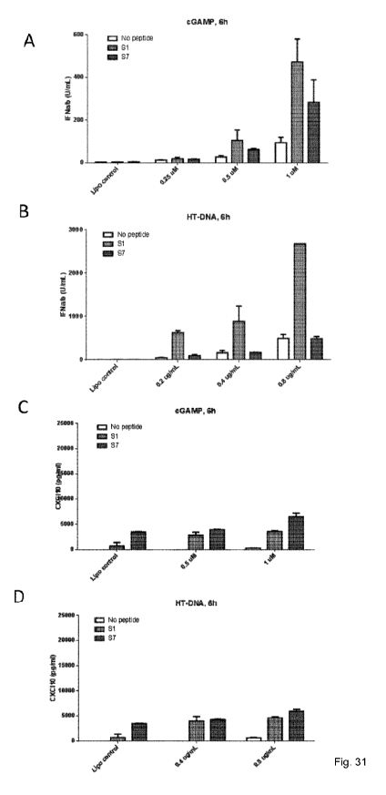
Figure 31. Preactivation of human primary macrophages with specific peptides
and co-
stimulation with cGAMP (panel A+C) or Herring testis DNA (HT-DNA) (panel B+D).
Results show that peptides are able to induce strong immune responses 6 hours
post
stimulation, measured by type I IFN (panel A+B) and T-cell recruitment
cytokine
CXCL10 (panel C+D).
Figure 32. Evaluation of peptide with N- and C-terminus modifications. Results
show
that peptides without biotin (-B) (SEQ ID NO: 25) is capable of inducing type
I IFN
(panel A) and CXCL10 (panel B) response in humane macrophages co-stimulated
with
HT-DNA that is significantly higher as compared to none-peptide treated cells.
In
addition, peptides without biotin (-B) respond in a similar manner as peptides
that
include both biotin and cell penetrating motif (51 (SEQ ID NO: 25). Peptides
without
CA 03069780 2020-01-13
WO 2018/029256 PCT/EP2017/070208
13
cell penetrating domain (-T) (SEQ ID NO: 26) but with biotin demonstrate
decreased
efficacy on CXCL10 production (panel B) but still strong typel IFN signalling
(panel A).
Figure 33. In vivo evaluation of peptides. Results show that subcutaneous
injection with
low doses of peptides generate strong innate immune activation 6 hours p.i. in
C57BL/6J mice, measured by fold induction of IFNb, CXCL10 and IFIT2 mRNA
expression. Peptides without cell penetration motif (-CPP) (SEQ ID NO: 26 (51
(-CPP))
and SEQ ID NO: 28 (S7 (-CPP))) demonstrate similar immune responses as
peptides
including the motif. Mock treated mice were injected with physiological salt
water.
Figure 34. Specifications for purification of peptide 51 (SEQ ID NO: 15)
Detailed description
Definitions
The term "comprising" should be understood in an inclusive manner. Hence, by
way of
example, a composition comprising compound X, may comprise compound X and
optionally additional compounds.
The term "polypeptide" as used herein refers to a chain of amino acid monomers
linked
by peptide (amide) bonds. Said chain may comprise any number of amino acid
monomers, but typically comprise at least 5 amino acids. The polypeptide may
comprise any amino acid, however preferably consists of naturally occurring
amino
acids.
The term "small organic molecules or compounds" refers herein to non-
oligomeric,
carbon containing compounds producible by chemical synthesis and generally
having a
size of less than 600 mass units.
Compound capable of binding to the pyrin-domain of 1F116
The invention relates to compounds capable of binding to the pyrin-domain of
1F116. In
particular said compounds may be capable of binding directly to the pyrin-
domain of
1F116. In particular, said compound may be a compound, which is capable of
inhibiting
1F116 activity and/or STING activity as described herein below in the section
"IFI16
CA 03069780 2020-01-13
WO 2018/029256 PCT/EP2017/070208
14
activity and STING activity". Such compounds may herein also be referred to
"IFI16
pyrin inhibitor" or simply as "compound of the invention".
The 1F116 pyrin inhibitor may be any compound capable of binding to the pyrin-
domain
of 1F116 or a fragment thereof. The pyrin-domain of 1F116 is described herein
below in
more detail in the section "IFI16". It is preferred that the compound is
capable of
selectively binding the pyrin-domain of 1F116, and thus said compound
preferably binds
the pyrin-domain of 1F116 with at least 10 times higher affinity than to a non-
specific
polypeptide (e.g. BSA). It may further be preferred that said compound binds
the pyrin-
domain of 1F116 with higher affinity, e.g, with at least 2x higher affinity
than it binds to
any other polypeptide.
In some embodiments the compound may be capable of binding the pyrin-domain of
1F116 or a fragment thereof with an affinity corresponding to a KD of about 10-
7 M or
less, such as about 10-8 M or less, such as about 10-9 M or less, for example
about
10-10 M or less, or even about 10-11M or even less.
The 1F116 pyrin inhibitor may be any kind of compound. In one embodiment the
1F116
pyrin inhibitor is a small molecule interacting with the pyrin-domain of
1F116. The small
molecule may in particular be a small organic molecule. Typically, small
molecules,
such as small organic molecules are molecules of 600 mass units or less.
In another embodiment the 1F116 pyrin inhibitor is a polypeptide. Polypeptides
capable
of binding to the pyrin-domain of 1F116 may be identified in any useful
manner, for
example by screening a library of test polypeptides with the pyrin-domain of
1F116 or a
fragment thereof for polypeptides capable of binding the pyrin-domain of
1F116. Non-
limiting examples of methods for identifying polypeptides capable of binding
the pyrin-
domain of 1F116 include phage display. phage-display peptide biopanning; pull-
down
binding competition assays; Fluorescent Resonance Energy Transfer assay
(FRET);
Biocore analysis; or Database/Bioinformatics based methods.
In one embodiment of the invention the 1F116 pyrin inhibitor is an antibody,
an antigen-
binding fragment of an antibody or a synthetic antibody specifically binding
the pyrin-
domain of 1F116 or a fragment thereof.
CA 03069780 2020-01-13
WO 2018/029256 PCT/EP2017/070208
The antibody may be any antibody. For example, the antibody may be a naturally
occurring antibody or a functional homologue thereof. A naturally occurring
antibody is
a heterotetrameric glycoproteins capable of recognising and binding an antigen
comprising two identical heavy (H) chains and two identical light (L) chains
inter-
5 connected by disulfide bonds. Each heavy chain comprises or preferably
consists of a
heavy chain variable region (abbreviated herein as VH) and a heavy chain
constant
region (abbreviated herein as CH). Each light chain comprises or preferably
consists a
light chain variable region (abbreviated herein as VL) and a light chain
constant region
(abbreviated herein as CO. The VH and VI_ regions can be further subdivided
into
10 regions of hypervariability, termed complementarity determining regions
(CDRs),
interspersed with regions that are more conserved, termed framework regions
(FRs).
The naturally occurring antibody may also be a heavy-chain antibody (HCAbs) as
produced by camelids (camels, dromedaries and llamas). HCAbs are homodimers of
15 heavy chains only, devoid of light chains and the first constant domain
(Hamers-
Casterman et al., 1993).
The naturally occurring antibody according to the invention may for example be
selected from the group consisting of IgG, IgM, IgA, IgD and IgE. The subunit
structures and three-dimensional configurations of these different classes of
immunoglobulins are well known.
Naturally occurring antibodies according to the invention may be antibodies of
a
particular species, for example the antibody may be a murine, a rat, a rabbit,
a goat, a
sheep, a chicken, a donkey, a camelid or a human antibody. The antibody
according to
the invention may however also be a hybrid between antibodies from several
species,
for example the antibody may be a chimeric antibody, such as a humanised
antibody.
The antibody according to the invention may be a monoclonal antibody, such as
a
naturally occurring monoclonal antibody or it may be polyclonal antibodies,
such as
naturally occurring polyclonal antibodies.
The antigen binding fragment of an antibody may be any protein or polypeptide
containing an antigen binding site. Preferably, the antigen binding site
comprises at
least one CDR, or more preferably a variable region.
CA 03069780 2020-01-13
WO 2018/029256 PCT/EP2017/070208
16
Thus the antigen binding site may comprise a VH and/or VL. It is preferred
that the
antigen binding site comprises one or more CDRs, preferably at least 1, more
preferably at least 2, yet more preferably at least 3, even more preferably at
least 4, yet
more preferably at least 5, even more preferably 6 CDRs. It is preferable that
the
antigen binding site comprises at least one CDR3, more preferably at least the
CDR3
of the heavy chain.
The antigen binding fragment of antibody may also be a heterospecific
antibody, a
single chain antibody or a recombinant antibody. The fragments may also be Fab
fragments or scFv.
Synthetic antibodies may for example be recombinant antibodies, nucleic acid
aptamers and non-immunoglobulin protein scaffolds.
Recombinant antibodies may be generated in vitro by expression from
recombinant
genes. The recombinant genes may be based on antibody genes from any species
of
antibody-producing animal, which optionally may be manipulated to generate new
antibodies or antibody fragments, such as Fab fragments and scFv.
Synthetic antibodies may also be non-immunoglobulin derived. Such molecules
typically differ in structure to that of an antibody and can for example be
generated
from nucleic acids, as in the case of aptamers, or from protein scaffolds, for
example
peptide aptamers, into which hypervariable loops are inserted to form the
antigen
binding site.
The synthetic antibody may also be an affimer protein, which is a small robust
affinity
reagents with a molecular weight of 12-14kDa. Affimers are engineered to bind
to their
target proteins with high affinity and specificity. The Affimer protein
scaffold is derived
from the cysteine protease inhibitor family of cystatins, which contains two
variable
peptide loops and a variable N-terminal sequence, which can be engineered to
provide
a high affinity binding surface for the pyrin-domain of IF116.
Pyrin-domain of IF116 analogues
CA 03069780 2020-01-13
WO 2018/029256 PCT/EP2017/070208
17
The invention also relates to compounds capable of mimicking the pyrin-domain
of
IF116. The term "mimicking", as used herein in relation to the pyrin-domain of
IF116 is
meant to indicate that the relevant compound is capable of exerting the same
inducing
effect of STING activity as IF116. Such compounds are herein referred to as
"pyrin-
domain analogues". Preferably, said pyrin-domain analogues are capable of
inducing
STING activity. Thus, the pyrin-domain analogues may be capable of inducing
any of
the STING activities described herein below in the section "IFI16 activity and
STING
activity". In particular, the pyrin-domain analogue may be capable of
facilitating
interaction between TBK1 and STING.
The pyrin-domain analogues may be any kind of compound. In one embodiment the
IF116 pyrin inhibitor is a small molecule capable of mimicking the pyrin-
domain of IF116.
The small molecule may in particular be a small organic molecule. Typically,
small
molecules, such as small organic molecules are molecules of 600 mass units or
less.
Preferably, the pyrin-domain analogue is a polypeptide. Polypeptides capable
of
inducing STING activity may be identified in any useful manner, for example by
screening a library of test polypeptides for polypeptides capable of inducing
STING
activity.
In preferred embodiments the pyrin-domain analogue is a polypeptide comprising
the
pyrin-domain of IF116 or a fragment thereof, wherein said polypeptide
optionally may
be conjucated to a conjugated moiety, such as at least one conjugated moiety.
In particular, the pyrin-domain analogue may be a polypeptide comprising:
o the pyrin-domain of human IF116 (human pyrin-domain) provided herein
as SEQ ID NO:1;
o a fragment of said human pyrin-domain consisting of a consecutive
sequence of at least 5 amino acids of SEQ ID NO:1; or
o a functional homologue of the human pyrin-domain sharing at least 70%
sequence identity with SEQ ID NO:1,
wherein the polypeptide optionally may be conjugated to a conjugated moeity
Thus, the invention also relates to polypeptides comprising or consisting of
the pyrin-
domain of IF116 or a fragment thereof, wherein said pyrin-domain or fragment
thereof
CA 03069780 2020-01-13
WO 2018/029256 PCT/EP2017/070208
18
may be any of the pyrin-domains or fragments thereof described herein below in
the
section "IFI16".
Polypeptides comprising the pyrin-domain or a fragment thereof according to
the
present invention are preferably not too large. Accordingly it may be
preferred that such
polypeptide consists of at the most 150 amino acids, such as of the most 100
amino,
for example at the most 80 amino acids.
In one embodiment, the polypeptide of the present invention is selected from
SEQ ID
NO: 5-28, as recited herein below, wherein the underlined sequence indicate a
conjugated cell penetrating motif:
SEQ ID NO: 5: KKYKNIVLLKGLEVINDYHFGRKKRRQRRRPQ-NH2
SEQ ID NO: 6: LEVINDYHFRMVKSLLSNDLGRKKRRQRRRPQ-NH2
SEQ ID NO: 7: LLSNDLKLNLKMREEYDKIQGRKKRRQRRRPQ-NH2
SEQ ID NO: 8: EEYDKIQIADLMEEKFRGDGRKKRRQRRRPQ-NH2
SEQ ID NO: 9: DLMEEKFRGDAGLGKLIKIFGRKKRRQRRRPQ-NH2
SEQ ID NO: 10: AGLGKLIKIFEDIPTLEDLAGRKKRRQRRRPQ-NH2
SEQ ID NO: 11: EDIPTLEDLAETLKKEKLKGRKKRRQRRRPQ-NH2
SEQ ID NO: 12:
NDLKLNLKMREEYDKIQIADLMEEKFRGDAGLGKLIKIFEDIPTLEDLAETLKKEKLKGR
KKRRQRRRPQ-NH2
SEQ ID NO: 13:
KKYKNIVLLKGLEVINDYHFRMVKSLLSNDLKLNLKMREEYDKIQIADLMEEKFGRKKR
RQRRRPQ-NH2
SEQ ID NO: 14:
HFRMVKSLLSNDLKLNLKMREEYDKIQIADLMEEKFRGDAGLGKLIKIFEGRKKRRQR
RRPQ-NH2
SEQ ID NO: 15:
51: Biotin-KKYKNIVLLKGLEVINDYHFGRKKRRQRRRPQ-NH2
SEQ ID NO: 16:
S2: Biotin-LEVINDYHFRMVKSLLSNDLGRKKRRQRRRPQ-NH2
SEQ ID NO: 17:
S3: Biotin-LLSNDLKLNLKMREEYDKIQGRKKRRQRRRPQ-NH2
SEQ ID NO: 18:
S4: Biotin-EEYDKIQIADLMEEKFRGDGRKKRRQRRRPQ-NH2
CA 03069780 2020-01-13
WO 2018/029256 PCT/EP2017/070208
19
SEQ ID NO: 19:
S5: Biotin-DLMEEKFRGDAGLGKLIKIFGRKKRRQRRRPQ-NH2
SEQ ID NO: 20:
S6: Biotin-AGLGKLIKIFEDIPTLEDLAGRKKRRQRRRPQ-NH2
SEQ ID NO: 21:
S7: Biotin-EDIPTLEDLAETLKKEKLKGRKKRRQRRRPQ-NH2
SEQ ID NO: 22:
L1: Biotin-
NDLKLNLKMREEYDKIQIADLMEEKFRGDAGLGKLIKIFEDIPTLEDLAETLKKEKLKGR
KKRRQRRRPQ-NH2
SEQ ID NO: 23:
L2: Biotin-
KKYKNIVLLKGLEVINDYHFRMVKSLLSN DLKLNLKMREEYDKIQIADLMEEKFGRKKR
RQRRRPQ-NH2
SEQ ID NO: 24:
L3: Biotin-
HFRMVKSLLSNDLKLNLKMREEYDKIQIADLMEEKFRGDAGLGKLIKIFEGRKKRRQR
RRPQ-NH2
The polypeptide may optionally be additionally conjugated to at least one
moiety. The
at least one conjugated moieties can be attached at the N-terminus or the C-
terminus
or even to an amino acid sidechain of the polypeptide.
In one embodiment the conjugated moiety is a peptide, a sugar, a lipid, a cell-
penetrating peptide (CPP) or any other chemical group that can be covalently
linked to
a polypeptide. The conjugated moiety may also improve physical properties of
the
polypeptide, such as its solubility, stability or half-life. In one
embodiment, the
conjugated moiety is a detectable moiety that could be used for imaging of the
polypeptide; for example, the conjugated moiety is a biotin molecule.
Specifically, the
polypeptide may be conjugated to one or more fatty acids or fatty acid-like
moieties in
order to prolong in vivo half-life.
In one embodiment, the conjugated moiety may be a compound that masks the
polypeptide from the host immune system, such as a polyethylene glycol (PEG)
CA 03069780 2020-01-13
WO 2018/029256
PCT/EP2017/070208
polymer chain or a modified PEG, for example NPEG. PEG or modified PEG may
also
prolong the in vivo half-life of the peptide.
In one embodiment, the polypeptide comprises an N-terminal biotin conjugated
moiety
5 and a C-terminal CPP conjugated moiety.
In one preferred embodiment, the polypeptide comprises a C-terminal CPP
conjugated
moiety.
10 IF116
Interferon-gamma-inducible protein 16 (IF116) is a cytosolic and nuclear
protein also
known as interferon-inducible myeloid differentiation transcriptional
activator. In
humans 1F116 is encoded by the IF116 gene, and the amino acid sequence of
human
1F116 is provided herein as SEQ ID NO:2.
1F116 contains several domains including a pyrin-domain, 2 HIN domains (HIN-A
and
HIN-B) and a BFP domain. An overview of the domain structure of 1F116 is
provided
herein in figure 5H. Three isoforms of 1F116 exists, which are generated by
alternative
splice sites. All three isoforms contain the Pyrin and HIN domains. The
present
invention relates to pyrin-domain analogues e.g. polypeptides comprising or
consisting
of the pyrin-domain of 1F116, as well as to compounds capable of binding the
pyrin-
domain of 1F116.
In human 1F116 the pyrin-domain is positioned at aa 10 to 88 of SEQ ID NO:2.
Pyrin-
domains of other 1F116 proteins can be determined by aligning the 1F116 to
human
1F116 of SEQ ID NO:2 and identifying the amino acids corresponding to amino
acid 10
to 88 of SEQ ID NO:2.
The pyrin-domain of 1F116 may in particular be the pyrin-domain of human
1F116. The
amino acid sequence of human 1F116 is provided herein as SEQ ID NO:1.
The pyrin-domain of IF116 may however also be a functional homologue of the
pyrin-
domain of human 1F116 sharing at least 70%, such as at least 75%, for example
at
least 80%, such as at least 85%, for example at least 90%m, such as at least
95%, for
example at least 98% sequence identity with SEQ ID NO:1. A functional
homologue of
CA 03069780 2020-01-13
WO 2018/029256 PCT/EP2017/070208
21
the pyrin-domain of human 1F116 preferably has one or more of the activities
of the
1F116 described herein below in the section "IFI16 activity and STING
activity".
The invention also relates to fragments of the pyrin domain of 1F116 as well
as to
compounds binding such fragments. Fragments of the pyrin-domain of 1F116 may
be
any fragment of any of the pyrin domains described above. Typically, the
fragments
comprise at least 5 consecutive amino acids of a pyrin-domain of 1F116.
In one embodiment, the fragment comprise at least 5, such as at least 10, for
example
at least 15, such as at least 20, for example in the range of 5 to 70, such as
in the
range of 5 to 60, for example in the range of 5 to 50, such as in the range of
10 to 70,
for example in the range of 10 to 60, such as in the range of 10 to 50
consecutive
amino acids of the pyrin-domain of human 1F116 of SEQ ID NO:1.
In another embodiment, the fragment comprise at least 5, such as at least 10,
for
example at least 15, such as at least 20, for example in the range of 5 to 70,
such as in
the range of 5 to 60, for example in the range of 5 to 50, such as in the
range of 10 to
70, for example in the range of 10 to 60, such as in the range of 10 to 50
consecutive
amino acids of a functional homologue of the pyrin-domain of human 1F116 of
SEQ ID
NO:1.
It may be preferred that aforementioned fragments of the pyrin-domain of IF116
also
retain one or more of the activities of 1F116 described herein below in the
section "IFI16
activity and STING activity".
Polypeptides
In generally preferred embodiments, the "IFI16 pyrin inhibitor" and/or "pyrin-
domain
analogues" as defined herein above are polypeptides. In a preferred
embodiment, the
polypeptide is selected from the group consisting of SEQ ID NO: 5-28, as
described
elsewhere herein.
In certain embodiment, the polypeptides additionally comprise one or more
conjugated
moieties. For example, the polypeptide may comprise an N- or C-terminal biotin
moiety.
In preferred embodiments, the polypeptide comprises a cell-penetrating peptide
(CPP),
which can be attached to the N- or C-terminus of a polypeptide of the
invention or even
CA 03069780 2020-01-13
WO 2018/029256 PCT/EP2017/070208
22
attached to one or more side chains. Cell-penetrating peptides (CPPs) are
short
peptides that facilitate cellular intake/uptake of the IF116 pyrin inhibitor
and/or pyrin-
domain analogues of the present invention. CPPs typically have an amino acid
composition that either contains a high relative abundance of positively
charged amino
acids such as lysine or arginine or has sequences that contain an alternating
pattern of
polar/charged amino acids and non-polar, hydrophobic amino acids. These two
types
of structures are referred to as polycationic or amphipathic, respectively. A
third class
of CPPs are the hydrophobic peptides, containing only apolar residues, with
low net
charge or have hydrophobic amino acid groups that are crucial for cellular
uptake.
CPPs can mediate cell penetration through different pathways, such as be
direct
penetration, endocytosis-mediated translocation, or translocation through the
formation
of a transitory structure (e.g. inverted micelles).
In one preferred embodiment, the CPP is the HIV TAT sequence or a modification
thereof.
Peptides of the present invention may be manufactured by standard chemical
synthetic
methods, or by using recombinant expression systems, or by any other suitable
state-
of-the-art method. Thus, the peptides of the invention may be synthesized in a
number
of ways, including, inter alia, methods comprising:
(a) synthesizing the peptide by means of solid-phase or liquid-phase
methodology,
either stepwise or by fragment assembly, and isolating and purifying the final
peptide
product; or
(b) expressing a nucleic acid construct that encodes the peptide in a host
cell, and
recovering the expression product from the host cell culture; or
(c) effecting cell-free in vitro expression of a nucleic acid construct that
encodes the
peptide, and recovering the expression product;
or employing any combination of methods as in (a), (b) and (c) to obtain
fragments of
the peptide, subsequently joining (e.g., ligating) the fragments to obtain the
complete
peptide, and recovering the peptide.
It may be preferable to synthesize compounds of the invention by means of
solid-phase
or liquid-phase peptide synthesis, the methodology of which is well known to
persons
of ordinary skill in the art of peptide synthesis. Reference may also be made
in this
respect to, for example, Fields, G.B. et al., 2002, "Principles and practice
of solid-phase
peptide synthesis" in: Synthetic Peptides (2nd Edition), and examples provided
therein.
CA 03069780 2020-01-13
WO 2018/029256 PCT/EP2017/070208
23
In one embodiment, the polypeptides are synthesized on a peptide synthesizer
using
standard Fmoc-peptide synthesis, using HBTU as activator and N-
methylmorpholine as
the tertiary amine during activations. NMP (n'-methyl pyrrolidone) may be used
as
solvent. The coupling times may be approximately 1h at RT. The peptides may
also be
side-chain deprotected in TFA:EDT:TIPS:H20 94:2:1:3. After precipitation in
diethyl
ether, the peptides should be dissolved, e.g. in H20, and purified on a 018-
column in
water acetonitrile gradients containing 0.1%TFA. Choice of resin is within the
capabilities of those of skill in the art, however, a preferred suitable resin
is resin
polystyrene aminomethyl-resin, which is preferable derivatized with a Rink-
amide
linker. Polypeptides are preferably provided with at least 90% purity.
Administration
Pharmaceutical compositions of the invention may be administered to a patient
in need
of such treatment at various sites, for example administration at sites which
bypass
absorption, such as in an artery or vein or in the brain, and at sites which
involve
absorption, such as in the skin, under the skin, in a muscle or in the
abdomen. More
generally, administration of pharmaceutical compositions according to the
invention
may be by a variety of routes of administration, such as for example
parenteral,
intracranial, epidermal, dermal or transdermal routes. In some embodiments,
other
routes such as lingual, sublingual, buccal, oral, vaginal or rectal may be
useful.
Parenteral administration (of a pharmaceutical composition of the invention)
may be
performed, for example, by subcutaneous, intramuscular, intraperitoneal or
intravenous
injection by means of a syringe, for example a pen-like syringe.
Alternatively,
parenteral administration can take place by means of an infusion pump, e. g.
in the
form of a device or system borne by a subject or patient and advantageously
comprising a reservoir containing a liquid composition of the invention and an
infusion
pump for delivery/administration of the composition to the subject or patient,
or in the
form of a corresponding miniaturized device suitable for implantation within
the body of
the subject or patient.
IF116 activity and STING activity
The invention relates to pyrin-domain analogues, e.g. polypeptides comprising
the
pyrin-domain of IF116 or fragments thereof. Said pyrin-domain of IF116 or
fragments
thereof preferably has one or more of the IF116 activities described in this
section.
CA 03069780 2020-01-13
WO 2018/029256 PCT/EP2017/070208
24
The invention also relates to compounds capable of binding the pyrin-domain of
IF116.
Preferably said compounds are capable of inhibiting one or more of the IF116
activities
described in this section.
The invention demonstrates that IF116 is capable of interacting with the TANK-
binding
kinase 1 (TBK1). The amino acid sequence of human TBK1 is provided herein as
SEQ
ID NO:3.
In one embodiment of the invention it is preferred that the pyrin-domain of
IF116 as well
as fragments thereof are capable of interacting with TBK1. It is also
preferred that IF116
pyrin inhibitors are capable of inhibiting or at least reducing interaction
between IF116
and TBK1. Reduction of interaction is preferably at least a 2-fold reduction
of the
interaction. Interaction with TBK1 may for example be determined by
immunoprecipitation of IF116, the pyrin-domain of IF116 or fragments thereof
using
antibodies to IF116 or said fragments, and subsequent detection of TBK1
precipitating
with IF116 or fragments thereof, e.g. by Western blotting with antibodies to
TBK1. The
interaction may also be performed in the reverse manner, by
immunoprecipitation of
TBK1 using antibodies to TBK1, and subsequent detection of IF116, the pyrin-
domain
of IF116 or a fragment thereof precipitating with TBK1, e.g. by Western
blotting. One
non-limiting example of determining interaction between IF116 and TBK1 is
described
herein below in Example 1 in the section "IFI16 recruits TBK1 to STING to
initiate IRF3
activation".
The invention demonstrates that IF116 is capable of interacting with the
endoplasmic
reticulum-bound protein stimulator of interferon genes (STING). The amino acid
sequence of human STING is provided herein as SEQ ID NO:4.
In one embodiment of the invention it is preferred that the pyrin-domain of
IF116 as well
as fragments thereof are capable of interacting with STING. It is furthermore,
preferred
that said pyrin-domain of IF116 as well as fragments thereof are capable of
increasing
STING activity. It is also preferred that IF116 pyrin inhibitors are capable
of inhibiting or
at least reducing interaction between IF116 and STING. Reduction of
interaction is
preferably at least a 2-fold reduction of the interaction. Interaction with
STING may for
example be determined by immunoprecipitation of IF116, the pyrin-domain of
IF116 or
CA 03069780 2020-01-13
WO 2018/029256 PCT/EP2017/070208
fragments thereof using antibodies to IF116 or said fragments, and subsequent
detection of STING precipitating with IF116 or fragments thereof, e.g. by
Western
blotting with antibodies to STING. The interaction may also be performed in
the reverse
manner, by immunoprecipitation of STING using antibodies to STING, and
subsequent
5 detection of IF116, the pyrin-domain of IF116 or a fragment thereof
precipitating with
STING, e.g. by Western blotting. One non-limiting example of determining
interaction
between IF116 and STING is described herein below in Example 1 in the section
"IFI16
recruits TBK1 to STING to initiate IRF3 activation".
10 The invention also demonstrates that IF116 is capable recruiting TBK1 to
STING. In
one embodiment of the invention it is preferred that the pyrin-domain of IF116
as well as
fragments thereof are capable of facilitating interaction between TBK1 and
STING. It is
also preferred that IF116 pyrin inhibitors are capable of inhibiting or at
least reducing
interaction between TBK1 and STING. Reduction of interaction is preferably at
least a
15 2-fold reduction of the interaction. Interaction between TBK1 and STING
may for
example be determined by immunoprecipitation of TBK1 using antibodies to TBK1,
and
subsequent detection of STING precipitating with TBK1, e.g. by Western
blotting with
antibodies to STING. The interaction may also be performed in the reverse
manner, by
immunoprecipitation of STING using antibodies to STING, and subsequent
detection of
20 TBK1 precipitating with STING, e.g. by Western blotting. One non-
limiting example of
determining interaction between IF116 and STING is described herein below in
Example 1 in the section "IFI16 recruits TBK1 to STING to initiate IRF3
activation".
The invention also demonstrates that the pyrin domain of IF116 is involved in
STING
25 activation through direct binding of cyclic-di-nucleotides (CDNs). In
one embodiment of
the invention it is preferred that the pyrin-domain of IF116 as well as
fragments thereof
are capable of inducing STING activation, in particular the pyrin-domain of
IF116 as well
as fragments thereof are capable of inducing STING activation in the presence
of
CDNs. It is also preferred that IF116 pyrin inhibitors are capable of
inhibiting or at least
reducing STING activation e.g. following the "introduction of" or "stimulation
with" CDNs
or any small molecule derived of or similar to CDNs.
STING activation may be determined in a number of different ways including the
following:
CA 03069780 2020-01-13
WO 2018/029256 PCT/EP2017/070208
26
STING activation may be determined by determining STING phosphorylation. Thus,
it
may be preferred that the pyrin domain of IF116 or fragments thereof are
capable of
inducing phosphorylation of STING, e.g inducing an at least 2 fold increase in
phosphorylation of STING. It is also preferred that IF116 pyrin inhibitors are
capable of
inhibiting or at least reducing phosphorylation of STING. Thus, preferably
said IF116
pyrin inhibitors are capable of reducing phosphorylation of STING at least 2-
fold. Said
phosphorylation of STING may in particular be phosphorylation of 5er366of
STING of
SEQ ID NO:4.
Phosphorylation of STING, and particularly phosphorylation of 5er366of STING
of SEQ
ID NO:4 may be determined in any useful manner, for example as described
herein
below in Example 1 in the section "The 1F116 PYR1N domain is essential for
promoting
cGAMP-mediated STING signalling".
STING activation may also be determined as activation of expression of type I
IFN or
inflammatory cytokines in cells capable of expressing type I IFN or cytokines.
Examples of such cells include macrophages, dendritic cells, keratinocytes,
fibroblasts,
monocytes, epithelia cells, B cells, or NK cells. Thus, STING activation may
be
determined by determining expression of type I IFN or cytokines in such cells.
Thus, it
may be preferred that the pyrin domain of IF116 or fragments thereof are
capable of
inducing expression of type I IFN or cytokines in such cells, e.g. inducing an
at least 2
fold increase in expression of type I IFN in such cells, e.g. in macrophages.
It is also
preferred that IF116 pyrin inhibitors are capable of inhibiting or at least
reducing
expression of type I IFN or cytokines in such cells, e.g. in macrophages.
Thus,
preferably said IF116 pyrin inhibitors are capable of reducing expression of
type I IFN or
of cytokines from such cells, e.g. macrophages by at least 2-fold.
Expression of type I IFN or cytokines may be determined by any useful manner,
for
example as described herein below in Example 1.
STING activation may also be determined as activation of IFN6 promoter
activity. Thus,
it may be preferred that the pyrin domain of IF116 or fragments thereof are
capable of
activating IFN6 promoter activity, e.g inducing an at least 2 fold increase in
IFN6
promoter activity. It is also preferred that IF116 pyrin inhibitors are
capable of inhibiting
CA 03069780 2020-01-13
WO 2018/029256 PCT/EP2017/070208
27
or at least reducing activity of the IFN6 promoter. Thus, preferably said
IF116 pyrin
inhibitors are capable of reducing activity of the IFN6 promoter by at least 2-
fold.
Activity of the IFN6 promoter may for example be determined in recombinant
cells
comprising a nucleic acid construct encoding a reporter protein under the
control of the
IFN6 promoter. IFN6 promoter can also be determined in cell free expression
systems
allowing expression of a reporter protein under the control of the IFN6
promoter. A non-
limiting useful method for determining IFN6 promoter activity is described
herein below
in Example 1 in the section "The IF116 PYRIN domain is essential for promoting
cGAMP-mediated STING signalling".
In one embodiment of the invention it is preferred that the pyrin-domain of
IF116 as well
as fragments thereof are capable of binding to the caspase recruitment domain
(CARD), which is contained in different proteins including the apoptotic speck
protein
(ASC). It is also preferred that the IF116 pyrin inhibitors of the invention
are capable of
inhibiting or at least reducing interaction between IF116 and CARD containing
proteins,
such as ASC. Reduction of interaction is preferably at least a 2-fold
reduction of the
interaction. Interaction between IF116 and CARD containing proteins such as
ASC may
for example be determined by immunoprecipitation of either protein or a
fragment
thereof, and subsequent detection of co-precipitating of the other protein.
ASC is an
adaptor protein necessary for the assemble of the IF116 inflammasome, and
accordingly IF116 pyrin inhibitors may block inflammasome mediated by CARD
containing proteins such as ASC.
Method of identifying
In one embodiment the invention relates to a method of identifying a compound
capable of binding the pyrin-domain of IF116, said method comprising the steps
of
= providing a pyrin-domain of IF116 or a fragment thereof
= providing a library of test compounds
= contacting the pyrin-domain of IF116 with said test compounds
= detecting and isolating test compounds, which interact with the pyrin-
domain of IF116 or the fragment thereof
thereby identifying a compound capable of binding the pyrin-domain of IF116.
Said compound may be useful as an anti-inflammatory agent, as an inhibitor
of STING or as an I-IFN antagonist.
CA 03069780 2020-01-13
WO 2018/029256 PCT/EP2017/070208
28
Said pyrin-domain of IF116 or fragment thereof may be any of the pyrin-domains
of
IF116 or fragments thereof described herein above in the section "IFI16".
The test compounds may be any of kind of compound, for example the test
compounds
may be selected from the group consisting of peptides, small organic
molecules,
antibodies, antigen binding fragments of antibodies and synthetic antibodies,
for
example any of the peptides, small organic molecules, antibodies, antigen
binding
fragments of antibodies and synthetic antibodies described herein above in the
section
"Compound capable of binding to the pyrin-domain of IF116".
In one embodiment the invention relates to a method of identifying a compound
capable of mimicking the pyrin-domain of IF116, said method comprising the
steps of
= providing a library of test compounds
= testing whether said test compounds are capable of inducing STING
activity
thereby identifying a compound capable of mimicking IF116.
The test compounds may be any of kind of compound, for example the test
compounds
may be selected from the group consisting of polypeptides and small organic
molecules, for example any of the polypeptides and small organic molecules
described
herein above in the section "Pyrin-domain of IF116 analogues".
The libraries may comprise any suitable number of test compounds, for example
at
least 100 different test compounds, such as 1000 different test compounds, for
example at least 10,000 different test compounds, such as 100,000 different
test
compounds.
The libraries may be in any useful format. Thus, the library may simply be a
mixture of
compounds. When the test compounds are peptides, then the library may be in
form of
organisms, vire or phages expressing the test compounds. It is also possible
that the
test compounds of the library are spatially separated from each other to allow
easy
identification of the compound(s) capable of binding the pyrin-domain of IF116
or a
fragment thereof. Spatial separation may be achieved in a number of ways, for
CA 03069780 2020-01-13
WO 2018/029256
PCT/EP2017/070208
29
example by use of small containers, such a microtiter plates or the test
compounds of
the library may be immobilised on solid support.
Disorder associated with STING activity
In one embodiment, the invention relates to compounds capable of binding to
the pyrin-
domain of 1F116 or a fragment thereof for use in the treatment of a disorder
associated
with STING activity. Said compounds may for example be any of the compounds
described herein above in the section "Compound capable of binding the pyrin-
domain
of 1F16, in particular the compound may be any of the 1F116 pyrin inhibitors
described
herein.
The disorder associated with STING activity may for example be a disorder
characterised with by increased STING activity or by undesired STING activity.
Said
STING activity may for example be any of the activities described herein above
in the
section "IFI16 activity and STING activity". The disorder may also be
associated with
TBK1 activity.
= Numerous disorders have been associated with STING activity for example
as
described in any of the following references: STING-mediated DNA sensing
promotes antitumor and autoimmune responses to dying cells.
http://www.ncbi.nlm.nih.gov/pubmed/25385820
= STING Promotes the Growth of Tumors Characterized by Low Antigen icity
via
1DO Activation. http://www.ncbi.nlm.nih.gov/pubmed/26964621
= Intrinsic Self-DNA Triggers Inflammatory Disease Dependent on STING.
http://www.jimmunol.org/content/193/9/4634.1ong
= STING Activation by Translocation from the ER Is Associated with
Infection and
Autoinflammatory Disease. http://www.ncbi.nlm.nih.gov/pubmed/26235147
= Activation of cyclic GMP-AMP synthase by self-DNA causes autoimmune
diseases. http://www.ncbi.nlm.nih.gov/pubmed/26371324
= Therapeutic potential of targeting TBK1 in autoimmune diseases and
interferonopathies. http://www.ncbi.nlm.nih.gov/pubmed/27353409
CA 03069780 2020-01-13
WO 2018/029256 PCT/EP2017/070208
In one embodiment the disorder associated with STING activity is an
inflammatory
disorder. Said inflammatory disorder may for example be selected from the
group
consisting of psoriasis, Crohn's disease and Inflammatory bowel disease (IBD).
5 In one embodiment the disorder associated with STING activity is an auto-
immune
disease. Said autoimmune disease may for example be selected from the group
consisting of systemic lupus erythematosus (SLE), Aicardi-Goutieres syndrome,
Sjogren's syndrome, STING-associated vasculopathy with onset in infancy
(SAVI), Type 1 diabetes and multiple sclerosis.
The disorder may also be both an inflammatory disorder and an auto-immune
disease.
Thus, many auto-immune diseases are also inflammatory disorders.
In one embodiment of the invention the disorder associated with STING activity
is
cancer. In particular, said cancer may be a cancer induced by chronic
inflammatory
signalling. However, cancer types, which are not related to chronic
inflammatory
signaling, are also relevant targets for treatment. For example said cancer
may be a
cutaneous skin tumour, for example basal cell (BCC) or squamous cell carcinoma
(SCC).
Disorder associated with insufficient STING activity
In one embodiment the invention relates to a polypeptide comprising or
consisting of
the pyrin-domain of 1F116 or a fragment thereof for use in the treatment of a
disorder
associated with insufficient STING activity.
As demonstrated by the present invention, the pyrin-domain of 1F116 or a
fragment
thereof may induce STING activity. Accordingly, the pyrin-domain of 1F116 or
fragments
thereof may be useful for treating disorders associated with insufficient
STING activity.
Useful pyrin-domains of 1F116 or fragments thereof are described above in the
section
"IFI16".
In one embodiment the disorder is cancer. Cancer (malignant neoplasm) is a
class of
diseases in which a group of cells display the traits of uncontrolled growth
(growth and
division beyond the normal limits), invasion (intrusion on and destruction of
adjacent
CA 03069780 2020-01-13
WO 2018/029256
PCT/EP2017/070208
31
tissues), and sometimes metastasis (spread to other locations in the body via
lymph or
blood). Most cancers form a tumor but some, like leukemia, do not.
Thus, the disorder may be cancer, for example a cancer selected from the group
consisting of: colon carcinoma, breast cancer, pancreatic cancer, ovarian
cancer,
prostate cancer, fibrosarcoma, myxosarcoma, liposarcoma, chondrosarcoma,
osteogenic sarcoma, chordoma, angiosarcoma, endotheliosarcoma,
lymphangeosarcoma, lymphangeoendothelia sarcoma, synovioma, mesothelioma,
Ewing's sarcoma, leiomyosarcoma, rhabdomyosarcoma, squamous cell carcinoma,
basal cell carcinoma, adenocarcinoma, sweat gland carcinoma, sebaceous gland
carcinoma, papillary carcinoma, papillary adenocarcinomas, cystandeocarcinoma,
medullary carcinoma, bronchogenic carcinoma, renal cell carcinoma, hepatoma,
bile
duct carcinoma, choriocarcinoma, seminoma, embryonal carcinoma, Wilms' tumor,
cervical cancer, testicular tumor, lung carcinoma, small cell lung carcinoma,
bladder
carcinoma, epithelial carcinoma, glioblastomas, neuronomas,
craniopharingiomas,
schwannomas, glioma, astrocytoma, medulloblastoma, craniopharyngioma,
ependymoma, pinealoma, hemangioblastoma, acoustic neuroama, oligodendroglioma,
meningioma, melanoma, neuroblastoma, retinoblastoma, leukemias and lymphomas,
acute lymphocytic leukemia and acute myelocytic polycythemia vera, multiple
myeloma, Waldenstrom's macroglobulinemia, and heavy chain disease, acute
nonlymphocytic leukemias, chronic lymphocytic leukemia, chronic myelogenous
leukemia, Hodgkin's Disease, non-Hodgkin's lymphomas, rectum cancer, urinary
cancers, uterine cancers, oral cancers, skin cancers, stomach cancer, brain
tumors,
liver cancer, laryngeal cancer, esophageal cancer, mammary tumors, childhood-
null
acute lymphoid leukemia (ALL), thymic ALL, B-cell ALL, acute myeloid leukemia,
myelomonocytoid leukemia, acute megakaryocytoid leukemia, Burkitt's lymphoma,
acute myeloid leukemia, chronic myeloid leukemia, and T cell leukemia, small
and
large non-small cell lung carcinoma, acute granulocytic leukemia, germ cell
tumors,
endometrial cancer, gastric cancer, cancer of the head and neck, chronic
lymphoid
leukemia, hairy cell leukemia and thyroid cancer.
The disorder may also be an infection with DNA pathogens, where IFN is
deleterious.
Such disorders include for example malaria or listeria.
CA 03069780 2020-01-13
WO 2018/029256 PCT/EP2017/070208
32
Method of treatment and combination therapy
As described herein the invention in some embodiments relates to compounds
capable
of binding the pyrin-domain of 1F116, as well as to pyrin-domain analogues
e.g.
polypeptides comprising the pyrin-domain of 1F116 or fragments for use in
methods of
treatment. Thus, a method is also provided of treating a disorder associated
with
STING activity comprising administering an 1F116 pyrin inhibitor and/or a
pyrin-domain
analogue, as defined herein above, to an individual in need thereof.
However, the 1F116 pyrin inhibitor and/or pyrin-domain analogue as defined
elsewhere
herein are also provided generally for use in medicine, i.e. for use as a
medicament.
These compounds can be used for the treatment of any clinical condition, which
can be
treated, prevented or ameliorated by modulation of STING activity.
In one aspect, a use is provided of an 1F116 pyrin inhibitor and/or a pyrin-
domain
analogue, as defined herein above, for the preparation of a medicament, for
example
for the preparation of a medicament for the treatment of a disorder associated
with
STING activity, either insufficient or excessive STING activity. As mentioned
above, the
disorder may be any clinical condition, which can be treated, prevented or
ameliorated
by modulation of STING activity.
The uses and methods provided herein for medical use and/or for treatment of a
disorder as specified herein, may also involve a combination therapy, where
the "IFI16
pyrin inhibitor" and/or "pyrin-domain analogues" as defined herein above are
combined
with at least one additional active compound. The at least one additional
active
compound may be administered before, concomitantly or subsequent to the
administration of the 1F116 pyrin inhibitor and/or pyrin-domain analogue.
In one preferred embodiment, the 1F116 pyrin inhibitor and/or pyrin-domain
analogue is
provided for use in the treatment of cancer, and in this embodiment,
administration of
an 1F116 pyrin inhibitor and/or pyrin-domain analogue is administered together
with an
anticancer agent.
This agent is preferably a chemotherapeutic agent. The chemotherapeutic agent
is
preferably administered by systemic administration, for example by intravenous
injection of a solution comprising the chemotherapeutic agent or by oral
administration.
CA 03069780 2020-01-13
WO 2018/029256 PCT/EP2017/070208
33
The chemotherapeutic agent may be selected from alkylating agents, anti-
metabolites,
anti-microtubule agents, topoisomerase inhibitors and cytotoxic antibiotics.
In one embodiment, the chemotherapeutic agent is an alkylating agent. An
alkylating
agent is used in cancer treatment as an antineoplastic agent that attaches an
alkyl
group (CnH2n+1) to DNA. The alkyl group is attached to the guanine base of
DNA, at
the number 7 nitrogen atom of the purine ring. Since cancer cells, in general,
proliferate
faster and with less error-correction than healthy cells, cancer cells are
more sensitive
to DNA damage, alkylated DNA. Dialkylating agents can react with two different
7-N-
guanine residues, and monoalkylating agents can react only with one 7-N of
guanine.
Examples of alkylating agents are Nitrogen mustards, such as Cyclophosphamide,
Mechlorethamine or mustine (HN2) (trade name Mustargen), Uramustine or uracil
mustard, Melphalan, Chlorambucil, lfosfamide and Bendamustine.
Other examples are Nitrosoureas, such as Carmustine, Lomustine and
Streptozocin. In
another embodiment, the alkylating agent is an Alkyl sulfonate, such as
Busulfan. In
another embodiment, the agent is Thiotepa or an analogue thereof.
The chemotherapeutic agent may also be a Platinum-based chemotherapeutic
agent,
which acts as an alkylating agent. These agents do not have an alkyl group,
but
nevertheless damage DNA, by permanently coordinating to DNA to interfere with
DNA
repair. These agents are sometimes referred to as "alkylating-like". Such
agents
include Cisplatin, Carboplatin, Nedaplatin, Oxaliplatin, Satraplatin, and
Triplatin
tetranitrate.
In yet another embodiment, the chemotherapeutic agent is an alkylating agent
selected
from procarbazine, altretamine, tetrazines, such as dacarbazine, mitozolomide
and
temozolomide.
In one embodiment, the chemotherapeutic agent is an alkylating agent, a
topoisomerase inhibitor, such as lrinotecan, which targets type 1
topoisomerase or
Etoposide, which targets type 2 topoisomerase. In another embodiment, the
chemotherapeutic agent is a vascular endothelial growth factor (VEGF)
inhibitor, such
as Bevazizumab.
CA 03069780 2020-01-13
WO 2018/029256 PCT/EP2017/070208
34
In another embodiment, the chemotherapeutic agent is selected from Nitrogen
mustards, such as Cyclophosphamide, Mechlorethamine or mustine (HN2) (trade
name Mustargen), Uramustine or uracil mustard, Melphalan, Chlorambucil,
lfosfamide
and Bendamustine. In another embodiment, the chemotherapeutic agent is
selected
from Nitrosoureas, such as Carmustine, Lomustine and Streptozocin. In another
embodiment, the chemotherapeutic agent is selected from Alkyl sulfonates, such
as
Busulfan. In another embodiment, the chemotherapeutic agent is Thiotepa or an
analogue thereof. In another embodiment, the chemotherapeutic agent is
selected from
Platinum-based chemotherapeutic agents, such as Cisplatin, Carboplatin,
Nedaplatin,
Oxaliplatin, Satraplatin, and Triplatin tetranitrate. In another embodiment,
the
chemotherapeutic agent is selected from procarbazine, altretamine or
tetrazines, such
as dacarbazine, mitozolomide and temozolomide. In another embodiment, the
chemotherapeutic agent is selected from topoisomerase inhibitors such as
amsacrine,
etoposide, etoposide phosphate, teniposide, doxorubicin, irinotecan,
topotecan,
exatecan, lurtotecan. In yet another embodiment, the chemotherapeutic agent is
selected from VEGF inhibitors, such as bevacizumab and ranibizumab.
Notably, the at least one additional active compound provided in the uses and
methods
for medical use and/or for treatment of a disorder as specified herein
together with the
"IFI16 pyrin inhibitor" and/or "pyrin-domain analogues" as defined herein
above may
also be a non-chemotherapeutic agent. In particular, the at least one
additional active
compound is in one embodiment one or more checkpoint inhibitors. Checkpoint
inhibitors are generally drugs that help the body recognize and attack cancer
cells
Moreover, the uses and methods provided herein for medical use and/or for
treatment
of a disorder as specified herein, may also involve a combination therapy,
where the
"IFI16 pyrin inhibitor" and/or "pyrin-domain analogues" as defined herein
above may be
combined with radiation therapy. Thus, of the "IFI16 pyrin inhibitor" and/or
"pyrin-
domain analogues" as defined herein are in certain embodiments administered,
optionally with the at least one additional active compound before, during
and/or after
the treated individual is subjected to radiation therapy. Provision of a
"IFI16 pyrin
inhibitor" and/or a "pyrin-domain analogue" as defined herein in combination
with
radiation therapy serves to boost the STING-dependent immune response, which
is
elicited by the radiation therapy and thereby maximizing the effect of the
radiation
therapy.
CA 03069780 2020-01-13
WO 2018/029256 PCT/EP2017/070208
In certain embodiments, the IF116 pyrin inhibitor and/or pyrin-domain analogue
is
provided for use in the treatment of disorders associated with insufficient or
excessive
STING activity. These disorders include immuno-deficiency disorders or auto-
immune
5 disorders. In these embodiments, the treatment may include one or more
additional
active compounds selected from immuno-modulating agents, such as TLR agonists,
cytokines, chemokines, interleukins, bacterias, vaccines and/or inactivated
viruses.
Pharmaceutical composition
10 Whilst it is possible for the compounds or polypeptides of the present
invention to be
administered as the raw chemical, it is preferred to present them in the form
of a
pharmaceutical composition. Accordingly, the present invention further
provides a
pharmaceutical composition, which comprises an IF116 pyrin inhibitor of the
present
invention or a pharmaceutically acceptable salt thereof, and a
pharmaceutically
15 acceptable carrier therefore. The invention also provides pharmaceutical
compositions
comprising a polypeptide comprising the pyrin-domain of IF116 or a fragment
thereof
and a pharmaceutically acceptable carrier therefore.
The pharmaceutical compositions may be prepared by conventional techniques,
e.g. as
20 described in Remington: The Science and Practice of Pharmacy 2005,
Lippincott,
Williams & Wilkins.
The pharmaceutically acceptable carriers can be either solid or liquid. Solid
form
preparations include powders, tablets, pills, capsules, cachets,
suppositories, and
25 dispersible granules. A solid carrier can be one or more excipients,
which may also act
as diluents, flavoring agents, solubilizers, lubricants, suspending agents,
binders,
preservatives, wetting agents, tablet disintegrating agents, or an
encapsulating
material.
30 Also included are solid form preparations, which are intended to be
converted, shortly
before use, to liquid form preparations for oral administration. Such liquid
forms include
solutions, suspensions, and emulsions. These preparations may contain, in
addition to
the active component, colorants, flavors, stabilizers, buffers, artificial and
natural
sweeteners, dispersants, thickeners, solubilizing agents, and the like.
CA 03069780 2020-01-13
WO 2018/029256 PCT/EP2017/070208
36
The compounds or polypeptides of the present invention may be formulated for
parenteral administration and may be presented in unit dose form in ampoules,
pre-
filled syringes, small volume infusion or in multi-dose containers, optionally
with an
added preservative. The compositions may take such forms as suspensions,
solutions,
or emulsions in oily or aqueous vehicles, for example solutions in aqueous
polyethylene glycol. Examples of oily or non-aqueous carriers, diluents,
solvents or
vehicles include propylene glycol, polyethylene glycol, vegetable oils (e.g.,
olive oil),
and injectable organic esters (e.g., ethyl oleate), and may contain agents
such as
preserving, wetting, emulsifying or suspending, stabilizing and/or dispersing
agents.
Alternatively, the active ingredient may be in powder form, obtained by
aseptic isolation
of sterile solid or by lyophilisation from solution for constitution before
use with a
suitable vehicle, e.g., sterile, pyrogen-free water.
Preferably, the formulation will comprise about 0.5% to 75% by weight of the
active
ingredient(s) with the remainder consisting of suitable pharmaceutical
excipients as
described herein.
Pharmaceutically acceptable salts of the IF116 pyrin inhibitors, where they
can be
prepared, are also intended to be covered by this invention. These salts will
be ones
that are acceptable in their application to a pharmaceutical use.
Pharmaceutically acceptable salts are prepared in a standard manner. If the
parent
compound is a base it is treated with an excess of an organic or inorganic
acid in a
suitable solvent. If the parent compound is an acid, it is treated with an
inorganic or
organic base in a suitable solvent.
The compounds or polypeptides of the invention are in general administered in
an
"effective amount" or an amount necessary to achieve an "effective level" in
the
individual patient. When the "effective level" is used as the preferred
endpoint for
dosing, the actual dose and schedule can vary, depending on inter-individual
differences in pharmacokinetics, drug distribution, and metabolism. The
"effective level"
can be defined, for example, as the blood or tissue level desired in the
patient that
corresponds to a concentration of the compounds or polypeptides according to
the
invention.
CA 03069780 2020-01-13
WO 2018/029256 PCT/EP2017/070208
37
The compounds or polypeptides of the invention may be administered together
with
one or more other active compounds, typically with one or more other active
compounds useful for treatment of the particular disorder to be treated. Thus,
in
embodiments of the invention, wherein the disorder is cancer, the compounds or
polypeptides of the invention may be administered together with one or more
anti-
cancer agents.
Certain embodiments of pharmaceutical compositions of the invention, which
preferably are liquid pharmaceutical compositions, may comprise a compound of
the
invention present in a concentration from about 0.01 mg/ml to about 50 mg/ml,
such as
from about 1 mg/ml to about 20 mg/ml, e. g. from about 1 mg/ml to about
10mg/ml. In
some embodiments, the composition has a pH from 2.0 to 10Ø A pharmaceutical
composition of the invention may further comprise a buffer system,
preservative(s),
isotonicity agent(s), chelating stabilizer(s) and/or surfactant(s).
Particularly useful
embodiments of liquid pharmaceutical compositions of the invention are aqueous
compositions, i.e. compositions comprising water. Such compositions may be in
the
form of an aqueous solution or an aqueous suspension. Preferred embodiments of
aqueous pharmaceutical compositions of the invention are aqueous solutions. In
the
context of the invention the term "aqueous composition" will normally refer to
a
composition comprising at least 50% by weight (50% w/w) of water. Likewise,
the term
"aqueous solution" will normally refer to a solution comprising at least 50
%w/w of
water, and the term "aqueous suspension" to a suspension comprising at least
50%
w/w of water. In some embodiments, a pharmaceutical composition of the
invention
comprises an aqueous solution of a compound (or a pharmaceutically acceptable
salt
or solvate thereof) of the invention present at a concentration of from 0.1
mg/ml or
above, together with a buffer, the composition having a pH from about 2.0 to
about
10.0, such as a pH from about 6.0 to about 8.5, e.g. from about 6.5 to about
8.5, such
as from about 7.0 to about 8.5, or from about 6.5 to about 8Ø In other
embodiments of
a pharmaceutical composition of the invention, the pH of the composition is a
pH
selected from the list consisting of 2.0, 2.1, 2.2, 2.3, 2.4, 2.5, 2.6, 2.7,
2.8, 2.9, 3.0, 3.1,
3.2, 3.3, 3.4, 3.5, 3.6, 3.7, 3.8, 3.9, 4.0, 4.1, 4.2, 4.3, 4.4, 4.5, 4,6,
4.7, 4.8, 4.9, 155.0,
5.1, 5.2, 5.3, 5.4, 5.5, 5.6, 5.7, 5.8, 5.9, 6.0, 6.1, 6.2, 6.3, 6.4, 6.5,
6.6, 6.7, 6.8, 6.9, 7.0,
7.1, 7.2, 7.3, 7.4, 7.5, 7.6, 7.7, 7.8, 7.9, 8.0, 8.1, 8.2, 8.3, 8.4, 8.5,
8.6, 8.7, 8.8, 8.9, 9.0,
9.1, 9.2, 9.3, 9.4, 9.5, 9.6, 9.8, 9.9, and 10Ø The pH of the composition
may be at
least 1 pH unit from (i.e., higher or lower than) the isoelectric point of the
constituent
CA 03069780 2020-01-13
WO 2018/029256 PCT/EP2017/070208
38
compound of the invention, such as at least 2 pH units from (i.e., higher or
lower than)
the isoelectric point of the peptide compound of the invention. In further
embodiments
of buffer-containing pharmaceutical compositions of the invention, the buffer
or buffer
substance is selected from the group consisting of: acetate buffers (e.g.
sodium
acetate), sodium carbonate, citrates (e.g. sodium citrate), glycylglycine,
histidine,
glycine, lysine, arginine, phosphates (e.g. chosen among sodium dihydrogen
phosphate, disodium hydrogen phosphate and trisodium phosphate), TRIS (i.e.,
tris(hydroxymethyl)aminomethane), HEPES (i.e., 4-(2-hydroxyethyl)-1-piperazine-
ethanesulfonic acid), BICINE (i.e., N,N-bis(2- hydroxyethyl)glycine), and
TRICINE (i.e.,
N4tris(hydroxymethyl)methyl]glycine), as well as succinate, malate, maleate,
fumarate,
tartrate, and aspartate buffers, and mixtures thereof.
Preservative
In further embodiments of pharmaceutical compositions of the invention, the
composition comprises a pharmaceutically acceptable preservative. Relevant
preservatives include preservatives selected from the group consisting of:
phenol, o-
cresol, m-cresol, p-cresol, methyl p-hydroxybenzoate, ethyl p-hydroxybenzoate,
propyl
p-hydroxybenzoate, butyl p- 5 hydroxybenzoate, 2-phenoxyethanol, 2-
phenylethanol,
benzyl alcohol, ethanol, chlorobutanol, thiomerosal, bronopol, benzoic acid,
imidurea,
chlorhexidine, sodium dehydroacetate, chlorocresol, benzethonium chloride,
chlorphenesine [i.e. 3-(p-chlorphenoxy)propane-1 ,2-diol] and mixtures
thereof. The
preservative may be present in a concentration of from 0.1 mg/ml to 30 mg/ml,
such as
from 0.1 mg/ml to 20mg/m1(e.g. from 0.1 mg/ml to 5 mg/ml, or from 5 mg/ml to
10
mg/ml, or from 10 mg/ml to 20 mg/ml) in the final liquid composition. The use
of a
preservative in pharmaceutical compositions is well known to the skilled
worker. In this
connection, reference may be made to Remington: The Science and Practice of
Pharmacy, 19th edition, 1995.
isotonicity agent
In further embodiments, a pharmaceutical composition of the invention
comprises an
isotonicity agent (i. e., a pharmaceutically acceptable agent which is
included in the
composition for the purpose of rendering the composition isotonic). In some
embodiments, the composition is administered to a subject by injection.
Relevant
isotonicity agents include agents selected from the group consisting of: salts
(e.g.,
sodium chloride), sugars and sugar alcohols, amino acids (including glycine,
arginine,
CA 03069780 2020-01-13
WO 2018/029256 PCT/EP2017/070208
39
lysine, isoleucine, aspartic acid, tryptophan and threonine), alditols
(including glycerol,
propyleneglycol (i.e. 1 ,2-propanediol), 1 ,3- propanediol and 1 ,3-
butanediol),
polyethylene glycols (including PEG400) and mixtures thereof. Suitable sugars
include
mono-, di- and polysaccharides, and water-soluble glucans, such as fructose,
glucose,
mannose, sorbose, xylose, maltose, lactose, sucrose, trehalose, dextran,
pullulan,
dextrin, cyclodextrin, soluble starch, hydroxyethyl starch and
carboxymethylcellulose
sodium salt. In some embodiments sucrose may be employed. Suitable sugar
alcohols
include hydroxylated 04-08 hydrocarbons, including mannitol, sorbitol,
inositol,
galacititol, dulcitol, xylitol and arabitol. In some embodiments mannitol may
be
employed. The sugars or sugar alcohols mentioned above may be used
individually or
in combination. There is no fixed limit to the amount of isotonicity agent
used, as long
as it is soluble in the liquid formulation, establishes isotonicity and does
not adversely
affect the stability of the composition. The concentration of isotonicity
agent (e.g. sugar
or sugar alcohol) in the final liquid composition may be, e.g., from about 1
mg/ml to
about 150 mg/ml, such as from 1 mg/ml to 50 mg/ml. In particular embodiments,
the
concentration may be from 1 mg/ml to 7 mg/ml, or from 8 mg/ml to 24 mg/ml, or
from
mg/ml to 50 mg/ml. The use of an isotonicity agent in pharmaceutical
compositions
is well known to the skilled person. In this connection, reference may be made
to
Remington: The Science and Practice of Pharmacy, 19th edition, 1995. In
further
20 embodiments of pharmaceutical compositions of the invention, the
composition
comprises a chelating agent. Relevant chelating agents include salts of
ethylenediaminetetraacetic acid (EDTA), citric acid or aspartic acid, and
mixtures
thereof. The chelating agent may suitably be present in the final liquid
composition in a
concentration of from 0.1 mg/ml to 5 mg/ml, such as from 0.1 mg/ml to 2 mg/ml,
or from
25 2 mg/ml to 5 mg/ml. The use of a chelating agent in pharmaceutical
compositions is
well-known to the skilled worker. In this connection, reference may be made to
Remington: The Science and Practice of Pharmacy, 19th edition, 1995.
Stabilizer
In further embodiments of pharmaceutical compositions of the invention, the
composition comprises a stabilizer. The use of a stabilizer in pharmaceutical
compositions is well-known to the skilled worker, and in this connection
reference may
be made to Remington: The Science and Practice of Pharmacy, 19th edition,
1995.
Particularly useful pharmaceutical compositions of the invention are
stabilized liquid
compositions with therapeutically active components that include a compound of
the
CA 03069780 2020-01-13
WO 2018/029256 PCT/EP2017/070208
invention (e.g., a peptide of the invention) that may otherwise possibly
exhibit
aggregate formation during storage in a liquid medium. In this context,
"aggregate
formation" refers to physical interactions between the peptide molecules that
result in
formation of larger assemblies that undergo some degree of visible
precipitation from
5 the solution. As used herein, "during storage in a liquid medium" refers
to the storage
of a liquid composition that, once prepared, is not necessarily immediately
administered to a subject. Instead, following preparation, it may be packaged
for
storage, either in a liquid form, in a frozen state, or in a dried form for
later
reconstitution into a liquid form or other form suitable for administration to
a subject. As
10 used herein, "dried form" refers to an initially liquid pharmaceutical
composition or
formulation that has been dried by freeze-drying (i.e., lyophilization), by
spray-drying or
by air-drying. Aggregate formation by a peptide during storage of a liquid
pharmaceutical composition thereof can adversely affect biological activity of
the
peptide in question, resulting in a loss of therapeutic efficacy of the
pharmaceutical
15 composition. Furthermore, aggregate formation may cause other problems,
such as
blockage of tubing, membranes, or pumps if such a peptide-containing
pharmaceutical
composition is administered using an infusion system. Thus, peptides of the
invention
may be beneficial in overcoming these problems. Examples of stabilizers
appropriate
for incorporation in pharmaceutical compositions of the invention include, but
are not
20 limited to, the following: amino acids in their free base form or salt
form, e. g. amino
acids carrying a charged side chain, such as arginine, lysine, aspartic acid
or glutamic
acid, or amino acids such as glycine or methionine (in that incorporation of
methionine
may additionally inhibit oxidation of methionine residues in peptides
comprising at least
one methionine residue susceptible to such oxidation); certain polymers (e.
g.,
25 polyethylene glycols (such as PEG 3350), polyvinylalcohol (PVA),
polyvinylpyrrolidone
(PVP), and carboxy-/hydroxycellulose and derivatives thereof); cyclodextrins;
sulfur-
containing substances (such as monothioglycerol, thioglycolic acid and 2-
methylthioethanol); and surfactants (such as non-ionic surfactants, including
non-ionic
surfactants of the Poloxamer or Polysorbate (Tween) types. The use of a
surfactant in
30 pharmaceutical compositions is well known to the skilled worker. In this
connection,
reference may be made to Remington: The Science and Practice of Pharmacy, 19th
edition, 1995.
Other types of constituents
CA 03069780 2020-01-13
WO 2018/029256 PCT/EP2017/070208
41
Additional types of constituents may also be present in pharmaceutical
compositions of
the present invention. Non-limiting examples of classes of such constituents
include
wetting agents, emulsifiers, antioxidants, bulking agents, oleaginous vehicles
and
proteins (e. g., human serum albumin or gelatin).
Virus-like particles (VLPs) comprising cGAMP
One aspect of the present invention relates to a method of producing virus-
like particles
comprising cGAMP, wherein the virus-like particles are produced in cells that
overexpress IF116 protein.
In particular, the method comprises preparing a virus-like particle comprising
cyclic
GMP-AMP packaged into said virus-like particle, wherein the method comprises:
a) co-expression of a Cyclic GMP synthase (cGAS) and a viral fusogenic
glycoprotein in an eukaryotic cell, which overexpress IF116 under conditions
allowing the synthesis of cGAMP and the viral fusogenic glycoprotein in said
cell; and
b) recovering of the virus-like particles produced by said cell, wherein the
virus-like
particle comprises cGAMP packaged into said virus-like particle.
The inventors have surprisingly found that a VLP containing cGAMP, can be very
efficiently prepared when cGAS is co-expressed with IF116 in a cell, which
also express
the components of VLPs.
The eukaryotic producer cell may further express a capsid from retroviridae,
and the
viral fusogenic glycoprotein is in a preferred embodiment a glycoprotein from
retroviridae (including lentivirus and retrovirus), herpesviridae, poxviridae,
hepadnaviridae, flaviviridae, togavoridae, coronaviridae, hepatitis D virus,
orthomyxoviridae, paramyxoviridae, rhabdoviridae, bunyaviridae, filoviridae,
and
orthopoxivridae (e.g. variola), preferably from orthomyxovirus, retroviruses,
and
rhabdovirus. The viral fusogenic glycoprotein can be a glycoprotein from HIV
(Human
Immunodeficiency Virus) including HIV-1 and HIV-2, Influenza including
Influenza A
CA 03069780 2020-01-13
WO 2018/029256 PCT/EP2017/070208
42
(e.g. subtypes H5N1 and H1N1) and Influenza B, and, thogotovirus, and VSV
(Vesicular Stomatitis Virus). The retroviral capsid is preferably from
retroviridae,
preferably lentivirus and retrovirus, preferably from HIV or MLV (Murine
Leukemia
Virus).
Cyclic GMP-AMP synthase belonging to EC 2.7.7.86 and is part of the
nucleotidyl
transferase superfamily. cGAS have also been well characterized in Bovine, pig
and
Vibrio cholera serotype 01 (respectively, see UniprotKB ID Nos E1BGN7, 13LM39
and
Q9KVG7) and can be also found in Drosophila (e.g., D. melanogaster), zebrafish
(D.
rerio), A. carolinensis, A. melanoleuca, A. mellifera, B. floridae, C. lupus
familiaris, E.
caballus, F. catus, G. gallus, G. gorilla gorilla, H magnipapillata, I.
scapularis, M.
brevicollis, M. domestica, M. gallopavo, M. mulatta, N. vectensis, N.
vitrioennis, 0.
anatinus, 0. aries, 0. cuniculus, 0. latipes, P. abelii, P. anubis, P.
paniscus, P.
troglodytes, R. norvegicus, S. harrisii, T. castaneum, T. guttata and X.
tropicalis or
laevis (Wu et al, 2014, Nucleic Acids Research, 42, 8243-8257; the disclosure
of which
is incorporated by reference). In a preferred embodiment, nucleic acid
sequence
encoding either the human or murine cGAS is used; cf. SEQ ID NO: 29 and 30,
respectively.
The present invention also pertains to virus-like particles obtainable by the
above
method, where the virus-like particles comprising cGAMP packaged into said
virus-like
particle.
The virus-like particles obtained according to the method provided herein by
overexpression of IF116 in the producer cells, can be used in therapy
according to the
invention. For example, these virus-like particles are provided for use in
medicine
and/or therapy. In particular, the virus-like particles are provided for use
in treatment of
an auto-immune or inflammatory disorder or an infectious disease or any
disorder
associated with STING activity. The virus-like particles may thus be used as a
vaccine
adjuvant.
Thus, the invention also relates to a method of treating an auto-immune or
inflammatory disorder or an infectious disease or any disorder associated with
STING
activity comprising administering the viral particles produced according to
the above
method.
CA 03069780 2020-01-13
WO 2018/029256 PCT/EP2017/070208
43
The virus-like particles are also provided for use in the preparation of a
medicament,
such as an adjuvant and/or a medicament for treatment of an auto-immune or
inflammatory disorder or an infectious disease or any disorder associated with
STING
activity.
The virus-like particles comprise a lipoprotein envelope including a viral
fusogenic
glycoprotein, and the virus-like particle contains cyclic guanosine
monophosphate-
adenosine monophosphate (cGAMP) packaged into the virus-like particle.
The viral fusogenic glycoprotein can be a glycoprotein from retroviridae
(including
lentivirus and retrovirus), herpesviridae, poxviridae, hepadnaviridae,
flaviviridae,
togavoridae, coronaviridae, hepatitis D virus, orthomyxoviridae,
paramyxoviridae,
filoviridae, rhabdoviridae, bunyaviridae, or orthopoxivridae (e.g. variola),
preferably
from orthomyxovirus, retroviruses, rhabdovirus.
The viral fusogenic glycoprotein may be a glycoprotein from HIV (Human
Immunodeficiency Virus) including HIV-1 and HIV-2, Influenza including
Influenza A
(e.g. subtypes H5N1 and H1N1) and Influenza B, thogotovirus, or VSV (Vesicular
Stomatitis Virus).
In a preferred embodiment, the virus-like particle comprises a capsid from
retroviridae,
such as from retroviridae, preferably lentivirus and retrovirus. The
retroviral capsid is in
a preferred embodiment from HIV or MLV (Murine Leukemia Virus).
The cyclic guanosine monophosphate-adenosine monophosphate is preferably cGAMP
(2'-3'-cyclic GMP-AMP) or (3'-3'-cyclic GMP-AMP).
The virus-like may further comprise an antigen or any other protein or nucleic
acid of
interest.
The invention also relates to a pharmaceutical composition, vaccine or
veterinary
composition comprising the virus-like particle and a pharmaceutically
acceptable
carrier. The pharmaceutical, vaccine or veterinary composition may further
comprise an
antigen or a therapeutic active agent.
CA 03069780 2020-01-13
WO 2018/029256
PCT/EP2017/070208
44
A non-exhaustive list of antigens which can be further included in VLPs, in
addition to
the viral glycoprotein and capsid proteins, includes any viral protein from
Hepatitis C
virus (HCV) such as core protein, p7 protein, NS3 and/or NS4A polypeptides,
human
immunodeficiency virus (HIV) including HIV-1 and HIV-2 such as gag, nef, Tat,
Pol,
Rev or reverse transcriptase, simian immunodeficiency virus (Sly), feline
immunodeficiency virus (Fly), Puma lentivirus, bovine immunodeficiency virus
(BIV),
Jembrana disease virus, Equine infections anemia virus, Visna/maedi virus,
Caprine
arthritis encephalitis virus, feline leukemia virus (FeLV), murine leukemia
virus (MLV),
bovine leukemia virus (BLV), human T-Iymphotropic virus (HTLV, e.g., HTLV-1, -
2, -3
or -4), Rous sarcoma virus (RSV), Avian sarcoma leucosis virus, Newcastle
disease
virus (ND), Dengue virus, Hantaan virus, Influenza viruses A or B such as
matrix
proteins M1 and M2, Hepatitis B virus (HBV), Vesicular Stomatitis Virus (VSV),
thogotovirus, hepatitis A virus (HAV), Ebola virus or Marburg virus such as
matric
VP40, Murray Valley encephalitis virus, Japanese encephalitis virus and West
Nile
virus. In a very specific aspect, the viral fusogenic glycoprotein is a
glycoprotein from
HIV (Human Immunodeficiency Virus) including HIV-1 and HIV-2, thogotovirus,
Chikungunya virus such as C protein, human papilloma virus (HPV) such as L1
proteins or antigenic fragment thereof, human severe acute respiratory
syndrome
coronavirus (SARS CoV), and VSV (Vesicular Stomatitis Virus). More
specifically,
VLPs can also include antigens from tumor associated antigens such as
Her2/neu,
CEA (carcinoembryogenic antigen), HER2/neu, MAGE2 and MAGE3 (Melanoma-
associated antigen), RAS, mesothelin or p53, from HIV such as Vpr, Vpx, Vpu,
Vif and
Env, from bacteria such as C. albicans SAP2 (secreted aspartyl proteinase 2),
Clostridium difficile, from parasites such as P. falciform proteins such as
CSP
(circumsporozoite protein), AMA-1 (apical membrane antigen-1), TRAP/SSP2
(sporozoite surface protein 2, LSA (liver stage antigen), Pf Exp1 (Pf exported
protein
1), SALSA (Pf antigen 2 sporozoite and liver stage antigen), STARP (sporozoite
threonine and asparagins-rich protein) or any protein as disclosed in
W02011/138251.
The invention also in one aspect relates to a method for inducing or enhancing
an
immune response in a subject comprising administrating the virus-like particle
or
according to claim 1 or a pharmaceutical composition above. A method for
preventing
or treating an infectious disease or a cancer in a subject comprising
administrating the
virus-like particles is also provided.
CA 03069780 2020-01-13
WO 2018/029256
PCT/EP2017/070208
In one aspect, a recombinant eukaryotic host cell is provided comprising a
sequence
encoding a cGAS (Cyclic GMP-AMP synthase) and a sequence encoding a viral
fusogenic glycoprotein and a sequence encoding Interferon-gamma-inducible
protein
5 16 (IF116) or part thereof. The sequence encoding 1F116 should preferably
ensure
overexpression of 1F116. Thus, the 1F116 sequence is in one embodiment not the
endogenous sequence. The host cell may also comprise a sequence encoding a
retroviral capsid protein.
10 Cyclic GMP synthase (cGAS) and a viral fusogenic glycoprotein in an
eukaryotic cell,
which
Overexpression of IF116 can be achived by stable or transient expression or a
combination thereof. In a preferred embodiment, however, 1F116 is stably
15 overexpressed.
The recombinant eukaryotic host may further comprise a sequence encoding an
antigen or any other protein or nucleic acid of interest.
20 Moreover, the recombinant eukaryotic host cell is provided as a
medicinal drug or a
vaccine adjuvant. The invention therefore also provides a method for inducing
or
enhancing an immune response or for preventing or treating an infectious
disease or a
cancer in a subject comprising administrating the recombinant eukaryotic host
cell.
25 Sequences
The following sequences are cited in the present disclosure. It is noted that
the biotin-
moiety conjugated to polypeptides of SEQ ID Nos: 15-24,26 and 28, recited
herein
below, are attached via a six carbon linear aminohexanoic (Ahx) linker.
30 SEQ ID NO: 1 ¨ pyrin-domain of human 1F116
KKYKNIVLLKGLEVINDYHFRMVKSLLSNDLKLNLKMREEYDKIQIADLMEEKFRGDAG
LGKLIKIFEDIPTLEDLAETLKKEKLK
SEQ ID NO: 2
35 gamma-interferon-inducible protein 16 isoform X1 [Homosapiens]
CA 03069780 2020-01-13
WO 2018/029256 PCT/EP2017/070208
46
MSVKMGKKYKNIVLLKGLEVINDYHFRMVKSLLSNDLKLNLKMREEYDKI
QIADLMEEKFRGDAGLGKLIKIFEDIPTLEDLAETLKKEKLKVKGPALSR
KRKKEVDATSPAPSTSSTVKTEGAEATPGAQKRKKSTKEKAGPKGSKVSE
EQTQPPSPAGAGMSTAMGRSPSPKTSLSAPPNSSSTENPKTVAKCQVTPR
RNVLQKRPVIVKVLSTTKP FEYETP EMEKKI MFHATVATQTQFFHVKVLN
TSLKEKFNGKKII I ISDYLEYDSLLEVN EESTVSEAGP NQTFEVPN KI IN
RAKETLKIDI LH KQASGN IVYGVFMLH KKTVNQKTTIYEIQDDRGKM DVV
GTGQCH N I PCEEGDKLQLFCFRLRKKNQMSKLISEM HSFIQIKKKTN PRN
NDPKSMKLPQEQRQLPYPSEASTTFPESHLRTPQMPPTTPSSSFFTKKSE
DTISKMNDFMRMQILKEGSHFPGPFMTSIGPAESH PHTPQMPPSTPSSSF
LTTKSEDTISKMNDFMRMQILKEGSHFPGPFMTSIGPAESHPHTPQMPPS
TPSSSFLTTLKPRLKTEPEEVSIEDSAQSDLKEVMVLNATESFVYEPKEQ
KKMFHATVATEN EVFRVKVFN I DLKEKFTPKKIIAIANYVCRNGFLEVYP
FTLVADVNADRN MEI PKGLI RSASVTPKI NQLCSQTKGSFVNGVFEVH KK
NVRGEFTYYEIQDNTGKMEVVVHGRLTTINCEEGDKLKLTCFELAPKSGN
TGELRSVIHSHIKVIKTRKNKKDILNPDSSMETSPDFFF
SEQ ID NO: 3
TBK1 serine/threonine-protein kinase TBK1 [Homo sapiens]
MQSTSNHLWLLSDILGQGATANVFRGRHKKTGDLFAIKVFNNISFLRPVDVQMREFEV
LKKLNHKNIVKLFAIEEETTTRHKVLIMEFCPCGSLYTVLEEPSNAYGLPESEFLIVLRD
VVGGMN H LRENGIVH RDI KPGN I MRVIGEDGQSVYKLTDFGAARELEDDEQFVSLYG
TEEYLH PDMYERAVLRKDHQKKYGATVDLWSIGVTFYHAATGSLPFRP FEGPRRN KE
VMYKI ITGKPSGAISGVQKAENGPI DWSGDMPVSCSLSRGLQVLLTPVLAN I LEADQEK
CWGFDQFFAETSDI LH RMVI HVFSLQQMTAH KIYI HSYNTATI FH ELVYKQTKI ISSNQE
LIYEGRRLVLEPGRLAQH FPKTTEEN PI FVVSREP LNTI GLIYEKISLPKVH PRYDLDGD
ASMAKAITGVVCYACRIASTLLLYQELMRKGI RWLI ELI KDDYN ETVH KKTEVVITLDFCI
RN I EKTVKVYEKLMKI N LEAAELGEISDIHTKLLRLSSSQGTIETSLQDIDSRLSPGGSLA
DAWAHQEGTH PKDRNVEKLQVLLNCMTEIYYQFKKDKAERRLAYNEEQIHKFDKQKL
YYHATKAMTH FTDECVKKYEAFLN KSEEW I RKMLH LRKQLLSLTNQCFDI EEEVSKYQ
EYTNELQETLPQKMFTASSGIKHTMTPIYPSSNTLVEMTLGMKKLKEEMEGVVKELAE
NNHILERFGSLTMDGGLRNVDCL
SEQ ID NO: 4
STING_TMEM173 stimulator of interferon genes protein isoform 1 [Homo sapiens]
CA 03069780 2020-01-13
WO 2018/029256 PCT/EP2017/070208
47
MPHSSLHPSIPCPRGHGAQKAALVLLSACLVTLWGLGEPPEHTLRYLVLHLASLQLGL
LLNGVCSLAEELRHIHSRYRGSYWRTVRACLGCPLRRGALLLLSIYFYYSLPNAVGPP
FTWMLALLGLSQALNILLGLKGLAPAEISAVCEKGNFNVAHGLAWSYYIGYLRLILPEL
QARIRTYNQHYNNLLRGAVSQRLYILLPLDCGVPDNLSMADPNIRFLDKLPQQTGDHA
GIKDRVYSNSIYELLENGQRAGTCVLEYATPLQTLFAMSQYSQAGFSREDRLEQAKLF
CRTLEDILADAPESQNNCRLIAYQEPADDSSFSLSQEVLRHLRQEEKEEVTVGSLKTS
AVPSTSTMSQEPELLISGMEKPLPLRTDFS
SEQ ID NO: 5:
KKYKNIVLLKGLEVINDYHFGRKKRRQRRRPQ-NH2
SEQ ID NO: 6:
LEVINDYHFRMVKSLLSNDLGRKKRRQRRRPQ-NH2
SEQ ID NO: 7:
LLSNDLKLNLKMREEYDKIQGRKKRRQRRRPQ-NH2
SEQ ID NO: 8:
EEYDKIQIADLMEEKFRGDGRKKRRQRRRPQ-NH2
SEQ ID NO: 9:
DLMEEKFRGDAGLGKLIKIFGRKKRRQRRRPQ-NH2
SEQ ID NO: 10:
AGLGKLIKIFEDIPTLEDLAGRKKRRQRRRPQ-NH2
SEQ ID NO: 11:
EDIPTLEDLAETLKKEKLKGRKKRRQRRRPQ-NH2
SEQ ID NO:12:
NDLKLNLKMREEYDKIQIADLMEEKFRGDAGLGKLIKIFEDIPTLEDLAETLKKEKLKGR
KKRRQRRRPQ-NH2
SEQ ID NO: 13:
CA 03069780 2020-01-13
WO 2018/029256 PCT/EP2017/070208
48
KKYKNIVLLKGLEVINDYHFRMVKSLLSNDLKLNLKMREEYDKIQIADLMEEKFGRKKR
RQRRRPQ-NH2
SEQ ID NO: 14:
HFRMVKSLLSNDLKLNLKMREEYDKIQIADLMEEKFRGDAGLGKLIKIFEGRKKRRQR
RRPQ-NH2
SEQ ID NO: 15:
51: Biotin-KKYKNIVLLKGLEVINDYHFGRKKRRQRRRPQ-NH2
SEQ ID NO: 16:
S2: Biotin-LEVINDYHFRMVKSLLSNDLGRKKRRQRRRPQ-NH2
SEQ ID NO: 17:
S3: Biotin-LLSNDLKLNLKMREEYDKIQGRKKRRQRRRPQ-NH2
SEQ ID NO: 18:
S4: Biotin-EEYDKIQIADLMEEKFRGDGRKKRRQRRRPQ-NH2
SEQ ID NO: 19:
S5: Biotin-DLMEEKFRGDAGLGKLIKIFGRKKRRQRRRPQ-NH2
SEQ ID NO: 20:
S6: Biotin-AGLGKLIKIFEDIPTLEDLAGRKKRRQRRRPQ-NH2
SEQ ID NO: 21:
S7: Biotin-EDIPTLEDLAETLKKEKLKGRKKRRQRRRPQ-NH2
SEQ ID NO: 22:
L1: Biotin-
NDLKLNLKMREEYDKIQIADLMEEKFRGDAGLGKLIKIFEDIPTLEDLAETLKKEKLKGR
KKRRQRRRPQ-NH2
SEQ ID NO: 23:
CA 03069780 2020-01-13
WO 2018/029256 PCT/EP2017/070208
49
L2: Biotin-
KKYKN IVLLKGLEVI N DYH FRMVKSLLSN DLKLN LKMREEYDKIQIADLMEEKFGRKKR
RQRRRPQ-NH2
SEQ ID NO: 24:
L3: Biotin-
H FRMVKSLLSN DLKLN LKMREEYDKIQIADLMEEKFRGDAGLGKLI KI FEGRKKRRQR
RRPQ-NH2
SEQ ID NO: 25:
Si (-B): KKYKNIVLLKGLEVINDYHFGRKKRRQRRRPQ-NH2
SEQ ID NO: 26:
Si (-T) = Si (-CPP):: Biotin-KKYKNIVLLKGLEVINDYHF-NH2
SEQ ID NO: 27:
S7 (-B): EDIPTLEDLAETLKKEKLKGRKKRRQRRRPQ-NH2
SEQ ID NO: 28:
S7 (-T) = S7 (-CPP): Biotin-EDIPTLEDLAETLKKEKLK-NH2
SEQ ID NO: 29: human cGAS
MQPWHGKAMQRASEAGATAPKASARNARGAPMDPTESPAAPEAALPKAGKFGPAR
KSGSRQKKSAPDTQERPPVRATGARAKKAPQRAQDTQPSDATSAPGAEGLEPPAAR
E PALS RAGSCRQRGARCSTKP RPP PGPWDVPS PG LPVSAPI LVRRDAAPGASKLRA
VLEKLKLSRDDISTAAGMVKGVVDHLLLRLKCDSAFRGVGLLNTGSYYEHVKISAPNE
FDVMFKLEVPRIQLEEYSNTRAYYFVKFKRNPKENPLSQFLEGEI LSASKM LSKFRKI I K
EEI N DI KDTDVI MKRKRGGSPAVTLLISEKISVDITLALESKSSWPASTQEGLRIQNWLS
AKVRKQLRLKPFYLVPKHAKEGNGFQEETVVRLSFSH I EKEI LNNHGKSKTCCENKEEK
CCRKDCLKLMKYLLEQLKERFKDKKHLDKFSSYHVKTAFFHVCTQNPQDSQWDRKD
LGLCFDNCVTYFLQCLRTEKLENYFIPEFNLFSSNLIDKRSKEFLTKQIEYERNNEFPVF
DEF
SEQ ID NO: 30: murine cGAS
CA 03069780 2020-01-13
WO 2018/029256 PCT/EP2017/070208
MEDPRRRTTAPRAKKPSAKRAPTQPSRTRAHAESCGPQRGARSRRAERDG
DTTEKPRAPGPRVHPARATELTKDAQPSAMDAAGATARPAVRVPQQQAIL
DPELPAVREPQPPADPEARKVVRGPSHRRGARSTGQPRAPRGSRKEPDKL
KKVLDKLRLKRKDISEAAETVNKVVERLLRRMQKRESEFKGVEQLNTGSY
5 YEHVKISAPNEFDVMFKLEVPRIELQEYYETGAFYLVKFKRIPRGNPLSH
FLEGEVLSATKMLSKFRKIIKEEVKEIKDIDVSVEKEKPGSPAVTLLIRN
PEEISVDIILALESKGSWPISTKEGLPIQGWLGTKVRTNLRREPFYLVPK
NAKDGNSFQGETWRLSFSHTEKYILNNHGIEKTCCESSGAKCCRKECLKL
MKYLLEQLKKEFQELDAFCSYHVKTAIFHMWTQDPQDSQWDPRNLSSCFD
10 KLLAFFLECLRTEKLDHYFIPKFNLFSQELIDRKSKEFLSKKIEYERNNG FPIFDKL.
Examples
Example 1
15 The Examples shows two novel functions of human 1F116 in the cGAS-STING
pathway.
Using human PMA-treated THP1 cells or human monocyte-derived macrophages
(MDMs) depleted of 1F116, it was found that early IFN expression in the
response to
viral infections or DNA transfection requires 1F116. Importantly, in 1F116-
deficient cells
stimulated with DNA, the level of STING dimerization, phosphorylation and
20 downstream signalling is compromised. Moreover, 1F116 is necessary for
efficient
cGAMP production through cGAS in response to DNA. Finally, 1F116 actively
recruits
TBK1 to the cGAMP-stimulated STING complex and thus promotes phosphorylation
of
STING. Collectively, this suggests that 1F116 serves to regulate STING
activation and is
an integrated part of the DNA sensing pathway in human macrophages.
Results
Macrophages lacking 1F116 showed impaired innate immune responses to viral
infections.
The CRISPR-Cas9 technology was used to generate knockout gene variants in
human
THP-1 cells, a monocytic cell line that adopts a macrophage-like phenotype
upon PMA
differentiation. Single cell clones carrying 1F116 mutations were generated
based on
three different guide RNAs (Fig. 9a). Verification of genetic KO was carried
out by
Western blot on PMA-treated THP1 cells (Figure 8b and c) and by single clone
sequencing (figure 16d). The THP-1 clones harbouring KO mutations in genes
CA 03069780 2020-01-13
WO 2018/029256 PCT/EP2017/070208
51
encoding STING and cGAS have been described in Holm et al., 2016. Members of
the
herpesviridae trigger robust innate immune responses in macrophages. It was
determined whether the KO cell lines exhibit a dependence on IF116 for herpes
virus-
induced type I IFN production. In control PMA-treated THP1 cells, elevated
type I IFN
secretion was observed 18 hrs after infection with both herpes simplex virus 1
(HSV-1;
figure la) and human cytomegalovirus (hCMV; figure 1b). This response was
completely abolished in THP1 cells lacking cGAS and STING, and only minor
residual
induction of type I IFNs in IFI16 KO cells was observed (figure 1a and 1b).
Using a
higher MOI of HSV1, we were able to explore IFN secretion at earlier time
points (2-8
hrs). The results showed that IF116 is required for potent early immune
activation
following HSV-1 infection (figure 1c).
To exclude potential off-target effects of the 1F116-directed CRISPR knockout,
innate
immune responses to the paramyxovirus Newcastle disease virus (NDV) were
evaluated, an RNA virus known to trigger RIG-I activation. In this case, type
I IFN and
TNF-a responses were not affected by knocking out IF116 (Figures. 10a and
10b).
Additionally, to exclude saturating effects of high NDV titres, we confirmed
our results
using sequential dilutions of viral inoculums (Figure 10c). Thus, IF116
specifically
enhanced the capacity of macrophages to sense infection by DNA-containing or -
producing viruses.
IF116 regulates early and robust activation of the STING signalling cascade
A reliable approach to trigger immune activation by cGAS and STING is
liposomal
transfection of synthetic DNA of various structures. Control and IF116 KO
cells were
transfected with different types and concentrations of synthetic DNA and early
type I
IFN secretion was evaluated. Both HSV-1 60mer (dsDNA) and herring testis DNA
(HT-
DNA) induced robust IFN responses in control THP1 cells. In comparison, THP1
cells
lacking IF116 showed a significantly weaker IFN response (figure 2a and figure
11 a). )
As a control, lipofectamine transfection alone did not induce substantial
amount of type
I IFN (figure 11b).
Control and IF116 KO THP1 cells were transfected with dsDNA and type I IFN
secretion
determined over a 12 hrs time course. In control cells, a continuing increase
of IFN
responses was observed, whereas IF116 KO cells depictured a significantly
attenuated
and delayed IFN response (figure 2b). This impairment in THP1 cells lacking
IF116 was
even more pronounced when expression of the IRF3-dependent target gene CXCL10
was evaluated, which was completely absent in IF116 KO cells (figure 11c). In
contrast,
CA 03069780 2020-01-13
WO 2018/029256 PCT/EP2017/070208
52
transfection with polyl:C that activates the RIG-I pathway, induced normal IFN
(figure
2c) and TNF-a responses (figure 11d) in control and IF116 KO cells. The
minimal IFN
response observed in IF116 KO THP1 cells could be due to effects on other
innate
signalling pathways. However, when control and IF116 KO cells were pre-treated
with
the TBKVIKKE kinase inhibitor BX-795 prior to DNA transfection a complete
block of
type I IFN responses was observed (figure 11e). To confirm that DNA
transfection did
not affect kinases other than TBK1 in the IFN induction pathway, IFN responses
in
TBK1 KO THP1 cells were determined (figure 11f). TBK1 KO cells did not produce
type
I IFN upon transfection with dsDNA, whereas control THP1 cells demonstrated
robust
induction of IFN (figure 11g). IF116 was knocked-down in monocyte-derived-
macrophages using siRNA (figure 12a) and transfected with dsDNA 48 hours after
the
final siRNA treatment. Efficient IF116 depletion was associated with
significantly
reduced IFN responses following transfection with dsDNA but not polyl:C
(figure 12b
and c).
These observations were further confirmed in additional THP1 IF116 KO clones
from a
total of three different gRNA sequences (figure 8b). All clones responded
normally to
polyl:C transfection or NDV infection but showed strongly impaired type I IFN
responses to dsDNA (figure 13a-e). Type I IFN responses were also absent upon
dsDNA treatment in THP1 cells lacking cGAS or STING (figure 13f). Finally, two
clones
were selected and IF116 expression was reconstituted by lentiviral gene
delivery.
These cells demonstrated robust IF116 expression 48 hrs after transduction
(figure 13g)
and gained the capacity to respond to dsDNA transfection with phosphorylation
of IRF3
(figure 13g) and production of type I IFN (figure 13h).
The attenuated IFN responses observed in IF116 KO THP1 cells could be due to
impaired regulation of the STING signalling cascade. To evaluate this,
immunoblotting
for STING, phospho-TBK1 and phospho-IRF3 was performed. Transfection of cells
with dsDNA resulted in the emergence of a slow migrating form of STING (figure
14a,
left side), possibly due to phosphorylation. To confirm this, samples were
treated with
alkaline phosphatase prior to SDS-PAGE electrophoresis. This resulted in
disappearance of the slower migrating signal (figure 14a right panel). Next,
control and
IF116 KO THP1 cells were transfected with dsDNA and evaluated for the
appearance of
the phosphorylated STING band. In control cells, this signal appeared after
one hour
and peaked between 4 and 6 hrs p.t. (figure 2d upper left panel). In contrast,
cells
lacking IF116 produced a very faint and transient signal after 4 to 6 hrs p.t.
(figure 2d
upper right panel). Evaluation of TBK1 phosphorylation further indicated that
control
CA 03069780 2020-01-13
WO 2018/029256 PCT/EP2017/070208
53
cells responded rapidly to dsDNA, whereas cells lacking IF116 had a delayed
and
transient phosphorylation pattern (figure 2d). This attenuated response was
also
apparent at the level of IRF3 phosphorylation where control THP1 cells induced
phosphor-IRF3 after 1 hour and peaked 8 hrs p.t., whereas IF116 KO cells
demonstrated at least a 4 hrs time delay and a weaker signal for phosphor-IRF3
(figure
2d, lane 2-4 versus lane 10-12).
Confocal microscopy was employed to visualise the kinetic of STING activation
as
assessed by STING puncta formation after dsDNA transfection in both control
and
IF116 KO cells. In control THP1 cells, IF116 and STING colocalized in distinct
spots with
saturated DAPI staining, indicating transfected dsDNA, which was completely
absent in
cells lacking IF116 (figure 2e). Furthermore, STING specks were clearly
observed in
control cells 2 hrs p.t., which further increased at 4 hrs (figure 2e). By
contrast, THP1
cells lacking IF116 formed no detectable specks at 2 hrs and very few at 4 hrs
p.t.
(figure 2e). This difference was significant when STING puncta were counted in
multiple cells (figure 14b and c). Together these data demonstrate that
macrophages,
which express cGAS and STING but lack IF116, do not have the capacity to
initiate
early and robust STING signalsome activation in response to cytosolic dsDNA.
STING dimerization, phosphorylation and downstream 1SG expression is regulated
by
1F116
Recently, it has been shown that dimerization of STING through cGAMP
interaction
precedes active TBK1 phosphorylation of STING and IRF3. An in-house STING
dimerization protocol described in Holm et al., 2016 was used that allows
investigating
the degree of dimerization, as well as level of phosphorylation of STING after
dsDNA
transfection. To pinpoint STING dimerization, all experiments were conducted
under
semi-native conditions, as reducing conditions would disrupt the dimerization
but not
the phosphorylation signal of STING (figure 14d). When control THP1 cells were
stimulated with dsDNA, an immediate dimerization signal was observed that
peaked in
intensity after 4 hrs (figure 3a lane 2-4). This signal was both delayed and
less intense
in THP1 cells lacking IF116 (figure 3a lane 10-14). Based on three individual
experiments, we calculated that STING dimerization formation peaked at 4 hrs
post
dsDNA transfection in control THP1 cells, whereas cells lacking IF116
demonstrated a
delay of at least 4 hrs (figure 3b). These observations were confirmed in a
IF116
knockout clone generated with a different gRNA (figure 14e).
CA 03069780 2020-01-13
WO 2018/029256 PCT/EP2017/070208
54
Next it was determined whether the delay in STING activation and
phosphorylation is
comparable in IF116 and cGAS KO THP1 cells. As expected, control cells
produced
rapid STING dimerization after 1 hour. The intensity of the dimerization
signal further
increased 4 hrs p.t. following a fading signal for STING monomer and increased
signal
for phosphor-TBK1 (figure 3c lane 2-4). As expected, cGAS KO THP1 cells
generated
a very faint STING dimerization signal (figure 3c lane 6-8) as well as a weak
signal of
phosphorylated TBK1 (figure 3c lane 7). In cells lacking IF116, STING
dimerization and
phosphorylation was not observed until 4 hrs post-transfection, in which case
the signal
intensity was inferior to the signals observed for the control cells (figure
3c; lane 2-3
versus 10-11). In parallel to the delayed and reduced dimerization of STING,
it was
also observed that THP1 cells lacking IF116 did not induce TBK1
phosphorylation
before 4 hours p.t (figure 3c lane 11).
Specific phosphorylation at STING 5er366 is essential for downstream
signalling to IRF3
and its activation. Thus, STING phosphorylation was evaluated using a phosphor-
specific antibody described by Liu et al., 2015 targeting 5er366. It was
confirmed that
control cells stimulated with dsDNA induced a clear signal after 1 hour, which
remained
elevated until 8 hrs p.t. (figure 3d lane 2-4). This signal was absent in cGAS
KO cells
(figure 3d lane 7) and strongly attenuated in IF116 KO cells (figure 3d lane
11). The
attenuation and delay of STING activation observed in IF116 KO cells would
influence
the capacity of macrophages to mount an early antiviral response, as indicated
in figure
1. Examination of six classical type I IFN stimulatory genes (ISGs) showed a
homogeneous increase of RNA reads in each biological sample of control
macrophages stimulated with dsDNA, whereas IF116 KO cells showed little if any
differences (figure 15). In conclusion, these data support that IF116 controls
STING
activation at or upstream of STING phosphorylation.
IF116 recruits TBK1 to STING to initiate IRF3 activation
An immunoprecipitation (IP) of STING or IF116 on cytosolic extracts from THP1
cells
and MDMs stimulated with dsDNA was performed (figure 4). In STING-IP samples a
robust signal for IF116 was observed 2 hrs p.t. that decreased at 4 hrs p.t.
(figure 4a,
lane 2-3), whereas no IF116 was pulled down in STING KO cells (figure 16). We
also
observed a strong association between STING and TBK1, as well as phosphor-
TBK1,
after DNA stimulation (figure 4a, lane 2). Consistent with these results, IP
with IF116
antibodies on cytosolic extracts resulted in a robust signal for STING 2 hrs
p.t., which
was no longer apparent at 4 hrs (figure 4a, lane 5-6). Additionally, it was
observed that
CA 03069780 2020-01-13
WO 2018/029256 PCT/EP2017/070208
1F116 strongly associated with TBK1 even in absence of stimulation (figure 4a,
lane 4).
This interaction was specific, since TBK1 was not precipitated by anti-1F116
in THP1
1F116 KO cells (figure 16). Upon dsDNA transfection, the phosphorylated form
of TBK1
was also precipitated together with 1F116 (figure 4a, lane 5-6). These results
were
5 recapitulated in cell lysates from primary MDMs using the STING-IP
protocol (figure
4b).
These data show that 1F116 may function as a bridge between STING and TBK1. To
test this hypothesis, we next conducted a STING IP of control and 1F116 KO
THP1 cells
stimulated with dsDNA. Remarkably, TBK1 recruitment to STING was absent in
cells
10 lacking 1F116 and these cells failed to mount phosphorylation of STING
at 5er366 (figure
4c, lane 5-6). In contrast, control THP1 cells demonstrated both TBK1
recruitment to
STING and strong phosphorylation of STING at position 5er366 (figure 4c, lane
2-3).
Next, we performed IP of 1F116 in STING KO THP1 cells and recapitulated the
constitutive association between TBK1 and 1F116 (figure 4d). Collectively,
these data
15 show that 1F116 may be important for recruitment of TBK1 onto STING
following dsDNA
stimulation.
Impaired STING and TBK1 interactions should result in reduced IRF3 activation
and
translocation to the nucleus. Indeed, confocal microscopy confirmed IRF3
accumulation in the nucleus of control but not of 1F116 KO cells (figure 4e
and f). Taken
20 together, these results suggest that 1F116 constitutively interacts with
TBK1 and is able
to recruit TBK1 to STING following DNA stimulation, thus supporting
phosphorylation of
STING and subsequent IRF3 activation.
Impaired cGAMP production in 1F116-deficeint cells.
25 A prerequisite for DNA-stimulated STING activation and TBK1 recruitment
is activation
of cGAS to produce cGAMP. In dendritic cells, this process has been suggested
controlled by a cellular cofactor PQBP1 during sensing of retroviral DNA.
Mammalian
2'5'-3'5-cGAMP (hereafter cGAMP) was measured by LC-MS/MS analysis (figure
5a).
Interestingly, 1F116 KO THP1 cells demonstrated much weaker cGAMP production
than
30 control cells following DNA transfection (figure 5b). Using the external
calibration curve
of synthetic cGAMP (figure 5a) we were able to quantify cGAMP production over
multiple experiments, confirming that control THP1 cells produced
significantly more
cGAMP after DNA challenge than 1F116 KO clones (figure Sc).
These data indicated that 1F116 directly supports the capacity of cGAS to
sense DNA
35 and activate the signalling complex. To confirm this in another system,
we used
CA 03069780 2020-01-13
WO 2018/029256 PCT/EP2017/070208
56
HEK293T cells, which do not express IF116, cGAS or STING, but do activate the
IFNI3
promoter in responsive to plasmid DNA transfection upon overexpression of
STING
and cGAS(Ablasser et al., 2013). Using Sleeping Beauty DNA transposon
technology,
a HEK293T cell model was generated stably expressing human IF116. These cells
demonstrated a distribution of IF116 reminiscent of macrophages (figure 5d),
with the
majority of IF116 accumulating in the nucleus, but a significant portion in
the cytoplasm
(figure 5d, CE and NE lanes). cGAS was titrated into these cells to examine
whether
IF116 supported cGAMP production by cGAS in response to sensing of the
transfected
plasmids. Indeed, HEK293TIF116cells generated significantly more cGAMP
measured by
LC-MS/MS than normal HEK293T cells (figure 5e).
It was then examined whether overexpression of IF116 in combination with cGAS
had
any synergistic effect in the HEK293T stable expressing human STING (figure
5f).
Overexpression of IF116 alone did not render cells responsive to plasmid DNA,
whereas co-expression of cGAS alone resulted in robust IFNI3 promoter-
stimulated
luciferase activity (figure 5g). When we titrated increasing doses of IF116 in
HEK293TSTING expressing cGAS we observed prominent dose-response effects of
IF116, indicating that IF116 enhances cGAS activation (figure 5g).
To identify the domain responsible for triggering cGAS activation,
HEK293TSTING cells
were transiently transfection with two different IF116 mutants: a PYRIN-domain
mutant
and a DNA-binding mutant, in which we deleted the HIN-a domain and made
specific
HIN-b site-directed mutagenesis (Jin et al., 2012) in residues essential for
DNA binding
(figure 5h). All IF116 plasmids contained an IRES-BFP cassette to control for
gene
expression by flow cytometry (figure 5h). When we overexpressed any of the
IF116
constructs in a HEK293TSTING background no IFNI3 promoter activity was
detected (data
not shown). However, expression of cGAS in combination with wildtype IF116
significantly elevated IFNI3 promoter activity above control plasmid
expression (figure
Si). Additionally, when each of the IF116 mutants were co-expressed together
with
cGAS, we observed significantly reduced IFNO-promotor activity compared to
IF116
wildtype. To examine whether this was a specific function by the IF116 PYRIN
domain,
two other PYRIN-domain containing proteins also reported as sensors of DNA
(MNDA
and IFIT)(Diner et al, 2015) were overexpressed in HEK293TSTING cells.
However,
increasing doses of MNDA and IFIX did not increase IFN[3-promoter activity
(figure 5j).
To further confirm specificity of IF116 for the STING pathway, the IFNI3
promoter was
activated by overexpression of the phosphor-mimic IRF3 mutant IRF3-5D, or the
adaptor protein MAVS (figure 17). Co-expression of IF116 did not elevate IFN13-
CA 03069780 2020-01-13
WO 2018/029256 PCT/EP2017/070208
57
promotor activity supporting that the function of IF116 is specific to the
STING pathway.
In conclusion, IF116 augments cGAS-dependent responsiveness to DNA, and this
is
dependent on the PYRIN and HIN domains of IF116.
1F116-deficient cells display impaired cGAMP-directed STING activation
It was determined whether the immune response in IF116 KO THP1 cells was
normalized when bypassing the cGAS-DNA sensing mechanism. This was done by
stimulating cells directly with cGAMP. As expected, control cells and cGAS KO
THP1
cells demonstrated a clear type I IFN response 4 and 8 hrs after infusion with
cGAMP,
whereas STING KO cells were insensitive to cGAMP stimulation (figure 6a).
Interestingly, IF116 KO cells behaved in a similar manner as STING KO cells
(figure
6a). In MDMs cGAMP infusion resulted in strong type I IFN responses in cells
treated
with scrambled siRNA whereas the response was significantly lower in MDMs
treated
with 1F116-specific siRNA (figure 6b). Using confocal microscopy we observed
that
control cells stimulated with cGAMP generated specific STING patterns,
including
multiple small cytoplasmic puncta and larger aggregations, in ER-like
formations (figure
18). In contrast, IF116 KO cells generated small aggregates in ER but no
cytoplasmic
spots (figure 18), indicating that IF116 participate in regulating the
function of STING
downstream of cGAMP activation.
Next, the kinetics of STING dimerization was evaluated and phosphorylation in
cells
stimulated with cGAMP infusion. Digitonin alone did not result in STING
dimerization
(figure 6c). However, upon stimulation with cGAMP, control THP1 cells
demonstrated
effective shift in STING dimerization after just 30 min (figure 6c).
Unexpectedly, IF116
KO THP1 cells produced a strong STING dimerization signal. However, this was
not
phosphorylated as observed for the control cells (figure 6c high exposure
plot; lane 2-3
versus 7-8), suggesting a lack of TBK1 recruitment to the STING dimerization
complex
in absence of IF116, which support earlier findings (see figure 4). This
reduced STING
phosphorylation would predict impaired IRF3 activation. This was confirmed by
confocal microscopy visualising IRF3 nuclear translocation one hour after
cGAMP
infusion (figure 6d). When multiple cells were evaluated, increased nuclear
accumulation of IRF3 in control versus IF116 KO cells was found (figure 6e).
These
results are supported by immunoblotting for phosphor-IFR3 in lysates from
control and
IF116 KO cells stimulated with cGAMP demonstrating strong signals for IRF3
phosphorylation in cytoplasmic fractions of control cells and a very faint
signal in IF116
KO cells (figure 6f). In nuclear fractions we observed an about 50% reduction
of the
CA 03069780 2020-01-13
WO 2018/029256 PCT/EP2017/070208
58
signal for phosphor-IRF3 in IF116 KO cells (figure 6f), recapitulating the
observations
from the confocal microscopy.
Altogether, these results indicate that IF116 not only cooperates with cGAS to
promote
cGAMP production but also has a key function downstream of cGAMP-STING
interaction involving its recruitment of TBK1 to STING for phosphorylation.
The 1F116 PYR1N domain is essential for promoting cGAMP-mediated STING
signalling
The data presented above suggest that IF116 and TBK1 cooperate for effective
activation of STING. To determine at which step IF116 acts in the cGAMP-
activated
pathway, STING dimerization and phosphorylation was measured in control, IF116
KO,
and TBK1 KO THP1 cells. First, it was confirmed that TBK1 KO cells stimulated
with
cGAMP do not produce type I IFN (figure 19a). Multiple THP1 IF116 KO clones
were
also evaluated for their response to low (50nM) and high (400nM) cGAMP
infusion,
demonstrating minimal type I IFN production (figure 19b and c). To exclude off-
target
effects in the THP1 IF116 KO clones, IF116 was reconstituted in two individual
KO
clones by lentiviral transduction prior to cGAMP infusion. These cells
responded to
cGAMP in a similar manner as THP1 control cells (figure 19d). It is known that
STING
can also be activated by bacterial cyclic di nucleotides such as cyclic-di-
AMP. It was
found that THP1 cells infused with high and low doses of cyclic-di-AMP
responded to
high doses of c-di-AMP in an IF116 dependent manner (figure 19e).
Next STING dimerization was investigated and it was observed that TBK1 KO
cells
produced STING-dimers at levels similar to control cells at early time points
p.i. (figure
7a), which was also similar to IF116 KO cells (figure 7b). However, in both
cases the
slower migrating phosphorylated band seen in the control cells was not
detected. Then
the degree of direct STING phosphorylation at 5er366as evaluated. Control THP1
cells
mounting a robust phosphor-signal 30 min to 1 hour after cGAMP infusion
(figure 7a
and 7b) but as expected, no signal was detected in TBK1 KO cells (figure 7a).
Moreover, in IF116 KO cells merely a very weak STING 5er366 phosphor-signal 1
hour
after stimulation was observed (figure 7b).
The IF116 PYRIN domain engages in protein-protein interactions, while the HINb
domain is central for DNA binding. To identify the domain(s) responsible for
triggering
STING activation after cGAMP stimulation, IFN6 promoter activity in
HEK293TSTING
cells overexpressing the various IF116 constructs was evaluated (see figure
Si). In cells
expressing wildtype IF116, a significant increase in the IFN6 promoter-
stimulated
luciferase activity response to cGAMP was observed compared to cells
expressing
CA 03069780 2020-01-13
WO 2018/029256 PCT/EP2017/070208
59
eGFP only, which was saturated between 500 and 1000 nM cGAMP (figure 20).
Interestingly, the AHin-IF116 mutant augmented reporter gene expression to the
same
extent as wildtype 1F116, whereas the APyrin-1F116 mutant was impaired in
enhancing
cGAMP-stimulated STING dependent IFN6 promoter activation (figure 7c). To
exclude
the possibility that cell activation levels had been oversaturated by infusion
of high
doses of recombinant cGAMP, the response of cGAMP production inside HEK293T
was also investigated and possible transfer to other cells through gap-
junctions.
HEK293T cells were transfected with cGAS-expressing plasmid and subsequently
co-
cultured with HEK293TSTIN3 co-expressing one of the three 1F116 variants. Co-
culturing
cGAS-expressing cells with HEK293TSTING resulted in minor IFN6 promoter-
stimulated
luciferase activity (figure 7d). Co-expressing eGFP from control plasmid
resulted in low
IFN6 activity possible due to a direct STING activation, but when these cells
were co-
cultured with cGAS-expressing cells the IFN6 promoter-stimulated luciferase
activity
significantly increased above background levels (figure 7d). When
HEK293TSTING_IF116
cells were co-cultured with cGAS-expressing cells, the IFN6 promoter-
stimulated
luciferase activity significantly increased, once again indicating that 1F116
expression
supports cGAMP-transferred activation of STING. A similar response was
observed
when the AHin-1F116 mutant was investigated (figure 7d). Finally,
overexpression of the
APyrin-1F116 did not increase IFN6 promoter-stimulated luciferase activity
(figure 7d),
indicating that the protein-protein interaction domain of 1F116 is necessary
for efficient
activation and signalling of STING following cGAMP production.
Materials and Methods
Cyclic [G(21,51)pA(31,51)p] (cGAMP) was obtained from BioLog. For details
regarding
dsDNA (HSV1-60mer) see Unterholzner et al., 2010 and ssDNA1 see Jakobsen et
al.,
2013. Herring testis DNA was from Sigma Aldrich (D6898); BX795 (tIrl-bx7) and
poly
I:C (tlrl-pic) were both acquired from InvivoGen.
Plasmids
1F116 mutant plasmid constructs were originated from a pCDNA3 human 1F116-HA
tagged expression construct kindly provided by Professor Andrew Bowie, Trinity
College Dublin. Overlap extension PCR was used to construct a Pyrin domain
(consisting of amino acids 87-729 of 1F116 of SEQ ID NO:2) or a DHIN-A domain
+
HIN-B domain specific mutations (consisting of amino acids 1-191 and 460-729
of
1F116 of SEQ ID NO:2 and the point mutations K572A, K607A, R61 1A, 5614A,
K618A,
N654A, K676A, K678A, K703A). Each PCR product was then recloned into a BamHI ¨
CA 03069780 2020-01-13
WO 2018/029256 PCT/EP2017/070208
and Xhol-digested pCCL-PGK-eGFP together with a PCR-amplified IRES-BFP
fragment by NEBuilder HiFi DNA Assembly according to manufactures
instructions. For
illustration see figure 5h.
The mBanana-cGAS fusion construct was engineered by PCR amplification of
5 mBanana and cGAS and subsequent cloning into a Notl-digested pT2/CMV-
eGFP.SV40-neo by NEBuilder HiFi DNA assembly according to manufactures
instructions.
Cell culture.
Human acute monocytic leukemia cell line (THP-1) was cultured in RPM! 1640
(Lonza)
10 supplemented with 10% heat inactivated fetal calf serum, 200 IU/mL
Penicillin, 100
pg/mL Streptomycin and 600 pg/mL glutamine (hereafter termed RPM! complete).
Mycoplasma infection was tested and ruled on a monthly basis using Lonza
MycoAlert
kit (LT07-703). To differentiate THP-1 cells into adherent phenotypically
macrophages,
cells were stimulated with 100 nM Phorbol 12-myristate 13-acetate (PMA, Sigma
15 Aldrich 79346 5MG) in RPM! complete for 24 hours before medium was
refreshed with
normal RPM! complete and allowed to further differentiate an additional day
(hereafter
defined as macrophages). Of note, the haplotype of STING in the THP-1 parental
cell
type has been identified to be HAQ.
Peripheral Blood Mononuclear cells (PBMCs) were isolated from healthy donors
by
20 Ficoll Paque gradient centrifugation (GE Healthcare). Monocytes were
separated from
PBMCs by adherence to plastic in RPM! 1640 supplemented with 10% AB-positive
human serum. Differentiation of monocytes to macrophages was achieved by
culturing
in Dulbecco's Modified Eagle Medium (DMEM) supplemented with 10% heat
inactivated AB-positive human serum; 200 IU/mL Penicillin; 100 pg/mL
Streptomycin
25 and 600 pg/mL glutamine for 10 days in the presence of 10 ng/mL M-CSF
(R&D
Systems).
HEK293T cells were cultured in Dulbecco's Modified Eagle Medium (DMEM)
supplemented with 10% heat inactivated FCS; 200 IU/mL Penicillin; 100 pg/mL
Streptomycin and 600 pg/mL glutamine. HEKBlueTM IFN-a/B (InvivoGen) cells were
30 cultured in DMEM supplemented with 10% heat inactivated FCS; 200 IU/mL
Penicillin,
100 pg/mL Streptomycin, 600 pg/mL glutamine, 100 pg/mL normocin (InvivoGen),
30
pg/mL blasticidin (InvivoGen) and 100 pg/mL zeocin (InvivoGen).
TZM- bl indicator cells (kindly provided by Drs. Kappes and Wu and Tranzyme
Inc.
through the NIH AIDS Reagent Program) were cultured in Dulbecco's Modified
Eagle
CA 03069780 2020-01-13
WO 2018/029256 PCT/EP2017/070208
61
Medium (DMEM) supplemented with 10% heat inactivated FCS; 200 IU/mL
Penicillin;
100 pg/mL Streptomycin and 600 pg/mL glutamine.
Viruses.
Newcastle Disease Virus (N DV) was kindly provided by professor Peter Palese
(The
mount Sinai Hospital, USA); HSV1+17 and hCMV (AD169) viral strains were
propagated in-house.
VSVg-pseudotyped Vpx-packed particles were generated by transfecting HEK293T
cells with the plasmid pMD.2G and 5IV3+ (containing all SIVmac proteins except
Env,
kindly provided by professor Gregory Towers, UCL). Supernatants were harvested
after 48 hours, concentrated through a 20% sucrose cushion. Viral pellets were
resuspended in PBS, DNase treated and stored at -80 C. Each Vpx prep was
concentration determined by p24 ELISA and used at 125pg p24 setup.
Transduction of THP-1 cells with lentiviral CRISPR/Cas9
We employed the CRISPR/Cas9 system to generate a set of THP-1 single clones
with
specific gene knockouts. Specifically, we used a lentiviral CRISPR/Cas9 vector
described by van de Weijer et al., 2014 that encodes a codon-optimized nuclear-
localized Cas9 gene N-terminally fused to the puromycin resistance gene via a
T2A
ribosome-skipping sequence. Additionally, the vector contains a human U6
promoter
driving expression of a guideRNA (gRNA) consisting of a gene-specific CRISPR
RNA
(crRNA) fused to the trans-activating crRNA (tracrRNA) and a terminator
sequence.
The gene-specific crRNA sequences cloned were: For Control KO cells we used
the
gene of beta-2-microglobulin (5'-GAGTAGCGCGAGCACAGCTA-3'); for IF116 KO (#1;
5.-GTACCAACGCTTGAAGACC-3'), (#2; 5.-GTTCCGAGGTGATGCTGGTT-3') or (#3;
5'- GACCAGCCCTATCAAGAAAG-3'); for cGAS KO (5'-
GACTCGGTGGGATCCATCG-3'); for STING KO (5'-GAGCACACTCTCCGGTACC-
3'); and for TBK1 KO (5'-GTCAGATTCTGGTAGTCCAT-3').
VSVg-pseudotyped lenti-CRISPR virions were produced by transfecting HEK293T
cells
with the following plasmids: CRISPR/Cas9 vector, pMD.2G, pRSV-REV, and pMDIg/p-
RRE. Viral supernatants were harvested after 72 hrs and used to transduce THP-
1
cells by infection in the presence of 4ug/m1 polybrene. Transduced cells were
selected
with 2 pg m1-1 puromycin at 2 days post transduction. After two weeks a single
cell
suspension culture was established using limiting dilution. After three weeks
individual
clones were subjected to western blotting to confirm absence of the targeted
gene
products. At least 15 clones were then assessed for proper cell proliferation
and
expansion, and dismissed if they were slow growing or increased cell death.
CA 03069780 2020-01-13
WO 2018/029256 PCT/EP2017/070208
62
Reconstitution of IF116 by lentiviral transduction.
For the 1F116 reconstitution, the lentiviral vector pCCUPGK-1F116 was
generated by
inserting a PCR amplified 1F116 fragment from pCCL/CMV-DH287-IRES-BFP into
BamHI ¨and Xhol-digested pCCL-PGK-eGFP by NEBuilder HiFi DNA assembly.
Packaging plasmids pMD2.G, pRSV-Rev and pMDIg/pRRE were calcium phosphate-
transfected together with pCCUPGK-IF116 into HEK293T cells. Vector-containing
supernatants were harvested by filtration through a 0.45 pm filter (Sarstedt)
and
ultracentrifuged at RPM 25,000 at 4 C for 2hr on a 20% sucrose cushion.
Pellets were
re-suspended in PBS and stored at -80 C. Two hundred and fifty ng p24 LV-1F116
inoculums were then used to infect 1-day old PMA-differentiated THP11F116 KO
cells
using 6 ug/ml polybrene. Forty-eight hours later, cells were stimulated with
DNA or
cGAMP. Supernatant was harvest after 8 or 20 hrs for type I IFN bioassay and
cells
were lysed for verification of 1F116 expression by immunoblotting.
Transfection of HEK293T cells
Human Embryonic Kidney 293T (HEK293T) cells were stably transfected with wild
type
human STING using the sleeping beauty-mediated transposition system. Human
STING and SB100X encoding vectors were mixed with Polyethyleneimine in a 1:3
relationship and administered to the cells which were allowed to incubate for
48-hours.
Cells were subsequently selected with 1pg m1-1 puromycin for two weeks and
allowed
to expand before analyzing stable expression of STING via western blotting.
DNA/RNA stimulation of cells
Standard stimulation of primary macrophages and two-days PMA-differentiated
THP-1
with dsDNA, ssDNA and poly (I:C) was conducted on 2x10^5 cells in a 24-well
format
with 500u1 medium using lipofectamine 2000 (Life Technologies 11668-019) as
carrier.
Transfection protocols were as according to the manufacturer's instructions
using a
ratio of lipo-DNA/RNA of 1:1. For experimental details regarding concentration
of
DNA/RNA and time points before supernatant harvest and lyses of cells see
figure
legends.
cGAMP stimulation of cells
Two-hundred thousand PMA-differentiated THP-1 cells in 24-well plates were
permeabilized with digitonin permeabilization buffer (50 nM HEPES, 100 mM KCI,
3
mM MgCl2, 0.1 mM DTT, 85 mM sucrose, 0.2% BSA, 1 mM ATP, 1 mM GTP, pH 7)
containing 5 pg/mL digitonin in the presence or absence of cGAMP. After
incubation at
37 C for 10 min, the permeabilization buffer was removed and replaced with
warm
RPM! medium with 10% FCS and 600 pg/mL glutamine. For experimental details
CA 03069780 2020-01-13
WO 2018/029256 PCT/EP2017/070208
63
regarding concentration of cGAMP and time points before supernatant harvest
and
lyses of cells see figure legends.
Functional type I IFN assay
To quantify functional type! IFN the reporter cell line HEKBlueTM IFN-a/8
(InvivoGen)
was utilized according to the manufacturers instructions. Thirty thousand HEK-
Blue
cells were seeded in a 96-well plates with 150p1 medium devoid of Blasticidin
and
Zeocin and given 50p1 supernatant the next day. This cell line expresses
secreted
embryonic alkaline phosphatase under the control of the IFN- a/8 inducible
ISG54
promotor. SEAP activity was assessed by measuring optical density (OD) at 620
nm on
a micro plate reader (ELx808, BioTEK). The standard range was made with IFN-a
(A2)
(PBL Assay Science).
Enzyme-linked Immunosorbent assay
Protein levels of the cytokines CXCL10 and TNF-a in supernatants, were
measured
using ELISA kits from PeproTech (CXCL10; 900-T39.) and BioLegend (TNF-a;
430201.) following the manufacturer's instructions.
RNA analysis
Gene expression was determined by real-time PCR, using TagMan detection
systems
(Applied Biosciences). Genomic RNA from cells was collected using the High
Pure
RNA Isolation kit (Applied Bioscience) and RNA was quality controlled by
Nanodrop
spectrometry. RNA for human I5G54 (H500533665), IFN8 (Hs01077958_s1), RNaseP
(ThermoFisher #4316844) and DAG1 (H500189308_M1) were analyzed with premade
TagMan assays and the RNA-to-01-1 kit following manufactures procedures
(Applied
Biosciences). The MX3005 (Stratagene) was used for PCR quantification.
Western blot
Generally, 2x10^5 cells were lysed in 150u1 Pierce RIPA buffer (Thermo
Scientific)
supplemented with 10 mM NaF, lx complete protease cocktail inhibitor (Roche),
0.2%
SDS, lx XT Sample Buffer (Bio-Rad) and lx XT Reducing Agent (Bio-Rad). Whole
cell
lysates were sonicated using a Biorupture (Diagenode) 5 min at high intensity
and
denatured at 95 C for 5 min prior to loading on gel. Separation was done on
10% or 4-
20% SDS-PAGE gel electrophoresis (CriterionTm TGXTm gels, Bio-Rad). Transfer
onto
poly-vinylidene difluoride membranes (Bio-Rad) was done using a Trans-Blot -
Turbo TM
transfer system. All western blots were incubated and washed with TBS
supplemented
with 0.05% Tween-20. The following specific antibodies were used with PVDF
membranes blocked in 5% skim-milk (Sigma Aldrich 70166-500G) and 1% skim-milk
in
antibody solutions: anti-1F116 (Santa Cruz sc-6050), anti-cGAS (Sigma
HPA031700),
CA 03069780 2020-01-13
WO 2018/029256 PCT/EP2017/070208
64
anti-STING (Cell Signaling #13647) and anti-vinculin (Sigma Aldrich v9131).
The
following specific antibodies were used with PVDF membranes blocked in 5% BSA
(Roche 10 739 086 001) and 1% BSA in antibody solutions: anti-STING S-366 (a
gift
from Zhijian James Chen, UT southwestern Medical school, Texas), anti-phospho-
TBK1 (Cell Signaling #4947s), anti-TBK1 (Cell Signaling cat not), anti-IRF-3
(Cell
Signaling #3013), and anti-phospho-IRF3 (Cell Signaling #5483s). Secondary
antibodies, peroxidase-conjugated F(ab.)2 donkey anti-mouse IgG (H+L),
peroxidase-
conjugated Affinipure F(ab.)2 donkey anti-rabbit IgG (H+L) and peroxidase
conjugated
F(ab.)2 donkey anti-goat IgG (H+L), were purchased from Jackson lmmuno
Research.
IRF3 western blotting was conducted on nuclear fractions according to the
manufacturer's instructions (Subcellular Protein Fractionation Kit for
Cultured Cells,
Thermo Scientific) using anti-IRF3 (Santa Cruz sc-9082). All membranes were
exposed
using ClarityTm Western ECL Blotting Substrate. The levels of proteins were
for some
experiments quantified by densitometry (ImageJ) as specified in the figure. To
verify
phosphorylation events on proteins, whole cell lysates were pre-treated with
10 units of
FastAP Thermosensitive Alkaline phosphatase according to manufaturer's
protocol
(Thermo Scientific).
Semi-Native WB STING dimerization assay
STING dimerization was assayed under semi-native conditions. Two hundred
thousand
cells were lysed in 150u1 Pierce RIPA buffer (Thermo Scientific) supplemented
with 10
mM NaF, lx complete protease cocktail inhibitor (Roche), lx XT Sample Buffer
(Bio-
Rad). Whole cell lysates were sonicated using a Biorupture (Diagenode) 5 min
at high
intensity prior to loading on gel without heating. Separation was done on 4-
20% SDS-
PAGE gel electrophoresis (CriterionTm TGXTm gels, Bio-Rad) where each gel was
run
initially for 10min at 70V and subsequently for 45min at 120V. Transfer onto
poly-
vinylidene difluoride membranes (Bio-Rad) was done using a Trans-Bloe-TurboTm
transfer system. The blots were incubated and washed with TBS supplemented
with
0.05% Tween-20. The following specific antibodies were used on membranes
blocked
in 5% skim-milk (Sigma Aldrich 70166-500G) and 1% skim-milk antibody
solutions:
anti-STING (Cell Signaling #13647) and anti-vinculin (Sigma Aldrich v9131).
Secondary
antibodies, peroxidase-conjugated F(ab.)2 donkey anti-mouse IgG (H+L) and
peroxidase-conjugated Affinipure F(ab.)2 donkey anti-rabbit IgG (H+L).
Membranes
were exposed using ClarityTm Western ECL Blotting Substrate.
Con focal Microscopy
CA 03069780 2020-01-13
WO 2018/029256 PCT/EP2017/070208
For visualization of 1F116 following transfection with DNA or cGAMP infusion,
50.000
cells on 1.2mm coverslips were fixed with 2% PFA for 15min and permeabilized
with
0.2% Triton X-100. Coverslips were stained with antibodies directed against
1F116 (C-
18, Santa Cruz sc-6050), STING (R&D Systems, AF6516) or IRF3 (Cell Signaling
5 4302s). Secondary antibodies included Alexa Fluor 488 Donkey-anti-rabbit
, Alexa
Fluor 568 Donkey-anti-sheep, and Alexa Fluor 488 Donkey-anti-goat (Molecular
probes, A11002, A21099, and A11055, resp). Images were acquired using a Zeiss
LSM 710 or LSM 780 confocal microscope using a 63x1.2 water lens. Images were
handled using Zen 2011 (Zeiss) and ImageJ.
10 Co-immunoprecipitation
Ten million THP-1 macrophages grown in T75 flask with 15m1 medium were
transfected with 4 ug/ml dsDNA using lipofectamine. Cells were harvested and
resuspended in 500u1 Pierce Co-IP lysis buffer (Thermo Scientific)
supplemented with
lx Complete Ultra (Roche) and NaF 10mM. Cells were allowed to lyse at 4C for
90min
15 under rotation and cytosolic supernatants were cleared by centrifugation
at 2000xg for
10 min. These lysates were then incubated at 4 C overnight with 6ug anti-1F116
(Santa
Cruz sc-6050) or 6 ug anti-STING (Cell Signaling 13647) primary antibodies. On
the
following day, each lysate was incubated with pre-washed Dynabeads magnetic
protein
G (Invitrogen), washed four times in Pierce Co-IP lysis buffer and proteins
were
20 detached from beads by incubating samples in a pH 2 elution buffer for
10min on ice.
Samples were subsequently neutralized, mixed with SDS-loading buffer and
reducing
agent, heated and loaded on SDS-page.
Determination of cGAMP by liquid chromatography tandem mass spectrometry (LC-
MS/MS)
25 Three million THP-1 cells were seeded in 6-well plates with 3 ml medium
and allowed
to PMA-differentiate for two days before transfecting with 2 ug/mIdsDNA. Cells
were
subsequently lyzed in (80% methanol, 2% acetic acid, 18% deionized water).
Lysates
were spun at 10.000xg for five minutes before collecting supernatant A. The
pellet was
resuspended in 2% acetic acid and incubated for five minutes on ice before
being spun
30 at 10.000xg. Supernatant B was collected and pooled with supernatant A.
The
previous step was repeated generating supernatant C which was pooled with A+B.
Supernatants were added to HyperSep aminopropyl solid phase extraction (SPE)
cartridges (Thermo Scientific) for cGAMP purification. The SPE cartridges were
conditioned with methanol following deionized water prior to applying
supernatants.
35 After supernatant run-through columns were washed in deionized water
followed by
CA 03069780 2020-01-13
WO 2018/029256 PCT/EP2017/070208
66
methanol. cGAMP was eluted in 1.5 ml alkaline methanol (80% methanol + 20%
concentrated aqueous ammonia (25% NH4OH)). Eluates were evaporated using a
vacuum centrifuge and redissolved in 50 pl mobile phase A (0.1% aqueous formic
acid).
The liquid chromatography system was a Waters Acquity UPLC system that
consisted
of a binary pump, a flow-through-needle sample manager thermostated at 5 2 C
and
a column oven set at 45 2 C (Waters). The tandem mass spectrometer was a
Waters
Xevo TQ-S triple-quadrupole instrument with an electrospray ionization (ESI)
source. A
volume of 10 pl of the purified cGAMP was injected onto a HSS T3 column (1.8
p.m,
200 A, 2.1 mm I.D. x 100 mm) (Waters) running 100% mobile phase A. The mobile
phase was changed through a linear gradient to 80% A and 20% B (0.1% FA in
acetonitrile) over 5 min. Then, the gradient was changed to 100% B over 0.1
min. Eight
minutes after injection, the gradient was returned to 100% A over 0.1 min, and
the
column was equilibrated for 3.9 min before the next injection, resulting in a
total runtime
of 12 min. The column flow rate was 400 pl/min. The source and desolvation
temperatures were set at 150 C and 600 C, respectively, and the cone and
desolvation
nitrogen gas flows were set at 1501/h and 10001/h, respectively. The mass
spectrometer was operated in both positive and negative ion modes with a
capillary
voltage of 2.3 kV and a cone voltage of 30 V. The dominant precursor ions were
the
double charged molecular ions, m/z 338 in ESI(+) and m/z 336 in ESI(-).
Several useful
transition products were obtained by collision-induced dissociation using
argon as
collision gas. The transition products measured in ESI(+) were m/z 152
(obtained by
applying a collision energy (CE) of 15 eV), m/z 524 (CE 9 eV) and m/z 136 (CE
22 eV).
In ESI(-) m/z 134 (CE 18 eV) and m/z 150 (CE 18 eV) were measured. To achieve
semi-quantitative results corresponding blank samples were spiked with cGAMP
to a
concentration of 10 nM (for single point calibration curves) or concentrations
of 0.1, 1,
10, 100, 200, 300 and 400 nM (for 7-point calibration curves). The m/z 152
product ion
obtained in ESI(+) was used as the primary quantifier.
siRNA mediated knock down
On days 6 and 8 post isolation, monocyte derived macrophages were transfected
with
a pool of 1F116 specific siRNAs (#H55105205,6,7; Life Technologies or # L-
020004-
00; Dharmacon) or the respective scrambled siRNA controls (45 nM) using
Lipofectamine RNAiMax (Life technologies) according to the manufacturer's
instructions, followed by infection or stimulation at day nine or day 10
respectively.
Luciferase assay
CA 03069780 2020-01-13
WO 2018/029256 PCT/EP2017/070208
67
HEK293T overexpressing STING cells were seeded in 6-well plates at a density
of 5 x
105 cells/well and cultured for 24 h. The cells were transiently transfected
with a
transfection mixture consisting of DNA and the transfection agent
polyethylenimine-
max (PEI-max) in a ratio of 1:3. For all experiments, the DNA mixture
contained 968,5
ng reporter plasmid containing firefly luciferase under the control of the IFN-
6 promoter,
31,2 ng reporter plasmid containing Renilla luciferase under the control of
the 13-actin
promoter and 1000 ng plasmid DNA of the 1F116-wt, 1F116-PYRIN, 1F116-HIN-A
type,
cGAS, or eGFP as a control. The DNA and PEI mixtures were mixed and incubated
15
min at room temperature before applied to the cells. Cells were incubated for
18 h and
reseeded on 96-well plate coated with poly-L-Lysine (Sigma 3438-100-01) and
incubated for 18 hours before infusion with cGAMP or digitonin buffer as a
control for
10 min. After incubation media was removed and cells were lyzed (Promega
E2920)
and luciferase and Renilla signal was measured according to manufactory
instructions.
Sequencing.
Genomic DNA was extracted and purified from THP1 KO cells using DNeasy Blood &
Tissue Kit (Qiagen) followed by PCR amplification with primers designed to
cover an
area of 350 nucleotide around the gRNA target sequence in the IF116 gene. PCR
fragments were inserted into the TOPO-TA plasmid following the manufactures
procedure. At least 10 individual clones from each gRNA target were evaluated
by
Sanger sequencing (GATC, Germany).
Statistical Analysis
For analysis of statistically significant differences between multiple groups
of data we
used unpaired Student t-test for multiple comparisons using Holm-Sidak. For
analysis
of three groups of data we used one-way ANOVA with Dunnett's multiple
comparisons
test.
References
Ablasser, A. et al. Cell intrinsic immunity spreads to bystander cells via the
intercellular
transfer of cGAMP. Nature 503, 530-534, doi:10.1038/nature12640 (2013).
Diner, B. A. et al. The functional interactome of PYHIN immune regulators
reveals IFIX
is a sensor of viral DNA. Mo/ Syst Biol 11, 787, doi:10.15252/msb.20145808
(2015).
CA 03069780 2020-01-13
WO 2018/029256 PCT/EP2017/070208
68
Holm, C. K. etal. Influenza A virus targets a cGAS-independent STING pathway
that
controls enveloped RNA viruses. Nature communications 7, 10680,
doi:10.1038/ncomms10680 (2016).
Jakobsen, M. R. etal. IF116 senses DNA forms of the lentiviral replication
cycle and
controls HIV-1 replication. Proceedings of the National Academy of Sciences of
the
United States of America 110, E4571-4580, doi:10.1073/pnas.1311669110 (2013).
Jin, T. etal. Structures of the HIN domain:DNA complexes reveal ligand binding
and
activation mechanisms of the AIM2 inflammasome and IF116 receptor. Immunity
36,
561-571, doi:10.1016/j.immuni.2012.02.014 (2012).
Liu, S. etal. Phosphorylation of innate immune adaptor proteins MAVS, STING,
and
TRIF induces IRF3 activation. Science 347, aaa2630,
doi:10.1126/science.aaa2630
(2015).
Unterholzner, L. etal. IF116 is an innate immune sensor for intracellular DNA.
Nature
immunology 11, 997-1004, doi:10.1038/ni.1932 (2010).
van de Weijer, M. L. etal. A high-coverage shRNA screen identifies TMEM129 as
an
E3 ligase involved in ER-associated protein degradation. Nature communications
5,
3832, doi:10.1038/ncomms4832 (2014).
Example 2
The example illustrate that a method designed to produce virus-like particles
comprising cGAMP can be significantly improved by the co-expression of IF116
in the
producer cells.
Results
Recently, it has been demonstrated that production of HIV-1-based lentiviral
particles
by plasmid transfection in HEK293T cells that co-expresses cGAS, allows the
generation of cGAMP by cGAS sensing plasmid DNA. The produced cGAMP is then
packed into newly generated lentiviral particles budding of the HEK293T cells
(REFs).
These particles psuedotyped with the surface receptor of VSVg are
immunostimulatory,
as they deliver cGAMP to target cells that activates STING (Figure 22). Bases
on our
recent data (see figure 5e) we speculated that the production of lentiviral
particles
CA 03069780 2020-01-13
WO 2018/029256 PCT/EP2017/070208
69
carrying cGAMP could be improved by co-expression of IF116, as it synergistic
enhances the function of cGAS. We generated HEK293T-1F116 stable expressing
cells
(figure 5d) and transfected them with plasmid expressing cGAS, VSVg and the
gag/pol
fraction of HIV-1 (figure 22). Seventy-two hours later supernatants were
harvests and
used to stimulate PMA-differentiated THP1 cells. Viral particles produced in
HEK293T
and HEK293T-1F116 cells co-expressing eGFP did not show any immunostimulatory
effects, measured by THP1 cells capacity to secrete type I interferon (Figure
23).
Lentiviral particles produced in HEK293T cells co-expressing cGAS has very low
immunostimulatory effects, whereas from HEK293T-1F116 cells the particles
generated
a significant 12-fold increased type I interferon production (figure 23 grey
bars), and
even a low dosis of particles were able to produce strong immunostimulatory
signals
(Figure 23 black bars).
To conclude, producer cells that express IF116 in combination with cGAS are
superior
to generate virus-like particles with cGAMP as immunostimulatory adjuvants
compared
to prior practise where IF116 has been excluded.
Methods
Cell culture.
Human acute monocytic leukemia cell line (THP-1) was cultured in RPM! 1640
(Lonza)
supplemented with 10% heat inactivated fetal calf serum, 200 IU/mL Penicillin,
100
pg/mL Streptomycin and 600 pg/mL glutamine (hereafter termed RPM! complete).
Mycoplasma infection was tested and ruled on a monthly basis using Lonza
MycoAlert
kit (LT07-703). To differentiate THP-1 cells into adherent phenotypically
macrophages,
cells were stimulated with 100 nM Phorbol 12-myristate 13-acetate (PMA, Sigma
Aldrich 79346 5MG) in RPM! complete for 24 hours before medium was refreshed
with
normal RPM! complete and allowed to further differentiate an additional day
(hereafter
defined as macrophages). Of note, the haplotype of STING in the THP-1 parental
cell
type has been identified to be HAQ.
HEK293T cells were cultured in Dulbecco's Modified Eagle Medium (DMEM)
supplemented with 10% heat inactivated FCS; 200 IU/mL Penicillin; 100 pg/mL
Streptomycin and 600 pg/mL glutamine. HEKBlueTM IFN-a/[3 (InvivoGen) cells
were
cultured in DMEM supplemented with 10% heat inactivated FCS; 200 IU/mL
Penicillin,
100 pg/mL Streptomycin, 600 pg/mL glutamine, 100 pg/mL normocin (InvivoGen),
30
pg/mL blasticidin (InvivoGen) and 100 pg/mL zeocin (InvivoGen).
CA 03069780 2020-01-13
WO 2018/029256 PCT/EP2017/070208
Cells
Human Embryonic Kidney 293T (HEK293T) cells were stably transfected with wild
type
human 1F116 using the sleeping beauty-mediated transposition system. Human
1F116
5 and SB100X encoding vectors were mixed with Polyethyleneimine in a 1:3
relationship
and administered to the cells which were allowed to incubate for 48-hours.
Cells were
subsequently selected with 1pg m1-1 puromycin for two weeks and allowed to
expand
before analyzing stable expression of 1F116 via western blotting.
10 Production of VLP-cGAMP
HEK293T and HEK293T overexpressing 1F116 cells were seeded in 6-well plates at
a
density of 5 x 10⁵ cells/well and cultured for 24 h. VSVg-pseudotyped virus-
like
particles (e.g. VLPs) were generated by transfecting HEK293T cells -/+ 1F116
with the
plasmid pMD.2G; SIV4+ and mBanana-cGAS fusion in a ratio of 1-2-1 using the
15 transfection agent polyethylenimine-max (PEI-max). Supernatants were
harvested after
48 hours and filtrated through 20um sterile membranes, aliquated in small
volumes and
stored at -80 C.
VLP-cGAMP stimulation and of cells
20 Two-hundred thousand PMA-differentiated THP-1 cells in 48-well plates
were
stimulated with increasing volume of the inoculum of VLPs produced in HEK293T
and
HEK293T-IF116 cells. Twenty hours later supernatants were harvest for
functional type
I IFN measurements.
To quantify functional type I IFN the reporter cell line HEKBlueTM IFN-a/6
(InvivoGen)
25 was utilized according to the manufacturers instructions. Thirty
thousand HEK-Blue
cells were seeded in a 96-well plates with 150p1 medium devoid of Blasticidin
and
Zeocin and given 50p1 supernatant the next day. This cell line expresses
secreted
embryonic alkaline phosphatase under the control of the IFN- a/6 inducible
ISG54
promotor. SEAP activity was assessed by measuring optical density (OD) at 620
nm on
30 a micro plate reader (ELx808, BioTEK). The standard range was made with
IFN-a (A2)
(PBL Assay Science).
Example 3
CA 03069780 2020-01-13
WO 2018/029256
PCT/EP2017/070208
71
Experiment 1: Uptake in HEK293T cells
Each peptid was diluted in water and added to HEK293T cells with increasing
doses.
After 20 hrs cells were washed 3 x times and then lysed for immunoblotting
with anti-
biotin.
Results demonstrate that all peptides are capable of penetrating cells at
different
degrees; cf. figure 24
Experiment 2: Uptake in human PBMCs
Each peptid was diluted in water and added to a culture of human PBMCs at a
final
concentration of 5ug/ml. After 20 hrs cells were washed and lysed for
immunoblotting
using anti-biotin.
Results demonstrate that all peptides aer able to penetrate PBMCs at variable
levels;
cf. figure 25.
Experiment 3a: Uptake in human PBMCs ¨ time kinetics
Each peptid was diluted in water and added to a culture of human PBMCs at a
final
concentration of 5ug/ml. After 1,2, and 4 hrs cells were washed and lysed for
immunoblotting using anti-biotin.
Results demonstrate that some peptides had faster uptake and stable expression
within the PBMC culture; cf. figure 26.
Experiment 3b: Stimulation of PBMCs with DNA in combination with peptides
Each peptid was diluted in water and added to a culture of human PBMCs at a
final
concentration of 5ug/ml. After 1,2, and 4 hrs cells were washed and stimulated
with
DNA (activates cGAS to produce cGAMP that stimulate STING and lead to IFN
secretion).
Figure 27: Results demonstrate that PBMCs stimulated with DNA gave a robust
IFN
signal but in combination with most peptides this response increase even
further. Also,
this increased response was dependent on the kinetic of peptide uptake.
Furthermore,
peptides alone did not lead to any IFN response.
Experiment 4: Stimulation of macrophages (PMA-differentiated THP1 cells) with
peptides
CA 03069780 2020-01-13
WO 2018/029256 PCT/EP2017/070208
72
Each peptid was diluted in water and added to a culture of macrophages at a
final
concentration of 5ug/ml. After 1, 4,6 and 20 hrs cells were washed and lysed
for
immunoblotting of STING, pTKB1 and Biotin
Figure 28: Results demonstrate that most peptides are degraded within cells
after 20
hrs but also that some peptides lead to a preactivated form of STING (e.g.
dimerization
of STING=STINGD) None of the peptides lead to phosphorylation of TBK1,
supporting
that peptides alone do not trigger IFN responses.
Experiment 5: Stimulation of macrophages (PMA-differentiated THP1 cells) with
peptides and cGAMP
Each peptid was diluted in water and added to a culture of macrophages at a
final
concentration of 5ug/ml. After 1, 4, and 6 hrs cells were washed and
stimulated with
cGAMP infusion.
Figure 29: Results demonstrate that some peptides had superior effects on
cGAMP
stimulation of up to 3 fold enhanced IFN responses compared to cells without
peptides.
Experiment 6: Stimulation of murine macrophages with peptides and cGAMP
Each peptid was diluted in water and added to a culture of murine macrophages
at a
final concentration of 5ug/ml. After 6 hrs cells were washed and stimulated
with
cGAMP infusion. After 20 hrs supernatants were harvest and cell lysed for
immunoblotting.
Figure 30: Results demonstrate that some peptides had poor stability in the
murine
macrophage model. However all peptides demonstrated superior enhanced immune
responses in combination with cGAMP ¨ measured by CXCL10 secretion.
Experiment 7: Stimulation of human primary macrophages with peptides and cGAMP
or HT-DNA costimulation.
Each peptide was diluted in water and added to a culture of primary human
macrophages generated from a donor monocyte population at a final
concentration of
5ug/ml. After 1 hour cells were washed and stimulated with increasing doses of
cGAMP (0.25uM, 0.5uM or 1uM) or HT-DNA (0.2 ug/ml, 0.4 ug/ml, 0.8ug/m1)
formulated with 1ipofectamine2000. After 6 hours supernatants were collected
and
evaluated for type I IFN and CXCL10 secretion by bioassay and ELISA,
respectively.
CA 03069780 2020-01-13
WO 2018/029256 PCT/EP2017/070208
73
Figure 31: Results demonstrate that peptides has superior effects on
stimulating
STING upon activation with either cGAMP or HT-DNA.
Experiment 8: Stimulation of human primary macrophages with peptide 51 with or
without biotin and HIV-Tat cell penetrating motif.
Peptide 51 was synthesised with (51(SEQ ID NO: 15)) or without biotin (-B)
(SEQ ID
NO: 25) or HIV-tat (-T) (SEQ ID NO: 26). Each peptide were diluted in water
and added
to a culture of primary human macrophages generated from a donor monocyte
population at a final concentration of 5ug/ml. After 1 hour cells were washed
and
stimulated with HT-DNA (0.4 ug/ml) formulated with 1ipofectamine2000. After 6
hours
supernatants were collected and evaluated for type I IFN and CXCL10 secretion
by
bioassay and ELI SA, respectively
Figure 32: Results demonstrate that a peptide without biotin comprise similar
functionality as peptides with biotin. However, a peptide lacking a cell
penetrating motif
have decreased effects to induce CXCL10 expression.
Experiment 9: In vivo stimulation with peptides.
Each peptide with (+) or without (-) cell penetrating peptide (CPP) was
diluted in
physiological salt water and subcutaneously injected into the flank of
C57BL/6J mice at
a dose of 20ug/mice. After 6 hours, each mice was killed and skin at injection
site was
surgical removed. Tissue was then homogenised and used to extract genomic RNA.
Quantitative RT-PCR was used to evaluate the gene expression of IFNb, CXCL10
and
the early interferon regulated gene IFIT2.
Figure 33: Results demonstrate that peptides with and without CPP is able to
induce a
strong immune response in vivo.
Items
Particularly preferred, however, nonlimiting embodiments of the present
invention are
described in the items set forth below.
1. A compound capable of binding to the pyrin-domain of IF116 or a
fragment
thereof for use in the treatment of a disorder associated with STING activity.
CA 03069780 2020-01-13
WO 2018/029256 PCT/EP2017/070208
74
2. The compound according to item 1, wherein the compound is capable of
binding
a polypeptide comprising or consisting of
a. the pyrin-domain of human IF116 (human pyrin-domain) provided
herein as SEQ
ID NO:1
b. a fragment of said human pyrin-domain consisting of a consecutive
sequence of
at least 5 amino acids of SEQ ID NO:1
c. a functional homologue of the human pyrin-domain sharing at least
70%
sequence identity with SEQ ID NO:1.
3. The compound according to any one of the preceding items, wherein said
compound is capable of inhibiting interaction between IF116 and TBK1.
4. The compound according to any one of the preceding items, wherein said
compound is capable of inhibiting interaction between TBK1 and STING
5. The compound according to any one of the preceding items, wherein said
compound is capable of inhibiting interaction between IF116 and STING.
6. The compound according to any one of the preceding items, wherein said
compound is capable of inhibiting STING activation.
7. The compound according to any one of the preceding items, wherein
said
compound is capable of inhibiting STING phosphorylation.
8. The compound according to any one of the preceding items, wherein said
compound is a peptide.
9. The compound according to any one of the preceding items, wherein
said
compound is a small molecule interacting with the Pyrin-domain of IF116.
10. The compound according to any one of the preceding items, wherein the
compound is an antibody, an antigen-binding fragment of an antibody or a
synthetic
antibody specifically binding the pyrin-domain of IF116 or a fragment thereof.
11. A method of treating a disorder associated with STING activity
comprising
administering a compound capable of binding to the pyrin-domain of IF116 or a
fragment thereof to an individual in need thereof.
12. The method according to item 11, wherein the compound is the compound
according to any one of items 1 to 10.
13. Use of a compound capable of binding to the pyrin-domain of IF116 or a
fragment
thereof for the preparation of a medicament for the treatment of a disorder
associated
with STING activity.
CA 03069780 2020-01-13
WO 2018/029256 PCT/EP2017/070208
14. Use according to item 13, wherein the compound is the compound according
to
any one of items 1 to 10.
15. The compound, method or use according to any one of the preceding
items,
wherein said disorder is associated with TBK1 activity.
5 16. The compound, method or use according to any one of the preceding
items,
wherein the disorder associated with STING activity is an inflammatory
disorder, for
example psoriasis, Crohn's disease, Inflammatory bowel disease (IBD).
17. The compound, method or use according to any one of the preceding
items,
wherein the disorder associated with STING activity is an auto-immune disease,
for
10 example systemic lupus erythematosus (SLE), Aicardi-Goutieres syndrome,
Sjogren's
syndrome, Type 1 diabetes and multiple sclerosis.
18. The compound, method or use according to any one of the preceding
items,
wherein the disorder associated with STING activity is cancer, for example a
cancer
induced by chronic inflammatory signalling.
15 19. The compound, method or use according to any one of the preceding
items,
wherein the cancer is a cutaneous skin tumour, for example basal cell (BCC) or
squamous cell carcinoma (SCC).
20. A method of identifying a compound capable of binding the pyrin-
domain of IF116,
said method comprising the steps of
20 a) providing a pyrin-domain of IF116 or a fragment thereof
b) providing a library of test compounds
c) contacting the pyrin-domain of IF116 with said test compounds
d) detecting and isolating test compounds, which interact with the pyrin-
domain of
IF116 or the fragment thereof
25 thereby identifying an anti-inflammatory agent.
21. The method according to item 20, wherein the test compounds are selected
from
the group consisting of peptides, small organic molecules, antibodies, antigen
binding
fragments of antibodies and synthetic antibodies.
22. The method according to any one of items 20 to 21, wherein the method
further
30 comprising the step of detecting and isolating test compounds, which
inhibit interaction
between IF116 and TBK1, and/or inhibit interaction between IF116 and STING.
23. A compound capable of mimicking the pyrin-domain of IF116, thereby
inducing
STING activity.
CA 03069780 2020-01-13
WO 2018/029256 PCT/EP2017/070208
76
24. A polypeptide comprising or consisting of the pyrin-domain of IF116
or a fragment
thereof, wherein the polypeptide optionally may be linked to at least one
conjugated
moiety.
25. The polypeptide according to item 24, wherein the polypeptide comprises or
consists of
a. the pyrin-domain of human IF116 (human pyrin-domain) provided herein as
SEQ
ID NO:1;
b. a fragment of said human pyrin-domain consisting of a consecutive
sequence of
at least 5 amino acids of SEQ ID NO:1; or
c. a functional homologue of the human pyrin-domain sharing at least 70%
sequence identity with SEQ ID NO:1.
26. The compound or polypeptide according to any one of items 23 to 25,
wherein
said compound or polypeptide is capable of interacting with TBK1 and/or STING.
27. The compound or polypeptide according to any one of items 23 to 26,
wherein
said compound or polypeptide is capable of inducing phosphorylation of STING
at
Ser366.
28. The compound or polypeptide according to any one of items 23 to 27,
wherein
said compound or polypeptide is capable of inducing STING activation.
29. The method according to any one of items 20 to 22, wherein the
pyrin-domain of
IF116 or a fragment thereof is the polypeptide according to any one of items
24 to 28.
30. A method of identifying a compound capable of mimicking the pyrin-
domain of
IF116, said method comprising the steps of
a) providing a library of test compounds
b) testing whether said test compounds are capable of inducing STING
activity
thereby identifying a compound capable of mimicking IF116.
31. A compound or polypeptide according to any one of items 23 to 28
for use in the
treatment of a disorder associated with insufficient STING activity.
32. A method of treating a disorder associated with insufficient STING
activity
comprising administering the compound or the polypeptide according to any one
of
items 23 to 28 to an individual in need thereof.
33. Use of the compound or the polypeptide according to any one of
items 23 to 28
for the preparation of a medicament for the treatment of a disorder associated
with
insufficient STING activity.
34. The polypeptide, the method or the use according to any one of
items 31 to 33,
wherein said disorder is cancer.
CA 03069780 2020-01-13
WO 2018/029256
PCT/EP2017/070208
77
35. The polypeptide, the method or the use according to any one of items 31
to 33,
wherein said disorder is an infection with a DNA pathogen, for example malaria
or
listeria.
36. The compound, the method, the use or the polypeptide according to any
one of
items 1 to 19 and 31 to 35, wherein said treatment of said disorder further
comprises
administration of one or more additional active compounds.
37. The compound, the method, the use or the polypeptide according to item
36,
wherein the additional active compound is an anti-cancer agent.
38. A method of producing viral particles comprising cGAMP, wherein the
virus-like
particles are produced in cells that stably overexpress 1F116 protein.
39. A virus-like particle comprising cGAMP obtainable by the method of item
38.
40. A method of treating an auto-immune or inflammatory disorder comprising
administering virus-like particle comprising cGAMP obtainable by the method of
item
38.
41. A virus-like particle comprising cGAMP obtainable by the method of item 38
for
use in medicine, such as for use as a vaccine adjuvant and/or for treatment of
an auto-
immune or inflammatory disorder or an infectious disease or any disorder
associated
with STING activity.
42. A recombinant eukaryotic host cell comprising a sequence encoding a cGAS
(Cyclic GMP-AMP synthase) and a sequence encoding a viral fusogenic
glycoprotein
and a sequence encoding Interferon-gamma-inducible protein 16 (IF116).