Note: Descriptions are shown in the official language in which they were submitted.
CA 03070596 2020-01-20
WO 2019/018818 PCT/US2018/043179
RE-AGGREGATION OF STEM CELL-DERIVED PANCREATIC BETA CELLS
CROSS-REFERENCE
[0001] This application claims the benefit of U.S. Provisional Application No.
62/535,659, filed
on July 21, 2017, which application is incorporated herein by reference in its
entirety.
BACKGROUND
[0002] Transplantation of pancreas or pancreatic islets has been used for
treating diabetes, such
as type I diabetes. Pancreatic islet transplantation does not need major
surgery and the function
of the islet grafts can be maintained for years in a recipient. However, a
shortage of pancreatic
islets donors prevents this therapy from being effectively implemented.
Artificial pancreas or
pancreatic islets provide an alternative source of transplantable islets.
Thus, there is a need for
methods of in vitro restitution of pancreatic islets whose function and
characteristics resemble
endogenous pancreatic islets.
SUMMARY
[0003] In one aspect, the present disclosure provides an in vitro cell cluster
comprising at least
one non-native pancreatic I cell that exhibits an in vitro glucose-stimulated
insulin secretion
response when exposed to a glucose challenge, wherein the cell cluster is an
unsorted cell
cluster, and wherein the cell cluster comprises: (a) at least about 35% cells
that express NKX6.1
and C-peptide; (b) at least about 70% cells that express chromogranin A; (c)
at most about 2%
cells that express 50X2; or (d) at most about 10% cells that express 50X9.
[0004] In another aspect, the present disclosure provides an in vitro cell
cluster comprising at
least one NKX6.1+ and C-peptide+ cell that comprises no fluorescent protein,
wherein the cell
cluster exhibits an in vitro glucose-stimulated insulin secretion response
when exposed to a
glucose challenge, and wherein the cell cluster comprises: (a) at least about
35% cells that
express NKX6.1 and C-peptide; (b) at least about 70% cells that express
chromogranin A; (c) at
most about 2% cells that express 50X2; or (d) at most about 10% cells that
express 50X9.
[0005] In some cases, the at least one NKX6.1+ and C-peptide+ cell is a non-
native pancreatic
cell exhibiting an in vitro glucose-stimulated insulin secretion response when
exposed to a
glucose challenge. In some cases, the cell cluster comprises at least about
35% cells that express
NKX6.1 and C-peptide. In some cases, the cell cluster comprises at least about
40% or at least
about 60% cells that express both NKX6.1 and C-peptide. In some cases, the
cell cluster
comprises at least about 70% cells that express chromogranin A. In some cases,
the cell cluster
comprises at least about 80%, at least about 90%, at least about 95%, or 100%
cells that express
chromogranin A. In some cases, the cell cluster comprises at most about 2%
cells in the cell
-1-
CA 03070596 2020-01-20
WO 2019/018818 PCT/US2018/043179
cluster that express SOX2. In some cases, the cell cluster comprises at most
about 1.5%, at most
about 1%, or at most about 0.5% cells that express SOX2. In some cases, the
cell cluster
comprises at most about 10% cells that express SOX9. In some cases, the cell
cluster comprises
at most about 5%, at most about 3%, or at most about 2 % cells that express
SOX9. In some
cases, the cell cluster comprises at most about 2%, at most about 1%, at most
about 0.5%, or at
most about 0.1% cells that express Ki67.
[0006] In some cases, the in vitro cell cluster has a diameter that is about
150 p.m. In some
cases, the in vitro cell cluster has a diameter that is from about 50 p.m to
about 250 p.m. In some
cases, the in vitro cell cluster has a diameter that is from about 100 p.m to
about 200 p.m. In
some cases, the in vitro cell cluster exhibits a higher insulin secretion in
response to a first
glucose concentration as compared to a second glucose concentration, wherein
the first glucose
concentration is higher than the second glucose concentration. In some cases,
the in vitro cell
cluster further comprises at least one cell expressing glucagon or at least
one cell expressing
somatostatin. In some cases, the in vitro cell cluster exhibits insulin
secretion in response to a
first concentration of K+. In some cases, when transplanted into a subject,
the in vitro cell
cluster exhibits an in vivo glucose-stimulated insulin secretion response to a
glucose challenge in
the subject within 28 days after transplantation. In some cases, when
transplanted into a subject,
the second in vitro cell cluster exhibits an in vivo glucose-stimulated
insulin secretion response
to a glucose challenge in the subject within 14 days after transplantation.
[0007] In some cases, (a) the at least one non-native pancreatic 0 cell
comprises one or more
crystalline insulin granules; (b) the at least one non-native pancreatic 0
cell expresses the
following genes: INS, PDX1, NKX6-1, and ZNT8; or (c) the at least one non-
native pancreatic 0
cell does not express at least one of somatostatin and glucagon. In some
cases, the at least one
non-native pancreatic 0 cell exhibits an in vitro glucose-stimulated insulin
secretion response to
a first glucose challenge, a second glucose challenge, and a third glucose
challenge, when the
first glucose challenge, the second glucose challenge, and the third glucose
challenge are applied
sequentially. In some cases, secretion of insulin by the at least one non-
native pancreatic 0 cell
in response to a glucose challenge is proportional to glucose concentration in
the glucose
challenge. In some cases, the at least one non-native pancreatic 0 cell is
genetically modified.
[0008] In some cases, the at least one non-native pancreatic 0 cell does not
comprise an
exogenous nucleic acid sequence coding for a signal polypeptide indicative of
insulin expression
in the at least one non-native pancreatic 0 cell. In some cases, expression of
the signal
polypeptide in the non-native pancreatic 0 cell is driven by an insulin
promoter. In some cases,
the signal polypeptide comprises a fluorescent protein.
-2-
CA 03070596 2020-01-20
WO 2019/018818 PCT/US2018/043179
[0009] In yet another aspect, the present disclosure provides a composition
comprising at least
one cell cluster as provided herein in a culture medium.
[0010] In some cases, the culture medium is serum-free. In some cases, the
reaggregation
culture medium does not comprise triiodothyronine. In some cases, the
reaggregation culture
medium does not comprise an exogenous thyroid hormone signaling pathway
activator. In some
cases, the reaggregation culture medium does not comprise an activing receptor-
like kinse-5
(Alk5) inhibitor. In some cases, the culture medium does not comprise an
exogenous inhibitor of
Rho-associated, coiled-coil containing protein kinase (ROCK). In some cases,
the culture
medium does not comprise an exogenous extracellular matrix molecule. In some
cases, the cell
cluster is in suspension in the culture medium. In some cases, the culture
medium is stirred at a
predetermined speed that is at least 30 rpm. In some cases, the culture medium
is stirred at a
predetermined speed that is at least 60 rpm. In some cases, the culture medium
comprises
MCDB131 basal medium. In some cases, the MCDB131 basal medium is supplemented
with
2% bovine serum albumin (BSA). In some cases, the culture medium comprises
CMRLs
medium. In some cases, the composition further comprises a bioreactor
containing the at least
one cell cluster and the culture medium. In some cases, the bioreactor
comprises a spinner flask.
[0011] In yet another aspect, the present disclosure provides a method,
comprising: (a)
differentiating a population of cells comprising at least one NKX6.1+ cell
into a first cell cluster
comprising at least one NKX6.1+ and C-peptide+ cell in a culture medium
comprising a factor
selected from the group consisting of: a transforming growth factor 0 (TGF-f3)
signaling
pathway inhibitor, a thyroid hormone (TH) signaling pathway activator, at
least one sonic-
hedgehog (SHE) pathway inhibitor, a retinoic acid (RA) signaling pathway
activator, a y-
secretase inhibitor, a bone morphogenic protein (BMP) signaling pathway
inhibitor, at least one
growth factor from epidermal growth factor (EGF) family, and any combination
thereof; (b)
dissociating a plurality of cells from the first cell cluster; and (c)
culturing the plurality of cells
from (b) in a reaggregation culture medium and allowing at least a portion of
the plurality of
cells to form a second in vitro cell cluster, wherein: (i) at least about 35%
cells that express
NKX6.1 and C-peptide; (ii) at least about 70% cells that express chromogranin
A; (iii) at most
about 2% cells that express SOX2; or (iv) at most about 10% cells that express
SOX9. In some
cases, the at least one growth factor from epidermal growth factor (EGF)
family comprises
betacellulin.
[0012] In yet another aspect, the present disclosure provides a method,
comprising: (a) obtaining
a first cell cluster comprising at least one NKX6.1+ and C-peptide+ cell; (b)
dissociating a
plurality of cells from the first cell cluster; and (c) culturing the
plurality of cells from (b) in a
-3-
CA 03070596 2020-01-20
WO 2019/018818 PCT/US2018/043179
reaggregation culture medium and allowing at least a portion of the plurality
of cells to form a
second in vitro cell cluster, wherein: (i) at least about 35% cells that
express NKX6.1 and C-
peptide; (ii) at least about 70% cells that express chromogranin A; (iii) at
most about 2% cells
that express SOX2; or (iv) at most about 10% cells that express SOX9.
[0013] In yet another aspect, the present disclosure provides a method,
comprising: (a) obtaining
a first cell cluster comprising at least one NKX6.1+ and C-peptide+ cell; (b)
dissociating a
plurality of cells from the first cell cluster, wherein the dissociating does
not comprise subjecting
the plurality of cells to flow cytometry; and (c) culturing the plurality of
cells from (b) in a
serum-free reaggregation culture medium and allowing at least a portion of the
plurality of cells
to form a second in vitro cell cluster, wherein: (i) the second in vitro cell
cluster comprises a
higher percentage cells that express chromogranin A as compared the first cell
cluster; (ii) the
second in vitro cell cluster comprises a higher percentage cells that express
NKX6.1 and C-
peptide as compared the first cell cluster; (iii) the second in vitro cell
cluster comprises a lower
percentage cells that express SOX2 as compared the first cell cluster; or (iv)
the second in vitro
cell cluster comprises a lower percentage of cells that express SOX9 as
compared the first cell
cluster.
[0014] In yet another aspect, the present disclosure provides a method,
comprising: (a) obtaining
a first cell cluster comprising at least one NKX6.1+ and C-peptide+ cell; (b)
dissociating a
plurality of cells from the first cell cluster, wherein the dissociating does
not comprise subjecting
the plurality of cells to flow cytometry; and (c) culturing the plurality of
cells from (b) in a
reaggregation culture medium and allowing at least a portion of the plurality
of cells to form a
second in vitro cell cluster, wherein the second in vitro cell cluster
comprises at least one non-
native pancreatic 0 cell that exhibits an in vitro glucose-stimulated insulin
secretion response
when exposed to a glucose challenge; and wherein: (i) at least about 35% cells
that express
NKX6.1 and C-peptide; (ii) at least about 70% cells that express chromogranin
A; (iii) at most
about 2% cells that express SOX2; or (iv) at most about 10% cells that express
SOX9.
[0015] In some cases, the second in vitro cell cluster comprises at least one
non-native
pancreatic 0 cell that exhibits an in vitro glucose-stimulated insulin
secretion response when
exposed to a glucose challenge. In some cases, the dissociating comprises
enzymatic
dissociation of the first cell cluster. In some cases, the dissociating
comprises contacting the first
cell cluster with trypsin. In some cases, the reaggregation culture medium is
serum-free. In some
cases, the reaggregation culture medium is stirred at a predetermined speed.
In some cases, the
predetermined speed is that is at least 30 rpm or at least 60 rpm. In some
cases, the
reaggregation culture medium comprises MCDB131 basal medium. In some cases,
the
-4-
CA 03070596 2020-01-20
WO 2019/018818 PCT/US2018/043179
MCDB131 basal medium is supplemented with 2% bovine serum albumin (BSA). In
some
cases, the reaggregation culture medium comprises CMRLs medium. In some cases,
the
reaggregation culture medium comprises MCDB131 basal medium.
[0016] In some cases, (a) the at least one non-native pancreatic f3 cell
comprises one or more
crystalline insulin granules; (b) the at least one non-native pancreatic 0
cell expresses the
following genes: INS, PDX1, NKX6-1, and ZNT8; or (c) the at least one non-
native pancreatic 0
cell does not express at least one of somatostatin and glucagon. In some
cases, the at least one
non-native pancreatic 0 cell exhibits an in vitro glucose-stimulated insulin
secretion response to
a first glucose challenge, a second glucose challenge, and a third glucose
challenge, when the
first glucose challenge, the second glucose challenge, and the third glucose
challenge are applied
sequentially. In some cases, the at least one non-native pancreatic 0 cell is
genetically modified.
In some cases, secretion of insulin by the at least one non-native pancreatic
0 cell in response to
a glucose challenge is proportional to glucose concentration in the glucose
challenge.
[0017] In some cases, the reaggregation culture medium does not comprise
triiodothyronine. In
some cases, the reaggregation culture medium does not comprise an exogenous
thyroid hormone
signaling pathway activator. In some cases, the reaggregation culture medium
does not comprise
an activing receptor-like kinse-5 (Alk5) inhibitor. In some cases, the
reaggregation culture
medium does not comprise an exogenous transforming growth factor 0 signaling
pathway
inhibitor. In some cases, the reaggregation culture medium does not comprise
an exogenous
inhibitor of Rho-associated, coiled-coil containing protein kinase (ROCK). In
some cases, the
reaggregation culture medium does not comprise an exogenous extracellular
matrix molecule.
[0018] In some cases, the first cell cluster is recovered from
cryopreservation. In some cases, the
method further comprises recovering the first cell cluster from
cryopreservation. In some cases,
the method further comprises cryopreserving the plurality of cells for a
period of time before the
culturing. In some cases, the method further comprises recovering the
plurality of cells from the
cryopreservation before the culturing.
[0019] In some cases, the second in vitro cell cluster has a diameter that is
at most 50% of the
first cell cluster. In some cases, the second in vitro cell cluster has a
diameter that is at most 30%
of the first cell cluster. In some cases, the second in vitro cell cluster has
a diameter that is about
150 p.m. In some cases, the second in vitro cell cluster has a diameter that
is from about 50 p.m
to about 250 p.m. In some cases, the second in vitro cell cluster has a
diameter that is from about
100 p.m to about 200 p.m. In some cases, a percentage of cells in the second
in vitro cell cluster
that express chromogranin A is at least 1.2, at least 1.3, at least 1.4, or at
least 1.5 times more
than a percentage of cells in the first cell cluster that express chromogranin
A. In some cases, the
-5-
CA 03070596 2020-01-20
WO 2019/018818 PCT/US2018/043179
second in vitro cell cluster comprises at least about 70%, at least about 80%,
at least about 90%,
at least about 95%, or 100% cells that express chromogranin A. In some cases,
a percentage of
cells in the second in vitro cell cluster that express both NKX6.1 and C-
peptide is at least 1.5, at
least 1.75, or at least 2 times more than a percentage of cells in the first
cell cluster that express
both NKX6.1 and C-peptide. In some cases, the second in vitro cell cluster
comprises at least
about 35%, at least about 40%, or at least about 60% cells that express both
NKX6.1 and C-
peptide. In some cases, a percentage of cells in the second in vitro cell
cluster that express SOX2
is at least 2, at least 3, at least 5, or at least 10 times lower than a
percentage of cells in the first
cell cluster that express SOX2. In some cases, the second in vitro cell
cluster comprises at most
about 2%, at most about 1.5%, at most about 1%, or at most about 0.5% cells
that express
SOX2. In some cases, a percentage of cells in the second in vitro cell cluster
that express SOX9
is at least 2, at least 3, at least 5, or at least 10 times lower than a
percentage of cells in the first
cell cluster that express SOX9. In some cases, the second in vitro cell
cluster comprises at most
about 10%, at most about 5%, at most about 3%, or at most about 2 % cells that
express SOX9.
In some cases, the second in vitro cell cluster has a lower percentage cells
expressing Ki67 as
compared to the first cell cluster. In some cases, the second in vitro cell
cluster comprises at
most about 2%, at most about 1%, at most about 0.5%, or at most about 0.1%
cells that express
Ki67. In some cases, the second in vitro cell cluster has a higher insulin
content as compared to
the first cell cluster. In some cases, the second in vitro cell cluster has at
least 1.1, at least 1.25 or
at least 1.5 times higher insulin content as compared to the first cell
cluster. In some cases, the
second in vitro cell cluster exhibits a higher stimulation index compared to
the first cell cluster,
wherein the stimulation index equals to a ratio of insulin secreted in
response to a first glucose
concentration as compared to a second glucose concentration, wherein the first
glucose
concentration is higher than the second glucose concentration.
[0020] In some cases, the second in vitro cell cluster exhibits insulin
secretion in response to a
first concentration of K+. In some cases, the second in vitro cell cluster
exhibits biphasic insulin
secretion in response to a high glucose concentration that is larger than 10
mM. In some cases,
the second in vitro cell cluster further comprises at least one cell
expressing glucagon or at least
one cell expressing somatostatin. In some cases, at least about 95%, at least
about 98%, or at
least about 99% of cells that express both NKX6.1 and C-peptide in the first
cell cluster are
retained in the second in vitro cell cluster. In some cases, the second in
vitro cell cluster has a
higher oxygen consumption rate as compared to the first cell cluster. In some
cases, the second
in vitro cell cluster has at least 1.1, at least 1.25 or at least 1.5 times
higher oxygen consumption
rate as compared to the first cell cluster. In some cases, when transplanted
into a subject, the
-6-
CA 03070596 2020-01-20
WO 2019/018818 PCT/US2018/043179
second in vitro cell cluster exhibits an in vivo glucose-stimulated insulin
secretion response to a
glucose challenge in the subject within 28 days after transplantation. In some
cases, when
transplanted into a subject, the second in vitro cell cluster exhibits an in
vivo glucose-stimulated
insulin secretion response to a glucose challenge in the subject within 14
days after
transplantation.
[0021] In yet another aspect, the present disclosure provides an in vitro cell
cluster that is
generated by the method provided herein.
[0022] In yet another aspect, the present disclosure provides a device
comprising the in vitro cell
cluster as disclosed herein, wherein the device is configured to produce and
release insulin when
implanted into a subject. In some cases, the device further comprises a
semipermeable
membrane, wherein the semipermeable membrane is configured to retain the cell
cluster in the
device and permit passage of insulin secreted by the cell cluster. In some
cases, the cell cluster is
encapsulated by the semipermeable membrane. In some cases, the semipermeable
membrane is
made of polysaccharide or polycation. In some cases, the semipermeable
membrane is made of a
material selected from the group consisting of: poly(lactide) (PLA),
poly(glycolic acid) (PGA),
poly(lactide-co-glycolide) (PLGA), and other polyhydroxyacids,
poly(caprolactone),
polycarbonates, polyami des, polyanhydrides, polyphosphazene, polyamino acids,
polyortho
esters, polyacetals, polycyanoacrylates, polytetrafluoroethylene (PTFE),
biodegradable
polyurethanes, albumin, collagen, fibrin, polyamino acids, prolamines,
alginate, agarose, agarose
with gelatin, dextran, polyacrylates, ethylene- vinyl acetate polymers and
other acyl-substituted
cellulose acetates and derivatives thereof, polyurethanes, polystyrenes,
polyvinyl chloride,
polyvinyl fluoride, poly(vinyl imidazole), chlorosulphonated polyolefins,
polyethylene oxide,
and any combinations thereof In some cases, the semipermeable membrane
comprises alginate.
In some cases, the cell cluster is encapsulated in a microcapsule that
comprises an alginate core
surrounded by the semipermeable membrane.
[0023] In yet another aspect, the present disclosure provides a method of
treating or preventing a
disease in a subject in need thereof, the method comprising administering the
cell cluster of any
one of claims as provided herein or a portion thereof to the subject.
[0024] In yet another aspect, the present disclosure provides a method of
treating or preventing a
disease in a subject in need thereof, the method comprising implanting the
device as provided
herein to the subject. In some cases, the subject has, or has an increased
risk of developing a
metabolic disorder. In some cases, the subject has diabetes selected from the
group consisting
of: Type I diabetes, Type II diabetes, and Type 1.5 diabetes.
-7-
CA 03070596 2020-01-20
WO 2019/018818 PCT/US2018/043179
[0025] In yet another aspect, the present disclosure provides composition,
methods, and kits.
The compositions can include a cell cluster comprising at least one non-native
pancreatic I cell.
The methods can include obtaining at least one non-native pancreatic I cell.
The kits can include
a cell cluster comprising at least one non-native pancreatic I cell. In some
aspects, this
disclosure provides a cell cluster comprising at least one non-native
pancreatic I cell. In some
embodiments, at least about 70% of cells in the cell cluster express
chromogranin A as measured
by flow cytometry. In some embodiments, the cell cluster exhibits an in vitro
glucose-stimulated
insulin secretion response when exposed to a glucose challenge. In some
aspects, this disclosure
provides a cell cluster comprising at least one non-native pancreatic I cell.
In some
embodiments, at least about 25% of cells in the cell cluster express NKX6.1
and C-peptide as
measured by flow cytometry. In some embodiments, at least about 30% of cells
in the cell
cluster express NKX6.1 and C-peptide as measured by flow cytometry. In some
embodiments,
at least about 35% of cells in the cell cluster express NKX6.1 and C-peptide
as measured by
flow cytometry. In some embodiments, at least about 40% of cells in the cell
cluster express
NKX6.1 and C-peptide as measured by flow cytometry. In some embodiments, at
least about
45% of cells in the cell cluster express NKX6.1 and C-peptide as measured by
flow cytometry.
In some embodiments, at least about 50% of cells in the cell cluster express
NKX6.1 and C-
peptide as measured by flow cytometry. In some embodiments, the cell cluster
exhibits an in
vitro glucose-stimulated insulin secretion response when exposed to a glucose
challenge. In
some aspects, this disclosure provides a cell cluster comprising at least one
non-native
pancreatic I cell. In some embodiments, at least about 70% of cells in the
cell cluster express
chromogranin A as measured by flow cytometry. In some embodiments, the cell
cluster exhibits
an in vivo glucose-stimulated insulin secretion response to a glucose
challenge in a subject when
transplanted into the subject. In some embodiments, the cell cluster exhibits
an in vivo glucose-
stimulated insulin secretion response to a glucose challenge in a subject
within 28 days after the
transplantation. In some aspects, this disclosure provides a cell cluster
comprising at least one
non-native pancreatic I cell, wherein at least about 25% of cells in the cell
cluster express
NKX6.1 and C-peptide as measured by flow cytometry. In some embodiments, at
least about
30% of cells in the cell cluster express NKX6.1 and C-peptide as measured by
flow cytometry.
In some embodiments, at least about 35% of cells in the cell cluster express
NKX6.1 and C-
peptide as measured by flow cytometry. In some embodiments, at least about 40%
of cells in the
cell cluster express NKX6.1 and C-peptide as measured by flow cytometry. In
some
embodiments, at least about 45% of cells in the cell cluster express NKX6.1
and C-peptide as
measured by flow cytometry. In some embodiments, at least about 50% of cells
in the cell
-8-
CA 03070596 2020-01-20
WO 2019/018818 PCT/US2018/043179
cluster express NKX6.1 and C-peptide as measured by flow cytometry. In some
embodiments,
the cell cluster exhibits an in vivo glucose-stimulated insulin secretion
response to a glucose
challenge in a subject when transplanted in the subject. In some embodiments,
the cell cluster
exhibits an in vivo glucose-stimulated insulin secretion response to a glucose
challenge in a
subject within about 28 days after the transplantation. In some embodiments,
at least about 90%
of cells in the cell cluster express chromogranin A as measured by flow
cytometry. In some
embodiments, at least about 70% of cells in the cell cluster express NKX6.1
and C-peptide as
measured by flow cytometry. In some embodiments, at least about 25% of cells
in the cell
cluster express NKX6.1 and C-peptide as measured by flow cytometry. In some
embodiments,
at least about 30% of cells in the cell cluster express NKX6.1 and C-peptide
as measured by
flow cytometry. In some embodiments, at least about 35% of cells in the cell
cluster express
NKX6.1 and C-peptide as measured by flow cytometry. In some embodiments, at
least about
40% of cells in the cell cluster express NKX6.1 and C-peptide as measured by
flow cytometry.
In some embodiments, at least about 45% of cells in the cell cluster express
NKX6.1 and C-
peptide as measured by flow cytometry. In some embodiments, at least about 50%
of cells in the
cell cluster express NKX6.1 and C-peptide as measured by flow cytometry. In
some
embodiments, the cell cluster exhibits an in vitro glucose-stimulated insulin
secretion response
to a first glucose challenge and a second glucose challenge when the first and
second glucose
challenges are applied sequentially. In some embodiments, when transplanted
into the subject,
the cell cluster exhibits an in vivo glucose-stimulated insulin secretion
response to a glucose
challenge in the subject within 14 days after the transplantation. In some
embodiments, the cell
cluster has a diameter less than about 200 p.m. In some embodiments, the cell
cluster has a
diameter less than about 300 p.m. In some embodiments, the cell cluster has a
diameter less than
about 400 p.m. In some embodiments, the diameter is less than about 100 p.m.
In some
embodiments, less than about 10% of cells in the cell cluster are not viable.
In some
embodiments, the cell cluster exhibits a glucose-stimulated calcium flux
response when exposed
to a glucose challenge. In some aspects, this disclosure provides a cell
cluster comprising at least
one non-native pancreatic 0 cell, wherein at least about 70% of cells in the
cell cluster express
chromogranin A, and at least about 25% of cells in the cell cluster express
NKX6.1 and C-
peptide, as measured by flow cytometry. In some aspects, this disclosure
provides a cell cluster
comprising at least one non-native pancreatic 0 cell, wherein at least about
70% of cells in the
cell cluster express chromogranin A, and at least about 30% of cells in the
cell cluster express
NKX6.1 and C-peptide, as measured by flow cytometry. In some aspects, this
disclosure
provides a cell cluster comprising at least one non-native pancreatic 0 cell,
wherein at least
-9-
CA 03070596 2020-01-20
WO 2019/018818 PCT/US2018/043179
about 70% of cells in the cell cluster express chromogranin A, and at least
about 35% of cells in
the cell cluster express NKX6.1 and C-peptide, as measured by flow cytometry.
In some aspects,
this disclosure provides a cell cluster comprising at least one non-native
pancreatic I cell,
wherein at least about 70% of cells in the cell cluster express chromogranin
A, and at least
about 40% of cells in the cell cluster express NKX6.1 and C-peptide, as
measured by flow
cytometry. In some aspects, this disclosure provides a cell cluster comprising
at least one non-
native pancreatic I cell, wherein at least about 70% of cells in the cell
cluster express
chromogranin A, and at least about 45% of cells in the cell cluster express
NKX6.1 and C-
peptide, as measured by flow cytometry. In some aspects, this disclosure
provides a cell cluster
comprising at least one non-native pancreatic I cell, wherein at least about
70% of cells in the
cell cluster express chromogranin A, and at least about 50% of cells in the
cell cluster express
NKX6.1 and C-peptide, as measured by flow cytometry. In some embodiments, the
cell cluster
exhibits an in vitro glucose-stimulated insulin secretion response when
exposed to a glucose
challenge. In some embodiments, when transplanted into a subject, the cell
cluster exhibits an in
vivo glucose-stimulated insulin secretion response to a glucose challenge in
the subject within
about 28 days after the transplantation. In some embodiments, this disclosure
provides
compositions comprising a scaffold. In some embodiments, the scaffold is a
biodegradable
scaffold.
[0026] In some aspects, this disclosure provides a method comprising obtaining
a first cell
cluster comprising at least one non-native pancreatic I cell, dissociating a
plurality of cells from
the first cell cluster, culturing the plurality of cells in a medium
comprising a thyroid hormone
signaling pathway activator and a transforming growth factor I signaling
pathway inhibitor,
thereby forming a second cell cluster in vitro using at least a portion of the
plurality of cells,
wherein the second cell cluster comprises at least one non-native pancreatic I
cell, and wherein
(i) at least about 70% of cells in the second cell cluster express
chromogranin A as measured by
flow cytometry, and/or (ii) at least about 40% of cells in the second cell
cluster express NKX6.1
and C-peptide as measured by flow cytometry. In some aspects, this disclosure
provides a
method comprising obtaining a first cell cluster comprising at least one non-
native pancreatic
cell, dissociating a plurality of cells from the first cell cluster, culturing
the plurality of cells
from (a) in a medium comprising serum, and one of a thyroid hormone signaling
pathway
activator and a transforming growth factor I signaling pathway inhibitor,
thereby forming a
second cell cluster in vitro using at least a portion of the plurality of
cells, wherein the second
cell cluster comprises a non-native pancreatic I cell, and wherein (i) at
least about 70% of cells
in the second cell cluster express chromogranin A as measured by flow
cytometry, and/or (ii) at
-10-
CA 03070596 2020-01-20
WO 2019/018818 PCT/US2018/043179
least about 40% of cells in the second cell cluster express NKX6.1 and C-
peptide as measured
by flow cytometry. In some embodiments, the medium further comprises a thyroid
hormone
signaling pathway activator. In some embodiments, the medium further comprises
a
transforming growth factor 0 signaling pathway inhibitor. In some embodiments,
the thyroid
hormone signaling pathway activator is triiodothyronine. In some embodiments,
a concentration
of the triiodothyronine in the medium is from about 0.1 i.tM to about 10 M.
In some
embodiments, the transforming growth factor 0 signaling pathway inhibitor is
an activin
receptor-like kinase-5 inhibitor. In some embodiments, a concentration of the
activin receptor-
like kinase-5 inhibitor in the medium is from about 1 i.tM to about 50 M. In
some
embodiments, the medium further comprises a serum selected from the group
consisting of
human serum, human platelet lysate, fetal bovine serum, and serum replacement.
In some
embodiments, the medium comprises from about 5% to about 15% serum. In some
embodiments, the medium comprises about 10% serum. In some embodiments, the
medium
further comprises Connought Medical Research Laboratories 1066 supplemented
islet media
(CMRLS) or MCDB131 basal medium. In some embodiments, the medium further
comprises an
extracellular matrix molecule. In some embodiments, the extracellular matrix
molecule is
heparin. In some embodiments, the extracellular matrix molecule is laminin. In
some
embodiments, the second cell cluster comprises a higher percentage of cells
expressing
chromogranin A as compared to the first cell cluster as measured by flow
cytometry. In some
embodiments, the second cell cluster comprises a higher percentage of cells
expressing NKX6.1
and C-peptide compared to the first cell cluster as measured by flow
cytometry. In some
embodiments, the second cell cluster comprises less inviable cells compared to
the first cell
cluster. In some embodiments, a portion of the plurality of cells fail to form
the second cell
cluster, and wherein less than about 10% of the portion of the plurality of
cells that fail to form
the second cell cluster express NKX6.1 and C-peptide as measured by flow
cytometry. In some
embodiments, a diameter of the second cell cluster is less than a diameter of
the first cell cluster.
In some embodiments, when exposed to a glucose challenge in vitro, the second
cell cluster
exhibits a higher stimulation index compared to the first cell cluster,
wherein the stimulation
index equals a ratio of insulin secreted in response to a first glucose
concentration as compared
to a second glucose concentration, wherein the first glucose concentration is
higher than the
second glucose concentration. In some embodiments, when exposed to a glucose
challenge in
vivo, the second cell cluster exhibits a higher stimulation index compared to
the first cell cluster,
wherein the stimulation index equals a ratio of insulin secreted in response
to a first glucose
concentration compared to a second glucose concentration, wherein the first
glucose
-11-
CA 03070596 2020-01-20
WO 2019/018818 PCT/US2018/043179
concentration is higher than the second glucose concentration. In some
embodiments, the
plurality of cells are cultured in a spinner flask. In some embodiments, the
plurality of cells are
generated from one or more stem cells in vitro. In some embodiments, the
plurality of cells are
generated from one or more induced pluripotent stem cells. In some
embodiments, the plurality
of cells are cultured in the medium for at least 1 day. In some embodiments,
the plurality of cells
are cultured in the medium for at least 4 days.
[0027] In some aspects, this disclosure provides a method for treating or
preventing a disease in
a subject in need thereof, the method comprising administering to the subject
any of the
compositions of the present disclosure. In some embodiments, the subject is a
human. In some
embodiments, the subject has, or has an increased risk of developing,
diabetes. In some
embodiments, the diabetes is Type I diabetes, Type II diabetes, Type 1.5
diabetes, or pre-
diabetes. In some embodiments, the administering comprises administering the
composition
under a kidney capsule, in a liver, or in a pancreas. In some embodiments, the
method further
comprises cryopreserving the second cell cluster, thereby producing a
cryopreserved second cell
cluster. In some embodiments, the method further comprises thawing the
cryopreserved second
cell cluster, thereby producing a thawed second cell cluster comprising at
least one non-native
pancreatic I cell. In some embodiments, at least about 70% of cells in the
thawed second cell
cluster express chromogranin A as measured by flow cytometry. In some
embodiments, at least
about 25% of cells in the thawed second cell cluster express NKX6.1 and C-
peptide as measured
by flow cytometry. In some embodiments, at least about 30% of cells in the
thawed second cell
cluster express NKX6.1 and C-peptide as measured by flow cytometry. In some
embodiments,
at least about 35% of cells in the thawed second cell cluster express NKX6.1
and C-peptide as
measured by flow cytometry. In some embodiments, at least about 40% of cells
in the thawed
second cell cluster express NKX6.1 and C-peptide as measured by flow
cytometry. In some
embodiments, at least about 45% of cells in the thawed second cell cluster
express NKX6.1 and
C-peptide as measured by flow cytometry. In some embodiments, at least about
50% of cells in
the thawed second cell cluster express NKX6.1 and C-peptide as measured by
flow cytometry.
[0028] In some aspects, the present disclosure provides a system for use in
any of the methods
of the present disclosure. In some embodiments, the system can comprise a
magnetic stir plate, a
spinner flask comprising a plurality of cells from the first cell cluster,
wherein the magnetic stir
plate and the spinner flask are configured to stir the plurality of cells from
the first cluster
thereby forming the second cell cluster in vitro using at least a portion of
the plurality of cells.
[0029] In some aspects, the present disclosure provides a method for treating
or preventing a
disease in a subject in need thereof, the method comprising obtaining a first
cell cluster
-12-
CA 03070596 2020-01-20
WO 2019/018818 PCT/US2018/043179
comprising at least one non-native pancreatic f3 cell, dissociating a
plurality of cells from the
first cell cluster, culturing the plurality of cells from (a) in a medium
comprising a thyroid
hormone signaling pathway activator and a transforming growth factor 0
signaling pathway
inhibitor, thereby forming a second cell cluster in vitro using at least a
portion of the plurality of
cells, wherein the second cell cluster comprises at least one non-native
pancreatic 0 cell, and
wherein (i) at least about 70% of cells in the second cell cluster express
chromogranin A as
measured by flow cytometry, and/or (ii) at least about 25% of cells in the
second cell cluster
express NKX6.1 and C-peptide as measured by flow cytometry, and administering
a
composition comprising the second cell cluster or a portion thereof to the
subject.
[0030] In some aspects, the present disclosure provides a method for treating
or preventing a
disease in a subject in need thereof, the method comprising obtaining a first
cell cluster
comprising at least one non-native pancreatic 0 cell, dissociating a plurality
of cells from the
first cell cluster, culturing the plurality of cells in a medium comprising
serum, and a thyroid
hormone signaling pathway activator or a transforming growth factor 0
signaling pathway
inhibitor, thereby forming a second cell cluster in vitro from at least a
portion of the plurality of
cells, wherein the second cell cluster comprises at least one non-native
pancreatic 0 cell, and
wherein (i) at least about 70% of cells in the second cell cluster express
chromogranin A as
measured by flow cytometry, and/or (ii) at least about 25% of cells in the
second cell cluster
express NKX6.1 and C-peptide as measured by flow cytometry, and administering
a
composition comprising the second cell cluster or a portion thereof to the
subject.
[0031] In some aspects, the present disclosure provides a pharmaceutical
composition
comprising a cell cluster of the present disclosure and at least one
pharmaceutically acceptable
excipient. In some embodiments, the pharmaceutical composition is a liquid
composition. In
some embodiments, the pharmaceutical composition is a capsule. In some
embodiments, the
capsule is a gel capsule. In some embodiments, the capsule is a liposome.
[0032] In some aspects, the present disclosure provides a cell cluster
comprising at least one
non-native pancreatic 0 cell, and the cell cluster is prepared by a process
comprising obtaining a
first cell cluster comprising at least one non-native pancreatic 0 cell,
dissociating a plurality of
cells from the first cell cluster, culturing the plurality of cells in a
medium comprising a thyroid
hormone signaling pathway activator and a transforming growth factor 0
signaling pathway
inhibitor, thereby forming a second cell cluster in vitro from at least a
portion of the plurality of
cells, wherein the second cell cluster comprises at least one non-native
pancreatic 0 cell, and
wherein (i) at least about 70% of cells in the second cell cluster express
chromogranin A as
-13-
CA 03070596 2020-01-20
WO 2019/018818 PCT/US2018/043179
measured by flow cytometry, and/or (ii) at least about 40% of cells in the
second cell cluster
express NKX6.1 and C-peptide as measured by flow cytometry.
[0033] In some aspects, the present disclosure provides a kit comprising (i) a
cell cluster
comprising at least one non-native pancreatic I cell, wherein at least about
70% of cells in the
cell cluster express chromogranin A as measured by flow cytometry, and wherein
the cell cluster
exhibits an in vitro glucose-stimulated insulin secretion response when
exposed to a glucose
challenge, and (ii) a buffer. In some aspects, the present disclosure provides
a kit comprising (i)
a cell cluster comprising at least one non-native pancreatic l cell, wherein
at least about 25% of
cells in the cell cluster express NKX6.1 and C-peptide as measured by flow
cytometry, and
wherein the cell cluster exhibits an in vitro glucose-stimulated insulin
secretion response when
exposed to a glucose challenge, and (ii) a buffer.
[0034] Provided herein in certain embodiments are improvements to methods,
compositions,
and systems to U.S. Application No. 14/684,101 filed April 10, 2015 and U.S.
Application No.
14/684,129 filed April 10, 2015, which claim priority to U.S. Provisional
Application No.
61/833,898 filed June 11, 2013 and U.S. Provisional Application No. 61/972,212
filed March
28, 2014, each of which is entirely incorporated herein by reference.
[0035] Additional aspects and advantages of the present disclosure will become
readily apparent
to those skilled in this art from the following detailed description, wherein
only illustrative
embodiments of the present disclosure are shown and described. As will be
realized, the present
disclosure is capable of other and different embodiments, and its several
details are capable of
modifications in various obvious respects, all without departing from the
disclosure.
Accordingly, the drawings and description are to be regarded as illustrative
in nature, and not as
restrictive.
INCORPORATION BY REFERENCE
[0036] All publications, patents, and patent applications mentioned in this
specification are
herein incorporated by reference to the same extent as if each individual
publication, patent, or
patent application was specifically and individually indicated to be
incorporated by reference.
To the extent publications and patents or patent applications incorporated by
reference
contradict the disclosure contained in the specification, the specification is
intended to supersede
and/or take precedence over any such contradictory material.
BRIEF DESCRIPTION OF THE DRAWINGS
[0037] The novel features of the disclosure are set forth with particularity
in the appended
claims. A better understanding of the features and advantages of the present
disclosure will be
-14-
CA 03070596 2020-01-20
WO 2019/018818 PCT/US2018/043179
obtained by reference to the following detailed description that sets forth
illustrative
embodiments, in which the principles of the disclosure are utilized, and the
accompanying
drawings (also "Figure" and "FIG." herein), of which:
[0038] FIG. 1 shows the effects of using various Stage 6 cell culture media on
in vitro glucose-
stimulated insulin secretion responses of re-aggregated cell clusters.
[0039] FIG. 2 shows fluorescence images (top) and quantification (bottom) of
cells expressing
LIN28.
[0040] FIG. 3 shows an overview of an exemplary protocol for re-aggregating
cell clusters and
the morphology of the clusters during the re-aggregation.
[0041] FIG. 4 shows immunofluorescence images of native cell clusters (top)
and re-aggregated
cell clusters (bottom) stained for DAPI (e.g., nuclei), Chromogranin A (CHGA),
or Insulin
(INS).
[0042] FIGS. 5A and 5B show the enrichment of cells expressing markers of
endogenous
mature pancreatic 0 cells in re-aggregated cell clusters. FIG. 5A shows the
flow cytometry
characterization of cells expressing different markers in native clusters, re-
aggregated cell
clusters, and re-aggregated cell clusters after cryopreservation. FIG. 5B
quantifies the
percentage of cells expressing different markers in native clusters, re-
aggregated cell clusters,
and re-aggregated cell clusters after cryopreservation.
[0043] FIG. 6 shows the enrichment of cells expressing markers of endogenous
mature
pancreatic 0 cells in re-aggregated cell clusters, as compared to human donor
islets (flow
cytometry characterization of human donor islets shown at the top).
[0044] FIG. 7 shows the distribution of cells expressing markers of endogenous
mature
pancreatic 0 cells in re-aggregated cell clusters and cells that failed to re-
aggregate.
[0045] FIG. 8 shows the depletion of cells expressing markers of off-target
cell populations
(50X2 expressing cells and 50X9 expressing cells) in re-aggregated cell
clusters with and
without cryopreservation.
[0046] FIG. 9 shows immunofluorescence images of native cell clusters (top)
and re-aggregated
cell clusters (bottom) stained for (left) DAPI (e.g., nuclei) or (right) Ki67.
[0047] FIG. 10 shows exemplary in vitro glucose-stimulated insulin secretion
responses of the
native clusters, re-aggregated cell clusters, and re-aggregated cell clusters
after cryopreservation.
SI stands for stimulation index as calculated as a ratio of insulin secretion
in response to 20 mM
glucose challenge versus 2.8 mM glucose challenge.
-15-
CA 03070596 2020-01-20
WO 2019/018818 PCT/US2018/043179
[0048] FIG. 11 shows in vitro glucose-stimulated insulin secretion responses
of re-aggregated
cell clusters after cryopreservation following sequential changes in glucose
concentration and in
vitro insulin secretion in response to KC1 challenge.
[0049] FIG. 12 shows insulin content in native clusters, re-aggregated cell
clusters, and re-
aggregated cell clusters after cryopreservation.
[0050] FIG. 13 shows in vivo glucose-stimulated insulin secretion responses of
native clusters
and re-aggregated cell clusters at 2 weeks.
[0051] FIG. 14 shows in vivo glucose-stimulated insulin secretion responses of
native clusters
and re-aggregated cell clusters at 4 weeks.
[0052] FIG. 15 shows off-target growth in explanted grafts of native cell
cluster and re-
aggregated cell clusters following transplant.
[0053] FIG. 16 shows (left) a sample graph of oxygen consumption rate (OCR)
over time of
cells with the sequential addition of various chemicals to measure parameters
of mitochondrial
function, and (right) a diagram of the mitochondrial membrane showing how the
various
chemicals affect mitochondrial function.
[0054] FIG. 17 shows oxygen consumption rate before and after glucose
stimulation, for native
cell clusters, re-aggregated cell clusters, and human islet cells.
[0055] FIG. 18 shows OCR levels in a mitochondrial stress test, over time for
human islets and
islets derived using the methods of the present disclosure.
[0056] FIG. 19 shows histological staining of a tissue cross section of the
kidney with a graft of
re-aggregated cell clusters (SC-Islet Graft) for CHGA and a human marker
(HuMit).
[0057] FIG. 20 shows immunofluorescence staining of a tissue cross section of
the kidney with
a graft of re-aggregated cell clusters (SC-Islet Graft) for C-peptide,
glucagon, and DAPI (e.g.,
nuclei).
[0058] FIG. 21 shows immunofluorescence images of re-aggregated cell clusters
for (left)
insulin and Hoechst or (right) chromogranin A and Hoechst showing the effects
of using
Laminin-332 (LN-332) as an extracellular matrix protein on cell growth.
[0059] FIG. 22 shows bar graphs of the percentage of cells positive for C-
peptide (left panel),
Chromogranin A (middle panel), and 50X9 (right panel) in native clusters, re-
aggregated
clusters (RA) and cells grown using an LN-332 matrix protein.
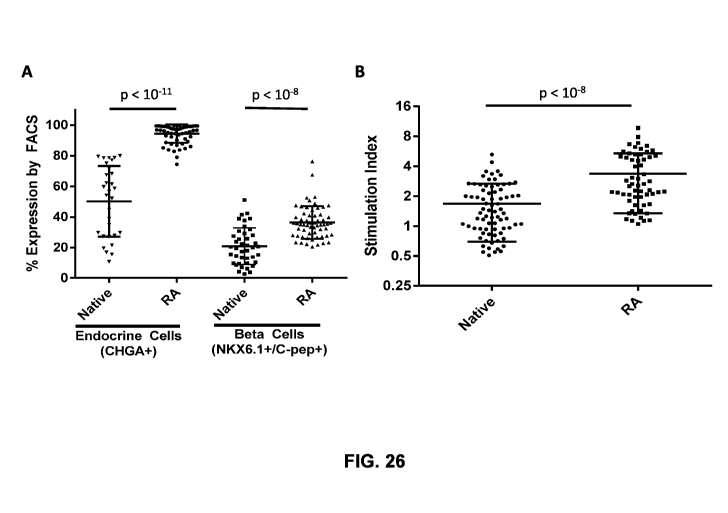
[0060] FIG. 23 shows in vitro glucose-stimulated insulin secretion of cells
grown using 3D
suspension culture (left) and 2D culture using LN-332 (right) 8 days post-
plating.
[0061] FIG. 24 shows effects of different stirring speeds on the size of re-
aggregated clusters
over the days post re-aggregation.
-16-
CA 03070596 2020-01-20
WO 2019/018818 PCT/US2018/043179
[0062] FIG. 25 shows heterogeneous composition of the re-aggregated cell
cluster as indicated
by expression of different cell markers.
[0063] FIG. 26A shows quantification of percentages of endocrine cells and f3
(beta) cells in
native and re-aggregated cell clusters, respectively, as measured by flow
cytometry (FACS).
FIG. 26B shows quantification of insulin stimulation indices of native and re-
aggregated cell
clusters.
[0064] FIG. 27 shows, in another example, in vitro glucose-stimulated insulin
secretion
responses of the native clusters, re-aggregated cell clusters, and re-
aggregated cell clusters after
cryopreservation. SI stands for stimulation index as a ratio of insulin
secretion in response to 20
mM glucose challenge versus 2.8 mM glucose challenge.
[0065] FIG. 28 shows dynamic glucose stimulated insulin secretion over time of
human donor
islets, native and re-aggregated cell clusters in response to a series of
dynamic glucose
challenges and KC1 challenge (left) and quantification of the responses
(right).
[0066] FIG. 29 shows long-term in vivo glycemic control by re-aggregated cell
clusters in
diabetic mice.
DETAILED DESCRIPTION OF THE DISCLOSURE
[0067] While various embodiments of the disclosure have been shown and
described herein, it
will be obvious to those skilled in the art that such embodiments are provided
by way of
example only. Numerous variations, changes, and substitutions may occur to
those skilled in the
art without departing from the disclosure. It should be understood that
various alternatives to
the embodiments of the disclosure described herein may be employed.
[0068] I. Overview
[0069] Provided herein are cell clusters that resemble the function and
characteristics of
endogenous pancreatic islets. Also provided herein are methods of making and
using such cell
clusters.
[0070] Cell clusters provided herein can resemble the characteristics of
endogenous pancreatic
islets. For example, the cell clusters can have a diameter similar to an
endogenous pancreatic
islet, e.g., between about 50 p.m and about 250 p.m, between about 75 p.m and
about 250 p.m, or
between about 100 p.m and about 200 p.m. The cell clusters can comprise a
plurality of cells
expressing marker genes of an endogenous mature pancreatic 0 cell. In some
cases, at least
about 70%, at least about 80%, at least about 90%, at least about 95%, or 100%
cells in the cell
cluster express chromogranin A (CHGA). In some cases, at least about 35%, at
least about 40%,
at least about 50%, or at least about 60% cells in the cell cluster express
both NKX6.1 and
insulin or C-peptide.
-17-
CA 03070596 2020-01-20
WO 2019/018818 PCT/US2018/043179
[0071] The cell clusters can also resemble the function of endogenous
pancreatic islets. For
example, the cell clusters can exhibit an in vitro glucose-stimulated insulin
secretion response to
a glucose challenge. When transplanted to a subject, the cell clusters can
also exhibit an in vivo
glucose-stimulated insulin secretion response to a glucose challenge in the
subject, e.g., within
14 days after the transplantation.
[0072] This disclosure also provides methods of treating diabetes (e.g., type
I or type II
diabetes) by administering, e.g., transplanting, the cell clusters resembling
the function and
characteristics of endogenous pancreatic islets to a subject in need thereof
The cell clusters can
be transplanted under a kidney capsule and with a biodegradable scaffold.
[0073] Also provided herein are methods for making cell clusters that resemble
the functions
and characteristics of endogenous pancreatic islets. The methods can comprise
dissociating a
first cell cluster. The cells from the first cell cluster can then be seeded
in a spinner flask and
cultured in a medium comprising triiodothyronine (T3) and an activin receptor-
like kinase-5
(Alk5) inhibitor. In some cases, the cells from the first cell cluster are
cultured in a medium that
does not comprise serum, e.g., does not comprise fetal bovine serum (FBS). In
some cases, the
medium does not comprise T3. In some cases, the medium does not comprise an
exogenous
thyroid hormone signaling pathway activator. In some cases, the medium does
not comprise
Alk5 inhibitor. In some cases, the medium does not comprise an exogenous small
molecule
compound. In some cases, the medium does not comprise an exogenous inhibitor
of Rho-
associated, coiled-coil containing protein kinase (ROCK). In the spinner
flask, the cells can re-
aggregate into a second cell cluster resembling the functions and
characteristics of endogenous
pancreatic islets. The second cell cluster can comprise at least one
pancreatic 0 cell (e.g., a non-
native 0 cell differentiated from a stem cell).
[0074] Further provided herein are methods for enriching pancreatic 0 cells
(e.g., a non-native f3
cell differentiated form a stem cell) in a cell cluster. The terms "enriching"
and its grammatical
equivalences can mean that the yield (fraction) of cells of one type is
increased by at least about
5% over the fraction of cells of that type in the starting cluster or culture.
The methods can
comprise dissociating a first cell cluster comprising at least a pancreatic 0
cell (e.g., a non-native
0 cell differentiated from a stem cell). The cells from the first cell can
then be seeded in a
spinner flask and cultured in a medium comprising T3 and an Alk5 inhibitor. In
the spinner
flask, the cells can re-aggregate into a second cell cluster. The second cell
cluster can comprise
more cells expressing markers of an endogenous mature pancreatic 0 cell and
exhibiting in vitro
and in vivo glucose-stimulated insulin secretion responses to glucose
challenges, compared to
the first cell cluster.
-18-
CA 03070596 2020-01-20
WO 2019/018818 PCT/US2018/043179
[0075] II. Cell clusters resembling the functions and characteristics of
endogenous
pancreatic islets
[0076] Provided herein are cell clusters that resemble the functions and
characteristics of
endogenous pancreatic islets. Such cell clusters can mimic the function of
endogenous
pancreatic islets in regulating metabolism, e.g., glucose metabolism in a
subject. Thus, the cell
clusters can be transplanted to a subject for treating disease resulting from
insufficient pancreatic
islet function, e.g., diabetes. The terms "cluster" and "aggregate" can be
used interchangeably,
and refer to a group of cells that have close cell-to-cell contact, and in
some cases, the cells in a
cluster can be adhered to one another.
[0077] A cell cluster herein can comprise at least one non-native cell, e.g.,
a non-native
pancreatic 0 cell. A non-native cell (e.g., a non-native pancreatic 0 cell)
can share characteristics
of an endogenous cell (e.g., an endogenous mature pancreatic 0 cell), but is
different in certain
aspects (e.g., gene expression profiles). A non-native cell can be a
genetically modified cell. A
non-native cell can be a cell differentiated from a progenitor cell, e.g., a
stem cell. The stem cell
can be an embryonic stem cell (ESC) or induced pluripotent stem cell (iPSC).
In some cases, the
non-native cell can be a cell differentiated from a progenitor cell in vitro.
In some cases, the
non-native cell can be a cell differentiated from a progenitor cell in in
vivo. For example, a cell
cluster can comprise at least one non-native pancreatic 0 cell. The non-native
pancreatic 0 cells
can be those described in U.S. Patent Application Nos. 14/684,129 and
14/684,101, which are
incorporated herein in their entireties. A cell cluster can comprise a
plurality of non-native
pancreatic 0 cells. In some cases, at least about 20%, 30%, 40%, 50%, 60%,
70%, 80%, 90%,
99% cells in a cell cluster are non-native pancreatic 0 cells. A cell cluster
can comprise one or
more native cells. For example, a cell cluster can comprise one or more
primary cells, e.g.,
primary cells from an endogenous pancreatic islet.
[0078] A cell cluster can comprise one or more cells expressing at least one
marker of an
endogenous cell, e.g., an endogenous mature pancreatic 13 cell. The term
"marker" can refer to a
molecule that can be observed or detected. For example, a marker can include,
but is not limited
to, a nucleic acid, such as a transcript of a specific gene, a polypeptide
product of a gene, a non-
gene product polypeptide, a glycoprotein, a carbohydrate, a glycolipid, a
lipid, a lipoprotein, or a
small molecule. In many cases, a marker can refer to a molecule that can be
characteristic of a
particular type of cell, so that the marker can be called as a marker of the
type of cell. For
instance, Insulin gene can be referred to as a marker of 13 cells. In some
cases, a marker is a
gene. Non-limiting of markers of an endogenous mature pancreatic 13 cell
include insulin, C-
-19-
CA 03070596 2020-01-20
WO 2019/018818 PCT/US2018/043179
peptide, PDX1, NKX6.1, CHGA, MAFA, ZNT8, PAX6, NEUROD1, glucokinase (GCK),
SLC2A, PCSK1, KCNJ11, ABCC8, SLC30A8, SNAP25, RAB3A, GAD2, and PTPRN.
[0079] A cell cluster can comprise one more cells expressing one or multiple
markers of an
endogenous cell, e.g., an endogenous mature pancreatic f3 cell. For example,
cell cluster can
comprise one or more cells co-expressing at least 2, 3, 4, 5, 6, 7, 8, 9, 10,
15, or 20 marker(s) of
an endogenous cell, e.g., an endogenous mature pancreatic 0 cell. In some
cases, a cell cluster
comprises cells that express NKX6.1 and C-peptide, both of which can be
markers of a f3 cell.
[0080] A cell cluster can comprise a plurality of cells expressing at least
one marker of an
endogenous cell. For example, at least about 1%, 5%, 10%, 20%, 30%, 40%, 50%,
60%, 70%,
80%, 90%, 95%, 99% cells in a cell cluster can express at least one marker of
an endogenous
cell. In some cases, all cells in a cell cluster can express a marker of an
endogenous cell. In some
cases, the endogenous cell can be a pancreatic cell, e.g., a pancreatic 0
cell, pancreatic a cells,
pancreatic 0 cells, pancreatic A cells, or pancreatic y cells. A cell cluster
as provided herein can
comprise a heterogeneous group of cells, e.g., cells of different types. For
example, the cell
cluster can comprises a cell expressing insulin/C-peptide, which can be a
marker of a pancreatic
0 cell, a cell expressing glucagon, which can be a marker of a pancreatic a
cell, a cell expressing
somatostatin, which can be a marker of a pancreatic A cell, a cell expressing
pancreatic
polypeptides, or any combination thereof
[0081] For example, the cell cluster herein can comprise a plurality of cells
expressing one or
more markers of an endogenous mature pancreatic 0 cell. For example, at least
about 10%, 20%,
30%, 40%, 50%, 60%, 70%, 80%, 90%, 95%, or 99% cells in the cell cluster can
express one or
more markers of an endogenous mature pancreatic 0 cell.
[0082] The cell cluster can comprise a plurality of cells expressing CHGA. In
some cases, at
least about 30%, 40%, 50%, 60%, 70%, 80%, 90%, 95%, or 99% cells in the cell
cluster express
CHGA. In some cases, at least about 85% cells in a cell cluster can express
CHGA. In some
cases, a cell cluster can comprise about 90% cell expressing CHGA. In some
cases, a cell cluster
can comprise about 95% cells expressing CHGA. In certain cases, all cells in a
cell cluster can
express CHGA.
[0083] The cell cluster can comprise a plurality of cells expressing NKX6.1.
For example, at
least about 30%, 40%, 50%, 60%, 70%, 80%, 90%, 95%, 99% cells in a cell
cluster can express
NKX6.1. In some cases, at least about 50% cells in a cell cluster can express
NKX6.1. In some
cases, all cells in a cell cluster can express NKX6.1.
[0084] The cell cluster can comprise a plurality of cells expressing C-
peptide. For example, at
least about 30%, 40%, 50%, 60%, 70%, 80%, 90%, 95%, or 99% cells in a cell
cluster can
-20-
CA 03070596 2020-01-20
WO 2019/018818 PCT/US2018/043179
express C-peptide. In some cases, at least about 60% cells in a cell cluster
can express C-
peptide. In some cases, all cells in a cell cluster can express C-peptide.
[0085] The cell cluster can comprise a plurality of cells expressing both
NKX6.1 and C-peptide.
For example, at least about 10%, 20%, 30%, 40%, 50%, 60%, 70%, 80%, 90%, 95%,
or 99%
cells in a cell cluster can express C-peptide. In some cases, at least about
35% cells in a cell
cluster can express NKX6.1 and C-peptide. In some cases, at least about 40%
cells in a cell
cluster can express NKX6.1 and C-peptide. In some cases, at least about 35%
cells in a cell
cluster can express NKX6.1 and C-peptide. In some cases, a cell cluster can
comprise about
60% cells expressing NKX6.1 and C-peptide. In some cases, a cell cluster can
comprise about
75% cell expressing NKX6.1 and C-peptide. In some cases, all cells in a cell
cluster can express
NKX6.1 and C-peptide.
[0086] The cell cluster can comprise very few to none of stem cells or
progenitor cells, e.g.,
pancreatic progenitor cells. For example, a cell cluster as provided herein
can comprise at most
about 5% cells, at most about 5% cells, at most about 5% cells, at most about
5% cells, at most
about 5% cells, at most about 2% cells, at most about 1% cells, at most about
0.5% cells, at most
about 0.1% cells, at most about 0.05% cells, at most about 0.01% cells, or no
cells expressing
LIN28. In some examples, a cell cluster as provided herein can comprise at
most about 5% cells,
at most about 5% cells, at most about 5% cells, at most about 5% cells, at
most about 5% cells,
at most about 2% cells, at most about 1% cells, at most about 0.5% cells, at
most about 0.1%
cells, at most about 0.05% cells, at most about 0.01% cells, or no cells
expressing Ki67.
[0087] In some cases, a cell cluster can comprise at most 3% cells, at most
about 2% cells, at
most about 1% cells, at most about 0.5% cells, at most about 0.1% cells, at
most about 0.05%
cells, at most about 0.01% cells, or no cells expressing SOX2. In some cases,
a cell cluster can
comprise about 1% cells expressing SOX2. In some cases, a cell cluster can
comprise about
0.6% cells expressing SOX2. In some cases, a cell cluster can comprise about
0.3% cells
expressing SOX2. In some cases, a cell cluster can comprise about 0.1% cells
expressing SOX2.
[0088] In some examples, a cell cluster can comprise at most 10% cells, at
most about 8% cells,
at most about 6% cells, at most about 5% cells, at most about 2% cells, at
most about 1% cells,
at most about 0.5% cells, at most about 0.1% cells, at most about 0.05% cells,
at most about
0.01% cells, or no cells expressing SOX9. In some cases, a cell cluster can
comprise about 2%
cells expressing SOX9. In some cases, a cell cluster can comprise about 6%
cells expressing
SOX9. In some cases, a cell cluster can comprise about 1.2% cells expressing
SOX9.
[0089] A cell cluster herein can exhibit one or multiple glucose stimulated
insulin secretion
(GSIS) response(s) in vitro when exposed to glucose challenge(s). The GSIS
responses can
-21-
CA 03070596 2020-01-20
WO 2019/018818 PCT/US2018/043179
resemble the GSIS responses of an endogenous pancreatic islet. In some cases,
the cell cluster
exhibits an in vitro GSIS response to a glucose challenge. In some cases, the
cell cluster exhibits
in vitro GSIS responses to multiple glucose challenges, such as sequential
glucose challenges.
For example, the cell cluster can exhibit in vitro GSIS responses to at least
2, 3, 4, 5, 6, 7, 8, 9,
sequential glucose challenges.
[0090] A cell cluster as provided herein can comprise at least one cell
exhibiting in vitro GSIS.
For example, at least one cell in the cell cluster can be referred to as a
mature pancreatic 0 cell.
In some cases, the at least one cell is a non-native pancreatic 0 cell. In
some cases, the at least
one cell is a pancreatic 0 cell resembling a native/endogenous 0 cell. In some
cases, the cell
exhibits an in vitro glucose stimulated insulin secretion (GSIS) response. In
some cases, the at
least one cell exhibits a GSIS response to at least one glucose challenge. In
some cases, the cell
exhibits a GSIS response to at least two sequential glucose challenges. In
some cases, the cell
exhibits a GSIS response to at least three sequential glucose challenges
[0091] As provided herein, a cell cluster can exhibit GSIS stimulation index
similar to an
endogenous pancreatic islet. Stimulation index of a cell cluster or a cell can
be characterized by
the ratio of insulin secreted in response to high glucose concentrations
compared to low glucose
concentrations. For example, a stimulation index of a cell cluster or a cell
as provided herein can
be calculated as a ration of insulin secreted in response to 20 mM glucose
stimulation versus
insulin secreted in response to 2.8 mM glucose stimulation. In some examples,
the stimulation
index of a cell cluster or a cell as provided herein is greater than or equal
to 1 , or greater than or
equal to 1.1 , or greater than or equal to 1 .3, or greater than or equal to
2, or greater than or
equal to 2.3, or greater than or equal to 2.6. In some instances, the cell
cluster or the cell exhibits
cytokine-induced apoptosis in response to a cytokine. In some cases, the
cytokine comprises
interleukin-f3 (IL-f3), interferon-y (INF- y), tumor necrosis factor-a (TNF-
a), or any combination
thereof. In some cases, insulin secretion from the cell cluster or the cell is
enhanced in response
to an anti-diabetic agent. In some cases, the anti-diabetic agent comprises a
secretagogue
selected from the group consisting of an incretin mimetic, a sulfonylurea, a
meglitinide, and
combinations thereof. In some cases, the cell cluster or the cell is
monohormonal. In some cases,
the cell cluster or the cell exhibits a morphology that resembles the
morphology of an
endogenous mature pancreatic 0 cell. In some cases, the cell cluster or the
cell exhibits
encapsulated crystalline insulin granules under electron microscopy that
resemble insulin
granules of an endogenous mature pancreatic 0 cell. In some cases, the cell
cluster or the cell
exhibits a low rate of replication. In some cases, the cell cluster or the
cell exhibits a glucose
stimulated Ca2+ flux (GSCF) that resembles the GSCF of an endogenous mature
pancreatic 0
-22-
CA 03070596 2020-01-20
WO 2019/018818 PCT/US2018/043179
cell. In some cases, the cell cluster or the cell exhibits a GSCF response to
at least one glucose
challenge. In some cases, the cell cluster or the cell exhibits a GSCF
response to at least two
glucose challenges. In some cases, the cell cluster or the cell exhibits a
GSCF response to at
least three glucose challenges. In some cases, the cell cluster or the cell
exhibits an increased
calcium flux. In some cases, the increased calcium flux comprises an increased
amount of influx
or a ratio of influx at low relative to high glucose concentrations.
[0092] A cell cluster as provided herein can exhibit biphasic insulin
secretion in response to a
high glucose concentration stimulation similar to an endogenous pancreatic
islet, e.g., a human
pancreatic islet. A biphasic insulin secretion can be a phenomenon
characteristic of an
endogenous pancreatic islet, e.g., human islet. As demonstrated in FIG. 28, in
response to a high
glucose concentration challenge, e.g., 10mM, 15mM, 20 mM, or 30mM, a cell
cluster as
provided herein, e.g., a reaggregated pancreatic cell cluster, can exhibit a
transient increase in
insulin secretion to a peak value followed by a rapid decrease to a relatively
elevated insulin
secretion level, e.g., a level that is higher than an insulin secretion level
in response to a lower
glucose concentration, e.g., 2.8 mM glucose. Such a transient increase and
decrease process can
be termed as a first phase of the biphasic insulin secretion pattern. With a
persistent high glucose
challenge, the first phase can be thus followed by a second phase, in which
the insulin secretion
by the cell cluster can be maintained at the relatively elevated level. The
second phase can last
for an extended period, e.g., as long as the high glucose concentration
challenge lasts, or
relatively longer than the first phase. Such a biphasic insulin secretion
pattern can be due to
intrinsic cellular signaling changes that are characteristic of a mature
native pancreatic 0 cell.
[0093] When transplanted to a subject, a cell cluster can exhibit one or more
in vivo GSIS
responses when exposed to glucose challenge(s). The cell cluster herein can be
capable of
exhibiting an in vivo GSIS response within a short period of time after
transplanted to a subject.
For example, the cell cluster can exhibit an in vivo GSIS within about 6, 12,
or 24 hours after
transplantation. In some cases, the cell cluster exhibits an in vivo GSIS
within about 2 days, 4
days, 6 days, 8 days, 10 days, 12 days, 14 days, 21 days, 28 days, 35 days, or
42 days after
transplantation. The amount of insulin secreted by the cell cluster can be
similar or higher than
an endogenous pancreatic islet. The term "about" in relation to a reference
numerical value as
used through the application can include a range of values plus or minus 10%
from that value.
For example, the amount "about 10" includes amounts from 9 to 11. For example,
the term
"about" in relation to a reference numerical value can also include a range of
values plus or
minus 10%, 9%, 8%, 7%, 6%, 5%, 4%, 3%, 2%, or 1% from that value.
-23-
CA 03070596 2020-01-20
WO 2019/018818 PCT/US2018/043179
[0094] The cell cluster can maintain the ability of exhibiting in vivo GSIS
responses for a period
of time after transplanted into a subject. For example, an in vivo GSIS
response of the cell
cluster can be observed up to at least 2 weeks, 3 weeks, 4 weeks, 5 weeks, 10
weeks, 15 weeks,
20 weeks, 6 months, 7 months, 8 months, 9 months, 10 months, 11 months, 1
year, 2 years, 3
years, 4 years, 5 years, 10 years, 20 years, 30 years, 40 years, 60 years, 80
years, or 100 years
after transplantation of the cell cluster into a subject (e.g., a human).
[0095] The GSIS of a cell cluster can be measured by a stimulation index. A
stimulation index
of a cell cluster can equal to the ratio of insulin secreted in response to a
high glucose
concentration compared to insulin secreted in response to a low glucose
concentration. A cell
cluster can have a stimulation index similar to an endogenous pancreatic
islet. In some cases, a
cell cluster has a stimulation index of at least 1, 1.1, 1.2, 1.3, 1.4, 1.5,
1.6, 1.7, 1.8, 1.9, 2, 2.1,
2.2, 2.3, 2.4, 2.5, 2.6, 2.7, 2.8, 2.9, 3.0, 3.1, 3.2, 3.3, 3.4, 3.5, 3.6,
3.7, 3.8, 3.9, 4.0, 4.1, 4.2, 4.3,
4.4, 4.5, 4.6, 4.7, 4.8, 4.9, or 5Ø
[0096] The amount of insulin secreted by a cell cluster in response to a
glucose challenge (e.g., a
high concentration, such as 20mM, of glucose) can range from about 0.1
[tIU/103 cells to about
IU/103 cells, from about 0.2 IU/103 cells to about 4 [tIU/103 cells, from
about 0.2 IU/103
cells to about 3 [tIU/103 cells, or from about 0.23 [tIU/103 cells to about
2.7 [tIU/103 cells. In
some cases, the amount of insulin secreted by a cell cluster in response to a
glucose challenge
(e.g., a high concentration, such as 20mM, of glucose) is at least 0.05, 0.1,
0.15, 0.2, 0.21, 0.22,
0.23, 0.24, 0.25, 0.26, 0.27, 0.28, 0.29, 0.3, 0.4, 0.5, 0.6, 0.7, 0.8, 0.9,
1, 1.1, 1.2, 1.3, 1.4, 1.5,
1.6, 1.7, 1.8, 1.9, 2, 2.1, 2.2, 2.3, 2.4, 2.5, 2.6, 2.7, 2.8, 2.9, or 3
IU/103 cells.
[0097] A cell cluster can secrete both pro-insulin and insulin. For example, a
cell cluster can
secrete pro-insulin and insulin at a proinsulin-to-insulin ratio substantially
the same as the ratio
of pro-insulin to insulin secreted by an endogenous pancreatic islet. In some
cases, a cell cluster
secretes pro-insulin and insulin at a proinsulin-to-insulin ratio of from
about 0.01 to about 0.05,
from about 0.02 to about 0.04, from about 0.02 to about 0.03, or from 0.029 to
about 0.031. In
some cases, a cell cluster secretes pro-insulin and insulin at a proinsulin-to-
insulin ratio of about
0.02, 0.021, 0.022, 0.023, 0.024, 0.025, 0.026, 0.027, 0.028, 0.029, 0.03,
0.031, 0.032, 0.033,
0.034, 0.035, 0.036, 0.037, 0.038, 0.039, or 0.04.
[0098] A cell cluster can be in a size similar to an endogenous pancreatic
islet. For example, a
cell cluster can have a diameter similar to an endogenous pancreatic islet. A
diameter of a cell
cluster can refer to the largest linear distance between two points on the
surface of the cell
cluster. In some cases, the diameter of a cell cluster is at most 300 m, 200
m, 150 m, 100
-24-
CA 03070596 2020-01-20
WO 2019/018818 PCT/US2018/043179
p.m, 90 p.m, 80 p.m, 70 p.m, 60 p.m, 50 p.m, or 40 p.m. The diameter of a cell
cluster can be from
about 75 p.m to about 250 p.m. The diameter of a cell cluster can be at most
100 p.m.
[0099] A cell cluster can comprise very few or no dead cells. The cell cluster
can be in a size
that allows effective diffusion of molecules (e.g., nutrition and gas) from
surrounding
environment into the core of the cell cluster. The diffused molecule can be
important for the
survival and function of the cells in the core. In some cases, the cell
cluster can have less than
about 1%, 2%, 3%, 4%, 5%, 6%, 7%, 8%, 9%, 10% of dead cells, e.g., dead cells
in its core. In
some cases, a cell cluster can have no dead cell. The dead cells can be
apoptotic cells, narcotic
cells or any combination thereof
[0100] A cell cluster can comprise one or multiple types of cells. In some
cases, a cell cluster
comprises one or more types of pancreatic cells. For example, the cell cluster
can comprise one
or more pancreatic 0 cell, pancreatic a cells, pancreatic A cells, pancreatic
y cells, and any
combination thereof In some cases, the pancreatic cells can be non-native
pancreatic cells, e.g.,
cells derived from stem cells, such as ESCs and/or iPSCs. In some cases, the
cell cluster can
also comprise one or more progenitor cells of mature pancreatic cells,
including iPSCs, ESCs,
definitive endoderm cells, primitive gut tube cells, Pdxl-positive pancreatic
progenitor cells,
Pdxl-positive/ NKX6.1-positive pancreatic progenitor cells, Ngn3-positive
endocrine progenitor
cells, and any combination thereof
[0101] A cell cluster can exhibit cytokine-induced apoptosis in response to
cytokines. For
example, the cell cluster can exhibit cytokine-induced apoptosis in response
to a cytokine such
as interleukin-10 (IL-f3), interferon-y (INF-y), tumor necrosis factor-a (TNF-
a), and
combinations thereof.
[0102] Insulin secretion from a cell cluster herein can be enhanced by an anti-
diabetic drug (e.g.,
an anti-diabetic drug acting on pancreatic 0 cells ex vivo, in vitro, and/or
in vivo). The disclosure
can contemplate any known anti-diabetic drug. In some cases, insulin secretion
from a cell
cluster can be enhanced by a secretagogue. The secretagogue can be an incretin
mimetic, a
sulfonylurea, a meglitinide, and combinations thereof
[0103] A cell cluster can comprise a monohormonal. For example, the cell
cluster can comprise
a pancreatic cell (e.g., a pancreatic 0 cell, pancreatic a cells, pancreatic 0
cells, pancreatic A
cells, or pancreatic y cells) that is monohormonal. In some cases, the cell
cluster comprises an
insulin-secreting non-native pancreatic cell that is monohormonal. A cell
cluster can comprise a
polyhormonal. In some case, a cell cluster comprises a monohormonal cell and a
polyhormonal
cell.
-25-
CA 03070596 2020-01-20
WO 2019/018818 PCT/US2018/043179
[0104] A cell cluster can comprise a cell (e.g., a non-native pancreatic cell)
having a
morphology that resembles the morphology of an endogenous mature pancreatic f3
cell. In some
cases, the cell cluster can comprise cell encapsulating crystalline insulin
granules that resemble
insulin granules of an endogenous mature pancreatic 0 cell, e.g., as detected
by electron
microscopy. A cell cluster can comprise a plurality cells having a morphology
that resembles the
morphology of an endogenous mature pancreatic 0 cell. For example, at least
about 10%, 20%,
30%, 40%, 50%, 60%, 70%, 80%, 90% cells in a cell cluster can encapsulate
crystalline insulin
granules that resemble insulin granules of an endogenous mature pancreatic 0
cell. In some
cases, 100% cells in a cell cluster encapsulate crystalline insulin granules
that resemble insulin
granules of an endogenous mature pancreatic 0 cell.
[0105] A cell cluster can exhibit glucose-stimulated calcium (Ca2+) flux to
one or more glucose
challenges. In some cases, a cell cluster exhibits a glucose-stimulated Ca2+
flux (GSCF) that
resembles the GSCF of an endogenous pancreatic islet. In some cases, a cell
cluster exhibits a
GSCF response to at least 1, 2, 3, 4, 5, 6, 8, or 10 sequential glucose
challenges in a manner that
resembles the GSCF response of an endogenous pancreatic islet to multiple
glucose challenges.
A cell cluster can exhibit an in vitro and/or in vivo GSCF response when
exposed to a glucose
challenge.
[0106] A cell cluster can comprise cells originated from any species. For
example, a cell cluster
can comprise cells from a mammalian species, with non-limiting examples
including a murine,
bovine, simian, porcine, equine, ovine, or human cell. In some cases, at least
one cell in the cell
cluster is a human cell.
[0107] Provided herein also include compositions comprising a cell clusters
disclosed through
the application. In addition to the cell cluster, the compositions can further
comprise a scaffold
or matrix that can be used for transplanting the cell clusters to a subject. A
scaffold can provide
a structure for the cell cluster to adhere to. The cell cluster can be
transplanted to a subject with
the scaffold. The scaffold can be biodegradable. In some cases, a scaffold
comprises a
biodegradable polymer. The biodegradable polymer can be a synthetic polymer,
such as
poly(lactide) (PLA), poly(glycolic acid) (PGA), poly(lactide-co-glycolide)
(PLGA), and other
polyhydroxyacids, poly(caprolactone), polycarbonates, polyamides,
polyanhydrides,
polyphosphazene, polyamino acids, polyortho esters, polyacetals,
polycyanoacrylates, and
biodegradable polyurethanes. The biodegradable polymer can also be a natural
polymer, such as
albumin, collagen, fibrin, polyamino acids, prolamines, and polysaccharides
(e.g., alginate,
heparin, and other naturally occurring biodegradable polymers of sugar units).
Alternatively, the
scaffold can be non-biodegradable. For example, a scaffold can comprise a non-
biodegradable
-26-
CA 03070596 2020-01-20
WO 2019/018818 PCT/US2018/043179
polymer, such as polyacrylates, ethylene- vinyl acetate polymers and other
acyl-substituted
cellulose acetates and derivatives thereof, polyurethanes, polystyrenes,
polyvinyl chloride,
polyvinyl fluoride, poly(vinyl imidazole), chlorosulphonated polyolefins, and
polyethylene
oxide.
[0108] III Pharmaceutical compositions
[0109] In some cases, the present disclosure provides pharmaceutical
compositions that can
utilize non-native pancreatic beta cell populations and cell components and
products in various
methods for treatment of a disease (e.g., diabetes). Certain cases encompass
pharmaceutical
compositions comprising live cells (e.g., non-native pancreatic beta cells
alone or admixed with
other cell types). Other cases encompass pharmaceutical compositions
comprising non-native
pancreatic beta cell components (e.g., cell lysates, soluble cell fractions,
conditioned medium,
ECM, or components of any of the foregoing) or products (e.g., trophic and
other biological
factors produced by non-native pancreatic beta cells or through genetic
modification,
conditioned medium from non-native pancreatic beta cell culture). In either
case, the
pharmaceutical composition may further comprise other active agents, such as
anti-
inflammatory agents, exogenous small molecule agonists, exogenous small
molecule
antagonists, anti-apoptotic agents, antioxidants, and/or growth factors known
to a person having
skill in the art.
[0110] Pharmaceutical compositions of the present disclosure can comprise non-
native
pancreatic beta cell, or components or products thereof, formulated with a
pharmaceutically
acceptable carrier (e.g. a medium or an excipient). The term pharmaceutically
acceptable carrier
(or medium), which may be used interchangeably with the term biologically
compatible carrier
or medium, refers to reagents, cells, compounds, materials, compositions,
and/or dosage forms
that are not only compatible with the cells and other agents to be
administered therapeutically,
but also are suitable for use in contact with the tissues of human beings and
animals without
excessive toxicity, irritation, allergic response, or other complication.
Suitable pharmaceutically
acceptable carriers can include water, salt solution (such as Ringer's
solution), alcohols, oils,
gelatins, and carbohydrates, such as lactose, amylose, or starch, fatty acid
esters,
hydroxymethylcellulose, and polyvinyl pyrolidine. Such preparations can be
sterilized, and if
desired, mixed with auxiliary agents such as lubricants, preservatives,
stabilizers, wetting agents,
emulsifiers, salts for influencing osmotic pressure, buffers, and coloring.
Pharmaceutical
compositions comprising cellular components or products, but not live cells,
can be formulated
as liquids. Pharmaceutical compositions comprising living non-native
pancreatic beta cells can
-27-
CA 03070596 2020-01-20
WO 2019/018818 PCT/US2018/043179
be formulated as liquids, semisolids (e.g., gels, gel capsules, or liposomes)
or solids (e.g.,
matrices, scaffolds and the like).
[0111] Pharmaceutical compositions may comprise auxiliary components as would
be familiar
to a person having skill in the art. For example, they may contain
antioxidants in ranges that
vary depending on the kind of antioxidant used. Reasonable ranges for commonly
used
antioxidants are about 0.01% to about 0.15% weight by volume of EDTA, about
0.01% to about
2.0% weight volume of sodium sulfite, and about 0.01% to about 2.0% weight by
volume of
sodium metabisulfite. One skilled in the art may use a concentration of about
0.1% weight by
volume for each of the above. Other representative compounds include
mercaptopropionyl
glycine, N-acetyl cysteine, beta-mercaptoethylamine, glutathione and similar
species, although
other anti-oxidant agents suitable for renal administration, e.g. ascorbic
acid and its salts or
sulfite or sodium metabisulfite may also be employed.
[0112] A buffering agent may be used to maintain the pH of formulations in the
range of about
4.0 to about 8.0; so as to minimize irritation in the target tissue. For
direct intraperitoneal
injection, formulations should be at pH 7.2 to 7.5, preferably at pH 7.35-
7.45. The compositions
may also include tonicity agents suitable for administration to the kidney.
Among those suitable
is sodium chloride to make formulations approximately isotonic with blood.
[0113] In certain cases, pharmaceutical compositions are formulated with
viscosity enhancing
agents. Exemplary agents are hydroxyethylcellulose, hydroxypropylcellulose,
methylcellulose,
and polyvinylpyrrolidone. The pharmaceutical compositions may have cosolvents
added if
needed. Suitable cosolvents may include glycerin, polyethylene glycol (PEG),
polysorbate,
propylene glycol, and polyvinyl alcohol. Preservatives may also be included,
e.g., benzalkonium
chloride, benzethonium chloride, chlorobutanol, phenylmercuric acetate or
nitrate, thimerosal, or
methyl or propylparabens.
[0114] Pharmaceutical compositions comprising cells, cell components or cell
products may be
delivered to the kidney of a patient in one or more of several methods of
delivery known in the
art. In some cases, the compositions are delivered to the kidney (e.g.,on the
renal capsule and/or
underneath the renal capsule). In another embodiment, the compositions may be
delivered to
various locations within the kidney via periodic intraperitoneal or intrarenal
injection.
Alternatively, the compositions may be applied in other dosage forms known to
those skilled in
the art, such as pre-formed or in situ-formed gels or liposomes.
[0115] Pharmaceutical compositions comprising live cells in a semi-solid or
solid carrier are
may be formulated for surgical implantation on or beneath the renal capsule.
It should be
appreciated that liquid compositions also may be administered by surgical
procedures. In
-28-
CA 03070596 2020-01-20
WO 2019/018818 PCT/US2018/043179
particular cases, semi-solid or solid pharmaceutical compositions may comprise
semi-permeable
gels, lattices, cellular scaffolds and the like, which may be non-
biodegradable or biodegradable.
For example, in certain cases, it may be desirable or appropriate to sequester
the exogenous cells
from their surroundings, yet enable the cells to secrete and deliver
biological molecules (e.g.,
insulin) to surrounding cells or the blood stream. In these cases, cells may
be formulated as
autonomous implants comprising living non-native pancreatic beta cells or cell
population
comprising non-native pancreatic beta cell surrounded by a non-degradable,
selectively
permeable barrier that physically separates the transplanted cells from host
tissue. Such implants
are sometimes referred to as "immunoprotective," as they have the capacity to
prevent immune
cells and macromolecules from killing the transplanted cells in the absence of
pharmacologically
induced immunosuppression.
[0116] In other cases, various degradable gels and networks can be used for
the pharmaceutical
compositions of the present disclosure. For example, degradable materials
particularly suitable
for sustained release formulations include biocompatible polymers, such as
poly(lactic acid),
poly (lactic-co-glycolic acid), methylcellulose, hyaluronic acid, collagen,
and the like.
[0117] In other cases, it may be desirable or appropriate to deliver the cells
on or in a
biodegradable, preferably bioresorbable or bioabsorbable, scaffold or matrix.
These typically
three-dimensional biomaterials contain the living cells attached to the
scaffold, dispersed within
the scaffold, or incorporated in an extracellular matrix entrapped in the
scaffold. Once implanted
into the target region of the body, these implants become integrated with the
host tissue, wherein
the transplanted cells gradually become established.
[0118] Examples of scaffold or matrix (sometimes referred to collectively as
"framework")
material that may be used in the present disclosure include nonwoven mats,
porous foams, or
self-assembling peptides. Nonwoven mats, for example, may be formed using
fibers comprising
a synthetic absorbable copolymer of glycolic and lactic acids (PGA/PLA),
foams, and/or
poly(epsilon-caprolactone)/poly(glycolic acid) (PCL/PGA) copolymer.
[0119] In another embodiment, the framework is a felt, which can be composed
of a
multifilament yarn made from a bioabsorbable material, e.g., PGA, PLA, PCL
copolymers or
blends, or hyaluronic acid. The yarn is made into a felt using standard
textile processing
techniques consisting of crimping, cutting, carding and needling. In another
embodiment, cells
are seeded onto foam scaffolds that may be composite structures. In many of
the
abovementioned cases, the framework may be molded into a useful shape.
Furthermore, it will
be appreciated that non-native pancreatic beta cells may be cultured on pre-
formed, non-
degradable surgical or implantable devices.
-29-
CA 03070596 2020-01-20
WO 2019/018818 PCT/US2018/043179
[0120] The matrix, scaffold or device may be treated prior to inoculation of
cells in order to
enhance cell attachment. For example, prior to inoculation, nylon matrices can
be treated with
0.1 molar acetic acid and incubated in polylysine, PBS, and/or collagen to
coat the nylon.
Polystyrene can be similarly treated using sulfuric acid. The external
surfaces of a framework
may also be modified to improve the attachment or growth of cells and
differentiation of tissue,
such as by plasma coating the framework or addition of one or more proteins
(e.g., collagens,
elastic fibers, reticular fibers), glycoproteins, glycosaminoglycans (e.g.,
heparin sulfate,
chondroitin-4-sulfate, chondroitin-6-sulfate, dermatan sulfate, keratin
sulfate), a cellular matrix,
and/or other materials such as, but not limited to, gelatin, alginates, agar,
agarose, and plant
gums, among others.
In one aspect, the present disclosure provided devices comprising a cell
cluster comprising at
least one pancreatic I cell. A device provided herein can be configured to
produce and release
insulin when implanted into a subject. A device can comprise a cell cluster
comprising at least
one pancreatic I cell, e.g., a non-native pancreatic I cell. A cell cluster in
the device can exhibit
in vitro GSIS. A device can further comprise a semipermeable membrane. The
semipermeable
membrane can be configured to retain the cell cluster in the device and permit
passage of insulin
secreted by the cell cluster. In some cases of the device, the cell cluster
can be encapsulated by
the semipermeable membrane. The encapsulation can be performed by any
technique available
to one skilled in the art. The semipermeable membrane can also be made of any
suitable material
as one skilled in the art would appreciate and verify. For example, the
semipermeable membrane
can be made of polysaccharide or polycation. In some cases, the semipermeable
membrane can
be made of poly(lactide) (PLA), poly(glycolic acid) (PGA), poly(lactide-co-
glycolide) (PLGA),
and other polyhydroxyacids, poly(caprolactone), polycarbonates, polyamides,
polyanhydrides,
polyphosphazene, polyamino acids, polyortho esters, polyacetals,
polycyanoacrylates,
biodegradable polyurethanes, albumin, collagen, fibrin, polyamino acids,
prolamines, alginate,
agarose, agarose with gelatin, dextran, polyacrylates, ethylene- vinyl acetate
polymers and other
acyl-substituted cellulose acetates and derivatives thereof, polyurethanes,
polystyrenes,
polyvinyl chloride, polyvinyl fluoride, poly(vinyl imidazole),
chlorosulphonated polyolefins,
polyethylene oxide, or any combinations thereof. In some cases, the
semipermeable membrane
comprises alginate. In some cases, the cell cluster is encapsulated in a
microcapsule that
comprises an alginate core surrounded by the semipermeable membrane. In some
cases, the
alginate core is modified, for example, to produce a scaffold comprising an
alginate core having
covalently conjugated oligopeptides with an RGD sequence (arginine, glycine,
aspartic acid). In
some cases, the alginate core is modified, for example, to produce a
covalently reinforced
-30-
CA 03070596 2020-01-20
WO 2019/018818 PCT/US2018/043179
microcapsule having a chemoenzymatically engineered alginate of enhanced
stability. In some
cases, the alginate core is modified, for example, to produce membrane-mimetic
films
assembled by in-situ polymerization of acrylate functionalized phospholipids,
In some cases,
microcapsules are composed of enzymatically modified alginates using
epimerases, In some
cases, microcapsules comprise covalent links between adjacent layers of the
microcapsule
membrane. In some embodiment, the microcapsule comprises a subsieve-size
capsule
comprising alginate coupled with phenol moieties. In some cases, the
microcapsule comprises a
scaffold comprising alginate-agarose. In some cases, the SC-f3 cell is
modified with PEG before
being encapsulated within alginate. In some cases, the isolated populations of
cells, e.g., SC-f3
cells are encapsulated in photoreactive liposomes and alginate. It should be
appreciated that the
alginate employed in the microcapsules can be replaced with other suitable
biomaterials,
including, without limitation, polyethylene glycol (PEG), chitosan, polyester
hollow fibers,
collagen, hyaluronic acid, dextran with ROD, BHD and polyethylene glycol-
diacrylate
(PEGDA), poly(MPC-co-n-butyl methacrylate-co-4-vinylphenyl boronic acid)
(PMBV) and
poly(vinyl alcohol) (PVA), agarose, agarose with gelatin, and multilayer cases
of these.
[0121] IV Method for making the cell clusters
[0122] Further disclosed herein are methods for making cell clusters that
resemble the function
and characteristics of an endogenous tissue or cell cluster, e.g., an
endogenous pancreatic islet.
The methods can comprise dissociating a first cell cluster and re-aggregating
the dissociated
cells to a second cell cluster, where the second cell cluster more closely
resembles the function
and characteristics of an endogenous tissue or cell cluster, e.g., an
endogenous pancreatic islet,
compared to the first cell cluster. The term "re-aggregating" and its
grammatical equivalences as
used herein can refer to, when clusters are dissociated into smaller clusters
or single cells, the
dissociated cells then form new cell-to-cell contacts and form new clusters.
The methods can be
used for producing a cell cluster in vitro by a) dissociating a plurality of
cells from a first cell
cluster; and b) culturing the plurality of cells from a) in a medium, thereby
allowing the plurality
of cells to form a second cell cluster. In some cases, the second cell cluster
is an in vitro cell
cluster. The first cell cluster can be an in vitro cell cluster, e.g., a
cluster formed by a suspension
of single cells in vitro in a culture medium. In some cases, the first cell
cluster can be an ex vivo
cell cluster, e.g., a cell cluster that is formed in a body of a live organism
and isolated from said
organism. For example, a first cell cluster that the method provided herein is
applicable to can
be a human pancreatic islet. In some cases, the first cell cluster can be a
cadaveric pancreatic
islet.
-31-
CA 03070596 2020-01-20
WO 2019/018818 PCT/US2018/043179
[0123] A method provided herein can enrich pancreatic cells in a cell cluster,
e.g., a pancreatic
cell, an endocrine cell, or an endocrine progenitor cell. In some examples,
the method can
reduce or eliminate stem cells or pancreatic progenitor cells from a cell
cluster. In some cases,
the second cell cluster comprises a higher percentage cells that express
chromogranin A as
compared the first cell cluster. In some cases, the second cell cluster
comprises a higher
percentage cells that express NKX6.1 and C-peptide as compared the first cell
cluster. In some
cases, the second cell cluster comprises a lower percentage cells that express
SOX2 as compared
the first cell cluster. In some cases, the second in vitro cell cluster
comprises a lower percentage
of cells that express SOX9 as compared the first cell cluster.
[0124] In some cases, the medium comprises a thyroid hormone signaling pathway
activator and
a transforming growth factor 0 (TGF-0) signaling pathway inhibitor. In some
cases, the medium
comprises a) serum, and b) one or both of a thyroid hormone signaling pathway
activator and a
TGF-f3 signaling pathway inhibitor. In some cases, the medium for
reaggregation as provided
herein (reaggregation medium) can comprise no small molecule compounds. For
example, the
reaggregation medium can comprise no thyroid hormone signaling pathway
activator. In some
cases, the reaggregation medium does not comprise triiodothyronine (T3), or
merely a trace
amount of T3. The reaggregation medium can comprise no TGFP signaling pathway
inhibitor. In
some cases, the reaggregation medium does not comprise an Alk5 inhibitor
(Alk5i), or merely a
trace amount of Alk5i.
[0125] Dissociating of the first cell cluster can be performed using methods
known in the art.
Non-limiting exemplary methods for dissociating cell clusters include physical
forces (e.g.,
mechanical dissociation such as cell scraper, trituration through a narrow
bore pipette, fine
needle aspiration, vortex disaggregation and forced filtration through a fine
nylon or stainless
steel mesh), enzymatic dissociation using enzymes such as trypsin,
collagenase, TrypLETm, and
the like, or a combination thereof. After dissociation, cells from the first
cell cluster can be in a
cell suspension, e.g., a single cell suspension. The term "suspension" as used
herein can refer to
cell culture conditions in which cells are not attached to a solid support.
Cells proliferating in
suspension can be stirred while proliferating using apparatus well known to
those skilled in the
art.
[0126] In some cases, the method provided herein does not comprise an active
cell sorting
process, e.g., flow cytometry. In some cases, a cell cluster as described
herein can be an
unsorted cell cluster. In some cases, a method provided herein does not rely
on an active cell
sorting for the enrichment or elimination of a particular type of cells in the
first cell cluster. In
some cases, a method merely requires dissociating the first cell cluster and
culturing the plurality
-32-
CA 03070596 2020-01-20
WO 2019/018818 PCT/US2018/043179
of cells dissociated from the first cell cluster in a medium, thereby allowing
formation of a
second cell cluster.
[0127] In some cases, the method provided herein can be applied to dissociate
a cell cluster and
reaggregate into a new cluster for more than once. For instance, a first cell
cluster can be
dissociate and reaggregated to form a second cell cluster according to the
method provided
herein, and the second cell cluster can be further dissociated and
reaggregated to form a third
cell cluster, and so on. Reaggregation as provided herein can be performed
sequentially to a cell
cluster for at least 1, 2, 3, 4, 5, 6, 7, 8, 9, or 10 times.
[0128] Cell sorting as described herein can refer to a process of isolating a
group of cells from a
plurality of cells by relying on differences in cell size, shape (morphology),
surface protein
expression, endogenous signal protein expression, or any combination thereof
In some cases,
cell sorting comprises subjecting the cells to flow cytometry. Flow cytometry
can be a laser- or
impedance-based, biophysical technology. During flow cytometry, one can
suspend cells in a
stream of fluid and pass them through an electronic detection apparatus. In
one type of flow
cytometry, fluorescent-activated cell sorting (FACS), based on one or more
parameters of the
cells' optical properties (e.g., emission wave length upon laser excitation),
one can physically
separate and thereby purify cells of interest using flow cytometry. As
described herein, an
unsorted cell cluster can be cell cluster that formed by a plurality of cells
that have not been
subject to an active cell sorting process, e.g., flow cytometry. An unsorted
cell cluster, in some
cases referred to as "reaggregated cell cluster," can be formed by a plurality
of cells that are
dissociated from an existing cell cluster, and before their reaggregation into
the new cell cluster,
there can be no active cell sorting process, e.g., flow cytometry or other
methods, to isolate one
or more particular cell types for the reaggregation as provided herein. In
some cases, flow
cytometry as discussed herein can be based on one or more signal peptides
expressed in the
cells. For example, a cell cluster can comprise cells that express a signal
peptide (e.g., a
fluorescent protein, e.g., green fluorescent protein (GFP) or tdTomato). In
some cases, the signal
peptide is expressed as an indicator of insulin expression in the cells. For
instance, a cell cluster
can comprise cell harboring an exogenous nucleic acid sequence coding for GFP
under the
control of an insulin promoter. The insulin promoter can be an endogenous or
exogenous
promoter. In some cases, the expression of GFP in these cells can be
indicative of insulin
expression in said cells. The GFP signal can thus be a marker of a pancreatic
I cell. In some
cases, cell sorting as described herein can comprise magnetic-activated flow
cytometry, where
magnetic antibody or other ligand is used to label cells of different types,
and the differences in
magnetic properties can be used for cell sorting.
-33-
CA 03070596 2020-01-20
WO 2019/018818 PCT/US2018/043179
[0129] The cells dissociated from the first cell cluster can be cultured in a
medium for re-
aggregating to a second cell cluster. The medium can comprise Connought
Medical Research
Laboratories 1066 supplemented islet media (CMRLS). In some cases, the
suitable culture
medium comprises a component of CMRLS (e.g., supplemental zinc). The CMRLS can
be
supplemented, e.g., with serum (e.g., human serum, human platelet lysate,
fetal bovine serum, or
serum replacements such as Knockout Serum Replacement).
[0130] The medium can comprise one or more compounds that regulate certain
signaling
pathways in cells. For example, the medium can comprise a thyroid hormone
signaling pathway
activator, a transforming growth factor 0 (TGF-f3) signaling pathway
inhibitor, or both.
[0131] The thyroid hormone signaling pathway activator in the medium used
herein can be
triiodothyronine (T3). In some cases, the thyroid hormone signaling pathway
activator can be an
analog or derivative of T3. Non-limiting exemplary analogs of T3 include
selective and non-
selective thyromimetics, TRf3 selective agonist-GC-1, GC-24,4-Hydroxy-PCB 106,
MB07811,
M1B07344,3,5-diiodothyropropionic acid (DITPA); the selective TR-f3 agonist GC-
1; 3-
Iodothyronamine (T(1)AM) and 3,3',5-triiodothyroacetic acid (Triac) (bioactive
metabolites of
the hormone thyroxine (T(4)); KB-2115 and KB-141; thyronamines; SKF L-94901;
DIBIT; 3'-
AC-T2; tetraiodothyroacetic acid (Tetrac) and triiodothyroacetic acid (Triac)
(via oxidative
deamination and decarboxylation of thyroxine (T4) and triiodothyronine (T3)
alanine chain),
3,3',5'-triiodothyronine (rT3) (via T4 and T3 deiodination), 3,31-
diiodothyronine (3,3'-T2) and
3,5-diiodothyronine (T2) (via T4, T3, and rT3 deiodination), and 3-
iodothyronamine (T1AM)
and thyronamine (TOAM) (via T4 and T3 deiodination and amino acid
decarboxylation), as well
as for TH structural analogs, such as 3,5,31-triiodothyropropionic acid
(Triprop), 3,5-dibromo-3-
pyridazinone-1-thyronine (L-940901), N43,5-dimethy1-4-(41-hydroxy-31-
isopropylphenoxy)-
phenylFoxamic acid (CGS 23425), 3,5-dimethy1-4-[(41-hydroxy-31-
isopropylbenzy1)-
phenoxy]acetic acid (GC-1), 3,5-dichloro-4-[(4-hydroxy-3-
isopropylphenoxy)phenyl]acetic acid
(KB-141), and 3,5-diiodothyropropionic acid (DITPA). In some cases, the
thyroid hormone
signaling pathway activator is a prodrug or prohormone of T3, such as T4
thyroid hormone (e.g.,
thyroxine or L-3,5,3',51-tetraiodothyronine). The thyroid hormone signaling
pathway activator
can also be an iodothyronine composition described in U.S. Pat. No. 7,163,918,
which is
incorporated by reference herein in its entirety.
[0132] The concentration of the thyroid hormone signaling pathway activator in
the medium can
be in a range suitable for cell aggregation. In some cases, the concentration
of the thyroid
hormone signaling pathway activator in the medium is from about 0.1 tM to
about 10 tM, such
as from about 0.5 tM to about 2 tM, from about 0.8 tM to about 1.5 tM, from
about 0.9 tM to
-34-
CA 03070596 2020-01-20
WO 2019/018818 PCT/US2018/043179
about 1.5 1..t.M, from about 0.9 1..t.M to about 1.2 1..t.M, or from about 0.9
1..t.M to about 1.2 M. In
some cases, the contraction of the thyroid hormone signaling pathway activator
in the medium is
at least about 0.1 1.1..M, 0.2 1.1..M, 0.4 1.1..M, 0.8 M, 0.9 1..t.M, 1
1..t.M, 1.1 1..t.M, 1.2 1..t.M, 1.3 1..t.M, 1.4
1..t.M, 1.5 1..t.M, 2 1..t.M, 3 1..t.M, 4 1..t.M, 5 1..t.M, 6 1..t.M, 7 tM, 8
tM, 91..t.M, or 10 M. In some case, the
contraction of the thyroid hormone signaling pathway activator (e.g., T3) in
the medium is about
1 M.
[0133] The TGF-f3 signaling pathway inhibitor used in the medium herein can be
an inhibitor of
TGF-f3 receptor type I kinase (TGF-f3 RI) signaling. The TGF-f3 signaling
pathway inhibitor can
be an activin receptor-like kinase-5 (Alk5) inhibitor, e.g., ALK5 inhibitor II
(CAS 446859-33-2,
an ATP-competitive inhibitor of TGF-f3 RI kinase, also known as RepSox, IUPAC
Name: 245-
(6-methylpyridin-2-y1)-1H-pyrazol-4-y1]-1,5-naphthyridine). In some cases, the
TGF-f3 signaling
pathway inhibitor is an analog or derivative of ALK5 inhibitor II, including
those described in in
U.S. Patent Publication Nos. 2012/0021519, 2010/0267731, 2009/0186076, and
2007/0142376,
which are incorporated by reference herein in their entireties. In some cases,
examples of TGF-
signaling pathway inhibitor that can be used in the medium herein also include
D 4476,
SB431542, A-83-01, also known as 3-(6-Methyl-2-pyridiny1)-N-phenyl-4-(4-
quinoliny1)-1H-p
yrazole-l-carbothioamide; 2-(3-(6- Methylpyridin-2-y1)-1H-pyrazol-4-y1)-1, 5-
naphthyridine,
Wnt3a/BIO, BMP4, GW788388 (- (443-(pyridin-2-y1)-1H-pyrazol-4-yl]pyridm-2-y1I-
N-
(tetrahydro-2H-pyran-4- yl)benzamide), SMI 6, FN- 1 130 (3-((5-(6-
methylpyridin-2-y1)-4-
(quinoxalin-6-y1)-1H-imidazol-2-yl)methyl)benzamide, GW6604 (2-pheny1-4-(3-
pyridin-2-y1-
1H-pyrazol-4-yl)pyridine), SB-505124 (2-(5-benzo[1,3]dioxo1-5-y1-2-tert-buty1-
3H- imidazol-4-
y1)-6-methylpyridine hydrochloride), 5U5416, lerdelimumb (CAT-152),
metelimumab (CAT-
192), GC-1008, D11, AP-12009, AP-1 1014, LY550410, LY580276, LY364947,
LY2109761,
SD-208, 5M16, NPC-30345, K126894, SB-203580, SD-093, ALX-270-448, EW-7195, SB-
525334, FN-1233, 5KI2162, Gleevec, 3,5,7,2',4'-pentahydroxyfiavone (Morin),
activin-M108A,
P144, soluble TBR2-Fc, pyrimidine derivatives and indolinones. Inhibition of
the TGF-0/activin
pathway can have similar effects. Thus, any inhibitor (e.g., upstream or
downstream) of the
TGF-0/activin pathway can be used in combination with, or instead of, TGF-
f3/ALK5 inhibitors
as described herein. Exemplary TGF-f3 /activin pathway inhibitors include, but
are not limited
to, TGF-f3 receptor inhibitors, inhibitors of SMAD 2/3 phosphorylation,
inhibitors of the
interaction of SMAD 2/3 and SMAD 4, and activators/agonists of SMAD 6 and SMAD
7.
Furthermore, the categorizations described herein are merely for
organizational purposes and
one of skill in the art would know that compounds can affect one or more
points within a
pathway, and thus compounds may function in more than one of the defined
categories. TGF-f3
-35-
CA 03070596 2020-01-20
WO 2019/018818 PCT/US2018/043179
receptor inhibitors may include any inhibitors of TGF signaling in general or
inhibitors specific
for TGF-f3 receptor (e.g., ALK5) inhibitors, which can include antibodies to,
dominant negative
variants of, and siRNA and antisense nucleic acids that suppress expression
of, TGF-f3 receptors.
[0134] The concentration of the TGF-f3 signaling pathway inhibitor in the
medium can be in a
range suitable for cell aggregation. In some cases, the concentration of the
TGF-f3 signaling
pathway inhibitor in the medium is from about 1 [tM to about 50 [tM, such as
from about 5 [tM
to about 15 [tM, from about 8 [tM to about 12 [tM, or from about 9 [tM to
about 11 M. In some
cases, the contraction of the TGF-f3 signaling pathway inhibitor in the medium
is at least about 1
[tM, 5 [tM, 8 [tM, 9 [tM, 10 [tM, 11 [tM, 12 [tM, 13 [tM, 14 [tM, 15 [tM, 20
[tM, 25 [tM, 30 [tM,
35 [tM, 40 [tM, 45 [tM, or 50 M. In some case, the contraction of the TGF-f3
signaling pathway
inhibitor (e.g., Alk5 inhibitor II) in the medium is about 10 M.
[0135] The medium used to culture the cells dissociated from the first cell
cluster can be xeno-
free. A xeno-free medium for culturing cells and/or cell clusters of
originated from an animal
can have no product from other animals. In some cases, a xeno-free medium for
culturing human
cells and/or cell clusters can have no products from any non-human animals.
For example, a
xeno-free medium for culturing human cells and/or cell clusters can comprise
human platelet
lysate (PLT) instead of fetal bovine serum (FBS). For example, a medium can
comprise from
about 1% to about 20%, from about 5% to about 15%, from about 8% to about 12%,
from about
9 to about 11% serum. In some cases, medium can comprise about 10% of serum.
In some cases,
the medium can be free of small molecules and/or FBS. For example, a medium
can comprise
MCDB131 basal medium supplemented with 2% BSA. In some cases, the medium is
serum-
free. In some examples, a medium can comprise no exogenous small molecules or
signaling
pathway agonists or antagonists, such as, growth factor from fibroblast growth
factor family
(FGF, such as FGF2, FGF8B, FGF 10, or FGF21), Sonic Hedgehog Antagonist (such
as Santl,
5ant2, Sant 4, 5ant4, Cur61414, forskolin, tomatidine, AY9944, triparanol,
cyclopamine, or
derivatives thereof), Retinoic Acid Signaling agonist (e.g., retinoic acid,
CD1530, A1V1580,
TTHPB, CD437, Ch55, BM5961, AC261066, AC55649, A1V180, BM5753, tazarotene,
adapalene, or CD2314), inhibitor of Rho-associated, coiled-coil containing
protein kinase
(ROCK) (e.g., Thiazovivin, Y-27632, Fasudil/HA1077, or 14-1152), activator of
protein kinase
C (PKC) (e.g., phorbol 12,13-dibutyrate (PDBU) , TPB, phorbol 12-myristate 13-
acetate,
bryostatin 1, or derivatives thereof), antagonist of TGF beta super family
(e.g, Alk5 inhibitor II
(CAS 446859-33-2), A83-01, SB431542, D4476, GW788388, LY364947, LY580276,
SB505124, GW6604, SB-525334, SD-208, SB-505124, or derivatives thereof),
inhibitor of
Bone Morphogenic Protein (BMP) type 1 receptor (e.g., LDN193189 or derivatives
thereof),
-36-
CA 03070596 2020-01-20
WO 2019/018818 PCT/US2018/043179
thyroid hormone signaling pathway activator (e.g., T3 or derivatives thereof),
gamma-secretase
inhibitor (e.g., XXI, DAPT, or derivatives thereof), activator of TGF-f3
signaling pathway (e.g.,
WNT3a or Activin A) growth factor from epidermal growth factor (EGF) family
(e.g.,
betacellulin or EGF), broad kinase (e.g., staurosporine or derivatives
thereof), non-essential
amino acids, vitamins or antioxidants (e.g., cyclopamine, vitamin D, vitamin
C, vitamin A, or
derivatives thereof), or other additions like N- acetyl cysteine, zinc
sulfate, or heparin. In some
cases, the reaggregation medium can comprise no exogenous extracellular matrix
molecule. In
some cases, the reaggregation medium does not comprise MatrigelTM. In some
cases, the
reaggregation medium does not comprise other extracellular matrix molecules or
materials, such
as, collagen, gelatin, poly-L-lysine, poly-D-lysine, vitronectin, laminin,
fibronectin, PLO
laminin, fibrin, thrombin, and RetroNectin and mixtures thereof, for example,
or lysed cell
membrane preparations.
[0136] A person of ordinary skill in the art will appreciate that that the
concentration of BSA
supplemented into the medium may vary. For example, a medium (e.g., MCDB131)
can
comprise about 0.01%, 0.05%, 0.1%, 1%, about 2%, about 3%, about 4%, about 5%,
about 10%,
or about 15% BSA. The medium used (e.g., MCDB131 medium) can contain
components not
found in traditional basal media, such as trace elements, putrescine, adenine,
thymidine, and
higher levels of some amino acids and vitamins. These additions can allow the
medium to be
supplemented with very low levels of serum or defined components. The medium
can be free of
proteins and/or growth factors, and may be supplemented with EGF,
hydrocortisone, and/or
glutamine. In some cases, the composition of the medium may be determined
empirically. For
example, as shown in FIG. 1, Stage 6 culture medium was determined based on
glucose-
stimulated insulin response of cells in vitro. Cells were exposed to various
concentrations of
Glucose or KCL (as a positive control), and insulin secretion was determined
for medium
compositions with and without small molecules. Cells cultured in MCDB131+2%
BSA
exhibited a greater glucose-stimulated insulin secretion as compared to cells
cultured in CMRLs
+ 10% FBS + Alk5i + T3. In some cases cell culture medium composition may be
determined
by staining cells cultured in different media with cell-specific markers.
Generally, any marker
corresponding to a cell desired in the final population or a cell that is not
desired in the final
population may be used as a marker to empirically determine the optimal medium
to culture
cells. In one example, as shown in FIG. 2, cells were stained for LIN28, and
counted using a
high throughput imaging technique (e.g., image cytometry). LIN28 is a marker
of
undifferentiated stem cells, and can enhance the efficiency of the formation
of induced
pluripotent stem cells (iPS) cells from fibroblasts. Culturing native cells
and re-aggregated cells
-37-
CA 03070596 2020-01-20
WO 2019/018818 PCT/US2018/043179
in MCDB131 medium resulted in approximately a 90% reduction in LIN28 positive
cells (e.g.,
undifferentiated cells), as compared to native cells cultured in CMRLs medium.
A person having
ordinary skill in the art will appreciate that any cell marker may be used
(e.g., SOX2, SOX9
and/or Ki67).
[0137] The medium can comprise one or more extracellular matrix molecules
(e.g., extracellular
proteins). Non-limiting exemplary extracellular matrix molecules used in the
medium can
include collagen, placental matrix, fibronectin, laminin, merosin, tenascin,
heparin, heparin
sulfate, chondroitin sulfate, dermatan sulfate, aggrecan, biglycan,
thrombospondin, vitronectin,
and decorin. In some cases, the medium comprises laminin, such as LN-332. In
some cases, the
medium comprises heparin.
[0138] The medium can be changed periodically in the culture, e.g., to provide
optimal
environment for the cells in the medium. When culturing the cells dissociated
from the first cell
cluster for re-aggregation, the medium can be changed at least or about every
4 hours, 12 hours,
24 hours, 48 hours, 3 days or 4 days. For example, the medium can be changed
about every 48
hours.
[0139] Cells dissociated from the first cell cluster can be seeded in a
container for re-
aggregation. The seeding density can correlate with the size of the re-
aggregated second cell
cluster. The seeding density can be controlled so that the size of the second
cell cluster can be
similar to an endogenous pancreatic islet. In some cases, the seeding density
is controlled so that
the size of the second cell cluster can be from about 75 p.m to about 250 p.m.
Cells dissociated
from the first cell cluster can be seeded at a density of from about 0.1
million cells per mL to
about 10 million cells per mL, e.g., from about 0.5 million cells per mL to
about 1.5 million
cells per mL, from about 0.8 million cells per mL to about 1.2 million cells
per mL, from about
0.9 million cells per mL to about 1.1 million cells per mL, from about 2
million cells per mL to
about 3 million cells per mL. In some cases, the cells dissociated from the
first cell cluster can
be seeded at a density of about 1 million cells per mL. In some cases, the
cells dissociated from
the first cell cluster can be seeded at a density of about 1.5 million cells
per mL. In some cases,
the cells dissociated from the first cell cluster can be seeded at a density
of about 2 million cells
per mL. In some cases, the cells dissociated from the first cell cluster can
be seeded at a density
of about 2.5 million cells per mL. In some cases, the cells dissociated from
the first cell cluster
can be seeded at a density of about 3 million cells per mL.
[0140] The cell dissociated from the first cell cluster can be cultured in a
culture vessel. The
culture vessel can be suitable for culturing a suspension of culture of cells.
The culture vessel
used for culturing the cells or cell clusters herein can include, but is not
limited to: flask, flask
-38-
CA 03070596 2020-01-20
WO 2019/018818 PCT/US2018/043179
for tissue culture, dish, petri dish, dish for tissue culture, multi dish,
micro plate, micro-well
plate, multi plate, multi-well plate, micro slide, chamber slide, tube, tray,
culture bag, and roller
bottle, stir tank bioreactors, or polymer (e.g., biopolymer or gel)
encapsulation as long as it is
capable of culturing the cells therein. The cells and/or cell clusters can be
cultured in a volume
of at least or about 0.2 ml, 0.5 ml, 1 ml, 5 ml, 10 ml, 20 ml, 30 ml, 40 ml,
50 ml, 100 ml, 150 ml,
200 ml, 250 ml, 300 ml, 350 ml, 400 ml, 450 ml, 500 ml, 600 ml, 800 ml, 1000
ml, 1500 ml,
2000 ml, 3000m1 or any range derivable therein, depending on the needs of the
culture.
[0141] In some cases, cells can be cultured under dynamic conditions (e.g.,
under conditions in
which the cells are subject to constant movement or stirring while in the
suspension culture). For
dynamic culturing of cells, the cells can be cultured in a container (e.g., an
non-adhesive
container such as a spinner flask (e.g., of 200 ml to 3000 ml, for example 250
ml; of 100 ml; or
in 125 ml Erlenmeyer), which can be connected to a control unit and thus
present a controlled
culturing system. In some cases, cells can be cultured under non-dynamic
conditions (e.g., a
static culture) while preserving their proliferative capacity. For non-dynamic
culturing of cells,
the cells can be cultured in an adherent culture vessel. An adhesive culture
vessel can be coated
with any of substrates for cell adhesion such as extracellular matrix (ECM) to
improve the
adhesiveness of the vessel surface to the cells. The substrate for cell
adhesion can be any
material intended to attach stem cells or feeder cells (if used). The
substrate for cell adhesion
includes collagen, gelatin, poly-L-lysine, poly-D-lysine, vitronectin,
laminin, fibronectin, PLO
laminin, fibrin, thrombin, and RetroNectin and mixtures thereof, for example,
MatrigelTM, and
lysed cell membrane preparations.
[0142] Medium in a dynamic cell culture vessel (e.g., a spinner flask) can be
stirred (e.g., by a
stirrer). The spinning speed can correlate with the size of the re-aggregated
second cell cluster.
The spinning speed can be controlled so that the size of the second cell
cluster can be similar to
an endogenous pancreatic islet. In some cases, the spinning speed is
controlled so that the size of
the second cell cluster can be from about 75 p.m to about 250 p.m. The
spinning speed of a
dynamic cell culture vessel (e.g., a spinner flask) can be about 20 rounds per
minute (rpm) to
about 100 rpm, e.g., from about 30 rpm to about 90 rpm, from about 40 rpm to
about 60 rpm,
from about 45 rpm to about 50 rpm. In some cases, the spinning speed can be
about 50 rpm.
[0143] The cells dissociated from the first cell cluster can be cultured for a
period of time to
allow them for re-aggregating. The cells dissociated from the first cell
cluster can be cultured for
at least 12 hours, 24 hours, 1 day, 2 days, 3 days, 4 days, 5 days, 6 days, 7
days 8 days, 9 days
days, 15 days, 20 days, 25 days, or 30 days. In some cases, the cells
dissociated from the first
cell cluster can be cultured for at least 4 days.
-39-
CA 03070596 2020-01-20
WO 2019/018818 PCT/US2018/043179
[0144] The methods herein can also be used to enrich cells resembling
endogenous cells, e.g.,
endogenous mature pancreatic f3 cells in a cell cluster. The methods can
comprise dissociating a
first cell cluster and re-aggregating the cells from the first cluster to a
second cluster. The second
cluster can comprise more cells resembling endogenous mature pancreatic 0
cells compared to
the first cluster. The dissociating and re-aggregating can be performed using
any methods and
reagents disclosed through the application.
[0145] After re-aggregation, the second cell cluster can comprise more cells
expressing one or
more markers of an endogenous cell compared to the first cell cluster. For
example, the second
cluster can comprise more cells expressing one or more markers of an
endogenous mature
pancreatic 0 cell, the markers including insulin, C-peptide, PDX1, NKX6.1,
CHGA, MAFA,
ZNT8, PAX6, NEUROD1, glucokinase (GCK), SLC2A, PCSK1, KCNJ11, ABCC8, SLC30A8,
SNAP25, RAB3A, GAD2, and PTPRN, compared to the first cell cluster. In some
cases, the
second cluster can comprise more cells expressing CHGA. In some cases, the
second cluster can
comprise more cells expressing NKX6.1. In some cases, the second cluster can
comprise more
cells expressing C-peptide. In some cases, the second cluster can comprise
more cells expressing
NKX6.1 and C-peptide. In some cases, the second cluster can comprise more
cells expressing
CHGA, NKX6.1 and C-peptide.
[0146] After re-aggregation, the second cell cluster can have a smaller size
(e.g., a smaller
diameter) compared to the first cell cluster. For example, as shown in FIG. 4,
immunofluorescence images of native cell clusters (top) and re-aggregated (RA)
cell clusters
(bottom) stained for endocrine cell markers (e.g., CHGA, INS, and DAPI) show a
decrease in a
cluster diameter in RA cells, as compared to native cell clusters. The smaller
size can allow
better exchange of molecules between the cell cluster and the surrounding
environment. For
example, a smaller size can allow better diffusion of molecules (e.g.,
reagents, gas, and/or
nutrition) from the medium to the cells in a cell cluster. Thus, being in a
smaller size, the
second cell cluster can exchange molecules with the surrounding environment in
a more
efficient way compared to the first cell cluster. Thus the second cell cluster
can have less dead
cells (e.g., cells died due to insufficient nutrition and/or gas) compared to
the first cell cluster.
[0147] A method provided herein can enrich endocrine cells, e.g., cells
expressing
chromogranin A (CHGA). For examples, a percentage of cells in the second cell
cluster that
express chromogranin A is at least 1.2, at least 1.3, at least 1.4, or at
least 1.5 times more than a
percentage of cells in the first cell cluster that express chromogranin A. In
some cases, the
second cell cluster comprises at least about 70%, at least about 80%, at least
about 90%, at least
about 95%, at least about 99%, or 100% cells expressing CHGA. In some cases,
at least about
-40-
CA 03070596 2020-01-20
WO 2019/018818 PCT/US2018/043179
85% cells in the second cell cluster can express CHGA. In some cases, the
second cell cluster
can comprise about 90% cell expressing CHGA. In some cases, the second cell
cluster can
comprise about 95% cells expressing CHGA. In certain cases, all cells in the
second cell cluster
can express CHGA.
[0148] A method provided herein can generate or enrich pancreatic 0 cell. For
example, the
second cell cluster comprises at least one pancreatic 0 cell, e.g., at least
one non-native
pancreatic 0 cell. For examples, a percentage of cells in the second cell
cluster that express both
NKX6.1 and C-peptide is at least 1.5, at least 1.75, or at least 2 times more
than a percentage of
cells in the first cell cluster that express both NKX6.1 and C-peptide. In
some cases, the second
cell cluster comprises at least about 35%, at least about 40%, at least about
50%, at least about
60%, at least about 70%, at least about 80%, at least about 90%, at least
about 95%, at least
about 99%, or 100% cells expressing NKX6.1 and C-peptide. In some cases, at
least about 35%
cells in the second cell cluster can express NKX6.1 and C-peptide. In some
cases, a cell cluster
can comprise about 60% cells expressing NKX6.1 and C-peptide. In some cases,
the second cell
cluster can comprise about 75% cell expressing NKX6.1 and C-peptide. In some
cases, all cells
in the second cell cluster can express NKX6.1 and C-peptide. In some cases, at
least about 70%
of the at least one non-native pancreatic 0 cell in the second cell cluster
express chromogranin A
as measured by flow cytometry. In some cases, at least about 25% of the at
least one non-native
pancreatic 0 cell in the second cell cluster express NKX6.1 and C-peptide as
measured by flow
cytometry.
[0149] A method provided herein can reduce or eliminate stem cells or
precursor cells of an
pancreatic endocrine cells. In some cases, a percentage of cells in the second
cell cluster that
express SOX2 is at least 2, at least 3, at least 5, or at least 10 times lower
than a percentage of
cells in the first cell cluster that express LIN28, Ki67, SOX2, or SOX9. For
example, the second
cell cluster can comprise at most about 5% cells, at most about 5% cells, at
most about 5% cells,
at most about 5% cells, at most about 5% cells, at most about 2% cells, at
most about 1% cells,
at most about 0.5% cells, at most about 0.1% cells, at most about 0.05% cells,
at most about
0.01% cells, or no cells expressing LIN28. In some examples, the second cell
cluster as provided
herein can comprise at most about 5% cells, at most about 5% cells, at most
about 5% cells, at
most about 5% cells, at most about 5% cells, at most about 2% cells, at most
about 1% cells, at
most about 0.5% cells, at most about 0.1% cells, at most about 0.05% cells, at
most about 0.01%
cells, or no cells expressing Ki67. For example, the second cell cluster can
comprise at most 3%
cells, at most about 2% cells, at most about 1% cells, at most about 0.5%
cells, at most about
0.1% cells, at most about 0.05% cells, at most about 0.01% cells, or no cells
expressing SOX2.
-41-
CA 03070596 2020-01-20
WO 2019/018818 PCT/US2018/043179
In some cases, the second cell cluster can comprise about 1% cells expressing
SOX2. In some
cases, the second cell cluster can comprise about 0.6% cells expressing SOX2.
In some cases,
the second cell cluster can comprise about 0.3% cells expressing SOX2. In some
cases, the
second cell cluster can comprise about 0.1% cells expressing SOX2. For
examples, the second
cell cluster can comprise at most 10% cells, at most about 8% cells, at most
about 6% cells, at
most about 5% cells, at most about 2% cells, at most about 1% cells, at most
about 0.5% cells, at
most about 0.1% cells, at most about 0.05% cells, at most about 0.01% cells,
or no cells
expressing SOX9. In some cases, the second cell cluster can comprise about 2%
cells expressing
SOX9. In some cases, the second cell cluster can comprise about 6% cells
expressing SOX9. In
some cases, the second cell cluster can comprise about 1.2% cells expressing
SOX9.
[0150] The second cell cluster can also function more similarly to an
endogenous pancreatic
islet compared to the first cell cluster. The second cell cluster can have a
higher insulin content
than the first cell cluster, for instance, at least 1.1, at least 1.25 or at
least 1.5 times higher insulin
content as compared to the first cell cluster. The second cluster can exhibit
a greater in vitro
GSIS than the first cell cluster, as measured by stimulation indexes. The
second cluster can also
exhibit a greater in vivo GSIS than the first cell cluster, as measured by
stimulation indexes. In
some cases, the second cluster can exhibit a greater in vitro GSIS and a
greater in vivo GSIS
compared to the first cell cluster, as measured by stimulation indexes. For
example, the second
cell cluster can secrete more insulin than the first cell cluster under the
same stimulation
conditions. The second cell cluster can also exhibit insulin secretion
response to a potassium
challenge (K+), e.g., a concentration of KC1, e.g., 30 mM KC1.
[0151] In some cases, the method provided herein can retain a large percentage
of cells from the
first cell cluster in the second cell cluster, e.g., pancreatic I cells or
endocrine cells. For
example, at least about 95%, at least about 98%, or at least about 99% of
cells that express both
NKX6.1 and C-peptide in the first cell cluster can be retained in the second
in vitro cell cluster.
In some cases, at most about 5%, at most about 2%, at most about 1%, at most
about 0.5%, or at
most about 0.1% of cells that express both NKX6.1 and C-peptide in the first
cell cluster are lost
during the dissociation and reaggregation process.
[0152] In some cases, the cell cluster as described herein is generated from
any starting cell
population in vitro. For example, the starting cell can include, without
limitation, insulin-
positive endocrine cells (e.g., chromogranina A-positive cells) or any
precursor thereof, such as
a Nkx6.1-positive pancreatic progenitor cell, a Pdxl-positive pancreatic
progenitor cell, and a
pluripotent stem cell, an embryonic stem cell, and induced pluripotent stern
cell. In some cases,
the method include differentiation of a reprogrammed cell, a partially
reprogrammed cell (e.g., a
-42-
CA 03070596 2020-01-20
WO 2019/018818 PCT/US2018/043179
somatic cell, e.g., a fibroblast which has been partially reprogrammed such
that it exists in an
intermediate state between an induced pluripotency cell and the somatic cell
from which it has
been derived), a transdifferentiated cell. In some cases, the cell cluster
comprising the pancreatic
0 cell disclosed herein can be differentiated in vitro from an insulin-
positive endocrine cell or a
precursor thereof In some cases, the cell cluster comprising the pancreatic 0
cell is
differentiated in vitro from a precursor selected from the group consisting of
a NKX6.1-positive
pancreatic progenitor cell, a Pdxl-positive pancreatic progenitor cell, and a
pluripotent stem cell.
In some cases, the pluripotent stem cell is selected from the group consisting
of an embryonic
stem cell and induced pluripotent stem cell. As discussed above, the non-
native pancreatic 0
cells can also be referred to as stem cell-derived 0 cells (SC-13 cells) as
they can be derived from
stem cells in vitro. In some cases, the SC-13 cell or the pluripotent stem
cell from which the SC-
0 cell is derived is human. In some cases, the SC-13 cell is human.
[0153] One aspect of the present disclosure provides a method of generating
non-native
pancreatic 0 cells. In some cases, the method can be any currently available
protocol, such as
those described in U.S. Patent Application Nos. 14/684,129 and 14/684,101,
each of which is
incorporated herein by its entirety. Aspects of the disclosure involve
definitive endoderm cells,
Definitive endoderm cells of use herein can be derived from any source or
generated in
accordance with any suitable protocol , In some aspects, pluripotent stem
cells, e.g., iPSCs or
hESCs, are differentiated to endoderm cells. In some aspects, the endoderm
cells (stage 1) are
further differentiated, e.g., to primitive gut tube cells (stage 2), Pdx1-
positive pancreatic
progenitor cells (stage 3), NKX6.1-positive pancreatic progenitor cells (stage
4), or Ngn3-
positive endocrine progenitor cells or insulin-positive endocrine cells (stage
5), followed by
induction or maturation to SC-f3 cells (stage 6).
[0154] In some cases, definitive endoderm cells can be obtained by
differentiating at least some
pluripotent cells in a population into definitive endoderm cells, e.g., by
contacting a population
of pluripotent cells with i) at least one growth factor from the TGF-f3
superfamily, and ii) a
WNT signaling pathway activator, to induce the differentiation of at least
some of the
pluripotent cells into definitive endoderm cells, wherein the definitive
endoderm cells express at
least one marker characteristic of definitive endoderm.
[0155] Any growth factor from the TGF-f3 superfamily capable of inducing the
pluripotent stem
cells to differentiate into definitive endoderm cells (e.g., alone, or in
combination with a WNT
signaling pathway activator) can be used in the method provided herein. In
some cases, the at
least one growth factor from the TGF-f3 superfamily comprises Activin A. In
some cases, the at
least one growth factor from the TGF-f3 superfamily comprises growth
differentiating factor 8
-43-
CA 03070596 2020-01-20
WO 2019/018818 PCT/US2018/043179
(GDF8). Any WNT signaling pathway activator capable of inducing the
pluripotent stem cells to
differentiate into definitive endoderm cells (e.g., alone, or in combination
with a growth factor
from the TGF-f3 superfamily) can be used in the method provided herein. In
some cases, the
WNT signaling pathway activator comprises CHIR99Q21 . In some cases, the WNT
signaling
pathway activator comprises Wnt3a recombinant protein.
[0156] In some cases, differentiating at least some pluripotent cells in a
population into
definitive endoderm cells is achieved by a process of contacting a population
of pluripotent cells
with i) Activin A, and ii) CHIR99021 for a period of 3 days, to induce the
differentiation of at
least some of the pluripotent cells in the population into definitive endoderm
cells, wherein the
definitive endoderm cells express at least one marker characteristic of
definitive endoderm.
[0157] In some cases, a definitive endoderm cell produced by the methods as
disclosed herein
expresses at least one marker selected from the group consisting of: Nodal,
Tmprss2, Tmem30b,
St14, Spink3, Sh3g12, Ripk4, Rab 1 S, Npnt, Clic6, CldnS, Cacnal b, Bnipl,
Anxa4, Emb, FoxA
1, Sox 1 7, and Rbm35a, wherein the expression of at least one marker is
upregulated to by a
statistically significant amount in the definitive endoderm cell relative to
the pluripotent stem
cell from which it was derived. In some cases, a definitive endoderm cell
produced by the
methods as disclosed herein does not express by a statistically significant
amount at least one
marker selected the group consisting of: Gata4, SPARC, AFP and Dab2 relative
to the
pluripotent stem cell from which it was derived. In some cases, a definitive
endoderm cell
produced by the methods as disclosed herein does not express by a
statistically significant
amount at least one marker selected the group consisting of: Zicl, Pax6, Flk 1
and CD3 1 relative
to the pluripotent stem cell from which it was derived. In some cases, a
definitive endoderm cell
produced by the methods as disclosed herein has a higher level of
phosphorylation of 5mad2 by
a statistically significant amount relative to the pluripotent stem cell from
which it was derived.
In some cases, a definitive endoderm cell produced by the methods as disclosed
herein has the
capacity to form gut tube in vivo. In some cases, a definitive endoderm cell
produced by the
methods as disclosed herein can diferentiate into a cell with morphology
characteristic of a gut
cell, and wherein a cell with morphology characteristic of a gut cell
expresses FoxA2 and/or
Claudin6, In some cases, a definitive endoderm cell produced by the methods as
disclosed
herein can be further differentiated into a cell of endoderm origin.
[0158] In some cases, a population of pluripotent stem cells are cultured in
the presence of at
least one 0 cell maturation factor prior to any differentiation or during the
first stage of
differentiation. One can use any pluripotent stem cell, such as a human
pluripotent stem cell, or
a human iPS cell or any of pluripotent stem cell as discussed herein or other
suitable pluripotent
-44-
CA 03070596 2020-01-20
WO 2019/018818 PCT/US2018/043179
stem cells. In some cases, a f3 cell maturation factor as described herein can
be present in the
culture medium of a population of pluripotent stem cells or may be added in
bolus or
periodically during growth (e.g. replication or propagation) of the population
of pluripotent stem
cells. In certain examples, a population of pluripotent stem cells can be
exposed to at least one f3
cell maturation factor prior to any differentiation. In other examples, a
population of pluripotent
stem cells may be exposed to at least one 0 cell maturation factor during the
first stage of
differentiation.
[0159] Aspects of the disclosure involve primitive gut tube cells. Primitive
gut tube cells of use
herein can be derived from any source or generated in accordance with any
suitable protocol . In
some aspects, definitive endoderm cells are differentiated to primitive gut
tube cells. In some
aspects, the primitive gut tube cells are further differentiated, e.g., to
Pdxl-positive pancreatic
progenitor cells, NKX6.1-positive pancreatic progenitor cells, Ngn3-positive
endocrine
progenitor cells, insulin-positive endocrine cells, followed by induction or
maturation to SC-f3
cells.
[0160] In some cases, primitive gut tube cells can be obtained by
differentiating at least some
definitive endoderm cells in a population into primitive gut tube cells, e.g.,
by contacting
definitive endoderm cel ls with at least one growth factor from the fibroblast
growth factor
(FGF) family, to induce the differentiation of at least some of the definitive
endoderm cells into
primitive gut tube cells, wherein the primitive gut tube cells express at
least one marker
characteristic of primitive gut tube cells.
[0161] Any growth factor from the FGF family capable of inducing definitive
endoderm cells to
differentiate into primitive gut tube cells (e.g., alone, or in combination
with other factors) can
be used in the method provided herein. In some cases, the at least one growth
factor from the
FGF family comprises keratinocyte growth factor (KGF). In some cases, the at
least one growth
factor from the FGF family comprises FGF2. In some cases, the at least one
growth factor from
the FGF family comprises FGF8B. In some cases, the at least one growth factor
from the FGF
family comprises FGF 10. In some cases, the at least one growth factor from
the FGF family
comprises FGF21.
[0162] In some cases, primitive gut tube cells can be obtained by
differentiating at least some
definitive endoderm cells in a population into primitive gut tube cells, e.g.,
by contacting
definitive endoderm cells with KGF for a period of 2 days, to induce the
differentiation of at
least some of the definitive endoderm cells into primitive gut tube cells.
[0163] Aspects of the disclosure involve Pdxl-positive pancreatic progenitor
cells. Pdxl-positive
pancreatic progenitor cells of use herein can be derived from any source or
generated in
-45-
CA 03070596 2020-01-20
WO 2019/018818 PCT/US2018/043179
accordance with any suitable protocol. In some aspects, primitive gut tube
cells are
differentiated to Pdxl -positive pancreatic progenitor cells. In some aspects,
the Pdxl -positive
pancreatic progenitor cells are further differentiated, e.g., NKX6.1 -positive
pancreatic
progenitor cells, Ngn3-positive endocrine progenitor cells, insulin-positive
endocrine cells,
followed by induction or maturation to SC-f3 cells,
[0164] In some aspects, Pdxl-positive pancreatic progenitor cells can be
obtained by
differentiating at least some primitive gut tube cells in a population into
Pdxl-positive pancreatic
progenitor cells, e.g., by contacting primitive gut tube cells with i) at
least one bone
morphogenic protein (BMP) signaling pathway inhibitor, ii) at least one growth
factor from the
FGF family, in) at least one SHE pathway inhibitor, iv) at least one retinoic
acid (RA) signaling
pathway activator; and v) at least one protein kinase C activator, to induce
the differentiation of
at least some of the primitive gut tube cells into Pdxl-positive pancreatic
progenitor cells,
wherein the Pdxl-positive pancreatic progenitor cells express Pdxl. In some
cases, Pdxl-positive
pancreatic progenitor cells can be obtained by differentiating at least some
primitive gut tube
cells in a population into Pdxl-positive pancreatic progenitor cells, e.g., by
contacting primitive
gut tube cells with i) at least one growth factor from the FGF family, and ii)
at least one retinoic
acid (RA) signaling pathway activator, to induce the differentiation of at
least some of the
primitive gut tube cells into Pdxl-positive pancreatic progenitor cells,
wherein the Pdxl -positive
pancreatic progenitor cells express Pdxl.
[0165] Any BMP signaling pathway inhibitor capable of inducing primitive gut
tube cells to
differentiate into Pdxl-positive pancreatic progenitor cells (e.g., alone, or
with any combination
of at least one growth factor from the FGF family, at least one SHE pathway
inhibitor, at least
one retinoic acid signaling pathway activator, and at least one protein kinase
C activator) can be
used in the method provided herein. In some cases, the BMP signaling pathway
inhibitor
comprises LDN193189.
[0166] Any growth factor from the FGF family capable of inducing primitive gut
tube cells to
differentiate into Pdxl-positive pancreatic progenitor cells (e.g., alone, or
with any combination
of at least one BMP signaling pathway inhibitor, at least one SHE pathway
inhibitor, at least one
retinoic acid signaling pathway activator, and at least one protein kinase C
activator) can be
used. In some cases, the at least one growth factor from the FGF family
comprises keratinocyte
growth factor (KGF). In some cases, the at least one growth factor from the
FGF family is
selected from the group consisting of FGF2, FGF8B, FGF 1 0, and FGF21.
[0167] Any SHE pathway inhibitor capable of inducing primitive gut tube cells
to differentiate
into Pdxl-positive pancreatic progenitor cells (e.g., alone, or with any
combination of at least
-46-
CA 03070596 2020-01-20
WO 2019/018818 PCT/US2018/043179
one BMP signaling pathway inhibitor, at least one growth factor from the FGF
family, at least
one retinoic acid signaling pathway activator, and at least one protein kinase
C activator) can be
used. In some cases, the SHE pathway inhibitor comprises Sant 1.
[0168] Any RA signaling pathway activator capable of inducing primitive gut
tube cells to
differentiate into Pdxl-positive pancreatic progenitor cells (e.g., alone, or
with any combination
of at least one BMP signaling pathway inhibitor, at least one growth factor
from the FGF family,
at least one SHE pathway inhibitor, and at least one protein kinase C
activator) can be used. In
some cases, the RA signaling pathway activator comprises retinoic acid.
[0169] Any PKC activator capable of inducing primitive gut tube cells to
differentiate into Pdxl-
positive pancreatic progenitor cells (e.g., alone, or with any combination of
at least one BMP
signaling pathway inhibitor, at least one growth factor from the FGF family,
at least one SHE
pathway inhibitor, and at least one RA signaling pathway activator) can be
used. In some cases,
the PKC activator comprises PdbU. In some cases, the PKC activator comprises
TPB.
[0170] In some cases, Pdxl-positive pancreatic progenitor cells can be
obtained by
differentiating at least some primitive gut tube cells in a population into
Pdxl-positive pancreatic
progenitor cells, e.g., by contacting primitive gut tube cells with retinoic
acid, KGF, Santl,
LDN193189, PdBU for a period of 2 days. In some cases, Pdxl-positive
pancreatic progenitor
cells can be obtained by differentiating at least some primitive gut tube
cells in a population into
Pdxl-positive pancreatic progenitor cells, e.g., by contacting primitive gut
tube cells with
retinoic acid and KGF for a period of 2 days. In some cases, Pdxl-positive
pancreatic progenitor
cells can be obtained by differentiating at least some primitive gut tube
cells in S3 medium
[0171] Aspects of the disclosure involve NKX6.1-positive pancreatic progenitor
cells. NKX6.1-
positive pancreatic progenitor cells of use herein can be derived from any
source or generated in
accordance with any suitable protocol. In some aspects, Pdxl-positive
pancreatic progenitor cells
are differentiated to NKX6.1-positive pancreatic progenitor cells. In some
aspects, the NKX6.1-
positive pancreatic progenitor cells are further differentiated, e.g., to Ngn3-
positive endocrine
progenitor cells, or insulin-positive endocrine cells, followed by induction
or maturation to SC-f3
cells.
[0172] In some aspects, a method of producing a NKX6.1-positive pancreatic
progenitor cell
from a Pdxl-positive pancreatic progenitor cell comprises contacting a
population of cells (e.g.,
under conditions that promote cell clustering) comprising Pdxl-positive
pancreatic progenitor
cells with at least two 0 cell-maturation factors comprising a) at least one
growth factor from the
fibroblast growth factor (FGF) family, b) a sonic hedgehog pathway inhibitor,
and optionally c)
a low concentration of a retinoic acid (RA) signaling pathway activator, to
induce the
-47-
CA 03070596 2020-01-20
WO 2019/018818 PCT/US2018/043179
differentiation of at least one Pdxl-positive pancreatic progenitor cell in
the population into
NKX6.1-positive pancreatic progenitor cells, wherein the NKX6.1-positive
pancreatic
progenitor cells expresses NKX6.1.
[0173] In some cases, the Pdxl-positive, NKX6.1-positive pancreatic progenitor
cells are
obtained by contacting Pdxl-positive pancreatic progenitor cells under
conditions that promote
cell clustering with i) at least one growth factor from the FGF family, ii) at
least one SHE
pathway inhibitor, and optionally iii) low concentrations of a RA signaling
pathway activator, to
induce the differentiation of at least some of the Pdxl-positive pancreatic
progenitor cells into
Pdxl-positive, NKX6.1-positive pancreatic progenitor cells, wherein the Pdxl-
positive, NKX6.1-
positive pancreatic progenitor cells expresses Pdxl and NKX6.1. In some cases,
the Pdxl-
positive, NKX6.1-positive pancreatic progenitor cells are obtained by
contacting Pdxl-positive
pancreatic progenitor cells under conditions that promote cell clustering with
i) at least one
growth factor from the FGF family, ii) at least one SHE pathway inhibitor, and
optionally iii)
low concentrations of a RA signaling pathway activator, iv) ROCK inhibitor,
and v) at least one
growth factor from the TGF-f3 superfamily, to induce the differentiation of at
least some of the
Pdxl-positive pancreatic progenitor cells into Pdxl-positive, NKX6.1-positive
pancreatic
progenitor cells. In some cases, the Pdxl-positive, NKX6.1-positive pancreatic
progenitor cells
are obtained by contacting Pdxl-positive pancreatic progenitor cells under
conditions that
promote cell clustering with at least one growth factor from the FGF family.
[0174] In some cases, the Pdxl-positive pancreatic progenitor cells are
produced from a
population of pluripotent cells, In some cases, the Pdxl-positive pancreatic
progenitor cells are
produced from a population of iPS cells. In some cases, the Pdxl-positive
pancreatic progenitor
cells are produced from a population of ESC cells. In some cases, the Pdxl-
positive pancreatic
progenitor cells are produced from a population of definitive endoderm cells.
In some cases, the
Pdxl-positive pancreatic progenitor cells are produced from a population of
primitive gut tube
cells.
[0175] Any growth factor from the FGF family capable of inducing Pdxl-positive
pancreatic-
progenitor cells to differentiate into NKX6.1-positive pancreatic progenitor
cells (e.g., alone, or
with any combination of at least one SHE pathway inhibitor, or optionally at
least one retinoic
acid signaling pathway activator) can be used in the method provided herein.
In some cases, the
at least one growth factor from the FGF family comprises keratinocyte growth
factor (KGF). In
some cases, the at least one growth factor from the FGF family is selected
from the group
consisting of FGF2, FGF8B, FGF 10, and FGF21.
-48-
CA 03070596 2020-01-20
WO 2019/018818 PCT/US2018/043179
[0176] Any SHE pathway inhibitor capable of inducing Pdxl-positive pancreatic
progenitor
cells to differentiate into NKX6.1-positive pancreatic progenitor cells (e.g.,
alone, or with any
combination of at least one growth factor from the FGF family, at least one
retinoic acid
signaling pathway activator, ROCK inhibitor, and at least one growth factor
from the TGF-f3
superfamily) can be used in the method provided herein. In some cases, the SHE
pathway
inhibitor comprises Sant 1.
[0177] Any RA signaling pathway activator capable of inducing Pdxl-positive
pancreatic
progenitor cells to differentiate into NKX6.1-positive pancreatic progenitor
cells (e.g., alone, or
with any combination of at least one growth factor from the FGF family, at
least one SHE
pathway inhibitor, ROCK inhibitor, and at least one growth factor from the TGF-
f3 superfamily)
can be used. In some cases, the RA signaling pathway activator comprises
retinoic acid.
[0178] Any ROCK inhibitor capable of inducing Pdxl-positive pancreatic
progenitor cells to
differentiate into NKX6.1-positive pancreatic progenitor cells (e.g., alone,
or with any
combination of at least one growth factor from the FGF family, at least one
SHH pathway
inhibitor, a RA signaling pathway activator, and at least one growth factor
from the TGF-f3
superfamily) can be used. In some cases, the ROCK inhibitor comprises
Thiazovivin, Y-27632,
Fasudil/HA1077, or 14-1152.
[0179] Any activator from the TGF-f3 superfamily capable of inducing Pdxl-
positive pancreatic
progenitor cells to differentiate into NKX6.1-positive pancreatic progenitor
cells (e.g., alone, or
with any combination of at least one growth factor from the FGF family, at
least one SHE
pathway inhibitor, a RA signaling pathway activator, and ROCK inhibitor) can
be used. In some
cases, the activator from the TGF-f3 superfamily comprises Activin A or GDF8.
[0180] In some cases, the Pdxl-positive, NKX6.1-positive pancreatic progenitor
cells are
obtained by contacting Pdxl-positive pancreatic progenitor cells under
conditions that promote
cell clustering with KGF, Santl, and RA, for a period of 5 days. In some
cases, the Pdxl-
positive, NKX6.1-positive pancreatic progenitor cells are obtained by
contacting Pdxl-positive
pancreatic progenitor cells under conditions that promote cell clustering with
KGF, Santl, RA,
Y27632, and Activin A, for a period of 5 days. In some cases, the Pdxl-
positive, NKX6.1-
positive pancreatic progenitor cells are obtained by contacting Pdxl-positive
pancreatic
progenitor cells under conditions that promote cell clustering with KGF for a
period of 5 days.
In some cases, the Pdxl-positive, NKX6.1-positive pancreatic progenitor cells
are obtained by
contacting Pdxl-positive pancreatic progenitor cells in a S3 medium.
[0181] Aspects of the disclosure involve insulin-positive endocrine cells.
Insulin-positive
endocrine cells of use herein can be derived from any source or generated in
accordance with
-49-
CA 03070596 2020-01-20
WO 2019/018818 PCT/US2018/043179
any suitable protocol , In some aspects, NKX6.1-positive pancreatic progenitor
cells are
differentiated to insulin-positive endocrine cells, In some aspects, the
insulin-positive endocrine
cells are further differentiated, e.g., by induction or maturation to SC-f3
cells.
[0182] In some aspects, a method of producing an insulin-positive endocrine
cell from an
NKX6.1-positive pancreatic progenitor cell comprises contacting a population
of cells (e.g.,
under conditions that promote cell clustering) comprising NKX6-1-positive
pancreatic progenitor
cells with a) a TGF-f3 signaling pathway inhibitor, and b) a thyroid hormone
signaling pathway
activator, to induce the differentiation of at least one NKX6.1-positive
pancreatic progenitor cell
in the population into an insulin-positive endocrine cell, wherein the insulin-
positive endocrine
ceil expresses insulin. In some cases, insulin-positive endocrine cells
express Pdxl, NKX6.1,
NKX2.2, Math, g1is3, Sur 1, Kir6.2, Znt8, SLC2A1, SLC2A3 and/or insulin.
[0183] Any TGF-f3 signaling pathway inhibitor capable of inducing the
differentiation of
NKX6.1-positive pancreatic progenitor cells to differentiate into insulin-
positive endocrine cells
(e.g., alone, or in combination with other 0 cell-maturation factors, e.g., a
thyroid hormone
signaling pathway activator) can be used. In some cases, the TGF-f3 signaling
pathway
comprises TGF-f3 receptor type I kinase signaling. In some cases, the TGF-f3
signaling pathway
inhibitor comprises Alk5 inhibitor II.
[0184] Any thyroid hormone signaling pathway activator capable of inducing the
differentiation
of NKX6.1-positive pancreatic progenitor cells to differentiate into insulin-
positive endocrine
cells (e.g., alone, or in combination with other 0 cell-maturation factors,
e.g., a TGF-f3 signaling
pathway inhibitor) can be used. In some cases, the thyroid hormone signaling
pathway activator
comprises triiodothyronine (T3).
[0185] In some cases, the method comprises contacting the population of cells
(e.g., NKX6.1-
positive pancreatic progenitor cells) with at least one additional factor. In
some cases, the
method comprises contacting the Pdxl-positive NKX6.1-positive pancreatic
progenitor cells
with at least one of i) a SHH pathway inhibitor, ii) a RA signaling pathway
activator, iii) a y-
secretase inhibitor, iv) at least one growth factor from the epidermal growth
factor (EGF)
family, and optionally v) a protein kinase inhibitor.
[0186] In some cases, the method comprises contacting the population of cells
(e.g., NKX6.1-
positive pancreatic progenitor cells) with at least one additional factor. In
some cases, the
method comprises contacting the Pdxl-positive NKX6.1-positive pancreatic
progenitor cells
with at least one of i) a SHH pathway inhibitor, ii) a RA signaling pathway
activator, iii) a y-
secretase inhibitor, iv) at least one growth factor from the epidermal growth
factor (EGF)
family, and v) at least one bone morphogenic protein (BMP) signaling pathway
inhibitor.
-50-
CA 03070596 2020-01-20
WO 2019/018818 PCT/US2018/043179
[0187] Any y-secretase inhibitor that is capable of inducing the
differentiation of NKX6.1-
positive pancreatic progenitor cells in a population into insulin-positive
endocrine cells (e.g.,
alone, or in combination with any of a TGF-f3 signaling pathway inhibitor
and/or a thyroid
hormone signaling pathway activator). In some cases, the y-secretase inhibitor
comprises XXI.
In some cases, the y-secretase inhibitor comprises DAPT.
[0188] Any growth factor from the EGF family capable of inducing the
differentiation of
NKX6.1-positive pancreatic progenitor cells in a population into insulin-
positive endocrine cells
(e.g., alone, or in combination with any of a TGF-f3 signaling pathway
inhibitor and/or a thyroid
hormone signaling pathway activator) can be used. In some cases, the at least
one growth factor
from the EG F family comprises betacellulin. In some cases, at least one
growth factor from the
EGF family comprises EGF.
[0189] Any RA signaling pathway activator capable of inducing the
differentiation of NKX6.1-
positive pancreatic progenitor cells to differentiate into insulin-positive
endocrine cells (e.g.,
alone, or in combination with any of a TGF-f3 signaling pathway inhibitor
and/or a thyroid
hormone signaling pathway activator) can be used. In some cases, the RA
signaling pathway
activator comprises RA.
[0190] Any SHH pathway inhibitor capable of inducing the differentiation of
NKX6.1-positive
pancreatic progenitor cells to differentiate into insulin-positive endocrine
cells (e.g., alone, or in
combination with any of a TGF-f3 signaling pathway inhibitor and/or a thyroid
hormone
signaling pathway activator) can be used in the method provided herein. In
some cases, the SHH
pathway inhibitor comprises Santl.
[0191] Any BMP signaling pathway inhibitor capable of inducing the
differentiation of
NKX6.1-positive pancreatic progenitor cells to differentiate into insulin-
positive endocrine cells
(e.g., alone, or in combination with any of a TGF-f3 signaling pathway
inhibitor and/or a thyroid
hormone signaling pathway activator) can be used. In some cases, the BMP
signaling pathway
inhibitor comprises LDN193189.
[0192] In some cases, the population of cells is optionally contacted with a
protein kinase
inhibitor. In some cases, the population of cells is not contacted with the
protein kinase inhibitor.
In some cases, the population of cells is contacted with the protein kinase
inhibitor. Any protein
kinase inhibitor that is capable of inducing the differentiation of NKX6.1-
positive pancreatic
progenitor cells in a population into insulin-positive endocrine cells (e.g.,
alone, or in
combination with any of a TGF-f3 signaling pathway inhibitor and/or a thyroid
hormone
signaling pathway activator). In some cases, the protein kinase inhibitor
comprises
staurosporine.
-51-
CA 03070596 2020-01-20
WO 2019/018818 PCT/US2018/043179
[0193] In some cases, the method comprises contacting the population of cells
(e.g., NKX6.1-
positive pancreatic progenitor cells) with XXI, Alk5i, T3, RA, Santl, and
betacellulin for a
period of 7 days, to induce the differentiation of at least one NKX6.1-
positive pancreatic
progenitor cell in the population into an insulin-positive endocrine cell,
wherein the insulin-
positive endocrine cell expresses insulin. In some cases, the method comprises
contacting the
population of cells (e.g., NKX6.1-positive pancreatic progenitor cells) with
XXI, Alk5i, T3, RA,
Santl, betacellulin, and LDN193189 for a period of 7 days, to induce the
differentiation of at
least one NKX6.1-positive pancreatic progenitor cell in the population into an
insulin-positive
endocrine cell, wherein the insulin-positive endocrine ceil expresses insulin.
[0194] In some cases, the method comprises culturing the population of cells
(e.g., NKX6.1-
positive pancreatic progenitor cells) in a BE5 medium, to induce the
differentiation of at least
one NKX6.1-positive pancreatic progenitor cell in the population into an
insulin-positive
endocrine cell, wherein the insulin-positive endocrine cell expresses insulin.
[0195] Aspects of the disclosure involve generating non-native pancreatic 0
cells which
resemble endogenous mature 0 cells in form and function, but nevertheless are
distinct from
native 0 cells.
[0196] In some cases, the insulin-positive pancreatic endocrine cells
generated using the method
provided herein can form a cell cluster, alone or together with other types of
cells, e.g.,
precursors thereof, e.g., stem cell, definitive endoderm cells, primitive gut
tube cell, Pdxl-
positive pancreatic progenitor cells, or NKX6.1-positive pancreatic progenitor
cells (e.g., stage 6
cells as shown in FIG. 1). In some cases, the cell cluster comprising the
insulin-positive
endocrine cells can be reaggregated using the method provided herein. The
reaggregation of the
cell cluster can enrich the insulin-positive endocrine cells. In some cases,
the insulin-positive
endocrine cells in the cell cluster can be further matured into pancreatic 0
cells. For example,
after reaggregation, the second cell cluster can exhibit in vitro GSIS,
resembling native
pancreatic islet. For example, after reaggregation, the second cell cluster
can comprise non-
native pancreatic 0 cell that exhibits in vitro GSIS.
[0197] Stage 6 cells as provided herein may or may not be subject to the
dissociation and
reaggregation process as described herein. In some cases, the cell population
comprising the
insulin-positive endocrine cells can be directly induced to mature into Sc-f3
cells. In some cases,
the maturation factors can comprise at least one inhibitor of TGF-f3 signaling
pathway and
thyroid hormone signaling pathway activator as described herein. In some
cases, Sc-f3 cells can
be obtained by contacting a population of cells comprising insulin-positive
endocrine cells with
Alk5i and T3. In some cases, the insulin-positive endocrine cells can be
matured in a CMRLs
-52-
CA 03070596 2020-01-20
WO 2019/018818 PCT/US2018/043179
medium supplemented with 10% FB S. In other cases, Sc-f3 cells can be obtained
by culturing
the population of cells containing the insulin-positive endocrine cells in a
MCDB131 medium
that can be supplemented by 2% BSA. In some cases, the MCDB131 medium with 2%
BSA for
maturation of insulin-positive endocrine cells into Sc-f3 cells can be
comprise no small molecule
factors as described herein. In some case, the MCDB131 medium with 2% BSA for
maturation
of insulin-positive endocrine cells into Sc-f3 cells can comprise no serum
(e.g., no FBS).
[0198] One aspect of the present disclosure provides a method of
cryopreservation. As provided
herein, the cell population comprising non-native pancreatic 0 cells can be
stored via
cryopreservation. For instances, the cell population comprising non-native 0
cells, e.g., Stage 6
cells in some cases, can be dissociated into cell suspension, e.g., single
cell suspension, and the
cell suspension can be cryopreserved, e.g., frozen in a cryopreservation
solution. The
dissociation of the cells can be conducted by any of the technique provided
herein, for example,
by enzymatic treatment. The cells can be frozen at a temperature of at highest
-20 C, at highest
-30 C, at highest -40 C, at highest -50 C, at highest -60 C, at highest -
70 C, at highest -80 C,
at highest -90 C, at highest -100 C, at highest -110 C, at highest -120 C,
at highest -130 C, at
highest -140 C, at highest -150 C, at highest -160 C, at highest -170 C,
at highest -180 C, at
highest -190 C, or at highest -200 C. In some cases, the cells are frozen at
a temperature of
about -80 C. In some cases, the cells are frozen at a temperature of about -
195 C. Any cooling
methods can be used for providing the low temperature needed for
cryopreservation, such as, but
not limited to, electric freezer, solid carbon dioxide, and liquid nitrogen.
In some cases, any
cryopreservation solution available to one skilled in the art can be used for
incubating the cells
for storage at low temperature, including both custom made and commercial
solutions. For
example, a solution containing a cryoprotectant can be used. The
cryoprotectant can be an agent
that is configured to protect the cell from freezing damage. For instance, a
cryoprotectant can be
a substance that can lower the glass transition temperature of the
cryopreservation solution.
Exemplary cryoprotectants that can be used include DMSO (dimethyl sulfoxide),
glycols (e.g.,
ethylene glycol, propylene glycol and glycerol), dextran (e.g., dextran-40),
and trehalose.
Additional agents can be added in to the cryopreservation solution for other
effects. In some
cases, commercially available cryopreservation solutions can be used in the
method provided
herein, for instance, FrostaLifeTM, pZervelm, PrimeXV , Gibco Synth-a-Freeze
Cryopreservation Medium, STEM-CELLBANKER , CryoStor Freezing Media,
HypoThermosol FRS Preservation Media, and CryoDefend Stem Cells Media.
[0199] In some cases, a cell cluster can be cryopreserved before subject to
reaggregation using
the method provided herein. In some cases, a cell cluster can be dissociated
into cell suspension
-53-
CA 03070596 2020-01-20
WO 2019/018818 PCT/US2018/043179
as provided herein and then cryopreserved. After cryopreservation for a
certain period of time,
the cryopreserved cells can be thawed and cultured for reaggregation using the
method as
provided herein. Cryopreservation as provided herein can prolong the
availability of the
pancreatic 0 cells or their precursors. In some cases, during differentiation
of non-native
pancreatic 0 cells from precursors thereof or stem cells, the intermediate
cell population can be
preserved following the method provided herein until the non-native pancreatic
0 cells are
desired, e.g., for transplanting into a human patient. In some cases, the
cells can be
cryopreserved for any desired period of time before their further use or
further processing of the
cells, e.g., reaggregation. For example, the cells can be cryopreserved for at
least 1 day, at least
days, at least 10 days, at least 1 month, at least 2 months, at least 3
months, at least 4 months,
at least 5 months, at least 6 months, at least 7 months, at least 8 months, at
least 9 months, at
least 10 months, at least 11 months, at least 1 year, at least 2 years, at
least 3 years, at least 4
years, at least 5 years, or at least 10 years.
[0200] Sc-f3 cells can exhibit a response to at least one glucose challenge.
In some cases, the SC-
0 cells exhibit a response to at least two sequential glucose challenges. In
some cases, the SC-f3
cells exhibit a response to at least three sequential glucose challenges. In
some cases, the SC-f3
cell exhibits a response to multiple (e.g., sequential) glucose challenges
that resembles the
response of endogenous human islets to multiple glucose challenges, In some
cases, the SC-f3
cells are capable of releasing or secreting insulin in response to two
consecutive glucose
challenges. In some cases, the SC-f3 cells are capable of releasing or
secreting insulin in response
to three consecutive glucose challenges, In some cases, the SC-f3 cells are
capable of releasing or
secreting insulin in response to four consecutive glucose challenges. In some
cases, the SC-f3
cells are capable of releasing or secreting insulin in response to five
consecutive glucose
challenges. In some cases, the SC-f3 cells release or secrete insulin in
response to perpetual
consecutive glucose challenges. In some cases, cells can be assayed to
determine whether they
respond to sequential glucose challenges by determining whether they
repeatedly increase
intracellular Ca2+, as described in the examples herein.
[0201] In some cases, a method as provided herein can start with a cell
population comprising
NKX6.1-positive pancreatic progenitor cells. NKX6.1-positive cells can be
differentiated into
NKX6.1-positive and C-peptide-positive endocrine cells by contacting the
NKX6.1-positive
cells with at least one factor from EGF superfamily, e.g., betacellulin. In
some cases, NKX6.1-
positive and C-peptide-positive endocrine cells can also be referred to as
insulin-positive
endocrine cells. In some cases, one characteristic of insulin-positive
endocrine cells can be
-54-
CA 03070596 2020-01-20
WO 2019/018818 PCT/US2018/043179
expression of chromogranin A. In some cases, the population comprising insulin-
endocrine cells
can be dissociated and reaggregated into a cell cluster as described above.
[0202] In some cases, conditions that promote cell clustering comprise a
suspension culture, In
some cases, the period of time comprises a period of time sufficient to
maximize the number of
cells co-expressing C-peptide and Nkx6- 1. In some cases, the period of time
is at least 5 days, In
some cases, the period of time is between 5 days and 7 days. In some cases,
the period of time is
at least 7 days. In some cases, the suspension culture is replenished every
day (e.g., with 0 cell-
maturation factors). In some cases, a period of time of between 5 days and 7
days maximizes the
number of cells co-expressing C-peptide and NKX6.1.
[0203] In some cases, at least 15% of the NKX6.1-positive pancreatic
progenitor cells in the
population are induced to differentiate into insulin-positive endocrine cells.
In some cases, at
least 99% of the NKX6.1-positive pancreatic progenitor cells in the population
are induced to
differentiate into insulin-positive endocrine cells.
[0204] In some aspects, the disclosure provides a method of generating SC-f3
cells from
pluripotent cells, the method comprising: a) differentiating pluripotent stem
cells in a population
into definitive endoderm cells by contacting the pluripotent stem cells with
at least one factor
from TGFP superfamily and a WNT signaling pathway activator for a period of 3
days; b)
differentiating at least some of the definitive endoderm cells into primitive
gut tube cells by a
process of contacting the definitive endoderm cells with at least one factor
from the FGF family
for a period of 3 days; c) differentiating at least some of the primitive gut
tube cells into Pdx I-
positive pancreatic progenitor cells by a process of contacting the primitive
gut tube cells with
i)retinoic acid signaling pathway activator, ii) at least one factor from the
FGF family, iii) a
SHE pathway inhibitor, iv) a BNIP signaling pathway inhibitor, v) a PKC
activator, and
optionally vi) a ROCK inhibitor, for a period of 2 days; d) differentiating at
least some of the
Pdx 1-positive pancreatic progenitor cells into Pdxl-positive, NKX6.1-positive
pancreatic
progenitor cells by a process of contacting the Pdx 1-positive pancreatic
progenitor cells under
conditions that promote cell clustering with i) at least one growth factor
from the FGF family, ii)
at least one SHE pathway inhibitor, and optionally iii) a RA signaling pathway
activator, and
optionally iv) ROCK inhibitor and v) at least one factor from TGFP
superfamily, every other
day for a period of 5 days, wherein the NKX6.1-positive pancreatic progenitor
cells expresses
Pdx 1 and NKX6.1 ; e) differentiating at least some of the Pdx 1-positive,
NKX6.1-positive
pancreeitic progenitor cells into Pdxl-positive, NKX6.1-positive, insulin-
positive endocrine cells
by a process of contacting the Pdxl-positive, NKX6.1-positive pancreatic
progenitor cells under
conditions that promote cell clustering with i) a TGF-f3 signaling pathway
inhibitor, ii) a TH
-55-
CA 03070596 2020-01-20
WO 2019/018818 PCT/US2018/043179
signaling pathway activator, iii) at least one SHH pathway inhibitor, iv) a RA
signaling pathway
activator, v) a y-secretase inhibitor, and vi) at least one growth factor from
the epidermal growth
factor (EGF) family, every other day for a period of between five and seven
days, wherein the
Pdxl-positive, NKX6.1 , insulin-positive endocrine cells express Pdxl, NKX6.1,
NKX2.2, Math,
g1is3, Sur 1 , Kir6.2, Znt8, SLC2A1 , SLC2A3 and/or insulin; and f)
differentiating at least some
of the Pdx 1-positive, NKX6.1-positive, insulin-positive endocrine cells into
SC-f3 cells by a
process of contacting the Pdxl-positive, NKX6.1-positive, insulin-positive
endocrine cells under
conditions that promote cell clustering with i) a transforming growth factor 0
(TGF-f3) signaling
pathway inhibitor, ii) a thyroid hormone signaling pathway activator, and
optionally iii) a
protein kinase inhibitor, every other day for a period of between 7 and 14
days to induce the in
vitro maturation of at least some of the Pdx 1-positive, NKX6.1-positive,
insulin-positive
endocrine cells into SC-f3 cells, wherein the SC-f3 cells exhibit a GSIS
response in vitro and/or in
vivo. In some cases, the GSIS response resembles the GSIS response of an
endogenous mature 0
cells.
[0205] In some aspects, the disclosure provides a method of generating SC-f3
cells from
pluripotent cells, the method comprising: a) differentiating pluripotent stem
cells in a population
into definitive endoderm cells by contacting the pluripotent stem cells with
at least one factor
from TGFP superfamily and a WNT signaling pathway activator for a period of 3
days; b)
differentiating at least some of the definitive endoderm cells into primitive
gut tube cells by a
process of contacting the definitive endoderm cells with at least one factor
from the FGF family
for a period of 3 days; c) differentiating at least some of the primitive gut
tube cells into Pdxl-
positive pancreatic progenitor cells by a process of contacting the primitive
gut tube cells with
i)retinoic acid signaling pathway activator and ii) at least one factor from
the FGF family for a
period of 2 days; d) differentiating at least some of the Pdxl-positive
pancreatic progenitor cells
into Pdxl-positive, NKX6.1-positive pancreatic progenitor cells by a process
of contacting the
Pdx 1-positive pancreatic progenitor cells under conditions that promote cell
clustering with at
least one growth factor from the FGF family every other day for a period of 5
days, wherein the
NKX6.1-positive pancreatic progenitor cells expresses Pdx 1 and NKX6.1 ; e)
differentiating at
least some of the Pdx 1-positive, NKX6.1-positive pancreeitic progenitor cells
into Pdxl-positive,
NKX6.1-positive, insulin-positive endocrine cells by a process of contacting
the Pdxl-positive,
NKX6.1-positive pancreatic progenitor cells under conditions that promote cell
clustering with
i) a TGF-f3 signaling pathway inhibitor, ii) a TH signaling pathway activator,
iii) at least one
SHH pathway inhibitor, iv) a RA signaling pathway activator, v) a y-secretase
inhibitor, vi) at
least one growth factor from the epidermal growth factor (EGF) family, and
vii) BMP signaling
-56-
CA 03070596 2020-01-20
WO 2019/018818 PCT/US2018/043179
pathway inhibitor, every other day for a period of between five and seven
days, wherein the
Pdxl-positive, NKX6.1 , insulin-positive endocrine cells express Pdxl, NKX6.1,
NKX2.2, Math,
g1is3, Sur 1 , Kir6.2, Znt8, SLC2A1 , SLC2A3 and/or insulin; and f)
differentiating at least some
of the Pdx 1-positive, NKX6.1-positive, insulin-positive endocrine cells into
SC-f3 cells by
culturing the Pdxl-positive, NKX6.1-positive, insulin-positive endocrine cells
in MCBD131
medium that is supplemented with 2% BSA to induce the in vitro maturation of
at least some of
the Pdxl-positive, NKX6.1-positive, insulin-positive endocrine cells into SC-
f3 cells, wherein
the SC-f3 cells exhibit a GS1S response in vitro and/or in vivo. In some
cases, the GSIS response
resembles the GSIS response of an endogenous mature 0 cells.
[0206] In some aspects, the disclosure provides a method of generating SC-f3
cells from
pluripotent cells, the method comprising: a) differentiating pluripotent stem
cells in a population
into Pdxl-positive, NKX6.1-positive pancreatic progenitor cells under suitable
conditions; b)
differentiating at least some of the Pdxl-positive, NKX6.1-positive
pancreeitic progenitor cells
into Pdxl-positive, NKX6.1-positive, insulin-positive endocrine cells by a
process of contacting
the Pdxl-positive, NKX6.1-positive pancreatic progenitor cells under
conditions that promote
cell clustering with i) a TGF-f3 signaling pathway inhibitor, ii) a TH
signaling pathway activator,
iii) at least one SHH pathway inhibitor, iv) a RA signaling pathway activator,
v) a y-secretase
inhibitor, vi) at least one growth factor from the epidermal growth factor
(EGF) family, and vii)
BMP signaling pathway inhibitor, every other day for a period of between five
and seven days,
wherein the Pdxl-positive, NKX6.1 , insulin-positive endocrine cells express
Pdxl, NKX6.1,
NKX2.2, Math, g1is3, Sur 1 , Kir6.2, Znt8, SLC2A1 , SLC2A3 and/or insulin; and
c)
differentiating at least some of the Pdx 1-positive, NKX6.1-positive, insulin-
positive endocrine
cells into SC-f3 cells by culturing the Pdxl-positive, NKX6.1-positive,
insulin-positive endocrine
cells in MCBD131 medium that is supplemented with 2% BSA to induce the in
vitro maturation
of at least some of the Pdxl-positive, NKX6.1-positive, insulin-positive
endocrine cells into SC-
0 cells, wherein the SC-f3 cells exhibit a GSIS response in vitro and/or in
vivo. In some cases, the
GSIS response resembles the GSIS response of an endogenous mature 0 cells.
[0207] In some aspects, the disclosure provides a method of generating a cell
cluster containing
pancreatic 0 cells, the method comprising: a) obtaining a cell population
comprising NKX6.1-
positive pancreatic progenitor cells; b) differentiating at least some of the
NKX6.1-positive
pancreeitic progenitor cells into NKX6.1-positive, insulin-positive (or C-
peptide-positive)
endocrine cells by a process of contacting the NKX6.1-positive pancreatic
progenitor cells with
at least one growth factor from the epidermal growth factor (EGF) family,
wherein the Pdxl-
positive, NKX6.1 , insulin-positive endocrine cells express Pdxl, NKX6.1,
NKX2.2, Mafb,
-57-
CA 03070596 2020-01-20
WO 2019/018818 PCT/US2018/043179
g1is3, Sur 1 , Kir6.2, Znt8, SLC2A1 , SLC2A3 and/or insulin; and c)
differentiating at least some
of the Pdx 1-positive, NKX6.1-positive, insulin-positive endocrine cells into
pancreatic f3 cells by
culturing the Pdxl-positive, NKX6.1-positive, insulin-positive endocrine cells
in MCBD131
medium that is supplemented with 2% BSA to induce the in vitro maturation of
at least some of
the Pdxl-positive, NKX6.1-positive, insulin-positive endocrine cells into
pancreatic 0 cells,
wherein the pancreatic 0 cells exhibit a GSIS response in vitro and/or in
vivo. In some cases, the
GSIS response resembles the GSIS response of an endogenous mature 0 cells.
[0208] V Method of Treating
[0209] Further provided herein are methods for treating or preventing a
disease in a subject. A
composition comprising the cell clusters resembling endogenous pancreatic
islets can be
administered into a subject to restore a degree of pancreatic function in the
subject. For
example, the cell clusters resembling endogenous pancreatic islets can be
transplanted to a
subject to treat diabetes.
[0210] The methods can comprise transplanting the cell cluster disclosed in
the application to a
subject, e.g., a subject in need thereof The terms "transplanting" and
"administering" can be
used interchangeably and can refer to the placement of cells or cell clusters,
any portion of the
cells or cell clusters thereof, or any compositions comprising cells, cell
clusters or any portion
thereof, into a subject, by a method or route which results in at least
partial localization of the
introduced cells or cell clusters at a desired site. The cells or cell
clusters can be implanted
directly to the pancreas, or alternatively be administered by any appropriate
route which results
in delivery to a desired location in the subject where at least a portion of
the implanted cells or
cell remain viable. The period of viability of the cells or cell clusters
after administration to a
subject can be as short as a few hours, e.g. twenty-four hours, to a few days,
to as long as several
years. In some instances, the cells or cell clusters, or any portion of the
cells or cell clusters
thereof, can also be transadministered at a non-pancreatic location, such as
in the liver or
subcutaneously, for example, in a capsule (e.g., microcapsule) to maintain the
implanted cells or
cell clusters at the implant location and avoid migration.
[0211] A subject that can be treated by the methods herein can be a human or a
non-human
animal. In some cases, a subject can be a mammal. Examples of a subject
include but are not
limited to primates, e.g., a monkey, a chimpanzee, a bamboo, or a human. In
some cases, a
subject is a human. A subject can be non-primate animals, including, but not
limited to, a dog, a
cat, a horse, a cow, a pig, a sheep, a goat, a rabbit, and the like. In some
cases, a subject
receiving the treatment is a subject in need thereof, e.g., a human in need
thereof.
-58-
CA 03070596 2020-01-20
WO 2019/018818 PCT/US2018/043179
[0212] As used herein, the term "treating" and "treatment" can refer to
administering to a
subject an effective amount of a composition (e.g., cell clusters or a portion
thereof) so that the
subject as a reduction in at least one symptom of the disease or an
improvement in the disease,
for example, beneficial or desired clinical results. For purposes of this
disclosure, beneficial or
desired clinical results include, but are not limited to, alleviation of one
or more symptoms,
diminishment of extent of disease, stabilized (e.g., not worsening) state of
disease, delay or
slowing of disease progression, amelioration or palliation of the disease
state, and remission
(e.g., partial or total), whether detectable or undetectable. Treating can
refer to prolonging
survival as compared to expected survival if not receiving treatment. Thus,
one of skill in the art
realizes that a treatment may improve the disease condition, but may not be a
complete cure for
the disease. As used herein, the term "treatment" includes prophylaxis.
[0213] The methods and compositions provided herein may be used to treat a
subject who has,
or has a risk (e.g., an increased risk) of developing a disease. In some
cases, the disease is
diabetes, including, but not limited to, type I diabetes, type II diabetes,
type 1.5 diabetes,
prediabetes, cystic fibrosis-related diabetes, surgical diabetes, gestational
diabetes, and
mitochondrial diabetes The disease may also be a diabetes complication,
including heart and
blood vessel diseases, diabetic nephropathy, diabetic neuropathy, diabetic
retinopathy, foot
damages, and hearing damages.
[0214] The methods can comprise transplanting the cell cluster to a subject
using any means in
the art. For example the methods can comprise transplanting the cell cluster
via the
intraperitoneal space, renal subcapsule, renal capsule, omentum, subcutaneous
space, or via
pancreatic bed infusion. For example, transplanting can be subcapsular
transplanting,
intramuscular transplanting, or intraportal transplanting, e.g., intraportal
infusion.
Immunoprotective encapsulation can be implemented to provide immunoprotection
to the cell
clusters.
[0215] EXAMPLES
[0216] Example 1. Method of re-aggregating cell clusters comprising non-native
13 cells.
[0217] This example shows exemplary methods for re-aggregating cells disclosed
from cell
clusters comprising stage 6 cells generated using methods described in U.S.
Patent Application
Nos. 14/684,129 and 14/684,101, which are incorporated herein in their
entireties. Through the
example, the starting clusters are referred as the native clusters. Compared
with the native
clusters, the re-aggregated cell clusters comprised higher percentage of cells
expressing markers
of an endogenous mature pancreatic I cell, and exhibited greater glucose-
stimulated insulin
secretion both in vitro and in vivo.
-59-
CA 03070596 2020-01-20
WO 2019/018818 PCT/US2018/043179
[0218] Dissociating native clusters
[0219] Native clusters cultured in 500mL spinner flask were taken out of the
incubator and
allowed to settle to the bottom of the flask for 3-5 minutes. The flask was
tilted at a 45 angle
and the liquid inside the flask was aspirated as much as possible through the
lid on one of the
two arms of the flask using an aspiration tip attached to a vacuum trap.
[0220] The sediment cell clusters were transferred to a 50 mL conical tube
using a serological
pipette. The spinner flask was washed by adding 10 mL of PBS. The remaining
clusters were
transferred to the 50 mL conical tube. The transferring was repeated until
substantially all
clusters are collected in the 50mL conical tube. The transferred clusters were
allowed to settle
for 2-4 minutes to the bottom of the 50mL conical tube, then the PBS was
aspirated away with
an aspiration tip.
[0221] 20mL of pre-warmed (to 37 C) TrypLETm (for ¨500 million cells) were
added to the
cells in the conical tube and incubated in water bath at 37C for 15 minutes.
The conical tube was
swirled at 3 minute intervals to stir the cluster-TrypLETm mixture.
[0222] After 15 minutes incubation in TrypLETh4, the cells were forcefully
pipetted up and down
by a P1000 pipette tip until substantially no clusters were visible. Majority
of the cells were in a
single cell suspension at this stage. The TrypLETm-cell mixture were then
quenched by 20mL of
Quench Media (at 1:1 ratio of TrypLETm:Quench Media).
[0223] The cell suspension was passed through a 20 p.m mesh filter to remove
any clusters that
were not dissociated. Only a few small clusters were on the mesh as most of
them had been
dissociated into single cells and passed through the filter.
[0224] The single cell suspension is centrifuged at 200g for 4 minutes. The
supernatant was
removed and the cell pellet was re-suspended in 30mL pre-warmed (to 37 C) MCDB
Culture
Media and mixed well till uniform single cell suspension was seen..
[0225] Seeding dissociated cells into spinner flasks
[0226] 0.5 mL of the single-cell suspension was taken. The cell viability
(membrane-integrity
assay) and concentration were measured with the Vi-Cell. The cells were seeded
into spinner
flasks at 1X10⁶ cells/mL (or higher density depending on desired cluster size
and experimental
objective).
[0227] Culturing cells in spinner flasks for re-aggregation
[0228] Flasks with seeded cells were placed into a 37 C incubator with 5% CO2
on a magnetic
stir plate at 50 rpm. The flasks were placed directly above the stirrer such
that the impeller
rotates uniformly. If 30mL Biott was used, Biott was placed on the designated
magnetic stirring
system and stir at 60rpm.
-60-
CA 03070596 2020-01-20
WO 2019/018818 PCT/US2018/043179
[0229] The cells were allowed to re-aggregate for 48 hours. The medium was
changed for every
48 hours. For the first media change, the re-aggregated clusters were
transferred to 50 mL
conical tubes and spun down for 3 minutes at 300g. The pellets were re-
suspended in fresh
MCDB Culture Media. For subsequent media changes (every 48 hours), the re-
aggregated
clusters were filtered with a 201.tm mesh filter. Non-aggregated cells passed
through the filter,
while re-aggregated clusters were captured by the mesh. The filtered was
flipped over and the
clusters were flushed using fresh MCDB Culture Media into the flask. After 4-7
days of culture,
re-aggregated clusters became round and spherical. The morphology of cells
during the re-
aggregation process was shown in FIG. 3.
[0230] Cryopreservation of cells
[0231] The supply of viable tissues and cells for autologous implantation and
heterologous
transplantation and study is limited in part by the time a tissue or organ can
be maintained in a
viable state. Increasing the length of time that a tissue or organ remains
viable may drastically
increase the likelihood that a particular tissue reaches a recipient or
researcher in a viable state.
In some cases of the present disclosure, cryopreservation of cells was
performed as described
below to freeze and store SC beta cells upon the completion of differentiation
to ensure
functional cells are preserved.
[0232] In order to cryopreserve SC beta cells, TrypLE, PBS, and Stage 6 media
may be pre-
warmed to 37C. Additionally, Prime-XV cryopreservation solution may be kept on
ice to keep
cold for the cryopreservation. Standard QC may be performed prior to the
dispersal process to
determine an estimated of the number of cells within the flask. Target spinner
flask(s) were
removed from the incubator and into a cell culture hood to allow cells to
settle for 3 to 5
minutes. Lids are removed from one arm of the flask to aspirate all the old
media from the flask.
For 0.05% HSA media filled flasks, media is removed with aspirating tip until
the level reaches
the rim at the bottom of the flask. After removing the aspirating pipette and
replace the lid to the
arm, the main lid is unscrewed from the spinner flask. A 25mL serological
pipette is prepared
and once the flask is tilted, the remaining media is carefully removed. The
Native cell cluster
pellet is collected with a 5mL serological pipette (there is no rim inside the
5mL serological
pipette so no clusters will be trapped) and transfered to a 50mL conical tube.
The spinner flask is
washed with 10mL of pre-warmed PBS to collect remaining Native clusters and
transferred to
the 50mL conical tube. The wash process is repeated three more times. The
clusters are allowed
to settle for 2 to 4 minutes before aspirating all but 5mL of the PBS from the
50mL conical tube.
40mL of pre-warmed PBS is added to the conical tube to wash cells again, and
the Native
clusters are allowed to settle for 2 to 4 minutes before aspirating out as
much PBS as possible.
-61-
CA 03070596 2020-01-20
WO 2019/018818 PCT/US2018/043179
20mL of warm TrypLE is added to every 300M cells and the conical tube is
incubated for 20
minutes in the incubator. After the first 10 minutes, the incubator door is
opened to swirl the
cells in the conical tube to ensure an even distribution of TrypLE to all
cells. The cells are
incubated for another 10 minutes. After incubation, the conical tube is
transferred to the hood
without agitating the pellet. The TrypLE is aspirated from the conical tube
and I OmL of warm
Stage 6 media is added to the conical tube. With a P1000 pipette tip, the
solution is pipetted up
and down 10 to 15 times to break up clusters. The conical tube is topped off
to 40mL with warm
Stage 6 media. After pre-wetting a 40um filter, the cells are transferred
through the filter to
remove any non-dissociated clusters from the solution. The conical tube is
rinsed with I OmL of
Stage 6 media to wash the tube and filter through the 40um mesh for a final
volume of 50mL. A
sample is taken to the Vi Cell cell counter for cell count and concentration
measurements. Once
the cell count is determined, the conical tube is spun down at 300xg for 3
minutes. Bring the
conical tube back to the hood and aspirate all the Stage 6 media. The cells
are re-suspended in
cold PRIME-XV to the concentration of 100M cells per milliliter and aliquoted
accordingly in
labeled cryovials. Finally, the filled cryovials are placed in Cool Cells to
sit overnight in the -80
freezer and transferred to liquid nitrogen storage 24 hours later. Cryovials
can also be rapidly
frozen with the Control Rate Freezer then transferred immediately to liquid
nitrogen.
[0233] Example 2. Method of thawing and culturing cryopreserved re-aggregated
cell
clusters
[0234] Thawing Cells
[0235] The supply of viable tissues and cells for autologous implantation and
heterologous
transplantation and study is limited in part by the time a tissue or organ can
be maintained in a
viable state. Increasing the length of time that a tissue or organ remains
viable may drastically
increase the likelihood that a particular tissue reaches a recipient or
researcher in a viable state.
In some cases of the present disclosure, thawing cryopreserved cells was
performed as described
below obtain functional SC beta cells.
[0236] In order to thaw and culture cryopreserved SC beta cells, a
cryopreserved vial is taken
and rapidly thawed at 37C in water bath for 2-3 minutes. When the vial is
thawed with a small
amount of ice crystal left, the vial is removed from the water bath and
transferred to the cell
culture hood after wiping down vial with ethanol. A 15mL conical tube is
prepared containing at
least the same volume of the cryo vials content with pre-warmed Stage 6 media.
For example, if
each cryo vial contains 2m1, if 1 vial is thawed, ensure that there is at
least 2m1 of media in the
conical tube. For thawing 1 vial, a 5mL serological pipette is used to
aspirate up 3mL of Stage 6
media, and then, using the same pipette, aspirate up the 2m1 contents of the
cryo vial. The entire
-62-
CA 03070596 2020-01-20
WO 2019/018818 PCT/US2018/043179
5mL cells+media is dispensed in the serological pipette into the 15mL conical
tube containing
the cells with Stage 6 media drop by drop while swirling it. Adding cells to a
conical tube
rapidly (instead of drop by drop) can cause osmotic shock and may damage
cells. Up an
additional 1 or 2mL of Stage 6 media is aspirated to wash the vial containing
the cells, and then
added to the 15mL conical tube. The media and cells is topped off with warmed
Stage 6 media
up to 14 or 15mL. This it to dilute out the freezing media, and to ensure that
any cells stuck to
the walls of the conical tube are washed down during centrifugation.
[0237] A triple spin down is subsequently performed by centrifuging the cells
at 100g for 3
minutes. The supernatant is collected, transferred into another 15mL conical
tube, and
centrifuged again at 200g for 3 minutes. The supernatant is collected,
transferred into another
15mL conical tube, and centrifuged again at 300g for 3 minutes. An additional
spin down can be
done if supernatant is still cloudy. When all spin downs are completed, the
supernatant is
aspirated out from the conical tubes. The cell pellet from all spin downs is
collected by re-
suspending in 10mL of Stage 6 media in one of the 15mL conical tubes. A small
sample of the
single-cell suspension is taken for cell count using either a hemacytometer or
Vi-Cell Counter.
The cells are seeded into Biott(s) at a density of 2x10⁶ viable cells per mL
(or higher density
depending on desired cluster size and experimental objective). For example, if
the total viable
cell count is 60 million cells of the 10mL solution, transfer the 10mL of
cells into the Biott, then
use 20mL of Stage 6 media to rinse the conical tube and add it back to the
flask, making the
final volume 30mL. The re-aggregated cluster diameter is <120um at 5t6d4 when
seeded at
2X10⁶ cells/mL. Small Biotts are able to contain up to 40mL of media (80
million viable cells).
Other protocols describe using 500mL spinner flasks, however thawed cells may
not aggregate
very well in them. Cell-seeded flask(s) are placed into a 37 C incubator with
5% CO2 on a
magnetic stir plate at 60 rpm. Ensure that the flask sits directly above the
stirrer such that the
impeller rotates uniformly, otherwise cluster formation may be poor. Cells are
allowed to re-
aggregate for 24 hours. Change media 24 hours post thaw.
[0238] Changing Media
[0239] Media may be changed at 24 hours (Day 2) and will require triple-spin
down of re-
aggregated clusters. Cells are transferred to a 50mL conical and centrifuge
for 3 minute at 100g.
The supernatant is collected and transfer using a serological pipette to
another 50mL conical
tube. The cell pellet is re-suspended in fresh 5mL of Stage 6 media and
transfer them back into
the spinner Biott. The supernatant is spun down again for 3 minutes at 200g.
The cell pellet is
re-suspended in fresh Stage 6 media and transferred back into the spinner
flask/Biott. The
supernatant is collected again and transfer using a serological pipette to
another conical tube and
-63-
CA 03070596 2020-01-20
WO 2019/018818 PCT/US2018/043179
centrifuge at 300g for 3 minutes. The supernatant is aspirated out. The cell
pellet is re-suspended
in fresh Stage 6 media and transferred them back into the spinner flask/Biott.
With the
remaining media needed to fill the Biott/flask to its full volume, the conical
tubes are rinsed for
any remaining cells. The rest of the flask/Biott is topped up with the Stage 6
media used to wash
the conical tubes to the original volume (where cell density is at 2x10⁶
cells/mL). The first spin-
down is only for 100g to avoid the newly re-aggregated clusters from clumping
together. The
subsequent two centrifugations of the supernatant is to collect all the single
cells that did not
cluster together. This is to allow the desired cells sufficient time to
cluster together into
aggregates. Filtering them after 24 hours will cause a loss of many good cells
and give lower
yield. Media may be changed a second time at Day 4 (72 hours after initial
seeding) by filtering
all the re-aggregated clusters through a 20-1.tm mesh filter. The mesh filter
is pre-wet with media
before using. Non-aggregated cells pass through the filter, while re-
aggregated clusters are
caught by the mesh. The filter is flipped straight over the Biott, and the
clusters are flushed into
the Biott using fresh Stage 6 media. When flushing the cells from the filter
back into the Biott, a
25mL serological pipette is used and pressed against the mesh. The serological
pipette is moved
around the entire mesh while dispensing the media. Media change is done using
a filter to
remove the unwanted cells that did not aggregate. After all the cells have
been allowed sufficient
time to re-aggregate, those cells that are still single cell after 4 days may
not form clusters.
Subsequent media changes at 56d6, 56d8 (e.g., every 48 hours) are performed
similarly. All the
re-aggregated clusters are filtered through a 20-1.tm mesh filter. The mesh
filter is pre-wet with
media before using. Non-aggregated cells pass through the filter, while re-
aggregated clusters
are caught by the mesh. The filter is flipped over and the clusters are
flushed into the flask using
fresh Stage 6 media. Re-aggregated clusters at the third media change are
usually compact and
spherical. Other protocols suggest that cells be centrifuged for media change.
However, there is
a bigger loss of cells when cells are centrifuged. Furthermore, centrifuging
can cause cells to
clump together, making the cluster size bigger. It is important to ensure all
the clusters on the
mesh are collected by flushing them down with a moving serological pipette
pressed against the
mesh. Cell yield is based off the viable cells seeded (not the total number).
For example, 60
million viable cells were seeded at 56d1 immediately post thaw. At 56d10,
after the non-re-
aggregated single cells have been filtered out, a sample is taken, dissociated
in TrypLE, and
counted. If the Biott is calculated to have 30mi11ion cells left, that
corresponds to a yield of 50%.
It is common to see ¨70% viability immediately post thaw. At 56d4, cell yield
may be between
30-50%. SC-Islets are optimal for GSIS and testing between 56d7-56d12. Beyond
56d12, a
decrease in cell yield, and an increase in cluster size, may be observed.
-64-
CA 03070596 2020-01-20
WO 2019/018818 PCT/US2018/043179
[0240] Example 3. Characterizing re-aggregated cell clusters
[0241] The re-aggregated cell clusters generated in Example 1 were
characterized to determine
whether they had function and characteristics of endogenous pancreatic islets.
The function and
features of the re-aggregated cell clusters were compared to those of the
native clusters. The
comparison showed that the re-aggregated cell clusters were more similar to
endogenous
pancreatic islets compared to the native clusters.
[0242] Flow Cytometry
[0243] The percentage of cells expressing markers of endogenous mature
pancreatic 0 cells
were detected in the re-aggregated clusters and native clusters by flow
cytometry and compared.
[0244] The native clusters and re-aggregated clusters were dispersed into
single-cell suspension
by incubation in TrypLETm Express. Cells, generally 1-2 million, were washed
once in PBS
(1mL) and transferred to a 1.7m1 microcentrifuge tube (Bioscience; 11510).
Cells were re-
suspended in 4%PFA and incubated on ice for 30 min. Cells were then washed
once in PBS
followed by incubation in blocking buffer (PBS + 0.1% Triton X-100 + 5% donkey
serum) on
ice for 1 hr.
[0245] Cells were then re-suspended in blocking buffer with primary antibodies
and incubated
at 4 C overnight. Primary antibodies against human NKX6.1, human CHGA, and
human C-
peptide were diluted and incubated with the cells. Cells were washed twice in
blocking buffer
and then incubated in blocking buffer with secondary antibodies on ice for 2
hr. Secondary
antibodies conjugated to Alexa Fluor 488 or 647 (Life Technologies) were used
to visualize
primary antibodies.
[0246] Cells were then washed 3 times in sorting buffer (PBS + 0.5% BSA
(Sigma; A8412) and
finally re-suspended in 500-700m1 sorting buffer, filtered through a 40mmny1on
mash into flow
cytometry tubes (BD Falcon; 352235), and analyzed using the LSR-II flow
cytometer (BD
Biosciences) with at least 30,000 events recorded. Analysis of the results was
performed using
FlowJo software. FIG. 5 shows that, in re-aggregated clusters with and without
cryopreservation, cells expressing CHGA, NKX6.1 and C-peptide were enriched,
compared to
the native clusters. In particular, approximately 95.5% and 94.6% of RA cells
and cryoRA cells,
respectively, expressed CHGA, as compared to 61.6% of native cells. Similarly,
approximately
94.4% and 89.8% of RA cells and cryoRA cells, respectively, expressed both
NKX6.1 and
CHGA, as compared to 57.8% of native cells. Finally, using the methods of the
present
disclosure, approximately 75.4% and 62.1% of RA cells and cryoRA cells,
respectively,
expressed both NKX6.1 and C-peptide, as compared to 32.4% of native cells.
Additionally, as
shown in FIG. 6, in re-aggregated cell clusters with and without
cryopreservation, cells
-65-
CA 03070596 2020-01-20
WO 2019/018818 PCT/US2018/043179
expressing CHGA, NKX6.1 and C-peptide were enriched, as compared to the human
cadaveric
islets. In particular, only 18.8%, 19.2%, and 56.6% of human cadaveric islets
expressed markers
for NKX6.1 and C-peptide, NKX6.1 and CHGA, or CHGA, respectively.
[0247] As described above, non-aggregated cells pass through the filter, while
re-aggregated
clusters are caught by the mesh. Flow cytometry characterization of cells that
fail to re-aggregate
(e.g., cells that pass through the filter) confirm that re-aggregation
captures all NKX6.1 and C-
peptide positive beta cells, and does not significantly impact cell yield. As
shown in FIG. 7, re-
aggregation resulted in greater than 98% of cells expressing beta cell-
specific markers (e.g., both
NKX6.1 and C-peptide) being captured by the mesh, with less than 2% of cells
being lost (e.g.,
passing through the mesh). Generally, of the cells that fail to form a cell
cluster (e.g., cells that
fail to reaggregate, and/or cells that pass through the mesh), less than about
10% express
NKX6.1 and C-peptide as measured by flow cytometry. In some cases, of the
cells that fail to
form a cell cluster (e.g., cells that fail to reaggregate, and/or cells that
pass through the mes), less
than about 9%, 8%, 7%, 6%, 5%, 4%, 3%, 2%, or 1% express NKX6.1 and C-peptide
as
measured by flow cytometry. Similarly, re-aggregation of cells also results in
in a reduction of
50X9-positive and SOX-2 positive off-target cell populations. As shown in FIG.
8, in re-
aggregated clusters with and without cryopreservation, cells expressing 50X2
and 50X9 were
depleted, as compared to the native clusters. In particular, approximately
0.3% and 0.6% of RA
cells and cryoRA cells, respectively, expressed 50X2, as compared to 3.1% of
native cells.
Similarly, approximately 1.2% and 6.4% of RA cells and cryoRA cells,
respectively, expressed
50X9, as compared to 11.9% of native cells. 50X2- and/or 50X9-positive cells
can represent
off-target cell populations (e.g., non-beta cells), such as proliferative or
partially differentiated
cells.
[0248] A person of ordinary skill in the art will appreciate that techniques
other than flow
cytometry may also be used to characterize cells following re-aggregation. Non-
limiting
examples of cell characterization methods include gene sequencing, microscopic
techniques
(fluorescence microscopy, atomic force microscopy), karyotyping, isoenzyme
analysis, DNA
properties, viral susceptibility. In one example, as shown in FIG. 9, native
cells (top) and re-
aggregated cells (bottom) are immunostained for (left) DAPI (e.g., to indicate
nuclei) and (right)
Ki67 (e.g., a cellular marker for proliferation). As shown, re-aggregation of
cells resulted in a
decrease in the number of Ki67-positive cells, indicating a reduction in the
number of
proliferative (e.g., off target) cell population. A persona having ordinary
skill will appreciate that
any cell marker may be used to detect off-target cell populations (e.g., 50X2,
50X9, LIN28).
-66-
CA 03070596 2020-01-20
WO 2019/018818 PCT/US2018/043179
[0249] In vitro Glucose-Stimulated Insulin Secretion
[0250] Krebs buffer (Krb) was prepared as follows: 128 mM NaCl, 5 mM KC1, 2.7
mM CaCl2,
1.2 mM MgCl2, 1 mM Na2HPO4, 1.2 mM KH2PO4, 5m MNaHCO3, 10m MHEPES (Life
Technologies; 15630080), 0.1% BSA (Proliant; 68700) in deionized water. Krb
solutions
containing 2mM glucose (low glucose), 20mM glucose (high glucose), or 2mM
glucose and
30mM KC1 (KC1 polarization challenge) were prepared and equilibrated to 37 C.
[0251] Native, re-aggregated clusters, and cryopreserved re-aggregated
clusters were washed
twice with 1 ml Krb buffer and then pre-incubated in 3 ml low glucose Krb for
two hours to
remove residual insulin. Clusters were washed 2 times in Krb and then
incubated in 1 ml low
glucose Krb for 30 min. A sample of 200u1 of the supernatant was collected
after incubation for
ELISA analysis (low glucose sample). Clusters were washed 2 times in Krb and
then incubated
in high glucose Krb for 30 min and 200u1 of supernatant was collected after
incubation (high
glucose sample). Finally, clusters were washed twice in Krb and then incubated
in Krb
containing 2mM glucose and 30mM KC1 (polarization challenge) for 30 min. A
sample of 200u1
of the supernatant was collected after incubation for ELISA analysis (KC1
polarization challenge
sample).
[0252] Supernatant samples containing secreted insulin were processed using
the Human
Ultrasensitive Insulin ELISA (ALPCO Diagnostics; 80-INSHUU-E01.1) and samples
were
measured by a FLUOstar optima spectrophotometer (BMG lantech) at 450 nm. The
levels
insulin secreted in response to glucose and KC1 (as a positive control) in
native cells, re-
aggregated cells, and cryopreserved re-aggregated cells are shown in FIG. 10.
And FIG. 27
shows another example of insulin responses of native cells, re-aggregated
cells, and
cryopreserved re-aggregated cells. Furthermore, cells were also treated with
sequential doses of
increasing or decreasing concentrations of glucose (e.g., glucose
concentration changed every 1
hour), to determine if cells are capable of an acute temporal response. As
shown in FIG. 11, re-
aggregated SC-islets were responsive to sequentially increasing and decreasing
concentrations
of glucose (e.g., insulin secretion in re-aggregated cells was proportional to
glucose
concentration).
[0253] In vitro glucose-stimulated insulin content was also measured.
Following cell lysis,
insulin content (e.g., the amount of insulin within a cell) was measured in
native cell lysate, re-
aggregated cell lysate, and cryopreserved re-aggregated cell lysate. As shown
in FIG. 12, re-
aggregated cells with (cryoRA) and without (RA) cryopreservation showed
increased
intracellular insulin content, as compared to native cells.
-67-
CA 03070596 2020-01-20
WO 2019/018818 PCT/US2018/043179
[0254] In vitro glucose-stimulated insulin secretion stimulation index was
also tested in native
and reaggregated cell clusters. As shown in FIG. 26A, reaggregation
significantly increased
percentage of chromogranin A (CHGA)-positive cells from around 50% in native
cell cluster to
close to 100% in reaggregated cell cluster (RA), and reaggregation also
increased percentage of
pancreatic 0 cells (NKX6.1 and C-peptide double positive). The enrichment of
endocrine cells
and pancreatic 0 cells in the reaggregated cell cluster was correlated with
enhancement of
pancreatic function as demonstrated by stimulation index, as FIG. 26B
demonstrates that the
stimulation index of reaggregated cell clusters was also significantly higher
than the native cell
clusters.
[0255] In vivo Glucose-Stimulated Insulin Secretion
[0256] To compare the in vivo GSIS responses of the native and re-aggregated
clusters, NRG-
Akita mice that have hyperglycemia were used. Blood glucose levels were
determined in
nonfasted mice prior to their entry into the experiment for confirmation of
hyperglycemia prior
to cell transplantation. For transplantation, the mice were fasted for 16 hr,
fasted blood glucose
levels were determined, and mice were transplanted with the native clusters or
re-aggregated
clusters.
[0257] The recipient mice were anesthetized by intraperitoneal injection of 5
ml/gram of a
mixture of 15 mg/ml ketamine and 2 mg/ml xylazine. All surgery was performed
using aseptic
technique in a sterile hood under aseptic conditions using sterilized gauze
pads as surgical
draping. An incision approximately 2.0 cm was made through the skin and then
through the
underlying musculature and the kidney gently pulled through the incision
space. A 16 gauge
trocar was used to implant the blood clot containing the cells under the
subrenal capsule and the
graft bearing kidney was placed back into the peritoneal cavity. A 4-0 coated
vicryl suture
(Ethicon vicryl J464G) using an interrupted stitch was used to suture the
musculature and the
skin site was then closed using surgical wound clips (BD 427631).
[0258] Animals were kept warm during recovery from surgery and were given a
single i.p. dose
of 2.4mg/kg buprenorphine SR-LAB to provide pain relief for 72 hr (ZooPharm
BZ8069317).
The mice were monitored three times a week and fourteen days later, the
surgical wound clips
were removed.
[0259] At select times, the animals were fasted for at least 16 hr, a baseline
blood glucose was
determined and the mice were challenged with an intraperitoneal injection of 2
g/kg D-(+)-
glucose. Thirty minutes post glucose challenge blood glucose levels were
determined and 150
ml of blood was obtained through facial vein puncture using a lancet, allowed
to clot for 60 min,
and serum recovered for the determination of human insulin were by ELISA, as
in in vitro GSIS
-68-
CA 03070596 2020-01-20
WO 2019/018818 PCT/US2018/043179
experiments. The results at 2 weeks (see, e.g., FIG. 13) and 4 weeks (see,
e.g., FIG. 14) showed
that re-aggregated clusters exhibited better in vivo GSIS responses compared
to native clusters.
[0260] Cell Metabolic Activity
Mitochondrial function and glycolysis play critical roles in a variety of
cellular processes,
including cellular activation, proliferation, differentiation, cell death, and
disease progression.
Cellular mitochondrial function was measured by sequential injections of
oligomycin, FCCP
(Carbonyl cyanide p-trifluoromethoxy phenylhydrazone) and Antimycin A /
rotenone to define
basal OCR, ATP-linked OCR, proton leak, maximal respiratory capacity, reserve
respiratory
capacity and non-mitochondrial oxygen consumption (see, e.g., FIG. 16). Prior
to treating the
cells with any chemicals, the first recordings correspond to basal
respiration. Various chemicals
may be added to the cells to determine the effects of the chemical on oxygen
consumption (e.g.,
mitochondrial function) of the cell. In one example, cells may be treated with
glucose to
determine the glucose-stimulated response in oxygen consumption rate.
Oligomycin A, which
inhibits ATP synthase by blocking proton channel and significantly reduce
electron flow
through the electron transport chain, is necessary for oxidative
phosphorylation of ADP to ATP.
To determine ATP production, oligomycin is added into the media. ATP
production can be
measured by the drop in basal respiration. The difference that is remaining
from basal
respiration, that ATP production does not account for, is called proton leak.
Proton leak can be
a sign of mitochondrial damage, however it can also be used as a mechanism to
regulate
mitochondrial ATP production or could be a normal process of the cell.
Elevated amounts of
proton leak can indicate damage. FCCP is a protonophore (H+ ionophore) and
uncoupler of
oxidative phosphorylation in mitochondria, which is capable of depolarizing
plasma and
mitochondrial membranes. After adding FCCP, electron flow through the electron
transport
chain (ETC) is uninhibited and oxygen is maximally consumed by complex IV.
Spare capacity,
which is measured as the difference between the top of the OCR curve
(following addition of
FCCP) to basal respiration, is how much "work" a cell could do under stressful
conditions
(ability of the cell to respond to increased energy demand). Rotenone, an
inhibitor of
mitochondrial electron transport at NADH:ubiquinone oxidoreductase, inhibits
the transfer of
electrons from iron-sulfur centers in complex Ito ubiquinone and interferes
with NADH. As
shown in FIG. 17, oxygen consumption rate in adult human islets, native cells,
and re-
aggregated cells was measured in response to glucose treatment to determine
mitochondrial
function. Re-aggregated cells exhibited approximately a 75% increase in OCR as
compared to as
compared to approximately a 25% increase in OCR exhibited by native cells.
Furthermore,
sequential addition of oligomycin A, FCCP and Rotenone/Antimycin A showed
similar OCR
-69-
CA 03070596 2020-01-20
WO 2019/018818 PCT/US2018/043179
profiles between adult human islets and re-aggregated cells (Semma SC-Islets),
indicating that
mitochondrial function in re-aggregated cells resemble adult human islets
(FIG. 18).
[0261] In vivo Graft Characterization
[0262] Following graft transplantation using re-aggregated cells, histological
sections were
obtained and stained for CHGA and HuMit (e.g., a human marker). As shown in
FIG. 19, tissue
sections exhibited uniform distribution of endocrine cells through the
transplanted graft with re-
aggregated clusters at 6 months post transplantation. Additionally, tissue
sections exhibited
increased numbers of alpha and beta cells at 6 months post transplantation, as
indicated by
immunofluorescence staining of C-peptide and glucagon (FIG. 20).
[0263] FIG. 29 shows an experiment in which exemplary cell clusters were
transplanted into
kidney capsule of diabetic mouse model. In these experiments, STZ-diabetic NSG
mice were
transplanted with re-aggreated cell clusters and their blood glucose level was
monitored before
and after the implant, and after explant of the implanted cell clusters. As
shown in the figure, all
4 mice tested showed improved glycemic control (decreased and stable blood
glucose level)
after implant and worsening of the glycemic control after the explant.
[0264] Correlation with stirring speed
[0265] FIG. 24 demonstrates exemplary correlation between stirring speed of
the culture
medium and the cell cluster size during reaggregation. Different stirring
speed regimens were
tested for a re-aggregation protocol: 40 rpm throughout the reaggregation
period of 10 days, 60
rpm throughout, 60 rpm for the first 48 hours followed by 90 rpm for the
remaining days, 60
rpm for the first 48 hours and 90 rpm for second 48 hours, followed by 110 rpm
for the
remaining days. As shown in the figure, increase in the stirring speed from 40
rpm to 60 rpm
significantly reduced the cell cluster size, the regimens with 60 rpm and 90
rpm yield the
smallest cluster size.
[0266] Heterogeneous composition
[0267] FIG. 25 shows immunostaining graphs of cells expressing chromogranin A,
insulin, or
glucagon in a re-aggregated cell cluster, demonstrating the homogeneous
composition in a
reaggregated cell cluster.
[0268] Biphasic insulin response
[0269] Dynamic insulin secretion in response to glucose challenges was also
tested. As
demonstrated in FIG. 28, in response to a high glucose concentration 20 mM
challenge,
exemplary native cell cluster, reaggregated pancreatic cell cluster, both
exhibited a biphasic
insulin response pattern. The biphasic response pattern was characterized by
the transient
increase in insulin secretion to a peak value followed by a rapid decrease to
a relatively elevated
-70-
CA 03070596 2020-01-20
WO 2019/018818 PCT/US2018/043179
insulin secretion level (around 25 min to 35 min).This first phase response
was followed by a
second phase, in which the insulin secretion by the cell cluster was
maintained at the relatively
elevated level for an extended period (around 35 min to around 60 min).
[0270] In addition, FIG. 28 also demonstrates the exemplary native cell
cluster, reaggregated
pancreatic cell cluster, both exhibited insulin secretion in response to 30 mM
KC1 challenge.
[0271] While preferred embodiments of the present disclosure have been shown
and described
herein, it will be obvious to those skilled in the art that such embodiments
are provided by way
of example only. It is not intended that the disclosure be limited by the
specific examples
provided within the specification. While the disclosure has been described
with reference to the
aforementioned specification, the descriptions and illustrations of the
embodiments herein are
not meant to be construed in a limiting sense. Numerous variations, changes,
and substitutions
will now occur to those skilled in the art without departing from the
disclosure. Furthermore, it
shall be understood that all aspects of the disclosure are not limited to the
specific depictions,
configurations or relative proportions set forth herein which depend upon a
variety of conditions
and variables. It should be understood that various alternatives to the
embodiments of the
disclosure described herein may be employed in practicing the disclosure. It
is therefore
contemplated that the disclosure shall also cover any such alternatives,
modifications, variations
or equivalents. It is intended that the following claims define the scope of
the disclosure and
that methods and structures within the scope of these claims and their
equivalents be covered
thereby.
-71-