Note: Descriptions are shown in the official language in which they were submitted.
CA 03070616 2020-01-21
WO 2019/018874 PCT/AU2018/000121
1
IMPROVEMENTS IN SOLID PHASE MICRO-EXTRACTION SUBSTRATE COATINGS
PRIORITY DOCUMENT
[0001] The present application claims priority from Australian Provisional
Patent Application No.
2017902914 titled "IMPROVEMENTS IN SOLID PHASE MICRO-EXTRACTION SUBSTRATE
COATINGS" and filed on 25 July 2017, the content of which is hereby
incorporated by reference in its
entirety.
TECHNICAL FIELD
[0002] The present disclosure relates to substrates for use in solid phase
micro-extraction and more
particularly to coatings for solid phase micro-extraction substrates, such as
solid phase micro-extraction
fibres.
BACKGROUND
[0003] Analytical techniques such as liquid -liquid extraction and solid phase
extraction are routinely
used for the analysis of specific components ("analytes") present in complex
mixtures. In general, such
analyses involve sampling, sample preparation, separation, detection and data
analysis. However, sample
preparation procedures using solvents (e.g. in liquid-liquid extractions) are
time consuming, labour-
intensive and multi-stage operations. Solid-phase extraction cartridges or
discs and microwell plates have
reduced many of the limitations of classical liquid-liquid extraction methods.
Nevertheless, solid-phase
extraction methods are still time-consuming multi-step processes.
[0004] Solid-phase microextraction ("SPME") techniques have overcome many of
the disadvantages of
liquid-liquid extraction and solid phase extraction methods. SPME integrates
sampling, extraction,
concentration and sample introduction into a single solvent-free step in gas
chromatography and
dramatically reduces solvent consumption in liquid chromatography. SPME
involves the use of an SPME
substrate, such as an SPME fibre coated with an extracting phase, that can be
a polymer or a solid
(sorbent), which extracts distinct kinds of analytes from various kinds of
matrices. Analytes in the sample
are directly extracted and concentrated to the SPME fibre. The method saves
preparation time and
disposal costs and can improve detection limits. SPME is now routinely used in
combination with gas
chromatography (GC), GC/mass spectrometry (GC-MS), high-performance liquid
chromatography
(HPLC) and HPLC/mass spectrometry (HPLC-MS). In GC and GC-MS systems, the
analyte(s) is
thermally desorbed from an SPME fibre, whereas in HPLC and HPLC-MS systems a
desorption chamber
is used for solvent desorption prior to liquid chromatographic separation. The
main advantage of SPME is
good analytical performance combined with simplicity and low cost.
CA 03070616 2020-01-21
WO 2019/018874 PCT/AU2018/000121
2
[0005] The analytical potential of SPME for extraction of the target analytes
from in-vivo systems has
become exceedingly important. Biocompatible SPME fibres for liquid phase/in-
vivo studies were first
reported in 2009 (United States Patent Application No. 2009/0026122 Al).
Fibres such as these remain in
commercial use today but, in use, it is often found that these fibres suffer
from relatively low binding
capacities and slow adsorption/desorption kinetics and cause stress in
organisms.
[0006] There is a need to develop an improved fibre based technology for SPME
which can overcome or
ameliorate one or more of the problems with known SPME fibres and/or provide a
useful alternative to
known SPME fibres.
SUMMARY
[0007] The present disclosure arises from the inventor(s) research into
coating of SPME fibres with
HPLC stationary phase sorbent particles that are attached to the fibres or
rods with a polymer matrix.
Specifically, the present inventor(s) have found that by using pre-
polymerization steps for the matrix
precursor and/or blocking the pores of the sorbent particles to prevent
ingress of the matrix precursor
prior to polymerization it is possible to form SPME fibres displaying an
increase in the binding capacity
per volume unit of bed is 400% compared to the commercial market leader
product. Furthermore, the
elution time was reduced from a recommended 30 minutes for the commercial
market leader product to
about 10 seconds.
[0008] According to a first aspect, there is provided a solid phase
microextraction substrate having a
sorbent coating on at least part of a surface thereof, the coating being
adapted for extracting at least one
analyte component from a fluid matrix, the coating comprising sorbent
particles in a polymeric adhesive
matrix and characterised in that a majority of pores in each sorbent particle
in the coating do not contain
substantially any of the polymeric adhesive matrix.
[0009] According to a second aspect, there is provided a solid phase
microextraction substrate having a
sorbent coating on at least part of a surface thereof, the coating being
adapted for extracting at least one
analyte component from a fluid matrix, the coating comprising sorbent
particles in a polymeric adhesive
matrix and characterised in that the binding capacity of the substrate per
volume unit of bed is greater
than 100% compared to a commercial SPME LC fibre probe coated with 45 [tm
thickness proprietary
polymeric material and C18 bonded porous silica sorbent particles and
available from Supelco INC. as
SPME LC Fibre Probe, C18; df 45 [im (Sigma-Aldrich Part.No.: 57281-U Supelco)
(hereafter referred to
as a "commercial C18 SPME LC Fibre Probe").
[0010] According to a third aspect, there is provided a solid phase
microextraction substrate having a
sorbent coating on at least part of a surface thereof, the coating being
adapted for extracting at least one
CA 03070616 2020-01-21
WO 2019/018874 PCT/AU2018/000121
3
analyte component from a fluid matrix, the coating comprising sorbent
particles in a polymeric adhesive
matrix and characterised in that the elution time for an analyte of interest
from the substrate is less than 30
minutes.
[0011] According to a fourth aspect, there is provided a process for preparing
a solid phase
microextraction substrate having a sorbent coating on at least part of a
surface thereof, the coating being
adapted for extracting at least one analyte component from a fluid matrix, the
process comprising:
forming a sorbent particle/adhesive precursor composition comprising a
polymeric matrix
adhesive precursor material and sorbent particles under conditions to
substantially prevent ingress of the
polymeric matrix adhesive precursor material into pores of the sorbent
particles in the sorbent
particle/adhesive precursor composition;
coating at least part of a substrate with the sorbent particle/adhesive
precursor composition; and
polymerising the polymeric adhesive precursor material in the sorbent
particle/adhesive precursor
composition under conditions to form a sorbent coating comprising sorbent
particles in a polymeric
adhesive matrix.
[0012] According to a fifth aspect, there is provided a process for preparing
a solid phase
microextraction substrate having a sorbent coating on at least part of a
surface thereof, the coating being
adapted for extracting at least one analyte component from a fluid matrix, the
process comprising:
treating sorbent particles with a pore filling agent under conditions to block
substantially all of
the pores of the particles to form blocked pore sorbent particles;
combining the blocked pore sorbent particles and a polymeric adhesive matrix
precursor material
to form a sorbent particle/adhesive precursor composition;
coating at least part of a substrate with the particle/adhesive precursor
composition;
polymerising the polymeric adhesive precursor material in the
particle/adhesive precursor
composition under conditions to form a coating on the substrate comprising
blocked pore sorbent
particles in a polymeric adhesive matrix; and
treating the blocked pore sorbent particles in the polymeric adhesive matrix
to substantially
remove the pore filling agent from the pores thereof to form the sorbent
coating comprising sorbent
particles in a polymeric adhesive matrix.
[0013] According to an sixth aspect, there is provided a process for preparing
a solid phase
microextraction substrate having a sorbent coating on at least part of a
surface thereof, the coating
comprising sorbent particles in a polymeric adhesive matrix and being adapted
for extracting at least one
analyte component from a fluid matrix, the process comprising:
combining a polymeric adhesive matrix precursor material and sorbent particles
to form a sorbent
particle/adhesive precursor composition comprising adhesive matrix precursor
polymers or pre-polymers
having a molecular size that is greater than a maximum pore size of the
sorbent particles;
CA 03070616 2020-01-21
WO 2019/018874 PCT/AU2018/000121
4
coating at least part of a substrate with the sorbent particle/adhesive
precursor composition;
polymerising the polymeric adhesive precursor material in the sorbent
particle/adhesive precursor
composition under conditions to form a sorbent coating comprising sorbent
particles in a polymeric
adhesive matrix.
[0014] The processes of the fourth, fifth and sixth aspects can be used to
prepare a solid phase
microextraction substrate in which a majority of pores in each sorbent
particle in the coating do not
contain any of the polymeric adhesive matrix.
[0015] According to a seventh aspect, there is provided a solid phase
microextraction substrate prepared
by the process of any one of the fourth, fifth or sixth aspects.
[0016] According to an eighth aspect, there is provided a use of a solid phase
microextraction substrate
of any one of the first, second or third aspects in a solid phase
microextraction process.
BRIEF DESCRIPTION OF DRAWINGS
[0017] Embodiments of the present disclosure will be discussed with reference
to the accompanying
drawings wherein:
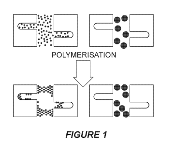
[0018] Figure 1 shows a schematic representation of the effect of molecular
size on polymerisation
(curing) of a polymeric adhesive matrix precursor material in the presence of
sorbent particle pores
exposed to a polymeric adhesive matrix precursor material having a small
molecular size (left) and one
having a larger molecular size (right);
[0019] Figure 2 shows a plot of withdrawing speed vs coating thickness;
[0020] Figure 3 shows a plot of the amount of particles (g) vs coating
thickness (microns);
[0021] Figure 4 shows a plot of the amount of particles (g) vs coating
thickness (microns);
[0022] Figure 5 shows a plot of concentration of 3-nitroaniline in feed
solution vs of concentration of 3-
nitroaniline in an eluted sample;
[0023] Figure 6 shows a plot of measured concentration of 3-nitroaniline (c*)
vs the amount of bound
3-nitroaniline per gram (q*);
[0024] Figure 7 shows a plot of measured concentration of 3-nitroaniline (c*)
vs the amount of bound
3-nitroaniline per gram/ measured concentration of 3-nitroaniline (q*/c*);
CA 03070616 2020-01-21
WO 2019/018874 PCT/AU2018/000121
[0025] Figure 8 shows a plot of the Renkin equation;
[0026] Figure 9 shows a schematic representation of the fabrication of a
silica coated fibre;
[0027] Figure 10 shows plots of (A) nitrogen adsorption-desorption isotherms;
and (B) pore-diameter
distribution on different days of pre-polymerization;
[0028] Figure 11 shows plots of binding capacity of the coated fibre at
different stages of the matrix pre-
polymerisation (1, 7 and 14 days) for the fl-blockers, propranolol (A) and
metoprolol (B);
[0029] Figure 12 shows a schematic representation of the dip coating protocol;
[0030] Figure 13 shows a plot showing the effect of different solvents on the
coating after the exposure
to the different solvents (n=3);
[0031] Figure 14 shows a plot of number of coatings v coating thickness for
multiple coating
measurements on the fibres;
[0032] Figure 15 shows a plot of number of fibres v coating thickness for the
coating process on
different fibres using an in-house build coating motor;
[0033] Figure 16 shows SEM micrographs of a pre-polymerized coated fibre at
different magnifications;
[0034] Figure 17 shows survey XPS spectra of: (A) control glass; (B) glass
treated with Piranha
solution; and (C) glass treated with (3-glycidyloxypropyl) trimethoxysilane;
[0035] Figure 18 shows the cell viability results of primary human foreskin
fibroblast (HFF) cells
exposed with "SiFT" at three exposure times (Day-1, 2 and 7). The cell
viability value was determined by
resazurin assay, and results are expressed as % cell viability after measuring
the fluorescence signal with
530 nm excitation and 590nm emission using 96 well plate reader. Data are
shown as mean SD (n = 3).
All the group shows non-significance results (<0.05);
[0036] Figure 19 shows survey XPS spectra of control stainless steel (lower
trace) and acrylic acid
plasma polymer treated stainless steel fibres (upper trace);
[0037] Figure 20 shows the peak fitting Cls core level of plasma polymer of
acrylic acid; and
[0038] Figure 21 shows SEM micrographs of a coated stainless steel fibres at
different magnifications.
CA 03070616 2020-01-21
WO 2019/018874 PCT/AU2018/000121
6
DESCRIPTION OF EMBODIMENTS
[0039] Provided herein is a solid phase microextraction substrate having a
sorbent coating on at least
part of a surface thereof. The coating is adapted for extracting at least one
analyte component from a fluid
matrix. The coating comprises sorbent particles in a polymeric adhesive
matrix. A majority of pores in
each sorbent particle in the coating do not contain substantially any of the
polymeric adhesive matrix.
[0040] The solid phase microextraction substrate described herein may provide
one or more
advantages. The lack of substantially any of the polymeric adhesive matrix in
the pores in each sorbent
particle causes a significant increase in the binding capacity of the coated
solid phase microextraction
substrate. The lack of substantially any of the polymeric adhesive matrix in
the pores in each sorbent
particle causes a dramatic reduction in the mass transfer restriction, thus
leading to faster adsorption and
desorption times. Faster on/off kinetics allow these substrates to be used in
high throughput experiments.
Faster elution in direct detection experiments (e.g. in an Open Port Probe)
generates sharper elution peaks
and therefore better sensitivities.
[0041] The solid phase microextraction substrate can be any substrate or
device that is suitable for use
in a SPME system. Suitable substrates include wires, rods or fibres
(collectively referred to herein as
"fibres"), as is known in the art. Suitable fibre materials include metal,
glass, silica, carbon, ceramic or
plastic. Suitable metals include Nitinol (Ni-Ti), stainless steel, titanium,
and copper. Suitable plastics
include polyether ether ketone (PEEK) or polyamide (nylon) for example. For
some applications, such as
in vivo applications, the fibre material is preferably biocompatible and,
therefore, amenable for use in a
biological matrix. The diameter of the fibre can be of millimetre to nanometre
dimensions. For example,
the outer diameter of the fibre can be between about 0.1 millimetres and about
6 millimetres. For some
applications, the outer diameter of the fibre is about 0.25 millimetres. For
other applications, such as for
use in an "Open Port Probe" the fibre will have a larger outer diameter, such
as about 5 mm. The
geometry of the solid phase microextraction substrate is not limited to fibres
and may have different
geometrical formats such as those for use in planar SPME (PSPME) or membrane
SPME (MSPME).
[0042] The sorbent coating comprises sorbent particles in a polymeric adhesive
matrix and is surface
bonded on at least part of a surface of the substrate. The thickness of the
sorbent coating may be from
about 3 microns to about 1000 microns, for example from about 3 microns to
about 350 microns. For
example, the thickness of the sorbent coating may be 3, 4, 5, 6, 7, 8, 9, 10,
11, 12, 13, 14, 15, 16, 17, 18,
19, 20, 21, 22, 23, 24, 25, 26, 27, 28, 29, 30, 31, 32, 33, 34, 35, 36, 37,
38, 39, 40, 41, 42, 43, 44, 45, 46,
47, 48, 49, 50, 51, 52, 53, 54, 55, 56, 57, 58, 59, 60, 61, 62, 63, 64, 65,
66, 67, 68, 69, 70, 71, 72, 73, 74,
75, 76, 77, 78, 79, 80, 81, 82, 83, 84, 85, 86, 87, 88, 89, 90, 91, 92, 93,
94, 95, 96, 97, 98, 99, 100, 110,
111, 112, 113, 114, 115, 116, 117, 118, 119, 120, 121, 122, 123, 124, 125,
126, 127, 128, 129, 130, 131,
132, 133, 134, 135, 136, 137, 138, 139, 140, 141, 142, 143, 144, 145, 146,
147, 148, 149, 150, 151, 152,
CA 03070616 2020-01-21
WO 2019/018874 PCT/AU2018/000121
7
153, 154, 155, 156, 157, 158, 159, 160, 161, 162, 163, 164, 165, 166, 167,
168, 169, 170, 171, 172, 173,
174, 175, 176, 177, 178, 179, 180, 181, 182, 183, 184, 185, 186, 187, 188,
189, 190, 191, 192, 193, 194,
195, 196, 197, 198, 199 or 200 microns. In certain embodiments, the thickness
of the sorbent coating is
30 microns. In certain embodiments, the thickness of the sorbent coating is 45
microns. In certain
embodiments, the thickness of the sorbent coating is 100 microns. In certain
embodiments, the coating
thickness is from about 20 microns to about 50 microns, leading to an increase
in diameter of the fibre of
from about 40 microns to about 70 microns. The thickness of the coating is
determined, at least in part by
the nature of the sorbent particles used, the composition of the polymeric
adhesive matrix, the outer
diameter of the SPME fibre and the speed at which the SPME substrate to be
coated is withdrawn from a
sorbent particle/adhesive matrix precursor composition.
[0043] Optionally, the substrate may be surface treated prior to it being
coated with the sorbent
particle/adhesive matrix precursor composition. The surface treatment may
promote polymer adhesion.
For example, silica substrates may be hydrolysed to expose the hydroxyl groups
on the surface of the
substrate. Silica substrates can be hydrolysed using techniques known in the
art such as chemical etching
with strong acid (e.g. piranha solution) or alkaline solution, hydrothermal
treatment or by exposure to
plasma. Optionally, silica substrates may be silanized by treating the
substrates with a silane. A wide
range of silanes are available commercially (see for example
www.gelest.com/product-
lines/silanes/?pl_page=l&perpage=100) and many of the known silanes can be
used. For example, the
silane may be aminopropyltriethoxysilane, 3-(trimethoxysily1) propyl
methacrylate or (3-
glycidyloxypropyl) trimethoxysilane.
[0044] The surface treatment may be a plasma treatment using a suitable
monomer to form a plasma
polymer intermediate coating. A range of monomers are known for use in plasma
surface modification of
metal surfaces (for example) and any of these may be suitable for the plasma
treatment. In certain
embodiments, the monomer is an alkene monomer, a carboxylate monomer or a
combination thereof By
way of example, the alkene monomer may be acrylic acid. By way of example, the
carboxylate monomer
may be propanoic acid.
[0045] The sorbent coating is adapted for extracting at least one analyte
component from a fluid matrix.
The matrix can be an environmental sample, a food sample, a biological fluid,
tissue, organ or cell. The
biological fluid can be whole blood, plasma, serum, urine, cerebrospinal
fluid, saliva or peritoneal fluid.
The analyte can be any compound whose presence at a location is indicative of
one with biological,
environmental, food, pharmaceutical, bio-analytical, clinical, forensic,
toxicological, national security,
public health, and/or safety implications. For example, the solid phase
microextraction substrate described
herein can be used for in vitro analysis of biological analytes as well as for
in vivo analysis of biological
analytes in a living animal. Alternatively, or in addition, the solid phase
microextraction
substrate described herein can be used for analysis of small molecules such as
drugs or biomarkers.
CA 03070616 2020-01-21
WO 2019/018874 PCT/AU2018/000121
8
[0046] It will be understood by those skilled in the art that the sorbent
particles can be chosen to analyse
specific target analytes. The sorbent particles can be particles of any
sorbent material that is able to bind
to one or more target analytes of interest. Sorbents that are commonly used in
liquid chromatography,
such as derivatized silica particles, can be used. For example, the sorbent
can be C-18/silica particles, RP-
amide/silica particles, HS-F5/silica particles, normal-phase silica particles,
C-1/silica particles, C-4/silica
particles, C-6/silica particles, C-8/silica particles, C-30/silica particles,
phenyl/silica particles, cyano/silica
particles, ionic liquid/silica particles, molecular imprinted polymer
particles, carboxen particles,
styrene/divinylbenzene particles, diol/silica particles, particles with
immobilised bio-specific ligands such
as antibodies or mixtures thereof The sorbent particles can be about 1 gm to
about 50 gm particles, such
as 3 gm, 5 gm or 10 m. The sorbent particles can have a surface area of about
20 m2/g to about 800
m2/g. The sorbent particles can have pore sizes from about 10 Angstroms to
about 2000 Angstroms,
such as about 100 Angstroms, about 120 Angstroms, about 200 Angstroms or about
300
Angstroms. Larger pore size sorbent particles may be useful for immobilisation
of proteins such as
antibodies.
[0047] The polymeric adhesive matrix is a polymeric adhesive material that
adheres to the surface of the
substrate and to the sorbent particles. The polymeric adhesive matrix is
formed from an adhesive matrix
precursor composition. In certain embodiments, the polymeric adhesive matrix
is a polyamine epoxy. The
epoxy component of the polyamine epoxy may be an epoxy resin having at least 2
epoxy groups.
Examples of epoxies that can be used include epoxy polyethers of polyhydric
phenols obtained by
reacting a polyhydric phenol with a halogen containing epoxide in an alkaline
medium. Polyhydric
phenols that can be used for this purpose include, among others, resorcinol,
catechol, hydroquinone,
methyl resorcinol, or polynuclear phenols, such as 2,2-his (hydroxyphenyl)
propane (bisphenol A), 2,2-
bis(4-hydroxyphenol) butane, 4,4'-dihydroxybenzophenone, bis(4 hydroxyphenyl)
ethane, and 2,2-bis(4-
hydroxy-phenol) pentane. The halogen-containing epoxides may be 3-chloro-1, 2-
epoxybutane, 3-bromo-
1, 3-epoxyhexane, 3-chloro-1, 2- epoxyoctane, and the like.
[0048] The polyamine component of the polyamine epoxy is a curing agent that
may be an aliphatic
polyamine or a cycloaliphatic polyamine. Useful polyamines contain from about
2 to about 6 amine
nitrogen atoms per molecule and from 2 to about 20 carbon atoms. Examples of
suitable amines are the
alkylene polyamines, ethylene diamine, 1,2-propylene diamine, 1,3-propylene
diamine, 1,2-butylene
diamine, 1,3-butylene diamine, 1,4-butylene diamine, 1,5-pentalene diamine,
1,6-hexylene diamine,
methane diamine, 1,4-diaminocyclohexane, diethylene triamine, triethylene
tetramine, tetraethylene
pentamine, pentaethylene hexamine, dipropylene triamine, tributylene
tetramine, hexamethylene diamine,
dihexamethylene triamine and the like. Mixtures of polyamines can also be
used.
CA 03070616 2020-01-21
WO 2019/018874 PCT/AU2018/000121
9
[0049] Optionally, solid phase microextraction substrates for in vivo analysis
may be coated with a
biocompatible outer coating, such as polyacrylonitrile (PAN), polyethylene
glycol, polypyrrole,
derivatised cellulose, polysulfone, polyamide, or polycarbohydrates such as
dextran or chitin. Examples
of the biocompatible coating that can be used include: a PAN/C-18 coating, a
PAN/RP-amide coating, a
polyethylene glycol/HS-F5 coating, a derivatised cellulose/C-18 coating, a
polypyrrole/C-30 coating, a
polysulfone/phenyl coating and polyamide/cyano coating.
[0050] Optionally, solid phase microextraction substrates for in vivo analysis
may be coated with a
hydrophilic outer coating. The hydrophilic coating can be used to suppress
binding of proteins and other
large molecules to the coating. Dextran is a suitable hydrophilic material.
[0051] Advantageously, a majority of pores in each sorbent particle in the
coating do not contain
substantially any of the polymeric adhesive matrix. As used herein, that term
means greater than 50% of
the pores in each sorbent particle in the coating do not contain any of the
polymeric adhesive matrix,
greater than 55% of the pores in each sorbent particle in the coating do not
contain any of the polymeric
adhesive matrix, greater than 60% of the pores in each sorbent particle in the
coating do not contain any
of the polymeric adhesive matrix, greater than 65% of the pores in each
sorbent particle in the coating do
not contain any of the polymeric adhesive matrix, greater than 70% of the
pores in each sorbent particle in
the coating do not contain any of the polymeric adhesive matrix, greater than
75% of the pores in each
sorbent particle in the coating do not contain any of the polymeric adhesive
matrix, greater than 80% of
the pores in each sorbent particle in the coating do not contain any of the
polymeric adhesive matrix,
greater than 85% of the pores in each sorbent particle in the coating do not
contain any of the polymeric
adhesive matrix, greater than 90% of the pores in each sorbent particle in the
coating do not contain any
of the polymeric adhesive matrix or greater than 95% of the pores in each
sorbent particle in the coating
do not contain any of the polymeric adhesive matrix.
[0052] Solid phase microextraction substrates can be formed so that a majority
of pores in each sorbent
particle in the coating do not contain substantially any of the polymeric
adhesive matrix by preventing or
minimising ingress of the polymeric matrix adhesive precursor material into
pores of the sorbent particles
in the sorbent prior to polymerisation or curing of the polymeric matrix
adhesive precursor material. Thus
provided herein is a process for preparing a solid phase microextraction
substrate having a sorbent coating
on at least part of a surface thereof The coating is adapted for extracting at
least one analyte component
from a fluid matrix. The process comprises forming a sorbent particle/adhesive
precursor composition
comprising a polymeric matrix adhesive precursor material and sorbent
particles under conditions to
substantially prevent ingress of the polymeric matrix adhesive precursor
material into pores of the sorbent
particles in the sorbent particle/adhesive precursor composition. At least
part of a substrate is then coated
with the sorbent particle/adhesive precursor composition and the polymeric
adhesive precursor material in
CA 03070616 2020-01-21
WO 2019/018874 PCT/AU2018/000121
the sorbent particle/adhesive precursor composition is polymerised under
conditions to form a sorbent
coating comprising sorbent particles in a polymeric adhesive matrix.
[0053] Our results show that preventing ingress of the polymeric matrix
adhesive precursor material
into the pores of the sorbent particles during the coating causes a
significant increase in the binding
capacity of the coated solid phase microextraction substrate and causes a
dramatic reduction in the mass
transfer restriction thus leading to faster adsorption and desorption times
relative to known SPME fibres.
[0054] In certain embodiments, the "conditions to substantially prevent
ingress of the polymeric matrix
adhesive precursor material into pores of the sorbent particles" comprise
blocking the pores of the sorbent
particles with a pore blocking agent. Thus, provided herein a process for
preparing a solid phase
microextraction substrate having a sorbent coating on at least part of a
surface thereof. The coating is
adapted for extracting at least one analyte component from a fluid matrix. The
process comprises treating
sorbent particles with a pore filling agent under conditions to block
substantially all the pores of the
particles to form blocked pore sorbent particles. The blocked pore sorbent
particles and a polymeric
adhesive matrix precursor material are then combined to form a sorbent
particle/adhesive precursor
composition. At least part of a substrate is then coated with the
particle/adhesive precursor composition
and the polymeric adhesive precursor material in the particle/adhesive
precursor composition is cured
under conditions to form a coating on the substrate comprising blocked pore
sorbent particles in a
polymeric adhesive matrix. The blocked pore sorbent particles in the polymeric
adhesive matrix are then
treated to substantially remove the pore filling agent from the pores thereof
to form the sorbent coating
comprising sorbent particles in a polymeric adhesive matrix.
[0055] The pore filling agent can be any material that is able to fill the
pores of the sorbent particles and
remain in the pores during the polymerisation step for the polymeric adhesive
precursor material. Suitable
pore filling agents include hexadecanol. Hexadecanol has a melting point of
49.3 degrees C and is,
therefore solid at room temperature and liquid at temperatures above about 50
degrees C. Thus, the
sorbent particles can be mixed with liquid hexadecanol and then removed from
the hexadecanol and
allowed to cool to less than 50 degrees C at which point the hexadecanol
solidifies in the pores. After
coating onto the substrate, the sorbent particles can be heated to over 50
degrees C and the liquid
hexadecanol can be removed from the pores of the sorbent particles under
vacuum. Other agents having
similar melting point and/or hydrophobicity (logP = 6.14) to hexadecanol could
also be used. Examples
of other agents that could be used include paraffin waxes and other long chain
alcohols.
[0056] The pore filling agent may be removed from the pores of the sorbent
particles using any suitable
technique, including heating, exposure to vacuum, solvation with a suitable
solvent, chemical
manipulation of the pore filling agent in the pores, chemical or physical
degradation of the pore filling
agent in the pores, and the like.
CA 03070616 2020-01-21
WO 2019/018874 PCT/AU2018/000121
11
[0057] In certain other embodiments, the "conditions to substantially prevent
ingress of the polymeric
matrix adhesive precursor material into pores of the sorbent particles"
comprise contacting the sorbent
particles with adhesive matrix precursor polymers or pre-polymers that have a
molecular size that is
greater than a maximum pore size of the sorbent particles. This is shown
schematically in Figure 1. In this
way, the adhesive matrix precursor polymers or pre-polymers are not able to
enter the pores of the sorbent
particles prior to or after curing. Thus, provided herein is a process for
preparing a solid phase
microextraction substrate having a sorbent coating on at least part of a
surface thereof The coating
comprises sorbent particles in a polymeric adhesive matrix that is adapted for
extracting at least one
analyte component from a fluid matrix. The process comprises combining a
polymeric adhesive matrix
precursor material and sorbent particles to form a sorbent particle/adhesive
precursor composition
comprising adhesive matrix precursor polymers or pre-polymers having a
molecular size that is greater
than a maximum pore size of the sorbent particles. At least part of a surface
of a substrate is then coated
with the sorbent particle/adhesive precursor composition after which it is
polymerised under conditions to
form a sorbent coating comprising sorbent particles in a polymeric adhesive
matrix.
[0058] The adhesive matrix precursor polymers or pre-polymers can be formed by
starting
polymerisation of the polymeric adhesive material and adding the sorbent
particles to the reaction after
polymerisation has started but before it is finished. If required, the state
of polymerisation of the adhesive
matrix precursor material can be determined using known techniques, including
by measuring the
viscosity of the reaction mixture and adding the sorbent particles at a time
when a predetermined
viscosity is reached. The skilled person will appreciate that the viscosity of
the reaction mixture increases
as the level of polymerisation increases.
[0059] We found that we were able to increase the binding capacity per volume
unit of bed by 400%
compared to the market leader product. Thus, also provided herein is a solid
phase microextraction
substrate having a sorbent coating on at least part of a surface thereof, the
coating being adapted for
extracting at least one analyte component from a fluid matrix, the coating
comprising SPME particles in a
polymeric adhesive matrix and characterised in that the binding capacity of
the substrate per volume unit
of bed is greater than 100% compared to the commercial C18 SPME LC Fibre
Probe.
[0060] The binding capacity of the substrate per volume unit of bed may be
greater than 110%, 120%,
130%, 140%, 150%, 160%, 170%, 180%, 190%, 200%, 210%, 220%, 230%, 240%, 250%,
260%, 270%,
280%, 290%, 300%, 310%, 320%, 330%, 340%, 350%, 360%, 370%, 380%, 390% or 400%
compared to
the commercial C18 SPME LC Fibre Probe.
[0061] In certain specific embodiments, the binding capacity of the substrate
per volume unit of bed is
400% compared to the commercial C18 SPME LC Fibre Probe.
CA 03070616 2020-01-21
WO 2019/018874 PCT/AU2018/000121
12
[0062] The desorption time for eluting an analyte from the commercial C18 SPME
LC Fibre Probe is
recommended to be 30 minutes. In contrast, we found that the elution time for
an analyte (i.e. 3-
nitoraniline) from a solid phase microextraction substrate described herein
was 10 seconds or less. Thus,
also provided herein is a solid phase microextraction substrate having a
sorbent coating on at least part of
a surface thereof, the coating being adapted for extracting at least one
analyte component from a fluid
matrix, the coating comprising SPME particles in a polymeric adhesive matrix
and characterised in that
the elution time for an analyte of interest from the substrate is less than 30
minutes.
[0063] The elution time for an analyte of interest from the substrate may be
less than 29 minutes, 28
minutes, 27 minutes, 26 minutes, 25 minutes, 24 minutes, 23 minutes, 22
minutes, 21 minutes, 20
minutes, 19 minutes, 18 minutes, 17 minutes, 16 minutes, 15 minutes, 14
minutes, 13 minutes, 12
minutes, 11 minutes, 10 minutes, 9 minutes, 8 minutes, 7 minutes, 6 minutes, 5
minutes, 4 minutes, 3
minutes, 2 minutes, 1 minute, 59 seconds, 58 seconds, 57 seconds, 56 seconds,
55 seconds, 54 seconds,
53 seconds, 52 seconds, 51 seconds, 50 seconds, 49 seconds, 48 seconds, 47
seconds, 46 seconds, 45
seconds, 44 seconds, 43 seconds, 42 seconds, 41 seconds, 40 seconds, 39
seconds, 38 seconds, 37
seconds, 36 seconds, 35 seconds, 34 seconds, 33 seconds, 32 seconds, 31
seconds, 30 seconds, 29
seconds, 28 seconds, 27 seconds, 26 seconds, 25 seconds, 24 seconds, 23
seconds, 22 seconds, 21
seconds, 20 seconds, 19 seconds, 18 seconds, 17 seconds, 16 seconds, 15
seconds, 14 seconds, 13
seconds, 12 seconds, 51 seconds, 10 seconds, 9 seconds, 8 seconds, 7 seconds,
5 seconds, 4 seconds, 3
seconds, 2 seconds or 1 second.
[0064] One or more embodiments of the present disclosure may provide one or
more the following
advantages:
= Keeping the pore system of the particles open during the coating causes a
significant increase in
the binding capacity of the coated device;
= Keeping the pore system of the particles open during the coating also
causes a dramatic
reduction in the mass transfer restriction thus leading to faster adsorption
and desorption times;
= Faster on/off kinetics allow these fibres to be used in high throughput
experiments;
= Faster elution in direct detection experiments (like the Open Port Probe)
generate sharper elution
peaks and therefore better sensitivities;
= The fibres do not swell in water or solvents; and
= The fibre coating is durable and reproducible.
CA 03070616 2020-01-21
WO 2019/018874 PCT/AU2018/000121
13
EXAMPLES
[0065] Example 1 ¨ Preparation of C18 coated SPME fibre using a "blocked pore"
method
[0066] Hexadecanol was dissolved in chloroform on a water bath at a ratio of
2:1. 1 ml of this solution
was added to lg of C18 silica in a closed vial. The specific pore volume of
the C18 silica used was lml/g.
The silica was agitated to allow for an even distribution of the solution in
the particles under the
assumption that capillary forces would draw the solution into the pores. The
silica particles were mixed
vigorously at 50 C for 5 minutes. Over this time period, the initial clumpy
silica became powdery. The
vial was then opened and the particles were kept at room temperature to let
chloroform evaporate from
the mixture for 24 hours. The process was then repeated using another 3341 of
the solution.
[0067] The hexadecanol modified C18 silica particles were then suspended in a
polymeric composite
formulation comprising poly(2-hydroxyethyl methacrylate-ethylene
dimethacrylate) formed from 2-
hydroxyethyl methacrylate (HEMA), ethylene dimethacrylate (EDMA) using a
phenylbis(2,4,6-
trimethylbenzoy1)-phosphine oxide (BAPO) initiator. A metal or glass SPME
fibre was dipped into the
slurry and withdrawn to form a coating which was then cured. The pre-filled
hexadecanol was then
washed out in the organic phase. The coating thickness was dependent on the
amount of silica, the
polymer composition, the fibre OD, and the withdrawal speed.
[0068] Example 2 ¨ Preparation of C18 coated SPME fibre using a
"prepolymerisation" method
[0069] A 3M-Scotch-weld DP240 epoxy adhesive was dissolved in chloroform at a
concentration of 10
weight% and the polymerisation time increased from 20 mm to about 2 weeks. The
polymerisation time is
dependent on the concentration and the storage temperature. Higher
concentrations and higher
temperatures decrease the polymerisation time. During the polymerisation the
viscosity of the solution
constantly increases. There is a window of several days where the solution has
polymerised enough that
the polymer precursors are large enough not to penetrate the pores and where
the solution is still liquid
enough to provide a uniform coating. Prior to coating, a slurry of 0.4g C18
silica (0.1g ¨ lg range is
possible) and 1 ml of adhesive solution was made. The desired coating
thickness was regulated by the
dipping speed. As the viscosity of the slurry increased the draw speed of the
substrate from the slurry had
to decrease.
[0070] Example 3¨ Performance of C18 coated SPME fibres
[0071] Binding capacities were measured with an aqueous solution of 3-
nitroaniline. The commercial
fibre was treated according to the recommendations from the supplier.
CA 03070616 2020-01-21
WO 2019/018874 PCT/AU2018/000121
14
[0072] The SPME fibre of the present disclosure was pre-treated with methanol
for 30 seconds followed
by water for 30 sec. Adsorption was performed in 60 sec and elution was
performed by trickling lml of
methanol along with the fibre within 20 seconds. 10 1 of the elution solution
was injected onto a 250x
4.6mm HPLC column for quantitation.
[0073] The Langmuir Adsorption Isotherm
[0074] One can look at the binding of an analyte to, for example, a C18
particle as a reversible reaction.
For every x analyte molecules bound to the particle (q*) there will be y
molecules left in solution (c*).
The proportion between x and y is dependent on the affinity between the
analyte and the C18 surface.
This proportion is the affinity constant (Kõõ).
[0075] The surface area of the C18 particle is limited and therefore there are
a limited number of analyte
molecules this surface can accommodate. This is the maximum binding capacity
(qm).
[0076] The relationship between all the above factors is given below
(Langmuir, I. (1918)J. Am. Chem.
Soc., 40, 1361-1403):
* qõ, = Ics = c*
q = 1+ Kass = c*
[0077] If one measures the amount bound (q*) for a number of different
concentrations (c*) the data can
be fitted to the Langmuir equation with a and Kaõ as fitting parameters.
_on
[0078] To compare two different sorbents with the same chemistry (e.g. C18)
one has to record only one
concentration (c*). Provided the chemistry is the same, then Kass has to be
the same. If q* for sorbent A is
twice as high as for sorbent B then qõ, for sorbent A has to be twice as high
a q11, for sorbent B.
[0079] Experimental
[0080] For comparative binding studies a solution 15mg of 3-nitroaniline in
100m1 of water was
prepared. The coated fibre was treated with methanol for 1 minute followed by
a rinse with water for one
minute. The coated fibre was then submerged in 10 ml of the 3-nitroaniline
solution and the solution was
stirred for 5 min to reach equilibrium. The fibre was rinsed with water and
the bound 3-nitroaniline was
eluted with methanol. 10 1 of the eluent was injected onto a 250mm x 4.6mm ID
C18 column. The
concentration of bound 3-nitroaniline was quantified using the peak area at
232nm wavelength.
CA 03070616 2020-01-21
WO 2019/018874 PCT/AU2018/000121
[0081] Restricted Pore Diffusion
[0082] Chromatographic particles are characterized by their physical
properties (particle size, pore size,
pore volume etc.) and their chemical properties (surface chemistry, ligand
density, end-capping, non-
specific binding etc.). These particles are usually packed in a bed or
immobilized onto a surface and thus
do not move and form a stationary phase. The sample and elution solution is a
liquid which is transported
across the stationary phase as a mobile phase. The surface chemistry rules the
interaction between the
sample and the stationary phase. Most particles used for SPE or chromatography
are porous with pore
sizes reaching from 5nm (50A) to 400nm (4000A). Pore volumes for porous silica
particles are fairly
constant at around 1 ml/g. The vast majority of the interactive surface lies
within the pore system, with
small pores having a much larger surface area (120A pore particles have 350m'
surface area) than large
pore size particles (1000A pore particles have 30m' surface area).
[0083] When liquid flows around a particle the solvent inside the pores is
stationary. In order to interact
with the particle surface the analyte molecule has to penetrate the pore
system and the only way it can do
this is by diffusion.
[0084] In a free solution the diffusion of a molecule is described by the
Einstein Stokes Equation:
Dk T
¨
67-cpR
where: Df = Diffusion rate constant in free solution; kB = Boltzmann constant;
= viscosity; R =
hydrodynamic radius of the molecule; and T = temperature.
[0085] Inside a pore, the diffusion becomes more complex with two extreme
positions:
= If the pore is infinitely wide then the diffusion will be equal to the
Einstein-Stokes diffusion; and
= If the analyte molecule is as big or bigger than the pore diameter then
the diffusion will be zero.
[0086] As the pore size becomes smaller there will be an increased chance that
the molecule interacts
with the wall of the pore and thus slowing the diffusion down. In 1954 E.M.
Renkin developed an
empirical equation to describe the restricted pore diffusion in a cylindrical
pore:
CA 03070616 2020-01-21
WO 2019/018874 PCT/AU2018/000121
16
2
r )3
(1 ¨ ) [1- 2.104 (sr + 2.09 (3 ¨ 0.95 (3r
D =D )51
P f
where: Dp = Pore diffusion rate; Df = Free diffusion rate; rs = radius of the
solute molecule; and rp =
radius of the pore.
[0087] When plotted the Renkin equation is as shown in Figure 8.
[0088] Any obstruction in a pore will slow the diffusion right down.
[0089] The Renkin model assumes cylindrical pores. When embedding porous
particles in a polymeric
matrix it is possible that only the pore entrance is getting obstructed while
the majority of the pore
volume remains open. These types of pores are commonly referred to as ink-
bottle type pores. The
diffusion in and out of the pore system is governed by the opening of the
pore.
[0090] Example 4¨ Preparation and performance of coated silica fibres
[0091] Reagents and materials
[0092] Polyimide coated silica fibres (357.7m) were obtained from Polymicro
Technologies (Phoenix,
AZ). (3-Glycidyloxypropyl) trimethoxysilane (98%), toluene, sulfuric acid
(99%), hydrogen peroxide
(30%), methanol, chloroform, diclofenac, metoprolol tartrate, propranolol
hydrochloride, and resazurin
sodium were purchased from Sigma Aldrich. Sulfuric acid (Merck Millipore),
hydrogen peroxide (30%)
and potassium hydroxide flakes were purchased from Chem-supply, SA-AUS. C-18
Bio-SPME fibres
were purchased from Supelco (Bellefonte, PA). 3M-Scotch-weld DP240 epoxy
adhesive was purchased
from 3M, Maplewood MN, USA. C18 silica particles were purchased from the Osaka
Soda Co. Ltd.
Japan. Dulbecco's Modified Eagle's Medium (DMEM), dimethyl sulphoxide (DMS0),
phosphate
buffered saline (PBS), lipopolysaccharide (LPS), and tris buffer were
purchased from Sigma Chemical
Co. Ltd. (St. Louis, MO, USA). Fetal Bovine Serum (FBS) and antibiotic-
antimycotic solution
(10,000U/m1 penicillin, 10 mg/ml streptomycin sulfate) was purchased from
Gibco, Invitrogen Co.
(Grand Island, N. Y., USA). Cell culture plates (nunc) were obtained from
ThermoFisher Scientific
(Roskilde, Denmark).
[0093] Instrumentation
[0094] Chromatographic experiments were carried out using an agilent 1260
infinity liquid
chromatograph (LC) with 6130 Quad mass spectrometer (MS). An analytical column
ZORBAX Eclipse
CA 03070616 2020-01-21
WO 2019/018874 PCT/AU2018/000121
17
Plus C-18, 4.6mm *150 mm, 3.5p.m particles with a guard column (Agilent
Eclipse XDB, C-18, 4.6 x
12.5 mm) was used. A Zeiss Merlin scanning electron microscope (SEM) was used
at 2kv to record the
images. The textural property of the coating material was analyzed using the
nitrogen (N2) sorption
analyzer (micrometrics ASAP 2420). X-ray photoelectron spectroscopy (XPS)
spectra of the fibre surface
were recorded using a SPECS SAGE XPS system with a phoibos 150 hemispherical
analyzer. The fibres
were coated by dip coating using an in-house built coating motor.
[0095] Pre-treatment of the fibre and hydroxylation
[0096] Polyamide-coated silica capillaries (357.511m OD and 49.8 pm ID) were
cut into 10 cm length.
The polyamide layer was removed for a length of 5 cm using a butane flame. The
silica capillaries were
then vortexed in methanol for 10 minutes to remove traces of burn polyamide
and 5 min in water
followed by nitrogen drying. The fibres were checked under a microscope to
make sure no polyamide
remained on the surface. The surface of the fibres was hydroxylated using
piranha solution (3:1 Conc.
H2SO4 and H202) for 90 minutes at room temperature (22 C) to remove the
contaminants from the surface
and expose the hydroxyl group to the surface of the fibres. Using
ultrasonication the acid treated fibres
were cleaned in water, ethanol and acetone for 5 minutes. Each fibre was
cleaned by ultrasonication and
then the fibres were subjected to further treatment. Fibres were dried under
nitrogen.
[0097] Silantfation
[0098] After hydroxylation with piranha solution, different surface treatments
were tested, namely: 3-
aminopropyltriethoxysilane (APTES), 3-(trimethoxysily1) propyl methacrylate;
and (3-
glycidyloxypropyl) trimethoxysilane (GPTS).
[0099] Using GPTS we observed a robust binding of the polymer with silica
particles on the fibre. The
protocol of the GPTS treatment was performed by treating the fibres with the
20% (v/v), GPTS/toluene at
55 C for 48 hours. To remove the weakly bound silane compounds fibres were
sonicated in toluene and
methylene chloride for 5 minute each respectively. Finally, fibres were dried
under nitrogen and cured at
70 C for 3 hours. (Kang CK and Lee YS, 2007). A schematic of the surface
modification process is
presented in Figure 9.
[00100] Pre-polynierifation and coating material preparation
[00101] A slurry was prepared by mixing the 1:10 3M-Scotch-weld DP240 epoxy
adhesive and
chloroform respectively. The slurry was mixed vigorously using vortex mixer
(Ratek-VM1) and pre-
polymerization was performed for 15 days at room temperature (22 C). The pre-
polymerization process
was performed to increase the binding capacities of the fibre. The schematics
of the pre-polymerization
CA 03070616 2020-01-21
WO 2019/018874 PCT/AU2018/000121
18
are shown in Figure 1. After the pre-polymerization period, 0.4gm/mL silica
particles were mixed with
the polymer and vortexed before coating the fibres.
[00102] N., adsorption-desorption isotherm analysis
[00103] BET (Brunauer-Emmett¨Teller) surface area and pore volume were
determined by
nitrogen adsorption/desorption isotherms. The analysis was performed on a
Micromeritics ASAP 2420
analyzer. Prior to the analysis the samples were degassed at 80 C for 24
hours. To understand the pore-
filling phenomenon, the BET analysis was performed on Day-1, 7 and 15. BET
surface area was
measured using the BET () method in relative pressure range oft- 0.05 - 0.20
and total pore volume (Vt)
was taken at P/Po= 0.99. Data was analyzed using Sigma plot software (Systat
software Inc, U.K). The
analysis was performed using mesoporous hysteresis type-4.
[00104] The textural properties of the fibre showed an increasing surface
area. We observed that
control silica particles had a surface area of 179.9 m2/g-lwith a wide pore
size distribution whereas on
Day-1 a 97.703 m2/g-1 area was calculated. In comparison there was much higher
surface area was
observed after the 15-day pre-polymerization.
[00105] BET surface area was calculated form N2 adsorption-desorption and
the data are
presented in Table 1 and Figure 10.
[00106] Table 1 - Textural properties of the pre-polymeri fed polymer mixed
with silica particles
on different days
BET SA
Sample Name
(nzig-)
Control 179.918
Day 1 97.703
Day 7 102.621
Day 15 115.361
CA 03070616 2020-01-21
WO 2019/018874 PCT/AU2018/000121
19
[00107] HPLC ¨ binding capacities
[00108] In order to validate the BET results from the pre-polymerized
polymer, fibres were
coated on different days and binding studies were performed using two I3-
blockers, metoprolol and
propranolol. The analysis was performed using liquid chromatography and mass-
spectrometry (Agilent).
Maximum binding capacity was observed from the fibre which was coated on day-
15. Figure 11(A) and
(B) shows the binding capacity on different days. This analysis establishes
the relationship with BET data
analysis as similar trends were observed in binding capacity studies.
[00109] Coating method and optimization
[00110] Dip-coating was performed using an in-house made computer software
operated motor, a
schematics of the dip coating procedure is shows in Figure 12. Before
performing the coating on the fibre,
the slurry was mixed vigorously to achieve a homogenous suspension. Fibres
were clamped on top of the
motor and were dipped in the slurry for a length of 15 mm in height for four
times (dip coating) to get the
desired coating thickness (45 p.m). After coating, fibres were cured at 75 C
for 60 minutes in an oven. In
order to optimize the coating thickness on one fibre, multiple coatings were
performed on each fibre and
after every coating fibres were thermally polymerized. The coating thickness
was measured
microscopically.
[00111] Fibre robustness testing
[00112] Most of the fibre-based solvent extraction technologies have a
problem of swelling and
reduction in the extraction efficiency after exposing the fibres to various
solvents. The breaking of the
fibre coating mostly happens due to exposure to acidic or basic solutions,
rigorous vortexing during the
extraction process and extraction of the analytes from the stronger matrix.
Swelling of the fibres was
calculated as the ratio of the difference in coating thickness before and
after solvent exposure to the
original coating thickness multiplied by 100%. The coating thickness of a
total of 39 fibres was measured
optically and then all were exposed to the water, acetonitrile/water (1:1),
acetonitrile, acetone, ethanol,
methanol/water (1:1), methanol, 70% isopropanol, formic acid (0.1 mol/L),
hydrochloric acid (0.1 mol/L),
dichloromethane, sodium hydroxide (NaOH) and hexane for 15 minutes in a set of
10 each. All the
experiments were performed in triplicate.
[00113] Table 2 and Figure 13 show the results of the swelling % of the
coating before and after
exposure of the fibres to various solvents. The results show that no swelling
was observed except
exposure to the hexane and sodium hydroxide (NaOH) where the maximum swelling
observed was 3.1%
and 2.80%, respectively. There was no visible coating breakage, and no silica
particles fallings were
observed in the solvent vials after the exposure.
CA 03070616 2020-01-21
WO 2019/018874 PCT/AU2018/000121
[00114] Table 2 - The effect of the solvent on coating thickness of
biocompatible SiFT.fibres
Solvent Swelling (%)
Water 0.00
Acetonitrile/Water (1:1 v/v) 0.01
Acetonitrile 0.04
Acetone 0.12
Ethanol 0.13
Methanol/Water (1:1 v/v) 0.06
Methanol 0.28
70% Isopropanol 0.309
Formic Acid (0.1 mol/L) 0.09
Hydrochloric Acid (0.1 mol/L) 0.26
Dichloromethane 0.82
NaOH 2.80
Hexane 3.14
[00115] Multiple coatings on. fibres
[00116] After performing multiple coatings on one fibre it was found that
the required coating
thickness (45 pm) was achieved after four dip coatings on the same fibre.
Initial coatings had some voids
on the surface whereas after two coatings the fibre surface was observed to be
covered entirely with silica
particles along with polymers and homogenous distribution throughout the
surface was observed under
the microscope. It was observed that a maximum of ten coatings can be
performed and the coatings were
very robust even after performing ten coatings on the fibre. However, it was
also observed that the fibre
could not hold any further coating after ten coatings. The maximum coating
thickness achieved was
CA 03070616 2020-01-21
WO 2019/018874 PCT/AU2018/000121
21
335p.m. Figure 14 shows the multiple coatings on the fibres. The coatings were
measured
microscopically, and the thickness was observed a total of the nine spots in
each fibre.
[00117] The coating thickness efficacy was also checked by measuring the
coating thickness on
15 different fibres. It was found that the motor is very effective and
controlled in terms of coating
thickness on different fibres. Upon coating on 15 different fibres it was
found that after four coatings a
coating thickness of 45 ( ) p.m was attained. A total RSD (%) was 5.5% on 15
different fibres. The
results of coating thickness observations are shown in Figure 15.
[00118] Topological characterifation
[00119] Scanning electron microscopy analysis
[00120] Surface morphology was assed using a Zeiss Merlin scanning
electron microscope
(Merlin, Carl Zeiss Co., Oberkochen, Germany) used at an operating voltage of
2kV. The SEM images of
the coated fibre demonstrate that the particles are completely covered with
the polymer and silica particle
distribution is homogenous throughout the coating Figure 16 shows the SEM
images of the coated fibre.
[00121] X-ray Photoelectron Spectroscopy (XPS)
[00122] X-ray photoelectron spectroscopy of the control, piranha treated,
and silane treated
substrate was carried out using a SPECS SAGE XPS system with a Phoibos 150
hemispherical analyzer
at a takeoff angle of 90 and MCD-9 detector. XPS spectra was recorded from 0
to 1000 eV at a pass
energy of 100 eV with the energy steps of 0.5 eV to determine the elements
available on the differently
treated glass substrates. Wide scan spectra were recorded for selected peaks
using 0.1 eV energy steps at a
pass energy of 20eV. Spectra recorded for both silane coated, and control
fibre were corrected by setting
the aliphatic carbon peak by following the methodology of Beamson G and Briggs
D., 1992. All the
recorded spectra were analyzed using CASAXPS (Neal Fairley, U.K.).
[00123] Glass substrates
[00124] The surface chemistry of the glass substrate was analyzed using
XPS, the survey
spectrum is displayed in Figure 17 and in Table 3. The XPS analysis was
performed on control, piranha
treated, and silane treated fibres. Questioning the absence of silanol
limiting the reaction, prior to
silanization substrate hydroxylation was performed with the piranha solution.
After the piranha treatment
the surface shows less amount of carbon whereas silane treatment significantly
increases the amount the
carbon on the surface. These results show that treatment with the silane was
found on the surface that lead
to the robust binding of the polymeric material.
CA 03070616 2020-01-21
WO 2019/018874 PCT/AU2018/000121
22
[00125] Table 3 - XES elemental composition on all three different steps
of modifications
Substrate modification 0 (%) C (%) Si (%)
Control 52.030 16.264 31.706
Piranha Treated 51.361 15.634 33.055
GPTS Treated 40.397 36.374 23.229
[00126] Biocornpatibility testing
[00127] Preparation of a successful biocompatible coating for fibres shows
a great capability of
towards developing a device that can be used for biomedical, pharmaceuticals
and forensic applications.
For these applications, fibre-based device coatings need to be robust, thin
and unbreakable. Resazurin
assay for the cell-viability is widely used to evaluate the biocompatibility
of polymeric materials. The
evaluation was performed by measuring the reduction of resazurin to resorufin
using spectrophotometers
which happens due to transference of electron from NADPH+H to resazurin
(Borra RC et al., 2009).
Human foreskin fibroblasts HFF-1 (ATCC) were cultured in the DMEM (Dulbecco's
modified Eagle's
medium) (Life technologies, Victoria, Australia) supplemented with the 10%
fetal calf serum (FCS)
(AusGenex, Australia), 1% penicillin/streptomycin (Sigma-Aldrich) at 37 C in
the humidified 95% air
and 5% CO2 incubator. The media was changed every 3 days after observing 90%
confluency of cells in
the culture flasks, cells were washed using phosphate buffer saline (PBS) and
detached using the 0.25%
(w/V) trypsin solution (Sigma-Aldrich) in PBS. For this test, 4.5x10⁴ cells mL-
I were seeded in a 6 well
plate with 3 distinct groups (Control, uncoated fibre and coated fibre) in
triplicates manner. All the
experiments were performed between the passage number 9 to 13.
[00128] After allowing the cells to adhere to the surface for 8 hours,
fibres placed in the nunc
inserts were kept in the well. Cells were observed under a fluorescence
microscope to check the cell
structure every day. Prior exposing the fibres to the cells, all the fibres
were sterilized in ethanol for 30
minutes followed by 45 minutes of ultra-violet (UV) exposure under controlled
conditions. Cell-viability
was quantified by resazurin assay at 24, 48 and 72 hours of incubation using
10% resazurin (Sigma-
Aldrich). 500 ?IL Resazurin was added to each well and plates were incubated
for 2 hours in CO2
incubator. After the incubation period 1004, of cell suspension was
transferred to the 96 well plate and
fluorescence was observed at 530ex/590em nm using a FLOUstar Optima plate
reader (BMG LabTech
CA 03070616 2020-01-21
WO 2019/018874 PCT/AU2018/000121
23
Pty. Ltd, Victoria, Australia). All the statistical analysis was performed on
GraphPad Prism software
operated at windows 10.
[00129] Biocompatibility evaluation
[00130] To test the cytotoxic potential of the coated fibres the resazurin
assay was used to
measure the cell viability after the exposure of the fibres to the primary HFF-
1. To evaluate the
biocompatible nature of the coated fibre primary human foreskin fibroblast
cells were used instead of
secondary cell lines. Fibroblast cells are an establish model to check the
biocompatibility of the coated
fibres. Figure 18 shows the metabolic activity on different days after
exposing the fibres to the cells. The
results were compared with control cells. There was no statistically
significant difference between the
control cells, uncoated fibres and coated fibres (p<0.005). It was observed
that there was no cell death
(%) on day-1 or day-2 and the coated fibre had <10% cell death on day-7, which
is an indication of over-
confluent of the cells within the wells and lead to the leaching of the cells.
These results suggest that
coated fibres with silica particles and polymer had no adverse effect on the
primary cells.
[00131] Surface modification of stainless steel substrates
[00132] A stainless-steel fibre (80 mm in length) was cleaned with
dimethylfonnide (DMF) for 5
minutes in an ultrasonic bath, followed by cleaning with acetone, methanol and
ultrapure water for 5
minutes each respectively. Fibres were dried under nitrogen and kept closed in
a vial for further
treatments. Plasma polymerization treatment was carried out in a custom made
(high frequency, 13.56
MHz) plasma polymerization system and the power was supplied using an
amplifier and matching unit of
a coaxial power system (Coaxial Power Systems Ltd, Eastbourne, United
Kingdom). Plasma polymer
precursor, acrylic acid (AA) was purchased from the Sigma-Aldrich. At first,
the plasma reactor chamber
was evacuated using a rotary pump to a base pressure of below 1x10⁻⁴ mbar to
remove all other
atmospheric gases and impurities inside the chamber. Acrylic acid precursor
was introduced to the
chamber via a needle valve (Chell, U.K.) using precursor flow rate of 4
(cm3/min). The 50-watt (W) air
plasma was run three times for 10 minutes each and fibres were rotated every
time to make sure the
coating of the plasma was even on the surface and covered the fibres. At the
end of the process, the power
was reduced to 5 watts for 20 minutes to deactivate the free radicals
(Michelmore Act al., 2014, Kirby
GT et al., 2017).
[00133] X-ray photoelectron spectroscopy (XPS,) of stainless steel fibres
[00134] XPS of the plasma coated stainless steel fibres was carried out
using a SPECS SAGE
XPS system with a Phoibos 150 hemispherical analyzer at a takeoff angle of 90
and MCD-9 detector. XP
survey spectra were recorded from 0 to 1000 eV at a pass energy of 100 eV with
the energy steps of 0.5
CA 03070616 2020-01-21
WO 2019/018874 PCT/AU2018/000121
24
eV to determine the elements available on the plasma coated and control
stainless steel fibres. Wide scan
spectra were recorded for selected peaks using 0.1 eV energy steps at a pass
energy of 20eV. Spectra
recorded for both plasma coated and control fibres were corrected by setting
the aliphatic carbon peak by
following the methodology of Beamson G and Briggs D., 1992. All the recorded
spectra were analyzed
using CASAXPS (Neal Fairley, U.K.).
[00135] The content of the surface analysis of control steel fibre and
acidic plasma coated fibres
are listed in Table 4. The Cis core level spectra of the acidic plasma
polymers were peak fitted using 70%
Lorentian/30% Gaussian peak shapes with full-width at half-maxima (fwhm)
between 1.6 and 1.9. There
was significant amount of oxygen and carbon was present on the surface of the
fibre. XPS spectra also
shows the increase of the carbon (%) from control substrate to the plasma
treated stainless steel substrate.
[00136] Table 4 - Content of the surface analysis of control steel .fibre
and acidic plasma coated
fibres
Name At %
COOH 12.86%
bCOOH 12.815%
CH 64.928%
C=0 6.471%
C-OH 2.959%
[00137] SEM analysis of stainless steel fibres
[00138] The morphology of the coating on the stainless-steel fiber was
observed under high-
resolution SEM show in the Figure 21. The SEM images analysis shows the
mixture of silica particles
and polymer distribution was coated homogenously on the surface of the
stainless fiber. It was found that
silica particles are evenly distributed throughout the surface with spherical
morphologies and polymer
attachment can be seen on the silica particles.
CA 03070616 2020-01-21
WO 2019/018874 PCT/AU2018/000121
[00139] Example 5 ¨ Substrate with protein resistant coating
[00140] SiFT fibres/rods were modified with C18 particles with 120A pores.
A 1 mg/ml solution
of 2-nitroaniline in water was added to water and foetal calf serum in ratios
1:1 and 1:5 resulting in
concentrations of 0.5 and 0.1 mg/ml respectively. A glass rod coated with 4mm
of coating was used for
the binding studies. The rod was submerged in the solutions for 5 minutes and
the bound analyte was
eluted with lml of methanol. 20p.1 of the elution solution was injected into a
HPLC.
[00141] When the sample is dissolved in serum, there are multiple sample
components competing
for the binding sites, hence, the binding of 2-nitroaniline is reduced. The
effect is more pronounced when
the analyte of interest is in a lower concentration.
[00142] In order to suppress protein binding the fibres were coated with a
second layer of dextran.
Dextran is a hydrophilic polysugar and only shows minimal interactions with
proteins. The dextran
chosen had a molecular weight of 450,000 to 600,000 with a Stokes radius of
150A. Therefore the
dextran molecules cannot penetrate the pores but form a hydrophilic barrier to
prevent proteins and other
large molecules to come in contact with the C18 particles. In brief, an
aqueous solution of dextran was
precipitated onto the coating and crosslinked with 1,4-butanediol
diglycidylether. The dextran coating
was performed on three rods and the binding properties were evaluated as
before however only with the
0.1mg/m1 concentration. The results are shown in Table 5.
[00143] Table 5 - Binding properties of dextran coated substrates
dextran coated
water serum %
Rod #1 108657 68132 63
Rod #2 109454 105439 96
Rod #3 121327 93960 77
[00144] With the untreated rods a42% relative binding was achieved when
the sample was in
serum. After the coating an increase the relative binding to 63, 77 and 96%
was observed. Thus, a
complex matrix can reduce the binding capacity of the substrates for a target
analyte and by suppressing
the proteins in serum to compete for the binding sites the relative binding
capacity for 2-NA can be
increased significantly.
[00145] Throughout the specification and the claims that follow, unless
the context requires
otherwise, the words "comprise" and "include" and variations such as
"comprising" and "including" will
CA 03070616 2020-01-21
WO 2019/018874 PCT/AU2018/000121
26
be understood to imply the inclusion of a stated integer or group of integers,
but not the exclusion of any
other integer or group of integers.
[00146] The reference to any prior art in this specification is not, and
should not be taken as, an
acknowledgment of any form of suggestion that such prior art forms part of the
common general
knowledge.
[00147] It will be appreciated by those skilled in the art that the invention
is not restricted in its use to the
particular application described. Neither is the present invention restricted
in its preferred embodiment
with regard to the particular elements and/or features described or depicted
herein. It will be appreciated
that the invention is not limited to the embodiment or embodiments disclosed,
but is capable of numerous
rearrangements, modifications and substitutions without departing from the
scope of the invention as set
forth and defined by the following claims.
CA 03070616 2020-01-21
WO 2019/018874 PCT/AU2018/000121
27
REFERENCES
[00148] Michelmore A, Whittle JD, Short RD, Boswell RW, Charles C. An
Experimental and
Analytical Study of an Asymmetric Capacitively Coupled Plasma Used for Plasma
Polymerization.
Plasma Processes Polym. 2014 11, 833.
[00149] Kirby GT, Mills SJ, Vandenpoel L, Pinxteren J, Ting A, Short RD,
Cowin AJ,
Michelmore A, Smith LE. Development of Advanced Dressings for the Delivery of
Progenitor Cells.
ACS Appl Mater Interfaces. 2017 Feb 1;9(4):3445-3454.
[00150] Beamson, G.; Briggs, D. High Resolution XPS of Organic Polymers:
The Scienta
ESCA300 Database; Wiley: Chichester, U.K., 1992.
[00151] Borra RC, Lotufo MA, Gagioti SM, Barros Fde M, Andrade PM. A
simple method to
measure cell viability in proliferation and cytotoxicity assays. Braz Oral
Res. 2009 Jul-Sep;23(3):255-62.