Note: Descriptions are shown in the official language in which they were submitted.
CA 03070643 2020-01-21
WO 2019/015936 PCT/EP2018/067641
MONO AND BISPECIFIC ANTIBODY BINDING TO HERG1 AND
HERG1/INTEGRIN BETA 1
FIELD OF THE INVENTION
The present invention refers to the field of antibodies and their application
for
diagnostic and therapeutic purposes in oncology and other fields of medical
sciences. In particular it relates to anti-hERG1 molecules and their
engineered
derivatives comprising bispecific antibodies targeting both hERG1 and (31
integrin.
STATE OF THE ART
Over the past two decades, the antibodies' production technology has been
significantly improved through antibody engineering; the advent of new
technologies
in the field of molecular engineering, led to the production of a wide variety
of
genetically engineered antibodies, such as fragments of type Fab, Fv form of
simple
chain of scFv, diabodies, triabodies, bispecifics, minibodies, nanobodies,
phage
antibodies. In fact there is a range of applications, in which the Fc-mediated
effects
are not required and even undesirable, because of their associated toxic
effects and
their capacity to evoke an immune response able to neutralise the antibody
efficacy,
when its Fc derived from a non human source.
Among the engineered antibody fragments, the Single Chain Variable Fragment
(scFv) is the most popular and one of the smallest recombinant format with an
antigen-binding activity function and with the property to be easily
manageable for
immunological application.
A scFv consists of variable regions of heavy (VH) and light (VL) chains, which
are
joined together by a flexible peptide linker, without compromising the
fidelity of the
VH-VL paring and antigen-binding sites. The choice of a linker can affect the
solubility, expression and correct folding of the scFv. Peptide linkers can
vary from
10 to 25 amino acids in length and are typically composed of hydrophilic amino
acids
such as glycine (G) and serine (S). Hydrophilic sequences prevent
intercalation of
the peptide within or between the variable domains throughout the protein
folding.
The most common linker used is the (01y45er)3 motif, due to its flexibility,
neutral
charge and solubility. The use of scFv in diagnostics and therapy provides
several
advantages over whole antibodies, especially in solid tumours' therapy; in
fact, the
1
CA 03070643 2020-01-21
WO 2019/015936 PCT/EP2018/067641
speed of penetration by a fragment versus an intact molecule is the most
remarkable
advantage. In 1988, it was established that an intact molecule of IgG took
fifty-four
hours to penetrate 1 mm into a solid tumour, while a Fab fragment managed the
same distance in sixteen hours. Moreover, the scFv, as well as all the other
antibody
fragments format, can be mold into multivalent and multispecific reagents or
easily
linked to therapeutic tools as radionuclides, toxins or nanoparticles and
engineered
to improve their diagnostic and therapeutic efficacy.
These engineered molecules are easy to produce in bacterial or yeast systems,
furthermore extravasate more efficiently and have a higher tissue penetration
ability
than full length Ig; the only limit of these molecules is the short half-life
due to their
small size. Many strategies has been developed to improve pharmacokinetic such
as multimerization of scFv (shortening their linker sequence) to form
triabodies (of
about 90 kDa) and tetrabodies (of about 120 kDa), or conjugation of antibodies
to
big molecules such as polyethylene glycol (PEG) (Natarajan et al. 2005) or
human
serum albumin (HSA).
Bispecific antibodies (bsAbs) have recently raised a lot of attention as
potential
cancer therapeutic agents because they offer several advantages:
- bsAbs can redirect specific immune cells towards tumour cells, thereby
enhancing tumor killing;
- bsAbs can simultaneously block two different targets in different
pathways that carry out unique or overlapping functions in
pathogenesis;
- bsAbs can potentially increase binding specificity by interacting with
two different cell-surface antigens instead of one.
The development of bispecific antibodies (bsAbs) has experienced many
difficulties,
mainly due to the manufacturing problems, poor yields, instability and
immunogenicity (Spiess C. eta!, 2015).
Concerning the methodology for bsAbs production, they are primarily produced
by
three methods, which include:
- quadroma technology, based on the somatic fusion of two different
hybridomas cell lines;
- chemical conjugation, through the use of chemical cross-linkers;
2
CA 03070643 2020-01-21
WO 2019/015936 PCT/EP2018/067641
- genetic approaches utilizing recombinant DNA technology.
Bispecific antibodies can be roughly divided into two main subgroups:
immunoglobulin G (IgG)-like molecules and non-IgG-like molecules and so far
there
are over 30 bsAbs in clinical development with two, Catumaxomab and
Blinatumomab, already approved for the market.
Non-IgG-like include mainly scFv-based bsAbs and nanobodies. It is known that
scFvs can become dimers, trimers, or tetramer depending on linker length,
antibody
sequence and other factors (Le Gall F. et aL, 1999). Such format is favored
and has
many possible clinical applications. Among scFv-based bsAbs formats there are:
- Tandem scFvs, which consist of two scFvs connected by a flexible
peptide linker, such as glycine-serine repeat motifs in a tandem
orientation. The famous bispecific T cell engager (BiTE) technology is
based on this format (Chames P. et aL, 2009).
-
Diabody format, in which the variable domains of two different
antibodies are connected by two linkers. These have the function to
increase the stability of the diabody.
- Single-chain diabodies (scDbs), the diabody format can be
converted into a single-chain diabody by adding an additional
connection linker between the chains
- Tandem diabodies (TandAbs) are formed by two pairs of VL and VH
domains, connected in a single polypeptide chain, forming a
tetravalent TandAb.
- Dual-affinity retargeting molecules (DARTs) DARTs are created by
the association of the VH of a first variable region linked to the VL on
a second chain, and the VH of the second variable region linked to the
VL on the first chain in a VLA - VHB + VLB - VHA configuration. Due
to their small size, DARTs are prone to elimination (Moore P.A. et aL,
2011).
Diabodies and scDbs are also the most effective way to generate bispecific
antibody
fragments, able to bind two different antigens and thus useful to crosslink
cells (e.g.
retargeting immune system effector cells); to recruit effector molecules (like
toxins,
drugs, cytokines, radioisotopes, or complement system), to retarget carrier
system
3
CA 03070643 2020-01-21
WO 2019/015936 PCT/EP2018/067641
(such as viral vectors for gene therapy) (Kontermann 2005); to target and
inhibit
macromolecular complexes involved in tumour progression.
Antibody engineering provided also methods to increase avidity (e.g antibody
fragments multimerisation); affinity (e.g. mutation in the variable regions of
whole Ig
or antibody fragments); and enhance effector functions (e.g. mutation in the
constant regions of whole Ig or conjugation of antibody fragments with
recombinant
Fc, toxin, drugs, cytokines, death ligands, radioisotopes, nanoparticles or
complement system molecules).
Over the past three decades, the human ether-a-go-go-related gene 1 (hERG1)
potassium channel has become a target in oncology as well as in other human
diseases. However, its exploitation for therapeutic purposes has been hindered
by
the fact that most drugs that cause hERG1 blockade as their primary or side
effect,
can cause cardiotoxicity (lengthening of the electrocardiographic QT interval
and
onset of ventricular arrhythmias). In search of biophysical and biomolecular
features
which distinguish hERG1 expressed in the heart from hERG1 expressed in cancer
cells and other disease characteristic cells, it was found that hERG1
complexes with
other plasma membrane proteins, in particular with the beta1 subunit of
integrin
receptors, on the plasma membrane of cancer cells. Such complex does not occur
in cardiac myocytes (Becchetti A. et al., Sci.Signaling, 10(473). pii:
eaaf3236. doi:
.. 10.1126/scisignal.aaf3236. PMID: 28377405, 2017). Hence, the hERG1/beta1
integrin complex configures as an oncogenic unit, peculiar of transformed
cells, a
fact that differentiate hERG1 in tumor from the channel expressed in the
heart. This
finding implies that any molecule (a small molecule drug or otherwise a
protein)
targeting the hERG1/beta1 integrin complex can be used for diagnostic and
therapeutic purposes, being devoid of cardiotoxicity. At the moment molecules
able
to target hERG1/beta1 integrin complex are not known.
In the Italian Patent IT1367861 is described a hybridoma cell line clone,
named A7,
able to secrete an anti-hERG1 monoclonal antibody (mAb) specific against the
S5-
pore extracellular portion of hERG1.
W02016020483 (Al) describes the detailed structure of an intact murine
monoclonal anti-hERG1 molecule and a corresponding anti-hERG1 scFv antibody
production, obtained after the isolation of the mAb anti-hERG1 VH and VL. Such
4
CA 03070643 2020-01-21
WO 2019/015936 PCT/EP2018/067641
scFv has the same specificity of the correspondent whole antibody, and thus it
is
able to recognize the same anti-hERG1 protein, aberrantly expressed in tumours
and other diseases. Nucleotide sequences SEQ ID NO:1 and SEQ ID NO:3
encoding respectively VH (SEQ ID NO:2) and VL (SEQ ID NO:4) were disclosed
together with nucleotide sequence SEQ ID NO:5 encoding for a scFV having SEQ
ID NO:6.
There are known in the art, for research purposes, anti-beta1 integrin mAb,
for
example are known among others T52/16 (Arroyo et al. J. Cell Biol. 1992,
117(3),
659-670) and BV7 (Martin-Padura et al. J. Biol. Chem. 1994, 269(8), 6124-
6132).
Aim of the present invention is to provide a bispecific antibody which targets
simultaneously both the hERG1 and the beta1 integrin proteins which are
complexed on the plasma membrane of cancer cells. Further aim of the present
invention is to provide an improved, or at least alternative, antibody against
hERG1.
SUMMARY OF THE INVENTION
Subject-matter of the present invention is a bispecfic antibody (bsAb)
comprising
the Heavy chain Variable (VH) domain and Light chain Variable (VL) domain of
an
anti-hERG1 Ab which binds the extracellular domain S5-P of hERG1 and the Heavy
chain Variable (VH) domain and Light chain Variable (VL) domain of a anti-131
integrin Ab which binds the extracellular domain of 131 integrin.
Surprisingly it was found that a bsAb according to the invention was able to
bind
selectively the complex hERG1+ 131 -integrin which is present only in tumor
cells. In
particular a bsAb according to the invention showed the capacity in vitro of
inhibiting
cell growth and migratory, pro-metastatic, activity on a panel of neoplastic
cell lines.
For an aspect the present invention relates also to an anti-hERG1 molecule
comprising a Heavy chain Variable (VH) domain having at least 85% identity
with
SEQ ID NO:8 wherein residue at position 95 is Cys, and a Light chain Variable
(VL)
domain having at least 85% identity with SEQ ID NO:4, said molecule having
specificity against hERG1 S5-pore extracellular portion.
Surprisingly it was found that an anti-hERG1 molecule (anti-hERG1-Cys)
according
to the invention and having a Cys in position 95 of the VH domain (SEQ ID No:
8),
showed a better affinity toward the immobilized antigen compared to a
molecule, as
5
CA 03070643 2020-01-21
WO 2019/015936 PCT/EP2018/067641
known in the state of the art, having a Phe in position 95 of the VH domain
(SEQ ID
No: 2). In particular an scFv molecule according to the invention showed the
capacity in vitro of inhibiting cell growth on a panel of neoplastic cell
lines.
DETAILED DESCRIPTION OF THE INVENTION
Preferably the bsAb of the invention are those wherein the Heavy chain
Variable
(VH) domain of anti-hERG1 Ab has 85% identity with SEQ ID No: 8 or SEQ ID No:
2; and the Light chain Variable (VL) domain of anti-hERG1 Ab has 85% identity
with
SEQ ID No: 4; and the Heavy chain Variable (VH) domain and a Light chain
Variable
(VL) domain of a anti-r31 integrin Ab have at least 85% identity with VH and
VL of
T52/16 (SEQ ID N: 26 and 24) or BV7 (SEQ ID N: 46 and 48).
Preferably the bsAb of the invention is a non-IgG-like bsAb, preferably a scFv-
based
bsAb. According to the invention the scFV-based bsAb has a format selected in
the
group consisting of Tandem scFvs, Diabody format, Single-chain diabodies,
Tandem diabodies (TandAbs) and Dual-affinity retargeting molecules (DARTs),
preferably scDb.
Preferably the VH domain of anti-r31 integrin Ab has an 90%, 95%, 99% or 100%
identity with SEQ ID NO:26 or SEQ ID NO:46, more preferably SEQ ID NO:26.
Preferably the VL domain of anti-r31 integrin Ab has an 90%, 95%, 99% or 100%
identity with SEQ ID NO 24 or SEQ ID NO 48, more preferably SEQ ID NO 24
Preferably the VH domain of anti-hERG1 Ab, still retaining the amino acid Cys
at
position 95, has an 90%, 95%, 99% or 100% identity with SEQ ID NO:8.
Preferably the VH domain of anti-hERG1 Ab has an 90%, 95%, 99% or 100%
identity with SEQ ID NO:2.
Preferably the VL domain of anti-hERG1 Ab has an 90%, 95%, 99% or 100%
identity
with SEQ ID NO:4.
The VH domain of anti-hERG1-Cys Ab according to the present invention is
preferably encoded by a sequence of nucleotides having at least 70%, 80%, 90%,
95%, 99% or 100% homology with SEQ ID No: 7 wherein the triplet from residue
283 to 285 can be TGT or TGC.
The VH domain of anti-hERG1 Ab according to the present invention is
preferably
encoded by a sequence of nucleotides having at least 70%, 80%, 90%, 95%, 99%
or 100% homology with SEQ ID No: 1.
6
CA 03070643 2020-01-21
WO 2019/015936 PCT/EP2018/067641
The VL domain of anti-hERG1 Ab according to the present invention is
preferably
encoded by a sequence of nucleotides having at least 70%, 80%, 90%, 95%, 99%
or 100% homology with SEQ ID No: 3.
Molecule according to the invention can be a fully humanized recombinant Ab.
Molecule according to the invention can be a scFv or any other engineered
antibody
such as Fab, Fv form of simple chain of scFv, diabodies, triabodies,
bispecifics,
minibodies, phage antibodies; preferred are scFv and diabodies (scDb).
Preferred linkers are the (Gly4Ser)3 motifs.
Particularly preferred is an anti-hERG1-Cys scFv wherein VH and VL are linked
by
a peptide linker; more preferred is an anti-hERG1-Cys scFv having SEQ ID No:
10.
Particularly preferred is a bsAb which is a single chain diabody (scDb)
comprising
an anti-hERG1-Cys scFv and an anti-81-integrin scFv, thus acting selectively
against the complex hERG1+ 81-integrin which is present only in tumor cells.
For a preferred aspect, therefore, the present invention relates to a bsAb
single
chain diabody (scDb) comprising a first Heavy chain Variable (VH) domain
having
at least 85% identity with SEQ ID NO:8 wherein residue at position 95 is Cys,
and
a first VL domain having at least 85% identity with SEQ ID NO:4, and a second
VH
domain having at least 85% identity with SEQ ID NO:26 or SEQ ID N: 46, and a
second VL domain having at least 85% identity with SEQ ID NO:24 or SEQ ID N:
48, said bispecific Ab having specificity against hERG1 S5-pore extracellular
portion
and against 81-integrin.
A bsAb or an anti-hERG1 molecule according to the invention is useful as
diagnostic
or therapeutic tool.
A diagnostic tool according to the invention is for in vitro hERG1 detection
(e.g. in
surgical samples or biopsies) comprise the anti-hERG1-Cys scFv and/or, the
bispecific Ab(preferably as scDb) according to the invention, either
unlabelled or
linked to a fluorophore, preferably fluorophoreAlexa 488.
Subject-matter of the present invention is therefore also an In Vitro
Diagnostics
(IVD) kit of parts for the simultaneous, separate or sequential use, said IVD
kit
comprising:
a container containing the anti-hERG1-Cys scFv as above described; and/or
a container containing the bispecific Ab as above described
7
CA 03070643 2020-01-21
WO 2019/015936 PCT/EP2018/067641
Preferably the IVD kit further comprises a container containing an Intact
Monoclonal
Antibody as described in W02016020483, as a reference control.
The IVD kit can be used either on fixed tissue samples for ImmunoHisto
Chemistry
.. techniques (with results being available in 2-3 weeks) or on FRESH BIOPTIC
TISSUE (obtained e.g. by Endoscopy or surgery), to be used with
lmmunofluorescence techniques, with results being available in 1 day.
The anti-hERG1-Cys scFv as above describes labelled with a fluorophore (for
example and preferably Alex 750) or radionuclide (for example and preferably
Tc99)
is a molecule for use in in vivo (humans) early diagnosis of hERG1 positive
cancers
are represented by the anti-hERG1-Cys scFv,. The anti-hERG1-Cys sc-FV antibody
according to the invention has a specific molecular structure allowing rapid
penetration into the cancer tissue, rapid binding to the hERG1 biomarker and
rapid
elimination, rendering it, when linked to a radionuclide, an ideal molecule
for
.. obtaining early in vivo (humans) diagnosis of hERG1 positive cancers. The
use in
one single administration and the fast half-life, 3.5 hours of the molecule
with no
systemic toxicity when injected intravenously at 8 mg/kg, and no alterations
at the
ECG (see Figure 19) allow to have no interaction with cardiac cells. Finally,
the anti-
hERG1-Cys scFv turned out to have a very good tumour/tissue ratio, when
injected
.. intravenously at 1 mg/kg in mice carrying a xenogafted pancreatic cancer in
the
pancreas.
Therapeutic tools: The bispecific antibody molecule according to the
invention,
specifically designed to inhibit cancer hERG1/beta1 integrin molecular complex
for
therapeutic purposes, is the single chain bispecific antibody (single chain
Diabody,
scDb) that is able to bind selectively hERG1 when expressed with the beta1
integrin
on Cancer cells, with no interaction with the heart. The scDb according to the
invention represents an ideal molecule to be used for repeated (chronic)
administration in patients with hERG1 positive Cancers, with no cardiac safety
concern, with a therapeutic potential both at an early as well as at an
advanced/metastatic stage, both as Single Agent as well as Combination Therapy
agent, to be added to Chemotherapy, Irradiation, Target Therapy and lmmuno-
Oncology Therapy. The rationale for the Combination Therapy is that the
pathways
8
CA 03070643 2020-01-21
WO 2019/015936 PCT/EP2018/067641
constitutionally activated in cancer cells by over-expression of the
hERG1/beta1
integrin molecular complex are complementary and integrative of the pathways
currently targeted by the available drugs. Furthermore, hERG1/beta1 integrin
cancer pathway can represent a mechanism of tumour escape in respect to
current
available Therapies. The anti-hERG1/beta1 integrin scDb turned out to have a
half
life of roughly 12 hours, with no systemic toxicity when injected
intravenously at 8
mg/kg, and no alterations at the ECG (see Fig. 20). Finally, the anti-
hERG1/beta1
integrin scDb turned out to have a very good therapeutic efficacy when
injected at
1 mg/Kg, twice a week for six times in mice carrying xenografted pancreatic
cancers
in the pancreas.
Pathologies which can be diagnosed or treated using a bsAb or a molecule
according to the invention are all those pathologies characterized by an over
expression or mis-expression of hERG1 protein. Among said pathologies can be
listed tumours, neurological diseases, endocrine diseases and neuro-endocrine
diseases.
A bsAb or a molecule according to the invention, in particular an anti-hERG1-
Cys
scFV or scDb, can also be used as a pharmaceutical delivery vector: so for
example
it can be covalently or not bonded to radionuclide, enzyme, drugs or toxin.
Further subject-matter of the present invention are therefore also a
pharmaceutical
composition comprising the bsAb or the molecule according to the invention and
at
least another pharmaceutically acceptable ingredient.
Further subject-matter of the present invention is also a sequence of
nucleotides
encoding the bispecific Ab or an anti-hERG1 molecule according to the
invention.
Suitable grades of homology (e.g. at least 85%) with the encoding sequences
which
allows to obtain a molecule according to the invention are intended to be
included.
A molecule according to the invention can be preferably prepared by employing
nucleotide sequences SEQ ID NO:7 and SEQ ID NO:2 encoding respectively VH
(SEQ ID NO:8) and VL (SEQ ID NO:4).
Particularly preferred according to the invention is a method for preparing an
anti-
hERG1-Cys scFv according to the invention, said method comprising the use of
nucleotide sequence SEQ ID NO:9 encoding for an anti-hERG1-Cys scFV having
SEQ ID NO:10.
9
CA 03070643 2020-01-21
WO 2019/015936 PCT/EP2018/067641
A scDb-hERG1131 according to the invention is preferably prepared by employing
nucleotide sequences SEQ ID NO:7, SEQ ID NO:2, SEQ ID NO:23 or SEQ ID N:
47 and SEQ ID NO:25 or SEQ ID N: 45 encoding respectively anti-hERG1-Cys VH
(SEQ ID NO:8), anti-hERG1-Cys VL (SEQ ID NO:4), anti (31-integrin VL (SEQ ID
No: 24 or SEQ ID N: 48) and anti 131-integrin VH (SEQ ID No: 26 or SEQ ID N:
46).
Preferably the domains are assembled in the following order: anti-hERG1-Cys VH
(SEQ ID NO:8), anti (31-integrin VL (SEQ ID No: 24 or SEQ ID N: 48), anti 131 -
integrin
VH (SEQ ID No: 26 or SEQ ID N:46) and anti-hERG1-Cys VL (SEQ ID NO:4).
Particularly preferred, according to the invention, is a method for preparing
a scDb-
hERG1131 according to the invention, said method comprising the use of
nucleotide
sequence SEQ ID NO:29 encoding for an anti-hERG1-Cys scFV having SEQ ID
NO:30.
The method according to the invention implies recombinant techniques.
Therefore subject-matter of the present invention are also an expression
vector or
a plasmid comprising the sequence of nucleotides encoding the bispecific Ab or
the
molecule according to the invention, preferably comprising SEQ ID NO:7 and SEQ
ID NO:2 as well as genetically modified microorganisms or a cell comprising an
expression vector according to the invention. The above expression vector or a
plasmid can also comprise SEQ ID NO:23 or 47 and SEQ ID NO:25 or 45.
The present invention could be better understood in light of the experimental
section
below.
BRIEF DESCRIPTION OF THE FIGURES
FIG. 1 - Electropherograms obtained from the DNA sequencing of four colonies
after
the mutagenesis performed on scFv-hERG1 construct, showing the proper mutation
from Phe to Cys.
FIG. 2 - (A): Yeast expression of scFv- hERG1. Slot blot on supernatants
collected
from induction of scFv-hERG1-G3 Pichia Pastoris clones at 24h, 48 h and 72 h.
Clones C7, C12, D9, E8, G3, G7 and the negative control of non-transformed
Pichia
strain GS115 were all analyzed. Stained was performed using DAB chromogene.
G3 is shown being the best expressing one. (B): Western-blot performed on
purified
samples of the six clones. G3 showed the highest expression level, while
almost no
CA 03070643 2020-01-21
WO 2019/015936 PCT/EP2018/067641
expression was detected for the G7 clone. (C): Slot blot on supernatants
collected
from induction of scFv-hERG1-D8Cys Pichia Pastoris clones at 24h, 48 h and 72
h.
Clones B11, C3, D8, D9, G4, G10 and the negative control of non-transformed
Pichia strain GS115 were all analyzed. Stained was performed using DAB
chromogene. D8 is shown being the best expressing one. (D): Western-blot
performed on purified samples of the six clones. D8 was the one that showed
the
highest expression level, while lower expression was detected for the C3
clone.
FIG. 3 - (A) SDS-PAGE of purified scFv-hERG1-G3 and scFv-hERG1-D8Cys
elution fractions; (B) gel filtration chromatography of both purified
antibodies, using
Superdex 75 HR 10/30. (C) scFv-hERG1-D8Cys antibody stability test. SDS-Pages
followed by Coomassie Brilliant blue staining are reported in the picture at
different
time points (0, 6, 12 and 18 months after protein purification).
FIG. 4 ¨ (A) lmmunofluorescence performed on fixed and live HEK293Mock and
HEK293-hERG1 cells, using both unlabelled (I. IF) and labelled scFv-hERG1-G3
antibody (D.IF). Representative images taken at 20X magnification, nuclei
staining
is represented by blue fluorescence, membraneous staining is represented by
the
green staining (Alexa 488).
(B) lmmunofluorescence performed on fixed and live HEK293-Mock and HEK293-
hERG1 cells, using both unlabelled (I. IF) and labelled scFv-hERG1-D8-Cys
antibody (D.IF). Representative images taken at 20X magnification, nuclei
staining
is represented by blue fluorescence, membraneous staining is represented by
the
green staining (Alexa 488).
(C) Graphs showing the IF intensity (A.U.) calculated using Image J Software
(ImageJ 1.38, U.S. National Institutes of Health). For each image, the mean of
the
fluorescence intensity of three different areas was calculated after the
subctraction
of the blue channels values (which refers to nuclei staining).
In all experiments results on HEK 293-hERG1 were significantly higher compared
to those obtained on HEK 293-Mock. Statistical analysis was performed
assessing
data normality and homoskedasticity assumptions applying Shapiro-Wilk test,
while
variance was analyzed through Anova. Pairwaise significance was estimated
applying t-test or Bonferroni test (* p<0,05).
11
CA 03070643 2020-01-21
WO 2019/015936 PCT/EP2018/067641
FIG. 5 - Trypan blue viability assay performed on HCT-116, MDA MB-231, MIA
PACA2, HEK 293 HERG1, HEK-MOCK, FLG 29.1, PANC-1, BxPC-3, using anti-
hERG1 monoclonal antibody (100 g/ml) and scFv-hERG1-D8Cys (10; 20 g/m1).
All experiments were performed in triplicate.
FIG. 6 - Panel A: HEK 293 HERG1 spheroids 10, 20 and 40 pg/m1 scFv-hERG1-
D8Cys were tested. Volume of the spheroids treated with 20 pg/m1 and 40 pg/m1
scFv-hERG1-D8Cys is smaller (see dotted and dash-dot lines, respectively)
compared to the control (solid line) at each timepoint. Panel B, instead,
shows the
growth curve of the HEK-MOCK (not expressing hERG1) spheroids, in which no
difference was found for treated spheroids at all the three concentrations
scFv-
hERG1-D8Cys tested, compared to the control. Panel A and B also show a
representative brightfield image of the control HEK293-hERG1 and HEK-MOCK
spheroids, respectively, as they appeared after 72 h culture. Panel C.
Pancreatic
ductal adenocarcinoma spheroids. Panel C shows the effect obtained on
pancreatic
ductal adenocarcinoma Mia Paca 2 cells. A decrease in the volume of spheroids
was observed both for cells treated with 20 g/mland 40 pg/mIscFv-hERG1-D8Cys,
with a more pronounced effect obtained at the highest concentration tested
(dash-
dot line) compared to the controls, at each timepoint. Also. pictures taken
after 72h,
reported on the right side of the panel, show a substantial decrease in the
spheroid
volume for cells treated with 40 pg/m1 scFv-hERG1-D8Cys antibody (see right
image), compared to the control (see left image). Panel D - Breast cancer
cells,
MDA-MB 231 spheroids: a marked effect of volume reduction is observed for all
the
three concentrations of scFv-hERG1-D8Cys tested (10, 20,40 g/ml) compared to
the control. Volume reduction can be inferred also from the pictures of MDA-MB
231
spheroids reported on the right side of the figure.
FIG. 7 - Calcein AM cell viability assay performed on spheroids after 72 h.
Green
staining represent live cells, while red staining represent dead cells. Image
on the
left (panel A) are pictures of the control for each cell line, while on the
right side
(panel B) there are pictures of spheroids treated with 40 pg/mIscFv-hERG1-
D8Cys.
From the image it is possible to note the volume reduction for spheroids
treated with
the antibody, especially for Mia Paca 2, MDA MB-231 and PANC-1 spheroids and,
12
CA 03070643 2020-01-21
WO 2019/015936 PCT/EP2018/067641
moreover, an increased number of dead cells, especially for MDA MB-231 and
PANC-1 spheroids treated with scFv-hERG1-D8Cys.
FIG. 8A ¨ anti b1-integrin (TS2/16) VL and VH domains nucleotide sequence (SEQ
ID No: 23 and 25) have been obtained by Automated DNA sequencing service
(PRIMM). Underlined in italics is shown VL sequence, in italics VH sequence.
FIG. 8B ¨ anti b1-integrin (BV7) VL and VH domains nucleotide sequence (SEQ ID
No: 45 and 47). In bold primers used to isolate the single domains are
reported,
while highlighted in grey is reported the region of VH sequence which is
unknown
but it resulted necessary for the correct frame.
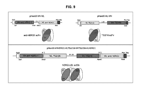
.. FIG. 9 - Upper panels: schematic structure of the two single-chain
antibody, anti-
hERG1 and anti-T52/16. Lower panels: schematic structure of the scDb-hERG1-
131.
FIG. 10 - Scheme representing SOE-PCR method for anti b1-integrin scFv
assembly in the order VL-linker-VH.
FIG. 11 - Panel A shows Coomassie staining of purified supernatants deriving
from
small scale induction of six clones grown after transformation with anti-hERG1-
Phe-
(31-scDb construct. One band is detectable corresponding to clone G5 with a
molecular weight around 60 KDa, consistent with the one expected.
Panel B shows chromatogram generated after purification of supernatant
deriving
.. from high- scale expression of G5 clone: one single peak is visible and
elutions
underlying the blue area have been analyzed and Coomassie staining is reported
in panel C, showing bands with proper molecular weight in all the elutions
tested.
FIG. 12 - Panel A. Results from cell-ELISA performed on HEK 293 HERG1 cells,
using different amounts of anti-hERG1-Phe-131-scDb bispecific antibody.
FIG. 13 - Indirect IF performed on cells seeded on different substrates, BSA
and
Fibronectin (FB). IF shows a stronger signal for cells HEK 293 HERG1 coated on
FB compared to that obtained for cells coated on BSA.
FIG. 14- Indirect IF performed on HEK 293 HERG1 cells (panels A and B).
Staining
of cells after administering of an excess of S5PORO peptide is shown in panels
D
and E. Panel C reports control cells stained only with secondary antibodies.
FIG. 15 - Expression and purification of scDb-hERG1-Cys-r31. Panel A.
Chromatogram resulting from the purification of Pichia Pastoris supernatants.
Panel
13
CA 03070643 2020-01-21
WO 2019/015936 PCT/EP2018/067641
B. Coomassie staining showing the analysis of the elutions from scDb-hERG1-Cys-
(31 purification underlying the blue peak of the chromatogram.
FIG. 16 - Direct IF with scDb-hERG1-Cys-r31-Alexa488. In these particular
experiments cells were incubated with scDb antibody directly conjugated with
Alexa
488 fluorophore. GD25 WT cells (negative for both hERG1 and (31 integrin
expression) incubated with scDb-hERG1-Cys-r31 antibody directly labelled with
Alexa488 fluorophore. No signal is present. Panel B. HEK 293 HERG1 cells
seeded
on fibronectin (FN) and BSA and stained with scDb-hERG1-Cys-r31-Alexa488: as
shown in the bar graph, signal is higher (=-17 A.U) in cells seeded on FN
compared
to cells seeded on BSA (=-10 A.U.). Panel C. HEK 293 WT cells seeded on FN and
BSA and incubated with scDb-hERG1-Cys-r31-Alexa488: signal is lower for cells
seeded on BSA (-= 7 A.U.), compared to cells seeded on FN (=-12 A.U.). As it
can
be inferred from panels B and C, fluorescence values are lower for HEK 293
HERG1
cells on FN (=-17 A.U), compared to HEK 293 WT cells seeded on FN (-= 12
A.U.).
FIG. 17- IC50 determination on MDA-MB 231 and PANC-1 cells . An effect on cell
viability was evident at 24 pg/m1 for PANC-1 cells and 42 pg/m1 for MDA-MB 231
cells.
FIG. 18 Lateral motility experiments on HCT 116, MDA-MB 231 HERG1, MDA-MB
231 cells and PANC-1 cells. A clear reduction of the MI (Motility Index) is
shown in
cells treated with scDb-hERG1-Cys-r31, compared to control cells. A more
pronounced effect is reported for MDA-MB 231 HERG1, compared to MDA-MB 231
cells, suggesting a hERG1 related effect on cell motility. Lateral motility
experiments
performed on PANC-1 cells, showing a lower MI on treated cells compared to the
control. Lateral motility experiments performed on HCT116 cells, showing a
lower
MI on treated cells compared to the control.
FIG.19 (A) Pharmacokinetics of scFv-hERG1-D8Cys in mice by iv dosing (n=2).
The
antibody concentration has been determined by ELISA, dosing the plasma
concentrations of mice blood samples collected 5, 15, 30 min and 1, 2, 6, 24
and 48
h after scFv injection. t1/2 = 3,5h Values are means of two measurements SD.
(B) ECG, electrocardiogram registration. ECG measurements are reported in the
left panel for the control mouse, injected with physiological solution, PBS,
the
adjusted value of the QT interval is 86 ms. The right panel shows the ECG
graph
14
CA 03070643 2020-01-21
WO 2019/015936 PCT/EP2018/067641
obtained after the administering of scFv-hERG1-D8Cys, showing no significant
changes compared to the control, with an adjusted value of the QT interval of
90
ms.
(C) In vivo analysis. Each panel reports the fluorescent signal in a
representative
mouse treated with scFv-hERG1-D8Cys antibody conjugated with Alexa750
compared to a control mouse treated with PBS solution. The maximum signal
detected was at 10 minutes after the injection; and no fluorescence signal was
detected after 24 hours from the intravenous administration.
(D) scFv-hERG1-D8Cys-Alexa750 uptake and retention of scFv-hERG1-D8Cys-
Alexa750 in a MIAPaCa-2-nu/nu mice model of PDA. Mice were administered
through tail vein injection with 6.5 pg of scFv-hERG1-D8Cys-Alexa750 antibody.
Representative pictures of mice i.v. injected with the labelled antibody
(left) have
been compared with control mice (right). Fluorescence intensity in the
abdominal
area, the site proximal to tumor has been analyzed. Fluorescent emission
spectra
were measured using Photon imager (Biospace Lab), images have been acquired
at different time-points, every 5 min, starting 5 min after injection until 60
min after
injection.
FIG. 20. (A) Pharmacokinetics of scDb-hERG1-Cys-r3 in mice by iv dosing (n=2)
.
The antibody concentration has been determined by ELISA, dosing the plasma
concentrations of mice blood samples collected 5, 15, 30 min and 1, 2, 6, 24
and 48
h after scDb injection. t1/2 -= 12h Values are means of two measurements SD.
(B) ECG, electrocardiogram registration. The ECG graph obtained after the
administering of scDb-hERG1-Cys-131 antibody shows no significant changes
compared to the control graph reported in Fig 20B, with an adjusted value of
the QT
interval of 83 ms, comparable to the Control.
(C) Table showing the pancreas volume (mm3) of MIAPaCa-2-nu/nu tumor-bearing
mice treated and untreated with the scDb-hERG1-Cys-r31 antibody. Metastatic
diffusion, % of necrotic area in the slide and number of vessels are also
reported.
(D) Images of pancreas after nescroscopy: 1, untreated; 2, treated with three
doses
of scDb-hERG1-Cys-r31 antibody; 3, treated with six doses of scDb-hERG1-Cys-
r31
antibody.
CA 03070643 2020-01-21
WO 2019/015936 PCT/EP2018/067641
EXPERIMENTAL SECTION
1. scFv-hERG1 mutagenesis
The amino acid sequence of the scFv hERG1 molecule as described in
W02016020483 (Al) presents the amino acid Phe in position 95 of the VH domain.
.. The nucleotide sequence SEQ ID No: 1 unraveled the presence of T in
position 283
(c283T) of the VH domain. According the present invention, the substitution of
a G
was introduced instead of the T in position 283 (c283T>0) of the VH domain
leading
to the switch of the Phe (TTT) in position 95 with a Cys (TOT). This mutation
resulted
in the introduction of one amino acid (Cys) in the position between Framework
3
.. and CDR3, which surprisingly resulted fundamental for the formation of the
disulfide
bond in the immunoglobulin variable domain. The Cys was introduced in the
original
construct, setting up a mutagenesis protocol (see Materials and Methods). The
cDNA obtained from four mutagenized scFv-hERG1 colonies was sequenced and
the sequencing results (Fig.1) demonstrated the proper mutation from TTT to
TOT
in position c283T>0, indicative of the desired mutation from Phe to Cys.
2. Expression and protein purification
Either plasmids, scFv-hERG1 and the mutagenized scFv-hERG1 (hence named
scFv-hERG1-Cys), were transformed into 0S115 P. pastoris host strain, using
the
spheroplasting technique. Six clones (C7, C12, D9, E8, 03, 07) among the scFv-
.. hERG1 transformants and six (B11, C3, D8, D9, 04, 010) from the scFv-hERG1-
Cys were analyzed. Results of the small-scale expression are shown in Fig. 2
panels
A and B for scFv-hERG1 and panels C and D for scFv-hERG1-Cys, respectively.
All the clones revealed protein expression after 72 h induction (upper
panels), as
also shown in slot blot.
After purification, the presence of the protein was assessed through western-
blot
(panel B and D).
The two best expressing clones were chosen for the two antibodies : 03 for
scFv-
hERG1 (hereafter named scFv-hERG1-03) and D8 for scFv-hERG1-Cys (named
scFv-hERG1-D8-Cys).
.. Larger-scale expression analyses are shown in Figure 3. The presence of the
protein was assessed through SDS-PAGE and Coomassie Brilliant Blue staining.
Fractions 11, 12, 13 (A, left panel) correspond to scFv-hERG1-G3; fractions
12, 13,
16
CA 03070643 2020-01-21
WO 2019/015936 PCT/EP2018/067641
14 (B, right panel) correspond to scFv-hERG1-D8Cys. Bands corresponding to the
molecular size of both antibodies (around 30KDa) are visible. Comparing the
yields
of the two proteins, scFv-hERG1-G3 and scFv-hERG1-D8Cys, significant
differences were found: scFv-hERG1-G3 concentration is 0,050 pg/ 1; scFv-
hERG1-D8Cys concentration is 0,444 pg/ 1.
PROTEIN YIELD (mg/I)
scFv-hERG1-G3 0,200
scFv-hERG1-D8Cys 1
a The yields were normalized to mg protein per liter of Pichia Pastoris yeast
culture.
3. Comparison of antigen affinity between scFv-hERG1-G3 and scFv-
hERG1-D8Cys
Chromatograms reported in Fig. 3 (panels B) show the results obtained from gel
filtration. Size-exclusion chromatography (SEC) was performed in order to
investigate the possible presence of aggregates which might affect the binding
capacities of the two antibodies. Several aggregates are detectable from the
analysis reported in B (left panel) that refers to scFv-hERG1-G3; instead scFv-
hERG1-D8Cys (B, right panel) appears in a monomeric form.
4. scFv-hERG1-D8Cys antibody stability test
The stability of the scFv-hERG1-D8Cys antibody was directly assessed analyzing
the protein through SDS-Page Coomassie Brilliant blue staining at different
time
points (6, 12, 18 months) after purification. Data in Fig.3C show that only
one neat
single band is visible at all time points, thus indicating that the protein
maintains its
stability without showing signs of degradation.
5. Evaluation of immunoreactivity of scFv-hERG1-G3 and scFv-hERG1-
D8Cys
Then an immunofluorescence analysis was performed using scFv-hERG1-G3 and
scFv-hERG1-D8Cys on fixed cells, to determine the immunoreactivity of the two
antibodies. Were used, as cellular model, HEK 293 transfected with the hERG1
cDNA (HEK-hERG1) and, as a control HEK-MOCK, that do not express the hERG1
protein. HEK-MOCK cells showed no or weak signal with both antibodies, while
HEK
17
CA 03070643 2020-01-21
WO 2019/015936 PCT/EP2018/067641
293 hERG1 showed a good labeling with the scFv-hERG1-G3 and, even better, with
the scFv-hERG1-D8Cys (Fig. 4, A and B). Data analysis obtained using ImageJ
Software is reported in the graphs reported in panel C. Values obtained from
scFv-
hERG1-D8Cys staining are significantly higher in cells overexpressing hERG1,
if
compared to the values of the control obtained in HEK-MOCK cells.
It was also tested the immunoreactivity of the two antibodies after direct
labelling
with the fluorescent molecule Alexa 488. scFv-hERG1-G3-Alexa488 and scFv-
hERG1-D8Cys-Alexa488 antibody was tested in IF on fixed cells (Fig. 4, A and
B)
showing the maintaining of the capacity to recognize the antigen in the native
conformation, even after the conjugation with the fluorophore. IF staining was
measured using ImageJ software and results are reported in the graphs reported
in
panel C. Signal obtained on HEK 293 HERG1 cells is stronger compared to the
control HEK-MOCK cells both for scFv-hERG1-G3-Alexa488 and scFv-hERG1-
D8Cys-Alexa488.
In order to assess and compare the potential use in vivo of scFv-hERG1-G3-
Alexa488 and scFv-hERG1-D8Cys- Alexa488 as molecular tools, both antibodies
were used in IF on live cells (Fig. 4 A and B).
The experiment confirmed the results obtained with the staining on fixed
cells;
HEK293 hERG1 cells appear to have a stronger signal, if compared with the
negative control HEK293 MOCK cells. HEK 293 hERG1 cells appear to have a more
specific spotty cellular labelling, while HEK-MOCK cells have a non-specific
diffuse
background. For this reason, it has been reported the bright-field image of
the same
section.
6. scFv-hERG1-D8Cys antibody viability inhibition and spheroids test
At this stage, it has been further explored the potential capacity of scFv-
hERG1-
D8Cys of inhibiting cell growth on a panel of neoplastic cell lines. As
reported in Fig.
5, a significant dose-dependent inhibition of cell proliferation was observed
for HCT-
116, MDA-MB 231, Mia Paca-2, HEK 293 HERG1, PANC-1 and BxPc3. Cells were
treated using anti-hERG1 monoclonal antibody (100 g/ml) and scFv-hERG1-
D8Cys (10; 20 g/m1). As expected, no significant decrease in cell viability
was found
in HEK-MOCK cells, which do not express hERG1.
18
CA 03070643 2020-01-21
WO 2019/015936 PCT/EP2018/067641
In order to investigate the effect of scFv-hERG1-D8Cys on a 3D cellular model
we
have tested three different concentrations of scFv-hERG1-D8Cys (10; 20; 40
g/m1)
on spheroids.
Figure 6 shows the graph reporting on the Y axis the volume of the spheroids
(mm3),
while on the X axis are reported the different timepoints (24h, 48h and 72h).
In fig. 6, panel A is reported the graph obtained for spheroids generated from
HEK293-hERG1. The volume of the spheroids treated with 20 pg/m1 and 40 pg/m1
scFv-hERG1-D8Cys is smaller compared to the control at each timepoint. Panel
B,
instead, shows the growth curve of the HEK-MOCK spheroids, in which no
difference was found for treated spheroids at all the three concentrations
scFv-
hERG1-D8Cys tested, compared to the control. Panel A and B also show a
representative brightfield image of the control HEK293-hERG1 and HEK-MOCK
spheroids, respectively, as they appeared after 72 h colture.
Panel C shows the effect obtained on pancreatic ductal adenocarcinoma Mia Paca
2 cells. A decrease in the volume of spheroids was observed both for cells
treated
with 20 pg/m1 and 40 pg/m1 scFv-hERG1-D8Cys, with a more pronounced effect
obtained at the highest concentration tested compared to the controls, at each
timepoint. Images taken at 72 h, reported on the right part of the figure,
show a
picture of a control spheroid of Mia Paca 2 taken at 4X magnification; while
the right
image shows a picture of a Mia Paca2 spheroid treated with 40 pg/mIscFv-hERG1-
D8Cys, taken at 10X magnification. In fact, it wasn't possible to acquire
pictures of
control Mia Paca 2 spheroids after 72 h with 10X magnification, as the volume
was
too enlarged to allow a proper focusing; while Mia Paca 2 spheroids at 72 h
treated
with scFv-hERG1-D8Cys can be visualized using 10X magnification, as their
volume, compared to control, was strongly reduced.
Panel D shows MDA-MB 231 spheroids: a marked effect of volume reduction is
observed for all the three concentrations of scFv-hERG1-D8Cys tested (10, 20,
40
g/ml) compared to the control. Volume reduction can be inferred also from the
pictures of MDA-MB 231 spheroids reported on the right side of the figure.
Fig. 7 shows the results obtained from Calcein AM cell viability assay
performed on
spheroids after 72 h. Green staining represents live cells, while red staining
represents dead cells. Image on the left (panel A) are pictures of the control
for each
19
CA 03070643 2020-01-21
WO 2019/015936 PCT/EP2018/067641
cell line, while on the right side (panel B) there are pictures of spheroids
treated with
40 g/m1 scFv-hERG1-D8Cys. From the image it is possible to note the volume
reduction for spheroids treated with the antibody, especially for Mia Paca 2,
MDA
MB-231 and PANC-1 spheroids and, moreover, an increased number of dead cells,
especially for MDA MB-231 and PANC-1 spheroids treated with scFv-hERG1-
D8Cys.
7. scDb-hERG1-I31
It has been developed a bispecific antibody (bsAb) comprising a single chain
antibody directed against (31-integrin (scFv-TS2/16) and a scFv-hERG1-Cys or
scFv-hERG1 (as above described).
Nucleotide sequence encoding VL domain of TS2/16 is SEQ ID No: 23; nucleotide
sequence encoding VH domain of T52/16 is SEQ ID No: 25 (see Fig. 8);
respectively VL amino acid sequence of T52/16 is SEQ ID No: 24 and VH amino
acid sequence of T52/16 is SEQ ID No: 26.
The bispecific antibody format is the single-chain diabody (scDb), which
comprising
the variable domains (VH and VL) of two antibodies, connected by peptide
linkers,
as showed in Fig. 9. The upper panel of the figure reports the two single-
chain
antibodies, anti-hERG1 scFv and anti-r31-integrin T52/1 6, scFv antibody.
The lower panel schematizes the final structure of the bispecific antibody
anti-
hERG1I31-integrin, which has been assembled using the variable domains of the
two antibodies in the following order: VH scFv-hERG1 antibody (SEQ ID No: 8 or
SEQ ID No: 2), VL scFv-T52/16 antibody (SEQ ID No: 24), VH scFv-T52/16
antibody (SEQ ID No: 26), VL scFv-hERG1 antibody (SEQ ID No: 4).
VL scFv-T52/16 antibody (SEQ ID No: 24) and VH scFv-T52/16 antibody (SEQ ID
No: 26), are linked by peptide linker.
VH scFv-hERG1 antibody (SEQ ID No: 2 or 8) and VL scFv-T52/16 antibody (SEQ
ID No: 24) are linked by a peptide linker.
VH scFv-T52/16 antibody (SEQ ID No: 26) and VL scFv-hERG1 antibody (SEQ ID
No: 4) are linked by a peptide linker
At 5' and 3' ends were inserted the Fspl and Avr11 restriction sites (reported
underlined below)
VLFspl:
CA 03070643 2020-01-21
WO 2019/015936 PCT/EP2018/067641
AAAATGCGCAGACTACAAAGATATTGTGATGACACAGAC (SEQ ID No: 27)
VHAvr11 :
GGGGCCTAGGATAGACAGATGGGGGTGTCGCGACACCCCCATCTGTCTAT
(SEQ ID No: 28).
The following sequence (SEQ ID No: 29) is the complete nucleotide sequence
encoding for the scDb-hERG1-81 (SEQ ID No: 30): scFv-hERG1-Cys VH sequence
is reported highlighted in grey, VL sequence of scFv-TS2/16 is reported in
underlined italics, in bold are reported A, M, B linker sequence, in
underlined bold
italics is reported VH sequence of scFv-TS2/16 antibody, in underlined
highlighted
in grey is reported VL sequence of scFv-hERG1-Cys.
Myc-tag is reported in italics bold, while His-tag is reported underlined in
bold.
Restriction sites are reported underlined.
SEQ ID No 29:
GAGGCTGAGTGCGCAGACGAGGTCCAACTGCAACAGTCTGGACCTGAACTG
GTGAAGCCTGGGGCTTCTGTGAAGATATCCTGCAAGACTTCAGGATACACAT
TCACTGAATACACCGTTCACTGGGTGAAACAGAGCCATGGAAAGAGCCTTGA
ATGGATTGGAGGCATTAATCCTAATGGTGGTACTACCTATAATCAGAAGTTCA
AGGGCAAGGCCACATTGACTATTGACAAGTCCTCCAGCTCAGCCTTCATGGA
GCTCCGCAGCCTGACATCTGAGGATTCTGCAGTCTATTACTGTGCAACAGGT
TGGGGACCTGACTACTGGGGCCAAGGCACCACTCTCACAGTCTCCTCAGCC
AAAACAACACCCCCATCAGTCTATCCACTGGCCCCTGGCTCGAGTGATATTG
TGATGACACAGACTCCAACCACCATGGCTGCATCTCCCGGGGACAAGATCAC
TATCACCTGCAGTGTCAGTTCAATTATAAGTTCCAATTACCTGCATTGGTATAG
TCAGAAGCCAGGATTCTCCCCTAAACTCTTGATTTATAGGACATCCAATCTGG
CTTCTGGAGTCCCACCTCGCTTCAGTGGCAGTGGGTCTGGGACCTCTTACTC
TCTCACAATTGGCACCATGGAGGCTGAAGATGTTGCCACTTACTACTGCCAG
CAGGGTTCTGATATTCCACTCACGTTCGGTGATGGGACCAAGCTGGACCTGA
AACGGGCTGATGCTGCACCAACTGTATCCGGTGGTGGTGGTTCTGGTGGTG
GTGGTTCTGGCGGCGGCGGCTCCGGTGGTGGTGGATCCGAGGTGAAGGTG
GTGGAATCTGGGGGAGGCTTAGTGAAGCCTGGAGGGTCCCTGAAACTCTC
CTGTGCAGCCTCTGGATTCACTTTCAGTAGCTATACCATGTCTTGGGTTCGC
CAGACTCCGGAGAAGAGGCTGGAGTGGGTCGCAACCATAAGTAGTGGTGG
21
CA 03070643 2020-01-21
WO 2019/015936 PCT/EP2018/067641
TTCTTACACCTACTATCCAGACAGTGTGAAGGGCCGATTCACCATTTCCAGA
GACAAAGCCAAGAACACCCTGTATTTGCAAATGGGCAGTCTGAAGTCTGAG
GACACAGCCATGTATTACTGTACAAGAATAGGTTACGACGAAGATTATGCT
ATGGACCACTGGGGTCAAGGAACCTCAGTCACCGTCTCCTCAGCCAAAAC
GACACCCCCATCTGTCTATAGTGCACTGGATATTGTGCTGACACAATCTCCA
CTCACTTTGTCGGTTAACATTGGTCAACCAGCCTCTATCTCTTGCAAGTCAAG
TCAGAGCCTCTTATATACTAATGGAAAAACCTATTTTAATTGGTTATTACAGAG
GCCAGGCCAGTCTCCAAAGCGCCTAATCTATCTGGTGTCTAAACTGGACTCT
GGAGTCCCTGACAGGTTCACTGGCAGTGGATCAGGAACAGATTTTACACTGA
AAATCAGCAGAGTGGAGGCTGAAGATTTGGGAGTTTATTACTGCGCGCAAGG
TACACATTTTCCGTGGACGTTCGGTGGAGGGACCAAGCTGGAAATCAAACGG
GCTGATGCTGCACCAACTGTATCCGCGGCCGCAGAACAAAAACTCATCTCA
GAAGAGGA TCTGAATGGGGCCCCTAGGCATCATCACCATCACCATCATCAC
TAATAG
The sequence has been cloned, using the restriction sites indicated above,
into the
commercially available vector pPIC9K (Life Technologies).
8. scDb-hERG1-Phe-131: expression and characterization
The construct expressing anti-hERG1-Phe-r31-scDb antibody has been cloned into
pPIC9K expression vector, which is a vector suitable for expression in Pichia
pastoris yeast cells.
GS115 Pichia pastoris strain was transformed, according to the spheroplasting
protocol and 96 clones were screened on YPD-agar plates containing G418 for
selection. Six clones were then induced on a small scale and purified using
Sepharose Ni beads (GE Healthcare), exploiting the Histidine tag introduced
with
the pPIC9K vector. Coomassie staining is reported in Fig. 11, panel A, and it
shows
one band, highlighted by the arrow, with a molecular weight (around 60 KDa)
consistent with the one expected for the anti-hERG1-Phe-r31-scDb antibody,
corresponding to clone G5.
Then it was started large-scale expression of the G5 anti-hERG1-Phe-r31-scDb
clone, adapting the induction protocol for bigger culture volumes.
Supernatant resulting from 1L Pichia pastoris cells culture has been purified
using
AKTA Pure (GE Healthcare). Results are reported in Fig.11, in which is shown
both
22
CA 03070643 2020-01-21
WO 2019/015936 PCT/EP2018/067641
the chromatogram resulting from the antibody purification (panel B), as well
as the
Coomassie staining (panel C) in which elutions underlying the blue area have
been
analyzed. Consistently with what expected a single band, corresponding to the
purified anti-hERG1-Phe-r31-scDb has been detected for each elution.
Anti-hERG1I31-scDb fractions 8; 9; 10; 11; 12; 13; 14; 15; 16; 18; 20 were
gathered
together, and dialyzed against PBS 1X. Thus, a detailed characterization of
the
antibody was started.
One of the crucial steps was the choice of a proper model to test the anti-
hERG1-
Phe-131-scDb antibody. The table below summarizes the expression profile
related
to hERG1 and 131 integrin of the cell lines, chosen for characterization
experiments.
Table HEK 293 HERG1, HEK 293 WT and GD25 expression profile related to
hERG1 and 131 integrin
hERG1 EXPRESSION 131 integrin EXPRESSION
HEK 293 hERG1 + +
HEK 293 WILD TYPE - +
GD25 - -
The bsAb was first analyzed on HEK 293 hERG1 cells which express both hERG1
and 131 antigens. Cell-ELISA was performed and results are reported on Fig.
12,
cell-ELISA showed a certain dose-dependent proportionality for the binding
with the
native antigen, with higher OD450, for cells expressing both hERG1 and 131
antigens,
as expected.
Moreover, the anti-hERG1-(31-scDb bispecific antibody showed the capacity to
bind
the antigen in native conditions, as it is for the antigen endogenously
expressed by
cells. Binding specificity of anti-hERG1-Phe-131-scDb bispecific antibody is
also
corroborated by the comparison between the same amount (0,5 g) of anti-hERG1-
Phe-131-scDb and anti-scFv-hERG1-Phe, which is one of the two single-chain
antibodies that form the bispecific antibody. In fact, the signal obtained
after
23
CA 03070643 2020-01-21
WO 2019/015936 PCT/EP2018/067641
incubating with anti-hERG1-Phe-r31-scDb is higher than the one obtained using
anti-
scFv-hERG1-Phe. Such result is in line with what expected, since the signal
obtained with anti-hERG1-Phe-131-scDb results from the binding to both
antigens,
hERG1 and 131; while the signal obtained using scFv-hERG1 results from the
binding to hERG1 antigen, only.
It has also been evaluated the immunoreactivity of the anti-hERG1-Phe-131-scDb
antibody through IF, on cells grown on BSA (Fig. 13, panel A and B) and
fibronectin
(FN) substrates (Fig. 13, panel C and D). In fact, it has been shown that 131
complex
formation is enhanced by FN-dependent integrin activation. As it can be seen
from
Fig 13, panels C and D, is displayed a strong membranous signal in cells
HEK293-
hERG1 seeded on Fibronectin, due to a strict complex formation. The signal has
been analyzed using ImageJ software and results are reported in the graph.
To further confirm the evidences obtained from previous experiments, it has
been
evaluated the binding of anti-hERG1-Phe-131-scDb, on HEK293-hERG1 cells
administering, before antibody incubation, an excess of peptide S5PORO, which
is
the peptide towards which the scFv-hERG1 antibody is directed. As it can be
inferred from Fig. 14, panels A and B, the signal on HEK293-hERG1 cells
incubated
with anti-hERG1-Phe-131-scDb, due to the binding both to hERG1 and to 131
integrin,
is confirmed. Panel C shows the negative control, while panels D and E show
the
results obtained after incubation with S5PORO peptide; it is clearly visible
that there
is a reduction in the signal which is consistent with what expected. In fact,
HEK293-
hERG1 cells that are positive for both antigens, after incubation with the
peptide
show a reduction in staining intensity probably due to the saturation of the
hERG1
antigen binding sites; thus the signal that is visible is the one originated
only from
the binding to 131 antigen. Such results are summarized in the graph obtained
from
ImageJ fluorescence intensity quantification.
9. scDb-hERG1-Cys-I31: expression and characterization
The construct expressing scDb-hERG1-Cys-r31 antibody cloned into pPIC9K
expression vector, which is a vector suitable for expression in Pichia
pastoris yeast
cells, has been transformed into GS115 yeast cells.
Clones derived from scDb-hERG1-Cys-r31 transformation have been screened
according to the protocol previously described for scDb-hERG1-Phe-(31
antibody.
24
CA 03070643 2020-01-21
WO 2019/015936 PCT/EP2018/067641
Supernatant resulting from 1L Pichia pastoris cells culture has been purified
using
AKTA Pure (GE Healthcare). Results are reported in Fig. 15, in which both the
chromatogram resulting from the antibody purification (panel A), as well as
the
Coomassie staining (panel B) are shown. Elutions underlying the blue area have
been analyzed and, consistently with what expected, a single band with a
molecular
weight of roughly 60 KDa, corresponding to the purified scDb-hERG1-Cys-r31,
has
been detected for each elution.
After the successful protein purification, the antibody has been tested in
direct
immunofluorescence (IF) after direct conjugation with Alexa488. Results are
reported in Fig.16 for GD25 WT, HEK 293 WT and HEK 293-hERG1 cells. Images
show that GD25 WT cells, panel A, (negative for both hERG1 and 131 integrin
expression) present no significant staining after incubation with scDb-hERG1-
Cys-
131 antibody, while panel B shows a clear membraneous staining for HEK 293-
hERG1 cells (which express both antigens), seeded on fibronectin (FN) which
has
the action of enhancing the hERG1131 complex formation, with a higher
fluorescence signal value (-= 17 A.U.) compared to cells seeded on BSA, used
as
control (-= 10 A.U.). Panel C shows the fluorescent staining obtained on HEK
293
WT cells (which express only 131 integrin), showing higher fluorescence signal
values for cells seeded on FN (-= 12 A.U.), compared to cells seeded on BSA (-
= 7
A.U.).The IC50 has been determined for both cell lines as shown in Fig. 17,
panels
A and B. An effect on cell viability was evident at 24 pg/m1 for PANC-1 cells
and 42
g/mlfor MDA-MB 231 cells. Such findings are consistent with the pattern of
hERG1
expression, whose expression is predominant in PANC-1 cells, compared to MDA-
MB 231 cells.
It has been thus tested the effect of scDb-hERG1-Cys-r31 on cancer cell
migratory
behavior through lateral motility assay. Experiments have been performed on
MDA-
MB 231, MDA-MB 231-hERG1, PANC-1 and HCT116 cells. Results are reported in
the graphs in Fig. 18. There is a clear reduction of the motility index (MI)
in treated
cells compared to control. Such effect is more pronounced on MDA MB 231-hERG1,
compared to MDA-MB 231 cells, suggesting a hERG1-dependent effect of the
antibody on cell migration.
CA 03070643 2020-01-21
WO 2019/015936 PCT/EP2018/067641
Promising results have also been obtained on PANC-1 and HCT116 cells, with a
reduction of motility behavior in treated cells compared to controls
MATERIALS AND METHODS
10. Cloning of the heavy and light chain of the hERG1 antibody.
The heavy and light chain of the monoclonal antibody against hERG1 (hERG1-mAb)
were isolated from cDNA obtained from the mRNA purified from hybridomas
secerning hERG1-mAb. For the amplification of VH and VL regions, a 5' primer
that
anneal to the framework 1 (FR1) of the variable domain of each chain (primer
forward) and a primer that anneal to the constant region near the variable
domain
of each chain (primer reverse) were chosen. For VL was designed a degenerate
primer that anneal to the kappa light chain, since this is the immunoglobulin
phenotype more expressed in mice (Honjo and Alt, 1995). The heavy chain (VH)
of
antibody was amplified by PCR using the following set of primers: degVH
forward,
5' GAGGTCCARCTGCAACARTC 3' (SEQ ID No: 11) and IgG2 reverse, 5'
AGGGGCCAGTGGATAGACTGATGG 3' (SEQ ID No: 12) (Wang, 2000). The
following set of primers was used to PCR amplify the light chain (VL) of
antibody:
degVL(K), 5' GAYATTGTGMTSACMCARWCTMCA 3' (SEQ ID No: 13) and K
reverse, 5' GGATACAGTTGGTGCAGCATC 3' (SEQ ID No: 14) (Wang, 2000). The
cDNA was amplified using Phusion High- Fidelity DNA Polymerase (Finnzymes
.. Reagents). Cycling conditions were: initial melt at 94 C for 2 min followed
by 25
cycles of a three-step program (94 C, 30 sec; 56 C (VH); 48 C (VL), 1 min; and
72 C, 1 min. The reactions were then held at 72 C for 10 min and cooled to 4
C.
The antibody fragments (VH and VL) isolated from agarose gel elecrophoresis,
were
purified using QIAquick PCR Purification Kit (QIAGEN) and then inserted into
pCRTM
-Blunt vector (Invitrogen) following the manufacturer's instructions. The
recombinant
plasmid were sequenced through Automated DNA sequencing service (PRIMM).
VH and VL fragments were then cloned into pHENIX expression vector, which
contain the linker sequence (Gly4Ser)3 between two different cloning sites.
Primers
with appropriate restriction sites to clone antibody fragments into pHenIX
vector
were designed. VL primers: forward VL-ApaLl, 5'
acgcgtgcactgGATATTGTGCTGACACAATCTCCA 3'(SEQ ID No: 15); reverse VL-
Notl, 5' ataagaatgcggccgcGGATACAGTTGGTGCAGCATC 3'(SEQ ID No: 16). VH
26
CA 03070643 2020-01-21
WO 2019/015936 PCT/EP2018/067641
primers: forward VH-Salk, 5' acgcgtcgacGAGGTCCAACTGCAACAGTC 3'(SEQ ID
No: 17); reverse VH-Xhol, 5' ccgctcgagccAGGGGCCAGTGGATAGACTGATGG
3'(SEQ ID No: 18). PCR products were digested either with ApaLl and Notl (for
VH)
or Sall and Xhol (for VL) restriction enzymes (New England BioLabs) and
ligated
into pHENIX vector in the compatible cloning sites. Digestion were performed 2
h at
37 C. To avoid re-ligation of compatible ends, the 5' phosphate group was
removed
from the 5' terminus of the vector using the calf intestine phosphatise (CIP)
according to the following protocol: pHENIX vector (50 ng/ pl), Buffer 3 (New
England BioLabs) 1X and CI P (0,5 u/ pg of vector). Dephosphorylation reaction
was
incubated for 1 hour at 37 C. Phosphorylated vector was purified with QIAquick
PCR
Purification Kit (QIAGEN).
Ligations between the scFv-hERG1 fragment and pHENIX were performed in a
mixture of Buffer 2 (New England BioLabs) and T4 Ligase. Vector: scFv ratios
of
1:3 and 1:10 were set up in the ligation mixture and incubation was done 15
min at
25 C.
2 pl of the ligation mixture were electroporated into E. coil TOP1OF' and
HB2151
cells (2500 mV pulse). The electroporated cells were recovered with 450 pl SOC
medium (SOB medium supplemented with 1 mM MgSO4, 1 mM MgCl2) and
incubated 1 hour at 37 C on shaking. Bacteria were plated into pre-warming LB-
.. Agar plates containing antibiotic and incubated lid-side down overnight at
37 C.
11. Cloning of scFv-hERG1-G3 in pPIC9K expression vector
The scFv-hERG1 expression cassette was cloned into a transformed pPIC9K vector
(kindly gifted by Prof. Ermanno Gherardi, University of Pavia), which contain
a 6xHis
tag. The scFv construct was isolated and amplified from pHENIX vector by PCR
using primers which allow the addiction of Fspl and Avr11 restriction sites
respectively at 3' and 5' ends of the sequence (forward VH- Fspl,
AAAATGCGCAGAGGTCCAACTGCAACAGTC (SEQ ID No: 19); reverse VL- Avrl I,
GGGGCCTAGGGGATACAGTTGGTGCAGCATC (SEQ ID No: 20)).
The vector pPIC9 is composed of the A0X1 promoter, 3A0X1 transcriptional
terminator (TT), and a multi-cloning site into which a foreign gene is
inserted.
The expression casette was cutted with Fspl and Avril and cloned into pPIC9K
cut
with Eco53K1 and Avr11 restriction enzymes (New England BioLabs).
27
CA 03070643 2020-01-21
WO 2019/015936 PCT/EP2018/067641
12. scFv-hERG1 mutagenesis
Mutagenesis was performed on the scFv-hERG1 expression cassette cloned into
pPIC9K using the QuikChange XL Site-Directed Mutagenesis Kit (Stratagene,
Agilent Technologies). Suitable primers for the introduction of the Cys
amminoacid
were designed according to the manufacturer's indications and designed by
Primm
Biotech, left
primer:
GGATTCTGCAGTCTATTACTGTGCAACAGGTTGGGGACCTG (SEQ ID No: 21);
right primer: CAGGTCCCCAACCTGTTGCACAGTAATAGACTGCAGAATCC
(SEQ ID No: 22)).
The sample reaction was prepared as follows: 5 I of 10X reaction buffer; 1 I
of
scFv-hERG1 dsDNA template (13ng/ I); 1,841.11 (125 ng) left primer; 1,841.11
(125 ng)
right primer; 1 I of dNTP mix; 31.11 of QuickSolution; 36, 321.11 ddH20. Then
1 I of
PfuTurbo DNA polymerase (2.5 U/ I) was added. Cycling conditions were
adjusted:
initial melt was performed at 95 C for 1 min, followed by 18 cycles of a three-
step
program (95 C, 50 sec; 60 C 50 sec and 68 C, 4 min) . The reaction was then
held
at 68 C for 7 min and cooled to 4 C.
After the amplification reaction, 1 pl of Dnpl restriction enzyme (10 U/ I)
was added
directly to the reaction mixture, that was incubated right after at 37 C for 1
h to digest
the parental.
At this point, bacterial DH5a ultra competent cells were transformed through
heat-
shock. Cells were gently thaw on ice and 2 pl of the Dpi-treated DNA was
transferred
in a separate aliquot of 200 pl of ultracompetent cells. The reaction was
incubated
on ice for 30 min. The tube was then heat-pulsed at 42 C in a dry-bath for 45
sec.
The tube was incubated on ice for 2 min. Cells were recovered with 450 pl SOC
medium (SOB medium supplemented with 1 mM MgSO4, 1 mM MgCl2) and
incubated 1 hour at 37 C on shaking. Bacteria were plated into pre-warming LB-
Agar plates containing Ampicillin antibiotic (50 g/ml) and incubated lid-side
down
overnight at 37 C.
The following day, several colonies were grown and some of them were picked
and
DNA was extracted and sequenced to verify the presence of the desired
mutation.
The construct obtained was labelled scFv-hERG1-Cys.
13. scFv-hERG1-G3 and scFv-hERG1-D8Cys expression in Pichia Pastoris
28
CA 03070643 2020-01-21
WO 2019/015936 PCT/EP2018/067641
Linearised scFv-hERG1 and scFv-hERG1-Cys were both digested with Sall and
transformed into the Pichia Pastoris strain GS115 by spheroplasting,
generating
Mut+ transformants. For the transformation we have referred to the Pichia
Expression Kit (Invitrogen) indications.
After five days from transformation, single colonies were visible to the naked
eye,
92 clones and 4 negative controls were picked and transferred in three
different 96-
well plates, with different concentration of G418: without G418, 5 mg/ml, 15
mg/ml.
G418 selection was performed exploiting the characteristic that pPIC9K
contains
the bacterial kanamycin gene that confers resistance to Geneticin in Pichia.
The
level of Geneticin resistance approximately depends on the number of
kanamycin
genes integrated. A single copy of pPIC9K integrated into the Pichia genome
confers resistance to Geneticin to a level of -0.25 mg/ml. Multiple
integrated
copies of pPIC9K can increase the Geneticin resistance level from 0,5 mg/ml
(1-2
copies) up to 4 mg/ml (7-12 copies). Due to the genetic linkage between the
kanamycin gene and the expression cassette (both under the PAOX1 promoter), we
can infer that Geneticin resistant clones contain multiple copies of the gene
of
interest. For this same reason, secreted protein expression may increase
because
of a gene dosage effect. Thus, the presence of pPIC9K was used as a tool to
reveal
pPIC9K transformants that harbor multiple copies of the genes of interest,
scFv-
hERG1 and scFv-hERG1-Cys.
After two days of growth at 30 C, six best grown clones from the 15mg/m1 G418
plates were picked up and evaluated for their capacity to expressed the
protein of
interest, setting up a small scale liquid culture, according to Pichia
Expression Kit
protocol (Invitrogen).
Samples from each clone's culture were collected at different timepoints: 24h,
48h,
72h. After three days of induction with 0,5% final concentration of 100%
methanol,
supernatants were collected and tested through slot blot.
14. Slot blot analysis
Yeast supernatants were collected and tested for protein expression through
slot
blot; 200 I of each supernatant were applied to a PVDF membrane (Amersham)
assembled in a slot blot device between two squares of 3MM Whatman paper. The
samples were left in incubation for 15 min, then vacuum was applied to dry the
29
CA 03070643 2020-01-21
WO 2019/015936 PCT/EP2018/067641
samples. The membrane was recovered and wash with T-PBS. Blocking was
performed with T-PBS 5% BSA for 45 min and then washed 10 min withT-PBS. The
membrane was incubated for 1 hour with anti-6xHis-HRP conjugated antibody
(Sigma) diluted 1:2000 in 15 ml T-PBS 5% BSA.
15. Ni Sepharose purification
Supernatants, obtained from the screening of the clones after yeast
transformation,
were incubated 0/N in rolling with Ni Sepharose 6 Fast Flow (Ge Healthcare)
according to manufacturer's instructions. After, two wash steps were carried
out with
500p1 Wash Buffer (20mM sodium phosphate, 500mM NaCI, pH 7.3) and elution
was performed using 250p1 Elution buffer (20mM sodium phosphate, 500mM
imidazole, pH 7.3).
16. AKTA purification
Purification of 1 liter yeast supernatant of scFv-hERG1-G3 and scFv-hERG1-
D8Cys, respectively, was performed by Affinity Chromatography, using an AKTA
Protein Purification System (Ge Healthcare Life Sciences) with HisTrap HP 1 ml
columns. Wash steps and equilibration were performed according to the
manufacturer's instructions, using Wash buffer (20mM sodium phosphate, 500mM
NaCI, pH 7.3); elution was performed utilizing a linear gradient of Elution
buffer
(20mM sodium phosphate, 500mM NaCI, 500mM imidazole, pH 7.3). Analysis was
accomplished using UNICORN 7.0 software.
17. Gel Filtration
Samples obtained from purification of both antibodies were gel filtered, using
Superdex 75 HR 10/30 (Ge Healthcare Life Sciences). Wash buffer composition
(20mM sodium phosphate, 150 mM NaCI, pH 7.3) was adjusted to optimize protocol
conditions. Elutions were analyzed through SDS-Page.
18. Sodium dodecyl sulphate polyacrylamide gel electrophoresis (SDS-
PAGE)
Each sample was applied with the same volume of 15 pl to a stacking gel (400
pl
acrylamid (40%)-bisacryamide (0.8%), 1 ml 0.5 M Tris-HCI, pH 6.8, 40 p110%
SDS,
20 p110% ammonium persulfate, 4 pl TEMED, 2,54 ml H20). Stacking gel were
added on the resolving gel (2,6 ml acrylamid (40%)-bisacryamide (0.8%), 1,75
ml
1.5 M Tris-HCI, pH 8.8, 70 p110% SDS, 35 p110% ammonium persulfate, 3,5 pl
CA 03070643 2020-01-21
WO 2019/015936 PCT/EP2018/067641
TEMED, 2,55 ml H20). Electrophoretic run were performed at 150 V. Gels were
either stained with Coomassie Brilliant Blue or transferred to PVDF membranes
for
western blotting analysis to assess the presence of the protein (around 30
KDa).
19. Western blotting
After SDS-PAGE gels were transferred to PVDF membrane (Amersham) in transfer
buffer (14,4 g, 3,03 g TrisHCI, 200 ml methanol, 800 ml H20) at 100 V for one
hour.
Membranes were washed in T-PBS (PBS 0.1% Tween) and then blocked with T-
PBS 5% BSA 0/N. Membranes were exposed to primary antibody peroxidise-
coupled (Sigma) diluted in T-PBS 5% BSA for one hour at room temperature.
After
washing the membranes three times for ten minutes, signals were visualized
using
ECL reagent (Amersham).
WB were performed by using the following antibodies: antimyc (1:1000) and anti-
6xHis-HRP conjugated antibody (Sigma).
20. scFv-hERG1-G3 and scFv-hERG1-D8Cys quantification, ELISA assay
and Biacore analisys
scFv-hERG1-G3 and scFv-hERG1-D8Cys were gathered together and dialyzed
against PBS 1X using Slide-A-LyzerTM Dialysis Cassettes (Thermo Fisher).
Protein
absorbance at 280 nm was measured and Lambert-Beer equation was applied.
To evaluate if the two engineered antibodies, scFv-hERG1-G3 and scFv-hERG1-
D8Cys, have still the ability to bind the antigen and afterwards to
investigate the
different affinity of the two antibodies ELISA assays were performed using
plates
coated with S5-Pore peptide (sequence: EQPHMDSRIGWLHN), towards which the
antibody is directed. This peptide is the same we used to screen anti-hERG1 A7
antibody.
21. Antibody labeling with Alexa 488
scFv-hERG1-G3 and scFv-hERG1-D8Cys were conjugated with Alexa Fluor 488
Microscale Protein Labeling Kit (Thermo Fisher Scientific), according to the
protocol
indications.
22. Immuno fluorescence on fixed cells
HEK 293 hERG1 (HEK293 stably transfected with pcDNA3.1-hERG1 cDNA
construct) and HEK-MOCK ( HEK 293 stably transfected with pcDNA 3.1 cDNA)
were grown in DMEM medium with 10% FBS EU serum in a 37 C incubator with
31
CA 03070643 2020-01-21
WO 2019/015936 PCT/EP2018/067641
5% CO2. Cells were seeded 0/N on glass coverslips and then washed once with
PBS and fixed with 4% paraformaldeide for 20 min at room temperature. Blocking
was performed with 10% BSA for 2 h at room temperature. Antibody incubation
was
performed using scFv-hERG1-G3, scFv-hERG1-D8Cys, diluted 1:20 in blocking
solution and incubated for 2 and half hours, followed by anti-His (1:250;
Abcam) 0/N
incubation in blocking solution. The following day, cells were washed three
times
with PBS and incubated with anti-mouse Alexa488 antibody (Invitrogen) for 1 h.
Whereas, scFv-hERG1-G3-Alexa488 and scFv-hERG1-D8Cys-Alexa488, diluted
1:20 in blocking solution, were incubated 0/N at 4 C.
For revelation cells were incubated with Hoechst (1:1000) for 30 min and then
mounted with propyl gallate. Cells were visualized on a confocal microscope
(Nikon,
Cl).
lmmunofluorescence quantification was performed using ImageJ software: for
each
image, the measure of three different areas was performed and the mean was
calculated.
23. Immuno fluorescence on live cells
Cells were grown 0/N on 60 mm plates (Sarstedt) using an agarose (15g/L) ring
in
order to isolate cells and minimize the volumes of reagents needed for
incubations.
Cells were incubated with scFv-hERG1-G3-Alexa488 and scFv-hERG1-D8Cys-
Alexa488 diluted 1:20 in complete culture medium in a 37 C incubator with 5%
CO2
for 4 h. Rivelation was performed using Hoechst, as previously described.
Cells
were visualized on a inverted light microscopy (Nikon, Eclipse TE300).
24. Cell viability
Cell viability was evaluated performing Trypan blue assay; briefly HCT-116,
MDA-
MB231, MIA PACA2, HEK293 hERG1, HEK-MOCK, FLG 29.1, PANC-1, BXPC3
cells at a density of 5 x 10³ cells/well were seeded in a 96-well plate. After
24h, the
medium was replaced by 100 pl of fresh medium containing different
concentrations
of the scFv-hERG1-D8Cys antibody (10 pg/ml and 20 pg/ml). The complete
immunoglobulin, anti-hERG1 monoclonal antibody was tested at a concentration
of
100 pg/ml. Cells were cultured 24h in a humidified incubator at 37 C and 5%
CO2.
After the treatment cells were detached and viable cells were counted.
Experiments
were performed in triplicate.
32
CA 03070643 2020-01-21
WO 2019/015936 PCT/EP2018/067641
25. Spheroids 3D culture and scFv-hERG1 test on spheroids
HEK-293 hERG1, HEK-MOCK, MDA-MB231, MIA PACA2 and PANC-1 were
seeded in a 96-well plate at a density of 103 cells/well for each well on a
1,5 %
agarose base-layer. 100 pL of fresh medium were added to each well and cells
were
left to grow for 72h in a 37 C incubator with 5% CO2 After 72h spheroids were
visible and the medium was replaced by 100 pl of fresh medium containing
different
concentrations of the scFv-hERG1-D8Cys antibody ( 10 pg/ml and 20 pg/ml and 40
pg/ml).
Pictures were taken using Nikon, Eclipse TE300 every 24h until 72h when
Calcein
AM cell viability assay was performed.
The volume of spheroids was evaluated analyzing the pictures taken at 24h, 48h
and 72h using MATLAB Software.
Experiment was performed in triplicate.
26.1 integrin mAb: RNA reverse transcription
T52/16 and BV7 RNA was reverse transcribed into cDNA using oligo(dT) primers
(Invitrogen) and SuperScript ll Reverse Transcriptase (Invitrogen) according
to
manufacturer's instructions in a total volume of 40 pl.
33
CA 03070643 2020-01-21
WO 2019/015936 PCT/EP2018/067641
Component RNA Stock concentration Final concentration 100 ng/ I
Amount 4 g
Oligo(dT)12-18 500 g/m1 25 g/m1 2 I
dNTPmix 10 mM each 0.5 mM 2 I
PCR grade H20 18 I
The mix was incubated in the thermocycler at 65 C for 5 minutes, and then
quickly
chilled on ice and added with the following components:
Component Stock concentration Final concentration lx
Amount 8 I
5x First Strand Buffer 5x
PCR grade H20 6 I
The mix was then heated at 42 C for 2 minutes and then added with the enzyme
Component Stock concentration Final concentration Amount
SuperScriptTM II RT 200 U/ I 5 U/ I 2 I
The mix (40 I) was then incubated at 42 C for 50 minutes and then the
reaction
was inactivated at 70 C for 15 minutes.
27./31 integrin mAb: isolation of variable domains BY Polymerase Chain
Reaction (PCR)
To isolate antibody variable domains (VL and VH) was performed a PCR with the
primers reported in Wang et al. (2000) with modifications (Table 2).
Table 2. Primers used for the isolation of VL and VH variable domains, form
Wang
et al., (2000) with modifications
Kappa light chain Primer name Sequence
Forward primer degKappadir GAYATTGTGMTSACMCARWCTMCA (SEQ ID No:31)
34
CA 03070643 2020-01-21
WO 2019/015936 PCT/EP2018/067641
Reverse primer Kapparev GGATACAGTTGGTGCAGCATC (SEQ ID No:32)
IgG1 heavy chain Primer name Sequence
Forward primer degH1dir CAGGTTACTCTGAAAGWGTSTG (SEQ ID No:33)
Forward primer degH2dir GAGGTCCARCTGCAACARTC (SEQ ID No:34)
Forward primer degH3dir CAGGTCCAAACTUCAGCARCC (SEQ ID No:35)
Forward primer degH4dir GAGGTGAASSTGGTGGAATC (SEQ ID No:36)
Forward primer degH5dir GATGTGAACTTGGAAGTGTC (SEQ ID No:37)
Reverse primer IgG1rev GGAAGATCTATAGACAGATGGGGGTGTCGTTTTGGC
(SEQ ID No:38)
There are two classes of light chain: kappa and lambda; but since the 95% of
mouse antibodies have kappa light chain (Honjo and Alt, 1995) were chosen
primers specific for kappa light chain ignoring the lambda type. Forward
primers
were designed using protein sequence alignment of Framework1 (FRW1) of each
chain variable region. Reverse primers were designed on the constant region
(CH1) next to the end of the variable domain of each chain (Kappa light chain
or
IgG1 heavy chain). For kappa light chain were used only one primer pair, while
for
heavy chain were used 5 primer pairs composed by IgG1rev in combination with
the 5 forward primers.
For VH isolation of both TS2/16 and BV7 were chosen IgG1rev-degH4dir primer
pair.
In order to prevent mutation due to DNA Polymerase, were used a high fidelity
DNA polymerase with proof reading activity: KOD Hot Start DNA Polymerase
(Novagen) using the following protocol:
Component 10x Buffer Stock Final Amount 5
concentration concentration lx I
10x
MgSO4 25 mM 1.5 mM 3 I
dNTPs 2 mM each 0.2 mM each 5 I
CA 03070643 2020-01-21
WO 2019/015936 PCT/EP2018/067641
Forward primer 10 M 0.3 I 1.5 pl
Reverse primer 10 M 0.3 I 1.5 pl
DNA 10 ng
KOD Hot Start DNA 0.02 U/ I 1 pl
Polym erase 1U/ I
PCR grade H20 to 50 pl
The mix was incubated in the thermocycler with following protocol:
Step 1 Temperature 95 C Time 2 min
2 95 C 20 sec
3 54 C (VH) or 46 C (VL) 10 sec
4 70 C 10 sec
70 C 5 min
5
Steps 2-4 were repeated 30 times.
28.,81 integrin mAb: cloning of variable domains without the use of
restriction enzymes
In order to sequence VH and VL variable domains were cloned without the use of
restriction enzymes in a vector suitable for DNA sequencing. We used TA-
Cloning
Kit or Zero-Blunt Cloning kit (Invitrogen) following manufacturer's
instructions.
DNA ELECTROPHORESIS AND PURIFICATION FROM AGAROSE GEL
DNA electrophoresis uses an electrical field to move the negatively charged
DNA
toward a positive electrode through an agarose gel matrix. PCR products and
restriction enzyme digested DNA were run on agarose gel (1.5 % agarose in TAE
(Tris, Acetic acid, EDTA) buffer) stained with ethidium bromide, alongside
2I0g DNA
ladder (NEB) in order to separate different size fragments. Electrophoresis
was run
at 100 V. The band of interest was thus excised from gel with a clean scalpel
and
36
CA 03070643 2020-01-21
WO 2019/015936
PCT/EP2018/067641
purified using QIAquick PCR Purification Kit (QIAGEN) following manufacturer's
instructions. Purified DNA was eluted with 30 I PCR grade H20.
Splicing by Overlap Extension PCR (SOE-PCR)
ScFv construct was assembled by Splicing by Overlap Extension PCR (SOE- PCR)
in the order VL-linker-VH, using the primers described in (Wang et al 2000)
with
modifications (Table 3). The polypeptide linker joining the variable domains
was
designed as four GGGGS repeats.
Table 3. Primer designed to assemble the construct VL-linker-VH by SOE-PCR
(VLREVSOE and VHFORSOE) and clone the sequence into pHenIX vector
(VLFORSFI and VHREVNOT), or into scFv- hERG-pHenIX vector (VLFORXHO and
VHREVAPALI). In italics are the portions of the primers that anneal to the
template,
highlighted in grey are the sequences added to clone the construct in frame
with the
expression cassette in pHenIX or in scFv-hERG1-pHenIX vector, underlined are
the
restriction sites, grey are the sequences added to facilitate enzyme
digestion, and
bold are the sequenced that overlap in SOE-PCR.
Name Sequence
VLFORSFI
GGCCCAGCCGGCCATGGCCGATATTGTGATGACACA
GACTCCA (SEQ ID No:39)
GGAGCCGCCGCCGCCAGAACCACCACCACCAGAACCACCAC
VLREVSOE
CACCGGATACAGTTGGTGCAGCATC (SEQ ID No :40)
VHFORSOE
GGCGGCGGCGGCTCCGGTGGTGGTGGATCCGAGGTGAAGG
TGGTGGAATC (SEQ ID No:41)
VHREVNOT GCGGCCGCATAGACAGATGGGGGTGTCGTTTT
GGC (SEQ ID No:42)
VLFORXHO "¨
CTCGAGTGATATTGTGATGACACAGACTCCA (SEQ ID
No:431
VHREVAPALI
,GTGCACTATAGACAGATGGGGGTGTCGTTTTGGC (SEQ
ID No:44)
The protocol consists in two steps described in Figure 10. The first step
allows to
add: at the 3' end of VL, a sequence that encode the first three GGGGS repeats
of
the linker, and at the 5' end of VH a sequence that encode the last two GGGGS
37
CA 03070643 2020-01-21
WO 2019/015936 PCT/EP2018/067641
repeats of the linker. During this step will be also attached at the 5' end of
VL and
at the 3' end of VH the restriction sites that will be used for the cloning of
VL-linker-
VH construct in the expression vector. The second step allows to join the two
PCR
products thanks to the overlapping sequences (15 bp) at the 3' end of VL and
at the
5' end of VH.
In the first step were performed two parallel PCR: one with VLFORSFI- VLREVSOE
primer pair and pCRII-VL template; and the other with VHFORSOE-VHREVNOT
primer pair and pCRII-VH template. PCR protocol using KOD DNA Polymerase was
performed as previously described.
In the second step, we settled up the SOE-PCR using as template 1 I of each
PCR
reaction performed in the first step, and VLFORSFI-VHREVNOT primer pair,
following the protocol below:
Component 10x Buffer Stock concentration 10x Final concentration lx
Amount 5 I
MgSO4 25 mM 1.5 mM 3p1
dNTPs 2 mM each 0.2 mM each 5 I
VLFORSFI primer 10 M 0.3 I 1.5 I
VHREVNOT primer 10 M 0.3 I 1.5 I
PCR VL (STEP 1) 1 I
PCR VH (STEP 1) 1 I
KOD Hot Start DNA Polymerase 1U/ I 0.02 U/ I 1 I
PCR grade H20 to 50 I
The mix (50 I) was incubated in the thermocycler according to the protocol
below:
Step 1 Temperature 95 C Time 2 min
2 95 C 20 sec
3 70 C 10 sec
38
CA 03070643 2020-01-21
WO 2019/015936 PCT/EP2018/067641
4 70 C 10 sec
70 C 5 min
Steps 2-4 were repeated 30 times
29. Production and characterization of anti-hERG1-I31 Single Chain
Diabody (scDb)- scDb-hERG1-I31 antibody
The construct hERG1-(31-scDb was transformed into GS115 Pichia pastoris strain
5 according to the spheroplasting technique previously described and the
protein has
been expressed and purified applying the expression and purification protocol
previously described for the scFv-hERG1 and scFv-hERG1-Cys antibodies.
CELL ELISA
Cell ELISA on living cells was performed according to Sette et aL, (2013). HEK
293
WT (hERG1-/ (31+) and HEK 293-hERG1 (hERG1+/ (31+) cells were seeded to
semiconfluence in 96-well plate in DMEM plus 10% fetal bovine serum (FBS) and
incubated overnight at 37 C and 5 % CO2. After three washes with PBS, anti-
hERG1131-scDb was diluted at different concentrations into culture medium and
added to the cells for two 2 hours at room temperature. The following steps
were
the same as described above.
IMMUNOFLUORESCENCE (IF)
IF was performed following the protocol which has been previously described.
Coverslips were coated with BSA and Fibronectin for two hours. IF was
performed
on HEK 293 WT (hERG1-/ 131+), HEK 293-hERG1 (hERG1+/ (31+) and GD25 WT
cells (hERG1-/ (31-).
30. Production and preliminary characterization of anti-hERG1-I31 Single
Chain Diabody (scDb) - scDb-hERG1-Cys-I31- antibody
scDb-hERG1-131 MUTAGENESIS
Mutagenesis was performed on the scDb-hERG1-131 expression cassette cloned
into pPIC9K using the QuikChange XL Site-Directed Mutagenesis Kit
(Stratagene,
Agilent Technologies). Suitable primers for the introduction of the Cys
amminoacid
were designed according to the manufacturer's indications and designed by
Primm
Biotech, left
primer:
GGATTCTGCAGTCTATTACTGTGCAACAGGTTGGGGACCTG (SEQ ID No: 21);
39
CA 03070643 2020-01-21
WO 2019/015936 PCT/EP2018/067641
right primer: CAGGTCCCCAACCTGTTGCACAGTAATAGACTGCAGAATCC
(SEQ ID No: 22)).
The sample reaction was prepared as follows: 5 I of 10X reaction buffer; 1 I
of
scFv-hERG1 dsDNA template (13ng/ I); 1,841.11(125 ng) left primer;
1,841.11(125 ng)
right primer; 1 I of dNTP mix; 31.11 of QuickSolution; 36, 321.11 ddH20. Then
1 I of
PfuTurbo DNA polymerase (2.5 U/ I) was added. Cycling conditions were
adjusted:
initial melt was performed at 95 C for 1 min, followed by 18 cycles of a three-
step
program (95 C, 50 sec; 60 C 50 sec and 68 C, 4 min) . The reaction was then
held
at 68 C for 7 min and cooled to 4 C.
After the amplification reaction, 1 I of Dnpl restriction enzyme (10 U/ I)
was added
directly to the reaction mixture, that was incubated right after at 37 C for 1
h to digest
the parental.At this point, bacterial DH5a ultra competent cells were
transformed
through heat-shock. Cells were gently thaw on ice and 2 I of the Dpi-treated
DNA
was transferred in a separate aliquot of 200 I of ultracompetent cells. The
reaction
was incubated on ice for 30 min. The tube was then heat-pulsed at 42 C in a
dry-
bath for 45 sec. The tube was incubated on ice for 2 min. Cells were recovered
with
450 pl SOC medium (SOB medium supplemented with 1 mM MgSO4, 1 mM MgCl2)
and incubated 1 hour at 37 C on shaking. Bacteria were plated into pre-warming
LB-Agar plates containing Ampicillin antibiotic (50 g/ml) and incubated lid-
side
down overnight at 37 C.
The following day, several colonies were grown and some of them were picked
and
DNA was extracted and sequenced to verify the presence of the desired
mutation.
The construct obtained was labelled scDb-hERG1-Cys-81.
EXPRESSION AND PURIFICATION OF scDb-hERG1-Cys-81 ANTIBODY.
scDb-hERG1-Cys-81 was transformed in G5115 Pichia Pastoris yeast strain
according to the spheroplasting technique previously described and the protein
has
been expressed and purified applying the expression and purification protocol
previously described for the scFv-hERG1 and scFv-hERG1-Cys antibodies using
AKTA Pure (Ge Healthcare). Chromatograms were analyzed using Unicorn 7.0
Software.
IMMUNOFLUORESCENCE (IF)
CA 03070643 2020-01-21
WO 2019/015936 PCT/EP2018/067641
IF was performed following the protocol which has been previously described.
Coverslips were coated with BSA and Fibronectin for two hours. IF was
performed
on GD25 WT cells (hERG1-/ (31-), HEK 293 WT (hERG1-/ (31+), HEK 293-hERG1
(hERG1+/ (31+), following the protocol previously described. IF was performed
using
scDb-hERG1-Cys-r31 conjugated with Alexa488 fluorophore.
VIABILITY ASSAY
PANC-1 (pancreatic ductal adenocarcinoma) cells and MDA-MB 231 (breast
cancer) cells were seeded at 5*1 05 in 96 well-plates and let grown 0/N. The
following day cells were treated with scDb-hERG1-Cys-r31 at different
dilutions (0,
10, 20, 40, 100 g/ml) and incubate with the antibody 24h. Each condition was
performed in triplicate.
After incubation cells were detached and counted, for IC50 determination
Origin
Software was applied.
3D SPHEROID CULTURE
103 PANC-1 and MDA-MB 231 cells were seeded on an agarose base layer (1,5g/1)
in 96 well plate and grown for 72 hours in a humidified incubator at 37 C and
5%
CO2. Then scDb-hERG1-Cys-r31 was administered (40 pg/ml), diluted in culture
medium, while fresh medium without antibody was added to wells containing
cells
treated as negative controls. Photos were taken at 24 hours to monitor cell
growth
using a Nikon, Eclipse TE300 microscope.
LATERAL MOTILITY ASSAY
Cells were plated in 35 mm Petri dishes at an initial density of 5*1 05 and
allowed to
settle for 24 h.
Lateral motility was assessed by a monolayer wound assay (Silletti et al.,
1995;
Peck and lsacke, 1996). Wound widths were determined immediately afterwards (0
h) by measuring the width of the wound at 45 fixed points
Cellular motility was quantified as the "motility index" (MI) defined as
follows:
MI=1-(WtANo)
MI = 0 indicated no movement of cells whilst values of MI=1 indicated complete
wound closure
BIODISTRIBUTION OF scFv-hERG1-D8Cys AND scDb-hERG1-Cys-r31
ANTIBODIES.
41
CA 03070643 2020-01-21
WO 2019/015936 PCT/EP2018/067641
160 pg of each antibody have been injected in two Balb/c mice i.v.. Blood
samples
have been collected from each mouse at different timpanist following the i.v.:
5, 10,
30 min lh, 2h, 6h, 24h after injection. Blood samples have been processed and
plasma isolated. ELISA test on plasma samples was performed following the
protocol previously described in this section. ti/2 were calculated applying
Precise
PK Pharmacokinetic Software.
ECG MESUREMENTS.
ECG measurements were performed before antibody administration and
continuously after i.v. injection of the antibody for 15 min.
IN VIVO EXPERIMENTS
In vivo analysis
Labeling of the scFv-hERG1-D8Cys with Alexa 750: 150 pg of scFv-hERG1-D8Cys
at a concentration of 2 mg/ml in PBS solution and 0.1 M sodium bicarbonate
buffer
pH 8.3, were incubated lh at 22 C in agitation with 12 I of Alexa Fluor 750
NHS
.. Ester (Succinimidyl Ester) (Thermo Fisher Scientific), resuspended in DMSO
at 10
mg/ml. The reaction was blocked for 5 min in ice and the labelled protein was
purified by size exclusion chromatography on a Sephadex G25 (Sigma) column
equilibrated with PBS.
In vivo imaging. Three six-week old, female immunodeficient Athymic Nude-Foxn1
nu/nu mice were injected intravenously with 50 I (1 nm dye/mouse) of scFv-
hERG1-D8Cys labeled with the fluorophore Alexa 750 and fluorescence was
measured 5, 10, 60 min and 24 h after antibody injection. One control mouse
was
treated with sterile PBS solution. All the fluorescent emission spectra were
measured using a Photon imager (Biospace Lab). The imager had a laser source
for fluorescence excitation (A=679 nm), an emission filter (A=702 nm) for
fluorescence detection, and a computer for data analysis.
Mouse Model: the MIAPaCa-2 cell line was used for tumor cell implantation, as
described in Lastraioli et al., 2015. Cells were cultured in DMEM supplemented
with
L-glutamine (4 mM), 10% fetal bovine serum and Geneticin (G418) (2.4 mg/ml)
(Gibco) at 37 C in a humidified atmosphere of 5% CO2. MIAPaCa-2-luc cells were
injected into the pancreas of nu/nu mice and the animals were monitored (as
42
CA 03070643 2020-01-21
WO 2019/015936 PCT/EP2018/067641
described in [17]) and 45 days after the cell inoculum, mice were administered
with
scFv-hERG1-D8Cys-Alexa750 antibody.
43