Note: Descriptions are shown in the official language in which they were submitted.
CA 03070941 2020-01-23
WO 2019/023217
PCT/US2018/043464
METHODS OF TREATING NEURODEGENERATIVE DISEASES
BACKGROUND
[0001] All
publications herein are incorporated by reference to the same extent as if
each individual publication or patent application was specifically and
individually indicated to
be incorporated by reference. The following description includes information
that may be
useful in understanding the present invention. It is not an admission that any
of the information
provided herein is prior art or relevant to the presently claimed invention,
or that any
publication specifically or implicitly referenced is prior art.
[0002]
Pituitary Adenylate Cyclase Activating Polypeptide (PACAP), as a small
peptide with either 38 amino acids in full length form (PACAP-38), or 27 amino
acids in short
form (PACAP-27), is broadly recognized as a neurotrophin associated with
stress. Both forms
strongly increase cyclic adenosine monophosphate (cAMP) by activating
adenylate cyclase,
and hence named as PACAP. Subsequent research showed that PACAP is not only an
endocrine hormone, but intrinsically expressed in multiple brain regions and
peripheral tissues.
PACAP is a potent neurotrophic and neuroprotective peptide in the central
nervous system
(CNS). PACAP-38 is the major form in brain, while PACAP-27 exists in minor
quantity. Both
forms of PACAP bind to and activate G protein-coupled receptors (PAC, VPAC1,
and
VPAC2). PAC1 is mainly localized in the CNS; while VPAC1 and VPAC2 are in the
vascular
system and the gastrointestinal tract.
[0003] PACAP
has been shown to promote synaptic transmission, long term
potentiation and memory under physiological conditions. However, the relevance
of PACAP
expression has not been extensively studied in the human brain, including
those suffering from
Alzheimer's disease (AD), a progressive mental deterioration and form of
dementia that often
occurs in old age due to generalized degeneration of the brain. AD is a
neurodegenerative
disorder that affects memory and other cognitive functions, and is the most
common cause of
dementia. Alzheimer's Disease Association (ADA) survey shows that 5.4 million
people in
the United States (US) currently have AD and 13.5 million are expected to have
AD within the
next 40 years. AD affects over 26 million people worldwide and currently there
is no cure for
the disease. With the growing number of people living to older ages, there is
an urgency to
better understand elements of the pathogenic pathway, discover agents that
target these
elements, and establish their roles in the treatment and prevention of AD. But
effective
biomarkers and treatment are lacking. There is no disease modifying medication
available on
the market.
1
CA 03070941 2020-01-23
WO 2019/023217
PCT/US2018/043464
[0004] Thus,
there is a need in the art for novel and effective methods of treating
neurodegenerative diseases.
SUMMARY OF THE INVENTION
[0005] The
following embodiments and aspects thereof are described and illustrated in
conjunction with compositions and methods which are meant to be exemplary and
illustrative,
not limiting in scope.
[0006] Various
embodiments include a method of reducing Tau phosphorylation in a
subject, comprising providing a composition comprising a neurotrophin or salts
thereof, and
administering a therapeutically effective dosage of the composition to the
subject. In another
embodiment, the neurotrophin is
Pituitary Adenylate Cyclase Activating Polypeptide
(PACAP). In another embodiment, the composition is intranasally administered.
In another
embodiment, the neurotrophin is administered in a dosage of at least 50 nm. In
another
embodiment, the neurotrophin is administered in a dosage between 50 nM and 100
nM. In
another embodiment, the subject is a human. In another embodiment, the Tau
phosphorylation
is associated with cognitive decline. In another embodiment, the Tau
phosphorylation is
associated with Alzheimer's Disease. In another embodiment, the neurotrophin
reduces
activity of the 0-secretase enzyme. In another embodiment, the neurotrophin
comprises SEQ
ID NO: 1.
[0007] Other
embodiments include a method of treating a condition associated with
cognitive decline in a subject, comprising providing a composition comprising
Pituitary
Adenylate Cyclase Activating Polypeptide (PACAP), or an analog, derivative,
pharmaceutical
equivalent, or salt thereof, and administering a therapeutically effective
dosage of the
composition to the subject. In another embodiment, the condition is
Alzheimer's Disease. In
another embodiment, the condition is dementia. In another embodiment, the
condition is
treated by reducing amyloid plaques and/or tau fibrils in the subject. In
another embodiment,
the PACAP is administered in a dosage between 50 nM and 100 nM. In another
embodiment,
the composition is intranasally administered. In another embodiment, the PACAP
comprises
SEQ ID NO: 1.
[0008] Other
embodiments include a method of treating and/or inhibiting 0-secretase
in an individual, comprising providing a composition comprising a neurotrophin
or salts
thereof, and administering a therapeutically effective dosage of the
composition to the
individual. In another embodiment, the neurotrophin is Pituitary Adenylate
Cyclase Activating
Polypeptide (PACAP). In another embodiment, the composition is intranasally
administered.
2
CA 03070941 2020-01-23
WO 2019/023217
PCT/US2018/043464
In another embodiment, the neurotrophin is administered in a dosage between 50
nM and 100
nM. In another embodiment, the inhibition of 0-secretase comprises reduced
activity of the 13-
secretase enzyme. In another embodiment, treating and/or inhibiting 0-
secretase improves
cognition in the individual. In another embodiment, treating and/or inhibiting
0-secretase treats
Alzheimer's Disease in the individual. In another embodiment, treating and/or
inhibiting 13-
secretase prevents susceptibility to Alzheimer's Disease.
[0009] Other
features and advantages of the invention will become apparent from the
following detailed description, taken in conjunction with the accompanying
drawings, which
illustrate, by way of example, various features of embodiments of the
invention.
BRIEF DESCRIPTION OF THE FIGURES
[0010]
Exemplary embodiments are illustrated in referenced figures. It is intended
that
the embodiments and figures disclosed herein are to be considered illustrative
rather than
restrictive.
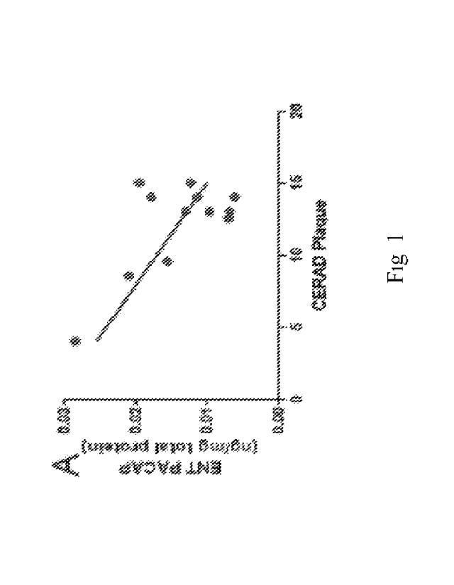
[0011] Figure 1
depicts, in accordance with embodiments herein, charts demonstrating
PACAP levels are inversely related to AD pathology. A-D. PACAP level in AD
brains were
analyzed for correlation with amyloid plaque quantity (indicated by CERAD
Plaque score).
PACAP levels were inversely correlated with CERAD in the ENT (Pearson r = -
0.6764, p
<0.05, Fig 1A) and SFG (Pearson r = -0.7088, p <0.05, Fig 1C) but not in the
MTG (Fig 1B)
or PVC (Fig 1D). PACAP level was quantified by ELISA and normalized with total
protein
(mg) of the brain tissue. E. PACAP level in CSF was quantified and correlated
with tau
pathology (indicated by Braak Stage). All CN cases were in Stage Four AD
cases were
in Stage III-IV while the other 5 AD cases in Stage V-VI. PACAP was lower in
advanced
Braak Stage V-VI than that in moderate Braak Stage III-IV (p<0.05). PACAP
level was
quantified by ELISA and normalized with CSF volume (m1). ENT= Entorhinal
Cortex, MTG=
Middle Temporal Lobe, SFG= Superior Frontal Cortex, PVC= Primary Visual
Cortex, AD=
Alzheimer's disease (blue dot), CN= cognitively normal controls (black dot).
[0012] Figure 2
depicts, in accordance with embodiments herein, reduction in PACAP
levels in CSF is specific to AD. PACAP levels were quantified by ELISA as ng
PACAP per
ml undiluted CSF. A. PACAP was reduced in AD but not in PDD or FTLD. B. PACAP
is
correlated with the Z score of DRS. CN and AD cases were separated by the
dotted line based
on their DRS score. AD= Alzheimer's disease, CN= cognitively normal controls,
PDD=
Parkinson Disease with Dementia, FTLD= Frontotemporal Lobe Dementia, DRS=
Dementia
3
CA 03070941 2020-01-23
WO 2019/023217
PCT/US2018/043464
Rating Scale-Revised (DRS-R), a global cognitive assessment.
* indicates p<0.05 and ** p<0.01.
[0013] Figure 3
depicts, in accordance with embodiments herein, a comparison of
PACAP abundance in selected cerebral regions among CN, MCI and AD. A: PACAP in
CSF
was expressed as ng PACAP per ml CSF. B: PACAP in SFG. C: PACAP in MTG D:
PACAP
in PVC. In B, C, and D, PACAP was expressed as ng PACAP per mg protein from
the same
sample. Bars indicate standard errors. One-way ANOVA with Post hoc Tukey test.
* indicates
p<0.05, ** p<0.01, ***p<0.001.
[0014] Figure 4
depicts, in accordance with embodiments herein, PACAP level in
selected cerebral regions correlate with region-specific cognitive tests. A-D
are charts depicting
PACAP level in selected cerebral regions correlate with region-specific
cognitive tests, with
E-F depicting PACAP in CSF inversely correlate with total amyloid plaque and
total tangles.
Solid fitted lines indicate significant Pearson correlations (p<0.05).
[0015] Figure 5
depicts, in accordance with embodiments herein, PAC1 receptor in
selected cerebral regions and the PACAP-PAC1 receptor interaction. A. PAC1
receptor
quantification in SFG, * indicate p<0.05, one way ANOVA with post hoc Tukey's
tests. B.
PAC1 receptor quantification in MTG; C. PAC1 receptor quantification in MTG.
D.
Pharmacodynamic model fit of SFG PACAP and SFG PAC1 level to predict the
correlation
with Stroop Color-Word Interference z scores. The dashed line marks the point
at significant
correlation (p<0.05). E. Pharmacodynamic model fit of MTG PACAP and MTG PAC1
levels
to predict the correlation with AVLT scores. The dashed line marks the point
at significant
correlation (p<0. 05).
[0016] Figure 6
depicts, in accordance with embodiments herein, that PACAP is
protective against Ar342 toxicity in cultured mouse cortical neurons. Note
that Ar342 (141M)
significantly killed 60% cells and PACAP (> or = 50 nM) effective protects the
toxicity. *
indicates p<0.05.
[0017] Figure 7
depicts, in accordance with embodiments herein, that PACAP inhibits
BACE1 activity. Left panel: BACE1 activity kinetic was measured and fitted
with Michaelis-
Menten Model. Control (round dot): Vmax= 105, K=2704; PACAP treatment
(triangle dot):
Vmax=79, K=3804. Right panel: steady state endpoint fluorsecent units (RFU).
Note PACAP
reduces the end product by BACE1.
[0018] Figure 8
depicts, in accordance with embodiments herein, that PACAP
administration reduces BACE1 expression. PACAP38 is the full length active
peptide.
4
CA 03070941 2020-01-23
WO 2019/023217
PCT/US2018/043464
PACAP6-38 is the truncated peptide used as a competitive PAC1 receptor
blocker, which
antagonized the BACE1 reduction effect of PACAP.
[0019] Figure 9
depicts, in accordance with embodiments herein, that PACAP
administration reduces ADAM17 Km but does not change Vmax. Control (round
dot): Km=48
[tM, Vmax=94; PACAP: Km=35 [tM, Vmax=92.
[0020] Figure
10 depicts, in accordance with embodiments herein, that PACAP
administration A1342 induced Tau phosphorylation at the site Thr231 in
cultured primary
neurons. A1342 (0.5 [tM) was incubated with the cells for 72 hours. PACAP38
(50 nM) with
or without PACAP receptor (PAC1) antagonist PACAP6-38 (2 [tM) was added 24
hours after
A1342 and incubated for 48 hours. The cells were harvested and homogenized for
Western blot
assay. The inventors used an antibody specific to phosphorylated Tau protein
at the site of
pThr231 (Pierce-antibody OPA1-03156).
[0021] Figure
11 depicts, in accordance with embodiments herein, that PACAP
administration reduced Ar342 induced Tau phosphorylation at the site Thr422 in
cultured
primary neurons (In-cell western approach). Ar342 (0.25 [tM) was incubated
with the cells for
72 hours. PACAP38 (50 nM) with or without PACAP receptor (PAC1) antagonist
PACAP6-
38 (2 [tM) was added 24 hours after Ar342 and incubated for 48 hours. The
cells were fixed in
96 wells plate for In-Cell Western assay. The inventors used an antibody
specific to
phosphorylated Tau protein at the site of 422 and Tau5 antibody for total Tau
quantification.
pTau/total Tau ratio was calculated and compared. Notice PACAP by itself did
not change Tau
phosphorylation but reduced Ar342 induced Tau phosphorylation. Okadaic acid
(OA) is a potent
phosphatase inhibitor to test maximal pTau capacity.
[0022] Figure
12 depicts, in accordance with embodiments herein, the impact of
PACAP's administration on synapses. Top panel: PACAP protects neurite
integrity. Cultured
neurons were treated with either nothing (top left), Ar342 (top middle) or
PACAP with Ar342
(top right). Bottom panel: PACAP enhances synaptic transmissions (EPSCs
frequency and
amplitude) in cultured neuronal network. PAC1 receptor antagonist PAC6-38
blocks the
enhancement.
[0023] Figure
13 depicts, in accordance with embodiments herein, the impact of
PACAP's administration on plaques in APP transgenic mice in that PACAP reduces
dense core
amyloid plaque size. A: a typical amyloid plaque in the hippocampal region of
an untreated
APP transgenic mouse at 9 month age. Bar=20 M. B: an amyloid plaque in the
hippocampal
region from an APP transgenic mouse (9 month) after intranasal PACAP
treatment.
CA 03070941 2020-01-23
WO 2019/023217
PCT/US2018/043464
Bar=2004. C. the average core diameter was significantly reduced by PACAP in
APP
transgenic mice. D. PACAP did not significant change the number of amyloid
plaques.
[0024] Figure 14 depicts, in accordance with embodiments herein, the impact
of
PACAP's administration on behavioral aspects of APP transgenic mice. Top
panel: sequential
protocol to perform Novel Objects Recognition (NOR) behavioral test. Red
cylinder is the old
object and the star is the new object. Bottom panel: PACAP treatment reduces
the DI
deterioration from 6 to 9 months.
[0025] Figure 15 depicts, in accordance with embodiments herein, the impact
of
PACAP's administration on behavioral aspects of APP transgenic mice in that
PACAP
prevents deterioration of MWM performance. (A) APP mice were either treated
with intranasal
PACAP (n=4) or treated with intranasal saline (n=4). (B) WT mice were either
treated with
intranasal PACAP (n=3) or saline (n=3).
[0026] Figure 16 depicts, in accordance with embodiments herein, PACAP
reduces
BACE1 expression. PACAP38 is the full length active peptide. PACAP6-38 is the
truncated
peptide used as a competitive PAC1 receptor blocker, which antagonized the
BACE1 reduction
effect of PACAP.
[0027] Figure 17 depicts, in accordance with embodiments herein, PACAP
reduces
ADAM17 Km but not change Vmax. Control (round dot): Km=48 [tM, Vmax=94; PACAP:
km=35 [tM, Vmax=92.
[0028] Figure 18 depicts, in accordance with embodiments herein, PACAP
reduced
Ar342 induced Tau phosphorylation at the site Thr231 in cultured primary
neurons. Ar342 (0.5
[tM) was incubated with the cells for 72 hours. PACAP38 (50 nM) with or
without PACAP
receptor (PAC1) antagonist PACAP6-38 (2 [tM) was added 24 hours after Ar342
and incubated
for 48 hours. The cells were harvested and homogenated for Western blot assay.
The inventors
used an antibody specific to phosphorylated Tau protein at the site of pThr231
(Pierce-antibody
OPA1-03156).
[0029] Figure 19 depicts, in accordance with embodiments herein, PACAP
reduced
Ar342 induced Tau phosphorylation at the site Thr422 in cultured primary
neurons (In-cell
western approach). Ar342 (0.25 [tM) was incubated with the cells for 72 hours.
PACAP38 (50
nM) with or without PACAP receptor (PAC1) antagonist PACAP6-38 (2 [tM) was
added 24
hours after Ar342 and incubated for 48 hours. The cells were fixed in 96 wells
plate for In-
Cell Western assay. The inventors used an antibody specific to phosphorylated
Tau protein
at the site of 422 and Tau5 antibody for total Tau quantification. pTau/total
Tau ratio were
calculated and compared. Notice PACAP by itself did not change Tau
phosphorylation but
6
CA 03070941 2020-01-23
WO 2019/023217
PCT/US2018/043464
reduced A1342 induced Tau phosphorylation. Okadaic acid (OA) is a potent
phosphatase
inhibitor to test maximal pTau capacity.
[0030] Figure
20 depicts, in accordance with embodiments herein, Top panel: PACAP
protects neurite integrity. Cultured neurons were treated with either nothing
(top left), A1342
(top middle) or PACAP with A1342 (top right). Bottom panel: PACAP enhances
synaptic
transmissions (EPSCs frequency and amplitude) in cultured neuronal network.
PAC1 receptor
antagonist PAC6-38 blocks the enhancement.
[0031] Figure
21 depicts, in accordance with embodiments herein, PACAP reduces
dense core amyloid plaque size. A: a typical amyloid plaque in the hippocampal
region of an
untreated APP transgenic mouse at 9 month age. Bar=20 M. B: an amyloid plaque
in the
hippocampal region from an APP transgenic mouse (9 month) after intranasal
PACAP
treatment. Bar=2004. C. the average core diameter was significantly reduced by
PACAP in
APP transgenic mice. D. PACAP did not significant change the number of amyloid
plaques.
[0032] Figure
22 depicts, in accordance with embodiments herein, Top panel:
sequential protocol to perform Novel Objects Recognition (NOR) behavioral
test. Red
cyclinder is the old object and the star is the new object. Bottom panel:
PACAP treatment
reduces the DI deterioration from 6 to 9 months.
[0033] Figure
23 depicts, in accordance with embodiments herein, PACAP prevents
the deterioration of MWM performance. (A) APP mice were either treated with
intranasal
PACAP (n=4) or treated with intranasal saline (n=4). (B) WT mice were either
treated with
intranasal PACAP (n=3) or saline (n=3).
DESCRIPTION OF THE INVENTION
[0034] All
references cited herein are incorporated by reference in their entirety as
though fully set forth. Unless defined otherwise, technical and scientific
terms used herein
have the same meaning as commonly understood by one of ordinary skill in the
art to which
this invention belongs. Hornyak, et al., Introduction to Nanoscience and
Nanotechnology,
CRC Press (2008); Singleton et al., Dictionary of Microbiology and Molecular
Biology 3rd
ed., J. Wiley & Sons (New York, NY 2001); March, Advanced Organic Chemistry
Reactions,
Mechanisms and Structure 7th ed., J. Wiley & Sons (New York, NY 2013); and
Sambrook and
Russel, Molecular Cloning: A Laboratory Manual 4th ed., Cold Spring Harbor
Laboratory
Press (Cold Spring Harbor, NY 2012), provide one skilled in the art with a
general guide to
many of the terms used in the present application. One skilled in the art will
recognize many
methods and materials similar or equivalent to those described herein, which
could be used in
7
CA 03070941 2020-01-23
WO 2019/023217
PCT/US2018/043464
the practice of the present invention. Indeed, the present invention is in no
way limited to the
methods and materials described.
[0035] As used herein, "PACAP" is an abbreviation of Pituitary Adenylate
Cyclase
Activating Polypeptide.
[0036] As used herein, "AD" is an abbreviation of Alzheimer's disease.
[0037] As used herein, "analog" of a molecule such as a peptide refers to a
molecule
similar in function to either the entire molecule or to a fragment thereof
Analogs typically
differ from naturally occurring peptides at one or a few positions, often by
virtue of
conservative substitutions. Analogs typically exhibit at least 80 or 90%
sequence identity with
natural peptides. Some analogs also include unnatural amino acids or
modifications of N or C
terminal amino acids. Examples of unnatural amino acids are, for example but
not limited to;
disubstituted amino acids, N-alkyl amino acids, lactic acid, 4-hydroxyproline,
y-
carboxyglutamate, E-N,N,N- trimethyllysine, E-N-acetyllysine, 0-phosphoserine,
N-
acetylserine, N- formylmethionine, 3-methylhistidine, 5-hydroxylysine, -N-
methylarginine.
Fragments and analogs can be screened for prophylactic or therapeutic efficacy
in transgenic
animal models.
[0038] As used herein, "conservative amino acid substitutions" result from
replacing
one amino acid with another having similar structural and/or chemical
properties, such as the
replacement of a leucine with an isoleucine or valine, an aspartate with a
glutamate, or a
threonine with a serine. Thus, a "conservative substitution" of a particular
amino acid sequence
refers to substitution of those amino acids that are not critical for
polypeptide activity or
substitution of amino acids with other amino acids having similar properties
(e.g., acidic, basic,
positively or negatively charged, polar or non-polar, etc.) such that the
substitution of even
critical amino acids does not reduce the activity of the peptide, (e.g., the
ability of the peptide
to penetrate the blood brain barrier (BBB)). Conservative substitution tables
providing
functionally similar amino acids are well known in the art. For example, the
following six
groups each contain amino acids that are conservative substitutions for one
another: 1) Alanine
(A), Serine (S), Threonine (T); 2) Aspartic acid (D), Glutamic acid (E); 3)
Asparagine (N),
Glutamine (Q); 4) Arginine (R), Lysine (K); 5) Isoleucine (I), Leucine (L),
Methionine (M),
Valine (V); and 6) Phenylalanine (F), Tyrosine (Y), Tryptophan (W). In some
embodiments,
individual substitutions, deletions or additions that alter, add or delete a
single amino acid or a
small percentage of amino acids can also be considered "conservative
substitutions" if the
change does not reduce the activity of the peptide. Insertions or deletions
are typically in the
range of about 1 to 5 amino acids. The choice of conservative amino acids may
be selected
8
CA 03070941 2020-01-23
WO 2019/023217
PCT/US2018/043464
based on the location of the amino acid to be substituted in the peptide, for
example if the
amino acid is on the exterior of the peptide and expose to solvents, or on the
interior and not
exposed to solvents.
[0039] In
alternative embodiments, one can select the amino acid which will substitute
an existing amino acid based on the location of the existing amino acid, i.e.,
its exposure to
solvents (i.e., if the amino acid is exposed to solvents or is present on the
outer surface of the
peptide or polypeptide as compared to internally localized amino acids not
exposed to
solvents). Selection of such conservative amino acid substitutions are well
known in the art,
for example as disclosed in Dordo et al, J. Mol. Biol, 1999, 217, 721-739 and
Taylor et al, J.
Theor. Biol. 119(1986);205-218 and S. French and B. Robson, J. MoI. Evol.
19(1983)171.
Accordingly, one can select conservative amino acid substitutions suitable for
amino acids on
the exterior of a protein or peptide (i.e., amino acids exposed to a solvent),
for example, but
not limited to, the following substitutions can be used: substitution of Y
with F, T with S or K,
P with A, E with D or Q, N with D or G, R with K, G with N or A, T with S or
K, D with N or
E, I with L or V, F with Y, S with T or A, R with K, G with N or A, K with R,
A with S, K or
P.
[0040] In
alternative embodiments, one can also select conservative amino acid
substitutions encompassed suitable for amino acids on the interior of a
protein or peptide, for
example one can use suitable conservative substitutions for amino acids is on
the interior of a
protein or peptide (i.e. the amino acids are not exposed to a solvent).
[0041] As used
herein, "derivative" refers to peptides which have been chemically
modified, for example but not limited to by techniques such as ubiquitination,
labeling,
pegylation (derivatization with polyethylene glycol), lipidation,
glycosylation, or addition of
other molecules. A molecule also a "derivative" of another molecule when it
contains
additional chemical moieties not normally a part of the molecule. Such
moieties can improve
the molecule's solubility, absorption, biological half-life, etc. The moieties
can alternatively
decrease the toxicity of the molecule, eliminate or attenuate any undesirable
side effect of the
molecule, etc.
[0042] As
readily apparent to one of skill in the art, there are any number of examples
of PACAP sequences that may be effectively used, including both PACAP
polypeptide and
polynucleotide sequences and expression, and in conjunction with various
embodiments
herein, the invention is in no way limited to only one example of a PACAP
sequence. For
example, SEQ ID NO: 1 herein provides an example of a PACAP polypeptide
sequence, but
the invention is in no way limited to only this example when referring to
PACAP.
9
CA 03070941 2020-01-23
WO 2019/023217
PCT/US2018/043464
[0043] As
further described herein, Pituitary Adenylate Cyclase Activating
Polypeptide (PACAP) is a neurotrophin. The inventors studied the brains of
pathologically
confirmed late onset AD patients and age matched cognitively normal (CN)
subjects to
investigate the expression of PACAP mRNA (34 AD and 14 CN) and protein (12 AD
and 11
CN). They found that PACAP levels are reduced in multiple brain regions,
including the
entorhinal cortex (ENT), the middle temporal gyms (MTG), the superior frontal
gyrus (SFG)
and the primary visual cortex (PVC). This reduction is inversely correlated
with amyloid
burden (CERAD plaque density) in the ENT and SFG but not the PVC, a region
spared in most
cases of AD. PACAP expression is lower in the advanced Braak Stage (V-VI) than
that in the
moderate stage (III-IV). PACAP level is correlated with Dementia Rating Scale,
a global
cognitive assessment. Furthermore, PACAP level in cerebrospinal fluid reflects
its level in the
brain and is reduced in AD but not in Parkinson's disease dementia or
Frontotemporal lobe
dementia. The close inverse relationship between PACAP reduction and AD
pathological
markers suggests that down regulation of PACAP contributes to AD pathogenesis.
[0044] Further,
AD is associated with a characteristic and progressive pattern of
reductions in regional cerebral metabolism as measured by flourodeoxyglucose
positron
emission tomography (FDG PET). These reductions begin years before the onset
of cognitive
symptoms and are correlated with clinical severity. Evidence supports the
possibility that
mitochondrial dysfunction is an initiating factor leading to apoptosis which
is a common
pathological mechanism for neurodegeneration. Insufficient energy metabolism
due to
complex I malfunction may contribute to tau phosphorylation, a crucial
pathological step in
Alzheimer's disease. PACAP targets mitochondria to improve its function. In
addition, PACAP
has a direct protective effect on neurons, likely targeting on intrinsic
apoptotic pathway. The
inventors found that PACAP 3 expression is reduced in AD patients and the
triple transgenic
mouse model of AD. PACAP protects against AP induced cell death by enhancing
mitochondrial function. In another embodiment, PACAP may be used as a
treatment for AD
or other neurodegenerative conditions.
[0045] The
inventors have previously shown that PACAP levels start to decline before
the onset of AD, as early as the MCI stage. This reduction in PACAP is region
specific,
targeting vulnerable regions of AD. These data support the possibility that
the PACAP deficit
is a risk factor for AD pathogenesis. Furthermore, the PACAP deficit in
selected cerebral
regions may predict region-specific cognitive function deterioration. They
have found the
strongest correlation between PACAP levels and cognitive performance in SFG
and MTG, two
regions heavily involved in AD pathology, and both of which represent
cognitive abilities that
CA 03070941 2020-01-23
WO 2019/023217
PCT/US2018/043464
are affected early in the course of AD. The CSF PACAP level inversely
correlates with the
total quantities of amyloid plaques and tangle plaques. PACAP specific
receptor PAC1 showed
a transient upregulation in the frontal lobe of MCI subjects but not in AD
patients, suggesting
a potential compensatory mechanism in MCI. AD patients may lose this
compensatory
capability as the disease progresses.
[0046] Using
ligand-receptor pharmacodymamic model, the inventors estimated that
an approximate 1:1-2:1 ratio of PACAP:PAC1 in SFG predicts the Stroop Color-
Word
Interference task performance. In the temporal lobe however, PACAP has to be
excessive (-5
fold) to reach a significant correlation with cognitive performance. This is
consistent with their
data that shows lower levels of PAC1 receptor in MTG compared to SFG. An
alternative
explanation is that a large proportion of measured PACAP in MTG is
intracellularly retained
and unavailable for paracrine secretion, as the measurement did not discern
multiple
compartments in tissues; or there is another subtype of PAC1 that has weak
ligand-receptor
interaction in this region. Taken together, the action target of PACAP is
predominantly
localized in the frontal lobe. This MTG-SFG pathway is consistent with the
proposed
neurodegenerative network most affected in AD.
[0047] The
PACAP level in SFG is higher than that of MTG regardless in controls,
MCIs or AD patients. But the PACAP deficit in AD is more severe in MTG (-60%
reduction)
than in SFG (45% reduction). Interestingly, the total AP in SFG was increased
10 fold in AD
compared to the control, whereas AP in MTG was increased 100 fold in AD. This
regional
difference in AP deposition is consistent with the inverse relationship
between PACAP and
amyloid load, as shown herein.
[0048] Further,
the inventors examined use of exogenous PACAP in human Tau (hTau)
transgenic mice. PACAP prevented the deterioration of MWM performance in hTau
mice at
months (Figure 14). Taken together, PACAP nasal injection decreases AD
pathology and
improves cognitive performance in two animal models of AD.
[0049] Various
embodiments of the present invention are based, at least in part, on
these findings.
Treatment of Dementia and Neurode generative Conditions
[0050] In
another embodiment, the present invention provides a method of treating
dementia and/or a neurological condition in an individual by providing a
composition
comprising PACAP, or a salt, analog, derivative, pharmaceutical equivalent
thereof, and
administering a therapeutically effective dosage of the composition to the
individual. In
11
CA 03070941 2020-01-23
WO 2019/023217
PCT/US2018/043464
another embodiment, the composition is administered to the individual
intravenously. In
another embodiment, the composition is administered intranasally. In another
embodiment, the
dementia is Alzheimer's disease (AD). In another embodiment, the individual is
human.
[0051] In
another embodiment, the present invention provides a method of treating
dementia and/or a neurological condition by providing a composition comprising
one or more
agonists of receptor(s) to PACAP, and administering a therapeutically
effective dosage of the
composition to the individual. In another embodiment, the dementia is
Alzheimer's disease.
[0052] In
various embodiments, the present invention provides a method of treating
dementia and/or neurodegenerative condition in an individual by administering
a
therapeutically effective dosage of a composition comprising one or more
neurotrophins to the
individual.
[0053] In
various embodiments, the present invention provides a method of treating
dementia and/or neurodegenerative condition, comprising administering a
therapeutically
effective dosage of a composition comprising one or more neurotrophins to a
subject who has
been determined to have dementia and/or the neurological condition by a method
of the present
invention.
[0054] In
various embodiments, the present invention provides a method of treating
dementia and/or neurodegenerative condition, comprising providing a
composition comprising
one or more neurotrophins; and administering a therapeutically effective
dosage of the
composition to a subject who has been determined to have dementia and/or the
neurological
condition by a method of the present invention.
[0055] In
various embodiments, the present invention provides for a method for
treating dementia and/or neurodegenerative condition in a subject, comprising:
obtaining the
results of an analysis of a PACAP level in a subject; and administering a
neurotrophin or an
agonist of a neurotrophin to the subject when the PACAP level is below a
reference value.
[0056] In
various embodiments, the present invention provide for a method for treating
dementia and/or neurological condition in a subject who has been determined to
have a PACAP
level below a reference value, comprising: administering a neurotrophin or an
agonist thereof
to the subject.
[0057] In
various embodiments, the neurotrophin includes PACAP, or a salt,
derivative, or pharmaceutical equivalent thereof In another embodiment, the
composition is
administered intravenously. In various embodiments, PACAP is the full length
form (PACAP-
38). In various embodiments, PACAP is the short form (PACAP-27). In other
embodiments,
PACAP is both the full length form and the short form.
12
CA 03070941 2020-01-23
WO 2019/023217
PCT/US2018/043464
[0058] In another embodiment, the individual is treated by decreasing
amyloid plaque
in the individual.
[0059] In another embodiment, the dementia is Alzheimer's Disease.
[0060] In various embodiments, the present invention provides for a method
of
reducing or treating AP-induced cytotoxicity in an individual by administering
a
therapeutically effective dosage of a composition comprising one or more
neurotrophins (e.g.,
PACAP) to the individual.
[0061] In various embodiments, the present invention provides for a method
of
inhibiting 0-secretase in an individual by administering a therapeutically
effective dosage of a
composition comprising one or more neurotrophins (e.g., PACAP) to the
individual. For
example, in some aspects, the inhibition of 0-secretase may comprise a
reduction of 0-secretase
expression and/or 0-secretase enzyme activity.
[0062] In various embodiments, the present invention provides for a method
of
inhibiting a-secretase activity in an individual by administering a
therapeutically effective
dosage of a composition comprising one or more neurotrophins (e.g., PACAP) to
the
individual.
[0063] In various embodiments, the present invention provides for a method
of
reducing Tau phosphorylation in an individual by administering a
therapeutically effective
dosage of a composition comprising one or more neurotrophins (e.g., PACAP) to
the
individual.
[0064] In some embodiments, the present invention provides for methods of
improving
memory and/or cognition in an individual in need thereof by administering a
therapeutically
effective dosage of a composition comprising one or more neurotrophins (e.g.,
PACAP) to the
individual.
[0065] In any of the aforementioned methodologies, some embodiments provide
methods of administration comprising intranasal administration.
[0066] In one embodiment, the present invention provides a composition
comprising
PACAP, or a salt, derivative, or pharmaceutical equivalent thereof, and an
acceptable carrier.
In another embodiment, the present invention provides a composition comprising
one or more
agonists of receptors of PACAP and an acceptable carrier.
[0067] In various embodiments, the present invention provides
pharmaceutical
compositions including a pharmaceutically acceptable excipient along with a
therapeutically
effective amount of PACAP, and/or one or more agonists of receptors to PACAP.
"Pharmaceutically acceptable excipient" means an excipient that is useful in
preparing a
13
CA 03070941 2020-01-23
WO 2019/023217
PCT/US2018/043464
pharmaceutical composition that is generally safe, non-toxic, and desirable,
and includes
excipients that are acceptable for veterinary use as well as for human
pharmaceutical use. Such
excipients may be solid, liquid, semisolid, or, in the case of an aerosol
composition, gaseous.
[0068] In
various embodiments, the pharmaceutical compositions according to the
invention may be formulated for delivery via any route of administration.
"Route of
administration" may refer to any administration pathway known in the art,
including but not
limited to aerosol, nasal, oral, transmucosal, transdermal or parenteral.
"Parenteral" refers to
a route of administration that is generally associated with injection,
including intraorbital,
infusion, intraarterial, intracapsular, intracardiac, intradermal,
intramuscular, intraperitoneal,
intrapulmonary, intraspinal, intrasternal, intrathecal, intrauterine,
intravenous, subarachnoid,
subcapsular, subcutaneous, transmucosal, or transtracheal. Via the parenteral
route, the
compositions may be in the form of solutions or suspensions for infusion or
for injection, or as
lyophilized powders.
[0069] The
pharmaceutical compositions according to the invention can also contain
any pharmaceutically acceptable carrier. "Pharmaceutically acceptable
carrier", or "acceptable
carrier", as used herein refers to a pharmaceutically acceptable material,
composition, or
vehicle that is involved in carrying or transporting a compound of interest
from one tissue,
organ, or portion of the body to another tissue, organ, or portion of the
body. For example, the
carrier may be a liquid or solid filler, diluent, excipient, solvent, or
encapsulating material, or
a combination thereof Each component of the carrier must be "pharmaceutically
acceptable"
in that it must be compatible with the other ingredients of the formulation.
It must also be
suitable for use in contact with any tissues or organs with which it may come
in contact,
meaning that it must not carry a risk of toxicity, irritation, allergic
response, immunogenicity,
or any other complication that excessively outweighs its therapeutic benefits.
[0070] The
pharmaceutical compositions according to the invention can also be
encapsulated, tableted or prepared in an emulsion or syrup for oral
administration.
Pharmaceutically acceptable solid or liquid carriers may be added to enhance
or stabilize the
composition, or to facilitate preparation of the composition. Liquid carriers
include syrup,
peanut oil, olive oil, glycerin, saline, alcohols and water. Solid carriers
include starch, lactose,
calcium sulfate, dihydrate, terra alba, magnesium stearate or stearic acid,
talc, pectin, acacia,
agar or gelatin. The carrier may also include a sustained release material
such as glyceryl
monostearate or glyceryl distearate, alone or with a wax.
[0071] The
pharmaceutical preparations are made following the conventional
techniques of pharmacy involving milling, mixing, granulation, and
compressing, when
14
CA 03070941 2020-01-23
WO 2019/023217
PCT/US2018/043464
necessary, for tablet forms; or milling, mixing and filling for hard gelatin
capsule forms. When
a liquid carrier is used, the preparation will be in the form of a syrup,
elixir, emulsion or an
aqueous or non-aqueous suspension. Such a liquid formulation may be
administered directly
p.o. or filled into a soft gelatin capsule.
[0072] The
pharmaceutical compositions according to the invention may be delivered
in a therapeutically effective amount. The precise therapeutically effective
amount is that
amount of the composition that will yield the most effective results in terms
of efficacy of
treatment in a given subject. This amount will vary depending upon a variety
of factors,
including but not limited to the characteristics of the therapeutic compound
(including activity,
pharmacokinetics, pharmacodynamics, and bioavailability), the physiological
condition of the
subject (including age, sex, disease type and stage, general physical
condition, responsiveness
to a given dosage, and type of medication), the nature of the pharmaceutically
acceptable carrier
or carriers in the formulation, and the route of administration. One skilled
in the clinical and
pharmacological arts will be able to determine a therapeutically effective
amount through
routine experimentation, for instance, by monitoring a subject's response to
administration of
a compound and adjusting the dosage accordingly. For additional guidance, see
Remington:
The Science and Practice of Pharmacy (Gennaro ed. 22nd edition, Williams &
Wilkins PA,
USA) (2012).
[0073] Typical
dosages of a therapeutically effective dosage of PACAP or agonists of
receptors to PACAP can be in the ranges recommended by the manufacturer where
known
therapeutic compounds are used, and also as indicated to the skilled artisan
by the in vitro
responses or responses in animal models. Such dosages typically can be reduced
by up to about
one order of magnitude in concentration or amount without losing the relevant
biological
activity. Thus, the actual dosage will depend upon the judgment of the
physician, the condition
of the patient, and the effectiveness of the therapeutic method based, for
example, on the in
vitro responsiveness of the relevant primary cultured cells or histocultured
tissue sample, such
as biopsied malignant tumors, or the responses observed in the appropriate
animal models, as
previously described.
Biological Samples
[0074]
Biological samples used in accordance with various embodiments of the present
invention can be mammalian body fluids, sera such as blood (including whole
blood as well as
its plasma and serum), CSF (spinal fluid), urine, sweat, saliva, tears,
pulmonary secretions,
breast aspirate, prostate fluid, seminal fluid, stool, cervical scraping,
cysts, amniotic fluid,
CA 03070941 2020-01-23
WO 2019/023217
PCT/US2018/043464
intraocular fluid, mucous, moisture in breath, animal tissue, cell lysates,
tumor tissue, hair,
skin, buccal scrapings, nails, bone marrow, cartilage, prions, bone powder,
ear wax, etc. or
even from external or archived sources such as tumor samples (i.e., fresh,
frozen or paraffin-
embedded). Samples, such as body fluids or sera, obtained during the course of
clinical trials
may be advantageous for, although samples obtained directly from living
subjects under
alternate conditions or for other purposes may be readily used as well. In
various embodiments,
the biological sample is cerebrospinal fluid (CSF). In various embodiments,
the biological
sample is plasma. In various embodiments, the biological sample is serum.
[0075]
Reference values
[0076] In various embodiments, the reference value can be the median or
mean
ADCYAP1 (the PACAP gene) expression level from a population of subjects with
without
dementia or the neurological condition.
[0077] The nucleic acid samples used to compute a reference value are
taken from at
least 1, 2, 5, 10, 20, 30, 40, 50, 100, or 200 different organisms of that
species. According to
certain aspects of the invention, nucleic acid "derived from" genomic DNA, as
used in the
methods of the invention, e.g., in hybridization experiments to determine
ADCYAP1
expression can be fragments of genomic nucleic acid generated by restriction
enzyme digestion
and/or ligation to other nucleic acid, and/or amplification products of
genomic nucleic acids,
pre-messenger RNA (pre-mRNA), or post-messenger RNA (the mature form of mRNA),
amplification products of pre- or post- mRNA, or genomic DNA fragments grown
up in cloning
vectors generated, e.g., by "shotgun" cloning methods. In certain embodiments,
genomic
nucleic acid samples are digested with restriction enzymes.
[0078] In various embodiments, the reference value can be the median or
mean PACAP
protein expression level from a population of subjects with without dementia
or the
neurological condition.
[0079] The protein samples used to compute a reference value are taken
from at least
1, 2, 5, 10, 20, 30, 40, 50, 100, or 200 different organisms of that species.
[0080] Enhancing Memory
[0081] Another aspect of the invention provides methods of enhancing
memory in a
subject, such as a subject with a neurodegenerative disorder, such as AD, The
subjects to the
provided method include but are not limited to mammals (particularly humans)
as well as other
16
CA 03070941 2020-01-23
WO 2019/023217
PCT/US2018/043464
mammals of economic or social importance, including those of an endangered
status. Further
examples include livestock or other animals generally bred for human
consumption and
domesticated companion animals.
[0082] Although
long-term memory deficits are the hallmark of AD, deficits in short-
term memory of information as well as higher level deficits result in AD
patients related to the
diminished ability to coordinate multiple tasks or to inhibit irrelevant
information. Short-term
memory is also referred to as working memory, primary memory, immediate
memory, operant
memory, or provisional memory. Short-term/working memory tasks are those that
require the
goal-oriented active monitoring or manipulation of information or behaviors in
the face of
interfering processes and distractions. Working memory can be divided into
separate systems
for retaining location information and object information (colors, shapes),
which are commonly
referred to as spatial working memory (SWM) and visual (or object) working
memory (VWM),
respectively. In one embodiment, the method provided enhances the short-term
memory in the
AD patient such that the impairments in dual-task performance, inhibitory
ability, and set-
shifting ability are alleviated. In one embodiment, the method provided
enhances the short-
term memory in the AD patient such that the ability to remember information
over a brief
period of time (in the order of seconds), and the ability to actively hold
information in the mind
needed to do complex tasks such as reasoning, comprehension and learning is
improved.
[0083] The
methods of enhancing working memory associated to AD patient may
comprise the step of testing the working memory capacity during and after the
treatment. The
working memory capacity can be tested by a variety of tasks. With animals,
such as rats, mazes
are commonly used to determine whether different treatments or conditions
affect learning and
memory in rats. For example, the Multiple T-maze, a complex maze made of many
T-junctions,
or the V-maze with three identical arms, can be used to answer questions of
place versus
response learning and cognitive maps; can be used to answer questions of place
versus response
learning and cognitive maps. The radial arm maze, in general, having a center
platform with
eight, twelve, or sixteen spokes radiating out from a central core, can be
used for testing short-
term memory. To test this, a single food pellet is placed at the end of each
arm. A rat is placed
on the central platform. The rat visits each arm and eats the pellet. To
successfully complete
the maze, the rat must go down each arm only once. The animal must use short-
term memory
and spatial cues to remember which arms the animal has already visited. If a
rat goes down an
arm twice, this counts as an error. The rats might be given particular drugs
or treatment
conditions to see if these impair or enhance short-term memory. In one
embodiment, the subject
may be administered a pharmaceutical composition comprising at PACAP to
enhance memory.
17
CA 03070941 2020-01-23
WO 2019/023217
PCT/US2018/043464
[0084] Working
memory can also be tested using the Morris water maze. In general,
the Morris water maze is a large round tub of opaque water with two small
hidden platforms
located 1-2 cm under the water's surface. The rat is placed on a start
platform. The rat swims
around until it finds the other platform to stand on. External cues, such as
patterns or the
standing researcher, are placed around the pool in the same spot every time to
help the rat learn
where the end platform is, The researcher measures how long it takes for a rat
to find hidden
platform, by changing or moving and using different spatial cues. The Morris
water maze tests
the spatial learning, cognitive maps and memory. The rats under the Morris
water maze test
may be Riven particular drugs or treatment conditions to see if these impair
or enhance short-
term memory. In one embodiment, the subject may be administered a
pharmaceutical
composition comprising PACAP.
[0085] The
various methods and techniques described above provide a number of ways
to carry out the invention. Of course, it is to be understood that not
necessarily all objectives
or advantages described may be achieved in accordance with any particular
embodiment
described herein. Thus, for example, those skilled in the art will recognize
that the methods
can be performed in a manner that achieves or optimizes one advantage or group
of advantages
as taught herein without necessarily achieving other objectives or advantages
as may be taught
or suggested herein. A variety of advantageous and disadvantageous
alternatives are
mentioned herein. It is to be understood that some preferred embodiments
specifically include
one, another, or several advantageous features, while others specifically
exclude one, another,
or several disadvantageous features, while still others specifically mitigate
a present
disadvantageous feature by inclusion of one, another, or several advantageous
features.
[0086]
Furthermore, the skilled artisan will recognize the applicability of various
features from different embodiments. Similarly, the various elements, features
and steps
discussed above, as well as other known equivalents for each such element,
feature or step, can
be mixed and matched by one of ordinary skill in this art to perform methods
in accordance
with principles described herein. Among the various elements, features, and
steps some will
be specifically included and others specifically excluded in diverse
embodiments.
[0087] Although
the invention has been disclosed in the context of certain
embodiments and examples, it will be understood by those skilled in the art
that the
embodiments of the invention extend beyond the specifically disclosed
embodiments to other
alternative embodiments and/or uses and modifications and equivalents thereof
[0088] Many
variations and alternative elements have been disclosed in embodiments
of the present invention. Still further variations and alternate elements will
be apparent to one
18
CA 03070941 2020-01-23
WO 2019/023217
PCT/US2018/043464
of skill in the art. Among these variations, without limitation, are the
selection of constituent
modules for the inventive compositions, and the diseases and other clinical
conditions that may
be treated therewith. Various embodiments of the invention can specifically
include or exclude
any of these variations or elements.
[0089] In some
embodiments, the numbers expressing quantities of ingredients,
properties such as concentration, reaction conditions, and so forth, used to
describe and claim
certain embodiments of the invention are to be understood as being modified in
some instances
by the term "about." Accordingly, in some embodiments, the numerical
parameters set forth
in the written description and attached claims are approximations that can
vary depending upon
the desired properties sought to be obtained by a particular embodiment. In
some
embodiments, the numerical parameters should be construed in light of the
number of reported
significant digits and by applying ordinary rounding techniques.
Notwithstanding that the
numerical ranges and parameters setting forth the broad scope of some
embodiments of the
invention are approximations, the numerical values set forth in the specific
examples are
reported as precisely as practicable. The numerical values presented in some
embodiments of
the invention may contain certain errors necessarily resulting from the
standard deviation found
in their respective testing measurements.
[0090] In some
embodiments, the terms "a" and "an" and "the" and similar references
used in the context of describing a particular embodiment of the invention
(especially in the
context of certain of the following claims) can be construed to cover both the
singular and the
plural. The recitation of ranges of values herein is merely intended to serve
as a shorthand
method of referring individually to each separate value falling within the
range. Unless
otherwise indicated herein, each individual value is incorporated into the
specification as if it
were individually recited herein. All methods described herein can be
performed in any
suitable order unless otherwise indicated herein or otherwise clearly
contradicted by context.
The use of any and all examples, or exemplary language (e.g. "such as")
provided with respect
to certain embodiments herein is intended merely to better illuminate the
invention and does
not pose a limitation on the scope of the invention otherwise claimed. No
language in the
specification should be construed as indicating any non-claimed element
essential to the
practice of the invention.
[0091]
Groupings of alternative elements or embodiments of the invention disclosed
herein are not to be construed as limitations. Each group member can be
referred to and
claimed individually or in any combination with other members of the group or
other elements
found herein. One or more members of a group can be included in, or deleted
from, a group
19
CA 03070941 2020-01-23
WO 2019/023217
PCT/US2018/043464
for reasons of convenience and/or patentability. When any such inclusion or
deletion occurs,
the specification is herein deemed to contain the group as modified thus
fulfilling the written
description of all Markush groups used in the appended claims.
[0092]
Preferred embodiments of this invention are described herein, including the
best
mode known to the inventors for carrying out the invention. Variations on
those preferred
embodiments will become apparent to those of ordinary skill in the art upon
reading the
foregoing description. It is contemplated that skilled artisans can employ
such variations as
appropriate, and the invention can be practiced otherwise than specifically
described herein.
Accordingly, many embodiments of this invention include all modifications and
equivalents of
the subject matter recited in the claims appended hereto as permitted by
applicable law.
Moreover, any combination of the above-described elements in all possible
variations thereof
is encompassed by the invention unless otherwise indicated herein or otherwise
clearly
contradicted by context.
[0093]
Furthermore, numerous references have been made to patents and printed
publications throughout this specification. Each of the above cited references
and printed
publications are herein individually incorporated by reference in their
entirety.
[0094] In
closing, it is to be understood that the embodiments of the invention
disclosed
herein are illustrative of the principles of the present invention. Other
modifications that can
be employed can be within the scope of the invention. Thus, by way of example,
but not of
limitation, alternative configurations of the present invention can be
utilized in accordance with
the teachings herein. Accordingly, embodiments of the present invention are
not limited to that
precisely as shown and described.
EXAMPLES
[0095] The
following examples are provided to better illustrate the claimed invention
and are not to be interpreted as limiting the scope of the invention. To the
extent that specific
materials are mentioned, it is merely for purposes of illustration and is not
intended to limit the
invention. One skilled in the art may develop equivalent means or reactants
without the
exercise of inventive capacity and without departing from the scope of the
invention.
Example 1
Methods
[0096]
Postmortem human brains were obtained from the Banner Sun Health Research
Institute Brain and Body Donation Program (BBDP). The operations of the BBDP
have been
CA 03070941 2020-01-23
WO 2019/023217
PCT/US2018/043464
approved by the Western Institutional Review Board. Frozen brain tissues were
obtained from
patients with a clinical and pathological diagnosis of late onset AD and from
age-matched
cognitive normal subjects (CN). Brain donors all underwent extensive
longitudinal clinical and
neuropsychological assessment antemortem. All AD cases were selected as being
"intermediate" or "high" probability for AD according to NIA-Reagan criteria
(National
Institute on Aging-Alzheimer' s Association criteria). CN subjects did not
meet the criteria for
AD or dementia. The AD cases and controls did not differ significantly in
their age at death,
gender, or educational level. Cortical AP neuritic plaque density (CERAD
evaluation) and
Braak tangle stage were determined by a neuropathologist (T.G.B). Postmortem
cisternal CSF
samples from the same cohort were obtained.
[0097] Protein
sample solution and CSF (AD=12 and CN=11) were quantified for
PACAP expression using standard ELISA kit (Cat# MBS160511, MyBiosource Inc.
San
Diego, CA) according to the manufacture protocol. Briefly, samples were loaded
and incubated
with biotin-labeled PACAP antibody at 370C for 60 minutes. The plated was
washed with the
washing buffer for five times, followed by incubation with chromogen solution
at 370C for 10
minutes. The reaction was stopped by adding stop solution. The final results
were read at OB
450 nm. The ELISA result of PACAP levels in the brain were compared to the
western blot
result. The PACAP level measured was in the middle range of the standard curve
and was
correlated nicely with the western blot results (CN: Pearson's r= 0.9093,
P<0.01; AD:
Pearson's r=0.8243, P<0.01).
[0098] Another
set of postmortem brain samples (AD=34, CN=14) were used for
transcriptome study. Briefly, brain sections were stained with a combination
of Thioflavin-S
(Sigma-Aldrich, Dallas, TX) and 1% neutral red (Fisher Scientific, Chicago,
IL). Pyramidal
neurons were identified and laser captured onto Arcturus CapSure Macro LCM
Caps and
extracted according to the manufacturer's protocol. Total RNA was isolated
from the neuronal
cell lysate with the Arcturus PicoPure RNA Isolation Kit with DNase I
treatment using
Qiagen's RNase-free DNase Set (Valencia, CA). Isolated total RNA from each
sample of ¨500
neurons was double-round amplified, cleaned, and biotin labeled with
Affymetrix's GeneChip
per the manufacturer's protocol. Amplified and labeled cRNA was quantitated on
a
spectrophotometer and run on a 1% Tris-acetate-EDTA (TAE) gel to check for an
evenly
distributed range of transcript sizes 8.
[0099] T-tests
were used to compare values of two groups. For comparing values across
multiple groups, a one way ANOVA with post-hoc Tukey's test was used.
Pearson's
21
CA 03070941 2020-01-23
WO 2019/023217
PCT/US2018/043464
correlation assay was applied for correlation analyses. All results were
reported as mean
standard error. Statistical significance was set at P<0.05.
PACAP is reduced in multiple areas of human AD brain.
[0100] Neurons
were laser captured and micro-dissected from multiple brain regions
of AD patients and CN subjects. ADCYAP1 (the PACAP gene) expression was
significantly
reduced in AD (Table1.1). Overall, the inventors identified significantly
decreased neuronal
expression of ADCYAP1 in the Middle Temporal Gyms (MTG), Superior Frontal Gyms
(SFG), and Primary Visual Cortex (PVC). To validate this neurotranscriptome-
based
screening, they selected a different cohort of 12 cases of AD postmortem brain
samples and 11
CN cases. They used ELISA to quantify PACAP protein expression. The PACAP
protein
levels were reduced in AD in all three regions and entorhinal cortex (ENT)
(Table 1.2).
Table 1.1: Neurotranscriptome of ADCYAP 1 gene expression
Oene Bran region Pold. change P \Paine
A.00YAP1 ENT .-3 .702532581 0.127308%5
AOCYAP1 MTG 4..639581566 0.005625227
AOCYAPI SFS 0.008381943
AOCUP1 PVC -5.9,044N845 0.00228059
ADCYAP1 is the PACAP gene. ENT= Entorhinal Cortex, MTG= Middle Temporal
Lobe, SFG= Superior Frontal Cortex, PVC= Primary Visual Cortex
Table 1.2: ELISA quantification of PACAP
CN An
PAW (Mean I SEM, N PACAP pap SEM, N P value
x10" ogfmg protein) :X I tr2 ngkr:g protein)
ENT
'1.41 12 0.035
MTG 1.24 0.11 0.73 + 0Ø5 11 000?
3}$3:t22 10 0.001
PVC 5,98 + 0,05 7 186 4- 0.45 11 0.004
22
CA 03070941 2020-01-23
WO 2019/023217
PCT/US2018/043464
[0101] PACAP
levels were normalized with total protein (mg) of the brain tissue. Data
were presented as Mean + standard error. ENT= Entorhinal Cortex, MTG = Middle
Temporal
Lobe, SFG= Superior Frontal Cortex, PVC= Primary Visual Cortex, AD=
Alzheimer's disease,
CN= cognitively normal controls.
PACAP reduction is associated with pathological hallmarks of AD
[0102] Amyloid
plaques and neurofibrillary tangle are the two pathological hallmarks
of AD. PACAP protein levels were inversely correlated with CERAD amyloid
plaque score in
the ENT and SFG but not in the MTG or PVC (Figure 1A-D), suggesting that lower
PACAP
level is associated with a more pronounced A13 deposition. In terms of
neurofibrillary tangles,
the AD cases had Braak stages ranging from IV to VI, whereas the CN samples
ranged from
III-IV. PACAP levels were reduced in Braak stage V-VI (all AD cases) than in
stage III-IV
(Figure 1E). It's noteworthy that in the same Braak stage AD cases
had lower PACAP
levels than CN cases; suggesting PACAP might be a more sensitive biomarker
than tau
pathology.
PACAP levels in CSF reflex its levels in brain
[0103] In AD
cases, the PACAP level in CSF was reduced as compared with CN (1.83
0.11 ng/ ml in AD, N=9, vs. 2.10 0.04 ng/ml in CN, N=7, p<0.01, Figure 2A).
In contrast,
PACAP levels in Parkinson Disease with Dementia (PDD) (2.11 0.08 ng per ml
CSF, N=8)
and in Frontotemporal Lobe Dementia (FTLD) (2.01 0.07 ng per ml CSF, N=8)
was
comparable to that of CN (Figure 2A). Furthermore, the PACAP level in CSF was
strongly
correlated with the Mattis Dementia Rating Scale-Revised (DRS-R), a measure of
global
cognitive functioning (Pearson's r = 0.8197, p=0.0001, Figure 2B herein).
Generally
[0104] In the
four brain areas examined, both PACAP mRNA transcription and protein
expression are significantly reduced in AD brains compared to matched CN
controls. PACAP
levels in the CSF were also significantly decreased in AD versus controls.
This supports that
the reduced neurotrophic effects from PACAP is an important factor
contributing to
Alzheimer's pathology.
[0105] The
CERAD amyloid plaque burden is inversely correlated with PACAP
expression in the ENT and SFG, but not the MTG or PVC. This is not surprising
because
amyloid deposition usually spares the PVC unless in the case of posterior
cortical atrophy. This
23
CA 03070941 2020-01-23
WO 2019/023217
PCT/US2018/043464
concurs with the animal study showing that PACAP may inhibit the amyloidogenic
processing
and facilitate amyloid clearance. The progression of neurofibrillary tangle is
defined by Braak
stage that follows the pathway from the entorhinal cortex to the temporal lobe
and then to the
distal neocortex. PACAP levels at stage III-IV (limbic) was relatively higher
than those at the
advanced stage V-VI (isocortical). Thus, PACAP is associated with both
pathological
hallmarks of AD.
[0106] PACAP as
a neurotrophin is abundant in the brain. PACAP levels in the CSF
can be a good surrogate for diagnosis and monitoring the disease progression
since CSF is more
readily available than brain biopsy. PACAP levels in the CSF are reduced in AD
but not in
PDD or FTLD, suggesting it is more specific to AD. This is consistent with the
close
relationship between PACAP and pathological markers of AD.
[0107] In
summary, PACAP, an effective neurotrophin, is reduced in the
pathonogmonic cortical regions of AD. PACAP levels inversely correlate with AD
pathology.
In addition to its role as an effective biomarker, therapeutic effects have
been shown in animal
models of AD.
Example 2
Methods
[0108]
Postmortem human cerebral cortex and cerebrospinal fluid (CSF) were obtained
from the Arizona Alzheimer's Disease Center and the Banner Sun Health Research
Institute
Brain and Body Donation Program (BBDP), including 16 AD, 9 MCI and 10 CN
subjects. All
AD cases were selected as being "intermediate" or "high" probability for AD
according to
NIA-Reagan criteria (National Institute on Aging-Alzheimer' s Association
criteria)
(Consensus recommendations for the postmortem diagnosis of Alzheimer's
disease. The
National Institute on Aging, and Reagan Institute Working Group on Diagnostic
Criteria for
the Neuropathological Assessment of Alzheimer's Disease 1997). In addition,
they were free
of other neurodegenerative disorders such as vascular dementia, Parkinson's
disease, dementia
with Lewy bodies, frontotemporal dementia, hippocampal sclerosis, progressive
supranuclear
palsy, dementia lacking distinctive histology, multiple system atrophy, motor
neuron disease
with dementia and corticobasal degeneration. MCI was diagnosed by consensus as
a syndrome
of cognitive impairment beyond age-adjusted norms that is not severe enough to
impair daily
function or fulfill clinical criteria for dementia. To ensure MCI is an early
stage of AD, we
excluded 3 cases of MCI that didn't have AD pathology. All patients underwent
longitudinal
clinical and neuropsychological assessment antemortem. Since PACAP levels may
change
24
CA 03070941 2020-01-23
WO 2019/023217
PCT/US2018/043464
with age, only the final antemortem assessment score within one year prior to
death, if
available, was included for PACAP correlation analysis. The raw scores from
Mattis Dementia
Rating Scale (DRS), Stroop Color-Word Interference trial, the total words
learned over trials
(new learning) score, Auditory Verbal Learning Test total learning (AVLT-TL)
and Judgment
of Line Orientation (JLO) were converted to age- and education-corrected z
scores.
[0109] They
measured samples from parenchymal cortical homogenate including
superior frontal gyms (SFG), middle temporal gyrus (MTG), primary visual
cortex (PVC), and
from cerebrospinal fluid (C SF) samples. Protein samples and CSF were
quantified for PACAP
and PAC1 receptor using standard ELISA kit (Cat# MB5160511 and MB5042704,
MyBiosource Inc. San Diego, CA) according to the manufacture protocol.
Briefly, protein
samples were loaded and incubated with biotin-labeled PACAP antibody or PAC1
receptor
antibody at 37 C for 60 minutes. The plate was washed with the washing buffer
for five times,
followed by incubation with chromogen solution at 37 C for 10 minutes. The
reaction was
stopped by adding stop solution. The final results were read at OB 450 nm. In
parallel, the
protein quantity was determined with Pierce BCA protein assay kit (Cat# 23227,
Thermo
Scientific, Rockford, IL). To eliminate the confounding factor of potential
neurodegeneration,
we normalized the PACAP level to the protein level in the brain tissue.
Therefore, PACAP in
cortical tissues was expressed as ng per mg total protein, whereas PACAP in
CSF was
expressed as ng/ml CSF.
[0110] The
inventors used ANOVA with post hoc Tukey pairwise comparisons and
Pearson correlations, setting p<0.05 as the level of significance. All results
are presented as
mean S.D in Table 2 and mean S.E. in figures. They hypothesized the PACAP-
PAC1
interaction would produce a net biological effect, which correlated with
specific cognitive
function. The ligand (L)-receptor (R) interaction pharmacodynamic model obeys
the chemical
kinetic principle. In this case, L represents PACAP and R represents PAC1
receptor. They
assumed that the biological activity (cognitive performance in our case) is
determined by the
interaction of n mole of PACAP with each mole of PAC1 receptor. Therefore,
n[L] + [R] =
[LnR] --> Biological activity. At equilibrium, Kd= [LnR]/[L]n [R], where Kd is
the
disassociation factor. Therefore, the inventors hypothesized that the z score
of cognitive
performance is linearly proportional to [L]11 [R]. They did a series of linear
correlation analyses
between the product of [PACAP]nx [PAC]] and cognitive performance z scores.
Results
CA 03070941 2020-01-23
WO 2019/023217
PCT/US2018/043464
[0111] Patient
demographic data and final antemortem cognitive scores are
summarized in the Table 2.
Table 2: Patient demographic and cognitive data
Cognitive Mild Cognitive Alzheimer
Normal (CN) Impairment (MCI) Disease (AD)4
Total case number 10 9 16
Gender (M/F) 8/2 4/5 7/9
Age, Mean (SD), years 86.3 (5.7) 87.2 (4.6) 81.7 ( 8.9)
Most recent antemortem cognitive task performance
DRS z-score, Mean 0.95 (0.50) -0.85 (0.81)*** -2.31 (0.65)***
(SD)
Stroop Words and 0.38 (1.05) -0.83 (1.17) -1.87 (1.11)***
Color Interference z
score, Mean (SD)
AVLT-TL z score, 0.34 (0.96) -1.15 (0.89)* -2.58 (1.73)***
Mean (SD)
JLO z score, Mean (SD) 1.33 (1.05) 0.93 (0.84) -1.03 (1.53)**
#: Cognitive testing done within a year of death. DRS: Dementia Rating Scale-
II, Stroop
Color-Word Interference Trial, AVLT-TL: Auditory Verbal Learning Test total
learning,
JLO: Judgment of Line Orientation. All scores converted to z-scores based on
age- and
education-corrected norms.
* p<0.05, ** P<0.01, ***p<0.001, compared to CN, one way ANOVA with post hoc
Tukey
analysis
[0112] The
average age was similar among the three groups. PACAP levels in CSF
were reduced in AD patients (1.61 0.06 ng/ml, n=16, p<0.01) compared to CN
subjects (2.08
0.08 ng/ml, n=10). Noteworthy, PACAP in MCI patients (1.80 0.07 ng/ml, n=9,
p<0.05)
was also significantly reduced, albeit to a lesser extent (Figure 3A). This
progressive reduction
of PACAP from CN to MCI and to AD was also apparent in SFG and MTG. PACAP in
SFG
(Figure 3B) was reduced in MCI [(3.77 0.26)x10-2 ng/mg, n=9, p<0.01] and AD
patients
[(2.85 0.19)x10' ng/mg, n=16, p<0.001] compared to CN subjects [(5.87
0.31)x10'
ng/mg, n=101. PACAP in MTG (Figure 3C) in MCI [(0.95 0.16)x102 ng/mg, n=8,
p<0.001]
and in AD patients [(0.75 0.06)x102 ng/mg, n=16, p<0.01] was reduced
compared to that of
CN subjects [(1.95 0.31)x102 ng/mg, n=101. In PVC, PACAP (Figure 3D) was
reduced in
AD patients [(4.62 0.39)x10-2 ng/mg, n=16, p<0.05] but not in MCI patients
[(5.31
0.36)x10-2 ng/mg, n=7, p=0.181, compared to CN subjects [(6.93 0.71)x102
ng/mg, n=81.
Therefore, PACAP reduction is evident at MCI stage that precedes the onset of
AD in the
expected brain regions.
[0113] Since
cognitive decline can occur in multiple cognitive domains, we analyzed
the relationship between PACAP levels and cognitive performance. PACAP levels
in CSF
26
CA 03070941 2020-01-23
WO 2019/023217
PCT/US2018/043464
were correlated with DRS z-scores (Pearson r=0.503, p<0.05, Figure 4A),
suggesting the
reduction of aggregated PACAP levels in CNS is correlated with poorer global
cognitive
function (DRS). Furthermore, PACAP levels in SFG were correlated with Stroop
Color-Word
Interference z-scores (Pearson r=0.584, p<0.01, Figure 4B), a task that is
sensitive to frontal
lobe function. The PACAP levels in the MTG correlated with the AVLT-TL
(Pearson r=0.329,
p<0.05, Figure 4C), a task that is sensitive to temporal lobe function. By
contrast, PACAP
levels in PVC did not correlate with MO (p=0.14, Figure 4D), a visuospatial
task sensitive to
occipitoparietal functioning. In addition, the CSF PACAP level inversely
correlates with the
total amyloid plaque number (r=-0.4805, p<0.01, Figure 4E) and total tangles
numbers (r=-
0.5464, p=0.01, Figure 4F).
[0114] Since
PACAP exert its physiological function by binding to PAC1 receptors in
the brain, we measured the quantity of PAC1 receptors in SFG, MTG and PVC. As
PAC1
receptors are located on the cell membrane, they are not detectable in CSF. In
SFG, the PAC1
level in the AD group (199 22 ng/mg, n=16) was not different from that of
controls (213
25 ng/mg, n=10, Figure 5A). Interestingly, PAC1 level in MCI (312 25 ng/mg,
n=9, p<0.05)
was higher than that of controls (Figure 5A). However, this PAC1 receptor
upregulation in
MCI was not observed in MTG or PVC. PAC1 in MTG was 58 14 in CN (n=9), 67
10.4 in
MCI (n=8) and 61 7.9 in AD (n=16, p=0.47, Figure 5B). PAC1 in PVC was 34
8.2 in CN
(n=9), 48 9.5 in MCI (n=8) and 44 4.2 in AD (n=16, p=0.51, Figure 5C). The
relative
abundance of PAC1 receptor was SFG >> MTG > PVC, and was not affected by the
disease
stages, i.e. MCI or AD.
[0115]
According to the pharmacodynamic model, the interaction of ligand and
receptor determines the biological activity. The inventors hypothesized that
the product of
[PACAPpx [PAC1R1 would predict the cognitive function. Based on the algorithms
described
in detail in methods, a series of Pearons R and associated correlation P value
are calculated by
varying ligand numbers in the range of zero to ten. The relationship between
Pearsons R (or P
value) and ligand number (n) was fit into a polynomial relationship. In the
SFG, 2 PCACP for
each PAC 1 receptor was required to reach a P value of less than 0.05 (Figure
5D). This
suggests a ratio of PACAP: PAC1 receptor at 2:1 best predicts the Stroop Word
and Color
interaction cognitive function in SFG. In MTG, more PACAP, at least 5, was
required to bind
to the PAC1 receptor to reach significant correlation with AVLT-TL. In PVC, P
value nadir
(P=0.16) did not reach significance regardless of the ligand number,
suggesting the PACAP-
PAC1 interaction in visual cortex did not predict MO. Thus, the ligand-
receptor interaction
predicts regional cognitive function best in SFG, less so in MTG, and failed
in PVC.
27
CA 03070941 2020-01-23
WO 2019/023217
PCT/US2018/043464
Example 3
[0116] Exogenous PACAP in culture media reduces A/3-induced toxicity
[0117] Since PACAP is reduced in AD patients, the inventors hypothesized
that
exogenous PACAP protects against AP toxicity. The inventors applied three
different doses of
AP overnight to primary cortical neuron cultures obtained from neonatal mice
and examined
cell survival using the MTT chemiluminescent assay (Figure 6). The MTT value
of control
cells (no AP added) was normalized to 100% and all other MTT values of cells
incubated with
AP were presented as a percentage of survival. Low dose AP (0.1 M) was not
sufficient to
induce cell death, whereas median (0.5 [tM) and high doses (1 [tM) of AP
reduced cell survival
to 78 5% (p<0.05) and 38 6% (p<0.01), respectively. Treatment with PACAP
at the same
time when AP was added protected against AP induced toxicity. This protective
effect of
PACAP was dose-dependent. At 50 nM and 100 nM, PACAP rescued the cells
incubated with
1[1.M AP to 79 5% and 87 4% (p <0.05), respectively.
[0118] As the amyloid accumulation in AD was contributed by amyloid
precursor
protein (APP) cleaved by overactive 0-secretase (BACE1) and subsequently by y-
secretase, the
desirable therapeutic goal is to inhibit BACE1 and/or y-secretase, while
enhancing a-secretase.
The inventors used APP-transgenic mice pups to establish cortical neuronal
culture with the
method that the inventors previously published (Han et al 2014, Neurobiology
of Aging). At
14 days, the inventors added PACAP and treated for 72 hours. Then the cells
were harvest for
BACE1, ADAMS, or other ELISA-based Assays.
[0119] BACE1 activity and expression
[0120] The inventors measured BACE1 activity using a BACE1 FRET assay kit
(Panvera, Life technologies) with a customized protocol. The artificial
substrate of BACE1 is
a short peptide with a rhodamine-derived fluorescent donor coupled to a
quenching acceptor
(Rh-EVNLDAEFK-Quencher). Upon cleavage by BACE1, the rhodamine donor is
released
from quencher and emitted fluorescence detected under a wavelength of Ex/Em =
545/585 nm.
The inventors applied a series of BACE1 substrate doses (0, 25, 50, 75, 100,
125, 150 [tM) and
measured the steady state fluorescent units (RFU), and fit into a Michaelis-
Menten curve to
analyze Km and Vmax.. Interestingly, PACAP decreased the Vmax, suggesting a
reduction of the
enzyme capacity (Figure 7), which could be related to a reduction of BACE1
expression
(Figure 8). In addition, PACAP increased Km (Figure 7), suggesting weakened
affinity of
BACE1, which indicates epigenetic modulations.
[0121] Alpha-secretase (ADAM] 7) Activity
28
CA 03070941 2020-01-23
WO 2019/023217
PCT/US2018/043464
[0122] Similar to the approach to measure BACE1 activity described above,
the
inventors use a FRET substrate (AnaSpect Inc. Fremont, CA) to determine a-
secretase activity.
PACAP reduced Km but did not change Vmax (Figure 9), suggesting that PACAP
modulates a-
secretase activity but may not be explained by a change of expression level.
Indeed, studies
from other groups (Rat et al 2011) did not find an increased expression of a-
secretase
(ADAM17 or ADAM10).
[0123] Taken together, these results strongly support the hypothesis that
PACAP
inhibits 0-secretase while enhancing a-secretase activity.
Example 4
[0124] Exogenous PACAP and Tau
[0125] Treating cultured cell with A1342 oligomers increased Tau
phosphorylation
(Figure 10). Because Tau hyperphosphorylation is contingent upon AP, this in
vitro model best
epitomizes AD pathogenesis and can differentiate AD from other tauopathy
(FTLD, PDD, etc).
The phosphorylated Tau to actin ratio was increased to 0.7 (Figure 10, Lane 1,
A1342 treated)
from 0.25 (Lane 2, non-treated). Meanwhile, PACAP38 (lane 4) reduced AP-
induced Tau
phosphorylation and the phosphorylated Tau to actin ratio was 0.5. PACAP6-38
is a truncated
peptide that can be used as a competitive antagonist, hence offsetting the
effect of PACAP
(Lane 3). In addition to the phosphorylation site at Thr231 of Tau, the
inventors tested pTau
422 using an in-cell Western approach on cultured cells (Figure 11). PACAP (50
nM) reduces
pTau422 by 40%. However, a low dose PACAP (10 nM) only showed marginal effect.
Taken
together, this data suggest PACAP reduces Tau phosphorylation dose
dependently.
Example 5
[0126] Impact of Exogenous PACAP on Synapses
[0127] PACAP protects AP-induced synaptic degeneration (Figure 12 top
panel).
Furthermore, PACAP increased the frequency and amplitude of Excitatory
Postsynaptic
Currents (EPSCs) in cultured neurons without AP in the media (Figure 12 bottom
panel). The
average EPSCs was 21 5 pA in control (n=7 cells), which was increased to 31
3 pA (n=9
cells) if the cultured neurons were pretreated with PACAP (20 nM) for 72 hours
before Patch
Clamp recording (p<0.05, one way ANOVA with posthoc Tukey test). PACAP6-38,
the
truncated non-active version mixed with PACAP38 blocked the effect of PACAP,
resulting in
29
CA 03070941 2020-01-23
WO 2019/023217
PCT/US2018/043464
an average EPSCs of 24 3pA (n=6). Thus, these results suggest a synaptic
protecting and
enhancing function by PACAP.
Example 6
[0128] Exogenous PACAP in APP Trans genic Mice
[0129] The inventors gave intranasal PACAP treatment to a group of APP
transgenic
mice and their wild-type littermates. Starting from the age of 6 months, these
groups of mice
were given bilateral nostril injection of PACAP (0.111g/ill, 3 ill per
nostril) three times per week
for eight continuous weeks. The injection procedure was described by Nonako et
al 2012. At
the age of 9 months, the inventors sacrificed the mice and examined amyloid
plaques in the
hippocampal regions using thioflavin-S staining and photographed under
fluorescent
microscope. These thioflavin-S positive dense core plaques are associated with
deleterious
effects whereas thioflavin-S negative diffusive amyloid plaques are non-
detrimental and often
associated with cognitive normal elderly individuals (Serrano-Pozo et al.,
2011). The inventors
found that plaques in PACAP-treated APP transgenic mice had smaller size
(diameter)
compared to untreated APP mice (Figure 13C).
[0130] Next, the inventors performed the Novel Objects Recognition (NOR)
test.
Briefly, the mice were acclimatized to a square chamber, then two identical
objects were placed
in the chamber on the second day. On the third day, one of the objects was
replaced with a
novel object. It is a natural behavior to take more time to explore the new
object. The
discriminating capacity on the new object vs the old object was calculated as
discrimination
index (DI) as follows:
DI at 9 month ¨ DI at 6 month
DI change (%) = x 100%
DI at 6 month
[0131] As shown in Figure 14, DI was not changed in WT (n=3), and
positively
changed in PACAP-treated WT mice (n=4). In APP mice, DI change was -110%
(n=4),
indicating deterioration on cognitive function from 6-9 month (without PACAP
treatment).
PACAP treatment slows the declining rate as the DI change was -40% in PACAP-
treated APP
mice group (n=4). Furthermore, the inventors tested the mice in the Morris
Water Maze
(MWM), which is a behavior test used to examine the spatial recognition and
memory function
of mice. PACAP prevented the deterioration of MWM performance in APP mice at
10 months
(Figure 15). Taken together, PACAP nasal injection decreases AD pathology and
improves
cognitive performance in an animal model of AD.
CA 03070941 2020-01-23
WO 2019/023217
PCT/US2018/043464
Example 7
[0132] Exogenous PACAP on Tau
[0133] Treating cultured cell with A1342 oligomers increased Tau
phosphorylation.
Because the Tau hyperphosphorylation is contingent upon AP, this in vitro
model best
epitomize the AD pathogenesis and differentiate AD from other tauopathy (FTLD,
PDD, etc).
The pTau231/actin ratio was increased to 0.7 (Lane 1, A1342 treated) from 0.25
(Lane 2, non-
treated). PACAP38 (lane 4) reduced AP-induced Tau phosphorylation; pTau/actin
ratio was
0.5. PACAP6-38 is a truncated peptide as a competitive antagonist, offsetting
the effect of
PACAP (Lane 3). In addition to the phosphorylation site at Thr231, the
inventors tested pTau
422 using an In-cell Western approach on cultured cells. Clearly, PACAP (50
nM) reduces
pTau422 by 40%. However, a low dose PACAP (10 nM) only showed marginal effect.
Taken
together, this data suggest PACAP reduces Tau phosphorylation dose
dependently.
Example 8
[0134] Exogenous PACAP on synapses
[0135] PACAP protects AP induced synaptic degeneration. Furthermore, PACAP
increased the frequency and amplitude of Excitatory Postsynaptic Currents
(EPSCs) in cultured
neurons without AP in the media. The average EPSCs was 21 5 pA in control
(n=7 cells); it
was increased to 31 3 pA (n=9 cells) if the cultured neurons were pretreated
with PACAP
(20 nM) for 72 hours before Patch Clamp recording (p<0.05, one way ANOVA with
posthoc
Tukey test). PACAP6-38, the truncated nonactive version mixing with PACAP38
blocked the
effect of PACAP, resulting in an average EPSCs of 24 3pA (n=6). Thus, the
result suggested
a synaptic protecting and enhancing function by PACAP.
Example 9
[0136] Exogenous PACAP in human Tau (hTau) transgenic mice
[0137] The inventors gave intranasal PACAP treatment for a group of hTau
transgenic
mice and their WT littermates. Starting from the age of 6 months, these groups
of mice were
given bilateral nostril injection of PACAP (0.1n4t1, 3 ill per nostril) 3
times a week for a
continuous 8 weeks. Human Tau mice are appropriate model to test Tau
phosphorylation and
its pathological consequence. In this strain (B6.Cg-Mapttml(EGFP)Klt
Tg(MAPT)8cPdav/J,
Jackson lab), the intrinsic mice tau was disrupted by inserting eGFP sequence
to the first exon
of mice tau, but all 6 isoforms of human Tau (both 3R and 4R) were knocked in.
The hTau
mice show age dependent accumulation of hyperphosphorylated and aggregated tau
in cortex
31
CA 03070941 2020-01-23
WO 2019/023217
PCT/US2018/043464
and hippocampus, neuronal loss, and cognitive decline. The inventors tested
the mice in Morris
Water Maze, which is a behavior test approach to examine the spatial
recognition and memory
function of mice. PACAP prevented the deterioration of MWM performance in hTau
mice at
months. Taken together, PACAP nasal injection decreases AD pathology and
improves
cognitive performance in two animal models of AD.
[0138]
[0139] Various
embodiments of the invention are described above in the Detailed
Description. While these descriptions directly describe the above embodiments,
it is
understood that those skilled in the art may conceive modifications and/or
variations to the
specific embodiments shown and described herein. Any such modifications or
variations that
fall within the purview of this description are intended to be included
therein as well. Unless
specifically noted, it is the intention of the inventors that the words and
phrases in the
specification and claims be given the ordinary and accustomed meanings to
those of ordinary
skill in the applicable art(s).
[0140] The
foregoing description of various embodiments of the invention known to
the applicant at this time of filing the application has been presented and is
intended for the
purposes of illustration and description. The present description is not
intended to be
exhaustive nor limit the invention to the precise form disclosed and many
modifications and
variations are possible in the light of the above teachings. The embodiments
described serve
to explain the principles of the invention and its practical application and
to enable others
skilled in the art to utilize the invention in various embodiments and with
various modifications
as are suited to the particular use contemplated. Therefore, it is intended
that the invention not
be limited to the particular embodiments disclosed for carrying out the
invention.
[0141] While
particular embodiments of the present invention have been shown and
described, it will be obvious to those skilled in the art that, based upon the
teachings herein,
changes and modifications may be made without departing from this invention
and its broader
aspects and, therefore, the appended claims are to encompass within their
scope all such
changes and modifications as are within the true spirit and scope of this
invention. It will be
understood by those within the art that, in general, terms used herein are
generally intended as
"open" terms (e.g., the term "including" should be interpreted as "including
but not limited to,"
the term "having" should be interpreted as "having at least," the term
"includes" should be
interpreted as "includes but is not limited to," etc.).
32